User login
Doctors Help Other Doctors Use Information Technology

Doctors Helping Doctors Transform Health Care, a foundation-supported, nonprofit campaign, was launched Dec. 1 in Washington, D.C., to spur greater and more effective use of health information technology (HIT) by physicians to improve quality, safety, and efficiency. The Doctors Helping Doctors website (www.doctorshelpingdoctorstransformhealthcare.org) provides physicians space to share their lessons learned and strategies via video, audio, written testimonials, and blog posts.
Chaired by Peter Basch, MD, a Washington internist and medical director of ambulatory electronic health records (HER) and HIT policy for MedStar Health, the collaborative campaign is sponsored by the Association of Medical Directors of Information Systems, the American Academy of Family Physicians, and several other medical societies. Doctors Helping Doctors aims to engage physicians from a diverse range of specialties and settings, including hospitalists.
Putting the Right Patient in the Right Bed
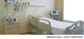
A hospitalist-led project to improve bed assignment practices at Baystate Medical Center in Springfield, Mass., reduced errors in patient placements to 3.1% from 9.4%, according to an abstract presentation at HM11.
The project identified incorrect placement of patients in open beds due to incomplete understanding of the patient’s medical picture, explains lead author Christine Bryson, DO, SFHM, Baystate’s associate medical director for hospital medicine. For example, a patient with a diagnosis of pneumonia who was receiving peritoneal dialysis might be admitted to the respiratory unit, but then would need transfer to the renal unit, where the dialysis could be performed. Such incorrect bed placements and lateral transfers were happening eight times a day, at a cost conservatively estimated at $106 each for nursing, a nonphysician patient placement manager (PPM), and housekeeping services and supplies. That puts potential annual cost savings is $232,000, Dr. Bryson explains.
A committee led by Baystate hospitalists examined current admission processes in detail and recommended a new process: ED physicians confer with the PPM, the PPM reviews the chart and discusses the case with the admitting hospitalist, and then the PPM and hospitalist have an informed, three-way phone conversation about placement.
Hospitalists have been directed to return these calls within 15 minutes, which can be an issue all its own. Another identified barrier was the communications technology, so ED physicians have been issued cellphones so they don’t have to wait at a terminal for a callback from the hospitalist. Dr. Bryson says overall booking process time fell, as did the number of placement errors.
Pediatric Hospitalists Share Lessons Learned on the Path to Executive Leadership

Pediatric hospitalist Jeff Sperring, MD, says he did not go into medicine with aspirations of becoming a hospital administrator. Last November, however, he was named president and CEO of Riley Hospital for Children at Indiana University Health in Indianapolis. It’s a path into healthcare leadership, he believes, that other pediatric hospitalists can and will follow.
“Being a hospitalist was critical to that progression. You are there; you understand what needs to be changed. More than anything else, it’s just being available, willing, and able to help,” Dr. Sperring says. “You lead one project, that leads to additional roles, and that leads to this.”
Dr. Sperring is one of a handful of pediatric hospitalists who have joined the C-suite and assumed major administrative responsibilities in their hospitals. Most say their HM experience was crucial to the journey.
Another pediatric hospitalist, Patrick Conway, MD, MSc, SFHM, earlier this year was named chief medical officer for the Centers for Medicare & Medicaid Services (see “Pediatric Hospitalist Takes CMS Leadership Position,” June 2011, p. 28), and is responsible for administering federal healthcare quality initiatives and setting the government’s quality agenda. Dr. Conway, previously director of hospital medicine at Cincinnati Children’s Hospital Medical Center, says that pediatric HM, in particular, lines up with major priorities in healthcare reform—most notably patient-centered care.
“Pediatricians often have strong communication skills honed by taking care of patients and their families,” Dr. Conway says. “Our training typically emphasizes team-based care and improving the health system.”
The path to hospital leadership might be a little different from the pediatric side. But he urges pediatric hospitalists to look for opportunities beyond pediatrics, within the larger healthcare system and the care of adult patients.
“I am an example of the potential for pediatric hospitalists to take on broader leadership roles,” Dr. Conway says. “I encourage medical students to consider pediatric hospital medicine, with its opportunities for leading change and taking care of patients at the same time.”
Change Agents
Leaders on the path to such C-suite positions as chief executive office (CEO), chief operating officer (COO), chief medical officer (CMO), or chief quality officer (CQO) stress the importance of finding mentors, both within and outside of the hospital, and creating effective teams in which to work. Whether a degree in business or a related field is an essential part of that journey is debatable. Dr. Sperring, for example, did not pursue formal business training, instead concentrating on leadership development. He took a one-year, part-time, multidisciplinary course on the subject offered by Indiana University. “To me, this is about understanding healthcare, how it is delivered, and then having the leadership skills to be able to make change,” he explains.
HM, with its bird’s-eye view of hospital processes and systems, is a good place to start, adds Paul Hain, MD, associate chief of staff and medical director for performance management and improvement at Monroe Carell Jr. Children’s Hospital at Vanderbilt in Nashville, Tenn. “I also think you have to understand quality improvement and be willing to measure, measure, measure.”
But advancing up the hospital’s organization chart requires something more, he notes. “A leader also needs to have a world view that things that are broken need to be fixed,” he says.
Dr. Hain studied engineering in college and worked as an engineer before attending medical school. That experience, he says, laid the foundation for “thinking about processes in healthcare systems, and the use of statistics to help understand those processes.”

—Jeff Sperring, MD, hospitalist, president, CEO, Riley Hospital for Children at Indiana University Health, Indianapolis
Spearhead QI
For Dr. Sperring, advancement to the C-suite was a journey that began nine years ago, following four years in community-based practice. “I absolutely loved the relationships with my kids and families, but I missed the acute-care role,” he says.
In 2002, Riley Hospital recruited him to help start its pediatric hospitalist program. As the program grew to include 22 hospitalists at four affiliated hospitals, his responsibilities also grew to associate chief medical officer in 2007 and chief medical officer in 2009. Along the way, he worked on partnering with pediatricians in the community, spearheaded a quality program that successfully reduced length of stay in the hospital, and developed an integrated call center for hospital admissions across the health system.
By contrast, Steve Narang, MD, CMO of Banner Health System’s pediatric services and its new Cardon Children’s Medical Center in Mesa, Ariz., says he always had one eye on healthcare system and policy issues, even during residency.
“What clearly became the center of my work is the value equation,” he says. “I wanted to be in a career where I could impact on delivering and disseminating best practices in medical care. I wanted to find out what are the best approaches for taking care of patients.”
After residency, Dr. Narang moved to New Orleans in 2000, where he started an academic pediatric hospitalist program at Louisiana State University Medical Center. He later helped launch a firm called Pediatric Hospitalists Louisiana, which collaborated with hospitals across the state to improve pediatric care delivery. “That got me thinking about things more from the management perspective, how to fix gaps in the system and advance our ability to measure quality in pediatric hospital medicine,” he says.
“When you take your first job in the hospital and you start trying to define and design best practices, people look at you differently—not as a young, emerging physician but more as a physician leader. They come to you and say: ‘Will you chair this committee, or lead that effort?’” he says. “And then, suddenly, you run out of tools in your toolbox. That’s what happened to me.”
He enrolled at Harvard University in pursuit of a business degree, along the way learning new ways of looking at systems change and basic principles of financing.
Retain a Clinical Presence
“The great thing about being a hospitalist is that you’re at the intersection of everything that happens in the hospital,” Dr. Narang says. As the pediatric chief medical officer for Banner Health, he is responsible for strategic planning, quality improvement (QI), and patient safety for a 210-bed hospital. He also co-chairs the Clinical Consensus Group, which represents all of Banner’s 23 hospitals, where he is able to influence care processes at the other hospitals as well.
Many hospitalist leaders eventually confront the dilemma of whether growing administrative responsibilities stand in the way of a continuing clinical practice. Dr. Narang moonlights some evenings and weekends on hospitalist and emergency medicine shifts. However, despite still wanting to see patients, he wonders if he has reached the point where growing administrative responsibilities will make that impossible.
“It was a challenge when I became CMO to squeeze in clinical responsibilities,” Dr. Narang says. “But I believed that in order to be the right kind of CMO, I still needed to practice medicine … to know what’s happening on the floor and what still needs to be fixed. You also want your colleagues to see you as a credible physician.”
He hopes to maintain some clinical practice, and says hospitalists have the advantage of blocking out scheduled times on service.
Dr. Sperring says it is “an exciting time” to be a hospitalist. “The way we’re used to defining care is going to change dramatically. Hospitalists will play a key role, both in direct care delivery but also in leadership,” he says. “I don’t think hospitalists have a choice but to lead change. It becomes part of our value proposition and a competency for all hospitalists.”
Dr. Hain often is asked by other hospitalists how to get started with quality initiatives that might lead to something more. “I always say the first one is free, in order to show that you can solve a quality problem while being a full-time clinician,” he explains. “It says to administrators that you’re someone who can deliver, and that starts you on your way. There’s always something to be done to improve quality in the hospital.”
Larry Beresford is a freelance writer based in Oakland, Calif.
Pediatric hospitalist Jeff Sperring, MD, says he did not go into medicine with aspirations of becoming a hospital administrator. Last November, however, he was named president and CEO of Riley Hospital for Children at Indiana University Health in Indianapolis. It’s a path into healthcare leadership, he believes, that other pediatric hospitalists can and will follow.
“Being a hospitalist was critical to that progression. You are there; you understand what needs to be changed. More than anything else, it’s just being available, willing, and able to help,” Dr. Sperring says. “You lead one project, that leads to additional roles, and that leads to this.”
Dr. Sperring is one of a handful of pediatric hospitalists who have joined the C-suite and assumed major administrative responsibilities in their hospitals. Most say their HM experience was crucial to the journey.
Another pediatric hospitalist, Patrick Conway, MD, MSc, SFHM, earlier this year was named chief medical officer for the Centers for Medicare & Medicaid Services (see “Pediatric Hospitalist Takes CMS Leadership Position,” June 2011, p. 28), and is responsible for administering federal healthcare quality initiatives and setting the government’s quality agenda. Dr. Conway, previously director of hospital medicine at Cincinnati Children’s Hospital Medical Center, says that pediatric HM, in particular, lines up with major priorities in healthcare reform—most notably patient-centered care.
“Pediatricians often have strong communication skills honed by taking care of patients and their families,” Dr. Conway says. “Our training typically emphasizes team-based care and improving the health system.”
The path to hospital leadership might be a little different from the pediatric side. But he urges pediatric hospitalists to look for opportunities beyond pediatrics, within the larger healthcare system and the care of adult patients.
“I am an example of the potential for pediatric hospitalists to take on broader leadership roles,” Dr. Conway says. “I encourage medical students to consider pediatric hospital medicine, with its opportunities for leading change and taking care of patients at the same time.”
Change Agents
Leaders on the path to such C-suite positions as chief executive office (CEO), chief operating officer (COO), chief medical officer (CMO), or chief quality officer (CQO) stress the importance of finding mentors, both within and outside of the hospital, and creating effective teams in which to work. Whether a degree in business or a related field is an essential part of that journey is debatable. Dr. Sperring, for example, did not pursue formal business training, instead concentrating on leadership development. He took a one-year, part-time, multidisciplinary course on the subject offered by Indiana University. “To me, this is about understanding healthcare, how it is delivered, and then having the leadership skills to be able to make change,” he explains.
HM, with its bird’s-eye view of hospital processes and systems, is a good place to start, adds Paul Hain, MD, associate chief of staff and medical director for performance management and improvement at Monroe Carell Jr. Children’s Hospital at Vanderbilt in Nashville, Tenn. “I also think you have to understand quality improvement and be willing to measure, measure, measure.”
But advancing up the hospital’s organization chart requires something more, he notes. “A leader also needs to have a world view that things that are broken need to be fixed,” he says.
Dr. Hain studied engineering in college and worked as an engineer before attending medical school. That experience, he says, laid the foundation for “thinking about processes in healthcare systems, and the use of statistics to help understand those processes.”
—Jeff Sperring, MD, hospitalist, president, CEO, Riley Hospital for Children at Indiana University Health, Indianapolis
Spearhead QI
For Dr. Sperring, advancement to the C-suite was a journey that began nine years ago, following four years in community-based practice. “I absolutely loved the relationships with my kids and families, but I missed the acute-care role,” he says.
In 2002, Riley Hospital recruited him to help start its pediatric hospitalist program. As the program grew to include 22 hospitalists at four affiliated hospitals, his responsibilities also grew to associate chief medical officer in 2007 and chief medical officer in 2009. Along the way, he worked on partnering with pediatricians in the community, spearheaded a quality program that successfully reduced length of stay in the hospital, and developed an integrated call center for hospital admissions across the health system.
By contrast, Steve Narang, MD, CMO of Banner Health System’s pediatric services and its new Cardon Children’s Medical Center in Mesa, Ariz., says he always had one eye on healthcare system and policy issues, even during residency.
“What clearly became the center of my work is the value equation,” he says. “I wanted to be in a career where I could impact on delivering and disseminating best practices in medical care. I wanted to find out what are the best approaches for taking care of patients.”
After residency, Dr. Narang moved to New Orleans in 2000, where he started an academic pediatric hospitalist program at Louisiana State University Medical Center. He later helped launch a firm called Pediatric Hospitalists Louisiana, which collaborated with hospitals across the state to improve pediatric care delivery. “That got me thinking about things more from the management perspective, how to fix gaps in the system and advance our ability to measure quality in pediatric hospital medicine,” he says.
“When you take your first job in the hospital and you start trying to define and design best practices, people look at you differently—not as a young, emerging physician but more as a physician leader. They come to you and say: ‘Will you chair this committee, or lead that effort?’” he says. “And then, suddenly, you run out of tools in your toolbox. That’s what happened to me.”
He enrolled at Harvard University in pursuit of a business degree, along the way learning new ways of looking at systems change and basic principles of financing.
Retain a Clinical Presence
“The great thing about being a hospitalist is that you’re at the intersection of everything that happens in the hospital,” Dr. Narang says. As the pediatric chief medical officer for Banner Health, he is responsible for strategic planning, quality improvement (QI), and patient safety for a 210-bed hospital. He also co-chairs the Clinical Consensus Group, which represents all of Banner’s 23 hospitals, where he is able to influence care processes at the other hospitals as well.
Many hospitalist leaders eventually confront the dilemma of whether growing administrative responsibilities stand in the way of a continuing clinical practice. Dr. Narang moonlights some evenings and weekends on hospitalist and emergency medicine shifts. However, despite still wanting to see patients, he wonders if he has reached the point where growing administrative responsibilities will make that impossible.
“It was a challenge when I became CMO to squeeze in clinical responsibilities,” Dr. Narang says. “But I believed that in order to be the right kind of CMO, I still needed to practice medicine … to know what’s happening on the floor and what still needs to be fixed. You also want your colleagues to see you as a credible physician.”
He hopes to maintain some clinical practice, and says hospitalists have the advantage of blocking out scheduled times on service.
Dr. Sperring says it is “an exciting time” to be a hospitalist. “The way we’re used to defining care is going to change dramatically. Hospitalists will play a key role, both in direct care delivery but also in leadership,” he says. “I don’t think hospitalists have a choice but to lead change. It becomes part of our value proposition and a competency for all hospitalists.”
Dr. Hain often is asked by other hospitalists how to get started with quality initiatives that might lead to something more. “I always say the first one is free, in order to show that you can solve a quality problem while being a full-time clinician,” he explains. “It says to administrators that you’re someone who can deliver, and that starts you on your way. There’s always something to be done to improve quality in the hospital.”
Larry Beresford is a freelance writer based in Oakland, Calif.
Massachusetts Healthcare Law Highlights Implications for National Healthcare Reform
Next month marks the sixth anniversary of former Massachusetts governor Mitt Romney’s signing into law a health insurance reform bill that brought near-universal coverage to the state’s residents. The Massachusetts experience represents a microcosm of what might be expected on a national scale with the Affordable Care Act (ACA): success in covering the uninsured, but persistent access and cost challenges that can only be overcome with fundamental payment reform.
“There is more going on in Massachusetts than anywhere else in the country, by far—in terms of coverage, delivery, and finance reform of healthcare,” says Stuart H. Altman, PhD, professor of national health policy at Brandeis University in Waltham, Mass. “The state’s example sends the positive message that the healthcare delivery system can be improved, but it takes time. Massachusetts is ahead of most states.”
Positives & Negatives
The Massachusetts law expands Medicaid enrollment to those earning up to 300% of the federal poverty level; offers state-subsidized commercial health insurance coverage to all other uninsured citizens; and allows young adults to remain on a parent’s plan until age 25. The law also mandates that employers with more than 10 employees offer subsidized health insurance coverage, and that every state resident over 18 purchase coverage or face tax penalties.

—Stuart H. Altman, PhD, professor of national health policy, Brandeis University, Waltham, Mass.
The law has brought coverage to nearly 412,000 previously uninsured residents (as of December 2010, the latest figures available), and less than 2% of residents remain uninsured—down from about 10% before the law was enacted.1
The law has not constrained the cost of healthcare in Massachusetts, which remains among the most expensive in the nation, and which current Massachusetts Gov. Deval Patrick acknowledges is continuing to rise at an unsustainable rate. A bill before the state legislature would give the governor the authority to review reimbursement contracts, to determine whether the fees paid by insurers to providers are appropriate, before approving insurance premium rates.
More surprising are the serious healthcare access challenges that persist in the state despite nearly universal health insurance coverage. The Massachusetts Medical Society outlined these challenges in a December 2011 white paper (see “Access Problems Persist Despite Insurance,” left).1
One of the biggest challenges the state faces is a dearth of primary-care physicians (PCPs). “One of the biggest lessons learned is that insurance expansion did not lead to better access,” observes Winthrop F. Whitcomb, MD, MHM, medical director of Healthcare Quality at Baystate Medical Center in Springfield, Mass., and co-founder of SHM. “Medicaid is a loss-leader and does not pay enough to cover the cost of running a medical practice. Expanding Medicaid may actually make access worse if primary-care physicians opt out of it.”
Insurance reform has not made it any easier for a hospitalist to find a PCP for a patient who comes to the hospital without one, Dr. Whitcomb says, or to discharge a patient to a long-term or post-acute-care setting that their insurance covers inadequately, if at all.
“We do continue to see ED visits and hospital admissions that would have been preventable had the patient seen a PCP first,” says SHM President Joseph Ming Wah Li, MD, SFHM, director of hospital medicine at Beth Israel Deaconess Medical Center in Boston.
Dr. Li says his group has experienced difficulties getting patients hooked up with timely and appropriate post-discharge follow-up care because of packed PCP schedules. In response, they developed a hospitalist-run, post-discharge clinic for outpatients to fill in the gap and provide their patients with the follow-up care they need.
“My hospitalists will manage the patient through their episode of care, regardless of whether they are an inpatient or outpatient, until their PCP is available,” Dr. Li says. “I would love to see our hospitalist-run, post-discharge clinic made obsolete by PCPs with available appointments.”
Fundamental Payment Reform
Access and cost problems like these have serious national implications, as Medicaid eligibility under the ACA is expected to grow by 16 million people by 2019, or roughly 25%. The Massachusetts experience suggests that decreasing financial barriers to care can raise other barriers, such as inadequate physician availability, and does nothing to address the 800-pound gorilla of spiraling costs.
Critics who dismiss the Massachusetts experiment as “doing nothing to control costs” miss the larger picture of innovation occurring in the state, however. Insurance reform was never intended to be the end of the story.
In response to a mandate to investigate reforming and restructuring the payment system as the next step in statewide healthcare reform, a Special Commission on the Health Care Payment System released recommendations in July 2009 (www.mass.gov/eohhs/docs/dhcfp/pc/final-report/final-report.pdf) that proposed that Massachusetts phase out fee-for-service reimbursement and replace it with an accountable-care approach that incorporated a global payment model combining elements of risk-adjusted capitation, pay-for-performance, evidence-based guidelines, and medical-home-style care coordination. Although a bill to accelerate statewide implementation of the model awaits a vote in the state legislature, the private health insurance market is well into the game.
Blue Cross Blue Shield of Massachusetts has been using a version of the model (known as the “alternative quality contract”) since 2009, with the goal of reducing healthcare cost growth by half over five years by holding providers accountable for cost and quality, and encouraging the most appropriate treatments by the right kind of providers in the most appropriate settings. Participating hospital and physician groups receive a monthly global fee for each patient (adjusted annually for patient health status and inflation) in return for providing them with all the preventive, primary, specialty, hospital, and follow-up care they need. Providers have the incentive to reduce inefficiencies, and they can earn additional incentive payments for meeting or exceeding clinical performance measures tied to process, outcomes, and patient care experience.
More than a third of the insurer’s provider network is participating in this alternative quality contract program, and early results are promising. A recent Harvard Medical School study found that medical spending at the end of the first year was nearly 2% lower among physicians and hospitals participating in the program compared with those working under traditional fee-for-service contracts, largely the result of physicians changing referral patterns and shifting care to lower-cost facilities.2 Quality of care among participants was significantly higher than that of non-participants in the insurer’s network, especially for adults with chronic illness and for children.
Several major healthcare delivery systems in Massachusetts are taking the accountable-care model to the next level this year by participating in the Pioneer Accountable Care Organizations (ACO) initiative, which also replaces fee-for-service with global revenue sharing plus quality and care-coordination incentives.
Part of the reason that providers in Massachusetts and around the country have a genuine interest in testing global payments and other value-based models is that they fear the day when the government and private sectors say “We just don’t have the money” and exert draconian fee-for-service rate control, Altman maintains.
Hospitalist Impacts
Hospitalists could find their referral patterns shifted slightly under global payment arrangements—potentially seeing fewer consults for low-risk patients and seeing greater demand for their services for more medically complex patients, Dr. Whitcomb says. HM likely will be the most heavily involved in ACOs that cover the Medicare population, whose patients are of higher acuity and more frequently hospitalized.
When fee-for-service reimbursement ultimately does give way to alternative reimbursement models, such as global payments, effective team-based care will become paramount to ensure effective hospital discharges and that preventable readmissions are minimized, Dr. Li says. He urges hospitalists to prepare their programs to manage a sicker patient population, and to cultivate the strongest possible coordination and alignment with PCPs, discharge planning professionals, and outpatient providers of all sorts. That way, hospitalists will be positioned to leverage their value in a healthcare system that requires value.
Christopher Guadagnino is a freelance medical writer based in Philadelphia.
For SHM’s official position on issues like healthcare reform, value-based purchasing, and medical errors, visit www.hospitalmedicine.org/advocacy
Next month marks the sixth anniversary of former Massachusetts governor Mitt Romney’s signing into law a health insurance reform bill that brought near-universal coverage to the state’s residents. The Massachusetts experience represents a microcosm of what might be expected on a national scale with the Affordable Care Act (ACA): success in covering the uninsured, but persistent access and cost challenges that can only be overcome with fundamental payment reform.
“There is more going on in Massachusetts than anywhere else in the country, by far—in terms of coverage, delivery, and finance reform of healthcare,” says Stuart H. Altman, PhD, professor of national health policy at Brandeis University in Waltham, Mass. “The state’s example sends the positive message that the healthcare delivery system can be improved, but it takes time. Massachusetts is ahead of most states.”
Positives & Negatives
The Massachusetts law expands Medicaid enrollment to those earning up to 300% of the federal poverty level; offers state-subsidized commercial health insurance coverage to all other uninsured citizens; and allows young adults to remain on a parent’s plan until age 25. The law also mandates that employers with more than 10 employees offer subsidized health insurance coverage, and that every state resident over 18 purchase coverage or face tax penalties.
—Stuart H. Altman, PhD, professor of national health policy, Brandeis University, Waltham, Mass.
The law has brought coverage to nearly 412,000 previously uninsured residents (as of December 2010, the latest figures available), and less than 2% of residents remain uninsured—down from about 10% before the law was enacted.1
The law has not constrained the cost of healthcare in Massachusetts, which remains among the most expensive in the nation, and which current Massachusetts Gov. Deval Patrick acknowledges is continuing to rise at an unsustainable rate. A bill before the state legislature would give the governor the authority to review reimbursement contracts, to determine whether the fees paid by insurers to providers are appropriate, before approving insurance premium rates.
More surprising are the serious healthcare access challenges that persist in the state despite nearly universal health insurance coverage. The Massachusetts Medical Society outlined these challenges in a December 2011 white paper (see “Access Problems Persist Despite Insurance,” left).1
One of the biggest challenges the state faces is a dearth of primary-care physicians (PCPs). “One of the biggest lessons learned is that insurance expansion did not lead to better access,” observes Winthrop F. Whitcomb, MD, MHM, medical director of Healthcare Quality at Baystate Medical Center in Springfield, Mass., and co-founder of SHM. “Medicaid is a loss-leader and does not pay enough to cover the cost of running a medical practice. Expanding Medicaid may actually make access worse if primary-care physicians opt out of it.”
Insurance reform has not made it any easier for a hospitalist to find a PCP for a patient who comes to the hospital without one, Dr. Whitcomb says, or to discharge a patient to a long-term or post-acute-care setting that their insurance covers inadequately, if at all.
“We do continue to see ED visits and hospital admissions that would have been preventable had the patient seen a PCP first,” says SHM President Joseph Ming Wah Li, MD, SFHM, director of hospital medicine at Beth Israel Deaconess Medical Center in Boston.
Dr. Li says his group has experienced difficulties getting patients hooked up with timely and appropriate post-discharge follow-up care because of packed PCP schedules. In response, they developed a hospitalist-run, post-discharge clinic for outpatients to fill in the gap and provide their patients with the follow-up care they need.
“My hospitalists will manage the patient through their episode of care, regardless of whether they are an inpatient or outpatient, until their PCP is available,” Dr. Li says. “I would love to see our hospitalist-run, post-discharge clinic made obsolete by PCPs with available appointments.”
Fundamental Payment Reform
Access and cost problems like these have serious national implications, as Medicaid eligibility under the ACA is expected to grow by 16 million people by 2019, or roughly 25%. The Massachusetts experience suggests that decreasing financial barriers to care can raise other barriers, such as inadequate physician availability, and does nothing to address the 800-pound gorilla of spiraling costs.
Critics who dismiss the Massachusetts experiment as “doing nothing to control costs” miss the larger picture of innovation occurring in the state, however. Insurance reform was never intended to be the end of the story.
In response to a mandate to investigate reforming and restructuring the payment system as the next step in statewide healthcare reform, a Special Commission on the Health Care Payment System released recommendations in July 2009 (www.mass.gov/eohhs/docs/dhcfp/pc/final-report/final-report.pdf) that proposed that Massachusetts phase out fee-for-service reimbursement and replace it with an accountable-care approach that incorporated a global payment model combining elements of risk-adjusted capitation, pay-for-performance, evidence-based guidelines, and medical-home-style care coordination. Although a bill to accelerate statewide implementation of the model awaits a vote in the state legislature, the private health insurance market is well into the game.
Blue Cross Blue Shield of Massachusetts has been using a version of the model (known as the “alternative quality contract”) since 2009, with the goal of reducing healthcare cost growth by half over five years by holding providers accountable for cost and quality, and encouraging the most appropriate treatments by the right kind of providers in the most appropriate settings. Participating hospital and physician groups receive a monthly global fee for each patient (adjusted annually for patient health status and inflation) in return for providing them with all the preventive, primary, specialty, hospital, and follow-up care they need. Providers have the incentive to reduce inefficiencies, and they can earn additional incentive payments for meeting or exceeding clinical performance measures tied to process, outcomes, and patient care experience.
More than a third of the insurer’s provider network is participating in this alternative quality contract program, and early results are promising. A recent Harvard Medical School study found that medical spending at the end of the first year was nearly 2% lower among physicians and hospitals participating in the program compared with those working under traditional fee-for-service contracts, largely the result of physicians changing referral patterns and shifting care to lower-cost facilities.2 Quality of care among participants was significantly higher than that of non-participants in the insurer’s network, especially for adults with chronic illness and for children.
Several major healthcare delivery systems in Massachusetts are taking the accountable-care model to the next level this year by participating in the Pioneer Accountable Care Organizations (ACO) initiative, which also replaces fee-for-service with global revenue sharing plus quality and care-coordination incentives.
Part of the reason that providers in Massachusetts and around the country have a genuine interest in testing global payments and other value-based models is that they fear the day when the government and private sectors say “We just don’t have the money” and exert draconian fee-for-service rate control, Altman maintains.
Hospitalist Impacts
Hospitalists could find their referral patterns shifted slightly under global payment arrangements—potentially seeing fewer consults for low-risk patients and seeing greater demand for their services for more medically complex patients, Dr. Whitcomb says. HM likely will be the most heavily involved in ACOs that cover the Medicare population, whose patients are of higher acuity and more frequently hospitalized.
When fee-for-service reimbursement ultimately does give way to alternative reimbursement models, such as global payments, effective team-based care will become paramount to ensure effective hospital discharges and that preventable readmissions are minimized, Dr. Li says. He urges hospitalists to prepare their programs to manage a sicker patient population, and to cultivate the strongest possible coordination and alignment with PCPs, discharge planning professionals, and outpatient providers of all sorts. That way, hospitalists will be positioned to leverage their value in a healthcare system that requires value.
Christopher Guadagnino is a freelance medical writer based in Philadelphia.
For SHM’s official position on issues like healthcare reform, value-based purchasing, and medical errors, visit www.hospitalmedicine.org/advocacy
Afghan-Born Hospitalist Gives Back Through Free Clinic

When Ahmad Nooristani, MD, became a physician, part of his motivation was to help his native country. “I wanted to become a physician so that I could give back,” says Dr. Nooristani, who emigrated to the U.S. from Afghanistan in 1981. He graduated medical school in 2008.
His opportunity to give back arrived in a different scenario, however. As a hospitalist with San Luis Hospitalists AMC, which provides coverage to Sierra Vista Regional Medical Center and French Hospital Medical Center in San Luis Obispo, Calif., Dr. Nooristani saw firsthand the downstream effects of uninsured patients without access to care for their chronic conditions.
One way to address preventive care for the uninsured, he reasoned, was to open a free clinic. In 2009, in addition to his seven-on/seven-off duties as a hospitalist, Dr. Nooristani began work on establishing a clinic for uninsured patients in his new hometown. Almost three years and countless fundraising events later, the Noor Foundation Clinic (www.slonoorfoundation.org) opened its doors in October 2011, offering not just primary care but ophthalmologic examinations, nutrition counseling, physical therapy, and point-of-service testing, too. For now, the clinic is open Friday and Saturday afternoons; all care is free.
Dr. Nooristani has worked 20 hours a week on the project. He’s recruited 400 volunteers, ranging from high-level administrators from the county’s hospitals to community fundraisers to off-duty nurses and physician colleagues.
“He’s a hard-working guy and is always looking for ways to improve things,” says hospitalist colleague Christian Voge, MD, medical director and president of San Luis Hospitalists. “I think he saw a need and is trying to give back.”
A Gap to Fill
Located on the central coast of California, San Luis Obispo County has a population of 269,637, according to 2010 U.S. Census figures. County public health officer Penny A. Borenstein, MD, MPH, says that figures from such surveys as the California Health Interview Survey and the Census Bureau indicate that approximately 35,000 of the county’s residents had no health insurance at some point in the last 12 months. The number of those who are underinsured (i.e. who carry minimal catastrophic insurance with high deductibles) is harder to quantify.
Although other clinic options exist in the county, through Medi-Cal and the County Medical Services Program, Dr. Borenstein believes that the Noor Foundation Clinic will help address gaps. “Even a sliding scale fee [such as those charged by community health centers] can sometimes be a deterrent to people,” she notes. “I give [Dr. Nooristani] many kudos for taking the bull by the horn and saying, ‘Let’s at least try to put something together to help fill the gaps in a very imperfect healthcare system.’”
New Skills Acquired
During an interview just days after the clinic opened its doors, Dr. Nooristani voiced some amazement about the long permitting process. “The hoops you have to jump through—it’s unbelievable,” he said.
Subject to federal, state, and county regulations, the clinic had to be retrofitted with a $25,000 air filtration and ducting system, among other upgrades. As a result, the foundation was paying rent for two years before the clinic opened its doors. “I could have seen a few thousand patients,” Dr. Nooristani says. “I mean, think about the complications I could have prevented.”
Still, he’s philosophical about the process. “On the flipside, I’m glad I did it this way. As tedious and time-consuming as it was, it served the purpose of bringing the whole community together,” he says.
Fundraising events for the foundation, as well as private donations, raised a total of $80,000 in a two-year period. Just before the clinic opened, the San Luis Obispo County Board of Supervisors approved a $75,000 grant to the Noor Foundation to cover the annual costs of point-of-service testing. And a broad swath of the county’s office holders, healthcare administrators, and community leaders attended the clinic’s grand opening.
—Christian Voge, MD, medical director and president, San Luis Hospitalists AMC
Geared to the Patient
Keeping in mind his patient population, Dr. Nooristani plans to incorporate patient education on managing chronic illnesses. An ophthalmologist has volunteered one day a month. A separate optometric examination room is outfitted with all the requisite equipment, and eyeglasses have been donated.
Furnished tastefully throughout, the clinic does not have the stark quality sometimes associated with free clinics. Dr. Nooristani also is respectful of patients’ time: “I don’t want anybody sitting in the waiting room for more than 30 minutes,” he says.
That’s why the appointment calendar is structured to accommodate future appointments, and he currently staffs each clinic day with two physicians and additional providers. He’s also savvy about his use of volunteers, limiting their hours to avoid burnout.
Catching Fire
Dr. Nooristani already has his sights set on more clinics, hopefully in his home country. In the meantime, though, he says, “people need care here.”
Like the meaning of the clinic name (“noor” means hope, and his name translates to “land of hope”), he hopes to inspire others to follow his lead. “Any community, big or small, can do this,” he says, enthusiasm in his voice. “You just have to keep your eyes on the prize.”
Gretchen Henkel is a freelance writer based in California.
SHM Boasts Diverse Membership, Leadership Lacks Non-Academic Presence

Who are you?
I am a 44-year-old Chinese-American male who works as a hospitalist at Beth Israel Deaconess Medical Center (BIDMC), an academic medical center in Boston. BIDMC is affiliated with Harvard Medical School, where I am an associate professor of medicine.
If you are about to join or renew your SHM membership, you can expect SHM to ask some questions it’s never asked before. What is your gender? What is your age? What is your ethnicity? I would not be surprised if you wondered why SHM is asking these questions. What does it have to do with my membership? Why are they asking now when they never asked before? I do not remember other professional medical societies asking these types of questions—should I be concerned? Is this an unnecessary invasion of privacy?
Call to Action
Nearly two years ago, when I had the good fortune of being elected SHM’s president-elect, I asked, What do we know about SHM members? As it turns out, it’s less than I thought we knew.
SHM has been in the survey business for years. The most visible survey is the annual productivity and compensation survey (see “Survey Insights,” p. TK). The data from this instrument have become very important, thanks to the many of you who have participated. In the early years of hospital medicine, everyone wanted to know how hard others were working and how much others were getting paid. If others had a better compensation package, it was the proof one needed to go marching into the C-suite asking for more support. If others were making less, it was the competitive advantage one needed to land the next hot-prospect hospitalist.
To be fair, I remember the surveys also asked about hospitalist age and employment model. But I don’t remember any questions about gender, race, or other personal information. The survey was the productivity and compensation survey, so maybe it had nothing to do with gender and race … but maybe it should.
Diverse, Yet Not So Much
Over the years, when I’ve walked the hallways at the SHM annual meeting, I got the sense that it was a reasonably diverse crowd. Take a look when you are at the San Diego Convention Center in April, and I expect you will agree. I grant you that it is generally a younger crowd than what one would find at most medical meetings, but I see people of many ethnic backgrounds, and there are equal parts women and men.
What was striking to me, however, was when I walked into some of the smaller conference rooms where the SHM committee meetings were being held and where the leaders sat. That crowd didn’t seem nearly as diverse as the crowd in the bigger rooms. I remember asking one of my colleagues whether he had the same perception. He told me he didn’t see it that way. Then again, it dawned on me that he is white and works at an academic medical center. What if he walked into leadership committee meetings filled with women from under-represented minority groups who work in community hospitals? My guess is that he would notice right away.
But my perception is biased, so when I became SHM president last spring, I asked that we assemble some facts about our members. SHM pulled together a task force, which developed a survey and took a snapshot of SHM membership. Some of you may have received this survey; it was sent out to thousands of SHM members.
The survey results, which were shared recently with SHM’s board of directors, confirmed my suspicions: SHM membership is a reasonably diverse crowd when it comes to gender and race. When it came to the SHM committee membership, I was right and wrong. The percentage of women and under-represented minorities on SHM committees reflected overall SHM membership reasonably well, but it was clear that fewer women and under-represented minorities held senior leadership positions, such as committee chairs and positions on the board. I suspect this is no different at other professional medical societies and more of a commentary on medicine than on SHM.
The most striking difference, however, did not have to do with race or gender, but instead had to do with employment model. Hospitalists who work at places other than academic medical centers are clearly under-represented in SHM leadership positions.
Action Item: New Knowledge, Better Understanding
So what do we do with this information? Am I suggesting that we set aside special seats at the board table for specific types of people or special-interest groups, some seats just for women, and some just for hospitalists who work in community hospitals? I am not advocating any such action.
I did ask SHM leadership to initiate action to help us continually understand the makeup of SHM membership and compare it to representation at the leadership level. SHM leadership overwhelmingly agreed. This is the reason you are being asked to volunteer personal information when you renew your membership.
It is my hope and belief that SHM will use this information appropriately when they organize committees and build leadership teams. This information, if used appropriately, will help SHM leadership understand its potential bias and guard against unintended consequences of their actions.
I recognize that some people will argue that the questions being asked are not sufficiently comprehensive. We should also be asking about other individual characteristics. You may or may not be right, but at this time, I think we are taking a step in the right direction. Further steps may be forthcoming in the future, but let’s not let perfection be the enemy of good.
If you have any comments about this article, please contact me at JosephLi@HospitalMedicine.org. I’m also available on LinkedIn (JosephLi) and Twitter (@_JosephLi).
Dr. Li is president of SHM.
How to Get the Most Out of the HM12 Toolkit

I have a problem. OK, many problems. Marital discord, balky kids, bloated mortgage? No, fortunately, not those kinds of domestic problems—although I do struggle with reliably differentiating whites from darks. My biggest problem is work-related. And this isn’t new. Turns out, I have different problems at work every year. “Time to find a new job,” you say. Tell the boss to shove it? Produce an epic, Jerry Maguire-esque manifesto and ride off into the sunset with my goldfish and Renee Zellweger? Hmmm, Renee Zellweger…
No, no, that’s not it. Much more mundane, yet crucial, problems.
Problems like trying to sort out the implications of the impending value-based purchasing program—what does it mean for my group? How do I keep my hospitalist partners engaged, satisfied, and not burned out? How do I produce a schedule that emphasizes high-quality patient care, efficiency, and physician work-life balance? How can I reduce readmissions so my hospital administrator can go back to “administrating” someone other than me all day? What do I do with the perioperative beta blockade now that some of the original data have been called into question due to academic dishonesty? What does the Affordable Care Act really say, is it going stand up, and what does it mean for me, my patients, my salary, and my career?
These are all questions I am grappling with currently. They also are all questions that will be addressed at HM12, April 1-4 in San Diego. As such, I view the annual meeting as a kind of toolkit: Have a problem, reach into the HM12 toolkit, and pull out your solution. The beauty is its breadth. You might not care one iota about healthcare reform, scheduling, or group satisfaction. Fine: How about updates in new medications, management of hyponatremia, the unique challenges of women in medicine, managing acute ventilator issues, acute pain management, information technology, quality improvement, professionalism in the digital age, or listening to the latest in the management of Clostridium difficile from the world-renowned Dr. John Bartlett? All are tools in this year’s toolkit.
And this type of breadth means the annual meeting evolves with you. Early in my career, I reached for the clinical tools. Then it was practice development and management tools; now I tend to look for healthcare policy solutions. Suffice to say, whatever solution you are looking for, with nine tracks, eight pathways, seven pre-courses, three plenary sessions by healthcare luminaries, and two Research, Innovation, and Vignette sessions, HM12 has your tool.
How can you best access this trove of information? Here is some advice culled from my 10 years of attending SHM annual meetings.
They Won’t Leave A Light On For You Forever
Unless you’re Tom Bodett, I’d recommend you get a hotel room now. For my first annual meeting (which was also in San Diego), I registered late, found no hotel rooms in the city, and had to commute 30 minutes both ways. Not only is this inconvenient and costly (I had to rent a car), but it also takes you out of the action. You want to be on-site, especially after meeting hours, when a lot of the networking and fun happens.
Stay Out Late
OK, now that you have a room, don’t use it. Rooms are for sleeping. If you find yourself in your room not sleeping, then you are missing out on some of the richest aspects of the conference—meeting new people, catching up with colleagues you see only once a year, and bathing in the general excitement of being at a meeting with thousands of peers. This remains the most satisfying part of the annual meeting experience for me. It’s dinner with a colleague from another part of the country, coffee with a new acquaintance, or a drink with an old friend. It’s energizing, engaging, and reignites my passion for HM.
Sleep Is For Vacation
Staying with the hotel room theme, don’t sleep in. I realize San Diego in April can feel like a vacation, and truth be told, it should. However, you came to learn. It’s tempting to maximize pillow time instead of heading down to the first plenary session at 8 a.m.—after all, you stayed out late networking! Anyway, how interesting can it be? Very. Dr. Patrick Conway is going to lead off the meeting with a look at the implications of the Affordable Care Act for hospitalists. As a hospitalist and CMO of CMS, he should know. Come to this session, and so will you.
Declare a Major and a Minor
Remember college? Me, neither. But I do have a vague recollection of that kid-in-a-candy-store feeling of choice my freshman year. The rest is a blur of late nights, hungover Sundays, and weight gain. Just like the college course book, the HM12 agenda can be overwhelming. Choice is great, but how do you choose what to go to? Just like college, you need a plan. Spend time before the meeting charting your course. What do you want to learn? What knowledge gaps do you want to fill? Throw in something for fun. Peruse the website, print out or download the slide decks from the talks you are interested in, and have a plan to maximize your time in San Diego.
Divide and Conquer
Next, make a plan with your friends. Most attendees have at least one other group member attending the meeting. Don’t go to the same sessions. Why? You should share your findings with the rest of your group.
You’ll no doubt pick up a new method for patient handoffs, moving patients through the hospital more efficiently, creating an incentive plan, or developing a post-discharge clinic. Bring it home; share it; implement it.
Go to the RIV Sessions
“But wait,” you say, “I’m not a researcher.” Perhaps true, but you are a hospitalist. And this is the material that is coming down the pike. It’s the cool case you’ll encounter next month, the innovation that’ll help your patients avoid hospital infections, or the research that will inform the next VTE prophylaxis guideline.
Go Viral
Bring your business cards. And like a rhinovirus, give them to everyone. Entranced person next to you at the plenary? Card. New face at the Special Interest Forum for rural hospitalists? Card. Erudite-appearing character scanning the poster abstract on readmissions with you? Card. Bagel-versus-English-muffin-debating person in the breakfast line? Card.
The point is, don’t be shy. You are there to be part of the hospitalist movement—to learn, to share, to be part of the discussion, to help define our collective future. Do that. This isn’t the time to be a wallflower. Rather, say “hi” to the person next to you. Strike up a conversation; you never know where it may lead.
You Had Me At “Hello”
So tell your boss to “show me the money,” so that you, too, can utilize the HM12 toolkit. If he or she balks, tell them to “help me help you.” Because after attending the meeting, I’m confident that with a tear in your eye, you’ll sappily utter, “HM12, you complete me.”
Dr. Glasheen is physician editor of The Hospitalist.
John Nelson: ED Patient Throughput Is New Core Measure

To understand one reason why your hospital cares so much about patient throughput and discharging patients before noon, think of tables in a restaurant. A restaurant has a limited number of tables, and the more quickly they serve customers, the sooner they’re able to seat a new party. So by improving their throughput, they can serve more customers and increase profitability without having to add tables.
Of course, the more a restaurant pushes to increase throughput, the more it risks a decrease in the satisfaction of its customers. Too much focus on throughput could annoy customers so much that it puts them out of business (Seinfeld’s Soup Nazi is one notable exception).
Yet hospitals are likely to increase their customers’ satisfaction by improving “front-end” throughput from the ED to the inpatient unit. In fact, CMS added two new core measures (known as inpatient quality reporting, or IQR) that hospitals began reporting on Jan. 1:
- Median time from ED arrival to ED departure for admitted patients, and
- Admit decision time to ED departure for admitted patients.
You already knew about these, right? Although they don’t directly influence Medicare payments to your hospital now, there is a pretty good chance they could become part of the Hospital Value-Based Purchasing program in the future. So expect administrative leaders at your hospital to continue pestering you about ED throughput with even greater intensity.
I confess that improving throughput feels like one of the most difficult things for me to do, and I sweat when in meetings about it. Yet I know intellectually that we can do better. After all, it wasn’t that long ago that a patient could expect to wait hours to see an ED doctor, and shortening the wait seemed an impossible task. But EDs everywhere have done just that, and now lots of them post the current ED wait time (typically less than 10 or 15 minutes!) on the Internet or billboards. So if they can do it, then I’m sure hospitalists can, too.
I wrote about hospital throughput in my April 2009 and May 2010 columns, and included some strategies you might want to try. Here are a few more directed at ED-to-inpatient ward throughput.
The One-Admitter Approach
Most practices have no option other than a single admitter at night, but if your one admitter during daytime hours reliably gets behind, then you should have “rounders” help with admitting duties. While this is stressful for the rounder, and could sometimes delay the rounder’s ability to discharge patients early (more on that later), in most cases, the benefit will be worth the cost. In fact, I think the best arrangement is for all, or nearly all, daytime hospitalists to share admissions every day.
Remember, this approach will eliminate the admitter-to-rounder handoff from Day One to Day Two, resulting in a net increase in efficiency (and probably patient satisfaction). Plus, reduction handoff errors should decrease, because only one doctor, rather than two, will have to get to know the new patient.
Eliminate Impediments
Some hospitalists with inefficient work habits have an opportunity to become more efficient at taking a history and reviewing records, along with other steps in the admission process, without risking poor quality. But most are reasonably efficient at those tasks already and shouldn’t make them the focus of throughput efforts.
That said, most, or nearly all, hospitalists could shorten the time from when the ED pages on a new admission to when we appear in the ED to see the patient.
Here is one scenario that gives rise to the type of delay I’m talking about. The ED doctor pages a triage hospitalist, and the two finally speak directly 10 minutes later. After a five-minute conversation, the triage doctor pages the hospitalist who will actually admit the patient, waits 10 minutes for the return call, relays the information (possibly leaving out or misrepresenting some of what the ED doc said), and the admitting hospitalist heads to the ED 15 minutes later. Each person in this scenario acted reasonably promptly, but the clock starts with the first ED page. The hospitalist is already at 45 minutes when they arrive to see the patient!
You need to minimize “daisy chain” communication between the ED doctor and the hospitalist who will actually see the patient. In the scenario above, I think it would be best to eliminate the triage doctor entirely (see my December 2008 column for ideas about how to do this) so that the hospitalist who will actually admit the patient learns of the new referral quickly and speaks directly with the ED doctor.
Another reason for a poor “page from ED to hospitalist at patient’s bedside” time is that the admitting hospitalist just doesn’t make it a priority to get to the ED quickly. They might decide to round on two or three more patients before working on the new admission. Of course, we must triage and sometimes delay getting to the ED while attending to an unstable floor patient, but not too often, in my experience.
I’m not suggesting that others adopt my habit: I typically don’t call the ED back at all. Instead, I just head to the ED immediately after being paged, and usually arrive in less time than I would have spent on hold waiting for the ED doc to come to the phone.
When presented with several ED patients to admit at the same time or tied up with a critical patient on the floor, getting to the ED quickly isn’t possible.
Write Admission or “Holding” Orders and Move the Patient to His/Her Room
This is frustrating for the admitting hospitalist, because among other problems, families often leave when the patient moves to the inpatient ward, and additional tests likely will take longer to complete from the floor rather than ED. And while it is hard to quantify, I think this practice increases the risk of errors—though some studies have shown that long ED stays are associated with worse outcomes for the patient, too.
One of the marquee principles of ED medicine has historically been “ED doctors should never write admission orders.” Even so, I sense that this view is shifting, and many now see this as clinically reasonable, and in some cases necessary, to ensure good performance. Our ED docs have agreed to do this if there is concern that the hospitalist can’t finish writing orders within an hour of first being paged about the patient. Ask me in a year and I’ll let you know just how this is going.
And, of course, another option is for the admitting hospitalist to provide admission orders or “holding” orders by phone. I think this is safe for many patients, as long as it is followed by a bedside admission visit by the hospitalist before too long.
Next month: What about throughput at the discharge end of the hospital stay? And yes, improving throughput risks quality of care, yet we have to do it while maintaining or improving quality.
Dr. Nelson has been a practicing hospitalist since 1988 and is co-founder and past president of SHM. He is a principal in Nelson Flores Hospital Medicine Consultants, a national hospitalist practice management consulting firm.
Incentives to Improve Hospital Readmission Rates OK

Iam a practicing hospitalist as well as a consultant to hospitals and health systems. One of my clients has a question: Is it legal to incentivize hospitalists to reduce readmission rates?
Alexander Strachan Jr., MD, MBA, CEO and managing director, CrossWalk Consulting Group LLC, Mission Viejo, Calif.
Dr. Hospitalist responds:
If you recall, a few months ago we discussed gainsharing and its attendant implications for physicians. The other side of that coin would be something like readmission rates. How so? Well, Medicare prohibits gainsharing (when hospitals share with physicians in the savings from improved service utilization), with the exception of two small, ongoing demonstration projects. For readmission rates, there are proposed penalties for hospitals, but not for physicians, so again there is no direct linkage of incentives.
Let’s take a look at our subject again: readmissions. Definitely in the news, as starting Oct. 1, 2012, hospitals can be penalized for exceeding the target readmission rate for the diagnoses of acute myocardial infarction, heart failure, and pneumonia. No surprise, but these are the same diagnoses that are part of the value-based purchasing payment methodology. These are both offshoots from the controversial Affordable Care Act of 2010.
The penalty, which is set at 1% for fiscal-year 2013 and escalates to a maximum of 3% for fiscal-year 2015, is based not just on the diagnosis-related group (DRG) payments for those specific conditions, but also for all DRG payments the hospital receives in that fiscal year. Understandably, hospitals are paying attention to this. Physicians, on the other hand, are not directly connected to the penalty. However, as a hospitalist, I imagine that you are either employed by the hospital or your group has a contract with the hospital—and the hospital is paying part of your salary. We aren’t exactly “independent professionals” these days.
As for the question at hand, then can a hospital incentivize a physician directly to reduce the readmission rate? Sure! Why not? Sounds easy enough.
However, it bears taking a closer look at how this might happen. Remember, Medicare frowns upon the potential denial of care or reduction in services, which is why gainsharing still hasn’t made its way forward. For example, a hospital could not pay a physician to turn away a potential readmission at the door. If the hospital wants to pay a bonus for reducing the readmit rate to 5% from 10%, great. If the hospital wants to pay a physician $500 for each potential readmission that is sent home from the ED, bad idea.
Similarly, the readmission rate is based on inpatient admissions, and patients who are admitted as observation technically are outpatients. So, if the hospital (employer) encouraged the inappropriate use of observation status, it would be a big no-no.
So what are the potential solutions? Well, as above, the hospital could construct a bonus based on the improvement (or maintenance) of a specific readmission rate, but it can’t dictate a process that might be interpreted as a denial of care. Alternately, it could pay for a process that might be expected to have a positive impact on the readmission rate. The hospital could require notification of pending discharges for the three “targeted” diagnoses, which would then allow for more resources to be directed to the patient prior to discharge. From the other end, the hospital could promote seeing those specific patients at risk for readmission earlier in their presentation (in the ED) to engage the hospitalists in management of the disease.
There are many ways to creatively design a solution, but the most important point is to avoid the incentive for the denial of care. Yes, Medicare wants you to improve the hospital’s readmission rate, but the preferred approach is to provide more resources to this population, not fewer.
Pediatric Hospitalists Climb the Corporate Ladder
Pediatric hospitalists around the country have made inroads into hospital administration roles. Here are some of the movers and shakers:
Erin Stucky Fisher, MD, FAAP, MHM, medical director for quality improvement at Rady Children’s Hospital, San Diego
Paul Hain, MD, associate chief of staff and medical director for performance management and improvement, Monroe Carell Jr. Children’s Hospital at Vanderbilt, Nashville, Tenn.
Sanford Meltzer, MD, MBA, senior vice president for strategic planning at Seattle Children’s Hospital
Stephen Muething, MD, vice president of safety at Cincinnati Children’s Hospital
Steve Narang, MD, chief medical officer of Banner Health System’s Cardon Children’s Medical Center, Mesa, Ariz.
Brian M. Pate, MD, FHM, FAAP, vice chair of inpatient services for the department of pediatrics at Children’s Mercy Hospitals and Clinics, Kansas City
Shannon Connor Phillips, MD, MPH, FAAP, quality and patient safety officer at the Cleveland Clinic
Jeff Sperring, MD, CEO, Riley Hospital for Children at Indiana University Health, Indianapolis, Ind.
Editor’s note: We, of course, don’t know of all the hospitalists around the country that have risen to C-suite positions. Let us know if we missed one; send Editor Jason Carris an email (jcarris@wiley.com) and we’ll add it to our monthly “Hospitalists on the Move” section.