User login
Study confirms increased adiposity in HSCT survivors
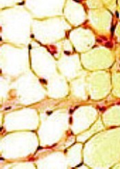
ORLANDO, FL—A recently conducted study confirms that survivors of hematopoietic stem cell transplant (HSCT) have increased body fat mass and lower lean mass compared to normal controls. And this is despite having a comparable body mass index (BMI).
Researchers say the abnormalities in adipokine levels—leptin and adiponectin—could provide insight into the mechanisms that contribute to the metabolic syndrome and cardiovascular complications that often develop in HSCT survivors.
Leptin and adiponectin are associated with obesity, insulin secretion, insulin resistance, endothelial function, vascular homeostasis, and atherosclerosis.
“So knowing that there is a dynamic interplay between obesity and insulin resistance and cytokine and adipokine profiles and, ultimately, insulin-resistance syndrome, we sought to evaluate, as part of a larger study, how treatment effects, including high-dose chemotherapy and radiation, alter cytokine profiles as well as obesity and body composition,” said Tyler G. Ketterl, MD, of the Fred Hutchinson Cancer Research Center in Seattle, Washington.
Dr Ketterl presented the findings at the 2017 BMT Tandem Meetings as abstract 52.*
Study design
The research team compared 151 HSCT recipients who had survived more than 2 years after transplant with 92 sibling controls.
HSCT survivors were randomly recruited from 2 centers—Fred Hutchinson Cancer Research Center and University of Minnesota Masonic Children’s Hospital—and were younger than 21 years when diagnosed.
The researchers evaluated all participants for body composition, cardiovascular risk factors, and adipokines using anthropomorphic measurements, DXA scans for muscle and fat mass, and laboratory bloodwork.
The team stratified the HSCT survivors by the preparative regimen they had received—total body irradiation (TBI) alone, TBI plus cranial radiation (CRT), and chemotherapy alone.
Study population
Males comprised more than half the study population in each arm, 58% of HSCT survivors and 54% of siblings.
Nine percent and 8% in the HSCT and sibling arms, respectively, were non-white and/or Hispanic, and the mean current ages were 24.0 (range, 10-51) for HSCT survivors and 24.2 (range, 10-48) for siblings.
The survivors’ mean age at diagnosis was 9.1 years (range, 0.4–20.6), their mean age at transplant was 11.2 years (range, 0.6–32.6), and the mean time from transplant to study participation was 13.5 years (range, 2.6–32).
Most patients received a transplant for leukemia—54 (36%) for acute myeloid leukemia, 46 (31%) for acute lymphoblastic leukemia, and 15 (10%) for chronic myeloid leukemia. Thirteen (9%) received transplants for myelodysplastic syndromes, 12 (8%) for Hodgkin lymphoma, and 10 (6%) for non-Hodgkin lymphoma.
A little more than half had TBI (85, 56%) as the preparative regimen, 31 (21%) had TBI plus CRT, and 35 (23%) had chemotherapy only.
About three-quarters (116, 77%) had an allogeneic transplant, and 35 (23%) had an autologous transplant.
Results
Overall, HSCT survivors had significantly lower adiponectin levels than siblings (P<0.001).
Survivors who received TBI with or without CRT had significantly lower adiponectin levels than siblings (P<0.001), while survivors who received chemotherapy alone did not (P=0.42).
Adiponectin is involved in insulin sensitization, hepatoprotective action, antiatherogenic action, protection against the development of diabetes, and regulation of lipid metabolism.
Overall, survivors had significantly higher leptin levels than siblings (P<0.001).
This held true regardless of conditioning regimen, although levels for patients who received chemotherapy only were not as significantly high (P=0.02) as for survivors who received TBI (P<0.001).
Leptin helps increase energy expenditure, decrease appetite and food uptake, modify insulin sensitivity on muscles and liver, prevent ectopic lipid deposition, and regulate immune function.
BMI adjusted for age, sex, and Tanner stage was not significantly different between survivors and siblings, but percent fat mass was significantly higher across all conditioning regimens for survivors compared to siblings (P<0.001).
“And this goes along with previous data,” Dr Ketterl said, “that shows sarcopenic obesity is common amongst transplant survivors.”
The researchers believe these significant differences may provide insight into the underlying risk of developing metabolic syndrome and cardiovascular complications in transplant survivors. 
*Some details in the abstract differ from the presentation.
ORLANDO, FL—A recently conducted study confirms that survivors of hematopoietic stem cell transplant (HSCT) have increased body fat mass and lower lean mass compared to normal controls. And this is despite having a comparable body mass index (BMI).
Researchers say the abnormalities in adipokine levels—leptin and adiponectin—could provide insight into the mechanisms that contribute to the metabolic syndrome and cardiovascular complications that often develop in HSCT survivors.
Leptin and adiponectin are associated with obesity, insulin secretion, insulin resistance, endothelial function, vascular homeostasis, and atherosclerosis.
“So knowing that there is a dynamic interplay between obesity and insulin resistance and cytokine and adipokine profiles and, ultimately, insulin-resistance syndrome, we sought to evaluate, as part of a larger study, how treatment effects, including high-dose chemotherapy and radiation, alter cytokine profiles as well as obesity and body composition,” said Tyler G. Ketterl, MD, of the Fred Hutchinson Cancer Research Center in Seattle, Washington.
Dr Ketterl presented the findings at the 2017 BMT Tandem Meetings as abstract 52.*
Study design
The research team compared 151 HSCT recipients who had survived more than 2 years after transplant with 92 sibling controls.
HSCT survivors were randomly recruited from 2 centers—Fred Hutchinson Cancer Research Center and University of Minnesota Masonic Children’s Hospital—and were younger than 21 years when diagnosed.
The researchers evaluated all participants for body composition, cardiovascular risk factors, and adipokines using anthropomorphic measurements, DXA scans for muscle and fat mass, and laboratory bloodwork.
The team stratified the HSCT survivors by the preparative regimen they had received—total body irradiation (TBI) alone, TBI plus cranial radiation (CRT), and chemotherapy alone.
Study population
Males comprised more than half the study population in each arm, 58% of HSCT survivors and 54% of siblings.
Nine percent and 8% in the HSCT and sibling arms, respectively, were non-white and/or Hispanic, and the mean current ages were 24.0 (range, 10-51) for HSCT survivors and 24.2 (range, 10-48) for siblings.
The survivors’ mean age at diagnosis was 9.1 years (range, 0.4–20.6), their mean age at transplant was 11.2 years (range, 0.6–32.6), and the mean time from transplant to study participation was 13.5 years (range, 2.6–32).
Most patients received a transplant for leukemia—54 (36%) for acute myeloid leukemia, 46 (31%) for acute lymphoblastic leukemia, and 15 (10%) for chronic myeloid leukemia. Thirteen (9%) received transplants for myelodysplastic syndromes, 12 (8%) for Hodgkin lymphoma, and 10 (6%) for non-Hodgkin lymphoma.
A little more than half had TBI (85, 56%) as the preparative regimen, 31 (21%) had TBI plus CRT, and 35 (23%) had chemotherapy only.
About three-quarters (116, 77%) had an allogeneic transplant, and 35 (23%) had an autologous transplant.
Results
Overall, HSCT survivors had significantly lower adiponectin levels than siblings (P<0.001).
Survivors who received TBI with or without CRT had significantly lower adiponectin levels than siblings (P<0.001), while survivors who received chemotherapy alone did not (P=0.42).
Adiponectin is involved in insulin sensitization, hepatoprotective action, antiatherogenic action, protection against the development of diabetes, and regulation of lipid metabolism.
Overall, survivors had significantly higher leptin levels than siblings (P<0.001).
This held true regardless of conditioning regimen, although levels for patients who received chemotherapy only were not as significantly high (P=0.02) as for survivors who received TBI (P<0.001).
Leptin helps increase energy expenditure, decrease appetite and food uptake, modify insulin sensitivity on muscles and liver, prevent ectopic lipid deposition, and regulate immune function.
BMI adjusted for age, sex, and Tanner stage was not significantly different between survivors and siblings, but percent fat mass was significantly higher across all conditioning regimens for survivors compared to siblings (P<0.001).
“And this goes along with previous data,” Dr Ketterl said, “that shows sarcopenic obesity is common amongst transplant survivors.”
The researchers believe these significant differences may provide insight into the underlying risk of developing metabolic syndrome and cardiovascular complications in transplant survivors.
*Some details in the abstract differ from the presentation.
ORLANDO, FL—A recently conducted study confirms that survivors of hematopoietic stem cell transplant (HSCT) have increased body fat mass and lower lean mass compared to normal controls. And this is despite having a comparable body mass index (BMI).
Researchers say the abnormalities in adipokine levels—leptin and adiponectin—could provide insight into the mechanisms that contribute to the metabolic syndrome and cardiovascular complications that often develop in HSCT survivors.
Leptin and adiponectin are associated with obesity, insulin secretion, insulin resistance, endothelial function, vascular homeostasis, and atherosclerosis.
“So knowing that there is a dynamic interplay between obesity and insulin resistance and cytokine and adipokine profiles and, ultimately, insulin-resistance syndrome, we sought to evaluate, as part of a larger study, how treatment effects, including high-dose chemotherapy and radiation, alter cytokine profiles as well as obesity and body composition,” said Tyler G. Ketterl, MD, of the Fred Hutchinson Cancer Research Center in Seattle, Washington.
Dr Ketterl presented the findings at the 2017 BMT Tandem Meetings as abstract 52.*
Study design
The research team compared 151 HSCT recipients who had survived more than 2 years after transplant with 92 sibling controls.
HSCT survivors were randomly recruited from 2 centers—Fred Hutchinson Cancer Research Center and University of Minnesota Masonic Children’s Hospital—and were younger than 21 years when diagnosed.
The researchers evaluated all participants for body composition, cardiovascular risk factors, and adipokines using anthropomorphic measurements, DXA scans for muscle and fat mass, and laboratory bloodwork.
The team stratified the HSCT survivors by the preparative regimen they had received—total body irradiation (TBI) alone, TBI plus cranial radiation (CRT), and chemotherapy alone.
Study population
Males comprised more than half the study population in each arm, 58% of HSCT survivors and 54% of siblings.
Nine percent and 8% in the HSCT and sibling arms, respectively, were non-white and/or Hispanic, and the mean current ages were 24.0 (range, 10-51) for HSCT survivors and 24.2 (range, 10-48) for siblings.
The survivors’ mean age at diagnosis was 9.1 years (range, 0.4–20.6), their mean age at transplant was 11.2 years (range, 0.6–32.6), and the mean time from transplant to study participation was 13.5 years (range, 2.6–32).
Most patients received a transplant for leukemia—54 (36%) for acute myeloid leukemia, 46 (31%) for acute lymphoblastic leukemia, and 15 (10%) for chronic myeloid leukemia. Thirteen (9%) received transplants for myelodysplastic syndromes, 12 (8%) for Hodgkin lymphoma, and 10 (6%) for non-Hodgkin lymphoma.
A little more than half had TBI (85, 56%) as the preparative regimen, 31 (21%) had TBI plus CRT, and 35 (23%) had chemotherapy only.
About three-quarters (116, 77%) had an allogeneic transplant, and 35 (23%) had an autologous transplant.
Results
Overall, HSCT survivors had significantly lower adiponectin levels than siblings (P<0.001).
Survivors who received TBI with or without CRT had significantly lower adiponectin levels than siblings (P<0.001), while survivors who received chemotherapy alone did not (P=0.42).
Adiponectin is involved in insulin sensitization, hepatoprotective action, antiatherogenic action, protection against the development of diabetes, and regulation of lipid metabolism.
Overall, survivors had significantly higher leptin levels than siblings (P<0.001).
This held true regardless of conditioning regimen, although levels for patients who received chemotherapy only were not as significantly high (P=0.02) as for survivors who received TBI (P<0.001).
Leptin helps increase energy expenditure, decrease appetite and food uptake, modify insulin sensitivity on muscles and liver, prevent ectopic lipid deposition, and regulate immune function.
BMI adjusted for age, sex, and Tanner stage was not significantly different between survivors and siblings, but percent fat mass was significantly higher across all conditioning regimens for survivors compared to siblings (P<0.001).
“And this goes along with previous data,” Dr Ketterl said, “that shows sarcopenic obesity is common amongst transplant survivors.”
The researchers believe these significant differences may provide insight into the underlying risk of developing metabolic syndrome and cardiovascular complications in transplant survivors.
*Some details in the abstract differ from the presentation.
VIDEO: Future therapies look promising for HER2 treatment-resistant breast cancer patients
MIAMI BEACH – Recent scientific discoveries about the myriad of ways a women with HER2 receptor–positive breast cancer can develop treatment resistance is spurring the development of some promising agents, Mark D. Pegram, MD, said at the annual Miami Breast Cancer Conference, held by Physicians’ Education Resource.
The antibody-drug conjugate T-DM1 holds significant potential, for example, because it seems to confer benefit regardless of a patients’ PIK3CA mutation status, said Dr. Pegram, director of the breast cancer oncology program at Stanford Women’s Cancer Center in California. Other antibody-drug conjugates and additional types of agents are showing enough promise overall that Dr. Pegram is predicting a bright future for improving treatment of this patient population.
Creating a number of new agents is a good thing, Dr. Pegram said in a video interview, because it’s unlikely any one therapy will work for everyone with HER2 treatment-resistant breast cancer. Therefore, precision medicine is expected to guide individual therapeutic choices in the future.
Dr. Pegram disclosed that he is a consultant for Genetech, Novartis, Oncothyreon, and Pfizer.
The video associated with this article is no longer available on this site. Please view all of our videos on the MDedge YouTube channel
MIAMI BEACH – Recent scientific discoveries about the myriad of ways a women with HER2 receptor–positive breast cancer can develop treatment resistance is spurring the development of some promising agents, Mark D. Pegram, MD, said at the annual Miami Breast Cancer Conference, held by Physicians’ Education Resource.
The antibody-drug conjugate T-DM1 holds significant potential, for example, because it seems to confer benefit regardless of a patients’ PIK3CA mutation status, said Dr. Pegram, director of the breast cancer oncology program at Stanford Women’s Cancer Center in California. Other antibody-drug conjugates and additional types of agents are showing enough promise overall that Dr. Pegram is predicting a bright future for improving treatment of this patient population.
Creating a number of new agents is a good thing, Dr. Pegram said in a video interview, because it’s unlikely any one therapy will work for everyone with HER2 treatment-resistant breast cancer. Therefore, precision medicine is expected to guide individual therapeutic choices in the future.
Dr. Pegram disclosed that he is a consultant for Genetech, Novartis, Oncothyreon, and Pfizer.
The video associated with this article is no longer available on this site. Please view all of our videos on the MDedge YouTube channel
MIAMI BEACH – Recent scientific discoveries about the myriad of ways a women with HER2 receptor–positive breast cancer can develop treatment resistance is spurring the development of some promising agents, Mark D. Pegram, MD, said at the annual Miami Breast Cancer Conference, held by Physicians’ Education Resource.
The antibody-drug conjugate T-DM1 holds significant potential, for example, because it seems to confer benefit regardless of a patients’ PIK3CA mutation status, said Dr. Pegram, director of the breast cancer oncology program at Stanford Women’s Cancer Center in California. Other antibody-drug conjugates and additional types of agents are showing enough promise overall that Dr. Pegram is predicting a bright future for improving treatment of this patient population.
Creating a number of new agents is a good thing, Dr. Pegram said in a video interview, because it’s unlikely any one therapy will work for everyone with HER2 treatment-resistant breast cancer. Therefore, precision medicine is expected to guide individual therapeutic choices in the future.
Dr. Pegram disclosed that he is a consultant for Genetech, Novartis, Oncothyreon, and Pfizer.
The video associated with this article is no longer available on this site. Please view all of our videos on the MDedge YouTube channel

VIDEO: Breast surgeons can and do provide genetic counseling to cancer patients
MIAMI BEACH – Breast surgeons can help relieve a “medical education crisis” by advising breast cancer patients about the implications of genetic testing results. The crisis continues to worsen given the remarkable rise in precision medicine and the limited number of genetic counselors in the United States, Patrick W. Whitworth, MD, said at the annual Miami Breast Cancer Conference, held by Physicians’ Education Resource.
Breast surgeons are qualified to help counsel patients, and, in fact, already order more than half of breast cancer genetic sequencing, a specialist survey reveals, said Dr. Whitworth, director of the Nashville (Tenn.) Breast Center, in a video interview.
At the same time, the one genotype–one phenotype paradigm for cancer genetics is rapidly shifting to testing patients for a whole panel of mutations simultaneously. Breast surgeons can partner with genetic counselors to support and expand the reach of this vital medical testing and counseling to more patients, he added.
Dr. Whitworth receives research grant support from and is a shareholder in Targeted Medical Education. He is also a shareholder in Advantage Consulting and Education.
The video associated with this article is no longer available on this site. Please view all of our videos on the MDedge YouTube channel
MIAMI BEACH – Breast surgeons can help relieve a “medical education crisis” by advising breast cancer patients about the implications of genetic testing results. The crisis continues to worsen given the remarkable rise in precision medicine and the limited number of genetic counselors in the United States, Patrick W. Whitworth, MD, said at the annual Miami Breast Cancer Conference, held by Physicians’ Education Resource.
Breast surgeons are qualified to help counsel patients, and, in fact, already order more than half of breast cancer genetic sequencing, a specialist survey reveals, said Dr. Whitworth, director of the Nashville (Tenn.) Breast Center, in a video interview.
At the same time, the one genotype–one phenotype paradigm for cancer genetics is rapidly shifting to testing patients for a whole panel of mutations simultaneously. Breast surgeons can partner with genetic counselors to support and expand the reach of this vital medical testing and counseling to more patients, he added.
Dr. Whitworth receives research grant support from and is a shareholder in Targeted Medical Education. He is also a shareholder in Advantage Consulting and Education.
The video associated with this article is no longer available on this site. Please view all of our videos on the MDedge YouTube channel
MIAMI BEACH – Breast surgeons can help relieve a “medical education crisis” by advising breast cancer patients about the implications of genetic testing results. The crisis continues to worsen given the remarkable rise in precision medicine and the limited number of genetic counselors in the United States, Patrick W. Whitworth, MD, said at the annual Miami Breast Cancer Conference, held by Physicians’ Education Resource.
Breast surgeons are qualified to help counsel patients, and, in fact, already order more than half of breast cancer genetic sequencing, a specialist survey reveals, said Dr. Whitworth, director of the Nashville (Tenn.) Breast Center, in a video interview.
At the same time, the one genotype–one phenotype paradigm for cancer genetics is rapidly shifting to testing patients for a whole panel of mutations simultaneously. Breast surgeons can partner with genetic counselors to support and expand the reach of this vital medical testing and counseling to more patients, he added.
Dr. Whitworth receives research grant support from and is a shareholder in Targeted Medical Education. He is also a shareholder in Advantage Consulting and Education.
The video associated with this article is no longer available on this site. Please view all of our videos on the MDedge YouTube channel
AT MBCC

VIDEO: Resistance to endocrine therapy a moving target
MIAMI BEACH – Hormonal ablation is a mainstay of therapy for women with hormone receptor–positive breast cancer. A significant proportion of patients, however, are either initially refractory to hormonal therapy or acquire resistance to it over time.
The difficulty for patients with breast cancer and for the physicians who treat them is that there are no simple answers to the question of which patients can continue to benefit from endocrine monotherapy. Are there adequate biomarkers for optimal follow-on therapy when a patient experiences disease progression, and what is the optimal sequence of targeted therapy with endocrine inhibitors, disrupters, or other agents?
In a video interview at the annual Miami Breast Cancer Conference, held by Physicians’ Education Resource, William J. Gradishar, MD, of Northwestern University, Chicago, discusses strategies for combating resistance to endocrine ablative therapy, and describes how new therapies and new treatment strategies are being incorporated into National Comprehensive Cancer Network breast cancer guidelines.
Dr. Gradishar reported having no clinical disclosures.
The video associated with this article is no longer available on this site. Please view all of our videos on the MDedge YouTube channel
MIAMI BEACH – Hormonal ablation is a mainstay of therapy for women with hormone receptor–positive breast cancer. A significant proportion of patients, however, are either initially refractory to hormonal therapy or acquire resistance to it over time.
The difficulty for patients with breast cancer and for the physicians who treat them is that there are no simple answers to the question of which patients can continue to benefit from endocrine monotherapy. Are there adequate biomarkers for optimal follow-on therapy when a patient experiences disease progression, and what is the optimal sequence of targeted therapy with endocrine inhibitors, disrupters, or other agents?
In a video interview at the annual Miami Breast Cancer Conference, held by Physicians’ Education Resource, William J. Gradishar, MD, of Northwestern University, Chicago, discusses strategies for combating resistance to endocrine ablative therapy, and describes how new therapies and new treatment strategies are being incorporated into National Comprehensive Cancer Network breast cancer guidelines.
Dr. Gradishar reported having no clinical disclosures.
The video associated with this article is no longer available on this site. Please view all of our videos on the MDedge YouTube channel
MIAMI BEACH – Hormonal ablation is a mainstay of therapy for women with hormone receptor–positive breast cancer. A significant proportion of patients, however, are either initially refractory to hormonal therapy or acquire resistance to it over time.
The difficulty for patients with breast cancer and for the physicians who treat them is that there are no simple answers to the question of which patients can continue to benefit from endocrine monotherapy. Are there adequate biomarkers for optimal follow-on therapy when a patient experiences disease progression, and what is the optimal sequence of targeted therapy with endocrine inhibitors, disrupters, or other agents?
In a video interview at the annual Miami Breast Cancer Conference, held by Physicians’ Education Resource, William J. Gradishar, MD, of Northwestern University, Chicago, discusses strategies for combating resistance to endocrine ablative therapy, and describes how new therapies and new treatment strategies are being incorporated into National Comprehensive Cancer Network breast cancer guidelines.
Dr. Gradishar reported having no clinical disclosures.
The video associated with this article is no longer available on this site. Please view all of our videos on the MDedge YouTube channel
AT MBCC

NetWorks: Mass shootings, MACRA, asthma in pregnancy Disaster Response Practice Operations Transplant Women’s Health
Mass shootings
There are multiple definitions for a mass shooting. Some definitions require a certain number of people be killed. Some definitions require a certain number of people be shot. Some definitions do not include gang violence. Regardless of the definition used, the number of mass shootings in the United States is increasing.
There are also multiple definitions of what qualifies as a medical disaster. These definitions can be summarized with the statement that a medical disaster is an event that produces a number of casualties that overwhelms the local health system.
Most mass shootings fit the definition of a medical disaster. When a mass shooting occurs, medical resources are diverted from current patients to those injured in the shooting. Patients with acute medical problems unrelated to the shooting must endure a prolonged wait for medical care.
The CHEST Disaster Response NetWork feels that it is necessary to take action to reduce the number of mass shootings. Unlike natural disasters, mass shootings are man-made. As such, we should proactively work to prevent them. Prevention is a large part of medicine. Working together with community leaders, law enforcement, and government officials, we can and should work to eliminate mass shootings so that we can minimize gun-related injury and death.
MACRA: Reincarnation of Medicare physician reimbursement model
In April 2015, President Obama signed the Medicare Access and CHIP Reauthorization Act (MACRA) eradicating the detested sustainable growth rate (SGR) formula. If this is your first dive into MACRA as an eligible professional (EP), it may be a bit baffling trying to understand its impact on your practice.
Under MIPS, rules are divided into four categories. During the first year, each category will make up a certain percentage to the physician’s overall score, which will result in a penalty or payment as a lump sum in 2019. If you are an Advanced APM in 2017 and receive 25% of Medicare payments or see 20% of your Medicare patients through this model, you can earn up to a 5% incentive payment in 2019.
The performance period started on January 1, 2017. Submission of performance data is due by March 31, 2018. MACRA is complicated and here to stay. Learn and educate yourself to avoid downward payment adjustment. For full details, please visit https://qpp.cms.gov/docs/QPP_Executive_Summary_of_Final_Rule.pdf.
References
1. CMS MACRA Proposed Rule. http://1.usa.gov/1PpBpMt
2. CMS MACRA Executive Summary. https://qpp.cms.gov/docs/QPP_Executive_Summary_of_Final_Rule.pdf
3. American Medical Association. http://bit.ly/1miEtBD
4. Policy and Medicine. MACRA http://bit.ly/1PTLkKa. MIPS http://bit.ly/20RoMzZ. APMs http://bit.ly/1OlxoxH
Frailty in lung transplantation
Two of the greatest challenges in lung transplantation are to identify optimal transplant candidates and to help those transplant recipients thrive in the years following surgery. Frailty is emerging as a marker of increased posttransplant morbidity and may represent an area where both the recipient selection process and posttransplant outcomes can be optimized. Described by some as “biologic age” rather than “chronologic age,” frailty is a syndrome of functional impairment and weakness that predisposes to adverse health outcomes. The adverse effects of frailty have been described in multiple clinical scenarios, including the ICU, chronic lung diseases, heart failure, liver transplant, kidney transplant, geriatrics, and others.

Remaining challenges include determining which clinical assessments best define frailty in the lung transplant population, documenting the adverse effects of frailty in well-designed multicenter prospective studies, and developing interventions to mitigate the adverse effects of frailty.
Asthma treatment during pregnancy
Asthma is common in pregnancy, occurring in 3% to 8% of pregnant women. While the course of asthma during pregnancy is variable, the objectives of asthma treatment do not change and aim to prevent acute exacerbations and optimize management. Uncontrolled asthma is associated with an increased risk of perinatal morbidity. Published guidelines on pharmacologic therapies during pregnancy recommend the same step-wise approach as in nonpregnant women.
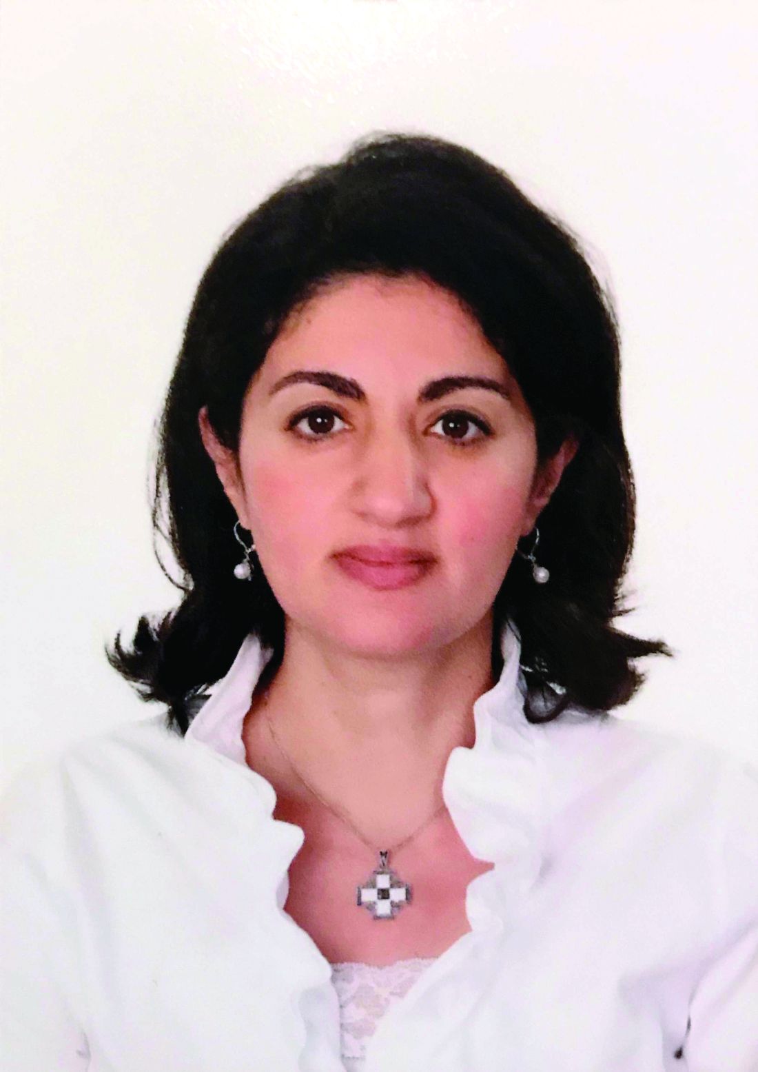
The diagnosis of asthma, the use of other concurrent medications, and medication compliance may all be potential confounders. ICS use in pregnancy was associated with endocrine and metabolic disturbances in the offspring in a national cohort (Tegethoff M et al. Am J Respir Crit Care Med. 2012;185[5]:557-63). However, this study did not report on systemic steroid use, asthma severity, or details of these disturbances.

Mass shootings
There are multiple definitions for a mass shooting. Some definitions require a certain number of people be killed. Some definitions require a certain number of people be shot. Some definitions do not include gang violence. Regardless of the definition used, the number of mass shootings in the United States is increasing.
There are also multiple definitions of what qualifies as a medical disaster. These definitions can be summarized with the statement that a medical disaster is an event that produces a number of casualties that overwhelms the local health system.
Most mass shootings fit the definition of a medical disaster. When a mass shooting occurs, medical resources are diverted from current patients to those injured in the shooting. Patients with acute medical problems unrelated to the shooting must endure a prolonged wait for medical care.
The CHEST Disaster Response NetWork feels that it is necessary to take action to reduce the number of mass shootings. Unlike natural disasters, mass shootings are man-made. As such, we should proactively work to prevent them. Prevention is a large part of medicine. Working together with community leaders, law enforcement, and government officials, we can and should work to eliminate mass shootings so that we can minimize gun-related injury and death.
MACRA: Reincarnation of Medicare physician reimbursement model
In April 2015, President Obama signed the Medicare Access and CHIP Reauthorization Act (MACRA) eradicating the detested sustainable growth rate (SGR) formula. If this is your first dive into MACRA as an eligible professional (EP), it may be a bit baffling trying to understand its impact on your practice.
Under MIPS, rules are divided into four categories. During the first year, each category will make up a certain percentage to the physician’s overall score, which will result in a penalty or payment as a lump sum in 2019. If you are an Advanced APM in 2017 and receive 25% of Medicare payments or see 20% of your Medicare patients through this model, you can earn up to a 5% incentive payment in 2019.
The performance period started on January 1, 2017. Submission of performance data is due by March 31, 2018. MACRA is complicated and here to stay. Learn and educate yourself to avoid downward payment adjustment. For full details, please visit https://qpp.cms.gov/docs/QPP_Executive_Summary_of_Final_Rule.pdf.
References
1. CMS MACRA Proposed Rule. http://1.usa.gov/1PpBpMt
2. CMS MACRA Executive Summary. https://qpp.cms.gov/docs/QPP_Executive_Summary_of_Final_Rule.pdf
3. American Medical Association. http://bit.ly/1miEtBD
4. Policy and Medicine. MACRA http://bit.ly/1PTLkKa. MIPS http://bit.ly/20RoMzZ. APMs http://bit.ly/1OlxoxH
Frailty in lung transplantation
Two of the greatest challenges in lung transplantation are to identify optimal transplant candidates and to help those transplant recipients thrive in the years following surgery. Frailty is emerging as a marker of increased posttransplant morbidity and may represent an area where both the recipient selection process and posttransplant outcomes can be optimized. Described by some as “biologic age” rather than “chronologic age,” frailty is a syndrome of functional impairment and weakness that predisposes to adverse health outcomes. The adverse effects of frailty have been described in multiple clinical scenarios, including the ICU, chronic lung diseases, heart failure, liver transplant, kidney transplant, geriatrics, and others.
Remaining challenges include determining which clinical assessments best define frailty in the lung transplant population, documenting the adverse effects of frailty in well-designed multicenter prospective studies, and developing interventions to mitigate the adverse effects of frailty.
Asthma treatment during pregnancy
Asthma is common in pregnancy, occurring in 3% to 8% of pregnant women. While the course of asthma during pregnancy is variable, the objectives of asthma treatment do not change and aim to prevent acute exacerbations and optimize management. Uncontrolled asthma is associated with an increased risk of perinatal morbidity. Published guidelines on pharmacologic therapies during pregnancy recommend the same step-wise approach as in nonpregnant women.
The diagnosis of asthma, the use of other concurrent medications, and medication compliance may all be potential confounders. ICS use in pregnancy was associated with endocrine and metabolic disturbances in the offspring in a national cohort (Tegethoff M et al. Am J Respir Crit Care Med. 2012;185[5]:557-63). However, this study did not report on systemic steroid use, asthma severity, or details of these disturbances.
Mass shootings
There are multiple definitions for a mass shooting. Some definitions require a certain number of people be killed. Some definitions require a certain number of people be shot. Some definitions do not include gang violence. Regardless of the definition used, the number of mass shootings in the United States is increasing.
There are also multiple definitions of what qualifies as a medical disaster. These definitions can be summarized with the statement that a medical disaster is an event that produces a number of casualties that overwhelms the local health system.
Most mass shootings fit the definition of a medical disaster. When a mass shooting occurs, medical resources are diverted from current patients to those injured in the shooting. Patients with acute medical problems unrelated to the shooting must endure a prolonged wait for medical care.
The CHEST Disaster Response NetWork feels that it is necessary to take action to reduce the number of mass shootings. Unlike natural disasters, mass shootings are man-made. As such, we should proactively work to prevent them. Prevention is a large part of medicine. Working together with community leaders, law enforcement, and government officials, we can and should work to eliminate mass shootings so that we can minimize gun-related injury and death.
MACRA: Reincarnation of Medicare physician reimbursement model
In April 2015, President Obama signed the Medicare Access and CHIP Reauthorization Act (MACRA) eradicating the detested sustainable growth rate (SGR) formula. If this is your first dive into MACRA as an eligible professional (EP), it may be a bit baffling trying to understand its impact on your practice.
Under MIPS, rules are divided into four categories. During the first year, each category will make up a certain percentage to the physician’s overall score, which will result in a penalty or payment as a lump sum in 2019. If you are an Advanced APM in 2017 and receive 25% of Medicare payments or see 20% of your Medicare patients through this model, you can earn up to a 5% incentive payment in 2019.
The performance period started on January 1, 2017. Submission of performance data is due by March 31, 2018. MACRA is complicated and here to stay. Learn and educate yourself to avoid downward payment adjustment. For full details, please visit https://qpp.cms.gov/docs/QPP_Executive_Summary_of_Final_Rule.pdf.
References
1. CMS MACRA Proposed Rule. http://1.usa.gov/1PpBpMt
2. CMS MACRA Executive Summary. https://qpp.cms.gov/docs/QPP_Executive_Summary_of_Final_Rule.pdf
3. American Medical Association. http://bit.ly/1miEtBD
4. Policy and Medicine. MACRA http://bit.ly/1PTLkKa. MIPS http://bit.ly/20RoMzZ. APMs http://bit.ly/1OlxoxH
Frailty in lung transplantation
Two of the greatest challenges in lung transplantation are to identify optimal transplant candidates and to help those transplant recipients thrive in the years following surgery. Frailty is emerging as a marker of increased posttransplant morbidity and may represent an area where both the recipient selection process and posttransplant outcomes can be optimized. Described by some as “biologic age” rather than “chronologic age,” frailty is a syndrome of functional impairment and weakness that predisposes to adverse health outcomes. The adverse effects of frailty have been described in multiple clinical scenarios, including the ICU, chronic lung diseases, heart failure, liver transplant, kidney transplant, geriatrics, and others.
Remaining challenges include determining which clinical assessments best define frailty in the lung transplant population, documenting the adverse effects of frailty in well-designed multicenter prospective studies, and developing interventions to mitigate the adverse effects of frailty.
Asthma treatment during pregnancy
Asthma is common in pregnancy, occurring in 3% to 8% of pregnant women. While the course of asthma during pregnancy is variable, the objectives of asthma treatment do not change and aim to prevent acute exacerbations and optimize management. Uncontrolled asthma is associated with an increased risk of perinatal morbidity. Published guidelines on pharmacologic therapies during pregnancy recommend the same step-wise approach as in nonpregnant women.
The diagnosis of asthma, the use of other concurrent medications, and medication compliance may all be potential confounders. ICS use in pregnancy was associated with endocrine and metabolic disturbances in the offspring in a national cohort (Tegethoff M et al. Am J Respir Crit Care Med. 2012;185[5]:557-63). However, this study did not report on systemic steroid use, asthma severity, or details of these disturbances.
Connect to CHEST members and leaders
Build your network and access the CHEST Member and Leader Directory. Use the directory to search for members and leaders. Easily search by name, specialty, location, or CHEST committee name to find others in the CHEST community. All members current in their dues are included in the directory.
Access to the directory is an exclusive CHEST member benefit. To view the directory, visit the following address: https://www.chestnet.org/Get-Involved/Membership/Member-Directory.
Build your network and access the CHEST Member and Leader Directory. Use the directory to search for members and leaders. Easily search by name, specialty, location, or CHEST committee name to find others in the CHEST community. All members current in their dues are included in the directory.
Access to the directory is an exclusive CHEST member benefit. To view the directory, visit the following address: https://www.chestnet.org/Get-Involved/Membership/Member-Directory.
Build your network and access the CHEST Member and Leader Directory. Use the directory to search for members and leaders. Easily search by name, specialty, location, or CHEST committee name to find others in the CHEST community. All members current in their dues are included in the directory.
Access to the directory is an exclusive CHEST member benefit. To view the directory, visit the following address: https://www.chestnet.org/Get-Involved/Membership/Member-Directory.
In memoriam
Sylvan Lee Weinberg, MD, FCCP, MACC, a Past President of the American College of Chest Physicians (1983-1984), died Jan 17, 2017, in Dayton, Ohio. Dr. Weinberg was born in Nashville, TN, and received both his bachelor of science and doctor of medicine degrees from Northwestern University in Evanston, IL. He spent his time as an intern, medical resident, and fellow in cardiology at the Michael Reese Hospital in Chicago and went on to serve as a physician at Good Samaritan Hospital in Dayton, Ohio, for more than 40 years, ultimately becoming chief of cardiology and founder of the first coronary care unit in Ohio. Dr. Weinberg was also a clinical professor of medicine at the Wright State University School of Medicine in Dayton, and led a group cardiology practice until his retirement in 2000.

CHEST extends its heartfelt condolences to Dr. Weinberg’s family and friends.
Sylvan Lee Weinberg, MD, FCCP, MACC, a Past President of the American College of Chest Physicians (1983-1984), died Jan 17, 2017, in Dayton, Ohio. Dr. Weinberg was born in Nashville, TN, and received both his bachelor of science and doctor of medicine degrees from Northwestern University in Evanston, IL. He spent his time as an intern, medical resident, and fellow in cardiology at the Michael Reese Hospital in Chicago and went on to serve as a physician at Good Samaritan Hospital in Dayton, Ohio, for more than 40 years, ultimately becoming chief of cardiology and founder of the first coronary care unit in Ohio. Dr. Weinberg was also a clinical professor of medicine at the Wright State University School of Medicine in Dayton, and led a group cardiology practice until his retirement in 2000.
CHEST extends its heartfelt condolences to Dr. Weinberg’s family and friends.
Sylvan Lee Weinberg, MD, FCCP, MACC, a Past President of the American College of Chest Physicians (1983-1984), died Jan 17, 2017, in Dayton, Ohio. Dr. Weinberg was born in Nashville, TN, and received both his bachelor of science and doctor of medicine degrees from Northwestern University in Evanston, IL. He spent his time as an intern, medical resident, and fellow in cardiology at the Michael Reese Hospital in Chicago and went on to serve as a physician at Good Samaritan Hospital in Dayton, Ohio, for more than 40 years, ultimately becoming chief of cardiology and founder of the first coronary care unit in Ohio. Dr. Weinberg was also a clinical professor of medicine at the Wright State University School of Medicine in Dayton, and led a group cardiology practice until his retirement in 2000.
CHEST extends its heartfelt condolences to Dr. Weinberg’s family and friends.

No rise in CV events seen with tocilizumab
Patients with refractory rheumatoid arthritis who switched to tocilizumab showed no increased cardiovascular risk when compared with those who switched to a tumor necrosis factor inhibitor in a large cohort study.
Rheumatoid arthritis is known to approximately double the risk of cardiovascular morbidity and mortality partly because of its associated chronic systemic inflammation. Several small trials and observational studies have reported that tocilizumab, an interleukin-6 receptor antagonist typically used as a second-line treatment for RA, elevates serum lipids, including LDL cholesterol. These serum lipid elevations caused by tocilizumab have brought concerns that the drug may further heighten CV risk in people with RA, said Seoyoung C. Kim, MD, ScD, of the division of pharmacoepidemiology and pharmacoeconomics and the division of rheumatology, immunology, and allergy at Brigham and Women’s Hospital, Boston, and her associates.

To minimize the effect of confounding by the severity of RA and the baseline CV risk, the researchers adjusted the data to account for more than 90 variables related to CV events and to RA severity.
The primary outcome — a composite of MI and stroke — occurred in 125 patients, 36 taking tocilizumab and 89 taking TNF inhibitors. The rate of this composite outcome was 0.52 per 100 person-years with tocilizumab and 0.59 per 100 person-years for TNF inhibitors, a nonsignificant difference, Dr. Kim and her associates reported (Arthritis Rheumatol. 2017 Feb 28. doi: 10.1002/art.40084).
There also were no significant differences between the two study groups in secondary endpoints, including rates of coronary revascularization, acute coronary syndrome, heart failure, and all-cause mortality. In addition, all subgroup analyses confirmed that tocilizumab did not raise CV risk, regardless of patient age (younger than or older than 60 years), the presence of cardiovascular disease at baseline, the presence of diabetes, the use of methotrexate, the use of oral steroids, or the use of statins.
These “reassuring” findings show that even though tocilizumab appears to raise LDL levels, “such increases do not appear to be associated with an increased risk of clinical CV events,” the investigators said.
The results confirm those reported at the 2016 American College of Rheumatology annual meeting for the 5-year, randomized, postmarketing ENTRACTE trial in which the lipid changes induced by tocilizumab did not translate into an increased risk of heart attack or stroke in RA patients.
This cohort study was sponsored by Genentech, which markets tocilizumab (Actemra). Dr. Kim reported ties to Genentech, Lilly, Pfizer, Bristol-Myers Squibb, and AstraZeneca. Her associates reported ties to Genentech, Lilly, Pfizer, AstraZeneca, Amgen, Corrona, Whiscon, Aetion, and Boehringer Ingelheim. Three of the seven authors were employees of Genentech.
Patients with refractory rheumatoid arthritis who switched to tocilizumab showed no increased cardiovascular risk when compared with those who switched to a tumor necrosis factor inhibitor in a large cohort study.
Rheumatoid arthritis is known to approximately double the risk of cardiovascular morbidity and mortality partly because of its associated chronic systemic inflammation. Several small trials and observational studies have reported that tocilizumab, an interleukin-6 receptor antagonist typically used as a second-line treatment for RA, elevates serum lipids, including LDL cholesterol. These serum lipid elevations caused by tocilizumab have brought concerns that the drug may further heighten CV risk in people with RA, said Seoyoung C. Kim, MD, ScD, of the division of pharmacoepidemiology and pharmacoeconomics and the division of rheumatology, immunology, and allergy at Brigham and Women’s Hospital, Boston, and her associates.
To minimize the effect of confounding by the severity of RA and the baseline CV risk, the researchers adjusted the data to account for more than 90 variables related to CV events and to RA severity.
The primary outcome — a composite of MI and stroke — occurred in 125 patients, 36 taking tocilizumab and 89 taking TNF inhibitors. The rate of this composite outcome was 0.52 per 100 person-years with tocilizumab and 0.59 per 100 person-years for TNF inhibitors, a nonsignificant difference, Dr. Kim and her associates reported (Arthritis Rheumatol. 2017 Feb 28. doi: 10.1002/art.40084).
There also were no significant differences between the two study groups in secondary endpoints, including rates of coronary revascularization, acute coronary syndrome, heart failure, and all-cause mortality. In addition, all subgroup analyses confirmed that tocilizumab did not raise CV risk, regardless of patient age (younger than or older than 60 years), the presence of cardiovascular disease at baseline, the presence of diabetes, the use of methotrexate, the use of oral steroids, or the use of statins.
These “reassuring” findings show that even though tocilizumab appears to raise LDL levels, “such increases do not appear to be associated with an increased risk of clinical CV events,” the investigators said.
The results confirm those reported at the 2016 American College of Rheumatology annual meeting for the 5-year, randomized, postmarketing ENTRACTE trial in which the lipid changes induced by tocilizumab did not translate into an increased risk of heart attack or stroke in RA patients.
This cohort study was sponsored by Genentech, which markets tocilizumab (Actemra). Dr. Kim reported ties to Genentech, Lilly, Pfizer, Bristol-Myers Squibb, and AstraZeneca. Her associates reported ties to Genentech, Lilly, Pfizer, AstraZeneca, Amgen, Corrona, Whiscon, Aetion, and Boehringer Ingelheim. Three of the seven authors were employees of Genentech.
Patients with refractory rheumatoid arthritis who switched to tocilizumab showed no increased cardiovascular risk when compared with those who switched to a tumor necrosis factor inhibitor in a large cohort study.
Rheumatoid arthritis is known to approximately double the risk of cardiovascular morbidity and mortality partly because of its associated chronic systemic inflammation. Several small trials and observational studies have reported that tocilizumab, an interleukin-6 receptor antagonist typically used as a second-line treatment for RA, elevates serum lipids, including LDL cholesterol. These serum lipid elevations caused by tocilizumab have brought concerns that the drug may further heighten CV risk in people with RA, said Seoyoung C. Kim, MD, ScD, of the division of pharmacoepidemiology and pharmacoeconomics and the division of rheumatology, immunology, and allergy at Brigham and Women’s Hospital, Boston, and her associates.
To minimize the effect of confounding by the severity of RA and the baseline CV risk, the researchers adjusted the data to account for more than 90 variables related to CV events and to RA severity.
The primary outcome — a composite of MI and stroke — occurred in 125 patients, 36 taking tocilizumab and 89 taking TNF inhibitors. The rate of this composite outcome was 0.52 per 100 person-years with tocilizumab and 0.59 per 100 person-years for TNF inhibitors, a nonsignificant difference, Dr. Kim and her associates reported (Arthritis Rheumatol. 2017 Feb 28. doi: 10.1002/art.40084).
There also were no significant differences between the two study groups in secondary endpoints, including rates of coronary revascularization, acute coronary syndrome, heart failure, and all-cause mortality. In addition, all subgroup analyses confirmed that tocilizumab did not raise CV risk, regardless of patient age (younger than or older than 60 years), the presence of cardiovascular disease at baseline, the presence of diabetes, the use of methotrexate, the use of oral steroids, or the use of statins.
These “reassuring” findings show that even though tocilizumab appears to raise LDL levels, “such increases do not appear to be associated with an increased risk of clinical CV events,” the investigators said.
The results confirm those reported at the 2016 American College of Rheumatology annual meeting for the 5-year, randomized, postmarketing ENTRACTE trial in which the lipid changes induced by tocilizumab did not translate into an increased risk of heart attack or stroke in RA patients.
This cohort study was sponsored by Genentech, which markets tocilizumab (Actemra). Dr. Kim reported ties to Genentech, Lilly, Pfizer, Bristol-Myers Squibb, and AstraZeneca. Her associates reported ties to Genentech, Lilly, Pfizer, AstraZeneca, Amgen, Corrona, Whiscon, Aetion, and Boehringer Ingelheim. Three of the seven authors were employees of Genentech.
FROM ARTHRITIS & RHEUMATOLOGY
Key clinical point: Patients with refractory RA who switch to tocilizumab show no increased cardiovascular risk, compared with those who switch to a TNF inhibitor.
Major finding: The primary outcome – the rate of a composite of MI and stroke – was 0.52 per 100 person-years with tocilizumab and 0.59 per 100 person-years for TNF inhibitors, a nonsignificant difference.
Data source: A cohort study involving 28,028 adults with RA enrolled in three large health care claims databases from all 50 states who were followed for a median of 1 year.
Disclosures: This study was sponsored by Genentech, which markets tocilizumab (Actemra). Dr. Kim reported ties to Genentech, Lilly, Pfizer, Bristol-Myers Squibb, and AstraZeneca. Her associates reported ties to Genentech, Lilly, Pfizer, AstraZeneca, Amgen, Corrona, Whiscon, Aetion, and Boehringer Ingelheim. Three of the seven authors were employees of Genentech.

Calls for faculty participation in the CHEST PREP program
About PREP
The CHEST PREP Clinical Immersion program is an unbranded, disease-state program that educates industry members and partners to advance their knowledge into understanding that builds their confidence for engagement in clinical
conversations with health-care teams. We are seeking faculty for the following initiatives:
1. The CHEST PREP program is embarking on a curriculum and content development initiative and is seeking interested faculty members to consider participating in the development of content in the areas of CTEPH, Alpha-1 Antitrypsin, and Bronchiectasis.
2. The CHEST PREP program is seeking interested CHEST members in Chicago-based institutions to consider participating as faculty presenters in the following disease areas: COPD, Asthma, PAH, CTEPH, IPF, SCLC, and NSCLC.
Requirements for participation
1. PREP welcomes faculty who would be interested in creating two or more presentations and/or cases on an assigned topic using a flipped classroom, interactive design. A minimum of four faculty experts will be needed per disease state indicated previously. Honorarium provided.
2. PREP welcomes faculty from Chicago-based institutions who would be interested in participating as faculty presenters in the disease states indicated previously. Honorarium provided.
Selection criteria
To be considered, please indicate the disease area in which you are interested in participating as content developer or faculty presenter, as well as providing the best way to contact you. For the asthma curriculum, we have a specific need for expertise/interest in moderate to severe asthma and the use of biologics in treatment.
If you are interested in participating in this initiative, please contact Jasmine Turner (jturner@chestnet.org). Thank you.
About PREP
The CHEST PREP Clinical Immersion program is an unbranded, disease-state program that educates industry members and partners to advance their knowledge into understanding that builds their confidence for engagement in clinical
conversations with health-care teams. We are seeking faculty for the following initiatives:
1. The CHEST PREP program is embarking on a curriculum and content development initiative and is seeking interested faculty members to consider participating in the development of content in the areas of CTEPH, Alpha-1 Antitrypsin, and Bronchiectasis.
2. The CHEST PREP program is seeking interested CHEST members in Chicago-based institutions to consider participating as faculty presenters in the following disease areas: COPD, Asthma, PAH, CTEPH, IPF, SCLC, and NSCLC.
Requirements for participation
1. PREP welcomes faculty who would be interested in creating two or more presentations and/or cases on an assigned topic using a flipped classroom, interactive design. A minimum of four faculty experts will be needed per disease state indicated previously. Honorarium provided.
2. PREP welcomes faculty from Chicago-based institutions who would be interested in participating as faculty presenters in the disease states indicated previously. Honorarium provided.
Selection criteria
To be considered, please indicate the disease area in which you are interested in participating as content developer or faculty presenter, as well as providing the best way to contact you. For the asthma curriculum, we have a specific need for expertise/interest in moderate to severe asthma and the use of biologics in treatment.
If you are interested in participating in this initiative, please contact Jasmine Turner (jturner@chestnet.org). Thank you.
About PREP
The CHEST PREP Clinical Immersion program is an unbranded, disease-state program that educates industry members and partners to advance their knowledge into understanding that builds their confidence for engagement in clinical
conversations with health-care teams. We are seeking faculty for the following initiatives:
1. The CHEST PREP program is embarking on a curriculum and content development initiative and is seeking interested faculty members to consider participating in the development of content in the areas of CTEPH, Alpha-1 Antitrypsin, and Bronchiectasis.
2. The CHEST PREP program is seeking interested CHEST members in Chicago-based institutions to consider participating as faculty presenters in the following disease areas: COPD, Asthma, PAH, CTEPH, IPF, SCLC, and NSCLC.
Requirements for participation
1. PREP welcomes faculty who would be interested in creating two or more presentations and/or cases on an assigned topic using a flipped classroom, interactive design. A minimum of four faculty experts will be needed per disease state indicated previously. Honorarium provided.
2. PREP welcomes faculty from Chicago-based institutions who would be interested in participating as faculty presenters in the disease states indicated previously. Honorarium provided.
Selection criteria
To be considered, please indicate the disease area in which you are interested in participating as content developer or faculty presenter, as well as providing the best way to contact you. For the asthma curriculum, we have a specific need for expertise/interest in moderate to severe asthma and the use of biologics in treatment.
If you are interested in participating in this initiative, please contact Jasmine Turner (jturner@chestnet.org). Thank you.
Pulmonary Hypertension Care Center initiative moves forward
The Pulmonary Hypertension Association (PHA) launched its Pulmonary Hypertension Care Center (PHCC) initiative 2 years ago. This initiative was designed to raise the quality of care, as well as long-term outcomes for this disease that is often misdiagnosed and progressive. The PHCC program has designated 41 adult and 6 pediatric sites as Comprehensive Care Centers with ongoing accreditation of new sites. As part of this program, the PHA Registry was established to provide input to improve the care of PH patients. The PHA Registry (PHAR) is a multicenter, prospective observational registry of newly evaluated patients with pulmonary arterial hypertension (PAH) and has enrolled 200 patients to date. PHAR participation is open to any PHCC-accredited center.
PHCC accreditation has two pathways: Comprehensive Care Centers and Regional Care Centers. Accreditation is based on adherence “to consensus guidelines for the diagnosis and treatment of PH, the scope of PH-related services provided at the center, and the expertise of the center’s PH Care Team members.” PHCC accreditation is potentially available to all PH centers that meet the established criteria that can be found at the PHCC website.
Additional information may be found at the PHCC website (https://phassociation.org/PHCareCenters).
The Pulmonary Hypertension Association (PHA) launched its Pulmonary Hypertension Care Center (PHCC) initiative 2 years ago. This initiative was designed to raise the quality of care, as well as long-term outcomes for this disease that is often misdiagnosed and progressive. The PHCC program has designated 41 adult and 6 pediatric sites as Comprehensive Care Centers with ongoing accreditation of new sites. As part of this program, the PHA Registry was established to provide input to improve the care of PH patients. The PHA Registry (PHAR) is a multicenter, prospective observational registry of newly evaluated patients with pulmonary arterial hypertension (PAH) and has enrolled 200 patients to date. PHAR participation is open to any PHCC-accredited center.
PHCC accreditation has two pathways: Comprehensive Care Centers and Regional Care Centers. Accreditation is based on adherence “to consensus guidelines for the diagnosis and treatment of PH, the scope of PH-related services provided at the center, and the expertise of the center’s PH Care Team members.” PHCC accreditation is potentially available to all PH centers that meet the established criteria that can be found at the PHCC website.
Additional information may be found at the PHCC website (https://phassociation.org/PHCareCenters).
The Pulmonary Hypertension Association (PHA) launched its Pulmonary Hypertension Care Center (PHCC) initiative 2 years ago. This initiative was designed to raise the quality of care, as well as long-term outcomes for this disease that is often misdiagnosed and progressive. The PHCC program has designated 41 adult and 6 pediatric sites as Comprehensive Care Centers with ongoing accreditation of new sites. As part of this program, the PHA Registry was established to provide input to improve the care of PH patients. The PHA Registry (PHAR) is a multicenter, prospective observational registry of newly evaluated patients with pulmonary arterial hypertension (PAH) and has enrolled 200 patients to date. PHAR participation is open to any PHCC-accredited center.
PHCC accreditation has two pathways: Comprehensive Care Centers and Regional Care Centers. Accreditation is based on adherence “to consensus guidelines for the diagnosis and treatment of PH, the scope of PH-related services provided at the center, and the expertise of the center’s PH Care Team members.” PHCC accreditation is potentially available to all PH centers that meet the established criteria that can be found at the PHCC website.
Additional information may be found at the PHCC website (https://phassociation.org/PHCareCenters).