User login
Psoriasis patients often have history of childhood trauma
reported Maria Luigia Crosta, of Catholic University of the Sacred Heart, Rome, and her associates.
Other studies have shown that among dermatologic disorders, psoriasis has the highest link to psychiatric illness such as mood, anxiety, and personality disorders, and that patients with psoriasis have an increased risk of suicidal ideation, the investigators said.
“Improving resilience with a multidisciplinary approach and an early psychological intervention could facilitate the management of psoriasis, by promoting the establishment of a stronger therapeutic alliance and a better acceptance of disease. Programs for psoriasis patients should focus on self-motivation and strengthening of self-efficacy,” Dr. Crosta and her associates concluded.
SOURCE: Crosta ML et al. J Psychosom Res. 2018;106:25-8.
reported Maria Luigia Crosta, of Catholic University of the Sacred Heart, Rome, and her associates.
Other studies have shown that among dermatologic disorders, psoriasis has the highest link to psychiatric illness such as mood, anxiety, and personality disorders, and that patients with psoriasis have an increased risk of suicidal ideation, the investigators said.
“Improving resilience with a multidisciplinary approach and an early psychological intervention could facilitate the management of psoriasis, by promoting the establishment of a stronger therapeutic alliance and a better acceptance of disease. Programs for psoriasis patients should focus on self-motivation and strengthening of self-efficacy,” Dr. Crosta and her associates concluded.
SOURCE: Crosta ML et al. J Psychosom Res. 2018;106:25-8.
reported Maria Luigia Crosta, of Catholic University of the Sacred Heart, Rome, and her associates.
Other studies have shown that among dermatologic disorders, psoriasis has the highest link to psychiatric illness such as mood, anxiety, and personality disorders, and that patients with psoriasis have an increased risk of suicidal ideation, the investigators said.
“Improving resilience with a multidisciplinary approach and an early psychological intervention could facilitate the management of psoriasis, by promoting the establishment of a stronger therapeutic alliance and a better acceptance of disease. Programs for psoriasis patients should focus on self-motivation and strengthening of self-efficacy,” Dr. Crosta and her associates concluded.
SOURCE: Crosta ML et al. J Psychosom Res. 2018;106:25-8.
FROM THE JOURNAL OF PSYCHOSOMATIC RESEARCH

Efavirenz-based ART may hamper vaginal ring contraception
BOSTON – Efavirenz-based antiretroviral therapy may significantly impair the effectiveness of vaginal ring contraceptives, investigators reported.
Over a 21-day period, levels of estrogen among women who used a vaginal ring (NuvaRing) while on efavirenz-based antiretroviral therapy (ART) were up to 79% lower, and levels of progestin were up to 57% lower, than in women with HIV infection who used the vaginal ring before starting ART, reported Kimberly K. Scarsi, PharmD, of the University of Nebraska, Omaha.
In contrast, women on an atazanavir-based ART regimen had lower estrogen levels than untreated controls who used a vaginal ring, but higher levels of progestin – the primary antiovulatory component of the ring – suggesting that it would retain contraceptive effectiveness, she said at the annual Conference on Retroviruses and Opportunistic Infections.
“In a broader context, these data can be applied to other drugs that behave similarly. So for example, erythromycins are also known to interfere with hormones in this way, as well as some anticonvulsant agents that may also impair the effectiveness of vaginal ring contraceptives,” she said at a brief, following her presentation of the data in an oral abstract session.
She noted that the findings have important implications for developers of vaginal rings designed to prevent HIV transmission as well as provide hormone-based contraception.
Dr. Scarsi and colleagues conducted a phase 2, international, nonrandomized, parallel pharmacokinetic study comparing levels of estrogen in the form of ethinyl estradiol (EE) and progestin in the form of etonogestrel among women with HIV infection who had not yet begun ART, as well as women on efavirenz- or atazanavir-based regimens.
Participants 16 years and older from centers in Africa, Asia, and North and South America were enrolled. The patients had to be willing to use a second, nonhormonal form of effective contraceptive, and if they were not on ART had to have CD4 cell counts of 350 cells/m3 or higher at screening. Participants on ART had to be on stable therapy for at least 30 days, and have HIV-1 RNA of 400 copies/mL or less.
A total of 25 control subjects, 25 women on efavirenz, and 24 on atazanavir were available for the primary pharmacokinetic analysis.
Over 21 days, EE levels among the efavirenz groups were 53%-57% lower than those of controls. Levels among the atazanavir groups were 29%-59% lower.
On days 7, 14, and 21, the EE geometric means ratios in efavirenz-treated patients versus controls were 0.47, 0.45, and 0.43, respectively (P less than .05 for each comparison).
In the atazanavir group, the EE geometric mean ratios versus controls at the same time points were 0.68 (P nonsignificant), 0.71 (P less than .05), and 0.65 (P less than .05).
For those in the efavirenz group, etonogestrel levels over 21 days were 76%-79% lower than in controls. In contrast, levels in the atazanavir group were 71%-79% higher than in controls.
The geometric mean ratios for the efavirenz group at 7, 14, and 21 days versus controls were 0.21, 0.22, and 0.24 (P less than .05 for all comparisons). In the atazanavir group, the respective geometric mean ratios were 1.71, 1.79, and 1.74 (P less than .05 for all comparisons).
Safety and tolerability of the ring, a secondary endpoint, was similar among the groups, with slightly more than one-fourth of participants in each arm having a mild adverse event, most commonly associated with abnormal vaginal discharge or menstrual irregularities such as spotting.
The investigators also looked at endogenous progesterone levels as a surrogate for ovulation. Although the ring’s package insert recommends to start using it within the first 5 days after the start of menses, the median enrollment time was 9 days after menses in all groups. Nonetheless, all participants in the control and atazanavir groups had undetectable progesterone values (less than 5 ng/mL) by day 14.
In contrast, among women in the efavirenz group, all women had undetectable progesterone values only at day 21.
The findings suggest that, when considering contraception and ART in HIV-infected women, developers of intravaginally administered drugs should consider systemic drug-drug interactions, because hormones released from a vaginal ring are extensively absorbed and act systemically. It is also important to consider local drug-drug interactions with the microbiome, because although it is known that dapivirine released from a vaginal ring can concentrate in the vaginal tract, possible interactions with the local microbiome, local drug transporters, and local drug-metabolizing enzymes are not known, Dr. Scarsi cautioned.
The study was supported by the National Institutes of Health. Merck provided the vaginal ring used in the study. Dr. Scarsi reported having no conflicts of interest to disclose.
SOURCE: Scarsi KK et al. CROI 2018, Abstract 141.
BOSTON – Efavirenz-based antiretroviral therapy may significantly impair the effectiveness of vaginal ring contraceptives, investigators reported.
Over a 21-day period, levels of estrogen among women who used a vaginal ring (NuvaRing) while on efavirenz-based antiretroviral therapy (ART) were up to 79% lower, and levels of progestin were up to 57% lower, than in women with HIV infection who used the vaginal ring before starting ART, reported Kimberly K. Scarsi, PharmD, of the University of Nebraska, Omaha.
In contrast, women on an atazanavir-based ART regimen had lower estrogen levels than untreated controls who used a vaginal ring, but higher levels of progestin – the primary antiovulatory component of the ring – suggesting that it would retain contraceptive effectiveness, she said at the annual Conference on Retroviruses and Opportunistic Infections.
“In a broader context, these data can be applied to other drugs that behave similarly. So for example, erythromycins are also known to interfere with hormones in this way, as well as some anticonvulsant agents that may also impair the effectiveness of vaginal ring contraceptives,” she said at a brief, following her presentation of the data in an oral abstract session.
She noted that the findings have important implications for developers of vaginal rings designed to prevent HIV transmission as well as provide hormone-based contraception.
Dr. Scarsi and colleagues conducted a phase 2, international, nonrandomized, parallel pharmacokinetic study comparing levels of estrogen in the form of ethinyl estradiol (EE) and progestin in the form of etonogestrel among women with HIV infection who had not yet begun ART, as well as women on efavirenz- or atazanavir-based regimens.
Participants 16 years and older from centers in Africa, Asia, and North and South America were enrolled. The patients had to be willing to use a second, nonhormonal form of effective contraceptive, and if they were not on ART had to have CD4 cell counts of 350 cells/m3 or higher at screening. Participants on ART had to be on stable therapy for at least 30 days, and have HIV-1 RNA of 400 copies/mL or less.
A total of 25 control subjects, 25 women on efavirenz, and 24 on atazanavir were available for the primary pharmacokinetic analysis.
Over 21 days, EE levels among the efavirenz groups were 53%-57% lower than those of controls. Levels among the atazanavir groups were 29%-59% lower.
On days 7, 14, and 21, the EE geometric means ratios in efavirenz-treated patients versus controls were 0.47, 0.45, and 0.43, respectively (P less than .05 for each comparison).
In the atazanavir group, the EE geometric mean ratios versus controls at the same time points were 0.68 (P nonsignificant), 0.71 (P less than .05), and 0.65 (P less than .05).
For those in the efavirenz group, etonogestrel levels over 21 days were 76%-79% lower than in controls. In contrast, levels in the atazanavir group were 71%-79% higher than in controls.
The geometric mean ratios for the efavirenz group at 7, 14, and 21 days versus controls were 0.21, 0.22, and 0.24 (P less than .05 for all comparisons). In the atazanavir group, the respective geometric mean ratios were 1.71, 1.79, and 1.74 (P less than .05 for all comparisons).
Safety and tolerability of the ring, a secondary endpoint, was similar among the groups, with slightly more than one-fourth of participants in each arm having a mild adverse event, most commonly associated with abnormal vaginal discharge or menstrual irregularities such as spotting.
The investigators also looked at endogenous progesterone levels as a surrogate for ovulation. Although the ring’s package insert recommends to start using it within the first 5 days after the start of menses, the median enrollment time was 9 days after menses in all groups. Nonetheless, all participants in the control and atazanavir groups had undetectable progesterone values (less than 5 ng/mL) by day 14.
In contrast, among women in the efavirenz group, all women had undetectable progesterone values only at day 21.
The findings suggest that, when considering contraception and ART in HIV-infected women, developers of intravaginally administered drugs should consider systemic drug-drug interactions, because hormones released from a vaginal ring are extensively absorbed and act systemically. It is also important to consider local drug-drug interactions with the microbiome, because although it is known that dapivirine released from a vaginal ring can concentrate in the vaginal tract, possible interactions with the local microbiome, local drug transporters, and local drug-metabolizing enzymes are not known, Dr. Scarsi cautioned.
The study was supported by the National Institutes of Health. Merck provided the vaginal ring used in the study. Dr. Scarsi reported having no conflicts of interest to disclose.
SOURCE: Scarsi KK et al. CROI 2018, Abstract 141.
BOSTON – Efavirenz-based antiretroviral therapy may significantly impair the effectiveness of vaginal ring contraceptives, investigators reported.
Over a 21-day period, levels of estrogen among women who used a vaginal ring (NuvaRing) while on efavirenz-based antiretroviral therapy (ART) were up to 79% lower, and levels of progestin were up to 57% lower, than in women with HIV infection who used the vaginal ring before starting ART, reported Kimberly K. Scarsi, PharmD, of the University of Nebraska, Omaha.
In contrast, women on an atazanavir-based ART regimen had lower estrogen levels than untreated controls who used a vaginal ring, but higher levels of progestin – the primary antiovulatory component of the ring – suggesting that it would retain contraceptive effectiveness, she said at the annual Conference on Retroviruses and Opportunistic Infections.
“In a broader context, these data can be applied to other drugs that behave similarly. So for example, erythromycins are also known to interfere with hormones in this way, as well as some anticonvulsant agents that may also impair the effectiveness of vaginal ring contraceptives,” she said at a brief, following her presentation of the data in an oral abstract session.
She noted that the findings have important implications for developers of vaginal rings designed to prevent HIV transmission as well as provide hormone-based contraception.
Dr. Scarsi and colleagues conducted a phase 2, international, nonrandomized, parallel pharmacokinetic study comparing levels of estrogen in the form of ethinyl estradiol (EE) and progestin in the form of etonogestrel among women with HIV infection who had not yet begun ART, as well as women on efavirenz- or atazanavir-based regimens.
Participants 16 years and older from centers in Africa, Asia, and North and South America were enrolled. The patients had to be willing to use a second, nonhormonal form of effective contraceptive, and if they were not on ART had to have CD4 cell counts of 350 cells/m3 or higher at screening. Participants on ART had to be on stable therapy for at least 30 days, and have HIV-1 RNA of 400 copies/mL or less.
A total of 25 control subjects, 25 women on efavirenz, and 24 on atazanavir were available for the primary pharmacokinetic analysis.
Over 21 days, EE levels among the efavirenz groups were 53%-57% lower than those of controls. Levels among the atazanavir groups were 29%-59% lower.
On days 7, 14, and 21, the EE geometric means ratios in efavirenz-treated patients versus controls were 0.47, 0.45, and 0.43, respectively (P less than .05 for each comparison).
In the atazanavir group, the EE geometric mean ratios versus controls at the same time points were 0.68 (P nonsignificant), 0.71 (P less than .05), and 0.65 (P less than .05).
For those in the efavirenz group, etonogestrel levels over 21 days were 76%-79% lower than in controls. In contrast, levels in the atazanavir group were 71%-79% higher than in controls.
The geometric mean ratios for the efavirenz group at 7, 14, and 21 days versus controls were 0.21, 0.22, and 0.24 (P less than .05 for all comparisons). In the atazanavir group, the respective geometric mean ratios were 1.71, 1.79, and 1.74 (P less than .05 for all comparisons).
Safety and tolerability of the ring, a secondary endpoint, was similar among the groups, with slightly more than one-fourth of participants in each arm having a mild adverse event, most commonly associated with abnormal vaginal discharge or menstrual irregularities such as spotting.
The investigators also looked at endogenous progesterone levels as a surrogate for ovulation. Although the ring’s package insert recommends to start using it within the first 5 days after the start of menses, the median enrollment time was 9 days after menses in all groups. Nonetheless, all participants in the control and atazanavir groups had undetectable progesterone values (less than 5 ng/mL) by day 14.
In contrast, among women in the efavirenz group, all women had undetectable progesterone values only at day 21.
The findings suggest that, when considering contraception and ART in HIV-infected women, developers of intravaginally administered drugs should consider systemic drug-drug interactions, because hormones released from a vaginal ring are extensively absorbed and act systemically. It is also important to consider local drug-drug interactions with the microbiome, because although it is known that dapivirine released from a vaginal ring can concentrate in the vaginal tract, possible interactions with the local microbiome, local drug transporters, and local drug-metabolizing enzymes are not known, Dr. Scarsi cautioned.
The study was supported by the National Institutes of Health. Merck provided the vaginal ring used in the study. Dr. Scarsi reported having no conflicts of interest to disclose.
SOURCE: Scarsi KK et al. CROI 2018, Abstract 141.
REPORTING FROM CROI 2018
Key clinical point:
Major finding: Estrogen exposure in women on efavirenz was 53%-57% lower than among controls.
Study details: A pharmacokinetic study in 74 HIV-infected women.
Disclosures: The study was supported by the National Institutes of Health. Merck provided the vaginal ring used in the study. Dr. Scarsi reported having no conflicts of interest to disclose.
Source: Scarsi KK et al. CROI 2018, Abstract 141.
RSV immunoprophylaxis in premature infants doesn’t prevent later asthma
reported Nienke M. Scheltema, MD, of Wilhelmina Children’s Hospital, Utrecht, the Netherlands, and associates.
In a study of 395 otherwise healthy premature infants who were randomized to receive palivizumab for respiratory syncytial virus (RSV) immunoprophylaxis or placebo and followed for 6 years, 14% of the 199 infants in the RSV prevention group had parent-reported asthma, compared with 24% of the 196 in the placebo group (absolute risk reduction, 9.9%). This was explained mostly by differences in infrequent wheeze, the researchers said. However, physician-diagnosed asthma in the past 12 months was not significantly different between the two groups at 6 years: 10.3% in the RSV prevention group and 9.9% in the placebo group.
SOURCE: Scheltema NM et al. Lancet. 2018 Feb 27. doi: 10.1016/S2213-2600(18)30055-9.
reported Nienke M. Scheltema, MD, of Wilhelmina Children’s Hospital, Utrecht, the Netherlands, and associates.
In a study of 395 otherwise healthy premature infants who were randomized to receive palivizumab for respiratory syncytial virus (RSV) immunoprophylaxis or placebo and followed for 6 years, 14% of the 199 infants in the RSV prevention group had parent-reported asthma, compared with 24% of the 196 in the placebo group (absolute risk reduction, 9.9%). This was explained mostly by differences in infrequent wheeze, the researchers said. However, physician-diagnosed asthma in the past 12 months was not significantly different between the two groups at 6 years: 10.3% in the RSV prevention group and 9.9% in the placebo group.
SOURCE: Scheltema NM et al. Lancet. 2018 Feb 27. doi: 10.1016/S2213-2600(18)30055-9.
reported Nienke M. Scheltema, MD, of Wilhelmina Children’s Hospital, Utrecht, the Netherlands, and associates.
In a study of 395 otherwise healthy premature infants who were randomized to receive palivizumab for respiratory syncytial virus (RSV) immunoprophylaxis or placebo and followed for 6 years, 14% of the 199 infants in the RSV prevention group had parent-reported asthma, compared with 24% of the 196 in the placebo group (absolute risk reduction, 9.9%). This was explained mostly by differences in infrequent wheeze, the researchers said. However, physician-diagnosed asthma in the past 12 months was not significantly different between the two groups at 6 years: 10.3% in the RSV prevention group and 9.9% in the placebo group.
SOURCE: Scheltema NM et al. Lancet. 2018 Feb 27. doi: 10.1016/S2213-2600(18)30055-9.
FROM THE LANCET
Telehealth for Rural Veterans With Neurologic Disorders (FULL)
Dr. Geppert. I have the pleasure of interviewing Dr. Larry Davis, Distinguished Professor of Neurology at the University of New Mexico School of Medicine in Albuquerque and chief of the NMVAHCS Neurology Service. Welcome, Dr. Davis. Can you describe a teleneurology visit?
Dr. Davis. The NMVAHCS is the only VA located in a large rural state. Location is a real challenge because we treat a lot of veterans who live rurally, and they often have to travel 5 to 6 hours to Albuquerque. When the VA set up its telehealth systems, it was obvious to the NMVAHCS Neurology Service that this was a gold mine. We have many patients who are unable to drive because of their neurologic condition, and they need a caregiver—sometimes a spouse—to drive them to NMVAHCS to see a neurologist, usually in an outpatient setting, often for 30 to 45 minutes, and then drive back 5 hours.
When I get a consult from a rural CBOC, I invite the patient to come to NMVAHCS for the first visit. First, I examine the patient face-to-face so the patient can get to know me. Second, I order special tests or imaging, which are not available in rural areas. Sometimes, for complicated cases, the patient stays overnight.
We discuss the diagnosis with the patient, and make the decision whether the patient is a good candidate for telehealth. If the patient consents and wants to be seen at a local community VA center, we set it up. On a given day, the patient travels—often 15, 20 minutes at most—to the VA facility and goes into a modified examination room. The patient sits in front of a TV screen with a camera focused on him or her. The patient can see me on the screen. In my office I have 2 screens, one has the patient record; the other allows me to see the patient.
Over the years, I have discovered that once I know the patient, it’s just like talking across the table. I can get a history of what has changed either with medications or chronic illness since the last follow-up.
Dr. Geppert. What are the issues and challenges in treating patients with epilepsy, multiple sclerosis, and other neurologic conditions?
Dr. Davis. We have the most difficultly with sensory examinations. I can perform a good motor examination via telehealth, but it is more difficult if I need to look carefully at the patient’s reflexes. We follow individuals with headaches, seizures, multiple sclerosis, Parkinson disease, and a variety of other illnesses. There are not too many we cannot follow that way. If patients have questions, I can look at their medical record and laboratory data while they are on the screen.
The caregiver or spouse can sit next to the patient, so they can be part of the conversation. If the patient is having trouble describing what is going on, the caregiver can offer comments.
Dr. Geppert. If I’m the patient and I’ve had a previous stroke and it looks as though I might have had another, wouldn’t you like to do a neurologic exam but can’t via telehealth?
Dr. Davis. That’s a very good point. When I talk to a patient and I don’t like what I see, I have the ability to ask the patient to come to NMVAHCS. I had one patient who suddenly started getting chest pains during the exam, so we called the CBOC primary care doctor to immediately move the patient to the local hospital. I had another patient who started talking about suicide. I kept the patient on the phone, but we got the primary care doctor in the room. We do have backup.
Dr. Geppert. You mentioned that you often involve family and that you work with local CBOC registered nurses (RNs). Can you tell us how you extend the reach of teleneurology through self-care and family education?
Dr. Davis. We have 2 qualified RN patient educators. One is an expert in Parkinson disease; the other follows up with the patient who has had a stroke, to reduce risk factors for a second stroke.
When I see a patient for a limited time, I deal with the drugs and things like that. I am less focused on what type of chair the patient should sit on, whether the patient might fall, how to safely get up, etc. So I set up a separate appointment with the nurse educator, who goes through all the day-to-day activities that the patient has to be able to do. Patients with Parkinson disease do not like to sit on low sofas, for example. What are the tricks for constipation? If a patient falls, how do you safely get up? The nurse will have the patient get down right in front of the camera and walk them through how to get up.
Dr. Geppert. I know that in many rural areas, especially in our state, there are real shortages of neurologists and psychiatrists. Can you talk about how this helps multiply your ability to care for patients?
Dr. Davis. With the exception of 3 different communities, there are no neurologists within 50 to 100 miles.
Dr. Geppert. Or neuropsychiatrists.
Dr. Davis. Or neuropsychiatrists or even a psychiatry office. So patients are very limited. We have an extremely loyal population of VA patients because it isn’t easy to say, “I’m just going to go down the street and get another doc.”
Dr. Geppert. What type of feedback do you get from patients and family about doing this virtually?
Dr. Davis. We sent a carefully worded satisfaction survey and received 700 responses. We found the following responses: 90% agreed that they received good care during their visit. Ninety-one percent reported that they were able to communicate and 87% would continue their care via teleneurology. Was the teleneurology more convenient than driving here? Yes, 90% said that it was. And did they have overall satisfaction with the visit? Literally 90% reported that they did. It’s a very high satisfaction rate.
We know that patients will say, “Oh, you can see me every 3 months, but I want to come down to see you face-to-face from time-to-time.” When I ask why, the most common answers are either “I live so rurally, there are no stores.” Or “I have family, and I want to come down.” So we then try to set up that face-to-face visit on a Friday.
If they’re traveling, they get their travel pay, but then they get to spend the weekend with a friend or family. There are often secondary reasons. Occasionally, they have to come back for another specialist; we make great efforts to put both those visits on the same day so they don’t have to go back and forth.
Dr. Geppert. So Dr. Davis, you’re a world-famous neurologist, but do you have to have special training or expertise to do teleneurology?
Dr. Davis. It helps to be a good clinician; I’ve been doing this for many years. Because we see patients on a sequence by television, I have to keep track of the time, whereas in the face-to-face clinic if something is really going wrong, I have the ability to shift things around right away and spend more time with that patient. I try to set up longer times for telehealth. I work with a clinical nurse practitioner colleague who takes care of headache patients, and she has a shorter time between visits. I never quite know what’s going to happen, and I want to leave a lot of time for the patient or the spouse to ask questions.
Dr. Geppert. We are both, certainly, senior clinicians and technology is a little different and new. Did you have problems learning to use the teleneurology?
Dr. Davis. Believe it or not, no. The television system is very similar to Skype. It is called Jabber and is encrypted, so it’s safe to transmit. You have to feel comfortable talking to somebody on a TV screen. And the first time I did it, I felt a little awkward. Within a day or two, I was fine.
Dr. Geppert. Federal Practitioner readers may want to start teleneurology or get more involved in it. Are you in touch with other VA and DoD providers?
Dr. Davis. Practitioners can contact me if they want to talk about how we do patient education. We’ve also spent a fair amount of effort developing a handbook that we send out on the ABCs of how to do it.
Dr. Geppert. Do you involve residents in this?
Dr. Davis. No, not at this point. My goal is to do it, but ironically enough, I’m trying to get students who rotate out here to get a feel for what telehealth could be. They don’t do the exam and everything else, but they get a chance to see what telehealth can be. If they want to live in a rural state, they may be involved in it.
Dr. Geppert. If the primary care provider was seeing a patient with multiple sclerosis, which we both know can have symptom flare ups, would they be able to contact you and say, “Hey, Dr. Davis, I’m seeing so and so, and she looks like she’s having some visual problems today.”
Dr. Davis. That’s harder to do because we’re not the only ones using the telehealth system. Telemental health, dietitians, and others are using it all the time, so I have to have a scheduled time to be able to see patients. I’m surprised that more rural states aren’t using it because it really is very enjoyable.
Dr. Geppert. What has the VA learned about telehealth from this program?
Dr. Davis. We know that it actually saves NMVAHCS a lot of money because we don’t pay as much travel pay. The patients like it. The CBOC physicians like it because if I have to, I can type the note right away, and they can read it if the patient is staying there.
Author disclosures
Dr. Hixson has received a collaborative grant from UCB Inc. for research on online epilepsy resources.
Disclaimer
The opinions expressed herein do not necessarily reflect those of Federal Practitioner, Frontline Medical Communications Inc., the U.S. Government, or any of its agencies.
Dr. Geppert. I have the pleasure of interviewing Dr. Larry Davis, Distinguished Professor of Neurology at the University of New Mexico School of Medicine in Albuquerque and chief of the NMVAHCS Neurology Service. Welcome, Dr. Davis. Can you describe a teleneurology visit?
Dr. Davis. The NMVAHCS is the only VA located in a large rural state. Location is a real challenge because we treat a lot of veterans who live rurally, and they often have to travel 5 to 6 hours to Albuquerque. When the VA set up its telehealth systems, it was obvious to the NMVAHCS Neurology Service that this was a gold mine. We have many patients who are unable to drive because of their neurologic condition, and they need a caregiver—sometimes a spouse—to drive them to NMVAHCS to see a neurologist, usually in an outpatient setting, often for 30 to 45 minutes, and then drive back 5 hours.
When I get a consult from a rural CBOC, I invite the patient to come to NMVAHCS for the first visit. First, I examine the patient face-to-face so the patient can get to know me. Second, I order special tests or imaging, which are not available in rural areas. Sometimes, for complicated cases, the patient stays overnight.
We discuss the diagnosis with the patient, and make the decision whether the patient is a good candidate for telehealth. If the patient consents and wants to be seen at a local community VA center, we set it up. On a given day, the patient travels—often 15, 20 minutes at most—to the VA facility and goes into a modified examination room. The patient sits in front of a TV screen with a camera focused on him or her. The patient can see me on the screen. In my office I have 2 screens, one has the patient record; the other allows me to see the patient.
Over the years, I have discovered that once I know the patient, it’s just like talking across the table. I can get a history of what has changed either with medications or chronic illness since the last follow-up.
Dr. Geppert. What are the issues and challenges in treating patients with epilepsy, multiple sclerosis, and other neurologic conditions?
Dr. Davis. We have the most difficultly with sensory examinations. I can perform a good motor examination via telehealth, but it is more difficult if I need to look carefully at the patient’s reflexes. We follow individuals with headaches, seizures, multiple sclerosis, Parkinson disease, and a variety of other illnesses. There are not too many we cannot follow that way. If patients have questions, I can look at their medical record and laboratory data while they are on the screen.
The caregiver or spouse can sit next to the patient, so they can be part of the conversation. If the patient is having trouble describing what is going on, the caregiver can offer comments.
Dr. Geppert. If I’m the patient and I’ve had a previous stroke and it looks as though I might have had another, wouldn’t you like to do a neurologic exam but can’t via telehealth?
Dr. Davis. That’s a very good point. When I talk to a patient and I don’t like what I see, I have the ability to ask the patient to come to NMVAHCS. I had one patient who suddenly started getting chest pains during the exam, so we called the CBOC primary care doctor to immediately move the patient to the local hospital. I had another patient who started talking about suicide. I kept the patient on the phone, but we got the primary care doctor in the room. We do have backup.
Dr. Geppert. You mentioned that you often involve family and that you work with local CBOC registered nurses (RNs). Can you tell us how you extend the reach of teleneurology through self-care and family education?
Dr. Davis. We have 2 qualified RN patient educators. One is an expert in Parkinson disease; the other follows up with the patient who has had a stroke, to reduce risk factors for a second stroke.
When I see a patient for a limited time, I deal with the drugs and things like that. I am less focused on what type of chair the patient should sit on, whether the patient might fall, how to safely get up, etc. So I set up a separate appointment with the nurse educator, who goes through all the day-to-day activities that the patient has to be able to do. Patients with Parkinson disease do not like to sit on low sofas, for example. What are the tricks for constipation? If a patient falls, how do you safely get up? The nurse will have the patient get down right in front of the camera and walk them through how to get up.
Dr. Geppert. I know that in many rural areas, especially in our state, there are real shortages of neurologists and psychiatrists. Can you talk about how this helps multiply your ability to care for patients?
Dr. Davis. With the exception of 3 different communities, there are no neurologists within 50 to 100 miles.
Dr. Geppert. Or neuropsychiatrists.
Dr. Davis. Or neuropsychiatrists or even a psychiatry office. So patients are very limited. We have an extremely loyal population of VA patients because it isn’t easy to say, “I’m just going to go down the street and get another doc.”
Dr. Geppert. What type of feedback do you get from patients and family about doing this virtually?
Dr. Davis. We sent a carefully worded satisfaction survey and received 700 responses. We found the following responses: 90% agreed that they received good care during their visit. Ninety-one percent reported that they were able to communicate and 87% would continue their care via teleneurology. Was the teleneurology more convenient than driving here? Yes, 90% said that it was. And did they have overall satisfaction with the visit? Literally 90% reported that they did. It’s a very high satisfaction rate.
We know that patients will say, “Oh, you can see me every 3 months, but I want to come down to see you face-to-face from time-to-time.” When I ask why, the most common answers are either “I live so rurally, there are no stores.” Or “I have family, and I want to come down.” So we then try to set up that face-to-face visit on a Friday.
If they’re traveling, they get their travel pay, but then they get to spend the weekend with a friend or family. There are often secondary reasons. Occasionally, they have to come back for another specialist; we make great efforts to put both those visits on the same day so they don’t have to go back and forth.
Dr. Geppert. So Dr. Davis, you’re a world-famous neurologist, but do you have to have special training or expertise to do teleneurology?
Dr. Davis. It helps to be a good clinician; I’ve been doing this for many years. Because we see patients on a sequence by television, I have to keep track of the time, whereas in the face-to-face clinic if something is really going wrong, I have the ability to shift things around right away and spend more time with that patient. I try to set up longer times for telehealth. I work with a clinical nurse practitioner colleague who takes care of headache patients, and she has a shorter time between visits. I never quite know what’s going to happen, and I want to leave a lot of time for the patient or the spouse to ask questions.
Dr. Geppert. We are both, certainly, senior clinicians and technology is a little different and new. Did you have problems learning to use the teleneurology?
Dr. Davis. Believe it or not, no. The television system is very similar to Skype. It is called Jabber and is encrypted, so it’s safe to transmit. You have to feel comfortable talking to somebody on a TV screen. And the first time I did it, I felt a little awkward. Within a day or two, I was fine.
Dr. Geppert. Federal Practitioner readers may want to start teleneurology or get more involved in it. Are you in touch with other VA and DoD providers?
Dr. Davis. Practitioners can contact me if they want to talk about how we do patient education. We’ve also spent a fair amount of effort developing a handbook that we send out on the ABCs of how to do it.
Dr. Geppert. Do you involve residents in this?
Dr. Davis. No, not at this point. My goal is to do it, but ironically enough, I’m trying to get students who rotate out here to get a feel for what telehealth could be. They don’t do the exam and everything else, but they get a chance to see what telehealth can be. If they want to live in a rural state, they may be involved in it.
Dr. Geppert. If the primary care provider was seeing a patient with multiple sclerosis, which we both know can have symptom flare ups, would they be able to contact you and say, “Hey, Dr. Davis, I’m seeing so and so, and she looks like she’s having some visual problems today.”
Dr. Davis. That’s harder to do because we’re not the only ones using the telehealth system. Telemental health, dietitians, and others are using it all the time, so I have to have a scheduled time to be able to see patients. I’m surprised that more rural states aren’t using it because it really is very enjoyable.
Dr. Geppert. What has the VA learned about telehealth from this program?
Dr. Davis. We know that it actually saves NMVAHCS a lot of money because we don’t pay as much travel pay. The patients like it. The CBOC physicians like it because if I have to, I can type the note right away, and they can read it if the patient is staying there.
Author disclosures
Dr. Hixson has received a collaborative grant from UCB Inc. for research on online epilepsy resources.
Disclaimer
The opinions expressed herein do not necessarily reflect those of Federal Practitioner, Frontline Medical Communications Inc., the U.S. Government, or any of its agencies.
Dr. Geppert. I have the pleasure of interviewing Dr. Larry Davis, Distinguished Professor of Neurology at the University of New Mexico School of Medicine in Albuquerque and chief of the NMVAHCS Neurology Service. Welcome, Dr. Davis. Can you describe a teleneurology visit?
Dr. Davis. The NMVAHCS is the only VA located in a large rural state. Location is a real challenge because we treat a lot of veterans who live rurally, and they often have to travel 5 to 6 hours to Albuquerque. When the VA set up its telehealth systems, it was obvious to the NMVAHCS Neurology Service that this was a gold mine. We have many patients who are unable to drive because of their neurologic condition, and they need a caregiver—sometimes a spouse—to drive them to NMVAHCS to see a neurologist, usually in an outpatient setting, often for 30 to 45 minutes, and then drive back 5 hours.
When I get a consult from a rural CBOC, I invite the patient to come to NMVAHCS for the first visit. First, I examine the patient face-to-face so the patient can get to know me. Second, I order special tests or imaging, which are not available in rural areas. Sometimes, for complicated cases, the patient stays overnight.
We discuss the diagnosis with the patient, and make the decision whether the patient is a good candidate for telehealth. If the patient consents and wants to be seen at a local community VA center, we set it up. On a given day, the patient travels—often 15, 20 minutes at most—to the VA facility and goes into a modified examination room. The patient sits in front of a TV screen with a camera focused on him or her. The patient can see me on the screen. In my office I have 2 screens, one has the patient record; the other allows me to see the patient.
Over the years, I have discovered that once I know the patient, it’s just like talking across the table. I can get a history of what has changed either with medications or chronic illness since the last follow-up.
Dr. Geppert. What are the issues and challenges in treating patients with epilepsy, multiple sclerosis, and other neurologic conditions?
Dr. Davis. We have the most difficultly with sensory examinations. I can perform a good motor examination via telehealth, but it is more difficult if I need to look carefully at the patient’s reflexes. We follow individuals with headaches, seizures, multiple sclerosis, Parkinson disease, and a variety of other illnesses. There are not too many we cannot follow that way. If patients have questions, I can look at their medical record and laboratory data while they are on the screen.
The caregiver or spouse can sit next to the patient, so they can be part of the conversation. If the patient is having trouble describing what is going on, the caregiver can offer comments.
Dr. Geppert. If I’m the patient and I’ve had a previous stroke and it looks as though I might have had another, wouldn’t you like to do a neurologic exam but can’t via telehealth?
Dr. Davis. That’s a very good point. When I talk to a patient and I don’t like what I see, I have the ability to ask the patient to come to NMVAHCS. I had one patient who suddenly started getting chest pains during the exam, so we called the CBOC primary care doctor to immediately move the patient to the local hospital. I had another patient who started talking about suicide. I kept the patient on the phone, but we got the primary care doctor in the room. We do have backup.
Dr. Geppert. You mentioned that you often involve family and that you work with local CBOC registered nurses (RNs). Can you tell us how you extend the reach of teleneurology through self-care and family education?
Dr. Davis. We have 2 qualified RN patient educators. One is an expert in Parkinson disease; the other follows up with the patient who has had a stroke, to reduce risk factors for a second stroke.
When I see a patient for a limited time, I deal with the drugs and things like that. I am less focused on what type of chair the patient should sit on, whether the patient might fall, how to safely get up, etc. So I set up a separate appointment with the nurse educator, who goes through all the day-to-day activities that the patient has to be able to do. Patients with Parkinson disease do not like to sit on low sofas, for example. What are the tricks for constipation? If a patient falls, how do you safely get up? The nurse will have the patient get down right in front of the camera and walk them through how to get up.
Dr. Geppert. I know that in many rural areas, especially in our state, there are real shortages of neurologists and psychiatrists. Can you talk about how this helps multiply your ability to care for patients?
Dr. Davis. With the exception of 3 different communities, there are no neurologists within 50 to 100 miles.
Dr. Geppert. Or neuropsychiatrists.
Dr. Davis. Or neuropsychiatrists or even a psychiatry office. So patients are very limited. We have an extremely loyal population of VA patients because it isn’t easy to say, “I’m just going to go down the street and get another doc.”
Dr. Geppert. What type of feedback do you get from patients and family about doing this virtually?
Dr. Davis. We sent a carefully worded satisfaction survey and received 700 responses. We found the following responses: 90% agreed that they received good care during their visit. Ninety-one percent reported that they were able to communicate and 87% would continue their care via teleneurology. Was the teleneurology more convenient than driving here? Yes, 90% said that it was. And did they have overall satisfaction with the visit? Literally 90% reported that they did. It’s a very high satisfaction rate.
We know that patients will say, “Oh, you can see me every 3 months, but I want to come down to see you face-to-face from time-to-time.” When I ask why, the most common answers are either “I live so rurally, there are no stores.” Or “I have family, and I want to come down.” So we then try to set up that face-to-face visit on a Friday.
If they’re traveling, they get their travel pay, but then they get to spend the weekend with a friend or family. There are often secondary reasons. Occasionally, they have to come back for another specialist; we make great efforts to put both those visits on the same day so they don’t have to go back and forth.
Dr. Geppert. So Dr. Davis, you’re a world-famous neurologist, but do you have to have special training or expertise to do teleneurology?
Dr. Davis. It helps to be a good clinician; I’ve been doing this for many years. Because we see patients on a sequence by television, I have to keep track of the time, whereas in the face-to-face clinic if something is really going wrong, I have the ability to shift things around right away and spend more time with that patient. I try to set up longer times for telehealth. I work with a clinical nurse practitioner colleague who takes care of headache patients, and she has a shorter time between visits. I never quite know what’s going to happen, and I want to leave a lot of time for the patient or the spouse to ask questions.
Dr. Geppert. We are both, certainly, senior clinicians and technology is a little different and new. Did you have problems learning to use the teleneurology?
Dr. Davis. Believe it or not, no. The television system is very similar to Skype. It is called Jabber and is encrypted, so it’s safe to transmit. You have to feel comfortable talking to somebody on a TV screen. And the first time I did it, I felt a little awkward. Within a day or two, I was fine.
Dr. Geppert. Federal Practitioner readers may want to start teleneurology or get more involved in it. Are you in touch with other VA and DoD providers?
Dr. Davis. Practitioners can contact me if they want to talk about how we do patient education. We’ve also spent a fair amount of effort developing a handbook that we send out on the ABCs of how to do it.
Dr. Geppert. Do you involve residents in this?
Dr. Davis. No, not at this point. My goal is to do it, but ironically enough, I’m trying to get students who rotate out here to get a feel for what telehealth could be. They don’t do the exam and everything else, but they get a chance to see what telehealth can be. If they want to live in a rural state, they may be involved in it.
Dr. Geppert. If the primary care provider was seeing a patient with multiple sclerosis, which we both know can have symptom flare ups, would they be able to contact you and say, “Hey, Dr. Davis, I’m seeing so and so, and she looks like she’s having some visual problems today.”
Dr. Davis. That’s harder to do because we’re not the only ones using the telehealth system. Telemental health, dietitians, and others are using it all the time, so I have to have a scheduled time to be able to see patients. I’m surprised that more rural states aren’t using it because it really is very enjoyable.
Dr. Geppert. What has the VA learned about telehealth from this program?
Dr. Davis. We know that it actually saves NMVAHCS a lot of money because we don’t pay as much travel pay. The patients like it. The CBOC physicians like it because if I have to, I can type the note right away, and they can read it if the patient is staying there.
Author disclosures
Dr. Hixson has received a collaborative grant from UCB Inc. for research on online epilepsy resources.
Disclaimer
The opinions expressed herein do not necessarily reflect those of Federal Practitioner, Frontline Medical Communications Inc., the U.S. Government, or any of its agencies.
Amgen withdraws application for darbepoetin alfa
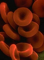
Amgen has withdrawn its application to expand the existing marketing authorization for darbepoetin alfa (Aranesp), according to the European Medicines Agency’s Committee for Medicinal Products for Human Use (CHMP).
The goal with this application was to extend the authorization for darbepoetin alfa to include the treatment of anemia in adults with low-risk or intermediate-1-risk myelodysplastic syndromes (MDS) who have low transfusion demand.
Darbepoetin alfa is currently approved in the European Union (EU) to treat anemia in adults and children with chronic renal failure and adults who are receiving chemotherapy for non-myeloid malignancies.
Amgen withdrew the application for darbepoetin alfa in MDS patients after the CHMP had evaluated initial documentation provided by the company and formulated a list of questions. Amgen had not yet responded to the questions when it notified the CHMP of the withdrawal.
At the time of the withdrawal, the CHMP was of the provisional opinion that darbepoetin alfa could not have been approved for the treatment of anemia in adults with MDS.
This opinion was based on concerns about the data supporting the application—results from the phase 3 ARCADE trial (NCT01362140) and a phase 2 trial (NCT00095264).
The CHMP said changes to the design of the phase 3 trial and the exclusion of a high number of patients from the analysis of the results raise questions about the validity of the data.
In addition, the phase 2 trial, which was conducted in the US, was not in line with EU recommendations for the treatment of MDS patients.
Therefore, at the time of the withdrawal, the CHMP had decided the marketing authorization could not be expanded based on the data provided.
In a letter to the CHMP, Amgen said its decision to withdraw the application is based on the CHMP’s negative opinion.
Amgen has withdrawn its application to expand the existing marketing authorization for darbepoetin alfa (Aranesp), according to the European Medicines Agency’s Committee for Medicinal Products for Human Use (CHMP).
The goal with this application was to extend the authorization for darbepoetin alfa to include the treatment of anemia in adults with low-risk or intermediate-1-risk myelodysplastic syndromes (MDS) who have low transfusion demand.
Darbepoetin alfa is currently approved in the European Union (EU) to treat anemia in adults and children with chronic renal failure and adults who are receiving chemotherapy for non-myeloid malignancies.
Amgen withdrew the application for darbepoetin alfa in MDS patients after the CHMP had evaluated initial documentation provided by the company and formulated a list of questions. Amgen had not yet responded to the questions when it notified the CHMP of the withdrawal.
At the time of the withdrawal, the CHMP was of the provisional opinion that darbepoetin alfa could not have been approved for the treatment of anemia in adults with MDS.
This opinion was based on concerns about the data supporting the application—results from the phase 3 ARCADE trial (NCT01362140) and a phase 2 trial (NCT00095264).
The CHMP said changes to the design of the phase 3 trial and the exclusion of a high number of patients from the analysis of the results raise questions about the validity of the data.
In addition, the phase 2 trial, which was conducted in the US, was not in line with EU recommendations for the treatment of MDS patients.
Therefore, at the time of the withdrawal, the CHMP had decided the marketing authorization could not be expanded based on the data provided.
In a letter to the CHMP, Amgen said its decision to withdraw the application is based on the CHMP’s negative opinion.
Amgen has withdrawn its application to expand the existing marketing authorization for darbepoetin alfa (Aranesp), according to the European Medicines Agency’s Committee for Medicinal Products for Human Use (CHMP).
The goal with this application was to extend the authorization for darbepoetin alfa to include the treatment of anemia in adults with low-risk or intermediate-1-risk myelodysplastic syndromes (MDS) who have low transfusion demand.
Darbepoetin alfa is currently approved in the European Union (EU) to treat anemia in adults and children with chronic renal failure and adults who are receiving chemotherapy for non-myeloid malignancies.
Amgen withdrew the application for darbepoetin alfa in MDS patients after the CHMP had evaluated initial documentation provided by the company and formulated a list of questions. Amgen had not yet responded to the questions when it notified the CHMP of the withdrawal.
At the time of the withdrawal, the CHMP was of the provisional opinion that darbepoetin alfa could not have been approved for the treatment of anemia in adults with MDS.
This opinion was based on concerns about the data supporting the application—results from the phase 3 ARCADE trial (NCT01362140) and a phase 2 trial (NCT00095264).
The CHMP said changes to the design of the phase 3 trial and the exclusion of a high number of patients from the analysis of the results raise questions about the validity of the data.
In addition, the phase 2 trial, which was conducted in the US, was not in line with EU recommendations for the treatment of MDS patients.
Therefore, at the time of the withdrawal, the CHMP had decided the marketing authorization could not be expanded based on the data provided.
In a letter to the CHMP, Amgen said its decision to withdraw the application is based on the CHMP’s negative opinion.
Infection risk from reprocessed duodenoscopes is low, but the goal is zero
BOSTON – Once attributed to human error, the outbreaks of infection from persistent contamination of reprocessed duodenoscopes have eluded an easy fix, according to three experts addressing the problem at the 2018 AGA Tech Summit, sponsored by the AGA Center for GI Innovation and Technology. This session is part of the AGA Center for GI Innovation and Technology’s ongoing efforts to convene stakeholders, including endoscope manufacturers and the Food and Drug Administration, and to collaborate on a solution to ensure zero device-transmitted infections.
Since the problem first was recognized, changes in device design and cleaning protocols have produced a fivefold reduction in the risk of infection.
“The risk is now very low and well beneath the benefits provided by endoscopy, but we will not settle for anything less than complete resolution of the problem,” said David R. Lichtenstein, MD, the director of the endoscopy program at Boston University. Leading off a program that outlined the problem and possible solutions, Dr. Lichtenstein said, “We need to make this a historical issue.”
After scrutiny by the many stakeholders, including the manufacturers, gastroenterologists, and regulatory agencies, the elevator mechanism has remained one focus of concern. The difficulty of cleaning this mechanism was recognized in the earliest devices, but redesigns to permit these channels to be flushed did not resolve the problem completely. In the AGA Tech Summit session, there was general consensus that even dedicated reprocessing technicians fully adherent to current protocols cannot reliably completely clean the currently available duodenoscopes in every case.
“In most instances, it is not the technician at fault and it is extremely difficult to get a flexible scope clean after a messy procedure,” explained Cori Ofstead, MSPH, an epidemiologist and president of Ofstead & Associates. In one study she conducted in endoscopes reprocessed using best practice recommendations, microbial presence could still be detected.
Although risk of contamination increases with repeated use of scopes, with damaged scopes, and after scopes have been used in procedures generating relatively high amounts of debris, Ms. Ofstead said, “If you look, you will find contamination.” While she emphasized the importance of developing incentives that reward quality over efficiency when attempting to reduce human error in duodenoscope reprocessing, she, like Dr. Lichtenstein, believes new strategies are needed to achieve zero tolerance for infection risk.
“Disinfection simply may not be enough,” Ms. Ofstead said. “The solution is likely to be something else, such as single-use scopes or sterilization.” Explaining how processes of sterilization and disinfection differ, David Weber, MD, MPH, professor of epidemiology, University of North Carolina School of Medicine, Chapel Hill, outlined the relative advantages and disadvantages of current options. Dr. Weber, who has been involved in numerous studies regarding duodenoscope decontamination, emphasized that that rigorous cleaning of biological debris is a central tenet of any solution, but he outlined evidence that the current standard of high level disinfection (HLD) has not been sufficient to bring the infection risk to zero.
“There is no simple answer, because every solution so far has introduced some challenges,” Dr. Lichtenstein observed. Listing some examples, he noted that autoclaving results in damage to endoscopes since they are heat labile; that some proposed sterilization techniques may be limited by sterilants that can’t penetrate biofilm formation into deeper layers that harbor viable bacteria and are associated with a risk of toxicity to technicians or patients; and that disposable endoscopes or elevator mechanisms may involve unacceptable costs or operating characteristics. However, he is convinced there is a solution.
“The problem is being addressed from different angles, and I think we will find the solution within several years. This may involve a series of additional incremental improvements, but I think no one with a stake in this issue will sit comfortably until it is no longer a problem,” Dr. Lichtenstein said.
In the discussion that followed the formal presentations, it was generally acknowledged that the ultimate solution remains unclear even though incremental progress is being made. In fact, there have been no major outbreaks in the United States in the last year following the attention given to this issue, which is likely to have promoted more diligence in disinfection, according to Dr. Lichtenstein.
“Disposable endoscopes or disposable parts on endoscopes that facilitate cleaning may be coming, but I think the best current step is a standardized method of testing scopes after cleaning to verify that disinfection has been achieved,” Dr. Lichtenstein said.
Other solutions, including new sterilization techniques may also emerge, but there was general consensus that the human factor will always be important even if the best solution proves to be a disposable endoscope. Dr. Weber said, “You can still place a disposable scope on a contaminated surface. The human factor is always important.”
Michael L. Kochman, MD, the Wilmott Professor of Medicine and director of the Center for Endoscopic Innovation, Research and Training at the University of Pennsylvania, Philadelphia, and past chair of the AGA Center for GI Innovation and Technology, added, “AGA will build on the relationships that we have with the manufacturers of the endoscopes along with companies developing other novel approaches to reprocessing, as well as the FDA and CDC, to further the discussion. Ultimately, we hope to see the availability of devices that are effective and reliably clean so that we do not have to be concerned about patient-to-patient transmission of infection.”
BOSTON – Once attributed to human error, the outbreaks of infection from persistent contamination of reprocessed duodenoscopes have eluded an easy fix, according to three experts addressing the problem at the 2018 AGA Tech Summit, sponsored by the AGA Center for GI Innovation and Technology. This session is part of the AGA Center for GI Innovation and Technology’s ongoing efforts to convene stakeholders, including endoscope manufacturers and the Food and Drug Administration, and to collaborate on a solution to ensure zero device-transmitted infections.
Since the problem first was recognized, changes in device design and cleaning protocols have produced a fivefold reduction in the risk of infection.
“The risk is now very low and well beneath the benefits provided by endoscopy, but we will not settle for anything less than complete resolution of the problem,” said David R. Lichtenstein, MD, the director of the endoscopy program at Boston University. Leading off a program that outlined the problem and possible solutions, Dr. Lichtenstein said, “We need to make this a historical issue.”
After scrutiny by the many stakeholders, including the manufacturers, gastroenterologists, and regulatory agencies, the elevator mechanism has remained one focus of concern. The difficulty of cleaning this mechanism was recognized in the earliest devices, but redesigns to permit these channels to be flushed did not resolve the problem completely. In the AGA Tech Summit session, there was general consensus that even dedicated reprocessing technicians fully adherent to current protocols cannot reliably completely clean the currently available duodenoscopes in every case.
“In most instances, it is not the technician at fault and it is extremely difficult to get a flexible scope clean after a messy procedure,” explained Cori Ofstead, MSPH, an epidemiologist and president of Ofstead & Associates. In one study she conducted in endoscopes reprocessed using best practice recommendations, microbial presence could still be detected.
Although risk of contamination increases with repeated use of scopes, with damaged scopes, and after scopes have been used in procedures generating relatively high amounts of debris, Ms. Ofstead said, “If you look, you will find contamination.” While she emphasized the importance of developing incentives that reward quality over efficiency when attempting to reduce human error in duodenoscope reprocessing, she, like Dr. Lichtenstein, believes new strategies are needed to achieve zero tolerance for infection risk.
“Disinfection simply may not be enough,” Ms. Ofstead said. “The solution is likely to be something else, such as single-use scopes or sterilization.” Explaining how processes of sterilization and disinfection differ, David Weber, MD, MPH, professor of epidemiology, University of North Carolina School of Medicine, Chapel Hill, outlined the relative advantages and disadvantages of current options. Dr. Weber, who has been involved in numerous studies regarding duodenoscope decontamination, emphasized that that rigorous cleaning of biological debris is a central tenet of any solution, but he outlined evidence that the current standard of high level disinfection (HLD) has not been sufficient to bring the infection risk to zero.
“There is no simple answer, because every solution so far has introduced some challenges,” Dr. Lichtenstein observed. Listing some examples, he noted that autoclaving results in damage to endoscopes since they are heat labile; that some proposed sterilization techniques may be limited by sterilants that can’t penetrate biofilm formation into deeper layers that harbor viable bacteria and are associated with a risk of toxicity to technicians or patients; and that disposable endoscopes or elevator mechanisms may involve unacceptable costs or operating characteristics. However, he is convinced there is a solution.
“The problem is being addressed from different angles, and I think we will find the solution within several years. This may involve a series of additional incremental improvements, but I think no one with a stake in this issue will sit comfortably until it is no longer a problem,” Dr. Lichtenstein said.
In the discussion that followed the formal presentations, it was generally acknowledged that the ultimate solution remains unclear even though incremental progress is being made. In fact, there have been no major outbreaks in the United States in the last year following the attention given to this issue, which is likely to have promoted more diligence in disinfection, according to Dr. Lichtenstein.
“Disposable endoscopes or disposable parts on endoscopes that facilitate cleaning may be coming, but I think the best current step is a standardized method of testing scopes after cleaning to verify that disinfection has been achieved,” Dr. Lichtenstein said.
Other solutions, including new sterilization techniques may also emerge, but there was general consensus that the human factor will always be important even if the best solution proves to be a disposable endoscope. Dr. Weber said, “You can still place a disposable scope on a contaminated surface. The human factor is always important.”
Michael L. Kochman, MD, the Wilmott Professor of Medicine and director of the Center for Endoscopic Innovation, Research and Training at the University of Pennsylvania, Philadelphia, and past chair of the AGA Center for GI Innovation and Technology, added, “AGA will build on the relationships that we have with the manufacturers of the endoscopes along with companies developing other novel approaches to reprocessing, as well as the FDA and CDC, to further the discussion. Ultimately, we hope to see the availability of devices that are effective and reliably clean so that we do not have to be concerned about patient-to-patient transmission of infection.”
BOSTON – Once attributed to human error, the outbreaks of infection from persistent contamination of reprocessed duodenoscopes have eluded an easy fix, according to three experts addressing the problem at the 2018 AGA Tech Summit, sponsored by the AGA Center for GI Innovation and Technology. This session is part of the AGA Center for GI Innovation and Technology’s ongoing efforts to convene stakeholders, including endoscope manufacturers and the Food and Drug Administration, and to collaborate on a solution to ensure zero device-transmitted infections.
Since the problem first was recognized, changes in device design and cleaning protocols have produced a fivefold reduction in the risk of infection.
“The risk is now very low and well beneath the benefits provided by endoscopy, but we will not settle for anything less than complete resolution of the problem,” said David R. Lichtenstein, MD, the director of the endoscopy program at Boston University. Leading off a program that outlined the problem and possible solutions, Dr. Lichtenstein said, “We need to make this a historical issue.”
After scrutiny by the many stakeholders, including the manufacturers, gastroenterologists, and regulatory agencies, the elevator mechanism has remained one focus of concern. The difficulty of cleaning this mechanism was recognized in the earliest devices, but redesigns to permit these channels to be flushed did not resolve the problem completely. In the AGA Tech Summit session, there was general consensus that even dedicated reprocessing technicians fully adherent to current protocols cannot reliably completely clean the currently available duodenoscopes in every case.
“In most instances, it is not the technician at fault and it is extremely difficult to get a flexible scope clean after a messy procedure,” explained Cori Ofstead, MSPH, an epidemiologist and president of Ofstead & Associates. In one study she conducted in endoscopes reprocessed using best practice recommendations, microbial presence could still be detected.
Although risk of contamination increases with repeated use of scopes, with damaged scopes, and after scopes have been used in procedures generating relatively high amounts of debris, Ms. Ofstead said, “If you look, you will find contamination.” While she emphasized the importance of developing incentives that reward quality over efficiency when attempting to reduce human error in duodenoscope reprocessing, she, like Dr. Lichtenstein, believes new strategies are needed to achieve zero tolerance for infection risk.
“Disinfection simply may not be enough,” Ms. Ofstead said. “The solution is likely to be something else, such as single-use scopes or sterilization.” Explaining how processes of sterilization and disinfection differ, David Weber, MD, MPH, professor of epidemiology, University of North Carolina School of Medicine, Chapel Hill, outlined the relative advantages and disadvantages of current options. Dr. Weber, who has been involved in numerous studies regarding duodenoscope decontamination, emphasized that that rigorous cleaning of biological debris is a central tenet of any solution, but he outlined evidence that the current standard of high level disinfection (HLD) has not been sufficient to bring the infection risk to zero.
“There is no simple answer, because every solution so far has introduced some challenges,” Dr. Lichtenstein observed. Listing some examples, he noted that autoclaving results in damage to endoscopes since they are heat labile; that some proposed sterilization techniques may be limited by sterilants that can’t penetrate biofilm formation into deeper layers that harbor viable bacteria and are associated with a risk of toxicity to technicians or patients; and that disposable endoscopes or elevator mechanisms may involve unacceptable costs or operating characteristics. However, he is convinced there is a solution.
“The problem is being addressed from different angles, and I think we will find the solution within several years. This may involve a series of additional incremental improvements, but I think no one with a stake in this issue will sit comfortably until it is no longer a problem,” Dr. Lichtenstein said.
In the discussion that followed the formal presentations, it was generally acknowledged that the ultimate solution remains unclear even though incremental progress is being made. In fact, there have been no major outbreaks in the United States in the last year following the attention given to this issue, which is likely to have promoted more diligence in disinfection, according to Dr. Lichtenstein.
“Disposable endoscopes or disposable parts on endoscopes that facilitate cleaning may be coming, but I think the best current step is a standardized method of testing scopes after cleaning to verify that disinfection has been achieved,” Dr. Lichtenstein said.
Other solutions, including new sterilization techniques may also emerge, but there was general consensus that the human factor will always be important even if the best solution proves to be a disposable endoscope. Dr. Weber said, “You can still place a disposable scope on a contaminated surface. The human factor is always important.”
Michael L. Kochman, MD, the Wilmott Professor of Medicine and director of the Center for Endoscopic Innovation, Research and Training at the University of Pennsylvania, Philadelphia, and past chair of the AGA Center for GI Innovation and Technology, added, “AGA will build on the relationships that we have with the manufacturers of the endoscopes along with companies developing other novel approaches to reprocessing, as well as the FDA and CDC, to further the discussion. Ultimately, we hope to see the availability of devices that are effective and reliably clean so that we do not have to be concerned about patient-to-patient transmission of infection.”
EXPERT ANALYSIS FROM 2018 AGA TECH SUMMIT

Entrepreneurs attempt to convince the Shark Tank experts that they can address unmet needs
BOSTON – At the 2018 AGA Tech Summit, this year’s Shark Tank line up included an automated system for video capture of endoscopy, a feeding tube that prevents aspiration in intubated patients, a tool to accurately measure polyps captured on colonoscopy, a device that targets gastrointestinal cancers with electrical pulses, and a new method for real-time stool testing of infectious pathogens.

In each case, the presenting entrepreneur vowed to the Shark Tank panel of experts that their innovation is addressing an important clinical need. In announcing the winner, V. Raman Muthusamy, MD, who is the chair of the AGA Center for GI Innovation and Technology (CGIT) and director of interventional endoscopy and general GI endoscopy, David Geffen School of Medicine at UCLA, Los Angeles, said, “We have rarely had such a strong group of candidates.”
New for 2018, both the Sharks and AGA Tech Summit attendees voted on a winner. The unanimous winner was Chang-Hee Kim, PhD, who presented a new rapid test for identifying infectious pathogens in stool. The sharks were looking for 1. Novelty/immediate patient impact, 2. Business plan, and 3. Pitch. While all of the participants were close, the immediate patient impact for Dr. Kim’s innovation gave him a leg up on his competitors.
New device permits real-time stool sample analysis
A new tool for rapid analysis of stool for pathogens may be revolutionary in that it can provide results within 15 minutes rather than the days normally required when stool samples are sent to a laboratory, according to Dr. Chang-Hee Kim, chief executive officer of GoDx, Inc. The patent-pending methodology developed at Dr. Kim’s company permits real-time analysis “at the point of need and without the need for a lab.” In addition to the efficiency, the paper-based test, which Dr. Kim compared to a pregnancy test in that there is a color change with positive results, has the potential to improve outcomes.
Real-time testing “will decrease the loss of patients to follow-up and accelerate the time to treatment,” Dr. Kim asserted. “The test is also likely to reduce the spread of nosocomial pathogens if rapid infection control reduces spread.”
The test, which Dr. Kim expected to be made available at a cost of $100, will also be substantially cheaper than current laboratory analyses. In experimental studies, the accuracy has been at least as accurate as polymerase-chain reaction (PCR) testing, according to Dr. Kim. He sees applications not only in hospitals but at sites where ordering laboratory studies are not normally available, such as on cruise ships, in nursing homes, or in the military.
In the Shark Tank session, the main concerns expressed were about how this test will fit with Clinical Laboratory Improvement Amendments (CLIA) compliance. Herb Lerner, MD, a former officer in the Food and Drug Administration (FDA) and now senior director of medical and regulatory affairs at Hogan Lovells, a Washington, D.C.–based law firm, urged Dr. Kim to approach the FDA as soon as possible for advice on how to address this and other potential obstacles to a commercial product.
GI endoscopy capture, storage, and sharing
In introducing an automated method of capturing videos taken during colonoscopy, Matthew Z. Schwartz, the cofounder of Virgo Surgical Video Solutions, Inc. said, “There is currently no low-cost, user-friendly way to systematically capture GI endoscopy video.” Relative to existing products that “require complicated formatting and time-consuming setup for each procedure recording,” the plug-and-play system developed at Virgo “works with any existing endoscopy system that has a video output.” The videos are designed for cloud storage.
“Our long-term vision is to create the largest and highest quality repository of GI endoscopy videos,” said Mr. Schwartz, who added that support tools, including clinical decision-making aided by artificial intelligence, are being developed to provide even more value for quality control, research, and training.
Several Sharks, including Tom Shehab, MD, managing director, Arboretum Ventures, Ann Arbor, Mich., asked for more information about the value proposition for a proposed cost of $500 per month for the system. The answer was that this value would differ for settings, such as in an academic center that might apply the system for training relative to a community setting where the main purpose might be quality improvement.
New NG tube addresses aspiration risk
Pulmonary aspiration, along with symptomatic gastric reflux, is a common clinical challenge in intubated patients on a feeding tube, but a novel nasogastric (NG) tube equipped with a dual balloon system is designed to solve this problem, according to Talal Sharaiha, MD, the founder of Aspisafe Solutions. He said that reducing the risk of aspiration is important because it is associated with pneumonia, erosive inflammation, and upper GI bleeding. He called it the most common cause of upper GI bleeding in intensive care units.
Of the two balloons, one serves as an anchor and sits in the esophagus. The other serves as an anti-aspiration reservoir and sits in the stomach. The anti-aspiration balloon, inflated after it is inserted in the stomach, blocks reflux of gastric contents. Contending that there is a large market for this device, Dr. Sharaiha said, “having a feeding tube in intubated patients that prevents gastric reflux and aspiration will dramatically reduce complications and likely help reduce length of ventilation time, ICU time, and length of hospitals stay.”
More than one Shark requested more information about safety. Michael L. Kochman, MD, the Wilmott Professor of Medicine and director of the Center for Endoscopic Innovation, Research and Training at the University of Pennsylvania, Philadelphia, noted that there have been adverse experiences in the past with other types of balloon devices introduced into the GI tract. He indicated that this may be a potential bar to clinician comfort, so a comprehensive approach to safety analyses should be a priority in clinical development.
Virtual tape measure for colonoscopy measures polyp size
There are risks from the current practice of estimating polyp size, according to Avishay Sidlesky, founder of VTM Technologies, Ltd. He called these estimates “inherently inaccurate” and asserted that they “lead to erroneous diagnoses, suboptimal treatment and follow-up.” He believes that a “virtual tape measure” dependent on an endoscopic laser emitter solves this problem and cited a published paper that confirmed the accuracy of polyp measurements conducted with this device in an animal model.
“Employing dedicated software that analyzes the laser curves in the endoscopic image, the system enables reporting of the size of lesions, diameter and profile of polyps, longitudinal cross-section of lumens and more,” Mr. Sidlesky reported, specifying that the tool can be used alone or integrated into third party endoscopes. He believes applications can eventually be developed for a variety of endoscopic procedures in addition to colonoscopy.
Of Shark comments, several regarded concern about how this measuring tool would be integrated with existing endoscopic systems. Even though the measuring tool can be introduced in an unused channel of available scopes, the image is displayed separately. Michael J. Docktor, MD, clinical director of innovation, Boston Children’s Hospital, cautioned that devices that add steps to the workflow in the endoscopy suite may not be as rapidly accepted as one in which the tool is totally integrated with existing systems.
New device treats esophageal cancers with electroporation
There are a variety of tools to treat upper GI cancers endoscopically, including with radiofrequency ablation, cryotechnology, and photodynamic therapy, but morbidity rates are high, according to Declan Soden, founder of Mirai Medical. He described the attributes of a new technology based on electroporation in which electrical pulses painlessly target neoplastic tissue while preserving adjacent healthy tissue. He said that the treatment is performed in a matter of minutes on an outpatient basis.
“Clinical results to date have demonstrated the benefit of the technology in the treatment of late-stage esophageal and colorectal disease,” Mr. Soden said. He believes that the “unique selling point” of this electroporation treatment, which is part of a complete treatment regimen, “is that it renders tumor tissue leaky or porous, allowing absorption and uptake of drugs.” Relative to current standards, the treatment has multiple advantages, not least of which is preservation of quality of life, he believes.
The Sharks posed many technical questions about this strategy that might narrow the applicability of this device. For example, Trey Reed, MD, medical director, Humana Inc., posed questions about the depth of penetration of the electoral pulses and where they can be adjusted for tumors of different sizes. He also questioned whether the device will be too large to penetrate lumens obstructed by tumor. Although Mr. Soden believes that the device will be versatile, he acknowledged that its role might be better understood when phase 2 trials begin later this year.
A moderator at the Shank Tank session and a previous CGIT Chair, Dr. Kochman reported that he was impressed with the crop of entries. As one of the creators of the AGA Tech Summit, Dr. Kochman has the experience to recognize good ideas when he hears them.
“It is gratifying to see the high quality of the Shark Tank presenters. Over the past years, a number of the presenting companies have gone on to obtain additional funding, be acquired, and on to successful launches,” Dr. Kochman observed. “We hope the same successes await this year’s group.”
BOSTON – At the 2018 AGA Tech Summit, this year’s Shark Tank line up included an automated system for video capture of endoscopy, a feeding tube that prevents aspiration in intubated patients, a tool to accurately measure polyps captured on colonoscopy, a device that targets gastrointestinal cancers with electrical pulses, and a new method for real-time stool testing of infectious pathogens.
In each case, the presenting entrepreneur vowed to the Shark Tank panel of experts that their innovation is addressing an important clinical need. In announcing the winner, V. Raman Muthusamy, MD, who is the chair of the AGA Center for GI Innovation and Technology (CGIT) and director of interventional endoscopy and general GI endoscopy, David Geffen School of Medicine at UCLA, Los Angeles, said, “We have rarely had such a strong group of candidates.”
New for 2018, both the Sharks and AGA Tech Summit attendees voted on a winner. The unanimous winner was Chang-Hee Kim, PhD, who presented a new rapid test for identifying infectious pathogens in stool. The sharks were looking for 1. Novelty/immediate patient impact, 2. Business plan, and 3. Pitch. While all of the participants were close, the immediate patient impact for Dr. Kim’s innovation gave him a leg up on his competitors.
New device permits real-time stool sample analysis
A new tool for rapid analysis of stool for pathogens may be revolutionary in that it can provide results within 15 minutes rather than the days normally required when stool samples are sent to a laboratory, according to Dr. Chang-Hee Kim, chief executive officer of GoDx, Inc. The patent-pending methodology developed at Dr. Kim’s company permits real-time analysis “at the point of need and without the need for a lab.” In addition to the efficiency, the paper-based test, which Dr. Kim compared to a pregnancy test in that there is a color change with positive results, has the potential to improve outcomes.
Real-time testing “will decrease the loss of patients to follow-up and accelerate the time to treatment,” Dr. Kim asserted. “The test is also likely to reduce the spread of nosocomial pathogens if rapid infection control reduces spread.”
The test, which Dr. Kim expected to be made available at a cost of $100, will also be substantially cheaper than current laboratory analyses. In experimental studies, the accuracy has been at least as accurate as polymerase-chain reaction (PCR) testing, according to Dr. Kim. He sees applications not only in hospitals but at sites where ordering laboratory studies are not normally available, such as on cruise ships, in nursing homes, or in the military.
In the Shark Tank session, the main concerns expressed were about how this test will fit with Clinical Laboratory Improvement Amendments (CLIA) compliance. Herb Lerner, MD, a former officer in the Food and Drug Administration (FDA) and now senior director of medical and regulatory affairs at Hogan Lovells, a Washington, D.C.–based law firm, urged Dr. Kim to approach the FDA as soon as possible for advice on how to address this and other potential obstacles to a commercial product.
GI endoscopy capture, storage, and sharing
In introducing an automated method of capturing videos taken during colonoscopy, Matthew Z. Schwartz, the cofounder of Virgo Surgical Video Solutions, Inc. said, “There is currently no low-cost, user-friendly way to systematically capture GI endoscopy video.” Relative to existing products that “require complicated formatting and time-consuming setup for each procedure recording,” the plug-and-play system developed at Virgo “works with any existing endoscopy system that has a video output.” The videos are designed for cloud storage.
“Our long-term vision is to create the largest and highest quality repository of GI endoscopy videos,” said Mr. Schwartz, who added that support tools, including clinical decision-making aided by artificial intelligence, are being developed to provide even more value for quality control, research, and training.
Several Sharks, including Tom Shehab, MD, managing director, Arboretum Ventures, Ann Arbor, Mich., asked for more information about the value proposition for a proposed cost of $500 per month for the system. The answer was that this value would differ for settings, such as in an academic center that might apply the system for training relative to a community setting where the main purpose might be quality improvement.
New NG tube addresses aspiration risk
Pulmonary aspiration, along with symptomatic gastric reflux, is a common clinical challenge in intubated patients on a feeding tube, but a novel nasogastric (NG) tube equipped with a dual balloon system is designed to solve this problem, according to Talal Sharaiha, MD, the founder of Aspisafe Solutions. He said that reducing the risk of aspiration is important because it is associated with pneumonia, erosive inflammation, and upper GI bleeding. He called it the most common cause of upper GI bleeding in intensive care units.
Of the two balloons, one serves as an anchor and sits in the esophagus. The other serves as an anti-aspiration reservoir and sits in the stomach. The anti-aspiration balloon, inflated after it is inserted in the stomach, blocks reflux of gastric contents. Contending that there is a large market for this device, Dr. Sharaiha said, “having a feeding tube in intubated patients that prevents gastric reflux and aspiration will dramatically reduce complications and likely help reduce length of ventilation time, ICU time, and length of hospitals stay.”
More than one Shark requested more information about safety. Michael L. Kochman, MD, the Wilmott Professor of Medicine and director of the Center for Endoscopic Innovation, Research and Training at the University of Pennsylvania, Philadelphia, noted that there have been adverse experiences in the past with other types of balloon devices introduced into the GI tract. He indicated that this may be a potential bar to clinician comfort, so a comprehensive approach to safety analyses should be a priority in clinical development.
Virtual tape measure for colonoscopy measures polyp size
There are risks from the current practice of estimating polyp size, according to Avishay Sidlesky, founder of VTM Technologies, Ltd. He called these estimates “inherently inaccurate” and asserted that they “lead to erroneous diagnoses, suboptimal treatment and follow-up.” He believes that a “virtual tape measure” dependent on an endoscopic laser emitter solves this problem and cited a published paper that confirmed the accuracy of polyp measurements conducted with this device in an animal model.
“Employing dedicated software that analyzes the laser curves in the endoscopic image, the system enables reporting of the size of lesions, diameter and profile of polyps, longitudinal cross-section of lumens and more,” Mr. Sidlesky reported, specifying that the tool can be used alone or integrated into third party endoscopes. He believes applications can eventually be developed for a variety of endoscopic procedures in addition to colonoscopy.
Of Shark comments, several regarded concern about how this measuring tool would be integrated with existing endoscopic systems. Even though the measuring tool can be introduced in an unused channel of available scopes, the image is displayed separately. Michael J. Docktor, MD, clinical director of innovation, Boston Children’s Hospital, cautioned that devices that add steps to the workflow in the endoscopy suite may not be as rapidly accepted as one in which the tool is totally integrated with existing systems.
New device treats esophageal cancers with electroporation
There are a variety of tools to treat upper GI cancers endoscopically, including with radiofrequency ablation, cryotechnology, and photodynamic therapy, but morbidity rates are high, according to Declan Soden, founder of Mirai Medical. He described the attributes of a new technology based on electroporation in which electrical pulses painlessly target neoplastic tissue while preserving adjacent healthy tissue. He said that the treatment is performed in a matter of minutes on an outpatient basis.
“Clinical results to date have demonstrated the benefit of the technology in the treatment of late-stage esophageal and colorectal disease,” Mr. Soden said. He believes that the “unique selling point” of this electroporation treatment, which is part of a complete treatment regimen, “is that it renders tumor tissue leaky or porous, allowing absorption and uptake of drugs.” Relative to current standards, the treatment has multiple advantages, not least of which is preservation of quality of life, he believes.
The Sharks posed many technical questions about this strategy that might narrow the applicability of this device. For example, Trey Reed, MD, medical director, Humana Inc., posed questions about the depth of penetration of the electoral pulses and where they can be adjusted for tumors of different sizes. He also questioned whether the device will be too large to penetrate lumens obstructed by tumor. Although Mr. Soden believes that the device will be versatile, he acknowledged that its role might be better understood when phase 2 trials begin later this year.
A moderator at the Shank Tank session and a previous CGIT Chair, Dr. Kochman reported that he was impressed with the crop of entries. As one of the creators of the AGA Tech Summit, Dr. Kochman has the experience to recognize good ideas when he hears them.
“It is gratifying to see the high quality of the Shark Tank presenters. Over the past years, a number of the presenting companies have gone on to obtain additional funding, be acquired, and on to successful launches,” Dr. Kochman observed. “We hope the same successes await this year’s group.”
BOSTON – At the 2018 AGA Tech Summit, this year’s Shark Tank line up included an automated system for video capture of endoscopy, a feeding tube that prevents aspiration in intubated patients, a tool to accurately measure polyps captured on colonoscopy, a device that targets gastrointestinal cancers with electrical pulses, and a new method for real-time stool testing of infectious pathogens.
In each case, the presenting entrepreneur vowed to the Shark Tank panel of experts that their innovation is addressing an important clinical need. In announcing the winner, V. Raman Muthusamy, MD, who is the chair of the AGA Center for GI Innovation and Technology (CGIT) and director of interventional endoscopy and general GI endoscopy, David Geffen School of Medicine at UCLA, Los Angeles, said, “We have rarely had such a strong group of candidates.”
New for 2018, both the Sharks and AGA Tech Summit attendees voted on a winner. The unanimous winner was Chang-Hee Kim, PhD, who presented a new rapid test for identifying infectious pathogens in stool. The sharks were looking for 1. Novelty/immediate patient impact, 2. Business plan, and 3. Pitch. While all of the participants were close, the immediate patient impact for Dr. Kim’s innovation gave him a leg up on his competitors.
New device permits real-time stool sample analysis
A new tool for rapid analysis of stool for pathogens may be revolutionary in that it can provide results within 15 minutes rather than the days normally required when stool samples are sent to a laboratory, according to Dr. Chang-Hee Kim, chief executive officer of GoDx, Inc. The patent-pending methodology developed at Dr. Kim’s company permits real-time analysis “at the point of need and without the need for a lab.” In addition to the efficiency, the paper-based test, which Dr. Kim compared to a pregnancy test in that there is a color change with positive results, has the potential to improve outcomes.
Real-time testing “will decrease the loss of patients to follow-up and accelerate the time to treatment,” Dr. Kim asserted. “The test is also likely to reduce the spread of nosocomial pathogens if rapid infection control reduces spread.”
The test, which Dr. Kim expected to be made available at a cost of $100, will also be substantially cheaper than current laboratory analyses. In experimental studies, the accuracy has been at least as accurate as polymerase-chain reaction (PCR) testing, according to Dr. Kim. He sees applications not only in hospitals but at sites where ordering laboratory studies are not normally available, such as on cruise ships, in nursing homes, or in the military.
In the Shark Tank session, the main concerns expressed were about how this test will fit with Clinical Laboratory Improvement Amendments (CLIA) compliance. Herb Lerner, MD, a former officer in the Food and Drug Administration (FDA) and now senior director of medical and regulatory affairs at Hogan Lovells, a Washington, D.C.–based law firm, urged Dr. Kim to approach the FDA as soon as possible for advice on how to address this and other potential obstacles to a commercial product.
GI endoscopy capture, storage, and sharing
In introducing an automated method of capturing videos taken during colonoscopy, Matthew Z. Schwartz, the cofounder of Virgo Surgical Video Solutions, Inc. said, “There is currently no low-cost, user-friendly way to systematically capture GI endoscopy video.” Relative to existing products that “require complicated formatting and time-consuming setup for each procedure recording,” the plug-and-play system developed at Virgo “works with any existing endoscopy system that has a video output.” The videos are designed for cloud storage.
“Our long-term vision is to create the largest and highest quality repository of GI endoscopy videos,” said Mr. Schwartz, who added that support tools, including clinical decision-making aided by artificial intelligence, are being developed to provide even more value for quality control, research, and training.
Several Sharks, including Tom Shehab, MD, managing director, Arboretum Ventures, Ann Arbor, Mich., asked for more information about the value proposition for a proposed cost of $500 per month for the system. The answer was that this value would differ for settings, such as in an academic center that might apply the system for training relative to a community setting where the main purpose might be quality improvement.
New NG tube addresses aspiration risk
Pulmonary aspiration, along with symptomatic gastric reflux, is a common clinical challenge in intubated patients on a feeding tube, but a novel nasogastric (NG) tube equipped with a dual balloon system is designed to solve this problem, according to Talal Sharaiha, MD, the founder of Aspisafe Solutions. He said that reducing the risk of aspiration is important because it is associated with pneumonia, erosive inflammation, and upper GI bleeding. He called it the most common cause of upper GI bleeding in intensive care units.
Of the two balloons, one serves as an anchor and sits in the esophagus. The other serves as an anti-aspiration reservoir and sits in the stomach. The anti-aspiration balloon, inflated after it is inserted in the stomach, blocks reflux of gastric contents. Contending that there is a large market for this device, Dr. Sharaiha said, “having a feeding tube in intubated patients that prevents gastric reflux and aspiration will dramatically reduce complications and likely help reduce length of ventilation time, ICU time, and length of hospitals stay.”
More than one Shark requested more information about safety. Michael L. Kochman, MD, the Wilmott Professor of Medicine and director of the Center for Endoscopic Innovation, Research and Training at the University of Pennsylvania, Philadelphia, noted that there have been adverse experiences in the past with other types of balloon devices introduced into the GI tract. He indicated that this may be a potential bar to clinician comfort, so a comprehensive approach to safety analyses should be a priority in clinical development.
Virtual tape measure for colonoscopy measures polyp size
There are risks from the current practice of estimating polyp size, according to Avishay Sidlesky, founder of VTM Technologies, Ltd. He called these estimates “inherently inaccurate” and asserted that they “lead to erroneous diagnoses, suboptimal treatment and follow-up.” He believes that a “virtual tape measure” dependent on an endoscopic laser emitter solves this problem and cited a published paper that confirmed the accuracy of polyp measurements conducted with this device in an animal model.
“Employing dedicated software that analyzes the laser curves in the endoscopic image, the system enables reporting of the size of lesions, diameter and profile of polyps, longitudinal cross-section of lumens and more,” Mr. Sidlesky reported, specifying that the tool can be used alone or integrated into third party endoscopes. He believes applications can eventually be developed for a variety of endoscopic procedures in addition to colonoscopy.
Of Shark comments, several regarded concern about how this measuring tool would be integrated with existing endoscopic systems. Even though the measuring tool can be introduced in an unused channel of available scopes, the image is displayed separately. Michael J. Docktor, MD, clinical director of innovation, Boston Children’s Hospital, cautioned that devices that add steps to the workflow in the endoscopy suite may not be as rapidly accepted as one in which the tool is totally integrated with existing systems.
New device treats esophageal cancers with electroporation
There are a variety of tools to treat upper GI cancers endoscopically, including with radiofrequency ablation, cryotechnology, and photodynamic therapy, but morbidity rates are high, according to Declan Soden, founder of Mirai Medical. He described the attributes of a new technology based on electroporation in which electrical pulses painlessly target neoplastic tissue while preserving adjacent healthy tissue. He said that the treatment is performed in a matter of minutes on an outpatient basis.
“Clinical results to date have demonstrated the benefit of the technology in the treatment of late-stage esophageal and colorectal disease,” Mr. Soden said. He believes that the “unique selling point” of this electroporation treatment, which is part of a complete treatment regimen, “is that it renders tumor tissue leaky or porous, allowing absorption and uptake of drugs.” Relative to current standards, the treatment has multiple advantages, not least of which is preservation of quality of life, he believes.
The Sharks posed many technical questions about this strategy that might narrow the applicability of this device. For example, Trey Reed, MD, medical director, Humana Inc., posed questions about the depth of penetration of the electoral pulses and where they can be adjusted for tumors of different sizes. He also questioned whether the device will be too large to penetrate lumens obstructed by tumor. Although Mr. Soden believes that the device will be versatile, he acknowledged that its role might be better understood when phase 2 trials begin later this year.
A moderator at the Shank Tank session and a previous CGIT Chair, Dr. Kochman reported that he was impressed with the crop of entries. As one of the creators of the AGA Tech Summit, Dr. Kochman has the experience to recognize good ideas when he hears them.
“It is gratifying to see the high quality of the Shark Tank presenters. Over the past years, a number of the presenting companies have gone on to obtain additional funding, be acquired, and on to successful launches,” Dr. Kochman observed. “We hope the same successes await this year’s group.”
REPORTING FROM 2018 AGA TECH SUMMIT

Medical treatment of perianal fistulae often warranted, despite limited evidence
PHILADELPHIA – Perianal fistulae are a common and difficult-to-treat complication of Crohn’s disease that may often require medical therapy, though not all treatment options have robust data supporting their use in this setting, according to Mark T. Osterman, MD.
“Most studies done on fistulae actually didn’t have that as the primary endpoint – it was a secondary endpoint, so they weren’t really designed to look at fistulae, specifically,” said Dr. Osterman, associate professor of medicine in the division of gastroenterology at the University of Pennsylvania in Philadelphia.
Dr. Osterman shared his own approach to medical treatment of perianal fistulae in a presentation he gave at Digestive Diseases: New Advances, jointly provided by Rutgers and Global Academy for Medical Education.
For simple perianal fistula with no rectal inflammation, a fistulotomy is reasonable, but medical treatment may be preferable, Dr. Osterman said.
“I always favor medical therapy because it attacks the root of the problem, which is the immune system,” he explained.
Helpful treatments in this scenario include antibiotics, along with anti–tumor necrosis factor therapy with or without immunomodulators, he said.
Antibiotic use in this setting is based on uncontrolled data, and efficacy is modest at best, according to Dr. Osterman, who noted that the treatments may reduce fistula drainage but likely do not heal fistulae.
The most commonly used antibiotics are metronidazole and ciprofloxacin given for up to 2-4 months, he added.
Infliximab is the drug that has by far the most robust fistula data, and one of only two drugs where a fistula was the primary outcome of the studies, according to Dr. Osterman.
In a randomized trial, infliximab induction treatment more than doubled fistula-related response and remission rates, compared with placebo, he said.
Maintenance infliximab treatment likewise showed an approximate doubling of both response and remission of fistula versus placebo, he added.
While not designed to look at fistulae as a primary outcome, the randomized CHARM study of adalimumab versus placebo for maintenance of Crohn’s disease remission did demonstrate remission rates about twice as high with the use of adalimumab, compared with placebo, in 117 patients who had draining fistulae at baseline, Dr. Osterman recounted.
For patients with complex fistulae, as well as patients with rectal inflammation, a seton and aggressive medical therapy are likely needed, Dr. Osterman said in his presentation.
An advancement flap or medical therapies such as vedolizumab or tacrolimus might be warranted for patients who fail other medical approaches, he added.
Relevant vedolizumab data come from GEMINI 2, a large clinical trial for Crohn’s disease that included 57 patients who had draining fistulas at baseline.
“We see an improvement in remission rates with fistula with vedolizumab, compared to placebo, but again, (GEMINI 2) wasn’t designed to look at fistula, but we do use it,” said Dr. Osterman.
Tacrolimus is the only drug besides infliximab that has randomized data for fistula, according to Dr. Osterman.
In the small randomized study, 48 patients with Crohn’s disease and draining perianal or enterocutaneous fistulae were treated for 10 weeks with oral tacrolimus at 0.2 mg/kg per day or placebo.
Fistula improvement was seen in 43% of tacrolimus-treated patients and 8% of placebo-treated patients (P = .004), while remission was seen in 10% and 8% of those groups, respectively (P = .86), according to published data on the trial.
“[Tacrolimus] showed a nice improvement in response rates, but very similar remission rates,” Dr. Osterman said, “but it does represent an option for us, for our patients.”
Dr. Osterman reported grant/research support from UCB and serving as a consultant for AbbVie, Janssen, Lycera, Merck, Pfizer, Takeda, and UCB.
Global Academy and this news organization are owned by the same company.
PHILADELPHIA – Perianal fistulae are a common and difficult-to-treat complication of Crohn’s disease that may often require medical therapy, though not all treatment options have robust data supporting their use in this setting, according to Mark T. Osterman, MD.
“Most studies done on fistulae actually didn’t have that as the primary endpoint – it was a secondary endpoint, so they weren’t really designed to look at fistulae, specifically,” said Dr. Osterman, associate professor of medicine in the division of gastroenterology at the University of Pennsylvania in Philadelphia.
Dr. Osterman shared his own approach to medical treatment of perianal fistulae in a presentation he gave at Digestive Diseases: New Advances, jointly provided by Rutgers and Global Academy for Medical Education.
For simple perianal fistula with no rectal inflammation, a fistulotomy is reasonable, but medical treatment may be preferable, Dr. Osterman said.
“I always favor medical therapy because it attacks the root of the problem, which is the immune system,” he explained.
Helpful treatments in this scenario include antibiotics, along with anti–tumor necrosis factor therapy with or without immunomodulators, he said.
Antibiotic use in this setting is based on uncontrolled data, and efficacy is modest at best, according to Dr. Osterman, who noted that the treatments may reduce fistula drainage but likely do not heal fistulae.
The most commonly used antibiotics are metronidazole and ciprofloxacin given for up to 2-4 months, he added.
Infliximab is the drug that has by far the most robust fistula data, and one of only two drugs where a fistula was the primary outcome of the studies, according to Dr. Osterman.
In a randomized trial, infliximab induction treatment more than doubled fistula-related response and remission rates, compared with placebo, he said.
Maintenance infliximab treatment likewise showed an approximate doubling of both response and remission of fistula versus placebo, he added.
While not designed to look at fistulae as a primary outcome, the randomized CHARM study of adalimumab versus placebo for maintenance of Crohn’s disease remission did demonstrate remission rates about twice as high with the use of adalimumab, compared with placebo, in 117 patients who had draining fistulae at baseline, Dr. Osterman recounted.
For patients with complex fistulae, as well as patients with rectal inflammation, a seton and aggressive medical therapy are likely needed, Dr. Osterman said in his presentation.
An advancement flap or medical therapies such as vedolizumab or tacrolimus might be warranted for patients who fail other medical approaches, he added.
Relevant vedolizumab data come from GEMINI 2, a large clinical trial for Crohn’s disease that included 57 patients who had draining fistulas at baseline.
“We see an improvement in remission rates with fistula with vedolizumab, compared to placebo, but again, (GEMINI 2) wasn’t designed to look at fistula, but we do use it,” said Dr. Osterman.
Tacrolimus is the only drug besides infliximab that has randomized data for fistula, according to Dr. Osterman.
In the small randomized study, 48 patients with Crohn’s disease and draining perianal or enterocutaneous fistulae were treated for 10 weeks with oral tacrolimus at 0.2 mg/kg per day or placebo.
Fistula improvement was seen in 43% of tacrolimus-treated patients and 8% of placebo-treated patients (P = .004), while remission was seen in 10% and 8% of those groups, respectively (P = .86), according to published data on the trial.
“[Tacrolimus] showed a nice improvement in response rates, but very similar remission rates,” Dr. Osterman said, “but it does represent an option for us, for our patients.”
Dr. Osterman reported grant/research support from UCB and serving as a consultant for AbbVie, Janssen, Lycera, Merck, Pfizer, Takeda, and UCB.
Global Academy and this news organization are owned by the same company.
PHILADELPHIA – Perianal fistulae are a common and difficult-to-treat complication of Crohn’s disease that may often require medical therapy, though not all treatment options have robust data supporting their use in this setting, according to Mark T. Osterman, MD.
“Most studies done on fistulae actually didn’t have that as the primary endpoint – it was a secondary endpoint, so they weren’t really designed to look at fistulae, specifically,” said Dr. Osterman, associate professor of medicine in the division of gastroenterology at the University of Pennsylvania in Philadelphia.
Dr. Osterman shared his own approach to medical treatment of perianal fistulae in a presentation he gave at Digestive Diseases: New Advances, jointly provided by Rutgers and Global Academy for Medical Education.
For simple perianal fistula with no rectal inflammation, a fistulotomy is reasonable, but medical treatment may be preferable, Dr. Osterman said.
“I always favor medical therapy because it attacks the root of the problem, which is the immune system,” he explained.
Helpful treatments in this scenario include antibiotics, along with anti–tumor necrosis factor therapy with or without immunomodulators, he said.
Antibiotic use in this setting is based on uncontrolled data, and efficacy is modest at best, according to Dr. Osterman, who noted that the treatments may reduce fistula drainage but likely do not heal fistulae.
The most commonly used antibiotics are metronidazole and ciprofloxacin given for up to 2-4 months, he added.
Infliximab is the drug that has by far the most robust fistula data, and one of only two drugs where a fistula was the primary outcome of the studies, according to Dr. Osterman.
In a randomized trial, infliximab induction treatment more than doubled fistula-related response and remission rates, compared with placebo, he said.
Maintenance infliximab treatment likewise showed an approximate doubling of both response and remission of fistula versus placebo, he added.
While not designed to look at fistulae as a primary outcome, the randomized CHARM study of adalimumab versus placebo for maintenance of Crohn’s disease remission did demonstrate remission rates about twice as high with the use of adalimumab, compared with placebo, in 117 patients who had draining fistulae at baseline, Dr. Osterman recounted.
For patients with complex fistulae, as well as patients with rectal inflammation, a seton and aggressive medical therapy are likely needed, Dr. Osterman said in his presentation.
An advancement flap or medical therapies such as vedolizumab or tacrolimus might be warranted for patients who fail other medical approaches, he added.
Relevant vedolizumab data come from GEMINI 2, a large clinical trial for Crohn’s disease that included 57 patients who had draining fistulas at baseline.
“We see an improvement in remission rates with fistula with vedolizumab, compared to placebo, but again, (GEMINI 2) wasn’t designed to look at fistula, but we do use it,” said Dr. Osterman.
Tacrolimus is the only drug besides infliximab that has randomized data for fistula, according to Dr. Osterman.
In the small randomized study, 48 patients with Crohn’s disease and draining perianal or enterocutaneous fistulae were treated for 10 weeks with oral tacrolimus at 0.2 mg/kg per day or placebo.
Fistula improvement was seen in 43% of tacrolimus-treated patients and 8% of placebo-treated patients (P = .004), while remission was seen in 10% and 8% of those groups, respectively (P = .86), according to published data on the trial.
“[Tacrolimus] showed a nice improvement in response rates, but very similar remission rates,” Dr. Osterman said, “but it does represent an option for us, for our patients.”
Dr. Osterman reported grant/research support from UCB and serving as a consultant for AbbVie, Janssen, Lycera, Merck, Pfizer, Takeda, and UCB.
Global Academy and this news organization are owned by the same company.
EXPERT ANALYSIS FROM DIGESTIVE DISEASES: NEW ADVANCES
Step-up diet: Less-intensive way to ID eosinophilic esophagitis food triggers?
PHILADELPHIA – While traditional elimination diets for eosinophilic esophagitis are highly restrictive and endoscopically intensive, a new “step-up” approach may offer a superior empiric scheme for identifying food triggers, according to Stuart J. Spechler, MD.
The recently described step-up approach may be preferable to the standard empiric six-food elimination diet, which has a 72% success rate but is challenging to implement, according to Dr. Spechler, chief of the division of gastroenterology, Baylor University Medical Center at Dallas.
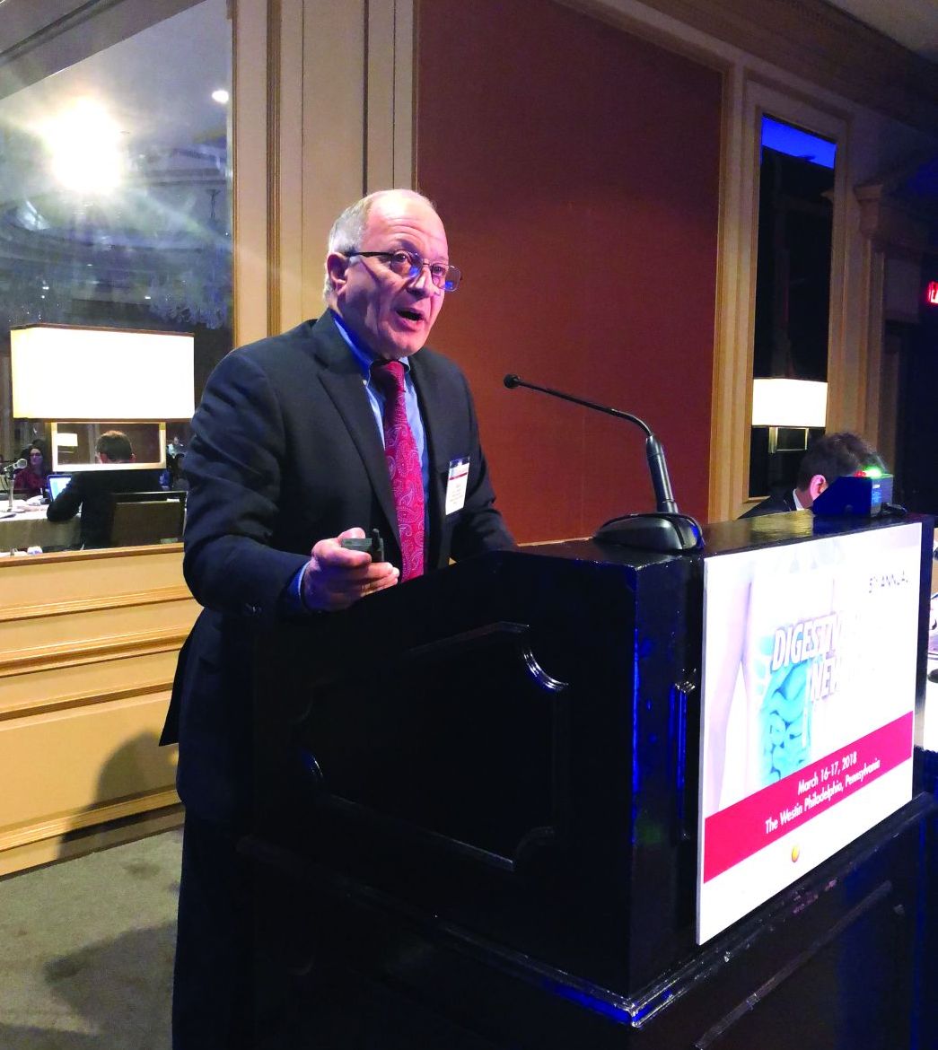
“That’s an extremely demanding, time-consuming, inconvenient, and expensive thing to do. It requires at least seven endoscopies, probably more, performed over a period of 42 weeks,” Dr. Spechler said here at Digestive Diseases: New Advances, jointly provided by Rutgers and Global Academy for Medical Education.
In contrast, the step-up approach recently described by Molina-Infante et al. in the Journal of Allergy and Clinical Immunology (2018 Mar 6. doi: 10.1016/j.jaci.2018.02.028) consists of a two-food elimination diet escalated to a four- and six-food elimination diet as needed.
This diet, which the researchers described as a “2-4-6” approach, starts with elimination of milk and wheat, the two most common food triggers for eosinophilic esophagitis. If patients do not respond, eggs and soy/legumes are eliminated, and if that fails, nuts and seafood are eliminated.
Once patients do respond, foods are reintroduced one at a time over 6 weeks with repeat endoscopy and biopsy, according to the report.
Molina-Infante and coauthors further described results of the diet in 220 patients with eosinophilic esophagitis. Of that group, investigators said 90 patients refused dietary therapy.
However, for the remaining 130 patients, 74 responded to the diet, the investigators reported.
Of 74 responders, 56 (43%) achieved remission at the first step in which milk and wheat were eliminated, results show, while an additional 10 had remission after the step up to four-food elimination, and another 8 had remission after the final step up to six-food elimination.
Dr. Spechler said his current take on diet therapy for eosinophilic esophagitis would be to start with the two-food elimination diet.
That first step alone identified about three-quarters of the patients who eventually would respond to the step-up approach in the Molina-Infante study, he observed.
For patients who do not respond and are motivated to continue, Dr. Spechler said he would move up to the four-food elimination diet, which identified about 90% of patients who eventually responded.
However, Dr. Spechler said he would consider the final step-up only in “exceptionally highly motivated” patients, since that step seems to identify very few additional responders.
“They got very little benefit here from going all the way up to a six-food elimination diet,” Dr. Spechler said.
Patient acceptance may be one major barrier to any dietary approach to treatment of eosinophilic esophagitis, regardless of how intensive the approach is.
“Ninety patients just flat-out refused to try the diet, so this is not a popular form of therapy,” Dr. Spechler noted.
However, diet is one of three valid treatment options for patients who do have an established diagnosis of eosinophilic esophagitis, the other two being proton pump inhibitors (PPIs) or 6-8 weeks of topical steroids, according to Dr. Spechler.
“If you’re going to use diet, I think you begin with that two-food elimination diet,“ he said. “Fortunately, most of the patients who don’t respond to diet will respond to PPIs or steroids.”
Dr. Spechler reported disclosures related to Ironwood Pharmaceuticals and Takeda Pharmaceuticals.
Global Academy and this news organization are owned by the same company.
PHILADELPHIA – While traditional elimination diets for eosinophilic esophagitis are highly restrictive and endoscopically intensive, a new “step-up” approach may offer a superior empiric scheme for identifying food triggers, according to Stuart J. Spechler, MD.
The recently described step-up approach may be preferable to the standard empiric six-food elimination diet, which has a 72% success rate but is challenging to implement, according to Dr. Spechler, chief of the division of gastroenterology, Baylor University Medical Center at Dallas.
“That’s an extremely demanding, time-consuming, inconvenient, and expensive thing to do. It requires at least seven endoscopies, probably more, performed over a period of 42 weeks,” Dr. Spechler said here at Digestive Diseases: New Advances, jointly provided by Rutgers and Global Academy for Medical Education.
In contrast, the step-up approach recently described by Molina-Infante et al. in the Journal of Allergy and Clinical Immunology (2018 Mar 6. doi: 10.1016/j.jaci.2018.02.028) consists of a two-food elimination diet escalated to a four- and six-food elimination diet as needed.
This diet, which the researchers described as a “2-4-6” approach, starts with elimination of milk and wheat, the two most common food triggers for eosinophilic esophagitis. If patients do not respond, eggs and soy/legumes are eliminated, and if that fails, nuts and seafood are eliminated.
Once patients do respond, foods are reintroduced one at a time over 6 weeks with repeat endoscopy and biopsy, according to the report.
Molina-Infante and coauthors further described results of the diet in 220 patients with eosinophilic esophagitis. Of that group, investigators said 90 patients refused dietary therapy.
However, for the remaining 130 patients, 74 responded to the diet, the investigators reported.
Of 74 responders, 56 (43%) achieved remission at the first step in which milk and wheat were eliminated, results show, while an additional 10 had remission after the step up to four-food elimination, and another 8 had remission after the final step up to six-food elimination.
Dr. Spechler said his current take on diet therapy for eosinophilic esophagitis would be to start with the two-food elimination diet.
That first step alone identified about three-quarters of the patients who eventually would respond to the step-up approach in the Molina-Infante study, he observed.
For patients who do not respond and are motivated to continue, Dr. Spechler said he would move up to the four-food elimination diet, which identified about 90% of patients who eventually responded.
However, Dr. Spechler said he would consider the final step-up only in “exceptionally highly motivated” patients, since that step seems to identify very few additional responders.
“They got very little benefit here from going all the way up to a six-food elimination diet,” Dr. Spechler said.
Patient acceptance may be one major barrier to any dietary approach to treatment of eosinophilic esophagitis, regardless of how intensive the approach is.
“Ninety patients just flat-out refused to try the diet, so this is not a popular form of therapy,” Dr. Spechler noted.
However, diet is one of three valid treatment options for patients who do have an established diagnosis of eosinophilic esophagitis, the other two being proton pump inhibitors (PPIs) or 6-8 weeks of topical steroids, according to Dr. Spechler.
“If you’re going to use diet, I think you begin with that two-food elimination diet,“ he said. “Fortunately, most of the patients who don’t respond to diet will respond to PPIs or steroids.”
Dr. Spechler reported disclosures related to Ironwood Pharmaceuticals and Takeda Pharmaceuticals.
Global Academy and this news organization are owned by the same company.
PHILADELPHIA – While traditional elimination diets for eosinophilic esophagitis are highly restrictive and endoscopically intensive, a new “step-up” approach may offer a superior empiric scheme for identifying food triggers, according to Stuart J. Spechler, MD.
The recently described step-up approach may be preferable to the standard empiric six-food elimination diet, which has a 72% success rate but is challenging to implement, according to Dr. Spechler, chief of the division of gastroenterology, Baylor University Medical Center at Dallas.
“That’s an extremely demanding, time-consuming, inconvenient, and expensive thing to do. It requires at least seven endoscopies, probably more, performed over a period of 42 weeks,” Dr. Spechler said here at Digestive Diseases: New Advances, jointly provided by Rutgers and Global Academy for Medical Education.
In contrast, the step-up approach recently described by Molina-Infante et al. in the Journal of Allergy and Clinical Immunology (2018 Mar 6. doi: 10.1016/j.jaci.2018.02.028) consists of a two-food elimination diet escalated to a four- and six-food elimination diet as needed.
This diet, which the researchers described as a “2-4-6” approach, starts with elimination of milk and wheat, the two most common food triggers for eosinophilic esophagitis. If patients do not respond, eggs and soy/legumes are eliminated, and if that fails, nuts and seafood are eliminated.
Once patients do respond, foods are reintroduced one at a time over 6 weeks with repeat endoscopy and biopsy, according to the report.
Molina-Infante and coauthors further described results of the diet in 220 patients with eosinophilic esophagitis. Of that group, investigators said 90 patients refused dietary therapy.
However, for the remaining 130 patients, 74 responded to the diet, the investigators reported.
Of 74 responders, 56 (43%) achieved remission at the first step in which milk and wheat were eliminated, results show, while an additional 10 had remission after the step up to four-food elimination, and another 8 had remission after the final step up to six-food elimination.
Dr. Spechler said his current take on diet therapy for eosinophilic esophagitis would be to start with the two-food elimination diet.
That first step alone identified about three-quarters of the patients who eventually would respond to the step-up approach in the Molina-Infante study, he observed.
For patients who do not respond and are motivated to continue, Dr. Spechler said he would move up to the four-food elimination diet, which identified about 90% of patients who eventually responded.
However, Dr. Spechler said he would consider the final step-up only in “exceptionally highly motivated” patients, since that step seems to identify very few additional responders.
“They got very little benefit here from going all the way up to a six-food elimination diet,” Dr. Spechler said.
Patient acceptance may be one major barrier to any dietary approach to treatment of eosinophilic esophagitis, regardless of how intensive the approach is.
“Ninety patients just flat-out refused to try the diet, so this is not a popular form of therapy,” Dr. Spechler noted.
However, diet is one of three valid treatment options for patients who do have an established diagnosis of eosinophilic esophagitis, the other two being proton pump inhibitors (PPIs) or 6-8 weeks of topical steroids, according to Dr. Spechler.
“If you’re going to use diet, I think you begin with that two-food elimination diet,“ he said. “Fortunately, most of the patients who don’t respond to diet will respond to PPIs or steroids.”
Dr. Spechler reported disclosures related to Ironwood Pharmaceuticals and Takeda Pharmaceuticals.
Global Academy and this news organization are owned by the same company.
EXPERT ANALYSIS FROM DIGESTIVE DISEASES: NEW ADVANCES
Axial SpA disease activity remains mostly stable throughout pregnancy
, according to results of a prospective study.
“In the largest prospective study to date exploring disease activity during pregnancy in women with axSpA, we found that the majority experienced stable, low disease activity,” Kristin Ursin, MD, of the Trondheim (Norway) University Hospital and her associates wrote in Rheumatology. “In accordance with two previous studies, we found a small increase in disease activity in the second trimester.”
All women in the study fulfilled the Assessment of SpondyloArthritis International Society criteria for axSpA and had seven clinic visits throughout their pregnancy: one before conception, one at each trimester, another at 6 weeks, and two visits 6 and 12 months after delivery. No differentiation was made between women with radiographic or nonradiographic axSpA. At each visit, patients’ disease activity was determined using the Bath Ankylosing Spondylitis Disease Activity Index (BASDAI), which is calculated based on six patient factors scored from 1 to 10, including: fatigue, back pain, peripheral joint pain and swelling, localized tenderness, duration of morning stiffness, and severity of morning stiffness. A disease score of 4 is commonly used to define active disease. Disease activity was also assessed by measuring C-reactive protein (CRP). Women’s function and health was also assessed using the Bath Ankylosing Spondylitis Functional Index (BASFI), scored similarly to the BASDAI, and the RAND-36 questionnaire to assess general health.
The research team found that, across the span of pregnancy, axSpA disease activity was relatively low and consistent throughout the study. But there was a significant relationship between disease activity and time (P = .029) in which disease activity was highest during the second trimester and proved to be significantly higher than 6 weeks after giving birth (mean BASDAI 3.97 vs. 3.46, P = .005). In fact, 45% of women had active disease in their second trimester (BASDAI of 4 or greater). But among women with data from the second trimester and 6 weeks post partum, 42% had a decrease in BASDAI score of 1 or more, and 22% had an equivalent increase. CRP values remained low and stable throughout the study.
Like the BASDAI scores, physical function and well being seemed to correlate with later time points during pregnancy. The lowest level of physical functioning for most women was during the second and third trimesters, and those periods were significantly worse than 6 weeks after giving birth (mean BASFI of 3.2 during the second trimester and 3.6 during the third vs. 2.6 at 6 weeks post partum). Similarly, patients reported overall worse general health during the second and third trimesters, compared with 6 weeks after giving birth, based on RAND-36 scores (mean physical functioning score of 63.1 in the second trimester and 54.5 in the third vs. 71.0 at 6 weeks post partum).
Use of nonbiologic medications remained relatively stable during pregnancy, while biologic disease-modifying antirheumatic drug (DMARD) use (all tumor necrosis factor [TNF] inhibitors) dropped significantly when pregnancy was confirmed. About 20% used NSAIDs during pregnancy, except for during the third trimester when use decreased to 8%. Prednisolone use remained stable at 5%-8%. A total of 11% had used synthetic DMARDs prior to pregnancy, and this rate stayed at 10%-16% during the three trimesters. Overall, 44% had used a biologic DMARD (TNF inhibitor) prior to pregnancy, but this declined sharply to 6% during the first trimester and 1% in the third.
The main limitations of the study included not having data from all of the women for all the time points. In fact, only 22% were included prior to conceiving, which could suggest that women with low disease activity were less likely to visit their rheumatologist, thereby biasing the sample toward an overestimated disease activity at preconception that could have potentially hidden a “more evident deterioration between the preconception visit and the second trimester,” the authors said.
“Future research on pregnancy in women with axSpA should differentiate between subgroups of the disease and aim to include objective assessment of inflammation.”
The study was funded by the Research Fund of the Norwegian Organization for People With Rheumatic Diseases. None of the authors had any conflicts of interest to declare.
SOURCE: Ursin K et al. Rheumatology. 2018 Mar 14. doi: 10.1093/rheumatology/key047.
, according to results of a prospective study.
“In the largest prospective study to date exploring disease activity during pregnancy in women with axSpA, we found that the majority experienced stable, low disease activity,” Kristin Ursin, MD, of the Trondheim (Norway) University Hospital and her associates wrote in Rheumatology. “In accordance with two previous studies, we found a small increase in disease activity in the second trimester.”
All women in the study fulfilled the Assessment of SpondyloArthritis International Society criteria for axSpA and had seven clinic visits throughout their pregnancy: one before conception, one at each trimester, another at 6 weeks, and two visits 6 and 12 months after delivery. No differentiation was made between women with radiographic or nonradiographic axSpA. At each visit, patients’ disease activity was determined using the Bath Ankylosing Spondylitis Disease Activity Index (BASDAI), which is calculated based on six patient factors scored from 1 to 10, including: fatigue, back pain, peripheral joint pain and swelling, localized tenderness, duration of morning stiffness, and severity of morning stiffness. A disease score of 4 is commonly used to define active disease. Disease activity was also assessed by measuring C-reactive protein (CRP). Women’s function and health was also assessed using the Bath Ankylosing Spondylitis Functional Index (BASFI), scored similarly to the BASDAI, and the RAND-36 questionnaire to assess general health.
The research team found that, across the span of pregnancy, axSpA disease activity was relatively low and consistent throughout the study. But there was a significant relationship between disease activity and time (P = .029) in which disease activity was highest during the second trimester and proved to be significantly higher than 6 weeks after giving birth (mean BASDAI 3.97 vs. 3.46, P = .005). In fact, 45% of women had active disease in their second trimester (BASDAI of 4 or greater). But among women with data from the second trimester and 6 weeks post partum, 42% had a decrease in BASDAI score of 1 or more, and 22% had an equivalent increase. CRP values remained low and stable throughout the study.
Like the BASDAI scores, physical function and well being seemed to correlate with later time points during pregnancy. The lowest level of physical functioning for most women was during the second and third trimesters, and those periods were significantly worse than 6 weeks after giving birth (mean BASFI of 3.2 during the second trimester and 3.6 during the third vs. 2.6 at 6 weeks post partum). Similarly, patients reported overall worse general health during the second and third trimesters, compared with 6 weeks after giving birth, based on RAND-36 scores (mean physical functioning score of 63.1 in the second trimester and 54.5 in the third vs. 71.0 at 6 weeks post partum).
Use of nonbiologic medications remained relatively stable during pregnancy, while biologic disease-modifying antirheumatic drug (DMARD) use (all tumor necrosis factor [TNF] inhibitors) dropped significantly when pregnancy was confirmed. About 20% used NSAIDs during pregnancy, except for during the third trimester when use decreased to 8%. Prednisolone use remained stable at 5%-8%. A total of 11% had used synthetic DMARDs prior to pregnancy, and this rate stayed at 10%-16% during the three trimesters. Overall, 44% had used a biologic DMARD (TNF inhibitor) prior to pregnancy, but this declined sharply to 6% during the first trimester and 1% in the third.
The main limitations of the study included not having data from all of the women for all the time points. In fact, only 22% were included prior to conceiving, which could suggest that women with low disease activity were less likely to visit their rheumatologist, thereby biasing the sample toward an overestimated disease activity at preconception that could have potentially hidden a “more evident deterioration between the preconception visit and the second trimester,” the authors said.
“Future research on pregnancy in women with axSpA should differentiate between subgroups of the disease and aim to include objective assessment of inflammation.”
The study was funded by the Research Fund of the Norwegian Organization for People With Rheumatic Diseases. None of the authors had any conflicts of interest to declare.
SOURCE: Ursin K et al. Rheumatology. 2018 Mar 14. doi: 10.1093/rheumatology/key047.
, according to results of a prospective study.
“In the largest prospective study to date exploring disease activity during pregnancy in women with axSpA, we found that the majority experienced stable, low disease activity,” Kristin Ursin, MD, of the Trondheim (Norway) University Hospital and her associates wrote in Rheumatology. “In accordance with two previous studies, we found a small increase in disease activity in the second trimester.”
All women in the study fulfilled the Assessment of SpondyloArthritis International Society criteria for axSpA and had seven clinic visits throughout their pregnancy: one before conception, one at each trimester, another at 6 weeks, and two visits 6 and 12 months after delivery. No differentiation was made between women with radiographic or nonradiographic axSpA. At each visit, patients’ disease activity was determined using the Bath Ankylosing Spondylitis Disease Activity Index (BASDAI), which is calculated based on six patient factors scored from 1 to 10, including: fatigue, back pain, peripheral joint pain and swelling, localized tenderness, duration of morning stiffness, and severity of morning stiffness. A disease score of 4 is commonly used to define active disease. Disease activity was also assessed by measuring C-reactive protein (CRP). Women’s function and health was also assessed using the Bath Ankylosing Spondylitis Functional Index (BASFI), scored similarly to the BASDAI, and the RAND-36 questionnaire to assess general health.
The research team found that, across the span of pregnancy, axSpA disease activity was relatively low and consistent throughout the study. But there was a significant relationship between disease activity and time (P = .029) in which disease activity was highest during the second trimester and proved to be significantly higher than 6 weeks after giving birth (mean BASDAI 3.97 vs. 3.46, P = .005). In fact, 45% of women had active disease in their second trimester (BASDAI of 4 or greater). But among women with data from the second trimester and 6 weeks post partum, 42% had a decrease in BASDAI score of 1 or more, and 22% had an equivalent increase. CRP values remained low and stable throughout the study.
Like the BASDAI scores, physical function and well being seemed to correlate with later time points during pregnancy. The lowest level of physical functioning for most women was during the second and third trimesters, and those periods were significantly worse than 6 weeks after giving birth (mean BASFI of 3.2 during the second trimester and 3.6 during the third vs. 2.6 at 6 weeks post partum). Similarly, patients reported overall worse general health during the second and third trimesters, compared with 6 weeks after giving birth, based on RAND-36 scores (mean physical functioning score of 63.1 in the second trimester and 54.5 in the third vs. 71.0 at 6 weeks post partum).
Use of nonbiologic medications remained relatively stable during pregnancy, while biologic disease-modifying antirheumatic drug (DMARD) use (all tumor necrosis factor [TNF] inhibitors) dropped significantly when pregnancy was confirmed. About 20% used NSAIDs during pregnancy, except for during the third trimester when use decreased to 8%. Prednisolone use remained stable at 5%-8%. A total of 11% had used synthetic DMARDs prior to pregnancy, and this rate stayed at 10%-16% during the three trimesters. Overall, 44% had used a biologic DMARD (TNF inhibitor) prior to pregnancy, but this declined sharply to 6% during the first trimester and 1% in the third.
The main limitations of the study included not having data from all of the women for all the time points. In fact, only 22% were included prior to conceiving, which could suggest that women with low disease activity were less likely to visit their rheumatologist, thereby biasing the sample toward an overestimated disease activity at preconception that could have potentially hidden a “more evident deterioration between the preconception visit and the second trimester,” the authors said.
“Future research on pregnancy in women with axSpA should differentiate between subgroups of the disease and aim to include objective assessment of inflammation.”
The study was funded by the Research Fund of the Norwegian Organization for People With Rheumatic Diseases. None of the authors had any conflicts of interest to declare.
SOURCE: Ursin K et al. Rheumatology. 2018 Mar 14. doi: 10.1093/rheumatology/key047.
FROM RHEUMATOLOGY
Key clinical point: Higher disease activity and lower physical well being correspond with late stage pregnancy.
Major finding: Disease activity appears to be highest in the second trimester, compared with 6 weeks post partum (mean BASDAI 3.97 vs. 3.46, P = .005).
Study details: A prospective study of 179 pregnancies in 166 Norwegian women with axSpA from a Norwegian health registry between 2006 and 2016.
Disclosures: The study was funded by the Research Fund of the Norwegian Organization for People With Rheumatic Diseases. None of the authors had any conflicts of interest to declare.
Source: Ursin K et al. Rheumatology. 2018 Mar 14. doi: 10.1093/rheumatology/key047.