User login
Pediatric infectious disease hospitalizations declined since 2000
The rate of pediatric hospitalizations for infectious diseases has decreased overall among U.S. children from 2000 to 2012, though skin infection hospitalizations have climbed, a recent study found.
“The observed reduction in infectious disease hospitalizations (vaccine-preventable diseases and others) supports a cautious optimism that the infectious disease-related morbidity can be further reduced,” Dr. Tadahiro Goto of the department of emergency medicine at Massachusetts General Hospital, Boston, and coauthors reported online. Yet approximately 625,000 children were still hospitalized for infectious diseases in 2012. “These findings should facilitate continued efforts, such as bridging the gaps in immunization coverage, to reduce the infectious disease–related morbidity and health care utilization nationally,” the authors wrote (Pediatr Infect Dis J. 2016 Mar 10. doi: 10.1097/INF.0000000000001134).

The researchers analyzed all cases of youth under age 20 years with an infectious disease diagnosis who were included in the nationally representative Kids’ Inpatient Database for 2000, 2003, 2006, 2009 and 2012.
In their cross-sectional analysis, the authors identified more than 2.2 million pediatric infectious disease hospitalizations, which translated to a weighted estimate of nearly 3.7 million across the five datasets. These hospitalizations comprised almost a quarter (24.5%) of all pediatric hospitalizations over those 12 years, but their rate dropped 16.5%, from 91/10,000 children in 2000 to 75.8/10,000 children in 2012 (P less than .001). A 30.3% decrease in hospitalizations among infants less than 1 year old primarily drove the overall rate decline, alongside a slighter (13.4%) drop in children aged 1-4 years.
Lower respiratory infections, including pneumonia and bronchiolitis, were the most common infectious diseases leading to pediatric hospitalization. Although these accounted for 42.8% of all infectious disease hospitalizations in 2012, their hospitalization rate had dropped 19.1% since 2000, from 40.1 children to 32.5 children per 10,000, driven mostly by a 25.5% drop in pneumonia hospitalizations.
Abdominal and rectal infections comprised 13.8% of all infectious disease hospitalizations in 2012 but had declined 6.9% since 2000. Upper respiratory infections had been the third most common subgroup in 2000 but was replaced by skin infections in 2012.
Hospitalization rates decreased for all infectious disease subgroups except skin infections, perinatal infections, septicemia, and postoperative infections. Skin infections had the biggest jump, 67.6% over the period studied (P less than .001), followed by a 16.7% increase in perinatal infections and smaller increases in the other two subgroups.
The biggest subgroup declines were HIV/AIDS, with an 81.5% drop, and nonviral meningitis, with a 64.9% drop. Mortality in the hospital also declined among children admitted for infectious disease: those admitted in 2012 had 37% reduced odds of death, compared with those admitted in 2000.
The median length of a hospital stay, 2 days, did not change across time, and the median cost for each hospitalization increased 9.6%, from $3,452 in 2003 to $3,784 in 2012. Nationwide, however, infectious disease hospitalizations cost $4.4 billion in 2012.
The research was funded by the National Institutes of Health. Information on disclosures was not provided.
The rate of pediatric hospitalizations for infectious diseases has decreased overall among U.S. children from 2000 to 2012, though skin infection hospitalizations have climbed, a recent study found.
“The observed reduction in infectious disease hospitalizations (vaccine-preventable diseases and others) supports a cautious optimism that the infectious disease-related morbidity can be further reduced,” Dr. Tadahiro Goto of the department of emergency medicine at Massachusetts General Hospital, Boston, and coauthors reported online. Yet approximately 625,000 children were still hospitalized for infectious diseases in 2012. “These findings should facilitate continued efforts, such as bridging the gaps in immunization coverage, to reduce the infectious disease–related morbidity and health care utilization nationally,” the authors wrote (Pediatr Infect Dis J. 2016 Mar 10. doi: 10.1097/INF.0000000000001134).
The researchers analyzed all cases of youth under age 20 years with an infectious disease diagnosis who were included in the nationally representative Kids’ Inpatient Database for 2000, 2003, 2006, 2009 and 2012.
In their cross-sectional analysis, the authors identified more than 2.2 million pediatric infectious disease hospitalizations, which translated to a weighted estimate of nearly 3.7 million across the five datasets. These hospitalizations comprised almost a quarter (24.5%) of all pediatric hospitalizations over those 12 years, but their rate dropped 16.5%, from 91/10,000 children in 2000 to 75.8/10,000 children in 2012 (P less than .001). A 30.3% decrease in hospitalizations among infants less than 1 year old primarily drove the overall rate decline, alongside a slighter (13.4%) drop in children aged 1-4 years.
Lower respiratory infections, including pneumonia and bronchiolitis, were the most common infectious diseases leading to pediatric hospitalization. Although these accounted for 42.8% of all infectious disease hospitalizations in 2012, their hospitalization rate had dropped 19.1% since 2000, from 40.1 children to 32.5 children per 10,000, driven mostly by a 25.5% drop in pneumonia hospitalizations.
Abdominal and rectal infections comprised 13.8% of all infectious disease hospitalizations in 2012 but had declined 6.9% since 2000. Upper respiratory infections had been the third most common subgroup in 2000 but was replaced by skin infections in 2012.
Hospitalization rates decreased for all infectious disease subgroups except skin infections, perinatal infections, septicemia, and postoperative infections. Skin infections had the biggest jump, 67.6% over the period studied (P less than .001), followed by a 16.7% increase in perinatal infections and smaller increases in the other two subgroups.
The biggest subgroup declines were HIV/AIDS, with an 81.5% drop, and nonviral meningitis, with a 64.9% drop. Mortality in the hospital also declined among children admitted for infectious disease: those admitted in 2012 had 37% reduced odds of death, compared with those admitted in 2000.
The median length of a hospital stay, 2 days, did not change across time, and the median cost for each hospitalization increased 9.6%, from $3,452 in 2003 to $3,784 in 2012. Nationwide, however, infectious disease hospitalizations cost $4.4 billion in 2012.
The research was funded by the National Institutes of Health. Information on disclosures was not provided.
The rate of pediatric hospitalizations for infectious diseases has decreased overall among U.S. children from 2000 to 2012, though skin infection hospitalizations have climbed, a recent study found.
“The observed reduction in infectious disease hospitalizations (vaccine-preventable diseases and others) supports a cautious optimism that the infectious disease-related morbidity can be further reduced,” Dr. Tadahiro Goto of the department of emergency medicine at Massachusetts General Hospital, Boston, and coauthors reported online. Yet approximately 625,000 children were still hospitalized for infectious diseases in 2012. “These findings should facilitate continued efforts, such as bridging the gaps in immunization coverage, to reduce the infectious disease–related morbidity and health care utilization nationally,” the authors wrote (Pediatr Infect Dis J. 2016 Mar 10. doi: 10.1097/INF.0000000000001134).
The researchers analyzed all cases of youth under age 20 years with an infectious disease diagnosis who were included in the nationally representative Kids’ Inpatient Database for 2000, 2003, 2006, 2009 and 2012.
In their cross-sectional analysis, the authors identified more than 2.2 million pediatric infectious disease hospitalizations, which translated to a weighted estimate of nearly 3.7 million across the five datasets. These hospitalizations comprised almost a quarter (24.5%) of all pediatric hospitalizations over those 12 years, but their rate dropped 16.5%, from 91/10,000 children in 2000 to 75.8/10,000 children in 2012 (P less than .001). A 30.3% decrease in hospitalizations among infants less than 1 year old primarily drove the overall rate decline, alongside a slighter (13.4%) drop in children aged 1-4 years.
Lower respiratory infections, including pneumonia and bronchiolitis, were the most common infectious diseases leading to pediatric hospitalization. Although these accounted for 42.8% of all infectious disease hospitalizations in 2012, their hospitalization rate had dropped 19.1% since 2000, from 40.1 children to 32.5 children per 10,000, driven mostly by a 25.5% drop in pneumonia hospitalizations.
Abdominal and rectal infections comprised 13.8% of all infectious disease hospitalizations in 2012 but had declined 6.9% since 2000. Upper respiratory infections had been the third most common subgroup in 2000 but was replaced by skin infections in 2012.
Hospitalization rates decreased for all infectious disease subgroups except skin infections, perinatal infections, septicemia, and postoperative infections. Skin infections had the biggest jump, 67.6% over the period studied (P less than .001), followed by a 16.7% increase in perinatal infections and smaller increases in the other two subgroups.
The biggest subgroup declines were HIV/AIDS, with an 81.5% drop, and nonviral meningitis, with a 64.9% drop. Mortality in the hospital also declined among children admitted for infectious disease: those admitted in 2012 had 37% reduced odds of death, compared with those admitted in 2000.
The median length of a hospital stay, 2 days, did not change across time, and the median cost for each hospitalization increased 9.6%, from $3,452 in 2003 to $3,784 in 2012. Nationwide, however, infectious disease hospitalizations cost $4.4 billion in 2012.
The research was funded by the National Institutes of Health. Information on disclosures was not provided.
FROM THE PEDIATRIC INFECTIOUS DISEASE JOURNAL
Key clinical point: Pediatric infectious disease hospitalizations have declined from 2000 to 2012.
Major finding: Infectious diseases hospitalizations have dropped 16.5% among U.S. children but still comprise 24.5% of all pediatric hospitalizations.
Data source: The findings are based on a cross-sectional analysis of more than 2.2 million infectious disease hospitalization cases reported to the Kids’ Inpatient Database in 2000, 2003, 2006, 2009 and 2012.
Disclosures: The research was funded by the National Institutes of Health. Information on disclosures was not provided.

Cannabinoids for seizures: Frankness, comfort, and follow-up are key to gathering data
Since Colorado is “ground zero” for medical marijuana use, the rest of the nation can learn from how pediatric neurologists there are taking care of children with seizures who use cannabinoids.
Dr. Amy Brooks-Kayal, chief of pediatric neurology at the University of Colorado at Denver, Aurora, shared her facility’s experiences at Marijuana and Cannabinoids: A Neuroscience Research Summit sponsored by the National Institutes of Health.
“Our odyssey in Colorado started before I arrived there,” she said, since the state has allowed medical use of marijuana since November 2000. Of the state’s 107,798 patients who hold the “red card” that permits medical marijuana use, 349, or about 0.3%, are minors.

Seizures are a relatively rare reason for medical marijuana use, and Dr. Brooks-Kayal said that she is not aware of any neurologists or pediatricians who prescribe cannabinoids for pediatric seizures (“red cards” can be issued by any physician who has a relationship with the patient; two physicians are needed for minors).
To examine the use of medical marijuana in Colorado for children with seizure disorders, Dr. Craig Press, then a pediatric neurology resident at Children’s Hospital Colorado, and his coauthors studied 75 pediatric seizure patients who used medical marijuana (Epilepsy Behav. 2015 Apr;45:49-52). “This was completely an observational study. Obviously, we had no ability to determine what was in the substances given, other than parental report,” Dr. Brooks-Kayal said.
Overall, 33% of parents reported a greater than 50% reduction in seizures; this group was judged to be responders, with no significant difference in response rate by seizure type. A variety of cannabis products were used, including cannabidiol alone and cannabidiol with other oral cannabis extracts (OCEs). All produced similar response rates.
However, only 30 patients had pre- and post-cannabis EEGs. Of this group, none of the cannabis responders had an improvement in their EEGs after cannabis use, whereas three of the nonresponders showed EEG improvement. “The most interesting finding that we saw was that the response rate dramatically varied depending on whether the families had moved out of state,” Dr. Brooks-Kayal said. Families who had moved to Colorado from another state for treatment were three times more likely to report response to OCEs, compared with those families who were from Colorado (16/34, 47%, vs. 9/41, 22%; odds ratio, 3.16; P less than .025).
This result, she said, raised the possibility that “the degree of investment that the family had made in getting this therapy might be impacting the parents’ perception of response.”
Since state policies vary widely and “federal policies are fragmented,” it’s hard to know what to do when a family comes to you asking about cannabis for pediatric seizure control, Dr. Brooks-Kayal said.
Dr. Brooks-Kayal outlined the approach used at Children’s Hospital Colorado. There, “providers do not recommend use of cannabinoids for treatment of epilepsy outside of a clinical trial,” she said.
However, families are provided with the most current information about cannabinoids. This includes being frank about the current lack of evidence regarding efficacy and safety, as well as unknowns around dosing and drug interactions. She said providers also share concerns about what’s in artisanal marijuana products, since purity and consistency of content aren’t regulated.
It’s critical for families to feel comfortable disclosing whether they’re using cannabinoids for their children with seizures, so providers can help track safety and efficacy. Disclosure may be more likely if you reinforce that you won’t stop caring for these children if they are on cannabinoids, Dr. Brooks-Kayal said. “We strongly encourage disclosure,” and it’s a standard part of intake at every appointment to ask about cannabinoids, she said.
When cannabinoids are being used, Dr. Brooks-Kayal recommends obtaining the following labs at baseline and monthly thereafter: CBC, liver function tests, basic metabolic panel, and trough anti-seizure medication levels. Clobazam, N-desmethylclobazam, and valproic acid levels have all been seen to change with concomitant cannabinoid use, she said.
“We ask families not to change other medications,” Dr. Brooks-Kayal said. Her practice frequently sees unwanted status epilepticus when other medications are stopped and cannabinoids started, she said. “That is a huge risk.”
To help families and providers track efficacy when cannabinoids are being used, Dr. Brooks-Kayal asks families to keep a seizure diary. She obtains a baseline EEG and repeats it about 3 months after the baseline. Since the EEG should capture seizure frequency, the length of the EEG is tailored to the patient’s seizure frequency. For Dr. Brooks-Kayal’s patients, this means she often obtains 24-hour EEGs.
If it’s appropriate, families can enroll their children in an observational research study, and families can also consider participating in pharmaceutical double-blind, placebo-controlled trials. Other practical tips include standardizing the way you care for cannabinoid-using children in your practice, and working with hospital administrators and the inpatient pharmacy in advance about how use of these products will be addressed for inpatients.
A 2014 Cochrane review concluded that “no reliable conclusions can be drawn at present regarding the efficacy of cannabinoids as a treatment for epilepsy,” Dr. Brooks-Kayal said (Cochrane Database Syst Rev. 2012 Jun 13;6:CD009270). The American Academy of Neurology’s own systematic review reached the same conclusions (Neurology. 2014 Apr 29;82[17]:1556-63). The American Epilepsy Society, the American Academy of Pediatrics, and the American Medical Association do not recommend routine clinical use of cannabinoids for seizures, but do call for additional research. “We need better data!” Dr. Brooks-Kayal said.
Dr. Brooks-Kayal disclosed that as past president of the American Epilepsy Society (AES), she was a coauthor and promoter of the AES position statement on the use of medical marijuana for epilepsy.
On Twitter @karioakes
Since Colorado is “ground zero” for medical marijuana use, the rest of the nation can learn from how pediatric neurologists there are taking care of children with seizures who use cannabinoids.
Dr. Amy Brooks-Kayal, chief of pediatric neurology at the University of Colorado at Denver, Aurora, shared her facility’s experiences at Marijuana and Cannabinoids: A Neuroscience Research Summit sponsored by the National Institutes of Health.
“Our odyssey in Colorado started before I arrived there,” she said, since the state has allowed medical use of marijuana since November 2000. Of the state’s 107,798 patients who hold the “red card” that permits medical marijuana use, 349, or about 0.3%, are minors.
Seizures are a relatively rare reason for medical marijuana use, and Dr. Brooks-Kayal said that she is not aware of any neurologists or pediatricians who prescribe cannabinoids for pediatric seizures (“red cards” can be issued by any physician who has a relationship with the patient; two physicians are needed for minors).
To examine the use of medical marijuana in Colorado for children with seizure disorders, Dr. Craig Press, then a pediatric neurology resident at Children’s Hospital Colorado, and his coauthors studied 75 pediatric seizure patients who used medical marijuana (Epilepsy Behav. 2015 Apr;45:49-52). “This was completely an observational study. Obviously, we had no ability to determine what was in the substances given, other than parental report,” Dr. Brooks-Kayal said.
Overall, 33% of parents reported a greater than 50% reduction in seizures; this group was judged to be responders, with no significant difference in response rate by seizure type. A variety of cannabis products were used, including cannabidiol alone and cannabidiol with other oral cannabis extracts (OCEs). All produced similar response rates.
However, only 30 patients had pre- and post-cannabis EEGs. Of this group, none of the cannabis responders had an improvement in their EEGs after cannabis use, whereas three of the nonresponders showed EEG improvement. “The most interesting finding that we saw was that the response rate dramatically varied depending on whether the families had moved out of state,” Dr. Brooks-Kayal said. Families who had moved to Colorado from another state for treatment were three times more likely to report response to OCEs, compared with those families who were from Colorado (16/34, 47%, vs. 9/41, 22%; odds ratio, 3.16; P less than .025).
This result, she said, raised the possibility that “the degree of investment that the family had made in getting this therapy might be impacting the parents’ perception of response.”
Since state policies vary widely and “federal policies are fragmented,” it’s hard to know what to do when a family comes to you asking about cannabis for pediatric seizure control, Dr. Brooks-Kayal said.
Dr. Brooks-Kayal outlined the approach used at Children’s Hospital Colorado. There, “providers do not recommend use of cannabinoids for treatment of epilepsy outside of a clinical trial,” she said.
However, families are provided with the most current information about cannabinoids. This includes being frank about the current lack of evidence regarding efficacy and safety, as well as unknowns around dosing and drug interactions. She said providers also share concerns about what’s in artisanal marijuana products, since purity and consistency of content aren’t regulated.
It’s critical for families to feel comfortable disclosing whether they’re using cannabinoids for their children with seizures, so providers can help track safety and efficacy. Disclosure may be more likely if you reinforce that you won’t stop caring for these children if they are on cannabinoids, Dr. Brooks-Kayal said. “We strongly encourage disclosure,” and it’s a standard part of intake at every appointment to ask about cannabinoids, she said.
When cannabinoids are being used, Dr. Brooks-Kayal recommends obtaining the following labs at baseline and monthly thereafter: CBC, liver function tests, basic metabolic panel, and trough anti-seizure medication levels. Clobazam, N-desmethylclobazam, and valproic acid levels have all been seen to change with concomitant cannabinoid use, she said.
“We ask families not to change other medications,” Dr. Brooks-Kayal said. Her practice frequently sees unwanted status epilepticus when other medications are stopped and cannabinoids started, she said. “That is a huge risk.”
To help families and providers track efficacy when cannabinoids are being used, Dr. Brooks-Kayal asks families to keep a seizure diary. She obtains a baseline EEG and repeats it about 3 months after the baseline. Since the EEG should capture seizure frequency, the length of the EEG is tailored to the patient’s seizure frequency. For Dr. Brooks-Kayal’s patients, this means she often obtains 24-hour EEGs.
If it’s appropriate, families can enroll their children in an observational research study, and families can also consider participating in pharmaceutical double-blind, placebo-controlled trials. Other practical tips include standardizing the way you care for cannabinoid-using children in your practice, and working with hospital administrators and the inpatient pharmacy in advance about how use of these products will be addressed for inpatients.
A 2014 Cochrane review concluded that “no reliable conclusions can be drawn at present regarding the efficacy of cannabinoids as a treatment for epilepsy,” Dr. Brooks-Kayal said (Cochrane Database Syst Rev. 2012 Jun 13;6:CD009270). The American Academy of Neurology’s own systematic review reached the same conclusions (Neurology. 2014 Apr 29;82[17]:1556-63). The American Epilepsy Society, the American Academy of Pediatrics, and the American Medical Association do not recommend routine clinical use of cannabinoids for seizures, but do call for additional research. “We need better data!” Dr. Brooks-Kayal said.
Dr. Brooks-Kayal disclosed that as past president of the American Epilepsy Society (AES), she was a coauthor and promoter of the AES position statement on the use of medical marijuana for epilepsy.
On Twitter @karioakes
Since Colorado is “ground zero” for medical marijuana use, the rest of the nation can learn from how pediatric neurologists there are taking care of children with seizures who use cannabinoids.
Dr. Amy Brooks-Kayal, chief of pediatric neurology at the University of Colorado at Denver, Aurora, shared her facility’s experiences at Marijuana and Cannabinoids: A Neuroscience Research Summit sponsored by the National Institutes of Health.
“Our odyssey in Colorado started before I arrived there,” she said, since the state has allowed medical use of marijuana since November 2000. Of the state’s 107,798 patients who hold the “red card” that permits medical marijuana use, 349, or about 0.3%, are minors.
Seizures are a relatively rare reason for medical marijuana use, and Dr. Brooks-Kayal said that she is not aware of any neurologists or pediatricians who prescribe cannabinoids for pediatric seizures (“red cards” can be issued by any physician who has a relationship with the patient; two physicians are needed for minors).
To examine the use of medical marijuana in Colorado for children with seizure disorders, Dr. Craig Press, then a pediatric neurology resident at Children’s Hospital Colorado, and his coauthors studied 75 pediatric seizure patients who used medical marijuana (Epilepsy Behav. 2015 Apr;45:49-52). “This was completely an observational study. Obviously, we had no ability to determine what was in the substances given, other than parental report,” Dr. Brooks-Kayal said.
Overall, 33% of parents reported a greater than 50% reduction in seizures; this group was judged to be responders, with no significant difference in response rate by seizure type. A variety of cannabis products were used, including cannabidiol alone and cannabidiol with other oral cannabis extracts (OCEs). All produced similar response rates.
However, only 30 patients had pre- and post-cannabis EEGs. Of this group, none of the cannabis responders had an improvement in their EEGs after cannabis use, whereas three of the nonresponders showed EEG improvement. “The most interesting finding that we saw was that the response rate dramatically varied depending on whether the families had moved out of state,” Dr. Brooks-Kayal said. Families who had moved to Colorado from another state for treatment were three times more likely to report response to OCEs, compared with those families who were from Colorado (16/34, 47%, vs. 9/41, 22%; odds ratio, 3.16; P less than .025).
This result, she said, raised the possibility that “the degree of investment that the family had made in getting this therapy might be impacting the parents’ perception of response.”
Since state policies vary widely and “federal policies are fragmented,” it’s hard to know what to do when a family comes to you asking about cannabis for pediatric seizure control, Dr. Brooks-Kayal said.
Dr. Brooks-Kayal outlined the approach used at Children’s Hospital Colorado. There, “providers do not recommend use of cannabinoids for treatment of epilepsy outside of a clinical trial,” she said.
However, families are provided with the most current information about cannabinoids. This includes being frank about the current lack of evidence regarding efficacy and safety, as well as unknowns around dosing and drug interactions. She said providers also share concerns about what’s in artisanal marijuana products, since purity and consistency of content aren’t regulated.
It’s critical for families to feel comfortable disclosing whether they’re using cannabinoids for their children with seizures, so providers can help track safety and efficacy. Disclosure may be more likely if you reinforce that you won’t stop caring for these children if they are on cannabinoids, Dr. Brooks-Kayal said. “We strongly encourage disclosure,” and it’s a standard part of intake at every appointment to ask about cannabinoids, she said.
When cannabinoids are being used, Dr. Brooks-Kayal recommends obtaining the following labs at baseline and monthly thereafter: CBC, liver function tests, basic metabolic panel, and trough anti-seizure medication levels. Clobazam, N-desmethylclobazam, and valproic acid levels have all been seen to change with concomitant cannabinoid use, she said.
“We ask families not to change other medications,” Dr. Brooks-Kayal said. Her practice frequently sees unwanted status epilepticus when other medications are stopped and cannabinoids started, she said. “That is a huge risk.”
To help families and providers track efficacy when cannabinoids are being used, Dr. Brooks-Kayal asks families to keep a seizure diary. She obtains a baseline EEG and repeats it about 3 months after the baseline. Since the EEG should capture seizure frequency, the length of the EEG is tailored to the patient’s seizure frequency. For Dr. Brooks-Kayal’s patients, this means she often obtains 24-hour EEGs.
If it’s appropriate, families can enroll their children in an observational research study, and families can also consider participating in pharmaceutical double-blind, placebo-controlled trials. Other practical tips include standardizing the way you care for cannabinoid-using children in your practice, and working with hospital administrators and the inpatient pharmacy in advance about how use of these products will be addressed for inpatients.
A 2014 Cochrane review concluded that “no reliable conclusions can be drawn at present regarding the efficacy of cannabinoids as a treatment for epilepsy,” Dr. Brooks-Kayal said (Cochrane Database Syst Rev. 2012 Jun 13;6:CD009270). The American Academy of Neurology’s own systematic review reached the same conclusions (Neurology. 2014 Apr 29;82[17]:1556-63). The American Epilepsy Society, the American Academy of Pediatrics, and the American Medical Association do not recommend routine clinical use of cannabinoids for seizures, but do call for additional research. “We need better data!” Dr. Brooks-Kayal said.
Dr. Brooks-Kayal disclosed that as past president of the American Epilepsy Society (AES), she was a coauthor and promoter of the AES position statement on the use of medical marijuana for epilepsy.
On Twitter @karioakes
AT MJ NEURO SUMMIT

Lupus patients’ transition to adult care leaves gaps, delays in care
Patients with childhood-onset systemic lupus erythematosus (SLE) who transitioned to adult care without a formal transitioning process had long periods without care despite having moderate disease activity and also frequently reported anxiety and depression in a retrospective study of 50 patients during a 3-year period at Brigham and Women’s Hospital Lupus Center in Boston.
Dr. Mary Beth Son of Boston Children’s Hospital and her colleagues found that the patients went a mean of nearly 9 months from their last pediatric rheumatology visit to their first adult rheumatology visit, and 72% had at least one gap in care, defined as no appointments in a recommended time frame. A total of 32% had more than one missed appointment, although there was an average of only 9% of appointments missed per patient. The investigators determined that missed appointments were significantly associated with white race, education below the high school level, and medication nonadherence (Lupus. 2016 Mar 23. doi: 10.1177/0961203316640913).

“The finding of lower educational level leading to a suboptimal transition outcome may help to delineate an at-risk population that requires further support during the process of transition. ... Although missed appointments weren’t prominent, the majority of our patients experienced gaps in care whereby they didn’t schedule appointments within the recommended time frame per the treating physician. The lack of appropriate scheduling with its concomitant potentially serious consequences demonstrates the need to educate transition patients regarding the importance of scheduling their own appointments,” the investigators wrote.
Scores of the SLE Disease Activity Index remained stable from a mean of 5.7 at baseline to 4.7 at year 3, but Systemic Lupus International Collaborating Clinics/ACR Damage Index for Systemic Lupus Erythematosus scores rose significantly from 0.46 to 0.78. Depression and anxiety diagnoses or symptoms increased significantly from 10% to 26%, and the investigators noted that “more than one-quarter of them required support from social work for a variety of serious circumstances including homelessness, substance abuse, and incarceration.”
The patients were diagnosed at a mean age of 14.5 years, and most were white (42%), African American (22%), Hispanic (22%), or Asian (10%). They had a mean age of 19.5 years at their first Lupus Center visit and 21.9 years at their last.
The investigators noted that the patients’ relatively high socioeconomic status (zip code–based annual household income of $50,000-$100,000 in 60% and $100,000 or more in 32%) may limit the generalizability of the study.
The authors reported having no relevant conflicts of interest. Dr. Son was supported by a Boston Children’s Hospital Career Development Fellowship and another author’s grant from the National Institutes of Health helped to support the study.
Patients with childhood-onset systemic lupus erythematosus (SLE) who transitioned to adult care without a formal transitioning process had long periods without care despite having moderate disease activity and also frequently reported anxiety and depression in a retrospective study of 50 patients during a 3-year period at Brigham and Women’s Hospital Lupus Center in Boston.
Dr. Mary Beth Son of Boston Children’s Hospital and her colleagues found that the patients went a mean of nearly 9 months from their last pediatric rheumatology visit to their first adult rheumatology visit, and 72% had at least one gap in care, defined as no appointments in a recommended time frame. A total of 32% had more than one missed appointment, although there was an average of only 9% of appointments missed per patient. The investigators determined that missed appointments were significantly associated with white race, education below the high school level, and medication nonadherence (Lupus. 2016 Mar 23. doi: 10.1177/0961203316640913).
“The finding of lower educational level leading to a suboptimal transition outcome may help to delineate an at-risk population that requires further support during the process of transition. ... Although missed appointments weren’t prominent, the majority of our patients experienced gaps in care whereby they didn’t schedule appointments within the recommended time frame per the treating physician. The lack of appropriate scheduling with its concomitant potentially serious consequences demonstrates the need to educate transition patients regarding the importance of scheduling their own appointments,” the investigators wrote.
Scores of the SLE Disease Activity Index remained stable from a mean of 5.7 at baseline to 4.7 at year 3, but Systemic Lupus International Collaborating Clinics/ACR Damage Index for Systemic Lupus Erythematosus scores rose significantly from 0.46 to 0.78. Depression and anxiety diagnoses or symptoms increased significantly from 10% to 26%, and the investigators noted that “more than one-quarter of them required support from social work for a variety of serious circumstances including homelessness, substance abuse, and incarceration.”
The patients were diagnosed at a mean age of 14.5 years, and most were white (42%), African American (22%), Hispanic (22%), or Asian (10%). They had a mean age of 19.5 years at their first Lupus Center visit and 21.9 years at their last.
The investigators noted that the patients’ relatively high socioeconomic status (zip code–based annual household income of $50,000-$100,000 in 60% and $100,000 or more in 32%) may limit the generalizability of the study.
The authors reported having no relevant conflicts of interest. Dr. Son was supported by a Boston Children’s Hospital Career Development Fellowship and another author’s grant from the National Institutes of Health helped to support the study.
Patients with childhood-onset systemic lupus erythematosus (SLE) who transitioned to adult care without a formal transitioning process had long periods without care despite having moderate disease activity and also frequently reported anxiety and depression in a retrospective study of 50 patients during a 3-year period at Brigham and Women’s Hospital Lupus Center in Boston.
Dr. Mary Beth Son of Boston Children’s Hospital and her colleagues found that the patients went a mean of nearly 9 months from their last pediatric rheumatology visit to their first adult rheumatology visit, and 72% had at least one gap in care, defined as no appointments in a recommended time frame. A total of 32% had more than one missed appointment, although there was an average of only 9% of appointments missed per patient. The investigators determined that missed appointments were significantly associated with white race, education below the high school level, and medication nonadherence (Lupus. 2016 Mar 23. doi: 10.1177/0961203316640913).
“The finding of lower educational level leading to a suboptimal transition outcome may help to delineate an at-risk population that requires further support during the process of transition. ... Although missed appointments weren’t prominent, the majority of our patients experienced gaps in care whereby they didn’t schedule appointments within the recommended time frame per the treating physician. The lack of appropriate scheduling with its concomitant potentially serious consequences demonstrates the need to educate transition patients regarding the importance of scheduling their own appointments,” the investigators wrote.
Scores of the SLE Disease Activity Index remained stable from a mean of 5.7 at baseline to 4.7 at year 3, but Systemic Lupus International Collaborating Clinics/ACR Damage Index for Systemic Lupus Erythematosus scores rose significantly from 0.46 to 0.78. Depression and anxiety diagnoses or symptoms increased significantly from 10% to 26%, and the investigators noted that “more than one-quarter of them required support from social work for a variety of serious circumstances including homelessness, substance abuse, and incarceration.”
The patients were diagnosed at a mean age of 14.5 years, and most were white (42%), African American (22%), Hispanic (22%), or Asian (10%). They had a mean age of 19.5 years at their first Lupus Center visit and 21.9 years at their last.
The investigators noted that the patients’ relatively high socioeconomic status (zip code–based annual household income of $50,000-$100,000 in 60% and $100,000 or more in 32%) may limit the generalizability of the study.
The authors reported having no relevant conflicts of interest. Dr. Son was supported by a Boston Children’s Hospital Career Development Fellowship and another author’s grant from the National Institutes of Health helped to support the study.
FROM LUPUS
Key clinical point: Lupus patients undergoing transition need better education on the importance of prompt follow-up care with adult rheumatologists.
Major finding: Patients went a mean of nearly 9 months from their last pediatric rheumatology visit to their first adult rheumatology visit, and 72% had at least one gap in care.
Data source: A retrospective cohort study of 50 patients with childhood-onset SLE.
Disclosures: The authors reported having no relevant conflicts of interest. Dr. Son was supported by a Boston Children’s Hospital Career Development Fellowship and another author’s grant from the National Institutes of Health helped to support the study.

April 2016: Click for Credit
Here are 4 articles in the April issue of Clinician Reviews (individual articles are valid for one year from date of publication—expiration dates below):
1. Later Menopause Lowers Risk for Later Depression
To take the posttest, go to: http://bit.ly/1U7I7f3
Expires January 6, 2017
VITALS
Key clinical point: Later menopause, with its longer estrogen exposure, appears tied to a lower risk of postmenopausal depression.
Major finding: The risk of depression decreased by 2% for each 2 premenopausal years after age 40.
Data source: The meta-analysis comprised 14 studies with more than 67,700 women.
Disclosures: Neither Dr. Georgakis nor any of the coauthors declared any financial conflicts.
2. Preschool ASD Prevalence Estimates Lower Than Grade School Estimates
To take the posttest, go to: http://bit.ly/24Mec0X
Expires January 5, 2017
VITALS
Key clinical point: The prevalence of autism spectrum disorders among 4-year-olds is about 30% lower than among 8-year-olds.
Major finding: Prevalence of ASD among 4-year-olds was 13/1,000 children across five U.S. states.
Data source: A comparison of health and medical records for nationally representative cohorts involving 58,467 4-year-olds and 56,727 8-year-olds in five U.S. states in 2010.
Disclosures: The Centers for Disease Control and Prevention funded the research. Dr. Christensen and her associates reported no disclosures.
3. Long-term PPI Use Linked to Increased Risk for Dementia
To take the posttest, go to: http://bit.ly/1nrCdsb
Expires February 24, 2017
VITALS
Key clinical point: Proton pump inhibitors may add to the risk of dementia in older adults.
Major finding: The risk of incident dementia was 44% higher in adults who used PPIs long term, compared with those who did not.
Data source: The prospective cohort study included 73,679 adults aged 75 years and older.
Disclosures: The researchers had no financial conflicts to disclose.
4. Elevated Cardiovascular Risks Linked to Hidradenitis Suppurativa
To take the posttest, go to: http://bit.ly/1nrEFz3
Expires February 17, 2017
VITALS
Key clinical point: Hidradenitis suppurativa is associated with a significantly increased risk of adverse cardiovascular events and all-cause mortality.
Major finding: Individuals with hidradenitis suppurativa had a 57% greater risk of myocardial infarction and 33% greater risk of ischemic stroke, compared with the general population.
Data source: A population-based cohort study in 5,964 patients with hidradenitis suppurativa.
Disclosures: No conflicts of interest were declared.
Here are 4 articles in the April issue of Clinician Reviews (individual articles are valid for one year from date of publication—expiration dates below):
1. Later Menopause Lowers Risk for Later Depression
To take the posttest, go to: http://bit.ly/1U7I7f3
Expires January 6, 2017
VITALS
Key clinical point: Later menopause, with its longer estrogen exposure, appears tied to a lower risk of postmenopausal depression.
Major finding: The risk of depression decreased by 2% for each 2 premenopausal years after age 40.
Data source: The meta-analysis comprised 14 studies with more than 67,700 women.
Disclosures: Neither Dr. Georgakis nor any of the coauthors declared any financial conflicts.
2. Preschool ASD Prevalence Estimates Lower Than Grade School Estimates
To take the posttest, go to: http://bit.ly/24Mec0X
Expires January 5, 2017
VITALS
Key clinical point: The prevalence of autism spectrum disorders among 4-year-olds is about 30% lower than among 8-year-olds.
Major finding: Prevalence of ASD among 4-year-olds was 13/1,000 children across five U.S. states.
Data source: A comparison of health and medical records for nationally representative cohorts involving 58,467 4-year-olds and 56,727 8-year-olds in five U.S. states in 2010.
Disclosures: The Centers for Disease Control and Prevention funded the research. Dr. Christensen and her associates reported no disclosures.
3. Long-term PPI Use Linked to Increased Risk for Dementia
To take the posttest, go to: http://bit.ly/1nrCdsb
Expires February 24, 2017
VITALS
Key clinical point: Proton pump inhibitors may add to the risk of dementia in older adults.
Major finding: The risk of incident dementia was 44% higher in adults who used PPIs long term, compared with those who did not.
Data source: The prospective cohort study included 73,679 adults aged 75 years and older.
Disclosures: The researchers had no financial conflicts to disclose.
4. Elevated Cardiovascular Risks Linked to Hidradenitis Suppurativa
To take the posttest, go to: http://bit.ly/1nrEFz3
Expires February 17, 2017
VITALS
Key clinical point: Hidradenitis suppurativa is associated with a significantly increased risk of adverse cardiovascular events and all-cause mortality.
Major finding: Individuals with hidradenitis suppurativa had a 57% greater risk of myocardial infarction and 33% greater risk of ischemic stroke, compared with the general population.
Data source: A population-based cohort study in 5,964 patients with hidradenitis suppurativa.
Disclosures: No conflicts of interest were declared.
Here are 4 articles in the April issue of Clinician Reviews (individual articles are valid for one year from date of publication—expiration dates below):
1. Later Menopause Lowers Risk for Later Depression
To take the posttest, go to: http://bit.ly/1U7I7f3
Expires January 6, 2017
VITALS
Key clinical point: Later menopause, with its longer estrogen exposure, appears tied to a lower risk of postmenopausal depression.
Major finding: The risk of depression decreased by 2% for each 2 premenopausal years after age 40.
Data source: The meta-analysis comprised 14 studies with more than 67,700 women.
Disclosures: Neither Dr. Georgakis nor any of the coauthors declared any financial conflicts.
2. Preschool ASD Prevalence Estimates Lower Than Grade School Estimates
To take the posttest, go to: http://bit.ly/24Mec0X
Expires January 5, 2017
VITALS
Key clinical point: The prevalence of autism spectrum disorders among 4-year-olds is about 30% lower than among 8-year-olds.
Major finding: Prevalence of ASD among 4-year-olds was 13/1,000 children across five U.S. states.
Data source: A comparison of health and medical records for nationally representative cohorts involving 58,467 4-year-olds and 56,727 8-year-olds in five U.S. states in 2010.
Disclosures: The Centers for Disease Control and Prevention funded the research. Dr. Christensen and her associates reported no disclosures.
3. Long-term PPI Use Linked to Increased Risk for Dementia
To take the posttest, go to: http://bit.ly/1nrCdsb
Expires February 24, 2017
VITALS
Key clinical point: Proton pump inhibitors may add to the risk of dementia in older adults.
Major finding: The risk of incident dementia was 44% higher in adults who used PPIs long term, compared with those who did not.
Data source: The prospective cohort study included 73,679 adults aged 75 years and older.
Disclosures: The researchers had no financial conflicts to disclose.
4. Elevated Cardiovascular Risks Linked to Hidradenitis Suppurativa
To take the posttest, go to: http://bit.ly/1nrEFz3
Expires February 17, 2017
VITALS
Key clinical point: Hidradenitis suppurativa is associated with a significantly increased risk of adverse cardiovascular events and all-cause mortality.
Major finding: Individuals with hidradenitis suppurativa had a 57% greater risk of myocardial infarction and 33% greater risk of ischemic stroke, compared with the general population.
Data source: A population-based cohort study in 5,964 patients with hidradenitis suppurativa.
Disclosures: No conflicts of interest were declared.
Same-day consult, tympanostomy tube surgery improve efficiency of care
ATLANTA – Same-day surgery for children with otitis media who need tympanostomy tubes appears to improve access and efficiency of care, according to results of a small study presented at the annual meeting of the National Association of Pediatric Nurse Practitioners.
The Same-day Surgery Program allows consultation for tympanostomy tube placement and surgery to be completed in one extended appointment after referral from a primary care provider, said Allison Rose, a certified nurse practitioner at the Ann & Robert H. Lurie Children’s Hospital of Chicago.
In a retrospective, matched series of 30 children with a median age of 16 months enrolled in the program and 30 controls with a median age of 18 months, children in the same-day program had significantly shorter total overall clinical visit and surgical encounter time (151 minutes [135-169 minutes]), compared with controls (196 minutes [169-234 minutes]). The average number of days from scheduling the primary care office visit to surgery was 7 (4-11 days) for the same-day surgery patients and 14 (6-21 days) for controls. Eight patients in each group were enrolled in Medicaid, and 22 had private insurance or managed care.
Parents also cited multiple advantages of the program, including less missed work, less missed school/day care, quicker resolution of the medical problem, ease for the toddler, and appointment availability. Disadvantages mentioned were unclear expectations for the day of surgery and longer appointment duration.
The investigators remarked that according to one study, nearly 7% of children have tympanostomy tubes by the age of 3 years, and going to day care doubles the risk. They also emphasized that children in rural areas or those who are underinsured may have problems obtaining access to care for such surgical treatment when it is indicated – delaying treatment and putting them at risk for complications of otitis media.
“Lurie Children’s Hospital’s Same-day Surgery Program for children in need of tympanostomy tubes rethinks the referral, evaluation, and treatment process of a low-risk procedure for a common problem,” Ms. Rose said in a later interview. “This program can streamline subspecialty access to care with high family satisfaction.”
The authors said they had no relevant financial disclosures.
ATLANTA – Same-day surgery for children with otitis media who need tympanostomy tubes appears to improve access and efficiency of care, according to results of a small study presented at the annual meeting of the National Association of Pediatric Nurse Practitioners.
The Same-day Surgery Program allows consultation for tympanostomy tube placement and surgery to be completed in one extended appointment after referral from a primary care provider, said Allison Rose, a certified nurse practitioner at the Ann & Robert H. Lurie Children’s Hospital of Chicago.
In a retrospective, matched series of 30 children with a median age of 16 months enrolled in the program and 30 controls with a median age of 18 months, children in the same-day program had significantly shorter total overall clinical visit and surgical encounter time (151 minutes [135-169 minutes]), compared with controls (196 minutes [169-234 minutes]). The average number of days from scheduling the primary care office visit to surgery was 7 (4-11 days) for the same-day surgery patients and 14 (6-21 days) for controls. Eight patients in each group were enrolled in Medicaid, and 22 had private insurance or managed care.
Parents also cited multiple advantages of the program, including less missed work, less missed school/day care, quicker resolution of the medical problem, ease for the toddler, and appointment availability. Disadvantages mentioned were unclear expectations for the day of surgery and longer appointment duration.
The investigators remarked that according to one study, nearly 7% of children have tympanostomy tubes by the age of 3 years, and going to day care doubles the risk. They also emphasized that children in rural areas or those who are underinsured may have problems obtaining access to care for such surgical treatment when it is indicated – delaying treatment and putting them at risk for complications of otitis media.
“Lurie Children’s Hospital’s Same-day Surgery Program for children in need of tympanostomy tubes rethinks the referral, evaluation, and treatment process of a low-risk procedure for a common problem,” Ms. Rose said in a later interview. “This program can streamline subspecialty access to care with high family satisfaction.”
The authors said they had no relevant financial disclosures.
ATLANTA – Same-day surgery for children with otitis media who need tympanostomy tubes appears to improve access and efficiency of care, according to results of a small study presented at the annual meeting of the National Association of Pediatric Nurse Practitioners.
The Same-day Surgery Program allows consultation for tympanostomy tube placement and surgery to be completed in one extended appointment after referral from a primary care provider, said Allison Rose, a certified nurse practitioner at the Ann & Robert H. Lurie Children’s Hospital of Chicago.
In a retrospective, matched series of 30 children with a median age of 16 months enrolled in the program and 30 controls with a median age of 18 months, children in the same-day program had significantly shorter total overall clinical visit and surgical encounter time (151 minutes [135-169 minutes]), compared with controls (196 minutes [169-234 minutes]). The average number of days from scheduling the primary care office visit to surgery was 7 (4-11 days) for the same-day surgery patients and 14 (6-21 days) for controls. Eight patients in each group were enrolled in Medicaid, and 22 had private insurance or managed care.
Parents also cited multiple advantages of the program, including less missed work, less missed school/day care, quicker resolution of the medical problem, ease for the toddler, and appointment availability. Disadvantages mentioned were unclear expectations for the day of surgery and longer appointment duration.
The investigators remarked that according to one study, nearly 7% of children have tympanostomy tubes by the age of 3 years, and going to day care doubles the risk. They also emphasized that children in rural areas or those who are underinsured may have problems obtaining access to care for such surgical treatment when it is indicated – delaying treatment and putting them at risk for complications of otitis media.
“Lurie Children’s Hospital’s Same-day Surgery Program for children in need of tympanostomy tubes rethinks the referral, evaluation, and treatment process of a low-risk procedure for a common problem,” Ms. Rose said in a later interview. “This program can streamline subspecialty access to care with high family satisfaction.”
The authors said they had no relevant financial disclosures.
FROM THE NAPNAP ANNUAL MEETING
Key clinical point: A same-day surgery program allows consultation for tympanostomy tube placement and surgery to be completed in one extended appointment after referral from a primary care provider.
Major finding: Children in the Same-day Surgery Program had significantly shorter total overall clinical visit and surgical encounter time (151 minutes), compared with controls (196 minutes).
Data source: Retrospective study of 30 children in the Same-day Surgery Program undergoing tympanostomy tube placement matched for age, gender, and insurance with 30 control children.
Disclosures: The authors said they had no relevant financial disclosures.
Chemo has greater impact on male fertility

Photo by Nina Matthews
Results of a large study suggest that female survivors of childhood cancer may have more luck than their male peers when it comes to conceiving a child.
Both male and female childhood cancer survivors (CCSs) reported fewer pregnancies and live births than their healthy siblings.
However, chemotherapeutic agents appeared to have a much greater impact on the fertility of male CCSs than female CCSs.
Eric Chow, MD, of the Fred Hutchinson Cancer Research Center in Seattle, Washington, and his colleagues reported these findings in The Lancet Oncology.
Previous research has shown that fertility can be compromised by several types of chemotherapy, mainly alkylating drugs. However, little is known about the dose effects on pregnancy from newer drugs, such as ifosfamide and cisplatin, in CCSs.
With this in mind, Dr Chow and his colleagues analyzed data from the Childhood Cancer Survivor Study, which tracks subjects who were diagnosed with the most common types of childhood cancer before the age of 21 and treated at 27 institutions across the US and Canada between 1970 and 1999.
Patients had been diagnosed with leukemias, lymphomas, and neuroblastoma, as well as kidney, brain, soft tissue, and bone tumors. All had survived at least 5 years after diagnosis.
The researchers examined the impact of various doses of 14 commonly used chemotherapy drugs on pregnancy and live birth in 10,938 male and female CCSs, compared with 3949 siblings.
The team specifically focused on CCSs who were treated with chemotherapy and did not receive any radiotherapy to the pelvis or the brain.
The drugs the researchers evaluated were busulfan, carboplatin, carmustine, chlorambucil, chlormethine, cisplatin, cyclophosphamide, dacarbazine, ifosfamide, lomustine, melphalan, procarbazine, temozolomide, and thiotepa.
Outcomes
Multivariable analysis showed that CCSs were significantly less likely than their siblings to have or sire a pregnancy. For male CCSs, the hazard ratio (HR) was 0.63 (P<0.0001). For female CCSs, the HR was 0.87 (P<0.0001).
CCSs were also significantly less likely to have a live birth. For male CCSs, the HR was 0.63 (P<0.0001). For female CCSs, the HR was 0.82 (P<0.0001).
The researchers noted that, overall, female CCSs were less likely to conceive and have a child when compared to their siblings, but the effect was much smaller than that observed among the men.
In addition, the difference between CCSs and siblings was more pronounced for women who delayed pregnancy until they were 30 or older, possibly because chemotherapy exposure might accelerate the natural depletion of eggs and hasten menopause.
Impact of specific drugs
In male CCSs, the reduced likelihood of siring a pregnancy was associated with upper tertile doses of cyclophosphamide (HR=0.60, P<0.0001), ifosfamide (HR=0.42, P=0.0069), procarbazine (HR=0.30, P<0.0001), and cisplatin (HR=0.56, P=0.0023).
Cyclophosphamide-equivalent dose in male CCSs was significantly associated with a decreased likelihood of siring a pregnancy per 5000 mg/m2 increments (HR=0.82, P<0.0001).
In female CCSs, the reduced likelihood of becoming pregnant was associated with busulfan—both at doses less than 450 mg/m2 (HR=0.22, P=0.020) and at doses of 450 mg/m2 or higher (HR=0.14, P=0.0051)—and with doses of lomustine at 411 mg/m2 or greater (HR=0.41, P=0.046).
Cyclophosphamide-equivalent dose in female CCSs was associated with risk only at the highest doses in analyses categorized by quartile (upper quartile vs no exposure, HR=0.85, P=0.023).
Limitations and implications
The researchers noted that a limitation of this study is that it relied on self-reported pregnancy and live birth, and some pregnancies may go unrecognized.
And although the findings are consistent with others in the field, this study did not account for other factors such as marital or cohabitation status, the intention to conceive, or length of time attempting to conceive.
The researchers also noted that, although the total number of CCSs in this study is large, the number of patients who were exposed to individual drugs varied significantly. So while the overall conclusions of the study are consistent with previous studies, more research is needed to estimate the exact risk of some less commonly used drugs.
“We think these results will be encouraging for most women who were treated with chemotherapy in childhood,” Dr Chow said. “However, I think we, as pediatric oncologists, still need to do a better job discussing fertility and fertility preservation options with patients and families upfront before starting cancer treatment.”
“In particular, all boys diagnosed post-puberty should be encouraged to bank their sperm to maximize their reproductive options in the future. The current options for post-pubertal girls remain more complicated but include oocyte and embryo cryopreservation.” 
Photo by Nina Matthews
Results of a large study suggest that female survivors of childhood cancer may have more luck than their male peers when it comes to conceiving a child.
Both male and female childhood cancer survivors (CCSs) reported fewer pregnancies and live births than their healthy siblings.
However, chemotherapeutic agents appeared to have a much greater impact on the fertility of male CCSs than female CCSs.
Eric Chow, MD, of the Fred Hutchinson Cancer Research Center in Seattle, Washington, and his colleagues reported these findings in The Lancet Oncology.
Previous research has shown that fertility can be compromised by several types of chemotherapy, mainly alkylating drugs. However, little is known about the dose effects on pregnancy from newer drugs, such as ifosfamide and cisplatin, in CCSs.
With this in mind, Dr Chow and his colleagues analyzed data from the Childhood Cancer Survivor Study, which tracks subjects who were diagnosed with the most common types of childhood cancer before the age of 21 and treated at 27 institutions across the US and Canada between 1970 and 1999.
Patients had been diagnosed with leukemias, lymphomas, and neuroblastoma, as well as kidney, brain, soft tissue, and bone tumors. All had survived at least 5 years after diagnosis.
The researchers examined the impact of various doses of 14 commonly used chemotherapy drugs on pregnancy and live birth in 10,938 male and female CCSs, compared with 3949 siblings.
The team specifically focused on CCSs who were treated with chemotherapy and did not receive any radiotherapy to the pelvis or the brain.
The drugs the researchers evaluated were busulfan, carboplatin, carmustine, chlorambucil, chlormethine, cisplatin, cyclophosphamide, dacarbazine, ifosfamide, lomustine, melphalan, procarbazine, temozolomide, and thiotepa.
Outcomes
Multivariable analysis showed that CCSs were significantly less likely than their siblings to have or sire a pregnancy. For male CCSs, the hazard ratio (HR) was 0.63 (P<0.0001). For female CCSs, the HR was 0.87 (P<0.0001).
CCSs were also significantly less likely to have a live birth. For male CCSs, the HR was 0.63 (P<0.0001). For female CCSs, the HR was 0.82 (P<0.0001).
The researchers noted that, overall, female CCSs were less likely to conceive and have a child when compared to their siblings, but the effect was much smaller than that observed among the men.
In addition, the difference between CCSs and siblings was more pronounced for women who delayed pregnancy until they were 30 or older, possibly because chemotherapy exposure might accelerate the natural depletion of eggs and hasten menopause.
Impact of specific drugs
In male CCSs, the reduced likelihood of siring a pregnancy was associated with upper tertile doses of cyclophosphamide (HR=0.60, P<0.0001), ifosfamide (HR=0.42, P=0.0069), procarbazine (HR=0.30, P<0.0001), and cisplatin (HR=0.56, P=0.0023).
Cyclophosphamide-equivalent dose in male CCSs was significantly associated with a decreased likelihood of siring a pregnancy per 5000 mg/m2 increments (HR=0.82, P<0.0001).
In female CCSs, the reduced likelihood of becoming pregnant was associated with busulfan—both at doses less than 450 mg/m2 (HR=0.22, P=0.020) and at doses of 450 mg/m2 or higher (HR=0.14, P=0.0051)—and with doses of lomustine at 411 mg/m2 or greater (HR=0.41, P=0.046).
Cyclophosphamide-equivalent dose in female CCSs was associated with risk only at the highest doses in analyses categorized by quartile (upper quartile vs no exposure, HR=0.85, P=0.023).
Limitations and implications
The researchers noted that a limitation of this study is that it relied on self-reported pregnancy and live birth, and some pregnancies may go unrecognized.
And although the findings are consistent with others in the field, this study did not account for other factors such as marital or cohabitation status, the intention to conceive, or length of time attempting to conceive.
The researchers also noted that, although the total number of CCSs in this study is large, the number of patients who were exposed to individual drugs varied significantly. So while the overall conclusions of the study are consistent with previous studies, more research is needed to estimate the exact risk of some less commonly used drugs.
“We think these results will be encouraging for most women who were treated with chemotherapy in childhood,” Dr Chow said. “However, I think we, as pediatric oncologists, still need to do a better job discussing fertility and fertility preservation options with patients and families upfront before starting cancer treatment.”
“In particular, all boys diagnosed post-puberty should be encouraged to bank their sperm to maximize their reproductive options in the future. The current options for post-pubertal girls remain more complicated but include oocyte and embryo cryopreservation.”
Photo by Nina Matthews
Results of a large study suggest that female survivors of childhood cancer may have more luck than their male peers when it comes to conceiving a child.
Both male and female childhood cancer survivors (CCSs) reported fewer pregnancies and live births than their healthy siblings.
However, chemotherapeutic agents appeared to have a much greater impact on the fertility of male CCSs than female CCSs.
Eric Chow, MD, of the Fred Hutchinson Cancer Research Center in Seattle, Washington, and his colleagues reported these findings in The Lancet Oncology.
Previous research has shown that fertility can be compromised by several types of chemotherapy, mainly alkylating drugs. However, little is known about the dose effects on pregnancy from newer drugs, such as ifosfamide and cisplatin, in CCSs.
With this in mind, Dr Chow and his colleagues analyzed data from the Childhood Cancer Survivor Study, which tracks subjects who were diagnosed with the most common types of childhood cancer before the age of 21 and treated at 27 institutions across the US and Canada between 1970 and 1999.
Patients had been diagnosed with leukemias, lymphomas, and neuroblastoma, as well as kidney, brain, soft tissue, and bone tumors. All had survived at least 5 years after diagnosis.
The researchers examined the impact of various doses of 14 commonly used chemotherapy drugs on pregnancy and live birth in 10,938 male and female CCSs, compared with 3949 siblings.
The team specifically focused on CCSs who were treated with chemotherapy and did not receive any radiotherapy to the pelvis or the brain.
The drugs the researchers evaluated were busulfan, carboplatin, carmustine, chlorambucil, chlormethine, cisplatin, cyclophosphamide, dacarbazine, ifosfamide, lomustine, melphalan, procarbazine, temozolomide, and thiotepa.
Outcomes
Multivariable analysis showed that CCSs were significantly less likely than their siblings to have or sire a pregnancy. For male CCSs, the hazard ratio (HR) was 0.63 (P<0.0001). For female CCSs, the HR was 0.87 (P<0.0001).
CCSs were also significantly less likely to have a live birth. For male CCSs, the HR was 0.63 (P<0.0001). For female CCSs, the HR was 0.82 (P<0.0001).
The researchers noted that, overall, female CCSs were less likely to conceive and have a child when compared to their siblings, but the effect was much smaller than that observed among the men.
In addition, the difference between CCSs and siblings was more pronounced for women who delayed pregnancy until they were 30 or older, possibly because chemotherapy exposure might accelerate the natural depletion of eggs and hasten menopause.
Impact of specific drugs
In male CCSs, the reduced likelihood of siring a pregnancy was associated with upper tertile doses of cyclophosphamide (HR=0.60, P<0.0001), ifosfamide (HR=0.42, P=0.0069), procarbazine (HR=0.30, P<0.0001), and cisplatin (HR=0.56, P=0.0023).
Cyclophosphamide-equivalent dose in male CCSs was significantly associated with a decreased likelihood of siring a pregnancy per 5000 mg/m2 increments (HR=0.82, P<0.0001).
In female CCSs, the reduced likelihood of becoming pregnant was associated with busulfan—both at doses less than 450 mg/m2 (HR=0.22, P=0.020) and at doses of 450 mg/m2 or higher (HR=0.14, P=0.0051)—and with doses of lomustine at 411 mg/m2 or greater (HR=0.41, P=0.046).
Cyclophosphamide-equivalent dose in female CCSs was associated with risk only at the highest doses in analyses categorized by quartile (upper quartile vs no exposure, HR=0.85, P=0.023).
Limitations and implications
The researchers noted that a limitation of this study is that it relied on self-reported pregnancy and live birth, and some pregnancies may go unrecognized.
And although the findings are consistent with others in the field, this study did not account for other factors such as marital or cohabitation status, the intention to conceive, or length of time attempting to conceive.
The researchers also noted that, although the total number of CCSs in this study is large, the number of patients who were exposed to individual drugs varied significantly. So while the overall conclusions of the study are consistent with previous studies, more research is needed to estimate the exact risk of some less commonly used drugs.
“We think these results will be encouraging for most women who were treated with chemotherapy in childhood,” Dr Chow said. “However, I think we, as pediatric oncologists, still need to do a better job discussing fertility and fertility preservation options with patients and families upfront before starting cancer treatment.”
“In particular, all boys diagnosed post-puberty should be encouraged to bank their sperm to maximize their reproductive options in the future. The current options for post-pubertal girls remain more complicated but include oocyte and embryo cryopreservation.”
A Click Is Not a Clunk: Developmental Dysplasia of the Hip in a Newborn
IN THIS ARTICLE
- Diagnosis
- Management
- Newborn hip evaluation algorithm
Developmental dysplasia of the hip (DDH), previously known as congenital dislocation of the hip, follows a spectrum of irregular anatomic hip development spanning from acetabular dysplasia to irreducible dislocation at birth. Early detection is critical to improve the overall prognosis. Prompt diagnosis requires understanding of potential risk factors, proficiency in physical examination techniques, and implementation of appropriate screening tools when indicated. Although current guidelines direct timing for physical exam screenings, imaging, and treatment, it is ultimately up to the provider to determine the best course of action on a case-by-case basis. This article provides a review of these topics and more.
CURRENT GUIDELINES
In 2000, the American Academy of Pediatrics (AAP) developed guidelines for detection of hip dysplasia, including recommendation of relevant physical exam screenings for all newborns.1 In 2007, the Pediatric Orthopaedic Society of North America (POSNA) encouraged providers to follow the AAP guidelines with a continued recommendation to perform newborn screening for hip instability and routine follow-up evaluations until the child achieves walking.2 The American Academy of Orthopaedic Surgeons (AAOS) also established clinical guidelines in 2014 that are endorsed by both AAP and POSNA.3 These guidelines support routine clinical screening; research evaluated infants up to 6 months old, however, limiting the recommendations to that age-group.
Failure to treat DDH early has been associated with serious negative sequelae that include chronic pain, degenerative arthritis, postural scoliosis, and early gait disturbances.4 Primary care providers are expected to perform thorough newborn hip exams with associated specialized tests (ie, Ortolani and Barlow, which are discussed in “Physical exam”) at each routine follow-up. Heightened clinical suspicion and risk factor awareness are key for primary care providers to promptly identify patients requiring orthopedic referral. With early diagnosis, a removable soft abduction brace can be applied as the initial treatment. When treatment is delayed, however, closed reduction under anesthesia or complex surgical intervention may be required.
EPIDEMIOLOGY
The etiology for DDH remains unknown. Hip dysplasia typically presents unilaterally but can also occur bilaterally. DDH is more likely to affect the left hip than the right.5
Reported incidence varies, ranging from 0.06 to 76.1 per 1,000 live births, and is largely affected by race and geographic location.5 Incidence is higher in countries where routine screening is required, by either physical examination or ultrasound (1.6 to 28.5 and 34.0 to 60.3 per 1,000, respectively), compared with countries not requiring routine screening (1.3 per 1,000). This may suggest that the majority of hip dysplasia cases are transient and resolve spontaneously without treatment.6,7
RISK FACTORS AND PATIENT HISTORY
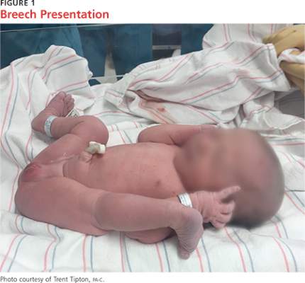
Known risk factors for DDH include breech presentation (see Figure 1), positive family history, and female gender.5,8-10 Female infants are eight times more likely than males to develop DDH.10 Firstborn status is also recognized as an associated risk factor, which may be attributable to space constraints in utero. This hypothesis is further supported by the relative DDH-protective effect of prematurity and low birth weight. Other potential risk factors include advanced maternal age, birth weight that is high for gestational age, decreased hip abduction, and joint laxity. However, the majority of patients with hip dysplasia have no identifiable risk factors.3,5,9,11,12
Swaddling, which often maintains the hips in an adducted and/or extended position, has also been strongly associated with hip dysplasia.5,13 Multiple organizations, including the AAOS,AAP, POSNA, and the International Hip Dysplasia Institute, have developed or endorsed hip-healthy swaddling recommendations to minimize the risk for DDH in swaddled infants.13-15 Such practices allow the infant’s legs to bend up and out at the hips, promoting free hip movement, flexion, and abduction.13,15 Swaddling has demonstrated multiple benefits (including improved sleep and relief of excessive crying13) and continues to be recommended by many US providers; however, those caring for infants at risk for DDH should avoid traditional swaddling and/or practice hip-healthy swaddling techniques.10,13,14 Early diagnosis starts with the clinician’s knowledge of DDH risk factors and the recommended screening protocols. The presence of multiple risk factors will increase the likelihood of this condition and should lower the clinician’s threshold for ordering additional screening, regardless of hip exam findings.
PHYSICAL EXAM
Both AAP and AAOS guidelines recommend clinical screening for DDH with physical exam in all newborns.1,3 A head-to-toe musculoskeletal exam is warranted during the initial evaluation of every newborn in order to assess for any known DDH-associated conditions, which may include neuromuscular disorders, torticollis, and metatarsus adductus.5
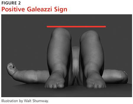
Initial evaluation of an infant with DDH may reveal nonspecific findings, including asymmetric skin folds and limb-length inequality. The Galeazzi sign should be sought by aligning flexed knees with the child in the supine position and assessing for uneven knee heights (see Figure 2). Unilateral posterior hip dislocation or femoral shortening represents a positive Galeazzi sign.16 Joint laxity and limited hip abduction have also been associated with DDH.1,10
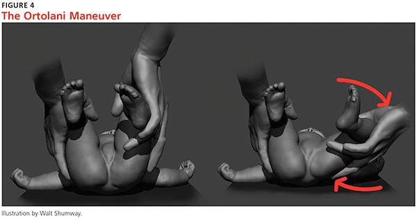
Barlow and Ortolani exams are more specific to DDH and should be completed at newborn screening and each subsequent well-baby exam.1 The Barlow maneuver is a provocative test with flexion, adduction, and posterior pressure through the infant’s hip (Figure 3). A palpable clunk during the Barlow maneuver indicates positive instability with posterior displacement. The Ortolani test is a reductive maneuver requiring abduction with posterior pressure to lift the greater trochanter (Figure 4). A clunk sensation with this test is positive for reduction of the hip.

The infant’s diaper should be removed during the hip evaluation. These exams are more reliable when each hip is evaluated separately with the pelvis stabilized.10 All physical exam findings must be carefully documented at each encounter.1,17
It is critical for the examiner to understand the appropriate technique and potential results when conducting each of these specialized hip exams. A true positive finding is the clunking sensation that occurs with the dislocation or relocation of the affected hip; this finding is better felt than heard. In contrast, a benign hip click with these maneuvers is a more subtle sensation—typically, a soft-tissue snapping or catching—and is not diagnostic of DDH. A click is not a clunk and is not indicative of DDH.1,3
DDH may present later in infancy or early childhood; therefore, DDH should remain within the differential diagnosis for gait asymmetry, unequal hip motion, or limb-length discrepancy. It may be beneficial to continue to evaluate for these developments during routine exams as part of a thorough pediatric musculoskeletal assessment, particularly in patients with documented risk factors for DDH.1,3,4 Delay in diagnosis of DDH, it should be noted, is a relatively common complaint in pediatric medical malpractice lawsuits; until the early 2000s, this condition represented about 75% of claims in one medical malpractice database.The decrease in claims has been attributed to better awareness and earlier diagnosis of DDH. 17
Continue for the diagnosis >>
DIAGNOSIS
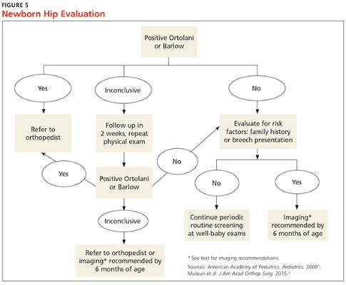
A positive Ortolani or Barlow sign is diagnostic and warrants prompt orthopedic referral (Figure 5). If physical examination results are equivocal or inconclusive, follow-up at two weeks is recommended, with continued routine follow-up until walking is achieved. Patients with persistent equivocal findings at the two-week follow-up warrant ultrasound at age 3 to 4 weeks or orthopedic referral. Infants with significant risk factors, particularly breech presentation at birth, should also undergo imaging.18 AAP recommends ultrasound at age 6 weeks or radiograph after 4 months of age.1,18 AAOS recommends performing an imaging study before age 6 months when at least one of the following risk factors is present: breech presentation, positive family history of DDH, or previous clinical instability (moderate level of evidence).3
IMAGING
Ultrasound is the diagnostic test of choice for infants because radiographs have limited value until the femoral heads begin to ossify at age 4 to 6 months.18 Ultrasonography allows for visualization of the cartilaginous portion of the acetabulum and femoral head.1 Dynamic stressing is performed during ultrasound to assess the level of hip stability. A provider trained in ultrasound will measure the depth of the acetabulum and identify any potential laxity or instability of the hip joint. Accuracy of these findings is largely dependent on the experience and skill of the examiner.
Ultrasound evaluation is not recommended until after age 3 to 4 weeks. Earlier findings may include mild laxity and immature morphology of the acetabulum, which often resolve spontaneously.1,18 Use of ultrasound is currently recommended only to confirm diagnostic suspicion, based on clinical findings, or for infants with significant risk factors.18 Universal ultrasound screening in newborns is not recommended and would incur unnecessary costs.1,3,9 Plain radiographs are used after age 4 months to confirm a diagnosis of DDH or to assess for residual dysplasia.3,18
Continue for management >>
MANAGEMENT
Once hip dysplasia is suggested by physical exam or imaging study, the child’s subsequent care should be provided by an orthopedic specialist with experience in treating this condition. Treatment is preferably initiated before age 6 weeks.12 The specifics of treatment are largely based on age at diagnosis and the severity of dysplasia.
The goal of treatment is to maintain the hips in a stable position with the femoral head well covered by the acetabulum. This will improve anatomic development and function. Early clinical diagnosis is often sufficient to justify initiating conservative treatment; additionally, early detection of DDH can considerably reduce the need for surgical intervention.12 Although the potential for spontaneous resolution is high, the consequences associated with delay in care can be significant.
Preferred initial management, which can be initiated before confirmation of DDH by ultrasound, involves implementation of soft abduction support.19 The Pavlik harness is the support design of choice (Figure 6).12 This harness maintains hip flexion and abduction, creating concentric reduction of the femoral head. The brace is highly successful when its use is initiated early. Treatment in a Pavlik harness requires nearly full-time wear and close monitoring by a clinician. Unlikely potential risks associated with this treatment include avascular necrosis and femoral nerve palsy.4
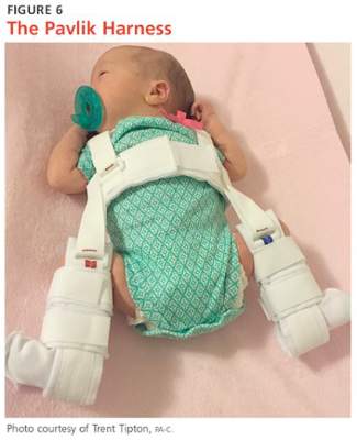
Ultrasonography is used to further monitor treatment and to determine length of wear. Long-term results suggest a success rate exceeding 90%.20,21 However, this rate may be falsely elevated due to the number of hips that likely would have improved spontaneously without treatment.6,19
The Pavlik harness becomes less effective with increasing age, and a more rigid abduction brace may be considered in infants older than 6 months.20 Overall outcomes improve once the femoral head is consistently maintained in the acetabulum. Delay in treatment is associated with an increase in the long-term complications associated with residual hip dysplasia.22
Once an infant is undergoing treatment for DDH in a Pavlik harness, there is no need for primary care providers to continue to perform provocative testing, such as the Ortolani or Barlow test, at routine well-baby checks. Unnecessary stress to the hips is not beneficial, and any new results will not change the treatment being provided by the orthopedic specialist. Adjustments to the fit of the harness should be made only by the orthopedist, unless femoral nerve palsy is noted on exam. This development warrants immediate discontinuation of harness use until symptoms resolve.21
Abduction bracing may not be suitable for all cases of hip dysplasia. Newborns with irreducible hips, more advanced dysplasia, or associated neuromuscular or syndromic disorder may require closed versus open reduction and casting. More invasive surgical options may also be considered in advanced dysplasia in order to reshape the joint and improve function.20,22
Continue for patient education >>
PATIENT EDUCATION
Parents should be fully educated on the options for managing hip dysplasia. Once DDH is diagnosed, prompt referral to an orthopedic specialist is critical in order to weigh the treatment options and to develop the appropriate individualized plan for each child. Once treatment is initiated, parental compliance is essential; frequent meetings between parents and the specialist are important.
Parents of infants with known risk factors for and/or suspicion of hip dysplasia should also be educated on hip-healthy swaddling to allow for free motion of the hips and knees.10,13 Advise them that some commercial baby carriers and slings may maintain the hips in an undesirable extended position. In both swaddling and with baby carriers, care should be taken to allow for hip abduction and flexion. Caution should also be taken during diaper changes to avoid lifting the legs and thereby causing unnecessary stress to the hips.
CONCLUSION
Developmental dysplasia of the hip can be a disabling pediatric condition. Early diagnosis improves the likelihood of successful treatment during infancy and can prevent serious complications. If untreated, DDH can lead to joint degeneration and premature arthritis. Recognition and treatment within the first six weeks of life is crucial to the overall outcome.
The role of a primary care provider is to identify hip dysplasia risk factors and recognize associated physical exam findings in order to refer to an orthopedic specialist in a timely manner. Guidelines from the AAP, POSNA, and AAOS help direct this process in order to effectively identify infants at risk and in need of treatment.
REFERENCES
1. American Academy of Pediatrics. Committee on Quality Improvement, Subcommittee on Developmental Dysplasia of the Hip. Clinical practice guideline: early detection of developmental dysplasia of the hip. Pediatrics. 2000;105(4 pt 1):896-905.
2. Schwend RM, Schoenecker P, Richards BS, et al. Screening the newborn for developmental dysplasia of the hip: now what do we do? J Pediatr Orthop. 2007;27(6):607-610.
3. Mulpuri K, Song KM, Goldberg MJ, Sevarino K. Detection and nonoperative management of pediatric developmental dysplasia of the hip in infants up to six months of age. J Am Acad Orthop Surg. 2015;23(3):202-205.
4. Thomas SRYW. A review of long-term outcomes for late presenting developmental hip dysplasia. Bone Joint J. 2015;97-B(6):729-733.
5. Loder RT, Skopelja EN. The epidemiology and demographics of hip dysplasia. ISRN Orthop. 2011;2011:238607.
6. US Preventive Services Task Force. Screening for developmental dysplasia of the hip: recommendation statement. Pediatrics. 2006;117(3):898-902.
7. Shorter D, Hong T, Osborn DA. Screening programmes for developmental dysplasia of the hip in newborn infants. Cochrane Database Syst Rev. 2011;(9):CD004595.
8. Loder RT, Shafer C. The demographics of developmental hip dysplasia in the Midwestern United States (Indiana). J Child Orthop. 2015;9(1):93-98.
9. Paton RW, Hinduja K, Thomas CD. The significance of at-risk factors in ultrasound surveillance of developmental dysplasia of the hip: a ten-year prospective study. J Bone Joint Surg Br. 2005;87(9):1264-1266.
10. Alsaleem M, Set KK, Saadeh L. Developmental dysplasia of hip: a review. Clin Pediatr (Phila). 2015;54(10):921-928.
11. Chan A, McCaul KA, Cundy PJ, et al. Perinatal risk factors for developmental dysplasia of the hip. Arch Dis Child. 1997;76(2):F94-F100.
12. Godley DR. Assessment, diagnosis, and treatment of developmental dysplasia of the hip. JAAPA. 2013;26(3):54-58.
13. Van Sleuwen BE, Engelberts AC, Boere-Boonekamp MM, et al. Swaddling: a systematic review. Pediatrics. 2007;120(4):e1097-e1106.
14. American Academy of Orthopaedic Surgeons, American Association of Orthopaedic Surgeons. Position statement: swaddling and developmental hip dysplasia. www.aaos.org/uploadedFiles/PreProduction/About/Opinion_Statements/position/1186%20Swaddling%20and%20Developmental%20Hip%20Dysplasia.pdf. Accessed January 22, 2016.
15. Clarke NM. Swaddling and hip dysplasia: an orthopaedic perspective. Arch Dis Child. 2014;99(1):5-6.
16. Storer SK, Skaggs DL. Developmental dysplasia of the hip. Am Fam Physician. 2006;74(8):1310-1316.
17. McAbee GN, Donn SM, Mendelson RA, et al. Medical diagnoses commonly associated with pediatric malpractice lawsuits in the United States. Pediatrics. 2008;122(6):e1282-e1286.
18. Imrie M, Scott V, Stearns P, et al. Is ultrasound screening for DDH in babies born breech sufficient? J Child Orthop. 2010;4(1):3-8.
19. Chen HW, Chang CH, Tsai ST, et al. Natural progression of hip dysplasia in newborns: a reflection of hip ultrasonographic screenings in newborn nurseries. J Pediatr Orthop B. 2010;19(5):418-423.
20. Gans I, Flynn JM, Sankar WN. Abduction bracing for residual acetabular dysplasia in infantile DDH. J Pediatr Orthop. 2013;33(7):714-718.
21. Murnaghan ML, Browne RH, Sucato DJ, Birch J. Femoral nerve palsy in Pavlik harness treatment for developmental dysplasia of the hip. J Bone Joint Surg Am. 2011;93(5):493-499.
22. Dezateux C, Rosendahl K. Developmental dysplasia of the hip. Lancet. 2007;369(9572):1541-1552.
IN THIS ARTICLE
- Diagnosis
- Management
- Newborn hip evaluation algorithm
Developmental dysplasia of the hip (DDH), previously known as congenital dislocation of the hip, follows a spectrum of irregular anatomic hip development spanning from acetabular dysplasia to irreducible dislocation at birth. Early detection is critical to improve the overall prognosis. Prompt diagnosis requires understanding of potential risk factors, proficiency in physical examination techniques, and implementation of appropriate screening tools when indicated. Although current guidelines direct timing for physical exam screenings, imaging, and treatment, it is ultimately up to the provider to determine the best course of action on a case-by-case basis. This article provides a review of these topics and more.
CURRENT GUIDELINES
In 2000, the American Academy of Pediatrics (AAP) developed guidelines for detection of hip dysplasia, including recommendation of relevant physical exam screenings for all newborns.1 In 2007, the Pediatric Orthopaedic Society of North America (POSNA) encouraged providers to follow the AAP guidelines with a continued recommendation to perform newborn screening for hip instability and routine follow-up evaluations until the child achieves walking.2 The American Academy of Orthopaedic Surgeons (AAOS) also established clinical guidelines in 2014 that are endorsed by both AAP and POSNA.3 These guidelines support routine clinical screening; research evaluated infants up to 6 months old, however, limiting the recommendations to that age-group.
Failure to treat DDH early has been associated with serious negative sequelae that include chronic pain, degenerative arthritis, postural scoliosis, and early gait disturbances.4 Primary care providers are expected to perform thorough newborn hip exams with associated specialized tests (ie, Ortolani and Barlow, which are discussed in “Physical exam”) at each routine follow-up. Heightened clinical suspicion and risk factor awareness are key for primary care providers to promptly identify patients requiring orthopedic referral. With early diagnosis, a removable soft abduction brace can be applied as the initial treatment. When treatment is delayed, however, closed reduction under anesthesia or complex surgical intervention may be required.
EPIDEMIOLOGY
The etiology for DDH remains unknown. Hip dysplasia typically presents unilaterally but can also occur bilaterally. DDH is more likely to affect the left hip than the right.5
Reported incidence varies, ranging from 0.06 to 76.1 per 1,000 live births, and is largely affected by race and geographic location.5 Incidence is higher in countries where routine screening is required, by either physical examination or ultrasound (1.6 to 28.5 and 34.0 to 60.3 per 1,000, respectively), compared with countries not requiring routine screening (1.3 per 1,000). This may suggest that the majority of hip dysplasia cases are transient and resolve spontaneously without treatment.6,7
RISK FACTORS AND PATIENT HISTORY
Known risk factors for DDH include breech presentation (see Figure 1), positive family history, and female gender.5,8-10 Female infants are eight times more likely than males to develop DDH.10 Firstborn status is also recognized as an associated risk factor, which may be attributable to space constraints in utero. This hypothesis is further supported by the relative DDH-protective effect of prematurity and low birth weight. Other potential risk factors include advanced maternal age, birth weight that is high for gestational age, decreased hip abduction, and joint laxity. However, the majority of patients with hip dysplasia have no identifiable risk factors.3,5,9,11,12
Swaddling, which often maintains the hips in an adducted and/or extended position, has also been strongly associated with hip dysplasia.5,13 Multiple organizations, including the AAOS,AAP, POSNA, and the International Hip Dysplasia Institute, have developed or endorsed hip-healthy swaddling recommendations to minimize the risk for DDH in swaddled infants.13-15 Such practices allow the infant’s legs to bend up and out at the hips, promoting free hip movement, flexion, and abduction.13,15 Swaddling has demonstrated multiple benefits (including improved sleep and relief of excessive crying13) and continues to be recommended by many US providers; however, those caring for infants at risk for DDH should avoid traditional swaddling and/or practice hip-healthy swaddling techniques.10,13,14 Early diagnosis starts with the clinician’s knowledge of DDH risk factors and the recommended screening protocols. The presence of multiple risk factors will increase the likelihood of this condition and should lower the clinician’s threshold for ordering additional screening, regardless of hip exam findings.
PHYSICAL EXAM
Both AAP and AAOS guidelines recommend clinical screening for DDH with physical exam in all newborns.1,3 A head-to-toe musculoskeletal exam is warranted during the initial evaluation of every newborn in order to assess for any known DDH-associated conditions, which may include neuromuscular disorders, torticollis, and metatarsus adductus.5
Initial evaluation of an infant with DDH may reveal nonspecific findings, including asymmetric skin folds and limb-length inequality. The Galeazzi sign should be sought by aligning flexed knees with the child in the supine position and assessing for uneven knee heights (see Figure 2). Unilateral posterior hip dislocation or femoral shortening represents a positive Galeazzi sign.16 Joint laxity and limited hip abduction have also been associated with DDH.1,10
Barlow and Ortolani exams are more specific to DDH and should be completed at newborn screening and each subsequent well-baby exam.1 The Barlow maneuver is a provocative test with flexion, adduction, and posterior pressure through the infant’s hip (Figure 3). A palpable clunk during the Barlow maneuver indicates positive instability with posterior displacement. The Ortolani test is a reductive maneuver requiring abduction with posterior pressure to lift the greater trochanter (Figure 4). A clunk sensation with this test is positive for reduction of the hip.
The infant’s diaper should be removed during the hip evaluation. These exams are more reliable when each hip is evaluated separately with the pelvis stabilized.10 All physical exam findings must be carefully documented at each encounter.1,17
It is critical for the examiner to understand the appropriate technique and potential results when conducting each of these specialized hip exams. A true positive finding is the clunking sensation that occurs with the dislocation or relocation of the affected hip; this finding is better felt than heard. In contrast, a benign hip click with these maneuvers is a more subtle sensation—typically, a soft-tissue snapping or catching—and is not diagnostic of DDH. A click is not a clunk and is not indicative of DDH.1,3
DDH may present later in infancy or early childhood; therefore, DDH should remain within the differential diagnosis for gait asymmetry, unequal hip motion, or limb-length discrepancy. It may be beneficial to continue to evaluate for these developments during routine exams as part of a thorough pediatric musculoskeletal assessment, particularly in patients with documented risk factors for DDH.1,3,4 Delay in diagnosis of DDH, it should be noted, is a relatively common complaint in pediatric medical malpractice lawsuits; until the early 2000s, this condition represented about 75% of claims in one medical malpractice database.The decrease in claims has been attributed to better awareness and earlier diagnosis of DDH. 17
Continue for the diagnosis >>
DIAGNOSIS
A positive Ortolani or Barlow sign is diagnostic and warrants prompt orthopedic referral (Figure 5). If physical examination results are equivocal or inconclusive, follow-up at two weeks is recommended, with continued routine follow-up until walking is achieved. Patients with persistent equivocal findings at the two-week follow-up warrant ultrasound at age 3 to 4 weeks or orthopedic referral. Infants with significant risk factors, particularly breech presentation at birth, should also undergo imaging.18 AAP recommends ultrasound at age 6 weeks or radiograph after 4 months of age.1,18 AAOS recommends performing an imaging study before age 6 months when at least one of the following risk factors is present: breech presentation, positive family history of DDH, or previous clinical instability (moderate level of evidence).3
IMAGING
Ultrasound is the diagnostic test of choice for infants because radiographs have limited value until the femoral heads begin to ossify at age 4 to 6 months.18 Ultrasonography allows for visualization of the cartilaginous portion of the acetabulum and femoral head.1 Dynamic stressing is performed during ultrasound to assess the level of hip stability. A provider trained in ultrasound will measure the depth of the acetabulum and identify any potential laxity or instability of the hip joint. Accuracy of these findings is largely dependent on the experience and skill of the examiner.
Ultrasound evaluation is not recommended until after age 3 to 4 weeks. Earlier findings may include mild laxity and immature morphology of the acetabulum, which often resolve spontaneously.1,18 Use of ultrasound is currently recommended only to confirm diagnostic suspicion, based on clinical findings, or for infants with significant risk factors.18 Universal ultrasound screening in newborns is not recommended and would incur unnecessary costs.1,3,9 Plain radiographs are used after age 4 months to confirm a diagnosis of DDH or to assess for residual dysplasia.3,18
Continue for management >>
MANAGEMENT
Once hip dysplasia is suggested by physical exam or imaging study, the child’s subsequent care should be provided by an orthopedic specialist with experience in treating this condition. Treatment is preferably initiated before age 6 weeks.12 The specifics of treatment are largely based on age at diagnosis and the severity of dysplasia.
The goal of treatment is to maintain the hips in a stable position with the femoral head well covered by the acetabulum. This will improve anatomic development and function. Early clinical diagnosis is often sufficient to justify initiating conservative treatment; additionally, early detection of DDH can considerably reduce the need for surgical intervention.12 Although the potential for spontaneous resolution is high, the consequences associated with delay in care can be significant.
Preferred initial management, which can be initiated before confirmation of DDH by ultrasound, involves implementation of soft abduction support.19 The Pavlik harness is the support design of choice (Figure 6).12 This harness maintains hip flexion and abduction, creating concentric reduction of the femoral head. The brace is highly successful when its use is initiated early. Treatment in a Pavlik harness requires nearly full-time wear and close monitoring by a clinician. Unlikely potential risks associated with this treatment include avascular necrosis and femoral nerve palsy.4
Ultrasonography is used to further monitor treatment and to determine length of wear. Long-term results suggest a success rate exceeding 90%.20,21 However, this rate may be falsely elevated due to the number of hips that likely would have improved spontaneously without treatment.6,19
The Pavlik harness becomes less effective with increasing age, and a more rigid abduction brace may be considered in infants older than 6 months.20 Overall outcomes improve once the femoral head is consistently maintained in the acetabulum. Delay in treatment is associated with an increase in the long-term complications associated with residual hip dysplasia.22
Once an infant is undergoing treatment for DDH in a Pavlik harness, there is no need for primary care providers to continue to perform provocative testing, such as the Ortolani or Barlow test, at routine well-baby checks. Unnecessary stress to the hips is not beneficial, and any new results will not change the treatment being provided by the orthopedic specialist. Adjustments to the fit of the harness should be made only by the orthopedist, unless femoral nerve palsy is noted on exam. This development warrants immediate discontinuation of harness use until symptoms resolve.21
Abduction bracing may not be suitable for all cases of hip dysplasia. Newborns with irreducible hips, more advanced dysplasia, or associated neuromuscular or syndromic disorder may require closed versus open reduction and casting. More invasive surgical options may also be considered in advanced dysplasia in order to reshape the joint and improve function.20,22
Continue for patient education >>
PATIENT EDUCATION
Parents should be fully educated on the options for managing hip dysplasia. Once DDH is diagnosed, prompt referral to an orthopedic specialist is critical in order to weigh the treatment options and to develop the appropriate individualized plan for each child. Once treatment is initiated, parental compliance is essential; frequent meetings between parents and the specialist are important.
Parents of infants with known risk factors for and/or suspicion of hip dysplasia should also be educated on hip-healthy swaddling to allow for free motion of the hips and knees.10,13 Advise them that some commercial baby carriers and slings may maintain the hips in an undesirable extended position. In both swaddling and with baby carriers, care should be taken to allow for hip abduction and flexion. Caution should also be taken during diaper changes to avoid lifting the legs and thereby causing unnecessary stress to the hips.
CONCLUSION
Developmental dysplasia of the hip can be a disabling pediatric condition. Early diagnosis improves the likelihood of successful treatment during infancy and can prevent serious complications. If untreated, DDH can lead to joint degeneration and premature arthritis. Recognition and treatment within the first six weeks of life is crucial to the overall outcome.
The role of a primary care provider is to identify hip dysplasia risk factors and recognize associated physical exam findings in order to refer to an orthopedic specialist in a timely manner. Guidelines from the AAP, POSNA, and AAOS help direct this process in order to effectively identify infants at risk and in need of treatment.
REFERENCES
1. American Academy of Pediatrics. Committee on Quality Improvement, Subcommittee on Developmental Dysplasia of the Hip. Clinical practice guideline: early detection of developmental dysplasia of the hip. Pediatrics. 2000;105(4 pt 1):896-905.
2. Schwend RM, Schoenecker P, Richards BS, et al. Screening the newborn for developmental dysplasia of the hip: now what do we do? J Pediatr Orthop. 2007;27(6):607-610.
3. Mulpuri K, Song KM, Goldberg MJ, Sevarino K. Detection and nonoperative management of pediatric developmental dysplasia of the hip in infants up to six months of age. J Am Acad Orthop Surg. 2015;23(3):202-205.
4. Thomas SRYW. A review of long-term outcomes for late presenting developmental hip dysplasia. Bone Joint J. 2015;97-B(6):729-733.
5. Loder RT, Skopelja EN. The epidemiology and demographics of hip dysplasia. ISRN Orthop. 2011;2011:238607.
6. US Preventive Services Task Force. Screening for developmental dysplasia of the hip: recommendation statement. Pediatrics. 2006;117(3):898-902.
7. Shorter D, Hong T, Osborn DA. Screening programmes for developmental dysplasia of the hip in newborn infants. Cochrane Database Syst Rev. 2011;(9):CD004595.
8. Loder RT, Shafer C. The demographics of developmental hip dysplasia in the Midwestern United States (Indiana). J Child Orthop. 2015;9(1):93-98.
9. Paton RW, Hinduja K, Thomas CD. The significance of at-risk factors in ultrasound surveillance of developmental dysplasia of the hip: a ten-year prospective study. J Bone Joint Surg Br. 2005;87(9):1264-1266.
10. Alsaleem M, Set KK, Saadeh L. Developmental dysplasia of hip: a review. Clin Pediatr (Phila). 2015;54(10):921-928.
11. Chan A, McCaul KA, Cundy PJ, et al. Perinatal risk factors for developmental dysplasia of the hip. Arch Dis Child. 1997;76(2):F94-F100.
12. Godley DR. Assessment, diagnosis, and treatment of developmental dysplasia of the hip. JAAPA. 2013;26(3):54-58.
13. Van Sleuwen BE, Engelberts AC, Boere-Boonekamp MM, et al. Swaddling: a systematic review. Pediatrics. 2007;120(4):e1097-e1106.
14. American Academy of Orthopaedic Surgeons, American Association of Orthopaedic Surgeons. Position statement: swaddling and developmental hip dysplasia. www.aaos.org/uploadedFiles/PreProduction/About/Opinion_Statements/position/1186%20Swaddling%20and%20Developmental%20Hip%20Dysplasia.pdf. Accessed January 22, 2016.
15. Clarke NM. Swaddling and hip dysplasia: an orthopaedic perspective. Arch Dis Child. 2014;99(1):5-6.
16. Storer SK, Skaggs DL. Developmental dysplasia of the hip. Am Fam Physician. 2006;74(8):1310-1316.
17. McAbee GN, Donn SM, Mendelson RA, et al. Medical diagnoses commonly associated with pediatric malpractice lawsuits in the United States. Pediatrics. 2008;122(6):e1282-e1286.
18. Imrie M, Scott V, Stearns P, et al. Is ultrasound screening for DDH in babies born breech sufficient? J Child Orthop. 2010;4(1):3-8.
19. Chen HW, Chang CH, Tsai ST, et al. Natural progression of hip dysplasia in newborns: a reflection of hip ultrasonographic screenings in newborn nurseries. J Pediatr Orthop B. 2010;19(5):418-423.
20. Gans I, Flynn JM, Sankar WN. Abduction bracing for residual acetabular dysplasia in infantile DDH. J Pediatr Orthop. 2013;33(7):714-718.
21. Murnaghan ML, Browne RH, Sucato DJ, Birch J. Femoral nerve palsy in Pavlik harness treatment for developmental dysplasia of the hip. J Bone Joint Surg Am. 2011;93(5):493-499.
22. Dezateux C, Rosendahl K. Developmental dysplasia of the hip. Lancet. 2007;369(9572):1541-1552.
IN THIS ARTICLE
- Diagnosis
- Management
- Newborn hip evaluation algorithm
Developmental dysplasia of the hip (DDH), previously known as congenital dislocation of the hip, follows a spectrum of irregular anatomic hip development spanning from acetabular dysplasia to irreducible dislocation at birth. Early detection is critical to improve the overall prognosis. Prompt diagnosis requires understanding of potential risk factors, proficiency in physical examination techniques, and implementation of appropriate screening tools when indicated. Although current guidelines direct timing for physical exam screenings, imaging, and treatment, it is ultimately up to the provider to determine the best course of action on a case-by-case basis. This article provides a review of these topics and more.
CURRENT GUIDELINES
In 2000, the American Academy of Pediatrics (AAP) developed guidelines for detection of hip dysplasia, including recommendation of relevant physical exam screenings for all newborns.1 In 2007, the Pediatric Orthopaedic Society of North America (POSNA) encouraged providers to follow the AAP guidelines with a continued recommendation to perform newborn screening for hip instability and routine follow-up evaluations until the child achieves walking.2 The American Academy of Orthopaedic Surgeons (AAOS) also established clinical guidelines in 2014 that are endorsed by both AAP and POSNA.3 These guidelines support routine clinical screening; research evaluated infants up to 6 months old, however, limiting the recommendations to that age-group.
Failure to treat DDH early has been associated with serious negative sequelae that include chronic pain, degenerative arthritis, postural scoliosis, and early gait disturbances.4 Primary care providers are expected to perform thorough newborn hip exams with associated specialized tests (ie, Ortolani and Barlow, which are discussed in “Physical exam”) at each routine follow-up. Heightened clinical suspicion and risk factor awareness are key for primary care providers to promptly identify patients requiring orthopedic referral. With early diagnosis, a removable soft abduction brace can be applied as the initial treatment. When treatment is delayed, however, closed reduction under anesthesia or complex surgical intervention may be required.
EPIDEMIOLOGY
The etiology for DDH remains unknown. Hip dysplasia typically presents unilaterally but can also occur bilaterally. DDH is more likely to affect the left hip than the right.5
Reported incidence varies, ranging from 0.06 to 76.1 per 1,000 live births, and is largely affected by race and geographic location.5 Incidence is higher in countries where routine screening is required, by either physical examination or ultrasound (1.6 to 28.5 and 34.0 to 60.3 per 1,000, respectively), compared with countries not requiring routine screening (1.3 per 1,000). This may suggest that the majority of hip dysplasia cases are transient and resolve spontaneously without treatment.6,7
RISK FACTORS AND PATIENT HISTORY
Known risk factors for DDH include breech presentation (see Figure 1), positive family history, and female gender.5,8-10 Female infants are eight times more likely than males to develop DDH.10 Firstborn status is also recognized as an associated risk factor, which may be attributable to space constraints in utero. This hypothesis is further supported by the relative DDH-protective effect of prematurity and low birth weight. Other potential risk factors include advanced maternal age, birth weight that is high for gestational age, decreased hip abduction, and joint laxity. However, the majority of patients with hip dysplasia have no identifiable risk factors.3,5,9,11,12
Swaddling, which often maintains the hips in an adducted and/or extended position, has also been strongly associated with hip dysplasia.5,13 Multiple organizations, including the AAOS,AAP, POSNA, and the International Hip Dysplasia Institute, have developed or endorsed hip-healthy swaddling recommendations to minimize the risk for DDH in swaddled infants.13-15 Such practices allow the infant’s legs to bend up and out at the hips, promoting free hip movement, flexion, and abduction.13,15 Swaddling has demonstrated multiple benefits (including improved sleep and relief of excessive crying13) and continues to be recommended by many US providers; however, those caring for infants at risk for DDH should avoid traditional swaddling and/or practice hip-healthy swaddling techniques.10,13,14 Early diagnosis starts with the clinician’s knowledge of DDH risk factors and the recommended screening protocols. The presence of multiple risk factors will increase the likelihood of this condition and should lower the clinician’s threshold for ordering additional screening, regardless of hip exam findings.
PHYSICAL EXAM
Both AAP and AAOS guidelines recommend clinical screening for DDH with physical exam in all newborns.1,3 A head-to-toe musculoskeletal exam is warranted during the initial evaluation of every newborn in order to assess for any known DDH-associated conditions, which may include neuromuscular disorders, torticollis, and metatarsus adductus.5
Initial evaluation of an infant with DDH may reveal nonspecific findings, including asymmetric skin folds and limb-length inequality. The Galeazzi sign should be sought by aligning flexed knees with the child in the supine position and assessing for uneven knee heights (see Figure 2). Unilateral posterior hip dislocation or femoral shortening represents a positive Galeazzi sign.16 Joint laxity and limited hip abduction have also been associated with DDH.1,10
Barlow and Ortolani exams are more specific to DDH and should be completed at newborn screening and each subsequent well-baby exam.1 The Barlow maneuver is a provocative test with flexion, adduction, and posterior pressure through the infant’s hip (Figure 3). A palpable clunk during the Barlow maneuver indicates positive instability with posterior displacement. The Ortolani test is a reductive maneuver requiring abduction with posterior pressure to lift the greater trochanter (Figure 4). A clunk sensation with this test is positive for reduction of the hip.
The infant’s diaper should be removed during the hip evaluation. These exams are more reliable when each hip is evaluated separately with the pelvis stabilized.10 All physical exam findings must be carefully documented at each encounter.1,17
It is critical for the examiner to understand the appropriate technique and potential results when conducting each of these specialized hip exams. A true positive finding is the clunking sensation that occurs with the dislocation or relocation of the affected hip; this finding is better felt than heard. In contrast, a benign hip click with these maneuvers is a more subtle sensation—typically, a soft-tissue snapping or catching—and is not diagnostic of DDH. A click is not a clunk and is not indicative of DDH.1,3
DDH may present later in infancy or early childhood; therefore, DDH should remain within the differential diagnosis for gait asymmetry, unequal hip motion, or limb-length discrepancy. It may be beneficial to continue to evaluate for these developments during routine exams as part of a thorough pediatric musculoskeletal assessment, particularly in patients with documented risk factors for DDH.1,3,4 Delay in diagnosis of DDH, it should be noted, is a relatively common complaint in pediatric medical malpractice lawsuits; until the early 2000s, this condition represented about 75% of claims in one medical malpractice database.The decrease in claims has been attributed to better awareness and earlier diagnosis of DDH. 17
Continue for the diagnosis >>
DIAGNOSIS
A positive Ortolani or Barlow sign is diagnostic and warrants prompt orthopedic referral (Figure 5). If physical examination results are equivocal or inconclusive, follow-up at two weeks is recommended, with continued routine follow-up until walking is achieved. Patients with persistent equivocal findings at the two-week follow-up warrant ultrasound at age 3 to 4 weeks or orthopedic referral. Infants with significant risk factors, particularly breech presentation at birth, should also undergo imaging.18 AAP recommends ultrasound at age 6 weeks or radiograph after 4 months of age.1,18 AAOS recommends performing an imaging study before age 6 months when at least one of the following risk factors is present: breech presentation, positive family history of DDH, or previous clinical instability (moderate level of evidence).3
IMAGING
Ultrasound is the diagnostic test of choice for infants because radiographs have limited value until the femoral heads begin to ossify at age 4 to 6 months.18 Ultrasonography allows for visualization of the cartilaginous portion of the acetabulum and femoral head.1 Dynamic stressing is performed during ultrasound to assess the level of hip stability. A provider trained in ultrasound will measure the depth of the acetabulum and identify any potential laxity or instability of the hip joint. Accuracy of these findings is largely dependent on the experience and skill of the examiner.
Ultrasound evaluation is not recommended until after age 3 to 4 weeks. Earlier findings may include mild laxity and immature morphology of the acetabulum, which often resolve spontaneously.1,18 Use of ultrasound is currently recommended only to confirm diagnostic suspicion, based on clinical findings, or for infants with significant risk factors.18 Universal ultrasound screening in newborns is not recommended and would incur unnecessary costs.1,3,9 Plain radiographs are used after age 4 months to confirm a diagnosis of DDH or to assess for residual dysplasia.3,18
Continue for management >>
MANAGEMENT
Once hip dysplasia is suggested by physical exam or imaging study, the child’s subsequent care should be provided by an orthopedic specialist with experience in treating this condition. Treatment is preferably initiated before age 6 weeks.12 The specifics of treatment are largely based on age at diagnosis and the severity of dysplasia.
The goal of treatment is to maintain the hips in a stable position with the femoral head well covered by the acetabulum. This will improve anatomic development and function. Early clinical diagnosis is often sufficient to justify initiating conservative treatment; additionally, early detection of DDH can considerably reduce the need for surgical intervention.12 Although the potential for spontaneous resolution is high, the consequences associated with delay in care can be significant.
Preferred initial management, which can be initiated before confirmation of DDH by ultrasound, involves implementation of soft abduction support.19 The Pavlik harness is the support design of choice (Figure 6).12 This harness maintains hip flexion and abduction, creating concentric reduction of the femoral head. The brace is highly successful when its use is initiated early. Treatment in a Pavlik harness requires nearly full-time wear and close monitoring by a clinician. Unlikely potential risks associated with this treatment include avascular necrosis and femoral nerve palsy.4
Ultrasonography is used to further monitor treatment and to determine length of wear. Long-term results suggest a success rate exceeding 90%.20,21 However, this rate may be falsely elevated due to the number of hips that likely would have improved spontaneously without treatment.6,19
The Pavlik harness becomes less effective with increasing age, and a more rigid abduction brace may be considered in infants older than 6 months.20 Overall outcomes improve once the femoral head is consistently maintained in the acetabulum. Delay in treatment is associated with an increase in the long-term complications associated with residual hip dysplasia.22
Once an infant is undergoing treatment for DDH in a Pavlik harness, there is no need for primary care providers to continue to perform provocative testing, such as the Ortolani or Barlow test, at routine well-baby checks. Unnecessary stress to the hips is not beneficial, and any new results will not change the treatment being provided by the orthopedic specialist. Adjustments to the fit of the harness should be made only by the orthopedist, unless femoral nerve palsy is noted on exam. This development warrants immediate discontinuation of harness use until symptoms resolve.21
Abduction bracing may not be suitable for all cases of hip dysplasia. Newborns with irreducible hips, more advanced dysplasia, or associated neuromuscular or syndromic disorder may require closed versus open reduction and casting. More invasive surgical options may also be considered in advanced dysplasia in order to reshape the joint and improve function.20,22
Continue for patient education >>
PATIENT EDUCATION
Parents should be fully educated on the options for managing hip dysplasia. Once DDH is diagnosed, prompt referral to an orthopedic specialist is critical in order to weigh the treatment options and to develop the appropriate individualized plan for each child. Once treatment is initiated, parental compliance is essential; frequent meetings between parents and the specialist are important.
Parents of infants with known risk factors for and/or suspicion of hip dysplasia should also be educated on hip-healthy swaddling to allow for free motion of the hips and knees.10,13 Advise them that some commercial baby carriers and slings may maintain the hips in an undesirable extended position. In both swaddling and with baby carriers, care should be taken to allow for hip abduction and flexion. Caution should also be taken during diaper changes to avoid lifting the legs and thereby causing unnecessary stress to the hips.
CONCLUSION
Developmental dysplasia of the hip can be a disabling pediatric condition. Early diagnosis improves the likelihood of successful treatment during infancy and can prevent serious complications. If untreated, DDH can lead to joint degeneration and premature arthritis. Recognition and treatment within the first six weeks of life is crucial to the overall outcome.
The role of a primary care provider is to identify hip dysplasia risk factors and recognize associated physical exam findings in order to refer to an orthopedic specialist in a timely manner. Guidelines from the AAP, POSNA, and AAOS help direct this process in order to effectively identify infants at risk and in need of treatment.
REFERENCES
1. American Academy of Pediatrics. Committee on Quality Improvement, Subcommittee on Developmental Dysplasia of the Hip. Clinical practice guideline: early detection of developmental dysplasia of the hip. Pediatrics. 2000;105(4 pt 1):896-905.
2. Schwend RM, Schoenecker P, Richards BS, et al. Screening the newborn for developmental dysplasia of the hip: now what do we do? J Pediatr Orthop. 2007;27(6):607-610.
3. Mulpuri K, Song KM, Goldberg MJ, Sevarino K. Detection and nonoperative management of pediatric developmental dysplasia of the hip in infants up to six months of age. J Am Acad Orthop Surg. 2015;23(3):202-205.
4. Thomas SRYW. A review of long-term outcomes for late presenting developmental hip dysplasia. Bone Joint J. 2015;97-B(6):729-733.
5. Loder RT, Skopelja EN. The epidemiology and demographics of hip dysplasia. ISRN Orthop. 2011;2011:238607.
6. US Preventive Services Task Force. Screening for developmental dysplasia of the hip: recommendation statement. Pediatrics. 2006;117(3):898-902.
7. Shorter D, Hong T, Osborn DA. Screening programmes for developmental dysplasia of the hip in newborn infants. Cochrane Database Syst Rev. 2011;(9):CD004595.
8. Loder RT, Shafer C. The demographics of developmental hip dysplasia in the Midwestern United States (Indiana). J Child Orthop. 2015;9(1):93-98.
9. Paton RW, Hinduja K, Thomas CD. The significance of at-risk factors in ultrasound surveillance of developmental dysplasia of the hip: a ten-year prospective study. J Bone Joint Surg Br. 2005;87(9):1264-1266.
10. Alsaleem M, Set KK, Saadeh L. Developmental dysplasia of hip: a review. Clin Pediatr (Phila). 2015;54(10):921-928.
11. Chan A, McCaul KA, Cundy PJ, et al. Perinatal risk factors for developmental dysplasia of the hip. Arch Dis Child. 1997;76(2):F94-F100.
12. Godley DR. Assessment, diagnosis, and treatment of developmental dysplasia of the hip. JAAPA. 2013;26(3):54-58.
13. Van Sleuwen BE, Engelberts AC, Boere-Boonekamp MM, et al. Swaddling: a systematic review. Pediatrics. 2007;120(4):e1097-e1106.
14. American Academy of Orthopaedic Surgeons, American Association of Orthopaedic Surgeons. Position statement: swaddling and developmental hip dysplasia. www.aaos.org/uploadedFiles/PreProduction/About/Opinion_Statements/position/1186%20Swaddling%20and%20Developmental%20Hip%20Dysplasia.pdf. Accessed January 22, 2016.
15. Clarke NM. Swaddling and hip dysplasia: an orthopaedic perspective. Arch Dis Child. 2014;99(1):5-6.
16. Storer SK, Skaggs DL. Developmental dysplasia of the hip. Am Fam Physician. 2006;74(8):1310-1316.
17. McAbee GN, Donn SM, Mendelson RA, et al. Medical diagnoses commonly associated with pediatric malpractice lawsuits in the United States. Pediatrics. 2008;122(6):e1282-e1286.
18. Imrie M, Scott V, Stearns P, et al. Is ultrasound screening for DDH in babies born breech sufficient? J Child Orthop. 2010;4(1):3-8.
19. Chen HW, Chang CH, Tsai ST, et al. Natural progression of hip dysplasia in newborns: a reflection of hip ultrasonographic screenings in newborn nurseries. J Pediatr Orthop B. 2010;19(5):418-423.
20. Gans I, Flynn JM, Sankar WN. Abduction bracing for residual acetabular dysplasia in infantile DDH. J Pediatr Orthop. 2013;33(7):714-718.
21. Murnaghan ML, Browne RH, Sucato DJ, Birch J. Femoral nerve palsy in Pavlik harness treatment for developmental dysplasia of the hip. J Bone Joint Surg Am. 2011;93(5):493-499.
22. Dezateux C, Rosendahl K. Developmental dysplasia of the hip. Lancet. 2007;369(9572):1541-1552.
Gender nonconforming LGBTQ youth more prone to bullying, harassment
WASHINGTON – Lesbian, gay, bisexual, transgender, questioning (LGBTQ) children and adolescents who are gender nonconforming are more susceptible to bullying at school than are those who don’t, so schools should make it a point to integrate LGBTQ outreach into its programs and to combat bullying against this group of students .
“In recent years, there’s been heightened national attention in the United States [on] harassment, bullying, and violence victimization, especially in schools and particularly targeting [LGBT] youth,” explained Allegra R. Gordon, Sc.D., of Boston Children’s Hospital.

“There’s substantial public health literature documenting an array of mental and physical health correlates of such victimization. However, there’s been less focus on discrimination and violence victimization targeting gender expression, broadly, although there is some evidence that this is an area of concern,” said Dr. Gordon, who presented the findings at the annual meeting of the Society for Adolescent Health and Medicine.
Dr. Gordon and her coinvestigators analyzed data on 5,503 public school students in grades 9-12 from four specific school districts in the United States: Los Angeles; San Diego; Chicago; and Broward County, Fla. These school districts were chosen because they had recently added a new question to the survey regarding socially assigned gender expression, making them the first districts in the nation to do so in 2013.
The data accumulated came from the 2013 Youth Risk Behavior Surveillance System, a survey conducted by the Centers for Disease Control and Prevention every 2 years to determine “the prevalence of health risk behaviors [and] assess whether health risk behaviors increase, decrease, or stay the same over time,” among other things.
The primary outcome of the study was to determine if sex and/or sexual orientation was in any way responsible for whether or not a student conformed to a specific gender – if not, the student was marked as GNC, or gender nonconformity – and if there was any association between GNC youths and in-school victimization. Youth who responded as being GNC were categorized as either most conforming, moderately nonconforming, or most noncomforming.
Nonconformity for each subject was defined based on “socially assigned gender expression, which is the extent to which a person is perceived by others as conforming or not conforming to culturally defined ideas of masculine or feminine appearance and behavior,” Dr. Gordon explained.
Results indicated a strong association between youths identifying as GNC and school-based victimization, with incidences of bullying increasing with greater GNC identification. Out of all GNC youth, 14% reported experiencing some form of bullying, 8% reported missing school because they felt unsafe being there, and 6% reported either being threatened or having been physically injured because of their gender identification.
After adjustment, Dr. Gordon and her coinvestigators found that the higher the degree of GNC a subject had, the more likely they would be victims of bullying, with steadily increasing odds ratios as the level of GNC rose: moderately nonconforming, 1.40 (range, 1.18-1.68) and most nonconforming, 2.01 (1.38-2.93). The latter group also had the highest odds of being absent from school, with an OR of 2.06 (2.10-4.44), and being either threatened with injury or actually getting injured having an OR of 2.79 (1.58-4.91).
“A key take-home here as well is given that the majority of the youth in the sample are heterosexual, the majority of youth in that most gender nonconforming group are heterosexual, so these are issues that apply across sexual orientation identities,” said Dr. Gordon.
Dr. Gordon did not report any relevant financial disclosures.
WASHINGTON – Lesbian, gay, bisexual, transgender, questioning (LGBTQ) children and adolescents who are gender nonconforming are more susceptible to bullying at school than are those who don’t, so schools should make it a point to integrate LGBTQ outreach into its programs and to combat bullying against this group of students .
“In recent years, there’s been heightened national attention in the United States [on] harassment, bullying, and violence victimization, especially in schools and particularly targeting [LGBT] youth,” explained Allegra R. Gordon, Sc.D., of Boston Children’s Hospital.
“There’s substantial public health literature documenting an array of mental and physical health correlates of such victimization. However, there’s been less focus on discrimination and violence victimization targeting gender expression, broadly, although there is some evidence that this is an area of concern,” said Dr. Gordon, who presented the findings at the annual meeting of the Society for Adolescent Health and Medicine.
Dr. Gordon and her coinvestigators analyzed data on 5,503 public school students in grades 9-12 from four specific school districts in the United States: Los Angeles; San Diego; Chicago; and Broward County, Fla. These school districts were chosen because they had recently added a new question to the survey regarding socially assigned gender expression, making them the first districts in the nation to do so in 2013.
The data accumulated came from the 2013 Youth Risk Behavior Surveillance System, a survey conducted by the Centers for Disease Control and Prevention every 2 years to determine “the prevalence of health risk behaviors [and] assess whether health risk behaviors increase, decrease, or stay the same over time,” among other things.
The primary outcome of the study was to determine if sex and/or sexual orientation was in any way responsible for whether or not a student conformed to a specific gender – if not, the student was marked as GNC, or gender nonconformity – and if there was any association between GNC youths and in-school victimization. Youth who responded as being GNC were categorized as either most conforming, moderately nonconforming, or most noncomforming.
Nonconformity for each subject was defined based on “socially assigned gender expression, which is the extent to which a person is perceived by others as conforming or not conforming to culturally defined ideas of masculine or feminine appearance and behavior,” Dr. Gordon explained.
Results indicated a strong association between youths identifying as GNC and school-based victimization, with incidences of bullying increasing with greater GNC identification. Out of all GNC youth, 14% reported experiencing some form of bullying, 8% reported missing school because they felt unsafe being there, and 6% reported either being threatened or having been physically injured because of their gender identification.
After adjustment, Dr. Gordon and her coinvestigators found that the higher the degree of GNC a subject had, the more likely they would be victims of bullying, with steadily increasing odds ratios as the level of GNC rose: moderately nonconforming, 1.40 (range, 1.18-1.68) and most nonconforming, 2.01 (1.38-2.93). The latter group also had the highest odds of being absent from school, with an OR of 2.06 (2.10-4.44), and being either threatened with injury or actually getting injured having an OR of 2.79 (1.58-4.91).
“A key take-home here as well is given that the majority of the youth in the sample are heterosexual, the majority of youth in that most gender nonconforming group are heterosexual, so these are issues that apply across sexual orientation identities,” said Dr. Gordon.
Dr. Gordon did not report any relevant financial disclosures.
WASHINGTON – Lesbian, gay, bisexual, transgender, questioning (LGBTQ) children and adolescents who are gender nonconforming are more susceptible to bullying at school than are those who don’t, so schools should make it a point to integrate LGBTQ outreach into its programs and to combat bullying against this group of students .
“In recent years, there’s been heightened national attention in the United States [on] harassment, bullying, and violence victimization, especially in schools and particularly targeting [LGBT] youth,” explained Allegra R. Gordon, Sc.D., of Boston Children’s Hospital.
“There’s substantial public health literature documenting an array of mental and physical health correlates of such victimization. However, there’s been less focus on discrimination and violence victimization targeting gender expression, broadly, although there is some evidence that this is an area of concern,” said Dr. Gordon, who presented the findings at the annual meeting of the Society for Adolescent Health and Medicine.
Dr. Gordon and her coinvestigators analyzed data on 5,503 public school students in grades 9-12 from four specific school districts in the United States: Los Angeles; San Diego; Chicago; and Broward County, Fla. These school districts were chosen because they had recently added a new question to the survey regarding socially assigned gender expression, making them the first districts in the nation to do so in 2013.
The data accumulated came from the 2013 Youth Risk Behavior Surveillance System, a survey conducted by the Centers for Disease Control and Prevention every 2 years to determine “the prevalence of health risk behaviors [and] assess whether health risk behaviors increase, decrease, or stay the same over time,” among other things.
The primary outcome of the study was to determine if sex and/or sexual orientation was in any way responsible for whether or not a student conformed to a specific gender – if not, the student was marked as GNC, or gender nonconformity – and if there was any association between GNC youths and in-school victimization. Youth who responded as being GNC were categorized as either most conforming, moderately nonconforming, or most noncomforming.
Nonconformity for each subject was defined based on “socially assigned gender expression, which is the extent to which a person is perceived by others as conforming or not conforming to culturally defined ideas of masculine or feminine appearance and behavior,” Dr. Gordon explained.
Results indicated a strong association between youths identifying as GNC and school-based victimization, with incidences of bullying increasing with greater GNC identification. Out of all GNC youth, 14% reported experiencing some form of bullying, 8% reported missing school because they felt unsafe being there, and 6% reported either being threatened or having been physically injured because of their gender identification.
After adjustment, Dr. Gordon and her coinvestigators found that the higher the degree of GNC a subject had, the more likely they would be victims of bullying, with steadily increasing odds ratios as the level of GNC rose: moderately nonconforming, 1.40 (range, 1.18-1.68) and most nonconforming, 2.01 (1.38-2.93). The latter group also had the highest odds of being absent from school, with an OR of 2.06 (2.10-4.44), and being either threatened with injury or actually getting injured having an OR of 2.79 (1.58-4.91).
“A key take-home here as well is given that the majority of the youth in the sample are heterosexual, the majority of youth in that most gender nonconforming group are heterosexual, so these are issues that apply across sexual orientation identities,” said Dr. Gordon.
Dr. Gordon did not report any relevant financial disclosures.
AT THE SAHM ANNUAL MEETING
Key clinical point: Gender identification among LGBTQ youth may predispose them to bullying at school by their peers.
Major finding: Among gender nonconforming youth, 14% reported being bullied, 8% reported missing school, and 6% reported being threatened or injured.
Data source: Retrospective cohort analysis of 5,503 students in grades 9-12 from four counties in the United States, collected in 2013.
Disclosures: Dr. Gordon did not report any relevant disclosures.

Considering learning disorders
Introduction
Often, it can be difficult for providers to fully understand why children may be presenting with behavioral problems that appear to be occurring principally in specific environments. Parents may bring their children for an evaluation based upon reports they are receiving from teachers and other school personnel who may be observing more prominent oppositionality, social struggles, troubles following instructions, and frustration intolerance than the parents are experiencing in the home. Additionally, youth may begin to display school refusal and voice strong negative feelings about school. Although at face value, these problems may indicate potential diagnoses like oppositional defiant disorder and attention-deficit/hyperactivity disorder, one also should consider other underlying “non–mental health” issues that could greatly influence school success and present with a range of emotional and behavioral struggles.
Case Summary

Brynn is an animated, social, and strong-willed 4 year-old girl who experienced delays in her receptive and expressive language that prompted her engagement in early intervention around 18 months of age. She and her family continued to receive developmental services over the next few years and, at the age of 3 years, because of ongoing speech and language challenges, she was enrolled in a preschool individualized education program. In her program, Brynn participates in an array of specialized instruction, but her educators comment that she is not making expected academic progress and has troubles “holding onto information,” focusing, and participating meaningfully with peers in a consistent manner.
At times, Brynn can be impulsive, aggressive, and volatile – behavioral traits that mom denies are occurring in the home. A diagnosis of an autism spectrum disorder has been ruled out, and Brynn’s hearing and vision are tested to be normal. Brynn’s mother feels confused by the reports she’s getting from school; “I want Brynn to be happy,” she shares. “I needed special help to read in high school, and I worry about her future.”
Discussion
Learning disorders (or learning disabilities, using educational terminology) are defined as neurologically rooted problems that affect academic achievement via the receiving, processing, or communication of information. They occur in 1 in 10 children (Pediatrics. 2007 Feb;119 Suppl 1:S77-83) and can present with specific problems in reading, writing, and mathematics while having considerable influence on related aptitudes in language, social ability, self-help, and motor functioning. Dyslexia – a developmental reading disorder – is the most common type of learning disorder (LD). Although it’s not clear what causes learning disorders, there are several factors that are thought to play a role in their development, including hereditary factors and problems with pregnancy and birth. Having developmental delays also can place children at risk for having later learning problems that may not be identifiable until a child enters a more structured learning environment. At clinical visits during the preschool years, particularly with a child who may have had earlier developmental concerns, the pediatrician should inquire about a range of “warning signs” that may indicate a need for additional screening and evaluation for specific learning issues.
The National Center for Learning Disabilities offers a range of practical tips for pediatricians who may want to further explore parental and teacher concerns by asking questions related to literacy, writing, language, and social-emotional skills, attention, and gross and fine motor movements in a developmentally informed manner. Further exploring Brynn’s mother’s comments in the context of her daughter not progressing in school revealed a history of difficulty retaining new words, troubles learning colors and shapes, challenges remembering rules, and particular difficulties engaging in group play with other 4-year-olds – all potential signals for a learning disorder. This alerted educators that she may indeed be struggling with issues beyond that of an enduring speech-language delay.
With the suspicion that Brynn was presenting with signs and symptoms suggestive of a learning disorder, her family was educated about the Individuals with Disabilities Education Act, and it was recommended she receive a comprehensive psychoeducational evaluation to further assess her intellectual profile, academic achievement, social functioning, and performance in the classroom using standardized tools. These tools, among other objectives, can help the child’s team offer a more definitive LD diagnosis while informing the potential development of special education supports and assessing for an intellectual disability. The DSM 5 indicates that LD diagnostic criteria are to be met based upon a clinical synthesis of history, school reports, and psychoeducational assessment.

It’s been long established that children with learning problems frequently have co-occurring emotional and behavioral troubles (Arch Dis Child. 1974 Apr; 49[4]:249-56), many of which also should be considered as differential diagnoses in a child with school problems – as such, a complicated interplay of learning disorders and internalizing and externalizing conditions is often appreciated in school-age children with academic difficulties. At times, learning disorders can lead to emotional distress and could also be misdiagnosed as primary emotional and behavioral challenges. Specifically, children with learning problems are at risk for struggling with low self-esteem, loneliness, and anxiety, which also can be associated with mood disorders, school dropout, victimization, and engaging in substance use.
In Brynn’s case, the results of the Teacher Report Forms and Child Behavior Checklists were reviewed, revealing some evidence that she was experiencing clinical-level problems with her attention, but a discrepancy was noted between the teacher and parent report (the teachers endorsing more clinically significant symptoms). Although co-occurring attention-deficit/hyperactivity disorder (ADHD) is not uncommon in children with a learning disorder (Pediatrics. 2011 Mar;127[3]:462-70), we did not feel Brynn met criteria for this. We elected not to provide an ADHD diagnosis but are mindful that her attentional concerns should be closely monitored over time; a diagnosis may be more relevant in the future, perhaps influencing Brynn’s eligibility for services and treatment planning. Furthermore, comorbidity with ADHD is predictive of worse mental health outcomes, compared with learning disabilities presenting without ADHD.
Clinical Pearl
Pediatricians should consider the possibility of a child having a learning disorder in youth who display risk factors (family history of learning concerns, atypical development, prematurity, etc.) and have problems at school. Such problems may be presenting with emotional and behavioral symptoms that could mask a child’s primary learning impairments. Learning disorders also frequently co-occur with psychiatric conditions, but engaging children in effective interventions (school-based supports) could potentially attenuate the risk for the development of mental health problems. Also, promoting emotional wellness and fostering self-worth may improve the academic performance of children with learning disorders.
Dr. Dickerson, a child and adolescent psychiatrist, is an assistant professor of psychiatry at the University of Vermont, Burlington. He is the director of the university’s autism diagnostic clinic. Dr. Dickerson said he had no relevant financial disclosures. Contact Dr. Dickerson at pdnews@frontlinemedcom.com.
Introduction
Often, it can be difficult for providers to fully understand why children may be presenting with behavioral problems that appear to be occurring principally in specific environments. Parents may bring their children for an evaluation based upon reports they are receiving from teachers and other school personnel who may be observing more prominent oppositionality, social struggles, troubles following instructions, and frustration intolerance than the parents are experiencing in the home. Additionally, youth may begin to display school refusal and voice strong negative feelings about school. Although at face value, these problems may indicate potential diagnoses like oppositional defiant disorder and attention-deficit/hyperactivity disorder, one also should consider other underlying “non–mental health” issues that could greatly influence school success and present with a range of emotional and behavioral struggles.
Case Summary
Brynn is an animated, social, and strong-willed 4 year-old girl who experienced delays in her receptive and expressive language that prompted her engagement in early intervention around 18 months of age. She and her family continued to receive developmental services over the next few years and, at the age of 3 years, because of ongoing speech and language challenges, she was enrolled in a preschool individualized education program. In her program, Brynn participates in an array of specialized instruction, but her educators comment that she is not making expected academic progress and has troubles “holding onto information,” focusing, and participating meaningfully with peers in a consistent manner.
At times, Brynn can be impulsive, aggressive, and volatile – behavioral traits that mom denies are occurring in the home. A diagnosis of an autism spectrum disorder has been ruled out, and Brynn’s hearing and vision are tested to be normal. Brynn’s mother feels confused by the reports she’s getting from school; “I want Brynn to be happy,” she shares. “I needed special help to read in high school, and I worry about her future.”
Discussion
Learning disorders (or learning disabilities, using educational terminology) are defined as neurologically rooted problems that affect academic achievement via the receiving, processing, or communication of information. They occur in 1 in 10 children (Pediatrics. 2007 Feb;119 Suppl 1:S77-83) and can present with specific problems in reading, writing, and mathematics while having considerable influence on related aptitudes in language, social ability, self-help, and motor functioning. Dyslexia – a developmental reading disorder – is the most common type of learning disorder (LD). Although it’s not clear what causes learning disorders, there are several factors that are thought to play a role in their development, including hereditary factors and problems with pregnancy and birth. Having developmental delays also can place children at risk for having later learning problems that may not be identifiable until a child enters a more structured learning environment. At clinical visits during the preschool years, particularly with a child who may have had earlier developmental concerns, the pediatrician should inquire about a range of “warning signs” that may indicate a need for additional screening and evaluation for specific learning issues.
The National Center for Learning Disabilities offers a range of practical tips for pediatricians who may want to further explore parental and teacher concerns by asking questions related to literacy, writing, language, and social-emotional skills, attention, and gross and fine motor movements in a developmentally informed manner. Further exploring Brynn’s mother’s comments in the context of her daughter not progressing in school revealed a history of difficulty retaining new words, troubles learning colors and shapes, challenges remembering rules, and particular difficulties engaging in group play with other 4-year-olds – all potential signals for a learning disorder. This alerted educators that she may indeed be struggling with issues beyond that of an enduring speech-language delay.
With the suspicion that Brynn was presenting with signs and symptoms suggestive of a learning disorder, her family was educated about the Individuals with Disabilities Education Act, and it was recommended she receive a comprehensive psychoeducational evaluation to further assess her intellectual profile, academic achievement, social functioning, and performance in the classroom using standardized tools. These tools, among other objectives, can help the child’s team offer a more definitive LD diagnosis while informing the potential development of special education supports and assessing for an intellectual disability. The DSM 5 indicates that LD diagnostic criteria are to be met based upon a clinical synthesis of history, school reports, and psychoeducational assessment.
It’s been long established that children with learning problems frequently have co-occurring emotional and behavioral troubles (Arch Dis Child. 1974 Apr; 49[4]:249-56), many of which also should be considered as differential diagnoses in a child with school problems – as such, a complicated interplay of learning disorders and internalizing and externalizing conditions is often appreciated in school-age children with academic difficulties. At times, learning disorders can lead to emotional distress and could also be misdiagnosed as primary emotional and behavioral challenges. Specifically, children with learning problems are at risk for struggling with low self-esteem, loneliness, and anxiety, which also can be associated with mood disorders, school dropout, victimization, and engaging in substance use.
In Brynn’s case, the results of the Teacher Report Forms and Child Behavior Checklists were reviewed, revealing some evidence that she was experiencing clinical-level problems with her attention, but a discrepancy was noted between the teacher and parent report (the teachers endorsing more clinically significant symptoms). Although co-occurring attention-deficit/hyperactivity disorder (ADHD) is not uncommon in children with a learning disorder (Pediatrics. 2011 Mar;127[3]:462-70), we did not feel Brynn met criteria for this. We elected not to provide an ADHD diagnosis but are mindful that her attentional concerns should be closely monitored over time; a diagnosis may be more relevant in the future, perhaps influencing Brynn’s eligibility for services and treatment planning. Furthermore, comorbidity with ADHD is predictive of worse mental health outcomes, compared with learning disabilities presenting without ADHD.
Clinical Pearl
Pediatricians should consider the possibility of a child having a learning disorder in youth who display risk factors (family history of learning concerns, atypical development, prematurity, etc.) and have problems at school. Such problems may be presenting with emotional and behavioral symptoms that could mask a child’s primary learning impairments. Learning disorders also frequently co-occur with psychiatric conditions, but engaging children in effective interventions (school-based supports) could potentially attenuate the risk for the development of mental health problems. Also, promoting emotional wellness and fostering self-worth may improve the academic performance of children with learning disorders.
Dr. Dickerson, a child and adolescent psychiatrist, is an assistant professor of psychiatry at the University of Vermont, Burlington. He is the director of the university’s autism diagnostic clinic. Dr. Dickerson said he had no relevant financial disclosures. Contact Dr. Dickerson at pdnews@frontlinemedcom.com.
Introduction
Often, it can be difficult for providers to fully understand why children may be presenting with behavioral problems that appear to be occurring principally in specific environments. Parents may bring their children for an evaluation based upon reports they are receiving from teachers and other school personnel who may be observing more prominent oppositionality, social struggles, troubles following instructions, and frustration intolerance than the parents are experiencing in the home. Additionally, youth may begin to display school refusal and voice strong negative feelings about school. Although at face value, these problems may indicate potential diagnoses like oppositional defiant disorder and attention-deficit/hyperactivity disorder, one also should consider other underlying “non–mental health” issues that could greatly influence school success and present with a range of emotional and behavioral struggles.
Case Summary
Brynn is an animated, social, and strong-willed 4 year-old girl who experienced delays in her receptive and expressive language that prompted her engagement in early intervention around 18 months of age. She and her family continued to receive developmental services over the next few years and, at the age of 3 years, because of ongoing speech and language challenges, she was enrolled in a preschool individualized education program. In her program, Brynn participates in an array of specialized instruction, but her educators comment that she is not making expected academic progress and has troubles “holding onto information,” focusing, and participating meaningfully with peers in a consistent manner.
At times, Brynn can be impulsive, aggressive, and volatile – behavioral traits that mom denies are occurring in the home. A diagnosis of an autism spectrum disorder has been ruled out, and Brynn’s hearing and vision are tested to be normal. Brynn’s mother feels confused by the reports she’s getting from school; “I want Brynn to be happy,” she shares. “I needed special help to read in high school, and I worry about her future.”
Discussion
Learning disorders (or learning disabilities, using educational terminology) are defined as neurologically rooted problems that affect academic achievement via the receiving, processing, or communication of information. They occur in 1 in 10 children (Pediatrics. 2007 Feb;119 Suppl 1:S77-83) and can present with specific problems in reading, writing, and mathematics while having considerable influence on related aptitudes in language, social ability, self-help, and motor functioning. Dyslexia – a developmental reading disorder – is the most common type of learning disorder (LD). Although it’s not clear what causes learning disorders, there are several factors that are thought to play a role in their development, including hereditary factors and problems with pregnancy and birth. Having developmental delays also can place children at risk for having later learning problems that may not be identifiable until a child enters a more structured learning environment. At clinical visits during the preschool years, particularly with a child who may have had earlier developmental concerns, the pediatrician should inquire about a range of “warning signs” that may indicate a need for additional screening and evaluation for specific learning issues.
The National Center for Learning Disabilities offers a range of practical tips for pediatricians who may want to further explore parental and teacher concerns by asking questions related to literacy, writing, language, and social-emotional skills, attention, and gross and fine motor movements in a developmentally informed manner. Further exploring Brynn’s mother’s comments in the context of her daughter not progressing in school revealed a history of difficulty retaining new words, troubles learning colors and shapes, challenges remembering rules, and particular difficulties engaging in group play with other 4-year-olds – all potential signals for a learning disorder. This alerted educators that she may indeed be struggling with issues beyond that of an enduring speech-language delay.
With the suspicion that Brynn was presenting with signs and symptoms suggestive of a learning disorder, her family was educated about the Individuals with Disabilities Education Act, and it was recommended she receive a comprehensive psychoeducational evaluation to further assess her intellectual profile, academic achievement, social functioning, and performance in the classroom using standardized tools. These tools, among other objectives, can help the child’s team offer a more definitive LD diagnosis while informing the potential development of special education supports and assessing for an intellectual disability. The DSM 5 indicates that LD diagnostic criteria are to be met based upon a clinical synthesis of history, school reports, and psychoeducational assessment.
It’s been long established that children with learning problems frequently have co-occurring emotional and behavioral troubles (Arch Dis Child. 1974 Apr; 49[4]:249-56), many of which also should be considered as differential diagnoses in a child with school problems – as such, a complicated interplay of learning disorders and internalizing and externalizing conditions is often appreciated in school-age children with academic difficulties. At times, learning disorders can lead to emotional distress and could also be misdiagnosed as primary emotional and behavioral challenges. Specifically, children with learning problems are at risk for struggling with low self-esteem, loneliness, and anxiety, which also can be associated with mood disorders, school dropout, victimization, and engaging in substance use.
In Brynn’s case, the results of the Teacher Report Forms and Child Behavior Checklists were reviewed, revealing some evidence that she was experiencing clinical-level problems with her attention, but a discrepancy was noted between the teacher and parent report (the teachers endorsing more clinically significant symptoms). Although co-occurring attention-deficit/hyperactivity disorder (ADHD) is not uncommon in children with a learning disorder (Pediatrics. 2011 Mar;127[3]:462-70), we did not feel Brynn met criteria for this. We elected not to provide an ADHD diagnosis but are mindful that her attentional concerns should be closely monitored over time; a diagnosis may be more relevant in the future, perhaps influencing Brynn’s eligibility for services and treatment planning. Furthermore, comorbidity with ADHD is predictive of worse mental health outcomes, compared with learning disabilities presenting without ADHD.
Clinical Pearl
Pediatricians should consider the possibility of a child having a learning disorder in youth who display risk factors (family history of learning concerns, atypical development, prematurity, etc.) and have problems at school. Such problems may be presenting with emotional and behavioral symptoms that could mask a child’s primary learning impairments. Learning disorders also frequently co-occur with psychiatric conditions, but engaging children in effective interventions (school-based supports) could potentially attenuate the risk for the development of mental health problems. Also, promoting emotional wellness and fostering self-worth may improve the academic performance of children with learning disorders.
Dr. Dickerson, a child and adolescent psychiatrist, is an assistant professor of psychiatry at the University of Vermont, Burlington. He is the director of the university’s autism diagnostic clinic. Dr. Dickerson said he had no relevant financial disclosures. Contact Dr. Dickerson at pdnews@frontlinemedcom.com.

Pushback on Part B drug payment proposal already beginning
Rheumatologists already are voicing concerns regarding a new proposal to test adjustments to how drugs administered in a physician’s office are paid for.
That proposal, published March 11 in the Federal Register, would test a change to the current reimbursement of average sales price plus 6% for Part B drugs with a lower add-on percentage of 2.5% plus $16.50.

In a fact sheet highlighting the proposals, the Centers for Medicare & Medicaid Services said the change to a lower percentage plus a flat fee “will cover the cost of any drug paid under Medicare Part B.”
However, there are already questions about that.
“While this may seem the way in which CMS will control costs, they fail to recognize the cost to facilities in obtaining approval for these treatments, receiving and storing, and ultimately safely administering these therapies in an environment that provides the best outcomes for patients,” Dr. Norman B. Gaylis, a rheumatologist in private practice in Aventura, Fla., said.
In fact, Dr. Gaylis adds that the focus on lowering drug expenses in the Part B space could have the unintended consequence of raising these expenses because it will force a change of venue.
“It has become so prohibitive that ultimately many patients will be referred to more expensive, less efficient outpatient facilities with, in fact, an increase in overall costs,” he said.
More than 300 provider and patient groups covering a range of specialties and including the American College of Rheumatology, the Coalition of State Rheumatology Organizations, and a number of state rheumatology organizations, are calling on Congress to ask CMS to withdraw the proposal.
In a March 17 letter to the majority and minority leaders in both chambers, the group is challenging the CMS assertion in the proposed rule that the current 6% add-on “may encourage the use of more expensive drugs because the 6% add-on generates more revenues for more expensive drugs.”
“This assumption fails to take into account the fact that providers’ prescribing decisions depend on a variety of factors, including clinical characteristics and the complex needs of the Medicare population,” the letter states. “Most importantly, there is no evidence indicating that the payment changes contemplated by the model will improve quality of care, and may adversely impact those patients that lose access to their most appropriate treatments.”
CMS offered two other pricing models that would be tested: indications-based pricing and reference pricing. The former would set payment rates based on the clinical effectiveness of a drug, while the latter would test the impact of setting a benchmark price for a group of drugs in a similar therapeutic class. Related to that is a proposal that CMS enter into voluntary risk-sharing agreements with drug manufacturers to link outcomes with price adjustments.
Dr. Gaylis suggested that CMS is going after the wrong party if cost containment is the ultimate goal here and should be focusing its efforts on the prices of the drugs themselves rather than how much they spend on physician reimbursement.
“Ironically, the major expense, i.e., the cost of drugs themselves, continues to spiral in the absence of any legitimate mechanism between CMS and pharma to contract prices that could save health care billions of dollars,” he said. “Ultimately, in my opinion, the solution rests in creating a fair and equal price for facilities administering these therapies and creating a pass-through where the drugs are not part of the physician’s risk, cost, or benefit and all payers, including CMS, can negotiate drug costs directly with the manufacturer.”
As part of the proposed rule, CMS also is considering creating feedback and decision-support tools to help, such as offering best practices for prescribing certain medications or providing feedback on prescribing patterns relative to local, regional, and national trends.
On the patient side, CMS is proposing to eliminate any patient cost sharing for office-administered drugs.
Comments on the proposals are due May 9.
Rheumatologists already are voicing concerns regarding a new proposal to test adjustments to how drugs administered in a physician’s office are paid for.
That proposal, published March 11 in the Federal Register, would test a change to the current reimbursement of average sales price plus 6% for Part B drugs with a lower add-on percentage of 2.5% plus $16.50.
In a fact sheet highlighting the proposals, the Centers for Medicare & Medicaid Services said the change to a lower percentage plus a flat fee “will cover the cost of any drug paid under Medicare Part B.”
However, there are already questions about that.
“While this may seem the way in which CMS will control costs, they fail to recognize the cost to facilities in obtaining approval for these treatments, receiving and storing, and ultimately safely administering these therapies in an environment that provides the best outcomes for patients,” Dr. Norman B. Gaylis, a rheumatologist in private practice in Aventura, Fla., said.
In fact, Dr. Gaylis adds that the focus on lowering drug expenses in the Part B space could have the unintended consequence of raising these expenses because it will force a change of venue.
“It has become so prohibitive that ultimately many patients will be referred to more expensive, less efficient outpatient facilities with, in fact, an increase in overall costs,” he said.
More than 300 provider and patient groups covering a range of specialties and including the American College of Rheumatology, the Coalition of State Rheumatology Organizations, and a number of state rheumatology organizations, are calling on Congress to ask CMS to withdraw the proposal.
In a March 17 letter to the majority and minority leaders in both chambers, the group is challenging the CMS assertion in the proposed rule that the current 6% add-on “may encourage the use of more expensive drugs because the 6% add-on generates more revenues for more expensive drugs.”
“This assumption fails to take into account the fact that providers’ prescribing decisions depend on a variety of factors, including clinical characteristics and the complex needs of the Medicare population,” the letter states. “Most importantly, there is no evidence indicating that the payment changes contemplated by the model will improve quality of care, and may adversely impact those patients that lose access to their most appropriate treatments.”
CMS offered two other pricing models that would be tested: indications-based pricing and reference pricing. The former would set payment rates based on the clinical effectiveness of a drug, while the latter would test the impact of setting a benchmark price for a group of drugs in a similar therapeutic class. Related to that is a proposal that CMS enter into voluntary risk-sharing agreements with drug manufacturers to link outcomes with price adjustments.
Dr. Gaylis suggested that CMS is going after the wrong party if cost containment is the ultimate goal here and should be focusing its efforts on the prices of the drugs themselves rather than how much they spend on physician reimbursement.
“Ironically, the major expense, i.e., the cost of drugs themselves, continues to spiral in the absence of any legitimate mechanism between CMS and pharma to contract prices that could save health care billions of dollars,” he said. “Ultimately, in my opinion, the solution rests in creating a fair and equal price for facilities administering these therapies and creating a pass-through where the drugs are not part of the physician’s risk, cost, or benefit and all payers, including CMS, can negotiate drug costs directly with the manufacturer.”
As part of the proposed rule, CMS also is considering creating feedback and decision-support tools to help, such as offering best practices for prescribing certain medications or providing feedback on prescribing patterns relative to local, regional, and national trends.
On the patient side, CMS is proposing to eliminate any patient cost sharing for office-administered drugs.
Comments on the proposals are due May 9.
Rheumatologists already are voicing concerns regarding a new proposal to test adjustments to how drugs administered in a physician’s office are paid for.
That proposal, published March 11 in the Federal Register, would test a change to the current reimbursement of average sales price plus 6% for Part B drugs with a lower add-on percentage of 2.5% plus $16.50.
In a fact sheet highlighting the proposals, the Centers for Medicare & Medicaid Services said the change to a lower percentage plus a flat fee “will cover the cost of any drug paid under Medicare Part B.”
However, there are already questions about that.
“While this may seem the way in which CMS will control costs, they fail to recognize the cost to facilities in obtaining approval for these treatments, receiving and storing, and ultimately safely administering these therapies in an environment that provides the best outcomes for patients,” Dr. Norman B. Gaylis, a rheumatologist in private practice in Aventura, Fla., said.
In fact, Dr. Gaylis adds that the focus on lowering drug expenses in the Part B space could have the unintended consequence of raising these expenses because it will force a change of venue.
“It has become so prohibitive that ultimately many patients will be referred to more expensive, less efficient outpatient facilities with, in fact, an increase in overall costs,” he said.
More than 300 provider and patient groups covering a range of specialties and including the American College of Rheumatology, the Coalition of State Rheumatology Organizations, and a number of state rheumatology organizations, are calling on Congress to ask CMS to withdraw the proposal.
In a March 17 letter to the majority and minority leaders in both chambers, the group is challenging the CMS assertion in the proposed rule that the current 6% add-on “may encourage the use of more expensive drugs because the 6% add-on generates more revenues for more expensive drugs.”
“This assumption fails to take into account the fact that providers’ prescribing decisions depend on a variety of factors, including clinical characteristics and the complex needs of the Medicare population,” the letter states. “Most importantly, there is no evidence indicating that the payment changes contemplated by the model will improve quality of care, and may adversely impact those patients that lose access to their most appropriate treatments.”
CMS offered two other pricing models that would be tested: indications-based pricing and reference pricing. The former would set payment rates based on the clinical effectiveness of a drug, while the latter would test the impact of setting a benchmark price for a group of drugs in a similar therapeutic class. Related to that is a proposal that CMS enter into voluntary risk-sharing agreements with drug manufacturers to link outcomes with price adjustments.
Dr. Gaylis suggested that CMS is going after the wrong party if cost containment is the ultimate goal here and should be focusing its efforts on the prices of the drugs themselves rather than how much they spend on physician reimbursement.
“Ironically, the major expense, i.e., the cost of drugs themselves, continues to spiral in the absence of any legitimate mechanism between CMS and pharma to contract prices that could save health care billions of dollars,” he said. “Ultimately, in my opinion, the solution rests in creating a fair and equal price for facilities administering these therapies and creating a pass-through where the drugs are not part of the physician’s risk, cost, or benefit and all payers, including CMS, can negotiate drug costs directly with the manufacturer.”
As part of the proposed rule, CMS also is considering creating feedback and decision-support tools to help, such as offering best practices for prescribing certain medications or providing feedback on prescribing patterns relative to local, regional, and national trends.
On the patient side, CMS is proposing to eliminate any patient cost sharing for office-administered drugs.
Comments on the proposals are due May 9.