User login
Serotonin syndrome risk with triptans and antidepressants ‘very low’
The risk of serotonin syndrome developing in individuals who are taking both triptans for migraine and either an SSRI or serotonin-norepinephrine reuptake inhibitor (SNRI) antidepressant is very low, according to analysis of electronic health record data of 47,968 patients prescribed triptans.
The study of 14 years’ worth of data, published online Feb. 26 in JAMA Neurology, identified only two confirmed cases of serotonin syndrome, representing an incidence rate of 0.6 cases per 10,000 person-years of exposure, and five probable cases.
“Our results do not show major changes in prescribing patterns as a result of the FDA advisory [warning in 2006]. Taken as a whole, our data suggest that the FDA advisory should be reconsidered,” wrote Yulia Orlova, MD, PhD, of the University of Florida, Gainesville, and her coauthors. Dr. Orlova was with the Graham Headache Center at Brigham and Women’s Hospital in Boston at the time she conducted the research.
Overall, the investigators found 19,017 patients who were prescribed both triptans and an SSRI or SNRI. Of these, 229 (1.2%) were diagnosed with extrapyramidal syndrome at some point. Serotonin syndrome was suspected in 17 patients, while other diagnoses included restless legs syndrome, periodic limb movement disorder of sleep, and akathisia.
Four cases met both the Sternbach and Hunter diagnostic criteria for serotonin syndrome, but in only two of these cases had triptans been used at around the time that the serotonin syndrome symptoms developed. However, the authors did note that, in both these cases, some symptoms developed before the patients ingested the triptans.
Even if serotonin syndrome was in fact the cause of symptoms in all the patients with suspected diagnoses, and who had documented coprescription of triptans and antidepressants, this still represented an incidence of 2.3 cases per 10,000 person-years.
“Our results provide additional reasons to be skeptical that triptans increase the risk of serotonin syndrome beyond the risk already associated with SSRIs and SNRIs alone,” Dr. Orlova and her colleagues wrote.
The investigators also pointed out that the biological plausibility of triptans as a cause of serotonin syndrome was questionable.
“Evidence suggests that serotonin syndrome is mediated by serotonin 2A receptors, with possible involvement of serotonin-1A receptors,” they wrote. “Triptans, however, are serotonin agonists with high affinity at serotonin-1B and -1D receptors and only low affinity for serotonin-1A receptors.”
They noted that many of the patients with suspected serotonin syndrome were taking a large number of other medications that could also have contributed to the symptoms.
“Acute dystonic reactions, akathisia, or drug-induced tremors are not rare in patients who are receiving treatment for migraine that includes phenothiazines or neuroleptic drugs for migraine-associated nausea or pain.”
However, they did qualify their findings by noting that the quality of medical documentation was “highly variable,” and they often had to make the diagnosis based on poor descriptions of symptoms or physical examination findings.
“Overall, our results are reassuring and suggest that patients with coexisting affective disorders and migraine need not forgo management of one condition to treat the other.”
The study was supported by the Harvard Catalyst and the Harvard Clinical and Translational Science Center. No conflicts of interest were declared.
SOURCE: Orlova Y et al. JAMA Neurol. 2018 Feb 26. doi: 10.1001/jamaneurol.2017.5144.
The risk of serotonin syndrome developing in individuals who are taking both triptans for migraine and either an SSRI or serotonin-norepinephrine reuptake inhibitor (SNRI) antidepressant is very low, according to analysis of electronic health record data of 47,968 patients prescribed triptans.
The study of 14 years’ worth of data, published online Feb. 26 in JAMA Neurology, identified only two confirmed cases of serotonin syndrome, representing an incidence rate of 0.6 cases per 10,000 person-years of exposure, and five probable cases.
“Our results do not show major changes in prescribing patterns as a result of the FDA advisory [warning in 2006]. Taken as a whole, our data suggest that the FDA advisory should be reconsidered,” wrote Yulia Orlova, MD, PhD, of the University of Florida, Gainesville, and her coauthors. Dr. Orlova was with the Graham Headache Center at Brigham and Women’s Hospital in Boston at the time she conducted the research.
Overall, the investigators found 19,017 patients who were prescribed both triptans and an SSRI or SNRI. Of these, 229 (1.2%) were diagnosed with extrapyramidal syndrome at some point. Serotonin syndrome was suspected in 17 patients, while other diagnoses included restless legs syndrome, periodic limb movement disorder of sleep, and akathisia.
Four cases met both the Sternbach and Hunter diagnostic criteria for serotonin syndrome, but in only two of these cases had triptans been used at around the time that the serotonin syndrome symptoms developed. However, the authors did note that, in both these cases, some symptoms developed before the patients ingested the triptans.
Even if serotonin syndrome was in fact the cause of symptoms in all the patients with suspected diagnoses, and who had documented coprescription of triptans and antidepressants, this still represented an incidence of 2.3 cases per 10,000 person-years.
“Our results provide additional reasons to be skeptical that triptans increase the risk of serotonin syndrome beyond the risk already associated with SSRIs and SNRIs alone,” Dr. Orlova and her colleagues wrote.
The investigators also pointed out that the biological plausibility of triptans as a cause of serotonin syndrome was questionable.
“Evidence suggests that serotonin syndrome is mediated by serotonin 2A receptors, with possible involvement of serotonin-1A receptors,” they wrote. “Triptans, however, are serotonin agonists with high affinity at serotonin-1B and -1D receptors and only low affinity for serotonin-1A receptors.”
They noted that many of the patients with suspected serotonin syndrome were taking a large number of other medications that could also have contributed to the symptoms.
“Acute dystonic reactions, akathisia, or drug-induced tremors are not rare in patients who are receiving treatment for migraine that includes phenothiazines or neuroleptic drugs for migraine-associated nausea or pain.”
However, they did qualify their findings by noting that the quality of medical documentation was “highly variable,” and they often had to make the diagnosis based on poor descriptions of symptoms or physical examination findings.
“Overall, our results are reassuring and suggest that patients with coexisting affective disorders and migraine need not forgo management of one condition to treat the other.”
The study was supported by the Harvard Catalyst and the Harvard Clinical and Translational Science Center. No conflicts of interest were declared.
SOURCE: Orlova Y et al. JAMA Neurol. 2018 Feb 26. doi: 10.1001/jamaneurol.2017.5144.
The risk of serotonin syndrome developing in individuals who are taking both triptans for migraine and either an SSRI or serotonin-norepinephrine reuptake inhibitor (SNRI) antidepressant is very low, according to analysis of electronic health record data of 47,968 patients prescribed triptans.
The study of 14 years’ worth of data, published online Feb. 26 in JAMA Neurology, identified only two confirmed cases of serotonin syndrome, representing an incidence rate of 0.6 cases per 10,000 person-years of exposure, and five probable cases.
“Our results do not show major changes in prescribing patterns as a result of the FDA advisory [warning in 2006]. Taken as a whole, our data suggest that the FDA advisory should be reconsidered,” wrote Yulia Orlova, MD, PhD, of the University of Florida, Gainesville, and her coauthors. Dr. Orlova was with the Graham Headache Center at Brigham and Women’s Hospital in Boston at the time she conducted the research.
Overall, the investigators found 19,017 patients who were prescribed both triptans and an SSRI or SNRI. Of these, 229 (1.2%) were diagnosed with extrapyramidal syndrome at some point. Serotonin syndrome was suspected in 17 patients, while other diagnoses included restless legs syndrome, periodic limb movement disorder of sleep, and akathisia.
Four cases met both the Sternbach and Hunter diagnostic criteria for serotonin syndrome, but in only two of these cases had triptans been used at around the time that the serotonin syndrome symptoms developed. However, the authors did note that, in both these cases, some symptoms developed before the patients ingested the triptans.
Even if serotonin syndrome was in fact the cause of symptoms in all the patients with suspected diagnoses, and who had documented coprescription of triptans and antidepressants, this still represented an incidence of 2.3 cases per 10,000 person-years.
“Our results provide additional reasons to be skeptical that triptans increase the risk of serotonin syndrome beyond the risk already associated with SSRIs and SNRIs alone,” Dr. Orlova and her colleagues wrote.
The investigators also pointed out that the biological plausibility of triptans as a cause of serotonin syndrome was questionable.
“Evidence suggests that serotonin syndrome is mediated by serotonin 2A receptors, with possible involvement of serotonin-1A receptors,” they wrote. “Triptans, however, are serotonin agonists with high affinity at serotonin-1B and -1D receptors and only low affinity for serotonin-1A receptors.”
They noted that many of the patients with suspected serotonin syndrome were taking a large number of other medications that could also have contributed to the symptoms.
“Acute dystonic reactions, akathisia, or drug-induced tremors are not rare in patients who are receiving treatment for migraine that includes phenothiazines or neuroleptic drugs for migraine-associated nausea or pain.”
However, they did qualify their findings by noting that the quality of medical documentation was “highly variable,” and they often had to make the diagnosis based on poor descriptions of symptoms or physical examination findings.
“Overall, our results are reassuring and suggest that patients with coexisting affective disorders and migraine need not forgo management of one condition to treat the other.”
The study was supported by the Harvard Catalyst and the Harvard Clinical and Translational Science Center. No conflicts of interest were declared.
SOURCE: Orlova Y et al. JAMA Neurol. 2018 Feb 26. doi: 10.1001/jamaneurol.2017.5144.
FROM JAMA NEUROLOGY
Key clinical point:
Major finding: The incidence of serotonin syndrome in patients coprescribed triptans and antidepressants was 0.6 cases per 10,000 person-years of exposure.
Data source: Analysis of electronic health record data of 47,968 patients prescribed triptans.
Disclosures: The study was supported by the Harvard Catalyst and the Harvard Clinical and Translational Science Center. No conflicts of interest were declared.
Source: Orlova Y et al. JAMA Neurol. 2018 Feb 26. doi: 10.1001/jamaneurol.2017.5144.
Serotonin syndrome warnings magnify its rare probability
Serotonin syndrome.
What do those words make you think of? A rare neurological condition? The differential diagnosis you got at 2:00 a.m. from an overly enthusiastic resident? Or a fax from a pharmacy that you get a few times a week?
I’ll say the last.
Triptans are not as common, but are still out there in large numbers. To date, they’re the most effective migraine treatment we have.
Inevitably, these roads will cross, especially because SNRIs, and their older cousins the tricylic antidepressants, are commonly used for migraine prevention. And that’s where the fun begins. There’s a suspected incidence of serotonin syndrome when the two are combined, which became widespread knowledge following a 2006 Food and Drug Administration alert. This is a fact drilled into us by multiple call-backs and faxes from pharmacies, terrified patients who use Google, and electronic prescribing systems that flag our attempts to combine them to make sure we know THIS IS DANGEROUS!
But a study published Feb. 26 in JAMA Neurology found that it’s rarer than anyone suspected (JAMA Neurol. 2018 Feb 26. doi: 10.1001/jamaneurol.2017.5144). Breaking down 14 years’ worth of patient data with more than 19,000 patients on both triptans and serotonergic drugs, there were only two cases (0.01%) that clearly met criteria for serotonin syndrome.

I also understand where the warnings come from. When things go bad, medicine becomes a blame game as each side points at another. The pharmacy wants to say they warned us. The e-prescribing system company wants to say they warned us. The patients want to know why no one warned them when a Google search makes it sound common. And the malpractice lawyers want to blame everyone.
But there are more serious side effects out there. Dilantin has been linked to lymphoma. Sinemet (possibly) to melanoma. But do you remember the last time you had to click or sign off on a pharmacy warning for those? Me neither.
Using any drug is a balance of risks and benefits. We make our judgments, discuss concerns with the patients, and roll the dice every day. Side effects aren’t uncommon. Most serious side effects are rare. But warnings that magnify issues with a rare probability don’t help the situation and can keep patients from receiving the help they need.
Dr. Block has a solo neurology practice in Scottsdale, Ariz.
Serotonin syndrome.
What do those words make you think of? A rare neurological condition? The differential diagnosis you got at 2:00 a.m. from an overly enthusiastic resident? Or a fax from a pharmacy that you get a few times a week?
I’ll say the last.
Triptans are not as common, but are still out there in large numbers. To date, they’re the most effective migraine treatment we have.
Inevitably, these roads will cross, especially because SNRIs, and their older cousins the tricylic antidepressants, are commonly used for migraine prevention. And that’s where the fun begins. There’s a suspected incidence of serotonin syndrome when the two are combined, which became widespread knowledge following a 2006 Food and Drug Administration alert. This is a fact drilled into us by multiple call-backs and faxes from pharmacies, terrified patients who use Google, and electronic prescribing systems that flag our attempts to combine them to make sure we know THIS IS DANGEROUS!
But a study published Feb. 26 in JAMA Neurology found that it’s rarer than anyone suspected (JAMA Neurol. 2018 Feb 26. doi: 10.1001/jamaneurol.2017.5144). Breaking down 14 years’ worth of patient data with more than 19,000 patients on both triptans and serotonergic drugs, there were only two cases (0.01%) that clearly met criteria for serotonin syndrome.
I also understand where the warnings come from. When things go bad, medicine becomes a blame game as each side points at another. The pharmacy wants to say they warned us. The e-prescribing system company wants to say they warned us. The patients want to know why no one warned them when a Google search makes it sound common. And the malpractice lawyers want to blame everyone.
But there are more serious side effects out there. Dilantin has been linked to lymphoma. Sinemet (possibly) to melanoma. But do you remember the last time you had to click or sign off on a pharmacy warning for those? Me neither.
Using any drug is a balance of risks and benefits. We make our judgments, discuss concerns with the patients, and roll the dice every day. Side effects aren’t uncommon. Most serious side effects are rare. But warnings that magnify issues with a rare probability don’t help the situation and can keep patients from receiving the help they need.
Dr. Block has a solo neurology practice in Scottsdale, Ariz.
Serotonin syndrome.
What do those words make you think of? A rare neurological condition? The differential diagnosis you got at 2:00 a.m. from an overly enthusiastic resident? Or a fax from a pharmacy that you get a few times a week?
I’ll say the last.
Triptans are not as common, but are still out there in large numbers. To date, they’re the most effective migraine treatment we have.
Inevitably, these roads will cross, especially because SNRIs, and their older cousins the tricylic antidepressants, are commonly used for migraine prevention. And that’s where the fun begins. There’s a suspected incidence of serotonin syndrome when the two are combined, which became widespread knowledge following a 2006 Food and Drug Administration alert. This is a fact drilled into us by multiple call-backs and faxes from pharmacies, terrified patients who use Google, and electronic prescribing systems that flag our attempts to combine them to make sure we know THIS IS DANGEROUS!
But a study published Feb. 26 in JAMA Neurology found that it’s rarer than anyone suspected (JAMA Neurol. 2018 Feb 26. doi: 10.1001/jamaneurol.2017.5144). Breaking down 14 years’ worth of patient data with more than 19,000 patients on both triptans and serotonergic drugs, there were only two cases (0.01%) that clearly met criteria for serotonin syndrome.
I also understand where the warnings come from. When things go bad, medicine becomes a blame game as each side points at another. The pharmacy wants to say they warned us. The e-prescribing system company wants to say they warned us. The patients want to know why no one warned them when a Google search makes it sound common. And the malpractice lawyers want to blame everyone.
But there are more serious side effects out there. Dilantin has been linked to lymphoma. Sinemet (possibly) to melanoma. But do you remember the last time you had to click or sign off on a pharmacy warning for those? Me neither.
Using any drug is a balance of risks and benefits. We make our judgments, discuss concerns with the patients, and roll the dice every day. Side effects aren’t uncommon. Most serious side effects are rare. But warnings that magnify issues with a rare probability don’t help the situation and can keep patients from receiving the help they need.
Dr. Block has a solo neurology practice in Scottsdale, Ariz.

Fingolimod switch from an injectable linked to improved outcomes in relapsing MS
SAN DIEGO – People with relapsing multiple sclerosis who switched from an injectable disease-modifying therapy to fingolimod after the randomized phase of the PREFERMS trial experienced improved annualized relapse rates, exposure-adjusted percentage brain volume loss, and satisfaction, compared with study participants who did not switch.
Importantly, there were no differences between switchers who were treatment-naive or previously treated with one injectable therapy class at baseline in the phase 4, active-controlled, open-label, and multicenter PREFERMS trial, according to the researchers.
Of the 875 patients with relapsing multiple sclerosis, 254 or 58% of those initially randomized to an injectable disease-modifying therapy (iDMT) switched to fingolimod (Gilenya) during the 12-month study. Participants in the trial were permitted to switch therapy once after at least 3 months, or sooner if warranted for safety or efficacy reasons.
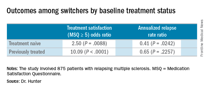
Compared with the end the randomization phase, patients who switched from an iDMT to fingolimod were more satisfied, defined as a Medication Satisfaction Questionnaire score of 5 or greater (odds ratio, 5.65; P less than .001) at 12 months. At the same time, their annualized relapse rate decreased (ARR ratio, 0.53; P = .0158) and their exposure-adjusted brain volume loss decreased from 0.874% to 0.463%.
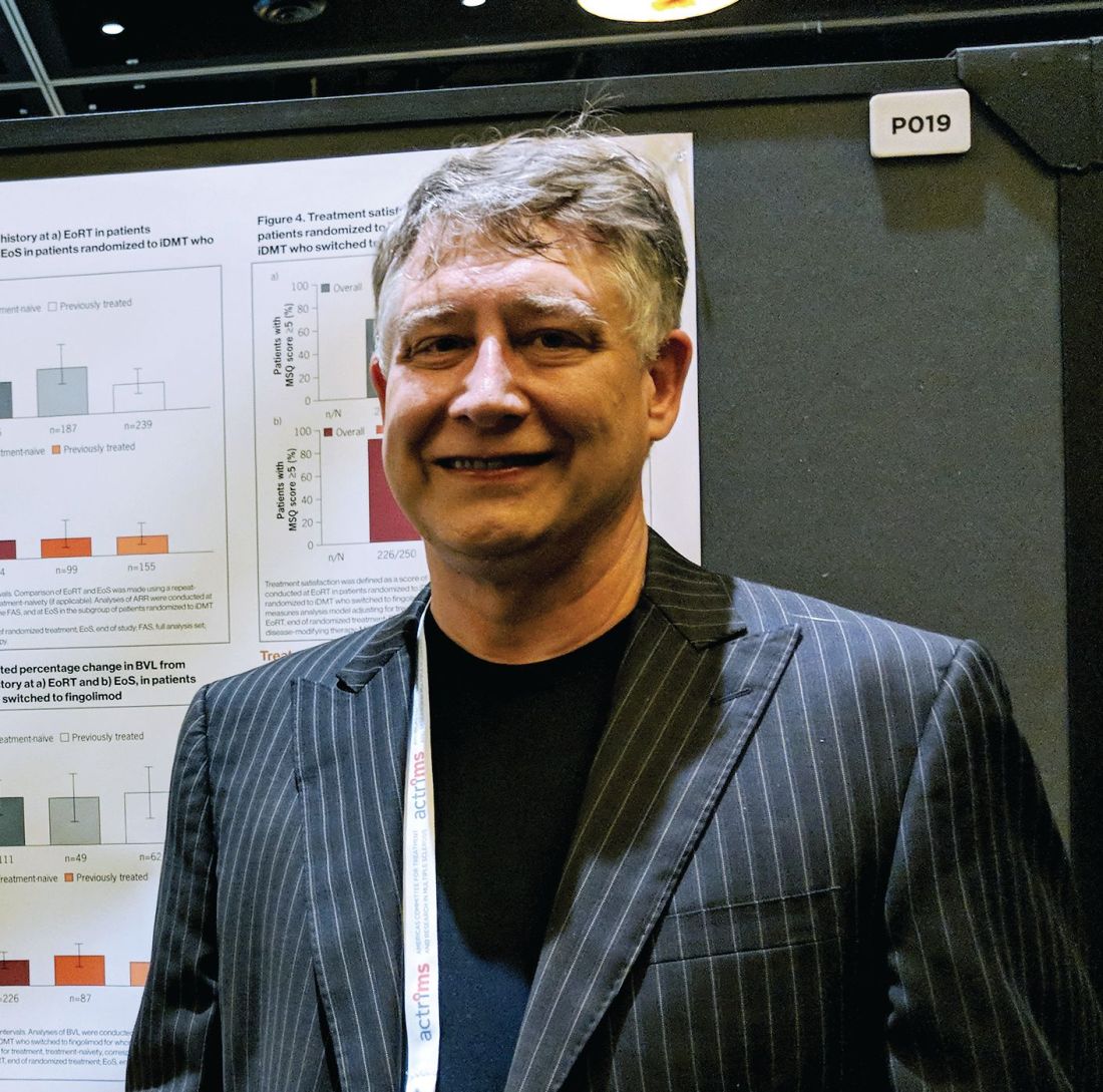
The benefit of these switches was observed regardless of previous treatment status, Dr. Hunter said during a poster presentation at ACTRIMS Forum 2018, held by the Americas Committee for Treatment and Research in Multiple Sclerosis.
At the start of the PREFERMS trial, patients were randomized 1:1 to fingolimod 0.5 mg/day or a preselected iDMT. Just more than half, 54%, were treatment-naive and 46% had previous interferon or glatiramer acetate treatment.
“Generally, because most of these people were switched, and we’re looking at people who were treatment-naive or previously treated, when you look at brain volume loss, it’s very low on fingolimod versus high while they’re on injectable therapies,” Dr. Hunter said.
Dr. Hunter and his colleagues reported similar relapse rates and brain volume loss outcomes among the 254 patients who switched to fingolimod mid-study according to baseline treatment history. The investigators noted, however, that these post hoc analyses and P values are intended for hypothesis generation only.
In the 99 treatment-naive switchers, brain volume loss was 0.54% at the end of the study, compared with 0.84% at the end of the randomization period. Among the 155 previously treated switchers, brain volume loss also decreased, from 0.90% at the end of randomization to 0.42% at study end.
Dr. Hunter disclosed that he is an investigator for Novartis, the sponsor of the study.
SOURCE: Hunter S et al. ACTRIMS Forum 2018 Abstract P019.
SAN DIEGO – People with relapsing multiple sclerosis who switched from an injectable disease-modifying therapy to fingolimod after the randomized phase of the PREFERMS trial experienced improved annualized relapse rates, exposure-adjusted percentage brain volume loss, and satisfaction, compared with study participants who did not switch.
Importantly, there were no differences between switchers who were treatment-naive or previously treated with one injectable therapy class at baseline in the phase 4, active-controlled, open-label, and multicenter PREFERMS trial, according to the researchers.
Of the 875 patients with relapsing multiple sclerosis, 254 or 58% of those initially randomized to an injectable disease-modifying therapy (iDMT) switched to fingolimod (Gilenya) during the 12-month study. Participants in the trial were permitted to switch therapy once after at least 3 months, or sooner if warranted for safety or efficacy reasons.
Compared with the end the randomization phase, patients who switched from an iDMT to fingolimod were more satisfied, defined as a Medication Satisfaction Questionnaire score of 5 or greater (odds ratio, 5.65; P less than .001) at 12 months. At the same time, their annualized relapse rate decreased (ARR ratio, 0.53; P = .0158) and their exposure-adjusted brain volume loss decreased from 0.874% to 0.463%.
The benefit of these switches was observed regardless of previous treatment status, Dr. Hunter said during a poster presentation at ACTRIMS Forum 2018, held by the Americas Committee for Treatment and Research in Multiple Sclerosis.
At the start of the PREFERMS trial, patients were randomized 1:1 to fingolimod 0.5 mg/day or a preselected iDMT. Just more than half, 54%, were treatment-naive and 46% had previous interferon or glatiramer acetate treatment.
“Generally, because most of these people were switched, and we’re looking at people who were treatment-naive or previously treated, when you look at brain volume loss, it’s very low on fingolimod versus high while they’re on injectable therapies,” Dr. Hunter said.
Dr. Hunter and his colleagues reported similar relapse rates and brain volume loss outcomes among the 254 patients who switched to fingolimod mid-study according to baseline treatment history. The investigators noted, however, that these post hoc analyses and P values are intended for hypothesis generation only.
In the 99 treatment-naive switchers, brain volume loss was 0.54% at the end of the study, compared with 0.84% at the end of the randomization period. Among the 155 previously treated switchers, brain volume loss also decreased, from 0.90% at the end of randomization to 0.42% at study end.
Dr. Hunter disclosed that he is an investigator for Novartis, the sponsor of the study.
SOURCE: Hunter S et al. ACTRIMS Forum 2018 Abstract P019.
SAN DIEGO – People with relapsing multiple sclerosis who switched from an injectable disease-modifying therapy to fingolimod after the randomized phase of the PREFERMS trial experienced improved annualized relapse rates, exposure-adjusted percentage brain volume loss, and satisfaction, compared with study participants who did not switch.
Importantly, there were no differences between switchers who were treatment-naive or previously treated with one injectable therapy class at baseline in the phase 4, active-controlled, open-label, and multicenter PREFERMS trial, according to the researchers.
Of the 875 patients with relapsing multiple sclerosis, 254 or 58% of those initially randomized to an injectable disease-modifying therapy (iDMT) switched to fingolimod (Gilenya) during the 12-month study. Participants in the trial were permitted to switch therapy once after at least 3 months, or sooner if warranted for safety or efficacy reasons.
Compared with the end the randomization phase, patients who switched from an iDMT to fingolimod were more satisfied, defined as a Medication Satisfaction Questionnaire score of 5 or greater (odds ratio, 5.65; P less than .001) at 12 months. At the same time, their annualized relapse rate decreased (ARR ratio, 0.53; P = .0158) and their exposure-adjusted brain volume loss decreased from 0.874% to 0.463%.
The benefit of these switches was observed regardless of previous treatment status, Dr. Hunter said during a poster presentation at ACTRIMS Forum 2018, held by the Americas Committee for Treatment and Research in Multiple Sclerosis.
At the start of the PREFERMS trial, patients were randomized 1:1 to fingolimod 0.5 mg/day or a preselected iDMT. Just more than half, 54%, were treatment-naive and 46% had previous interferon or glatiramer acetate treatment.
“Generally, because most of these people were switched, and we’re looking at people who were treatment-naive or previously treated, when you look at brain volume loss, it’s very low on fingolimod versus high while they’re on injectable therapies,” Dr. Hunter said.
Dr. Hunter and his colleagues reported similar relapse rates and brain volume loss outcomes among the 254 patients who switched to fingolimod mid-study according to baseline treatment history. The investigators noted, however, that these post hoc analyses and P values are intended for hypothesis generation only.
In the 99 treatment-naive switchers, brain volume loss was 0.54% at the end of the study, compared with 0.84% at the end of the randomization period. Among the 155 previously treated switchers, brain volume loss also decreased, from 0.90% at the end of randomization to 0.42% at study end.
Dr. Hunter disclosed that he is an investigator for Novartis, the sponsor of the study.
SOURCE: Hunter S et al. ACTRIMS Forum 2018 Abstract P019.
REPORTING FROM ACTRIMS FORUM 2018
Key clinical point:
Major finding: Patients who switched from an injectable disease-modifying therapy to fingolimod were more satisfied, defined as a Medication Satisfaction Questionnaire score of 5 or greater (odds ratio, 5.65; P less than .001).
Study details: Post hoc analyses of the extension study of the PREFERMS trial.
Disclosures: Dr. Hunter disclosed that he is an investigator for Novartis, the sponsor of the study.
Source: Hunter S et al. ACTRIMS Forum 2018 Abstract P019.
Maternal hypertension QI initiative cuts severe morbidity by 41%
DALLAS – A statewide quality improvement initiative to address treatment strategies for women with severe maternal hypertension resulted in a 41% reduction in severe maternal morbidity, from 15% before the initiative to 9% after the suite of interventions was fully implemented.
A total of 102 Illinois hospitals participated in the quality improvement (QI) initiative, which eventually included 12,718 women with severe maternal hypertension, defined as blood pressure greater than 160/110 mm Hg.
The number of patients who were scheduled for follow-up appointments within 10 days of discharge increased from 53% to 83% after the QI initiative was implemented. The proportion of cases of severe maternal hypertension that were subject to a provider-nurse debrief analyzing time to treatment went from 2% before the intervention to 53%.
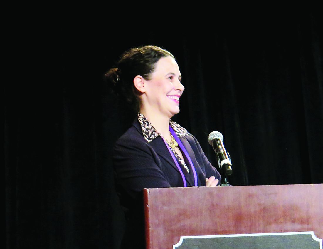
Dr. Lee King presented the findings to a receptive audience at a plenary session of the meeting sponsored by the Society for Maternal-Fetal Medicine. “We had a philosophy of ‘leave no hospital behind,’ and of ‘leave no patient behind,’ ” she said, to wide applause.
“The key prevention of maternal morbidity and mortality associated with severe maternal hypertension is timely treatment; the primary cause of maternal death associated with hypertensive disorders of pregnancy is maternal hemorrhagic stroke,” said Dr. Lee King.
The multidisciplinary, multistakeholder project was coordinated by the Illinois Perinatal Quality Collaborative (ILPQC), housed at Northwestern University, Chicago. The participating hospitals account for over 95% of the births in the state of Illinois, said Dr. Lee King, ILPQC state project director.
The approach used for implementation of the hypertension QI initiative included collaborative learning and the use of rapid response data to give near–real time feedback on how institutions were faring in meeting national guidelines to treat severe maternal hypertension.
Specific clinical interventions included both “system changes and culture changes,” said Dr. Lee King. They included adopting facility-wide protocols for timely identification and treatment of women with severe maternal hypertension; standardizing patient education and discharge planning; increasing provider and nurse education about severe maternal hypertension protocols; and creating rapid access to medication and standardized treatment order sets.
The ILPQC facilitated collaboration between hospitals in meeting these aims, including monthly web-based updates and seminars and construction of the database that gave institutions rapid access to data for ongoing QI efforts. Newsletters, website updates, and QI support were also part of the ILPQC package.
The proportion of participating hospitals meeting time-to-treatment goals for severe maternal hypertension went from 13% at baseline to 71% at the end of data collection, said Dr. Lee King.
In looking at hospital characteristics such as rurality, size, and patient mix, Dr. Lee King and her collaborators found no significant differences in time to treatment, patient education, and number of cases that were debriefed.
“These strategies helped hospitals achieve initiative goals, regardless of hospital characteristics,” said Dr. Lee King. The only significant association the research team found was that hospitals with fewer than 500 deliveries per year were less likely to reach the goal of early follow-up appointments (P less than .02).
“Participation in a statewide perinatal quality collaborative increases hospitals’ QI capacity. A statewide QI initiative can achieve significant population-level improvements in care for women with severe maternal hypertension and – more importantly – a reduction in severe maternal morbidity,” Dr. Lee King said.
The QI initiative and the follow-up study were conducted by the Illinois Perinatal Quality Collaborative, and the work was funded by the Centers for Disease Control and Prevention, the Illinois Department of Health, and the Alliance for Intervention on Maternal Health. Materials related to the maternal hypertension initiative are available at http://ilpqc.org/?q=Hypertension. Dr. Lee King reported no conflicts of interest.
SOURCE: Lee King P et al. Am J Obstet Gynecol. 2018 Jan;218:S4.
DALLAS – A statewide quality improvement initiative to address treatment strategies for women with severe maternal hypertension resulted in a 41% reduction in severe maternal morbidity, from 15% before the initiative to 9% after the suite of interventions was fully implemented.
A total of 102 Illinois hospitals participated in the quality improvement (QI) initiative, which eventually included 12,718 women with severe maternal hypertension, defined as blood pressure greater than 160/110 mm Hg.
The number of patients who were scheduled for follow-up appointments within 10 days of discharge increased from 53% to 83% after the QI initiative was implemented. The proportion of cases of severe maternal hypertension that were subject to a provider-nurse debrief analyzing time to treatment went from 2% before the intervention to 53%.
Dr. Lee King presented the findings to a receptive audience at a plenary session of the meeting sponsored by the Society for Maternal-Fetal Medicine. “We had a philosophy of ‘leave no hospital behind,’ and of ‘leave no patient behind,’ ” she said, to wide applause.
“The key prevention of maternal morbidity and mortality associated with severe maternal hypertension is timely treatment; the primary cause of maternal death associated with hypertensive disorders of pregnancy is maternal hemorrhagic stroke,” said Dr. Lee King.
The multidisciplinary, multistakeholder project was coordinated by the Illinois Perinatal Quality Collaborative (ILPQC), housed at Northwestern University, Chicago. The participating hospitals account for over 95% of the births in the state of Illinois, said Dr. Lee King, ILPQC state project director.
The approach used for implementation of the hypertension QI initiative included collaborative learning and the use of rapid response data to give near–real time feedback on how institutions were faring in meeting national guidelines to treat severe maternal hypertension.
Specific clinical interventions included both “system changes and culture changes,” said Dr. Lee King. They included adopting facility-wide protocols for timely identification and treatment of women with severe maternal hypertension; standardizing patient education and discharge planning; increasing provider and nurse education about severe maternal hypertension protocols; and creating rapid access to medication and standardized treatment order sets.
The ILPQC facilitated collaboration between hospitals in meeting these aims, including monthly web-based updates and seminars and construction of the database that gave institutions rapid access to data for ongoing QI efforts. Newsletters, website updates, and QI support were also part of the ILPQC package.
The proportion of participating hospitals meeting time-to-treatment goals for severe maternal hypertension went from 13% at baseline to 71% at the end of data collection, said Dr. Lee King.
In looking at hospital characteristics such as rurality, size, and patient mix, Dr. Lee King and her collaborators found no significant differences in time to treatment, patient education, and number of cases that were debriefed.
“These strategies helped hospitals achieve initiative goals, regardless of hospital characteristics,” said Dr. Lee King. The only significant association the research team found was that hospitals with fewer than 500 deliveries per year were less likely to reach the goal of early follow-up appointments (P less than .02).
“Participation in a statewide perinatal quality collaborative increases hospitals’ QI capacity. A statewide QI initiative can achieve significant population-level improvements in care for women with severe maternal hypertension and – more importantly – a reduction in severe maternal morbidity,” Dr. Lee King said.
The QI initiative and the follow-up study were conducted by the Illinois Perinatal Quality Collaborative, and the work was funded by the Centers for Disease Control and Prevention, the Illinois Department of Health, and the Alliance for Intervention on Maternal Health. Materials related to the maternal hypertension initiative are available at http://ilpqc.org/?q=Hypertension. Dr. Lee King reported no conflicts of interest.
SOURCE: Lee King P et al. Am J Obstet Gynecol. 2018 Jan;218:S4.
DALLAS – A statewide quality improvement initiative to address treatment strategies for women with severe maternal hypertension resulted in a 41% reduction in severe maternal morbidity, from 15% before the initiative to 9% after the suite of interventions was fully implemented.
A total of 102 Illinois hospitals participated in the quality improvement (QI) initiative, which eventually included 12,718 women with severe maternal hypertension, defined as blood pressure greater than 160/110 mm Hg.
The number of patients who were scheduled for follow-up appointments within 10 days of discharge increased from 53% to 83% after the QI initiative was implemented. The proportion of cases of severe maternal hypertension that were subject to a provider-nurse debrief analyzing time to treatment went from 2% before the intervention to 53%.
Dr. Lee King presented the findings to a receptive audience at a plenary session of the meeting sponsored by the Society for Maternal-Fetal Medicine. “We had a philosophy of ‘leave no hospital behind,’ and of ‘leave no patient behind,’ ” she said, to wide applause.
“The key prevention of maternal morbidity and mortality associated with severe maternal hypertension is timely treatment; the primary cause of maternal death associated with hypertensive disorders of pregnancy is maternal hemorrhagic stroke,” said Dr. Lee King.
The multidisciplinary, multistakeholder project was coordinated by the Illinois Perinatal Quality Collaborative (ILPQC), housed at Northwestern University, Chicago. The participating hospitals account for over 95% of the births in the state of Illinois, said Dr. Lee King, ILPQC state project director.
The approach used for implementation of the hypertension QI initiative included collaborative learning and the use of rapid response data to give near–real time feedback on how institutions were faring in meeting national guidelines to treat severe maternal hypertension.
Specific clinical interventions included both “system changes and culture changes,” said Dr. Lee King. They included adopting facility-wide protocols for timely identification and treatment of women with severe maternal hypertension; standardizing patient education and discharge planning; increasing provider and nurse education about severe maternal hypertension protocols; and creating rapid access to medication and standardized treatment order sets.
The ILPQC facilitated collaboration between hospitals in meeting these aims, including monthly web-based updates and seminars and construction of the database that gave institutions rapid access to data for ongoing QI efforts. Newsletters, website updates, and QI support were also part of the ILPQC package.
The proportion of participating hospitals meeting time-to-treatment goals for severe maternal hypertension went from 13% at baseline to 71% at the end of data collection, said Dr. Lee King.
In looking at hospital characteristics such as rurality, size, and patient mix, Dr. Lee King and her collaborators found no significant differences in time to treatment, patient education, and number of cases that were debriefed.
“These strategies helped hospitals achieve initiative goals, regardless of hospital characteristics,” said Dr. Lee King. The only significant association the research team found was that hospitals with fewer than 500 deliveries per year were less likely to reach the goal of early follow-up appointments (P less than .02).
“Participation in a statewide perinatal quality collaborative increases hospitals’ QI capacity. A statewide QI initiative can achieve significant population-level improvements in care for women with severe maternal hypertension and – more importantly – a reduction in severe maternal morbidity,” Dr. Lee King said.
The QI initiative and the follow-up study were conducted by the Illinois Perinatal Quality Collaborative, and the work was funded by the Centers for Disease Control and Prevention, the Illinois Department of Health, and the Alliance for Intervention on Maternal Health. Materials related to the maternal hypertension initiative are available at http://ilpqc.org/?q=Hypertension. Dr. Lee King reported no conflicts of interest.
SOURCE: Lee King P et al. Am J Obstet Gynecol. 2018 Jan;218:S4.
REPORTING FROM THE PREGNANCY MEETING
Key clinical point: The statewide QI initiative targeted treatment of women with severe maternal hypertension.
Major finding: Severe maternal morbidity fell 41%, from 15% to 9%, with the initiative.
Study details: Pre-post QI initiative analysis of data from 102 Illinois hospitals treating 12,718 women with severe maternal hypertension.
Disclosures: The study was funded by the Centers for Disease Control and Prevention, the Illinois Department of Health, and the Alliance for Intervention on Mental Health. Dr. Lee King is employed by the Illinois Perinatal Quality Collaborative, which conducted the study.
Source: Lee King P et al. Am J Obstet Gynecol. 2018 Jan;218:S4.
MDedge Daily News: Do you really need cyberliability insurance?
The video associated with this article is no longer available on this site. Please view all of our videos on the MDedge YouTube channel
Does your practice need cyberliability insurance? Hemorrhoid treatment guidelines get an overhaul, why college students’ poor sleep can be deadly, and Indiana’s Medicaid waiver may find its way into court.
Listen to the MDedge Daily News podcast for all the details on today’s top news.
The video associated with this article is no longer available on this site. Please view all of our videos on the MDedge YouTube channel
Does your practice need cyberliability insurance? Hemorrhoid treatment guidelines get an overhaul, why college students’ poor sleep can be deadly, and Indiana’s Medicaid waiver may find its way into court.
Listen to the MDedge Daily News podcast for all the details on today’s top news.
The video associated with this article is no longer available on this site. Please view all of our videos on the MDedge YouTube channel
Does your practice need cyberliability insurance? Hemorrhoid treatment guidelines get an overhaul, why college students’ poor sleep can be deadly, and Indiana’s Medicaid waiver may find its way into court.
Listen to the MDedge Daily News podcast for all the details on today’s top news.

VA is Trying New Ways to Treat TBI and PTSD
The VA is examining two alternative treatments for TBI and PTSD: a light-emitting diode (LED) treatment for mild to moderate TBI and stellate ganglion block (SGB) for PTSD.
In the LED treatment, which takes about 30 minutes, a lightweight LED-lined helmet is placed on the patient’s head, and more diodes are placed inside the nose to deliver photons to the deeper parts of the brain. The light is painless and generates no heat.
Although it is considered investigational, LED therapy is available at the VAHS Boston, as well as for veterans to use at home. Dr. Margaret Naeser, a professor of neurology at Boston University School of Medicine and lead investigator of the Boston study team, interviewed in VA Research Currents, says the technology has been around a while, but it was previously used on the body for wound healing and pain. Using it on the brain is new. The LED light has been shown to boost the output of nitric oxide, improving blood flow. Studies have shown that LED improves brain function, including attention and memory, emotions, and sleep.
Naeser says most of the TBI and PTSD cases helped so far with LEDs on the head included cognitive rehabilitation therapy. The patients showed additional progress after the LED treatments. A combination of both treatments would likely produce the best results, she says.
Providers at the Long Beach VAMC have been using SGB, commonly used in pain management with ropivacaine or bupivacaine, to reduce the symptoms of PTSD. According to the VA Evidence-based Synthesis Program (ESP), SGB may ease anxiety and the alert response by inhibiting connections between the peripheral sympathetic nerve system and regions of the cerebral cortex, such as the amygdala, thought to be abnormally activated in PTSD. Stellate ganglion block also has been associated with biologic markers of sedation.
The ESP experts say there is insufficient information to determine which veterans are most likely to benefit from SGB for PTSD, but an uncontrolled, unblinded case series of 30 active duty service members with combat-related PTSD suggests that those with predominantly hyperarousal and avoidance symptoms might be the best candidates. Patients who have undergone SGB have found it highly acceptable, although the invasive nature of SGB may be a barrier for some. Findings from the first randomized controlled trial of SGB for PTSD were inconclusive, the panel said; further research is warranted.
The VA is examining two alternative treatments for TBI and PTSD: a light-emitting diode (LED) treatment for mild to moderate TBI and stellate ganglion block (SGB) for PTSD.
In the LED treatment, which takes about 30 minutes, a lightweight LED-lined helmet is placed on the patient’s head, and more diodes are placed inside the nose to deliver photons to the deeper parts of the brain. The light is painless and generates no heat.
Although it is considered investigational, LED therapy is available at the VAHS Boston, as well as for veterans to use at home. Dr. Margaret Naeser, a professor of neurology at Boston University School of Medicine and lead investigator of the Boston study team, interviewed in VA Research Currents, says the technology has been around a while, but it was previously used on the body for wound healing and pain. Using it on the brain is new. The LED light has been shown to boost the output of nitric oxide, improving blood flow. Studies have shown that LED improves brain function, including attention and memory, emotions, and sleep.
Naeser says most of the TBI and PTSD cases helped so far with LEDs on the head included cognitive rehabilitation therapy. The patients showed additional progress after the LED treatments. A combination of both treatments would likely produce the best results, she says.
Providers at the Long Beach VAMC have been using SGB, commonly used in pain management with ropivacaine or bupivacaine, to reduce the symptoms of PTSD. According to the VA Evidence-based Synthesis Program (ESP), SGB may ease anxiety and the alert response by inhibiting connections between the peripheral sympathetic nerve system and regions of the cerebral cortex, such as the amygdala, thought to be abnormally activated in PTSD. Stellate ganglion block also has been associated with biologic markers of sedation.
The ESP experts say there is insufficient information to determine which veterans are most likely to benefit from SGB for PTSD, but an uncontrolled, unblinded case series of 30 active duty service members with combat-related PTSD suggests that those with predominantly hyperarousal and avoidance symptoms might be the best candidates. Patients who have undergone SGB have found it highly acceptable, although the invasive nature of SGB may be a barrier for some. Findings from the first randomized controlled trial of SGB for PTSD were inconclusive, the panel said; further research is warranted.
The VA is examining two alternative treatments for TBI and PTSD: a light-emitting diode (LED) treatment for mild to moderate TBI and stellate ganglion block (SGB) for PTSD.
In the LED treatment, which takes about 30 minutes, a lightweight LED-lined helmet is placed on the patient’s head, and more diodes are placed inside the nose to deliver photons to the deeper parts of the brain. The light is painless and generates no heat.
Although it is considered investigational, LED therapy is available at the VAHS Boston, as well as for veterans to use at home. Dr. Margaret Naeser, a professor of neurology at Boston University School of Medicine and lead investigator of the Boston study team, interviewed in VA Research Currents, says the technology has been around a while, but it was previously used on the body for wound healing and pain. Using it on the brain is new. The LED light has been shown to boost the output of nitric oxide, improving blood flow. Studies have shown that LED improves brain function, including attention and memory, emotions, and sleep.
Naeser says most of the TBI and PTSD cases helped so far with LEDs on the head included cognitive rehabilitation therapy. The patients showed additional progress after the LED treatments. A combination of both treatments would likely produce the best results, she says.
Providers at the Long Beach VAMC have been using SGB, commonly used in pain management with ropivacaine or bupivacaine, to reduce the symptoms of PTSD. According to the VA Evidence-based Synthesis Program (ESP), SGB may ease anxiety and the alert response by inhibiting connections between the peripheral sympathetic nerve system and regions of the cerebral cortex, such as the amygdala, thought to be abnormally activated in PTSD. Stellate ganglion block also has been associated with biologic markers of sedation.
The ESP experts say there is insufficient information to determine which veterans are most likely to benefit from SGB for PTSD, but an uncontrolled, unblinded case series of 30 active duty service members with combat-related PTSD suggests that those with predominantly hyperarousal and avoidance symptoms might be the best candidates. Patients who have undergone SGB have found it highly acceptable, although the invasive nature of SGB may be a barrier for some. Findings from the first randomized controlled trial of SGB for PTSD were inconclusive, the panel said; further research is warranted.
Minihaplo-BMT can cure severe hemoglobinopathies
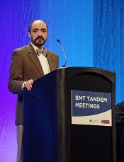
SALT LAKE CITY—Cure of severe hemoglobinopathies is now possible for most patients, according to a speaker at the 2018 BMT Tandem Meetings.
Results of a small study suggest that nonmyeloablative haploidentical bone marrow transplant (minihaplo-BMT), when used with 400 cGy total body irradiation and post-transplant cyclophosphamide, can cure sickle cell disease (SCD) and beta-thalassemia while conferring limited toxicity.
“This is our second study showing that you can certainly get rid of these conditions using haploidentical donors and, also, that you can cure these conditions with a nonmyeloablative regimen,” said Javier Bolaños-Meade, MD, of Johns Hopkins University in Baltimore, Maryland.
“What we saw [in the current study] is that increasing the total body irradiation from 200 cGy to 400 cGy can overcome the very high rate of graft failure we observed in our prior study.”
However, the results also suggest that major ABO incompatible donors should be avoided.
Dr Bolaños-Meade presented these findings as a late-breaking abstract (LBA3) at this year’s BMT Tandem Meetings.
He began the presentation by pointing out that allogeneic BMT is the only potential cure for SCD and beta-thalassemia. Unfortunately, most of these patients have been excluded from transplant due to a lack of matched donors, concerns over graft-vs-host disease (GVHD), and/or compromised end organ function precluding them from myeloablative conditioning.
With a prior study, Dr Bolaños-Meade and his colleagues found that minihaplo-BMT with post-transplant cyclophosphamide expanded the donor pool and was well tolerated in patients with SCD. However, the rate of secondary graft failure was high, at 43%.
“We hypothesized that increasing the amount of radiation that was given to these patients, making the regimen absolutely more intensive, will help us to overcome this rate of graft failure and retain the tolerability of the regimen,” Dr Bolaños-Meade said.
He and his colleagues tested this theory in 12 patients with SCD and 5 with beta-thalassemia.
The SCD patients had a median age of 26 (range, 6-31), and most (n=8) were female. Ten were African-American, and 2 were of Arab descent.
All SCD patients had at least 2 hospital admissions in the last 2 years, 5 had osteonecrosis, 3 were transfusion-dependent, 2 had a history of acute chest syndrome, and 1 patient had changes in brain magnetic resonance imaging.
The thalassemia patients had a median age of 7 (range, 6-16). Four were female, and all 5 were of Arab descent. All patients were transfusion-dependent from infancy.
Treatment
All patients received antithymocyte globulin (rabbit) at 0.5 mg/kg on day -9 and 2 mg/kg on days -8 and -7, fludarabine at 30 mg/m2 on days -6 to -2, cyclophosphamide at 14.5 mg/kg on days -6 and -5, and total body irradiation at 400 cGy on day -1.
Patients received unmanipulated bone marrow that was collected and infused on day 0. Donors included 1 aunt, 4 brothers, 3 sisters, 5 mothers, and 4 fathers. There were 2 major ABO incompatible pairs, 5 minor ABO incompatible pairs, and 10 ABO compatible pairs.
For GVHD prophylaxis, patients received 50 mg/kg/day of cyclophosphamide on days 3 and 4, sirolimus (levels of 5-10 ng/dl) from days 5 to 365, and mycophenolate mofetil at 1 g every 8 hours from days 5 to 35.
Efficacy
All 17 patients are still alive at a median follow-up of 15 months (range, 3-34).
“Of all the patients transplanted, we only observed 1 graft failure,” Dr Bolaños-Meade said. “This is in sharp contrast to the 40% graft failure in our prior study.”
Thirteen patients achieved full donor chimerism, and 3 had mixed chimerism.
Four of the 5 beta-thalassemia patients achieved full donor chimerism, and 1 had mixed chimerism. All 5 patients became transfusion-independent.
Eleven of the 12 SCD patients engrafted, and 9 achieved full donor chimerism.
All but one of the engrafted SCD patients became transfusion-independent. The exception was a mixed chimeric patient who had a major ABO mismatched donor. The patient remains transfusion-dependent more than 30 months after BMT.
“This is the main problem we have seen in all our sickle cell and hemoglobinopathy experiences,” Dr Bolaños-Meade said. “If you have a major ABO incompatibility, and there’s mixed chimerism, you’re asking for trouble.”
Toxicity
All SCD patients had sickle cell crises after antithymocyte globulin. Other toxicities included sirolimus-induced diabetes in 1 patient, worsening of Meniere’s disease in 1 patient, and BK cystitis in 1 patient.
Two patients developed grade 2 acute GVHD, and 1 developed grade 3 acute GVHD. Three patients had chronic GVHD, 2 mild and 1 moderate.
However, all cases of GVHD resolved. And, of the 8 engrafted patients with more than 1 year of follow-up, 6 are off systemic immunosuppression.
SALT LAKE CITY—Cure of severe hemoglobinopathies is now possible for most patients, according to a speaker at the 2018 BMT Tandem Meetings.
Results of a small study suggest that nonmyeloablative haploidentical bone marrow transplant (minihaplo-BMT), when used with 400 cGy total body irradiation and post-transplant cyclophosphamide, can cure sickle cell disease (SCD) and beta-thalassemia while conferring limited toxicity.
“This is our second study showing that you can certainly get rid of these conditions using haploidentical donors and, also, that you can cure these conditions with a nonmyeloablative regimen,” said Javier Bolaños-Meade, MD, of Johns Hopkins University in Baltimore, Maryland.
“What we saw [in the current study] is that increasing the total body irradiation from 200 cGy to 400 cGy can overcome the very high rate of graft failure we observed in our prior study.”
However, the results also suggest that major ABO incompatible donors should be avoided.
Dr Bolaños-Meade presented these findings as a late-breaking abstract (LBA3) at this year’s BMT Tandem Meetings.
He began the presentation by pointing out that allogeneic BMT is the only potential cure for SCD and beta-thalassemia. Unfortunately, most of these patients have been excluded from transplant due to a lack of matched donors, concerns over graft-vs-host disease (GVHD), and/or compromised end organ function precluding them from myeloablative conditioning.
With a prior study, Dr Bolaños-Meade and his colleagues found that minihaplo-BMT with post-transplant cyclophosphamide expanded the donor pool and was well tolerated in patients with SCD. However, the rate of secondary graft failure was high, at 43%.
“We hypothesized that increasing the amount of radiation that was given to these patients, making the regimen absolutely more intensive, will help us to overcome this rate of graft failure and retain the tolerability of the regimen,” Dr Bolaños-Meade said.
He and his colleagues tested this theory in 12 patients with SCD and 5 with beta-thalassemia.
The SCD patients had a median age of 26 (range, 6-31), and most (n=8) were female. Ten were African-American, and 2 were of Arab descent.
All SCD patients had at least 2 hospital admissions in the last 2 years, 5 had osteonecrosis, 3 were transfusion-dependent, 2 had a history of acute chest syndrome, and 1 patient had changes in brain magnetic resonance imaging.
The thalassemia patients had a median age of 7 (range, 6-16). Four were female, and all 5 were of Arab descent. All patients were transfusion-dependent from infancy.
Treatment
All patients received antithymocyte globulin (rabbit) at 0.5 mg/kg on day -9 and 2 mg/kg on days -8 and -7, fludarabine at 30 mg/m2 on days -6 to -2, cyclophosphamide at 14.5 mg/kg on days -6 and -5, and total body irradiation at 400 cGy on day -1.
Patients received unmanipulated bone marrow that was collected and infused on day 0. Donors included 1 aunt, 4 brothers, 3 sisters, 5 mothers, and 4 fathers. There were 2 major ABO incompatible pairs, 5 minor ABO incompatible pairs, and 10 ABO compatible pairs.
For GVHD prophylaxis, patients received 50 mg/kg/day of cyclophosphamide on days 3 and 4, sirolimus (levels of 5-10 ng/dl) from days 5 to 365, and mycophenolate mofetil at 1 g every 8 hours from days 5 to 35.
Efficacy
All 17 patients are still alive at a median follow-up of 15 months (range, 3-34).
“Of all the patients transplanted, we only observed 1 graft failure,” Dr Bolaños-Meade said. “This is in sharp contrast to the 40% graft failure in our prior study.”
Thirteen patients achieved full donor chimerism, and 3 had mixed chimerism.
Four of the 5 beta-thalassemia patients achieved full donor chimerism, and 1 had mixed chimerism. All 5 patients became transfusion-independent.
Eleven of the 12 SCD patients engrafted, and 9 achieved full donor chimerism.
All but one of the engrafted SCD patients became transfusion-independent. The exception was a mixed chimeric patient who had a major ABO mismatched donor. The patient remains transfusion-dependent more than 30 months after BMT.
“This is the main problem we have seen in all our sickle cell and hemoglobinopathy experiences,” Dr Bolaños-Meade said. “If you have a major ABO incompatibility, and there’s mixed chimerism, you’re asking for trouble.”
Toxicity
All SCD patients had sickle cell crises after antithymocyte globulin. Other toxicities included sirolimus-induced diabetes in 1 patient, worsening of Meniere’s disease in 1 patient, and BK cystitis in 1 patient.
Two patients developed grade 2 acute GVHD, and 1 developed grade 3 acute GVHD. Three patients had chronic GVHD, 2 mild and 1 moderate.
However, all cases of GVHD resolved. And, of the 8 engrafted patients with more than 1 year of follow-up, 6 are off systemic immunosuppression.
SALT LAKE CITY—Cure of severe hemoglobinopathies is now possible for most patients, according to a speaker at the 2018 BMT Tandem Meetings.
Results of a small study suggest that nonmyeloablative haploidentical bone marrow transplant (minihaplo-BMT), when used with 400 cGy total body irradiation and post-transplant cyclophosphamide, can cure sickle cell disease (SCD) and beta-thalassemia while conferring limited toxicity.
“This is our second study showing that you can certainly get rid of these conditions using haploidentical donors and, also, that you can cure these conditions with a nonmyeloablative regimen,” said Javier Bolaños-Meade, MD, of Johns Hopkins University in Baltimore, Maryland.
“What we saw [in the current study] is that increasing the total body irradiation from 200 cGy to 400 cGy can overcome the very high rate of graft failure we observed in our prior study.”
However, the results also suggest that major ABO incompatible donors should be avoided.
Dr Bolaños-Meade presented these findings as a late-breaking abstract (LBA3) at this year’s BMT Tandem Meetings.
He began the presentation by pointing out that allogeneic BMT is the only potential cure for SCD and beta-thalassemia. Unfortunately, most of these patients have been excluded from transplant due to a lack of matched donors, concerns over graft-vs-host disease (GVHD), and/or compromised end organ function precluding them from myeloablative conditioning.
With a prior study, Dr Bolaños-Meade and his colleagues found that minihaplo-BMT with post-transplant cyclophosphamide expanded the donor pool and was well tolerated in patients with SCD. However, the rate of secondary graft failure was high, at 43%.
“We hypothesized that increasing the amount of radiation that was given to these patients, making the regimen absolutely more intensive, will help us to overcome this rate of graft failure and retain the tolerability of the regimen,” Dr Bolaños-Meade said.
He and his colleagues tested this theory in 12 patients with SCD and 5 with beta-thalassemia.
The SCD patients had a median age of 26 (range, 6-31), and most (n=8) were female. Ten were African-American, and 2 were of Arab descent.
All SCD patients had at least 2 hospital admissions in the last 2 years, 5 had osteonecrosis, 3 were transfusion-dependent, 2 had a history of acute chest syndrome, and 1 patient had changes in brain magnetic resonance imaging.
The thalassemia patients had a median age of 7 (range, 6-16). Four were female, and all 5 were of Arab descent. All patients were transfusion-dependent from infancy.
Treatment
All patients received antithymocyte globulin (rabbit) at 0.5 mg/kg on day -9 and 2 mg/kg on days -8 and -7, fludarabine at 30 mg/m2 on days -6 to -2, cyclophosphamide at 14.5 mg/kg on days -6 and -5, and total body irradiation at 400 cGy on day -1.
Patients received unmanipulated bone marrow that was collected and infused on day 0. Donors included 1 aunt, 4 brothers, 3 sisters, 5 mothers, and 4 fathers. There were 2 major ABO incompatible pairs, 5 minor ABO incompatible pairs, and 10 ABO compatible pairs.
For GVHD prophylaxis, patients received 50 mg/kg/day of cyclophosphamide on days 3 and 4, sirolimus (levels of 5-10 ng/dl) from days 5 to 365, and mycophenolate mofetil at 1 g every 8 hours from days 5 to 35.
Efficacy
All 17 patients are still alive at a median follow-up of 15 months (range, 3-34).
“Of all the patients transplanted, we only observed 1 graft failure,” Dr Bolaños-Meade said. “This is in sharp contrast to the 40% graft failure in our prior study.”
Thirteen patients achieved full donor chimerism, and 3 had mixed chimerism.
Four of the 5 beta-thalassemia patients achieved full donor chimerism, and 1 had mixed chimerism. All 5 patients became transfusion-independent.
Eleven of the 12 SCD patients engrafted, and 9 achieved full donor chimerism.
All but one of the engrafted SCD patients became transfusion-independent. The exception was a mixed chimeric patient who had a major ABO mismatched donor. The patient remains transfusion-dependent more than 30 months after BMT.
“This is the main problem we have seen in all our sickle cell and hemoglobinopathy experiences,” Dr Bolaños-Meade said. “If you have a major ABO incompatibility, and there’s mixed chimerism, you’re asking for trouble.”
Toxicity
All SCD patients had sickle cell crises after antithymocyte globulin. Other toxicities included sirolimus-induced diabetes in 1 patient, worsening of Meniere’s disease in 1 patient, and BK cystitis in 1 patient.
Two patients developed grade 2 acute GVHD, and 1 developed grade 3 acute GVHD. Three patients had chronic GVHD, 2 mild and 1 moderate.
However, all cases of GVHD resolved. And, of the 8 engrafted patients with more than 1 year of follow-up, 6 are off systemic immunosuppression.
Gut grief
The video associated with this article is no longer available on this site. Please view all of our videos on the MDedge YouTube channel
This video was filmed at Metabolic & Endocrine Disease Summit (MEDS). Click here to learn more.
The video associated with this article is no longer available on this site. Please view all of our videos on the MDedge YouTube channel
This video was filmed at Metabolic & Endocrine Disease Summit (MEDS). Click here to learn more.
The video associated with this article is no longer available on this site. Please view all of our videos on the MDedge YouTube channel
This video was filmed at Metabolic & Endocrine Disease Summit (MEDS). Click here to learn more.
Role of musculoskeletal ultrasound expands in rheumatic diseases
MAUI, HAWAII – Recent studies highlight novel applications of musculoskeletal ultrasound across a broad spectrum of rheumatic diseases beyond the imaging method’s well-established role in diagnosis and follow-up of patients with rheumatoid arthritis, Alvin F. Wells, MD, PhD, said at the 2018 Rheumatology Winter Clinical Symposium.

He cited recently published evidence in support of his contention:
Systemic lupus erythematosus
Portuguese investigators reported that 77% of their study participants with asymptomatic systemic lupus erythematosus (SLE) – that is, no arthralgia or musculoskeletal signs or symptoms on physical examination – demonstrated gray-scale ultrasound evidence of grade 1 or greater synovial hypertrophy of joints in the hands and/or wrists on a semiquantitative 0-3 scale. So did 100% of patients with symptomatic SLE in this small study, in which physicians evaluated 22 joints per person.
Moreover, grade 1 or greater intra-articular power Doppler changes were also detected in one-fifth of the asymptomatic patients and 83% of those with symptomatic SLE. What the researchers considered persuasive ultrasound evidence of synovitis – grade 2 or 3 synovial hypertrophy or at least grade 1 power Doppler findings – was noted in 23% of the asymptomatic SLE patients, 83% of those who were symptomatic, and none of the healthy controls.
The investigators concluded that asymptomatic patients with SLE may present with subclinical joint inflammation that often goes undetected by physical examination and x-ray. They added that ultrasound with power Doppler may be important in disease evaluation and therapeutic monitoring in patients with lupus (Lupus Sci Med. 2017 Jan 19;4[1]:e000184).
For Dr. Wells, the take-home message was clear: “Don’t overlook the hands.”
“In my lupus patients, sure, I look at the hips, making sure there are no subtle signs of osteonecrosis, but I think we often forget to look at the hands. We’re looking at the hair, looking at the mouth, we’re worried about the lungs. But I think we often overlook the synovitis. These patients are going to the ER because they’re having pain in their hands, and they’re getting told, ‘This is fibromyalgia, go back and see your rheumatologist.’ So it’s something to think about: When you’ve got a patient with active lupus, ultrasound may have a role to play,” according to Dr. Wells.
The big unanswered question is whether early aggressive treatment in SLE patients with these synovial abnormalities – that is, a treat-to-target, ultrasound-guided strategy – can limit erosive changes, avoid disease progression, and improve functional outcomes, as is now known to be the case in RA. Long-term studies will be required to find out, he noted.
Ultrasound of parotid glands in suspected Sjögren’s syndrome is biopsy sparing
Members of the European League Against Rheumatism Ultrasound Primary Sjögren’s Syndrome Study Group recently reported that in a prospective study of 103 consecutive outpatients with clinically suspected primary Sjögren’s syndrome, a positive ultrasound examination of the major salivary glands in combination with the presence of anti-SSA/Ro antibodies was highly predictive of positive parotid and labial gland biopsies. Indeed, a positive ultrasound plus anti-SSA/Ro antibodies showed a high predictive value for Sjögren’s syndrome regardless of whether the classification was based upon American College of Rheumatology, American European Consensus Group, or ACR-EULAR criteria (Ann Rheum Dis. 2017;76[11]:1883-9).
“Ophthalmologists, dentists, and primary care physicians frequently refer patients with dry eyes and dry mouth to us to sort out whether they have Sjögren’s syndrome. The data show now that with a positive salivary gland ultrasound score and a positive anti-SSA/Ro antibody test you can avoid the biopsies,” the rheumatologist said.
That’s a fine thing for patients because these biopsies often result in residual numbness and tingling, he added.
Dr. Wells noted that a multicenter European group recently fine-tuned the criteria for an abnormal salivary gland ultrasound score. They identified echogenicity and homogeneity as the two most reliable items on the score. They also reported that the mean time it took five ultrasonographers to perform salivary gland assessments was reasonable at 11-27 minutes (RMD Open. 2017 Jun 9;3[1]:e000364).
“You can do this essentially in an office visit. The changes in the salivary glands are pathognomonic for Sjögren’s – you won’t get it confused with a neoplasm. I schedule these patients for a 15-minute time slot, and based upon the ultrasound and whether they’re positive or negative for anti-SSA/Ro antibodies I can make some decisions from there,” Dr. Wells explained.
He described what to look for on ultrasound: “A normal parotid looks like a homogeneous gray sponge on ultrasound. In Sjögren’s syndrome, you see icepicklike holes in the sponge. And there’s thickening of the intima, so you see blunting of the arterial wave on pulse wave.”
Implications of ultrasound enthesitis in early spondyloarthritis
French rheumatologists reported on the implications of an ultrasound peripheral enthesis assessment in 402 patients with early axial spondyloarthritis in the French DESIR cohort. The ultrasound evaluation covered the lateral epicondyles, Achilles tendon, and superior and inferior patellar ligament entheses. The results were summed up in ultrasound structural and power Doppler scores. In addition, the investigators obtained spine and sacroiliac joint MRI scans and spine x-rays, all scored centrally.
It turned out that ultrasound abnormalities weren’t useful in monitoring axial spondyloarthritis clinical disease activity as reflected in Bath Ankylosing Spondylitis Disease Activity Index scores, nor were the ultrasound structural and power Doppler scores associated with MRI spine inflammatory lesions or sacroiliitis. However, ultrasound enthesophytes were strongly associated with axial syndesmophytes, suggesting that these ultrasound abnormalities might be a marker of disease severity in axial spondyloarthritis (RMD Open. 2017 Sep 7;3[2]:e000482).
“Whether or not by inhibiting these peripheral enthesophytes you’ll ultimately inhibit further enthesophytes or syndesmophytes from forming is yet to be seen,” Dr. Wells cautioned.
He added, however, that a recent short-term, randomized, placebo-controlled trial in 185 patients with nonradiographic axial spondyloarthritis holds hope in that regard. Investigators in the ongoing multicenter EMBARK (Effect of Etanercept on Symptoms and Objective Inflammation in Nonradiographic Spondyloarthritis) trial reported that treatment with etanercept was associated with a significantly greater reduction in erosions, an increase in backfill, and improved clinical outcomes, compared with placebo, as assessed by MRIs obtained at baseline and 12 weeks.
The investigators viewed these findings as consistent with an early reparative response to the anti–tumor necrosis factor agent, although they noted that understanding of the biologic agent’s true impact on disease progression awaits longer-term follow-up in the 104-week study (Ann Rheum Dis. 2018 Jan;77[1]:78-84).
Dr. Wells reported serving as a consultant to more than half a dozen pharmaceutical companies and receiving research funding from Abbott, Celgene, and UCB.
MAUI, HAWAII – Recent studies highlight novel applications of musculoskeletal ultrasound across a broad spectrum of rheumatic diseases beyond the imaging method’s well-established role in diagnosis and follow-up of patients with rheumatoid arthritis, Alvin F. Wells, MD, PhD, said at the 2018 Rheumatology Winter Clinical Symposium.
He cited recently published evidence in support of his contention:
Systemic lupus erythematosus
Portuguese investigators reported that 77% of their study participants with asymptomatic systemic lupus erythematosus (SLE) – that is, no arthralgia or musculoskeletal signs or symptoms on physical examination – demonstrated gray-scale ultrasound evidence of grade 1 or greater synovial hypertrophy of joints in the hands and/or wrists on a semiquantitative 0-3 scale. So did 100% of patients with symptomatic SLE in this small study, in which physicians evaluated 22 joints per person.
Moreover, grade 1 or greater intra-articular power Doppler changes were also detected in one-fifth of the asymptomatic patients and 83% of those with symptomatic SLE. What the researchers considered persuasive ultrasound evidence of synovitis – grade 2 or 3 synovial hypertrophy or at least grade 1 power Doppler findings – was noted in 23% of the asymptomatic SLE patients, 83% of those who were symptomatic, and none of the healthy controls.
The investigators concluded that asymptomatic patients with SLE may present with subclinical joint inflammation that often goes undetected by physical examination and x-ray. They added that ultrasound with power Doppler may be important in disease evaluation and therapeutic monitoring in patients with lupus (Lupus Sci Med. 2017 Jan 19;4[1]:e000184).
For Dr. Wells, the take-home message was clear: “Don’t overlook the hands.”
“In my lupus patients, sure, I look at the hips, making sure there are no subtle signs of osteonecrosis, but I think we often forget to look at the hands. We’re looking at the hair, looking at the mouth, we’re worried about the lungs. But I think we often overlook the synovitis. These patients are going to the ER because they’re having pain in their hands, and they’re getting told, ‘This is fibromyalgia, go back and see your rheumatologist.’ So it’s something to think about: When you’ve got a patient with active lupus, ultrasound may have a role to play,” according to Dr. Wells.
The big unanswered question is whether early aggressive treatment in SLE patients with these synovial abnormalities – that is, a treat-to-target, ultrasound-guided strategy – can limit erosive changes, avoid disease progression, and improve functional outcomes, as is now known to be the case in RA. Long-term studies will be required to find out, he noted.
Ultrasound of parotid glands in suspected Sjögren’s syndrome is biopsy sparing
Members of the European League Against Rheumatism Ultrasound Primary Sjögren’s Syndrome Study Group recently reported that in a prospective study of 103 consecutive outpatients with clinically suspected primary Sjögren’s syndrome, a positive ultrasound examination of the major salivary glands in combination with the presence of anti-SSA/Ro antibodies was highly predictive of positive parotid and labial gland biopsies. Indeed, a positive ultrasound plus anti-SSA/Ro antibodies showed a high predictive value for Sjögren’s syndrome regardless of whether the classification was based upon American College of Rheumatology, American European Consensus Group, or ACR-EULAR criteria (Ann Rheum Dis. 2017;76[11]:1883-9).
“Ophthalmologists, dentists, and primary care physicians frequently refer patients with dry eyes and dry mouth to us to sort out whether they have Sjögren’s syndrome. The data show now that with a positive salivary gland ultrasound score and a positive anti-SSA/Ro antibody test you can avoid the biopsies,” the rheumatologist said.
That’s a fine thing for patients because these biopsies often result in residual numbness and tingling, he added.
Dr. Wells noted that a multicenter European group recently fine-tuned the criteria for an abnormal salivary gland ultrasound score. They identified echogenicity and homogeneity as the two most reliable items on the score. They also reported that the mean time it took five ultrasonographers to perform salivary gland assessments was reasonable at 11-27 minutes (RMD Open. 2017 Jun 9;3[1]:e000364).
“You can do this essentially in an office visit. The changes in the salivary glands are pathognomonic for Sjögren’s – you won’t get it confused with a neoplasm. I schedule these patients for a 15-minute time slot, and based upon the ultrasound and whether they’re positive or negative for anti-SSA/Ro antibodies I can make some decisions from there,” Dr. Wells explained.
He described what to look for on ultrasound: “A normal parotid looks like a homogeneous gray sponge on ultrasound. In Sjögren’s syndrome, you see icepicklike holes in the sponge. And there’s thickening of the intima, so you see blunting of the arterial wave on pulse wave.”
Implications of ultrasound enthesitis in early spondyloarthritis
French rheumatologists reported on the implications of an ultrasound peripheral enthesis assessment in 402 patients with early axial spondyloarthritis in the French DESIR cohort. The ultrasound evaluation covered the lateral epicondyles, Achilles tendon, and superior and inferior patellar ligament entheses. The results were summed up in ultrasound structural and power Doppler scores. In addition, the investigators obtained spine and sacroiliac joint MRI scans and spine x-rays, all scored centrally.
It turned out that ultrasound abnormalities weren’t useful in monitoring axial spondyloarthritis clinical disease activity as reflected in Bath Ankylosing Spondylitis Disease Activity Index scores, nor were the ultrasound structural and power Doppler scores associated with MRI spine inflammatory lesions or sacroiliitis. However, ultrasound enthesophytes were strongly associated with axial syndesmophytes, suggesting that these ultrasound abnormalities might be a marker of disease severity in axial spondyloarthritis (RMD Open. 2017 Sep 7;3[2]:e000482).
“Whether or not by inhibiting these peripheral enthesophytes you’ll ultimately inhibit further enthesophytes or syndesmophytes from forming is yet to be seen,” Dr. Wells cautioned.
He added, however, that a recent short-term, randomized, placebo-controlled trial in 185 patients with nonradiographic axial spondyloarthritis holds hope in that regard. Investigators in the ongoing multicenter EMBARK (Effect of Etanercept on Symptoms and Objective Inflammation in Nonradiographic Spondyloarthritis) trial reported that treatment with etanercept was associated with a significantly greater reduction in erosions, an increase in backfill, and improved clinical outcomes, compared with placebo, as assessed by MRIs obtained at baseline and 12 weeks.
The investigators viewed these findings as consistent with an early reparative response to the anti–tumor necrosis factor agent, although they noted that understanding of the biologic agent’s true impact on disease progression awaits longer-term follow-up in the 104-week study (Ann Rheum Dis. 2018 Jan;77[1]:78-84).
Dr. Wells reported serving as a consultant to more than half a dozen pharmaceutical companies and receiving research funding from Abbott, Celgene, and UCB.
MAUI, HAWAII – Recent studies highlight novel applications of musculoskeletal ultrasound across a broad spectrum of rheumatic diseases beyond the imaging method’s well-established role in diagnosis and follow-up of patients with rheumatoid arthritis, Alvin F. Wells, MD, PhD, said at the 2018 Rheumatology Winter Clinical Symposium.
He cited recently published evidence in support of his contention:
Systemic lupus erythematosus
Portuguese investigators reported that 77% of their study participants with asymptomatic systemic lupus erythematosus (SLE) – that is, no arthralgia or musculoskeletal signs or symptoms on physical examination – demonstrated gray-scale ultrasound evidence of grade 1 or greater synovial hypertrophy of joints in the hands and/or wrists on a semiquantitative 0-3 scale. So did 100% of patients with symptomatic SLE in this small study, in which physicians evaluated 22 joints per person.
Moreover, grade 1 or greater intra-articular power Doppler changes were also detected in one-fifth of the asymptomatic patients and 83% of those with symptomatic SLE. What the researchers considered persuasive ultrasound evidence of synovitis – grade 2 or 3 synovial hypertrophy or at least grade 1 power Doppler findings – was noted in 23% of the asymptomatic SLE patients, 83% of those who were symptomatic, and none of the healthy controls.
The investigators concluded that asymptomatic patients with SLE may present with subclinical joint inflammation that often goes undetected by physical examination and x-ray. They added that ultrasound with power Doppler may be important in disease evaluation and therapeutic monitoring in patients with lupus (Lupus Sci Med. 2017 Jan 19;4[1]:e000184).
For Dr. Wells, the take-home message was clear: “Don’t overlook the hands.”
“In my lupus patients, sure, I look at the hips, making sure there are no subtle signs of osteonecrosis, but I think we often forget to look at the hands. We’re looking at the hair, looking at the mouth, we’re worried about the lungs. But I think we often overlook the synovitis. These patients are going to the ER because they’re having pain in their hands, and they’re getting told, ‘This is fibromyalgia, go back and see your rheumatologist.’ So it’s something to think about: When you’ve got a patient with active lupus, ultrasound may have a role to play,” according to Dr. Wells.
The big unanswered question is whether early aggressive treatment in SLE patients with these synovial abnormalities – that is, a treat-to-target, ultrasound-guided strategy – can limit erosive changes, avoid disease progression, and improve functional outcomes, as is now known to be the case in RA. Long-term studies will be required to find out, he noted.
Ultrasound of parotid glands in suspected Sjögren’s syndrome is biopsy sparing
Members of the European League Against Rheumatism Ultrasound Primary Sjögren’s Syndrome Study Group recently reported that in a prospective study of 103 consecutive outpatients with clinically suspected primary Sjögren’s syndrome, a positive ultrasound examination of the major salivary glands in combination with the presence of anti-SSA/Ro antibodies was highly predictive of positive parotid and labial gland biopsies. Indeed, a positive ultrasound plus anti-SSA/Ro antibodies showed a high predictive value for Sjögren’s syndrome regardless of whether the classification was based upon American College of Rheumatology, American European Consensus Group, or ACR-EULAR criteria (Ann Rheum Dis. 2017;76[11]:1883-9).
“Ophthalmologists, dentists, and primary care physicians frequently refer patients with dry eyes and dry mouth to us to sort out whether they have Sjögren’s syndrome. The data show now that with a positive salivary gland ultrasound score and a positive anti-SSA/Ro antibody test you can avoid the biopsies,” the rheumatologist said.
That’s a fine thing for patients because these biopsies often result in residual numbness and tingling, he added.
Dr. Wells noted that a multicenter European group recently fine-tuned the criteria for an abnormal salivary gland ultrasound score. They identified echogenicity and homogeneity as the two most reliable items on the score. They also reported that the mean time it took five ultrasonographers to perform salivary gland assessments was reasonable at 11-27 minutes (RMD Open. 2017 Jun 9;3[1]:e000364).
“You can do this essentially in an office visit. The changes in the salivary glands are pathognomonic for Sjögren’s – you won’t get it confused with a neoplasm. I schedule these patients for a 15-minute time slot, and based upon the ultrasound and whether they’re positive or negative for anti-SSA/Ro antibodies I can make some decisions from there,” Dr. Wells explained.
He described what to look for on ultrasound: “A normal parotid looks like a homogeneous gray sponge on ultrasound. In Sjögren’s syndrome, you see icepicklike holes in the sponge. And there’s thickening of the intima, so you see blunting of the arterial wave on pulse wave.”
Implications of ultrasound enthesitis in early spondyloarthritis
French rheumatologists reported on the implications of an ultrasound peripheral enthesis assessment in 402 patients with early axial spondyloarthritis in the French DESIR cohort. The ultrasound evaluation covered the lateral epicondyles, Achilles tendon, and superior and inferior patellar ligament entheses. The results were summed up in ultrasound structural and power Doppler scores. In addition, the investigators obtained spine and sacroiliac joint MRI scans and spine x-rays, all scored centrally.
It turned out that ultrasound abnormalities weren’t useful in monitoring axial spondyloarthritis clinical disease activity as reflected in Bath Ankylosing Spondylitis Disease Activity Index scores, nor were the ultrasound structural and power Doppler scores associated with MRI spine inflammatory lesions or sacroiliitis. However, ultrasound enthesophytes were strongly associated with axial syndesmophytes, suggesting that these ultrasound abnormalities might be a marker of disease severity in axial spondyloarthritis (RMD Open. 2017 Sep 7;3[2]:e000482).
“Whether or not by inhibiting these peripheral enthesophytes you’ll ultimately inhibit further enthesophytes or syndesmophytes from forming is yet to be seen,” Dr. Wells cautioned.
He added, however, that a recent short-term, randomized, placebo-controlled trial in 185 patients with nonradiographic axial spondyloarthritis holds hope in that regard. Investigators in the ongoing multicenter EMBARK (Effect of Etanercept on Symptoms and Objective Inflammation in Nonradiographic Spondyloarthritis) trial reported that treatment with etanercept was associated with a significantly greater reduction in erosions, an increase in backfill, and improved clinical outcomes, compared with placebo, as assessed by MRIs obtained at baseline and 12 weeks.
The investigators viewed these findings as consistent with an early reparative response to the anti–tumor necrosis factor agent, although they noted that understanding of the biologic agent’s true impact on disease progression awaits longer-term follow-up in the 104-week study (Ann Rheum Dis. 2018 Jan;77[1]:78-84).
Dr. Wells reported serving as a consultant to more than half a dozen pharmaceutical companies and receiving research funding from Abbott, Celgene, and UCB.
EXPERT ANALYSIS FROM RWCS 2018

Goldwater Rule should be modified, debate audience at The College agrees
TAMPA, FLA. – Most psychiatrists believe that the so-called “Goldwater Rule” should be amended if an informal poll conducted at the annual meeting of the American College of Psychiatrists reflects dominant opinion.
The poll was conducted at the end of a debate between Nada L. Stotland, MD, who argued for no change, and Steven S. Sharfstein, MD, who argued that the ethical standard may have made sense when created in 1973 “but is now outmoded.”

The American Psychiatric Association introduced what is widely known as the Goldwater Rule into the APA code of ethics following an infamous survey of psychiatrists published in 1964. In that survey, the respondents overwhelmingly expressed the opinion that Barry Goldwater, the Arizona senator and 1964 candidate for president of the United States, was unfit to serve, an outcome that many considered an embarrassment for the APA.
As written, the ethical standard introduced by the APA proscribes psychiatrists from pronouncing a diagnosis of mental illness in public figures who they have not examined. The standard was later amended to disallow any professional opinion on mental health in public figures, not just a diagnosis.
This standard, always controversial, has been increasingly challenged as a result of concerns expressed frequently in public forums about the mental health of the current president. The moderator of the debate, John M. Oldman, MD, chair for personality disorders in the department of psychiatry and behavioral sciences at Baylor College of Medicine, Houston, commented that President Donald Trump “may be the death of the Goldwater rule.”
Even though not all agreed that a psychiatric diagnosis requires a face-to-face evaluation, the debate centered on the justification for banning psychiatrists from offering any opinion about the mental health of a public figure. Can an opinion be justified on the basis of First Amendment guarantees of free speech if the speaker identifies him or herself as a psychiatrist?
Dr. Sharfstein, who is president emeritus, Sheppard Pratt Health System, Baltimore, concluded that this prohibition is too far reaching. By his interpretation, psychiatrists who call a public figure “a jerk” are potentially violating the Goldwater Rule. Although he conceded that he is sensitive to the etiquette of demeaning public figures when speaking in the capacity of a psychiatrist, he said that banning the expression of opinions “is unenforceable.”
Dr. Stotland, professor of psychiatry at Rush Medical College, Chicago, disagreed. She argued that comments on mental health status expressed by a psychiatrist carry different weight than other citizens. Like boxers, whose fists are considered legal weapons in some states, a psychiatrist “should give up the right to express casual opinions” about psychopathology in a public figure, she said.
As professional opinions will almost certainly differ between psychiatrists, Dr. Stotland also suggested that an inevitable variety of opinions expressed by different psychiatrists about a public figure is not likely to contribute usefully to the general discourse. “Dissension in our public remarks undermines the credibility of our profession,” she said.
Although Dr. Sharfstein questioned whether psychiatrists have an ethical or moral obligation to “make the profession look good,” he conceded that there is room for civility when making pronouncements about the mental health status of public figures. Nevertheless, he reiterated that a ban on the expression of opinion is untenable. Citing the words of another critic of the Goldwater Rule, he argued, “Every psychiatrist has a first amendment right to make an idiot of themselves.”
For her part, Dr. Stotland suggested it is reasonable to ask whether psychiatrists questioning the mental health of a political leader are offering “a diagnosis or an accusation.” She cautioned about “using psychiatry as a weapon” in political debate.
Distinguishing professional from political opinions when speaking about mental health is a “slippery slope,” agreed Dr. Sharfstein, but he expressed concern about the ethics of not speaking out when a psychiatrist is convinced that a leader is psychologically unstable.
In the informal vote following the debate, the audience was provided with four options: retain the Goldwater Rule, abandon the Goldwater Rule, modify the Goldwater Rule, or abstain from voting. A large majority of the audience voted for a modification, overwhelming all three of the other options. Yet, it could not be determined from the show of hands what specific changes the audience members had in mind.
Dr. Oldham, Dr. Sharfstein, and Dr. Stotland reported no conflicts of interest relevant to this topic.
TAMPA, FLA. – Most psychiatrists believe that the so-called “Goldwater Rule” should be amended if an informal poll conducted at the annual meeting of the American College of Psychiatrists reflects dominant opinion.
The poll was conducted at the end of a debate between Nada L. Stotland, MD, who argued for no change, and Steven S. Sharfstein, MD, who argued that the ethical standard may have made sense when created in 1973 “but is now outmoded.”
The American Psychiatric Association introduced what is widely known as the Goldwater Rule into the APA code of ethics following an infamous survey of psychiatrists published in 1964. In that survey, the respondents overwhelmingly expressed the opinion that Barry Goldwater, the Arizona senator and 1964 candidate for president of the United States, was unfit to serve, an outcome that many considered an embarrassment for the APA.
As written, the ethical standard introduced by the APA proscribes psychiatrists from pronouncing a diagnosis of mental illness in public figures who they have not examined. The standard was later amended to disallow any professional opinion on mental health in public figures, not just a diagnosis.
This standard, always controversial, has been increasingly challenged as a result of concerns expressed frequently in public forums about the mental health of the current president. The moderator of the debate, John M. Oldman, MD, chair for personality disorders in the department of psychiatry and behavioral sciences at Baylor College of Medicine, Houston, commented that President Donald Trump “may be the death of the Goldwater rule.”
Even though not all agreed that a psychiatric diagnosis requires a face-to-face evaluation, the debate centered on the justification for banning psychiatrists from offering any opinion about the mental health of a public figure. Can an opinion be justified on the basis of First Amendment guarantees of free speech if the speaker identifies him or herself as a psychiatrist?
Dr. Sharfstein, who is president emeritus, Sheppard Pratt Health System, Baltimore, concluded that this prohibition is too far reaching. By his interpretation, psychiatrists who call a public figure “a jerk” are potentially violating the Goldwater Rule. Although he conceded that he is sensitive to the etiquette of demeaning public figures when speaking in the capacity of a psychiatrist, he said that banning the expression of opinions “is unenforceable.”
Dr. Stotland, professor of psychiatry at Rush Medical College, Chicago, disagreed. She argued that comments on mental health status expressed by a psychiatrist carry different weight than other citizens. Like boxers, whose fists are considered legal weapons in some states, a psychiatrist “should give up the right to express casual opinions” about psychopathology in a public figure, she said.
As professional opinions will almost certainly differ between psychiatrists, Dr. Stotland also suggested that an inevitable variety of opinions expressed by different psychiatrists about a public figure is not likely to contribute usefully to the general discourse. “Dissension in our public remarks undermines the credibility of our profession,” she said.
Although Dr. Sharfstein questioned whether psychiatrists have an ethical or moral obligation to “make the profession look good,” he conceded that there is room for civility when making pronouncements about the mental health status of public figures. Nevertheless, he reiterated that a ban on the expression of opinion is untenable. Citing the words of another critic of the Goldwater Rule, he argued, “Every psychiatrist has a first amendment right to make an idiot of themselves.”
For her part, Dr. Stotland suggested it is reasonable to ask whether psychiatrists questioning the mental health of a political leader are offering “a diagnosis or an accusation.” She cautioned about “using psychiatry as a weapon” in political debate.
Distinguishing professional from political opinions when speaking about mental health is a “slippery slope,” agreed Dr. Sharfstein, but he expressed concern about the ethics of not speaking out when a psychiatrist is convinced that a leader is psychologically unstable.
In the informal vote following the debate, the audience was provided with four options: retain the Goldwater Rule, abandon the Goldwater Rule, modify the Goldwater Rule, or abstain from voting. A large majority of the audience voted for a modification, overwhelming all three of the other options. Yet, it could not be determined from the show of hands what specific changes the audience members had in mind.
Dr. Oldham, Dr. Sharfstein, and Dr. Stotland reported no conflicts of interest relevant to this topic.
TAMPA, FLA. – Most psychiatrists believe that the so-called “Goldwater Rule” should be amended if an informal poll conducted at the annual meeting of the American College of Psychiatrists reflects dominant opinion.
The poll was conducted at the end of a debate between Nada L. Stotland, MD, who argued for no change, and Steven S. Sharfstein, MD, who argued that the ethical standard may have made sense when created in 1973 “but is now outmoded.”
The American Psychiatric Association introduced what is widely known as the Goldwater Rule into the APA code of ethics following an infamous survey of psychiatrists published in 1964. In that survey, the respondents overwhelmingly expressed the opinion that Barry Goldwater, the Arizona senator and 1964 candidate for president of the United States, was unfit to serve, an outcome that many considered an embarrassment for the APA.
As written, the ethical standard introduced by the APA proscribes psychiatrists from pronouncing a diagnosis of mental illness in public figures who they have not examined. The standard was later amended to disallow any professional opinion on mental health in public figures, not just a diagnosis.
This standard, always controversial, has been increasingly challenged as a result of concerns expressed frequently in public forums about the mental health of the current president. The moderator of the debate, John M. Oldman, MD, chair for personality disorders in the department of psychiatry and behavioral sciences at Baylor College of Medicine, Houston, commented that President Donald Trump “may be the death of the Goldwater rule.”
Even though not all agreed that a psychiatric diagnosis requires a face-to-face evaluation, the debate centered on the justification for banning psychiatrists from offering any opinion about the mental health of a public figure. Can an opinion be justified on the basis of First Amendment guarantees of free speech if the speaker identifies him or herself as a psychiatrist?
Dr. Sharfstein, who is president emeritus, Sheppard Pratt Health System, Baltimore, concluded that this prohibition is too far reaching. By his interpretation, psychiatrists who call a public figure “a jerk” are potentially violating the Goldwater Rule. Although he conceded that he is sensitive to the etiquette of demeaning public figures when speaking in the capacity of a psychiatrist, he said that banning the expression of opinions “is unenforceable.”
Dr. Stotland, professor of psychiatry at Rush Medical College, Chicago, disagreed. She argued that comments on mental health status expressed by a psychiatrist carry different weight than other citizens. Like boxers, whose fists are considered legal weapons in some states, a psychiatrist “should give up the right to express casual opinions” about psychopathology in a public figure, she said.
As professional opinions will almost certainly differ between psychiatrists, Dr. Stotland also suggested that an inevitable variety of opinions expressed by different psychiatrists about a public figure is not likely to contribute usefully to the general discourse. “Dissension in our public remarks undermines the credibility of our profession,” she said.
Although Dr. Sharfstein questioned whether psychiatrists have an ethical or moral obligation to “make the profession look good,” he conceded that there is room for civility when making pronouncements about the mental health status of public figures. Nevertheless, he reiterated that a ban on the expression of opinion is untenable. Citing the words of another critic of the Goldwater Rule, he argued, “Every psychiatrist has a first amendment right to make an idiot of themselves.”
For her part, Dr. Stotland suggested it is reasonable to ask whether psychiatrists questioning the mental health of a political leader are offering “a diagnosis or an accusation.” She cautioned about “using psychiatry as a weapon” in political debate.
Distinguishing professional from political opinions when speaking about mental health is a “slippery slope,” agreed Dr. Sharfstein, but he expressed concern about the ethics of not speaking out when a psychiatrist is convinced that a leader is psychologically unstable.
In the informal vote following the debate, the audience was provided with four options: retain the Goldwater Rule, abandon the Goldwater Rule, modify the Goldwater Rule, or abstain from voting. A large majority of the audience voted for a modification, overwhelming all three of the other options. Yet, it could not be determined from the show of hands what specific changes the audience members had in mind.
Dr. Oldham, Dr. Sharfstein, and Dr. Stotland reported no conflicts of interest relevant to this topic.
AT THE COLLEGE 2018