User login
Reducing Deep Joint Infection in Hip Hemiarthroplasty—A Quality Improvement Project
From the Department of Trauma and Orthopaedics, Royal Victoria Hospital, Belfast, N. Ireland.
Abstract
- Objective: To improve the deep wound infection rate in patients undergoing hip hemiarthroplasty in our regional trauma center.
- Methods: We conducted a retrospective audit of patients who had undergone hip hemiarthroplasty between January 2013 and July 2014 and found that in 750 hip hemiarthroplasties performed, 20 (2.7%) developed a deep infection, a figure in excess of the literature standard. In line with international consensus recommendations, 4 changes to our perioperative practice were implemented: standardized draping of the affected extremity, improved skin preparation using a 2% chlorhexidine gluconate solution, change of incision drapes to iodophor-impregnated adhesive film drapes, and the use of interactive wound dressing. We conducted staff education to highlight the impact of deep wound infection, introduce the changes, and underscore the importance of strict adherence to intraoperative sterility.
- Results: One year after introducing the changes, we audited the period April 2015 to March 2016, during which time 457 hip hemiarthoplasties were performed. Five (1.1%) deep infections were identified.
- Conclusion: Improvement in the perioperative care of our hip hemiarthroplasty patients has resulted in a reduced risk of the development of deep wound infection. This improvement was maintained in a third audit period, with continued implementation of these changes in practice.
Keywords: deep infection; hip hemiarthroplasty; quality improvement; proximal femoral fracture; risk reduction strategies.
Deep wound infection following hip hemiarthroplasty is a catastrophic outcome for the patient, resulting in a prolonged stay in hospital, a poor outcome and increased costs. There is limited evidence in the literature reporting early deep infection rates specific to hip hemiarthroplasty. A number of studies describe the incidence of deep infection in proximal femur fractures treated by arthroplasty and fixation [1], with only a single study reporting on solely hip hemiarthroplasty [2]. The reported incidence of early deep infection following hip hemiarthroplasty specifically varies from 1.6% [1] to 4.9% [2,3]. These figures are primarily provided by retrospective, descriptive studies, with variable lengths of follow-up.
Early deep infection occurs more frequently in hip hemiarthroplasty for trauma than elective total hip arthroplasty. This is thought to be due to several factors including the advanced age of hip hemiarthroplasty patients and their comorbid status, in addition to the shorter time frame in which to medically optimize trauma patients, including less opportunity to address nutritional elements known to impact recovery.
A number of prognostic factors have been identified as increasing the chance of developing a deep periprosthetic infection following hip hemiarthroplasty. Although these are debated they include cognitive impairment, high body mass index, development of wound hematoma post-operatively and increased operating time [8].
Many of the measures taken to reduce the risk of deep infection in arthroplasty have a limited evidence base, with a significant amount of practice based on expert opinion [10]. This is due to the difficulty in designing robust randomized controlled trials with sufficient numbers to identify significant trends. It is generally accepted that parenteral antibiotic prophylaxis [4] and antibiotic-loaded cement reduce the incidence of infection [5]. Increased theatre traffic has long been accepted as increasing bacterial counts in theatre [6]. Sterile skin preparation and draping with impermeable drapes and an iodophor-impregnated adhesive skin drape have been shown to reduce bacterial contamination and recolonization rates in vitro [4], although this has not resulted in a clinical reduction in deep periprosthetic joint infections. Other practices such as the use of laminar flow theatres are less well evidenced [7].
Following concerns regarding a perceived spike in infection rates in our hip hemiarthroplasty patients, the senior author, who is the training liaison officer for trauma and orthopedics in the hospital, convened a meeting with the first 2 authors regarding how best to investigate this potential issue. It was decided that an audit of practice should be conducted, as well as a literature review to assess acceptable infection rates within the literature and any potential areas for improvement.
Setting
The Royal Victoria Hospital in Belfast is one of the UK’s largest dedicated trauma units, treating over 900 proximal femur fractures per year. Of these, approximately 500 are displaced intracapsular neck of femur fractures requiring hip hemiarthroplasty. Patients are managed on dedicated trauma wards, and in accordance with British Orthopaedic Association guidelines there is a focus on multidisciplinary rehabilitation including a fully integrated orthogeriatric service [8]. We routinely use a modular Exeter trauma stem (Stryker, Kalamazoo MI) prosthesis with gentamycin-loaded cement and an antibiotic prophylaxis regimen of flucloxacillin and gentamicin prior to incision, followed by 2 further doses of flucloxacillin over 24 hours. A preoperative checklist is conducted to ensure that antibiotics are administered prior to skin incision and that there are no concerns regarding equipment sterility. Four trauma theatres are run each weekday, prioritizing medically optimized proximal femoral fracture patients.
Quality Improvement Project
Pre-intervention Audit
A retrospective audit was carried out via interrogation of the Fracture Outcome Research Database (FORD) between January 2013 and July 2014. This is a prospectively collected database of demographic data and outcome measurements that is managed by a dedicated team employed by the institution. This ensures accurate documentation of hospital admissions for trauma, operations conducted, and outcomes, such as discharge destination and further procedures.
The search terms used were wound washout, irrigation and debridement, first stage revision, girdlestone, and excision arthroplasty. Exclusion criteria included washouts for septic arthritis of a native hip joint, open injuries, and repeated washouts on the same patient. Data were collected including demographics, comorbidities, surgeon level, ward, theatre and causative organism by reviewing the electronic and written records.
725 patients were identified who met the inclusion criteria and underwent a hip hemiarthroplasty. Of these, 20 had undergone a washout procedure for deep infection, a rate of 2.7%. There were 14 females, nine males, 12 were right hips, 8 left, with a mean age of 81 years (range, 66–92). The mean American Society of Anesthetists (ASA) score was 3.2 (range, 2–4). Fourteen infections were identified within 4 weeks postoperatively, 6 within 8 weeks. Nineteen out of 20 of the causative organisms isolated were sensitive to the standard prophylactic antibiotic regimen. There was no association identified with a particular theatre, presence of laminar flow, ward, or grade of operating surgeon.
Changes to Perioperative Practice
We met on 2 further occasions to discuss the findings of the literature review and strategy for improvement prior to institution of changes.
We reviewed the National Institute for Clinical Excellence (NICE) Clinical Guideline [9] and the “International Consensus on Periprosthetic Joint Infection” [10] to compare our perioperative practice to national and international recommendations. We identified that we were compliant with a large majority of recommended practices, for example using antibiotic prophylaxis, laminar flow theatres, and sterile disposable drapes. We defined an acceptable infection rate to be 1.6% following a comprehensive literature review [1–3].
Four potential changes to our perioperative practice were chosen based on our review of the clinical guidelines and consensus document. These were chosen due to the strong expert opinion that they commanded within the consensus document and their relative ease and speed of implementation.
- Standardized draping of the affected extremity using stockinette isolation and windowed drape towards patient’s upper body.
- Use of a chlorhexidine gluconate (2% [w/v] in 70% [v/v] isopropyl alcohol) preoperative skin solution in theatre as a preliminary antiseptic skin preparation prior to formal preparation with povidone-iodine. Darouiche et al [11] demonstrated that preoperative cleansing of the patient’s skin with chlorhexidine-alcohol is superior to cleansing with povidone-iodine for preventing surgical site infection. Subsequent studies have suggested that concurrent application of the 2 antiseptic agents confer a further potential benefit by reducing the number of viable colony forming organisms and, subsequently, deep surgical site infection [12,13].
- Change from non-impregnated adhesive incision drapes to Ioban (3M, St Paul, MN) (other manufacturers available) iodophor-impregnated adhesive incision drapes. Experimental studies have demonstrated a lower rate of skin recolonization with bacteria following the use of impregnated drapes compared to non-impregnated drapes [14,15] although this has not been correlated to rates of deep infection.
- Change from simple absorbent dressings to interactive wound dressings (Aquacel and Duoderm; ConvaTec Ltd., Flintshire, UK) (alternative manufacturers available). There is evidence to show that Aquacel and Duoderm dressings were associated with reduced rates of skin blistering and infection in elective arthroplasty [16].
We also felt that staff education would be important for implementing change. We presented the results of the initial audit at departmental and regional quality improvement meetings, demonstrating the need for change in practice. Following the literature search and decision to implement 4 changes, medical staff were re-educated at the departmental audit meeting on the rationale behind the changes being made. Via liaison with the nurse lead of trauma theatres, nursing and auxiliary staff underwent education sessions. These were small group sessions, with visual aids, designed to fit in to staff breaks to reduce disruption of their work. Groups consisted of 4 to 6 people per session. They were led by the authors and focused on highlighting the reasoning behind the changes in practices and answering any questions that staff had. During these sessions, a revision of good theatre etiquette was conducted. This included reinforcing basic theatre principles, for example, reducing theatre traffic, ensuring correct theatre dress and head coverings are worn at all times, highlighting the need to regularly wash hands and wear gloves when required, and to respect the sterile areas and instruments appropriately.
Results
A re-audit of hip hemiarthroplasties was conducted after a 12-month interval to allow proposed changes to become routine practice. Re-audit was undertaken retrospectively from April 2015 to March 2016 using the same methods and search strategy as before. 457 (male 43.3%, female 56.7%) hip hemiarthroplasty procedures were carried out in this time period with 5 deep infections occurring, a rate of 1.1%, demonstrating a statistically significant reduction in periprosthetic joint infection rate (P = 0.03, chi square test). There were 3 males and 4 females, with a mean age of 79 years (range 57–91), and mean ASA of 3.1 (range, 2–4). Two were right hips, 3 were left hips. Four infections occurred within 4 weeks and one at day 50. The overall mortality rate for those patients who developed deep periprosthetic infection within our study time frame was 28%.
Findings were presented at the regional audit meeting. This highlighted the positive impact of the changes to practice and stimulated discussion on further improvements to practice that could be instituted. Prior to implementation of any further changes to practice a re-audit was conducted over a further 12-month period. This demonstrated maintenance of an infection rate below the literature standard of 1.6% and a continued reduction in the initial audit rate of 2.7%
Lessons and Limitations
This quality improvement project demonstrates how simple changes can deliver large benefits to both patients and the health system. There is considerable variability in worldwide orthopedic practice, due in part to the limited evidence base for some perioperative infection precautions. This was the first attempt in Northern Ireland to quantify the effect of some of these precautions and to contribute to the evidence in support of their implementation. We acknowledge that the numbers involved in our project are small, and the effect size is likely to be overestimated. Factors contributing to this include the Hawthorne effect, improved staff awareness of postoperative infection, and that patients who either died or were treated conservatively did not undergo a washout procedure and therefore would not have been identified.
Institutional change is challenging. We selected the changes to practice that we felt would likely provide the largest benefit, with minimal cultural resistance. All materials (eg, Ioban drapes and Chloraprep skin solution) were already stocked in theatre suite and therefore did not have to undergo procurement procedures. Junior medical staff were instructed on strict standardised draping technique, as agreed by revision arthroplasty surgeons working within the unit.
We would advocate that theatre staff at every level are involved in this process from the outset in order to maximise the overall benefit. It is important that medical, nursing, and auxiliary staff are involved in decision making and implementation to facilitate uptake of new practices. All staff were re-educated on the impact of deep infections in these patients and the importance of perioperative practice in minimising these. Whenever resistance was met we addressed with open discussion and answering all questions to ensure staff understanding and acceptance.
Conclusion
Deep joint infection represents a significant cause of morbidity and mortality in the elderly population and a financial burden on the health service. The implementation of these simple perioperative interventions has achieved a significantly reduced rate of infection in a regional trauma center. Our interventions have been straightforward to implement, cost-effective and, most importantly, have demonstrated a significant, tangible benefit to our patients.
Corresponding author: Mr. Brendan Gallagher, Department of Trauma and Orthopedics, Royal Victoria Hospital, 274 Grosvenor Road, Belfast, N. Ireland, BT12 6BA, brendan.gallagher@belfasttrust.hscni.net .
Financial disclosures: None.
1. Duckworth AD, Phillips S-A, Stone O, et al. Deep infection after hip fracture surgery: predictors of early mortality. Injury 2012;43:1182–6.
2. de Jong L, Klem TMAL, Kuijper TM, Roukema GR. Factors affecting the rate of surgical site infection in patients after hemiarthroplasty of the hip following a fracture of the neck of the femur. Bone Joint J 2017;99-B:1088–94
3. Ridgeway S, Wilson J, Charlet A, et al. Infection of the surgical site after arthroplasty of the hip. J Bone Joint Surg [Br] 2005;87–B(6):844–50.
4. Matar WY, Jafari SM, Restrepo C, et al. Preventing Infection in Total Joint Arthroplasty. J Bone Joint Surg [Am] 2010;92(Suppl 2):36–46.
5. Parvizi J, Saleh KJ, Ragland PS, et al. Efficacy of antibiotic-impregnated cement in total hip replacement. Acta Orthop 2008;79:335–41.
6. Ritter MA, Eitzen H, French ML, Hart JB. The operating room environment as affected by people and the surgical face mask. Clin Orthop Rel Res 1975;:147–50.
7. Hooper GJ, Rothwell a G, Frampton C, Wyatt MC. Does the use of laminar flow and space suits reduce early deep infection after total hip and knee replacement?: the ten-year results of the New Zealand Joint Registry. J Bone Joint Surg [Br] 2011;93:85–90.
8. British Orthopaedic Association. British Orthopaedic Association standards for trauma. 2012. Available at www.boa.ac.uk/wpcontent/uploads/2014/12/BOAST-1.pdf.
9. NICE. Hip fracture: management | 1-Guidance | Guidance and guidelines | NICE. Health Technol Assess (Rockv). NICE; 2014. Available at www.nice.org.uk/guidance/cg124/chapter/1-guidance.
10. Parvizi J, Gehrke T. International consensus on periprosthetic joint infection. J Bone Joint Surg [Am] 2014;96:441.
11. Darouiche RO, Wall Jr MJ, Itani KMF, et al. Chlorhexidine–alcohol versus povidone–iodine for surgical-site antisepsis. N Engl J Med 2010;362:18–26.
12. Anderson MJ, Horn ME, Lin YC et al. Efficacy of concurrent application of chlorhexidine gluconate and povidone iodine against six nosocomial pathogens. Am J Infect Control 2010;38:826–31.
13. Patrick S, McDowell A, Lee A et al. Antisepsis of the skin before spinal surgery with povidone iodine-alcohol followed by chlorhexidine gluconate-alcohol versus povidone iodine-alcohol applied twice for the prevention of contamination of the wound by bacteria. Bone Joint J 2017;99-B:1354–65.
14. Johnston DH, Fairclough JA, Brown EM, Morris R. Rate of bacterial recolonization of the skin after preparation: four methods compared. Br J Surg 1987;74:64.
15. Dewan PA, Van Rij AM, Robinson RG, et al. The use of an iodophor-impregnated plastic incise drape in abdominal surgery--a controlled clinical trial. Aust N Z J Surg 1987;57:859–63.
16. Clarke JV, Deakin AH, Dillon JM, et al. A prospective clinical audit of a new dressing design for lower limb arthroplasty wounds. J Wound Care 2009;18:5–8, 10–11.
From the Department of Trauma and Orthopaedics, Royal Victoria Hospital, Belfast, N. Ireland.
Abstract
- Objective: To improve the deep wound infection rate in patients undergoing hip hemiarthroplasty in our regional trauma center.
- Methods: We conducted a retrospective audit of patients who had undergone hip hemiarthroplasty between January 2013 and July 2014 and found that in 750 hip hemiarthroplasties performed, 20 (2.7%) developed a deep infection, a figure in excess of the literature standard. In line with international consensus recommendations, 4 changes to our perioperative practice were implemented: standardized draping of the affected extremity, improved skin preparation using a 2% chlorhexidine gluconate solution, change of incision drapes to iodophor-impregnated adhesive film drapes, and the use of interactive wound dressing. We conducted staff education to highlight the impact of deep wound infection, introduce the changes, and underscore the importance of strict adherence to intraoperative sterility.
- Results: One year after introducing the changes, we audited the period April 2015 to March 2016, during which time 457 hip hemiarthoplasties were performed. Five (1.1%) deep infections were identified.
- Conclusion: Improvement in the perioperative care of our hip hemiarthroplasty patients has resulted in a reduced risk of the development of deep wound infection. This improvement was maintained in a third audit period, with continued implementation of these changes in practice.
Keywords: deep infection; hip hemiarthroplasty; quality improvement; proximal femoral fracture; risk reduction strategies.
Deep wound infection following hip hemiarthroplasty is a catastrophic outcome for the patient, resulting in a prolonged stay in hospital, a poor outcome and increased costs. There is limited evidence in the literature reporting early deep infection rates specific to hip hemiarthroplasty. A number of studies describe the incidence of deep infection in proximal femur fractures treated by arthroplasty and fixation [1], with only a single study reporting on solely hip hemiarthroplasty [2]. The reported incidence of early deep infection following hip hemiarthroplasty specifically varies from 1.6% [1] to 4.9% [2,3]. These figures are primarily provided by retrospective, descriptive studies, with variable lengths of follow-up.
Early deep infection occurs more frequently in hip hemiarthroplasty for trauma than elective total hip arthroplasty. This is thought to be due to several factors including the advanced age of hip hemiarthroplasty patients and their comorbid status, in addition to the shorter time frame in which to medically optimize trauma patients, including less opportunity to address nutritional elements known to impact recovery.
A number of prognostic factors have been identified as increasing the chance of developing a deep periprosthetic infection following hip hemiarthroplasty. Although these are debated they include cognitive impairment, high body mass index, development of wound hematoma post-operatively and increased operating time [8].
Many of the measures taken to reduce the risk of deep infection in arthroplasty have a limited evidence base, with a significant amount of practice based on expert opinion [10]. This is due to the difficulty in designing robust randomized controlled trials with sufficient numbers to identify significant trends. It is generally accepted that parenteral antibiotic prophylaxis [4] and antibiotic-loaded cement reduce the incidence of infection [5]. Increased theatre traffic has long been accepted as increasing bacterial counts in theatre [6]. Sterile skin preparation and draping with impermeable drapes and an iodophor-impregnated adhesive skin drape have been shown to reduce bacterial contamination and recolonization rates in vitro [4], although this has not resulted in a clinical reduction in deep periprosthetic joint infections. Other practices such as the use of laminar flow theatres are less well evidenced [7].
Following concerns regarding a perceived spike in infection rates in our hip hemiarthroplasty patients, the senior author, who is the training liaison officer for trauma and orthopedics in the hospital, convened a meeting with the first 2 authors regarding how best to investigate this potential issue. It was decided that an audit of practice should be conducted, as well as a literature review to assess acceptable infection rates within the literature and any potential areas for improvement.
Setting
The Royal Victoria Hospital in Belfast is one of the UK’s largest dedicated trauma units, treating over 900 proximal femur fractures per year. Of these, approximately 500 are displaced intracapsular neck of femur fractures requiring hip hemiarthroplasty. Patients are managed on dedicated trauma wards, and in accordance with British Orthopaedic Association guidelines there is a focus on multidisciplinary rehabilitation including a fully integrated orthogeriatric service [8]. We routinely use a modular Exeter trauma stem (Stryker, Kalamazoo MI) prosthesis with gentamycin-loaded cement and an antibiotic prophylaxis regimen of flucloxacillin and gentamicin prior to incision, followed by 2 further doses of flucloxacillin over 24 hours. A preoperative checklist is conducted to ensure that antibiotics are administered prior to skin incision and that there are no concerns regarding equipment sterility. Four trauma theatres are run each weekday, prioritizing medically optimized proximal femoral fracture patients.
Quality Improvement Project
Pre-intervention Audit
A retrospective audit was carried out via interrogation of the Fracture Outcome Research Database (FORD) between January 2013 and July 2014. This is a prospectively collected database of demographic data and outcome measurements that is managed by a dedicated team employed by the institution. This ensures accurate documentation of hospital admissions for trauma, operations conducted, and outcomes, such as discharge destination and further procedures.
The search terms used were wound washout, irrigation and debridement, first stage revision, girdlestone, and excision arthroplasty. Exclusion criteria included washouts for septic arthritis of a native hip joint, open injuries, and repeated washouts on the same patient. Data were collected including demographics, comorbidities, surgeon level, ward, theatre and causative organism by reviewing the electronic and written records.
725 patients were identified who met the inclusion criteria and underwent a hip hemiarthroplasty. Of these, 20 had undergone a washout procedure for deep infection, a rate of 2.7%. There were 14 females, nine males, 12 were right hips, 8 left, with a mean age of 81 years (range, 66–92). The mean American Society of Anesthetists (ASA) score was 3.2 (range, 2–4). Fourteen infections were identified within 4 weeks postoperatively, 6 within 8 weeks. Nineteen out of 20 of the causative organisms isolated were sensitive to the standard prophylactic antibiotic regimen. There was no association identified with a particular theatre, presence of laminar flow, ward, or grade of operating surgeon.
Changes to Perioperative Practice
We met on 2 further occasions to discuss the findings of the literature review and strategy for improvement prior to institution of changes.
We reviewed the National Institute for Clinical Excellence (NICE) Clinical Guideline [9] and the “International Consensus on Periprosthetic Joint Infection” [10] to compare our perioperative practice to national and international recommendations. We identified that we were compliant with a large majority of recommended practices, for example using antibiotic prophylaxis, laminar flow theatres, and sterile disposable drapes. We defined an acceptable infection rate to be 1.6% following a comprehensive literature review [1–3].
Four potential changes to our perioperative practice were chosen based on our review of the clinical guidelines and consensus document. These were chosen due to the strong expert opinion that they commanded within the consensus document and their relative ease and speed of implementation.
- Standardized draping of the affected extremity using stockinette isolation and windowed drape towards patient’s upper body.
- Use of a chlorhexidine gluconate (2% [w/v] in 70% [v/v] isopropyl alcohol) preoperative skin solution in theatre as a preliminary antiseptic skin preparation prior to formal preparation with povidone-iodine. Darouiche et al [11] demonstrated that preoperative cleansing of the patient’s skin with chlorhexidine-alcohol is superior to cleansing with povidone-iodine for preventing surgical site infection. Subsequent studies have suggested that concurrent application of the 2 antiseptic agents confer a further potential benefit by reducing the number of viable colony forming organisms and, subsequently, deep surgical site infection [12,13].
- Change from non-impregnated adhesive incision drapes to Ioban (3M, St Paul, MN) (other manufacturers available) iodophor-impregnated adhesive incision drapes. Experimental studies have demonstrated a lower rate of skin recolonization with bacteria following the use of impregnated drapes compared to non-impregnated drapes [14,15] although this has not been correlated to rates of deep infection.
- Change from simple absorbent dressings to interactive wound dressings (Aquacel and Duoderm; ConvaTec Ltd., Flintshire, UK) (alternative manufacturers available). There is evidence to show that Aquacel and Duoderm dressings were associated with reduced rates of skin blistering and infection in elective arthroplasty [16].
We also felt that staff education would be important for implementing change. We presented the results of the initial audit at departmental and regional quality improvement meetings, demonstrating the need for change in practice. Following the literature search and decision to implement 4 changes, medical staff were re-educated at the departmental audit meeting on the rationale behind the changes being made. Via liaison with the nurse lead of trauma theatres, nursing and auxiliary staff underwent education sessions. These were small group sessions, with visual aids, designed to fit in to staff breaks to reduce disruption of their work. Groups consisted of 4 to 6 people per session. They were led by the authors and focused on highlighting the reasoning behind the changes in practices and answering any questions that staff had. During these sessions, a revision of good theatre etiquette was conducted. This included reinforcing basic theatre principles, for example, reducing theatre traffic, ensuring correct theatre dress and head coverings are worn at all times, highlighting the need to regularly wash hands and wear gloves when required, and to respect the sterile areas and instruments appropriately.
Results
A re-audit of hip hemiarthroplasties was conducted after a 12-month interval to allow proposed changes to become routine practice. Re-audit was undertaken retrospectively from April 2015 to March 2016 using the same methods and search strategy as before. 457 (male 43.3%, female 56.7%) hip hemiarthroplasty procedures were carried out in this time period with 5 deep infections occurring, a rate of 1.1%, demonstrating a statistically significant reduction in periprosthetic joint infection rate (P = 0.03, chi square test). There were 3 males and 4 females, with a mean age of 79 years (range 57–91), and mean ASA of 3.1 (range, 2–4). Two were right hips, 3 were left hips. Four infections occurred within 4 weeks and one at day 50. The overall mortality rate for those patients who developed deep periprosthetic infection within our study time frame was 28%.
Findings were presented at the regional audit meeting. This highlighted the positive impact of the changes to practice and stimulated discussion on further improvements to practice that could be instituted. Prior to implementation of any further changes to practice a re-audit was conducted over a further 12-month period. This demonstrated maintenance of an infection rate below the literature standard of 1.6% and a continued reduction in the initial audit rate of 2.7%
Lessons and Limitations
This quality improvement project demonstrates how simple changes can deliver large benefits to both patients and the health system. There is considerable variability in worldwide orthopedic practice, due in part to the limited evidence base for some perioperative infection precautions. This was the first attempt in Northern Ireland to quantify the effect of some of these precautions and to contribute to the evidence in support of their implementation. We acknowledge that the numbers involved in our project are small, and the effect size is likely to be overestimated. Factors contributing to this include the Hawthorne effect, improved staff awareness of postoperative infection, and that patients who either died or were treated conservatively did not undergo a washout procedure and therefore would not have been identified.
Institutional change is challenging. We selected the changes to practice that we felt would likely provide the largest benefit, with minimal cultural resistance. All materials (eg, Ioban drapes and Chloraprep skin solution) were already stocked in theatre suite and therefore did not have to undergo procurement procedures. Junior medical staff were instructed on strict standardised draping technique, as agreed by revision arthroplasty surgeons working within the unit.
We would advocate that theatre staff at every level are involved in this process from the outset in order to maximise the overall benefit. It is important that medical, nursing, and auxiliary staff are involved in decision making and implementation to facilitate uptake of new practices. All staff were re-educated on the impact of deep infections in these patients and the importance of perioperative practice in minimising these. Whenever resistance was met we addressed with open discussion and answering all questions to ensure staff understanding and acceptance.
Conclusion
Deep joint infection represents a significant cause of morbidity and mortality in the elderly population and a financial burden on the health service. The implementation of these simple perioperative interventions has achieved a significantly reduced rate of infection in a regional trauma center. Our interventions have been straightforward to implement, cost-effective and, most importantly, have demonstrated a significant, tangible benefit to our patients.
Corresponding author: Mr. Brendan Gallagher, Department of Trauma and Orthopedics, Royal Victoria Hospital, 274 Grosvenor Road, Belfast, N. Ireland, BT12 6BA, brendan.gallagher@belfasttrust.hscni.net .
Financial disclosures: None.
From the Department of Trauma and Orthopaedics, Royal Victoria Hospital, Belfast, N. Ireland.
Abstract
- Objective: To improve the deep wound infection rate in patients undergoing hip hemiarthroplasty in our regional trauma center.
- Methods: We conducted a retrospective audit of patients who had undergone hip hemiarthroplasty between January 2013 and July 2014 and found that in 750 hip hemiarthroplasties performed, 20 (2.7%) developed a deep infection, a figure in excess of the literature standard. In line with international consensus recommendations, 4 changes to our perioperative practice were implemented: standardized draping of the affected extremity, improved skin preparation using a 2% chlorhexidine gluconate solution, change of incision drapes to iodophor-impregnated adhesive film drapes, and the use of interactive wound dressing. We conducted staff education to highlight the impact of deep wound infection, introduce the changes, and underscore the importance of strict adherence to intraoperative sterility.
- Results: One year after introducing the changes, we audited the period April 2015 to March 2016, during which time 457 hip hemiarthoplasties were performed. Five (1.1%) deep infections were identified.
- Conclusion: Improvement in the perioperative care of our hip hemiarthroplasty patients has resulted in a reduced risk of the development of deep wound infection. This improvement was maintained in a third audit period, with continued implementation of these changes in practice.
Keywords: deep infection; hip hemiarthroplasty; quality improvement; proximal femoral fracture; risk reduction strategies.
Deep wound infection following hip hemiarthroplasty is a catastrophic outcome for the patient, resulting in a prolonged stay in hospital, a poor outcome and increased costs. There is limited evidence in the literature reporting early deep infection rates specific to hip hemiarthroplasty. A number of studies describe the incidence of deep infection in proximal femur fractures treated by arthroplasty and fixation [1], with only a single study reporting on solely hip hemiarthroplasty [2]. The reported incidence of early deep infection following hip hemiarthroplasty specifically varies from 1.6% [1] to 4.9% [2,3]. These figures are primarily provided by retrospective, descriptive studies, with variable lengths of follow-up.
Early deep infection occurs more frequently in hip hemiarthroplasty for trauma than elective total hip arthroplasty. This is thought to be due to several factors including the advanced age of hip hemiarthroplasty patients and their comorbid status, in addition to the shorter time frame in which to medically optimize trauma patients, including less opportunity to address nutritional elements known to impact recovery.
A number of prognostic factors have been identified as increasing the chance of developing a deep periprosthetic infection following hip hemiarthroplasty. Although these are debated they include cognitive impairment, high body mass index, development of wound hematoma post-operatively and increased operating time [8].
Many of the measures taken to reduce the risk of deep infection in arthroplasty have a limited evidence base, with a significant amount of practice based on expert opinion [10]. This is due to the difficulty in designing robust randomized controlled trials with sufficient numbers to identify significant trends. It is generally accepted that parenteral antibiotic prophylaxis [4] and antibiotic-loaded cement reduce the incidence of infection [5]. Increased theatre traffic has long been accepted as increasing bacterial counts in theatre [6]. Sterile skin preparation and draping with impermeable drapes and an iodophor-impregnated adhesive skin drape have been shown to reduce bacterial contamination and recolonization rates in vitro [4], although this has not resulted in a clinical reduction in deep periprosthetic joint infections. Other practices such as the use of laminar flow theatres are less well evidenced [7].
Following concerns regarding a perceived spike in infection rates in our hip hemiarthroplasty patients, the senior author, who is the training liaison officer for trauma and orthopedics in the hospital, convened a meeting with the first 2 authors regarding how best to investigate this potential issue. It was decided that an audit of practice should be conducted, as well as a literature review to assess acceptable infection rates within the literature and any potential areas for improvement.
Setting
The Royal Victoria Hospital in Belfast is one of the UK’s largest dedicated trauma units, treating over 900 proximal femur fractures per year. Of these, approximately 500 are displaced intracapsular neck of femur fractures requiring hip hemiarthroplasty. Patients are managed on dedicated trauma wards, and in accordance with British Orthopaedic Association guidelines there is a focus on multidisciplinary rehabilitation including a fully integrated orthogeriatric service [8]. We routinely use a modular Exeter trauma stem (Stryker, Kalamazoo MI) prosthesis with gentamycin-loaded cement and an antibiotic prophylaxis regimen of flucloxacillin and gentamicin prior to incision, followed by 2 further doses of flucloxacillin over 24 hours. A preoperative checklist is conducted to ensure that antibiotics are administered prior to skin incision and that there are no concerns regarding equipment sterility. Four trauma theatres are run each weekday, prioritizing medically optimized proximal femoral fracture patients.
Quality Improvement Project
Pre-intervention Audit
A retrospective audit was carried out via interrogation of the Fracture Outcome Research Database (FORD) between January 2013 and July 2014. This is a prospectively collected database of demographic data and outcome measurements that is managed by a dedicated team employed by the institution. This ensures accurate documentation of hospital admissions for trauma, operations conducted, and outcomes, such as discharge destination and further procedures.
The search terms used were wound washout, irrigation and debridement, first stage revision, girdlestone, and excision arthroplasty. Exclusion criteria included washouts for septic arthritis of a native hip joint, open injuries, and repeated washouts on the same patient. Data were collected including demographics, comorbidities, surgeon level, ward, theatre and causative organism by reviewing the electronic and written records.
725 patients were identified who met the inclusion criteria and underwent a hip hemiarthroplasty. Of these, 20 had undergone a washout procedure for deep infection, a rate of 2.7%. There were 14 females, nine males, 12 were right hips, 8 left, with a mean age of 81 years (range, 66–92). The mean American Society of Anesthetists (ASA) score was 3.2 (range, 2–4). Fourteen infections were identified within 4 weeks postoperatively, 6 within 8 weeks. Nineteen out of 20 of the causative organisms isolated were sensitive to the standard prophylactic antibiotic regimen. There was no association identified with a particular theatre, presence of laminar flow, ward, or grade of operating surgeon.
Changes to Perioperative Practice
We met on 2 further occasions to discuss the findings of the literature review and strategy for improvement prior to institution of changes.
We reviewed the National Institute for Clinical Excellence (NICE) Clinical Guideline [9] and the “International Consensus on Periprosthetic Joint Infection” [10] to compare our perioperative practice to national and international recommendations. We identified that we were compliant with a large majority of recommended practices, for example using antibiotic prophylaxis, laminar flow theatres, and sterile disposable drapes. We defined an acceptable infection rate to be 1.6% following a comprehensive literature review [1–3].
Four potential changes to our perioperative practice were chosen based on our review of the clinical guidelines and consensus document. These were chosen due to the strong expert opinion that they commanded within the consensus document and their relative ease and speed of implementation.
- Standardized draping of the affected extremity using stockinette isolation and windowed drape towards patient’s upper body.
- Use of a chlorhexidine gluconate (2% [w/v] in 70% [v/v] isopropyl alcohol) preoperative skin solution in theatre as a preliminary antiseptic skin preparation prior to formal preparation with povidone-iodine. Darouiche et al [11] demonstrated that preoperative cleansing of the patient’s skin with chlorhexidine-alcohol is superior to cleansing with povidone-iodine for preventing surgical site infection. Subsequent studies have suggested that concurrent application of the 2 antiseptic agents confer a further potential benefit by reducing the number of viable colony forming organisms and, subsequently, deep surgical site infection [12,13].
- Change from non-impregnated adhesive incision drapes to Ioban (3M, St Paul, MN) (other manufacturers available) iodophor-impregnated adhesive incision drapes. Experimental studies have demonstrated a lower rate of skin recolonization with bacteria following the use of impregnated drapes compared to non-impregnated drapes [14,15] although this has not been correlated to rates of deep infection.
- Change from simple absorbent dressings to interactive wound dressings (Aquacel and Duoderm; ConvaTec Ltd., Flintshire, UK) (alternative manufacturers available). There is evidence to show that Aquacel and Duoderm dressings were associated with reduced rates of skin blistering and infection in elective arthroplasty [16].
We also felt that staff education would be important for implementing change. We presented the results of the initial audit at departmental and regional quality improvement meetings, demonstrating the need for change in practice. Following the literature search and decision to implement 4 changes, medical staff were re-educated at the departmental audit meeting on the rationale behind the changes being made. Via liaison with the nurse lead of trauma theatres, nursing and auxiliary staff underwent education sessions. These were small group sessions, with visual aids, designed to fit in to staff breaks to reduce disruption of their work. Groups consisted of 4 to 6 people per session. They were led by the authors and focused on highlighting the reasoning behind the changes in practices and answering any questions that staff had. During these sessions, a revision of good theatre etiquette was conducted. This included reinforcing basic theatre principles, for example, reducing theatre traffic, ensuring correct theatre dress and head coverings are worn at all times, highlighting the need to regularly wash hands and wear gloves when required, and to respect the sterile areas and instruments appropriately.
Results
A re-audit of hip hemiarthroplasties was conducted after a 12-month interval to allow proposed changes to become routine practice. Re-audit was undertaken retrospectively from April 2015 to March 2016 using the same methods and search strategy as before. 457 (male 43.3%, female 56.7%) hip hemiarthroplasty procedures were carried out in this time period with 5 deep infections occurring, a rate of 1.1%, demonstrating a statistically significant reduction in periprosthetic joint infection rate (P = 0.03, chi square test). There were 3 males and 4 females, with a mean age of 79 years (range 57–91), and mean ASA of 3.1 (range, 2–4). Two were right hips, 3 were left hips. Four infections occurred within 4 weeks and one at day 50. The overall mortality rate for those patients who developed deep periprosthetic infection within our study time frame was 28%.
Findings were presented at the regional audit meeting. This highlighted the positive impact of the changes to practice and stimulated discussion on further improvements to practice that could be instituted. Prior to implementation of any further changes to practice a re-audit was conducted over a further 12-month period. This demonstrated maintenance of an infection rate below the literature standard of 1.6% and a continued reduction in the initial audit rate of 2.7%
Lessons and Limitations
This quality improvement project demonstrates how simple changes can deliver large benefits to both patients and the health system. There is considerable variability in worldwide orthopedic practice, due in part to the limited evidence base for some perioperative infection precautions. This was the first attempt in Northern Ireland to quantify the effect of some of these precautions and to contribute to the evidence in support of their implementation. We acknowledge that the numbers involved in our project are small, and the effect size is likely to be overestimated. Factors contributing to this include the Hawthorne effect, improved staff awareness of postoperative infection, and that patients who either died or were treated conservatively did not undergo a washout procedure and therefore would not have been identified.
Institutional change is challenging. We selected the changes to practice that we felt would likely provide the largest benefit, with minimal cultural resistance. All materials (eg, Ioban drapes and Chloraprep skin solution) were already stocked in theatre suite and therefore did not have to undergo procurement procedures. Junior medical staff were instructed on strict standardised draping technique, as agreed by revision arthroplasty surgeons working within the unit.
We would advocate that theatre staff at every level are involved in this process from the outset in order to maximise the overall benefit. It is important that medical, nursing, and auxiliary staff are involved in decision making and implementation to facilitate uptake of new practices. All staff were re-educated on the impact of deep infections in these patients and the importance of perioperative practice in minimising these. Whenever resistance was met we addressed with open discussion and answering all questions to ensure staff understanding and acceptance.
Conclusion
Deep joint infection represents a significant cause of morbidity and mortality in the elderly population and a financial burden on the health service. The implementation of these simple perioperative interventions has achieved a significantly reduced rate of infection in a regional trauma center. Our interventions have been straightforward to implement, cost-effective and, most importantly, have demonstrated a significant, tangible benefit to our patients.
Corresponding author: Mr. Brendan Gallagher, Department of Trauma and Orthopedics, Royal Victoria Hospital, 274 Grosvenor Road, Belfast, N. Ireland, BT12 6BA, brendan.gallagher@belfasttrust.hscni.net .
Financial disclosures: None.
1. Duckworth AD, Phillips S-A, Stone O, et al. Deep infection after hip fracture surgery: predictors of early mortality. Injury 2012;43:1182–6.
2. de Jong L, Klem TMAL, Kuijper TM, Roukema GR. Factors affecting the rate of surgical site infection in patients after hemiarthroplasty of the hip following a fracture of the neck of the femur. Bone Joint J 2017;99-B:1088–94
3. Ridgeway S, Wilson J, Charlet A, et al. Infection of the surgical site after arthroplasty of the hip. J Bone Joint Surg [Br] 2005;87–B(6):844–50.
4. Matar WY, Jafari SM, Restrepo C, et al. Preventing Infection in Total Joint Arthroplasty. J Bone Joint Surg [Am] 2010;92(Suppl 2):36–46.
5. Parvizi J, Saleh KJ, Ragland PS, et al. Efficacy of antibiotic-impregnated cement in total hip replacement. Acta Orthop 2008;79:335–41.
6. Ritter MA, Eitzen H, French ML, Hart JB. The operating room environment as affected by people and the surgical face mask. Clin Orthop Rel Res 1975;:147–50.
7. Hooper GJ, Rothwell a G, Frampton C, Wyatt MC. Does the use of laminar flow and space suits reduce early deep infection after total hip and knee replacement?: the ten-year results of the New Zealand Joint Registry. J Bone Joint Surg [Br] 2011;93:85–90.
8. British Orthopaedic Association. British Orthopaedic Association standards for trauma. 2012. Available at www.boa.ac.uk/wpcontent/uploads/2014/12/BOAST-1.pdf.
9. NICE. Hip fracture: management | 1-Guidance | Guidance and guidelines | NICE. Health Technol Assess (Rockv). NICE; 2014. Available at www.nice.org.uk/guidance/cg124/chapter/1-guidance.
10. Parvizi J, Gehrke T. International consensus on periprosthetic joint infection. J Bone Joint Surg [Am] 2014;96:441.
11. Darouiche RO, Wall Jr MJ, Itani KMF, et al. Chlorhexidine–alcohol versus povidone–iodine for surgical-site antisepsis. N Engl J Med 2010;362:18–26.
12. Anderson MJ, Horn ME, Lin YC et al. Efficacy of concurrent application of chlorhexidine gluconate and povidone iodine against six nosocomial pathogens. Am J Infect Control 2010;38:826–31.
13. Patrick S, McDowell A, Lee A et al. Antisepsis of the skin before spinal surgery with povidone iodine-alcohol followed by chlorhexidine gluconate-alcohol versus povidone iodine-alcohol applied twice for the prevention of contamination of the wound by bacteria. Bone Joint J 2017;99-B:1354–65.
14. Johnston DH, Fairclough JA, Brown EM, Morris R. Rate of bacterial recolonization of the skin after preparation: four methods compared. Br J Surg 1987;74:64.
15. Dewan PA, Van Rij AM, Robinson RG, et al. The use of an iodophor-impregnated plastic incise drape in abdominal surgery--a controlled clinical trial. Aust N Z J Surg 1987;57:859–63.
16. Clarke JV, Deakin AH, Dillon JM, et al. A prospective clinical audit of a new dressing design for lower limb arthroplasty wounds. J Wound Care 2009;18:5–8, 10–11.
1. Duckworth AD, Phillips S-A, Stone O, et al. Deep infection after hip fracture surgery: predictors of early mortality. Injury 2012;43:1182–6.
2. de Jong L, Klem TMAL, Kuijper TM, Roukema GR. Factors affecting the rate of surgical site infection in patients after hemiarthroplasty of the hip following a fracture of the neck of the femur. Bone Joint J 2017;99-B:1088–94
3. Ridgeway S, Wilson J, Charlet A, et al. Infection of the surgical site after arthroplasty of the hip. J Bone Joint Surg [Br] 2005;87–B(6):844–50.
4. Matar WY, Jafari SM, Restrepo C, et al. Preventing Infection in Total Joint Arthroplasty. J Bone Joint Surg [Am] 2010;92(Suppl 2):36–46.
5. Parvizi J, Saleh KJ, Ragland PS, et al. Efficacy of antibiotic-impregnated cement in total hip replacement. Acta Orthop 2008;79:335–41.
6. Ritter MA, Eitzen H, French ML, Hart JB. The operating room environment as affected by people and the surgical face mask. Clin Orthop Rel Res 1975;:147–50.
7. Hooper GJ, Rothwell a G, Frampton C, Wyatt MC. Does the use of laminar flow and space suits reduce early deep infection after total hip and knee replacement?: the ten-year results of the New Zealand Joint Registry. J Bone Joint Surg [Br] 2011;93:85–90.
8. British Orthopaedic Association. British Orthopaedic Association standards for trauma. 2012. Available at www.boa.ac.uk/wpcontent/uploads/2014/12/BOAST-1.pdf.
9. NICE. Hip fracture: management | 1-Guidance | Guidance and guidelines | NICE. Health Technol Assess (Rockv). NICE; 2014. Available at www.nice.org.uk/guidance/cg124/chapter/1-guidance.
10. Parvizi J, Gehrke T. International consensus on periprosthetic joint infection. J Bone Joint Surg [Am] 2014;96:441.
11. Darouiche RO, Wall Jr MJ, Itani KMF, et al. Chlorhexidine–alcohol versus povidone–iodine for surgical-site antisepsis. N Engl J Med 2010;362:18–26.
12. Anderson MJ, Horn ME, Lin YC et al. Efficacy of concurrent application of chlorhexidine gluconate and povidone iodine against six nosocomial pathogens. Am J Infect Control 2010;38:826–31.
13. Patrick S, McDowell A, Lee A et al. Antisepsis of the skin before spinal surgery with povidone iodine-alcohol followed by chlorhexidine gluconate-alcohol versus povidone iodine-alcohol applied twice for the prevention of contamination of the wound by bacteria. Bone Joint J 2017;99-B:1354–65.
14. Johnston DH, Fairclough JA, Brown EM, Morris R. Rate of bacterial recolonization of the skin after preparation: four methods compared. Br J Surg 1987;74:64.
15. Dewan PA, Van Rij AM, Robinson RG, et al. The use of an iodophor-impregnated plastic incise drape in abdominal surgery--a controlled clinical trial. Aust N Z J Surg 1987;57:859–63.
16. Clarke JV, Deakin AH, Dillon JM, et al. A prospective clinical audit of a new dressing design for lower limb arthroplasty wounds. J Wound Care 2009;18:5–8, 10–11.
Outcomes After Endoscopic Dilation of Laryngotracheal Stenosis: An Analysis of ACS-NSQIP
From the Northwestern University, Feinberg School of Medicine, Chicago, IL (Mr. Bavishi, Dr. Lavin), the Johns Hopkins University, Baltimore, MD (Dr. Boss), Children’s National Medical Center, Washington, DC (Dr. Shah), and Ann & Robert H. Lurie Children’s Hospital of Chicago, Chicago, IL (Dr. Lavin).
Abstract
- Background: Endoscopic management of pediatric subglottic stenosis is common; however, no multiinstitutional studies have assessed its perioperative outcomes. The American College of Surgeon’s National Surgical Quality Improvement Program – Pediatric (ACS-NSQIP-P) represents a source of such data.
- Objective: To investigate 30-day outcomes of endoscopic dilation of the pediatric airway and to compare these outcomes to those seen with open reconstruction techniques.
- Methods: Current procedural terminology (CPT) codes were queried for endoscopic or open airway reconstruction in the 2015 ACS-NSQIP-P Public Use File (PUF). Demographics and 30-day events were abstracted to compare open to endoscopic techniques and to assess for risk factors for varied outcomes after endoscopic dilation. Outcome measures included length of stay (LOS), 30-day rates of reintubation, readmission, and reoperation.
- Results: 171 endoscopic and 116 open procedures were identified. Mean age at endoscopic and open procedures was 4.1 (SEM = 0.37) and 5.4 years (SEM = 0.40). Mean LOS was shorter after endoscopic procedures (5.5 days, SEM = 1.13 vs. 11.3 days SEM = 1.01, P < 0.001). Open procedures had higher rates of reintubation (OR = 7.41, P = 0.026) and reoperation (OR = 3.09, P = 0.009). In patients undergoing endoscopic dilation, children < 1 year were more likely to require readmission (OR = 4.21, P = 0.03) and reoperation (OR = 4.39, P = 0.03) when compared with older children.
- Conclusion: Open airway reconstruction is associated with longer LOS and increased reintubations and reoperations, suggesting a possible opportunity to improve value in health care in the appropriately selected patient. Reoperations and readmissions following endoscopic dilation are more prevalent in children younger than 1 year.
Keywords: airway stenosis; subglottic stenosis; endoscopic dilation; pediatrics; outcomes.
Historically, pediatric laryngotracheal stenosis was managed using open reconstruction techniques, including laryngoplasty, tracheal resection, and cervical tracheoplasty. Initial reports of endoscopic dilation were described in the 1980s as a means to salvage re-stenosis after open reconstruction [1]. Currently, primary endoscopic dilation has become commonplace in otolaryngology due to its less invasive nature as well as—in cases of balloon dilation—minimization of tissue damage [2]. The advancements made in endoscopic balloon dilation have reduced the frequency with which open reconstruction is performed.
Systematic reviews and case series investigating endoscopic dilation indicate a 70% to 80% success rate in preventing future open surgery or tracheostomy [2–5]. While increased severity of stenosis has been associated with poorer outcomes in endoscopic procedures, few other risk factors that influence surgical success have been identified [4,5]. In a single study in the adult literature, open surgical management of idiopathic subglottic stenosis was associated with improved outcomes when compared to endoscopic techniques [5]. Such findings suggest a need to identify these factors for the purpose of optimizing clinical decision-making.
As laryngotracheal stenosis is rare, postoperative outcomes and risk factors are best identified on a multiinstitutional level. Due to its participation from 80 hospitals and its accurate and reliable reporting of both demographic and risk-stratified 30-day outcomes data, the American College of Surgeon’s National Surgical Quality Improvement Program – Pediatric (ACS NSQIP-P) provides such a platform [6–8]. Thirty-day outcomes and risk factors for open reconstruction utilizing the ACS NSQIP-P database have previously been reported; however, no such outcomes for endoscopic dilation have been described, and no comparison between endoscopic and open procedures has been made [9]. The purpose of this study was to utilize the 2015 ACS-NSQIP-P database to investigate 30-day outcomes of endoscopic dilation of the pediatric airway and to compare these outcomes to open reconstruction techniques. Secondarily, we aimed to determine if any demographic factors or medical comorbidities are associated with varied outcomes in endoscopic reconstruction. While these data reflect safety and quality of this procedure in the United States, findings may potentially be applied across international settings.
Methods
Data Source
Data was obtained from the 2015 ACS-NSQIP-P Public Use File (PUF). Due to the de-identified and public nature of these data, this research was exempt from review by the Ann & Robert H. Lurie Children’s Hospital of Chicago review board. Data collection methods for ACS-NSQIP-P have previously been described [10]. In brief, data was collected from 80 hospitals on approximately 120 preoperative, intraoperative, and postoperative variables. Cases are systematically sampled on an 8-day cycle basis, where the first 35 cases meeting the inclusion criteria in each hospital in each cycle are submitted to ACS-NSQIP-P.
Variables and Outcomes
Airway procedures for endoscopic dilations and open reconstructions were obtained by CPT code. Endoscopic dilations (CPT 31528) were compared to open reconstructions, which included laryngoplasty (31580, 31582), cervical tracheoplasty (31750), cricoid split (31587), and tracheal resection (31780). Demographic variables included age, sex, race, and history of prematurity. Presence of specific comorbid diseases were also collected and tested for significance.
Dependent outcomes of interest were unplanned 30-day postoperative events grouped as reoperation, unplanned readmission, and postoperative reintubation. In the case of endoscopic procedures, the presence of salvage open reconstruction or tracheostomy within 30 days of surgery was also recorded. Length of stay (LOS) after the procedure was collected. Specific postoperative complications and reasons for readmission were recorded within the limitations of data available in the PUF.
Analysis
Analysis was performed using descriptive statistics and frequency analysis where appropriate. Chi-square analysis was used to compare adverse events between open and endoscopic procedures. Logistic regression with calculation of odds ratio (OR) was performed to determine predictive factors for reoperation, readmission, and reintubation in all pediatric airway reconstructive procedures in adjusted and unadjusted models. T-test and linear regression was performed on the continuous outcome of length of stay. For all analyses, a p value of < 0.05 was considered statistically significant. All variable recoding and statistical analyses were performed in SAS/STAT software (Cary, NC).
Results
A total of 84,056 pediatric procedures were extracted from the 2015 NSQIP-P PUFs. Using the above CPT codes, 171 endoscopic dilations and 116 open airway reconstructions were identified, with patient age ranging from 0 days to 17.6 years. Average age of patients undergoing endoscopic dilation and open reconstruction was 4.1 and 5.4 years, respectively (Table 1). 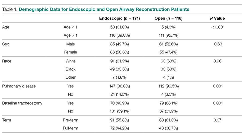
Potential confounders were tested with univariate logistic regression to determine if they had a significant impact on readmission, reintubation, or reoperation rates. These variables (Table 2) 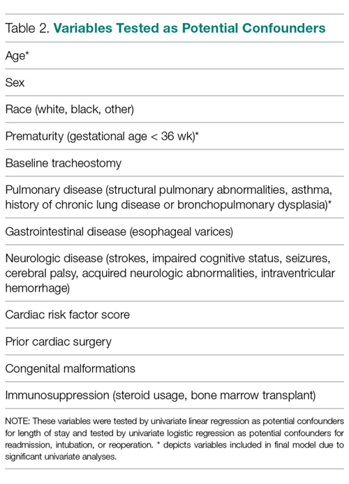
In patients undergoing endoscopic dilation, average length of stay was 5.5 days (SEM = 1.13), with 79 (48.5%) patients having a length of stay of zero days. Of all patients who had endoscopic dilations, 70 (40.1%) had a pre-existing tracheostomy and these accounted for the majority (73%) of patients who had zero days as their LOS. LOS after endoscopic management was significantly shorter than the mean of 11.3 days (SEM = 1.01) reported in those undergoing open reconstruction (P < 0.001).
With respect to 30-day adverse events, 2 patients in the endoscopic group (1.1%) required reintubation. Thirteen endoscopic dilation cases (7.6%) had an unplanned readmission, four (2.3%) of which were associated with reoperation within 30 days of the primary surgical procedure. There were 9 other reoperations unassociated with unplanned readmission. Three of these reoperations were due to failed endoscopic dilations, resulting in 2 tracheostomies and one open airway reconstruction. There was one patient death, in a 0-day old with tetralogy of Fallot, trachea-esophageal fistula, and ventilator dependence who underwent emergent endoscopic dilation and died the same day.
Open procedures were associated with 11 unplanned readmissions (9.5%), 7 re-intubations (6%) and 18 reoperations (15.5%). Of patents undergoing reoperation, one patient undergoing open reconstruction underwent tracheostomy within 30 days of surgery.
When comparing open reconstruction to endoscopic dilation, there was a significant increase in reintubation (OR = 7.41, P = 0.026) and reoperation (OR = 3.09, P = 0.009) for open procedures, even with adjustment for age, tracheostomy status, and pulmonary disease. There was no significant difference between the two for unplanned readmissions (OR = 1.19, P = 0.79) (Figure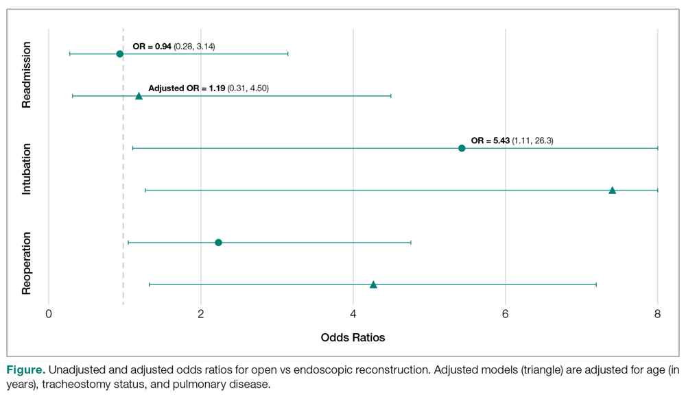
Younger age was also found to be significantly associated with reoperation rates, in an adjusted logistic model that accounted for tracheostomy status, type of surgery, and pulmonary disease. Per year of life, younger children had higher reoperation rates than older children (OR = 1.91, P = 0.017). When endoscopic dilation was individually examined, children younger than 1 year of age were more likely to undergo reoperation after an endoscopic dilation than children older than 1 (OR = 4.39, P = 0.03). Children under age 1 were also more likely to have an unplanned readmission after an endoscopic dilation (OR = 4.21, P = 0.03). The relationship between age and re-intubation was not significant (OR = 0, P = 0.95). For open reconstruction, this age dichotomization was not associated with any increased reoperation (OR = 2.3, P = 0.52), readmission (OR = 0, P = 0.97), or reintubation (OR = 0, P = 0.94).
T-test analysis was performed to determine if children < 1 year old also had significantly longer hospital stays after endoscopic dilation than older children (mean 14.1 days vs 1.9 days, P < 0.001). This relationship held true in a linear regression after adjustment for pulmonary disease and tracheostomy, with length of stay decreasing by 0.48 days per year of life (P = 0.03). For endoscopic dilations, the same relationship held true, where length of stay decreased by 0.75 days per year of life.
Discussion
Endoscopic dilation for primary management of pediatric laryngotracheal stenosis has become commonplace. Despite this, outcomes of this procedure have only been described in case series and meta-analyses [2–5]. The relative rarity of pediatric laryngotracheal stenosis suggests the need for large, multi-institutional data for purposes of patient selection and medical decision-making.
This study utilized the ACS-NSQIP-Pediatric database to highlight 30-day outcomes of endoscopic dilation and to compare these outcomes to those of open airway reconstruction procedures. The ACS-NSQIP database has been endorsed by multiple organizations, including the Center for Medicare and Medicaid Services, the Joint Commission, and the American Medical Association. It has been shown to have higher sensitivity and a lower false-positive rate when compared to administrative data, in part due to data collection from trained personnel [11]. Furthermore, ACS-NSQIP use has the additional benefit of reporting an unplanned admission—a feature unavailable in review of claims data [12].
With respect to adverse events, our study demonstrates that endoscopic dilation is associated with an equally high rate of unplanned readmission when compared to open reconstruction. The high prevalence of comorbid disease such as chronic lung disease (32% of endoscopic dilation and 43% of open reconstruction) can account for some of the morbidity associated with any airway procedures.
Despite high rates of unplanned readmission, patients undergoing endoscopic dilation were less likely to have reoperations within 30 days of initial surgery when compared to those undergoing open reconstruction. While differences in disease severity may be partially responsible for this difference in the reoperation rate, this finding is notable given the health care costs associated with multiple operations as well as safety concerns with multiple anesthetics in the very young [13,14].
The ACS-NSQIP platform does not distinguish unplanned from planned reoperations. In the setting of airway surgery, where multiple planned reoperations are commonplace, this metric is a suboptimal stand-alone indicator of adverse outcomes. Other markers available in the database—such as reintubations and performance of tracheostomy or open airway reconstruction within 30-days of surgery—are more indicative of surgical outcome in the setting of airway surgery. We found that both reintubations and salvage open reconstruction within 30-days were rare. It should be noted that the ACS-NSQIP data does not report any events occurring outside of the 30-day postoperative period, representing potential limitation of the use of this database. As was previously advocated by Roxbury and colleagues, procedure/subspecialty specific outcome data collection would also improve outcome analysis of airway and other otolaryngologic procedures [9]. In the setting of airway reconstruction, this would include data pertaining to Cotton-Meyer grading systems well as postoperative voice and swallow outcomes.
In addition to safety profile, endoscopic procedures were associated with shorter LOS when compared with open reconstruction, representing another potential source of cost savings with this less invasive method. This is especially significant given that open reconstruction patients spend much of their inpatient stay in an ICU setting. In patients who are candidates for endoscopic procedures, this lower-risk, lower-cost profile of endoscopic dilation has the opportunity to improve value in health care and may be the source of future improvement initiatives.
In addition to comparing overall outcomes between endoscopic and open management of laryngotracheal stenosis, our study aimed to identify factors that were associated with varied outcomes in patients undergoing primary endoscopic dilation. We found that children younger than 1 year of age were 5.8 times more likely to undergo an unplanned reoperation after an endoscopic dilation than children over 1 year. A similar finding was reported in open airway surgeries, with increased reoperation rates in children < 3 years old [9]. The justification of a dichotomization at 1 year was made as expert opinion recognizes that the infant airway is less forgiving to intervention given its small size. Young age was also a factor in prolonged LOS as was determined by linear regression. It is likely that this increased LOS may be in part due to associations of young age and the neonatal ICU population. One must balance the increased risk of surgery in the young with that of tracheostomy, which has a published complication rate of 18% to 50% and direct mortality rate of 1% to 2% in the pediatric population [15–18]. Understanding these relative risks may help guide the airway surgeon in preoperative counseling with families and medical decision-making.
As discussed above, the limitation of data to a 30-day period is a relative weakness of ACS-NSQIP database use for studies of airway reconstruction, as the ultimate outcome—a stable, decannulated airway—may occur outside of this time period. As many quality metrics utilize data from the 30-day postoperative period, knowledge of these outcomes remains valuable in surgical decision-making. Ultimately, collection of data in a large, long-term dataset would allow broader generalizations to be made about the differences between open and endoscopic procedures and would also give a more comprehensive picture of the outcomes of endoscopic dilation.
In conclusion, this study is the first to analyze 30-day postoperative outcomes in pediatric endoscopic airway dilations using data aggregated by ACS-NSQIP from institutions across the United States. This data indicates that endoscopic airway dilation is a relatively safe procedure, especially compared with open reconstruction; however, additional data on disease severity and other outcomes is necessary to draw final conclusions of superiority of technique. Future improvement initiatives could be aimed at the impact of this lower-risk, lower-cost procedure in the appropriately selected patient. Outcomes of endoscopic dilation are poorer in those less than 1 year of age, as they are associated with increased reoperation rates and increased length of stay compared to older children. One must balance these risks in the very young with the risks associated with tracheostomy and other alternative airway management modalities.
Note: This work was presented in a paper at the AAO-HNS 2017 meeting, Chicago, IL, 10 Sep 2017.
Corresponding author: Jennifer Lavin, MD, MS, 225 E Chicago Ave., Box 25, Chicago, IL 60611, JLavin@luriechildrens.org.
Financial disclosures: None.
1. Cohen MD, Weber TR, Rao CC. Balloon dilatation of tracheal and bronchial stenosis. AJR Am J Roentgenol 1984;142:477–8.
2. Chueng K, Chadha NK. Primary dilatation as a treatment for pediatric laryngotracheal stenosis: a systematic review. Int J Pediatr Otorhinolaryngol 2013;77:623–8.
3. Hautefort C, Teissier N, Viala P, Van Den Abbeele T. Balloon dilation laryngoplasty for subglottic stenosis in children: eight years’ experience. Arch Otolaryngol Head Neck Surg 2012;138:235–40.
4. Lang M, Brietzke SE. A systematic review and meta-analysis of endoscopic balloon dilation of pediatric subglottic stenosis. Otolaryngol Head Neck Surg 2014;150:174–9.
5. Maresh A, Preciado DA, O’Connell AP, Zalzal GH. A comparative analysis of open surgery vs endoscopic balloon dilation for pediatric subglottic stenosis. JAMA Otolaryngol Head Neck Surg 2014;140:901–5.
6. Gelbard A, Donovan DT, Ongkasuwan J, et al. Disease homogeneity and treatment heterogeneity in idiopathic subglottic stenosis. Laryngoscope 2016;126:1390–6.
7. ACS-NSQIP. ACS National Surgical Quality Improvement Program® (ACS NSQIP®). 2017. Available at: http://site.acsnsqip.org/program-specifics/scr-training-and-resources. Accessed June 2 2017.
8. Shiloach M, Frencher SK Jr, Steeger JE, et al. Toward robust information: data quality and inter-rater reliability in the American College of Surgeons National Surgical Quality Improvement Program. J Am Coll Surg 2010;210:6–16.
9. Roxbury CR, Jatana KR, Shah RK, Boss EF. Safety and postoperative adverse events in pediatric airway reconstruction: Analysis of ACS-NSQIP-P 30-day outcomes. Laryngoscope 2017;127:504–8.
10. Raval MV, Dillon PW, Bruny JL, et al. Pediatric American College of Surgeons National Surgical Quality Improvement Program: feasibility of a novel, prospective assessment of surgical outcomes. J Pediatr Surg 2011;46:115–21.
11. Lawson EH, Louie R, Zingmond DS, et al. A comparison of clinical registry versus administrative claims data for reporting of 30-day surgical complications. Ann Surg 2012;256:973–81.
12. Sellers MM, Merkow RP, Halverson A, et al. Validation of new readmission data in the American College of Surgeons National Surgical Quality Improvement Program. J Am Coll Surg 2013;216:420–7.
13. Jevtovic-Todorovic V, Hartman RE, Izumi Y, et al. Early exposure to common anesthetic agents causes widespread neurodegeneration in the developing rat brain and persistent learning deficits. J Neurosci 2003;23:876–82.
14. Patel P, Sun L. Update on neonatal anesthetic neurotoxicity: insight into molecular mechanisms and relevance to humans. Anesthesiology 2009;110:703–8.
15. Crysdale WS, Feldman RI, Naito K. Tracheotomies: a 10-year experience in 319 children. Ann Otol Rhinol Laryngol 1988;97(5 Pt 1):439–43.
16. Goldenberg D, Ari EG, Golz A, et al. Tracheotomy complications: a retrospective study of 1130 cases. Otolaryngol Head Neck Surg 2000;123:495–500.
17. Mahadevan M, Barber C, Salkeld L, et al N. Pediatric tracheotomy: 17 year review. Int J Pediatr Otorhinolaryngol 2007;71:1829–35.
18. Ozmen S, Ozmen OA, Unal OF. Pediatric tracheotomies: a 37-year experience in 282 children. Int J Pediatr Otorhinolaryngol 2009;73:959–61.
From the Northwestern University, Feinberg School of Medicine, Chicago, IL (Mr. Bavishi, Dr. Lavin), the Johns Hopkins University, Baltimore, MD (Dr. Boss), Children’s National Medical Center, Washington, DC (Dr. Shah), and Ann & Robert H. Lurie Children’s Hospital of Chicago, Chicago, IL (Dr. Lavin).
Abstract
- Background: Endoscopic management of pediatric subglottic stenosis is common; however, no multiinstitutional studies have assessed its perioperative outcomes. The American College of Surgeon’s National Surgical Quality Improvement Program – Pediatric (ACS-NSQIP-P) represents a source of such data.
- Objective: To investigate 30-day outcomes of endoscopic dilation of the pediatric airway and to compare these outcomes to those seen with open reconstruction techniques.
- Methods: Current procedural terminology (CPT) codes were queried for endoscopic or open airway reconstruction in the 2015 ACS-NSQIP-P Public Use File (PUF). Demographics and 30-day events were abstracted to compare open to endoscopic techniques and to assess for risk factors for varied outcomes after endoscopic dilation. Outcome measures included length of stay (LOS), 30-day rates of reintubation, readmission, and reoperation.
- Results: 171 endoscopic and 116 open procedures were identified. Mean age at endoscopic and open procedures was 4.1 (SEM = 0.37) and 5.4 years (SEM = 0.40). Mean LOS was shorter after endoscopic procedures (5.5 days, SEM = 1.13 vs. 11.3 days SEM = 1.01, P < 0.001). Open procedures had higher rates of reintubation (OR = 7.41, P = 0.026) and reoperation (OR = 3.09, P = 0.009). In patients undergoing endoscopic dilation, children < 1 year were more likely to require readmission (OR = 4.21, P = 0.03) and reoperation (OR = 4.39, P = 0.03) when compared with older children.
- Conclusion: Open airway reconstruction is associated with longer LOS and increased reintubations and reoperations, suggesting a possible opportunity to improve value in health care in the appropriately selected patient. Reoperations and readmissions following endoscopic dilation are more prevalent in children younger than 1 year.
Keywords: airway stenosis; subglottic stenosis; endoscopic dilation; pediatrics; outcomes.
Historically, pediatric laryngotracheal stenosis was managed using open reconstruction techniques, including laryngoplasty, tracheal resection, and cervical tracheoplasty. Initial reports of endoscopic dilation were described in the 1980s as a means to salvage re-stenosis after open reconstruction [1]. Currently, primary endoscopic dilation has become commonplace in otolaryngology due to its less invasive nature as well as—in cases of balloon dilation—minimization of tissue damage [2]. The advancements made in endoscopic balloon dilation have reduced the frequency with which open reconstruction is performed.
Systematic reviews and case series investigating endoscopic dilation indicate a 70% to 80% success rate in preventing future open surgery or tracheostomy [2–5]. While increased severity of stenosis has been associated with poorer outcomes in endoscopic procedures, few other risk factors that influence surgical success have been identified [4,5]. In a single study in the adult literature, open surgical management of idiopathic subglottic stenosis was associated with improved outcomes when compared to endoscopic techniques [5]. Such findings suggest a need to identify these factors for the purpose of optimizing clinical decision-making.
As laryngotracheal stenosis is rare, postoperative outcomes and risk factors are best identified on a multiinstitutional level. Due to its participation from 80 hospitals and its accurate and reliable reporting of both demographic and risk-stratified 30-day outcomes data, the American College of Surgeon’s National Surgical Quality Improvement Program – Pediatric (ACS NSQIP-P) provides such a platform [6–8]. Thirty-day outcomes and risk factors for open reconstruction utilizing the ACS NSQIP-P database have previously been reported; however, no such outcomes for endoscopic dilation have been described, and no comparison between endoscopic and open procedures has been made [9]. The purpose of this study was to utilize the 2015 ACS-NSQIP-P database to investigate 30-day outcomes of endoscopic dilation of the pediatric airway and to compare these outcomes to open reconstruction techniques. Secondarily, we aimed to determine if any demographic factors or medical comorbidities are associated with varied outcomes in endoscopic reconstruction. While these data reflect safety and quality of this procedure in the United States, findings may potentially be applied across international settings.
Methods
Data Source
Data was obtained from the 2015 ACS-NSQIP-P Public Use File (PUF). Due to the de-identified and public nature of these data, this research was exempt from review by the Ann & Robert H. Lurie Children’s Hospital of Chicago review board. Data collection methods for ACS-NSQIP-P have previously been described [10]. In brief, data was collected from 80 hospitals on approximately 120 preoperative, intraoperative, and postoperative variables. Cases are systematically sampled on an 8-day cycle basis, where the first 35 cases meeting the inclusion criteria in each hospital in each cycle are submitted to ACS-NSQIP-P.
Variables and Outcomes
Airway procedures for endoscopic dilations and open reconstructions were obtained by CPT code. Endoscopic dilations (CPT 31528) were compared to open reconstructions, which included laryngoplasty (31580, 31582), cervical tracheoplasty (31750), cricoid split (31587), and tracheal resection (31780). Demographic variables included age, sex, race, and history of prematurity. Presence of specific comorbid diseases were also collected and tested for significance.
Dependent outcomes of interest were unplanned 30-day postoperative events grouped as reoperation, unplanned readmission, and postoperative reintubation. In the case of endoscopic procedures, the presence of salvage open reconstruction or tracheostomy within 30 days of surgery was also recorded. Length of stay (LOS) after the procedure was collected. Specific postoperative complications and reasons for readmission were recorded within the limitations of data available in the PUF.
Analysis
Analysis was performed using descriptive statistics and frequency analysis where appropriate. Chi-square analysis was used to compare adverse events between open and endoscopic procedures. Logistic regression with calculation of odds ratio (OR) was performed to determine predictive factors for reoperation, readmission, and reintubation in all pediatric airway reconstructive procedures in adjusted and unadjusted models. T-test and linear regression was performed on the continuous outcome of length of stay. For all analyses, a p value of < 0.05 was considered statistically significant. All variable recoding and statistical analyses were performed in SAS/STAT software (Cary, NC).
Results
A total of 84,056 pediatric procedures were extracted from the 2015 NSQIP-P PUFs. Using the above CPT codes, 171 endoscopic dilations and 116 open airway reconstructions were identified, with patient age ranging from 0 days to 17.6 years. Average age of patients undergoing endoscopic dilation and open reconstruction was 4.1 and 5.4 years, respectively (Table 1).
Potential confounders were tested with univariate logistic regression to determine if they had a significant impact on readmission, reintubation, or reoperation rates. These variables (Table 2)
In patients undergoing endoscopic dilation, average length of stay was 5.5 days (SEM = 1.13), with 79 (48.5%) patients having a length of stay of zero days. Of all patients who had endoscopic dilations, 70 (40.1%) had a pre-existing tracheostomy and these accounted for the majority (73%) of patients who had zero days as their LOS. LOS after endoscopic management was significantly shorter than the mean of 11.3 days (SEM = 1.01) reported in those undergoing open reconstruction (P < 0.001).
With respect to 30-day adverse events, 2 patients in the endoscopic group (1.1%) required reintubation. Thirteen endoscopic dilation cases (7.6%) had an unplanned readmission, four (2.3%) of which were associated with reoperation within 30 days of the primary surgical procedure. There were 9 other reoperations unassociated with unplanned readmission. Three of these reoperations were due to failed endoscopic dilations, resulting in 2 tracheostomies and one open airway reconstruction. There was one patient death, in a 0-day old with tetralogy of Fallot, trachea-esophageal fistula, and ventilator dependence who underwent emergent endoscopic dilation and died the same day.
Open procedures were associated with 11 unplanned readmissions (9.5%), 7 re-intubations (6%) and 18 reoperations (15.5%). Of patents undergoing reoperation, one patient undergoing open reconstruction underwent tracheostomy within 30 days of surgery.
When comparing open reconstruction to endoscopic dilation, there was a significant increase in reintubation (OR = 7.41, P = 0.026) and reoperation (OR = 3.09, P = 0.009) for open procedures, even with adjustment for age, tracheostomy status, and pulmonary disease. There was no significant difference between the two for unplanned readmissions (OR = 1.19, P = 0.79) (Figure
Younger age was also found to be significantly associated with reoperation rates, in an adjusted logistic model that accounted for tracheostomy status, type of surgery, and pulmonary disease. Per year of life, younger children had higher reoperation rates than older children (OR = 1.91, P = 0.017). When endoscopic dilation was individually examined, children younger than 1 year of age were more likely to undergo reoperation after an endoscopic dilation than children older than 1 (OR = 4.39, P = 0.03). Children under age 1 were also more likely to have an unplanned readmission after an endoscopic dilation (OR = 4.21, P = 0.03). The relationship between age and re-intubation was not significant (OR = 0, P = 0.95). For open reconstruction, this age dichotomization was not associated with any increased reoperation (OR = 2.3, P = 0.52), readmission (OR = 0, P = 0.97), or reintubation (OR = 0, P = 0.94).
T-test analysis was performed to determine if children < 1 year old also had significantly longer hospital stays after endoscopic dilation than older children (mean 14.1 days vs 1.9 days, P < 0.001). This relationship held true in a linear regression after adjustment for pulmonary disease and tracheostomy, with length of stay decreasing by 0.48 days per year of life (P = 0.03). For endoscopic dilations, the same relationship held true, where length of stay decreased by 0.75 days per year of life.
Discussion
Endoscopic dilation for primary management of pediatric laryngotracheal stenosis has become commonplace. Despite this, outcomes of this procedure have only been described in case series and meta-analyses [2–5]. The relative rarity of pediatric laryngotracheal stenosis suggests the need for large, multi-institutional data for purposes of patient selection and medical decision-making.
This study utilized the ACS-NSQIP-Pediatric database to highlight 30-day outcomes of endoscopic dilation and to compare these outcomes to those of open airway reconstruction procedures. The ACS-NSQIP database has been endorsed by multiple organizations, including the Center for Medicare and Medicaid Services, the Joint Commission, and the American Medical Association. It has been shown to have higher sensitivity and a lower false-positive rate when compared to administrative data, in part due to data collection from trained personnel [11]. Furthermore, ACS-NSQIP use has the additional benefit of reporting an unplanned admission—a feature unavailable in review of claims data [12].
With respect to adverse events, our study demonstrates that endoscopic dilation is associated with an equally high rate of unplanned readmission when compared to open reconstruction. The high prevalence of comorbid disease such as chronic lung disease (32% of endoscopic dilation and 43% of open reconstruction) can account for some of the morbidity associated with any airway procedures.
Despite high rates of unplanned readmission, patients undergoing endoscopic dilation were less likely to have reoperations within 30 days of initial surgery when compared to those undergoing open reconstruction. While differences in disease severity may be partially responsible for this difference in the reoperation rate, this finding is notable given the health care costs associated with multiple operations as well as safety concerns with multiple anesthetics in the very young [13,14].
The ACS-NSQIP platform does not distinguish unplanned from planned reoperations. In the setting of airway surgery, where multiple planned reoperations are commonplace, this metric is a suboptimal stand-alone indicator of adverse outcomes. Other markers available in the database—such as reintubations and performance of tracheostomy or open airway reconstruction within 30-days of surgery—are more indicative of surgical outcome in the setting of airway surgery. We found that both reintubations and salvage open reconstruction within 30-days were rare. It should be noted that the ACS-NSQIP data does not report any events occurring outside of the 30-day postoperative period, representing potential limitation of the use of this database. As was previously advocated by Roxbury and colleagues, procedure/subspecialty specific outcome data collection would also improve outcome analysis of airway and other otolaryngologic procedures [9]. In the setting of airway reconstruction, this would include data pertaining to Cotton-Meyer grading systems well as postoperative voice and swallow outcomes.
In addition to safety profile, endoscopic procedures were associated with shorter LOS when compared with open reconstruction, representing another potential source of cost savings with this less invasive method. This is especially significant given that open reconstruction patients spend much of their inpatient stay in an ICU setting. In patients who are candidates for endoscopic procedures, this lower-risk, lower-cost profile of endoscopic dilation has the opportunity to improve value in health care and may be the source of future improvement initiatives.
In addition to comparing overall outcomes between endoscopic and open management of laryngotracheal stenosis, our study aimed to identify factors that were associated with varied outcomes in patients undergoing primary endoscopic dilation. We found that children younger than 1 year of age were 5.8 times more likely to undergo an unplanned reoperation after an endoscopic dilation than children over 1 year. A similar finding was reported in open airway surgeries, with increased reoperation rates in children < 3 years old [9]. The justification of a dichotomization at 1 year was made as expert opinion recognizes that the infant airway is less forgiving to intervention given its small size. Young age was also a factor in prolonged LOS as was determined by linear regression. It is likely that this increased LOS may be in part due to associations of young age and the neonatal ICU population. One must balance the increased risk of surgery in the young with that of tracheostomy, which has a published complication rate of 18% to 50% and direct mortality rate of 1% to 2% in the pediatric population [15–18]. Understanding these relative risks may help guide the airway surgeon in preoperative counseling with families and medical decision-making.
As discussed above, the limitation of data to a 30-day period is a relative weakness of ACS-NSQIP database use for studies of airway reconstruction, as the ultimate outcome—a stable, decannulated airway—may occur outside of this time period. As many quality metrics utilize data from the 30-day postoperative period, knowledge of these outcomes remains valuable in surgical decision-making. Ultimately, collection of data in a large, long-term dataset would allow broader generalizations to be made about the differences between open and endoscopic procedures and would also give a more comprehensive picture of the outcomes of endoscopic dilation.
In conclusion, this study is the first to analyze 30-day postoperative outcomes in pediatric endoscopic airway dilations using data aggregated by ACS-NSQIP from institutions across the United States. This data indicates that endoscopic airway dilation is a relatively safe procedure, especially compared with open reconstruction; however, additional data on disease severity and other outcomes is necessary to draw final conclusions of superiority of technique. Future improvement initiatives could be aimed at the impact of this lower-risk, lower-cost procedure in the appropriately selected patient. Outcomes of endoscopic dilation are poorer in those less than 1 year of age, as they are associated with increased reoperation rates and increased length of stay compared to older children. One must balance these risks in the very young with the risks associated with tracheostomy and other alternative airway management modalities.
Note: This work was presented in a paper at the AAO-HNS 2017 meeting, Chicago, IL, 10 Sep 2017.
Corresponding author: Jennifer Lavin, MD, MS, 225 E Chicago Ave., Box 25, Chicago, IL 60611, JLavin@luriechildrens.org.
Financial disclosures: None.
From the Northwestern University, Feinberg School of Medicine, Chicago, IL (Mr. Bavishi, Dr. Lavin), the Johns Hopkins University, Baltimore, MD (Dr. Boss), Children’s National Medical Center, Washington, DC (Dr. Shah), and Ann & Robert H. Lurie Children’s Hospital of Chicago, Chicago, IL (Dr. Lavin).
Abstract
- Background: Endoscopic management of pediatric subglottic stenosis is common; however, no multiinstitutional studies have assessed its perioperative outcomes. The American College of Surgeon’s National Surgical Quality Improvement Program – Pediatric (ACS-NSQIP-P) represents a source of such data.
- Objective: To investigate 30-day outcomes of endoscopic dilation of the pediatric airway and to compare these outcomes to those seen with open reconstruction techniques.
- Methods: Current procedural terminology (CPT) codes were queried for endoscopic or open airway reconstruction in the 2015 ACS-NSQIP-P Public Use File (PUF). Demographics and 30-day events were abstracted to compare open to endoscopic techniques and to assess for risk factors for varied outcomes after endoscopic dilation. Outcome measures included length of stay (LOS), 30-day rates of reintubation, readmission, and reoperation.
- Results: 171 endoscopic and 116 open procedures were identified. Mean age at endoscopic and open procedures was 4.1 (SEM = 0.37) and 5.4 years (SEM = 0.40). Mean LOS was shorter after endoscopic procedures (5.5 days, SEM = 1.13 vs. 11.3 days SEM = 1.01, P < 0.001). Open procedures had higher rates of reintubation (OR = 7.41, P = 0.026) and reoperation (OR = 3.09, P = 0.009). In patients undergoing endoscopic dilation, children < 1 year were more likely to require readmission (OR = 4.21, P = 0.03) and reoperation (OR = 4.39, P = 0.03) when compared with older children.
- Conclusion: Open airway reconstruction is associated with longer LOS and increased reintubations and reoperations, suggesting a possible opportunity to improve value in health care in the appropriately selected patient. Reoperations and readmissions following endoscopic dilation are more prevalent in children younger than 1 year.
Keywords: airway stenosis; subglottic stenosis; endoscopic dilation; pediatrics; outcomes.
Historically, pediatric laryngotracheal stenosis was managed using open reconstruction techniques, including laryngoplasty, tracheal resection, and cervical tracheoplasty. Initial reports of endoscopic dilation were described in the 1980s as a means to salvage re-stenosis after open reconstruction [1]. Currently, primary endoscopic dilation has become commonplace in otolaryngology due to its less invasive nature as well as—in cases of balloon dilation—minimization of tissue damage [2]. The advancements made in endoscopic balloon dilation have reduced the frequency with which open reconstruction is performed.
Systematic reviews and case series investigating endoscopic dilation indicate a 70% to 80% success rate in preventing future open surgery or tracheostomy [2–5]. While increased severity of stenosis has been associated with poorer outcomes in endoscopic procedures, few other risk factors that influence surgical success have been identified [4,5]. In a single study in the adult literature, open surgical management of idiopathic subglottic stenosis was associated with improved outcomes when compared to endoscopic techniques [5]. Such findings suggest a need to identify these factors for the purpose of optimizing clinical decision-making.
As laryngotracheal stenosis is rare, postoperative outcomes and risk factors are best identified on a multiinstitutional level. Due to its participation from 80 hospitals and its accurate and reliable reporting of both demographic and risk-stratified 30-day outcomes data, the American College of Surgeon’s National Surgical Quality Improvement Program – Pediatric (ACS NSQIP-P) provides such a platform [6–8]. Thirty-day outcomes and risk factors for open reconstruction utilizing the ACS NSQIP-P database have previously been reported; however, no such outcomes for endoscopic dilation have been described, and no comparison between endoscopic and open procedures has been made [9]. The purpose of this study was to utilize the 2015 ACS-NSQIP-P database to investigate 30-day outcomes of endoscopic dilation of the pediatric airway and to compare these outcomes to open reconstruction techniques. Secondarily, we aimed to determine if any demographic factors or medical comorbidities are associated with varied outcomes in endoscopic reconstruction. While these data reflect safety and quality of this procedure in the United States, findings may potentially be applied across international settings.
Methods
Data Source
Data was obtained from the 2015 ACS-NSQIP-P Public Use File (PUF). Due to the de-identified and public nature of these data, this research was exempt from review by the Ann & Robert H. Lurie Children’s Hospital of Chicago review board. Data collection methods for ACS-NSQIP-P have previously been described [10]. In brief, data was collected from 80 hospitals on approximately 120 preoperative, intraoperative, and postoperative variables. Cases are systematically sampled on an 8-day cycle basis, where the first 35 cases meeting the inclusion criteria in each hospital in each cycle are submitted to ACS-NSQIP-P.
Variables and Outcomes
Airway procedures for endoscopic dilations and open reconstructions were obtained by CPT code. Endoscopic dilations (CPT 31528) were compared to open reconstructions, which included laryngoplasty (31580, 31582), cervical tracheoplasty (31750), cricoid split (31587), and tracheal resection (31780). Demographic variables included age, sex, race, and history of prematurity. Presence of specific comorbid diseases were also collected and tested for significance.
Dependent outcomes of interest were unplanned 30-day postoperative events grouped as reoperation, unplanned readmission, and postoperative reintubation. In the case of endoscopic procedures, the presence of salvage open reconstruction or tracheostomy within 30 days of surgery was also recorded. Length of stay (LOS) after the procedure was collected. Specific postoperative complications and reasons for readmission were recorded within the limitations of data available in the PUF.
Analysis
Analysis was performed using descriptive statistics and frequency analysis where appropriate. Chi-square analysis was used to compare adverse events between open and endoscopic procedures. Logistic regression with calculation of odds ratio (OR) was performed to determine predictive factors for reoperation, readmission, and reintubation in all pediatric airway reconstructive procedures in adjusted and unadjusted models. T-test and linear regression was performed on the continuous outcome of length of stay. For all analyses, a p value of < 0.05 was considered statistically significant. All variable recoding and statistical analyses were performed in SAS/STAT software (Cary, NC).
Results
A total of 84,056 pediatric procedures were extracted from the 2015 NSQIP-P PUFs. Using the above CPT codes, 171 endoscopic dilations and 116 open airway reconstructions were identified, with patient age ranging from 0 days to 17.6 years. Average age of patients undergoing endoscopic dilation and open reconstruction was 4.1 and 5.4 years, respectively (Table 1).
Potential confounders were tested with univariate logistic regression to determine if they had a significant impact on readmission, reintubation, or reoperation rates. These variables (Table 2)
In patients undergoing endoscopic dilation, average length of stay was 5.5 days (SEM = 1.13), with 79 (48.5%) patients having a length of stay of zero days. Of all patients who had endoscopic dilations, 70 (40.1%) had a pre-existing tracheostomy and these accounted for the majority (73%) of patients who had zero days as their LOS. LOS after endoscopic management was significantly shorter than the mean of 11.3 days (SEM = 1.01) reported in those undergoing open reconstruction (P < 0.001).
With respect to 30-day adverse events, 2 patients in the endoscopic group (1.1%) required reintubation. Thirteen endoscopic dilation cases (7.6%) had an unplanned readmission, four (2.3%) of which were associated with reoperation within 30 days of the primary surgical procedure. There were 9 other reoperations unassociated with unplanned readmission. Three of these reoperations were due to failed endoscopic dilations, resulting in 2 tracheostomies and one open airway reconstruction. There was one patient death, in a 0-day old with tetralogy of Fallot, trachea-esophageal fistula, and ventilator dependence who underwent emergent endoscopic dilation and died the same day.
Open procedures were associated with 11 unplanned readmissions (9.5%), 7 re-intubations (6%) and 18 reoperations (15.5%). Of patents undergoing reoperation, one patient undergoing open reconstruction underwent tracheostomy within 30 days of surgery.
When comparing open reconstruction to endoscopic dilation, there was a significant increase in reintubation (OR = 7.41, P = 0.026) and reoperation (OR = 3.09, P = 0.009) for open procedures, even with adjustment for age, tracheostomy status, and pulmonary disease. There was no significant difference between the two for unplanned readmissions (OR = 1.19, P = 0.79) (Figure
Younger age was also found to be significantly associated with reoperation rates, in an adjusted logistic model that accounted for tracheostomy status, type of surgery, and pulmonary disease. Per year of life, younger children had higher reoperation rates than older children (OR = 1.91, P = 0.017). When endoscopic dilation was individually examined, children younger than 1 year of age were more likely to undergo reoperation after an endoscopic dilation than children older than 1 (OR = 4.39, P = 0.03). Children under age 1 were also more likely to have an unplanned readmission after an endoscopic dilation (OR = 4.21, P = 0.03). The relationship between age and re-intubation was not significant (OR = 0, P = 0.95). For open reconstruction, this age dichotomization was not associated with any increased reoperation (OR = 2.3, P = 0.52), readmission (OR = 0, P = 0.97), or reintubation (OR = 0, P = 0.94).
T-test analysis was performed to determine if children < 1 year old also had significantly longer hospital stays after endoscopic dilation than older children (mean 14.1 days vs 1.9 days, P < 0.001). This relationship held true in a linear regression after adjustment for pulmonary disease and tracheostomy, with length of stay decreasing by 0.48 days per year of life (P = 0.03). For endoscopic dilations, the same relationship held true, where length of stay decreased by 0.75 days per year of life.
Discussion
Endoscopic dilation for primary management of pediatric laryngotracheal stenosis has become commonplace. Despite this, outcomes of this procedure have only been described in case series and meta-analyses [2–5]. The relative rarity of pediatric laryngotracheal stenosis suggests the need for large, multi-institutional data for purposes of patient selection and medical decision-making.
This study utilized the ACS-NSQIP-Pediatric database to highlight 30-day outcomes of endoscopic dilation and to compare these outcomes to those of open airway reconstruction procedures. The ACS-NSQIP database has been endorsed by multiple organizations, including the Center for Medicare and Medicaid Services, the Joint Commission, and the American Medical Association. It has been shown to have higher sensitivity and a lower false-positive rate when compared to administrative data, in part due to data collection from trained personnel [11]. Furthermore, ACS-NSQIP use has the additional benefit of reporting an unplanned admission—a feature unavailable in review of claims data [12].
With respect to adverse events, our study demonstrates that endoscopic dilation is associated with an equally high rate of unplanned readmission when compared to open reconstruction. The high prevalence of comorbid disease such as chronic lung disease (32% of endoscopic dilation and 43% of open reconstruction) can account for some of the morbidity associated with any airway procedures.
Despite high rates of unplanned readmission, patients undergoing endoscopic dilation were less likely to have reoperations within 30 days of initial surgery when compared to those undergoing open reconstruction. While differences in disease severity may be partially responsible for this difference in the reoperation rate, this finding is notable given the health care costs associated with multiple operations as well as safety concerns with multiple anesthetics in the very young [13,14].
The ACS-NSQIP platform does not distinguish unplanned from planned reoperations. In the setting of airway surgery, where multiple planned reoperations are commonplace, this metric is a suboptimal stand-alone indicator of adverse outcomes. Other markers available in the database—such as reintubations and performance of tracheostomy or open airway reconstruction within 30-days of surgery—are more indicative of surgical outcome in the setting of airway surgery. We found that both reintubations and salvage open reconstruction within 30-days were rare. It should be noted that the ACS-NSQIP data does not report any events occurring outside of the 30-day postoperative period, representing potential limitation of the use of this database. As was previously advocated by Roxbury and colleagues, procedure/subspecialty specific outcome data collection would also improve outcome analysis of airway and other otolaryngologic procedures [9]. In the setting of airway reconstruction, this would include data pertaining to Cotton-Meyer grading systems well as postoperative voice and swallow outcomes.
In addition to safety profile, endoscopic procedures were associated with shorter LOS when compared with open reconstruction, representing another potential source of cost savings with this less invasive method. This is especially significant given that open reconstruction patients spend much of their inpatient stay in an ICU setting. In patients who are candidates for endoscopic procedures, this lower-risk, lower-cost profile of endoscopic dilation has the opportunity to improve value in health care and may be the source of future improvement initiatives.
In addition to comparing overall outcomes between endoscopic and open management of laryngotracheal stenosis, our study aimed to identify factors that were associated with varied outcomes in patients undergoing primary endoscopic dilation. We found that children younger than 1 year of age were 5.8 times more likely to undergo an unplanned reoperation after an endoscopic dilation than children over 1 year. A similar finding was reported in open airway surgeries, with increased reoperation rates in children < 3 years old [9]. The justification of a dichotomization at 1 year was made as expert opinion recognizes that the infant airway is less forgiving to intervention given its small size. Young age was also a factor in prolonged LOS as was determined by linear regression. It is likely that this increased LOS may be in part due to associations of young age and the neonatal ICU population. One must balance the increased risk of surgery in the young with that of tracheostomy, which has a published complication rate of 18% to 50% and direct mortality rate of 1% to 2% in the pediatric population [15–18]. Understanding these relative risks may help guide the airway surgeon in preoperative counseling with families and medical decision-making.
As discussed above, the limitation of data to a 30-day period is a relative weakness of ACS-NSQIP database use for studies of airway reconstruction, as the ultimate outcome—a stable, decannulated airway—may occur outside of this time period. As many quality metrics utilize data from the 30-day postoperative period, knowledge of these outcomes remains valuable in surgical decision-making. Ultimately, collection of data in a large, long-term dataset would allow broader generalizations to be made about the differences between open and endoscopic procedures and would also give a more comprehensive picture of the outcomes of endoscopic dilation.
In conclusion, this study is the first to analyze 30-day postoperative outcomes in pediatric endoscopic airway dilations using data aggregated by ACS-NSQIP from institutions across the United States. This data indicates that endoscopic airway dilation is a relatively safe procedure, especially compared with open reconstruction; however, additional data on disease severity and other outcomes is necessary to draw final conclusions of superiority of technique. Future improvement initiatives could be aimed at the impact of this lower-risk, lower-cost procedure in the appropriately selected patient. Outcomes of endoscopic dilation are poorer in those less than 1 year of age, as they are associated with increased reoperation rates and increased length of stay compared to older children. One must balance these risks in the very young with the risks associated with tracheostomy and other alternative airway management modalities.
Note: This work was presented in a paper at the AAO-HNS 2017 meeting, Chicago, IL, 10 Sep 2017.
Corresponding author: Jennifer Lavin, MD, MS, 225 E Chicago Ave., Box 25, Chicago, IL 60611, JLavin@luriechildrens.org.
Financial disclosures: None.
1. Cohen MD, Weber TR, Rao CC. Balloon dilatation of tracheal and bronchial stenosis. AJR Am J Roentgenol 1984;142:477–8.
2. Chueng K, Chadha NK. Primary dilatation as a treatment for pediatric laryngotracheal stenosis: a systematic review. Int J Pediatr Otorhinolaryngol 2013;77:623–8.
3. Hautefort C, Teissier N, Viala P, Van Den Abbeele T. Balloon dilation laryngoplasty for subglottic stenosis in children: eight years’ experience. Arch Otolaryngol Head Neck Surg 2012;138:235–40.
4. Lang M, Brietzke SE. A systematic review and meta-analysis of endoscopic balloon dilation of pediatric subglottic stenosis. Otolaryngol Head Neck Surg 2014;150:174–9.
5. Maresh A, Preciado DA, O’Connell AP, Zalzal GH. A comparative analysis of open surgery vs endoscopic balloon dilation for pediatric subglottic stenosis. JAMA Otolaryngol Head Neck Surg 2014;140:901–5.
6. Gelbard A, Donovan DT, Ongkasuwan J, et al. Disease homogeneity and treatment heterogeneity in idiopathic subglottic stenosis. Laryngoscope 2016;126:1390–6.
7. ACS-NSQIP. ACS National Surgical Quality Improvement Program® (ACS NSQIP®). 2017. Available at: http://site.acsnsqip.org/program-specifics/scr-training-and-resources. Accessed June 2 2017.
8. Shiloach M, Frencher SK Jr, Steeger JE, et al. Toward robust information: data quality and inter-rater reliability in the American College of Surgeons National Surgical Quality Improvement Program. J Am Coll Surg 2010;210:6–16.
9. Roxbury CR, Jatana KR, Shah RK, Boss EF. Safety and postoperative adverse events in pediatric airway reconstruction: Analysis of ACS-NSQIP-P 30-day outcomes. Laryngoscope 2017;127:504–8.
10. Raval MV, Dillon PW, Bruny JL, et al. Pediatric American College of Surgeons National Surgical Quality Improvement Program: feasibility of a novel, prospective assessment of surgical outcomes. J Pediatr Surg 2011;46:115–21.
11. Lawson EH, Louie R, Zingmond DS, et al. A comparison of clinical registry versus administrative claims data for reporting of 30-day surgical complications. Ann Surg 2012;256:973–81.
12. Sellers MM, Merkow RP, Halverson A, et al. Validation of new readmission data in the American College of Surgeons National Surgical Quality Improvement Program. J Am Coll Surg 2013;216:420–7.
13. Jevtovic-Todorovic V, Hartman RE, Izumi Y, et al. Early exposure to common anesthetic agents causes widespread neurodegeneration in the developing rat brain and persistent learning deficits. J Neurosci 2003;23:876–82.
14. Patel P, Sun L. Update on neonatal anesthetic neurotoxicity: insight into molecular mechanisms and relevance to humans. Anesthesiology 2009;110:703–8.
15. Crysdale WS, Feldman RI, Naito K. Tracheotomies: a 10-year experience in 319 children. Ann Otol Rhinol Laryngol 1988;97(5 Pt 1):439–43.
16. Goldenberg D, Ari EG, Golz A, et al. Tracheotomy complications: a retrospective study of 1130 cases. Otolaryngol Head Neck Surg 2000;123:495–500.
17. Mahadevan M, Barber C, Salkeld L, et al N. Pediatric tracheotomy: 17 year review. Int J Pediatr Otorhinolaryngol 2007;71:1829–35.
18. Ozmen S, Ozmen OA, Unal OF. Pediatric tracheotomies: a 37-year experience in 282 children. Int J Pediatr Otorhinolaryngol 2009;73:959–61.
1. Cohen MD, Weber TR, Rao CC. Balloon dilatation of tracheal and bronchial stenosis. AJR Am J Roentgenol 1984;142:477–8.
2. Chueng K, Chadha NK. Primary dilatation as a treatment for pediatric laryngotracheal stenosis: a systematic review. Int J Pediatr Otorhinolaryngol 2013;77:623–8.
3. Hautefort C, Teissier N, Viala P, Van Den Abbeele T. Balloon dilation laryngoplasty for subglottic stenosis in children: eight years’ experience. Arch Otolaryngol Head Neck Surg 2012;138:235–40.
4. Lang M, Brietzke SE. A systematic review and meta-analysis of endoscopic balloon dilation of pediatric subglottic stenosis. Otolaryngol Head Neck Surg 2014;150:174–9.
5. Maresh A, Preciado DA, O’Connell AP, Zalzal GH. A comparative analysis of open surgery vs endoscopic balloon dilation for pediatric subglottic stenosis. JAMA Otolaryngol Head Neck Surg 2014;140:901–5.
6. Gelbard A, Donovan DT, Ongkasuwan J, et al. Disease homogeneity and treatment heterogeneity in idiopathic subglottic stenosis. Laryngoscope 2016;126:1390–6.
7. ACS-NSQIP. ACS National Surgical Quality Improvement Program® (ACS NSQIP®). 2017. Available at: http://site.acsnsqip.org/program-specifics/scr-training-and-resources. Accessed June 2 2017.
8. Shiloach M, Frencher SK Jr, Steeger JE, et al. Toward robust information: data quality and inter-rater reliability in the American College of Surgeons National Surgical Quality Improvement Program. J Am Coll Surg 2010;210:6–16.
9. Roxbury CR, Jatana KR, Shah RK, Boss EF. Safety and postoperative adverse events in pediatric airway reconstruction: Analysis of ACS-NSQIP-P 30-day outcomes. Laryngoscope 2017;127:504–8.
10. Raval MV, Dillon PW, Bruny JL, et al. Pediatric American College of Surgeons National Surgical Quality Improvement Program: feasibility of a novel, prospective assessment of surgical outcomes. J Pediatr Surg 2011;46:115–21.
11. Lawson EH, Louie R, Zingmond DS, et al. A comparison of clinical registry versus administrative claims data for reporting of 30-day surgical complications. Ann Surg 2012;256:973–81.
12. Sellers MM, Merkow RP, Halverson A, et al. Validation of new readmission data in the American College of Surgeons National Surgical Quality Improvement Program. J Am Coll Surg 2013;216:420–7.
13. Jevtovic-Todorovic V, Hartman RE, Izumi Y, et al. Early exposure to common anesthetic agents causes widespread neurodegeneration in the developing rat brain and persistent learning deficits. J Neurosci 2003;23:876–82.
14. Patel P, Sun L. Update on neonatal anesthetic neurotoxicity: insight into molecular mechanisms and relevance to humans. Anesthesiology 2009;110:703–8.
15. Crysdale WS, Feldman RI, Naito K. Tracheotomies: a 10-year experience in 319 children. Ann Otol Rhinol Laryngol 1988;97(5 Pt 1):439–43.
16. Goldenberg D, Ari EG, Golz A, et al. Tracheotomy complications: a retrospective study of 1130 cases. Otolaryngol Head Neck Surg 2000;123:495–500.
17. Mahadevan M, Barber C, Salkeld L, et al N. Pediatric tracheotomy: 17 year review. Int J Pediatr Otorhinolaryngol 2007;71:1829–35.
18. Ozmen S, Ozmen OA, Unal OF. Pediatric tracheotomies: a 37-year experience in 282 children. Int J Pediatr Otorhinolaryngol 2009;73:959–61.
HIPEC for Ovarian Cancer: Standard of Care or Experimental Approach?
Study Overview
Objective. To evaluate whether the addition of hyperthermic intraperotoneal chemotherapy (HIPEC) to interval cytoreductive surgery would improve outcomes among patients who were receiving neoadjuvant chemotherapy for stage III epithelial ovarian cancer.
Design. Phase 3 prospective randomized clinical trial.
Setting and participants. The trial was conducted at 8 hospitals in the Netherlands and Belgium at which medical personnel had experience in administering HIPEC in patients with peritoneal disease from colon cancer or from pseudomyxoma perotinei. Eligible patients had newly diagnosed stage III epithelial ovarian, fallopian tube, or peritoneal cancer and were referred for neoadjuvant chemotherapy because of extensive abdominal disease or incomplete cytoreductive surgery (one or more residual tumors measuring > 1 cm in diameter). Eligibility criteria also including performance status score of 0 to 2, normal blood counts, and adequate renal function.
Intervention. At the time of surgery, patients were randomly assigned in a 1:1 ratio to undergo interval cytoreductive surgery either with HIPEC (surgery-plus-HIPEC group) or without HIPEC (surgery group). HIPEC was administered at the end of the cytoreductive surgical procedure. The abdomen was filled with saline that circulated continuously with the use of a roller pump through a heat exchanger. Perfusion with cisplatin at a dose of 100 mg per square meter and at a flow rate of 1 liter per minute was then initiated. The procedure took 120 minutes in total. To prevent nephrotoxicity, sodium thiosulphate was administered at the start of perfusion as an intravenous bolus (9 g per square meter in 200 mL), followed by a continuous infusion (12 g per square meter in 1000 mL) over 6 hours. Patient received in addition 3 cycles of carboplatin and paclitaxel after surgery. During follow-up, physical examinations and measurement of CA-125 level were repeated every 3 months for 2 years and then every 6 months until 5 years after the completion of chemotherapy. Computed tomography was performed at 1, 6, 12, and 24 months after the last cycle of chemotherapy.
Main outcome measure. The primary endpoint was recurrence-free survival in the intent-to-treat population. Secondary endpoints included overall survival, the side-effect profile, and health-related quality of life.
Main results. A total of 245 women were randomized between April 2007 and April 2016. The median follow-up at the time of recurrence-free survival analysis was 4.7 years. Recurrence-free survival events occurred in 81% of the HIPEC group vs 89% of the control group; median recurrence-free survival was 14.2 months vs 10.7 months, respectively (hazard ratio [HR] 0.66, P = 0.003). The benefit of HIPEC was consistent across stratification factors and post hoc subgroups. Hazard ratios (none reaching statistical significance) were 0.63 and 0.72 for those aged ≥ 65 and < 65 years; 0.69 and 0.56 for those with high-grade serous and other histology; 0.71 and 0.47 for those with no previous surgery and previous surgery; 0.64 and 0.66 for those with 0 to 5 and 6 to 8 involved regions; and 0.69 and 0.61 for those with no laparoscopy vs laparoscopy before surgery. Death occurred in 50% of the hyperthermic intraperitoneal chemotherapy group vs 62% of the control group; median overall survival was 45.7 vs 33.9 months (HR 0.67, P = 0.02).
No significant differences between the HIPEC and control groups were observed in the incidence of adverse events of any grade. The most common adverse events of any grade in the HIPEC group were nausea (63% vs 57%), abdominal pain (60% vs 575), and fatigue (37% vs 30%). Grade ≥ 3 adverse events occurred in 27% vs 25% of patients (P = 0.76). The most common grade 3 or 4 adverse events in the HIPEC group were infection (6% vs 2%), abdominal pain (5% vs 6%), and ileus (4% vs 2%). Among the patients who underwent bowel resection, a colostomy or ileostomy was performed more commonly among patients in the surgery-plus-HIPEC group (21 of 29 patients [72%]) than among those in the surgery group (13 of 30 patients [43%]) (P = 0.04).
Conclusion. Among patients with stage III epithelial ovarian cancer, the addition of hyperthermic intraperitoneal chemotherapy to interval cytoreductive surgery resulted in longer recurrence-free survival and overall survival than surgery alone and did not result in higher rates of side effects.
Commentary
Ovarian cancer is associated with the highest mortality of all gynecologic cancers in the Western world [1].The majority of the patients have advanced disease at diagnosis and the most effective treatment for advanced disease involved maximum debulking surgery followed by chemotherapy. For those patients for whom primary surgery is not feasible, primary chemotherapy is given, which is followed by interval debulking after 3 courses of chemotherapy [2].However, outcome remains dismal for patients with advanced disease. Regional (intraperitoneal) chemotherapy theoretically results in a decreased rate of systemic toxic effects and may improve outcomes by eliminating residual microscopic disease more effectively than intravenous chemotherapy [3].
Intraperitoneal chemotherapy during surgery that can be delivered under hyperthermic conditions is termed hyperthermic intraperitoneal chemotherapy. Rationale for using hyperthermic conditions when delivering intraperitoneal chemotherapy is multifactorial. Clinical hyperthermia is defined as the use of temperatures of 41oC and higher. Hyperthermia itself has a direct cytotoxic effects on cells caused by impaired DNA repair, denaturation of proteins, inductions of heat-shock proteins which may serve as receptors for natural killer-cells, induction of apoptosis, and inhibition of angiogenesis. In addition to its intrinsic cytotoxic effect, hyperthermia acts in synergy with some chemotherapeutics agents and increase peritoneal and tumour drug penetration [4].
The study by van Driel et al evaluates the impact of addition of HIPEC to interval cytoreductive surgery in patients who received neoadjuvant chemotherapy for stage III epithelial ovarian cancer. Authors found that addition of HIPEC resulted in 11.8 months improvement in overall survival compared to surgery alone without increased rate of side effects.
The outcomes of the trial by van Driel et al are encouraging, but questions remain about how to apply these results in everyday clinical practice. First, with the extensive reported experience with HIPEC in select single center or multicenter trials, it is reasonable to conclude the procedure can be successfully undertaken by well-trained surgical/gynecologic oncologists and at institutions experienced in the approach. However, clinical trials have limited external validity, and while providing evidence regarding efficacy (ie, the effect of the intervention under highly selected conditions), they generally do not provide evidence of effectiveness (ie, the benefit to the general population of patients with the disease). Can the same results be reproduced in hospitals across the country? Second, what part of HIPEC was responsible for benefit? Was it merely administration of chemotherapy through intraperitoneal route? Is hyperthermia necessary to see the observed benefit in this trial? The answers to these questions are not known. Third, the assessment of cost-benefit ratio warrants serious consideration as well. As authors pointed, the addition of HIPEC resulted in extension of duration of surgery by 2 hours and a perfusionist was needed. Additional standard costs are incurred due to the use of HIPEC machine, the disposable products needed to administer HIPEC, and the 1-day stay in the ICU. Increased use of diverting colostomy and ileostomy will also increase the overall cost of the treatment.
Applications for Clinical Practice
This trial is an important step in establishing the efficacy of adding HIPEC to interval cytoreductive surgery without increasing the side effects. However, whether the same results can be reproduced at centers at which surgeons do not have as much expertise in administering HIPEC remains to be seen. New confirmatory clinical trials of HIPEC are needed before it can be recommended as a common treatment strategy.
—Deval Rajyaguru, MD, Gundersen Health System, La Crosse, WI
1. Levi F, Lucchini F, Negri E, La Vecchia C. Trends in mortality from major cancers in the European Union, including acceding countries, in 2004. Cancer 2006;101:2843–50.
2. van der Burg MEL, van Lent M, Buyse M, et al. The effect of debulking surgery after induction chemotherapy on the prognosis in advanced epithelial ovarian cancer. N Engl J Med 1995;332:629–34.
3. Armstrong DK, Bundy B, Wenzel L, et al. Intraperitoneal cisplatin and paclitaxel in ovarian cancer. N Engl J Med 2006;354:34–43.
4. Ohno S, Siddik ZH, Kido Y, et al. Thermal enhancement of drug uptake and DNA adducts as a possible mechanism for the effect of sequencing hyperthermia on cisplatin-induced cytotoxicity in L1210 cells. Cancer Chemother Pharmacol 1994;34:302–6.
Study Overview
Objective. To evaluate whether the addition of hyperthermic intraperotoneal chemotherapy (HIPEC) to interval cytoreductive surgery would improve outcomes among patients who were receiving neoadjuvant chemotherapy for stage III epithelial ovarian cancer.
Design. Phase 3 prospective randomized clinical trial.
Setting and participants. The trial was conducted at 8 hospitals in the Netherlands and Belgium at which medical personnel had experience in administering HIPEC in patients with peritoneal disease from colon cancer or from pseudomyxoma perotinei. Eligible patients had newly diagnosed stage III epithelial ovarian, fallopian tube, or peritoneal cancer and were referred for neoadjuvant chemotherapy because of extensive abdominal disease or incomplete cytoreductive surgery (one or more residual tumors measuring > 1 cm in diameter). Eligibility criteria also including performance status score of 0 to 2, normal blood counts, and adequate renal function.
Intervention. At the time of surgery, patients were randomly assigned in a 1:1 ratio to undergo interval cytoreductive surgery either with HIPEC (surgery-plus-HIPEC group) or without HIPEC (surgery group). HIPEC was administered at the end of the cytoreductive surgical procedure. The abdomen was filled with saline that circulated continuously with the use of a roller pump through a heat exchanger. Perfusion with cisplatin at a dose of 100 mg per square meter and at a flow rate of 1 liter per minute was then initiated. The procedure took 120 minutes in total. To prevent nephrotoxicity, sodium thiosulphate was administered at the start of perfusion as an intravenous bolus (9 g per square meter in 200 mL), followed by a continuous infusion (12 g per square meter in 1000 mL) over 6 hours. Patient received in addition 3 cycles of carboplatin and paclitaxel after surgery. During follow-up, physical examinations and measurement of CA-125 level were repeated every 3 months for 2 years and then every 6 months until 5 years after the completion of chemotherapy. Computed tomography was performed at 1, 6, 12, and 24 months after the last cycle of chemotherapy.
Main outcome measure. The primary endpoint was recurrence-free survival in the intent-to-treat population. Secondary endpoints included overall survival, the side-effect profile, and health-related quality of life.
Main results. A total of 245 women were randomized between April 2007 and April 2016. The median follow-up at the time of recurrence-free survival analysis was 4.7 years. Recurrence-free survival events occurred in 81% of the HIPEC group vs 89% of the control group; median recurrence-free survival was 14.2 months vs 10.7 months, respectively (hazard ratio [HR] 0.66, P = 0.003). The benefit of HIPEC was consistent across stratification factors and post hoc subgroups. Hazard ratios (none reaching statistical significance) were 0.63 and 0.72 for those aged ≥ 65 and < 65 years; 0.69 and 0.56 for those with high-grade serous and other histology; 0.71 and 0.47 for those with no previous surgery and previous surgery; 0.64 and 0.66 for those with 0 to 5 and 6 to 8 involved regions; and 0.69 and 0.61 for those with no laparoscopy vs laparoscopy before surgery. Death occurred in 50% of the hyperthermic intraperitoneal chemotherapy group vs 62% of the control group; median overall survival was 45.7 vs 33.9 months (HR 0.67, P = 0.02).
No significant differences between the HIPEC and control groups were observed in the incidence of adverse events of any grade. The most common adverse events of any grade in the HIPEC group were nausea (63% vs 57%), abdominal pain (60% vs 575), and fatigue (37% vs 30%). Grade ≥ 3 adverse events occurred in 27% vs 25% of patients (P = 0.76). The most common grade 3 or 4 adverse events in the HIPEC group were infection (6% vs 2%), abdominal pain (5% vs 6%), and ileus (4% vs 2%). Among the patients who underwent bowel resection, a colostomy or ileostomy was performed more commonly among patients in the surgery-plus-HIPEC group (21 of 29 patients [72%]) than among those in the surgery group (13 of 30 patients [43%]) (P = 0.04).
Conclusion. Among patients with stage III epithelial ovarian cancer, the addition of hyperthermic intraperitoneal chemotherapy to interval cytoreductive surgery resulted in longer recurrence-free survival and overall survival than surgery alone and did not result in higher rates of side effects.
Commentary
Ovarian cancer is associated with the highest mortality of all gynecologic cancers in the Western world [1].The majority of the patients have advanced disease at diagnosis and the most effective treatment for advanced disease involved maximum debulking surgery followed by chemotherapy. For those patients for whom primary surgery is not feasible, primary chemotherapy is given, which is followed by interval debulking after 3 courses of chemotherapy [2].However, outcome remains dismal for patients with advanced disease. Regional (intraperitoneal) chemotherapy theoretically results in a decreased rate of systemic toxic effects and may improve outcomes by eliminating residual microscopic disease more effectively than intravenous chemotherapy [3].
Intraperitoneal chemotherapy during surgery that can be delivered under hyperthermic conditions is termed hyperthermic intraperitoneal chemotherapy. Rationale for using hyperthermic conditions when delivering intraperitoneal chemotherapy is multifactorial. Clinical hyperthermia is defined as the use of temperatures of 41oC and higher. Hyperthermia itself has a direct cytotoxic effects on cells caused by impaired DNA repair, denaturation of proteins, inductions of heat-shock proteins which may serve as receptors for natural killer-cells, induction of apoptosis, and inhibition of angiogenesis. In addition to its intrinsic cytotoxic effect, hyperthermia acts in synergy with some chemotherapeutics agents and increase peritoneal and tumour drug penetration [4].
The study by van Driel et al evaluates the impact of addition of HIPEC to interval cytoreductive surgery in patients who received neoadjuvant chemotherapy for stage III epithelial ovarian cancer. Authors found that addition of HIPEC resulted in 11.8 months improvement in overall survival compared to surgery alone without increased rate of side effects.
The outcomes of the trial by van Driel et al are encouraging, but questions remain about how to apply these results in everyday clinical practice. First, with the extensive reported experience with HIPEC in select single center or multicenter trials, it is reasonable to conclude the procedure can be successfully undertaken by well-trained surgical/gynecologic oncologists and at institutions experienced in the approach. However, clinical trials have limited external validity, and while providing evidence regarding efficacy (ie, the effect of the intervention under highly selected conditions), they generally do not provide evidence of effectiveness (ie, the benefit to the general population of patients with the disease). Can the same results be reproduced in hospitals across the country? Second, what part of HIPEC was responsible for benefit? Was it merely administration of chemotherapy through intraperitoneal route? Is hyperthermia necessary to see the observed benefit in this trial? The answers to these questions are not known. Third, the assessment of cost-benefit ratio warrants serious consideration as well. As authors pointed, the addition of HIPEC resulted in extension of duration of surgery by 2 hours and a perfusionist was needed. Additional standard costs are incurred due to the use of HIPEC machine, the disposable products needed to administer HIPEC, and the 1-day stay in the ICU. Increased use of diverting colostomy and ileostomy will also increase the overall cost of the treatment.
Applications for Clinical Practice
This trial is an important step in establishing the efficacy of adding HIPEC to interval cytoreductive surgery without increasing the side effects. However, whether the same results can be reproduced at centers at which surgeons do not have as much expertise in administering HIPEC remains to be seen. New confirmatory clinical trials of HIPEC are needed before it can be recommended as a common treatment strategy.
—Deval Rajyaguru, MD, Gundersen Health System, La Crosse, WI
Study Overview
Objective. To evaluate whether the addition of hyperthermic intraperotoneal chemotherapy (HIPEC) to interval cytoreductive surgery would improve outcomes among patients who were receiving neoadjuvant chemotherapy for stage III epithelial ovarian cancer.
Design. Phase 3 prospective randomized clinical trial.
Setting and participants. The trial was conducted at 8 hospitals in the Netherlands and Belgium at which medical personnel had experience in administering HIPEC in patients with peritoneal disease from colon cancer or from pseudomyxoma perotinei. Eligible patients had newly diagnosed stage III epithelial ovarian, fallopian tube, or peritoneal cancer and were referred for neoadjuvant chemotherapy because of extensive abdominal disease or incomplete cytoreductive surgery (one or more residual tumors measuring > 1 cm in diameter). Eligibility criteria also including performance status score of 0 to 2, normal blood counts, and adequate renal function.
Intervention. At the time of surgery, patients were randomly assigned in a 1:1 ratio to undergo interval cytoreductive surgery either with HIPEC (surgery-plus-HIPEC group) or without HIPEC (surgery group). HIPEC was administered at the end of the cytoreductive surgical procedure. The abdomen was filled with saline that circulated continuously with the use of a roller pump through a heat exchanger. Perfusion with cisplatin at a dose of 100 mg per square meter and at a flow rate of 1 liter per minute was then initiated. The procedure took 120 minutes in total. To prevent nephrotoxicity, sodium thiosulphate was administered at the start of perfusion as an intravenous bolus (9 g per square meter in 200 mL), followed by a continuous infusion (12 g per square meter in 1000 mL) over 6 hours. Patient received in addition 3 cycles of carboplatin and paclitaxel after surgery. During follow-up, physical examinations and measurement of CA-125 level were repeated every 3 months for 2 years and then every 6 months until 5 years after the completion of chemotherapy. Computed tomography was performed at 1, 6, 12, and 24 months after the last cycle of chemotherapy.
Main outcome measure. The primary endpoint was recurrence-free survival in the intent-to-treat population. Secondary endpoints included overall survival, the side-effect profile, and health-related quality of life.
Main results. A total of 245 women were randomized between April 2007 and April 2016. The median follow-up at the time of recurrence-free survival analysis was 4.7 years. Recurrence-free survival events occurred in 81% of the HIPEC group vs 89% of the control group; median recurrence-free survival was 14.2 months vs 10.7 months, respectively (hazard ratio [HR] 0.66, P = 0.003). The benefit of HIPEC was consistent across stratification factors and post hoc subgroups. Hazard ratios (none reaching statistical significance) were 0.63 and 0.72 for those aged ≥ 65 and < 65 years; 0.69 and 0.56 for those with high-grade serous and other histology; 0.71 and 0.47 for those with no previous surgery and previous surgery; 0.64 and 0.66 for those with 0 to 5 and 6 to 8 involved regions; and 0.69 and 0.61 for those with no laparoscopy vs laparoscopy before surgery. Death occurred in 50% of the hyperthermic intraperitoneal chemotherapy group vs 62% of the control group; median overall survival was 45.7 vs 33.9 months (HR 0.67, P = 0.02).
No significant differences between the HIPEC and control groups were observed in the incidence of adverse events of any grade. The most common adverse events of any grade in the HIPEC group were nausea (63% vs 57%), abdominal pain (60% vs 575), and fatigue (37% vs 30%). Grade ≥ 3 adverse events occurred in 27% vs 25% of patients (P = 0.76). The most common grade 3 or 4 adverse events in the HIPEC group were infection (6% vs 2%), abdominal pain (5% vs 6%), and ileus (4% vs 2%). Among the patients who underwent bowel resection, a colostomy or ileostomy was performed more commonly among patients in the surgery-plus-HIPEC group (21 of 29 patients [72%]) than among those in the surgery group (13 of 30 patients [43%]) (P = 0.04).
Conclusion. Among patients with stage III epithelial ovarian cancer, the addition of hyperthermic intraperitoneal chemotherapy to interval cytoreductive surgery resulted in longer recurrence-free survival and overall survival than surgery alone and did not result in higher rates of side effects.
Commentary
Ovarian cancer is associated with the highest mortality of all gynecologic cancers in the Western world [1].The majority of the patients have advanced disease at diagnosis and the most effective treatment for advanced disease involved maximum debulking surgery followed by chemotherapy. For those patients for whom primary surgery is not feasible, primary chemotherapy is given, which is followed by interval debulking after 3 courses of chemotherapy [2].However, outcome remains dismal for patients with advanced disease. Regional (intraperitoneal) chemotherapy theoretically results in a decreased rate of systemic toxic effects and may improve outcomes by eliminating residual microscopic disease more effectively than intravenous chemotherapy [3].
Intraperitoneal chemotherapy during surgery that can be delivered under hyperthermic conditions is termed hyperthermic intraperitoneal chemotherapy. Rationale for using hyperthermic conditions when delivering intraperitoneal chemotherapy is multifactorial. Clinical hyperthermia is defined as the use of temperatures of 41oC and higher. Hyperthermia itself has a direct cytotoxic effects on cells caused by impaired DNA repair, denaturation of proteins, inductions of heat-shock proteins which may serve as receptors for natural killer-cells, induction of apoptosis, and inhibition of angiogenesis. In addition to its intrinsic cytotoxic effect, hyperthermia acts in synergy with some chemotherapeutics agents and increase peritoneal and tumour drug penetration [4].
The study by van Driel et al evaluates the impact of addition of HIPEC to interval cytoreductive surgery in patients who received neoadjuvant chemotherapy for stage III epithelial ovarian cancer. Authors found that addition of HIPEC resulted in 11.8 months improvement in overall survival compared to surgery alone without increased rate of side effects.
The outcomes of the trial by van Driel et al are encouraging, but questions remain about how to apply these results in everyday clinical practice. First, with the extensive reported experience with HIPEC in select single center or multicenter trials, it is reasonable to conclude the procedure can be successfully undertaken by well-trained surgical/gynecologic oncologists and at institutions experienced in the approach. However, clinical trials have limited external validity, and while providing evidence regarding efficacy (ie, the effect of the intervention under highly selected conditions), they generally do not provide evidence of effectiveness (ie, the benefit to the general population of patients with the disease). Can the same results be reproduced in hospitals across the country? Second, what part of HIPEC was responsible for benefit? Was it merely administration of chemotherapy through intraperitoneal route? Is hyperthermia necessary to see the observed benefit in this trial? The answers to these questions are not known. Third, the assessment of cost-benefit ratio warrants serious consideration as well. As authors pointed, the addition of HIPEC resulted in extension of duration of surgery by 2 hours and a perfusionist was needed. Additional standard costs are incurred due to the use of HIPEC machine, the disposable products needed to administer HIPEC, and the 1-day stay in the ICU. Increased use of diverting colostomy and ileostomy will also increase the overall cost of the treatment.
Applications for Clinical Practice
This trial is an important step in establishing the efficacy of adding HIPEC to interval cytoreductive surgery without increasing the side effects. However, whether the same results can be reproduced at centers at which surgeons do not have as much expertise in administering HIPEC remains to be seen. New confirmatory clinical trials of HIPEC are needed before it can be recommended as a common treatment strategy.
—Deval Rajyaguru, MD, Gundersen Health System, La Crosse, WI
1. Levi F, Lucchini F, Negri E, La Vecchia C. Trends in mortality from major cancers in the European Union, including acceding countries, in 2004. Cancer 2006;101:2843–50.
2. van der Burg MEL, van Lent M, Buyse M, et al. The effect of debulking surgery after induction chemotherapy on the prognosis in advanced epithelial ovarian cancer. N Engl J Med 1995;332:629–34.
3. Armstrong DK, Bundy B, Wenzel L, et al. Intraperitoneal cisplatin and paclitaxel in ovarian cancer. N Engl J Med 2006;354:34–43.
4. Ohno S, Siddik ZH, Kido Y, et al. Thermal enhancement of drug uptake and DNA adducts as a possible mechanism for the effect of sequencing hyperthermia on cisplatin-induced cytotoxicity in L1210 cells. Cancer Chemother Pharmacol 1994;34:302–6.
1. Levi F, Lucchini F, Negri E, La Vecchia C. Trends in mortality from major cancers in the European Union, including acceding countries, in 2004. Cancer 2006;101:2843–50.
2. van der Burg MEL, van Lent M, Buyse M, et al. The effect of debulking surgery after induction chemotherapy on the prognosis in advanced epithelial ovarian cancer. N Engl J Med 1995;332:629–34.
3. Armstrong DK, Bundy B, Wenzel L, et al. Intraperitoneal cisplatin and paclitaxel in ovarian cancer. N Engl J Med 2006;354:34–43.
4. Ohno S, Siddik ZH, Kido Y, et al. Thermal enhancement of drug uptake and DNA adducts as a possible mechanism for the effect of sequencing hyperthermia on cisplatin-induced cytotoxicity in L1210 cells. Cancer Chemother Pharmacol 1994;34:302–6.
Effect of Romosozumab vs. Alendronate on Osteoporosis Fracture Risk
Study Overview
Objective. To determine if romosuzumab, an antisclerostin antibody, is superior to alendronate in reducing the incidence of fracture in postmenopausal women with osteoporosis at high-risk for fracture.
Design. Multicenter, international, double-blind, randomized clinical trial.
Setting and participants. 4093 postmenopausal women with osteoporosis and a previous fragility fracture were enrolled from over 40 countries worldwide. Patients were eligible for the study if they were 55 to 90 years old and were deemed at high risk for future fracture based on bone mineral density (BMD) T score at the total hip or femoral neck and fracture history. This included T score ≤ –2.5 and ≥ 1 moderate or severe vertebral fractures or ≥ 2 mild vertebral fractures; T score ≤ –2.0 and either ≥ 2 moderate or severe vertebral fractures or proximal femur fracture within 3 to 24 months before randomization. Subjects with a history of prior use of medications that affect bone metabolism were excluded, as were those with other metabolic bone disease, vitamin D deficiency, uncontrolled metabolic disease, malabsorption syndromes, history of transplant, severe renal insufficiency, malignancy or severe illness.
Intervention. Patients were randomized to either subcutaneous romosuzumab 210 mg monthly or oral alendronate 70 mg weekly for 12 months. Following the 12-month double-blind period, all patients received open-label weekly alendronate until the end of the trial, with maintenance of blinding to the initial treatment assignment. Primary analysis occurred when all subjects had completed the 24-month visit and clinical fractures had been confirmed in at least 330 patients. All patients received daily calcium and vitamin D. Lateral radiographs of the thoracic and lumbar spine were obtained at screening and months 12 and 24. The BMD at the lumbar spine and proximal femur was evaluated by dual-energy x-ray absorptiometry at baseline and every 12 months thereafter. Serum concentrations of bone-turnover markers were measured in a subgroup of patients.
Main outcome measures. The primary outcomes were the incidence of new vertebral fracture and the incidence of clinical fracture at 24 months. Clinical fractures included symptomatic vertebral fracture and nonvertebral fractures. The secondary outcomes were the BMD at the lumbar spine, total hip, and femoral neck at 12 and 24 months, the incidence of nonvertebral fracture, and fracture category. Safety outcomes included the incidence of adjudicated clinical events, including serious cardiovascular adverse events, osteonecrosis of the jaw, and atypical femoral fracture. Serious cardiovascular events were defined as cardiac ischemic event, cerebrovascular event, heart failure, death, non-coronary revascularization and peripheral vascular ischemic event not requiring revascularization.
Analysis. An intention to treat approach was used for data analysis. For the incidence of fractures, the treatment groups were compared using a Cox proportional-hazards model and the Mantel-Haenszel method with adjustment for age (< 75 vs ≥ 75 years), the presence or absence of severe vertebral fracture at baseline, and baseline BMD T score at the total hip. Between-group comparisons of the percentage change in BMD from baseline were analyzed by means of a repeated-measures model with adjustment for treatment, age category, baseline severe vertebral fracture, visit, treatment-by-visit interaction, and baseline BMD. Percentage changes from baseline in bone turnover were assessed using a Wilcoxon rank-sum test. The safety analysis included cumulated incidence rates of adverse outcomes. Odds ratios and confidence intervals were estimated for serious cardiovascular adverse events with the use of a logistic regression model.
Main results. 2046 participants were randomized to the romosozumab group and 2047 to the alendronate group. A total of 3654 participants from both groups (89.3%) completed 12 months of the trial, and 3150 (77.0%) completed the primary analysis period. The treatment groups were similar in baseline age, ethnicity, and fracture history. The majority of patients in both groups were non-Hispanic (> 60%) and ≥ 75 years old (> 50%). The mean age of the patients was 74.3 years. Baseline mean bone mineral density T scores were –2.96 at the lumbar spine, –2.8 at the total hip, and –2.9 at the femoral neck.
After 24 months of treatment, 6.2% of patients in the romosozumab-alendronate group had a new vertebral fracture as compared to 11.9% in the alendronate-alendronate group. This represents a 48% lower risk (risk ratio 0.52, 95% confidence interval [CI] 0.4–0.66; P < 0.001) of new vertebral fractures with romosozumab. At the time of the primary analysis, romosozumab followed by alendronate resulted in a 27% lower risk of clinical fracture than alendronate alone (hazard ratio 0.73, 95% CI 0.61–0.88; P < 0.001). 8.7% of the romosozumab-alendronate group had a nonvertebral fracture versus 10.6% in the alendronate-alendronate group, representing a 19% lower risk with romosozumab (hazard ratio 0.81, 95% CI 0.66–0.99; P = 0.04). Hip fractures occurred in 2.0% of the romosozumab-alendronate group as compared with 3.2% in the alendronate-alendronate group, representing a 38% lower risk with romosozumab (hazard ratio 0.62, 95% CI 0.42–0.92; P
Patients in the romosozumab-alendronate group had greater gains in BMD from baseline at the lumbar spine (14.9% vs 8.5%) and total hip (7% vs 3.6%) compared to the alendronate-alendronate group. (P < 0.001 for all comparisons). At 12 months, romosozumab treatment resulted in decreased levels of bone resorption marker β-CTX and increased levels of bone formation marker P1NP. β-CTX and P1NP decreased and remained below baseline levels after transitioning to alendronate. In the alendronate-alendronate group, P1NP and β-CTX decreased within 1 month and remained below baseline levels at 36 months.
Overall, the adverse events and serious event rates were similar between the 2 treatment groups during the double-blind period with 2 exceptions. In the first 12 months, injection-site reactions were reported in 4.4% of patients receiving romosozumab compared to 2.6% in those receiving alendronate. Patients in the romosozumab group had an increased incidence of adjudicated serious cardiovascular outcomes during the double-blind period, 2.5% (50 of 2040 patients) compared to 1.9% (38 of 2014 patients) in the alendronate group. During the open-label period, osteonecrosis of the jaw occurred in one patient in each group. Two atypical femoral fractures occurred in the romosozumab-alendronate group, compared to 4 in the alendronate-alendronate group. During the first 18 months of the study, binding anti-romosozumab antibodies were observed in 15.3% of the romosozumab group, with neutralizing antibodies in 0.6%.
Conclusion. In postmenopausal woman with osteoporosis and high fracture risk, 12 months of romosozumab treatment followed by alendronate resulted in significantly lower risk of fracture than use of alendronate alone.
Commentary
Osteoporosis-related fragility fractures carry a substantial risk of morbidity and mortality [1]. The goal of osteoporosis treatment is to ameliorate this risk. The current FDA-approved medications for osteoporosis can be divided into anabolic (teriparatide, abaloparatide) and anti-resorptive (bisphosphonate, denosumab, selective estrogen receptor modulators) categories. Sclerostin is a glycoprotein produced by osteocytes that inhibits the Wnt signaling pathway, thereby impeding osteoblast proliferation and activity. Romosozumab is a monoclonal antisclerostin antibody that results in both increased bone formation and decreased bone resorption [1]. By apparently uncoupling bone formation and resorption to increase bone mass, this medication holds promise to become the ideal osteoporosis drug.
Initial studies have shown that 12 months of romosozumab treatment significantly increased BMD at the lumbar spine (+11.3%), as compared to placebo (–0.1%), alendronate (+4.1%), and teriparatide (+7.1%) [2]. The Fracture Study in Postmenopausal Women with Osteoporosis (FRAME) was a large (7180 patients) randomized controlled trial that demonstrated that 12 months of romosozumab resulted in a 73% lower risk of vertebral fracture and 36% lower risk of clinical fracture compared to placebo [3]. However, there was no significant reduction in non-vertebral facture [3]. This may be due to the fact that FRAME excluded women at the highest risk for fracture. That is, exclusion criteria included history of hip fracture, any severe vertebral facture, or more than 2 moderate vertebral fractures. The current phase 3 ARCH trial (Active-Controlled Fracture Study in Postmenopausal Women with Osteoporosis at High Risk) attempts to clarify the potential benefit of romosozumab treatment in this very high-risk patient population, compared to a common first-line osteoporosis treatment, alendronate.
Indeed, ARCH demonstrates that sequential therapy with romosozumab followed by alendronate is superior to alendronate alone in improving BMD at all sites and preventing new vertebral, clinical, and non-vertebral fractures in postmenopausal women with osteoporosis and a history of fragility fracture. While ARCH was not designed as a cardiovascular outcomes trial, the higher rate of serious cardiovascular adverse events in the romosozumab group raises concern that romosozumab may have a negative effect on vascular tissue. Sclerostin is expressed in vascular smooth muscle [4] and upregulated at sites of vascular calcification [5]. It is possible that inhibiting sclerostin activity could alter vascular remodeling or increase vascular calcification. However, it is interesting that in the larger FRAME trial, no increase in adverse cardiovascular events was seen in the romosozumab group compared to placebo. This may be due to the fact that the average age of patients in FRAME was lower than ARCH. However, it also raises the hypothesis that alendronate itself may be protective in terms of cardiovascular risk. It has been postulated that bisphosphonates may have cardiovascular protective effects, given animal studies have demonstrated that alendronate downregulates monocyte chemoattractant protein 1 and macrophage inflammatory protein 1 [6]. However no cardioprotective benefit was seen in meta-analysis [7].
ARCH has several strengths, including its design as an international, double-blind, and randomized clinical trial. The primary outcome of cumulative fracture incidence is a hard endpoint and is clinically relevant. The intervention is simple and the results are clearly defined. The statistical assessment yields significant results. However, there are some limitations to the study. The lead author has received research support from Amgen and UCB Pharma, the makers of romosuzumab. Amgen and UCB Pharma designed the trial, and Amgen was responsible for trial oversight and data analyses per a pre-specified statistical analysis plan. An external independent data monitoring committee monitored unblinded safety data. Because there was no placebo-controlled arm, it is difficult to determine whether the unexpected cardiovascular signal was due to romosuzumab itself or a protective effect of alendronate. In addition, the majority of study participants were non-Hispanic from Central or Eastern Europe and Latin America, with only ~2% of patients from North America. As a result, ARCH findings may not be generalizable to other regional or ethnic populations. Furthermore, the majority of the patients were ≥ 75 years of age and were at very high fracture risk. It is unclear if younger patients or those with lower risk of fracture would see the same fracture prevention and BMD gain. In addition, because of the relatively short length of the trial, the durability of the metabolic bone benefit and cardiovascular risk is unknown. While the authors reported the increased anti-romosozumab antibodies in the romosozumab group had no detectable effect on efficacy or safety, given the short duration of the trial, this has not been proven.
Applications for Clinical Practice
The dual anti-resorptive and anabolic effect of romosozumab makes it an attractive and promising new osteoporosis therapy. ARCH suggests that sequential therapy with romosuzumab and alendronate is superior in terms of fracture prevention to alendronate alone in elderly postmenopausal women with osteoporosis and a history of fragility fractures, although longer term studies are needed to define the durability of this effect. While the absolute number of serious adjudicated cardiovascular events was low, the increased incidence in the romosuzumab group will likely prevent the FDA from approving this medication for widespread use at this time. Additional studies are needed to clarify the cause and magnitude of this cardiovascular risk and to determine whether prevention of fracture-associated morbidity and mortality is enough to mitigate it.
—Simona Frunza-Stefan, MD, and Hillary B. Whitlach, MD, University of Maryland School of Medicine, Baltimore, MD
1. Cummings SR, Melton IJ. Epidemiology and outcomes of osteoporotic fractures. Lancet 2002; 359:176107.
2. McClung MR, Grauer A, Boonen S, et al. Romosozumab in postmenopausal women with low bone mineral density. N Engl J Med 2014;370:412–20.
3. Cosman F, Crittenden DB, Adachi JD, et al. Romosozumab treatment in postmenopausal women with osteoporosis. N Engl J Med 2016;375:1532–43.
4. Zhu D, Mackenzie NCW, Millán JL, et al. The appearance and modulation of osteocyte marker expres- sion during calcification of vascular smooth muscle cells. PLoS One 2011;6:e19595.
5. Evenepoel P, Goffin E, Meijers B, et al. Sclerostin serum levels and vascular calcification progression in prevalent renal transplant recipients. J Clin Endocrinol Metab 2015;100:4669–76.
6. Masuda T, Deng X, Tamai R. Mouse macrophages primed with alendronate down-regulate monocyte chemoattractant protein-1 (MCP-1) and macrophage inflammatory protein-1alpha (MIP-1alpha) production in response to Toll-like receptor (TLR) 2 and TLR4 agonist via Smad3 activation. Int Immunopharmacol 2009;9:1115–21.
7. Kim DH, Rogers JR, Fulchino LA, et al. Bisphosphonates and risk of cardiovascular events: a meta-analysis. PLoS One 2015;10:e0122646.
Study Overview
Objective. To determine if romosuzumab, an antisclerostin antibody, is superior to alendronate in reducing the incidence of fracture in postmenopausal women with osteoporosis at high-risk for fracture.
Design. Multicenter, international, double-blind, randomized clinical trial.
Setting and participants. 4093 postmenopausal women with osteoporosis and a previous fragility fracture were enrolled from over 40 countries worldwide. Patients were eligible for the study if they were 55 to 90 years old and were deemed at high risk for future fracture based on bone mineral density (BMD) T score at the total hip or femoral neck and fracture history. This included T score ≤ –2.5 and ≥ 1 moderate or severe vertebral fractures or ≥ 2 mild vertebral fractures; T score ≤ –2.0 and either ≥ 2 moderate or severe vertebral fractures or proximal femur fracture within 3 to 24 months before randomization. Subjects with a history of prior use of medications that affect bone metabolism were excluded, as were those with other metabolic bone disease, vitamin D deficiency, uncontrolled metabolic disease, malabsorption syndromes, history of transplant, severe renal insufficiency, malignancy or severe illness.
Intervention. Patients were randomized to either subcutaneous romosuzumab 210 mg monthly or oral alendronate 70 mg weekly for 12 months. Following the 12-month double-blind period, all patients received open-label weekly alendronate until the end of the trial, with maintenance of blinding to the initial treatment assignment. Primary analysis occurred when all subjects had completed the 24-month visit and clinical fractures had been confirmed in at least 330 patients. All patients received daily calcium and vitamin D. Lateral radiographs of the thoracic and lumbar spine were obtained at screening and months 12 and 24. The BMD at the lumbar spine and proximal femur was evaluated by dual-energy x-ray absorptiometry at baseline and every 12 months thereafter. Serum concentrations of bone-turnover markers were measured in a subgroup of patients.
Main outcome measures. The primary outcomes were the incidence of new vertebral fracture and the incidence of clinical fracture at 24 months. Clinical fractures included symptomatic vertebral fracture and nonvertebral fractures. The secondary outcomes were the BMD at the lumbar spine, total hip, and femoral neck at 12 and 24 months, the incidence of nonvertebral fracture, and fracture category. Safety outcomes included the incidence of adjudicated clinical events, including serious cardiovascular adverse events, osteonecrosis of the jaw, and atypical femoral fracture. Serious cardiovascular events were defined as cardiac ischemic event, cerebrovascular event, heart failure, death, non-coronary revascularization and peripheral vascular ischemic event not requiring revascularization.
Analysis. An intention to treat approach was used for data analysis. For the incidence of fractures, the treatment groups were compared using a Cox proportional-hazards model and the Mantel-Haenszel method with adjustment for age (< 75 vs ≥ 75 years), the presence or absence of severe vertebral fracture at baseline, and baseline BMD T score at the total hip. Between-group comparisons of the percentage change in BMD from baseline were analyzed by means of a repeated-measures model with adjustment for treatment, age category, baseline severe vertebral fracture, visit, treatment-by-visit interaction, and baseline BMD. Percentage changes from baseline in bone turnover were assessed using a Wilcoxon rank-sum test. The safety analysis included cumulated incidence rates of adverse outcomes. Odds ratios and confidence intervals were estimated for serious cardiovascular adverse events with the use of a logistic regression model.
Main results. 2046 participants were randomized to the romosozumab group and 2047 to the alendronate group. A total of 3654 participants from both groups (89.3%) completed 12 months of the trial, and 3150 (77.0%) completed the primary analysis period. The treatment groups were similar in baseline age, ethnicity, and fracture history. The majority of patients in both groups were non-Hispanic (> 60%) and ≥ 75 years old (> 50%). The mean age of the patients was 74.3 years. Baseline mean bone mineral density T scores were –2.96 at the lumbar spine, –2.8 at the total hip, and –2.9 at the femoral neck.
After 24 months of treatment, 6.2% of patients in the romosozumab-alendronate group had a new vertebral fracture as compared to 11.9% in the alendronate-alendronate group. This represents a 48% lower risk (risk ratio 0.52, 95% confidence interval [CI] 0.4–0.66; P < 0.001) of new vertebral fractures with romosozumab. At the time of the primary analysis, romosozumab followed by alendronate resulted in a 27% lower risk of clinical fracture than alendronate alone (hazard ratio 0.73, 95% CI 0.61–0.88; P < 0.001). 8.7% of the romosozumab-alendronate group had a nonvertebral fracture versus 10.6% in the alendronate-alendronate group, representing a 19% lower risk with romosozumab (hazard ratio 0.81, 95% CI 0.66–0.99; P = 0.04). Hip fractures occurred in 2.0% of the romosozumab-alendronate group as compared with 3.2% in the alendronate-alendronate group, representing a 38% lower risk with romosozumab (hazard ratio 0.62, 95% CI 0.42–0.92; P
Patients in the romosozumab-alendronate group had greater gains in BMD from baseline at the lumbar spine (14.9% vs 8.5%) and total hip (7% vs 3.6%) compared to the alendronate-alendronate group. (P < 0.001 for all comparisons). At 12 months, romosozumab treatment resulted in decreased levels of bone resorption marker β-CTX and increased levels of bone formation marker P1NP. β-CTX and P1NP decreased and remained below baseline levels after transitioning to alendronate. In the alendronate-alendronate group, P1NP and β-CTX decreased within 1 month and remained below baseline levels at 36 months.
Overall, the adverse events and serious event rates were similar between the 2 treatment groups during the double-blind period with 2 exceptions. In the first 12 months, injection-site reactions were reported in 4.4% of patients receiving romosozumab compared to 2.6% in those receiving alendronate. Patients in the romosozumab group had an increased incidence of adjudicated serious cardiovascular outcomes during the double-blind period, 2.5% (50 of 2040 patients) compared to 1.9% (38 of 2014 patients) in the alendronate group. During the open-label period, osteonecrosis of the jaw occurred in one patient in each group. Two atypical femoral fractures occurred in the romosozumab-alendronate group, compared to 4 in the alendronate-alendronate group. During the first 18 months of the study, binding anti-romosozumab antibodies were observed in 15.3% of the romosozumab group, with neutralizing antibodies in 0.6%.
Conclusion. In postmenopausal woman with osteoporosis and high fracture risk, 12 months of romosozumab treatment followed by alendronate resulted in significantly lower risk of fracture than use of alendronate alone.
Commentary
Osteoporosis-related fragility fractures carry a substantial risk of morbidity and mortality [1]. The goal of osteoporosis treatment is to ameliorate this risk. The current FDA-approved medications for osteoporosis can be divided into anabolic (teriparatide, abaloparatide) and anti-resorptive (bisphosphonate, denosumab, selective estrogen receptor modulators) categories. Sclerostin is a glycoprotein produced by osteocytes that inhibits the Wnt signaling pathway, thereby impeding osteoblast proliferation and activity. Romosozumab is a monoclonal antisclerostin antibody that results in both increased bone formation and decreased bone resorption [1]. By apparently uncoupling bone formation and resorption to increase bone mass, this medication holds promise to become the ideal osteoporosis drug.
Initial studies have shown that 12 months of romosozumab treatment significantly increased BMD at the lumbar spine (+11.3%), as compared to placebo (–0.1%), alendronate (+4.1%), and teriparatide (+7.1%) [2]. The Fracture Study in Postmenopausal Women with Osteoporosis (FRAME) was a large (7180 patients) randomized controlled trial that demonstrated that 12 months of romosozumab resulted in a 73% lower risk of vertebral fracture and 36% lower risk of clinical fracture compared to placebo [3]. However, there was no significant reduction in non-vertebral facture [3]. This may be due to the fact that FRAME excluded women at the highest risk for fracture. That is, exclusion criteria included history of hip fracture, any severe vertebral facture, or more than 2 moderate vertebral fractures. The current phase 3 ARCH trial (Active-Controlled Fracture Study in Postmenopausal Women with Osteoporosis at High Risk) attempts to clarify the potential benefit of romosozumab treatment in this very high-risk patient population, compared to a common first-line osteoporosis treatment, alendronate.
Indeed, ARCH demonstrates that sequential therapy with romosozumab followed by alendronate is superior to alendronate alone in improving BMD at all sites and preventing new vertebral, clinical, and non-vertebral fractures in postmenopausal women with osteoporosis and a history of fragility fracture. While ARCH was not designed as a cardiovascular outcomes trial, the higher rate of serious cardiovascular adverse events in the romosozumab group raises concern that romosozumab may have a negative effect on vascular tissue. Sclerostin is expressed in vascular smooth muscle [4] and upregulated at sites of vascular calcification [5]. It is possible that inhibiting sclerostin activity could alter vascular remodeling or increase vascular calcification. However, it is interesting that in the larger FRAME trial, no increase in adverse cardiovascular events was seen in the romosozumab group compared to placebo. This may be due to the fact that the average age of patients in FRAME was lower than ARCH. However, it also raises the hypothesis that alendronate itself may be protective in terms of cardiovascular risk. It has been postulated that bisphosphonates may have cardiovascular protective effects, given animal studies have demonstrated that alendronate downregulates monocyte chemoattractant protein 1 and macrophage inflammatory protein 1 [6]. However no cardioprotective benefit was seen in meta-analysis [7].
ARCH has several strengths, including its design as an international, double-blind, and randomized clinical trial. The primary outcome of cumulative fracture incidence is a hard endpoint and is clinically relevant. The intervention is simple and the results are clearly defined. The statistical assessment yields significant results. However, there are some limitations to the study. The lead author has received research support from Amgen and UCB Pharma, the makers of romosuzumab. Amgen and UCB Pharma designed the trial, and Amgen was responsible for trial oversight and data analyses per a pre-specified statistical analysis plan. An external independent data monitoring committee monitored unblinded safety data. Because there was no placebo-controlled arm, it is difficult to determine whether the unexpected cardiovascular signal was due to romosuzumab itself or a protective effect of alendronate. In addition, the majority of study participants were non-Hispanic from Central or Eastern Europe and Latin America, with only ~2% of patients from North America. As a result, ARCH findings may not be generalizable to other regional or ethnic populations. Furthermore, the majority of the patients were ≥ 75 years of age and were at very high fracture risk. It is unclear if younger patients or those with lower risk of fracture would see the same fracture prevention and BMD gain. In addition, because of the relatively short length of the trial, the durability of the metabolic bone benefit and cardiovascular risk is unknown. While the authors reported the increased anti-romosozumab antibodies in the romosozumab group had no detectable effect on efficacy or safety, given the short duration of the trial, this has not been proven.
Applications for Clinical Practice
The dual anti-resorptive and anabolic effect of romosozumab makes it an attractive and promising new osteoporosis therapy. ARCH suggests that sequential therapy with romosuzumab and alendronate is superior in terms of fracture prevention to alendronate alone in elderly postmenopausal women with osteoporosis and a history of fragility fractures, although longer term studies are needed to define the durability of this effect. While the absolute number of serious adjudicated cardiovascular events was low, the increased incidence in the romosuzumab group will likely prevent the FDA from approving this medication for widespread use at this time. Additional studies are needed to clarify the cause and magnitude of this cardiovascular risk and to determine whether prevention of fracture-associated morbidity and mortality is enough to mitigate it.
—Simona Frunza-Stefan, MD, and Hillary B. Whitlach, MD, University of Maryland School of Medicine, Baltimore, MD
Study Overview
Objective. To determine if romosuzumab, an antisclerostin antibody, is superior to alendronate in reducing the incidence of fracture in postmenopausal women with osteoporosis at high-risk for fracture.
Design. Multicenter, international, double-blind, randomized clinical trial.
Setting and participants. 4093 postmenopausal women with osteoporosis and a previous fragility fracture were enrolled from over 40 countries worldwide. Patients were eligible for the study if they were 55 to 90 years old and were deemed at high risk for future fracture based on bone mineral density (BMD) T score at the total hip or femoral neck and fracture history. This included T score ≤ –2.5 and ≥ 1 moderate or severe vertebral fractures or ≥ 2 mild vertebral fractures; T score ≤ –2.0 and either ≥ 2 moderate or severe vertebral fractures or proximal femur fracture within 3 to 24 months before randomization. Subjects with a history of prior use of medications that affect bone metabolism were excluded, as were those with other metabolic bone disease, vitamin D deficiency, uncontrolled metabolic disease, malabsorption syndromes, history of transplant, severe renal insufficiency, malignancy or severe illness.
Intervention. Patients were randomized to either subcutaneous romosuzumab 210 mg monthly or oral alendronate 70 mg weekly for 12 months. Following the 12-month double-blind period, all patients received open-label weekly alendronate until the end of the trial, with maintenance of blinding to the initial treatment assignment. Primary analysis occurred when all subjects had completed the 24-month visit and clinical fractures had been confirmed in at least 330 patients. All patients received daily calcium and vitamin D. Lateral radiographs of the thoracic and lumbar spine were obtained at screening and months 12 and 24. The BMD at the lumbar spine and proximal femur was evaluated by dual-energy x-ray absorptiometry at baseline and every 12 months thereafter. Serum concentrations of bone-turnover markers were measured in a subgroup of patients.
Main outcome measures. The primary outcomes were the incidence of new vertebral fracture and the incidence of clinical fracture at 24 months. Clinical fractures included symptomatic vertebral fracture and nonvertebral fractures. The secondary outcomes were the BMD at the lumbar spine, total hip, and femoral neck at 12 and 24 months, the incidence of nonvertebral fracture, and fracture category. Safety outcomes included the incidence of adjudicated clinical events, including serious cardiovascular adverse events, osteonecrosis of the jaw, and atypical femoral fracture. Serious cardiovascular events were defined as cardiac ischemic event, cerebrovascular event, heart failure, death, non-coronary revascularization and peripheral vascular ischemic event not requiring revascularization.
Analysis. An intention to treat approach was used for data analysis. For the incidence of fractures, the treatment groups were compared using a Cox proportional-hazards model and the Mantel-Haenszel method with adjustment for age (< 75 vs ≥ 75 years), the presence or absence of severe vertebral fracture at baseline, and baseline BMD T score at the total hip. Between-group comparisons of the percentage change in BMD from baseline were analyzed by means of a repeated-measures model with adjustment for treatment, age category, baseline severe vertebral fracture, visit, treatment-by-visit interaction, and baseline BMD. Percentage changes from baseline in bone turnover were assessed using a Wilcoxon rank-sum test. The safety analysis included cumulated incidence rates of adverse outcomes. Odds ratios and confidence intervals were estimated for serious cardiovascular adverse events with the use of a logistic regression model.
Main results. 2046 participants were randomized to the romosozumab group and 2047 to the alendronate group. A total of 3654 participants from both groups (89.3%) completed 12 months of the trial, and 3150 (77.0%) completed the primary analysis period. The treatment groups were similar in baseline age, ethnicity, and fracture history. The majority of patients in both groups were non-Hispanic (> 60%) and ≥ 75 years old (> 50%). The mean age of the patients was 74.3 years. Baseline mean bone mineral density T scores were –2.96 at the lumbar spine, –2.8 at the total hip, and –2.9 at the femoral neck.
After 24 months of treatment, 6.2% of patients in the romosozumab-alendronate group had a new vertebral fracture as compared to 11.9% in the alendronate-alendronate group. This represents a 48% lower risk (risk ratio 0.52, 95% confidence interval [CI] 0.4–0.66; P < 0.001) of new vertebral fractures with romosozumab. At the time of the primary analysis, romosozumab followed by alendronate resulted in a 27% lower risk of clinical fracture than alendronate alone (hazard ratio 0.73, 95% CI 0.61–0.88; P < 0.001). 8.7% of the romosozumab-alendronate group had a nonvertebral fracture versus 10.6% in the alendronate-alendronate group, representing a 19% lower risk with romosozumab (hazard ratio 0.81, 95% CI 0.66–0.99; P = 0.04). Hip fractures occurred in 2.0% of the romosozumab-alendronate group as compared with 3.2% in the alendronate-alendronate group, representing a 38% lower risk with romosozumab (hazard ratio 0.62, 95% CI 0.42–0.92; P
Patients in the romosozumab-alendronate group had greater gains in BMD from baseline at the lumbar spine (14.9% vs 8.5%) and total hip (7% vs 3.6%) compared to the alendronate-alendronate group. (P < 0.001 for all comparisons). At 12 months, romosozumab treatment resulted in decreased levels of bone resorption marker β-CTX and increased levels of bone formation marker P1NP. β-CTX and P1NP decreased and remained below baseline levels after transitioning to alendronate. In the alendronate-alendronate group, P1NP and β-CTX decreased within 1 month and remained below baseline levels at 36 months.
Overall, the adverse events and serious event rates were similar between the 2 treatment groups during the double-blind period with 2 exceptions. In the first 12 months, injection-site reactions were reported in 4.4% of patients receiving romosozumab compared to 2.6% in those receiving alendronate. Patients in the romosozumab group had an increased incidence of adjudicated serious cardiovascular outcomes during the double-blind period, 2.5% (50 of 2040 patients) compared to 1.9% (38 of 2014 patients) in the alendronate group. During the open-label period, osteonecrosis of the jaw occurred in one patient in each group. Two atypical femoral fractures occurred in the romosozumab-alendronate group, compared to 4 in the alendronate-alendronate group. During the first 18 months of the study, binding anti-romosozumab antibodies were observed in 15.3% of the romosozumab group, with neutralizing antibodies in 0.6%.
Conclusion. In postmenopausal woman with osteoporosis and high fracture risk, 12 months of romosozumab treatment followed by alendronate resulted in significantly lower risk of fracture than use of alendronate alone.
Commentary
Osteoporosis-related fragility fractures carry a substantial risk of morbidity and mortality [1]. The goal of osteoporosis treatment is to ameliorate this risk. The current FDA-approved medications for osteoporosis can be divided into anabolic (teriparatide, abaloparatide) and anti-resorptive (bisphosphonate, denosumab, selective estrogen receptor modulators) categories. Sclerostin is a glycoprotein produced by osteocytes that inhibits the Wnt signaling pathway, thereby impeding osteoblast proliferation and activity. Romosozumab is a monoclonal antisclerostin antibody that results in both increased bone formation and decreased bone resorption [1]. By apparently uncoupling bone formation and resorption to increase bone mass, this medication holds promise to become the ideal osteoporosis drug.
Initial studies have shown that 12 months of romosozumab treatment significantly increased BMD at the lumbar spine (+11.3%), as compared to placebo (–0.1%), alendronate (+4.1%), and teriparatide (+7.1%) [2]. The Fracture Study in Postmenopausal Women with Osteoporosis (FRAME) was a large (7180 patients) randomized controlled trial that demonstrated that 12 months of romosozumab resulted in a 73% lower risk of vertebral fracture and 36% lower risk of clinical fracture compared to placebo [3]. However, there was no significant reduction in non-vertebral facture [3]. This may be due to the fact that FRAME excluded women at the highest risk for fracture. That is, exclusion criteria included history of hip fracture, any severe vertebral facture, or more than 2 moderate vertebral fractures. The current phase 3 ARCH trial (Active-Controlled Fracture Study in Postmenopausal Women with Osteoporosis at High Risk) attempts to clarify the potential benefit of romosozumab treatment in this very high-risk patient population, compared to a common first-line osteoporosis treatment, alendronate.
Indeed, ARCH demonstrates that sequential therapy with romosozumab followed by alendronate is superior to alendronate alone in improving BMD at all sites and preventing new vertebral, clinical, and non-vertebral fractures in postmenopausal women with osteoporosis and a history of fragility fracture. While ARCH was not designed as a cardiovascular outcomes trial, the higher rate of serious cardiovascular adverse events in the romosozumab group raises concern that romosozumab may have a negative effect on vascular tissue. Sclerostin is expressed in vascular smooth muscle [4] and upregulated at sites of vascular calcification [5]. It is possible that inhibiting sclerostin activity could alter vascular remodeling or increase vascular calcification. However, it is interesting that in the larger FRAME trial, no increase in adverse cardiovascular events was seen in the romosozumab group compared to placebo. This may be due to the fact that the average age of patients in FRAME was lower than ARCH. However, it also raises the hypothesis that alendronate itself may be protective in terms of cardiovascular risk. It has been postulated that bisphosphonates may have cardiovascular protective effects, given animal studies have demonstrated that alendronate downregulates monocyte chemoattractant protein 1 and macrophage inflammatory protein 1 [6]. However no cardioprotective benefit was seen in meta-analysis [7].
ARCH has several strengths, including its design as an international, double-blind, and randomized clinical trial. The primary outcome of cumulative fracture incidence is a hard endpoint and is clinically relevant. The intervention is simple and the results are clearly defined. The statistical assessment yields significant results. However, there are some limitations to the study. The lead author has received research support from Amgen and UCB Pharma, the makers of romosuzumab. Amgen and UCB Pharma designed the trial, and Amgen was responsible for trial oversight and data analyses per a pre-specified statistical analysis plan. An external independent data monitoring committee monitored unblinded safety data. Because there was no placebo-controlled arm, it is difficult to determine whether the unexpected cardiovascular signal was due to romosuzumab itself or a protective effect of alendronate. In addition, the majority of study participants were non-Hispanic from Central or Eastern Europe and Latin America, with only ~2% of patients from North America. As a result, ARCH findings may not be generalizable to other regional or ethnic populations. Furthermore, the majority of the patients were ≥ 75 years of age and were at very high fracture risk. It is unclear if younger patients or those with lower risk of fracture would see the same fracture prevention and BMD gain. In addition, because of the relatively short length of the trial, the durability of the metabolic bone benefit and cardiovascular risk is unknown. While the authors reported the increased anti-romosozumab antibodies in the romosozumab group had no detectable effect on efficacy or safety, given the short duration of the trial, this has not been proven.
Applications for Clinical Practice
The dual anti-resorptive and anabolic effect of romosozumab makes it an attractive and promising new osteoporosis therapy. ARCH suggests that sequential therapy with romosuzumab and alendronate is superior in terms of fracture prevention to alendronate alone in elderly postmenopausal women with osteoporosis and a history of fragility fractures, although longer term studies are needed to define the durability of this effect. While the absolute number of serious adjudicated cardiovascular events was low, the increased incidence in the romosuzumab group will likely prevent the FDA from approving this medication for widespread use at this time. Additional studies are needed to clarify the cause and magnitude of this cardiovascular risk and to determine whether prevention of fracture-associated morbidity and mortality is enough to mitigate it.
—Simona Frunza-Stefan, MD, and Hillary B. Whitlach, MD, University of Maryland School of Medicine, Baltimore, MD
1. Cummings SR, Melton IJ. Epidemiology and outcomes of osteoporotic fractures. Lancet 2002; 359:176107.
2. McClung MR, Grauer A, Boonen S, et al. Romosozumab in postmenopausal women with low bone mineral density. N Engl J Med 2014;370:412–20.
3. Cosman F, Crittenden DB, Adachi JD, et al. Romosozumab treatment in postmenopausal women with osteoporosis. N Engl J Med 2016;375:1532–43.
4. Zhu D, Mackenzie NCW, Millán JL, et al. The appearance and modulation of osteocyte marker expres- sion during calcification of vascular smooth muscle cells. PLoS One 2011;6:e19595.
5. Evenepoel P, Goffin E, Meijers B, et al. Sclerostin serum levels and vascular calcification progression in prevalent renal transplant recipients. J Clin Endocrinol Metab 2015;100:4669–76.
6. Masuda T, Deng X, Tamai R. Mouse macrophages primed with alendronate down-regulate monocyte chemoattractant protein-1 (MCP-1) and macrophage inflammatory protein-1alpha (MIP-1alpha) production in response to Toll-like receptor (TLR) 2 and TLR4 agonist via Smad3 activation. Int Immunopharmacol 2009;9:1115–21.
7. Kim DH, Rogers JR, Fulchino LA, et al. Bisphosphonates and risk of cardiovascular events: a meta-analysis. PLoS One 2015;10:e0122646.
1. Cummings SR, Melton IJ. Epidemiology and outcomes of osteoporotic fractures. Lancet 2002; 359:176107.
2. McClung MR, Grauer A, Boonen S, et al. Romosozumab in postmenopausal women with low bone mineral density. N Engl J Med 2014;370:412–20.
3. Cosman F, Crittenden DB, Adachi JD, et al. Romosozumab treatment in postmenopausal women with osteoporosis. N Engl J Med 2016;375:1532–43.
4. Zhu D, Mackenzie NCW, Millán JL, et al. The appearance and modulation of osteocyte marker expres- sion during calcification of vascular smooth muscle cells. PLoS One 2011;6:e19595.
5. Evenepoel P, Goffin E, Meijers B, et al. Sclerostin serum levels and vascular calcification progression in prevalent renal transplant recipients. J Clin Endocrinol Metab 2015;100:4669–76.
6. Masuda T, Deng X, Tamai R. Mouse macrophages primed with alendronate down-regulate monocyte chemoattractant protein-1 (MCP-1) and macrophage inflammatory protein-1alpha (MIP-1alpha) production in response to Toll-like receptor (TLR) 2 and TLR4 agonist via Smad3 activation. Int Immunopharmacol 2009;9:1115–21.
7. Kim DH, Rogers JR, Fulchino LA, et al. Bisphosphonates and risk of cardiovascular events: a meta-analysis. PLoS One 2015;10:e0122646.
Which Herpes Zoster Vaccine is Most Cost-Effective?
Study Overview
Objective. To assess the cost-effectiveness of the new adjuvanted herpes zoster subunit vaccine (HZ/su) as compared with that of the current live attenuated herpes zoster vaccine (ZVL), or no vaccine.
Design. Markov decision model evaluating 3 strategies from a societal perspective: (1) no vaccination, (2) vaccination with single dose ZVL, and (3) vaccination with 2-dose series of HZ/su.
Setting and participants. Data for the model were extracted from the US medical literature using PubMed through January 2015. Data were derived from studies of fewer than 100 patients to more than 30,000 patients, depending on the variable assessed. Variables included epidemiologic parameters, vaccine efficacy and adverse events, quality-adjusted life-years (QALYs), and costs. Because there is no standard willingness-to-pay (WTP) threshold for cost-effectiveness in the United States, $50,000 per QALY was chosen.
Main outcome measures. Total costs and QALYs.
Main results. At all ages, no vaccination was always the least expensive and least effective option, while HZ/su was always the most effective and less expensive than ZVL. At a proposed price of $280 per series ($140 per dose), HZ/su was more effective and less expensive than ZVL at all ages. The incremental cost-effectiveness ratios compared with no vaccination ranged from $20,038 to $30,084 per QALY, depending on vaccination age. The cost-effectiveness of HZ/su was insensitive to the waning rate of either vaccine due to its high efficacy, with initial level of protection close to 90% even among people 70 years or older.
Conclusion. At a manufacturer suggested price of $280 per series ($140 per dose), HZ/su would cost less than ZVL and has a high probability of offering good value.
Commentary
Herpes zosters is a localized, usually painful, cutaneous eruption resulting from reactivation of latent varicella zoster virus. It is a common disease with approximately one million cases occurring each year in the United States [1]. The incidence increases with age, from 5 cases per 1000 population in adults aged 50–59 years to 11 cases per 1000 population in persons aged ≥ 80 years. Postherpetic neuralgia, commonly defined as persistent pain for at least 90 days following the resolution of the herpes zoster rash, is the most common complication and occurs in 10% to 13% of herpes zoster cases in persons aged > 50 years [2,3].
In 2006, the US Food and Drug Administration (FDA) approved the ZVL vaccine Zostavax (Merck) for prevention of postherpetic neuralgia. By 2016, 33% of adults aged ≥ 60 years reported receipt of the vaccine [4]. However, ZVL does not prevent all herpes zoster, particularly among the elderly. Moreover, the efficacy wanes completely after approximately 10 years [5]. To address these shortcomings, a 2-dose HZ/su (Shingrix; GlaxoSmithKline) containing recombinant glycoprotein E in combination with a novel adjuvant (AS01B) was approved by the FDA in adults aged ≥ 50 years. In randomized controlled trials, HZ/su has an efficacy of close to 97%, even after age 70 years [6].
With the approval of the new attenuated herpes zoster vaccine, clinicians and patients face the question of which vaccine to get and when. The cost-effectiveness analysis published by Le and Rothberg in this study compare the value of HZ/su with ZVL vaccine and a no-vaccine strategy for individuals 60 years or older from the US societal perspective. The results suggest that, at $140 per dose, using HZ/su vaccine compared with no vaccine would cost between $20,038 and $30,084 per QALY and thus is a cost-effective strategy. The deterministic sensitivity analysis indicates that the overall results do not change under different assumptions about model input parameters, even if patients are nonadherent to the second dose of HZ/su vaccine.
As with any simulation study, the major limitation of this study is the accuracy of the model and the assumptions on which it is based. The body of evidence for benefits of ZVL was large, including multiple pre-licensure and post-licensure RCTs, as well as observational studies of effectiveness. On the other hand, the body of evidence for benefits of RZV was primarily informed by one high-quality RCT that studied vaccine efficacy through 4 years post-vaccination [4,6]. Currently, 3 other independent cost-effectiveness analysis are available. The Centers for Disease Control and Prevention model estimated HZ/su vaccine cost per QALY of $31,000 when vaccination occurred at age ≥ 50 years. The GlaxoSmithKline model, manufacturer of HZ/su vaccine, estimated a HZ/su vaccine cost per QALY of $12,000. While the Merck model, manufacturer of the ZVL vaccine, estimated a HZ/su vaccine cost per QALY of $107,000 [4]. In addition to model variables, the key assumption by Le and Rothberg are based on the HZ/su vaccine cost at $140 per dose and ZVL at $213. The study results need to be interpreted carefully if the vaccine prices turn out to be different in the future.
Applications for Clinical Practice
The current study by Le and Rothberg demonstrated the cost-effectiveness of the new HZ/su vaccine. Since the study’s publication, the CDC has updated their recommendations on immunization practices for use of herpes zoster vaccine [4]. HZ/su vaccine, also known as the recombinant zoster vaccine (RZV), is now preferred over ZVL for the prevention of herpes zoster and related complications. RZV is recommended for immunocompetent adults age 50 or older, 10 years earlier than previously for the ZVL. In addition, RZV is recommended for adults who previously received ZVL. Finally, RZV can be administered concomitantly with other adult vaccines, does not require screening for a history of varicella, and is likely safe for immunocompromised persons.
—Ka Ming Gordon Ngai, MD, MPH
1. Insinga RP, Itzler RF, Pellissier JM, et al. The incidence of herpes zoster in a United States administrative database. J Gen Intern Med 2005;20:748–53.
2. Yawn BP, Saddier P, Wollan PC, et al. A population-based study of the incidence and complication rates of herpes zoster before zoster vaccine introduction. Mayo Clin Proc 2007;82:1341–9.
3. Oxman MN, Levin MJ, Johnson GR, et al. Shingles Prevention Study Group. A vaccine to prevent herpes zoster and postherpetic neuralgia in older adults. N Eng J Med 2005;352:2271-84.
4. Dooling KL, Guo A, Patel M, et al. Recommendations of the Advisory Committee on Immunization Practices for use of herpes zoster vaccines. MMWR Morb Mortal Wkly Rep 2018;67:103–8.
5. Morrison VA, Johnson GR, Schmader KE, et al; Shingles Prevention Study Group. Long-term persistence of zoster vaccine efficacy. Clin Infect Dis 2015;60:900–9.
6. Lai H, Cunningham AL, Godeaux O, et al; ZOE-50 Study Group. Efficacy of an adjuvanted herpes zoster subunit vaccine in older adults. N Engl J Med 2015;372:2087–96.
Study Overview
Objective. To assess the cost-effectiveness of the new adjuvanted herpes zoster subunit vaccine (HZ/su) as compared with that of the current live attenuated herpes zoster vaccine (ZVL), or no vaccine.
Design. Markov decision model evaluating 3 strategies from a societal perspective: (1) no vaccination, (2) vaccination with single dose ZVL, and (3) vaccination with 2-dose series of HZ/su.
Setting and participants. Data for the model were extracted from the US medical literature using PubMed through January 2015. Data were derived from studies of fewer than 100 patients to more than 30,000 patients, depending on the variable assessed. Variables included epidemiologic parameters, vaccine efficacy and adverse events, quality-adjusted life-years (QALYs), and costs. Because there is no standard willingness-to-pay (WTP) threshold for cost-effectiveness in the United States, $50,000 per QALY was chosen.
Main outcome measures. Total costs and QALYs.
Main results. At all ages, no vaccination was always the least expensive and least effective option, while HZ/su was always the most effective and less expensive than ZVL. At a proposed price of $280 per series ($140 per dose), HZ/su was more effective and less expensive than ZVL at all ages. The incremental cost-effectiveness ratios compared with no vaccination ranged from $20,038 to $30,084 per QALY, depending on vaccination age. The cost-effectiveness of HZ/su was insensitive to the waning rate of either vaccine due to its high efficacy, with initial level of protection close to 90% even among people 70 years or older.
Conclusion. At a manufacturer suggested price of $280 per series ($140 per dose), HZ/su would cost less than ZVL and has a high probability of offering good value.
Commentary
Herpes zosters is a localized, usually painful, cutaneous eruption resulting from reactivation of latent varicella zoster virus. It is a common disease with approximately one million cases occurring each year in the United States [1]. The incidence increases with age, from 5 cases per 1000 population in adults aged 50–59 years to 11 cases per 1000 population in persons aged ≥ 80 years. Postherpetic neuralgia, commonly defined as persistent pain for at least 90 days following the resolution of the herpes zoster rash, is the most common complication and occurs in 10% to 13% of herpes zoster cases in persons aged > 50 years [2,3].
In 2006, the US Food and Drug Administration (FDA) approved the ZVL vaccine Zostavax (Merck) for prevention of postherpetic neuralgia. By 2016, 33% of adults aged ≥ 60 years reported receipt of the vaccine [4]. However, ZVL does not prevent all herpes zoster, particularly among the elderly. Moreover, the efficacy wanes completely after approximately 10 years [5]. To address these shortcomings, a 2-dose HZ/su (Shingrix; GlaxoSmithKline) containing recombinant glycoprotein E in combination with a novel adjuvant (AS01B) was approved by the FDA in adults aged ≥ 50 years. In randomized controlled trials, HZ/su has an efficacy of close to 97%, even after age 70 years [6].
With the approval of the new attenuated herpes zoster vaccine, clinicians and patients face the question of which vaccine to get and when. The cost-effectiveness analysis published by Le and Rothberg in this study compare the value of HZ/su with ZVL vaccine and a no-vaccine strategy for individuals 60 years or older from the US societal perspective. The results suggest that, at $140 per dose, using HZ/su vaccine compared with no vaccine would cost between $20,038 and $30,084 per QALY and thus is a cost-effective strategy. The deterministic sensitivity analysis indicates that the overall results do not change under different assumptions about model input parameters, even if patients are nonadherent to the second dose of HZ/su vaccine.
As with any simulation study, the major limitation of this study is the accuracy of the model and the assumptions on which it is based. The body of evidence for benefits of ZVL was large, including multiple pre-licensure and post-licensure RCTs, as well as observational studies of effectiveness. On the other hand, the body of evidence for benefits of RZV was primarily informed by one high-quality RCT that studied vaccine efficacy through 4 years post-vaccination [4,6]. Currently, 3 other independent cost-effectiveness analysis are available. The Centers for Disease Control and Prevention model estimated HZ/su vaccine cost per QALY of $31,000 when vaccination occurred at age ≥ 50 years. The GlaxoSmithKline model, manufacturer of HZ/su vaccine, estimated a HZ/su vaccine cost per QALY of $12,000. While the Merck model, manufacturer of the ZVL vaccine, estimated a HZ/su vaccine cost per QALY of $107,000 [4]. In addition to model variables, the key assumption by Le and Rothberg are based on the HZ/su vaccine cost at $140 per dose and ZVL at $213. The study results need to be interpreted carefully if the vaccine prices turn out to be different in the future.
Applications for Clinical Practice
The current study by Le and Rothberg demonstrated the cost-effectiveness of the new HZ/su vaccine. Since the study’s publication, the CDC has updated their recommendations on immunization practices for use of herpes zoster vaccine [4]. HZ/su vaccine, also known as the recombinant zoster vaccine (RZV), is now preferred over ZVL for the prevention of herpes zoster and related complications. RZV is recommended for immunocompetent adults age 50 or older, 10 years earlier than previously for the ZVL. In addition, RZV is recommended for adults who previously received ZVL. Finally, RZV can be administered concomitantly with other adult vaccines, does not require screening for a history of varicella, and is likely safe for immunocompromised persons.
—Ka Ming Gordon Ngai, MD, MPH
Study Overview
Objective. To assess the cost-effectiveness of the new adjuvanted herpes zoster subunit vaccine (HZ/su) as compared with that of the current live attenuated herpes zoster vaccine (ZVL), or no vaccine.
Design. Markov decision model evaluating 3 strategies from a societal perspective: (1) no vaccination, (2) vaccination with single dose ZVL, and (3) vaccination with 2-dose series of HZ/su.
Setting and participants. Data for the model were extracted from the US medical literature using PubMed through January 2015. Data were derived from studies of fewer than 100 patients to more than 30,000 patients, depending on the variable assessed. Variables included epidemiologic parameters, vaccine efficacy and adverse events, quality-adjusted life-years (QALYs), and costs. Because there is no standard willingness-to-pay (WTP) threshold for cost-effectiveness in the United States, $50,000 per QALY was chosen.
Main outcome measures. Total costs and QALYs.
Main results. At all ages, no vaccination was always the least expensive and least effective option, while HZ/su was always the most effective and less expensive than ZVL. At a proposed price of $280 per series ($140 per dose), HZ/su was more effective and less expensive than ZVL at all ages. The incremental cost-effectiveness ratios compared with no vaccination ranged from $20,038 to $30,084 per QALY, depending on vaccination age. The cost-effectiveness of HZ/su was insensitive to the waning rate of either vaccine due to its high efficacy, with initial level of protection close to 90% even among people 70 years or older.
Conclusion. At a manufacturer suggested price of $280 per series ($140 per dose), HZ/su would cost less than ZVL and has a high probability of offering good value.
Commentary
Herpes zosters is a localized, usually painful, cutaneous eruption resulting from reactivation of latent varicella zoster virus. It is a common disease with approximately one million cases occurring each year in the United States [1]. The incidence increases with age, from 5 cases per 1000 population in adults aged 50–59 years to 11 cases per 1000 population in persons aged ≥ 80 years. Postherpetic neuralgia, commonly defined as persistent pain for at least 90 days following the resolution of the herpes zoster rash, is the most common complication and occurs in 10% to 13% of herpes zoster cases in persons aged > 50 years [2,3].
In 2006, the US Food and Drug Administration (FDA) approved the ZVL vaccine Zostavax (Merck) for prevention of postherpetic neuralgia. By 2016, 33% of adults aged ≥ 60 years reported receipt of the vaccine [4]. However, ZVL does not prevent all herpes zoster, particularly among the elderly. Moreover, the efficacy wanes completely after approximately 10 years [5]. To address these shortcomings, a 2-dose HZ/su (Shingrix; GlaxoSmithKline) containing recombinant glycoprotein E in combination with a novel adjuvant (AS01B) was approved by the FDA in adults aged ≥ 50 years. In randomized controlled trials, HZ/su has an efficacy of close to 97%, even after age 70 years [6].
With the approval of the new attenuated herpes zoster vaccine, clinicians and patients face the question of which vaccine to get and when. The cost-effectiveness analysis published by Le and Rothberg in this study compare the value of HZ/su with ZVL vaccine and a no-vaccine strategy for individuals 60 years or older from the US societal perspective. The results suggest that, at $140 per dose, using HZ/su vaccine compared with no vaccine would cost between $20,038 and $30,084 per QALY and thus is a cost-effective strategy. The deterministic sensitivity analysis indicates that the overall results do not change under different assumptions about model input parameters, even if patients are nonadherent to the second dose of HZ/su vaccine.
As with any simulation study, the major limitation of this study is the accuracy of the model and the assumptions on which it is based. The body of evidence for benefits of ZVL was large, including multiple pre-licensure and post-licensure RCTs, as well as observational studies of effectiveness. On the other hand, the body of evidence for benefits of RZV was primarily informed by one high-quality RCT that studied vaccine efficacy through 4 years post-vaccination [4,6]. Currently, 3 other independent cost-effectiveness analysis are available. The Centers for Disease Control and Prevention model estimated HZ/su vaccine cost per QALY of $31,000 when vaccination occurred at age ≥ 50 years. The GlaxoSmithKline model, manufacturer of HZ/su vaccine, estimated a HZ/su vaccine cost per QALY of $12,000. While the Merck model, manufacturer of the ZVL vaccine, estimated a HZ/su vaccine cost per QALY of $107,000 [4]. In addition to model variables, the key assumption by Le and Rothberg are based on the HZ/su vaccine cost at $140 per dose and ZVL at $213. The study results need to be interpreted carefully if the vaccine prices turn out to be different in the future.
Applications for Clinical Practice
The current study by Le and Rothberg demonstrated the cost-effectiveness of the new HZ/su vaccine. Since the study’s publication, the CDC has updated their recommendations on immunization practices for use of herpes zoster vaccine [4]. HZ/su vaccine, also known as the recombinant zoster vaccine (RZV), is now preferred over ZVL for the prevention of herpes zoster and related complications. RZV is recommended for immunocompetent adults age 50 or older, 10 years earlier than previously for the ZVL. In addition, RZV is recommended for adults who previously received ZVL. Finally, RZV can be administered concomitantly with other adult vaccines, does not require screening for a history of varicella, and is likely safe for immunocompromised persons.
—Ka Ming Gordon Ngai, MD, MPH
1. Insinga RP, Itzler RF, Pellissier JM, et al. The incidence of herpes zoster in a United States administrative database. J Gen Intern Med 2005;20:748–53.
2. Yawn BP, Saddier P, Wollan PC, et al. A population-based study of the incidence and complication rates of herpes zoster before zoster vaccine introduction. Mayo Clin Proc 2007;82:1341–9.
3. Oxman MN, Levin MJ, Johnson GR, et al. Shingles Prevention Study Group. A vaccine to prevent herpes zoster and postherpetic neuralgia in older adults. N Eng J Med 2005;352:2271-84.
4. Dooling KL, Guo A, Patel M, et al. Recommendations of the Advisory Committee on Immunization Practices for use of herpes zoster vaccines. MMWR Morb Mortal Wkly Rep 2018;67:103–8.
5. Morrison VA, Johnson GR, Schmader KE, et al; Shingles Prevention Study Group. Long-term persistence of zoster vaccine efficacy. Clin Infect Dis 2015;60:900–9.
6. Lai H, Cunningham AL, Godeaux O, et al; ZOE-50 Study Group. Efficacy of an adjuvanted herpes zoster subunit vaccine in older adults. N Engl J Med 2015;372:2087–96.
1. Insinga RP, Itzler RF, Pellissier JM, et al. The incidence of herpes zoster in a United States administrative database. J Gen Intern Med 2005;20:748–53.
2. Yawn BP, Saddier P, Wollan PC, et al. A population-based study of the incidence and complication rates of herpes zoster before zoster vaccine introduction. Mayo Clin Proc 2007;82:1341–9.
3. Oxman MN, Levin MJ, Johnson GR, et al. Shingles Prevention Study Group. A vaccine to prevent herpes zoster and postherpetic neuralgia in older adults. N Eng J Med 2005;352:2271-84.
4. Dooling KL, Guo A, Patel M, et al. Recommendations of the Advisory Committee on Immunization Practices for use of herpes zoster vaccines. MMWR Morb Mortal Wkly Rep 2018;67:103–8.
5. Morrison VA, Johnson GR, Schmader KE, et al; Shingles Prevention Study Group. Long-term persistence of zoster vaccine efficacy. Clin Infect Dis 2015;60:900–9.
6. Lai H, Cunningham AL, Godeaux O, et al; ZOE-50 Study Group. Efficacy of an adjuvanted herpes zoster subunit vaccine in older adults. N Engl J Med 2015;372:2087–96.
Non-Culprit Lesion PCI Strategies in Patients with Acute Myocardial Infarction and Cardiogenic Shock
Study Overview
Objective. To determine if percutaneous coronary intervention (PCI) of non-culprit vessels should be performed in patients with acute myocardial infarction and cardiogenic shock.
Design. Multicenter randomized controlled trial.
Setting and participants. 706 patients who had multivessel disease, acute myocardial infarction, and cardiogenic shock were assigned to one of 2 revascularization strategies: PCI of the culprit lesion only with the option of staged revascularization of non-culprit lesions, or immediate multivessel PCI.
Main outcome measures. The primary endpoint was the composite of death or severe renal failure leading to renal replacement therapy within 30 days after randomization. Safety endpoints included bleeding and stroke.
Main results. The primary endpoint of death or renal replacement therapy occurred in 158 /344 patients (45.9%) in the culprit lesion–only PCI group and 189/341 patients (55.4%) in the multivessel PCI group (relative risk [RR] 0.83, 95% CI 0.72–0.96, P = 0.01). The rate of death from any cause was lower in the culprit lesion–only PCI group compared to multivessel PCI group (RR 0.84, 95% CI 0.72–0.98, P = 0.03). There was no difference in stroke and numerically lower risk of bleeding in culprit lesion–only PCI group (RR 0.75, 95% CI 0.55–1.03).
Conclusion. Among patients who had multivessel coronary artery disease and acute myocardial infarction with cardiogenic shock, the 30-day risk of death or severe renal failure leading to renal replacement therapy was lower in patients who initially underwent PCI of the culprit lesion only compared with patients who underwent immediate multivessel PCI.
Commentary
Patients presenting with cardiogenic shock at the time of acute myocardial infarction have the highest mortality—up to 50%. Since the original SHOCK trial in 1999, it is known that the mortality can be reduced by early revascularization of the culprit vessel [1]. However, whether the non-culprit vessel should be revascularized at the time of presentation with acute myocardial infarction is unknown.
Recently, there have been multiple trials suggesting the benefit of non-culprit vessel revascularization in patients with acute myocardial infarction who are hemodynamically stable at the time of their presentation. Three recent trials—PRAMI, CvPRIT and DANAMI-PRIMULTI—investigated this clinical question and found benefit of non-culprit vessel revascularization [2–4]. The results of these trials led to a focused update of the 2011 ACCF/AHA/SCAI guideline for percutaneous coronary intervention in 2015 [5]. Noninfarct-related artery PCI in hemodynamically stable patients presenting with acute myocardial infarction was upgraded to class IIb from class III [5]. Whether these findings can be extended to hemodynamically unstable (cardiogenic shock) patients is not mentioned in the guidelines.
In the current CULPRIT-SHOCK trial, Thiele et al investigated this clinical question by performing a well-designed clinical trial in patients with acute myocardial infarction and cardiogenic shock. They found that the composite endpoint of death and renal replacement therapy at 30 days occurred more frequently in the multivessel PCI group compared with the culprit lesion–only group (relative risk [RR] 0.83, 95% CI 0.71–0.96, P = 0.01). The composite endpoint was mainly driven by death (43.3% vs 51.6%, RR 0.84, 95% CI 0.72–0.98, P = 0.03), and the rate of renal replacement therapy was numerically higher in the mutivessel PCI group (11.6% vs 16.4%, P = 0.07). The study was conducted in the sickest population compared to prior trials as evidenced by high rate of mechanical ventilation (~80%), requirement of catecholamine support (~90%), and long ICU stay (median 5 days). The significance of non-culprit lesion was determined by angiogram (stenosis > 70%). The culprit vessel–only group had treatment of the culprit vessel only initially, but the staged intervention for non-culprit vessel was encouraged.
A unique point of this trial is that patients with chronic total occlusion (CTO) were included in the study and it was encouraged to attempt revascularization of CTO lesions, contrary to previous trials. Although CTO intervention improves angina and ejection fraction [6,7], whether CTO intervention has a mortality benefit needs further investigation. In the CULPRIT-SHOCK trial, 24% of patients had one or more CTO lesions. This most likely contributed to the increased contrast use in the multivessel PCI group (250 vs 190 mL, P < 0.01). CTO is considered a most challenging lesion to treat, and expertise and skill level vary among operators. In the hybrid CTO intervention model, it is recommended to stage the intervention as much as possible, as this type of intervention requires meticulous planning [8]. There is a possibility that attempting CTO intervention in this acute setting caused more harm than benefit. Furthermore, the investigators did not report the success rate of CTO intervention.
Another interesting finding of this trial is that the mortality of both groups was high (43.3% vs 51.6%). The revascularization arm of the original shock trial almost 20 years ago had a 30-day mortality of 46.7%, which is almost identical with the current CULPRIT-SHOCK study. Despite improvement in hemodynamic support such as Impella, TandemHeart, extracorporeal membrane oxygenation device, and improvement in medical therapy over the years, patients with cardiogenic shock with acute myocardial infarction have a dismal prognosis.
The CULPRIT-SHOCK trial has number of strengths, including low drop-out rate (3%) and adequate power, however, there are some limitations. Some patients crossed over from culprit-vessel only to multivessel PCI group due to lack of hemodynamic improvement, plaque shifts, and newly detected lesions after treatment of the culprit lesion. On the other hand, some patients crossed over from multivessel PCI from culprit lesion only due to multiple reasons, including technical difficulty of intervention.
Applications for Clinical Practice
In patients presenting with cardiogenic shock and acute myocardial infarction, culprit lesion–only intervention and focusing on hemodynamic support with a staged intervention if necessary seems to be better strategy than immediate multivessel PCI, including non-culprit vessel PCI.
—Taishi Hirai, MD, University of Chicago Medical Center, Chicago, IL
1. Hochman JS, Sleeper LA, Webb JG, et al. Early revascularization in acute myocardial infarction complicated by cardiogenic shock. SHOCK Investigators. Should we emergently revascularize occluded coronaries for cardiogenic shock. N Engl J Med 1999;341:625–34.
2. Wald DS, Morris JK, Wald NJ, et al. Randomized trial of preventive angioplasty in myocardial infarction. N Engl J Med 2013;369:1115–23.
3. Gershlick AH, Khan JN, Kelly DJ, et al. Randomized trial of complete versus lesion-only revascularization in patients undergoing primary percutaneous coronary intervention for STEMI and multivessel disease: the CvLPRIT trial. J Am Coll Cardiol 2015;65:963–72.
4. Engstrom T, Kelbaek H, Helqvist S, et al. Complete revasculari Outcomes Research in Review www.mdedge.com/jcomjournal Vol. 25, No. 3 March 2018 JCOM 103 sation versus treatment of the culprit lesion only in patients with ST-segment elevation myocardial infarction and multivessel disease (DANAMI-3-PRIMULTI): an open-label, randomised controlled trial. Lancet 2015;386:665–71.
5. Levine GN, Bates ER, Blankenship JC, et al. 2015 ACC/AHA/SCAI Focused update on primary percutaneous coronary intervention for patients with ST-elevation myocardial infarction: an update of the 2011 ACCF/AHA/SCAI guideline for percutaneous coronary intervention and the 2013 ACCF/AHA guideline for the management of ST-elevation myocardial infarction. J Am Coll Cardiol 2016;67:1235–50.
6. Sapontis J, Salisbury AC, Yeh RW, et al. Early procedural and health status outcomes after chronic total occlusion angioplasty: a report from the OPEN-CTO Registry (Outcomes, Patient Health Status, and Efficiency in Chronic Total Occlusion Hybrid Procedures). JACC Cardiovasc Interv 2017;10:1523–34.
7. Henriques JP, Hoebers LP, Ramunddal T, et al. Percutaneous intervention for concurrent chronic total occlusions in patients with STEMI: the EXPLORE trial. J Am Coll Cardiol 2016;68:1622–32.
8. Brilakis ES, Grantham JA, Rinfret S, et al. A percutaneous treatment algorithm for crossing coronary chronic total occlusions. JACC Cardiovasc Interv 2012;5:367–79.
Study Overview
Objective. To determine if percutaneous coronary intervention (PCI) of non-culprit vessels should be performed in patients with acute myocardial infarction and cardiogenic shock.
Design. Multicenter randomized controlled trial.
Setting and participants. 706 patients who had multivessel disease, acute myocardial infarction, and cardiogenic shock were assigned to one of 2 revascularization strategies: PCI of the culprit lesion only with the option of staged revascularization of non-culprit lesions, or immediate multivessel PCI.
Main outcome measures. The primary endpoint was the composite of death or severe renal failure leading to renal replacement therapy within 30 days after randomization. Safety endpoints included bleeding and stroke.
Main results. The primary endpoint of death or renal replacement therapy occurred in 158 /344 patients (45.9%) in the culprit lesion–only PCI group and 189/341 patients (55.4%) in the multivessel PCI group (relative risk [RR] 0.83, 95% CI 0.72–0.96, P = 0.01). The rate of death from any cause was lower in the culprit lesion–only PCI group compared to multivessel PCI group (RR 0.84, 95% CI 0.72–0.98, P = 0.03). There was no difference in stroke and numerically lower risk of bleeding in culprit lesion–only PCI group (RR 0.75, 95% CI 0.55–1.03).
Conclusion. Among patients who had multivessel coronary artery disease and acute myocardial infarction with cardiogenic shock, the 30-day risk of death or severe renal failure leading to renal replacement therapy was lower in patients who initially underwent PCI of the culprit lesion only compared with patients who underwent immediate multivessel PCI.
Commentary
Patients presenting with cardiogenic shock at the time of acute myocardial infarction have the highest mortality—up to 50%. Since the original SHOCK trial in 1999, it is known that the mortality can be reduced by early revascularization of the culprit vessel [1]. However, whether the non-culprit vessel should be revascularized at the time of presentation with acute myocardial infarction is unknown.
Recently, there have been multiple trials suggesting the benefit of non-culprit vessel revascularization in patients with acute myocardial infarction who are hemodynamically stable at the time of their presentation. Three recent trials—PRAMI, CvPRIT and DANAMI-PRIMULTI—investigated this clinical question and found benefit of non-culprit vessel revascularization [2–4]. The results of these trials led to a focused update of the 2011 ACCF/AHA/SCAI guideline for percutaneous coronary intervention in 2015 [5]. Noninfarct-related artery PCI in hemodynamically stable patients presenting with acute myocardial infarction was upgraded to class IIb from class III [5]. Whether these findings can be extended to hemodynamically unstable (cardiogenic shock) patients is not mentioned in the guidelines.
In the current CULPRIT-SHOCK trial, Thiele et al investigated this clinical question by performing a well-designed clinical trial in patients with acute myocardial infarction and cardiogenic shock. They found that the composite endpoint of death and renal replacement therapy at 30 days occurred more frequently in the multivessel PCI group compared with the culprit lesion–only group (relative risk [RR] 0.83, 95% CI 0.71–0.96, P = 0.01). The composite endpoint was mainly driven by death (43.3% vs 51.6%, RR 0.84, 95% CI 0.72–0.98, P = 0.03), and the rate of renal replacement therapy was numerically higher in the mutivessel PCI group (11.6% vs 16.4%, P = 0.07). The study was conducted in the sickest population compared to prior trials as evidenced by high rate of mechanical ventilation (~80%), requirement of catecholamine support (~90%), and long ICU stay (median 5 days). The significance of non-culprit lesion was determined by angiogram (stenosis > 70%). The culprit vessel–only group had treatment of the culprit vessel only initially, but the staged intervention for non-culprit vessel was encouraged.
A unique point of this trial is that patients with chronic total occlusion (CTO) were included in the study and it was encouraged to attempt revascularization of CTO lesions, contrary to previous trials. Although CTO intervention improves angina and ejection fraction [6,7], whether CTO intervention has a mortality benefit needs further investigation. In the CULPRIT-SHOCK trial, 24% of patients had one or more CTO lesions. This most likely contributed to the increased contrast use in the multivessel PCI group (250 vs 190 mL, P < 0.01). CTO is considered a most challenging lesion to treat, and expertise and skill level vary among operators. In the hybrid CTO intervention model, it is recommended to stage the intervention as much as possible, as this type of intervention requires meticulous planning [8]. There is a possibility that attempting CTO intervention in this acute setting caused more harm than benefit. Furthermore, the investigators did not report the success rate of CTO intervention.
Another interesting finding of this trial is that the mortality of both groups was high (43.3% vs 51.6%). The revascularization arm of the original shock trial almost 20 years ago had a 30-day mortality of 46.7%, which is almost identical with the current CULPRIT-SHOCK study. Despite improvement in hemodynamic support such as Impella, TandemHeart, extracorporeal membrane oxygenation device, and improvement in medical therapy over the years, patients with cardiogenic shock with acute myocardial infarction have a dismal prognosis.
The CULPRIT-SHOCK trial has number of strengths, including low drop-out rate (3%) and adequate power, however, there are some limitations. Some patients crossed over from culprit-vessel only to multivessel PCI group due to lack of hemodynamic improvement, plaque shifts, and newly detected lesions after treatment of the culprit lesion. On the other hand, some patients crossed over from multivessel PCI from culprit lesion only due to multiple reasons, including technical difficulty of intervention.
Applications for Clinical Practice
In patients presenting with cardiogenic shock and acute myocardial infarction, culprit lesion–only intervention and focusing on hemodynamic support with a staged intervention if necessary seems to be better strategy than immediate multivessel PCI, including non-culprit vessel PCI.
—Taishi Hirai, MD, University of Chicago Medical Center, Chicago, IL
Study Overview
Objective. To determine if percutaneous coronary intervention (PCI) of non-culprit vessels should be performed in patients with acute myocardial infarction and cardiogenic shock.
Design. Multicenter randomized controlled trial.
Setting and participants. 706 patients who had multivessel disease, acute myocardial infarction, and cardiogenic shock were assigned to one of 2 revascularization strategies: PCI of the culprit lesion only with the option of staged revascularization of non-culprit lesions, or immediate multivessel PCI.
Main outcome measures. The primary endpoint was the composite of death or severe renal failure leading to renal replacement therapy within 30 days after randomization. Safety endpoints included bleeding and stroke.
Main results. The primary endpoint of death or renal replacement therapy occurred in 158 /344 patients (45.9%) in the culprit lesion–only PCI group and 189/341 patients (55.4%) in the multivessel PCI group (relative risk [RR] 0.83, 95% CI 0.72–0.96, P = 0.01). The rate of death from any cause was lower in the culprit lesion–only PCI group compared to multivessel PCI group (RR 0.84, 95% CI 0.72–0.98, P = 0.03). There was no difference in stroke and numerically lower risk of bleeding in culprit lesion–only PCI group (RR 0.75, 95% CI 0.55–1.03).
Conclusion. Among patients who had multivessel coronary artery disease and acute myocardial infarction with cardiogenic shock, the 30-day risk of death or severe renal failure leading to renal replacement therapy was lower in patients who initially underwent PCI of the culprit lesion only compared with patients who underwent immediate multivessel PCI.
Commentary
Patients presenting with cardiogenic shock at the time of acute myocardial infarction have the highest mortality—up to 50%. Since the original SHOCK trial in 1999, it is known that the mortality can be reduced by early revascularization of the culprit vessel [1]. However, whether the non-culprit vessel should be revascularized at the time of presentation with acute myocardial infarction is unknown.
Recently, there have been multiple trials suggesting the benefit of non-culprit vessel revascularization in patients with acute myocardial infarction who are hemodynamically stable at the time of their presentation. Three recent trials—PRAMI, CvPRIT and DANAMI-PRIMULTI—investigated this clinical question and found benefit of non-culprit vessel revascularization [2–4]. The results of these trials led to a focused update of the 2011 ACCF/AHA/SCAI guideline for percutaneous coronary intervention in 2015 [5]. Noninfarct-related artery PCI in hemodynamically stable patients presenting with acute myocardial infarction was upgraded to class IIb from class III [5]. Whether these findings can be extended to hemodynamically unstable (cardiogenic shock) patients is not mentioned in the guidelines.
In the current CULPRIT-SHOCK trial, Thiele et al investigated this clinical question by performing a well-designed clinical trial in patients with acute myocardial infarction and cardiogenic shock. They found that the composite endpoint of death and renal replacement therapy at 30 days occurred more frequently in the multivessel PCI group compared with the culprit lesion–only group (relative risk [RR] 0.83, 95% CI 0.71–0.96, P = 0.01). The composite endpoint was mainly driven by death (43.3% vs 51.6%, RR 0.84, 95% CI 0.72–0.98, P = 0.03), and the rate of renal replacement therapy was numerically higher in the mutivessel PCI group (11.6% vs 16.4%, P = 0.07). The study was conducted in the sickest population compared to prior trials as evidenced by high rate of mechanical ventilation (~80%), requirement of catecholamine support (~90%), and long ICU stay (median 5 days). The significance of non-culprit lesion was determined by angiogram (stenosis > 70%). The culprit vessel–only group had treatment of the culprit vessel only initially, but the staged intervention for non-culprit vessel was encouraged.
A unique point of this trial is that patients with chronic total occlusion (CTO) were included in the study and it was encouraged to attempt revascularization of CTO lesions, contrary to previous trials. Although CTO intervention improves angina and ejection fraction [6,7], whether CTO intervention has a mortality benefit needs further investigation. In the CULPRIT-SHOCK trial, 24% of patients had one or more CTO lesions. This most likely contributed to the increased contrast use in the multivessel PCI group (250 vs 190 mL, P < 0.01). CTO is considered a most challenging lesion to treat, and expertise and skill level vary among operators. In the hybrid CTO intervention model, it is recommended to stage the intervention as much as possible, as this type of intervention requires meticulous planning [8]. There is a possibility that attempting CTO intervention in this acute setting caused more harm than benefit. Furthermore, the investigators did not report the success rate of CTO intervention.
Another interesting finding of this trial is that the mortality of both groups was high (43.3% vs 51.6%). The revascularization arm of the original shock trial almost 20 years ago had a 30-day mortality of 46.7%, which is almost identical with the current CULPRIT-SHOCK study. Despite improvement in hemodynamic support such as Impella, TandemHeart, extracorporeal membrane oxygenation device, and improvement in medical therapy over the years, patients with cardiogenic shock with acute myocardial infarction have a dismal prognosis.
The CULPRIT-SHOCK trial has number of strengths, including low drop-out rate (3%) and adequate power, however, there are some limitations. Some patients crossed over from culprit-vessel only to multivessel PCI group due to lack of hemodynamic improvement, plaque shifts, and newly detected lesions after treatment of the culprit lesion. On the other hand, some patients crossed over from multivessel PCI from culprit lesion only due to multiple reasons, including technical difficulty of intervention.
Applications for Clinical Practice
In patients presenting with cardiogenic shock and acute myocardial infarction, culprit lesion–only intervention and focusing on hemodynamic support with a staged intervention if necessary seems to be better strategy than immediate multivessel PCI, including non-culprit vessel PCI.
—Taishi Hirai, MD, University of Chicago Medical Center, Chicago, IL
1. Hochman JS, Sleeper LA, Webb JG, et al. Early revascularization in acute myocardial infarction complicated by cardiogenic shock. SHOCK Investigators. Should we emergently revascularize occluded coronaries for cardiogenic shock. N Engl J Med 1999;341:625–34.
2. Wald DS, Morris JK, Wald NJ, et al. Randomized trial of preventive angioplasty in myocardial infarction. N Engl J Med 2013;369:1115–23.
3. Gershlick AH, Khan JN, Kelly DJ, et al. Randomized trial of complete versus lesion-only revascularization in patients undergoing primary percutaneous coronary intervention for STEMI and multivessel disease: the CvLPRIT trial. J Am Coll Cardiol 2015;65:963–72.
4. Engstrom T, Kelbaek H, Helqvist S, et al. Complete revasculari Outcomes Research in Review www.mdedge.com/jcomjournal Vol. 25, No. 3 March 2018 JCOM 103 sation versus treatment of the culprit lesion only in patients with ST-segment elevation myocardial infarction and multivessel disease (DANAMI-3-PRIMULTI): an open-label, randomised controlled trial. Lancet 2015;386:665–71.
5. Levine GN, Bates ER, Blankenship JC, et al. 2015 ACC/AHA/SCAI Focused update on primary percutaneous coronary intervention for patients with ST-elevation myocardial infarction: an update of the 2011 ACCF/AHA/SCAI guideline for percutaneous coronary intervention and the 2013 ACCF/AHA guideline for the management of ST-elevation myocardial infarction. J Am Coll Cardiol 2016;67:1235–50.
6. Sapontis J, Salisbury AC, Yeh RW, et al. Early procedural and health status outcomes after chronic total occlusion angioplasty: a report from the OPEN-CTO Registry (Outcomes, Patient Health Status, and Efficiency in Chronic Total Occlusion Hybrid Procedures). JACC Cardiovasc Interv 2017;10:1523–34.
7. Henriques JP, Hoebers LP, Ramunddal T, et al. Percutaneous intervention for concurrent chronic total occlusions in patients with STEMI: the EXPLORE trial. J Am Coll Cardiol 2016;68:1622–32.
8. Brilakis ES, Grantham JA, Rinfret S, et al. A percutaneous treatment algorithm for crossing coronary chronic total occlusions. JACC Cardiovasc Interv 2012;5:367–79.
1. Hochman JS, Sleeper LA, Webb JG, et al. Early revascularization in acute myocardial infarction complicated by cardiogenic shock. SHOCK Investigators. Should we emergently revascularize occluded coronaries for cardiogenic shock. N Engl J Med 1999;341:625–34.
2. Wald DS, Morris JK, Wald NJ, et al. Randomized trial of preventive angioplasty in myocardial infarction. N Engl J Med 2013;369:1115–23.
3. Gershlick AH, Khan JN, Kelly DJ, et al. Randomized trial of complete versus lesion-only revascularization in patients undergoing primary percutaneous coronary intervention for STEMI and multivessel disease: the CvLPRIT trial. J Am Coll Cardiol 2015;65:963–72.
4. Engstrom T, Kelbaek H, Helqvist S, et al. Complete revasculari Outcomes Research in Review www.mdedge.com/jcomjournal Vol. 25, No. 3 March 2018 JCOM 103 sation versus treatment of the culprit lesion only in patients with ST-segment elevation myocardial infarction and multivessel disease (DANAMI-3-PRIMULTI): an open-label, randomised controlled trial. Lancet 2015;386:665–71.
5. Levine GN, Bates ER, Blankenship JC, et al. 2015 ACC/AHA/SCAI Focused update on primary percutaneous coronary intervention for patients with ST-elevation myocardial infarction: an update of the 2011 ACCF/AHA/SCAI guideline for percutaneous coronary intervention and the 2013 ACCF/AHA guideline for the management of ST-elevation myocardial infarction. J Am Coll Cardiol 2016;67:1235–50.
6. Sapontis J, Salisbury AC, Yeh RW, et al. Early procedural and health status outcomes after chronic total occlusion angioplasty: a report from the OPEN-CTO Registry (Outcomes, Patient Health Status, and Efficiency in Chronic Total Occlusion Hybrid Procedures). JACC Cardiovasc Interv 2017;10:1523–34.
7. Henriques JP, Hoebers LP, Ramunddal T, et al. Percutaneous intervention for concurrent chronic total occlusions in patients with STEMI: the EXPLORE trial. J Am Coll Cardiol 2016;68:1622–32.
8. Brilakis ES, Grantham JA, Rinfret S, et al. A percutaneous treatment algorithm for crossing coronary chronic total occlusions. JACC Cardiovasc Interv 2012;5:367–79.
Pacritinib bests BAT, doesn’t seem to affect survival
Final results from the PERSIST-2 trial suggest pacritinib can be more effective than best available therapy (BAT) for patients with myelofibrosis and thrombocytopenia, and the drug has no significant effect on survival.
Patients who received pacritinib were more likely to experience at least a 35% reduction in spleen volume and a 50% reduction in total symptom score (TSS).
In addition, there was no significant difference in survival between patients who received pacritinib and those who received BAT.
Interim results from PERSIST-2 had indicated that pacritinib negatively impacted survival, which was consistent with results from PERSIST-1. Because of this, the US Food and Drug Administration placed pacritinib trials on clinical hold in February 2016. The hold was lifted in January 2017.
The final results from PERSIST-2 were published in JAMA Oncology. The study was sponsored by CTI BioPharma Corp.
In this phase 3 study, researchers compared 2 dosing schedules of pacritinib to BAT. The study enrolled patients with previously treated or untreated myelofibrosis (intermediate-1/2 or high-risk) and thrombocytopenia (platelet counts ≤ 100 x 109/L).
There were 311 patients randomized to receive pacritinib once daily (n=104), pacritinib twice daily (n=107), or BAT (n=100). Patients in the BAT arm received ruxolitinib (n=44), hydroxyurea (n=19), prednisone and/or prednisolone (n=13), as well as “watchful waiting” (n=19).
Patients could crossover from BAT to pacritinib after week 24 or for progression of splenomegaly. Fifty patients in the BAT arm did cross over.
All patients had discontinued treatment at a median of 23 weeks (pacritinib once daily), 25 weeks (twice daily), and 21 weeks (BAT) from the start of treatment.
Common reasons for discontinuation (in the once daily, twice daily, and BAT arms, respectively) were the clinical hold (59%, 71%, and 27%), adverse events (14%, 9%, and 4%), physician decision (5%, 3%, and 41%), progressive disease (5%, 7%, and 11%), and death (5%, 2%, and 5%).
Efficacy
The intention-to-treat efficacy population included 75 patients in the once-daily arm, 74 in the twice-daily arm, and 72 in the BAT arm. The researchers said baseline characteristics were balanced across the arms.
The co-primary endpoints were the rate of patients achieving a spleen volume reduction (SVR) of 35% or more and a 50% or more reduction in TSS at week 24.
Eighteen percent of patients in the combined pacritinib arms and 3% of patients in the BAT arm achieved an SVR of 35% or more (P=0.001). Fifteen percent of patients in the pacritinib once-daily arm and 22% of patients in the twice-daily arm achieved this endpoint (P values of 0.02 and 0.001, respectively, for comparison with BAT).
Twenty-five percent of patients in the combined pacritinib arms and 14% in the BAT arm had at least a 50% reduction in TSS (P=0.079). Seventeen percent of patients in the pacritinib once-daily arm and 32% of patients in the twice-daily arm achieved this endpoint (P values of 0.65 and 0.01, respectively, for comparison with BAT).
“Pacritinib was shown to reduce both spleen volume and total symptom score, 2 very important clinical measures in myelofibrosis patients with thrombocytopenia, including those patients who received prior treatment with ruxolitinib,” said study author John Mascarenhas, MD, of Icahn School of Medicine at Mount Sinai in New York, New York.
Survival
When the clinical hold was placed, there was no significant difference in overall survival between the 3 treatment arms.
The rates of death were 14% (n=14) in the pacritinib once-daily arm, 9% (n=10) in the twice-daily arm, and 14% (n=14) in the BAT arm. For patients in the BAT arm, the death rate was lower for those who crossed over to pacritinib (8%, n=4) than for those who did not (20%, n=10).
The hazard ratios for death were 1.18 in the once-daily pacritinib arm and 0.68 in the twice-daily pacritinib arm.
Safety
Dr Mascarenhas said pacritinib had “a generally manageable safety profile.”
Common adverse events—in the once daily, twice daily, and BAT arms, respectively—included:
- Diarrhea—67%, 48%, and 15%
- Nausea—38%, 32%, and 11%
- Thrombocytopenia—33%, 34%, and 23%
- Anemia—28%, 24%, and 15%
- Vomiting—21%, 19%, and 5%
- Fatigue—17%, 17%, and 16%
- Peripheral edema—13%, 2%, and 15%
- Dizziness—14%, 15%, and 5%
- Abdominal pain—19%, 9%, and 19%
- Pyrexia—11%, 15%, and 3%.
Grade 3/4 events—in the once daily, twice daily, and BAT arms, respectively—included:
- Thrombocytopenia—31%, 32%, and 18%
- Anemia—27%, 22%, and 14%
- Neutropenia—9%, 7%, and 5%
- Pneumonia—4%, 7%, and 3%
- Fatigue—7%, 3%, and 5%
- Diarrhea—5%, 4%, and 0%
- Epistaxis—2%, 5%, and 1%.
Serious adverse events—in the once daily, twice daily, and BAT arms, respectively—included:
- Anemia—5%, 8%, and 3%
- Thrombocytopenia—2%, 6%, and 2%
- Pneumonia—5%, 6%, and 4%
- Acute renal failure—5%, 2%, and 2%
- Congestive heart failure—1%, 4%, and 2%
- Atrial fibrillation—3%, 0%, and 3%
- Cardiac arrest—2%, 0%, and 0%
- Epistaxis—2%, 2%, and 1%
- Subdural hematoma—2%, 0%, and 0%.
Final results from the PERSIST-2 trial suggest pacritinib can be more effective than best available therapy (BAT) for patients with myelofibrosis and thrombocytopenia, and the drug has no significant effect on survival.
Patients who received pacritinib were more likely to experience at least a 35% reduction in spleen volume and a 50% reduction in total symptom score (TSS).
In addition, there was no significant difference in survival between patients who received pacritinib and those who received BAT.
Interim results from PERSIST-2 had indicated that pacritinib negatively impacted survival, which was consistent with results from PERSIST-1. Because of this, the US Food and Drug Administration placed pacritinib trials on clinical hold in February 2016. The hold was lifted in January 2017.
The final results from PERSIST-2 were published in JAMA Oncology. The study was sponsored by CTI BioPharma Corp.
In this phase 3 study, researchers compared 2 dosing schedules of pacritinib to BAT. The study enrolled patients with previously treated or untreated myelofibrosis (intermediate-1/2 or high-risk) and thrombocytopenia (platelet counts ≤ 100 x 109/L).
There were 311 patients randomized to receive pacritinib once daily (n=104), pacritinib twice daily (n=107), or BAT (n=100). Patients in the BAT arm received ruxolitinib (n=44), hydroxyurea (n=19), prednisone and/or prednisolone (n=13), as well as “watchful waiting” (n=19).
Patients could crossover from BAT to pacritinib after week 24 or for progression of splenomegaly. Fifty patients in the BAT arm did cross over.
All patients had discontinued treatment at a median of 23 weeks (pacritinib once daily), 25 weeks (twice daily), and 21 weeks (BAT) from the start of treatment.
Common reasons for discontinuation (in the once daily, twice daily, and BAT arms, respectively) were the clinical hold (59%, 71%, and 27%), adverse events (14%, 9%, and 4%), physician decision (5%, 3%, and 41%), progressive disease (5%, 7%, and 11%), and death (5%, 2%, and 5%).
Efficacy
The intention-to-treat efficacy population included 75 patients in the once-daily arm, 74 in the twice-daily arm, and 72 in the BAT arm. The researchers said baseline characteristics were balanced across the arms.
The co-primary endpoints were the rate of patients achieving a spleen volume reduction (SVR) of 35% or more and a 50% or more reduction in TSS at week 24.
Eighteen percent of patients in the combined pacritinib arms and 3% of patients in the BAT arm achieved an SVR of 35% or more (P=0.001). Fifteen percent of patients in the pacritinib once-daily arm and 22% of patients in the twice-daily arm achieved this endpoint (P values of 0.02 and 0.001, respectively, for comparison with BAT).
Twenty-five percent of patients in the combined pacritinib arms and 14% in the BAT arm had at least a 50% reduction in TSS (P=0.079). Seventeen percent of patients in the pacritinib once-daily arm and 32% of patients in the twice-daily arm achieved this endpoint (P values of 0.65 and 0.01, respectively, for comparison with BAT).
“Pacritinib was shown to reduce both spleen volume and total symptom score, 2 very important clinical measures in myelofibrosis patients with thrombocytopenia, including those patients who received prior treatment with ruxolitinib,” said study author John Mascarenhas, MD, of Icahn School of Medicine at Mount Sinai in New York, New York.
Survival
When the clinical hold was placed, there was no significant difference in overall survival between the 3 treatment arms.
The rates of death were 14% (n=14) in the pacritinib once-daily arm, 9% (n=10) in the twice-daily arm, and 14% (n=14) in the BAT arm. For patients in the BAT arm, the death rate was lower for those who crossed over to pacritinib (8%, n=4) than for those who did not (20%, n=10).
The hazard ratios for death were 1.18 in the once-daily pacritinib arm and 0.68 in the twice-daily pacritinib arm.
Safety
Dr Mascarenhas said pacritinib had “a generally manageable safety profile.”
Common adverse events—in the once daily, twice daily, and BAT arms, respectively—included:
- Diarrhea—67%, 48%, and 15%
- Nausea—38%, 32%, and 11%
- Thrombocytopenia—33%, 34%, and 23%
- Anemia—28%, 24%, and 15%
- Vomiting—21%, 19%, and 5%
- Fatigue—17%, 17%, and 16%
- Peripheral edema—13%, 2%, and 15%
- Dizziness—14%, 15%, and 5%
- Abdominal pain—19%, 9%, and 19%
- Pyrexia—11%, 15%, and 3%.
Grade 3/4 events—in the once daily, twice daily, and BAT arms, respectively—included:
- Thrombocytopenia—31%, 32%, and 18%
- Anemia—27%, 22%, and 14%
- Neutropenia—9%, 7%, and 5%
- Pneumonia—4%, 7%, and 3%
- Fatigue—7%, 3%, and 5%
- Diarrhea—5%, 4%, and 0%
- Epistaxis—2%, 5%, and 1%.
Serious adverse events—in the once daily, twice daily, and BAT arms, respectively—included:
- Anemia—5%, 8%, and 3%
- Thrombocytopenia—2%, 6%, and 2%
- Pneumonia—5%, 6%, and 4%
- Acute renal failure—5%, 2%, and 2%
- Congestive heart failure—1%, 4%, and 2%
- Atrial fibrillation—3%, 0%, and 3%
- Cardiac arrest—2%, 0%, and 0%
- Epistaxis—2%, 2%, and 1%
- Subdural hematoma—2%, 0%, and 0%.
Final results from the PERSIST-2 trial suggest pacritinib can be more effective than best available therapy (BAT) for patients with myelofibrosis and thrombocytopenia, and the drug has no significant effect on survival.
Patients who received pacritinib were more likely to experience at least a 35% reduction in spleen volume and a 50% reduction in total symptom score (TSS).
In addition, there was no significant difference in survival between patients who received pacritinib and those who received BAT.
Interim results from PERSIST-2 had indicated that pacritinib negatively impacted survival, which was consistent with results from PERSIST-1. Because of this, the US Food and Drug Administration placed pacritinib trials on clinical hold in February 2016. The hold was lifted in January 2017.
The final results from PERSIST-2 were published in JAMA Oncology. The study was sponsored by CTI BioPharma Corp.
In this phase 3 study, researchers compared 2 dosing schedules of pacritinib to BAT. The study enrolled patients with previously treated or untreated myelofibrosis (intermediate-1/2 or high-risk) and thrombocytopenia (platelet counts ≤ 100 x 109/L).
There were 311 patients randomized to receive pacritinib once daily (n=104), pacritinib twice daily (n=107), or BAT (n=100). Patients in the BAT arm received ruxolitinib (n=44), hydroxyurea (n=19), prednisone and/or prednisolone (n=13), as well as “watchful waiting” (n=19).
Patients could crossover from BAT to pacritinib after week 24 or for progression of splenomegaly. Fifty patients in the BAT arm did cross over.
All patients had discontinued treatment at a median of 23 weeks (pacritinib once daily), 25 weeks (twice daily), and 21 weeks (BAT) from the start of treatment.
Common reasons for discontinuation (in the once daily, twice daily, and BAT arms, respectively) were the clinical hold (59%, 71%, and 27%), adverse events (14%, 9%, and 4%), physician decision (5%, 3%, and 41%), progressive disease (5%, 7%, and 11%), and death (5%, 2%, and 5%).
Efficacy
The intention-to-treat efficacy population included 75 patients in the once-daily arm, 74 in the twice-daily arm, and 72 in the BAT arm. The researchers said baseline characteristics were balanced across the arms.
The co-primary endpoints were the rate of patients achieving a spleen volume reduction (SVR) of 35% or more and a 50% or more reduction in TSS at week 24.
Eighteen percent of patients in the combined pacritinib arms and 3% of patients in the BAT arm achieved an SVR of 35% or more (P=0.001). Fifteen percent of patients in the pacritinib once-daily arm and 22% of patients in the twice-daily arm achieved this endpoint (P values of 0.02 and 0.001, respectively, for comparison with BAT).
Twenty-five percent of patients in the combined pacritinib arms and 14% in the BAT arm had at least a 50% reduction in TSS (P=0.079). Seventeen percent of patients in the pacritinib once-daily arm and 32% of patients in the twice-daily arm achieved this endpoint (P values of 0.65 and 0.01, respectively, for comparison with BAT).
“Pacritinib was shown to reduce both spleen volume and total symptom score, 2 very important clinical measures in myelofibrosis patients with thrombocytopenia, including those patients who received prior treatment with ruxolitinib,” said study author John Mascarenhas, MD, of Icahn School of Medicine at Mount Sinai in New York, New York.
Survival
When the clinical hold was placed, there was no significant difference in overall survival between the 3 treatment arms.
The rates of death were 14% (n=14) in the pacritinib once-daily arm, 9% (n=10) in the twice-daily arm, and 14% (n=14) in the BAT arm. For patients in the BAT arm, the death rate was lower for those who crossed over to pacritinib (8%, n=4) than for those who did not (20%, n=10).
The hazard ratios for death were 1.18 in the once-daily pacritinib arm and 0.68 in the twice-daily pacritinib arm.
Safety
Dr Mascarenhas said pacritinib had “a generally manageable safety profile.”
Common adverse events—in the once daily, twice daily, and BAT arms, respectively—included:
- Diarrhea—67%, 48%, and 15%
- Nausea—38%, 32%, and 11%
- Thrombocytopenia—33%, 34%, and 23%
- Anemia—28%, 24%, and 15%
- Vomiting—21%, 19%, and 5%
- Fatigue—17%, 17%, and 16%
- Peripheral edema—13%, 2%, and 15%
- Dizziness—14%, 15%, and 5%
- Abdominal pain—19%, 9%, and 19%
- Pyrexia—11%, 15%, and 3%.
Grade 3/4 events—in the once daily, twice daily, and BAT arms, respectively—included:
- Thrombocytopenia—31%, 32%, and 18%
- Anemia—27%, 22%, and 14%
- Neutropenia—9%, 7%, and 5%
- Pneumonia—4%, 7%, and 3%
- Fatigue—7%, 3%, and 5%
- Diarrhea—5%, 4%, and 0%
- Epistaxis—2%, 5%, and 1%.
Serious adverse events—in the once daily, twice daily, and BAT arms, respectively—included:
- Anemia—5%, 8%, and 3%
- Thrombocytopenia—2%, 6%, and 2%
- Pneumonia—5%, 6%, and 4%
- Acute renal failure—5%, 2%, and 2%
- Congestive heart failure—1%, 4%, and 2%
- Atrial fibrillation—3%, 0%, and 3%
- Cardiac arrest—2%, 0%, and 0%
- Epistaxis—2%, 2%, and 1%
- Subdural hematoma—2%, 0%, and 0%.
The ACA and Multiple Sclerosis
Q) How has the Affordable Care Act affected people living with multiple sclerosis—an Americans with Disabilities Act recognized disease?
The Affordable Care Act (ACA) has been a source of controversy since it became law in 2010. Perhaps some of the tension surrounding it stems from misunderstanding; however, it is clear that individual experiences and/or perceptions flavor the ongoing debate. Rather than perpetuate the contention, we’d simply like to outline some of the ways in which patients with multiple sclerosis (MS) have benefited from the ACA—and what we must do to ensure continued quality and affordability of care in the event of changes to the law.
Living with MS in the United States is costly. According to the National Multiple Sclerosis Society, average annual costs—both direct and indirect (ie, lost wages)—are about $69,000. Health care costs account for more than half of this total (about $39,000). Total costs for all people in the US living with MS are estimated at $28 billion per year.1
In 2016, according to the US Census Bureau, almost 13% of Americans lived below the federal poverty level, and 6% of Americans reported “deep poverty”—defined as household income below 50% of the poverty threshold for that year.2 It has been reported that while at least 90% of people living with MS are insured, 70% are struggling to pay for health care. In fact, 30% put off seeking care because of costs; one consequence is delay in filling prescriptions.3
The burden of expense for our MS patients is considerable. Here’s how the ACA has impacted our patients by attempting to minimize the devastating cost.
Guaranteed Health Insurance Coverage for Pre-existing Conditions. When the ACA became law in March 2010, there were three main goals: making affordable health insurance available to more people, expanding the Medicaid program to cover all adults with income below 138% of the federal poverty level, and supporting innovative medical care delivery methods to lower the cost of health care.4
Following the ACA’s full implementation in 2014, private health insurance companies were prevented from refusing coverage to those with pre-existing conditions, such as MS. This was a game changer, since patients, regardless of their MS diagnosis, were now guaranteed individual insurance. Furthermore, they could not be charged increased premiums based on their prior medical history.5
Preventive Services Covered Without Cost-sharing. Under the ACA, health plans generally must provide preventive services, such as those rated A or B by the US Preventive Services Task Force. This includes routine immunizations for both adults and children, which represents a cost savings to patients living with MS. Another advantage is that women, including those living with MS, have access to sexually transmitted infection screenings, breastfeeding support and supplies, domestic violence screening, and contraceptives.6
Improved Coverage Through Medicare. The ACA mandated improvement in coverage with Medicare Part D benefits. In addition to the preventive care benefits noted above, which apply to Medicare recipients as well, the ACA reduced federal payments to Medicare Advantage plans over time and provided bonus payments to plans with high quality ratings.7
Further changes in Medicare spending included the creation of a 15-person, by-appointment board (known as the Independent Payment Advisory Board) tasked with identifying ways to “modify benefits, eligibility, premiums, or taxes,” which will hopefully continue to optimize the cost of care for patients living with MS and utilizing Medicare.7
Cost Savings With Medicaid Expansion. Medicaid expansion was enacted to keep patients with a costly illness, such as MS, from financial destitution because of their condition. As of January 2018, 32 states and the District of Columbia have seen expansion of their programs.8 In those states, people with a household income below 138% of the poverty level (less than $27,000 for a family of three) can now qualify for Medicaid. States that have not expanded coverage include Idaho, Wyoming, Utah, South Dakota, Nebraska, Kansas, Oklahoma, Texas, Missouri, Wisconsin, Tennessee, Mississippi, Alabama, Georgia, Virginia, North Carolina, South Carolina, and Florida.8 The expansion of Medicaid helps MS patients by shrinking the ever-present gap that still prevents some from qualifying for the additional financial assistance they need due to their chronic illness.
One thing we have learned is that MS patients may not realize they have access to some of these services—particularly preventive care—or they may hesitate to obtain services due to a lack of clarity on whether they are covered. Health care providers can remind patients that they may qualify for “unrealized services,” which could provide value and optimize general preventive care. MS patients with Medicare and Medicaid, for example, may not know that they have access to colorectal cancer screenings via a waived deductible.6
Since last year, there has been vigorous discussion about repealing, replacing, or otherwise amending the ACA. While a political discussion is beyond the bounds of this column, we do need to be aware of how changes to the ACA would affect patients with MS.
Optimizing wellness and prevention and providing access to care to patients with a costly disease, such as MS, is important. In addition to ensuring ongoing access to affordable services, we need to do more to improve mental health access and reduce the cost of needed medications. We also need to close the insurance gap in all 50 states. Continued dialogue will be necessary to help government leaders understand the cost impact of MS (and other diseases), in order to keep our country moving in a positive direction that optimizes wellness and health care reform. —ALD
Amy L. Dix, MPAS, PA-C, MSCS
Department of Neurology at Kansas City Multiple Sclerosis Center in Overland Park, Kansas
1. National Multiple Sclerosis Society. Health Policy Fact Sheet #2: Financial burdens for people with MS, their families, and society. www.nationalmssociety.org/NationalMSSociety/media/MSNationalFiles/Documents/Health-Policy-Fact-Sheet-2-Costs.pdf. Accessed February 8, 2018.
2. Center for Poverty Research, University of California—Davis. What is the current poverty rate in the United States? https://poverty.ucdavis.edu/faq/what-current-poverty-rate-united-states. Accessed February 8, 2018.
3. Iezzoni LI, Ngo L. Health, disability, and life insurance experiences of working-age persons with multiple sclerosis. Mult Scler. 2007;13(4):534-546.
4. Centers for Medicare & Medicaid Services. Affordable Care Act (ACA). HealthCare.gov. www.healthcare.gov/glossary/affordable-care-act. Accessed February 8, 2018.
5. US Department of Health and Human Services. About the ACA: pre-existing conditions. www.hhs.gov/healthcare/about-the-aca/pre-existing-conditions/index.html. Accessed February 8, 2018.
6. Tolbert J. The coverage provisions in the Affordable Care Act: an update. Kaiser Family Foundation. www.kff.org/report-section/the-coverage-provisions-in-the-affordable-care-act-an-update-health-insurance-market-reforms. Accessed February 8, 2018.
7. Kaiser Family Foundation. Focus on health reform: summary of key changes to Medicare in 2010 health reform law. https://kaiserfamilyfoundation.files.wordpress.com/2013/01/7948-02.pdf. Accessed February 8, 2018.
8. Families USA. A 50-state look at Medicaid expansion. http://familiesusa.org/product/50-state-look-medicaid-expansion. Accessed February 8, 2018.
Clinician Reviews in partnership with
MS Consult is edited by Colleen J. Harris, MN, NP, MSCN, Nurse Practitioner/Manager of the Multiple Sclerosis Clinic at Foothills Medical Centre in Calgary, Alberta, Canada, and Bryan Walker, MHS, PA-C, who is in the Department of Neurology, Division of MS and Neuroimmunology, at Duke University Medical Center in Durham, North Carolina. This month's responses were authored by Christen Kutz, PhD, PA-C, who practices at Colorado Springs Neurological Associates, and Amy L. Dix, MPAS, PA-C, MSCS, who practices in the Department of Neurology at Kansas City Multiple Sclerosis Center in Overland Park, Kansas.
Clinician Reviews in partnership with
MS Consult is edited by Colleen J. Harris, MN, NP, MSCN, Nurse Practitioner/Manager of the Multiple Sclerosis Clinic at Foothills Medical Centre in Calgary, Alberta, Canada, and Bryan Walker, MHS, PA-C, who is in the Department of Neurology, Division of MS and Neuroimmunology, at Duke University Medical Center in Durham, North Carolina. This month's responses were authored by Christen Kutz, PhD, PA-C, who practices at Colorado Springs Neurological Associates, and Amy L. Dix, MPAS, PA-C, MSCS, who practices in the Department of Neurology at Kansas City Multiple Sclerosis Center in Overland Park, Kansas.
Clinician Reviews in partnership with
MS Consult is edited by Colleen J. Harris, MN, NP, MSCN, Nurse Practitioner/Manager of the Multiple Sclerosis Clinic at Foothills Medical Centre in Calgary, Alberta, Canada, and Bryan Walker, MHS, PA-C, who is in the Department of Neurology, Division of MS and Neuroimmunology, at Duke University Medical Center in Durham, North Carolina. This month's responses were authored by Christen Kutz, PhD, PA-C, who practices at Colorado Springs Neurological Associates, and Amy L. Dix, MPAS, PA-C, MSCS, who practices in the Department of Neurology at Kansas City Multiple Sclerosis Center in Overland Park, Kansas.
Q) How has the Affordable Care Act affected people living with multiple sclerosis—an Americans with Disabilities Act recognized disease?
The Affordable Care Act (ACA) has been a source of controversy since it became law in 2010. Perhaps some of the tension surrounding it stems from misunderstanding; however, it is clear that individual experiences and/or perceptions flavor the ongoing debate. Rather than perpetuate the contention, we’d simply like to outline some of the ways in which patients with multiple sclerosis (MS) have benefited from the ACA—and what we must do to ensure continued quality and affordability of care in the event of changes to the law.
Living with MS in the United States is costly. According to the National Multiple Sclerosis Society, average annual costs—both direct and indirect (ie, lost wages)—are about $69,000. Health care costs account for more than half of this total (about $39,000). Total costs for all people in the US living with MS are estimated at $28 billion per year.1
In 2016, according to the US Census Bureau, almost 13% of Americans lived below the federal poverty level, and 6% of Americans reported “deep poverty”—defined as household income below 50% of the poverty threshold for that year.2 It has been reported that while at least 90% of people living with MS are insured, 70% are struggling to pay for health care. In fact, 30% put off seeking care because of costs; one consequence is delay in filling prescriptions.3
The burden of expense for our MS patients is considerable. Here’s how the ACA has impacted our patients by attempting to minimize the devastating cost.
Guaranteed Health Insurance Coverage for Pre-existing Conditions. When the ACA became law in March 2010, there were three main goals: making affordable health insurance available to more people, expanding the Medicaid program to cover all adults with income below 138% of the federal poverty level, and supporting innovative medical care delivery methods to lower the cost of health care.4
Following the ACA’s full implementation in 2014, private health insurance companies were prevented from refusing coverage to those with pre-existing conditions, such as MS. This was a game changer, since patients, regardless of their MS diagnosis, were now guaranteed individual insurance. Furthermore, they could not be charged increased premiums based on their prior medical history.5
Preventive Services Covered Without Cost-sharing. Under the ACA, health plans generally must provide preventive services, such as those rated A or B by the US Preventive Services Task Force. This includes routine immunizations for both adults and children, which represents a cost savings to patients living with MS. Another advantage is that women, including those living with MS, have access to sexually transmitted infection screenings, breastfeeding support and supplies, domestic violence screening, and contraceptives.6
Improved Coverage Through Medicare. The ACA mandated improvement in coverage with Medicare Part D benefits. In addition to the preventive care benefits noted above, which apply to Medicare recipients as well, the ACA reduced federal payments to Medicare Advantage plans over time and provided bonus payments to plans with high quality ratings.7
Further changes in Medicare spending included the creation of a 15-person, by-appointment board (known as the Independent Payment Advisory Board) tasked with identifying ways to “modify benefits, eligibility, premiums, or taxes,” which will hopefully continue to optimize the cost of care for patients living with MS and utilizing Medicare.7
Cost Savings With Medicaid Expansion. Medicaid expansion was enacted to keep patients with a costly illness, such as MS, from financial destitution because of their condition. As of January 2018, 32 states and the District of Columbia have seen expansion of their programs.8 In those states, people with a household income below 138% of the poverty level (less than $27,000 for a family of three) can now qualify for Medicaid. States that have not expanded coverage include Idaho, Wyoming, Utah, South Dakota, Nebraska, Kansas, Oklahoma, Texas, Missouri, Wisconsin, Tennessee, Mississippi, Alabama, Georgia, Virginia, North Carolina, South Carolina, and Florida.8 The expansion of Medicaid helps MS patients by shrinking the ever-present gap that still prevents some from qualifying for the additional financial assistance they need due to their chronic illness.
One thing we have learned is that MS patients may not realize they have access to some of these services—particularly preventive care—or they may hesitate to obtain services due to a lack of clarity on whether they are covered. Health care providers can remind patients that they may qualify for “unrealized services,” which could provide value and optimize general preventive care. MS patients with Medicare and Medicaid, for example, may not know that they have access to colorectal cancer screenings via a waived deductible.6
Since last year, there has been vigorous discussion about repealing, replacing, or otherwise amending the ACA. While a political discussion is beyond the bounds of this column, we do need to be aware of how changes to the ACA would affect patients with MS.
Optimizing wellness and prevention and providing access to care to patients with a costly disease, such as MS, is important. In addition to ensuring ongoing access to affordable services, we need to do more to improve mental health access and reduce the cost of needed medications. We also need to close the insurance gap in all 50 states. Continued dialogue will be necessary to help government leaders understand the cost impact of MS (and other diseases), in order to keep our country moving in a positive direction that optimizes wellness and health care reform. —ALD
Amy L. Dix, MPAS, PA-C, MSCS
Department of Neurology at Kansas City Multiple Sclerosis Center in Overland Park, Kansas
Q) How has the Affordable Care Act affected people living with multiple sclerosis—an Americans with Disabilities Act recognized disease?
The Affordable Care Act (ACA) has been a source of controversy since it became law in 2010. Perhaps some of the tension surrounding it stems from misunderstanding; however, it is clear that individual experiences and/or perceptions flavor the ongoing debate. Rather than perpetuate the contention, we’d simply like to outline some of the ways in which patients with multiple sclerosis (MS) have benefited from the ACA—and what we must do to ensure continued quality and affordability of care in the event of changes to the law.
Living with MS in the United States is costly. According to the National Multiple Sclerosis Society, average annual costs—both direct and indirect (ie, lost wages)—are about $69,000. Health care costs account for more than half of this total (about $39,000). Total costs for all people in the US living with MS are estimated at $28 billion per year.1
In 2016, according to the US Census Bureau, almost 13% of Americans lived below the federal poverty level, and 6% of Americans reported “deep poverty”—defined as household income below 50% of the poverty threshold for that year.2 It has been reported that while at least 90% of people living with MS are insured, 70% are struggling to pay for health care. In fact, 30% put off seeking care because of costs; one consequence is delay in filling prescriptions.3
The burden of expense for our MS patients is considerable. Here’s how the ACA has impacted our patients by attempting to minimize the devastating cost.
Guaranteed Health Insurance Coverage for Pre-existing Conditions. When the ACA became law in March 2010, there were three main goals: making affordable health insurance available to more people, expanding the Medicaid program to cover all adults with income below 138% of the federal poverty level, and supporting innovative medical care delivery methods to lower the cost of health care.4
Following the ACA’s full implementation in 2014, private health insurance companies were prevented from refusing coverage to those with pre-existing conditions, such as MS. This was a game changer, since patients, regardless of their MS diagnosis, were now guaranteed individual insurance. Furthermore, they could not be charged increased premiums based on their prior medical history.5
Preventive Services Covered Without Cost-sharing. Under the ACA, health plans generally must provide preventive services, such as those rated A or B by the US Preventive Services Task Force. This includes routine immunizations for both adults and children, which represents a cost savings to patients living with MS. Another advantage is that women, including those living with MS, have access to sexually transmitted infection screenings, breastfeeding support and supplies, domestic violence screening, and contraceptives.6
Improved Coverage Through Medicare. The ACA mandated improvement in coverage with Medicare Part D benefits. In addition to the preventive care benefits noted above, which apply to Medicare recipients as well, the ACA reduced federal payments to Medicare Advantage plans over time and provided bonus payments to plans with high quality ratings.7
Further changes in Medicare spending included the creation of a 15-person, by-appointment board (known as the Independent Payment Advisory Board) tasked with identifying ways to “modify benefits, eligibility, premiums, or taxes,” which will hopefully continue to optimize the cost of care for patients living with MS and utilizing Medicare.7
Cost Savings With Medicaid Expansion. Medicaid expansion was enacted to keep patients with a costly illness, such as MS, from financial destitution because of their condition. As of January 2018, 32 states and the District of Columbia have seen expansion of their programs.8 In those states, people with a household income below 138% of the poverty level (less than $27,000 for a family of three) can now qualify for Medicaid. States that have not expanded coverage include Idaho, Wyoming, Utah, South Dakota, Nebraska, Kansas, Oklahoma, Texas, Missouri, Wisconsin, Tennessee, Mississippi, Alabama, Georgia, Virginia, North Carolina, South Carolina, and Florida.8 The expansion of Medicaid helps MS patients by shrinking the ever-present gap that still prevents some from qualifying for the additional financial assistance they need due to their chronic illness.
One thing we have learned is that MS patients may not realize they have access to some of these services—particularly preventive care—or they may hesitate to obtain services due to a lack of clarity on whether they are covered. Health care providers can remind patients that they may qualify for “unrealized services,” which could provide value and optimize general preventive care. MS patients with Medicare and Medicaid, for example, may not know that they have access to colorectal cancer screenings via a waived deductible.6
Since last year, there has been vigorous discussion about repealing, replacing, or otherwise amending the ACA. While a political discussion is beyond the bounds of this column, we do need to be aware of how changes to the ACA would affect patients with MS.
Optimizing wellness and prevention and providing access to care to patients with a costly disease, such as MS, is important. In addition to ensuring ongoing access to affordable services, we need to do more to improve mental health access and reduce the cost of needed medications. We also need to close the insurance gap in all 50 states. Continued dialogue will be necessary to help government leaders understand the cost impact of MS (and other diseases), in order to keep our country moving in a positive direction that optimizes wellness and health care reform. —ALD
Amy L. Dix, MPAS, PA-C, MSCS
Department of Neurology at Kansas City Multiple Sclerosis Center in Overland Park, Kansas
1. National Multiple Sclerosis Society. Health Policy Fact Sheet #2: Financial burdens for people with MS, their families, and society. www.nationalmssociety.org/NationalMSSociety/media/MSNationalFiles/Documents/Health-Policy-Fact-Sheet-2-Costs.pdf. Accessed February 8, 2018.
2. Center for Poverty Research, University of California—Davis. What is the current poverty rate in the United States? https://poverty.ucdavis.edu/faq/what-current-poverty-rate-united-states. Accessed February 8, 2018.
3. Iezzoni LI, Ngo L. Health, disability, and life insurance experiences of working-age persons with multiple sclerosis. Mult Scler. 2007;13(4):534-546.
4. Centers for Medicare & Medicaid Services. Affordable Care Act (ACA). HealthCare.gov. www.healthcare.gov/glossary/affordable-care-act. Accessed February 8, 2018.
5. US Department of Health and Human Services. About the ACA: pre-existing conditions. www.hhs.gov/healthcare/about-the-aca/pre-existing-conditions/index.html. Accessed February 8, 2018.
6. Tolbert J. The coverage provisions in the Affordable Care Act: an update. Kaiser Family Foundation. www.kff.org/report-section/the-coverage-provisions-in-the-affordable-care-act-an-update-health-insurance-market-reforms. Accessed February 8, 2018.
7. Kaiser Family Foundation. Focus on health reform: summary of key changes to Medicare in 2010 health reform law. https://kaiserfamilyfoundation.files.wordpress.com/2013/01/7948-02.pdf. Accessed February 8, 2018.
8. Families USA. A 50-state look at Medicaid expansion. http://familiesusa.org/product/50-state-look-medicaid-expansion. Accessed February 8, 2018.
1. National Multiple Sclerosis Society. Health Policy Fact Sheet #2: Financial burdens for people with MS, their families, and society. www.nationalmssociety.org/NationalMSSociety/media/MSNationalFiles/Documents/Health-Policy-Fact-Sheet-2-Costs.pdf. Accessed February 8, 2018.
2. Center for Poverty Research, University of California—Davis. What is the current poverty rate in the United States? https://poverty.ucdavis.edu/faq/what-current-poverty-rate-united-states. Accessed February 8, 2018.
3. Iezzoni LI, Ngo L. Health, disability, and life insurance experiences of working-age persons with multiple sclerosis. Mult Scler. 2007;13(4):534-546.
4. Centers for Medicare & Medicaid Services. Affordable Care Act (ACA). HealthCare.gov. www.healthcare.gov/glossary/affordable-care-act. Accessed February 8, 2018.
5. US Department of Health and Human Services. About the ACA: pre-existing conditions. www.hhs.gov/healthcare/about-the-aca/pre-existing-conditions/index.html. Accessed February 8, 2018.
6. Tolbert J. The coverage provisions in the Affordable Care Act: an update. Kaiser Family Foundation. www.kff.org/report-section/the-coverage-provisions-in-the-affordable-care-act-an-update-health-insurance-market-reforms. Accessed February 8, 2018.
7. Kaiser Family Foundation. Focus on health reform: summary of key changes to Medicare in 2010 health reform law. https://kaiserfamilyfoundation.files.wordpress.com/2013/01/7948-02.pdf. Accessed February 8, 2018.
8. Families USA. A 50-state look at Medicaid expansion. http://familiesusa.org/product/50-state-look-medicaid-expansion. Accessed February 8, 2018.
Osteoporosis: Overview, Workup, Diagnosis
The video associated with this article is no longer available on this site. Please view all of our videos on the MDedge YouTube channel
This video was filmed at Metabolic & Endocrine Disease Summit (MEDS). Click here to learn more.
The video associated with this article is no longer available on this site. Please view all of our videos on the MDedge YouTube channel
This video was filmed at Metabolic & Endocrine Disease Summit (MEDS). Click here to learn more.
The video associated with this article is no longer available on this site. Please view all of our videos on the MDedge YouTube channel
This video was filmed at Metabolic & Endocrine Disease Summit (MEDS). Click here to learn more.

Genotype did not significantly affect evacetrapib response
Evacetrapib missed its primary MACE endpoint compared with placebo among patients with high-risk vascular disease, including those who were homozygous (AA) for polymorphism rs1967309 of the ADCY gene, in a large nested case-control analysis of the ACCELERATE trial.
The results contradict those for another cholesteryl ester transfer protein (CETP) inhibitor – dalcetrapib – which has shown significant cardiovascular benefits only among AA patients.
“Although directionally similar to the dalcetrapib analysis, there was no significant interaction between genotype and cardiovascular outcome with evacetrapib,” Steven E. Nissen, MD, and his associates wrote simultaneously in JAMA Cardiology and reported at the annual meeting of the American College of Cardiology.
Four CETP inhibitors have reached full-scale development: evacetrapib, dalcetrapib, torcetrapib, and anacetrapib. They all markedly increase circulating HDL, and all except dalcetrapib cut circulating LDL. But those benefits largely haven’t extended to the key endpoint, major adverse cardiovascular events (MACE). In large trials, torcetrapib increased MACE, anacetrapib reduced MACE by such a small amount that its maker did not file for FDA approval, and evacetrapib and dalcetrapib had no effect on MACE.
But there was a caveat for dalcetrapib. In a post-hoc analysis of its placebo-controlled trial, the CETP inhibitor reduced MACE by 39% among AA individuals and increased MACE by 27% among GG individuals, those homozygous negative for the SNP rs1967309.
These findings could make sense because ADCY gene variants have been linked to carotid intimal medial thickness, high-sensitivity C-reactive protein, and cholesterol efflux capacity, wrote Dr. Nissen of the department of cardiovascular medicine, Cleveland Clinic, Cleveland, Ohio (JAMA Cardiol. 2018 Mar 11. doi: 10.1001/jamacardio.2018.0569).
To explore whether ADCY genotypep also affects evacetrapib response, he and his associates compared 1,427 cases with MACE with 1,532 matched controls from the international, randomized, double-blind ACCELERATE (Assessment of Clinical Effects of Cholesteryl Ester Transfer Protein Inhibition with Evacetrapib in Patients at a High Risk for Vascular Outcomes) trial (NCT01687998). Participants had cerebrovascular atherosclerotic disease, peripheral arterial disease, coronary artery disease with diabetes, or recent acute coronary syndrome. They received oral evacetrapib (130 mg) or placebo, and the primary endpoint was a composite of cardiovascular death, myocardial infarction, stroke, coronary revascularization, or hospitalization for unstable angina.
Evacetrapib missed this primary endpoint in all genetic subgroups. Odds ratios for evacetrapib compared with placebo were 0.88 (95% confidence interval, 0.69 to 1.12) among AA patients, 1.04 (95% CI, 0.90 to 1.21) among heterozygous (AG) patients, and 1.18 (95% CI, 0.98 to 1.41) among GG patients. A test for interaction also was insignificant (P = .17). A test for trend nearly reached significance (P = .06), but weakened when the investigators controlled for cardiovascular risk factors or looked only at hard cardiovascular outcomes, they said.
Thus, the relationship between evacetrapib response and AA genotype “was far less in magnitude than observed in the pharmacogenetic study with dalcetrapib,” they wrote. Dalcetrapib is a weaker CETP inhibitor than evacetrapib, the study populations weren’t identical, and the trials used different statistical methods, all of which could explain the discrepant findings, they added. “The completion of the dalcetrapib pharmacogenetics outcome trial should clarify whether this is a false signal or a paradigm-shifting discovery.”
Eli Lilly provided funding, helped design and conduct the study, and helped write the manuscript. Dr. Nissen reported receiving grants and nonfinancial support from Eli Lilly while conducting the study. Several coinvestigators also disclosed ties to Eli Lilly and six reported being employees of the company.
Source: JAMA Cardiol. doi:10.1001/jamacardio.2018.0569.
Evacetrapib missed its primary MACE endpoint compared with placebo among patients with high-risk vascular disease, including those who were homozygous (AA) for polymorphism rs1967309 of the ADCY gene, in a large nested case-control analysis of the ACCELERATE trial.
The results contradict those for another cholesteryl ester transfer protein (CETP) inhibitor – dalcetrapib – which has shown significant cardiovascular benefits only among AA patients.
“Although directionally similar to the dalcetrapib analysis, there was no significant interaction between genotype and cardiovascular outcome with evacetrapib,” Steven E. Nissen, MD, and his associates wrote simultaneously in JAMA Cardiology and reported at the annual meeting of the American College of Cardiology.
Four CETP inhibitors have reached full-scale development: evacetrapib, dalcetrapib, torcetrapib, and anacetrapib. They all markedly increase circulating HDL, and all except dalcetrapib cut circulating LDL. But those benefits largely haven’t extended to the key endpoint, major adverse cardiovascular events (MACE). In large trials, torcetrapib increased MACE, anacetrapib reduced MACE by such a small amount that its maker did not file for FDA approval, and evacetrapib and dalcetrapib had no effect on MACE.
But there was a caveat for dalcetrapib. In a post-hoc analysis of its placebo-controlled trial, the CETP inhibitor reduced MACE by 39% among AA individuals and increased MACE by 27% among GG individuals, those homozygous negative for the SNP rs1967309.
These findings could make sense because ADCY gene variants have been linked to carotid intimal medial thickness, high-sensitivity C-reactive protein, and cholesterol efflux capacity, wrote Dr. Nissen of the department of cardiovascular medicine, Cleveland Clinic, Cleveland, Ohio (JAMA Cardiol. 2018 Mar 11. doi: 10.1001/jamacardio.2018.0569).
To explore whether ADCY genotypep also affects evacetrapib response, he and his associates compared 1,427 cases with MACE with 1,532 matched controls from the international, randomized, double-blind ACCELERATE (Assessment of Clinical Effects of Cholesteryl Ester Transfer Protein Inhibition with Evacetrapib in Patients at a High Risk for Vascular Outcomes) trial (NCT01687998). Participants had cerebrovascular atherosclerotic disease, peripheral arterial disease, coronary artery disease with diabetes, or recent acute coronary syndrome. They received oral evacetrapib (130 mg) or placebo, and the primary endpoint was a composite of cardiovascular death, myocardial infarction, stroke, coronary revascularization, or hospitalization for unstable angina.
Evacetrapib missed this primary endpoint in all genetic subgroups. Odds ratios for evacetrapib compared with placebo were 0.88 (95% confidence interval, 0.69 to 1.12) among AA patients, 1.04 (95% CI, 0.90 to 1.21) among heterozygous (AG) patients, and 1.18 (95% CI, 0.98 to 1.41) among GG patients. A test for interaction also was insignificant (P = .17). A test for trend nearly reached significance (P = .06), but weakened when the investigators controlled for cardiovascular risk factors or looked only at hard cardiovascular outcomes, they said.
Thus, the relationship between evacetrapib response and AA genotype “was far less in magnitude than observed in the pharmacogenetic study with dalcetrapib,” they wrote. Dalcetrapib is a weaker CETP inhibitor than evacetrapib, the study populations weren’t identical, and the trials used different statistical methods, all of which could explain the discrepant findings, they added. “The completion of the dalcetrapib pharmacogenetics outcome trial should clarify whether this is a false signal or a paradigm-shifting discovery.”
Eli Lilly provided funding, helped design and conduct the study, and helped write the manuscript. Dr. Nissen reported receiving grants and nonfinancial support from Eli Lilly while conducting the study. Several coinvestigators also disclosed ties to Eli Lilly and six reported being employees of the company.
Source: JAMA Cardiol. doi:10.1001/jamacardio.2018.0569.
Evacetrapib missed its primary MACE endpoint compared with placebo among patients with high-risk vascular disease, including those who were homozygous (AA) for polymorphism rs1967309 of the ADCY gene, in a large nested case-control analysis of the ACCELERATE trial.
The results contradict those for another cholesteryl ester transfer protein (CETP) inhibitor – dalcetrapib – which has shown significant cardiovascular benefits only among AA patients.
“Although directionally similar to the dalcetrapib analysis, there was no significant interaction between genotype and cardiovascular outcome with evacetrapib,” Steven E. Nissen, MD, and his associates wrote simultaneously in JAMA Cardiology and reported at the annual meeting of the American College of Cardiology.
Four CETP inhibitors have reached full-scale development: evacetrapib, dalcetrapib, torcetrapib, and anacetrapib. They all markedly increase circulating HDL, and all except dalcetrapib cut circulating LDL. But those benefits largely haven’t extended to the key endpoint, major adverse cardiovascular events (MACE). In large trials, torcetrapib increased MACE, anacetrapib reduced MACE by such a small amount that its maker did not file for FDA approval, and evacetrapib and dalcetrapib had no effect on MACE.
But there was a caveat for dalcetrapib. In a post-hoc analysis of its placebo-controlled trial, the CETP inhibitor reduced MACE by 39% among AA individuals and increased MACE by 27% among GG individuals, those homozygous negative for the SNP rs1967309.
These findings could make sense because ADCY gene variants have been linked to carotid intimal medial thickness, high-sensitivity C-reactive protein, and cholesterol efflux capacity, wrote Dr. Nissen of the department of cardiovascular medicine, Cleveland Clinic, Cleveland, Ohio (JAMA Cardiol. 2018 Mar 11. doi: 10.1001/jamacardio.2018.0569).
To explore whether ADCY genotypep also affects evacetrapib response, he and his associates compared 1,427 cases with MACE with 1,532 matched controls from the international, randomized, double-blind ACCELERATE (Assessment of Clinical Effects of Cholesteryl Ester Transfer Protein Inhibition with Evacetrapib in Patients at a High Risk for Vascular Outcomes) trial (NCT01687998). Participants had cerebrovascular atherosclerotic disease, peripheral arterial disease, coronary artery disease with diabetes, or recent acute coronary syndrome. They received oral evacetrapib (130 mg) or placebo, and the primary endpoint was a composite of cardiovascular death, myocardial infarction, stroke, coronary revascularization, or hospitalization for unstable angina.
Evacetrapib missed this primary endpoint in all genetic subgroups. Odds ratios for evacetrapib compared with placebo were 0.88 (95% confidence interval, 0.69 to 1.12) among AA patients, 1.04 (95% CI, 0.90 to 1.21) among heterozygous (AG) patients, and 1.18 (95% CI, 0.98 to 1.41) among GG patients. A test for interaction also was insignificant (P = .17). A test for trend nearly reached significance (P = .06), but weakened when the investigators controlled for cardiovascular risk factors or looked only at hard cardiovascular outcomes, they said.
Thus, the relationship between evacetrapib response and AA genotype “was far less in magnitude than observed in the pharmacogenetic study with dalcetrapib,” they wrote. Dalcetrapib is a weaker CETP inhibitor than evacetrapib, the study populations weren’t identical, and the trials used different statistical methods, all of which could explain the discrepant findings, they added. “The completion of the dalcetrapib pharmacogenetics outcome trial should clarify whether this is a false signal or a paradigm-shifting discovery.”
Eli Lilly provided funding, helped design and conduct the study, and helped write the manuscript. Dr. Nissen reported receiving grants and nonfinancial support from Eli Lilly while conducting the study. Several coinvestigators also disclosed ties to Eli Lilly and six reported being employees of the company.
Source: JAMA Cardiol. doi:10.1001/jamacardio.2018.0569.
FROM ACC 2018
Key clinical point: Evacetrapib missed its primary MACE endpoint compared with placebo among patients with high-risk vascular disease, including those who were homozygous (AA) for polymorphism rs1967309 of the ADCY gene.
Major finding: Odds ratios for evacetrapib compared with placebo were 0.88 (95% confidence interval, 0.69 to 1.12) among AA patients; 1.04 (95% CI, 0.90 to 1.21) among heterozygous (AG) patients; and 1.18 (95% CI, 0.98 to 1.41) among GG patients. P-values for tests for trend exceeded .05.
Data source: A nested study of 1,427 cases with major adverse cardiovascular events and 1,532 matched controls from the ACCELERATE trial.
Disclosures: Eli Lilly provided funding and was involved in all aspects of the study and manuscript preparation. Dr. Nissen reported receiving grants and nonfinancial support from Eli Lilly while conducting the study. Several coinvestigators also disclosed ties to Eli Lilly and six reported being employees of the company.
Source: JAMA Cardiol. doi:10.1001/jamacardio.2018.0569.