User login
Outpatient CAR T infusions feasible using liso-cel
SALT LAKE CITY – A CD19-directed 4-1BB chimeric antigen receptor (CAR) T cell product showed efficacy and a low rate of cytokine release syndrome and neurotoxicity in patients with aggressive lymphomas and poor prognoses, raising the possibility of outpatient administration and fewer hospitalization days in this high-risk group.
A total of 86 patients who received inpatient infusions of lisocabtagene maraleucel (liso-cel, also known as JCAR017) had a mean 15.6 days of hospitalization, compared with 9.3 days for 8 outpatient recipients, said Jeremy Abramson, MD, speaking at a top abstracts session of the combined annual meetings of the Center for International Blood & Marrow Transplant Research and the American Society for Blood and Marrow Transplantation.

As of October 2017, eight patients had received liso-cel infusion as outpatients with at least 28 days of postinfusion data, Dr. Abramson said.
Although all but one required hospital admission, at a median of 5 days postinfusion (range, 4-22 days), there had been no intensive care unit admissions, and no outpatient recipients had experienced severe cytokine release syndrome (CRS) or neurotoxicity. All admitted patients presented with fever.
Among the study population, “Cytokine release syndrome was only seen in 35% of our entire dataset,” with neurologic toxicity seen in 19% of participants, Dr. Abramson said. “The majority of subjects had no CRS and no toxicity,” he said. Severe CRS occurred in 1% of the study population, and severe neurotoxicity in 12%. There were no deaths related to either complication.
Dr. Abramson reported these results from the TRANSCEND NHL 001 trial, a seamless design phase 1 pivotal trial of liso-cel enrolling patients with relapsed and refractory aggressive B cell non-Hodgkin lymphoma (NHL). Liso-cel delivers CD19-directed CD4 and CD8 CAR T cells in a 1:1 ratio, said Dr. Abramson, director of the lymphoma program at the Massachusetts General Hospital Cancer Center, Boston.
A total of 91 patients were randomized to one of the three dose-finding cohorts of the multicenter trial of liso-cel. One cohort received 5 x 10⁷ cells in a single dose; a second cohort received the same number of cells but in two doses administered 14 days apart; the third cohort received a single dose of 1 x 10⁸ cells.
The seamless trial design then moved to dose expansion, using the two single doses established in the dose-finding phase of the study. Ultimately, Dr. Abramson said, the third and pivotal diffuse large B-cell lymphoma (DLBCL) cohort received the higher single dose, since a dose-response relationship was seen in the earlier cohorts. No increase in cytokine release syndrome or neurotoxicity has been seen with the higher dose in patients evaluated to date.
Patients (median age, 61 years) were eligible to participate in the trial if they had relapsed or refractory DLBCL, primary mediastinal B-cell lymphoma, grade 3B follicular lymphoma, or mantle cell lymphoma. Patients with a failed prior allogeneic stem cell transplant or secondary central nervous system involvement were eligible, but all patients had to have an Eastern Cooperative Oncology Group (ECOG) performance status of 0-2.
As the trial moved to the core pivotal phase, eligibility requirements shifted slightly to include patients with ECOG status 0 or 1, and lymphoma diagnoses narrowed to include only DLBCL not otherwise specified (NOS), transformed follicular lymphoma, and high-grade B-cell lymphoma with double- and triple-hit cytogenetics. The core group was nearing completion of accrual at the time of the presentation, which presented preliminary results from this phase of the trial.
Among the 88 evaluable patients in the initial population with DLBCL receiving any of three dose levels, the best overall response rate (ORR) was 74% (95% confidence interval, 63%-83%); 52% of these patients achieved complete response (CR; 95% CI, 41%-63%).
For patients receiving the higher dose of liso-cel, the ORR was 81% (95% CI, 62%-94%), with a 63% CR rate (95% CI, 42%-81%), bearing out the dose-response rate that had been seen earlier in the trial, Dr. Abramson said.
The median duration of response in all TRANSCEND patients was 9.2 months; the median duration of remission has not been reached, he said. “We see evidence of durable response at 3 months in all our high-risk subsets, and that includes double- and triple-hit lymphomas, double-expresser lymphomas, patients who’ve never achieved prior complete remission, and patients with refractory disease.”
“The overall results are similarly encouraging,” Dr. Abramson said, with 86% of all patients alive at 6 months. Among the complete responders, 94% are alive at the 6-month mark. “The median duration of complete responders has not been reached in this cohort,” he said.
These results are notable, Dr. Abramson said, since about 90% of study participants have at least one disease risk factor that would predict median overall survival of 3-6 months.
During the period after leukapheresis while the CAR T cells were in production, patients could have ongoing treatment, but received PET scans to confirm disease before continuing enrollment in the trial and receiving liso-cel. The time from apheresis to product release for the pivotal cohort is now under 21 days, he said.
The study was supported by Juno Therapeutics, which plans to market liso-cel. Dr. Abramson reported ties with Celgene, Gilead, Seattle Genetics, Novartis, and Genentech.
SOURCE: Abramson J et al. Abstract 5.
SALT LAKE CITY – A CD19-directed 4-1BB chimeric antigen receptor (CAR) T cell product showed efficacy and a low rate of cytokine release syndrome and neurotoxicity in patients with aggressive lymphomas and poor prognoses, raising the possibility of outpatient administration and fewer hospitalization days in this high-risk group.
A total of 86 patients who received inpatient infusions of lisocabtagene maraleucel (liso-cel, also known as JCAR017) had a mean 15.6 days of hospitalization, compared with 9.3 days for 8 outpatient recipients, said Jeremy Abramson, MD, speaking at a top abstracts session of the combined annual meetings of the Center for International Blood & Marrow Transplant Research and the American Society for Blood and Marrow Transplantation.
As of October 2017, eight patients had received liso-cel infusion as outpatients with at least 28 days of postinfusion data, Dr. Abramson said.
Although all but one required hospital admission, at a median of 5 days postinfusion (range, 4-22 days), there had been no intensive care unit admissions, and no outpatient recipients had experienced severe cytokine release syndrome (CRS) or neurotoxicity. All admitted patients presented with fever.
Among the study population, “Cytokine release syndrome was only seen in 35% of our entire dataset,” with neurologic toxicity seen in 19% of participants, Dr. Abramson said. “The majority of subjects had no CRS and no toxicity,” he said. Severe CRS occurred in 1% of the study population, and severe neurotoxicity in 12%. There were no deaths related to either complication.
Dr. Abramson reported these results from the TRANSCEND NHL 001 trial, a seamless design phase 1 pivotal trial of liso-cel enrolling patients with relapsed and refractory aggressive B cell non-Hodgkin lymphoma (NHL). Liso-cel delivers CD19-directed CD4 and CD8 CAR T cells in a 1:1 ratio, said Dr. Abramson, director of the lymphoma program at the Massachusetts General Hospital Cancer Center, Boston.
A total of 91 patients were randomized to one of the three dose-finding cohorts of the multicenter trial of liso-cel. One cohort received 5 x 10⁷ cells in a single dose; a second cohort received the same number of cells but in two doses administered 14 days apart; the third cohort received a single dose of 1 x 10⁸ cells.
The seamless trial design then moved to dose expansion, using the two single doses established in the dose-finding phase of the study. Ultimately, Dr. Abramson said, the third and pivotal diffuse large B-cell lymphoma (DLBCL) cohort received the higher single dose, since a dose-response relationship was seen in the earlier cohorts. No increase in cytokine release syndrome or neurotoxicity has been seen with the higher dose in patients evaluated to date.
Patients (median age, 61 years) were eligible to participate in the trial if they had relapsed or refractory DLBCL, primary mediastinal B-cell lymphoma, grade 3B follicular lymphoma, or mantle cell lymphoma. Patients with a failed prior allogeneic stem cell transplant or secondary central nervous system involvement were eligible, but all patients had to have an Eastern Cooperative Oncology Group (ECOG) performance status of 0-2.
As the trial moved to the core pivotal phase, eligibility requirements shifted slightly to include patients with ECOG status 0 or 1, and lymphoma diagnoses narrowed to include only DLBCL not otherwise specified (NOS), transformed follicular lymphoma, and high-grade B-cell lymphoma with double- and triple-hit cytogenetics. The core group was nearing completion of accrual at the time of the presentation, which presented preliminary results from this phase of the trial.
Among the 88 evaluable patients in the initial population with DLBCL receiving any of three dose levels, the best overall response rate (ORR) was 74% (95% confidence interval, 63%-83%); 52% of these patients achieved complete response (CR; 95% CI, 41%-63%).
For patients receiving the higher dose of liso-cel, the ORR was 81% (95% CI, 62%-94%), with a 63% CR rate (95% CI, 42%-81%), bearing out the dose-response rate that had been seen earlier in the trial, Dr. Abramson said.
The median duration of response in all TRANSCEND patients was 9.2 months; the median duration of remission has not been reached, he said. “We see evidence of durable response at 3 months in all our high-risk subsets, and that includes double- and triple-hit lymphomas, double-expresser lymphomas, patients who’ve never achieved prior complete remission, and patients with refractory disease.”
“The overall results are similarly encouraging,” Dr. Abramson said, with 86% of all patients alive at 6 months. Among the complete responders, 94% are alive at the 6-month mark. “The median duration of complete responders has not been reached in this cohort,” he said.
These results are notable, Dr. Abramson said, since about 90% of study participants have at least one disease risk factor that would predict median overall survival of 3-6 months.
During the period after leukapheresis while the CAR T cells were in production, patients could have ongoing treatment, but received PET scans to confirm disease before continuing enrollment in the trial and receiving liso-cel. The time from apheresis to product release for the pivotal cohort is now under 21 days, he said.
The study was supported by Juno Therapeutics, which plans to market liso-cel. Dr. Abramson reported ties with Celgene, Gilead, Seattle Genetics, Novartis, and Genentech.
SOURCE: Abramson J et al. Abstract 5.
SALT LAKE CITY – A CD19-directed 4-1BB chimeric antigen receptor (CAR) T cell product showed efficacy and a low rate of cytokine release syndrome and neurotoxicity in patients with aggressive lymphomas and poor prognoses, raising the possibility of outpatient administration and fewer hospitalization days in this high-risk group.
A total of 86 patients who received inpatient infusions of lisocabtagene maraleucel (liso-cel, also known as JCAR017) had a mean 15.6 days of hospitalization, compared with 9.3 days for 8 outpatient recipients, said Jeremy Abramson, MD, speaking at a top abstracts session of the combined annual meetings of the Center for International Blood & Marrow Transplant Research and the American Society for Blood and Marrow Transplantation.
As of October 2017, eight patients had received liso-cel infusion as outpatients with at least 28 days of postinfusion data, Dr. Abramson said.
Although all but one required hospital admission, at a median of 5 days postinfusion (range, 4-22 days), there had been no intensive care unit admissions, and no outpatient recipients had experienced severe cytokine release syndrome (CRS) or neurotoxicity. All admitted patients presented with fever.
Among the study population, “Cytokine release syndrome was only seen in 35% of our entire dataset,” with neurologic toxicity seen in 19% of participants, Dr. Abramson said. “The majority of subjects had no CRS and no toxicity,” he said. Severe CRS occurred in 1% of the study population, and severe neurotoxicity in 12%. There were no deaths related to either complication.
Dr. Abramson reported these results from the TRANSCEND NHL 001 trial, a seamless design phase 1 pivotal trial of liso-cel enrolling patients with relapsed and refractory aggressive B cell non-Hodgkin lymphoma (NHL). Liso-cel delivers CD19-directed CD4 and CD8 CAR T cells in a 1:1 ratio, said Dr. Abramson, director of the lymphoma program at the Massachusetts General Hospital Cancer Center, Boston.
A total of 91 patients were randomized to one of the three dose-finding cohorts of the multicenter trial of liso-cel. One cohort received 5 x 10⁷ cells in a single dose; a second cohort received the same number of cells but in two doses administered 14 days apart; the third cohort received a single dose of 1 x 10⁸ cells.
The seamless trial design then moved to dose expansion, using the two single doses established in the dose-finding phase of the study. Ultimately, Dr. Abramson said, the third and pivotal diffuse large B-cell lymphoma (DLBCL) cohort received the higher single dose, since a dose-response relationship was seen in the earlier cohorts. No increase in cytokine release syndrome or neurotoxicity has been seen with the higher dose in patients evaluated to date.
Patients (median age, 61 years) were eligible to participate in the trial if they had relapsed or refractory DLBCL, primary mediastinal B-cell lymphoma, grade 3B follicular lymphoma, or mantle cell lymphoma. Patients with a failed prior allogeneic stem cell transplant or secondary central nervous system involvement were eligible, but all patients had to have an Eastern Cooperative Oncology Group (ECOG) performance status of 0-2.
As the trial moved to the core pivotal phase, eligibility requirements shifted slightly to include patients with ECOG status 0 or 1, and lymphoma diagnoses narrowed to include only DLBCL not otherwise specified (NOS), transformed follicular lymphoma, and high-grade B-cell lymphoma with double- and triple-hit cytogenetics. The core group was nearing completion of accrual at the time of the presentation, which presented preliminary results from this phase of the trial.
Among the 88 evaluable patients in the initial population with DLBCL receiving any of three dose levels, the best overall response rate (ORR) was 74% (95% confidence interval, 63%-83%); 52% of these patients achieved complete response (CR; 95% CI, 41%-63%).
For patients receiving the higher dose of liso-cel, the ORR was 81% (95% CI, 62%-94%), with a 63% CR rate (95% CI, 42%-81%), bearing out the dose-response rate that had been seen earlier in the trial, Dr. Abramson said.
The median duration of response in all TRANSCEND patients was 9.2 months; the median duration of remission has not been reached, he said. “We see evidence of durable response at 3 months in all our high-risk subsets, and that includes double- and triple-hit lymphomas, double-expresser lymphomas, patients who’ve never achieved prior complete remission, and patients with refractory disease.”
“The overall results are similarly encouraging,” Dr. Abramson said, with 86% of all patients alive at 6 months. Among the complete responders, 94% are alive at the 6-month mark. “The median duration of complete responders has not been reached in this cohort,” he said.
These results are notable, Dr. Abramson said, since about 90% of study participants have at least one disease risk factor that would predict median overall survival of 3-6 months.
During the period after leukapheresis while the CAR T cells were in production, patients could have ongoing treatment, but received PET scans to confirm disease before continuing enrollment in the trial and receiving liso-cel. The time from apheresis to product release for the pivotal cohort is now under 21 days, he said.
The study was supported by Juno Therapeutics, which plans to market liso-cel. Dr. Abramson reported ties with Celgene, Gilead, Seattle Genetics, Novartis, and Genentech.
SOURCE: Abramson J et al. Abstract 5.
REPORTING FROM THE 2018 BMT TANDEM MEETINGS
Key clinical point:
Major finding: High-risk lymphoma patients had more than 6 fewer inpatient days with outpatient CAR T infusion.
Study details: Seamless phase 1 trial initially evaluating 91 patients with relapsed/refractory diffuse large B cell lymphoma.
Disclosures: Juno Therapeutics sponsored the study. Dr. Abramson reported ties with Celgene, Gilead, Seattle Genetics, Novartis, and Genentech.
Source: Abramson J et al. Abstract 5.

MDedge Daily News: Improving wearable cardioverter defibrillators
The video associated with this article is no longer available on this site. Please view all of our videos on the MDedge YouTube channel
, alirocumab cuts deaths in post–acute coronary syndrome patients, the ODYSSEY Outcomes trial redefines secondary cardiovascular prevention, and how walking cuts postmenopausal women’s heart failure risk.
Listen to the MDedge Daily News podcast for all the details on today’s top news.
The video associated with this article is no longer available on this site. Please view all of our videos on the MDedge YouTube channel
, alirocumab cuts deaths in post–acute coronary syndrome patients, the ODYSSEY Outcomes trial redefines secondary cardiovascular prevention, and how walking cuts postmenopausal women’s heart failure risk.
Listen to the MDedge Daily News podcast for all the details on today’s top news.
The video associated with this article is no longer available on this site. Please view all of our videos on the MDedge YouTube channel
, alirocumab cuts deaths in post–acute coronary syndrome patients, the ODYSSEY Outcomes trial redefines secondary cardiovascular prevention, and how walking cuts postmenopausal women’s heart failure risk.
Listen to the MDedge Daily News podcast for all the details on today’s top news.

A Peek at Our March 2018 Issue
The video associated with this article is no longer available on this site. Please view all of our videos on the MDedge YouTube channel
The video associated with this article is no longer available on this site. Please view all of our videos on the MDedge YouTube channel
The video associated with this article is no longer available on this site. Please view all of our videos on the MDedge YouTube channel

Hospital urine screening reduces TB deaths in HIV+ adults
BOSTON – Urine-based screening for tuberculosis added to standard sputum-based screening can reduce the risk of TB-associated deaths among hospitalized patients with advanced HIV compared with sputum-based screening alone, results of a randomized trial indicate.
Among 2,574 hospitalized HIV-positive patients, the rate of death at 56 days, the primary endpoint, was 21.1% for patients screened with standard-of-care sputum testing using the Xpert MTB/RIF assay, compared with 18.3% for patients screened with sputum testing and urine-sample testing by the Xpert MTB/RIF and the Determine TB-LAM urine dipstick test, reported Ankur Gupta-Wright, MBBS, MRCP, from the London School of Hygiene and Tropical Medicine.

“For every 100 patients who are HIV-positive admitted to hospital, with screening for TB using new urine-based testing in addition to sputum testing we can diagnose approximately seven extra TB case, and save approximately three lives,” he said at a briefing following his presentation of the data in a late-breaking oral abstract session.
“The findings our trial support the use of urine-based TB screening, and we hope to change how we screen for TB in HIV-positive admissions in the hospital in places with high burden of HIV and TB,” he said, speaking on behalf of colleagues in the STAMP Study.
HIV-related TB caused an estimated 400,000 deaths worldwide in 2016, and is responsible for between 32% and 67% of deaths in HIV-positive adults admit to hospitals in Africa. Yet half of all of those cases of TB are undiagnosed at the time of death, he said.
Urine based screens are easily obtained and have good diagnostic yield for disseminated TB, which is common in patients with advanced HIV, he said.
To see whether adding urine screening could increase TB diagnosis and treatment and reduce TB deaths among hospitalized HIV+ patients, the investigators enrolled 2,600 unselected HIV+ adults admitted to hospitals in South Africa and Malawi. Patients less than 18 years of age, those who had been treated for TB with the last 12 months or had received TB prophylaxis with isoniazid within the last 6 months were excluded, leaving a sample size of 2,574.
The patients were randomly assigned to standard-of-care Xpert MTB/RIF sputum screening either alone or with the addition of the two urine-based screens already described.
The overall absolute difference in risk of death at 56 days was -2.8% favoring urine screening (P = .074). As noted, this difference was only significant among patients with CD4 counts below 100/uL. Among these patients, mortality rates for nonurine screened versus screened were 35.7% vs. 28.8%, respectively (P = .036).
Baseline hemoglobin below 8 g/dl and TB suspected at admission were also predictive of better outcomes with the addition of urine-based screening.
An analysis of time to death stratified by baseline CD4 cell count also showed the advantage of urine screening for the sickest patients, with an adjusted hazard ratio for patients with CD4 counts below 100 of 0.77 (P = .047).
The urine-based screening also lead to significantly more microbiologically confirmed and clinically diagnosed TB, and more TB treatment (P less than .001 for each).
A separate cost analysis, presented in a scientific poster (CROI 2018 Abstract 1117LB), showed that the incremental cost-effectiveness ratios for the urine-based intervention were $490 per year of life save in Malawi, and $850 per year of life saved in South Africa.
The study was funded by UK AID, the UK Medical Research Council, and the Wellcome Trust. Dr. Gupta-Wright reported having no conflicts of interest to disclose.
SOURCE: Gupta-Wright, A et al. CROI 2018, Abstract 38LB.
BOSTON – Urine-based screening for tuberculosis added to standard sputum-based screening can reduce the risk of TB-associated deaths among hospitalized patients with advanced HIV compared with sputum-based screening alone, results of a randomized trial indicate.
Among 2,574 hospitalized HIV-positive patients, the rate of death at 56 days, the primary endpoint, was 21.1% for patients screened with standard-of-care sputum testing using the Xpert MTB/RIF assay, compared with 18.3% for patients screened with sputum testing and urine-sample testing by the Xpert MTB/RIF and the Determine TB-LAM urine dipstick test, reported Ankur Gupta-Wright, MBBS, MRCP, from the London School of Hygiene and Tropical Medicine.
“For every 100 patients who are HIV-positive admitted to hospital, with screening for TB using new urine-based testing in addition to sputum testing we can diagnose approximately seven extra TB case, and save approximately three lives,” he said at a briefing following his presentation of the data in a late-breaking oral abstract session.
“The findings our trial support the use of urine-based TB screening, and we hope to change how we screen for TB in HIV-positive admissions in the hospital in places with high burden of HIV and TB,” he said, speaking on behalf of colleagues in the STAMP Study.
HIV-related TB caused an estimated 400,000 deaths worldwide in 2016, and is responsible for between 32% and 67% of deaths in HIV-positive adults admit to hospitals in Africa. Yet half of all of those cases of TB are undiagnosed at the time of death, he said.
Urine based screens are easily obtained and have good diagnostic yield for disseminated TB, which is common in patients with advanced HIV, he said.
To see whether adding urine screening could increase TB diagnosis and treatment and reduce TB deaths among hospitalized HIV+ patients, the investigators enrolled 2,600 unselected HIV+ adults admitted to hospitals in South Africa and Malawi. Patients less than 18 years of age, those who had been treated for TB with the last 12 months or had received TB prophylaxis with isoniazid within the last 6 months were excluded, leaving a sample size of 2,574.
The patients were randomly assigned to standard-of-care Xpert MTB/RIF sputum screening either alone or with the addition of the two urine-based screens already described.
The overall absolute difference in risk of death at 56 days was -2.8% favoring urine screening (P = .074). As noted, this difference was only significant among patients with CD4 counts below 100/uL. Among these patients, mortality rates for nonurine screened versus screened were 35.7% vs. 28.8%, respectively (P = .036).
Baseline hemoglobin below 8 g/dl and TB suspected at admission were also predictive of better outcomes with the addition of urine-based screening.
An analysis of time to death stratified by baseline CD4 cell count also showed the advantage of urine screening for the sickest patients, with an adjusted hazard ratio for patients with CD4 counts below 100 of 0.77 (P = .047).
The urine-based screening also lead to significantly more microbiologically confirmed and clinically diagnosed TB, and more TB treatment (P less than .001 for each).
A separate cost analysis, presented in a scientific poster (CROI 2018 Abstract 1117LB), showed that the incremental cost-effectiveness ratios for the urine-based intervention were $490 per year of life save in Malawi, and $850 per year of life saved in South Africa.
The study was funded by UK AID, the UK Medical Research Council, and the Wellcome Trust. Dr. Gupta-Wright reported having no conflicts of interest to disclose.
SOURCE: Gupta-Wright, A et al. CROI 2018, Abstract 38LB.
BOSTON – Urine-based screening for tuberculosis added to standard sputum-based screening can reduce the risk of TB-associated deaths among hospitalized patients with advanced HIV compared with sputum-based screening alone, results of a randomized trial indicate.
Among 2,574 hospitalized HIV-positive patients, the rate of death at 56 days, the primary endpoint, was 21.1% for patients screened with standard-of-care sputum testing using the Xpert MTB/RIF assay, compared with 18.3% for patients screened with sputum testing and urine-sample testing by the Xpert MTB/RIF and the Determine TB-LAM urine dipstick test, reported Ankur Gupta-Wright, MBBS, MRCP, from the London School of Hygiene and Tropical Medicine.
“For every 100 patients who are HIV-positive admitted to hospital, with screening for TB using new urine-based testing in addition to sputum testing we can diagnose approximately seven extra TB case, and save approximately three lives,” he said at a briefing following his presentation of the data in a late-breaking oral abstract session.
“The findings our trial support the use of urine-based TB screening, and we hope to change how we screen for TB in HIV-positive admissions in the hospital in places with high burden of HIV and TB,” he said, speaking on behalf of colleagues in the STAMP Study.
HIV-related TB caused an estimated 400,000 deaths worldwide in 2016, and is responsible for between 32% and 67% of deaths in HIV-positive adults admit to hospitals in Africa. Yet half of all of those cases of TB are undiagnosed at the time of death, he said.
Urine based screens are easily obtained and have good diagnostic yield for disseminated TB, which is common in patients with advanced HIV, he said.
To see whether adding urine screening could increase TB diagnosis and treatment and reduce TB deaths among hospitalized HIV+ patients, the investigators enrolled 2,600 unselected HIV+ adults admitted to hospitals in South Africa and Malawi. Patients less than 18 years of age, those who had been treated for TB with the last 12 months or had received TB prophylaxis with isoniazid within the last 6 months were excluded, leaving a sample size of 2,574.
The patients were randomly assigned to standard-of-care Xpert MTB/RIF sputum screening either alone or with the addition of the two urine-based screens already described.
The overall absolute difference in risk of death at 56 days was -2.8% favoring urine screening (P = .074). As noted, this difference was only significant among patients with CD4 counts below 100/uL. Among these patients, mortality rates for nonurine screened versus screened were 35.7% vs. 28.8%, respectively (P = .036).
Baseline hemoglobin below 8 g/dl and TB suspected at admission were also predictive of better outcomes with the addition of urine-based screening.
An analysis of time to death stratified by baseline CD4 cell count also showed the advantage of urine screening for the sickest patients, with an adjusted hazard ratio for patients with CD4 counts below 100 of 0.77 (P = .047).
The urine-based screening also lead to significantly more microbiologically confirmed and clinically diagnosed TB, and more TB treatment (P less than .001 for each).
A separate cost analysis, presented in a scientific poster (CROI 2018 Abstract 1117LB), showed that the incremental cost-effectiveness ratios for the urine-based intervention were $490 per year of life save in Malawi, and $850 per year of life saved in South Africa.
The study was funded by UK AID, the UK Medical Research Council, and the Wellcome Trust. Dr. Gupta-Wright reported having no conflicts of interest to disclose.
SOURCE: Gupta-Wright, A et al. CROI 2018, Abstract 38LB.
REPORTING FROM CROI
Key clinical point: Adding urine screening for TB to sputum screening in hospitalized HIV+ patients can reduce risk for death from TB.
Major finding: Among patients with CD4 counts below 100/uL, the 56-day mortality rates was of 35.7% with sputum screening alone vs. 28.8% for combined urine and sputum screening.
Data source: Randomized pragmatic trial in 2,574 HIV+ hospitalized adults in Malawi and South Africa.
Disclosures: The study was funded by UK AID, the UK Medical Research Council, and the Wellcome Trust. Dr. Gupta-Wright reported having no conflicts of interest to disclose.
Source: Gupta-Wright A et al. CROI 2018, Abstract 38LB.

Get Pocket Versions of Practice Guidelines
SVS has partnered with Guidelines Central to create a Pocket Guide version of the new AAA guidelines. Also available are pocket guides on Management of Diabetic Foot, Peripheral Arterial Disease and Venous Leg Ulcers.
SVS members can access the digital versions for free; printed guidelines vary in price. The guidelines also are available as a bundled set. Slide sets of the guidelines, useful as educational tools, also are available online.
SVS has partnered with Guidelines Central to create a Pocket Guide version of the new AAA guidelines. Also available are pocket guides on Management of Diabetic Foot, Peripheral Arterial Disease and Venous Leg Ulcers.
SVS members can access the digital versions for free; printed guidelines vary in price. The guidelines also are available as a bundled set. Slide sets of the guidelines, useful as educational tools, also are available online.
SVS has partnered with Guidelines Central to create a Pocket Guide version of the new AAA guidelines. Also available are pocket guides on Management of Diabetic Foot, Peripheral Arterial Disease and Venous Leg Ulcers.
SVS members can access the digital versions for free; printed guidelines vary in price. The guidelines also are available as a bundled set. Slide sets of the guidelines, useful as educational tools, also are available online.
Watch for QTc interval prolongation in patients taking antipsychotics
LAS VEGAS – Many potential but rare side effects, primarily arrhythmias, can occur from taking antipsychotics, according to Carrie L. Ernst, MD.
Risk factors for QT prolongation and torsades de pointes (TdP) include medications such as antiarrhythmics, many antibiotics such as macrolides and quinolones, antifungals, antiemetics, antimalarials, methadone, and antipsychotics and other psychotropics. Other risk factors include cardiac disease, electrolyte abnormalities, being female, age 65 and older, being on another medication that inhibits metabolism of the drug, genetic predisposition to prolonged QT, and impaired liver function.

All antipsychotics can cause an increase in the QT interval and can cause TdP, although some likely confer a greater risk than others. “It’s most likely not an idiosyncratic effect but rather a dose-related effect,” she said. “So the higher the dose, the more the risk.” There are some data suggesting that phenothiazines, particularly thioridazine, seem to present the greatest risk for prolonging the QTc interval. , but IV haloperidol has been linked to prolonged QTc and TdP. “It’s difficult to know whether that’s the impact of the drug or a reflection of the fact that patients on IV haloperidol are usually medically complicated patients,” Dr. Ernst noted. “Unfortunately, there is limited direct comparison data between agents. Atypicals are only implicated in TdP in rare case reports, and there is less available data for the newest atypicals.”
Two drugs have the Food and Drug Administration boxed warning for prolonged QTc and TdP: thioridazine and mesoridazine. In addition, six medications carry a warning/precaution about prolonged QTc: ziprasidone, paliperidone, IV haloperidol, iloperidone, quetiapine, and asenapine. “Even though haloperidol is not approved for IV administration, FDA created a label warning for IV haloperidol knowing that many clinicians are using it,” she said. “The label advises clinicians to use particular caution in treating patients who have high risk for QTc prolongation and TdP.”
All antipsychotics prolong the QTc by some amount, but differences exist between agents. One prospective, randomized trial showed that ziprasidone and thioridazine prolonged the QTc more than four other antipsychotic drugs (J Clin Psychopharmacol. 2004;24:62-9). “I think the important point is that nobody had a QTc over 500 milliseconds and nobody had TdP,” Dr. Ernst said. “Some data estimate that even if the QTc is 600 milliseconds, the rate of a life-threatening event is probably no more than one in 4,000, so it’s very rare.”
In a more recent meta-analysis, researchers compared 15 antipsychotics among 43,049 adults participating in 212 clinical trials (Lancet. 2013;382:951-62). They found that the following drugs were not associated with QTc prolongation, compared with placebo: lurasidone, aripiprazole, paliperidone, and asenapine. A separate meta-analysis comparing numerous different antipsychotics in children found that ziprasidone was the only one to be associated with an increased risk of QTc prolongation (J Am Acad Child Adolesc Psychiatry. 2015:54:25-36).
There are also data to suggest an association between antipsychotic use and an increased risk of ventricular arrhythmia or sudden cardiac death. The relationship between prolonged QTc and sudden cardiac death is unclear. One large retrospective cohort study found that all antipsychotics carried a 2.5-fold increased risk of sudden cardiac death, compared with the general population not taking those agents (N Eng J Med. 2009;360[3]:225-35). “Risk seemed to be dose related,” Dr. Ernst said. All antipsychotics carry a boxed warning for increased mortality in elderly patients with dementia-related psychosis. Most of the deaths reported appear related to cardiovascular and infectious causes.
For patients with cardiac disease or some of the risk factors for prolonged QTc, Dr. Ernst recommends that psychiatrists order a baseline and steady state EKG, with intermittent QTc monitoring if warranted. “Closely weigh the risks and benefits and consider avoiding the antipsychotic if the baseline QTc is over 500 milliseconds,” she said. “Avoid low-potency typicals, IV haloperidol, and ziprasidone.”
For patients on IV haloperidol, she recommends obtaining a baseline and at least daily QTc, as well as electrolyte monitoring. Some guidelines suggest continuous EKG monitoring for those with baseline QTc of more than 500 milliseconds, risk factors, and/or high dose requirements. “If the QTc increases beyond 500 milliseconds, check and replete electrolytes, review the medication regimen for other agents that prolong the QTc, consider holding until the QTc is less than 500 milliseconds, consider alternative agents, and perform frequent EKG monitoring and a cardiology consult if you decide to continue,” she said.
In patients without any risk factors for prolonged QTc, there is no consensus on obtaining a baseline QTc or doing serial QTc monitoring. “Do a careful risk assessment, and if the QTc increases beyond 500 milliseconds or greater than 60 milliseconds from baseline during treatment, use the same approach as in high-risk patients,” Dr. Ernst said. “Maintain an updated medication list, and if QT prolonging medications are added or new pharmacokinetic interactions occur, approach them the same as you would a high-risk patient.”
She reported having no financial disclosures.
LAS VEGAS – Many potential but rare side effects, primarily arrhythmias, can occur from taking antipsychotics, according to Carrie L. Ernst, MD.
Risk factors for QT prolongation and torsades de pointes (TdP) include medications such as antiarrhythmics, many antibiotics such as macrolides and quinolones, antifungals, antiemetics, antimalarials, methadone, and antipsychotics and other psychotropics. Other risk factors include cardiac disease, electrolyte abnormalities, being female, age 65 and older, being on another medication that inhibits metabolism of the drug, genetic predisposition to prolonged QT, and impaired liver function.
All antipsychotics can cause an increase in the QT interval and can cause TdP, although some likely confer a greater risk than others. “It’s most likely not an idiosyncratic effect but rather a dose-related effect,” she said. “So the higher the dose, the more the risk.” There are some data suggesting that phenothiazines, particularly thioridazine, seem to present the greatest risk for prolonging the QTc interval. , but IV haloperidol has been linked to prolonged QTc and TdP. “It’s difficult to know whether that’s the impact of the drug or a reflection of the fact that patients on IV haloperidol are usually medically complicated patients,” Dr. Ernst noted. “Unfortunately, there is limited direct comparison data between agents. Atypicals are only implicated in TdP in rare case reports, and there is less available data for the newest atypicals.”
Two drugs have the Food and Drug Administration boxed warning for prolonged QTc and TdP: thioridazine and mesoridazine. In addition, six medications carry a warning/precaution about prolonged QTc: ziprasidone, paliperidone, IV haloperidol, iloperidone, quetiapine, and asenapine. “Even though haloperidol is not approved for IV administration, FDA created a label warning for IV haloperidol knowing that many clinicians are using it,” she said. “The label advises clinicians to use particular caution in treating patients who have high risk for QTc prolongation and TdP.”
All antipsychotics prolong the QTc by some amount, but differences exist between agents. One prospective, randomized trial showed that ziprasidone and thioridazine prolonged the QTc more than four other antipsychotic drugs (J Clin Psychopharmacol. 2004;24:62-9). “I think the important point is that nobody had a QTc over 500 milliseconds and nobody had TdP,” Dr. Ernst said. “Some data estimate that even if the QTc is 600 milliseconds, the rate of a life-threatening event is probably no more than one in 4,000, so it’s very rare.”
In a more recent meta-analysis, researchers compared 15 antipsychotics among 43,049 adults participating in 212 clinical trials (Lancet. 2013;382:951-62). They found that the following drugs were not associated with QTc prolongation, compared with placebo: lurasidone, aripiprazole, paliperidone, and asenapine. A separate meta-analysis comparing numerous different antipsychotics in children found that ziprasidone was the only one to be associated with an increased risk of QTc prolongation (J Am Acad Child Adolesc Psychiatry. 2015:54:25-36).
There are also data to suggest an association between antipsychotic use and an increased risk of ventricular arrhythmia or sudden cardiac death. The relationship between prolonged QTc and sudden cardiac death is unclear. One large retrospective cohort study found that all antipsychotics carried a 2.5-fold increased risk of sudden cardiac death, compared with the general population not taking those agents (N Eng J Med. 2009;360[3]:225-35). “Risk seemed to be dose related,” Dr. Ernst said. All antipsychotics carry a boxed warning for increased mortality in elderly patients with dementia-related psychosis. Most of the deaths reported appear related to cardiovascular and infectious causes.
For patients with cardiac disease or some of the risk factors for prolonged QTc, Dr. Ernst recommends that psychiatrists order a baseline and steady state EKG, with intermittent QTc monitoring if warranted. “Closely weigh the risks and benefits and consider avoiding the antipsychotic if the baseline QTc is over 500 milliseconds,” she said. “Avoid low-potency typicals, IV haloperidol, and ziprasidone.”
For patients on IV haloperidol, she recommends obtaining a baseline and at least daily QTc, as well as electrolyte monitoring. Some guidelines suggest continuous EKG monitoring for those with baseline QTc of more than 500 milliseconds, risk factors, and/or high dose requirements. “If the QTc increases beyond 500 milliseconds, check and replete electrolytes, review the medication regimen for other agents that prolong the QTc, consider holding until the QTc is less than 500 milliseconds, consider alternative agents, and perform frequent EKG monitoring and a cardiology consult if you decide to continue,” she said.
In patients without any risk factors for prolonged QTc, there is no consensus on obtaining a baseline QTc or doing serial QTc monitoring. “Do a careful risk assessment, and if the QTc increases beyond 500 milliseconds or greater than 60 milliseconds from baseline during treatment, use the same approach as in high-risk patients,” Dr. Ernst said. “Maintain an updated medication list, and if QT prolonging medications are added or new pharmacokinetic interactions occur, approach them the same as you would a high-risk patient.”
She reported having no financial disclosures.
LAS VEGAS – Many potential but rare side effects, primarily arrhythmias, can occur from taking antipsychotics, according to Carrie L. Ernst, MD.
Risk factors for QT prolongation and torsades de pointes (TdP) include medications such as antiarrhythmics, many antibiotics such as macrolides and quinolones, antifungals, antiemetics, antimalarials, methadone, and antipsychotics and other psychotropics. Other risk factors include cardiac disease, electrolyte abnormalities, being female, age 65 and older, being on another medication that inhibits metabolism of the drug, genetic predisposition to prolonged QT, and impaired liver function.
All antipsychotics can cause an increase in the QT interval and can cause TdP, although some likely confer a greater risk than others. “It’s most likely not an idiosyncratic effect but rather a dose-related effect,” she said. “So the higher the dose, the more the risk.” There are some data suggesting that phenothiazines, particularly thioridazine, seem to present the greatest risk for prolonging the QTc interval. , but IV haloperidol has been linked to prolonged QTc and TdP. “It’s difficult to know whether that’s the impact of the drug or a reflection of the fact that patients on IV haloperidol are usually medically complicated patients,” Dr. Ernst noted. “Unfortunately, there is limited direct comparison data between agents. Atypicals are only implicated in TdP in rare case reports, and there is less available data for the newest atypicals.”
Two drugs have the Food and Drug Administration boxed warning for prolonged QTc and TdP: thioridazine and mesoridazine. In addition, six medications carry a warning/precaution about prolonged QTc: ziprasidone, paliperidone, IV haloperidol, iloperidone, quetiapine, and asenapine. “Even though haloperidol is not approved for IV administration, FDA created a label warning for IV haloperidol knowing that many clinicians are using it,” she said. “The label advises clinicians to use particular caution in treating patients who have high risk for QTc prolongation and TdP.”
All antipsychotics prolong the QTc by some amount, but differences exist between agents. One prospective, randomized trial showed that ziprasidone and thioridazine prolonged the QTc more than four other antipsychotic drugs (J Clin Psychopharmacol. 2004;24:62-9). “I think the important point is that nobody had a QTc over 500 milliseconds and nobody had TdP,” Dr. Ernst said. “Some data estimate that even if the QTc is 600 milliseconds, the rate of a life-threatening event is probably no more than one in 4,000, so it’s very rare.”
In a more recent meta-analysis, researchers compared 15 antipsychotics among 43,049 adults participating in 212 clinical trials (Lancet. 2013;382:951-62). They found that the following drugs were not associated with QTc prolongation, compared with placebo: lurasidone, aripiprazole, paliperidone, and asenapine. A separate meta-analysis comparing numerous different antipsychotics in children found that ziprasidone was the only one to be associated with an increased risk of QTc prolongation (J Am Acad Child Adolesc Psychiatry. 2015:54:25-36).
There are also data to suggest an association between antipsychotic use and an increased risk of ventricular arrhythmia or sudden cardiac death. The relationship between prolonged QTc and sudden cardiac death is unclear. One large retrospective cohort study found that all antipsychotics carried a 2.5-fold increased risk of sudden cardiac death, compared with the general population not taking those agents (N Eng J Med. 2009;360[3]:225-35). “Risk seemed to be dose related,” Dr. Ernst said. All antipsychotics carry a boxed warning for increased mortality in elderly patients with dementia-related psychosis. Most of the deaths reported appear related to cardiovascular and infectious causes.
For patients with cardiac disease or some of the risk factors for prolonged QTc, Dr. Ernst recommends that psychiatrists order a baseline and steady state EKG, with intermittent QTc monitoring if warranted. “Closely weigh the risks and benefits and consider avoiding the antipsychotic if the baseline QTc is over 500 milliseconds,” she said. “Avoid low-potency typicals, IV haloperidol, and ziprasidone.”
For patients on IV haloperidol, she recommends obtaining a baseline and at least daily QTc, as well as electrolyte monitoring. Some guidelines suggest continuous EKG monitoring for those with baseline QTc of more than 500 milliseconds, risk factors, and/or high dose requirements. “If the QTc increases beyond 500 milliseconds, check and replete electrolytes, review the medication regimen for other agents that prolong the QTc, consider holding until the QTc is less than 500 milliseconds, consider alternative agents, and perform frequent EKG monitoring and a cardiology consult if you decide to continue,” she said.
In patients without any risk factors for prolonged QTc, there is no consensus on obtaining a baseline QTc or doing serial QTc monitoring. “Do a careful risk assessment, and if the QTc increases beyond 500 milliseconds or greater than 60 milliseconds from baseline during treatment, use the same approach as in high-risk patients,” Dr. Ernst said. “Maintain an updated medication list, and if QT prolonging medications are added or new pharmacokinetic interactions occur, approach them the same as you would a high-risk patient.”
She reported having no financial disclosures.
REPORTING FROM NPA 2018

VAM Registration Now Open
Registration and housing for the 2018 Vascular Annual Meeting are now open. Register today for VAM, June 20 to 23 in Boston, including looking over housing options. Following a full day of postgraduate courses, VESS abstracts, workshops and international programming on Wednesday, June 20, abstract-based scientific sessions will open June 21 and continue to June 23. The Exhibit Hall will be open June 21 to 22.
Catch the highlights of this year's annual meeting here.
Registration and housing for the 2018 Vascular Annual Meeting are now open. Register today for VAM, June 20 to 23 in Boston, including looking over housing options. Following a full day of postgraduate courses, VESS abstracts, workshops and international programming on Wednesday, June 20, abstract-based scientific sessions will open June 21 and continue to June 23. The Exhibit Hall will be open June 21 to 22.
Catch the highlights of this year's annual meeting here.
Registration and housing for the 2018 Vascular Annual Meeting are now open. Register today for VAM, June 20 to 23 in Boston, including looking over housing options. Following a full day of postgraduate courses, VESS abstracts, workshops and international programming on Wednesday, June 20, abstract-based scientific sessions will open June 21 and continue to June 23. The Exhibit Hall will be open June 21 to 22.
Catch the highlights of this year's annual meeting here.
Medical Marijuana Redux
There were so many developments that occurred in the first months of 2018 that could potentially affect federal health care—the government shutdown, the proposed change in rights of conscience protections for federal health care professionals (HCPs), and more debate about medical marijuana in the VA—that it was hard to pick just one topic to discuss this month. In the end I felt it was time to examine how and in what ways the new VA policy on medical marijuana may have changed.
In 2014, before I became editor-in-chief of Federal Practitioner, I wrote an article analyzing the legal and ethical conflicts that arise for VA clinicians who practice under the federal regulations that prohibit them from prescribing medical marijuana or from completing forms or providing referrals for their patients who live in states where medical marijuana is legal.2 The article summarized the events and issues that led to the VA issuing a policy on medical marijuana in 2011. When that article was written, medical marijuana had been legalized in 20 states.
Now in March 2018, 29 states have passed legislation to permit marijuana use for medical purposes.3 Prior to issuing the revised version of its medical marijuana policy, the VA rumor mill went into high gear. Anticipatory stories predicted dramatic changes from the extreme of the VA penalizing veterans who used medical marijuana to allowing doctors to prescribe it. Such massive shifts are not typical of any bureaucracy, and indeed some VA officials denied that the revision represented any substantive movement in either direction.4
VHA Directive 1315, Access to Clinical Programs for Veterans Participating in State Medical Marijuana Programs was issued December 8, 2017.5 In accordance with federal regulation, its issuance superceded VHA Directive 2011-04 of the same title.6 According to the directive, its emphasis on discussion with veterans was a significant policy shift. “Major changes include adding policy to support the Veteran-provider relationship when discussing the use of medical marijuana and its impact on health including Veteran-specific treatment plans.” It should be noted that the prior directive did not prohibit or even discourage such conversations, and accompanying less official guidance actually promoted them.7
Interestingly, the new directive does not instruct HCPs to ask about medical marijuana in the way questions about alcohol, tobacco, and drug use as well as many other lifestyle factors are mandated. Asking a veteran about marijuana use would be a step toward medical mainstreaming. The burden is still on the veteran to bring up the subject—not an easy thing to do in light of the fear among some veterans that the VA will curtail benefits for a veteran caught using medical marijuana.
The new directive is a minor move toward appropriate medicalization. Practitioners are advised to discuss medical marijuana use with any veteran for whom it “may have clinical relevance” or who asks about medical marijuana. This underscores the need for VA practitioners to have access to up-to-date information in order to keep up with their Internet savvy patients and combat ever proliferating myths about the panacea-like properties of medical marijuana.
But when it comes down to the devilish details, the primary rules provide no deliverance from the impasse between state and federal law. Marijuana remains a Schedule I drug under the Controlled Substances Act. For purposes of federal health care, it still is, “a substance with a high potential for abuse without a currently acceptable medical use in treatment in the United States, and lacking accepted safety for use under medical supervision.”8 Although many vocal veterans as well as some federal practitioners, HCPs in the wider medical community, and more recently a number of politicians would challenge this regulation, federal lawprohibits prescribing medical marijuana. The new VA directive is more explicit in stating that VA practitioners cannot complete forms enrolling veterans or permitting their registration in state-approved medical marijuana programs. This restriction was implicit in the prior directive but has been a continuing source of confusion for HCPs. The new directive at least clarifies these restrictions.
Another point of clinical misunderstanding had been about whether HCPs in the VA could refer patients to state-approved medical marijuana programs and what exactly referral entails. There is a direct prohibition in the new directive on making referrals, yet the term remains undefined. Nothing in the directive contradicts the right of a veteran to access their medical records for purposes of registering for state-approved programs. But the directive does forcefully restate that if a veteran appears in an HCP’s office or at the pharmacy with an authorization or registration for medical marijuana from a state-approved program, the VA will neither provide the product nor pay for its purchase elsewhere. The more rules-based form of this directive also strongly states that possession of marijuana on VA grounds even for medical purposes and with state approval is a violation of federal regulation that may be prosecuted under the Controlled Substance Act.
The new directive does clarify a question that had arisen about VA employees’ participation in state-approved medical marijuana programs. VA employees, even those who do not receive their care at the VA, are prohibited from using medical marijuana. Individuals who use marijuana for medical indications often do so daily. Considering that a person may test positive for marijuana months after regular use, a segment of VA staff may be at risk for violating federal drug-free workplace regulations.9,10
The administrative aspects of the directive are tightened, which will help clinicians know what they are supposed to do when a veteran reports medical marijuana use; it is hoped that this will bring more consistency and fairness to the process. Practitioners continue to be required to enter a veteran’s reported use of medical marijuana in the electronic medical record under the section Non-VA/Herbal Medication/Over the Counter. When HCPs discuss the use of medical marijuana with patients, the requirement to document those discussions is instructive.
Those looking for a relaxation in the VA’s clinical approach will find little to cheer about. But there are a few rays of hope for those HCPs and patients trying to do the best they can in this catch-22 situation. First, the VA has stood firm that veterans cannot be excluded from other types of VA medical care due to their use of medical marijuana. “Veterans must not be denied VHA services solely because they are participating in State-approved marijuana programs.”5 The directive specifically acknowledges the clinical areas in which veteran medical marijuana use has been the most contentious: PTSD, substance use, and pain management. It also encourages HCPs to review potential drug interactions and how marijuana use may affect other types of medical or psychiatric care. These 3 areas also are the object of intensified congressional pressure and veteran service organization lobbying for the VA to not only incorporate these modalities into VA care, but also to expand research.11
Second, the phrase “modifying treatment plans,” which understandably makes patients and their advocates apprehensive, is qualified. To those clinicians who would prefer, either because of concerns of professional liability or personal belief, to have a black-and-white stance on the use of medical marijuana, the directive mandates that they must deal with the gray. “Providers need to make decisions to modify treatment plans based on marijuana use on a case-by-case basis.”5
Third, those modifications cannot be unilateral pronouncements, but must be the result of shared decisions making and mutual discussion. The only ground on which a practitioner can exercise any degree of soft paternalism is when the use of medical marijuana and treatment for another condition represents an evidence-based threat to the health and safety of the veteran. “Providers need to
Overall the policy has no big surprises, leaving those who hoped the revision would bring a softening of the VA’s institutional position and federal law frustrated. Those who sought a strengthening of VA policy based on those same regulations regarding the use of medical marijuana will be equally thwarted. And those clinicians who are just trying to do the right thing as HCPs who work for the federal government and for their patients who are interested only in relief from their most troubling ailments, will stay right where they were, suspended over the ethical chasm that medical marijuana generates between state and federal law.
1. Curie M. Pierre Curie With Autobiographical Notes. Kellogg C, Kellogg V, trans. New York: Macmillan; 1923.
2. Geppert CMA. Legal and clinical evolution of Veterans Health Administration policy on medical marijuana. Fed Pract. 2014;31(3):6-12.
3. National Conference of State Legislators. State Medical Marijuana Laws. http://www.ncsl.org/research/health/state-medical-marijuana-laws.aspx Updated February 15, 2018. Accessed March 2, 2018.
4. Shane L. VA refutes rumors of new policy on medical marijuana. https://www.militarytimes.com/veterans/2017/12/19/va-refutes-rumors-of-a-new-policy-on-medical-marijuana. Published December 19, 2017. Accessed March 2, 2018.
5. U.S. Department of Veterans Affairs, Veterans Health Administration. VHA Directive 1315, Access to Clinical Programs for Veterans Participating in State Medical Marijuana Programs. December 8, 2017.
6. U.S. Department of Veterans Affairs, Veterans Health Administration. VHA Directive 2011-004, Access to Clinical Programs for Veterans Participating in State-Approved Marijuana Programs, dated January 31, 2011 (rescinded).
7. U.S. Department of Veterans Affairs, Veterans Health Administration. Clinical considerations regarding veteran patients who participate in state-approved medical marijuana programs. Washington, DC; 2010. [Nonpublic document.]
8. 21 U.S.C. 801 et al, the Controlled Substances Act.
9. Welch SA. The pharmacology of cannabinoids. In: Principles of Addiction Medicine: The Essentials. Cavacuiti CA, ed. Philadelphia, PA: Lippincott-Williams & Wilkins; 2011:62.
10. U.S. Department of Veterans Affairs. VA Handbook 5383.2, VA drug-free workplace plan. https://www.va.gov/vapubs/search_action.cfm?dType=2. Published April 11, 1997. Accessed March 2, 2018.
11. Zezima K. VA says it won’t study medical marijuana’s effect on veterans. The Washington Post. https://www.washingtonpost.com/news/post-nation/wp/2018/01/16/va-says-it-wont-study-medical-marijuanas-effect-on-veterans/?utm_term=.9d554109d135. Published January 16, 2018. Accessed March 2, 2018.
There were so many developments that occurred in the first months of 2018 that could potentially affect federal health care—the government shutdown, the proposed change in rights of conscience protections for federal health care professionals (HCPs), and more debate about medical marijuana in the VA—that it was hard to pick just one topic to discuss this month. In the end I felt it was time to examine how and in what ways the new VA policy on medical marijuana may have changed.
In 2014, before I became editor-in-chief of Federal Practitioner, I wrote an article analyzing the legal and ethical conflicts that arise for VA clinicians who practice under the federal regulations that prohibit them from prescribing medical marijuana or from completing forms or providing referrals for their patients who live in states where medical marijuana is legal.2 The article summarized the events and issues that led to the VA issuing a policy on medical marijuana in 2011. When that article was written, medical marijuana had been legalized in 20 states.
Now in March 2018, 29 states have passed legislation to permit marijuana use for medical purposes.3 Prior to issuing the revised version of its medical marijuana policy, the VA rumor mill went into high gear. Anticipatory stories predicted dramatic changes from the extreme of the VA penalizing veterans who used medical marijuana to allowing doctors to prescribe it. Such massive shifts are not typical of any bureaucracy, and indeed some VA officials denied that the revision represented any substantive movement in either direction.4
VHA Directive 1315, Access to Clinical Programs for Veterans Participating in State Medical Marijuana Programs was issued December 8, 2017.5 In accordance with federal regulation, its issuance superceded VHA Directive 2011-04 of the same title.6 According to the directive, its emphasis on discussion with veterans was a significant policy shift. “Major changes include adding policy to support the Veteran-provider relationship when discussing the use of medical marijuana and its impact on health including Veteran-specific treatment plans.” It should be noted that the prior directive did not prohibit or even discourage such conversations, and accompanying less official guidance actually promoted them.7
Interestingly, the new directive does not instruct HCPs to ask about medical marijuana in the way questions about alcohol, tobacco, and drug use as well as many other lifestyle factors are mandated. Asking a veteran about marijuana use would be a step toward medical mainstreaming. The burden is still on the veteran to bring up the subject—not an easy thing to do in light of the fear among some veterans that the VA will curtail benefits for a veteran caught using medical marijuana.
The new directive is a minor move toward appropriate medicalization. Practitioners are advised to discuss medical marijuana use with any veteran for whom it “may have clinical relevance” or who asks about medical marijuana. This underscores the need for VA practitioners to have access to up-to-date information in order to keep up with their Internet savvy patients and combat ever proliferating myths about the panacea-like properties of medical marijuana.
But when it comes down to the devilish details, the primary rules provide no deliverance from the impasse between state and federal law. Marijuana remains a Schedule I drug under the Controlled Substances Act. For purposes of federal health care, it still is, “a substance with a high potential for abuse without a currently acceptable medical use in treatment in the United States, and lacking accepted safety for use under medical supervision.”8 Although many vocal veterans as well as some federal practitioners, HCPs in the wider medical community, and more recently a number of politicians would challenge this regulation, federal lawprohibits prescribing medical marijuana. The new VA directive is more explicit in stating that VA practitioners cannot complete forms enrolling veterans or permitting their registration in state-approved medical marijuana programs. This restriction was implicit in the prior directive but has been a continuing source of confusion for HCPs. The new directive at least clarifies these restrictions.
Another point of clinical misunderstanding had been about whether HCPs in the VA could refer patients to state-approved medical marijuana programs and what exactly referral entails. There is a direct prohibition in the new directive on making referrals, yet the term remains undefined. Nothing in the directive contradicts the right of a veteran to access their medical records for purposes of registering for state-approved programs. But the directive does forcefully restate that if a veteran appears in an HCP’s office or at the pharmacy with an authorization or registration for medical marijuana from a state-approved program, the VA will neither provide the product nor pay for its purchase elsewhere. The more rules-based form of this directive also strongly states that possession of marijuana on VA grounds even for medical purposes and with state approval is a violation of federal regulation that may be prosecuted under the Controlled Substance Act.
The new directive does clarify a question that had arisen about VA employees’ participation in state-approved medical marijuana programs. VA employees, even those who do not receive their care at the VA, are prohibited from using medical marijuana. Individuals who use marijuana for medical indications often do so daily. Considering that a person may test positive for marijuana months after regular use, a segment of VA staff may be at risk for violating federal drug-free workplace regulations.9,10
The administrative aspects of the directive are tightened, which will help clinicians know what they are supposed to do when a veteran reports medical marijuana use; it is hoped that this will bring more consistency and fairness to the process. Practitioners continue to be required to enter a veteran’s reported use of medical marijuana in the electronic medical record under the section Non-VA/Herbal Medication/Over the Counter. When HCPs discuss the use of medical marijuana with patients, the requirement to document those discussions is instructive.
Those looking for a relaxation in the VA’s clinical approach will find little to cheer about. But there are a few rays of hope for those HCPs and patients trying to do the best they can in this catch-22 situation. First, the VA has stood firm that veterans cannot be excluded from other types of VA medical care due to their use of medical marijuana. “Veterans must not be denied VHA services solely because they are participating in State-approved marijuana programs.”5 The directive specifically acknowledges the clinical areas in which veteran medical marijuana use has been the most contentious: PTSD, substance use, and pain management. It also encourages HCPs to review potential drug interactions and how marijuana use may affect other types of medical or psychiatric care. These 3 areas also are the object of intensified congressional pressure and veteran service organization lobbying for the VA to not only incorporate these modalities into VA care, but also to expand research.11
Second, the phrase “modifying treatment plans,” which understandably makes patients and their advocates apprehensive, is qualified. To those clinicians who would prefer, either because of concerns of professional liability or personal belief, to have a black-and-white stance on the use of medical marijuana, the directive mandates that they must deal with the gray. “Providers need to make decisions to modify treatment plans based on marijuana use on a case-by-case basis.”5
Third, those modifications cannot be unilateral pronouncements, but must be the result of shared decisions making and mutual discussion. The only ground on which a practitioner can exercise any degree of soft paternalism is when the use of medical marijuana and treatment for another condition represents an evidence-based threat to the health and safety of the veteran. “Providers need to
Overall the policy has no big surprises, leaving those who hoped the revision would bring a softening of the VA’s institutional position and federal law frustrated. Those who sought a strengthening of VA policy based on those same regulations regarding the use of medical marijuana will be equally thwarted. And those clinicians who are just trying to do the right thing as HCPs who work for the federal government and for their patients who are interested only in relief from their most troubling ailments, will stay right where they were, suspended over the ethical chasm that medical marijuana generates between state and federal law.
There were so many developments that occurred in the first months of 2018 that could potentially affect federal health care—the government shutdown, the proposed change in rights of conscience protections for federal health care professionals (HCPs), and more debate about medical marijuana in the VA—that it was hard to pick just one topic to discuss this month. In the end I felt it was time to examine how and in what ways the new VA policy on medical marijuana may have changed.
In 2014, before I became editor-in-chief of Federal Practitioner, I wrote an article analyzing the legal and ethical conflicts that arise for VA clinicians who practice under the federal regulations that prohibit them from prescribing medical marijuana or from completing forms or providing referrals for their patients who live in states where medical marijuana is legal.2 The article summarized the events and issues that led to the VA issuing a policy on medical marijuana in 2011. When that article was written, medical marijuana had been legalized in 20 states.
Now in March 2018, 29 states have passed legislation to permit marijuana use for medical purposes.3 Prior to issuing the revised version of its medical marijuana policy, the VA rumor mill went into high gear. Anticipatory stories predicted dramatic changes from the extreme of the VA penalizing veterans who used medical marijuana to allowing doctors to prescribe it. Such massive shifts are not typical of any bureaucracy, and indeed some VA officials denied that the revision represented any substantive movement in either direction.4
VHA Directive 1315, Access to Clinical Programs for Veterans Participating in State Medical Marijuana Programs was issued December 8, 2017.5 In accordance with federal regulation, its issuance superceded VHA Directive 2011-04 of the same title.6 According to the directive, its emphasis on discussion with veterans was a significant policy shift. “Major changes include adding policy to support the Veteran-provider relationship when discussing the use of medical marijuana and its impact on health including Veteran-specific treatment plans.” It should be noted that the prior directive did not prohibit or even discourage such conversations, and accompanying less official guidance actually promoted them.7
Interestingly, the new directive does not instruct HCPs to ask about medical marijuana in the way questions about alcohol, tobacco, and drug use as well as many other lifestyle factors are mandated. Asking a veteran about marijuana use would be a step toward medical mainstreaming. The burden is still on the veteran to bring up the subject—not an easy thing to do in light of the fear among some veterans that the VA will curtail benefits for a veteran caught using medical marijuana.
The new directive is a minor move toward appropriate medicalization. Practitioners are advised to discuss medical marijuana use with any veteran for whom it “may have clinical relevance” or who asks about medical marijuana. This underscores the need for VA practitioners to have access to up-to-date information in order to keep up with their Internet savvy patients and combat ever proliferating myths about the panacea-like properties of medical marijuana.
But when it comes down to the devilish details, the primary rules provide no deliverance from the impasse between state and federal law. Marijuana remains a Schedule I drug under the Controlled Substances Act. For purposes of federal health care, it still is, “a substance with a high potential for abuse without a currently acceptable medical use in treatment in the United States, and lacking accepted safety for use under medical supervision.”8 Although many vocal veterans as well as some federal practitioners, HCPs in the wider medical community, and more recently a number of politicians would challenge this regulation, federal lawprohibits prescribing medical marijuana. The new VA directive is more explicit in stating that VA practitioners cannot complete forms enrolling veterans or permitting their registration in state-approved medical marijuana programs. This restriction was implicit in the prior directive but has been a continuing source of confusion for HCPs. The new directive at least clarifies these restrictions.
Another point of clinical misunderstanding had been about whether HCPs in the VA could refer patients to state-approved medical marijuana programs and what exactly referral entails. There is a direct prohibition in the new directive on making referrals, yet the term remains undefined. Nothing in the directive contradicts the right of a veteran to access their medical records for purposes of registering for state-approved programs. But the directive does forcefully restate that if a veteran appears in an HCP’s office or at the pharmacy with an authorization or registration for medical marijuana from a state-approved program, the VA will neither provide the product nor pay for its purchase elsewhere. The more rules-based form of this directive also strongly states that possession of marijuana on VA grounds even for medical purposes and with state approval is a violation of federal regulation that may be prosecuted under the Controlled Substance Act.
The new directive does clarify a question that had arisen about VA employees’ participation in state-approved medical marijuana programs. VA employees, even those who do not receive their care at the VA, are prohibited from using medical marijuana. Individuals who use marijuana for medical indications often do so daily. Considering that a person may test positive for marijuana months after regular use, a segment of VA staff may be at risk for violating federal drug-free workplace regulations.9,10
The administrative aspects of the directive are tightened, which will help clinicians know what they are supposed to do when a veteran reports medical marijuana use; it is hoped that this will bring more consistency and fairness to the process. Practitioners continue to be required to enter a veteran’s reported use of medical marijuana in the electronic medical record under the section Non-VA/Herbal Medication/Over the Counter. When HCPs discuss the use of medical marijuana with patients, the requirement to document those discussions is instructive.
Those looking for a relaxation in the VA’s clinical approach will find little to cheer about. But there are a few rays of hope for those HCPs and patients trying to do the best they can in this catch-22 situation. First, the VA has stood firm that veterans cannot be excluded from other types of VA medical care due to their use of medical marijuana. “Veterans must not be denied VHA services solely because they are participating in State-approved marijuana programs.”5 The directive specifically acknowledges the clinical areas in which veteran medical marijuana use has been the most contentious: PTSD, substance use, and pain management. It also encourages HCPs to review potential drug interactions and how marijuana use may affect other types of medical or psychiatric care. These 3 areas also are the object of intensified congressional pressure and veteran service organization lobbying for the VA to not only incorporate these modalities into VA care, but also to expand research.11
Second, the phrase “modifying treatment plans,” which understandably makes patients and their advocates apprehensive, is qualified. To those clinicians who would prefer, either because of concerns of professional liability or personal belief, to have a black-and-white stance on the use of medical marijuana, the directive mandates that they must deal with the gray. “Providers need to make decisions to modify treatment plans based on marijuana use on a case-by-case basis.”5
Third, those modifications cannot be unilateral pronouncements, but must be the result of shared decisions making and mutual discussion. The only ground on which a practitioner can exercise any degree of soft paternalism is when the use of medical marijuana and treatment for another condition represents an evidence-based threat to the health and safety of the veteran. “Providers need to
Overall the policy has no big surprises, leaving those who hoped the revision would bring a softening of the VA’s institutional position and federal law frustrated. Those who sought a strengthening of VA policy based on those same regulations regarding the use of medical marijuana will be equally thwarted. And those clinicians who are just trying to do the right thing as HCPs who work for the federal government and for their patients who are interested only in relief from their most troubling ailments, will stay right where they were, suspended over the ethical chasm that medical marijuana generates between state and federal law.
1. Curie M. Pierre Curie With Autobiographical Notes. Kellogg C, Kellogg V, trans. New York: Macmillan; 1923.
2. Geppert CMA. Legal and clinical evolution of Veterans Health Administration policy on medical marijuana. Fed Pract. 2014;31(3):6-12.
3. National Conference of State Legislators. State Medical Marijuana Laws. http://www.ncsl.org/research/health/state-medical-marijuana-laws.aspx Updated February 15, 2018. Accessed March 2, 2018.
4. Shane L. VA refutes rumors of new policy on medical marijuana. https://www.militarytimes.com/veterans/2017/12/19/va-refutes-rumors-of-a-new-policy-on-medical-marijuana. Published December 19, 2017. Accessed March 2, 2018.
5. U.S. Department of Veterans Affairs, Veterans Health Administration. VHA Directive 1315, Access to Clinical Programs for Veterans Participating in State Medical Marijuana Programs. December 8, 2017.
6. U.S. Department of Veterans Affairs, Veterans Health Administration. VHA Directive 2011-004, Access to Clinical Programs for Veterans Participating in State-Approved Marijuana Programs, dated January 31, 2011 (rescinded).
7. U.S. Department of Veterans Affairs, Veterans Health Administration. Clinical considerations regarding veteran patients who participate in state-approved medical marijuana programs. Washington, DC; 2010. [Nonpublic document.]
8. 21 U.S.C. 801 et al, the Controlled Substances Act.
9. Welch SA. The pharmacology of cannabinoids. In: Principles of Addiction Medicine: The Essentials. Cavacuiti CA, ed. Philadelphia, PA: Lippincott-Williams & Wilkins; 2011:62.
10. U.S. Department of Veterans Affairs. VA Handbook 5383.2, VA drug-free workplace plan. https://www.va.gov/vapubs/search_action.cfm?dType=2. Published April 11, 1997. Accessed March 2, 2018.
11. Zezima K. VA says it won’t study medical marijuana’s effect on veterans. The Washington Post. https://www.washingtonpost.com/news/post-nation/wp/2018/01/16/va-says-it-wont-study-medical-marijuanas-effect-on-veterans/?utm_term=.9d554109d135. Published January 16, 2018. Accessed March 2, 2018.
1. Curie M. Pierre Curie With Autobiographical Notes. Kellogg C, Kellogg V, trans. New York: Macmillan; 1923.
2. Geppert CMA. Legal and clinical evolution of Veterans Health Administration policy on medical marijuana. Fed Pract. 2014;31(3):6-12.
3. National Conference of State Legislators. State Medical Marijuana Laws. http://www.ncsl.org/research/health/state-medical-marijuana-laws.aspx Updated February 15, 2018. Accessed March 2, 2018.
4. Shane L. VA refutes rumors of new policy on medical marijuana. https://www.militarytimes.com/veterans/2017/12/19/va-refutes-rumors-of-a-new-policy-on-medical-marijuana. Published December 19, 2017. Accessed March 2, 2018.
5. U.S. Department of Veterans Affairs, Veterans Health Administration. VHA Directive 1315, Access to Clinical Programs for Veterans Participating in State Medical Marijuana Programs. December 8, 2017.
6. U.S. Department of Veterans Affairs, Veterans Health Administration. VHA Directive 2011-004, Access to Clinical Programs for Veterans Participating in State-Approved Marijuana Programs, dated January 31, 2011 (rescinded).
7. U.S. Department of Veterans Affairs, Veterans Health Administration. Clinical considerations regarding veteran patients who participate in state-approved medical marijuana programs. Washington, DC; 2010. [Nonpublic document.]
8. 21 U.S.C. 801 et al, the Controlled Substances Act.
9. Welch SA. The pharmacology of cannabinoids. In: Principles of Addiction Medicine: The Essentials. Cavacuiti CA, ed. Philadelphia, PA: Lippincott-Williams & Wilkins; 2011:62.
10. U.S. Department of Veterans Affairs. VA Handbook 5383.2, VA drug-free workplace plan. https://www.va.gov/vapubs/search_action.cfm?dType=2. Published April 11, 1997. Accessed March 2, 2018.
11. Zezima K. VA says it won’t study medical marijuana’s effect on veterans. The Washington Post. https://www.washingtonpost.com/news/post-nation/wp/2018/01/16/va-says-it-wont-study-medical-marijuanas-effect-on-veterans/?utm_term=.9d554109d135. Published January 16, 2018. Accessed March 2, 2018.

Approaches to Enhancing Patient-Centered Communication In Caring For Hispanic/Latino Patients With Diabetes
From the University of Texas at El Paso, El Paso, TX.
Abstract
- Objective: To demonstrate the applied use of recommended cultural competency communication tools.
- Methods: An overview of several cultural competency tools is presented and vignettes are used to demonstrate the use of these tools with Hispanic patients with diabetes.
- Results: Three communication mnemonic instruments, ie, BELIEF, ETHNIC, and BATHE, may be useful for engaging health professionals in patient-centered communication with their Hispanic patients and shared decision making. Health professionals can also employ nonjudgmental probing as part of engaging patients in setting diabetes treatment goals.
- Conclusion: Health professionals are in an influential position to leverage a patient- and culture-centered communication style to improve communication with Hispanic patients. Using mnemonic tools can help facilitate this communication and improve health professionals’ understanding on how cultural and social factors influence diabetes management in this population.
Key words: Hispanic/Latino; diabetes; patient-centered communication; cultural-competency.
The 2017 American Diabetes Association (ADA) Standards of Medical Care recommend that health professionals engage in a patient-centered communication style with patient to facilitate shared decision-making and improve diabetes outcomes. The ADA defines patient-centered communication as “a style that uses active listening, elicits patient preferences and beliefs, and assesses literacy, numeracy, and potential barriers to care” [1]. One of the main goals of using patient-centered communication is to create a collaborative, personal, and non-judgmental relationship with patients. These guidelines, however, provide less direction on the type of communication skills training that would facilitate this type of communication, particularly as it relates to ethnic/racial minority groups most at risk for diabetes and related complications.
The US Hispanic/Latino population, in particular, is a group that is burdened by the diabetes epidemic, with a prevalence that is 130% higher than non-Hispanic whites [2]. It is widely known that certain social determinants of health, like socioeconomic status, social injustices, poor access to health care, food insecurity, or living in environments that do not support health behaviors, all contribute to health disparities for Hispanics/Latinos [3]. Understanding how Hispanics/Latinos cope with these social determinants of health is important for health care professionals, and a patient-centered communication style is an ideal approach for active listening and eliciting information about the social barriers/challenges that may influence diabetes self-care. However, there is some evidence that suggests this approach is not fully used by health care professionals when communicating with Hispanics/Latinos with diabetes, and Hispanics/Latinos continue to be more likely to experience disparities in the quality of diabetes care they receive compared to non-Hispanic whites [4–9]. One of the identified contributors to these disparities is the poor communication between physicians and Hispanic/Latino patients [10–16]. Given that health care professionals are the primary source of health care and diabetes information for Hispanics/Latinos, it is important for health professionals to enhance their patient-centered communications skills to improve the quality of care that is provided to this population [12].
Cultural Competence and Patient-Centered Communication
Not all health professional communication skills are perceived as unsatisfactory by Hispanic/Latino patients with diabetes. In fact, Hispanics/Latinos report a positive provider-patient clinical interaction when health professionals display cultural competency skills [15,17–20]. Moreover, evidence suggest that Hispanic/Latino patients with diabetes reported better quality of care and improved self-management behaviors with a culturally competent provider [18–20]. Cultural competency is described as “understanding and responding effectively to the cultural and linguistic needs brought by the patient to the health care encounter” and “valuing diversity, provider self-assessment, managing dynamics of differences, acquiring and institutionalizing knowledge, and adapting to diversity and the cultural context of individuals served” [9,11,12]. One approach for gaining cultural competency skills is to understand how the disease process is conceptualized within a culture and how that influences a patient’s own theory about their disease etiology, prognosis, and outcome [21]. This approach is known as culture-centered in the health communications literature and may be useful when communicating with Hispanic/Latino patients with diabetes because there is extensive literature describing unique indigenous Latin American explanatory models for diabetes [22–26].
Language Discordance in Physician-Patient Communication
The process of patient-physician communication includes “attending to one another and begin interpreting one another’s verbal and nonverbal” interactions [9]. A conventional assumption regarding the disparities in diabetes care quality for Hispanic/Latino patients is that it stems from language discordant patient-physician interactions, which result in errors in the provision of diabetes information and treatment instructions regarding medications and self-care behaviors [9]. While language is a contributing factor, the US Census reports that over half of US Hispanics/Latinos are bilingual and speak English “very well” [27]. Thus, other underlying mechanisms must be contributing to patient-physician miscommunication and suboptimal diabetes outcomes. Moreover, the findings from studies of patient-physician language concordance and diabetes management are inconsistent. For example, language concordance between Hispanic/Latino patients and physicians is associated with improvement in HbA1c but not self-care behaviors (ie, healthy eating, self-monitoring, medicine adherence, exercise) [20]. Thus, there is need to move beyond spoken language to address elements of interpersonal communication around diabetes care through addressing cultural health beliefs and explanatory models of diabetes.
Cultural Explanatory Models of Diabetes
Explanatory models for diabetes among Hispanics/Latinos are diverse and often include a biomedical framework (eg, obesity, unhealthy eating, sedentary lifestyle, genetics); however, there is one unique indigenous belief that continues to be held within this population. Specifically, there is a cultural belief that diabetes is caused by strong or negative emotions, like fright sickness (susto), stress (estres), anger (coraje), or nerves (nervios) [22–26]. Although this cultural belief has been in existence long before scientific evidence has shown the bi-directional relationship between stress/depression and diabetes, the integration of emotions in diabetes self-management in patient-provider communication has not been standardized [28–31]. Health professionals’ interest in how patients view their own disease process may help build rapport with patients. Enhancing health professionals’ cultural competency skills can be a critical first step for improving patient-provider communication. For instance, it can (1) present an opportunity to integrate cultural belief systems into diabetes care for Hispanics/Latinos, (2) open the door for other important conversations about Hispanic/Latino patients’ psychosocial and familial environment and identify barriers or motivators in diabetes self-management, and (3) build rapport and trust between the health professional and patient.
Additionally, inquiring about emotional beliefs or emotions about diabetes in general can help improve the patient-provider relationship, giving Hispanic/Latino patients a sense that their provider cares about their feelings and emotional well-being. For example, in a study conducted by Concha et al, a Hispanic/Latino male patient with diabetes expresses his appreciation of his doctor for attending to his emotional problems and suggests that his diabetes is in control because of the encouragement he receives from the doctor [22].
…I believe the doctors..can encourage one with ..diabetes… I am very grateful to God before anything that till today I have my sugar controlled. I am a diabetic, but controlled. And Dr. [name omitted], he’s a blessing from God. He knows my body like my mother….Because whatever little thing, he attends to me, he gives me a lot of encouragement with my emotional problems. He sent me to a counselor, I have a specialist for my problem with my urinary tracts. I have attention, I have all the attention from the doctor…
Inquiring about emotional well-being may also be beneficial because Hispanics/Latinos with diabetes have reported that they would feel more comfortable talking to a professional about personal problems compared to Hispanics/Latinos without diabetes [32]. Having a physical illness may provide an opportunity for these patients to discuss stress or depression in tandem with diabetes to diminish any possible stigma or shame associated with having a mental health problem. It is important for health care providers to be aware of emotional or social problems that may be negatively influencing diabetes self-care behaviors.
Models of Effective Cross-Cultural Communication
Cultural competency training for health professionals is one strategy for reducing health disparities and ensuring that racial/ethnic populations receive “equitable, effective, and culturally appropriate clinical care” [9,11,12,33]. The Association of American Medical Colleges’ guide for cultural competence education in medical school cites several models of effective cross-cultural communication for physicians and/or physician assistants [34]. I describe 3 communication tools below that may help health care professionals initiate conversations and aid them in understanding how to better manage sociocultural and environmental issues that may impede patients’ ability to manage diabetes. For each tool, a vignette is offered that illustrates how the tool may be used in communicating with Hispanic/Latino patients.
BELIEF
The BELIEF instrument (Dobbie 2003) is a teaching tool designed to elicit patients’ health beliefs and to assist preclinical medical students or medical professionals in understanding how explanatory models of a disease influence patient engagement in care. The BELIEF instrument is straightforward and can be easily implemented into clinical case vignettes and or role-play as part of cultural competency training [35]. The specific questions corresponding to the BELIEF prompts are
- B: Beliefs about health (What caused your illness/problem?)
- E: Explanation (Why did it happen at this time?)
- L: Learn (Help me understand your belief/opinion)
- I: Impact (How is this illness/problem impacting your life?)
- E: Empathy (This must be very hard for you)
- F: Feelings (How are you feeling about it?)
Vignette 1
The following vignette is a conversation between a Spanish-speaking Hispanic women, a language interpreter, and medical professional. The patient, Mrs. Chavez, has come into the clinic for the third time after experiencing symptoms due to hypoglycemia. Mrs. Chavez believes stress may have something to do with her hypoglycemia but is not quite sure how. By using the BELIEF mnemonic, the medical professional is able to ask more about the stress that led into a discussion about how stress actually influenced her eating and medication intake behaviors. Through this probing, the medical professional was able to identify the possible cause of her hypoglycemia and work with Mrs. Chavez on finding a solution every time she experiences the stressful event. The vignette also demonstrates how interpreters may share cultural information that may clarify problems.
Medical Professional: Mrs. Chavez, I see you are here again for hypoglycemia. Can you tell me what has happened? Do you have your medications with you today?
Interpreter: Sra. Chavez, entiendo que está aquí nuevamente por hipoglucemia. ¿Puedes decirme qué ha pasado? ¿Tiene sus medicamentos con usted hoy?
Mrs. Chavez: Si, me ha sentido débil y mareado.
Interpreter: Yes I have felt faint and dizzy?
Medical Professional: Have you been taking your metformin as prescribed?
Interpreter: ¿Ha estado tomando su metformin según lo recetado?
Mrs. Chavez: Si (YES).
Medical Professional: We might have to consider adjusting your dosage.
Interpreter: Es posible que tengamos que considerar ajustar su dosis.
Mrs. Chavez privately to the Interpreter: ¿no es posible que las emociones o nervios puedan causar algo? He escuchado que este puede ser el problema?
Interpreter to Mrs. Chavez: Déjame preguntarle al doctor, si?
Let me ask the doctor, yes? (Mrs. Chavez, nods in agreement)
Interpreter to Medical Professional: She is asking if emotions or nervousness could be the cause. She has heard that this could be the problem.
Medical Profesional to Interpreter: What does she mean?
Interpreter: There is a cultural belief that stress or nerves can cause diabetes or affect diabetes. You may want to ask about this.
Medical Professional: Yes emotions like stress can cause changes in you glucose. (Beliefs) Do you believe that some emotions are causing your hypoglycemia?
Interpreter: Sí, las emociones como el estrés pueden provocar cambios en la glucosa. ¿Crees que algunas emociones están causando tu hipoglucemia?
Mrs. Chavez: (Shrugs shoulders).
Medical Professional: I see here in your records, that the other two times you had hypoglycemica were 1 month and 3 months ago. (Beliefs) What do you think caused these events and (Explanation) why do you think it happened during these times?
Interpreter: Veo aquí en sus registros que las otras dos veces que tuvo hipoglucemia fueron hace 1 mes y 3 meses. ¿Qué crees que causó estos eventos y por qué crees que sucedió durante estos tiempos?
Mrs. Chavez: Pues, no sé.
Interpreter: Well I don’t know.
Medical Professional: (Learn) Help me understand what you think happened 3 and 1 month ago and this month that may have caused your glucose to drop. What was happening emotionally during these times? Do you remember?
Interpreter: Ayúdame a entender lo que piensas que sucedió hace 3 y 1 meses y este mes, puede haber causado que tu glucosa baje. ¿Qué estaba pasando emocionalmente durante este tiempo? ¿Te acuerdas?
Mrs. Chavez: Hmm. Pues, hace 3 meses fui a visitar a mi madre y hace 1 meses fui a visitar a mi hermano al norte.
Interpreter: Hmm. Well 3 months ago I went to visit my mother and 1 months ago I went to visit my brother up north.
Medical Professional: (Learn) How were those trips for you. Did you have fun? What types of emotions were you feeling during these trips?
Interpreter: ¿Cómo fueron esos viajes para ti? ¿Te divertiste? ¿Qué tipo de emociones sentías durante estos viajes?
Mrs. Chavez: Pues, yo estaba muy estresado durante mis viajes.
Interpreter: Well, I was very stressed during my trips.
Medical Professional: (Learn) Would you like to share why you were stressed? What was happening for you to feel so stressed?
Interpreter: ¿Te gustaría compartir por qué estabas estresado? ¿Qué estaba pasando para que te sientas tan estresado?
Mrs. Chavez: Bueno, tenemos muchos problemas familiares y conflictos. Hay muchos argumentos familiares y se vuelve estresante.
Interpreter: Well we have a lot of family problems and conflict. There are a lot of family arguments and it gets stressful.
Medical Professional: (Impact) How do you think this has affected your glucose?
Interpreter: ¿Cómo crees que esto ha afectado tu glucosa?
Mrs. Chavez: No lo sé.
Interpreter: I don’t know.
Medical Professional: (Learn) Do you think the stress maybe led you to forget to take your medication or affected your eating?
Interprter: ¿Crees que el estrés puede llevarte a olvidarte de tomar tu medicación o afectó su alimentación?
Mrs. Chavez: Pues si y no comí. Estaba demasiado estresado para comer. Raramente comí mientras estaba allí. Estaba tan estresado que no tenía apetito.
Interpreter: Well yes and I didn’t eat. I was too stressed to eat. I rarely ate while I was there. I was so stressed i did not have an appetite.
Medical Professional: (Empathy) Mrs. Chavez, that must have been very hard for you. I’m sorry you have had to feel this way. (Feelings) Is this how you feel everytime you visit your family?
Interpreter: Sra. Chávez, eso debe haber sido muy difícil para usted. Lamento que hayas tenido que sentirte de esta manera. ¿Es así como te sientes cada vez que visitas a tu familia?
Mrs. Chavez: Sí, todos se involucran en los problemas familiares y es muy estresante visitarlos, pero tengo que ir a ayudar a mi madre porque mi hermano está enfermo y no puede ayudarla. Soy el único que está cerca y mis otros hermanos discuten sobre lo que debería hacer. Tengo que estar ahí. Tengo que visitar.
Interpreter: Yes, everyone gets involved in the family problems and it is very stressful to visit but I have to go to help my mom because my brother is sick and can’t help her. I’m the only one close by and my other siblings argue about what I should do. I have to be there. I have to visit.
Medical Profesional: (Empathy) Yes, that must be very difficult for you. Okay, now I understand what is happening. Your visits are necessary but it seems like the stress is affecting your eating patterns and whether you remember to take your metformin. How do you feel if we come up with a plan for when you visit your family now that we know what might be causing your hypoglycemia? Do you think it is a good idea to take high glucose snacks or candy and have them with you on the trip so when you feel dizzy or faint you can eat them?
Interpreter: Sí, eso debe ser muy difícil para ti. De acuerdo, ahora entiendo lo que está pasando. Sus visitas son necesarias, pero parece que el estrés está afectando sus patrones de alimentación y si recuerda tomar su metformina. ¿Cómo se siente si elaboramos un plan para su familia ahora que sabemos lo que podría estar causando su hipoglucemia? ¿Cree que es una buena idea tomar refrigerios con alto contenido de glucosa o dulces y llevarlos consigo durante el viaje para que cuando se sienta mareado o desmayado pueda comerlos?
Mrs. Chavez: Sí, por supuesto. Ni siquiera me di cuenta de eso hasta ahora que hablamos sobre eso. Tienes razón, no he estado comiendo bien cuando lo visito. Me siento terrible cuando estoy allí como si quisiera desmayarme.
Interpreter: Yes of course. I didn’t even really realize that until now that we talked about it. You are right, I haven’t been eating right when I visit. I feel terrible when I am there like I want to faint.
Medical Professional: Mrs. Chavez, sometimes it is helpful to talk about our stress and problems we encournter in life, just to talk through it. Is this something you would be interested in? If so, we can arrange for you to come talk to the social worker.
Interpreter: Sra. Chavez, a veces es útil hablar sobre nuestro estrés y los problemas que alegramos en la vida, solo para hablar sobre ello. ¿Esto es algo que te interesaría? Si tu quieres, podemos hacer arreglos para que vengas a hablar con el trabajador social.
Mrs. Chavez: Tal vez, no estoy seguro, pero tal vez
Interpreter: Maybe, I’m not sure but maybe.
Medical Professional: Okay, you let me know if this is something you would like to do. You can call and let us know and I’ll ask again during our next visit and see how you are dealing with the stress when you visit your family.
Interpreter: De acuerdo, dime si esto es algo que te gustaría hacer. Puede llamar y dejarnos saber, y volveré a preguntar durante nuestra próxima visita y verá cómo lidia con el estrés cuando visita a su familia.
ETHNIC
The ETHNIC interviewing tool (Levin SJ 2000) can be used to explore cross cultural issues and facilitate collaboration during clinical encounters and is designed for clinical students or health professionals permitted to diagnose and provide therapeutic interventions [36]. The specific questions corresponding to the ETHNIC prompts:
- E: Explanation (How do you explain your illness?)
- T: Treatment (What treatment have you tried?)
- H: Healers (Have you sought any advice from folk healers?)
- N: Negotiate (mutually acceptable options)
- I: Intervention (agreed on)
- C: Collaboration (with patient, family, and healers)
Vignette 2
The second vignette is a discussion between a conscientious patient, Mrs. Rodriguez, and her doctor. Mrs. Rodriguez is determined to keep her glucose levels within optimal range by eating healthy and living a natural lifestyle. Included in her natural lifestyle is the use of herbs from her garden and herbal supplements sold to her by her neighbor. Because her numbers have been in the normal range she discontinues her prescribed medication to rely on natural products. However, a trip with family members interrupts her daily routine, which is replaced with fast foods and little rest. Upon returning from her trip her glucose levels increase and she cannot decrease her glucose numbers.
Medical Professional: Good morning Mrs. Rodriguez. I see you are here today for high blood sugar because of your diabetes. (Explanation) Can you share why you think you have recently had higher numbers than normal?
Mrs. Rodriguez: Good morning Doctor. Yes, I am usually very good with my numbers but lately they have gone up and I know why.
Medical Professional: Yes, that is good that you have had your glucose managed. (Explanation) What has caused your numbers to go up Mrs. Rodriguez?
Mrs. Rodriguez: All this American food. I went on a trip to visit my daughter and all we did was eat out, hamburgers, fast food restaurants. They do not cook at home and we were always doing something so I could not cook. It was terrible, all we did was keep busy out and about, I was tired. I spent so much money on food that has chemicals. Look at me now. I can’t seem to get my numbers down.
Medical Professional: Oh, I understand. Yes, a trip can sometimes mess with our routine. (Treatment) Since you have been good at managing your glucose in the past what have you been doing now to get your glucose in normal range?
Mrs. Rodriguez: Well I have been doing the same thing I have always been doing. Eating healthy, resting, gardening, and praying.
Medical Professional: Gardening? That’s really nice. What do you garden?
Mrs. Rodriguez: Oh I love gardening. I plant all types of herbs, vegetables, flowers.
Medical Professional: That is so good Mrs. Rodriguez. I wish I had more time to garden. Do you use your own vegetable and herbs when you cook?
Mrs. Rodriguez: Haha. Yes of course. That is why my sugar was fine before I went on this trip. I rely on my garden to keep me healthy.
Medical Professional: There are so many herbs that are helpful for diabetes. (Treatment) Do you use any to help with your diabetes?
Mrs. Rodriguez: Well yes, in fact, I do. I know you doctors don’t like us to use our herbs but I do. And that was what keeps my sugars normal.
Medical Professional: Oh Mrs. Rodriguez, yes sometimes you hear doctors say this but some herbs are helpful. We just like to know what other things our patients do so we know how to make sure your medications work with certain herbs. (Treatment) I know many people use nopal (cactus), do you eat nopal?
Mrs. Rodriguez: ¿Como no? (of course). I eat them all the time. I make my morning licuado (drink); a little bit of parsley, oregano, oatmeal, lemon, nopal mixed with milk and yogurt.
Medical Professional: (Treatment) Sounds good, what else do you use?
Mrs. Rodriguez: Oh you know other things. Apple cider, cinnamon, cayenne.
Medical Professional: All from your garden?
Mrs. Rodriguez: Yes.
Medical Professional: (Treatment) Do you use any type of herbal supplements from stores or online?
Mrs. Rodriguez: My neighbor sells supplements specifically for diabetes. I started to use a natural supplement with cinnamon, vitamin D, and fish oil. All very natural.
Medical Professional: (Healer) Oh does your neighbor also have diabetes?
Mrs. Rodriguez: No, she sells products. But she sells to many people with diabetes and the supplement works. We get together and she tells us about how they work.
Medical Professional: Do you know the name of this supplement Mrs. Rodriguez?
Mrs. Rodriguez: Oh, gluco…something, it has a heart in the name. But I read the ingredients and it has only natural ingredients. (An FDA banned supplement for false claims)
Medical Professional: Oh I see. I may be familiar with that. Can you bring the supplement along with the medications I have prescribed? I just want to make sure there is not interaction between the two. To get your glucose back to normal I want to make sure we think of every possible situation that could be causing your high numbers. (Treatment) How have you been taking the supplement and the prescribed medications?
Mrs. Rodriguez: Well, I have not refilled your prescription. I was doing so well with my numbers, I’d rather go natural than take all those chemicals.
Medical Professional: Oh, I see Mrs. Rodriguez. Okay, you have done so well controlling your glucose before your trip and now we are in a situation where your numbers are not coming down so let’s try to figure this out. I’d like to learn more about this supplement, so for our next visit can you bring in your supplements so I can take a look at it? (Negotiate) We can then talk about the benefits or cautions. But in the meantime I’d like you to refill your medication and take as followed to see if that helps your sugars. (Negotiate) What do you think? Is this possible?
Mrs. Rodriguez: Oh Doctor, I just don’t like taking chemicals.
Medical Professional: I understand Mrs. Rodriguez. You did take them to start when you first came in for diabetes, is this correct?
Mrs. Rodriguez: Well yes but, I slowly got off them.
Medical Professional: I know it’s difficult to take medications every day but they can help in addition to your healthy eating. I know you are concerned about medications but for now we have to focus on getting your numbers down or your diabetes could become worse and we don’t want you to get there. Do you agree? (Negotiating process)
Mrs. Rodriguez: Well, I am concerned about my numbers.
Medical Professional: (Intervention) Okay well let’s try to get back to where you were before the trip. Let’s get you back on the medication and let’s see if this helps. (Negotiate) For the next visit would you be willing to bring any supplements you are taking along with the medication? (Collaboration) We can talk about the benefits and cautions for the medications and supplements. How does this sound?
Mrs. Rodriguez: Well, I am here to fix my numbers. I have everything I take in a box so I can bring that.
Medical Professional: Yes that would be great. (Collaboration) We can both talk about your daily routine and what have you learned about the supplement, any information or papers that you have, in addition to the medication I prescribed.
Mrs. Rodriguez: Yes I can do that.
Medical Professional: Thank you Mrs. Rodriguez. (Intervention) Also, I’ll have the nurse call you today or tomorrow and you can give her the name of the supplement and she’ll make sure the drug store has a refill for you. Would this work for you Mrs. Rodriguez?
Mrs. Rodriguez: Yes Doctor. You can call me. I’ll have the list ready.
BATHE
Unlike the BELIEF and ETHNIC instruments, the BATHE mnemonic (Lieberman 1999)
- B: Background (What is going on in your life?)
- A: Affect (How do you feel about what is going on?)
- T: Trouble (What troubles you most?)
- H: Handling (How are you handling that?)
- E: Empathy (This must be very difficult for you)
Vignette 3
The last vignette features Mr. Gonzalez, who typically shows a positive outlook on life when visiting his doctor. He is a patient who would not necessarily discuss his emotions if not asked specific questions about his emotional well-being.
Medical Professional: Hello Mr. Gonzalez. (Background) How are you doing today? What’s new? What’s going on in your life lately?
Mr. Gonzalez: Hey, Doctor! I’m good you know just living day to day.
Medical Professional: Good. (Affect) So how is your diabetes treating you day to day? How are you feeling with life and managing your diabetes?
Mr. Gonzalez: Ah well you know, just dealing with it as best as I can.
Medical Professional: Ah okay. As best as you can. (Troubles) What troubles you the most about managing diabetes?
Mr. Gonzalez: Well you know the aches and pains. But what can I do.
Medical Professional: (Affect) How do you feel about those aches and pains?
Mr. Gonzalez: Well it does limit me. I can’t move as well as I use to.
Medical Professional: (Handling) Hmm. I see. How are you handling those aches and pains? What are you doing to relieve it?
Mr. Gonzalez: I do my best with pain cream.
Medical Professional: (Empathy) Pains and aches, that must be difficult. (Affect) How do the aches and pains make you feel?
Mr. Gonzalez: Ahh, Doc, it’s not the same. You know I can’t do the same things anymore.
Medical Professional: (Background) What do you mean? What is not the same anymore?
Mr. Gonzalez: This diabetes, I’m not the same person. I use to be able to be there for my family and community and now I feel like I can’t be there for them the way I use to. I use to be the strong one for my family, helping those in need, and now I’m limited, my body is limited.
Medical Professional: (Affect) How do you feel about that? How does it make you feel emotionally and physically?
Mr. Gonzalez: Bad. I feel bad about it.
Medical Professional: Bad emotionally or physically?
Mr. Gonzalez: Both
Medical Professional: I see. (Handling) How are you dealing with the emotional part of it?
Mr. Gonzalez: I don’t know. I don’t know how.
Medical Professional: (Empathy) Dealing with diabetes and how it makes your body feel can be very emotionally distressing. It is common to feel this way but I want to make sure we also work on how you feel emotionally or how your emotions is affecting your diabetes.
Conclusion
Three mnemonic tools have been suggested to initiate patient-centered and culture-centered communication with patients. Beyond the use of these instruments, there are 2 key skills that are essential for engaging a Hispanic/Latino patient. The first is a non-judgmental, warm communication approach, and the second is astute probing. Once when I was interpreting for a Spanish-speaking Hispanic/Latino patient and English-speaking doctor, the patient expressed to me that she felt that the medical professionals “tienen una cultura fría” (“have a cold culture”), and she did not feel comfortable sharing more about herself because of it. It is also important for medical professionals to be aware of a patient’s doublespeak as a way to share enough information but not all information to keep from being judged by medical professionals. Thus, non-judgmental probing can uncover important information that may be useful for collaborative goal setting and treatment decisions.
The management of diabetes is multifaceted and complex, particularly for populations who face social barriers and challenges. The Hispanic/Latino population is more likely to encounter disparities in access to quality health care and disparities in social determinants of health compared to non-Hispanic whites [4–9]. Therefore, it is important for health professionals to engage Hispanic/Latino patients in self-care by eliciting information that best facilitates collaborative goal setting. Health professionals are in an influential position to leverage a communication style that is empathetic, trusting, and open, setting the tone for a positive patient-physician encounter and, in turn, positive patient outcomes [18–20].
Corresponding author: Jeannie Belinda Concha, PhD, MPH, Dept. of Public Health Sciences, The University of Texas at El Paso, 500 W. University Ave., El Paso, TX 79968, jeannie@utep.edu.
Funding support: Support for this research was provided by the Office of Research and Sponsored Projects at The University of Texas at El Paso.
Financial disclosures: None.
1. American Diabetes Association. American Diabetes Association Standards of Medical Care in Diabetes. Diabetes Care 2017; 40: Suppl 1.
2. Dominguez K, Penman-Aguilar A, Chang MH, et al. Vital signs: Leading causes of death, prevalence of diseases and risk factors, and use of health services among Hispanics in the United States, 2009–2013. MMWR 2015;64:1–10.
3. Commission on Social Determinants of Heath. Closing the gap in a generation: health equity through action on the social determinants of health. Final report on the Commission on Social Determinants of Health. Geneva, World Health Organization. Accessed 10 Sept 2017 at http://apps.who.int/iris/bitstream/10665/43943/1/9789241563703_eng.pdf.
4. Laiterapong N, Fairchild PC, Chou CH, et al. Revisiting disparities in quality of care among US adults with diabetes in the era of individualized care, NHANES 2007-2010. Med Care 2015;53:25–31.
5. Vaccaro JA, Feaster DJ, Lobar SL, et al. Medical advice and diabetes self-management reported by Mexican-American, Black, and White-non-Hispanic adults across the United States. BMC Public Health 2012;12:185.
6. Correa-de-Araujo R, McDermott K, Moy E. Gender differences across racial and ethnic groups in quality of care for diabetes. Women Health Iss 2006;16:56–65.
7. Chawla N, Rodriguez MA, Babey SH, Brown ER. Health policy fact sheet: diabetes among Latinos in California; disparities in access and management. UCLA Center for Health Policy Research. Accessed 1 Aug 2017 at http://healthpolicy.ucla.edu/publications/Documents/PDF/Diabetes%20among%20Latinos%20in%20California%20Disparities%20in%20Access%20and%20Management.pdf.
8. Pu J, Chewning B. Racial differences in diabetes preventive care. Res Social Adm Pharm 2013;9:790–6.
9. Institute of Medicine. Unequal treatment: Confronting racial and ethnic disparities in healthcare. Washington, DC: National Academies Press; 2003.
10. Saha S, Arbeaez JJ, Cooper LA. Patient-physician relationships and racial disparities in the quality of care. Am J Public Health 2003;93:1713–9.
11. What is cultural and linguistic competence?: Definitions, February 2003. Agency for Healthcare Research and Quality, Rockville, MD. Accessed 28 Aug 2017 at http://www.ahrq.gov/pprofessionals/systems/primary -care/cultural-competence-mco/cultcompdef.html.
12. U.S. Department of Health and Human Services, Office of Minority Health. 2000. Assuring cultural competence in health care: Recommendations for National Standards and an outcomes-focused research agenda. Accessed 17 August 2017 at http://www.omhrc.gov/clas/finalpo.htm.
13. Zhao X. Relationships between sources of health information and diabetes knowledge in the US Hispanic population. Health Commun 2014;29:574–85.
14. Perchman ML, Flannagan D, Ferrer RL, Matamoras M. Communication compete c e, self-care behaviors, and glucose control in patients with type 2 diabetes. Patient Educ Couns 2009;77:55–9.
15. Reimann JOF, Talavera GA, Salmon M, et al. Cultural competence among physicians treating Mexican Americans who have diabetes: a structural model. Soc Sci Med 2004;59:2195–205.
16. Gordon HS, Gerber BS. What we’ve got here is a failure to communication. J Gen Intern Med 2011;26:104–6.
17. Gonzalez A, Salas D, Umpierrez GE. Special considerations on the management of Latino patients with type 2 diabetes mellitus. Curr Med Res Opin 2011;27:969–79.
18. Peek ME, Cargill A, Juang ES. Diabetes health disparities: A systematic review of health care interventions. Med Care Res Rev 2007;64(5 Supp):101S–156S.
19. Kutob RM, Bormanis J, Crago M, et al. Assessing culturally competent diabetes care with unannounced standardized patients. Fam Med 2013;45:400–8.
20. Weller SC, Baer RD, Garcia de Alba Garcia J, Salcedo Rocha AL. Are differences between patient and provider explanatory models of diabetes associated with patient self-management and glycemic control? J Health Care Poor Underserved 2013:24;1498–510.
21. Duta MJ. Communicating about culture and health: Theorizing culture-centered and cultural sensitivity approaches. Commun Theory 2007;17:304–28.
22. Concha JB, Mayer SD, Mezuk B, Avula D. Diabetes causation beliefs among Spanish speaking patients. Diabetes Educ 2015:42:116–25.
23. Arcury TA, Skelly AH, Gesler WM, et al. Diabetes meanings among those without diabetes: explanatory models of immigrant Latinos in rural North Carolina. Soc Sci Med 2004;59:2183–3.
24. Daniulaityte R. Making sense of diabetes: cultural models, gender and individual adjustment to type 2 diabetes in a Mexican community. Soc Sci Med 2004;59:1899–912.
25. Poss J, Jezewski MA. The role and meaning of susto in Mexican American’s explanatory model of type 2 diabetes. Med Anthropol Q 2002;16:360–77.
26. Weller SC, Baer RD, Pachter LM, et al. Latino beliefs about diabetes. Diabetes Care 1999;22:722–8.
27. United States Census Bureau. Language use. Accessed Sept 2017 at https://www.census.gov/topics/population/language-use/about.html.
28. Mezuk B, Albrecht S, Eaton WW, Golden SH. Depression and type 2 diabetes over the lifespan: a meta-analysis. Diabetes Care 2008;31:2383–90.
29. Li C, Ford ES, Strine TW, Mokdad AH. Prevalence of depression among US adults with diabetes. Diabetes Care 2007;31:105–7.
30. Ali S, Stone MA, Peters JL, Davies MJ, Khunti K. The prevalence of comorbid depression in adults with diabetes: a meta-analysis. Diabetes Care 2006;24:1069–78.
31. Li C, Barker L, Ford ES, et al. Diabetes and anxiety In US adults: findings from the 2006 Behavioral Risk Factor Surveillance System. Diabetes Med 2008;25:878–81.
32. Concha JB, Mezuk B, Duran B. Culture-centered approaches: the relevance of assessing emotional health for Latinos with type 2 diabetes. BMJ Open Diab Res Care 2015;3.
33. Bentancourt JR, Green AR, Carillo Je, Park ER. Cultural competence and health care: Key perspectives and trends. Health Affairs 2015;24:499–506.
34. Association of American Medical Colleges. Cultural Competence Education 2005. Accessed Feb 2016 at https://www.aamc.org/download/54338/data/.
35. Dobbie AE, Medrano M, Tysinger J, Olney C. The BELIEF Instrument: A preclinical teaching tool to elicit patients’ health beliefs. Family Med 2003;35:316–9.
36. Levin SJ, Like RC, Gottlieb JE. ETHNIC: A framework for culturally competent ethical practice. Patient Care 2003;34:188–9.
37. Stuart MR, Leibermann JR. The fifteen-minute hour: applied psychotherapy for the primary care physician. New York: Praeger.
38. Pace EJ, Somerville NJ, Enyoha C, et al. Effects of a brief psychosocial intervention on inpatient satisfaction: a randomized control trial. Fam Med 2017;49:675–8.
From the University of Texas at El Paso, El Paso, TX.
Abstract
- Objective: To demonstrate the applied use of recommended cultural competency communication tools.
- Methods: An overview of several cultural competency tools is presented and vignettes are used to demonstrate the use of these tools with Hispanic patients with diabetes.
- Results: Three communication mnemonic instruments, ie, BELIEF, ETHNIC, and BATHE, may be useful for engaging health professionals in patient-centered communication with their Hispanic patients and shared decision making. Health professionals can also employ nonjudgmental probing as part of engaging patients in setting diabetes treatment goals.
- Conclusion: Health professionals are in an influential position to leverage a patient- and culture-centered communication style to improve communication with Hispanic patients. Using mnemonic tools can help facilitate this communication and improve health professionals’ understanding on how cultural and social factors influence diabetes management in this population.
Key words: Hispanic/Latino; diabetes; patient-centered communication; cultural-competency.
The 2017 American Diabetes Association (ADA) Standards of Medical Care recommend that health professionals engage in a patient-centered communication style with patient to facilitate shared decision-making and improve diabetes outcomes. The ADA defines patient-centered communication as “a style that uses active listening, elicits patient preferences and beliefs, and assesses literacy, numeracy, and potential barriers to care” [1]. One of the main goals of using patient-centered communication is to create a collaborative, personal, and non-judgmental relationship with patients. These guidelines, however, provide less direction on the type of communication skills training that would facilitate this type of communication, particularly as it relates to ethnic/racial minority groups most at risk for diabetes and related complications.
The US Hispanic/Latino population, in particular, is a group that is burdened by the diabetes epidemic, with a prevalence that is 130% higher than non-Hispanic whites [2]. It is widely known that certain social determinants of health, like socioeconomic status, social injustices, poor access to health care, food insecurity, or living in environments that do not support health behaviors, all contribute to health disparities for Hispanics/Latinos [3]. Understanding how Hispanics/Latinos cope with these social determinants of health is important for health care professionals, and a patient-centered communication style is an ideal approach for active listening and eliciting information about the social barriers/challenges that may influence diabetes self-care. However, there is some evidence that suggests this approach is not fully used by health care professionals when communicating with Hispanics/Latinos with diabetes, and Hispanics/Latinos continue to be more likely to experience disparities in the quality of diabetes care they receive compared to non-Hispanic whites [4–9]. One of the identified contributors to these disparities is the poor communication between physicians and Hispanic/Latino patients [10–16]. Given that health care professionals are the primary source of health care and diabetes information for Hispanics/Latinos, it is important for health professionals to enhance their patient-centered communications skills to improve the quality of care that is provided to this population [12].
Cultural Competence and Patient-Centered Communication
Not all health professional communication skills are perceived as unsatisfactory by Hispanic/Latino patients with diabetes. In fact, Hispanics/Latinos report a positive provider-patient clinical interaction when health professionals display cultural competency skills [15,17–20]. Moreover, evidence suggest that Hispanic/Latino patients with diabetes reported better quality of care and improved self-management behaviors with a culturally competent provider [18–20]. Cultural competency is described as “understanding and responding effectively to the cultural and linguistic needs brought by the patient to the health care encounter” and “valuing diversity, provider self-assessment, managing dynamics of differences, acquiring and institutionalizing knowledge, and adapting to diversity and the cultural context of individuals served” [9,11,12]. One approach for gaining cultural competency skills is to understand how the disease process is conceptualized within a culture and how that influences a patient’s own theory about their disease etiology, prognosis, and outcome [21]. This approach is known as culture-centered in the health communications literature and may be useful when communicating with Hispanic/Latino patients with diabetes because there is extensive literature describing unique indigenous Latin American explanatory models for diabetes [22–26].
Language Discordance in Physician-Patient Communication
The process of patient-physician communication includes “attending to one another and begin interpreting one another’s verbal and nonverbal” interactions [9]. A conventional assumption regarding the disparities in diabetes care quality for Hispanic/Latino patients is that it stems from language discordant patient-physician interactions, which result in errors in the provision of diabetes information and treatment instructions regarding medications and self-care behaviors [9]. While language is a contributing factor, the US Census reports that over half of US Hispanics/Latinos are bilingual and speak English “very well” [27]. Thus, other underlying mechanisms must be contributing to patient-physician miscommunication and suboptimal diabetes outcomes. Moreover, the findings from studies of patient-physician language concordance and diabetes management are inconsistent. For example, language concordance between Hispanic/Latino patients and physicians is associated with improvement in HbA1c but not self-care behaviors (ie, healthy eating, self-monitoring, medicine adherence, exercise) [20]. Thus, there is need to move beyond spoken language to address elements of interpersonal communication around diabetes care through addressing cultural health beliefs and explanatory models of diabetes.
Cultural Explanatory Models of Diabetes
Explanatory models for diabetes among Hispanics/Latinos are diverse and often include a biomedical framework (eg, obesity, unhealthy eating, sedentary lifestyle, genetics); however, there is one unique indigenous belief that continues to be held within this population. Specifically, there is a cultural belief that diabetes is caused by strong or negative emotions, like fright sickness (susto), stress (estres), anger (coraje), or nerves (nervios) [22–26]. Although this cultural belief has been in existence long before scientific evidence has shown the bi-directional relationship between stress/depression and diabetes, the integration of emotions in diabetes self-management in patient-provider communication has not been standardized [28–31]. Health professionals’ interest in how patients view their own disease process may help build rapport with patients. Enhancing health professionals’ cultural competency skills can be a critical first step for improving patient-provider communication. For instance, it can (1) present an opportunity to integrate cultural belief systems into diabetes care for Hispanics/Latinos, (2) open the door for other important conversations about Hispanic/Latino patients’ psychosocial and familial environment and identify barriers or motivators in diabetes self-management, and (3) build rapport and trust between the health professional and patient.
Additionally, inquiring about emotional beliefs or emotions about diabetes in general can help improve the patient-provider relationship, giving Hispanic/Latino patients a sense that their provider cares about their feelings and emotional well-being. For example, in a study conducted by Concha et al, a Hispanic/Latino male patient with diabetes expresses his appreciation of his doctor for attending to his emotional problems and suggests that his diabetes is in control because of the encouragement he receives from the doctor [22].
…I believe the doctors..can encourage one with ..diabetes… I am very grateful to God before anything that till today I have my sugar controlled. I am a diabetic, but controlled. And Dr. [name omitted], he’s a blessing from God. He knows my body like my mother….Because whatever little thing, he attends to me, he gives me a lot of encouragement with my emotional problems. He sent me to a counselor, I have a specialist for my problem with my urinary tracts. I have attention, I have all the attention from the doctor…
Inquiring about emotional well-being may also be beneficial because Hispanics/Latinos with diabetes have reported that they would feel more comfortable talking to a professional about personal problems compared to Hispanics/Latinos without diabetes [32]. Having a physical illness may provide an opportunity for these patients to discuss stress or depression in tandem with diabetes to diminish any possible stigma or shame associated with having a mental health problem. It is important for health care providers to be aware of emotional or social problems that may be negatively influencing diabetes self-care behaviors.
Models of Effective Cross-Cultural Communication
Cultural competency training for health professionals is one strategy for reducing health disparities and ensuring that racial/ethnic populations receive “equitable, effective, and culturally appropriate clinical care” [9,11,12,33]. The Association of American Medical Colleges’ guide for cultural competence education in medical school cites several models of effective cross-cultural communication for physicians and/or physician assistants [34]. I describe 3 communication tools below that may help health care professionals initiate conversations and aid them in understanding how to better manage sociocultural and environmental issues that may impede patients’ ability to manage diabetes. For each tool, a vignette is offered that illustrates how the tool may be used in communicating with Hispanic/Latino patients.
BELIEF
The BELIEF instrument (Dobbie 2003) is a teaching tool designed to elicit patients’ health beliefs and to assist preclinical medical students or medical professionals in understanding how explanatory models of a disease influence patient engagement in care. The BELIEF instrument is straightforward and can be easily implemented into clinical case vignettes and or role-play as part of cultural competency training [35]. The specific questions corresponding to the BELIEF prompts are
- B: Beliefs about health (What caused your illness/problem?)
- E: Explanation (Why did it happen at this time?)
- L: Learn (Help me understand your belief/opinion)
- I: Impact (How is this illness/problem impacting your life?)
- E: Empathy (This must be very hard for you)
- F: Feelings (How are you feeling about it?)
Vignette 1
The following vignette is a conversation between a Spanish-speaking Hispanic women, a language interpreter, and medical professional. The patient, Mrs. Chavez, has come into the clinic for the third time after experiencing symptoms due to hypoglycemia. Mrs. Chavez believes stress may have something to do with her hypoglycemia but is not quite sure how. By using the BELIEF mnemonic, the medical professional is able to ask more about the stress that led into a discussion about how stress actually influenced her eating and medication intake behaviors. Through this probing, the medical professional was able to identify the possible cause of her hypoglycemia and work with Mrs. Chavez on finding a solution every time she experiences the stressful event. The vignette also demonstrates how interpreters may share cultural information that may clarify problems.
Medical Professional: Mrs. Chavez, I see you are here again for hypoglycemia. Can you tell me what has happened? Do you have your medications with you today?
Interpreter: Sra. Chavez, entiendo que está aquí nuevamente por hipoglucemia. ¿Puedes decirme qué ha pasado? ¿Tiene sus medicamentos con usted hoy?
Mrs. Chavez: Si, me ha sentido débil y mareado.
Interpreter: Yes I have felt faint and dizzy?
Medical Professional: Have you been taking your metformin as prescribed?
Interpreter: ¿Ha estado tomando su metformin según lo recetado?
Mrs. Chavez: Si (YES).
Medical Professional: We might have to consider adjusting your dosage.
Interpreter: Es posible que tengamos que considerar ajustar su dosis.
Mrs. Chavez privately to the Interpreter: ¿no es posible que las emociones o nervios puedan causar algo? He escuchado que este puede ser el problema?
Interpreter to Mrs. Chavez: Déjame preguntarle al doctor, si?
Let me ask the doctor, yes? (Mrs. Chavez, nods in agreement)
Interpreter to Medical Professional: She is asking if emotions or nervousness could be the cause. She has heard that this could be the problem.
Medical Profesional to Interpreter: What does she mean?
Interpreter: There is a cultural belief that stress or nerves can cause diabetes or affect diabetes. You may want to ask about this.
Medical Professional: Yes emotions like stress can cause changes in you glucose. (Beliefs) Do you believe that some emotions are causing your hypoglycemia?
Interpreter: Sí, las emociones como el estrés pueden provocar cambios en la glucosa. ¿Crees que algunas emociones están causando tu hipoglucemia?
Mrs. Chavez: (Shrugs shoulders).
Medical Professional: I see here in your records, that the other two times you had hypoglycemica were 1 month and 3 months ago. (Beliefs) What do you think caused these events and (Explanation) why do you think it happened during these times?
Interpreter: Veo aquí en sus registros que las otras dos veces que tuvo hipoglucemia fueron hace 1 mes y 3 meses. ¿Qué crees que causó estos eventos y por qué crees que sucedió durante estos tiempos?
Mrs. Chavez: Pues, no sé.
Interpreter: Well I don’t know.
Medical Professional: (Learn) Help me understand what you think happened 3 and 1 month ago and this month that may have caused your glucose to drop. What was happening emotionally during these times? Do you remember?
Interpreter: Ayúdame a entender lo que piensas que sucedió hace 3 y 1 meses y este mes, puede haber causado que tu glucosa baje. ¿Qué estaba pasando emocionalmente durante este tiempo? ¿Te acuerdas?
Mrs. Chavez: Hmm. Pues, hace 3 meses fui a visitar a mi madre y hace 1 meses fui a visitar a mi hermano al norte.
Interpreter: Hmm. Well 3 months ago I went to visit my mother and 1 months ago I went to visit my brother up north.
Medical Professional: (Learn) How were those trips for you. Did you have fun? What types of emotions were you feeling during these trips?
Interpreter: ¿Cómo fueron esos viajes para ti? ¿Te divertiste? ¿Qué tipo de emociones sentías durante estos viajes?
Mrs. Chavez: Pues, yo estaba muy estresado durante mis viajes.
Interpreter: Well, I was very stressed during my trips.
Medical Professional: (Learn) Would you like to share why you were stressed? What was happening for you to feel so stressed?
Interpreter: ¿Te gustaría compartir por qué estabas estresado? ¿Qué estaba pasando para que te sientas tan estresado?
Mrs. Chavez: Bueno, tenemos muchos problemas familiares y conflictos. Hay muchos argumentos familiares y se vuelve estresante.
Interpreter: Well we have a lot of family problems and conflict. There are a lot of family arguments and it gets stressful.
Medical Professional: (Impact) How do you think this has affected your glucose?
Interpreter: ¿Cómo crees que esto ha afectado tu glucosa?
Mrs. Chavez: No lo sé.
Interpreter: I don’t know.
Medical Professional: (Learn) Do you think the stress maybe led you to forget to take your medication or affected your eating?
Interprter: ¿Crees que el estrés puede llevarte a olvidarte de tomar tu medicación o afectó su alimentación?
Mrs. Chavez: Pues si y no comí. Estaba demasiado estresado para comer. Raramente comí mientras estaba allí. Estaba tan estresado que no tenía apetito.
Interpreter: Well yes and I didn’t eat. I was too stressed to eat. I rarely ate while I was there. I was so stressed i did not have an appetite.
Medical Professional: (Empathy) Mrs. Chavez, that must have been very hard for you. I’m sorry you have had to feel this way. (Feelings) Is this how you feel everytime you visit your family?
Interpreter: Sra. Chávez, eso debe haber sido muy difícil para usted. Lamento que hayas tenido que sentirte de esta manera. ¿Es así como te sientes cada vez que visitas a tu familia?
Mrs. Chavez: Sí, todos se involucran en los problemas familiares y es muy estresante visitarlos, pero tengo que ir a ayudar a mi madre porque mi hermano está enfermo y no puede ayudarla. Soy el único que está cerca y mis otros hermanos discuten sobre lo que debería hacer. Tengo que estar ahí. Tengo que visitar.
Interpreter: Yes, everyone gets involved in the family problems and it is very stressful to visit but I have to go to help my mom because my brother is sick and can’t help her. I’m the only one close by and my other siblings argue about what I should do. I have to be there. I have to visit.
Medical Profesional: (Empathy) Yes, that must be very difficult for you. Okay, now I understand what is happening. Your visits are necessary but it seems like the stress is affecting your eating patterns and whether you remember to take your metformin. How do you feel if we come up with a plan for when you visit your family now that we know what might be causing your hypoglycemia? Do you think it is a good idea to take high glucose snacks or candy and have them with you on the trip so when you feel dizzy or faint you can eat them?
Interpreter: Sí, eso debe ser muy difícil para ti. De acuerdo, ahora entiendo lo que está pasando. Sus visitas son necesarias, pero parece que el estrés está afectando sus patrones de alimentación y si recuerda tomar su metformina. ¿Cómo se siente si elaboramos un plan para su familia ahora que sabemos lo que podría estar causando su hipoglucemia? ¿Cree que es una buena idea tomar refrigerios con alto contenido de glucosa o dulces y llevarlos consigo durante el viaje para que cuando se sienta mareado o desmayado pueda comerlos?
Mrs. Chavez: Sí, por supuesto. Ni siquiera me di cuenta de eso hasta ahora que hablamos sobre eso. Tienes razón, no he estado comiendo bien cuando lo visito. Me siento terrible cuando estoy allí como si quisiera desmayarme.
Interpreter: Yes of course. I didn’t even really realize that until now that we talked about it. You are right, I haven’t been eating right when I visit. I feel terrible when I am there like I want to faint.
Medical Professional: Mrs. Chavez, sometimes it is helpful to talk about our stress and problems we encournter in life, just to talk through it. Is this something you would be interested in? If so, we can arrange for you to come talk to the social worker.
Interpreter: Sra. Chavez, a veces es útil hablar sobre nuestro estrés y los problemas que alegramos en la vida, solo para hablar sobre ello. ¿Esto es algo que te interesaría? Si tu quieres, podemos hacer arreglos para que vengas a hablar con el trabajador social.
Mrs. Chavez: Tal vez, no estoy seguro, pero tal vez
Interpreter: Maybe, I’m not sure but maybe.
Medical Professional: Okay, you let me know if this is something you would like to do. You can call and let us know and I’ll ask again during our next visit and see how you are dealing with the stress when you visit your family.
Interpreter: De acuerdo, dime si esto es algo que te gustaría hacer. Puede llamar y dejarnos saber, y volveré a preguntar durante nuestra próxima visita y verá cómo lidia con el estrés cuando visita a su familia.
ETHNIC
The ETHNIC interviewing tool (Levin SJ 2000) can be used to explore cross cultural issues and facilitate collaboration during clinical encounters and is designed for clinical students or health professionals permitted to diagnose and provide therapeutic interventions [36]. The specific questions corresponding to the ETHNIC prompts:
- E: Explanation (How do you explain your illness?)
- T: Treatment (What treatment have you tried?)
- H: Healers (Have you sought any advice from folk healers?)
- N: Negotiate (mutually acceptable options)
- I: Intervention (agreed on)
- C: Collaboration (with patient, family, and healers)
Vignette 2
The second vignette is a discussion between a conscientious patient, Mrs. Rodriguez, and her doctor. Mrs. Rodriguez is determined to keep her glucose levels within optimal range by eating healthy and living a natural lifestyle. Included in her natural lifestyle is the use of herbs from her garden and herbal supplements sold to her by her neighbor. Because her numbers have been in the normal range she discontinues her prescribed medication to rely on natural products. However, a trip with family members interrupts her daily routine, which is replaced with fast foods and little rest. Upon returning from her trip her glucose levels increase and she cannot decrease her glucose numbers.
Medical Professional: Good morning Mrs. Rodriguez. I see you are here today for high blood sugar because of your diabetes. (Explanation) Can you share why you think you have recently had higher numbers than normal?
Mrs. Rodriguez: Good morning Doctor. Yes, I am usually very good with my numbers but lately they have gone up and I know why.
Medical Professional: Yes, that is good that you have had your glucose managed. (Explanation) What has caused your numbers to go up Mrs. Rodriguez?
Mrs. Rodriguez: All this American food. I went on a trip to visit my daughter and all we did was eat out, hamburgers, fast food restaurants. They do not cook at home and we were always doing something so I could not cook. It was terrible, all we did was keep busy out and about, I was tired. I spent so much money on food that has chemicals. Look at me now. I can’t seem to get my numbers down.
Medical Professional: Oh, I understand. Yes, a trip can sometimes mess with our routine. (Treatment) Since you have been good at managing your glucose in the past what have you been doing now to get your glucose in normal range?
Mrs. Rodriguez: Well I have been doing the same thing I have always been doing. Eating healthy, resting, gardening, and praying.
Medical Professional: Gardening? That’s really nice. What do you garden?
Mrs. Rodriguez: Oh I love gardening. I plant all types of herbs, vegetables, flowers.
Medical Professional: That is so good Mrs. Rodriguez. I wish I had more time to garden. Do you use your own vegetable and herbs when you cook?
Mrs. Rodriguez: Haha. Yes of course. That is why my sugar was fine before I went on this trip. I rely on my garden to keep me healthy.
Medical Professional: There are so many herbs that are helpful for diabetes. (Treatment) Do you use any to help with your diabetes?
Mrs. Rodriguez: Well yes, in fact, I do. I know you doctors don’t like us to use our herbs but I do. And that was what keeps my sugars normal.
Medical Professional: Oh Mrs. Rodriguez, yes sometimes you hear doctors say this but some herbs are helpful. We just like to know what other things our patients do so we know how to make sure your medications work with certain herbs. (Treatment) I know many people use nopal (cactus), do you eat nopal?
Mrs. Rodriguez: ¿Como no? (of course). I eat them all the time. I make my morning licuado (drink); a little bit of parsley, oregano, oatmeal, lemon, nopal mixed with milk and yogurt.
Medical Professional: (Treatment) Sounds good, what else do you use?
Mrs. Rodriguez: Oh you know other things. Apple cider, cinnamon, cayenne.
Medical Professional: All from your garden?
Mrs. Rodriguez: Yes.
Medical Professional: (Treatment) Do you use any type of herbal supplements from stores or online?
Mrs. Rodriguez: My neighbor sells supplements specifically for diabetes. I started to use a natural supplement with cinnamon, vitamin D, and fish oil. All very natural.
Medical Professional: (Healer) Oh does your neighbor also have diabetes?
Mrs. Rodriguez: No, she sells products. But she sells to many people with diabetes and the supplement works. We get together and she tells us about how they work.
Medical Professional: Do you know the name of this supplement Mrs. Rodriguez?
Mrs. Rodriguez: Oh, gluco…something, it has a heart in the name. But I read the ingredients and it has only natural ingredients. (An FDA banned supplement for false claims)
Medical Professional: Oh I see. I may be familiar with that. Can you bring the supplement along with the medications I have prescribed? I just want to make sure there is not interaction between the two. To get your glucose back to normal I want to make sure we think of every possible situation that could be causing your high numbers. (Treatment) How have you been taking the supplement and the prescribed medications?
Mrs. Rodriguez: Well, I have not refilled your prescription. I was doing so well with my numbers, I’d rather go natural than take all those chemicals.
Medical Professional: Oh, I see Mrs. Rodriguez. Okay, you have done so well controlling your glucose before your trip and now we are in a situation where your numbers are not coming down so let’s try to figure this out. I’d like to learn more about this supplement, so for our next visit can you bring in your supplements so I can take a look at it? (Negotiate) We can then talk about the benefits or cautions. But in the meantime I’d like you to refill your medication and take as followed to see if that helps your sugars. (Negotiate) What do you think? Is this possible?
Mrs. Rodriguez: Oh Doctor, I just don’t like taking chemicals.
Medical Professional: I understand Mrs. Rodriguez. You did take them to start when you first came in for diabetes, is this correct?
Mrs. Rodriguez: Well yes but, I slowly got off them.
Medical Professional: I know it’s difficult to take medications every day but they can help in addition to your healthy eating. I know you are concerned about medications but for now we have to focus on getting your numbers down or your diabetes could become worse and we don’t want you to get there. Do you agree? (Negotiating process)
Mrs. Rodriguez: Well, I am concerned about my numbers.
Medical Professional: (Intervention) Okay well let’s try to get back to where you were before the trip. Let’s get you back on the medication and let’s see if this helps. (Negotiate) For the next visit would you be willing to bring any supplements you are taking along with the medication? (Collaboration) We can talk about the benefits and cautions for the medications and supplements. How does this sound?
Mrs. Rodriguez: Well, I am here to fix my numbers. I have everything I take in a box so I can bring that.
Medical Professional: Yes that would be great. (Collaboration) We can both talk about your daily routine and what have you learned about the supplement, any information or papers that you have, in addition to the medication I prescribed.
Mrs. Rodriguez: Yes I can do that.
Medical Professional: Thank you Mrs. Rodriguez. (Intervention) Also, I’ll have the nurse call you today or tomorrow and you can give her the name of the supplement and she’ll make sure the drug store has a refill for you. Would this work for you Mrs. Rodriguez?
Mrs. Rodriguez: Yes Doctor. You can call me. I’ll have the list ready.
BATHE
Unlike the BELIEF and ETHNIC instruments, the BATHE mnemonic (Lieberman 1999)
- B: Background (What is going on in your life?)
- A: Affect (How do you feel about what is going on?)
- T: Trouble (What troubles you most?)
- H: Handling (How are you handling that?)
- E: Empathy (This must be very difficult for you)
Vignette 3
The last vignette features Mr. Gonzalez, who typically shows a positive outlook on life when visiting his doctor. He is a patient who would not necessarily discuss his emotions if not asked specific questions about his emotional well-being.
Medical Professional: Hello Mr. Gonzalez. (Background) How are you doing today? What’s new? What’s going on in your life lately?
Mr. Gonzalez: Hey, Doctor! I’m good you know just living day to day.
Medical Professional: Good. (Affect) So how is your diabetes treating you day to day? How are you feeling with life and managing your diabetes?
Mr. Gonzalez: Ah well you know, just dealing with it as best as I can.
Medical Professional: Ah okay. As best as you can. (Troubles) What troubles you the most about managing diabetes?
Mr. Gonzalez: Well you know the aches and pains. But what can I do.
Medical Professional: (Affect) How do you feel about those aches and pains?
Mr. Gonzalez: Well it does limit me. I can’t move as well as I use to.
Medical Professional: (Handling) Hmm. I see. How are you handling those aches and pains? What are you doing to relieve it?
Mr. Gonzalez: I do my best with pain cream.
Medical Professional: (Empathy) Pains and aches, that must be difficult. (Affect) How do the aches and pains make you feel?
Mr. Gonzalez: Ahh, Doc, it’s not the same. You know I can’t do the same things anymore.
Medical Professional: (Background) What do you mean? What is not the same anymore?
Mr. Gonzalez: This diabetes, I’m not the same person. I use to be able to be there for my family and community and now I feel like I can’t be there for them the way I use to. I use to be the strong one for my family, helping those in need, and now I’m limited, my body is limited.
Medical Professional: (Affect) How do you feel about that? How does it make you feel emotionally and physically?
Mr. Gonzalez: Bad. I feel bad about it.
Medical Professional: Bad emotionally or physically?
Mr. Gonzalez: Both
Medical Professional: I see. (Handling) How are you dealing with the emotional part of it?
Mr. Gonzalez: I don’t know. I don’t know how.
Medical Professional: (Empathy) Dealing with diabetes and how it makes your body feel can be very emotionally distressing. It is common to feel this way but I want to make sure we also work on how you feel emotionally or how your emotions is affecting your diabetes.
Conclusion
Three mnemonic tools have been suggested to initiate patient-centered and culture-centered communication with patients. Beyond the use of these instruments, there are 2 key skills that are essential for engaging a Hispanic/Latino patient. The first is a non-judgmental, warm communication approach, and the second is astute probing. Once when I was interpreting for a Spanish-speaking Hispanic/Latino patient and English-speaking doctor, the patient expressed to me that she felt that the medical professionals “tienen una cultura fría” (“have a cold culture”), and she did not feel comfortable sharing more about herself because of it. It is also important for medical professionals to be aware of a patient’s doublespeak as a way to share enough information but not all information to keep from being judged by medical professionals. Thus, non-judgmental probing can uncover important information that may be useful for collaborative goal setting and treatment decisions.
The management of diabetes is multifaceted and complex, particularly for populations who face social barriers and challenges. The Hispanic/Latino population is more likely to encounter disparities in access to quality health care and disparities in social determinants of health compared to non-Hispanic whites [4–9]. Therefore, it is important for health professionals to engage Hispanic/Latino patients in self-care by eliciting information that best facilitates collaborative goal setting. Health professionals are in an influential position to leverage a communication style that is empathetic, trusting, and open, setting the tone for a positive patient-physician encounter and, in turn, positive patient outcomes [18–20].
Corresponding author: Jeannie Belinda Concha, PhD, MPH, Dept. of Public Health Sciences, The University of Texas at El Paso, 500 W. University Ave., El Paso, TX 79968, jeannie@utep.edu.
Funding support: Support for this research was provided by the Office of Research and Sponsored Projects at The University of Texas at El Paso.
Financial disclosures: None.
From the University of Texas at El Paso, El Paso, TX.
Abstract
- Objective: To demonstrate the applied use of recommended cultural competency communication tools.
- Methods: An overview of several cultural competency tools is presented and vignettes are used to demonstrate the use of these tools with Hispanic patients with diabetes.
- Results: Three communication mnemonic instruments, ie, BELIEF, ETHNIC, and BATHE, may be useful for engaging health professionals in patient-centered communication with their Hispanic patients and shared decision making. Health professionals can also employ nonjudgmental probing as part of engaging patients in setting diabetes treatment goals.
- Conclusion: Health professionals are in an influential position to leverage a patient- and culture-centered communication style to improve communication with Hispanic patients. Using mnemonic tools can help facilitate this communication and improve health professionals’ understanding on how cultural and social factors influence diabetes management in this population.
Key words: Hispanic/Latino; diabetes; patient-centered communication; cultural-competency.
The 2017 American Diabetes Association (ADA) Standards of Medical Care recommend that health professionals engage in a patient-centered communication style with patient to facilitate shared decision-making and improve diabetes outcomes. The ADA defines patient-centered communication as “a style that uses active listening, elicits patient preferences and beliefs, and assesses literacy, numeracy, and potential barriers to care” [1]. One of the main goals of using patient-centered communication is to create a collaborative, personal, and non-judgmental relationship with patients. These guidelines, however, provide less direction on the type of communication skills training that would facilitate this type of communication, particularly as it relates to ethnic/racial minority groups most at risk for diabetes and related complications.
The US Hispanic/Latino population, in particular, is a group that is burdened by the diabetes epidemic, with a prevalence that is 130% higher than non-Hispanic whites [2]. It is widely known that certain social determinants of health, like socioeconomic status, social injustices, poor access to health care, food insecurity, or living in environments that do not support health behaviors, all contribute to health disparities for Hispanics/Latinos [3]. Understanding how Hispanics/Latinos cope with these social determinants of health is important for health care professionals, and a patient-centered communication style is an ideal approach for active listening and eliciting information about the social barriers/challenges that may influence diabetes self-care. However, there is some evidence that suggests this approach is not fully used by health care professionals when communicating with Hispanics/Latinos with diabetes, and Hispanics/Latinos continue to be more likely to experience disparities in the quality of diabetes care they receive compared to non-Hispanic whites [4–9]. One of the identified contributors to these disparities is the poor communication between physicians and Hispanic/Latino patients [10–16]. Given that health care professionals are the primary source of health care and diabetes information for Hispanics/Latinos, it is important for health professionals to enhance their patient-centered communications skills to improve the quality of care that is provided to this population [12].
Cultural Competence and Patient-Centered Communication
Not all health professional communication skills are perceived as unsatisfactory by Hispanic/Latino patients with diabetes. In fact, Hispanics/Latinos report a positive provider-patient clinical interaction when health professionals display cultural competency skills [15,17–20]. Moreover, evidence suggest that Hispanic/Latino patients with diabetes reported better quality of care and improved self-management behaviors with a culturally competent provider [18–20]. Cultural competency is described as “understanding and responding effectively to the cultural and linguistic needs brought by the patient to the health care encounter” and “valuing diversity, provider self-assessment, managing dynamics of differences, acquiring and institutionalizing knowledge, and adapting to diversity and the cultural context of individuals served” [9,11,12]. One approach for gaining cultural competency skills is to understand how the disease process is conceptualized within a culture and how that influences a patient’s own theory about their disease etiology, prognosis, and outcome [21]. This approach is known as culture-centered in the health communications literature and may be useful when communicating with Hispanic/Latino patients with diabetes because there is extensive literature describing unique indigenous Latin American explanatory models for diabetes [22–26].
Language Discordance in Physician-Patient Communication
The process of patient-physician communication includes “attending to one another and begin interpreting one another’s verbal and nonverbal” interactions [9]. A conventional assumption regarding the disparities in diabetes care quality for Hispanic/Latino patients is that it stems from language discordant patient-physician interactions, which result in errors in the provision of diabetes information and treatment instructions regarding medications and self-care behaviors [9]. While language is a contributing factor, the US Census reports that over half of US Hispanics/Latinos are bilingual and speak English “very well” [27]. Thus, other underlying mechanisms must be contributing to patient-physician miscommunication and suboptimal diabetes outcomes. Moreover, the findings from studies of patient-physician language concordance and diabetes management are inconsistent. For example, language concordance between Hispanic/Latino patients and physicians is associated with improvement in HbA1c but not self-care behaviors (ie, healthy eating, self-monitoring, medicine adherence, exercise) [20]. Thus, there is need to move beyond spoken language to address elements of interpersonal communication around diabetes care through addressing cultural health beliefs and explanatory models of diabetes.
Cultural Explanatory Models of Diabetes
Explanatory models for diabetes among Hispanics/Latinos are diverse and often include a biomedical framework (eg, obesity, unhealthy eating, sedentary lifestyle, genetics); however, there is one unique indigenous belief that continues to be held within this population. Specifically, there is a cultural belief that diabetes is caused by strong or negative emotions, like fright sickness (susto), stress (estres), anger (coraje), or nerves (nervios) [22–26]. Although this cultural belief has been in existence long before scientific evidence has shown the bi-directional relationship between stress/depression and diabetes, the integration of emotions in diabetes self-management in patient-provider communication has not been standardized [28–31]. Health professionals’ interest in how patients view their own disease process may help build rapport with patients. Enhancing health professionals’ cultural competency skills can be a critical first step for improving patient-provider communication. For instance, it can (1) present an opportunity to integrate cultural belief systems into diabetes care for Hispanics/Latinos, (2) open the door for other important conversations about Hispanic/Latino patients’ psychosocial and familial environment and identify barriers or motivators in diabetes self-management, and (3) build rapport and trust between the health professional and patient.
Additionally, inquiring about emotional beliefs or emotions about diabetes in general can help improve the patient-provider relationship, giving Hispanic/Latino patients a sense that their provider cares about their feelings and emotional well-being. For example, in a study conducted by Concha et al, a Hispanic/Latino male patient with diabetes expresses his appreciation of his doctor for attending to his emotional problems and suggests that his diabetes is in control because of the encouragement he receives from the doctor [22].
…I believe the doctors..can encourage one with ..diabetes… I am very grateful to God before anything that till today I have my sugar controlled. I am a diabetic, but controlled. And Dr. [name omitted], he’s a blessing from God. He knows my body like my mother….Because whatever little thing, he attends to me, he gives me a lot of encouragement with my emotional problems. He sent me to a counselor, I have a specialist for my problem with my urinary tracts. I have attention, I have all the attention from the doctor…
Inquiring about emotional well-being may also be beneficial because Hispanics/Latinos with diabetes have reported that they would feel more comfortable talking to a professional about personal problems compared to Hispanics/Latinos without diabetes [32]. Having a physical illness may provide an opportunity for these patients to discuss stress or depression in tandem with diabetes to diminish any possible stigma or shame associated with having a mental health problem. It is important for health care providers to be aware of emotional or social problems that may be negatively influencing diabetes self-care behaviors.
Models of Effective Cross-Cultural Communication
Cultural competency training for health professionals is one strategy for reducing health disparities and ensuring that racial/ethnic populations receive “equitable, effective, and culturally appropriate clinical care” [9,11,12,33]. The Association of American Medical Colleges’ guide for cultural competence education in medical school cites several models of effective cross-cultural communication for physicians and/or physician assistants [34]. I describe 3 communication tools below that may help health care professionals initiate conversations and aid them in understanding how to better manage sociocultural and environmental issues that may impede patients’ ability to manage diabetes. For each tool, a vignette is offered that illustrates how the tool may be used in communicating with Hispanic/Latino patients.
BELIEF
The BELIEF instrument (Dobbie 2003) is a teaching tool designed to elicit patients’ health beliefs and to assist preclinical medical students or medical professionals in understanding how explanatory models of a disease influence patient engagement in care. The BELIEF instrument is straightforward and can be easily implemented into clinical case vignettes and or role-play as part of cultural competency training [35]. The specific questions corresponding to the BELIEF prompts are
- B: Beliefs about health (What caused your illness/problem?)
- E: Explanation (Why did it happen at this time?)
- L: Learn (Help me understand your belief/opinion)
- I: Impact (How is this illness/problem impacting your life?)
- E: Empathy (This must be very hard for you)
- F: Feelings (How are you feeling about it?)
Vignette 1
The following vignette is a conversation between a Spanish-speaking Hispanic women, a language interpreter, and medical professional. The patient, Mrs. Chavez, has come into the clinic for the third time after experiencing symptoms due to hypoglycemia. Mrs. Chavez believes stress may have something to do with her hypoglycemia but is not quite sure how. By using the BELIEF mnemonic, the medical professional is able to ask more about the stress that led into a discussion about how stress actually influenced her eating and medication intake behaviors. Through this probing, the medical professional was able to identify the possible cause of her hypoglycemia and work with Mrs. Chavez on finding a solution every time she experiences the stressful event. The vignette also demonstrates how interpreters may share cultural information that may clarify problems.
Medical Professional: Mrs. Chavez, I see you are here again for hypoglycemia. Can you tell me what has happened? Do you have your medications with you today?
Interpreter: Sra. Chavez, entiendo que está aquí nuevamente por hipoglucemia. ¿Puedes decirme qué ha pasado? ¿Tiene sus medicamentos con usted hoy?
Mrs. Chavez: Si, me ha sentido débil y mareado.
Interpreter: Yes I have felt faint and dizzy?
Medical Professional: Have you been taking your metformin as prescribed?
Interpreter: ¿Ha estado tomando su metformin según lo recetado?
Mrs. Chavez: Si (YES).
Medical Professional: We might have to consider adjusting your dosage.
Interpreter: Es posible que tengamos que considerar ajustar su dosis.
Mrs. Chavez privately to the Interpreter: ¿no es posible que las emociones o nervios puedan causar algo? He escuchado que este puede ser el problema?
Interpreter to Mrs. Chavez: Déjame preguntarle al doctor, si?
Let me ask the doctor, yes? (Mrs. Chavez, nods in agreement)
Interpreter to Medical Professional: She is asking if emotions or nervousness could be the cause. She has heard that this could be the problem.
Medical Profesional to Interpreter: What does she mean?
Interpreter: There is a cultural belief that stress or nerves can cause diabetes or affect diabetes. You may want to ask about this.
Medical Professional: Yes emotions like stress can cause changes in you glucose. (Beliefs) Do you believe that some emotions are causing your hypoglycemia?
Interpreter: Sí, las emociones como el estrés pueden provocar cambios en la glucosa. ¿Crees que algunas emociones están causando tu hipoglucemia?
Mrs. Chavez: (Shrugs shoulders).
Medical Professional: I see here in your records, that the other two times you had hypoglycemica were 1 month and 3 months ago. (Beliefs) What do you think caused these events and (Explanation) why do you think it happened during these times?
Interpreter: Veo aquí en sus registros que las otras dos veces que tuvo hipoglucemia fueron hace 1 mes y 3 meses. ¿Qué crees que causó estos eventos y por qué crees que sucedió durante estos tiempos?
Mrs. Chavez: Pues, no sé.
Interpreter: Well I don’t know.
Medical Professional: (Learn) Help me understand what you think happened 3 and 1 month ago and this month that may have caused your glucose to drop. What was happening emotionally during these times? Do you remember?
Interpreter: Ayúdame a entender lo que piensas que sucedió hace 3 y 1 meses y este mes, puede haber causado que tu glucosa baje. ¿Qué estaba pasando emocionalmente durante este tiempo? ¿Te acuerdas?
Mrs. Chavez: Hmm. Pues, hace 3 meses fui a visitar a mi madre y hace 1 meses fui a visitar a mi hermano al norte.
Interpreter: Hmm. Well 3 months ago I went to visit my mother and 1 months ago I went to visit my brother up north.
Medical Professional: (Learn) How were those trips for you. Did you have fun? What types of emotions were you feeling during these trips?
Interpreter: ¿Cómo fueron esos viajes para ti? ¿Te divertiste? ¿Qué tipo de emociones sentías durante estos viajes?
Mrs. Chavez: Pues, yo estaba muy estresado durante mis viajes.
Interpreter: Well, I was very stressed during my trips.
Medical Professional: (Learn) Would you like to share why you were stressed? What was happening for you to feel so stressed?
Interpreter: ¿Te gustaría compartir por qué estabas estresado? ¿Qué estaba pasando para que te sientas tan estresado?
Mrs. Chavez: Bueno, tenemos muchos problemas familiares y conflictos. Hay muchos argumentos familiares y se vuelve estresante.
Interpreter: Well we have a lot of family problems and conflict. There are a lot of family arguments and it gets stressful.
Medical Professional: (Impact) How do you think this has affected your glucose?
Interpreter: ¿Cómo crees que esto ha afectado tu glucosa?
Mrs. Chavez: No lo sé.
Interpreter: I don’t know.
Medical Professional: (Learn) Do you think the stress maybe led you to forget to take your medication or affected your eating?
Interprter: ¿Crees que el estrés puede llevarte a olvidarte de tomar tu medicación o afectó su alimentación?
Mrs. Chavez: Pues si y no comí. Estaba demasiado estresado para comer. Raramente comí mientras estaba allí. Estaba tan estresado que no tenía apetito.
Interpreter: Well yes and I didn’t eat. I was too stressed to eat. I rarely ate while I was there. I was so stressed i did not have an appetite.
Medical Professional: (Empathy) Mrs. Chavez, that must have been very hard for you. I’m sorry you have had to feel this way. (Feelings) Is this how you feel everytime you visit your family?
Interpreter: Sra. Chávez, eso debe haber sido muy difícil para usted. Lamento que hayas tenido que sentirte de esta manera. ¿Es así como te sientes cada vez que visitas a tu familia?
Mrs. Chavez: Sí, todos se involucran en los problemas familiares y es muy estresante visitarlos, pero tengo que ir a ayudar a mi madre porque mi hermano está enfermo y no puede ayudarla. Soy el único que está cerca y mis otros hermanos discuten sobre lo que debería hacer. Tengo que estar ahí. Tengo que visitar.
Interpreter: Yes, everyone gets involved in the family problems and it is very stressful to visit but I have to go to help my mom because my brother is sick and can’t help her. I’m the only one close by and my other siblings argue about what I should do. I have to be there. I have to visit.
Medical Profesional: (Empathy) Yes, that must be very difficult for you. Okay, now I understand what is happening. Your visits are necessary but it seems like the stress is affecting your eating patterns and whether you remember to take your metformin. How do you feel if we come up with a plan for when you visit your family now that we know what might be causing your hypoglycemia? Do you think it is a good idea to take high glucose snacks or candy and have them with you on the trip so when you feel dizzy or faint you can eat them?
Interpreter: Sí, eso debe ser muy difícil para ti. De acuerdo, ahora entiendo lo que está pasando. Sus visitas son necesarias, pero parece que el estrés está afectando sus patrones de alimentación y si recuerda tomar su metformina. ¿Cómo se siente si elaboramos un plan para su familia ahora que sabemos lo que podría estar causando su hipoglucemia? ¿Cree que es una buena idea tomar refrigerios con alto contenido de glucosa o dulces y llevarlos consigo durante el viaje para que cuando se sienta mareado o desmayado pueda comerlos?
Mrs. Chavez: Sí, por supuesto. Ni siquiera me di cuenta de eso hasta ahora que hablamos sobre eso. Tienes razón, no he estado comiendo bien cuando lo visito. Me siento terrible cuando estoy allí como si quisiera desmayarme.
Interpreter: Yes of course. I didn’t even really realize that until now that we talked about it. You are right, I haven’t been eating right when I visit. I feel terrible when I am there like I want to faint.
Medical Professional: Mrs. Chavez, sometimes it is helpful to talk about our stress and problems we encournter in life, just to talk through it. Is this something you would be interested in? If so, we can arrange for you to come talk to the social worker.
Interpreter: Sra. Chavez, a veces es útil hablar sobre nuestro estrés y los problemas que alegramos en la vida, solo para hablar sobre ello. ¿Esto es algo que te interesaría? Si tu quieres, podemos hacer arreglos para que vengas a hablar con el trabajador social.
Mrs. Chavez: Tal vez, no estoy seguro, pero tal vez
Interpreter: Maybe, I’m not sure but maybe.
Medical Professional: Okay, you let me know if this is something you would like to do. You can call and let us know and I’ll ask again during our next visit and see how you are dealing with the stress when you visit your family.
Interpreter: De acuerdo, dime si esto es algo que te gustaría hacer. Puede llamar y dejarnos saber, y volveré a preguntar durante nuestra próxima visita y verá cómo lidia con el estrés cuando visita a su familia.
ETHNIC
The ETHNIC interviewing tool (Levin SJ 2000) can be used to explore cross cultural issues and facilitate collaboration during clinical encounters and is designed for clinical students or health professionals permitted to diagnose and provide therapeutic interventions [36]. The specific questions corresponding to the ETHNIC prompts:
- E: Explanation (How do you explain your illness?)
- T: Treatment (What treatment have you tried?)
- H: Healers (Have you sought any advice from folk healers?)
- N: Negotiate (mutually acceptable options)
- I: Intervention (agreed on)
- C: Collaboration (with patient, family, and healers)
Vignette 2
The second vignette is a discussion between a conscientious patient, Mrs. Rodriguez, and her doctor. Mrs. Rodriguez is determined to keep her glucose levels within optimal range by eating healthy and living a natural lifestyle. Included in her natural lifestyle is the use of herbs from her garden and herbal supplements sold to her by her neighbor. Because her numbers have been in the normal range she discontinues her prescribed medication to rely on natural products. However, a trip with family members interrupts her daily routine, which is replaced with fast foods and little rest. Upon returning from her trip her glucose levels increase and she cannot decrease her glucose numbers.
Medical Professional: Good morning Mrs. Rodriguez. I see you are here today for high blood sugar because of your diabetes. (Explanation) Can you share why you think you have recently had higher numbers than normal?
Mrs. Rodriguez: Good morning Doctor. Yes, I am usually very good with my numbers but lately they have gone up and I know why.
Medical Professional: Yes, that is good that you have had your glucose managed. (Explanation) What has caused your numbers to go up Mrs. Rodriguez?
Mrs. Rodriguez: All this American food. I went on a trip to visit my daughter and all we did was eat out, hamburgers, fast food restaurants. They do not cook at home and we were always doing something so I could not cook. It was terrible, all we did was keep busy out and about, I was tired. I spent so much money on food that has chemicals. Look at me now. I can’t seem to get my numbers down.
Medical Professional: Oh, I understand. Yes, a trip can sometimes mess with our routine. (Treatment) Since you have been good at managing your glucose in the past what have you been doing now to get your glucose in normal range?
Mrs. Rodriguez: Well I have been doing the same thing I have always been doing. Eating healthy, resting, gardening, and praying.
Medical Professional: Gardening? That’s really nice. What do you garden?
Mrs. Rodriguez: Oh I love gardening. I plant all types of herbs, vegetables, flowers.
Medical Professional: That is so good Mrs. Rodriguez. I wish I had more time to garden. Do you use your own vegetable and herbs when you cook?
Mrs. Rodriguez: Haha. Yes of course. That is why my sugar was fine before I went on this trip. I rely on my garden to keep me healthy.
Medical Professional: There are so many herbs that are helpful for diabetes. (Treatment) Do you use any to help with your diabetes?
Mrs. Rodriguez: Well yes, in fact, I do. I know you doctors don’t like us to use our herbs but I do. And that was what keeps my sugars normal.
Medical Professional: Oh Mrs. Rodriguez, yes sometimes you hear doctors say this but some herbs are helpful. We just like to know what other things our patients do so we know how to make sure your medications work with certain herbs. (Treatment) I know many people use nopal (cactus), do you eat nopal?
Mrs. Rodriguez: ¿Como no? (of course). I eat them all the time. I make my morning licuado (drink); a little bit of parsley, oregano, oatmeal, lemon, nopal mixed with milk and yogurt.
Medical Professional: (Treatment) Sounds good, what else do you use?
Mrs. Rodriguez: Oh you know other things. Apple cider, cinnamon, cayenne.
Medical Professional: All from your garden?
Mrs. Rodriguez: Yes.
Medical Professional: (Treatment) Do you use any type of herbal supplements from stores or online?
Mrs. Rodriguez: My neighbor sells supplements specifically for diabetes. I started to use a natural supplement with cinnamon, vitamin D, and fish oil. All very natural.
Medical Professional: (Healer) Oh does your neighbor also have diabetes?
Mrs. Rodriguez: No, she sells products. But she sells to many people with diabetes and the supplement works. We get together and she tells us about how they work.
Medical Professional: Do you know the name of this supplement Mrs. Rodriguez?
Mrs. Rodriguez: Oh, gluco…something, it has a heart in the name. But I read the ingredients and it has only natural ingredients. (An FDA banned supplement for false claims)
Medical Professional: Oh I see. I may be familiar with that. Can you bring the supplement along with the medications I have prescribed? I just want to make sure there is not interaction between the two. To get your glucose back to normal I want to make sure we think of every possible situation that could be causing your high numbers. (Treatment) How have you been taking the supplement and the prescribed medications?
Mrs. Rodriguez: Well, I have not refilled your prescription. I was doing so well with my numbers, I’d rather go natural than take all those chemicals.
Medical Professional: Oh, I see Mrs. Rodriguez. Okay, you have done so well controlling your glucose before your trip and now we are in a situation where your numbers are not coming down so let’s try to figure this out. I’d like to learn more about this supplement, so for our next visit can you bring in your supplements so I can take a look at it? (Negotiate) We can then talk about the benefits or cautions. But in the meantime I’d like you to refill your medication and take as followed to see if that helps your sugars. (Negotiate) What do you think? Is this possible?
Mrs. Rodriguez: Oh Doctor, I just don’t like taking chemicals.
Medical Professional: I understand Mrs. Rodriguez. You did take them to start when you first came in for diabetes, is this correct?
Mrs. Rodriguez: Well yes but, I slowly got off them.
Medical Professional: I know it’s difficult to take medications every day but they can help in addition to your healthy eating. I know you are concerned about medications but for now we have to focus on getting your numbers down or your diabetes could become worse and we don’t want you to get there. Do you agree? (Negotiating process)
Mrs. Rodriguez: Well, I am concerned about my numbers.
Medical Professional: (Intervention) Okay well let’s try to get back to where you were before the trip. Let’s get you back on the medication and let’s see if this helps. (Negotiate) For the next visit would you be willing to bring any supplements you are taking along with the medication? (Collaboration) We can talk about the benefits and cautions for the medications and supplements. How does this sound?
Mrs. Rodriguez: Well, I am here to fix my numbers. I have everything I take in a box so I can bring that.
Medical Professional: Yes that would be great. (Collaboration) We can both talk about your daily routine and what have you learned about the supplement, any information or papers that you have, in addition to the medication I prescribed.
Mrs. Rodriguez: Yes I can do that.
Medical Professional: Thank you Mrs. Rodriguez. (Intervention) Also, I’ll have the nurse call you today or tomorrow and you can give her the name of the supplement and she’ll make sure the drug store has a refill for you. Would this work for you Mrs. Rodriguez?
Mrs. Rodriguez: Yes Doctor. You can call me. I’ll have the list ready.
BATHE
Unlike the BELIEF and ETHNIC instruments, the BATHE mnemonic (Lieberman 1999)
- B: Background (What is going on in your life?)
- A: Affect (How do you feel about what is going on?)
- T: Trouble (What troubles you most?)
- H: Handling (How are you handling that?)
- E: Empathy (This must be very difficult for you)
Vignette 3
The last vignette features Mr. Gonzalez, who typically shows a positive outlook on life when visiting his doctor. He is a patient who would not necessarily discuss his emotions if not asked specific questions about his emotional well-being.
Medical Professional: Hello Mr. Gonzalez. (Background) How are you doing today? What’s new? What’s going on in your life lately?
Mr. Gonzalez: Hey, Doctor! I’m good you know just living day to day.
Medical Professional: Good. (Affect) So how is your diabetes treating you day to day? How are you feeling with life and managing your diabetes?
Mr. Gonzalez: Ah well you know, just dealing with it as best as I can.
Medical Professional: Ah okay. As best as you can. (Troubles) What troubles you the most about managing diabetes?
Mr. Gonzalez: Well you know the aches and pains. But what can I do.
Medical Professional: (Affect) How do you feel about those aches and pains?
Mr. Gonzalez: Well it does limit me. I can’t move as well as I use to.
Medical Professional: (Handling) Hmm. I see. How are you handling those aches and pains? What are you doing to relieve it?
Mr. Gonzalez: I do my best with pain cream.
Medical Professional: (Empathy) Pains and aches, that must be difficult. (Affect) How do the aches and pains make you feel?
Mr. Gonzalez: Ahh, Doc, it’s not the same. You know I can’t do the same things anymore.
Medical Professional: (Background) What do you mean? What is not the same anymore?
Mr. Gonzalez: This diabetes, I’m not the same person. I use to be able to be there for my family and community and now I feel like I can’t be there for them the way I use to. I use to be the strong one for my family, helping those in need, and now I’m limited, my body is limited.
Medical Professional: (Affect) How do you feel about that? How does it make you feel emotionally and physically?
Mr. Gonzalez: Bad. I feel bad about it.
Medical Professional: Bad emotionally or physically?
Mr. Gonzalez: Both
Medical Professional: I see. (Handling) How are you dealing with the emotional part of it?
Mr. Gonzalez: I don’t know. I don’t know how.
Medical Professional: (Empathy) Dealing with diabetes and how it makes your body feel can be very emotionally distressing. It is common to feel this way but I want to make sure we also work on how you feel emotionally or how your emotions is affecting your diabetes.
Conclusion
Three mnemonic tools have been suggested to initiate patient-centered and culture-centered communication with patients. Beyond the use of these instruments, there are 2 key skills that are essential for engaging a Hispanic/Latino patient. The first is a non-judgmental, warm communication approach, and the second is astute probing. Once when I was interpreting for a Spanish-speaking Hispanic/Latino patient and English-speaking doctor, the patient expressed to me that she felt that the medical professionals “tienen una cultura fría” (“have a cold culture”), and she did not feel comfortable sharing more about herself because of it. It is also important for medical professionals to be aware of a patient’s doublespeak as a way to share enough information but not all information to keep from being judged by medical professionals. Thus, non-judgmental probing can uncover important information that may be useful for collaborative goal setting and treatment decisions.
The management of diabetes is multifaceted and complex, particularly for populations who face social barriers and challenges. The Hispanic/Latino population is more likely to encounter disparities in access to quality health care and disparities in social determinants of health compared to non-Hispanic whites [4–9]. Therefore, it is important for health professionals to engage Hispanic/Latino patients in self-care by eliciting information that best facilitates collaborative goal setting. Health professionals are in an influential position to leverage a communication style that is empathetic, trusting, and open, setting the tone for a positive patient-physician encounter and, in turn, positive patient outcomes [18–20].
Corresponding author: Jeannie Belinda Concha, PhD, MPH, Dept. of Public Health Sciences, The University of Texas at El Paso, 500 W. University Ave., El Paso, TX 79968, jeannie@utep.edu.
Funding support: Support for this research was provided by the Office of Research and Sponsored Projects at The University of Texas at El Paso.
Financial disclosures: None.
1. American Diabetes Association. American Diabetes Association Standards of Medical Care in Diabetes. Diabetes Care 2017; 40: Suppl 1.
2. Dominguez K, Penman-Aguilar A, Chang MH, et al. Vital signs: Leading causes of death, prevalence of diseases and risk factors, and use of health services among Hispanics in the United States, 2009–2013. MMWR 2015;64:1–10.
3. Commission on Social Determinants of Heath. Closing the gap in a generation: health equity through action on the social determinants of health. Final report on the Commission on Social Determinants of Health. Geneva, World Health Organization. Accessed 10 Sept 2017 at http://apps.who.int/iris/bitstream/10665/43943/1/9789241563703_eng.pdf.
4. Laiterapong N, Fairchild PC, Chou CH, et al. Revisiting disparities in quality of care among US adults with diabetes in the era of individualized care, NHANES 2007-2010. Med Care 2015;53:25–31.
5. Vaccaro JA, Feaster DJ, Lobar SL, et al. Medical advice and diabetes self-management reported by Mexican-American, Black, and White-non-Hispanic adults across the United States. BMC Public Health 2012;12:185.
6. Correa-de-Araujo R, McDermott K, Moy E. Gender differences across racial and ethnic groups in quality of care for diabetes. Women Health Iss 2006;16:56–65.
7. Chawla N, Rodriguez MA, Babey SH, Brown ER. Health policy fact sheet: diabetes among Latinos in California; disparities in access and management. UCLA Center for Health Policy Research. Accessed 1 Aug 2017 at http://healthpolicy.ucla.edu/publications/Documents/PDF/Diabetes%20among%20Latinos%20in%20California%20Disparities%20in%20Access%20and%20Management.pdf.
8. Pu J, Chewning B. Racial differences in diabetes preventive care. Res Social Adm Pharm 2013;9:790–6.
9. Institute of Medicine. Unequal treatment: Confronting racial and ethnic disparities in healthcare. Washington, DC: National Academies Press; 2003.
10. Saha S, Arbeaez JJ, Cooper LA. Patient-physician relationships and racial disparities in the quality of care. Am J Public Health 2003;93:1713–9.
11. What is cultural and linguistic competence?: Definitions, February 2003. Agency for Healthcare Research and Quality, Rockville, MD. Accessed 28 Aug 2017 at http://www.ahrq.gov/pprofessionals/systems/primary -care/cultural-competence-mco/cultcompdef.html.
12. U.S. Department of Health and Human Services, Office of Minority Health. 2000. Assuring cultural competence in health care: Recommendations for National Standards and an outcomes-focused research agenda. Accessed 17 August 2017 at http://www.omhrc.gov/clas/finalpo.htm.
13. Zhao X. Relationships between sources of health information and diabetes knowledge in the US Hispanic population. Health Commun 2014;29:574–85.
14. Perchman ML, Flannagan D, Ferrer RL, Matamoras M. Communication compete c e, self-care behaviors, and glucose control in patients with type 2 diabetes. Patient Educ Couns 2009;77:55–9.
15. Reimann JOF, Talavera GA, Salmon M, et al. Cultural competence among physicians treating Mexican Americans who have diabetes: a structural model. Soc Sci Med 2004;59:2195–205.
16. Gordon HS, Gerber BS. What we’ve got here is a failure to communication. J Gen Intern Med 2011;26:104–6.
17. Gonzalez A, Salas D, Umpierrez GE. Special considerations on the management of Latino patients with type 2 diabetes mellitus. Curr Med Res Opin 2011;27:969–79.
18. Peek ME, Cargill A, Juang ES. Diabetes health disparities: A systematic review of health care interventions. Med Care Res Rev 2007;64(5 Supp):101S–156S.
19. Kutob RM, Bormanis J, Crago M, et al. Assessing culturally competent diabetes care with unannounced standardized patients. Fam Med 2013;45:400–8.
20. Weller SC, Baer RD, Garcia de Alba Garcia J, Salcedo Rocha AL. Are differences between patient and provider explanatory models of diabetes associated with patient self-management and glycemic control? J Health Care Poor Underserved 2013:24;1498–510.
21. Duta MJ. Communicating about culture and health: Theorizing culture-centered and cultural sensitivity approaches. Commun Theory 2007;17:304–28.
22. Concha JB, Mayer SD, Mezuk B, Avula D. Diabetes causation beliefs among Spanish speaking patients. Diabetes Educ 2015:42:116–25.
23. Arcury TA, Skelly AH, Gesler WM, et al. Diabetes meanings among those without diabetes: explanatory models of immigrant Latinos in rural North Carolina. Soc Sci Med 2004;59:2183–3.
24. Daniulaityte R. Making sense of diabetes: cultural models, gender and individual adjustment to type 2 diabetes in a Mexican community. Soc Sci Med 2004;59:1899–912.
25. Poss J, Jezewski MA. The role and meaning of susto in Mexican American’s explanatory model of type 2 diabetes. Med Anthropol Q 2002;16:360–77.
26. Weller SC, Baer RD, Pachter LM, et al. Latino beliefs about diabetes. Diabetes Care 1999;22:722–8.
27. United States Census Bureau. Language use. Accessed Sept 2017 at https://www.census.gov/topics/population/language-use/about.html.
28. Mezuk B, Albrecht S, Eaton WW, Golden SH. Depression and type 2 diabetes over the lifespan: a meta-analysis. Diabetes Care 2008;31:2383–90.
29. Li C, Ford ES, Strine TW, Mokdad AH. Prevalence of depression among US adults with diabetes. Diabetes Care 2007;31:105–7.
30. Ali S, Stone MA, Peters JL, Davies MJ, Khunti K. The prevalence of comorbid depression in adults with diabetes: a meta-analysis. Diabetes Care 2006;24:1069–78.
31. Li C, Barker L, Ford ES, et al. Diabetes and anxiety In US adults: findings from the 2006 Behavioral Risk Factor Surveillance System. Diabetes Med 2008;25:878–81.
32. Concha JB, Mezuk B, Duran B. Culture-centered approaches: the relevance of assessing emotional health for Latinos with type 2 diabetes. BMJ Open Diab Res Care 2015;3.
33. Bentancourt JR, Green AR, Carillo Je, Park ER. Cultural competence and health care: Key perspectives and trends. Health Affairs 2015;24:499–506.
34. Association of American Medical Colleges. Cultural Competence Education 2005. Accessed Feb 2016 at https://www.aamc.org/download/54338/data/.
35. Dobbie AE, Medrano M, Tysinger J, Olney C. The BELIEF Instrument: A preclinical teaching tool to elicit patients’ health beliefs. Family Med 2003;35:316–9.
36. Levin SJ, Like RC, Gottlieb JE. ETHNIC: A framework for culturally competent ethical practice. Patient Care 2003;34:188–9.
37. Stuart MR, Leibermann JR. The fifteen-minute hour: applied psychotherapy for the primary care physician. New York: Praeger.
38. Pace EJ, Somerville NJ, Enyoha C, et al. Effects of a brief psychosocial intervention on inpatient satisfaction: a randomized control trial. Fam Med 2017;49:675–8.
1. American Diabetes Association. American Diabetes Association Standards of Medical Care in Diabetes. Diabetes Care 2017; 40: Suppl 1.
2. Dominguez K, Penman-Aguilar A, Chang MH, et al. Vital signs: Leading causes of death, prevalence of diseases and risk factors, and use of health services among Hispanics in the United States, 2009–2013. MMWR 2015;64:1–10.
3. Commission on Social Determinants of Heath. Closing the gap in a generation: health equity through action on the social determinants of health. Final report on the Commission on Social Determinants of Health. Geneva, World Health Organization. Accessed 10 Sept 2017 at http://apps.who.int/iris/bitstream/10665/43943/1/9789241563703_eng.pdf.
4. Laiterapong N, Fairchild PC, Chou CH, et al. Revisiting disparities in quality of care among US adults with diabetes in the era of individualized care, NHANES 2007-2010. Med Care 2015;53:25–31.
5. Vaccaro JA, Feaster DJ, Lobar SL, et al. Medical advice and diabetes self-management reported by Mexican-American, Black, and White-non-Hispanic adults across the United States. BMC Public Health 2012;12:185.
6. Correa-de-Araujo R, McDermott K, Moy E. Gender differences across racial and ethnic groups in quality of care for diabetes. Women Health Iss 2006;16:56–65.
7. Chawla N, Rodriguez MA, Babey SH, Brown ER. Health policy fact sheet: diabetes among Latinos in California; disparities in access and management. UCLA Center for Health Policy Research. Accessed 1 Aug 2017 at http://healthpolicy.ucla.edu/publications/Documents/PDF/Diabetes%20among%20Latinos%20in%20California%20Disparities%20in%20Access%20and%20Management.pdf.
8. Pu J, Chewning B. Racial differences in diabetes preventive care. Res Social Adm Pharm 2013;9:790–6.
9. Institute of Medicine. Unequal treatment: Confronting racial and ethnic disparities in healthcare. Washington, DC: National Academies Press; 2003.
10. Saha S, Arbeaez JJ, Cooper LA. Patient-physician relationships and racial disparities in the quality of care. Am J Public Health 2003;93:1713–9.
11. What is cultural and linguistic competence?: Definitions, February 2003. Agency for Healthcare Research and Quality, Rockville, MD. Accessed 28 Aug 2017 at http://www.ahrq.gov/pprofessionals/systems/primary -care/cultural-competence-mco/cultcompdef.html.
12. U.S. Department of Health and Human Services, Office of Minority Health. 2000. Assuring cultural competence in health care: Recommendations for National Standards and an outcomes-focused research agenda. Accessed 17 August 2017 at http://www.omhrc.gov/clas/finalpo.htm.
13. Zhao X. Relationships between sources of health information and diabetes knowledge in the US Hispanic population. Health Commun 2014;29:574–85.
14. Perchman ML, Flannagan D, Ferrer RL, Matamoras M. Communication compete c e, self-care behaviors, and glucose control in patients with type 2 diabetes. Patient Educ Couns 2009;77:55–9.
15. Reimann JOF, Talavera GA, Salmon M, et al. Cultural competence among physicians treating Mexican Americans who have diabetes: a structural model. Soc Sci Med 2004;59:2195–205.
16. Gordon HS, Gerber BS. What we’ve got here is a failure to communication. J Gen Intern Med 2011;26:104–6.
17. Gonzalez A, Salas D, Umpierrez GE. Special considerations on the management of Latino patients with type 2 diabetes mellitus. Curr Med Res Opin 2011;27:969–79.
18. Peek ME, Cargill A, Juang ES. Diabetes health disparities: A systematic review of health care interventions. Med Care Res Rev 2007;64(5 Supp):101S–156S.
19. Kutob RM, Bormanis J, Crago M, et al. Assessing culturally competent diabetes care with unannounced standardized patients. Fam Med 2013;45:400–8.
20. Weller SC, Baer RD, Garcia de Alba Garcia J, Salcedo Rocha AL. Are differences between patient and provider explanatory models of diabetes associated with patient self-management and glycemic control? J Health Care Poor Underserved 2013:24;1498–510.
21. Duta MJ. Communicating about culture and health: Theorizing culture-centered and cultural sensitivity approaches. Commun Theory 2007;17:304–28.
22. Concha JB, Mayer SD, Mezuk B, Avula D. Diabetes causation beliefs among Spanish speaking patients. Diabetes Educ 2015:42:116–25.
23. Arcury TA, Skelly AH, Gesler WM, et al. Diabetes meanings among those without diabetes: explanatory models of immigrant Latinos in rural North Carolina. Soc Sci Med 2004;59:2183–3.
24. Daniulaityte R. Making sense of diabetes: cultural models, gender and individual adjustment to type 2 diabetes in a Mexican community. Soc Sci Med 2004;59:1899–912.
25. Poss J, Jezewski MA. The role and meaning of susto in Mexican American’s explanatory model of type 2 diabetes. Med Anthropol Q 2002;16:360–77.
26. Weller SC, Baer RD, Pachter LM, et al. Latino beliefs about diabetes. Diabetes Care 1999;22:722–8.
27. United States Census Bureau. Language use. Accessed Sept 2017 at https://www.census.gov/topics/population/language-use/about.html.
28. Mezuk B, Albrecht S, Eaton WW, Golden SH. Depression and type 2 diabetes over the lifespan: a meta-analysis. Diabetes Care 2008;31:2383–90.
29. Li C, Ford ES, Strine TW, Mokdad AH. Prevalence of depression among US adults with diabetes. Diabetes Care 2007;31:105–7.
30. Ali S, Stone MA, Peters JL, Davies MJ, Khunti K. The prevalence of comorbid depression in adults with diabetes: a meta-analysis. Diabetes Care 2006;24:1069–78.
31. Li C, Barker L, Ford ES, et al. Diabetes and anxiety In US adults: findings from the 2006 Behavioral Risk Factor Surveillance System. Diabetes Med 2008;25:878–81.
32. Concha JB, Mezuk B, Duran B. Culture-centered approaches: the relevance of assessing emotional health for Latinos with type 2 diabetes. BMJ Open Diab Res Care 2015;3.
33. Bentancourt JR, Green AR, Carillo Je, Park ER. Cultural competence and health care: Key perspectives and trends. Health Affairs 2015;24:499–506.
34. Association of American Medical Colleges. Cultural Competence Education 2005. Accessed Feb 2016 at https://www.aamc.org/download/54338/data/.
35. Dobbie AE, Medrano M, Tysinger J, Olney C. The BELIEF Instrument: A preclinical teaching tool to elicit patients’ health beliefs. Family Med 2003;35:316–9.
36. Levin SJ, Like RC, Gottlieb JE. ETHNIC: A framework for culturally competent ethical practice. Patient Care 2003;34:188–9.
37. Stuart MR, Leibermann JR. The fifteen-minute hour: applied psychotherapy for the primary care physician. New York: Praeger.
38. Pace EJ, Somerville NJ, Enyoha C, et al. Effects of a brief psychosocial intervention on inpatient satisfaction: a randomized control trial. Fam Med 2017;49:675–8.
Update on Management of Barrett’s Esophagus for Primary Care Providers
From the Gastroenterology and Hepatology Section, Baylor College of Medicine, Houston, TX.
Abstract
- Objective: To provide an update on management of Barrett’s esophagus.
- Methods: Review of the literature.
- Results: Management of Barrett’s esophagus depends on the degree of dysplasia. Surveillance by endoscopy every 3–5 years is recommended in patients with Barrett’s esophagus without dysplasia. Patients with Barrett’s esophagus and low-grade dysplasia should undergo surveillance by endoscopy in 3 months for confirmation of the diagnosis; if the diagnosis is confirmed then surveillance by endoscopy or eradication of Barrett’s epithelium by ablation or endoscopic resection are recommended. There is a sufficient evidence to recommend radiofrequency ablation of high-grade dysplasia within Barrett’s esophagus or to perform endoscopic mucosal resection of nodular Barrett’s esophagus with any degree of dysplasia. Early esophageal cancers that are limited to the mucosa can be treated by endoscopic resection, while cancer invading into the deep submucosa or muscularis propria may need esophagectomy with or without chemoradiation.
- Conclusion: The management of Barrett’s esophagus depends on the degree of dysplasia. Radiofrequency ablation and endoscopic mucosal resection are the most commonly used treatment for Barrett’s esophagus with dysplasia.
Keywords: Barrett’s esophagus; radiofrequency ablation; endoscopic submucosal dissection; endoscopic mucosal resection; early esophageal cancer.
Barrett’s esophagus is a common complication of chronic reflux disease [1]. Metaplastic changes that occur at the distal esophageal epithelium are usually asymptomatic [2,3] and occur as reparative adaptations to the insult of the gastric acid [4]. The management of Barrett’s esophagus after diagnosis is currently debated amongst experts without a clear consensus [5,6]. This review is generally consistent with the 2016 guidelines from the American College of Gastroenterology [1], a 2012 guideline from the American Society of Gastrointestinal Endoscopy [7], a 2011 guideline, and a 2016 expert review from the American Gastroenterological Association [8,9].
D efinition
Barrett’s esophagus is a metaplasia of the stratified squamous epithelium to a specialized columnar intestinal epithelium of mucus cells and goblet cells at the distal esophagus secondary to gastroesophageal reflux disease (GERD) [7,10]. Barrett’s esophagus is unstable tissue which can progress to esophageal adenocarcinoma. When unmanaged, the risk of cancer in dysplastic mucosa is at least thirty-fold greater than that for the general population [11–14], with recent studies suggesting a 0.4–0.7 occurrence rate per year [11,15]. With no dysplasia, the risk is low [16].
Epidemiology
The prevalence of Barrett’s esophagus is ~10% in patients with GERD [11–13,17], an estimate tied to the prevalence of GERD. However, due to the lack of symptoms of Barrett’s esophagus, no solid data supports this assumption [18–20]. A European study estimated the prevalence of Barrett’s esophagus to be 1.6% among the general population [21,22]. Barrett’s esophagus is usually diagnosed during endoscopic examinations of middle-aged and older adults, with the mean age being 55 years of age. It is most commonly found in Caucasian males and associated with the use of smoking tobacco. The male-to-female ratio is approximately 2:1 [1] and it appears to be uncommon in African Americans [23,24]. Abdominal obesity as measured by an increased waist-to-hip ratio is associated with an increased risk of Barrett’s esophagus [25,26]. Germline mutations in the MSR1, ASCC1, and CTHRC1 genes have been associated with the presence of Barrett’s esophagus and esophageal adenocarcinoma [27]. Risk factors are listed in Table 1. 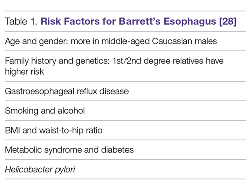
Clinical Symptoms
Columnar metaplasia itself does not cause any symptoms but is merely the adaptation of the cells to the repeated effect of the acid. The main clinical symptoms of the disease would initially be symptoms associated with GERD, such as heartburn, water brash, and dysphagia [1]. Severe presentations of GERD, such as esophageal ulceration, stricture, and hemorrhage, usually occur with long-segment Barrett’s esophagus [29,30]. However, 40% of patients presenting with adenocarcinoma had no history of GERD or symptoms of heartburn [13]. Furthermore, as few as 5% of those presenting with adenocarcinoma were known to have Barrett’s esophagus [31].
D iagnosis
Barrett’s esophagus generally requires an endoscopic examination with biopsy confirmation from the distal esophagus showing specialized intestinal columnar epithelium [32]. The biopsy specimen is acquired from the cellular lining proximal to gastroesophageal junction [5,33]. Barrett’s esophagus is classified into long- and short-segment based on the length of salmon-colored mucosa in the distal esophagus. A distance longer than 3 cm is 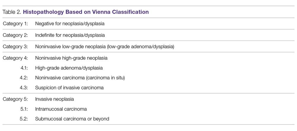
The Prague classification was presented by an international research group in 2006 and is regarded as the standard for measuring the length of Barrett’s esophagus. The lower measurement boundary is formed by the proximal cardial notch, and the 2 upper measurement boundaries are marked by the proximal limit of the circumferential Barrett’s segment and the longest tongue of Barrett’s [37].Confirmation of the diagnosis of dysplastic 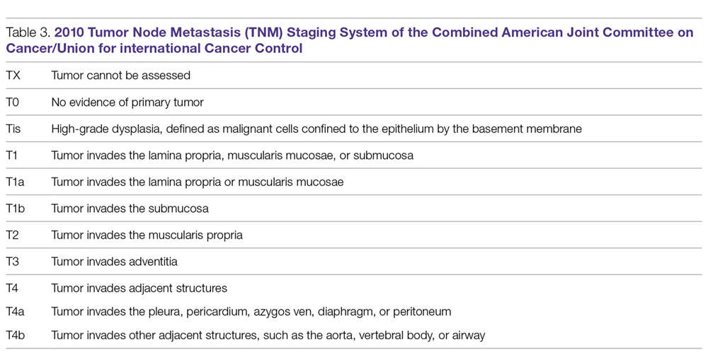
Once the initial diagnosis of Barrett’s esophagus is made, we recommend referring the patient to a Barrett’s esophagus specialized center in order to offer the patient a second opinion from a team of experts in Barrett’s esophagus. This would avoid possible false-positive results, which can be as high as 40% [1,8]. It would also offer the patient a comprehensive multidisciplinary approach and adequate long-term management. It is further preferred if an advanced intervention is offered to the patient such as endoscopic mucosal resection or endoscopic submucosal dissection. Expert endoscopists 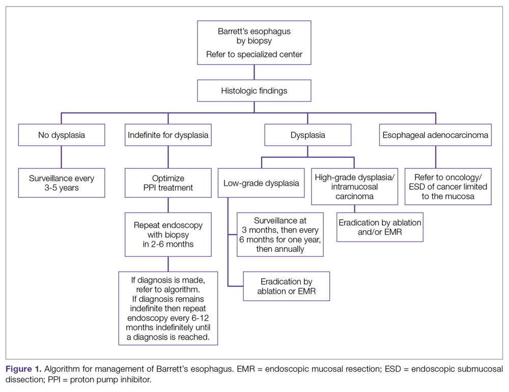
The availability of advanced endoscopic tools improves diagnostic yield. Adopting advanced techniques can reduce errors during biopsy sampling [40–42]. Some of the newer advanced imaging endoscopic tools include chromoendoscopy, optical coherence tomography, confocal microendoscopy, autofluorescence endoscopy, narrow band imaging (NBI), and Fujinon intelligent chromoendoscopy (FICE) [43,44]. In a meta-analysis examining whether advanced techniques improved diagnostic yield, it was found that advanced imaging increased the diagnostic yield by 34%. Advanced technology mentioned here is not mandated in current guidelines [5,45].
Histopathology categories and TNM staging system for Barrett’s esophagus are shown in Table 2 and Table 3. A management algorithm based on histologic findings is presented in Figure 1.
S creening
Logically, Barrett’s esophagus screening should be offered to every patient with GERD; however, this is against current recommendations because it is cost-prohibitive [46,47]. It is a given that the rationale behind the screening is to decrease morbidity and mortality from esophageal adenocarcinoma by offering early and definitive intervention [14,48,49]. The gold standard for screening is upper endoscopy, although nonendoscopic methods are being studied [1]. For example, a capsule attached to a string can be swallowed by the patient, the capsule is then deployed and pulled through the esophagus to obtains a brush sample of the cells. It is a promising technology due to high sensitivity and specificity [50]; however, it is not regularly utilized in practice.
Approaches to screening for Barrett’s esophagus has been addressed by multiple societies with several guidelines currently available [51]. None of these approaches have been proven to be superior in clinical studies. The American Gastroenterological Association (AGA) recommends screening patients with multiple risk factors associated with esophageal adenocarcinoma for Barrett’s esophagus. Risk factors listed by the AGA include white patients, male patients, patients above the age of 50 with a history of chronic GERD, hiatal hernia, elevated body mass index, and certain body fat distribution. The AGA recommends against screening the general population with GERD [9]. The American College of Gastroenterology (ACG) recommends upper endoscopy only in the presence of alarm symptoms (eg, dysphagia, weight loss, gastrointestinal bleeding) and for screening of patients at high risk for complications [52]. The American College of Physicians recommends upper endoscopy for screening for Barrett’s esophagus in men older than 50 years with GERD symptoms for more than 5 years with any risk factors like nocturnal reflux symptoms, hiatus hernia, elevated body mass index, tobacco use, and intra-abdominal distribution of fat [1].
Overall, the sensitivity of endoscopy to diagnose Barrett’s esophagus, as seen in a Veterans Affairs (VA) cohort study for the detection of Barrett’s, is about 80%. The concluded 80% sensitivity rate was based on the performance of 2 endoscopies, 6 weeks apart for each patient, with subsequent labeling of the diagnosis if intestinal metaplasia was found in either of the 2 biopsy samples taken from the 2 procedures [53].
Promising molecular biomarkers have been associated with Barrett’s esophagus including p53 and cyclin D1 expression. However, additional studies are needed before they are incorporated as part of screening practices [5].
General Management
Medical Management of GERD
All patients with Barrett’s esophagus should be treated with a proton pump inhibitor (PPI) indefinitely based on multiple studies [54–56]. Effective control of GERD was associated with a decreased risk of dysplasia and adenocarcinoma (adjusted odds ratio 0.29, 95% CI 0.12–0.79) [57].
Effective control of GERD decreases chronic esophageal inflammation, which could progress to Barrett’s esophagus and has a risk of possible progression to adenocarcinoma. In one study, patients with proven Barrett’s esophagus showed partial regression of the intestinal metaplasia with aggressive PPI therapy [57–59]. Despite this, it is not clear whether the regression decreased risk of malignant progression [58,60,61]. In a study of 68 patients, aggressive acid reduction with omeprazole 40 mg twice a day lead to partial regression of Barrett’s esophagus when compared to mild suppression with ranitidine 150 mg twice a day. However, there was no reduction in the risk of cancer [59].
Surveillance
The reasoning behind surveillance is to detect dysplasia or adenocarcinoma in patients known to have Barrett’s esophagus early enough to provide early and efficient treatment to improve the outcome. Surveillance is done by endoscopy with biopsies in addition to sampling any irregularity [1,62,63]; however, the evidence to back up the benefit of surveillance is not clear [5,64]. It should be noted that surveillance carries risks, and morbidity associated with repeated procedures may affect patients psychologically and financially. Patients who have Barrett’s esophagus are more likely to die from other more common diseases, such as coronary heart disease, prior to developing adenocarcinoma [65]. In a recent meta-analysis, the mortality rate due to esophageal adenocarcinoma was 3.0 per 1000 person-years, whereas the mortality rate due to other causes was 37.1 per 1000 person-years [65]. An ongoing randomized, multicenter trial which assesses scheduled endoscopy every 2 years will shed more light on the overall survival after applying surveillance recommendations for each grade of dysplasia [66].
The following are ACG recommendations for surveillance of Barrett’s esophagus based on the histopathology report. The histopathology report delineates 1 of 3 types of columnar epithelium [68]: cardiac epithelium of mucus-secreting cells, atrophic gastric fundic type epithelium, or specialized columnar cells with goblet cells. The latter type is the most common with high potential for cancer [32]. Degrees of dysplasia and possible adenocarcinoma is usually described in the report as well. The degree of dysplasia if found helps the endoscopist plan the next step in management. It is worth mentioning that sampling errors can lead to missing a diagnosis. In a meta-analysis, 13% of patients diagnosed with high-grade dysplasia who underwent resection were found to have invasive cancer [69].
Surveillance for Patients with No Dysplasia
We suggest surveillance every 3 to 5 years since the rate of neoplasia is low [14]. For management of select patients with no dysplasia and with additional risk factors, radiofrequency ablation (RFA) may be an option, although it remains a controversial approach. For example, in a patient under 50 years of age, family history would be an argument for proceeding with RFA instead of prolonged surveillance. In a prospective cohort study of 139 patients with 10-year follow-up after ablation, recurrent Barrett’s occurred in less than 5% of the patients. [70]
Surveillance for Patients with Biopsy Showing “Indefinite for Dysplasia”
Aggressive treatment with PPI twice daily is recommended to avoid the misinterpretation of reactive esophageal changes secondary to reflux as dysplasia on the following endoscopy with biopsy. These patients will require a repeat endoscopy with biopsies after 3 months of aggressive treatment with PPI. Biopsies should be taken every 1 centimeter within Barrett’s epithelium [1].
If it remains indefinite, biopsies should be examined by a second pathologist with expertise in Barrett’s esophagus. If the second pathologist agrees on the indefinite diagnosis for dysplasia, then endoscopy every 12 months is recommended [1]. Treatment versus surveillance after repeat endoscopy and biopsy should be tailored to the new histopathology results on the most recent exam.
Surveillance for Patients with Biopsy Results showing Low-Grade Dysplasia (LGD), High-Grade Dysplasia (HGD), or Intramucosal Carcinoma
Surveillance recommendations are discussed under Management of Dysplasia or Intramucosal Carcinoma, below.
Efficacy of Surveillance
Asymptomatic adenocarcinoma could be discovered during surveillance, and neoplasia detected during surveillance is usually less advanced than those found after development of symptoms such as dysphagia, bleeding or weight loss [2,3,71–75]. These studies obviously had lead–time bias and did not document terminal cancer in patients adherent to surveillance protocol.
Management of Dysplasia or Intramucosal Carcinoma
Overview
Historically, dysplasia was managed with esophagectomy, which was associated with high morbidity and mortality. With advancement in the field of endoscopy, dysplasia is managed quite differently today, with endoscopic eradication therapy, which includes the use of endoscopic ablation techniques and endoscopic resection. The advantage of endoscopic resection is preservation of resected tissue for further examination, thus providing valuable information regarding the stage of the tumor (depth). Histological examination is not possible with photo or thermal ablation techniques as destroyed mucosa cannot be submitted for tissue analysis.
Low-Grade Dysplasia
If low-grade dysplasia is found, it is followed by a repeat endoscopy 8 weeks after aggressive PPI therapy. The repeat endoscopy should be performed with high definition/high-resolution endoscopy. The rationale of a second endoscopy is to ensure that the metaplastic mucosa was adequately inspected and biopsied prior to further intervention [1,9]. If the diagnosis is confirmed as low-grade dysplasia, and the patient prefers to go with the path of intervention instead of conservative management (endoscopic surveillance every 6 months for 1 year then annually with biopsies), then multiple options are available for the patient, including RFA or cryotherapy [8] [76]. In a randomized clinical trial in patients with Barrett’s esophagus and LGD, RFA was shown to reduce risk of neoplastic progression over a 3-year follow-up. The study included 136 patients randomized to receive ablation and 68 patients who underwent endoscopic surveillance. In the ablation group, the risk of progression to HGD or esophageal adenocarcinoma was reduced by 25% and the risk of progression to adenocarcinoma was reduced by 7.4%. In the ablation group, complete eradication of dysplasia and intestinal metaplasia occurred in 92.6% and 88.2% of patients respectively. In the endoscopic surveillance group, complete eradication of dysplasia and intestinal metaplasia was seen in 27.9% and 0.0% of patients respectively. [76]. Treatment-related adverse events occurred in 19.1% of patients receiving ablation (P < 0.001). The most common adverse event was stricture, occurring in 8 patients receiving ablation (11.8%), all resolved by endoscopic dilation [76,78].
Surveillance after ablation of LGD is still an ongoing debate, and further evidence is needed to establish guidelines [8]. Due to lack of evidence, we would lean towards surveillance for those patients with an annual esophagogastroduodenoscopy with biopsy examination (author’s opinion, no associated level of evidence).
High-Grade Dysplasia or Intramucosal Carcinoma
For patients with HGD or intramucosal carcinoma without submucosal invasion, eradication is the treatment of choice. Current guidelines advocate for endoscopic eradication therapy for most if not all patients with HGD or intramucosal carcinoma with a goal of removing all metaplastic and dysplastic tissue [1,5,62,64]. It should be noted that the biopsy specimen should be extensive to decrease the error margin. If the diagnosis were made on endoscopy without procuring extensive biopsies, then repeat endoscopy with extensive biopsies is needed prior to deciding the treatment path. The rationale behind extensive biopsies is to confirm the diagnosis and to determine the extent of dysplasia. Other factors potentially influencing the treatment path include the patient’s age, comorbid conditions, quality of life, and available access to an advanced endoscopist or specialized surgeon. Patient’s preferences and adherence to follow-up visits should also be a consideration.
The long-term benefits of endoscopic intervention versus surgical intervention are not well established. Esophagectomy is no longer a preferred method of treatment due to high morbidity and mortality associated with the procedure when compared to endoscopic interventions. However, it is still a preferred choice amongst a select group of patients unwilling to follow-up. A cost-effective analysis found that endoscopic ablation provided the longest quality adjusted life expectancy for Barrett’s esophagus with HGD [79,80].
Endoscopic Therapy in Barrett’s Esophagus
Endoscopic Ablative Therapies
With the advancement of endoscopic intervention, we now have multiple tools to ablate abnormal epithelium in Barrett’s esophagus. Examples of ablation techniques include thermal, photochemical, and mechanical techniques [81,82]. RFA is the treatment of choice for ablation [83]. However, non-contact ablative therapy, such as cryoablation, may be prefered if topography of the esophagus doesn’t allow contact ablation.
Radiofrequency Ablation (Figure 2). RFA is a procedure in which heat is generated from medium frequency alternating current and leads to thermal injury [84]. In Barrett’s esophagus, RFA uses radiofrequency energy delivered by a balloon that has a series of closely spaced electrodes in a 
Most patients will require multiple sessions of RFA to achieve eradication. It is very rarely a one-time procedure. In a meta-analysis of 18 studies including 3082 patients, the most common adverse effects of RFA were stricture in 5% [76,98], bleeding in 1%, and pain in 3% of patients [99].
It is crucial for successful RFA to continue medical treatment for acid suppression, in order to allow healthy regeneration of the squamous cell lining. It is suggested to use PPI twice a day with sucralfate and ranitidine after the intervention [100,101]. Adhering to a liquid diet for 24 hours is needed, followed by a soft diet to allow faster regeneration of the epithelium.
The caveat with RFA is that new evidence shows a higher rate of recurrence than previously thought. In one study of 246 patients, recurrence of dysplasia occurred in 25% of patients at 48 months after eradication in 80% of the patients, and metaplasia occurred in 50% at 60 months [102]. The other risk is buried Barrett’s, a condition occurring after incomplete ablation, in which squamous cell epithelium covers patches of incompletely destroyed intestinal lining, leading to possible progression of the disease to adenocarcinoma under the surface [103].
It has been reported that patients who underwent RFA had remarkable improvement in quality of life even if RFA did not achieve eradication. Patients reported less depression, less stress and better quality of life [104].
Based on a survey of experts, follow-up at 3 months, 6 months, and then annually is recommended after ablation [1,105]. Biopsies should be taken distal to neosquamous epithelium and from suspicious areas [97,106].
Endoscopic Spray Cryotherapy. This technique involves application of liquid nitrogen or carbon dioxide gas by endoscope on the tissue to freeze it off. Although it has been shown to eliminate HGD in over 95% of the cases and all dysplasia in over 85% of the cases, it was effective in eradicating intestinal metaplasia in only 55% of patients [103,108,109]. Thus, RFA as ablation therapy is still superior to cryotherapy and is still the first-line treatment for dysplastic Barrett’s esophagus. In comparison to cryotherapy, RFA efficacy has been studied extensively with well documented outcomes. However, there is a role for cryotherapy over RFA in certain clinical situations (such as severe chest pain from RFA or lack of efficacy in eradicating intestinal metaplasia or dysplasia by RFA).
Similar to RFA, on occasions of partial ablation, the remaining metaplastic tissue may get buried beneath a layer of squamous epithelium and can possibly progress to adenocarcinoma [110].
Photodynamic Therapy (PDT). This technique works by producing cytotoxicity at the cellular level by exposure to light at a specific wavelength in the presence of a chemical agent known as photosensitize [107]. Although superior to omeprazole, PDT has a significant rate of complications, mainly stricture, and a high occurrence of esophageal cancer during follow-up. For this reason, it is less favorable compared to RFA [107] and mentioned here as a historical therapy.
Endoscopic Resection Techniques: Endoscopic Mucosal Resection (EMR) and Endoscopic Submucosal Dissection (ESD)
Unlike flat mucosa in Barrett’s esophagus, which respond to ablative techniques such as RFA or cryotherapy, nodular Barrett’s esophagus is hard to treat and requires endoscopic resection prior to ablation. Endoscopic mucosal resection (EMR) is the most widely used technique and it is available in most tertiary referral centers. Another technique named endoscopic submucosal dissection (ESD) allows the removal of large nodular areas of Barrett’s esophagus in one piece to ensure complete removal of nodular dysplasia. ESD is technically challenging and it is only available in a handful of centers in the US. Endoscopic resection techniques are the preferred interventions for nodular dysplasia due to their ability to provide valuable information for staging the lesion [111–113]. Endoscopic resection techniques are safer and more effective with similar or better results when compared with other approaches [114].
EMR is completed by the excision of esophageal mucosa down to the submucosa and submitting a large tissue specimen to the pathologist. It additionally serves as a therapeutic measure in cases of no submucosal extension. Another advantage of EMR is the ability to predict lymph node metastasis. The rationale is based on the fact that the most important predictor of lymph node metastasis is the depth of the tumor; hence, invasive tumors would likely be associated with lymph node metastasis [115,116].
In a systematic review of 11 studies, complete EMR was as equally effective in the short-term treatment of dysplastic Barrett’s esophagus when compared to RFA, but adverse event rates were greater with complete EMR (mainly strictures). Strictures are more likely to occur in patients undergoing extensive EMR. In another meta-analysis of 22 studies comparing the efficacy of EMR to RFA, both techniques were effective in eradicating dysplasia (95% in EMR group and 92% in RFA group). However, extensive EMR was associated with higher complication rates suggesting that a combined endoscopic approach of focal EMR followed by RFA is preferred over extensive EMR alone [86].
It should be noted that EMR and ESD information were derived from highly specialized center and these results may not be duplicated in community settings [113,117].
Efficacy of Endoscopic Resection. Endoscopic resection has a success rate comparable to surgical esophagectomy with fewer complications [113,114,118–121] in patients with HGD and early stages of esophageal cancer [122]. Complete remission can be as high as 89%. Recurrence occurred in 6% to 30% of patients [114,118,119], which was attributed to incomplete removal, large lesions, failure to use adjunct therapy, or lack of follow-up [123]. Even when recurrence occurred, it was successfully managed by endoscopic intervention [124].
In a large cohort study of 1000 patients with early mucosal adenocarcinoma who were treated with endoscopic resection, long-term complete remission occurred in 94% of patients. There was no mortality and less than 2% of patients had major complications. Infrequent complications include bleeding, perforations, and strictures [123,125,126]. The rate of complications is lower in highly specialized centers [127–129].
Surgery was necessary in 12 patients (3.7%) after endoscopic therapy failed [123]. Post-resection care and follow-up is similar to the post-RFA care discussed above.
Management of Invasive Esophageal Adenocarcinoma
Patients diagnosed with an invasive adenocarcinoma need to be referred to an oncologist for staging and to discuss treatment options. A select number of patients may be referred by oncology for endoscopic resection, yet the need for a multidisciplinary approach in these situations is absolutely necessary [1].
Esophagectomy
Esophagectomy offers the complete removal of the HGD along with any adenocarcinoma in the regional lymph nodes. However, mortality rates are as high as 12% immediately after the procedure [130]. The multitude of short- and long-term morbidity has significant effects on quality of life. Short-term morbidity is as high as 30%. Patients may develop serious postoperative complications such as myocardial infarction, hospital associated pneumonia, or anatomic leak [131].
Examples of long-term morbidity include dysphagia, transection of vagal nerve, and dumping syndrome. Recent development in minimally invasive surgeries for esophagectomy has not reduced postoperative morbidity rates [132].
Advocates of esophagectomy illustrate the advantage of eradication of occult lymph node metastasis. The counter argument has been established by a systemic review in which occult lymph node metastasis occurred in less than 2% of patients with HGD and intramucosal carcinoma; whereas the mortality rate after esophagectomy is substantially higher with no guarantee of curing metastatic disease [133].
Prevention of Barrett’s Esophagus
Since Barrett’s esophagus precedes most of the cases of EAC if not all [1,134], methods that aim at decreasing the incidence of Barrett’s esophagus could help in prevention. The modifiable risk factors listed by the AGA include BMI, GERD, and hiatal hernia management. Along with diet and exercise, the advent of new therapies to help patients manage their weight could in return help in avoiding a plethora of medical conditions including Barrett’s esophagus. Hiatal hernia management could lower the risk of Barrett’s by restoring normal anatomy. Lastly, proper management of GERD would lower the risk of developing Barrett’s esophagus as discussed in this article [1,9].
It is worth noting that a large trial on the efficacy and safety of aspirin for prevention of adenocarcinoma progression in Barrett’s esophagus is ongoing in the UK (AspECT trial). The AspECT trial examines the efficacy of low dose vs. high dose PPI with or without aspirin for the chemoprevention of esophageal adenocarcinoma. The theory behind the study is the inhibition of COX 2 receptors in Barrett’s cells can decrease tissue progression to cancer. This chempreventive effect of nonsteroidal anti-inflammatory drugs was shown to be augmented when combined with statin intake [56,135–138].
Conclusion
Barrett’s esophagus is usually diagnosed during routine endoscopic examination. The initial symptoms are those associated with GERD, like heartburn, dyspepsia, and regurgitation. Specialized columnar epithelium is the hallmark of histopathological diagnosis. Recommendations of the ACG and AGA suggest treatment based on biopsy results. The intervention would vary on a wide spectrum starting from acid suppression, radiofrequency ablation, endoscopic resection therapy, and rarely, esophagectomy.
Corresponding author: Mohamed O. Othman, MD, Gastroenterology and Hepatology Section, Baylor College of Medicine, 7200 Cambridge St., Suite 8C, Houston, TX 77030, mohamed.othman@bcm.edu.
Financial disclosures: Dr. Othman has received grant support from Abbvie and has served as a consultant for Olympus.
1. Shaheen NJ, Falk GW, Iyer PG, Gerson LB; American College of Gastroenterology. ACG Clinical Guideline: diagnosis and management of Barrett’s esophagus. Am J Gastroenterol 2016;111:30–50; quiz 51.
2. Peters JH, Clark GW, Ireland AP. Outcome of adenocarcinoma arising in Barrett’s esophagus in endoscopically surveyed and nonsurveyed patients. J Thorac Cardiovasc Surg 1994;108:813–21; discussion 821–2.
3. Streitz JM Jr, Andrews CW Jr, Ellis FH Jr. Endoscopic surveillance of Barrett’s esophagus. Does it help? J Thorac Cardiovasc Surg 1993;105:383–7; discussion 387–8.
4. Spechler SJ, Souza RF. Barrett’s esophagus. N Engl J Med 2014;371:836–45.
5. Spechler SJ, Sharma P, Souza RF, et al; American Gastroenterological Association. American Gastroenterological Association medical position statement on the management of Barrett’s esophagus. Gastroenterology 2011;140:1084–91.
6. Sharma P, McQuaid K, Dent J, et al; AGA Chicago Workshop. A critical review of the diagnosis and management of Barrett’s esophagus: the AGA Chicago Workshop. Gastroenterology 2004;127:310–30.
7. ASGE Standards of Practice Committee, Evans JA, Early DS; Standards of Practice Committee of the American Society for Gastrointestinal Endoscopy. The role of endoscopy in Barrett’s esophagus and other premalignant conditions of the esophagus. Gastrointest Endosc 2012;76(6):1087–94.
8. Wani S, Rubenstein JH, Vieth M, Bergman J. Diagnosis and management of low–grade dysplasia in Barrett’s esophagus: Expert review from the clinical practice updates Committee of the American Gastroenterological Association. Gastroenterology 2016;151:822–835.
9. American Gastroenterological, A., et al., American Gastroenterological Association medical position statement on the management of Barrett’s esophagus. Gastroenterology, 2011. 140(3): p. 1084–91. [Same as #5]
10. Van Eyken P. Definition of Barrett’s oesophagus. Acta Gastroenterol Belg 2000;63:10–2.
11. Hirota WK, Loughney TM, Lazas DJ, et al. Specialized intestinal metaplasia, dysplasia, and cancer of the esophagus and esophagogastric junction: prevalence and clinical data. Gastroenterology 1999;116(2):277–85.
12. Cameron AJ, Zinsmeister AR, Ballard DJ, Carney JA. Prevalence of columnar–lined (Barrett’s) esophagus. Comparison of population–based clinical and autopsy findings. Gastroenterology 1990;99:918–22.
13. Gerson LB, Shetler K, Triadafilopoulos G. Prevalence of Barrett’s esophagus in asymptomatic individuals. Gastroenterology 2002;123:461–7.
14. Van der Veen AH, Dees J, Blankensteijn JD, Van Blankenstein M. Adenocarcinoma in Barrett’s oesophagus: an overrated risk. Gut 1989;30:14–8.
15. Winters C Jr, Spurling TJ, Chobanian SJ, et al. Barrett’s esophagus. A prevalent, occult complication of gastroesophageal reflux disease. Gastroenterology 1987;92:118–24.
16. Wani S, Falk G, Hall M, et al. Patients with nondysplastic Barrett’s esophagus have low risks for developing dysplasia or esophageal adenocarcinoma. Clin Gastroenterol Hepatol 2011;9:220–7;quiz e26.
17. Ward EM, Wolfsen HC, Achem SR, et al. Barrett’s esophagus is common in older men and women undergoing screening colonoscopy regardless of reflux symptoms. Am J Gastroenterol 2006;101:12–7.
18. Rex DK, Cummings OW, Shaw M. Screening for Barrett’s esophagus in colonoscopy patients with and without heartburn. Gastroenterology 2003;125:1670–7.
19. Rubenstein JH, Taylor JB. Meta–analysis: the association of oesophageal adenocarcinoma with symptoms of gastro–oesophageal reflux. Aliment Pharmacol Ther 2010;32:1222–7.
20. Lagergren J, Bergström R, Lindgren A, Nyrén O. Symptomatic gastroesophageal reflux as a risk factor for esophageal adenocarcinoma. N Engl J Med 1999;340:825–31.
21. Ronkainen J, Aro P, Storskrubb T, et al. Prevalence of Barrett’s esophagus in the general population: an endoscopic study. Gastroenterology, 2005;129:1825–31.
22. Sampliner RE. A population prevalence of Barrett’s esophagus––finally. Gastroenterology 2005; 129:2101–3.
23. Abrams JA, Fields S, Lightdale CJ, Neugut AI. Racial and ethnic disparities in the prevalence of Barrett’s esophagus among patients who undergo upper endoscopy. Clin Gastroenterol Hepatol 2008;6:30–4.
24. Corley DA, Kubo A, Levin TR, et al. Race, ethnicity, sex and temporal differences in Barrett’s oesophagus diagnosis: a large community–based study, 1994–2006. Gut 2009;58:182–8.
25. Kamat P, Wen S, Morris J, Anandasabapathy S. Exploring the association between elevated body mass index and Barrett’s esophagus: a systematic review and meta–analysis. Ann Thorac Surg 2009;87:655–62.
26. Jacobson BC, Chan AT, Giovannucci EL, Fuchs CS. Body mass index and Barrett’s oesophagus in women. Gut 2009;58:1460–6.
27. Orloff M, Peterson C, He X, et al. Germline mutations in MSR1, ASCC1, and CTHRC1 in patients with Barrett esophagus and esophageal adenocarcinoma. JAMA 2011;306:410–9.
28. Sharma N, Ho KY. Risk Factors for Barrett’s oesophagus. Gastrointest Tumors 2016;3:103–8.
29. Spechler SJ. Barrett’s esophagus. Semin Gastrointest Dis 1996;7:51–60.
30. Ronkainen J, Talley NJ, Storskrubb T. Erosive esophagitis is a risk factor for Barrett’s esophagus: a community–based endoscopic follow–up study. Am J Gastroenterol 2011;106:1946–52.
31. Kim SL, Wo JM, Hunter JG. The prevalence of intestinal metaplasia in patients with and without peptic strictures. Am J Gastroenterol 1998;93:53–5.
32. Spechler SJ. Clinical practice. Barrett’s esophagus. N Engl J Med 2002;346:836–42.
33. Riddell RH, Odze RD. Definition of Barrett’s esophagus: time for a rethink––is intestinal metaplasia dead? Am J Gastroenterol 2009;104:2588–94.
34. Sharma P, Morales TG, Sampliner RE. Short segment Barrett’s esophagus––the need for standardization of the definition and of endoscopic criteria. Am J Gastroenterol 1998;93:1033–6.
35. Eloubeidi MA, Provenzale D. Does this patient have Barrett’s esophagus? The utility of predicting Barrett’s esophagus at the index endoscopy. Am J Gastroenterol 1999;94:937–43.
36. Rudolph RE, Vaughan TL, Storer BE, et al. Effect of segment length on risk for neoplastic progression in patients with Barrett esophagus. Ann Intern Med 2000;132:612–20.
37. Sharma P, Dent J, Armstrong D, et al. The development and validation of an endoscopic grading system for Barrett’s esophagus: the Prague C & M criteria. Gastroenterology 2006;131:1392–9.
38. Bhat S, Coleman HG, Yousef F, et al. Risk of malignant progression in Barrett’s esophagus patients: results from a large population–based study. J Natl Cancer Inst 2011;103:1049–57.
39. Shaheen NJ, Dulai GS, Ascher B, et al. Effect of a new diagnosis of Barrett’s esophagus on insurance status. Am J Gastroenterol 2005;100:577–80.
40. Canto MI, Setrakian S, Willis J, et al. Methylene blue–directed biopsies improve detection of intestinal metaplasia and dysplasia in Barrett’s esophagus. Gastrointest Endosc 2000;51:560–8.
41. Scotiniotis IA, Kochman ML, Lewis JD. Accuracy of EUS in the evaluation of Barrett’s esophagus and high–grade dysplasia or intramucosal carcinoma. Gastrointest Endosc 2001;54:689–96.
42. Kobayashi K, Izatt JA, Kulkarni MD, et al. High–resolution cross–sectional imaging of the gastrointestinal tract using optical coherence tomography: preliminary results. Gastrointest Endosc 1998;47:515–23.
43. Georgakoudi I, Jacobson BC, Van Dam J, et al. Fluorescence, reflectance, and light–scattering spectroscopy for evaluating dysplasia in patients with Barrett’s esophagus. Gastroenterology 2001. 120: 1620–9.
44. Wallace MB, Sharma P, Lightdale C, et al. Preliminary accuracy and interobserver agreement for the detection of intraepithelial neoplasia in Barrett’s esophagus with probe–based confocal laser endomicroscopy. Gastrointest Endosc 2010;72:19–24.
45. Qumseya BJ, Wang H, Badie N, et al. Advanced imaging technologies increase detection of dysplasia and neoplasia in patients with Barrett’s esophagus: a meta–analysis and systematic review. Clin Gastroenterol Hepatol 2013;11:1562–70.e1–2.–
46. DeVault KR, Castell DO. Updated guidelines for the diagnosis and treatment of gastroesophageal reflux disease. The Practice Parameters Committee of the American College of Gastroenterology. Am J Gastroenterol 1999;94:1434–42.
47. Inadomi JM, Sampliner R, Lagergren J. Screening and surveillance for Barrett esophagus in high–risk groups: a cost–utility analysis. Ann Intern Med 2003;138:176–86.
48. Conio M, Blanchi S, Lapertosa G, et al. Long–term endoscopic surveillance of patients with Barrett’s esophagus. Incidence of dysplasia and adenocarcinoma: a prospective study. Am J Gastroenterol 2003;98:1931–9.
49. Rastogi A, Puli S, El–Serag HB, et al. Incidence of esophageal adenocarcinoma in patients with Barrett’s esophagus and high–grade dysplasia: a meta–analysis. Gastrointest Endosc 2008;67:394–8.
50. Kadri SR, Lao–Sirieix P, O’Donovan M, et al. Acceptability and accuracy of a non–endoscopic screening test for Barrett’s oesophagus in primary care: cohort study. BMJ 2010;341:c4372.
51. Sharma, P., et al., A critical review of the diagnosis and management of Barrett’s esophagus: the AGA Chicago Workshop. Gastroenterology, 2004. 127(1): p. 310–30. [Same as #6]
52. Katz PO, Gerson LB, Vela MF. Guidelines for the diagnosis and management of gastroesophageal reflux disease. Am J Gastroenterol 2013;108:308–28; quiz 329.
53. Kim SL, Waring JP, Spechler SJ, et al. Diagnostic inconsistencies in Barrett’s esophagus. Department of Veterans Affairs Gastroesophageal Reflux Study Group. Gastroenterology 1994;107:945–9.
54. Kastelein F, Spaander MC, Steyerberg EW, et al; ProBar Study Group. Proton pump inhibitors reduce the risk of neoplastic progression in patients with Barrett’s esophagus. Clin Gastroenterol Hepatol 2013;11:382–8.
55. El–Serag HB, Aguirre TV, Davis S, et al. Proton pump inhibitors are associated with reduced incidence of dysplasia in Barrett’s esophagus. Am J Gastroenterol 2004;99:1877–83.
56. Nguyen DM, El–Serag HB, Henderson L, et al. Medication usage and the risk of neoplasia in patients with Barrett’s esophagus. Clin Gastroenterol Hepatol 2009;7:1299–304.
57. Singh S, Garg SK, Singh PP, et al. Acid–suppressive medications and risk of oesophageal adenocarcinoma in patients with Barrett’s oesophagus: a systematic review and meta–analysis. Gut 2014;63:1229–37.
58. Spechler SJ, Lee E, Ahnen D, et al. Long–term outcome of medical and surgical therapies for gastroesophageal reflux disease: follow–up of a randomized controlled trial. JAMA 2001;285:2331–8.
59. Peters FT, Ganesh S, Kuipers EJ, et al. Endoscopic regression of Barrett’s oesophagus during omeprazole treatment; a randomised double blind study. Gut 1999;45:489–94.
60. Ouatu–Lascar R, Triadafilopoulos G. –Complete elimination of reflux symptoms does not guarantee normalization of intraesophageal acid reflux in patients with Barrett’s esophagus. Am J Gastroenterol 1998;93:711–6.
61. Maret–Ouda J, Konings P, Lagergren J, Brusselaers N. Antireflux surgery and risk of esophageal adenocarcinoma: A systematic review and meta–analysis. Ann Surg 2016;263:251–7.
62. Evans, J.A., et al., The role of endoscopy in Barrett’s esophagus and other premalignant conditions of the esophagus. Gastrointest Endosc, 2012. 76(6): p. 1087–94. [Same as #7]
63. ASGE Standards of Practice Committee, Evans JA, Early DS,et al; American Society for Gastrointestinal Endoscopy. The role of endoscopy in the assessment and treatment of esophageal cancer. Gastrointest Endosc 2013;77:328–34.
64. Bennett C, Moayyedi P, Corley DA, et al; BOB CAT Consortium. BOB CAT: A large–scale review and delphi consensus for ,anagement of Barrett’s esophagus with no dysplasia, indefinite for, or low–grade dysplasia. Am J Gastroenterol 2015;110:662–82; quiz 683.
65. Sikkema M, de Jonge PJ, Steyerberg EW, Kuipers EJ. Risk of esophageal adenocarcinoma and mortality in patients with Barrett’s esophagus: a systematic review and meta–analysis. Clin Gastroenterol Hepatol 2010;8:235–44; quiz e32.
66. Old O, Moayyedi P, Love S, et al; BOSS Trial Team. Barrett’s Oesophagus Surveillance versus endoscopy at need Study (BOSS): protocol and analysis plan for a multicentre randomized controlled trial. J Med Screen 2015;22:158–64.
67. Weston AP, Sharma P, Topalovski M, et al. Long–term follow–up of Barrett’s high–grade dysplasia. Am J Gastroenterol 2000;95:1888–93.
68. Paull A, Trier JS, Dalton MD, et al. The histologic spectrum of Barrett’s esophagus. N Engl J Med 1976;295:476–80.
69. Konda VJ, Ross AS, Ferguson MK, et al. Is the risk of concomitant invasive esophageal cancer in high–grade dysplasia in Barrett’s esophagus overestimated? Clin Gastroenterol Hepatol 2008;6:159–64.
70. Allison H, Banchs MA, Bonis PA, Guelrud M. Long–term remission of nondysplastic Barrett’s esophagus after multipolar electrocoagulation ablation: report of 139 patients with 10 years of follow–up. Gastrointest Endosc 2011;73:651–8.
71. Corley DA, Levin TR, Habel LA, Weiss NS, et al. Surveillance and survival in Barrett’s adenocarcinomas: a population–based study. Gastroenterology 2002;122:633–40.
72. Wong T, Tian J, Nagar AB. Barrett’s surveillance identifies patients with early esophageal adenocarcinoma. Am J Med 2010;123:462–7.
73. Fountoulakis A, Zafirellis KD, Dolan K, et al. Effect of surveillance of Barrett’s oesophagus on the clinical outcome of oesophageal cancer. Br J Surg 2004;91:997–1003.
74. Verbeek RE, Leenders M, Ten Kate FJ, et al. Surveillance of Barrett’s esophagus and mortality from esophageal adenocarcinoma: a population–based cohort study. Am J Gastroenterol 2014;109:1215–22.
75. Kastelein F, van Olphen SH, Steyerberg EW, et al. Impact of surveillance for Barrett’s oesophagus on tumour stage and survival of patients with neoplastic progression. Gut 2016;65:548–54.
76. Phoa KN, van Vilsteren FG, Weusten BL, et al. Radiofrequency ablation vs endoscopic surveillance for patients with Barrett esophagus and low–grade dysplasia: a randomized clinical trial. JAMA 2014;311: 1209–17.
77. Wang WL, Chang IW, Chen CC, et al. Radiofrequency ablation versus endoscopic submucosal dissection in treating large early esophageal squamous cell neoplasia. Medicine (Baltimore) 2015;94:e2240.
78. Lim CH, Treanor D, Dixon MF, Axon AT. Low–grade dysplasia in Barrett’s esophagus has a high risk of progression. Endoscopy 2007;39:581–7.
79. Shaheen NJ, Inadomi JM, Overholt BF, Sharma P. What is the best management strategy for high grade dysplasia in Barrett’s oesophagus? A cost effectiveness analysis. Gut 2004;53:1736–44.
80. Vij R, Triadafilopoulos G, Owens DK, et al. Cost–effectiveness of photodynamic therapy for high–grade dysplasia in Barrett’s esophagus. Gastrointest Endosc 2004;60:739–56.
81. van den Boogert J, v an Hillegersberg R, Siersema PD, et al. Endoscopic ablation therapy for Barrett’s esophagus with high–grade dysplasia: a review. Am J Gastroenterol 1999;94:1153–60.
82. Sampliner RE. Endoscopic ablative therapy for Barrett’s esophagus: current status. Gastrointest Endosc 2004;59:66–9.
83. Sharma VK, Wang KK, Overholt BF, et al. Balloon–based, circumferential, endoscopic radiofrequency ablation of Barrett’s esophagus: 1–year follow–up of 100 patients. Gastrointest Endosc 2007;65:185–95.
84. Hanlon CR. Textbook of surgery: The biological basis of modern surgical practice, 14th edition. Ann Surg 1992;216:94.
85. Bright T, Watson DI, Tam W, et al. Randomized trial of argon plasma coagulation versus endoscopic surveillance for barrett esophagus after antireflux surgery: late results. Ann Surg 2007;246:1016–20.
86. Chadwick G, Groene O, Markar SR, et al. Systematic review comparing radiofrequency ablation and complete endoscopic resection in treating dysplastic Barrett’s esophagus: a critical assessment of histologic outcomes and adverse events. Gastrointest Endosc 2014;79:718–31.e3.
87. Gondrie JJ, Pouw RE, Sondermeijer CM, et al. Effective treatment of early Barrett’s neoplasia with stepwise circumferential and focal ablation using the HALO system. Endoscopy 2008;40:370–9.
88. Pouw RE, Wirths K, Eisendrath P, et al. Efficacy of radiofrequency ablation combined with endoscopic resection for barrett’s esophagus with early neoplasia. Clin Gastroenterol Hepatol 2010;8:23–9.
89. Kim HP, Bulsiewicz WJ, Cotton CC, et al. Focal endoscopic mucosal resection before radiofrequency ablation is equally effective and safe compared with radiofrequency ablation alone for the eradication of Barrett’s esophagus with advanced neoplasia. Gastrointest Endosc 2012;76:733–9.
90. Fleischer DE, Overholt BF, Sharma VK, et al. Endoscopic ablation of Barrett’s esophagus: a multicenter study with 2.5–year follow–up. Gastrointest Endosc 2008;68:867–76.
91. Sharma VK, Jae Kim H, Das A, et al. Circumferential and focal ablation of Barrett’s esophagus containing dysplasia. Am J Gastroenterol 2009;104:310–7.
92. Ganz RA, Overholt BF, Sharma VK, et al. Circumferential ablation of Barrett’s esophagus that contains high–grade dysplasia: a U.S. multicenter registry. Gastrointest Endosc 2008;68:35–40.
93. Gupta M, Iyer PG, Lutzke L, et al. Recurrence of esophageal intestinal metaplasia after endoscopic mucosal resection and radiofrequency ablation of Barrett’s esophagus: results from a US Multicenter Consortium. Gastroenterology 2013;145:79–86.e1.
94. Lyday WD, Corbett FS, Kuperman DA, et al. Radiofrequency ablation of Barrett’s esophagus: outcomes of 429 patients from a multicenter community practice registry. Endoscopy 2010;42:272–8.
95. Pasricha S, Bulsiewicz WJ, Hathorn KE, et al. Durability and predictors of successful radiofrequency ablation for Barrett’s esophagus. Clin Gastroenterol Hepatol 2014;12:1840–7.e1.
96. Fleischer DE, Overholt BF, Sharma VK, et al. Endoscopic radiofrequency ablation for Barrett’s esophagus: 5–year outcomes from a prospective multicenter trial. Endoscopy 2010;42:781–9.
97. Weston AP, Sharma P, Banerjee S, et al. Visible endoscopic and histologic changes in the cardia, before and after complete Barrett’s esophagus ablation. Gastrointest Endosc 2005;61:515–21.
98. Beaumont H, Gondrie JJ, McMahon BP, et al. Stepwise radiofrequency ablation of Barrett’s esophagus preserves esophageal inner diameter, compliance, and motility. Endoscopy 2009;41:2–8.
99. Orman ES, Li N, Shaheen NJ.Efficacy and durability of radiofrequency ablation for Barrett’s Esophagus: systematic review and meta–analysis. Clin Gastroenterol Hepatol 2013;11:1245–55.
100. Krishnan K, Pandolfino JE, Kahrilas PJ, et al. Increased risk for persistent intestinal metaplasia in patients with Barrett’s esophagus and uncontrolled reflux exposure before radiofrequency ablation. Gastroenterology 2012;143:576–81.
101. Akiyama J, Marcus SN, Triadafilopoulos G. Effective intra–esophageal acid control is associated with improved radiofrequency ablation outcomes in Barrett’s esophagus. Dig Dis Sci 2012;57:2625–32.
102. Small AJ, Sutherland SE, Hightower JS, et al. Comparative risk of recurrence of dysplasia and carcinoma after endoluminal eradication therapy of high–grade dysplasia versus intramucosal carcinoma in Barrett’s esophagus. Gastrointest Endosc 2015;81:1158–66.e1–4.
103. Shaheen NJ, Greenwald BD, Peery AF, et al. Safety and efficacy of endoscopic spray cryotherapy for Barrett’s esophagus with high–grade dysplasia. Gastrointest Endosc 2010;71:680–5.
104. Shaheen NJ, Peery AF, Hawes RH, et al. Quality of life following radiofrequency ablation of dysplastic Barrett’s esophagus. Endoscopy 2010;42:790–9.
105. Bedi AO, Kwon RS, Rubenstein JH, et al. A survey of expert follow–up practices after successful endoscopic eradication therapy for Barrett’s esophagus with high–grade dysplasia and intramucosal adenocarcinoma. Gastrointest Endosc 2013;78:696–701.
106. Sampliner RE, Camargo E, Prasad AR. Prasad, Association of ablation of Barrett’s esophagus with high grade dysplasia and adenocarcinoma of the gastric cardia. Dis Esophagus 2006;19:277–9.
107. Overholt BF, Panjehpour M, Halberg DL. Photodynamic therapy for Barrett’s esophagus with dysplasia and/or early stage carcinoma: long–term results. Gastrointest Endosc 2003;58:183–8.
108. Gosain S, Mercer K, Twaddell WS, et al. Liquid nitrogen spray cryotherapy in Barrett’s esophagus with high–grade dysplasia: long–term results. Gastrointest Endosc 2013;78:260–5.
109. Canto MI, Shin EJ, Khashab MA, et al. Safety and efficacy of carbon dioxide cryotherapy for treatment of neoplastic Barrett’s esophagus. Endoscopy 2015;47:582–91.
110. Van Laethem JL, Peny MO, Salmon I, et al. Intramucosal adenocarcinoma arising under squamous re–epithelialisation of Barrett’s oesophagus. Gut 2000;46:574–7.
111. Pech O, May A, Gossner L, et al. Management of pre–malignant and malignant lesions by endoscopic resection. Best Pract Res Clin Gastroenterol 2004;18:61–76.
112. Soetikno RM, Gotoda T, Nakanishi Y, Soehendra N. Endoscopic mucosal resection. Gastrointest Endosc 2003;57:567–79.
113. Ell C, May A, Gossner L, et al. Endoscopic mucosal resection of early cancer and high–grade dysplasia in Barrett’s esophagus. Gastroenterology 2000;118:670–7.
114. Pech O, Behrens A, May A, et al. Long–term results and risk factor analysis for recurrence after curative endoscopic therapy in 349 patients with high–grade intraepithelial neoplasia and mucosal adenocarcinoma in Barrett’s oesophagus. Gut 2008;57:1200–6.
115. Vieth M, Ell C, Gossner L, et al. Histological analysis of endoscopic resection specimens from 326 patients with Barrett’s esophagus and early neoplasia. Endoscopy 2004;36:776–81.
116. Buskens CJ, Westerterp M, Lagarde SM, et al. Prediction of appropriateness of local endoscopic treatment for high–grade dysplasia and early adenocarcinoma by EUS and histopathologic features. Gastrointest Endosc 2004;60:703–10.
117. Nijhawan PK, Wang KK. Endoscopic mucosal resection for lesions with endoscopic features suggestive of malignancy and high–grade dysplasia within Barrett’s esophagus. Gastrointest Endosc 2000;52:328–32.
118. Esaki M, Matsumoto T, Hirakawa K, et al. Risk factors for local recurrence of superficial esophageal cancer after treatment by endoscopic mucosal resection. Endoscopy 2007;39:41–5.
119. Ell C, May A, Pech O, et al. Curative endoscopic resection of early esophageal adenocarcinomas (Barrett’s cancer). Gastrointest Endosc 2007;65:3–10.
120. Chennat J, Konda VJ, Ross AS, et al. Complete Barrett’s eradication endoscopic mucosal resection: an effective treatment modality for high–grade dysplasia and intramucosal carcinoma––an American single–center experience. Am J Gastroenterol 2009;104:2684–92.
121. Pech O, Bollschweiler E, Manner H, et al. Comparison between endoscopic and surgical resection of mucosal esophageal adenocarcinoma in Barrett’s esophagus at two high–volume centers. Ann Surg 2011;254:67–72.
122. Wu J, Pan YM, Wang TT, et al. Endotherapy versus surgery for early neoplasia in Barrett’s esophagus: a meta–analysis. Gastrointest Endosc 2014;79:233–241.e2.
123. Pech O, May A, Manner H, et al. Long–term efficacy and safety of endoscopic resection for patients with mucosal adenocarcinoma of the esophagus. Gastroenterology 2014;146:652–660.e1.
124. Prasad GA, Wu TT, Wigle DA, et al. Endoscopic and surgical treatment of mucosal (T1a) esophageal adenocarcinoma in Barrett’s esophagus. Gastroenterology 2009;137:815–23.
125. May A, Gossner L, Pech O, et al. Local endoscopic therapy for intraepithelial high–grade neoplasia and early adenocarcinoma in Barrett’s oesophagus: acute–phase and intermediate results of a new treatment approach. Eur J Gastroenterol Hepatol 2002;14(10):1085–91.
126. Gerke H, Siddiqui J, Nasr I, et al. Efficacy and safety of EMR to completely remove Barrett’s esophagus: experience in 41 patients. Gastrointest Endosc 2011;74:761–71.
127. Lewis JJ, Rubenstein JH, Singal AG, et al. Factors associated with esophageal stricture formation after endoscopic mucosal resection for neoplastic Barrett’s esophagus. Gastrointest Endosc 2011;74:753–60.
128. Choi IJ, Kim CG, Chang HJ, et al. The learning curve for EMR with circumferential mucosal incision in treating intramucosal gastric neoplasm. Gastrointest Endosc 2005;62:860–5.
129. Deprez PH, Bergman JJ, Meisner S, et al. Current practice with endoscopic submucosal dissection in Europe: position statement from a panel of experts. Endoscopy 2010;42:853–8.
130. van Lanschot JJ, Hulscher JB, Buskens CJ, et al. Hospital volume and hospital mortality for esophagectomy. Cancer 2001;91:1574–8.
131. Karl RC, Schreiber R, Boulware D, et al. Factors affecting morbidity, mortality, and survival in patients undergoing Ivor Lewis esophagogastrectomy. Ann Surg 2000;231:635–43.
132. Young MM, Deschamps C, Trastek VF, et al. Esophageal reconstruction for benign disease: early morbidity, mortality, and functional results. Ann Thorac Surg 2000;70:1651–5.
133. Dunbar KB, Spechler SJ. The risk of lymph–node metastases in patients with high–grade dysplasia or intramucosal carcinoma in Barrett’s esophagus: a systematic review. Am J Gastroenterol 2012;107:850–62; quiz 863.
134. Morales CP, Souza RF, Spechler SJ. Hallmarks of cancer progression in Barrett’s oesophagus. Lancet 2002;360:1587–9.
135. Omer ZB, Ananthakrishnan AN, Nattinger KJ, et al. Aspirin protects against Barrett’s esophagus in a multivariate logistic regression analysis. Clin Gastroenterol Hepatol 2012;10:722–7.
136. Abnet CC, Freedman ND, Kamangar F, et al. Non–steroidal anti–inflammatory drugs and risk of gastric and oesophageal adenocarcinomas: results from a cohort study and a meta–analysis. Br J Cancer 2009;100:551–7.
137. Kastelein F, Spaander MC, Biermann K, et al. Nonsteroidal anti–inflammatory drugs and statins have chemopreventative effects in patients with Barrett’s esophagus. Gastroenterology 2011;141:2000–8; quiz e13–4.
138. Zhang S, Zhang XQ, Ding XW, et al. Cyclooxygenase inhibitors use is associated with reduced risk of esophageal adenocarcinoma in patients with Barrett’s esophagus: a meta–analysis. Br J Cancer 2014;110: 2378–88.
From the Gastroenterology and Hepatology Section, Baylor College of Medicine, Houston, TX.
Abstract
- Objective: To provide an update on management of Barrett’s esophagus.
- Methods: Review of the literature.
- Results: Management of Barrett’s esophagus depends on the degree of dysplasia. Surveillance by endoscopy every 3–5 years is recommended in patients with Barrett’s esophagus without dysplasia. Patients with Barrett’s esophagus and low-grade dysplasia should undergo surveillance by endoscopy in 3 months for confirmation of the diagnosis; if the diagnosis is confirmed then surveillance by endoscopy or eradication of Barrett’s epithelium by ablation or endoscopic resection are recommended. There is a sufficient evidence to recommend radiofrequency ablation of high-grade dysplasia within Barrett’s esophagus or to perform endoscopic mucosal resection of nodular Barrett’s esophagus with any degree of dysplasia. Early esophageal cancers that are limited to the mucosa can be treated by endoscopic resection, while cancer invading into the deep submucosa or muscularis propria may need esophagectomy with or without chemoradiation.
- Conclusion: The management of Barrett’s esophagus depends on the degree of dysplasia. Radiofrequency ablation and endoscopic mucosal resection are the most commonly used treatment for Barrett’s esophagus with dysplasia.
Keywords: Barrett’s esophagus; radiofrequency ablation; endoscopic submucosal dissection; endoscopic mucosal resection; early esophageal cancer.
Barrett’s esophagus is a common complication of chronic reflux disease [1]. Metaplastic changes that occur at the distal esophageal epithelium are usually asymptomatic [2,3] and occur as reparative adaptations to the insult of the gastric acid [4]. The management of Barrett’s esophagus after diagnosis is currently debated amongst experts without a clear consensus [5,6]. This review is generally consistent with the 2016 guidelines from the American College of Gastroenterology [1], a 2012 guideline from the American Society of Gastrointestinal Endoscopy [7], a 2011 guideline, and a 2016 expert review from the American Gastroenterological Association [8,9].
D efinition
Barrett’s esophagus is a metaplasia of the stratified squamous epithelium to a specialized columnar intestinal epithelium of mucus cells and goblet cells at the distal esophagus secondary to gastroesophageal reflux disease (GERD) [7,10]. Barrett’s esophagus is unstable tissue which can progress to esophageal adenocarcinoma. When unmanaged, the risk of cancer in dysplastic mucosa is at least thirty-fold greater than that for the general population [11–14], with recent studies suggesting a 0.4–0.7 occurrence rate per year [11,15]. With no dysplasia, the risk is low [16].
Epidemiology
The prevalence of Barrett’s esophagus is ~10% in patients with GERD [11–13,17], an estimate tied to the prevalence of GERD. However, due to the lack of symptoms of Barrett’s esophagus, no solid data supports this assumption [18–20]. A European study estimated the prevalence of Barrett’s esophagus to be 1.6% among the general population [21,22]. Barrett’s esophagus is usually diagnosed during endoscopic examinations of middle-aged and older adults, with the mean age being 55 years of age. It is most commonly found in Caucasian males and associated with the use of smoking tobacco. The male-to-female ratio is approximately 2:1 [1] and it appears to be uncommon in African Americans [23,24]. Abdominal obesity as measured by an increased waist-to-hip ratio is associated with an increased risk of Barrett’s esophagus [25,26]. Germline mutations in the MSR1, ASCC1, and CTHRC1 genes have been associated with the presence of Barrett’s esophagus and esophageal adenocarcinoma [27]. Risk factors are listed in Table 1.
Clinical Symptoms
Columnar metaplasia itself does not cause any symptoms but is merely the adaptation of the cells to the repeated effect of the acid. The main clinical symptoms of the disease would initially be symptoms associated with GERD, such as heartburn, water brash, and dysphagia [1]. Severe presentations of GERD, such as esophageal ulceration, stricture, and hemorrhage, usually occur with long-segment Barrett’s esophagus [29,30]. However, 40% of patients presenting with adenocarcinoma had no history of GERD or symptoms of heartburn [13]. Furthermore, as few as 5% of those presenting with adenocarcinoma were known to have Barrett’s esophagus [31].
D iagnosis
Barrett’s esophagus generally requires an endoscopic examination with biopsy confirmation from the distal esophagus showing specialized intestinal columnar epithelium [32]. The biopsy specimen is acquired from the cellular lining proximal to gastroesophageal junction [5,33]. Barrett’s esophagus is classified into long- and short-segment based on the length of salmon-colored mucosa in the distal esophagus. A distance longer than 3 cm is
The Prague classification was presented by an international research group in 2006 and is regarded as the standard for measuring the length of Barrett’s esophagus. The lower measurement boundary is formed by the proximal cardial notch, and the 2 upper measurement boundaries are marked by the proximal limit of the circumferential Barrett’s segment and the longest tongue of Barrett’s [37].Confirmation of the diagnosis of dysplastic
Once the initial diagnosis of Barrett’s esophagus is made, we recommend referring the patient to a Barrett’s esophagus specialized center in order to offer the patient a second opinion from a team of experts in Barrett’s esophagus. This would avoid possible false-positive results, which can be as high as 40% [1,8]. It would also offer the patient a comprehensive multidisciplinary approach and adequate long-term management. It is further preferred if an advanced intervention is offered to the patient such as endoscopic mucosal resection or endoscopic submucosal dissection. Expert endoscopists
The availability of advanced endoscopic tools improves diagnostic yield. Adopting advanced techniques can reduce errors during biopsy sampling [40–42]. Some of the newer advanced imaging endoscopic tools include chromoendoscopy, optical coherence tomography, confocal microendoscopy, autofluorescence endoscopy, narrow band imaging (NBI), and Fujinon intelligent chromoendoscopy (FICE) [43,44]. In a meta-analysis examining whether advanced techniques improved diagnostic yield, it was found that advanced imaging increased the diagnostic yield by 34%. Advanced technology mentioned here is not mandated in current guidelines [5,45].
Histopathology categories and TNM staging system for Barrett’s esophagus are shown in Table 2 and Table 3. A management algorithm based on histologic findings is presented in Figure 1.
S creening
Logically, Barrett’s esophagus screening should be offered to every patient with GERD; however, this is against current recommendations because it is cost-prohibitive [46,47]. It is a given that the rationale behind the screening is to decrease morbidity and mortality from esophageal adenocarcinoma by offering early and definitive intervention [14,48,49]. The gold standard for screening is upper endoscopy, although nonendoscopic methods are being studied [1]. For example, a capsule attached to a string can be swallowed by the patient, the capsule is then deployed and pulled through the esophagus to obtains a brush sample of the cells. It is a promising technology due to high sensitivity and specificity [50]; however, it is not regularly utilized in practice.
Approaches to screening for Barrett’s esophagus has been addressed by multiple societies with several guidelines currently available [51]. None of these approaches have been proven to be superior in clinical studies. The American Gastroenterological Association (AGA) recommends screening patients with multiple risk factors associated with esophageal adenocarcinoma for Barrett’s esophagus. Risk factors listed by the AGA include white patients, male patients, patients above the age of 50 with a history of chronic GERD, hiatal hernia, elevated body mass index, and certain body fat distribution. The AGA recommends against screening the general population with GERD [9]. The American College of Gastroenterology (ACG) recommends upper endoscopy only in the presence of alarm symptoms (eg, dysphagia, weight loss, gastrointestinal bleeding) and for screening of patients at high risk for complications [52]. The American College of Physicians recommends upper endoscopy for screening for Barrett’s esophagus in men older than 50 years with GERD symptoms for more than 5 years with any risk factors like nocturnal reflux symptoms, hiatus hernia, elevated body mass index, tobacco use, and intra-abdominal distribution of fat [1].
Overall, the sensitivity of endoscopy to diagnose Barrett’s esophagus, as seen in a Veterans Affairs (VA) cohort study for the detection of Barrett’s, is about 80%. The concluded 80% sensitivity rate was based on the performance of 2 endoscopies, 6 weeks apart for each patient, with subsequent labeling of the diagnosis if intestinal metaplasia was found in either of the 2 biopsy samples taken from the 2 procedures [53].
Promising molecular biomarkers have been associated with Barrett’s esophagus including p53 and cyclin D1 expression. However, additional studies are needed before they are incorporated as part of screening practices [5].
General Management
Medical Management of GERD
All patients with Barrett’s esophagus should be treated with a proton pump inhibitor (PPI) indefinitely based on multiple studies [54–56]. Effective control of GERD was associated with a decreased risk of dysplasia and adenocarcinoma (adjusted odds ratio 0.29, 95% CI 0.12–0.79) [57].
Effective control of GERD decreases chronic esophageal inflammation, which could progress to Barrett’s esophagus and has a risk of possible progression to adenocarcinoma. In one study, patients with proven Barrett’s esophagus showed partial regression of the intestinal metaplasia with aggressive PPI therapy [57–59]. Despite this, it is not clear whether the regression decreased risk of malignant progression [58,60,61]. In a study of 68 patients, aggressive acid reduction with omeprazole 40 mg twice a day lead to partial regression of Barrett’s esophagus when compared to mild suppression with ranitidine 150 mg twice a day. However, there was no reduction in the risk of cancer [59].
Surveillance
The reasoning behind surveillance is to detect dysplasia or adenocarcinoma in patients known to have Barrett’s esophagus early enough to provide early and efficient treatment to improve the outcome. Surveillance is done by endoscopy with biopsies in addition to sampling any irregularity [1,62,63]; however, the evidence to back up the benefit of surveillance is not clear [5,64]. It should be noted that surveillance carries risks, and morbidity associated with repeated procedures may affect patients psychologically and financially. Patients who have Barrett’s esophagus are more likely to die from other more common diseases, such as coronary heart disease, prior to developing adenocarcinoma [65]. In a recent meta-analysis, the mortality rate due to esophageal adenocarcinoma was 3.0 per 1000 person-years, whereas the mortality rate due to other causes was 37.1 per 1000 person-years [65]. An ongoing randomized, multicenter trial which assesses scheduled endoscopy every 2 years will shed more light on the overall survival after applying surveillance recommendations for each grade of dysplasia [66].
The following are ACG recommendations for surveillance of Barrett’s esophagus based on the histopathology report. The histopathology report delineates 1 of 3 types of columnar epithelium [68]: cardiac epithelium of mucus-secreting cells, atrophic gastric fundic type epithelium, or specialized columnar cells with goblet cells. The latter type is the most common with high potential for cancer [32]. Degrees of dysplasia and possible adenocarcinoma is usually described in the report as well. The degree of dysplasia if found helps the endoscopist plan the next step in management. It is worth mentioning that sampling errors can lead to missing a diagnosis. In a meta-analysis, 13% of patients diagnosed with high-grade dysplasia who underwent resection were found to have invasive cancer [69].
Surveillance for Patients with No Dysplasia
We suggest surveillance every 3 to 5 years since the rate of neoplasia is low [14]. For management of select patients with no dysplasia and with additional risk factors, radiofrequency ablation (RFA) may be an option, although it remains a controversial approach. For example, in a patient under 50 years of age, family history would be an argument for proceeding with RFA instead of prolonged surveillance. In a prospective cohort study of 139 patients with 10-year follow-up after ablation, recurrent Barrett’s occurred in less than 5% of the patients. [70]
Surveillance for Patients with Biopsy Showing “Indefinite for Dysplasia”
Aggressive treatment with PPI twice daily is recommended to avoid the misinterpretation of reactive esophageal changes secondary to reflux as dysplasia on the following endoscopy with biopsy. These patients will require a repeat endoscopy with biopsies after 3 months of aggressive treatment with PPI. Biopsies should be taken every 1 centimeter within Barrett’s epithelium [1].
If it remains indefinite, biopsies should be examined by a second pathologist with expertise in Barrett’s esophagus. If the second pathologist agrees on the indefinite diagnosis for dysplasia, then endoscopy every 12 months is recommended [1]. Treatment versus surveillance after repeat endoscopy and biopsy should be tailored to the new histopathology results on the most recent exam.
Surveillance for Patients with Biopsy Results showing Low-Grade Dysplasia (LGD), High-Grade Dysplasia (HGD), or Intramucosal Carcinoma
Surveillance recommendations are discussed under Management of Dysplasia or Intramucosal Carcinoma, below.
Efficacy of Surveillance
Asymptomatic adenocarcinoma could be discovered during surveillance, and neoplasia detected during surveillance is usually less advanced than those found after development of symptoms such as dysphagia, bleeding or weight loss [2,3,71–75]. These studies obviously had lead–time bias and did not document terminal cancer in patients adherent to surveillance protocol.
Management of Dysplasia or Intramucosal Carcinoma
Overview
Historically, dysplasia was managed with esophagectomy, which was associated with high morbidity and mortality. With advancement in the field of endoscopy, dysplasia is managed quite differently today, with endoscopic eradication therapy, which includes the use of endoscopic ablation techniques and endoscopic resection. The advantage of endoscopic resection is preservation of resected tissue for further examination, thus providing valuable information regarding the stage of the tumor (depth). Histological examination is not possible with photo or thermal ablation techniques as destroyed mucosa cannot be submitted for tissue analysis.
Low-Grade Dysplasia
If low-grade dysplasia is found, it is followed by a repeat endoscopy 8 weeks after aggressive PPI therapy. The repeat endoscopy should be performed with high definition/high-resolution endoscopy. The rationale of a second endoscopy is to ensure that the metaplastic mucosa was adequately inspected and biopsied prior to further intervention [1,9]. If the diagnosis is confirmed as low-grade dysplasia, and the patient prefers to go with the path of intervention instead of conservative management (endoscopic surveillance every 6 months for 1 year then annually with biopsies), then multiple options are available for the patient, including RFA or cryotherapy [8] [76]. In a randomized clinical trial in patients with Barrett’s esophagus and LGD, RFA was shown to reduce risk of neoplastic progression over a 3-year follow-up. The study included 136 patients randomized to receive ablation and 68 patients who underwent endoscopic surveillance. In the ablation group, the risk of progression to HGD or esophageal adenocarcinoma was reduced by 25% and the risk of progression to adenocarcinoma was reduced by 7.4%. In the ablation group, complete eradication of dysplasia and intestinal metaplasia occurred in 92.6% and 88.2% of patients respectively. In the endoscopic surveillance group, complete eradication of dysplasia and intestinal metaplasia was seen in 27.9% and 0.0% of patients respectively. [76]. Treatment-related adverse events occurred in 19.1% of patients receiving ablation (P < 0.001). The most common adverse event was stricture, occurring in 8 patients receiving ablation (11.8%), all resolved by endoscopic dilation [76,78].
Surveillance after ablation of LGD is still an ongoing debate, and further evidence is needed to establish guidelines [8]. Due to lack of evidence, we would lean towards surveillance for those patients with an annual esophagogastroduodenoscopy with biopsy examination (author’s opinion, no associated level of evidence).
High-Grade Dysplasia or Intramucosal Carcinoma
For patients with HGD or intramucosal carcinoma without submucosal invasion, eradication is the treatment of choice. Current guidelines advocate for endoscopic eradication therapy for most if not all patients with HGD or intramucosal carcinoma with a goal of removing all metaplastic and dysplastic tissue [1,5,62,64]. It should be noted that the biopsy specimen should be extensive to decrease the error margin. If the diagnosis were made on endoscopy without procuring extensive biopsies, then repeat endoscopy with extensive biopsies is needed prior to deciding the treatment path. The rationale behind extensive biopsies is to confirm the diagnosis and to determine the extent of dysplasia. Other factors potentially influencing the treatment path include the patient’s age, comorbid conditions, quality of life, and available access to an advanced endoscopist or specialized surgeon. Patient’s preferences and adherence to follow-up visits should also be a consideration.
The long-term benefits of endoscopic intervention versus surgical intervention are not well established. Esophagectomy is no longer a preferred method of treatment due to high morbidity and mortality associated with the procedure when compared to endoscopic interventions. However, it is still a preferred choice amongst a select group of patients unwilling to follow-up. A cost-effective analysis found that endoscopic ablation provided the longest quality adjusted life expectancy for Barrett’s esophagus with HGD [79,80].
Endoscopic Therapy in Barrett’s Esophagus
Endoscopic Ablative Therapies
With the advancement of endoscopic intervention, we now have multiple tools to ablate abnormal epithelium in Barrett’s esophagus. Examples of ablation techniques include thermal, photochemical, and mechanical techniques [81,82]. RFA is the treatment of choice for ablation [83]. However, non-contact ablative therapy, such as cryoablation, may be prefered if topography of the esophagus doesn’t allow contact ablation.
Radiofrequency Ablation (Figure 2). RFA is a procedure in which heat is generated from medium frequency alternating current and leads to thermal injury [84]. In Barrett’s esophagus, RFA uses radiofrequency energy delivered by a balloon that has a series of closely spaced electrodes in a
Most patients will require multiple sessions of RFA to achieve eradication. It is very rarely a one-time procedure. In a meta-analysis of 18 studies including 3082 patients, the most common adverse effects of RFA were stricture in 5% [76,98], bleeding in 1%, and pain in 3% of patients [99].
It is crucial for successful RFA to continue medical treatment for acid suppression, in order to allow healthy regeneration of the squamous cell lining. It is suggested to use PPI twice a day with sucralfate and ranitidine after the intervention [100,101]. Adhering to a liquid diet for 24 hours is needed, followed by a soft diet to allow faster regeneration of the epithelium.
The caveat with RFA is that new evidence shows a higher rate of recurrence than previously thought. In one study of 246 patients, recurrence of dysplasia occurred in 25% of patients at 48 months after eradication in 80% of the patients, and metaplasia occurred in 50% at 60 months [102]. The other risk is buried Barrett’s, a condition occurring after incomplete ablation, in which squamous cell epithelium covers patches of incompletely destroyed intestinal lining, leading to possible progression of the disease to adenocarcinoma under the surface [103].
It has been reported that patients who underwent RFA had remarkable improvement in quality of life even if RFA did not achieve eradication. Patients reported less depression, less stress and better quality of life [104].
Based on a survey of experts, follow-up at 3 months, 6 months, and then annually is recommended after ablation [1,105]. Biopsies should be taken distal to neosquamous epithelium and from suspicious areas [97,106].
Endoscopic Spray Cryotherapy. This technique involves application of liquid nitrogen or carbon dioxide gas by endoscope on the tissue to freeze it off. Although it has been shown to eliminate HGD in over 95% of the cases and all dysplasia in over 85% of the cases, it was effective in eradicating intestinal metaplasia in only 55% of patients [103,108,109]. Thus, RFA as ablation therapy is still superior to cryotherapy and is still the first-line treatment for dysplastic Barrett’s esophagus. In comparison to cryotherapy, RFA efficacy has been studied extensively with well documented outcomes. However, there is a role for cryotherapy over RFA in certain clinical situations (such as severe chest pain from RFA or lack of efficacy in eradicating intestinal metaplasia or dysplasia by RFA).
Similar to RFA, on occasions of partial ablation, the remaining metaplastic tissue may get buried beneath a layer of squamous epithelium and can possibly progress to adenocarcinoma [110].
Photodynamic Therapy (PDT). This technique works by producing cytotoxicity at the cellular level by exposure to light at a specific wavelength in the presence of a chemical agent known as photosensitize [107]. Although superior to omeprazole, PDT has a significant rate of complications, mainly stricture, and a high occurrence of esophageal cancer during follow-up. For this reason, it is less favorable compared to RFA [107] and mentioned here as a historical therapy.
Endoscopic Resection Techniques: Endoscopic Mucosal Resection (EMR) and Endoscopic Submucosal Dissection (ESD)
Unlike flat mucosa in Barrett’s esophagus, which respond to ablative techniques such as RFA or cryotherapy, nodular Barrett’s esophagus is hard to treat and requires endoscopic resection prior to ablation. Endoscopic mucosal resection (EMR) is the most widely used technique and it is available in most tertiary referral centers. Another technique named endoscopic submucosal dissection (ESD) allows the removal of large nodular areas of Barrett’s esophagus in one piece to ensure complete removal of nodular dysplasia. ESD is technically challenging and it is only available in a handful of centers in the US. Endoscopic resection techniques are the preferred interventions for nodular dysplasia due to their ability to provide valuable information for staging the lesion [111–113]. Endoscopic resection techniques are safer and more effective with similar or better results when compared with other approaches [114].
EMR is completed by the excision of esophageal mucosa down to the submucosa and submitting a large tissue specimen to the pathologist. It additionally serves as a therapeutic measure in cases of no submucosal extension. Another advantage of EMR is the ability to predict lymph node metastasis. The rationale is based on the fact that the most important predictor of lymph node metastasis is the depth of the tumor; hence, invasive tumors would likely be associated with lymph node metastasis [115,116].
In a systematic review of 11 studies, complete EMR was as equally effective in the short-term treatment of dysplastic Barrett’s esophagus when compared to RFA, but adverse event rates were greater with complete EMR (mainly strictures). Strictures are more likely to occur in patients undergoing extensive EMR. In another meta-analysis of 22 studies comparing the efficacy of EMR to RFA, both techniques were effective in eradicating dysplasia (95% in EMR group and 92% in RFA group). However, extensive EMR was associated with higher complication rates suggesting that a combined endoscopic approach of focal EMR followed by RFA is preferred over extensive EMR alone [86].
It should be noted that EMR and ESD information were derived from highly specialized center and these results may not be duplicated in community settings [113,117].
Efficacy of Endoscopic Resection. Endoscopic resection has a success rate comparable to surgical esophagectomy with fewer complications [113,114,118–121] in patients with HGD and early stages of esophageal cancer [122]. Complete remission can be as high as 89%. Recurrence occurred in 6% to 30% of patients [114,118,119], which was attributed to incomplete removal, large lesions, failure to use adjunct therapy, or lack of follow-up [123]. Even when recurrence occurred, it was successfully managed by endoscopic intervention [124].
In a large cohort study of 1000 patients with early mucosal adenocarcinoma who were treated with endoscopic resection, long-term complete remission occurred in 94% of patients. There was no mortality and less than 2% of patients had major complications. Infrequent complications include bleeding, perforations, and strictures [123,125,126]. The rate of complications is lower in highly specialized centers [127–129].
Surgery was necessary in 12 patients (3.7%) after endoscopic therapy failed [123]. Post-resection care and follow-up is similar to the post-RFA care discussed above.
Management of Invasive Esophageal Adenocarcinoma
Patients diagnosed with an invasive adenocarcinoma need to be referred to an oncologist for staging and to discuss treatment options. A select number of patients may be referred by oncology for endoscopic resection, yet the need for a multidisciplinary approach in these situations is absolutely necessary [1].
Esophagectomy
Esophagectomy offers the complete removal of the HGD along with any adenocarcinoma in the regional lymph nodes. However, mortality rates are as high as 12% immediately after the procedure [130]. The multitude of short- and long-term morbidity has significant effects on quality of life. Short-term morbidity is as high as 30%. Patients may develop serious postoperative complications such as myocardial infarction, hospital associated pneumonia, or anatomic leak [131].
Examples of long-term morbidity include dysphagia, transection of vagal nerve, and dumping syndrome. Recent development in minimally invasive surgeries for esophagectomy has not reduced postoperative morbidity rates [132].
Advocates of esophagectomy illustrate the advantage of eradication of occult lymph node metastasis. The counter argument has been established by a systemic review in which occult lymph node metastasis occurred in less than 2% of patients with HGD and intramucosal carcinoma; whereas the mortality rate after esophagectomy is substantially higher with no guarantee of curing metastatic disease [133].
Prevention of Barrett’s Esophagus
Since Barrett’s esophagus precedes most of the cases of EAC if not all [1,134], methods that aim at decreasing the incidence of Barrett’s esophagus could help in prevention. The modifiable risk factors listed by the AGA include BMI, GERD, and hiatal hernia management. Along with diet and exercise, the advent of new therapies to help patients manage their weight could in return help in avoiding a plethora of medical conditions including Barrett’s esophagus. Hiatal hernia management could lower the risk of Barrett’s by restoring normal anatomy. Lastly, proper management of GERD would lower the risk of developing Barrett’s esophagus as discussed in this article [1,9].
It is worth noting that a large trial on the efficacy and safety of aspirin for prevention of adenocarcinoma progression in Barrett’s esophagus is ongoing in the UK (AspECT trial). The AspECT trial examines the efficacy of low dose vs. high dose PPI with or without aspirin for the chemoprevention of esophageal adenocarcinoma. The theory behind the study is the inhibition of COX 2 receptors in Barrett’s cells can decrease tissue progression to cancer. This chempreventive effect of nonsteroidal anti-inflammatory drugs was shown to be augmented when combined with statin intake [56,135–138].
Conclusion
Barrett’s esophagus is usually diagnosed during routine endoscopic examination. The initial symptoms are those associated with GERD, like heartburn, dyspepsia, and regurgitation. Specialized columnar epithelium is the hallmark of histopathological diagnosis. Recommendations of the ACG and AGA suggest treatment based on biopsy results. The intervention would vary on a wide spectrum starting from acid suppression, radiofrequency ablation, endoscopic resection therapy, and rarely, esophagectomy.
Corresponding author: Mohamed O. Othman, MD, Gastroenterology and Hepatology Section, Baylor College of Medicine, 7200 Cambridge St., Suite 8C, Houston, TX 77030, mohamed.othman@bcm.edu.
Financial disclosures: Dr. Othman has received grant support from Abbvie and has served as a consultant for Olympus.
From the Gastroenterology and Hepatology Section, Baylor College of Medicine, Houston, TX.
Abstract
- Objective: To provide an update on management of Barrett’s esophagus.
- Methods: Review of the literature.
- Results: Management of Barrett’s esophagus depends on the degree of dysplasia. Surveillance by endoscopy every 3–5 years is recommended in patients with Barrett’s esophagus without dysplasia. Patients with Barrett’s esophagus and low-grade dysplasia should undergo surveillance by endoscopy in 3 months for confirmation of the diagnosis; if the diagnosis is confirmed then surveillance by endoscopy or eradication of Barrett’s epithelium by ablation or endoscopic resection are recommended. There is a sufficient evidence to recommend radiofrequency ablation of high-grade dysplasia within Barrett’s esophagus or to perform endoscopic mucosal resection of nodular Barrett’s esophagus with any degree of dysplasia. Early esophageal cancers that are limited to the mucosa can be treated by endoscopic resection, while cancer invading into the deep submucosa or muscularis propria may need esophagectomy with or without chemoradiation.
- Conclusion: The management of Barrett’s esophagus depends on the degree of dysplasia. Radiofrequency ablation and endoscopic mucosal resection are the most commonly used treatment for Barrett’s esophagus with dysplasia.
Keywords: Barrett’s esophagus; radiofrequency ablation; endoscopic submucosal dissection; endoscopic mucosal resection; early esophageal cancer.
Barrett’s esophagus is a common complication of chronic reflux disease [1]. Metaplastic changes that occur at the distal esophageal epithelium are usually asymptomatic [2,3] and occur as reparative adaptations to the insult of the gastric acid [4]. The management of Barrett’s esophagus after diagnosis is currently debated amongst experts without a clear consensus [5,6]. This review is generally consistent with the 2016 guidelines from the American College of Gastroenterology [1], a 2012 guideline from the American Society of Gastrointestinal Endoscopy [7], a 2011 guideline, and a 2016 expert review from the American Gastroenterological Association [8,9].
D efinition
Barrett’s esophagus is a metaplasia of the stratified squamous epithelium to a specialized columnar intestinal epithelium of mucus cells and goblet cells at the distal esophagus secondary to gastroesophageal reflux disease (GERD) [7,10]. Barrett’s esophagus is unstable tissue which can progress to esophageal adenocarcinoma. When unmanaged, the risk of cancer in dysplastic mucosa is at least thirty-fold greater than that for the general population [11–14], with recent studies suggesting a 0.4–0.7 occurrence rate per year [11,15]. With no dysplasia, the risk is low [16].
Epidemiology
The prevalence of Barrett’s esophagus is ~10% in patients with GERD [11–13,17], an estimate tied to the prevalence of GERD. However, due to the lack of symptoms of Barrett’s esophagus, no solid data supports this assumption [18–20]. A European study estimated the prevalence of Barrett’s esophagus to be 1.6% among the general population [21,22]. Barrett’s esophagus is usually diagnosed during endoscopic examinations of middle-aged and older adults, with the mean age being 55 years of age. It is most commonly found in Caucasian males and associated with the use of smoking tobacco. The male-to-female ratio is approximately 2:1 [1] and it appears to be uncommon in African Americans [23,24]. Abdominal obesity as measured by an increased waist-to-hip ratio is associated with an increased risk of Barrett’s esophagus [25,26]. Germline mutations in the MSR1, ASCC1, and CTHRC1 genes have been associated with the presence of Barrett’s esophagus and esophageal adenocarcinoma [27]. Risk factors are listed in Table 1.
Clinical Symptoms
Columnar metaplasia itself does not cause any symptoms but is merely the adaptation of the cells to the repeated effect of the acid. The main clinical symptoms of the disease would initially be symptoms associated with GERD, such as heartburn, water brash, and dysphagia [1]. Severe presentations of GERD, such as esophageal ulceration, stricture, and hemorrhage, usually occur with long-segment Barrett’s esophagus [29,30]. However, 40% of patients presenting with adenocarcinoma had no history of GERD or symptoms of heartburn [13]. Furthermore, as few as 5% of those presenting with adenocarcinoma were known to have Barrett’s esophagus [31].
D iagnosis
Barrett’s esophagus generally requires an endoscopic examination with biopsy confirmation from the distal esophagus showing specialized intestinal columnar epithelium [32]. The biopsy specimen is acquired from the cellular lining proximal to gastroesophageal junction [5,33]. Barrett’s esophagus is classified into long- and short-segment based on the length of salmon-colored mucosa in the distal esophagus. A distance longer than 3 cm is
The Prague classification was presented by an international research group in 2006 and is regarded as the standard for measuring the length of Barrett’s esophagus. The lower measurement boundary is formed by the proximal cardial notch, and the 2 upper measurement boundaries are marked by the proximal limit of the circumferential Barrett’s segment and the longest tongue of Barrett’s [37].Confirmation of the diagnosis of dysplastic
Once the initial diagnosis of Barrett’s esophagus is made, we recommend referring the patient to a Barrett’s esophagus specialized center in order to offer the patient a second opinion from a team of experts in Barrett’s esophagus. This would avoid possible false-positive results, which can be as high as 40% [1,8]. It would also offer the patient a comprehensive multidisciplinary approach and adequate long-term management. It is further preferred if an advanced intervention is offered to the patient such as endoscopic mucosal resection or endoscopic submucosal dissection. Expert endoscopists
The availability of advanced endoscopic tools improves diagnostic yield. Adopting advanced techniques can reduce errors during biopsy sampling [40–42]. Some of the newer advanced imaging endoscopic tools include chromoendoscopy, optical coherence tomography, confocal microendoscopy, autofluorescence endoscopy, narrow band imaging (NBI), and Fujinon intelligent chromoendoscopy (FICE) [43,44]. In a meta-analysis examining whether advanced techniques improved diagnostic yield, it was found that advanced imaging increased the diagnostic yield by 34%. Advanced technology mentioned here is not mandated in current guidelines [5,45].
Histopathology categories and TNM staging system for Barrett’s esophagus are shown in Table 2 and Table 3. A management algorithm based on histologic findings is presented in Figure 1.
S creening
Logically, Barrett’s esophagus screening should be offered to every patient with GERD; however, this is against current recommendations because it is cost-prohibitive [46,47]. It is a given that the rationale behind the screening is to decrease morbidity and mortality from esophageal adenocarcinoma by offering early and definitive intervention [14,48,49]. The gold standard for screening is upper endoscopy, although nonendoscopic methods are being studied [1]. For example, a capsule attached to a string can be swallowed by the patient, the capsule is then deployed and pulled through the esophagus to obtains a brush sample of the cells. It is a promising technology due to high sensitivity and specificity [50]; however, it is not regularly utilized in practice.
Approaches to screening for Barrett’s esophagus has been addressed by multiple societies with several guidelines currently available [51]. None of these approaches have been proven to be superior in clinical studies. The American Gastroenterological Association (AGA) recommends screening patients with multiple risk factors associated with esophageal adenocarcinoma for Barrett’s esophagus. Risk factors listed by the AGA include white patients, male patients, patients above the age of 50 with a history of chronic GERD, hiatal hernia, elevated body mass index, and certain body fat distribution. The AGA recommends against screening the general population with GERD [9]. The American College of Gastroenterology (ACG) recommends upper endoscopy only in the presence of alarm symptoms (eg, dysphagia, weight loss, gastrointestinal bleeding) and for screening of patients at high risk for complications [52]. The American College of Physicians recommends upper endoscopy for screening for Barrett’s esophagus in men older than 50 years with GERD symptoms for more than 5 years with any risk factors like nocturnal reflux symptoms, hiatus hernia, elevated body mass index, tobacco use, and intra-abdominal distribution of fat [1].
Overall, the sensitivity of endoscopy to diagnose Barrett’s esophagus, as seen in a Veterans Affairs (VA) cohort study for the detection of Barrett’s, is about 80%. The concluded 80% sensitivity rate was based on the performance of 2 endoscopies, 6 weeks apart for each patient, with subsequent labeling of the diagnosis if intestinal metaplasia was found in either of the 2 biopsy samples taken from the 2 procedures [53].
Promising molecular biomarkers have been associated with Barrett’s esophagus including p53 and cyclin D1 expression. However, additional studies are needed before they are incorporated as part of screening practices [5].
General Management
Medical Management of GERD
All patients with Barrett’s esophagus should be treated with a proton pump inhibitor (PPI) indefinitely based on multiple studies [54–56]. Effective control of GERD was associated with a decreased risk of dysplasia and adenocarcinoma (adjusted odds ratio 0.29, 95% CI 0.12–0.79) [57].
Effective control of GERD decreases chronic esophageal inflammation, which could progress to Barrett’s esophagus and has a risk of possible progression to adenocarcinoma. In one study, patients with proven Barrett’s esophagus showed partial regression of the intestinal metaplasia with aggressive PPI therapy [57–59]. Despite this, it is not clear whether the regression decreased risk of malignant progression [58,60,61]. In a study of 68 patients, aggressive acid reduction with omeprazole 40 mg twice a day lead to partial regression of Barrett’s esophagus when compared to mild suppression with ranitidine 150 mg twice a day. However, there was no reduction in the risk of cancer [59].
Surveillance
The reasoning behind surveillance is to detect dysplasia or adenocarcinoma in patients known to have Barrett’s esophagus early enough to provide early and efficient treatment to improve the outcome. Surveillance is done by endoscopy with biopsies in addition to sampling any irregularity [1,62,63]; however, the evidence to back up the benefit of surveillance is not clear [5,64]. It should be noted that surveillance carries risks, and morbidity associated with repeated procedures may affect patients psychologically and financially. Patients who have Barrett’s esophagus are more likely to die from other more common diseases, such as coronary heart disease, prior to developing adenocarcinoma [65]. In a recent meta-analysis, the mortality rate due to esophageal adenocarcinoma was 3.0 per 1000 person-years, whereas the mortality rate due to other causes was 37.1 per 1000 person-years [65]. An ongoing randomized, multicenter trial which assesses scheduled endoscopy every 2 years will shed more light on the overall survival after applying surveillance recommendations for each grade of dysplasia [66].
The following are ACG recommendations for surveillance of Barrett’s esophagus based on the histopathology report. The histopathology report delineates 1 of 3 types of columnar epithelium [68]: cardiac epithelium of mucus-secreting cells, atrophic gastric fundic type epithelium, or specialized columnar cells with goblet cells. The latter type is the most common with high potential for cancer [32]. Degrees of dysplasia and possible adenocarcinoma is usually described in the report as well. The degree of dysplasia if found helps the endoscopist plan the next step in management. It is worth mentioning that sampling errors can lead to missing a diagnosis. In a meta-analysis, 13% of patients diagnosed with high-grade dysplasia who underwent resection were found to have invasive cancer [69].
Surveillance for Patients with No Dysplasia
We suggest surveillance every 3 to 5 years since the rate of neoplasia is low [14]. For management of select patients with no dysplasia and with additional risk factors, radiofrequency ablation (RFA) may be an option, although it remains a controversial approach. For example, in a patient under 50 years of age, family history would be an argument for proceeding with RFA instead of prolonged surveillance. In a prospective cohort study of 139 patients with 10-year follow-up after ablation, recurrent Barrett’s occurred in less than 5% of the patients. [70]
Surveillance for Patients with Biopsy Showing “Indefinite for Dysplasia”
Aggressive treatment with PPI twice daily is recommended to avoid the misinterpretation of reactive esophageal changes secondary to reflux as dysplasia on the following endoscopy with biopsy. These patients will require a repeat endoscopy with biopsies after 3 months of aggressive treatment with PPI. Biopsies should be taken every 1 centimeter within Barrett’s epithelium [1].
If it remains indefinite, biopsies should be examined by a second pathologist with expertise in Barrett’s esophagus. If the second pathologist agrees on the indefinite diagnosis for dysplasia, then endoscopy every 12 months is recommended [1]. Treatment versus surveillance after repeat endoscopy and biopsy should be tailored to the new histopathology results on the most recent exam.
Surveillance for Patients with Biopsy Results showing Low-Grade Dysplasia (LGD), High-Grade Dysplasia (HGD), or Intramucosal Carcinoma
Surveillance recommendations are discussed under Management of Dysplasia or Intramucosal Carcinoma, below.
Efficacy of Surveillance
Asymptomatic adenocarcinoma could be discovered during surveillance, and neoplasia detected during surveillance is usually less advanced than those found after development of symptoms such as dysphagia, bleeding or weight loss [2,3,71–75]. These studies obviously had lead–time bias and did not document terminal cancer in patients adherent to surveillance protocol.
Management of Dysplasia or Intramucosal Carcinoma
Overview
Historically, dysplasia was managed with esophagectomy, which was associated with high morbidity and mortality. With advancement in the field of endoscopy, dysplasia is managed quite differently today, with endoscopic eradication therapy, which includes the use of endoscopic ablation techniques and endoscopic resection. The advantage of endoscopic resection is preservation of resected tissue for further examination, thus providing valuable information regarding the stage of the tumor (depth). Histological examination is not possible with photo or thermal ablation techniques as destroyed mucosa cannot be submitted for tissue analysis.
Low-Grade Dysplasia
If low-grade dysplasia is found, it is followed by a repeat endoscopy 8 weeks after aggressive PPI therapy. The repeat endoscopy should be performed with high definition/high-resolution endoscopy. The rationale of a second endoscopy is to ensure that the metaplastic mucosa was adequately inspected and biopsied prior to further intervention [1,9]. If the diagnosis is confirmed as low-grade dysplasia, and the patient prefers to go with the path of intervention instead of conservative management (endoscopic surveillance every 6 months for 1 year then annually with biopsies), then multiple options are available for the patient, including RFA or cryotherapy [8] [76]. In a randomized clinical trial in patients with Barrett’s esophagus and LGD, RFA was shown to reduce risk of neoplastic progression over a 3-year follow-up. The study included 136 patients randomized to receive ablation and 68 patients who underwent endoscopic surveillance. In the ablation group, the risk of progression to HGD or esophageal adenocarcinoma was reduced by 25% and the risk of progression to adenocarcinoma was reduced by 7.4%. In the ablation group, complete eradication of dysplasia and intestinal metaplasia occurred in 92.6% and 88.2% of patients respectively. In the endoscopic surveillance group, complete eradication of dysplasia and intestinal metaplasia was seen in 27.9% and 0.0% of patients respectively. [76]. Treatment-related adverse events occurred in 19.1% of patients receiving ablation (P < 0.001). The most common adverse event was stricture, occurring in 8 patients receiving ablation (11.8%), all resolved by endoscopic dilation [76,78].
Surveillance after ablation of LGD is still an ongoing debate, and further evidence is needed to establish guidelines [8]. Due to lack of evidence, we would lean towards surveillance for those patients with an annual esophagogastroduodenoscopy with biopsy examination (author’s opinion, no associated level of evidence).
High-Grade Dysplasia or Intramucosal Carcinoma
For patients with HGD or intramucosal carcinoma without submucosal invasion, eradication is the treatment of choice. Current guidelines advocate for endoscopic eradication therapy for most if not all patients with HGD or intramucosal carcinoma with a goal of removing all metaplastic and dysplastic tissue [1,5,62,64]. It should be noted that the biopsy specimen should be extensive to decrease the error margin. If the diagnosis were made on endoscopy without procuring extensive biopsies, then repeat endoscopy with extensive biopsies is needed prior to deciding the treatment path. The rationale behind extensive biopsies is to confirm the diagnosis and to determine the extent of dysplasia. Other factors potentially influencing the treatment path include the patient’s age, comorbid conditions, quality of life, and available access to an advanced endoscopist or specialized surgeon. Patient’s preferences and adherence to follow-up visits should also be a consideration.
The long-term benefits of endoscopic intervention versus surgical intervention are not well established. Esophagectomy is no longer a preferred method of treatment due to high morbidity and mortality associated with the procedure when compared to endoscopic interventions. However, it is still a preferred choice amongst a select group of patients unwilling to follow-up. A cost-effective analysis found that endoscopic ablation provided the longest quality adjusted life expectancy for Barrett’s esophagus with HGD [79,80].
Endoscopic Therapy in Barrett’s Esophagus
Endoscopic Ablative Therapies
With the advancement of endoscopic intervention, we now have multiple tools to ablate abnormal epithelium in Barrett’s esophagus. Examples of ablation techniques include thermal, photochemical, and mechanical techniques [81,82]. RFA is the treatment of choice for ablation [83]. However, non-contact ablative therapy, such as cryoablation, may be prefered if topography of the esophagus doesn’t allow contact ablation.
Radiofrequency Ablation (Figure 2). RFA is a procedure in which heat is generated from medium frequency alternating current and leads to thermal injury [84]. In Barrett’s esophagus, RFA uses radiofrequency energy delivered by a balloon that has a series of closely spaced electrodes in a
Most patients will require multiple sessions of RFA to achieve eradication. It is very rarely a one-time procedure. In a meta-analysis of 18 studies including 3082 patients, the most common adverse effects of RFA were stricture in 5% [76,98], bleeding in 1%, and pain in 3% of patients [99].
It is crucial for successful RFA to continue medical treatment for acid suppression, in order to allow healthy regeneration of the squamous cell lining. It is suggested to use PPI twice a day with sucralfate and ranitidine after the intervention [100,101]. Adhering to a liquid diet for 24 hours is needed, followed by a soft diet to allow faster regeneration of the epithelium.
The caveat with RFA is that new evidence shows a higher rate of recurrence than previously thought. In one study of 246 patients, recurrence of dysplasia occurred in 25% of patients at 48 months after eradication in 80% of the patients, and metaplasia occurred in 50% at 60 months [102]. The other risk is buried Barrett’s, a condition occurring after incomplete ablation, in which squamous cell epithelium covers patches of incompletely destroyed intestinal lining, leading to possible progression of the disease to adenocarcinoma under the surface [103].
It has been reported that patients who underwent RFA had remarkable improvement in quality of life even if RFA did not achieve eradication. Patients reported less depression, less stress and better quality of life [104].
Based on a survey of experts, follow-up at 3 months, 6 months, and then annually is recommended after ablation [1,105]. Biopsies should be taken distal to neosquamous epithelium and from suspicious areas [97,106].
Endoscopic Spray Cryotherapy. This technique involves application of liquid nitrogen or carbon dioxide gas by endoscope on the tissue to freeze it off. Although it has been shown to eliminate HGD in over 95% of the cases and all dysplasia in over 85% of the cases, it was effective in eradicating intestinal metaplasia in only 55% of patients [103,108,109]. Thus, RFA as ablation therapy is still superior to cryotherapy and is still the first-line treatment for dysplastic Barrett’s esophagus. In comparison to cryotherapy, RFA efficacy has been studied extensively with well documented outcomes. However, there is a role for cryotherapy over RFA in certain clinical situations (such as severe chest pain from RFA or lack of efficacy in eradicating intestinal metaplasia or dysplasia by RFA).
Similar to RFA, on occasions of partial ablation, the remaining metaplastic tissue may get buried beneath a layer of squamous epithelium and can possibly progress to adenocarcinoma [110].
Photodynamic Therapy (PDT). This technique works by producing cytotoxicity at the cellular level by exposure to light at a specific wavelength in the presence of a chemical agent known as photosensitize [107]. Although superior to omeprazole, PDT has a significant rate of complications, mainly stricture, and a high occurrence of esophageal cancer during follow-up. For this reason, it is less favorable compared to RFA [107] and mentioned here as a historical therapy.
Endoscopic Resection Techniques: Endoscopic Mucosal Resection (EMR) and Endoscopic Submucosal Dissection (ESD)
Unlike flat mucosa in Barrett’s esophagus, which respond to ablative techniques such as RFA or cryotherapy, nodular Barrett’s esophagus is hard to treat and requires endoscopic resection prior to ablation. Endoscopic mucosal resection (EMR) is the most widely used technique and it is available in most tertiary referral centers. Another technique named endoscopic submucosal dissection (ESD) allows the removal of large nodular areas of Barrett’s esophagus in one piece to ensure complete removal of nodular dysplasia. ESD is technically challenging and it is only available in a handful of centers in the US. Endoscopic resection techniques are the preferred interventions for nodular dysplasia due to their ability to provide valuable information for staging the lesion [111–113]. Endoscopic resection techniques are safer and more effective with similar or better results when compared with other approaches [114].
EMR is completed by the excision of esophageal mucosa down to the submucosa and submitting a large tissue specimen to the pathologist. It additionally serves as a therapeutic measure in cases of no submucosal extension. Another advantage of EMR is the ability to predict lymph node metastasis. The rationale is based on the fact that the most important predictor of lymph node metastasis is the depth of the tumor; hence, invasive tumors would likely be associated with lymph node metastasis [115,116].
In a systematic review of 11 studies, complete EMR was as equally effective in the short-term treatment of dysplastic Barrett’s esophagus when compared to RFA, but adverse event rates were greater with complete EMR (mainly strictures). Strictures are more likely to occur in patients undergoing extensive EMR. In another meta-analysis of 22 studies comparing the efficacy of EMR to RFA, both techniques were effective in eradicating dysplasia (95% in EMR group and 92% in RFA group). However, extensive EMR was associated with higher complication rates suggesting that a combined endoscopic approach of focal EMR followed by RFA is preferred over extensive EMR alone [86].
It should be noted that EMR and ESD information were derived from highly specialized center and these results may not be duplicated in community settings [113,117].
Efficacy of Endoscopic Resection. Endoscopic resection has a success rate comparable to surgical esophagectomy with fewer complications [113,114,118–121] in patients with HGD and early stages of esophageal cancer [122]. Complete remission can be as high as 89%. Recurrence occurred in 6% to 30% of patients [114,118,119], which was attributed to incomplete removal, large lesions, failure to use adjunct therapy, or lack of follow-up [123]. Even when recurrence occurred, it was successfully managed by endoscopic intervention [124].
In a large cohort study of 1000 patients with early mucosal adenocarcinoma who were treated with endoscopic resection, long-term complete remission occurred in 94% of patients. There was no mortality and less than 2% of patients had major complications. Infrequent complications include bleeding, perforations, and strictures [123,125,126]. The rate of complications is lower in highly specialized centers [127–129].
Surgery was necessary in 12 patients (3.7%) after endoscopic therapy failed [123]. Post-resection care and follow-up is similar to the post-RFA care discussed above.
Management of Invasive Esophageal Adenocarcinoma
Patients diagnosed with an invasive adenocarcinoma need to be referred to an oncologist for staging and to discuss treatment options. A select number of patients may be referred by oncology for endoscopic resection, yet the need for a multidisciplinary approach in these situations is absolutely necessary [1].
Esophagectomy
Esophagectomy offers the complete removal of the HGD along with any adenocarcinoma in the regional lymph nodes. However, mortality rates are as high as 12% immediately after the procedure [130]. The multitude of short- and long-term morbidity has significant effects on quality of life. Short-term morbidity is as high as 30%. Patients may develop serious postoperative complications such as myocardial infarction, hospital associated pneumonia, or anatomic leak [131].
Examples of long-term morbidity include dysphagia, transection of vagal nerve, and dumping syndrome. Recent development in minimally invasive surgeries for esophagectomy has not reduced postoperative morbidity rates [132].
Advocates of esophagectomy illustrate the advantage of eradication of occult lymph node metastasis. The counter argument has been established by a systemic review in which occult lymph node metastasis occurred in less than 2% of patients with HGD and intramucosal carcinoma; whereas the mortality rate after esophagectomy is substantially higher with no guarantee of curing metastatic disease [133].
Prevention of Barrett’s Esophagus
Since Barrett’s esophagus precedes most of the cases of EAC if not all [1,134], methods that aim at decreasing the incidence of Barrett’s esophagus could help in prevention. The modifiable risk factors listed by the AGA include BMI, GERD, and hiatal hernia management. Along with diet and exercise, the advent of new therapies to help patients manage their weight could in return help in avoiding a plethora of medical conditions including Barrett’s esophagus. Hiatal hernia management could lower the risk of Barrett’s by restoring normal anatomy. Lastly, proper management of GERD would lower the risk of developing Barrett’s esophagus as discussed in this article [1,9].
It is worth noting that a large trial on the efficacy and safety of aspirin for prevention of adenocarcinoma progression in Barrett’s esophagus is ongoing in the UK (AspECT trial). The AspECT trial examines the efficacy of low dose vs. high dose PPI with or without aspirin for the chemoprevention of esophageal adenocarcinoma. The theory behind the study is the inhibition of COX 2 receptors in Barrett’s cells can decrease tissue progression to cancer. This chempreventive effect of nonsteroidal anti-inflammatory drugs was shown to be augmented when combined with statin intake [56,135–138].
Conclusion
Barrett’s esophagus is usually diagnosed during routine endoscopic examination. The initial symptoms are those associated with GERD, like heartburn, dyspepsia, and regurgitation. Specialized columnar epithelium is the hallmark of histopathological diagnosis. Recommendations of the ACG and AGA suggest treatment based on biopsy results. The intervention would vary on a wide spectrum starting from acid suppression, radiofrequency ablation, endoscopic resection therapy, and rarely, esophagectomy.
Corresponding author: Mohamed O. Othman, MD, Gastroenterology and Hepatology Section, Baylor College of Medicine, 7200 Cambridge St., Suite 8C, Houston, TX 77030, mohamed.othman@bcm.edu.
Financial disclosures: Dr. Othman has received grant support from Abbvie and has served as a consultant for Olympus.
1. Shaheen NJ, Falk GW, Iyer PG, Gerson LB; American College of Gastroenterology. ACG Clinical Guideline: diagnosis and management of Barrett’s esophagus. Am J Gastroenterol 2016;111:30–50; quiz 51.
2. Peters JH, Clark GW, Ireland AP. Outcome of adenocarcinoma arising in Barrett’s esophagus in endoscopically surveyed and nonsurveyed patients. J Thorac Cardiovasc Surg 1994;108:813–21; discussion 821–2.
3. Streitz JM Jr, Andrews CW Jr, Ellis FH Jr. Endoscopic surveillance of Barrett’s esophagus. Does it help? J Thorac Cardiovasc Surg 1993;105:383–7; discussion 387–8.
4. Spechler SJ, Souza RF. Barrett’s esophagus. N Engl J Med 2014;371:836–45.
5. Spechler SJ, Sharma P, Souza RF, et al; American Gastroenterological Association. American Gastroenterological Association medical position statement on the management of Barrett’s esophagus. Gastroenterology 2011;140:1084–91.
6. Sharma P, McQuaid K, Dent J, et al; AGA Chicago Workshop. A critical review of the diagnosis and management of Barrett’s esophagus: the AGA Chicago Workshop. Gastroenterology 2004;127:310–30.
7. ASGE Standards of Practice Committee, Evans JA, Early DS; Standards of Practice Committee of the American Society for Gastrointestinal Endoscopy. The role of endoscopy in Barrett’s esophagus and other premalignant conditions of the esophagus. Gastrointest Endosc 2012;76(6):1087–94.
8. Wani S, Rubenstein JH, Vieth M, Bergman J. Diagnosis and management of low–grade dysplasia in Barrett’s esophagus: Expert review from the clinical practice updates Committee of the American Gastroenterological Association. Gastroenterology 2016;151:822–835.
9. American Gastroenterological, A., et al., American Gastroenterological Association medical position statement on the management of Barrett’s esophagus. Gastroenterology, 2011. 140(3): p. 1084–91. [Same as #5]
10. Van Eyken P. Definition of Barrett’s oesophagus. Acta Gastroenterol Belg 2000;63:10–2.
11. Hirota WK, Loughney TM, Lazas DJ, et al. Specialized intestinal metaplasia, dysplasia, and cancer of the esophagus and esophagogastric junction: prevalence and clinical data. Gastroenterology 1999;116(2):277–85.
12. Cameron AJ, Zinsmeister AR, Ballard DJ, Carney JA. Prevalence of columnar–lined (Barrett’s) esophagus. Comparison of population–based clinical and autopsy findings. Gastroenterology 1990;99:918–22.
13. Gerson LB, Shetler K, Triadafilopoulos G. Prevalence of Barrett’s esophagus in asymptomatic individuals. Gastroenterology 2002;123:461–7.
14. Van der Veen AH, Dees J, Blankensteijn JD, Van Blankenstein M. Adenocarcinoma in Barrett’s oesophagus: an overrated risk. Gut 1989;30:14–8.
15. Winters C Jr, Spurling TJ, Chobanian SJ, et al. Barrett’s esophagus. A prevalent, occult complication of gastroesophageal reflux disease. Gastroenterology 1987;92:118–24.
16. Wani S, Falk G, Hall M, et al. Patients with nondysplastic Barrett’s esophagus have low risks for developing dysplasia or esophageal adenocarcinoma. Clin Gastroenterol Hepatol 2011;9:220–7;quiz e26.
17. Ward EM, Wolfsen HC, Achem SR, et al. Barrett’s esophagus is common in older men and women undergoing screening colonoscopy regardless of reflux symptoms. Am J Gastroenterol 2006;101:12–7.
18. Rex DK, Cummings OW, Shaw M. Screening for Barrett’s esophagus in colonoscopy patients with and without heartburn. Gastroenterology 2003;125:1670–7.
19. Rubenstein JH, Taylor JB. Meta–analysis: the association of oesophageal adenocarcinoma with symptoms of gastro–oesophageal reflux. Aliment Pharmacol Ther 2010;32:1222–7.
20. Lagergren J, Bergström R, Lindgren A, Nyrén O. Symptomatic gastroesophageal reflux as a risk factor for esophageal adenocarcinoma. N Engl J Med 1999;340:825–31.
21. Ronkainen J, Aro P, Storskrubb T, et al. Prevalence of Barrett’s esophagus in the general population: an endoscopic study. Gastroenterology, 2005;129:1825–31.
22. Sampliner RE. A population prevalence of Barrett’s esophagus––finally. Gastroenterology 2005; 129:2101–3.
23. Abrams JA, Fields S, Lightdale CJ, Neugut AI. Racial and ethnic disparities in the prevalence of Barrett’s esophagus among patients who undergo upper endoscopy. Clin Gastroenterol Hepatol 2008;6:30–4.
24. Corley DA, Kubo A, Levin TR, et al. Race, ethnicity, sex and temporal differences in Barrett’s oesophagus diagnosis: a large community–based study, 1994–2006. Gut 2009;58:182–8.
25. Kamat P, Wen S, Morris J, Anandasabapathy S. Exploring the association between elevated body mass index and Barrett’s esophagus: a systematic review and meta–analysis. Ann Thorac Surg 2009;87:655–62.
26. Jacobson BC, Chan AT, Giovannucci EL, Fuchs CS. Body mass index and Barrett’s oesophagus in women. Gut 2009;58:1460–6.
27. Orloff M, Peterson C, He X, et al. Germline mutations in MSR1, ASCC1, and CTHRC1 in patients with Barrett esophagus and esophageal adenocarcinoma. JAMA 2011;306:410–9.
28. Sharma N, Ho KY. Risk Factors for Barrett’s oesophagus. Gastrointest Tumors 2016;3:103–8.
29. Spechler SJ. Barrett’s esophagus. Semin Gastrointest Dis 1996;7:51–60.
30. Ronkainen J, Talley NJ, Storskrubb T. Erosive esophagitis is a risk factor for Barrett’s esophagus: a community–based endoscopic follow–up study. Am J Gastroenterol 2011;106:1946–52.
31. Kim SL, Wo JM, Hunter JG. The prevalence of intestinal metaplasia in patients with and without peptic strictures. Am J Gastroenterol 1998;93:53–5.
32. Spechler SJ. Clinical practice. Barrett’s esophagus. N Engl J Med 2002;346:836–42.
33. Riddell RH, Odze RD. Definition of Barrett’s esophagus: time for a rethink––is intestinal metaplasia dead? Am J Gastroenterol 2009;104:2588–94.
34. Sharma P, Morales TG, Sampliner RE. Short segment Barrett’s esophagus––the need for standardization of the definition and of endoscopic criteria. Am J Gastroenterol 1998;93:1033–6.
35. Eloubeidi MA, Provenzale D. Does this patient have Barrett’s esophagus? The utility of predicting Barrett’s esophagus at the index endoscopy. Am J Gastroenterol 1999;94:937–43.
36. Rudolph RE, Vaughan TL, Storer BE, et al. Effect of segment length on risk for neoplastic progression in patients with Barrett esophagus. Ann Intern Med 2000;132:612–20.
37. Sharma P, Dent J, Armstrong D, et al. The development and validation of an endoscopic grading system for Barrett’s esophagus: the Prague C & M criteria. Gastroenterology 2006;131:1392–9.
38. Bhat S, Coleman HG, Yousef F, et al. Risk of malignant progression in Barrett’s esophagus patients: results from a large population–based study. J Natl Cancer Inst 2011;103:1049–57.
39. Shaheen NJ, Dulai GS, Ascher B, et al. Effect of a new diagnosis of Barrett’s esophagus on insurance status. Am J Gastroenterol 2005;100:577–80.
40. Canto MI, Setrakian S, Willis J, et al. Methylene blue–directed biopsies improve detection of intestinal metaplasia and dysplasia in Barrett’s esophagus. Gastrointest Endosc 2000;51:560–8.
41. Scotiniotis IA, Kochman ML, Lewis JD. Accuracy of EUS in the evaluation of Barrett’s esophagus and high–grade dysplasia or intramucosal carcinoma. Gastrointest Endosc 2001;54:689–96.
42. Kobayashi K, Izatt JA, Kulkarni MD, et al. High–resolution cross–sectional imaging of the gastrointestinal tract using optical coherence tomography: preliminary results. Gastrointest Endosc 1998;47:515–23.
43. Georgakoudi I, Jacobson BC, Van Dam J, et al. Fluorescence, reflectance, and light–scattering spectroscopy for evaluating dysplasia in patients with Barrett’s esophagus. Gastroenterology 2001. 120: 1620–9.
44. Wallace MB, Sharma P, Lightdale C, et al. Preliminary accuracy and interobserver agreement for the detection of intraepithelial neoplasia in Barrett’s esophagus with probe–based confocal laser endomicroscopy. Gastrointest Endosc 2010;72:19–24.
45. Qumseya BJ, Wang H, Badie N, et al. Advanced imaging technologies increase detection of dysplasia and neoplasia in patients with Barrett’s esophagus: a meta–analysis and systematic review. Clin Gastroenterol Hepatol 2013;11:1562–70.e1–2.–
46. DeVault KR, Castell DO. Updated guidelines for the diagnosis and treatment of gastroesophageal reflux disease. The Practice Parameters Committee of the American College of Gastroenterology. Am J Gastroenterol 1999;94:1434–42.
47. Inadomi JM, Sampliner R, Lagergren J. Screening and surveillance for Barrett esophagus in high–risk groups: a cost–utility analysis. Ann Intern Med 2003;138:176–86.
48. Conio M, Blanchi S, Lapertosa G, et al. Long–term endoscopic surveillance of patients with Barrett’s esophagus. Incidence of dysplasia and adenocarcinoma: a prospective study. Am J Gastroenterol 2003;98:1931–9.
49. Rastogi A, Puli S, El–Serag HB, et al. Incidence of esophageal adenocarcinoma in patients with Barrett’s esophagus and high–grade dysplasia: a meta–analysis. Gastrointest Endosc 2008;67:394–8.
50. Kadri SR, Lao–Sirieix P, O’Donovan M, et al. Acceptability and accuracy of a non–endoscopic screening test for Barrett’s oesophagus in primary care: cohort study. BMJ 2010;341:c4372.
51. Sharma, P., et al., A critical review of the diagnosis and management of Barrett’s esophagus: the AGA Chicago Workshop. Gastroenterology, 2004. 127(1): p. 310–30. [Same as #6]
52. Katz PO, Gerson LB, Vela MF. Guidelines for the diagnosis and management of gastroesophageal reflux disease. Am J Gastroenterol 2013;108:308–28; quiz 329.
53. Kim SL, Waring JP, Spechler SJ, et al. Diagnostic inconsistencies in Barrett’s esophagus. Department of Veterans Affairs Gastroesophageal Reflux Study Group. Gastroenterology 1994;107:945–9.
54. Kastelein F, Spaander MC, Steyerberg EW, et al; ProBar Study Group. Proton pump inhibitors reduce the risk of neoplastic progression in patients with Barrett’s esophagus. Clin Gastroenterol Hepatol 2013;11:382–8.
55. El–Serag HB, Aguirre TV, Davis S, et al. Proton pump inhibitors are associated with reduced incidence of dysplasia in Barrett’s esophagus. Am J Gastroenterol 2004;99:1877–83.
56. Nguyen DM, El–Serag HB, Henderson L, et al. Medication usage and the risk of neoplasia in patients with Barrett’s esophagus. Clin Gastroenterol Hepatol 2009;7:1299–304.
57. Singh S, Garg SK, Singh PP, et al. Acid–suppressive medications and risk of oesophageal adenocarcinoma in patients with Barrett’s oesophagus: a systematic review and meta–analysis. Gut 2014;63:1229–37.
58. Spechler SJ, Lee E, Ahnen D, et al. Long–term outcome of medical and surgical therapies for gastroesophageal reflux disease: follow–up of a randomized controlled trial. JAMA 2001;285:2331–8.
59. Peters FT, Ganesh S, Kuipers EJ, et al. Endoscopic regression of Barrett’s oesophagus during omeprazole treatment; a randomised double blind study. Gut 1999;45:489–94.
60. Ouatu–Lascar R, Triadafilopoulos G. –Complete elimination of reflux symptoms does not guarantee normalization of intraesophageal acid reflux in patients with Barrett’s esophagus. Am J Gastroenterol 1998;93:711–6.
61. Maret–Ouda J, Konings P, Lagergren J, Brusselaers N. Antireflux surgery and risk of esophageal adenocarcinoma: A systematic review and meta–analysis. Ann Surg 2016;263:251–7.
62. Evans, J.A., et al., The role of endoscopy in Barrett’s esophagus and other premalignant conditions of the esophagus. Gastrointest Endosc, 2012. 76(6): p. 1087–94. [Same as #7]
63. ASGE Standards of Practice Committee, Evans JA, Early DS,et al; American Society for Gastrointestinal Endoscopy. The role of endoscopy in the assessment and treatment of esophageal cancer. Gastrointest Endosc 2013;77:328–34.
64. Bennett C, Moayyedi P, Corley DA, et al; BOB CAT Consortium. BOB CAT: A large–scale review and delphi consensus for ,anagement of Barrett’s esophagus with no dysplasia, indefinite for, or low–grade dysplasia. Am J Gastroenterol 2015;110:662–82; quiz 683.
65. Sikkema M, de Jonge PJ, Steyerberg EW, Kuipers EJ. Risk of esophageal adenocarcinoma and mortality in patients with Barrett’s esophagus: a systematic review and meta–analysis. Clin Gastroenterol Hepatol 2010;8:235–44; quiz e32.
66. Old O, Moayyedi P, Love S, et al; BOSS Trial Team. Barrett’s Oesophagus Surveillance versus endoscopy at need Study (BOSS): protocol and analysis plan for a multicentre randomized controlled trial. J Med Screen 2015;22:158–64.
67. Weston AP, Sharma P, Topalovski M, et al. Long–term follow–up of Barrett’s high–grade dysplasia. Am J Gastroenterol 2000;95:1888–93.
68. Paull A, Trier JS, Dalton MD, et al. The histologic spectrum of Barrett’s esophagus. N Engl J Med 1976;295:476–80.
69. Konda VJ, Ross AS, Ferguson MK, et al. Is the risk of concomitant invasive esophageal cancer in high–grade dysplasia in Barrett’s esophagus overestimated? Clin Gastroenterol Hepatol 2008;6:159–64.
70. Allison H, Banchs MA, Bonis PA, Guelrud M. Long–term remission of nondysplastic Barrett’s esophagus after multipolar electrocoagulation ablation: report of 139 patients with 10 years of follow–up. Gastrointest Endosc 2011;73:651–8.
71. Corley DA, Levin TR, Habel LA, Weiss NS, et al. Surveillance and survival in Barrett’s adenocarcinomas: a population–based study. Gastroenterology 2002;122:633–40.
72. Wong T, Tian J, Nagar AB. Barrett’s surveillance identifies patients with early esophageal adenocarcinoma. Am J Med 2010;123:462–7.
73. Fountoulakis A, Zafirellis KD, Dolan K, et al. Effect of surveillance of Barrett’s oesophagus on the clinical outcome of oesophageal cancer. Br J Surg 2004;91:997–1003.
74. Verbeek RE, Leenders M, Ten Kate FJ, et al. Surveillance of Barrett’s esophagus and mortality from esophageal adenocarcinoma: a population–based cohort study. Am J Gastroenterol 2014;109:1215–22.
75. Kastelein F, van Olphen SH, Steyerberg EW, et al. Impact of surveillance for Barrett’s oesophagus on tumour stage and survival of patients with neoplastic progression. Gut 2016;65:548–54.
76. Phoa KN, van Vilsteren FG, Weusten BL, et al. Radiofrequency ablation vs endoscopic surveillance for patients with Barrett esophagus and low–grade dysplasia: a randomized clinical trial. JAMA 2014;311: 1209–17.
77. Wang WL, Chang IW, Chen CC, et al. Radiofrequency ablation versus endoscopic submucosal dissection in treating large early esophageal squamous cell neoplasia. Medicine (Baltimore) 2015;94:e2240.
78. Lim CH, Treanor D, Dixon MF, Axon AT. Low–grade dysplasia in Barrett’s esophagus has a high risk of progression. Endoscopy 2007;39:581–7.
79. Shaheen NJ, Inadomi JM, Overholt BF, Sharma P. What is the best management strategy for high grade dysplasia in Barrett’s oesophagus? A cost effectiveness analysis. Gut 2004;53:1736–44.
80. Vij R, Triadafilopoulos G, Owens DK, et al. Cost–effectiveness of photodynamic therapy for high–grade dysplasia in Barrett’s esophagus. Gastrointest Endosc 2004;60:739–56.
81. van den Boogert J, v an Hillegersberg R, Siersema PD, et al. Endoscopic ablation therapy for Barrett’s esophagus with high–grade dysplasia: a review. Am J Gastroenterol 1999;94:1153–60.
82. Sampliner RE. Endoscopic ablative therapy for Barrett’s esophagus: current status. Gastrointest Endosc 2004;59:66–9.
83. Sharma VK, Wang KK, Overholt BF, et al. Balloon–based, circumferential, endoscopic radiofrequency ablation of Barrett’s esophagus: 1–year follow–up of 100 patients. Gastrointest Endosc 2007;65:185–95.
84. Hanlon CR. Textbook of surgery: The biological basis of modern surgical practice, 14th edition. Ann Surg 1992;216:94.
85. Bright T, Watson DI, Tam W, et al. Randomized trial of argon plasma coagulation versus endoscopic surveillance for barrett esophagus after antireflux surgery: late results. Ann Surg 2007;246:1016–20.
86. Chadwick G, Groene O, Markar SR, et al. Systematic review comparing radiofrequency ablation and complete endoscopic resection in treating dysplastic Barrett’s esophagus: a critical assessment of histologic outcomes and adverse events. Gastrointest Endosc 2014;79:718–31.e3.
87. Gondrie JJ, Pouw RE, Sondermeijer CM, et al. Effective treatment of early Barrett’s neoplasia with stepwise circumferential and focal ablation using the HALO system. Endoscopy 2008;40:370–9.
88. Pouw RE, Wirths K, Eisendrath P, et al. Efficacy of radiofrequency ablation combined with endoscopic resection for barrett’s esophagus with early neoplasia. Clin Gastroenterol Hepatol 2010;8:23–9.
89. Kim HP, Bulsiewicz WJ, Cotton CC, et al. Focal endoscopic mucosal resection before radiofrequency ablation is equally effective and safe compared with radiofrequency ablation alone for the eradication of Barrett’s esophagus with advanced neoplasia. Gastrointest Endosc 2012;76:733–9.
90. Fleischer DE, Overholt BF, Sharma VK, et al. Endoscopic ablation of Barrett’s esophagus: a multicenter study with 2.5–year follow–up. Gastrointest Endosc 2008;68:867–76.
91. Sharma VK, Jae Kim H, Das A, et al. Circumferential and focal ablation of Barrett’s esophagus containing dysplasia. Am J Gastroenterol 2009;104:310–7.
92. Ganz RA, Overholt BF, Sharma VK, et al. Circumferential ablation of Barrett’s esophagus that contains high–grade dysplasia: a U.S. multicenter registry. Gastrointest Endosc 2008;68:35–40.
93. Gupta M, Iyer PG, Lutzke L, et al. Recurrence of esophageal intestinal metaplasia after endoscopic mucosal resection and radiofrequency ablation of Barrett’s esophagus: results from a US Multicenter Consortium. Gastroenterology 2013;145:79–86.e1.
94. Lyday WD, Corbett FS, Kuperman DA, et al. Radiofrequency ablation of Barrett’s esophagus: outcomes of 429 patients from a multicenter community practice registry. Endoscopy 2010;42:272–8.
95. Pasricha S, Bulsiewicz WJ, Hathorn KE, et al. Durability and predictors of successful radiofrequency ablation for Barrett’s esophagus. Clin Gastroenterol Hepatol 2014;12:1840–7.e1.
96. Fleischer DE, Overholt BF, Sharma VK, et al. Endoscopic radiofrequency ablation for Barrett’s esophagus: 5–year outcomes from a prospective multicenter trial. Endoscopy 2010;42:781–9.
97. Weston AP, Sharma P, Banerjee S, et al. Visible endoscopic and histologic changes in the cardia, before and after complete Barrett’s esophagus ablation. Gastrointest Endosc 2005;61:515–21.
98. Beaumont H, Gondrie JJ, McMahon BP, et al. Stepwise radiofrequency ablation of Barrett’s esophagus preserves esophageal inner diameter, compliance, and motility. Endoscopy 2009;41:2–8.
99. Orman ES, Li N, Shaheen NJ.Efficacy and durability of radiofrequency ablation for Barrett’s Esophagus: systematic review and meta–analysis. Clin Gastroenterol Hepatol 2013;11:1245–55.
100. Krishnan K, Pandolfino JE, Kahrilas PJ, et al. Increased risk for persistent intestinal metaplasia in patients with Barrett’s esophagus and uncontrolled reflux exposure before radiofrequency ablation. Gastroenterology 2012;143:576–81.
101. Akiyama J, Marcus SN, Triadafilopoulos G. Effective intra–esophageal acid control is associated with improved radiofrequency ablation outcomes in Barrett’s esophagus. Dig Dis Sci 2012;57:2625–32.
102. Small AJ, Sutherland SE, Hightower JS, et al. Comparative risk of recurrence of dysplasia and carcinoma after endoluminal eradication therapy of high–grade dysplasia versus intramucosal carcinoma in Barrett’s esophagus. Gastrointest Endosc 2015;81:1158–66.e1–4.
103. Shaheen NJ, Greenwald BD, Peery AF, et al. Safety and efficacy of endoscopic spray cryotherapy for Barrett’s esophagus with high–grade dysplasia. Gastrointest Endosc 2010;71:680–5.
104. Shaheen NJ, Peery AF, Hawes RH, et al. Quality of life following radiofrequency ablation of dysplastic Barrett’s esophagus. Endoscopy 2010;42:790–9.
105. Bedi AO, Kwon RS, Rubenstein JH, et al. A survey of expert follow–up practices after successful endoscopic eradication therapy for Barrett’s esophagus with high–grade dysplasia and intramucosal adenocarcinoma. Gastrointest Endosc 2013;78:696–701.
106. Sampliner RE, Camargo E, Prasad AR. Prasad, Association of ablation of Barrett’s esophagus with high grade dysplasia and adenocarcinoma of the gastric cardia. Dis Esophagus 2006;19:277–9.
107. Overholt BF, Panjehpour M, Halberg DL. Photodynamic therapy for Barrett’s esophagus with dysplasia and/or early stage carcinoma: long–term results. Gastrointest Endosc 2003;58:183–8.
108. Gosain S, Mercer K, Twaddell WS, et al. Liquid nitrogen spray cryotherapy in Barrett’s esophagus with high–grade dysplasia: long–term results. Gastrointest Endosc 2013;78:260–5.
109. Canto MI, Shin EJ, Khashab MA, et al. Safety and efficacy of carbon dioxide cryotherapy for treatment of neoplastic Barrett’s esophagus. Endoscopy 2015;47:582–91.
110. Van Laethem JL, Peny MO, Salmon I, et al. Intramucosal adenocarcinoma arising under squamous re–epithelialisation of Barrett’s oesophagus. Gut 2000;46:574–7.
111. Pech O, May A, Gossner L, et al. Management of pre–malignant and malignant lesions by endoscopic resection. Best Pract Res Clin Gastroenterol 2004;18:61–76.
112. Soetikno RM, Gotoda T, Nakanishi Y, Soehendra N. Endoscopic mucosal resection. Gastrointest Endosc 2003;57:567–79.
113. Ell C, May A, Gossner L, et al. Endoscopic mucosal resection of early cancer and high–grade dysplasia in Barrett’s esophagus. Gastroenterology 2000;118:670–7.
114. Pech O, Behrens A, May A, et al. Long–term results and risk factor analysis for recurrence after curative endoscopic therapy in 349 patients with high–grade intraepithelial neoplasia and mucosal adenocarcinoma in Barrett’s oesophagus. Gut 2008;57:1200–6.
115. Vieth M, Ell C, Gossner L, et al. Histological analysis of endoscopic resection specimens from 326 patients with Barrett’s esophagus and early neoplasia. Endoscopy 2004;36:776–81.
116. Buskens CJ, Westerterp M, Lagarde SM, et al. Prediction of appropriateness of local endoscopic treatment for high–grade dysplasia and early adenocarcinoma by EUS and histopathologic features. Gastrointest Endosc 2004;60:703–10.
117. Nijhawan PK, Wang KK. Endoscopic mucosal resection for lesions with endoscopic features suggestive of malignancy and high–grade dysplasia within Barrett’s esophagus. Gastrointest Endosc 2000;52:328–32.
118. Esaki M, Matsumoto T, Hirakawa K, et al. Risk factors for local recurrence of superficial esophageal cancer after treatment by endoscopic mucosal resection. Endoscopy 2007;39:41–5.
119. Ell C, May A, Pech O, et al. Curative endoscopic resection of early esophageal adenocarcinomas (Barrett’s cancer). Gastrointest Endosc 2007;65:3–10.
120. Chennat J, Konda VJ, Ross AS, et al. Complete Barrett’s eradication endoscopic mucosal resection: an effective treatment modality for high–grade dysplasia and intramucosal carcinoma––an American single–center experience. Am J Gastroenterol 2009;104:2684–92.
121. Pech O, Bollschweiler E, Manner H, et al. Comparison between endoscopic and surgical resection of mucosal esophageal adenocarcinoma in Barrett’s esophagus at two high–volume centers. Ann Surg 2011;254:67–72.
122. Wu J, Pan YM, Wang TT, et al. Endotherapy versus surgery for early neoplasia in Barrett’s esophagus: a meta–analysis. Gastrointest Endosc 2014;79:233–241.e2.
123. Pech O, May A, Manner H, et al. Long–term efficacy and safety of endoscopic resection for patients with mucosal adenocarcinoma of the esophagus. Gastroenterology 2014;146:652–660.e1.
124. Prasad GA, Wu TT, Wigle DA, et al. Endoscopic and surgical treatment of mucosal (T1a) esophageal adenocarcinoma in Barrett’s esophagus. Gastroenterology 2009;137:815–23.
125. May A, Gossner L, Pech O, et al. Local endoscopic therapy for intraepithelial high–grade neoplasia and early adenocarcinoma in Barrett’s oesophagus: acute–phase and intermediate results of a new treatment approach. Eur J Gastroenterol Hepatol 2002;14(10):1085–91.
126. Gerke H, Siddiqui J, Nasr I, et al. Efficacy and safety of EMR to completely remove Barrett’s esophagus: experience in 41 patients. Gastrointest Endosc 2011;74:761–71.
127. Lewis JJ, Rubenstein JH, Singal AG, et al. Factors associated with esophageal stricture formation after endoscopic mucosal resection for neoplastic Barrett’s esophagus. Gastrointest Endosc 2011;74:753–60.
128. Choi IJ, Kim CG, Chang HJ, et al. The learning curve for EMR with circumferential mucosal incision in treating intramucosal gastric neoplasm. Gastrointest Endosc 2005;62:860–5.
129. Deprez PH, Bergman JJ, Meisner S, et al. Current practice with endoscopic submucosal dissection in Europe: position statement from a panel of experts. Endoscopy 2010;42:853–8.
130. van Lanschot JJ, Hulscher JB, Buskens CJ, et al. Hospital volume and hospital mortality for esophagectomy. Cancer 2001;91:1574–8.
131. Karl RC, Schreiber R, Boulware D, et al. Factors affecting morbidity, mortality, and survival in patients undergoing Ivor Lewis esophagogastrectomy. Ann Surg 2000;231:635–43.
132. Young MM, Deschamps C, Trastek VF, et al. Esophageal reconstruction for benign disease: early morbidity, mortality, and functional results. Ann Thorac Surg 2000;70:1651–5.
133. Dunbar KB, Spechler SJ. The risk of lymph–node metastases in patients with high–grade dysplasia or intramucosal carcinoma in Barrett’s esophagus: a systematic review. Am J Gastroenterol 2012;107:850–62; quiz 863.
134. Morales CP, Souza RF, Spechler SJ. Hallmarks of cancer progression in Barrett’s oesophagus. Lancet 2002;360:1587–9.
135. Omer ZB, Ananthakrishnan AN, Nattinger KJ, et al. Aspirin protects against Barrett’s esophagus in a multivariate logistic regression analysis. Clin Gastroenterol Hepatol 2012;10:722–7.
136. Abnet CC, Freedman ND, Kamangar F, et al. Non–steroidal anti–inflammatory drugs and risk of gastric and oesophageal adenocarcinomas: results from a cohort study and a meta–analysis. Br J Cancer 2009;100:551–7.
137. Kastelein F, Spaander MC, Biermann K, et al. Nonsteroidal anti–inflammatory drugs and statins have chemopreventative effects in patients with Barrett’s esophagus. Gastroenterology 2011;141:2000–8; quiz e13–4.
138. Zhang S, Zhang XQ, Ding XW, et al. Cyclooxygenase inhibitors use is associated with reduced risk of esophageal adenocarcinoma in patients with Barrett’s esophagus: a meta–analysis. Br J Cancer 2014;110: 2378–88.
1. Shaheen NJ, Falk GW, Iyer PG, Gerson LB; American College of Gastroenterology. ACG Clinical Guideline: diagnosis and management of Barrett’s esophagus. Am J Gastroenterol 2016;111:30–50; quiz 51.
2. Peters JH, Clark GW, Ireland AP. Outcome of adenocarcinoma arising in Barrett’s esophagus in endoscopically surveyed and nonsurveyed patients. J Thorac Cardiovasc Surg 1994;108:813–21; discussion 821–2.
3. Streitz JM Jr, Andrews CW Jr, Ellis FH Jr. Endoscopic surveillance of Barrett’s esophagus. Does it help? J Thorac Cardiovasc Surg 1993;105:383–7; discussion 387–8.
4. Spechler SJ, Souza RF. Barrett’s esophagus. N Engl J Med 2014;371:836–45.
5. Spechler SJ, Sharma P, Souza RF, et al; American Gastroenterological Association. American Gastroenterological Association medical position statement on the management of Barrett’s esophagus. Gastroenterology 2011;140:1084–91.
6. Sharma P, McQuaid K, Dent J, et al; AGA Chicago Workshop. A critical review of the diagnosis and management of Barrett’s esophagus: the AGA Chicago Workshop. Gastroenterology 2004;127:310–30.
7. ASGE Standards of Practice Committee, Evans JA, Early DS; Standards of Practice Committee of the American Society for Gastrointestinal Endoscopy. The role of endoscopy in Barrett’s esophagus and other premalignant conditions of the esophagus. Gastrointest Endosc 2012;76(6):1087–94.
8. Wani S, Rubenstein JH, Vieth M, Bergman J. Diagnosis and management of low–grade dysplasia in Barrett’s esophagus: Expert review from the clinical practice updates Committee of the American Gastroenterological Association. Gastroenterology 2016;151:822–835.
9. American Gastroenterological, A., et al., American Gastroenterological Association medical position statement on the management of Barrett’s esophagus. Gastroenterology, 2011. 140(3): p. 1084–91. [Same as #5]
10. Van Eyken P. Definition of Barrett’s oesophagus. Acta Gastroenterol Belg 2000;63:10–2.
11. Hirota WK, Loughney TM, Lazas DJ, et al. Specialized intestinal metaplasia, dysplasia, and cancer of the esophagus and esophagogastric junction: prevalence and clinical data. Gastroenterology 1999;116(2):277–85.
12. Cameron AJ, Zinsmeister AR, Ballard DJ, Carney JA. Prevalence of columnar–lined (Barrett’s) esophagus. Comparison of population–based clinical and autopsy findings. Gastroenterology 1990;99:918–22.
13. Gerson LB, Shetler K, Triadafilopoulos G. Prevalence of Barrett’s esophagus in asymptomatic individuals. Gastroenterology 2002;123:461–7.
14. Van der Veen AH, Dees J, Blankensteijn JD, Van Blankenstein M. Adenocarcinoma in Barrett’s oesophagus: an overrated risk. Gut 1989;30:14–8.
15. Winters C Jr, Spurling TJ, Chobanian SJ, et al. Barrett’s esophagus. A prevalent, occult complication of gastroesophageal reflux disease. Gastroenterology 1987;92:118–24.
16. Wani S, Falk G, Hall M, et al. Patients with nondysplastic Barrett’s esophagus have low risks for developing dysplasia or esophageal adenocarcinoma. Clin Gastroenterol Hepatol 2011;9:220–7;quiz e26.
17. Ward EM, Wolfsen HC, Achem SR, et al. Barrett’s esophagus is common in older men and women undergoing screening colonoscopy regardless of reflux symptoms. Am J Gastroenterol 2006;101:12–7.
18. Rex DK, Cummings OW, Shaw M. Screening for Barrett’s esophagus in colonoscopy patients with and without heartburn. Gastroenterology 2003;125:1670–7.
19. Rubenstein JH, Taylor JB. Meta–analysis: the association of oesophageal adenocarcinoma with symptoms of gastro–oesophageal reflux. Aliment Pharmacol Ther 2010;32:1222–7.
20. Lagergren J, Bergström R, Lindgren A, Nyrén O. Symptomatic gastroesophageal reflux as a risk factor for esophageal adenocarcinoma. N Engl J Med 1999;340:825–31.
21. Ronkainen J, Aro P, Storskrubb T, et al. Prevalence of Barrett’s esophagus in the general population: an endoscopic study. Gastroenterology, 2005;129:1825–31.
22. Sampliner RE. A population prevalence of Barrett’s esophagus––finally. Gastroenterology 2005; 129:2101–3.
23. Abrams JA, Fields S, Lightdale CJ, Neugut AI. Racial and ethnic disparities in the prevalence of Barrett’s esophagus among patients who undergo upper endoscopy. Clin Gastroenterol Hepatol 2008;6:30–4.
24. Corley DA, Kubo A, Levin TR, et al. Race, ethnicity, sex and temporal differences in Barrett’s oesophagus diagnosis: a large community–based study, 1994–2006. Gut 2009;58:182–8.
25. Kamat P, Wen S, Morris J, Anandasabapathy S. Exploring the association between elevated body mass index and Barrett’s esophagus: a systematic review and meta–analysis. Ann Thorac Surg 2009;87:655–62.
26. Jacobson BC, Chan AT, Giovannucci EL, Fuchs CS. Body mass index and Barrett’s oesophagus in women. Gut 2009;58:1460–6.
27. Orloff M, Peterson C, He X, et al. Germline mutations in MSR1, ASCC1, and CTHRC1 in patients with Barrett esophagus and esophageal adenocarcinoma. JAMA 2011;306:410–9.
28. Sharma N, Ho KY. Risk Factors for Barrett’s oesophagus. Gastrointest Tumors 2016;3:103–8.
29. Spechler SJ. Barrett’s esophagus. Semin Gastrointest Dis 1996;7:51–60.
30. Ronkainen J, Talley NJ, Storskrubb T. Erosive esophagitis is a risk factor for Barrett’s esophagus: a community–based endoscopic follow–up study. Am J Gastroenterol 2011;106:1946–52.
31. Kim SL, Wo JM, Hunter JG. The prevalence of intestinal metaplasia in patients with and without peptic strictures. Am J Gastroenterol 1998;93:53–5.
32. Spechler SJ. Clinical practice. Barrett’s esophagus. N Engl J Med 2002;346:836–42.
33. Riddell RH, Odze RD. Definition of Barrett’s esophagus: time for a rethink––is intestinal metaplasia dead? Am J Gastroenterol 2009;104:2588–94.
34. Sharma P, Morales TG, Sampliner RE. Short segment Barrett’s esophagus––the need for standardization of the definition and of endoscopic criteria. Am J Gastroenterol 1998;93:1033–6.
35. Eloubeidi MA, Provenzale D. Does this patient have Barrett’s esophagus? The utility of predicting Barrett’s esophagus at the index endoscopy. Am J Gastroenterol 1999;94:937–43.
36. Rudolph RE, Vaughan TL, Storer BE, et al. Effect of segment length on risk for neoplastic progression in patients with Barrett esophagus. Ann Intern Med 2000;132:612–20.
37. Sharma P, Dent J, Armstrong D, et al. The development and validation of an endoscopic grading system for Barrett’s esophagus: the Prague C & M criteria. Gastroenterology 2006;131:1392–9.
38. Bhat S, Coleman HG, Yousef F, et al. Risk of malignant progression in Barrett’s esophagus patients: results from a large population–based study. J Natl Cancer Inst 2011;103:1049–57.
39. Shaheen NJ, Dulai GS, Ascher B, et al. Effect of a new diagnosis of Barrett’s esophagus on insurance status. Am J Gastroenterol 2005;100:577–80.
40. Canto MI, Setrakian S, Willis J, et al. Methylene blue–directed biopsies improve detection of intestinal metaplasia and dysplasia in Barrett’s esophagus. Gastrointest Endosc 2000;51:560–8.
41. Scotiniotis IA, Kochman ML, Lewis JD. Accuracy of EUS in the evaluation of Barrett’s esophagus and high–grade dysplasia or intramucosal carcinoma. Gastrointest Endosc 2001;54:689–96.
42. Kobayashi K, Izatt JA, Kulkarni MD, et al. High–resolution cross–sectional imaging of the gastrointestinal tract using optical coherence tomography: preliminary results. Gastrointest Endosc 1998;47:515–23.
43. Georgakoudi I, Jacobson BC, Van Dam J, et al. Fluorescence, reflectance, and light–scattering spectroscopy for evaluating dysplasia in patients with Barrett’s esophagus. Gastroenterology 2001. 120: 1620–9.
44. Wallace MB, Sharma P, Lightdale C, et al. Preliminary accuracy and interobserver agreement for the detection of intraepithelial neoplasia in Barrett’s esophagus with probe–based confocal laser endomicroscopy. Gastrointest Endosc 2010;72:19–24.
45. Qumseya BJ, Wang H, Badie N, et al. Advanced imaging technologies increase detection of dysplasia and neoplasia in patients with Barrett’s esophagus: a meta–analysis and systematic review. Clin Gastroenterol Hepatol 2013;11:1562–70.e1–2.–
46. DeVault KR, Castell DO. Updated guidelines for the diagnosis and treatment of gastroesophageal reflux disease. The Practice Parameters Committee of the American College of Gastroenterology. Am J Gastroenterol 1999;94:1434–42.
47. Inadomi JM, Sampliner R, Lagergren J. Screening and surveillance for Barrett esophagus in high–risk groups: a cost–utility analysis. Ann Intern Med 2003;138:176–86.
48. Conio M, Blanchi S, Lapertosa G, et al. Long–term endoscopic surveillance of patients with Barrett’s esophagus. Incidence of dysplasia and adenocarcinoma: a prospective study. Am J Gastroenterol 2003;98:1931–9.
49. Rastogi A, Puli S, El–Serag HB, et al. Incidence of esophageal adenocarcinoma in patients with Barrett’s esophagus and high–grade dysplasia: a meta–analysis. Gastrointest Endosc 2008;67:394–8.
50. Kadri SR, Lao–Sirieix P, O’Donovan M, et al. Acceptability and accuracy of a non–endoscopic screening test for Barrett’s oesophagus in primary care: cohort study. BMJ 2010;341:c4372.
51. Sharma, P., et al., A critical review of the diagnosis and management of Barrett’s esophagus: the AGA Chicago Workshop. Gastroenterology, 2004. 127(1): p. 310–30. [Same as #6]
52. Katz PO, Gerson LB, Vela MF. Guidelines for the diagnosis and management of gastroesophageal reflux disease. Am J Gastroenterol 2013;108:308–28; quiz 329.
53. Kim SL, Waring JP, Spechler SJ, et al. Diagnostic inconsistencies in Barrett’s esophagus. Department of Veterans Affairs Gastroesophageal Reflux Study Group. Gastroenterology 1994;107:945–9.
54. Kastelein F, Spaander MC, Steyerberg EW, et al; ProBar Study Group. Proton pump inhibitors reduce the risk of neoplastic progression in patients with Barrett’s esophagus. Clin Gastroenterol Hepatol 2013;11:382–8.
55. El–Serag HB, Aguirre TV, Davis S, et al. Proton pump inhibitors are associated with reduced incidence of dysplasia in Barrett’s esophagus. Am J Gastroenterol 2004;99:1877–83.
56. Nguyen DM, El–Serag HB, Henderson L, et al. Medication usage and the risk of neoplasia in patients with Barrett’s esophagus. Clin Gastroenterol Hepatol 2009;7:1299–304.
57. Singh S, Garg SK, Singh PP, et al. Acid–suppressive medications and risk of oesophageal adenocarcinoma in patients with Barrett’s oesophagus: a systematic review and meta–analysis. Gut 2014;63:1229–37.
58. Spechler SJ, Lee E, Ahnen D, et al. Long–term outcome of medical and surgical therapies for gastroesophageal reflux disease: follow–up of a randomized controlled trial. JAMA 2001;285:2331–8.
59. Peters FT, Ganesh S, Kuipers EJ, et al. Endoscopic regression of Barrett’s oesophagus during omeprazole treatment; a randomised double blind study. Gut 1999;45:489–94.
60. Ouatu–Lascar R, Triadafilopoulos G. –Complete elimination of reflux symptoms does not guarantee normalization of intraesophageal acid reflux in patients with Barrett’s esophagus. Am J Gastroenterol 1998;93:711–6.
61. Maret–Ouda J, Konings P, Lagergren J, Brusselaers N. Antireflux surgery and risk of esophageal adenocarcinoma: A systematic review and meta–analysis. Ann Surg 2016;263:251–7.
62. Evans, J.A., et al., The role of endoscopy in Barrett’s esophagus and other premalignant conditions of the esophagus. Gastrointest Endosc, 2012. 76(6): p. 1087–94. [Same as #7]
63. ASGE Standards of Practice Committee, Evans JA, Early DS,et al; American Society for Gastrointestinal Endoscopy. The role of endoscopy in the assessment and treatment of esophageal cancer. Gastrointest Endosc 2013;77:328–34.
64. Bennett C, Moayyedi P, Corley DA, et al; BOB CAT Consortium. BOB CAT: A large–scale review and delphi consensus for ,anagement of Barrett’s esophagus with no dysplasia, indefinite for, or low–grade dysplasia. Am J Gastroenterol 2015;110:662–82; quiz 683.
65. Sikkema M, de Jonge PJ, Steyerberg EW, Kuipers EJ. Risk of esophageal adenocarcinoma and mortality in patients with Barrett’s esophagus: a systematic review and meta–analysis. Clin Gastroenterol Hepatol 2010;8:235–44; quiz e32.
66. Old O, Moayyedi P, Love S, et al; BOSS Trial Team. Barrett’s Oesophagus Surveillance versus endoscopy at need Study (BOSS): protocol and analysis plan for a multicentre randomized controlled trial. J Med Screen 2015;22:158–64.
67. Weston AP, Sharma P, Topalovski M, et al. Long–term follow–up of Barrett’s high–grade dysplasia. Am J Gastroenterol 2000;95:1888–93.
68. Paull A, Trier JS, Dalton MD, et al. The histologic spectrum of Barrett’s esophagus. N Engl J Med 1976;295:476–80.
69. Konda VJ, Ross AS, Ferguson MK, et al. Is the risk of concomitant invasive esophageal cancer in high–grade dysplasia in Barrett’s esophagus overestimated? Clin Gastroenterol Hepatol 2008;6:159–64.
70. Allison H, Banchs MA, Bonis PA, Guelrud M. Long–term remission of nondysplastic Barrett’s esophagus after multipolar electrocoagulation ablation: report of 139 patients with 10 years of follow–up. Gastrointest Endosc 2011;73:651–8.
71. Corley DA, Levin TR, Habel LA, Weiss NS, et al. Surveillance and survival in Barrett’s adenocarcinomas: a population–based study. Gastroenterology 2002;122:633–40.
72. Wong T, Tian J, Nagar AB. Barrett’s surveillance identifies patients with early esophageal adenocarcinoma. Am J Med 2010;123:462–7.
73. Fountoulakis A, Zafirellis KD, Dolan K, et al. Effect of surveillance of Barrett’s oesophagus on the clinical outcome of oesophageal cancer. Br J Surg 2004;91:997–1003.
74. Verbeek RE, Leenders M, Ten Kate FJ, et al. Surveillance of Barrett’s esophagus and mortality from esophageal adenocarcinoma: a population–based cohort study. Am J Gastroenterol 2014;109:1215–22.
75. Kastelein F, van Olphen SH, Steyerberg EW, et al. Impact of surveillance for Barrett’s oesophagus on tumour stage and survival of patients with neoplastic progression. Gut 2016;65:548–54.
76. Phoa KN, van Vilsteren FG, Weusten BL, et al. Radiofrequency ablation vs endoscopic surveillance for patients with Barrett esophagus and low–grade dysplasia: a randomized clinical trial. JAMA 2014;311: 1209–17.
77. Wang WL, Chang IW, Chen CC, et al. Radiofrequency ablation versus endoscopic submucosal dissection in treating large early esophageal squamous cell neoplasia. Medicine (Baltimore) 2015;94:e2240.
78. Lim CH, Treanor D, Dixon MF, Axon AT. Low–grade dysplasia in Barrett’s esophagus has a high risk of progression. Endoscopy 2007;39:581–7.
79. Shaheen NJ, Inadomi JM, Overholt BF, Sharma P. What is the best management strategy for high grade dysplasia in Barrett’s oesophagus? A cost effectiveness analysis. Gut 2004;53:1736–44.
80. Vij R, Triadafilopoulos G, Owens DK, et al. Cost–effectiveness of photodynamic therapy for high–grade dysplasia in Barrett’s esophagus. Gastrointest Endosc 2004;60:739–56.
81. van den Boogert J, v an Hillegersberg R, Siersema PD, et al. Endoscopic ablation therapy for Barrett’s esophagus with high–grade dysplasia: a review. Am J Gastroenterol 1999;94:1153–60.
82. Sampliner RE. Endoscopic ablative therapy for Barrett’s esophagus: current status. Gastrointest Endosc 2004;59:66–9.
83. Sharma VK, Wang KK, Overholt BF, et al. Balloon–based, circumferential, endoscopic radiofrequency ablation of Barrett’s esophagus: 1–year follow–up of 100 patients. Gastrointest Endosc 2007;65:185–95.
84. Hanlon CR. Textbook of surgery: The biological basis of modern surgical practice, 14th edition. Ann Surg 1992;216:94.
85. Bright T, Watson DI, Tam W, et al. Randomized trial of argon plasma coagulation versus endoscopic surveillance for barrett esophagus after antireflux surgery: late results. Ann Surg 2007;246:1016–20.
86. Chadwick G, Groene O, Markar SR, et al. Systematic review comparing radiofrequency ablation and complete endoscopic resection in treating dysplastic Barrett’s esophagus: a critical assessment of histologic outcomes and adverse events. Gastrointest Endosc 2014;79:718–31.e3.
87. Gondrie JJ, Pouw RE, Sondermeijer CM, et al. Effective treatment of early Barrett’s neoplasia with stepwise circumferential and focal ablation using the HALO system. Endoscopy 2008;40:370–9.
88. Pouw RE, Wirths K, Eisendrath P, et al. Efficacy of radiofrequency ablation combined with endoscopic resection for barrett’s esophagus with early neoplasia. Clin Gastroenterol Hepatol 2010;8:23–9.
89. Kim HP, Bulsiewicz WJ, Cotton CC, et al. Focal endoscopic mucosal resection before radiofrequency ablation is equally effective and safe compared with radiofrequency ablation alone for the eradication of Barrett’s esophagus with advanced neoplasia. Gastrointest Endosc 2012;76:733–9.
90. Fleischer DE, Overholt BF, Sharma VK, et al. Endoscopic ablation of Barrett’s esophagus: a multicenter study with 2.5–year follow–up. Gastrointest Endosc 2008;68:867–76.
91. Sharma VK, Jae Kim H, Das A, et al. Circumferential and focal ablation of Barrett’s esophagus containing dysplasia. Am J Gastroenterol 2009;104:310–7.
92. Ganz RA, Overholt BF, Sharma VK, et al. Circumferential ablation of Barrett’s esophagus that contains high–grade dysplasia: a U.S. multicenter registry. Gastrointest Endosc 2008;68:35–40.
93. Gupta M, Iyer PG, Lutzke L, et al. Recurrence of esophageal intestinal metaplasia after endoscopic mucosal resection and radiofrequency ablation of Barrett’s esophagus: results from a US Multicenter Consortium. Gastroenterology 2013;145:79–86.e1.
94. Lyday WD, Corbett FS, Kuperman DA, et al. Radiofrequency ablation of Barrett’s esophagus: outcomes of 429 patients from a multicenter community practice registry. Endoscopy 2010;42:272–8.
95. Pasricha S, Bulsiewicz WJ, Hathorn KE, et al. Durability and predictors of successful radiofrequency ablation for Barrett’s esophagus. Clin Gastroenterol Hepatol 2014;12:1840–7.e1.
96. Fleischer DE, Overholt BF, Sharma VK, et al. Endoscopic radiofrequency ablation for Barrett’s esophagus: 5–year outcomes from a prospective multicenter trial. Endoscopy 2010;42:781–9.
97. Weston AP, Sharma P, Banerjee S, et al. Visible endoscopic and histologic changes in the cardia, before and after complete Barrett’s esophagus ablation. Gastrointest Endosc 2005;61:515–21.
98. Beaumont H, Gondrie JJ, McMahon BP, et al. Stepwise radiofrequency ablation of Barrett’s esophagus preserves esophageal inner diameter, compliance, and motility. Endoscopy 2009;41:2–8.
99. Orman ES, Li N, Shaheen NJ.Efficacy and durability of radiofrequency ablation for Barrett’s Esophagus: systematic review and meta–analysis. Clin Gastroenterol Hepatol 2013;11:1245–55.
100. Krishnan K, Pandolfino JE, Kahrilas PJ, et al. Increased risk for persistent intestinal metaplasia in patients with Barrett’s esophagus and uncontrolled reflux exposure before radiofrequency ablation. Gastroenterology 2012;143:576–81.
101. Akiyama J, Marcus SN, Triadafilopoulos G. Effective intra–esophageal acid control is associated with improved radiofrequency ablation outcomes in Barrett’s esophagus. Dig Dis Sci 2012;57:2625–32.
102. Small AJ, Sutherland SE, Hightower JS, et al. Comparative risk of recurrence of dysplasia and carcinoma after endoluminal eradication therapy of high–grade dysplasia versus intramucosal carcinoma in Barrett’s esophagus. Gastrointest Endosc 2015;81:1158–66.e1–4.
103. Shaheen NJ, Greenwald BD, Peery AF, et al. Safety and efficacy of endoscopic spray cryotherapy for Barrett’s esophagus with high–grade dysplasia. Gastrointest Endosc 2010;71:680–5.
104. Shaheen NJ, Peery AF, Hawes RH, et al. Quality of life following radiofrequency ablation of dysplastic Barrett’s esophagus. Endoscopy 2010;42:790–9.
105. Bedi AO, Kwon RS, Rubenstein JH, et al. A survey of expert follow–up practices after successful endoscopic eradication therapy for Barrett’s esophagus with high–grade dysplasia and intramucosal adenocarcinoma. Gastrointest Endosc 2013;78:696–701.
106. Sampliner RE, Camargo E, Prasad AR. Prasad, Association of ablation of Barrett’s esophagus with high grade dysplasia and adenocarcinoma of the gastric cardia. Dis Esophagus 2006;19:277–9.
107. Overholt BF, Panjehpour M, Halberg DL. Photodynamic therapy for Barrett’s esophagus with dysplasia and/or early stage carcinoma: long–term results. Gastrointest Endosc 2003;58:183–8.
108. Gosain S, Mercer K, Twaddell WS, et al. Liquid nitrogen spray cryotherapy in Barrett’s esophagus with high–grade dysplasia: long–term results. Gastrointest Endosc 2013;78:260–5.
109. Canto MI, Shin EJ, Khashab MA, et al. Safety and efficacy of carbon dioxide cryotherapy for treatment of neoplastic Barrett’s esophagus. Endoscopy 2015;47:582–91.
110. Van Laethem JL, Peny MO, Salmon I, et al. Intramucosal adenocarcinoma arising under squamous re–epithelialisation of Barrett’s oesophagus. Gut 2000;46:574–7.
111. Pech O, May A, Gossner L, et al. Management of pre–malignant and malignant lesions by endoscopic resection. Best Pract Res Clin Gastroenterol 2004;18:61–76.
112. Soetikno RM, Gotoda T, Nakanishi Y, Soehendra N. Endoscopic mucosal resection. Gastrointest Endosc 2003;57:567–79.
113. Ell C, May A, Gossner L, et al. Endoscopic mucosal resection of early cancer and high–grade dysplasia in Barrett’s esophagus. Gastroenterology 2000;118:670–7.
114. Pech O, Behrens A, May A, et al. Long–term results and risk factor analysis for recurrence after curative endoscopic therapy in 349 patients with high–grade intraepithelial neoplasia and mucosal adenocarcinoma in Barrett’s oesophagus. Gut 2008;57:1200–6.
115. Vieth M, Ell C, Gossner L, et al. Histological analysis of endoscopic resection specimens from 326 patients with Barrett’s esophagus and early neoplasia. Endoscopy 2004;36:776–81.
116. Buskens CJ, Westerterp M, Lagarde SM, et al. Prediction of appropriateness of local endoscopic treatment for high–grade dysplasia and early adenocarcinoma by EUS and histopathologic features. Gastrointest Endosc 2004;60:703–10.
117. Nijhawan PK, Wang KK. Endoscopic mucosal resection for lesions with endoscopic features suggestive of malignancy and high–grade dysplasia within Barrett’s esophagus. Gastrointest Endosc 2000;52:328–32.
118. Esaki M, Matsumoto T, Hirakawa K, et al. Risk factors for local recurrence of superficial esophageal cancer after treatment by endoscopic mucosal resection. Endoscopy 2007;39:41–5.
119. Ell C, May A, Pech O, et al. Curative endoscopic resection of early esophageal adenocarcinomas (Barrett’s cancer). Gastrointest Endosc 2007;65:3–10.
120. Chennat J, Konda VJ, Ross AS, et al. Complete Barrett’s eradication endoscopic mucosal resection: an effective treatment modality for high–grade dysplasia and intramucosal carcinoma––an American single–center experience. Am J Gastroenterol 2009;104:2684–92.
121. Pech O, Bollschweiler E, Manner H, et al. Comparison between endoscopic and surgical resection of mucosal esophageal adenocarcinoma in Barrett’s esophagus at two high–volume centers. Ann Surg 2011;254:67–72.
122. Wu J, Pan YM, Wang TT, et al. Endotherapy versus surgery for early neoplasia in Barrett’s esophagus: a meta–analysis. Gastrointest Endosc 2014;79:233–241.e2.
123. Pech O, May A, Manner H, et al. Long–term efficacy and safety of endoscopic resection for patients with mucosal adenocarcinoma of the esophagus. Gastroenterology 2014;146:652–660.e1.
124. Prasad GA, Wu TT, Wigle DA, et al. Endoscopic and surgical treatment of mucosal (T1a) esophageal adenocarcinoma in Barrett’s esophagus. Gastroenterology 2009;137:815–23.
125. May A, Gossner L, Pech O, et al. Local endoscopic therapy for intraepithelial high–grade neoplasia and early adenocarcinoma in Barrett’s oesophagus: acute–phase and intermediate results of a new treatment approach. Eur J Gastroenterol Hepatol 2002;14(10):1085–91.
126. Gerke H, Siddiqui J, Nasr I, et al. Efficacy and safety of EMR to completely remove Barrett’s esophagus: experience in 41 patients. Gastrointest Endosc 2011;74:761–71.
127. Lewis JJ, Rubenstein JH, Singal AG, et al. Factors associated with esophageal stricture formation after endoscopic mucosal resection for neoplastic Barrett’s esophagus. Gastrointest Endosc 2011;74:753–60.
128. Choi IJ, Kim CG, Chang HJ, et al. The learning curve for EMR with circumferential mucosal incision in treating intramucosal gastric neoplasm. Gastrointest Endosc 2005;62:860–5.
129. Deprez PH, Bergman JJ, Meisner S, et al. Current practice with endoscopic submucosal dissection in Europe: position statement from a panel of experts. Endoscopy 2010;42:853–8.
130. van Lanschot JJ, Hulscher JB, Buskens CJ, et al. Hospital volume and hospital mortality for esophagectomy. Cancer 2001;91:1574–8.
131. Karl RC, Schreiber R, Boulware D, et al. Factors affecting morbidity, mortality, and survival in patients undergoing Ivor Lewis esophagogastrectomy. Ann Surg 2000;231:635–43.
132. Young MM, Deschamps C, Trastek VF, et al. Esophageal reconstruction for benign disease: early morbidity, mortality, and functional results. Ann Thorac Surg 2000;70:1651–5.
133. Dunbar KB, Spechler SJ. The risk of lymph–node metastases in patients with high–grade dysplasia or intramucosal carcinoma in Barrett’s esophagus: a systematic review. Am J Gastroenterol 2012;107:850–62; quiz 863.
134. Morales CP, Souza RF, Spechler SJ. Hallmarks of cancer progression in Barrett’s oesophagus. Lancet 2002;360:1587–9.
135. Omer ZB, Ananthakrishnan AN, Nattinger KJ, et al. Aspirin protects against Barrett’s esophagus in a multivariate logistic regression analysis. Clin Gastroenterol Hepatol 2012;10:722–7.
136. Abnet CC, Freedman ND, Kamangar F, et al. Non–steroidal anti–inflammatory drugs and risk of gastric and oesophageal adenocarcinomas: results from a cohort study and a meta–analysis. Br J Cancer 2009;100:551–7.
137. Kastelein F, Spaander MC, Biermann K, et al. Nonsteroidal anti–inflammatory drugs and statins have chemopreventative effects in patients with Barrett’s esophagus. Gastroenterology 2011;141:2000–8; quiz e13–4.
138. Zhang S, Zhang XQ, Ding XW, et al. Cyclooxygenase inhibitors use is associated with reduced risk of esophageal adenocarcinoma in patients with Barrett’s esophagus: a meta–analysis. Br J Cancer 2014;110: 2378–88.