User login
Innovation as a key to patient outcomes
For about 10 years, the AGA Tech Summit, sponsored by the AGA Center for GI Innovation and Technology, has brought together physician innovators, entrepreneurs, and industry representatives to facilitate innovation in gastroenterology practice, and this year is no exception.
Attendees will get a “comprehensive, almost immersive experience to understand what’s hot in gastroenterology and innovation in 2018, where the gaps are, and where we need future innovators to be successful to improve patient outcomes,” said Srinadh (Sri) Komanduri, MD, the medical director of the GI laboratory and director of interventional endoscopy at Northwestern University in Chicago, as well as one of the meeting’s organizers.
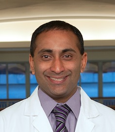
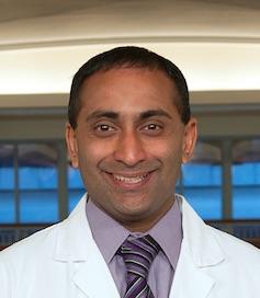
The meeting is designed to aid entrepreneurs, as well as physician innovators, who have ideas how to improve the field of gastroenterology but have found it daunting to take their ideas and commercialize them, added meeting organizer V. Raman Muthusamy, MD, the director of endoscopy for the University of California, Los Angeles, Health System. Dr. Muthusamy and Dr. Komanduri serve as cochairs of the AGA Center for GI Innovation and Technology executive committee.
“Innovation is key to evolving and thriving in any field, and we’re trying to foster that in gastroenterology,” Dr. Muthusamy said. “There are many good ideas in GI that probably never see the light of day simply because of real and perceived barriers to successful innovation. We’re trying to demystify and simplify that process.”
New this year will be a Wednesday preconference entrepreneur and innovator package session, in which innovators can meet one-on-one with experts – including intellectual property attorneys, physician innovators, venture capitalists, and payers – for personalized advice on how to move their products forward. An evening dinner reception for summit sponsors will feature a talk by Kevin Volpp, MD, PhD, founding director of the Center for Health Incentives and Behavior Economics at the University of Pennsylvania, Philadelphia, about the psychology of purchasing and how economic decisions are made in health care.
Also new for 2018 is a Thursday afternoon session on the evolving role of digital health in GI diseases. “We’ll look at where medicine is headed in terms of artificial intelligence, patient-driven mobile applications, and everything becoming digitally balanced,” said Dr. Komanduri, who will moderate the session. “We need to understand the space and how to utilize it on a more global aspect when it comes to innovation because there is a need for further education.”
The main summit will kick off Thursday with an address by Vadim Backman, PhD, the Walter Dill Scott Professor of Biomedical Engineering at Northwestern University, about nanotechnology and how it applies to gastroenterology. “We have a lot of entrepreneurs wondering what’s the next thing,” Dr. Muthusamy said. “I think Vadim is going to open up some new horizons and get us thinking about things that we haven’t traditionally thought of.”
The morning will focus on avoiding critical mistakes in startups and on medical device development, including lessons learned about developing a product and practical approaches to getting funded. Sessions scheduled for after lunch will highlight how to achieve physician adoption, including how to obtain a CPT code and physician barriers to incorporating new technology.
Friday’s sessions are unofficially titled “the next generation of endoscopy,” Dr. Muthusamy said. The day will begin with a look at bariatric endoscopy and surgical techniques and at the challenges and unmet needs of bariatric procedures, as well as organ-sparing resection techniques. Following the Shark Tank presentations, in which small companies can present their ideas to an expert panel, and lunch, the afternoon program will feature discussions on the reprocessing of duodenoscopes and quality in endoscopy.
“Our goal is not just to have these sessions be an interesting 90 minutes but to inspire a group of people with passion and interest in these areas to guide the field over the next 5-10 years in terms of where our energies should be focused, what’s needed, and what trials should be done so we can achieve the best results the quickest to achieve adoption of novel technologies,” Dr. Muthusamy said.
Dr. Komanduri is a consultant for Boston Scientific, Cook Medical, Olympus and Medtronic. Dr. Muthusamy disclosed financial relationships with CapsoVision, Boston Scientific and Medtronic.
For about 10 years, the AGA Tech Summit, sponsored by the AGA Center for GI Innovation and Technology, has brought together physician innovators, entrepreneurs, and industry representatives to facilitate innovation in gastroenterology practice, and this year is no exception.
Attendees will get a “comprehensive, almost immersive experience to understand what’s hot in gastroenterology and innovation in 2018, where the gaps are, and where we need future innovators to be successful to improve patient outcomes,” said Srinadh (Sri) Komanduri, MD, the medical director of the GI laboratory and director of interventional endoscopy at Northwestern University in Chicago, as well as one of the meeting’s organizers.
The meeting is designed to aid entrepreneurs, as well as physician innovators, who have ideas how to improve the field of gastroenterology but have found it daunting to take their ideas and commercialize them, added meeting organizer V. Raman Muthusamy, MD, the director of endoscopy for the University of California, Los Angeles, Health System. Dr. Muthusamy and Dr. Komanduri serve as cochairs of the AGA Center for GI Innovation and Technology executive committee.
“Innovation is key to evolving and thriving in any field, and we’re trying to foster that in gastroenterology,” Dr. Muthusamy said. “There are many good ideas in GI that probably never see the light of day simply because of real and perceived barriers to successful innovation. We’re trying to demystify and simplify that process.”
New this year will be a Wednesday preconference entrepreneur and innovator package session, in which innovators can meet one-on-one with experts – including intellectual property attorneys, physician innovators, venture capitalists, and payers – for personalized advice on how to move their products forward. An evening dinner reception for summit sponsors will feature a talk by Kevin Volpp, MD, PhD, founding director of the Center for Health Incentives and Behavior Economics at the University of Pennsylvania, Philadelphia, about the psychology of purchasing and how economic decisions are made in health care.
Also new for 2018 is a Thursday afternoon session on the evolving role of digital health in GI diseases. “We’ll look at where medicine is headed in terms of artificial intelligence, patient-driven mobile applications, and everything becoming digitally balanced,” said Dr. Komanduri, who will moderate the session. “We need to understand the space and how to utilize it on a more global aspect when it comes to innovation because there is a need for further education.”
The main summit will kick off Thursday with an address by Vadim Backman, PhD, the Walter Dill Scott Professor of Biomedical Engineering at Northwestern University, about nanotechnology and how it applies to gastroenterology. “We have a lot of entrepreneurs wondering what’s the next thing,” Dr. Muthusamy said. “I think Vadim is going to open up some new horizons and get us thinking about things that we haven’t traditionally thought of.”
The morning will focus on avoiding critical mistakes in startups and on medical device development, including lessons learned about developing a product and practical approaches to getting funded. Sessions scheduled for after lunch will highlight how to achieve physician adoption, including how to obtain a CPT code and physician barriers to incorporating new technology.
Friday’s sessions are unofficially titled “the next generation of endoscopy,” Dr. Muthusamy said. The day will begin with a look at bariatric endoscopy and surgical techniques and at the challenges and unmet needs of bariatric procedures, as well as organ-sparing resection techniques. Following the Shark Tank presentations, in which small companies can present their ideas to an expert panel, and lunch, the afternoon program will feature discussions on the reprocessing of duodenoscopes and quality in endoscopy.
“Our goal is not just to have these sessions be an interesting 90 minutes but to inspire a group of people with passion and interest in these areas to guide the field over the next 5-10 years in terms of where our energies should be focused, what’s needed, and what trials should be done so we can achieve the best results the quickest to achieve adoption of novel technologies,” Dr. Muthusamy said.
Dr. Komanduri is a consultant for Boston Scientific, Cook Medical, Olympus and Medtronic. Dr. Muthusamy disclosed financial relationships with CapsoVision, Boston Scientific and Medtronic.
For about 10 years, the AGA Tech Summit, sponsored by the AGA Center for GI Innovation and Technology, has brought together physician innovators, entrepreneurs, and industry representatives to facilitate innovation in gastroenterology practice, and this year is no exception.
Attendees will get a “comprehensive, almost immersive experience to understand what’s hot in gastroenterology and innovation in 2018, where the gaps are, and where we need future innovators to be successful to improve patient outcomes,” said Srinadh (Sri) Komanduri, MD, the medical director of the GI laboratory and director of interventional endoscopy at Northwestern University in Chicago, as well as one of the meeting’s organizers.
The meeting is designed to aid entrepreneurs, as well as physician innovators, who have ideas how to improve the field of gastroenterology but have found it daunting to take their ideas and commercialize them, added meeting organizer V. Raman Muthusamy, MD, the director of endoscopy for the University of California, Los Angeles, Health System. Dr. Muthusamy and Dr. Komanduri serve as cochairs of the AGA Center for GI Innovation and Technology executive committee.
“Innovation is key to evolving and thriving in any field, and we’re trying to foster that in gastroenterology,” Dr. Muthusamy said. “There are many good ideas in GI that probably never see the light of day simply because of real and perceived barriers to successful innovation. We’re trying to demystify and simplify that process.”
New this year will be a Wednesday preconference entrepreneur and innovator package session, in which innovators can meet one-on-one with experts – including intellectual property attorneys, physician innovators, venture capitalists, and payers – for personalized advice on how to move their products forward. An evening dinner reception for summit sponsors will feature a talk by Kevin Volpp, MD, PhD, founding director of the Center for Health Incentives and Behavior Economics at the University of Pennsylvania, Philadelphia, about the psychology of purchasing and how economic decisions are made in health care.
Also new for 2018 is a Thursday afternoon session on the evolving role of digital health in GI diseases. “We’ll look at where medicine is headed in terms of artificial intelligence, patient-driven mobile applications, and everything becoming digitally balanced,” said Dr. Komanduri, who will moderate the session. “We need to understand the space and how to utilize it on a more global aspect when it comes to innovation because there is a need for further education.”
The main summit will kick off Thursday with an address by Vadim Backman, PhD, the Walter Dill Scott Professor of Biomedical Engineering at Northwestern University, about nanotechnology and how it applies to gastroenterology. “We have a lot of entrepreneurs wondering what’s the next thing,” Dr. Muthusamy said. “I think Vadim is going to open up some new horizons and get us thinking about things that we haven’t traditionally thought of.”
The morning will focus on avoiding critical mistakes in startups and on medical device development, including lessons learned about developing a product and practical approaches to getting funded. Sessions scheduled for after lunch will highlight how to achieve physician adoption, including how to obtain a CPT code and physician barriers to incorporating new technology.
Friday’s sessions are unofficially titled “the next generation of endoscopy,” Dr. Muthusamy said. The day will begin with a look at bariatric endoscopy and surgical techniques and at the challenges and unmet needs of bariatric procedures, as well as organ-sparing resection techniques. Following the Shark Tank presentations, in which small companies can present their ideas to an expert panel, and lunch, the afternoon program will feature discussions on the reprocessing of duodenoscopes and quality in endoscopy.
“Our goal is not just to have these sessions be an interesting 90 minutes but to inspire a group of people with passion and interest in these areas to guide the field over the next 5-10 years in terms of where our energies should be focused, what’s needed, and what trials should be done so we can achieve the best results the quickest to achieve adoption of novel technologies,” Dr. Muthusamy said.
Dr. Komanduri is a consultant for Boston Scientific, Cook Medical, Olympus and Medtronic. Dr. Muthusamy disclosed financial relationships with CapsoVision, Boston Scientific and Medtronic.
A global snapshot of leukemia incidence
, according to an analysis of World Health Organization cancer databases.
Incidence also is generally higher in males, with a global male to female ratio of 1.4. For men, the highest regional leukemia rate – estimated at 11.3 per 100,000 population for 2012 – was found in Australia and New Zealand, with northern America (the United States and Canada) next at 10.5 per 100,000. Australia/New Zealand and northern America had the highest rate for women at 7.2 per 100,000, followed by western Europe and northern Europe at 6.0 per 100,000, reported Adalberto Miranda-Filho, PhD, of the WHO’s International Agency for Research on Cancer in Lyon, France, and his associates.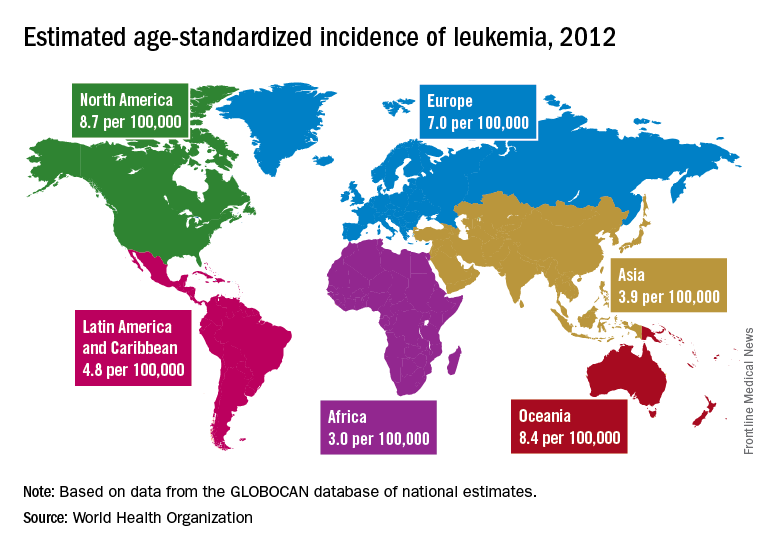
The lowest regional rates for women were found in western Africa (1.2 per 100,000), middle Africa (1.8), and Micronesia/Polynesia (2.1). For men, leukemia incidence was lowest in western Africa (1.4 per 100,000), middle Africa (2.6), and south-central Asia (3.4), according to data from the WHO’s GLOBOCAN database. The report was published in The Lancet Haematology.
Estimates for leukemia subtypes in 2003-2007 – calculated for 54 countries, not regions – also showed a great deal of variation. For acute lymphoblastic leukemia, Ecuador had the highest rates for both males (2.8 per 100,000) and females (3.3), with high rates seen in Costa Rica, Columbia, and Cyprus. Rates in the United States were near the top: 2.1 for males and 1.6 for females. Rates were lowest for men in Jamaica (0.4) and Serbia (0.6) and for women in India (0.5) and Serbia and Cuba (0.6), Dr. Miranda-Filho and his associates said.
Incidence rates for acute myeloid leukemia were highest in Australia for men (2.8 per 100,000) and Austria for women (2.2), with the United States near the top for both men (2.6) and women (1.9). The lowest rates occurred in Cuba and Egypt for men (0.9 per 100,000) and Cuba for women (0.4), data from the WHO’s Cancer Incidence in Five Continents Volume X show.
Chronic lymphocytic leukemia incidence was highest for men in Canada (4.5 per 100,000), Ireland and Lithuania (4.4), and Slovakia (4.3). The incidence was highest for women in Lithuania (2.5), Canada (2.3), and Slovakia and Denmark (2.1). Incidence in the United States was 3.5 for men and 1.8 for women. At the other end of the scale, the lowest rates for both men and women were in Japan and Malaysia (0.1), the investigators’ analysis showed.
Chronic myeloid leukemia rates were the lowest of the subtypes, and Tunisia was the lowest for men at 0.4 per 100,000 and tied for lowest with Serbia, Slovenia, and Puerto Rico for women at 0.3. Incidence was highest for men in Australia at 1.8 per 100,000 and highest for women in Uruguay at 1.1. Rates in the United States were 1.3 for men and 0.8 for women, Dr. Miranda-Filho and his associates said.
“The higher incidence of acute lymphoblastic leukaemia in parts of South America, as well as of chronic lymphocytic leukaemia in populations across North America and Oceania, alongside a lower incidence in Asia, might be important markers for further epidemiological study, and a means to better understand the underlying factors to support future cancer prevention strategies,” the investigators wrote.
SOURCE: Miranda-Filho A et al. Lancet Haematol. 2018;5:e14-24.
, according to an analysis of World Health Organization cancer databases.
Incidence also is generally higher in males, with a global male to female ratio of 1.4. For men, the highest regional leukemia rate – estimated at 11.3 per 100,000 population for 2012 – was found in Australia and New Zealand, with northern America (the United States and Canada) next at 10.5 per 100,000. Australia/New Zealand and northern America had the highest rate for women at 7.2 per 100,000, followed by western Europe and northern Europe at 6.0 per 100,000, reported Adalberto Miranda-Filho, PhD, of the WHO’s International Agency for Research on Cancer in Lyon, France, and his associates.
The lowest regional rates for women were found in western Africa (1.2 per 100,000), middle Africa (1.8), and Micronesia/Polynesia (2.1). For men, leukemia incidence was lowest in western Africa (1.4 per 100,000), middle Africa (2.6), and south-central Asia (3.4), according to data from the WHO’s GLOBOCAN database. The report was published in The Lancet Haematology.
Estimates for leukemia subtypes in 2003-2007 – calculated for 54 countries, not regions – also showed a great deal of variation. For acute lymphoblastic leukemia, Ecuador had the highest rates for both males (2.8 per 100,000) and females (3.3), with high rates seen in Costa Rica, Columbia, and Cyprus. Rates in the United States were near the top: 2.1 for males and 1.6 for females. Rates were lowest for men in Jamaica (0.4) and Serbia (0.6) and for women in India (0.5) and Serbia and Cuba (0.6), Dr. Miranda-Filho and his associates said.
Incidence rates for acute myeloid leukemia were highest in Australia for men (2.8 per 100,000) and Austria for women (2.2), with the United States near the top for both men (2.6) and women (1.9). The lowest rates occurred in Cuba and Egypt for men (0.9 per 100,000) and Cuba for women (0.4), data from the WHO’s Cancer Incidence in Five Continents Volume X show.
Chronic lymphocytic leukemia incidence was highest for men in Canada (4.5 per 100,000), Ireland and Lithuania (4.4), and Slovakia (4.3). The incidence was highest for women in Lithuania (2.5), Canada (2.3), and Slovakia and Denmark (2.1). Incidence in the United States was 3.5 for men and 1.8 for women. At the other end of the scale, the lowest rates for both men and women were in Japan and Malaysia (0.1), the investigators’ analysis showed.
Chronic myeloid leukemia rates were the lowest of the subtypes, and Tunisia was the lowest for men at 0.4 per 100,000 and tied for lowest with Serbia, Slovenia, and Puerto Rico for women at 0.3. Incidence was highest for men in Australia at 1.8 per 100,000 and highest for women in Uruguay at 1.1. Rates in the United States were 1.3 for men and 0.8 for women, Dr. Miranda-Filho and his associates said.
“The higher incidence of acute lymphoblastic leukaemia in parts of South America, as well as of chronic lymphocytic leukaemia in populations across North America and Oceania, alongside a lower incidence in Asia, might be important markers for further epidemiological study, and a means to better understand the underlying factors to support future cancer prevention strategies,” the investigators wrote.
SOURCE: Miranda-Filho A et al. Lancet Haematol. 2018;5:e14-24.
, according to an analysis of World Health Organization cancer databases.
Incidence also is generally higher in males, with a global male to female ratio of 1.4. For men, the highest regional leukemia rate – estimated at 11.3 per 100,000 population for 2012 – was found in Australia and New Zealand, with northern America (the United States and Canada) next at 10.5 per 100,000. Australia/New Zealand and northern America had the highest rate for women at 7.2 per 100,000, followed by western Europe and northern Europe at 6.0 per 100,000, reported Adalberto Miranda-Filho, PhD, of the WHO’s International Agency for Research on Cancer in Lyon, France, and his associates.
The lowest regional rates for women were found in western Africa (1.2 per 100,000), middle Africa (1.8), and Micronesia/Polynesia (2.1). For men, leukemia incidence was lowest in western Africa (1.4 per 100,000), middle Africa (2.6), and south-central Asia (3.4), according to data from the WHO’s GLOBOCAN database. The report was published in The Lancet Haematology.
Estimates for leukemia subtypes in 2003-2007 – calculated for 54 countries, not regions – also showed a great deal of variation. For acute lymphoblastic leukemia, Ecuador had the highest rates for both males (2.8 per 100,000) and females (3.3), with high rates seen in Costa Rica, Columbia, and Cyprus. Rates in the United States were near the top: 2.1 for males and 1.6 for females. Rates were lowest for men in Jamaica (0.4) and Serbia (0.6) and for women in India (0.5) and Serbia and Cuba (0.6), Dr. Miranda-Filho and his associates said.
Incidence rates for acute myeloid leukemia were highest in Australia for men (2.8 per 100,000) and Austria for women (2.2), with the United States near the top for both men (2.6) and women (1.9). The lowest rates occurred in Cuba and Egypt for men (0.9 per 100,000) and Cuba for women (0.4), data from the WHO’s Cancer Incidence in Five Continents Volume X show.
Chronic lymphocytic leukemia incidence was highest for men in Canada (4.5 per 100,000), Ireland and Lithuania (4.4), and Slovakia (4.3). The incidence was highest for women in Lithuania (2.5), Canada (2.3), and Slovakia and Denmark (2.1). Incidence in the United States was 3.5 for men and 1.8 for women. At the other end of the scale, the lowest rates for both men and women were in Japan and Malaysia (0.1), the investigators’ analysis showed.
Chronic myeloid leukemia rates were the lowest of the subtypes, and Tunisia was the lowest for men at 0.4 per 100,000 and tied for lowest with Serbia, Slovenia, and Puerto Rico for women at 0.3. Incidence was highest for men in Australia at 1.8 per 100,000 and highest for women in Uruguay at 1.1. Rates in the United States were 1.3 for men and 0.8 for women, Dr. Miranda-Filho and his associates said.
“The higher incidence of acute lymphoblastic leukaemia in parts of South America, as well as of chronic lymphocytic leukaemia in populations across North America and Oceania, alongside a lower incidence in Asia, might be important markers for further epidemiological study, and a means to better understand the underlying factors to support future cancer prevention strategies,” the investigators wrote.
SOURCE: Miranda-Filho A et al. Lancet Haematol. 2018;5:e14-24.
FROM THE LANCET HAEMATOLOGY
Dolutegravir-based regimen effective in HIV/TB coinfected
BOSTON – In antiretroviral therapy–naive patients with HIV and tuberculosis coinfection, a combination of dolutegravir-based ART and rifampin-based TB therapy was associated with good efficacy and immunological responses through at least 24 weeks, investigators reported.
Dolutegravir-based ART appeared to be comparable to efavirenz-based therapy at viral suppression with a similar safety profile, although the trial was not powered for head-to-head comparison, said Kelly Dooley, MD, of Johns Hopkins University, Baltimore.
“We know that rifampin reduces concentrations of dolutegravir substantially, but that drug interaction can be mitigated by increasing the dose of dolutegravir to twice daily. That information was from a healthy volunteer trial, so we thought it would be important to test HIV/TB cotreatment among patients who have both infections,” she said at the Conference on Retroviruses and Opportunistic Infections.
Dr. Dooley and her colleagues looked at dolutegravir or efavirenz plus rifampin-based TB therapy in the INSPIRING trial.
The ongoing study is a phase 3b, noncomparative, randomized, open-label trial in ART-naive adults with HIV-1 and drug-sensitive TB infections.
In a briefing following her presentation of the data in an oral session, Dr. Dooley explained that the reason for the parallel but noncomparative arms in the trial was that it was primarily designed to see how dolutegravir works in HIV/TB coinfected patients. Enrollment of 113 patients from 37 sites in seven countries took about 2 years, and it would have required significantly more time to enroll the more than 500 patients necessary for an adequately powered comparison trial.
The trial was conducted in Argentina, Brazil, Mexico, Peru, Russia, South Africa, and Thailand. Dr. Dooley presented results from a 24-week interim analysis.

Participants who were on rifampin-based TB therapy for up to 8 weeks were randomized on a 3:2 basis to receive either dolutegravir 50 mg twice daily during and for 2 weeks post-TB therapy, followed by 50 mg once daily (69 patients), or efavirenz 600 mg daily plus two nucleoside reverse transcriptase inhibitors of the investigator’s choice for 52 weeks.
At 24 weeks, the percentage of patients deemed to have virologic success, defined as HIV-1 RNA less than 50 copies/mL, was 81% in the dolutegravir arm and 89% in the efavirenz arm. The respective rates of virologic nonresponse were 10% and 7%. No virologic data were available because of treatment discontinuations or missing information for 9% and 5% of patients, respectively.
The difference in response rates between the arms was due to non–treatment associated withdrawals among patients on dolutegravir, Dr. Dooley said.
Virologic withdrawals for confirmed plasma HIV-1 RNA of 400 copies/mL or more at or after week 24 for two consecutive tests occurred in one patient on dolutegravir and in none on efavirenz. There were no treatment-emergent resistance-associated mutations to any anti-HIV drugs in the dolutegravir group (data not available for the efavirenz group).
Two patients, both in the efavirenz arm, discontinued therapy because of adverse events. TB-associated rates of the immune reconstitution inflammatory syndrome (IRIS) occurred in 4% of patients in each treatment arm. No patients discontinued therapy because of IRIS or liver-related adverse events.
“We think that this study provides evidence that dolutegravir is effective and well tolerated in adults with HIV/TB coinfection who are receiving rifampin-based tuberculosis therapy, and we hope that this will increase the number of options for TB/HIV coinfected patients for whom there are relatively few treatment possibilities,” she concluded.
Dr. Dooley reported research support via her university from ViiV Healthcare, which funded the study.
SOURCE: Dooley K et al. Abstract 33.
BOSTON – In antiretroviral therapy–naive patients with HIV and tuberculosis coinfection, a combination of dolutegravir-based ART and rifampin-based TB therapy was associated with good efficacy and immunological responses through at least 24 weeks, investigators reported.
Dolutegravir-based ART appeared to be comparable to efavirenz-based therapy at viral suppression with a similar safety profile, although the trial was not powered for head-to-head comparison, said Kelly Dooley, MD, of Johns Hopkins University, Baltimore.
“We know that rifampin reduces concentrations of dolutegravir substantially, but that drug interaction can be mitigated by increasing the dose of dolutegravir to twice daily. That information was from a healthy volunteer trial, so we thought it would be important to test HIV/TB cotreatment among patients who have both infections,” she said at the Conference on Retroviruses and Opportunistic Infections.
Dr. Dooley and her colleagues looked at dolutegravir or efavirenz plus rifampin-based TB therapy in the INSPIRING trial.
The ongoing study is a phase 3b, noncomparative, randomized, open-label trial in ART-naive adults with HIV-1 and drug-sensitive TB infections.
In a briefing following her presentation of the data in an oral session, Dr. Dooley explained that the reason for the parallel but noncomparative arms in the trial was that it was primarily designed to see how dolutegravir works in HIV/TB coinfected patients. Enrollment of 113 patients from 37 sites in seven countries took about 2 years, and it would have required significantly more time to enroll the more than 500 patients necessary for an adequately powered comparison trial.
The trial was conducted in Argentina, Brazil, Mexico, Peru, Russia, South Africa, and Thailand. Dr. Dooley presented results from a 24-week interim analysis.
Participants who were on rifampin-based TB therapy for up to 8 weeks were randomized on a 3:2 basis to receive either dolutegravir 50 mg twice daily during and for 2 weeks post-TB therapy, followed by 50 mg once daily (69 patients), or efavirenz 600 mg daily plus two nucleoside reverse transcriptase inhibitors of the investigator’s choice for 52 weeks.
At 24 weeks, the percentage of patients deemed to have virologic success, defined as HIV-1 RNA less than 50 copies/mL, was 81% in the dolutegravir arm and 89% in the efavirenz arm. The respective rates of virologic nonresponse were 10% and 7%. No virologic data were available because of treatment discontinuations or missing information for 9% and 5% of patients, respectively.
The difference in response rates between the arms was due to non–treatment associated withdrawals among patients on dolutegravir, Dr. Dooley said.
Virologic withdrawals for confirmed plasma HIV-1 RNA of 400 copies/mL or more at or after week 24 for two consecutive tests occurred in one patient on dolutegravir and in none on efavirenz. There were no treatment-emergent resistance-associated mutations to any anti-HIV drugs in the dolutegravir group (data not available for the efavirenz group).
Two patients, both in the efavirenz arm, discontinued therapy because of adverse events. TB-associated rates of the immune reconstitution inflammatory syndrome (IRIS) occurred in 4% of patients in each treatment arm. No patients discontinued therapy because of IRIS or liver-related adverse events.
“We think that this study provides evidence that dolutegravir is effective and well tolerated in adults with HIV/TB coinfection who are receiving rifampin-based tuberculosis therapy, and we hope that this will increase the number of options for TB/HIV coinfected patients for whom there are relatively few treatment possibilities,” she concluded.
Dr. Dooley reported research support via her university from ViiV Healthcare, which funded the study.
SOURCE: Dooley K et al. Abstract 33.
BOSTON – In antiretroviral therapy–naive patients with HIV and tuberculosis coinfection, a combination of dolutegravir-based ART and rifampin-based TB therapy was associated with good efficacy and immunological responses through at least 24 weeks, investigators reported.
Dolutegravir-based ART appeared to be comparable to efavirenz-based therapy at viral suppression with a similar safety profile, although the trial was not powered for head-to-head comparison, said Kelly Dooley, MD, of Johns Hopkins University, Baltimore.
“We know that rifampin reduces concentrations of dolutegravir substantially, but that drug interaction can be mitigated by increasing the dose of dolutegravir to twice daily. That information was from a healthy volunteer trial, so we thought it would be important to test HIV/TB cotreatment among patients who have both infections,” she said at the Conference on Retroviruses and Opportunistic Infections.
Dr. Dooley and her colleagues looked at dolutegravir or efavirenz plus rifampin-based TB therapy in the INSPIRING trial.
The ongoing study is a phase 3b, noncomparative, randomized, open-label trial in ART-naive adults with HIV-1 and drug-sensitive TB infections.
In a briefing following her presentation of the data in an oral session, Dr. Dooley explained that the reason for the parallel but noncomparative arms in the trial was that it was primarily designed to see how dolutegravir works in HIV/TB coinfected patients. Enrollment of 113 patients from 37 sites in seven countries took about 2 years, and it would have required significantly more time to enroll the more than 500 patients necessary for an adequately powered comparison trial.
The trial was conducted in Argentina, Brazil, Mexico, Peru, Russia, South Africa, and Thailand. Dr. Dooley presented results from a 24-week interim analysis.
Participants who were on rifampin-based TB therapy for up to 8 weeks were randomized on a 3:2 basis to receive either dolutegravir 50 mg twice daily during and for 2 weeks post-TB therapy, followed by 50 mg once daily (69 patients), or efavirenz 600 mg daily plus two nucleoside reverse transcriptase inhibitors of the investigator’s choice for 52 weeks.
At 24 weeks, the percentage of patients deemed to have virologic success, defined as HIV-1 RNA less than 50 copies/mL, was 81% in the dolutegravir arm and 89% in the efavirenz arm. The respective rates of virologic nonresponse were 10% and 7%. No virologic data were available because of treatment discontinuations or missing information for 9% and 5% of patients, respectively.
The difference in response rates between the arms was due to non–treatment associated withdrawals among patients on dolutegravir, Dr. Dooley said.
Virologic withdrawals for confirmed plasma HIV-1 RNA of 400 copies/mL or more at or after week 24 for two consecutive tests occurred in one patient on dolutegravir and in none on efavirenz. There were no treatment-emergent resistance-associated mutations to any anti-HIV drugs in the dolutegravir group (data not available for the efavirenz group).
Two patients, both in the efavirenz arm, discontinued therapy because of adverse events. TB-associated rates of the immune reconstitution inflammatory syndrome (IRIS) occurred in 4% of patients in each treatment arm. No patients discontinued therapy because of IRIS or liver-related adverse events.
“We think that this study provides evidence that dolutegravir is effective and well tolerated in adults with HIV/TB coinfection who are receiving rifampin-based tuberculosis therapy, and we hope that this will increase the number of options for TB/HIV coinfected patients for whom there are relatively few treatment possibilities,” she concluded.
Dr. Dooley reported research support via her university from ViiV Healthcare, which funded the study.
SOURCE: Dooley K et al. Abstract 33.
REPORTING FROM CROI
Key clinical point:
Major finding: The virologic response rate at 24 weeks with dolutegravir was 81%.
Data source: Randomized noncomparative trial of 113 patients with HIV/TB coinfections in seven countries.
Disclosures: Dr. Dooley reported research support via her university from ViiV Healthcare, which funded the study.
Source: Dooley K et al. Abstract 33.

Levodopa Monotherapy May Be Adequate After DBS for Parkinson’s Disease
LAS VEGAS—For pharmacotherapy following deep brain stimulation (DBS), many patients can be adequately treated with levodopa alone, but only a small group can be managed with dopamine agonists alone, according to a study presented at the 21st Annual Meeting of the North American Neuromodulation Society. The data address a question about which there has been little objective guidance.
Based on the findings, efforts to reduce anti-Parkinson’s disease medications following DBS should be directed toward dopamine agonists first, according to Alfonso Fasano, MD, PhD, Codirector of the Surgical Program at the Movement Disorder Center of Toronto Western Hospital in Ontario. The data nevertheless provide the option for individualized therapy, he added. For example, for patients with good outcome after DBS and no significant history of impulse-

In the randomized controlled trial, 36 patients were assigned to receive monotherapy with a dopamine agonist or levodopa following subthalamic nucleus (STN) DBS. All patients were on both medications before surgery. The investigators did not identify any between-group differences in variables predicted to affect symptom control. Yet the proportion of patients that were able to stay on monotherapy at three months was different between the study arms.
“Perhaps the main result of this study was that we found it difficult to keep patients on dopamine-agonist monotherapy, even after a successful DBS procedure,” said Dr. Fasano.
At three months after surgery, the proportion of patients remaining on monotherapy was 81% in the levodopa group and 32% in the dopamine-agonist group. At that point in the follow-up, all other patients in the two groups were on both treatments. At six months, fewer patients were taking their assigned drug as a monotherapy. The proportion in the levodopa arm had decreased to 63%, and the proportion in the dopamine-agonist arm had decreased to 13%.
More of the failures in the dopamine-agonist arm, relative to the levodopa arm, resulted from inadequate control of motor symptoms. Failure in the levodopa arm more commonly occurred following mood and sleep disturbances. But the investigators observed failures for both reasons in both study arms.
DBS permits a reduction in anti-Parkinson’s disease medications, but dose reductions should be undertaken slowly, because mood disturbances have been reported after abrupt discontinuation of dopamine agonists, according to Dr. Fasano. Specific strategies for reducing anti-Parkinson’s disease medications following DBS have not been based on evidence, however.
Overall, the results of the randomized study “are in keeping with a common-sense” approach, Dr. Fasano said. While the results emphasize the limited proportion of patients who can remain on dopamine agonists alone following DBS, they also suggest that most patients receiving levodopa will require low doses of both therapies.
With these results and observations, “maybe we are getting closer to a guideline of what to do with respect to medication after surgery,” Dr. Fasano said.
—Ted Bosworth
Suggested Reading
Fasano A, Appel-Cresswell S, Jog M, et al. Medical management of Parkinson’s disease after initiation of deep brain stimulation. Can J Neurol Sci. 2016;43(5):626-634.
LAS VEGAS—For pharmacotherapy following deep brain stimulation (DBS), many patients can be adequately treated with levodopa alone, but only a small group can be managed with dopamine agonists alone, according to a study presented at the 21st Annual Meeting of the North American Neuromodulation Society. The data address a question about which there has been little objective guidance.
Based on the findings, efforts to reduce anti-Parkinson’s disease medications following DBS should be directed toward dopamine agonists first, according to Alfonso Fasano, MD, PhD, Codirector of the Surgical Program at the Movement Disorder Center of Toronto Western Hospital in Ontario. The data nevertheless provide the option for individualized therapy, he added. For example, for patients with good outcome after DBS and no significant history of impulse-
In the randomized controlled trial, 36 patients were assigned to receive monotherapy with a dopamine agonist or levodopa following subthalamic nucleus (STN) DBS. All patients were on both medications before surgery. The investigators did not identify any between-group differences in variables predicted to affect symptom control. Yet the proportion of patients that were able to stay on monotherapy at three months was different between the study arms.
“Perhaps the main result of this study was that we found it difficult to keep patients on dopamine-agonist monotherapy, even after a successful DBS procedure,” said Dr. Fasano.
At three months after surgery, the proportion of patients remaining on monotherapy was 81% in the levodopa group and 32% in the dopamine-agonist group. At that point in the follow-up, all other patients in the two groups were on both treatments. At six months, fewer patients were taking their assigned drug as a monotherapy. The proportion in the levodopa arm had decreased to 63%, and the proportion in the dopamine-agonist arm had decreased to 13%.
More of the failures in the dopamine-agonist arm, relative to the levodopa arm, resulted from inadequate control of motor symptoms. Failure in the levodopa arm more commonly occurred following mood and sleep disturbances. But the investigators observed failures for both reasons in both study arms.
DBS permits a reduction in anti-Parkinson’s disease medications, but dose reductions should be undertaken slowly, because mood disturbances have been reported after abrupt discontinuation of dopamine agonists, according to Dr. Fasano. Specific strategies for reducing anti-Parkinson’s disease medications following DBS have not been based on evidence, however.
Overall, the results of the randomized study “are in keeping with a common-sense” approach, Dr. Fasano said. While the results emphasize the limited proportion of patients who can remain on dopamine agonists alone following DBS, they also suggest that most patients receiving levodopa will require low doses of both therapies.
With these results and observations, “maybe we are getting closer to a guideline of what to do with respect to medication after surgery,” Dr. Fasano said.
—Ted Bosworth
Suggested Reading
Fasano A, Appel-Cresswell S, Jog M, et al. Medical management of Parkinson’s disease after initiation of deep brain stimulation. Can J Neurol Sci. 2016;43(5):626-634.
LAS VEGAS—For pharmacotherapy following deep brain stimulation (DBS), many patients can be adequately treated with levodopa alone, but only a small group can be managed with dopamine agonists alone, according to a study presented at the 21st Annual Meeting of the North American Neuromodulation Society. The data address a question about which there has been little objective guidance.
Based on the findings, efforts to reduce anti-Parkinson’s disease medications following DBS should be directed toward dopamine agonists first, according to Alfonso Fasano, MD, PhD, Codirector of the Surgical Program at the Movement Disorder Center of Toronto Western Hospital in Ontario. The data nevertheless provide the option for individualized therapy, he added. For example, for patients with good outcome after DBS and no significant history of impulse-
In the randomized controlled trial, 36 patients were assigned to receive monotherapy with a dopamine agonist or levodopa following subthalamic nucleus (STN) DBS. All patients were on both medications before surgery. The investigators did not identify any between-group differences in variables predicted to affect symptom control. Yet the proportion of patients that were able to stay on monotherapy at three months was different between the study arms.
“Perhaps the main result of this study was that we found it difficult to keep patients on dopamine-agonist monotherapy, even after a successful DBS procedure,” said Dr. Fasano.
At three months after surgery, the proportion of patients remaining on monotherapy was 81% in the levodopa group and 32% in the dopamine-agonist group. At that point in the follow-up, all other patients in the two groups were on both treatments. At six months, fewer patients were taking their assigned drug as a monotherapy. The proportion in the levodopa arm had decreased to 63%, and the proportion in the dopamine-agonist arm had decreased to 13%.
More of the failures in the dopamine-agonist arm, relative to the levodopa arm, resulted from inadequate control of motor symptoms. Failure in the levodopa arm more commonly occurred following mood and sleep disturbances. But the investigators observed failures for both reasons in both study arms.
DBS permits a reduction in anti-Parkinson’s disease medications, but dose reductions should be undertaken slowly, because mood disturbances have been reported after abrupt discontinuation of dopamine agonists, according to Dr. Fasano. Specific strategies for reducing anti-Parkinson’s disease medications following DBS have not been based on evidence, however.
Overall, the results of the randomized study “are in keeping with a common-sense” approach, Dr. Fasano said. While the results emphasize the limited proportion of patients who can remain on dopamine agonists alone following DBS, they also suggest that most patients receiving levodopa will require low doses of both therapies.
With these results and observations, “maybe we are getting closer to a guideline of what to do with respect to medication after surgery,” Dr. Fasano said.
—Ted Bosworth
Suggested Reading
Fasano A, Appel-Cresswell S, Jog M, et al. Medical management of Parkinson’s disease after initiation of deep brain stimulation. Can J Neurol Sci. 2016;43(5):626-634.

Good definitions, research lacking for COPD-asthma overlap
ORLANDO – to give clinicians data they can actually use.
The topic is even more pressing given the growing interest and research into biological treatments for asthma and consideration of their possible use in COPD, experts said at the joint congress of the American Academy of Allergy, Asthma, and Immunology and the World Asthma Organization. Their remarks came in what was ostensibly a “debate” on whether ACOS is a distinct entity requiring special treatment but largely turned into a discussion about gaps in knowledge on the topic.
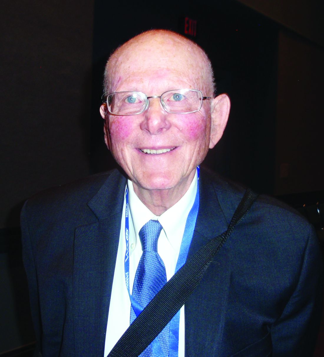
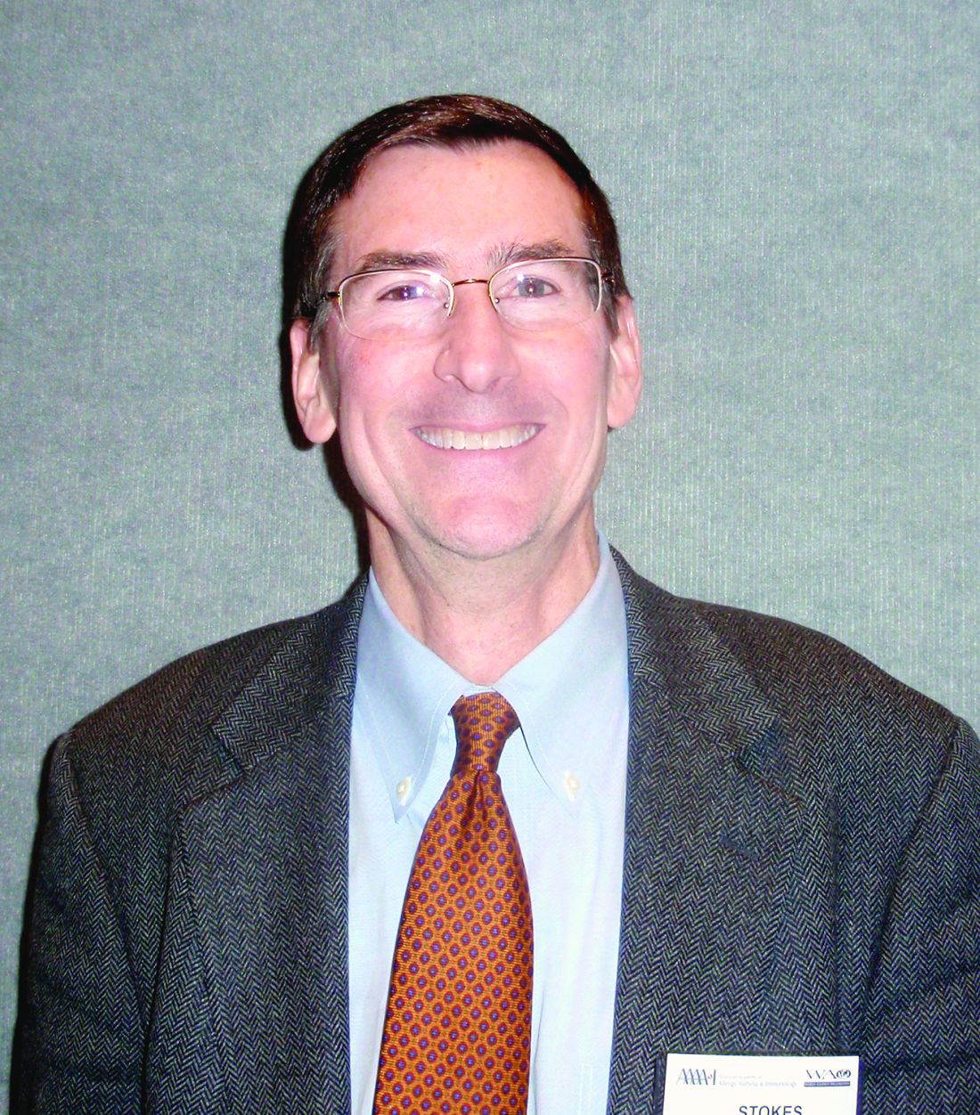
“The problem here is that it has not been defined in a way that everyone agrees on – that does create a problem because, if there’s no consensus on the diagnostic criteria, then it may be difficult to study this overlap,” said Donald Tashkin, MD, director of the pulmonary function laboratories at the University of California, Los Angeles. “Because there is no agreement on how to diagnose ACOS, it hasn’t been studied with respect to its responsiveness to different treatment options.”R. Stokes Peebles Jr., MD, professor of allergy, pulmonary, and critical care medicine at Vanderbilt University Medical Center, Nashville, Tenn., said that, although the number of published articles on ACOS has skyrocketed over the last several years, review articles have outnumbered original research articles.
There is disagreement in published definitions: One set of definitions includes a criterion of fractional exhaled nitric oxide not seen in any other definitions, whereas some other definitions require a history of smoking while others don’t, he said.
“How does one manage a disease without a definition and without clinical studies? It’s impossible for me to know,” Dr. Peebles said.
Jeffrey Drazen, MD, the Distinguished Parker B. Francis Professor of Medicine at Harvard Medical School, Boston, and the editor of the New England Journal of Medicine, also lamented the polar nature of the research.
“We all treat patients in the middle, everybody does, all the time – and we would love more guidance,” he said. “One of the reasons the number of articles has gone up is that there have been lots of case definitions. But we can’t get consensus. So who do we have to bring to the table to get a consensus definition so we can get the funding we need from the drug companies or governmental bodies to do the research that we all want?”
Dr. Tashkin said the way forward could be to draw on the points of consensus that do exist on certain criteria.
Dr. Peebles said an international panel is needed to draw up a consensus guideline, with the panel including both pulmonologists and allergists – “people experienced with clinical trials, who take care of a lot of patients.”
ORLANDO – to give clinicians data they can actually use.
The topic is even more pressing given the growing interest and research into biological treatments for asthma and consideration of their possible use in COPD, experts said at the joint congress of the American Academy of Allergy, Asthma, and Immunology and the World Asthma Organization. Their remarks came in what was ostensibly a “debate” on whether ACOS is a distinct entity requiring special treatment but largely turned into a discussion about gaps in knowledge on the topic.
“The problem here is that it has not been defined in a way that everyone agrees on – that does create a problem because, if there’s no consensus on the diagnostic criteria, then it may be difficult to study this overlap,” said Donald Tashkin, MD, director of the pulmonary function laboratories at the University of California, Los Angeles. “Because there is no agreement on how to diagnose ACOS, it hasn’t been studied with respect to its responsiveness to different treatment options.”R. Stokes Peebles Jr., MD, professor of allergy, pulmonary, and critical care medicine at Vanderbilt University Medical Center, Nashville, Tenn., said that, although the number of published articles on ACOS has skyrocketed over the last several years, review articles have outnumbered original research articles.
There is disagreement in published definitions: One set of definitions includes a criterion of fractional exhaled nitric oxide not seen in any other definitions, whereas some other definitions require a history of smoking while others don’t, he said.
“How does one manage a disease without a definition and without clinical studies? It’s impossible for me to know,” Dr. Peebles said.
Jeffrey Drazen, MD, the Distinguished Parker B. Francis Professor of Medicine at Harvard Medical School, Boston, and the editor of the New England Journal of Medicine, also lamented the polar nature of the research.
“We all treat patients in the middle, everybody does, all the time – and we would love more guidance,” he said. “One of the reasons the number of articles has gone up is that there have been lots of case definitions. But we can’t get consensus. So who do we have to bring to the table to get a consensus definition so we can get the funding we need from the drug companies or governmental bodies to do the research that we all want?”
Dr. Tashkin said the way forward could be to draw on the points of consensus that do exist on certain criteria.
Dr. Peebles said an international panel is needed to draw up a consensus guideline, with the panel including both pulmonologists and allergists – “people experienced with clinical trials, who take care of a lot of patients.”
ORLANDO – to give clinicians data they can actually use.
The topic is even more pressing given the growing interest and research into biological treatments for asthma and consideration of their possible use in COPD, experts said at the joint congress of the American Academy of Allergy, Asthma, and Immunology and the World Asthma Organization. Their remarks came in what was ostensibly a “debate” on whether ACOS is a distinct entity requiring special treatment but largely turned into a discussion about gaps in knowledge on the topic.
“The problem here is that it has not been defined in a way that everyone agrees on – that does create a problem because, if there’s no consensus on the diagnostic criteria, then it may be difficult to study this overlap,” said Donald Tashkin, MD, director of the pulmonary function laboratories at the University of California, Los Angeles. “Because there is no agreement on how to diagnose ACOS, it hasn’t been studied with respect to its responsiveness to different treatment options.”R. Stokes Peebles Jr., MD, professor of allergy, pulmonary, and critical care medicine at Vanderbilt University Medical Center, Nashville, Tenn., said that, although the number of published articles on ACOS has skyrocketed over the last several years, review articles have outnumbered original research articles.
There is disagreement in published definitions: One set of definitions includes a criterion of fractional exhaled nitric oxide not seen in any other definitions, whereas some other definitions require a history of smoking while others don’t, he said.
“How does one manage a disease without a definition and without clinical studies? It’s impossible for me to know,” Dr. Peebles said.
Jeffrey Drazen, MD, the Distinguished Parker B. Francis Professor of Medicine at Harvard Medical School, Boston, and the editor of the New England Journal of Medicine, also lamented the polar nature of the research.
“We all treat patients in the middle, everybody does, all the time – and we would love more guidance,” he said. “One of the reasons the number of articles has gone up is that there have been lots of case definitions. But we can’t get consensus. So who do we have to bring to the table to get a consensus definition so we can get the funding we need from the drug companies or governmental bodies to do the research that we all want?”
Dr. Tashkin said the way forward could be to draw on the points of consensus that do exist on certain criteria.
Dr. Peebles said an international panel is needed to draw up a consensus guideline, with the panel including both pulmonologists and allergists – “people experienced with clinical trials, who take care of a lot of patients.”
EXPERT ANALYSIS FROM AAAAI/WAO JOINT CONGRESS
Commonality
I grew up in a diversity-free zone. The bubble surrounding Pleasantville, New York, in the 1950s and 1960s didn’t include people of color. We were all middle-class, some upper, some lower, some blue collar, some white collar – but, all of us comfortably in the middle. The children with disabilities must have been hidden in their homes or housed in institutions. They certainly weren’t our classmates. We were spread across the broad Judeo-Christian spectrum. Who knew there were other religions?
Of course, when I left for college I entered another even less inclusive bubble that didn’t admit women.

For many years, the process that brought about this dramatic change was a fortuitous conglomeration of brush wars fought by courageous individuals and minority groups. However, in the last decade or two, the struggle for inclusion has broadened under the banner of diversity, a term once primarily used to describe evolving ecologic populations. In light of this expanding definition, it is not surprising that the American Academy of Pediatrics has begun to consider its role in promoting diversity. As reported in AAP News (Anne Hegland, March 2018) the American Academy of Pediatrics board of directors recently discussed a plan for implementing at “all levels of the Academy” the suggestions of its Task Force on Diversity and Inclusion.

The academy is in the enviable positive of having a membership that agrees in general terms where its priorities should be – the health and welfare of children. It can afford to invest some of its energies in being more inclusive. However, the United States currently is struggling to rediscover a set of priorities that its citizens can agree on. We have politicians who would rather win a battle over their adversaries than address the obvious needs of the country. And, we have journalists who prefer to feast on these battles rather than search for evidence of cooperation. This is not a time to sharpen our focus on how different we are from one another. It is time to raise another flag along side the “diversity” banner. It should read “commonality,” and remind us that while we are celebrating our differences, we must work harder to uncover the core values that we share.
Dr. Wilkoff practiced primary care pediatrics in Brunswick, Maine for nearly 40 years. He has authored several books on behavioral pediatrics, including “How to Say No to Your Toddler.” Email him at pdnews@frontlinemedcom.com.
I grew up in a diversity-free zone. The bubble surrounding Pleasantville, New York, in the 1950s and 1960s didn’t include people of color. We were all middle-class, some upper, some lower, some blue collar, some white collar – but, all of us comfortably in the middle. The children with disabilities must have been hidden in their homes or housed in institutions. They certainly weren’t our classmates. We were spread across the broad Judeo-Christian spectrum. Who knew there were other religions?
Of course, when I left for college I entered another even less inclusive bubble that didn’t admit women.
For many years, the process that brought about this dramatic change was a fortuitous conglomeration of brush wars fought by courageous individuals and minority groups. However, in the last decade or two, the struggle for inclusion has broadened under the banner of diversity, a term once primarily used to describe evolving ecologic populations. In light of this expanding definition, it is not surprising that the American Academy of Pediatrics has begun to consider its role in promoting diversity. As reported in AAP News (Anne Hegland, March 2018) the American Academy of Pediatrics board of directors recently discussed a plan for implementing at “all levels of the Academy” the suggestions of its Task Force on Diversity and Inclusion.
The academy is in the enviable positive of having a membership that agrees in general terms where its priorities should be – the health and welfare of children. It can afford to invest some of its energies in being more inclusive. However, the United States currently is struggling to rediscover a set of priorities that its citizens can agree on. We have politicians who would rather win a battle over their adversaries than address the obvious needs of the country. And, we have journalists who prefer to feast on these battles rather than search for evidence of cooperation. This is not a time to sharpen our focus on how different we are from one another. It is time to raise another flag along side the “diversity” banner. It should read “commonality,” and remind us that while we are celebrating our differences, we must work harder to uncover the core values that we share.
Dr. Wilkoff practiced primary care pediatrics in Brunswick, Maine for nearly 40 years. He has authored several books on behavioral pediatrics, including “How to Say No to Your Toddler.” Email him at pdnews@frontlinemedcom.com.
I grew up in a diversity-free zone. The bubble surrounding Pleasantville, New York, in the 1950s and 1960s didn’t include people of color. We were all middle-class, some upper, some lower, some blue collar, some white collar – but, all of us comfortably in the middle. The children with disabilities must have been hidden in their homes or housed in institutions. They certainly weren’t our classmates. We were spread across the broad Judeo-Christian spectrum. Who knew there were other religions?
Of course, when I left for college I entered another even less inclusive bubble that didn’t admit women.
For many years, the process that brought about this dramatic change was a fortuitous conglomeration of brush wars fought by courageous individuals and minority groups. However, in the last decade or two, the struggle for inclusion has broadened under the banner of diversity, a term once primarily used to describe evolving ecologic populations. In light of this expanding definition, it is not surprising that the American Academy of Pediatrics has begun to consider its role in promoting diversity. As reported in AAP News (Anne Hegland, March 2018) the American Academy of Pediatrics board of directors recently discussed a plan for implementing at “all levels of the Academy” the suggestions of its Task Force on Diversity and Inclusion.
The academy is in the enviable positive of having a membership that agrees in general terms where its priorities should be – the health and welfare of children. It can afford to invest some of its energies in being more inclusive. However, the United States currently is struggling to rediscover a set of priorities that its citizens can agree on. We have politicians who would rather win a battle over their adversaries than address the obvious needs of the country. And, we have journalists who prefer to feast on these battles rather than search for evidence of cooperation. This is not a time to sharpen our focus on how different we are from one another. It is time to raise another flag along side the “diversity” banner. It should read “commonality,” and remind us that while we are celebrating our differences, we must work harder to uncover the core values that we share.
Dr. Wilkoff practiced primary care pediatrics in Brunswick, Maine for nearly 40 years. He has authored several books on behavioral pediatrics, including “How to Say No to Your Toddler.” Email him at pdnews@frontlinemedcom.com.

New approaches needed for food allergies in minority children
ORLANDO – compared with their white counterparts, an expert said.
These ethnic groups have higher odds of food sensitization compared with whites, and an analysis of the U.S. National Mortality Database found a higher rate of food-related anaphylaxis that turned fatal more often among African-Americans than among whites, Mahboobeh Mahdavinia, MD, PhD, an allergist and immunologist at Rush University Medical Center, Chicago, said at the joint congress of the American Academy of Asthma, Allergy, and Immunology and the World Asthma Organization.
The “sadder news,” she said, is that the rate of fatal food-related anaphylaxis has been getting worse with time. Rates of fatal food-related anaphylaxis per million significantly increased in African American males from the period of 1999-2001 (.06), compared with 2008-2010 (.21) (P less than .001). Fatal anaphylaxis caused by food was significantly associated with African American race (P less than .001) (J Allergy Clin Immunol. 2014 Dec;134[6]:1318-28.e7).
“There has been a lot of research and increasing awareness about food allergy, but this has certainly not affected minorities, and they’re even dying more from these diseases,” Dr. Mahdavinia said.
Studies also have shown that African-American and Hispanic children have a higher rate of emergency department visits for food allergy, compared with white children. Dr. Mahdavinia said this might be because the severity of their allergies is worse, because they have less access to primary care, they have inferior practices at home to manage the allergies, and that higher asthma rates in these children is likely leading to worse food allergy incidents.

Compared with white children, African American children were significantly more likely to have allergy to wheat, soy, corn, fish, and shellfish (P less than .01). Compared with white children, Hispanic children were significantly more likely to have allergy to corn, fish, and shellfish (P less than .01) (J Allergy Clin Immunol Pract. 2017 Mar-Apr;5[2]:352-7.e1).
Children from low-income backgrounds, she noted, spend less on specialty outpatient care.
The difference in food allergy rates is likely linked, in part, to familial and cultural differences in childrearing, she said. African-American and Hispanic parents tend to introduce solid foods earlier, and breastfeed children at lower rates than those of white families.
Dr. Mahdavinia noted that while more affluent families are able to sidestep allergies by making a simple stop at a high-end grocer to get an allergen-free version of a food, poorer families are less able to buy these more expensive alternatives.
“The higher rate of asthma anaphylaxis observed in these minority children is concerning, especially when it’s considered in the context of the reported higher rate of fatal anaphylaxis associated food allergy in African Americans,” she said. “So there’s a tremendous need for future studies.”
ORLANDO – compared with their white counterparts, an expert said.
These ethnic groups have higher odds of food sensitization compared with whites, and an analysis of the U.S. National Mortality Database found a higher rate of food-related anaphylaxis that turned fatal more often among African-Americans than among whites, Mahboobeh Mahdavinia, MD, PhD, an allergist and immunologist at Rush University Medical Center, Chicago, said at the joint congress of the American Academy of Asthma, Allergy, and Immunology and the World Asthma Organization.
The “sadder news,” she said, is that the rate of fatal food-related anaphylaxis has been getting worse with time. Rates of fatal food-related anaphylaxis per million significantly increased in African American males from the period of 1999-2001 (.06), compared with 2008-2010 (.21) (P less than .001). Fatal anaphylaxis caused by food was significantly associated with African American race (P less than .001) (J Allergy Clin Immunol. 2014 Dec;134[6]:1318-28.e7).
“There has been a lot of research and increasing awareness about food allergy, but this has certainly not affected minorities, and they’re even dying more from these diseases,” Dr. Mahdavinia said.
Studies also have shown that African-American and Hispanic children have a higher rate of emergency department visits for food allergy, compared with white children. Dr. Mahdavinia said this might be because the severity of their allergies is worse, because they have less access to primary care, they have inferior practices at home to manage the allergies, and that higher asthma rates in these children is likely leading to worse food allergy incidents.
Compared with white children, African American children were significantly more likely to have allergy to wheat, soy, corn, fish, and shellfish (P less than .01). Compared with white children, Hispanic children were significantly more likely to have allergy to corn, fish, and shellfish (P less than .01) (J Allergy Clin Immunol Pract. 2017 Mar-Apr;5[2]:352-7.e1).
Children from low-income backgrounds, she noted, spend less on specialty outpatient care.
The difference in food allergy rates is likely linked, in part, to familial and cultural differences in childrearing, she said. African-American and Hispanic parents tend to introduce solid foods earlier, and breastfeed children at lower rates than those of white families.
Dr. Mahdavinia noted that while more affluent families are able to sidestep allergies by making a simple stop at a high-end grocer to get an allergen-free version of a food, poorer families are less able to buy these more expensive alternatives.
“The higher rate of asthma anaphylaxis observed in these minority children is concerning, especially when it’s considered in the context of the reported higher rate of fatal anaphylaxis associated food allergy in African Americans,” she said. “So there’s a tremendous need for future studies.”
ORLANDO – compared with their white counterparts, an expert said.
These ethnic groups have higher odds of food sensitization compared with whites, and an analysis of the U.S. National Mortality Database found a higher rate of food-related anaphylaxis that turned fatal more often among African-Americans than among whites, Mahboobeh Mahdavinia, MD, PhD, an allergist and immunologist at Rush University Medical Center, Chicago, said at the joint congress of the American Academy of Asthma, Allergy, and Immunology and the World Asthma Organization.
The “sadder news,” she said, is that the rate of fatal food-related anaphylaxis has been getting worse with time. Rates of fatal food-related anaphylaxis per million significantly increased in African American males from the period of 1999-2001 (.06), compared with 2008-2010 (.21) (P less than .001). Fatal anaphylaxis caused by food was significantly associated with African American race (P less than .001) (J Allergy Clin Immunol. 2014 Dec;134[6]:1318-28.e7).
“There has been a lot of research and increasing awareness about food allergy, but this has certainly not affected minorities, and they’re even dying more from these diseases,” Dr. Mahdavinia said.
Studies also have shown that African-American and Hispanic children have a higher rate of emergency department visits for food allergy, compared with white children. Dr. Mahdavinia said this might be because the severity of their allergies is worse, because they have less access to primary care, they have inferior practices at home to manage the allergies, and that higher asthma rates in these children is likely leading to worse food allergy incidents.
Compared with white children, African American children were significantly more likely to have allergy to wheat, soy, corn, fish, and shellfish (P less than .01). Compared with white children, Hispanic children were significantly more likely to have allergy to corn, fish, and shellfish (P less than .01) (J Allergy Clin Immunol Pract. 2017 Mar-Apr;5[2]:352-7.e1).
Children from low-income backgrounds, she noted, spend less on specialty outpatient care.
The difference in food allergy rates is likely linked, in part, to familial and cultural differences in childrearing, she said. African-American and Hispanic parents tend to introduce solid foods earlier, and breastfeed children at lower rates than those of white families.
Dr. Mahdavinia noted that while more affluent families are able to sidestep allergies by making a simple stop at a high-end grocer to get an allergen-free version of a food, poorer families are less able to buy these more expensive alternatives.
“The higher rate of asthma anaphylaxis observed in these minority children is concerning, especially when it’s considered in the context of the reported higher rate of fatal anaphylaxis associated food allergy in African Americans,” she said. “So there’s a tremendous need for future studies.”
EXPERT ANALYSIS FROM AAAAI/WAO JOINT CONGRESS

Transporting stroke patients directly to thrombectomy boosts outcomes
LOS ANGELES – Evidence continues to mount that in the new era of thrombectomy treatment for selected acute ischemic stroke patients outcomes are better when patients go directly to the closest comprehensive stroke center that offers intravascular procedures rather than first being taken to a closer hospital and then needing transfer.
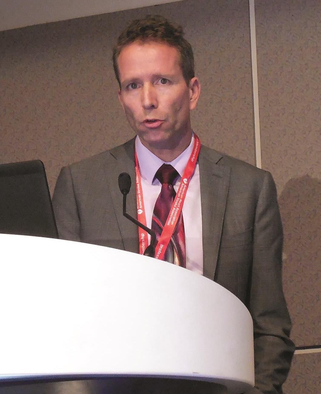
Nils H. Mueller-Kronast, MD, presented a modeled analysis of data collected in a registry on 236 real-world U.S. patients who underwent mechanical thrombectomy for an acute, large-vessel occlusion stroke following transfer from a hospital that could perform thrombolysis but couldn’t offer thrombectomy. The analysis showed that if the patients had instead gone directly to the closest thrombectomy center the result would have been a 16-percentage-point increase in patients with a modified Rankin Scale (mRS) score of 0-1 after 90 days, and a 9-percentage-point increase in mRS 0-2 outcomes, Dr. Mueller-Kronast said at the International Stroke Conference, sponsored by the American Heart Association.
The analysis he presented used data from the Systematic Evaluation of Patients Treated With Stroke Devices for Acute Ischemic Stroke (STRATIS) registry, which included 984 acute ischemic stroke patients who underwent mechanical thrombectomy at any one of 55 participating U.S. sites (Stroke. 2017 Oct;48[10]:2760-8). A previously-reported analysis of the STRATIS data showed that the 55% of patients taken directly to a center that performed thrombectomy had a 60% rate of mRS score 0-2 after 90 days, compared with 52% of patients taken first to a hospital unable to perform thrombectomy and then transferred (Circulation. 2017 Dec 12;136[24]:2311-21).
The current analysis focused on 236 of the transferred patients with complete information on their location at the time of their stroke and subsequent time intervals during their transport and treatment, including 117 patients with ground transfer from their first hospital to the thrombectomy site, 114 with air transfer, and 5 with an unreported means of transport.
Dr. Mueller-Kronast and his associates calculated the time it would have taken each of the 117 ground transported patients to have gone directly to the closest thrombectomy center (adjusted by traffic conditions at the time of the stroke), and modeled the likely outcomes of these patients based on the data collected in the registry. This projected a 47% rate of mRS scores 0-1 (good outcomes) after 90 days, and a 60% rate of mRS 0-2 scores with a direct-to-thrombectomy strategy, compared with actual rates of 31% and 51%, respectively, among the patients who were transferred from their initial hospital.
“Bypass to an endovascular-capable center may be an option to improve rapid access to mechanical thrombectomy,” he concluded.
The STRATIS registry is sponsored by Medtronic. Dr. Mueller-Kronast has been a consultant to Medtronic.
SOURCE: Mueller-Kronast N et al. Abstract LB12.
LOS ANGELES – Evidence continues to mount that in the new era of thrombectomy treatment for selected acute ischemic stroke patients outcomes are better when patients go directly to the closest comprehensive stroke center that offers intravascular procedures rather than first being taken to a closer hospital and then needing transfer.
Nils H. Mueller-Kronast, MD, presented a modeled analysis of data collected in a registry on 236 real-world U.S. patients who underwent mechanical thrombectomy for an acute, large-vessel occlusion stroke following transfer from a hospital that could perform thrombolysis but couldn’t offer thrombectomy. The analysis showed that if the patients had instead gone directly to the closest thrombectomy center the result would have been a 16-percentage-point increase in patients with a modified Rankin Scale (mRS) score of 0-1 after 90 days, and a 9-percentage-point increase in mRS 0-2 outcomes, Dr. Mueller-Kronast said at the International Stroke Conference, sponsored by the American Heart Association.
The analysis he presented used data from the Systematic Evaluation of Patients Treated With Stroke Devices for Acute Ischemic Stroke (STRATIS) registry, which included 984 acute ischemic stroke patients who underwent mechanical thrombectomy at any one of 55 participating U.S. sites (Stroke. 2017 Oct;48[10]:2760-8). A previously-reported analysis of the STRATIS data showed that the 55% of patients taken directly to a center that performed thrombectomy had a 60% rate of mRS score 0-2 after 90 days, compared with 52% of patients taken first to a hospital unable to perform thrombectomy and then transferred (Circulation. 2017 Dec 12;136[24]:2311-21).
The current analysis focused on 236 of the transferred patients with complete information on their location at the time of their stroke and subsequent time intervals during their transport and treatment, including 117 patients with ground transfer from their first hospital to the thrombectomy site, 114 with air transfer, and 5 with an unreported means of transport.
Dr. Mueller-Kronast and his associates calculated the time it would have taken each of the 117 ground transported patients to have gone directly to the closest thrombectomy center (adjusted by traffic conditions at the time of the stroke), and modeled the likely outcomes of these patients based on the data collected in the registry. This projected a 47% rate of mRS scores 0-1 (good outcomes) after 90 days, and a 60% rate of mRS 0-2 scores with a direct-to-thrombectomy strategy, compared with actual rates of 31% and 51%, respectively, among the patients who were transferred from their initial hospital.
“Bypass to an endovascular-capable center may be an option to improve rapid access to mechanical thrombectomy,” he concluded.
The STRATIS registry is sponsored by Medtronic. Dr. Mueller-Kronast has been a consultant to Medtronic.
SOURCE: Mueller-Kronast N et al. Abstract LB12.
LOS ANGELES – Evidence continues to mount that in the new era of thrombectomy treatment for selected acute ischemic stroke patients outcomes are better when patients go directly to the closest comprehensive stroke center that offers intravascular procedures rather than first being taken to a closer hospital and then needing transfer.
Nils H. Mueller-Kronast, MD, presented a modeled analysis of data collected in a registry on 236 real-world U.S. patients who underwent mechanical thrombectomy for an acute, large-vessel occlusion stroke following transfer from a hospital that could perform thrombolysis but couldn’t offer thrombectomy. The analysis showed that if the patients had instead gone directly to the closest thrombectomy center the result would have been a 16-percentage-point increase in patients with a modified Rankin Scale (mRS) score of 0-1 after 90 days, and a 9-percentage-point increase in mRS 0-2 outcomes, Dr. Mueller-Kronast said at the International Stroke Conference, sponsored by the American Heart Association.
The analysis he presented used data from the Systematic Evaluation of Patients Treated With Stroke Devices for Acute Ischemic Stroke (STRATIS) registry, which included 984 acute ischemic stroke patients who underwent mechanical thrombectomy at any one of 55 participating U.S. sites (Stroke. 2017 Oct;48[10]:2760-8). A previously-reported analysis of the STRATIS data showed that the 55% of patients taken directly to a center that performed thrombectomy had a 60% rate of mRS score 0-2 after 90 days, compared with 52% of patients taken first to a hospital unable to perform thrombectomy and then transferred (Circulation. 2017 Dec 12;136[24]:2311-21).
The current analysis focused on 236 of the transferred patients with complete information on their location at the time of their stroke and subsequent time intervals during their transport and treatment, including 117 patients with ground transfer from their first hospital to the thrombectomy site, 114 with air transfer, and 5 with an unreported means of transport.
Dr. Mueller-Kronast and his associates calculated the time it would have taken each of the 117 ground transported patients to have gone directly to the closest thrombectomy center (adjusted by traffic conditions at the time of the stroke), and modeled the likely outcomes of these patients based on the data collected in the registry. This projected a 47% rate of mRS scores 0-1 (good outcomes) after 90 days, and a 60% rate of mRS 0-2 scores with a direct-to-thrombectomy strategy, compared with actual rates of 31% and 51%, respectively, among the patients who were transferred from their initial hospital.
“Bypass to an endovascular-capable center may be an option to improve rapid access to mechanical thrombectomy,” he concluded.
The STRATIS registry is sponsored by Medtronic. Dr. Mueller-Kronast has been a consultant to Medtronic.
SOURCE: Mueller-Kronast N et al. Abstract LB12.
REPORTING FROM ISC 2018
Key clinical point: A direct-to-thrombectomy strategy maximizes good stroke outcomes.
Major finding: Modeling showed a 47% rate of good 90-day outcomes by taking patients to the closest thrombectomy center, compared with an actual 31% rate with transfers.
Study details: A simulation-model analysis of data collected by the STRATIS registry of acute stroke thrombectomies.
Disclosures: The STRATIS registry is sponsored by Medtronic. Dr. Mueller-Kronast has been a consultant to Medtronic.
Source: Mueller-Kronast N et al. Abstract LB12.
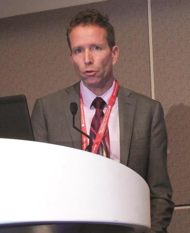
Nonmyeloablative conditioning gets a radiation boost for severe hemoglobinopathies
SALT LAKE CITY – A nonmyeloablative conditioning regimen with a boosted dose of total body irradiation yielded success for a cohort of patients with severe hemoglobinopathy and haploidentical donors.
Of 17 patients with severe sickle cell disease or beta-thalassemia who received allogeneic bone marrow transplants, all but one had successful engraftment, and 13 have achieved full donor chimerism, said Javier Bolaños-Meade, MD.
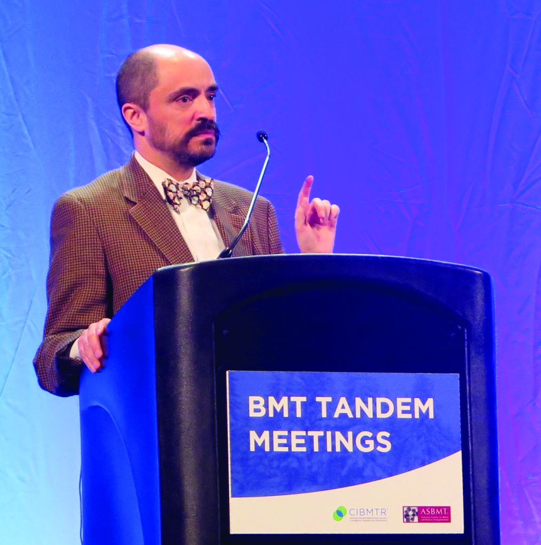
“Cure of severe hemoglobinopathies is now possible for most patients,” said Dr. Bolaños-Meade. “It should no longer be considered as available to only a fraction of such patients,” such as those who come with a fully-matched donor and those able to tolerate myeloablative conditioning, he said.
Of the patients who received bone marrow transplants, five patients have stopped immunosuppressive therapy, and all patients are alive, having been followed for a median of 15 months (range, 3-34 months).
The rate of graft versus host disease (GVHD) was low: Two patients developed grade 2 acute GVHD, and one patient developed grade 3 acute GVHD; another three patients had mild to moderate chronic GVHD, but all GVHD has resolved, said Dr. Bolaños-Meade.
Historically, the difficulties with transplant in this population were numerous. “No. 1, it’s very difficult to find an HLA-matched donor,” said Dr. Bolaños-Meade. Also, since there’s no target for graft-versus-tumor effect post-transplant, any amount of chronic GVHD is also high on the list of concerns when considering a transplant for hemoglobinopathy.
“The other problem in this group of patients is their ability to tolerate myeloablation,” he said. The accumulated burden of disease, as well as sequelae of transfusion dependence for some, may make a myeloablative regime too risky.
Dr. Bolaños-Meade said that he and his collaborators at Johns Hopkins University, Baltimore, wanted to be able to address all of these concerns in one regimen. “We were trying to work out a system that may be able to solve all the problems – to use nonmyeloablation and to use whatever donor is available.”
His research group had previously shown that nonmyeloablative transplants were well tolerated in patients with sickle cell disease and that haploidentical donors could be used (Blood. 2012 Nov 22;120[22]:4285-91). “However, we had a very high incidence of graft failure,” Dr. Bolaños-Meade said.
A strategy to increase the engraftment rate while still limiting toxicity, he said, would be to increase the dose of total body irradiation used in the conditioning regimen, from 200 to 400 centigray (cGy); this higher dose was incorporated into the study protocol.
Patients were enrolled if they had severe sickle cell disease (SCD; n = 12) or beta-thalassemia (n = 5).
To enroll in the study, SCD patients had to have been hospitalized at least twice a year in the preceding 2 years. The patients with SCD were a median 26 years of age (range, 6-31 years); four were male, and eight were female. Three of the SCD patients were transfusion dependent, and several had such serious complications as osteonecrosis, brain changes seen in medical imaging, and acute coronary syndrome.
The beta-thalassemia patients were a median 7 years of age (range, 6-16 years); all but one were female, and all had been transfusion dependent since infancy.
Bone marrow donors were not all first degree relatives: There were five mothers, four fathers, four brothers, and three sisters, but also an aunt. Two pairs had major ABO incompatibility, and five had minor ABO incompatibility. Ten were ABO compatible.
The conditioning regimen for all patients involved rabbit antithymocyte globulin, fludarabine, and cyclophosphamide, and then total body irradiation given the day before transplant.
After transplant, in addition to standard supportive care, patients received cyclophosphamide on days 3 and 4. Beginning on day 5, patients received mycophenolate mofetil through day 35 and sirolimus for 1 full year after transplant.
The antithymocyte globulin induced sickle cell crises in all SCD patients, and one patient developed sirolimus-induced diabetes. One other patient had a worsening of Meniere disease, and another patient developed BK virus cystitis.
Breaking down outcomes by disease type, Dr. Bolaños-Meade said that the one engraftment failure occurred in an SCD patient. Of the remaining 11 engrafted patients, 9 have full donor chimerism, and all but 1 of the 11 are transfusion independent now. The patient who remains transfusion dependent has mixed chimerism and received bone marrow from a donor with major ABO mismatch. Although one of the five beta-thalassemia patients also has mixed chimerism, all are now transfusion independent.
The boost in hemoglobin post-transplant was relatively modest for the beta-thalassemia group, from a median 9.5 to 10.1 g/dL at the most recent visit. However, the pretransplant levels were boosted by transfusions for all patients in this group, Dr. Bolaños-Meade pointed out.
The SCD patients saw their hemoglobin go from a median 8.65 to 11.4 g/dL (P = .001). Median bilirubin for this group dropped from 2.4 to 0.2 mg/dL (P = .002) with the cessation of sickling-related hemolysis; significant improvements were also seen in absolute reticulocyte counts and lactate dehydrogenase levels.
Dr. Bolaños-Meade reported that he is on the data safety monitoring board of Incyte.
SOURCE: Bolaños-Meade J et al. BMT Tandem Meetings, Abstract LBA-3.
SALT LAKE CITY – A nonmyeloablative conditioning regimen with a boosted dose of total body irradiation yielded success for a cohort of patients with severe hemoglobinopathy and haploidentical donors.
Of 17 patients with severe sickle cell disease or beta-thalassemia who received allogeneic bone marrow transplants, all but one had successful engraftment, and 13 have achieved full donor chimerism, said Javier Bolaños-Meade, MD.
“Cure of severe hemoglobinopathies is now possible for most patients,” said Dr. Bolaños-Meade. “It should no longer be considered as available to only a fraction of such patients,” such as those who come with a fully-matched donor and those able to tolerate myeloablative conditioning, he said.
Of the patients who received bone marrow transplants, five patients have stopped immunosuppressive therapy, and all patients are alive, having been followed for a median of 15 months (range, 3-34 months).
The rate of graft versus host disease (GVHD) was low: Two patients developed grade 2 acute GVHD, and one patient developed grade 3 acute GVHD; another three patients had mild to moderate chronic GVHD, but all GVHD has resolved, said Dr. Bolaños-Meade.
Historically, the difficulties with transplant in this population were numerous. “No. 1, it’s very difficult to find an HLA-matched donor,” said Dr. Bolaños-Meade. Also, since there’s no target for graft-versus-tumor effect post-transplant, any amount of chronic GVHD is also high on the list of concerns when considering a transplant for hemoglobinopathy.
“The other problem in this group of patients is their ability to tolerate myeloablation,” he said. The accumulated burden of disease, as well as sequelae of transfusion dependence for some, may make a myeloablative regime too risky.
Dr. Bolaños-Meade said that he and his collaborators at Johns Hopkins University, Baltimore, wanted to be able to address all of these concerns in one regimen. “We were trying to work out a system that may be able to solve all the problems – to use nonmyeloablation and to use whatever donor is available.”
His research group had previously shown that nonmyeloablative transplants were well tolerated in patients with sickle cell disease and that haploidentical donors could be used (Blood. 2012 Nov 22;120[22]:4285-91). “However, we had a very high incidence of graft failure,” Dr. Bolaños-Meade said.
A strategy to increase the engraftment rate while still limiting toxicity, he said, would be to increase the dose of total body irradiation used in the conditioning regimen, from 200 to 400 centigray (cGy); this higher dose was incorporated into the study protocol.
Patients were enrolled if they had severe sickle cell disease (SCD; n = 12) or beta-thalassemia (n = 5).
To enroll in the study, SCD patients had to have been hospitalized at least twice a year in the preceding 2 years. The patients with SCD were a median 26 years of age (range, 6-31 years); four were male, and eight were female. Three of the SCD patients were transfusion dependent, and several had such serious complications as osteonecrosis, brain changes seen in medical imaging, and acute coronary syndrome.
The beta-thalassemia patients were a median 7 years of age (range, 6-16 years); all but one were female, and all had been transfusion dependent since infancy.
Bone marrow donors were not all first degree relatives: There were five mothers, four fathers, four brothers, and three sisters, but also an aunt. Two pairs had major ABO incompatibility, and five had minor ABO incompatibility. Ten were ABO compatible.
The conditioning regimen for all patients involved rabbit antithymocyte globulin, fludarabine, and cyclophosphamide, and then total body irradiation given the day before transplant.
After transplant, in addition to standard supportive care, patients received cyclophosphamide on days 3 and 4. Beginning on day 5, patients received mycophenolate mofetil through day 35 and sirolimus for 1 full year after transplant.
The antithymocyte globulin induced sickle cell crises in all SCD patients, and one patient developed sirolimus-induced diabetes. One other patient had a worsening of Meniere disease, and another patient developed BK virus cystitis.
Breaking down outcomes by disease type, Dr. Bolaños-Meade said that the one engraftment failure occurred in an SCD patient. Of the remaining 11 engrafted patients, 9 have full donor chimerism, and all but 1 of the 11 are transfusion independent now. The patient who remains transfusion dependent has mixed chimerism and received bone marrow from a donor with major ABO mismatch. Although one of the five beta-thalassemia patients also has mixed chimerism, all are now transfusion independent.
The boost in hemoglobin post-transplant was relatively modest for the beta-thalassemia group, from a median 9.5 to 10.1 g/dL at the most recent visit. However, the pretransplant levels were boosted by transfusions for all patients in this group, Dr. Bolaños-Meade pointed out.
The SCD patients saw their hemoglobin go from a median 8.65 to 11.4 g/dL (P = .001). Median bilirubin for this group dropped from 2.4 to 0.2 mg/dL (P = .002) with the cessation of sickling-related hemolysis; significant improvements were also seen in absolute reticulocyte counts and lactate dehydrogenase levels.
Dr. Bolaños-Meade reported that he is on the data safety monitoring board of Incyte.
SOURCE: Bolaños-Meade J et al. BMT Tandem Meetings, Abstract LBA-3.
SALT LAKE CITY – A nonmyeloablative conditioning regimen with a boosted dose of total body irradiation yielded success for a cohort of patients with severe hemoglobinopathy and haploidentical donors.
Of 17 patients with severe sickle cell disease or beta-thalassemia who received allogeneic bone marrow transplants, all but one had successful engraftment, and 13 have achieved full donor chimerism, said Javier Bolaños-Meade, MD.
“Cure of severe hemoglobinopathies is now possible for most patients,” said Dr. Bolaños-Meade. “It should no longer be considered as available to only a fraction of such patients,” such as those who come with a fully-matched donor and those able to tolerate myeloablative conditioning, he said.
Of the patients who received bone marrow transplants, five patients have stopped immunosuppressive therapy, and all patients are alive, having been followed for a median of 15 months (range, 3-34 months).
The rate of graft versus host disease (GVHD) was low: Two patients developed grade 2 acute GVHD, and one patient developed grade 3 acute GVHD; another three patients had mild to moderate chronic GVHD, but all GVHD has resolved, said Dr. Bolaños-Meade.
Historically, the difficulties with transplant in this population were numerous. “No. 1, it’s very difficult to find an HLA-matched donor,” said Dr. Bolaños-Meade. Also, since there’s no target for graft-versus-tumor effect post-transplant, any amount of chronic GVHD is also high on the list of concerns when considering a transplant for hemoglobinopathy.
“The other problem in this group of patients is their ability to tolerate myeloablation,” he said. The accumulated burden of disease, as well as sequelae of transfusion dependence for some, may make a myeloablative regime too risky.
Dr. Bolaños-Meade said that he and his collaborators at Johns Hopkins University, Baltimore, wanted to be able to address all of these concerns in one regimen. “We were trying to work out a system that may be able to solve all the problems – to use nonmyeloablation and to use whatever donor is available.”
His research group had previously shown that nonmyeloablative transplants were well tolerated in patients with sickle cell disease and that haploidentical donors could be used (Blood. 2012 Nov 22;120[22]:4285-91). “However, we had a very high incidence of graft failure,” Dr. Bolaños-Meade said.
A strategy to increase the engraftment rate while still limiting toxicity, he said, would be to increase the dose of total body irradiation used in the conditioning regimen, from 200 to 400 centigray (cGy); this higher dose was incorporated into the study protocol.
Patients were enrolled if they had severe sickle cell disease (SCD; n = 12) or beta-thalassemia (n = 5).
To enroll in the study, SCD patients had to have been hospitalized at least twice a year in the preceding 2 years. The patients with SCD were a median 26 years of age (range, 6-31 years); four were male, and eight were female. Three of the SCD patients were transfusion dependent, and several had such serious complications as osteonecrosis, brain changes seen in medical imaging, and acute coronary syndrome.
The beta-thalassemia patients were a median 7 years of age (range, 6-16 years); all but one were female, and all had been transfusion dependent since infancy.
Bone marrow donors were not all first degree relatives: There were five mothers, four fathers, four brothers, and three sisters, but also an aunt. Two pairs had major ABO incompatibility, and five had minor ABO incompatibility. Ten were ABO compatible.
The conditioning regimen for all patients involved rabbit antithymocyte globulin, fludarabine, and cyclophosphamide, and then total body irradiation given the day before transplant.
After transplant, in addition to standard supportive care, patients received cyclophosphamide on days 3 and 4. Beginning on day 5, patients received mycophenolate mofetil through day 35 and sirolimus for 1 full year after transplant.
The antithymocyte globulin induced sickle cell crises in all SCD patients, and one patient developed sirolimus-induced diabetes. One other patient had a worsening of Meniere disease, and another patient developed BK virus cystitis.
Breaking down outcomes by disease type, Dr. Bolaños-Meade said that the one engraftment failure occurred in an SCD patient. Of the remaining 11 engrafted patients, 9 have full donor chimerism, and all but 1 of the 11 are transfusion independent now. The patient who remains transfusion dependent has mixed chimerism and received bone marrow from a donor with major ABO mismatch. Although one of the five beta-thalassemia patients also has mixed chimerism, all are now transfusion independent.
The boost in hemoglobin post-transplant was relatively modest for the beta-thalassemia group, from a median 9.5 to 10.1 g/dL at the most recent visit. However, the pretransplant levels were boosted by transfusions for all patients in this group, Dr. Bolaños-Meade pointed out.
The SCD patients saw their hemoglobin go from a median 8.65 to 11.4 g/dL (P = .001). Median bilirubin for this group dropped from 2.4 to 0.2 mg/dL (P = .002) with the cessation of sickling-related hemolysis; significant improvements were also seen in absolute reticulocyte counts and lactate dehydrogenase levels.
Dr. Bolaños-Meade reported that he is on the data safety monitoring board of Incyte.
SOURCE: Bolaños-Meade J et al. BMT Tandem Meetings, Abstract LBA-3.
REPORTING FROM THE 2018 BMT TANDEM MEETINGS
Key clinical point:
Major finding: Of the 17 patients who received haploidentical bone marrow transplant, 13 have achieved full chimerism.
Study details: Report of 17 consecutive patients with severe sickle cell disease or beta-thalassemia who received nonmyeloablative conditioning and bone marrow transplant from haploidentical donors.
Disclosures: Dr. Bolaños-Meade reported no outside sources of funding for the study. He is on the data safety monitoring board of Incyte.
Source: Bolaños-Meade J et al. 2018 BMT Tandem Meetings, Abstract LBA-3.
Consider steroid-induced hypertension when treating pediatric ALL
Nearly 15% of children undergoing induction therapy for acute lymphoblastic leukemia (ALL) developed and were treated for steroid-induced hypertension, according to a study conducted by researchers at the Ohio State University in Columbus.
Ian Bakk and his associates performed a retrospective review of data from the Pediatric Health Information System, a database of the Child Health Corporation of America consisting of inpatient information from 40 free-standing children’s hospitals in the United States. They looked at new cases of ALL from the period of 2009-2013 and analyzed data from 5,578 children who received induction chemotherapy for ALL. In all, .
An adjusted regression analysis showed that infants less than 1 year of age had the highest odds of developing steroid-induced hypertension (adjusted odds ratio, 4.05), followed by children with abnormal glucose (aOR, 2.09), those with secondary diabetes mellitus (aOR, 1.67), and obese patients (aOR, 1.63).
“These findings can help physicians identify patients at high risk for [hypertension] at the time of ALL diagnosis,” the researchers wrote.
mschneider@frontlinemedcom.com
SOURCE: Bakk, I et al. Am J Pediatr Hematol Oncol. 2018;40:27-30.
Nearly 15% of children undergoing induction therapy for acute lymphoblastic leukemia (ALL) developed and were treated for steroid-induced hypertension, according to a study conducted by researchers at the Ohio State University in Columbus.
Ian Bakk and his associates performed a retrospective review of data from the Pediatric Health Information System, a database of the Child Health Corporation of America consisting of inpatient information from 40 free-standing children’s hospitals in the United States. They looked at new cases of ALL from the period of 2009-2013 and analyzed data from 5,578 children who received induction chemotherapy for ALL. In all, .
An adjusted regression analysis showed that infants less than 1 year of age had the highest odds of developing steroid-induced hypertension (adjusted odds ratio, 4.05), followed by children with abnormal glucose (aOR, 2.09), those with secondary diabetes mellitus (aOR, 1.67), and obese patients (aOR, 1.63).
“These findings can help physicians identify patients at high risk for [hypertension] at the time of ALL diagnosis,” the researchers wrote.
mschneider@frontlinemedcom.com
SOURCE: Bakk, I et al. Am J Pediatr Hematol Oncol. 2018;40:27-30.
Nearly 15% of children undergoing induction therapy for acute lymphoblastic leukemia (ALL) developed and were treated for steroid-induced hypertension, according to a study conducted by researchers at the Ohio State University in Columbus.
Ian Bakk and his associates performed a retrospective review of data from the Pediatric Health Information System, a database of the Child Health Corporation of America consisting of inpatient information from 40 free-standing children’s hospitals in the United States. They looked at new cases of ALL from the period of 2009-2013 and analyzed data from 5,578 children who received induction chemotherapy for ALL. In all, .
An adjusted regression analysis showed that infants less than 1 year of age had the highest odds of developing steroid-induced hypertension (adjusted odds ratio, 4.05), followed by children with abnormal glucose (aOR, 2.09), those with secondary diabetes mellitus (aOR, 1.67), and obese patients (aOR, 1.63).
“These findings can help physicians identify patients at high risk for [hypertension] at the time of ALL diagnosis,” the researchers wrote.
mschneider@frontlinemedcom.com
SOURCE: Bakk, I et al. Am J Pediatr Hematol Oncol. 2018;40:27-30.