User login
Vaccine priming determines teen susceptibility to pertussis
It is the initial type of pertussis vaccine given in infancy – acellular or whole cell – that primes the immune system and determines how soon adolescents become susceptible to pertussis, regardless of later acellular booster vaccination, noted the authors of a new study.
The IgG4 subclass proportion for IgG4-specific antibodies remained lower in patients who had whole-cell pertussis (wP) priming in infancy, even though they had received acellular pertussis booster vaccinations at ages 4 and 9 years, compared with patients who underwent acellular pertussis (aP) priming in infancy. This was true for all vaccine antigens, other than filamentous hemagglutinin and tetanus, 1 year after the preadolescent booster, noted researcher Saskia van der Lee of the National Institute for Public Health and the Environment, Bilthoven, The Netherlands, and her associates.
“ compared to aP-primed children, even after booster vaccinations. This is in line with epidemiological data indicating that adolescents, after aP vaccination in infancy, are more susceptible to pertussis, compared with wP-primed adolescents, though wP-primed individuals become also susceptible over time,” the researchers said.
In addition, children primed with DTwP vaccines have a more Th1-skewed response for pertussis vaccine antigens after receiving a DTap booster vaccine or clinical infection with Bordetella pertussis, whereas children primed with DTaP have a more mixed pertussis-specific Th1/Th2 response, the researchers said. So “new adjuvants that skew the immune response towards a Th1 profile are desired” for better protection against pertussis over time.
SOURCE: van der Lee S et al. Vaccine. 2018 Jan 4;36(2):220-6.
It is the initial type of pertussis vaccine given in infancy – acellular or whole cell – that primes the immune system and determines how soon adolescents become susceptible to pertussis, regardless of later acellular booster vaccination, noted the authors of a new study.
The IgG4 subclass proportion for IgG4-specific antibodies remained lower in patients who had whole-cell pertussis (wP) priming in infancy, even though they had received acellular pertussis booster vaccinations at ages 4 and 9 years, compared with patients who underwent acellular pertussis (aP) priming in infancy. This was true for all vaccine antigens, other than filamentous hemagglutinin and tetanus, 1 year after the preadolescent booster, noted researcher Saskia van der Lee of the National Institute for Public Health and the Environment, Bilthoven, The Netherlands, and her associates.
“ compared to aP-primed children, even after booster vaccinations. This is in line with epidemiological data indicating that adolescents, after aP vaccination in infancy, are more susceptible to pertussis, compared with wP-primed adolescents, though wP-primed individuals become also susceptible over time,” the researchers said.
In addition, children primed with DTwP vaccines have a more Th1-skewed response for pertussis vaccine antigens after receiving a DTap booster vaccine or clinical infection with Bordetella pertussis, whereas children primed with DTaP have a more mixed pertussis-specific Th1/Th2 response, the researchers said. So “new adjuvants that skew the immune response towards a Th1 profile are desired” for better protection against pertussis over time.
SOURCE: van der Lee S et al. Vaccine. 2018 Jan 4;36(2):220-6.
It is the initial type of pertussis vaccine given in infancy – acellular or whole cell – that primes the immune system and determines how soon adolescents become susceptible to pertussis, regardless of later acellular booster vaccination, noted the authors of a new study.
The IgG4 subclass proportion for IgG4-specific antibodies remained lower in patients who had whole-cell pertussis (wP) priming in infancy, even though they had received acellular pertussis booster vaccinations at ages 4 and 9 years, compared with patients who underwent acellular pertussis (aP) priming in infancy. This was true for all vaccine antigens, other than filamentous hemagglutinin and tetanus, 1 year after the preadolescent booster, noted researcher Saskia van der Lee of the National Institute for Public Health and the Environment, Bilthoven, The Netherlands, and her associates.
“ compared to aP-primed children, even after booster vaccinations. This is in line with epidemiological data indicating that adolescents, after aP vaccination in infancy, are more susceptible to pertussis, compared with wP-primed adolescents, though wP-primed individuals become also susceptible over time,” the researchers said.
In addition, children primed with DTwP vaccines have a more Th1-skewed response for pertussis vaccine antigens after receiving a DTap booster vaccine or clinical infection with Bordetella pertussis, whereas children primed with DTaP have a more mixed pertussis-specific Th1/Th2 response, the researchers said. So “new adjuvants that skew the immune response towards a Th1 profile are desired” for better protection against pertussis over time.
SOURCE: van der Lee S et al. Vaccine. 2018 Jan 4;36(2):220-6.
FROM VACCINE
Congenital Heart Disease Is Associated With a Higher Risk of Dementia
Congenital heart disease (CHD) is associated with an increased risk of dementia, especially early onset dementia, according to research published online ahead of print February 12 in Circulation. A particularly increased risk of dementia was observed among middle-aged adults with CHD and signs of early onset dementia, the authors noted.
“These results support the importance of understanding the risk of adverse long-term neurologic outcomes in the growing and aging population with CHD,” said Carina N. Bagge, a medical student at Aarhus University Hospital in Aarhus, Denmark, and colleagues.

Examining Dementia Risk in Patients With CHD
CHD occurs in six to 10 per 1,000 live births and represents the most common group of congenital defects. Dementia is the sixth most common cause of death in the United States. Although neurodevelopmental deficits among infants and children with CHD are well studied, research on long-term neurologic outcomes in adults with CHD is limited. Based on the increased incidence of neurodevelopmental impairments and associated risk factors for dementia among patients with CHD, Ms. Bagge and colleagues hypothesized that the risk of dementia is higher in adults with CHD than in the general population.
To test this hypothesis, the authors performed a nationwide, population-based cohort study in Denmark using linked medical registries that included data from all Danish hospitals. They identified all adults who received a diagnosis of CHD between 1963 and 1974 (before age 15) and between 1977 and 2012 (at any age). For each patient with CHD, 10 individuals from the general population were matched by sex and birth year,
The primary outcome was a first-time hospital diagnosis of all-cause dementia in the inpatient or outpatient clinic setting. Investigators categorized dementia diagnoses as Alzheimer’s disease, vascular dementia, or other dementias.
CHD Severity Correlated With Increased Risk of Dementia
The investigators identified 10,632 adults with CHD and 103,403 adults from the general population;46% were male in both cohorts. The cumulative incidence of dementia was 4% at age 80 in both cohorts. The overall hazard ratio (HR) of dementia was 1.6 among adults with CHD, compared with the general population. The HR of dementia was 1.4 among patients with CHD without extracardiac defects, 1.5 among patients with mild-to-moderate CHD, and 2.0 for patients with severe CHD, including univentricular hearts. Finally, the HR for early-onset dementia (ie, onset younger than age 65) was 2.6, and the HR for late-onset dementia was 1.3 among patients with CHD.
“These results cannot be directly applied to young adults diagnosed with CHD in the present eras. However, it is important to recognize healthcare needs and risk factors affecting the larger number of middle-aged and older adults currently living with CHD,” said Ms. Bagge and colleagues.
—Erica Tricarico
Suggested Reading
Bagge CN, Henderson VW, Laursen HB, et al. Risk of dementia in adults with congenital heart disease: population-based cohort study. Circulation. 2018 Feb 12 [Epub ahead of print].
Congenital heart disease (CHD) is associated with an increased risk of dementia, especially early onset dementia, according to research published online ahead of print February 12 in Circulation. A particularly increased risk of dementia was observed among middle-aged adults with CHD and signs of early onset dementia, the authors noted.
“These results support the importance of understanding the risk of adverse long-term neurologic outcomes in the growing and aging population with CHD,” said Carina N. Bagge, a medical student at Aarhus University Hospital in Aarhus, Denmark, and colleagues.
Examining Dementia Risk in Patients With CHD
CHD occurs in six to 10 per 1,000 live births and represents the most common group of congenital defects. Dementia is the sixth most common cause of death in the United States. Although neurodevelopmental deficits among infants and children with CHD are well studied, research on long-term neurologic outcomes in adults with CHD is limited. Based on the increased incidence of neurodevelopmental impairments and associated risk factors for dementia among patients with CHD, Ms. Bagge and colleagues hypothesized that the risk of dementia is higher in adults with CHD than in the general population.
To test this hypothesis, the authors performed a nationwide, population-based cohort study in Denmark using linked medical registries that included data from all Danish hospitals. They identified all adults who received a diagnosis of CHD between 1963 and 1974 (before age 15) and between 1977 and 2012 (at any age). For each patient with CHD, 10 individuals from the general population were matched by sex and birth year,
The primary outcome was a first-time hospital diagnosis of all-cause dementia in the inpatient or outpatient clinic setting. Investigators categorized dementia diagnoses as Alzheimer’s disease, vascular dementia, or other dementias.
CHD Severity Correlated With Increased Risk of Dementia
The investigators identified 10,632 adults with CHD and 103,403 adults from the general population;46% were male in both cohorts. The cumulative incidence of dementia was 4% at age 80 in both cohorts. The overall hazard ratio (HR) of dementia was 1.6 among adults with CHD, compared with the general population. The HR of dementia was 1.4 among patients with CHD without extracardiac defects, 1.5 among patients with mild-to-moderate CHD, and 2.0 for patients with severe CHD, including univentricular hearts. Finally, the HR for early-onset dementia (ie, onset younger than age 65) was 2.6, and the HR for late-onset dementia was 1.3 among patients with CHD.
“These results cannot be directly applied to young adults diagnosed with CHD in the present eras. However, it is important to recognize healthcare needs and risk factors affecting the larger number of middle-aged and older adults currently living with CHD,” said Ms. Bagge and colleagues.
—Erica Tricarico
Suggested Reading
Bagge CN, Henderson VW, Laursen HB, et al. Risk of dementia in adults with congenital heart disease: population-based cohort study. Circulation. 2018 Feb 12 [Epub ahead of print].
Congenital heart disease (CHD) is associated with an increased risk of dementia, especially early onset dementia, according to research published online ahead of print February 12 in Circulation. A particularly increased risk of dementia was observed among middle-aged adults with CHD and signs of early onset dementia, the authors noted.
“These results support the importance of understanding the risk of adverse long-term neurologic outcomes in the growing and aging population with CHD,” said Carina N. Bagge, a medical student at Aarhus University Hospital in Aarhus, Denmark, and colleagues.
Examining Dementia Risk in Patients With CHD
CHD occurs in six to 10 per 1,000 live births and represents the most common group of congenital defects. Dementia is the sixth most common cause of death in the United States. Although neurodevelopmental deficits among infants and children with CHD are well studied, research on long-term neurologic outcomes in adults with CHD is limited. Based on the increased incidence of neurodevelopmental impairments and associated risk factors for dementia among patients with CHD, Ms. Bagge and colleagues hypothesized that the risk of dementia is higher in adults with CHD than in the general population.
To test this hypothesis, the authors performed a nationwide, population-based cohort study in Denmark using linked medical registries that included data from all Danish hospitals. They identified all adults who received a diagnosis of CHD between 1963 and 1974 (before age 15) and between 1977 and 2012 (at any age). For each patient with CHD, 10 individuals from the general population were matched by sex and birth year,
The primary outcome was a first-time hospital diagnosis of all-cause dementia in the inpatient or outpatient clinic setting. Investigators categorized dementia diagnoses as Alzheimer’s disease, vascular dementia, or other dementias.
CHD Severity Correlated With Increased Risk of Dementia
The investigators identified 10,632 adults with CHD and 103,403 adults from the general population;46% were male in both cohorts. The cumulative incidence of dementia was 4% at age 80 in both cohorts. The overall hazard ratio (HR) of dementia was 1.6 among adults with CHD, compared with the general population. The HR of dementia was 1.4 among patients with CHD without extracardiac defects, 1.5 among patients with mild-to-moderate CHD, and 2.0 for patients with severe CHD, including univentricular hearts. Finally, the HR for early-onset dementia (ie, onset younger than age 65) was 2.6, and the HR for late-onset dementia was 1.3 among patients with CHD.
“These results cannot be directly applied to young adults diagnosed with CHD in the present eras. However, it is important to recognize healthcare needs and risk factors affecting the larger number of middle-aged and older adults currently living with CHD,” said Ms. Bagge and colleagues.
—Erica Tricarico
Suggested Reading
Bagge CN, Henderson VW, Laursen HB, et al. Risk of dementia in adults with congenital heart disease: population-based cohort study. Circulation. 2018 Feb 12 [Epub ahead of print].

Personalizing Seizure Mapping with a Computer Algorithm
A software algorithm has the potential to increase the likelihood of achieving freedom from seizures after epilepsy surgery suggests an analysis of 3 illustrative case studies.
- Researchers from the University of South Florida created the algorithm to individualize the mapping of patients’ epilepsy networks.
- The computer program combines noninvasive EEG source localization with nonconcurrent resting state functional MRI.
- Investigators used scalp EEG and resting state fMRI to gather data on a healthy control subject, a patient with right temporal lobe epilepsy (TLE), and one with bitemporal seizure onset.
- Results from the healthy subject found symmetrical global connectivity.
- The patient with right TLE had asymmetry in global connectivity and very little connectivity ipsolaterial to the epileptogenic cortex.
- The patient who had bitemporal lobe epilepsy had symmetrical global connectivity and a network of correlating activity in the cortex that was localized in the epileptogenic tissue of both temporal lobes.
Neal EG, Maciver S, Vale FL. Multimodal, noninvasive seizure network mapping software: A novel tool for preoperative epilepsy evaluation. Epilepsy Behav. 2018; 81:25-32. https://doi.org/10.1016/j.yebeh.2018.01.033
A software algorithm has the potential to increase the likelihood of achieving freedom from seizures after epilepsy surgery suggests an analysis of 3 illustrative case studies.
- Researchers from the University of South Florida created the algorithm to individualize the mapping of patients’ epilepsy networks.
- The computer program combines noninvasive EEG source localization with nonconcurrent resting state functional MRI.
- Investigators used scalp EEG and resting state fMRI to gather data on a healthy control subject, a patient with right temporal lobe epilepsy (TLE), and one with bitemporal seizure onset.
- Results from the healthy subject found symmetrical global connectivity.
- The patient with right TLE had asymmetry in global connectivity and very little connectivity ipsolaterial to the epileptogenic cortex.
- The patient who had bitemporal lobe epilepsy had symmetrical global connectivity and a network of correlating activity in the cortex that was localized in the epileptogenic tissue of both temporal lobes.
Neal EG, Maciver S, Vale FL. Multimodal, noninvasive seizure network mapping software: A novel tool for preoperative epilepsy evaluation. Epilepsy Behav. 2018; 81:25-32. https://doi.org/10.1016/j.yebeh.2018.01.033
A software algorithm has the potential to increase the likelihood of achieving freedom from seizures after epilepsy surgery suggests an analysis of 3 illustrative case studies.
- Researchers from the University of South Florida created the algorithm to individualize the mapping of patients’ epilepsy networks.
- The computer program combines noninvasive EEG source localization with nonconcurrent resting state functional MRI.
- Investigators used scalp EEG and resting state fMRI to gather data on a healthy control subject, a patient with right temporal lobe epilepsy (TLE), and one with bitemporal seizure onset.
- Results from the healthy subject found symmetrical global connectivity.
- The patient with right TLE had asymmetry in global connectivity and very little connectivity ipsolaterial to the epileptogenic cortex.
- The patient who had bitemporal lobe epilepsy had symmetrical global connectivity and a network of correlating activity in the cortex that was localized in the epileptogenic tissue of both temporal lobes.
Neal EG, Maciver S, Vale FL. Multimodal, noninvasive seizure network mapping software: A novel tool for preoperative epilepsy evaluation. Epilepsy Behav. 2018; 81:25-32. https://doi.org/10.1016/j.yebeh.2018.01.033
Using Quantitative EEG Spectrograms to Detect Seizure Subtypes
Using quantitative EEG (QEEG) can help differentiate among several types of seizures suggests an analysis of 562 seizures among 58 patitents.
- Investigators from Montefiore Medical Center and Albert Einstein College of Medicine in New York evaluated the sensitivity of the Persyst 12 QEEG spectrograms for detecting focal, focal with secondary generalized, and generalized onset seizures.
- Patients included in the analysis had 2 or more seizures successfully recorded during continuous EEG monitoring in the ICU or epilepsy monitoring unit from July 2016 to January 2017.
- QEEG spectrograms detected seizures with a sensitivity of 43% to 72%.
- Asymmetry spectrograms generated the highest sensitivity for detecting local seizures (94%).
- FFT spectrograms were most sensitive for diagnosing secondarily generalized seizures (84%).
- Seizure detection trend was most sensitive for generalized onset seizures (79%).
Goenka A, Boro A, Yozawitz E. Comparative sensitivity of quantitative EEG (QEEG) spectrograms for detecting seizure subtypes. Seizures. 2018;55:70-75.
Using quantitative EEG (QEEG) can help differentiate among several types of seizures suggests an analysis of 562 seizures among 58 patitents.
- Investigators from Montefiore Medical Center and Albert Einstein College of Medicine in New York evaluated the sensitivity of the Persyst 12 QEEG spectrograms for detecting focal, focal with secondary generalized, and generalized onset seizures.
- Patients included in the analysis had 2 or more seizures successfully recorded during continuous EEG monitoring in the ICU or epilepsy monitoring unit from July 2016 to January 2017.
- QEEG spectrograms detected seizures with a sensitivity of 43% to 72%.
- Asymmetry spectrograms generated the highest sensitivity for detecting local seizures (94%).
- FFT spectrograms were most sensitive for diagnosing secondarily generalized seizures (84%).
- Seizure detection trend was most sensitive for generalized onset seizures (79%).
Goenka A, Boro A, Yozawitz E. Comparative sensitivity of quantitative EEG (QEEG) spectrograms for detecting seizure subtypes. Seizures. 2018;55:70-75.
Using quantitative EEG (QEEG) can help differentiate among several types of seizures suggests an analysis of 562 seizures among 58 patitents.
- Investigators from Montefiore Medical Center and Albert Einstein College of Medicine in New York evaluated the sensitivity of the Persyst 12 QEEG spectrograms for detecting focal, focal with secondary generalized, and generalized onset seizures.
- Patients included in the analysis had 2 or more seizures successfully recorded during continuous EEG monitoring in the ICU or epilepsy monitoring unit from July 2016 to January 2017.
- QEEG spectrograms detected seizures with a sensitivity of 43% to 72%.
- Asymmetry spectrograms generated the highest sensitivity for detecting local seizures (94%).
- FFT spectrograms were most sensitive for diagnosing secondarily generalized seizures (84%).
- Seizure detection trend was most sensitive for generalized onset seizures (79%).
Goenka A, Boro A, Yozawitz E. Comparative sensitivity of quantitative EEG (QEEG) spectrograms for detecting seizure subtypes. Seizures. 2018;55:70-75.
Cognitive Impairment Linked to Depression in Epilepsy
Cognitive impairment affects 40% of adult patients with epilepsy and is associated with mental depression, seizure frequency, and the number of adverse effects of antiepileptic drugs, according to an analysis of a Cleveland Clinic database.
- Adults with epilepsy enrolled in the Cleveland Clinic Knowledge Program Data Registry from January to May 2015 were included in the analysis.
- The patients had completed several assessment scales, including the Aldenkamp–Baker Neuropsychological Assessment Schedule (ABNAS), the Patient Health Questionnaire (PHQ-9), the Generalized Anxiety Disorder scale (GAD-7), and the Quality of Life in Epilepsy questionnaire.
- Since topiramate poses a high risk of cognitive impairment, it was used as the basis for the evaluation.
- 270 of 670 patients (40%) reported cognitive impairment, with PHQ-9 scores, the number of adverse drug effects and the frequency of seizures most closely associated with such impairment., suggesting that these three variables could be used as predictors.
Feldman L, Lapin B, Busch RM, et al. Evaluating subjective cognitive impairment in the adult epilepsy clinic: Effects of depression, number of antiepileptic medications, and seizure frequency. Epilepsy Behav. 2018; 81:18-24.
Cognitive impairment affects 40% of adult patients with epilepsy and is associated with mental depression, seizure frequency, and the number of adverse effects of antiepileptic drugs, according to an analysis of a Cleveland Clinic database.
- Adults with epilepsy enrolled in the Cleveland Clinic Knowledge Program Data Registry from January to May 2015 were included in the analysis.
- The patients had completed several assessment scales, including the Aldenkamp–Baker Neuropsychological Assessment Schedule (ABNAS), the Patient Health Questionnaire (PHQ-9), the Generalized Anxiety Disorder scale (GAD-7), and the Quality of Life in Epilepsy questionnaire.
- Since topiramate poses a high risk of cognitive impairment, it was used as the basis for the evaluation.
- 270 of 670 patients (40%) reported cognitive impairment, with PHQ-9 scores, the number of adverse drug effects and the frequency of seizures most closely associated with such impairment., suggesting that these three variables could be used as predictors.
Feldman L, Lapin B, Busch RM, et al. Evaluating subjective cognitive impairment in the adult epilepsy clinic: Effects of depression, number of antiepileptic medications, and seizure frequency. Epilepsy Behav. 2018; 81:18-24.
Cognitive impairment affects 40% of adult patients with epilepsy and is associated with mental depression, seizure frequency, and the number of adverse effects of antiepileptic drugs, according to an analysis of a Cleveland Clinic database.
- Adults with epilepsy enrolled in the Cleveland Clinic Knowledge Program Data Registry from January to May 2015 were included in the analysis.
- The patients had completed several assessment scales, including the Aldenkamp–Baker Neuropsychological Assessment Schedule (ABNAS), the Patient Health Questionnaire (PHQ-9), the Generalized Anxiety Disorder scale (GAD-7), and the Quality of Life in Epilepsy questionnaire.
- Since topiramate poses a high risk of cognitive impairment, it was used as the basis for the evaluation.
- 270 of 670 patients (40%) reported cognitive impairment, with PHQ-9 scores, the number of adverse drug effects and the frequency of seizures most closely associated with such impairment., suggesting that these three variables could be used as predictors.
Feldman L, Lapin B, Busch RM, et al. Evaluating subjective cognitive impairment in the adult epilepsy clinic: Effects of depression, number of antiepileptic medications, and seizure frequency. Epilepsy Behav. 2018; 81:18-24.
Time to rethink MCL treatment, trial design
Classic mantle cell lymphoma (cMCL) has long been treated as a uniformly aggressive disease in need of similarly aggressive treatment, but that approach may be leading to overtreatment, according to one expert.
“The cMCL encompasses a broad category of lymphomas with highly variable clinical behaviors. A contemporary categorization of cMCL as a predominantly aggressive entity is misleading, as only an estimated 20% to 25% of patients with cMCL present with a symptomatic or aggressively behaving disease,” Leonid L. Yavorkovsky, MD, PhD, of the Kaiser Permanente San Jose Medical Center (Calif.), wrote in a commentary in JAMA Oncology.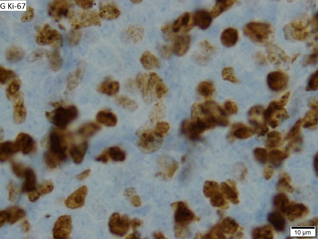
While groups like the National Comprehensive Cancer Network and the European Society for Medical Oncology recommend that only older patients and those with significant comorbidities should be exempted from intensive therapy, Dr. Yavorkovsky said to identify asymptomatic patients who might be able to safely delay aggressive treatment. He also called for more risk-adapted patient assignment to cMCL clinical trials.
“The failure to recognize the erratic nature of cMCL in clinical studies may confound the outcome gains and, ultimately, undermine the ensuing treatment recommendations,” Dr. Yavorkovsky wrote.
Read his full commentary in JAMA Oncology.
mschneider@frontlinemedcom.com
SOURCE: Yavorkovsky L, JAMA Oncology. 2018 Mar 1. doi: 10.1001/jamaoncol.2017.5685.
Classic mantle cell lymphoma (cMCL) has long been treated as a uniformly aggressive disease in need of similarly aggressive treatment, but that approach may be leading to overtreatment, according to one expert.
“The cMCL encompasses a broad category of lymphomas with highly variable clinical behaviors. A contemporary categorization of cMCL as a predominantly aggressive entity is misleading, as only an estimated 20% to 25% of patients with cMCL present with a symptomatic or aggressively behaving disease,” Leonid L. Yavorkovsky, MD, PhD, of the Kaiser Permanente San Jose Medical Center (Calif.), wrote in a commentary in JAMA Oncology.
While groups like the National Comprehensive Cancer Network and the European Society for Medical Oncology recommend that only older patients and those with significant comorbidities should be exempted from intensive therapy, Dr. Yavorkovsky said to identify asymptomatic patients who might be able to safely delay aggressive treatment. He also called for more risk-adapted patient assignment to cMCL clinical trials.
“The failure to recognize the erratic nature of cMCL in clinical studies may confound the outcome gains and, ultimately, undermine the ensuing treatment recommendations,” Dr. Yavorkovsky wrote.
Read his full commentary in JAMA Oncology.
mschneider@frontlinemedcom.com
SOURCE: Yavorkovsky L, JAMA Oncology. 2018 Mar 1. doi: 10.1001/jamaoncol.2017.5685.
Classic mantle cell lymphoma (cMCL) has long been treated as a uniformly aggressive disease in need of similarly aggressive treatment, but that approach may be leading to overtreatment, according to one expert.
“The cMCL encompasses a broad category of lymphomas with highly variable clinical behaviors. A contemporary categorization of cMCL as a predominantly aggressive entity is misleading, as only an estimated 20% to 25% of patients with cMCL present with a symptomatic or aggressively behaving disease,” Leonid L. Yavorkovsky, MD, PhD, of the Kaiser Permanente San Jose Medical Center (Calif.), wrote in a commentary in JAMA Oncology.
While groups like the National Comprehensive Cancer Network and the European Society for Medical Oncology recommend that only older patients and those with significant comorbidities should be exempted from intensive therapy, Dr. Yavorkovsky said to identify asymptomatic patients who might be able to safely delay aggressive treatment. He also called for more risk-adapted patient assignment to cMCL clinical trials.
“The failure to recognize the erratic nature of cMCL in clinical studies may confound the outcome gains and, ultimately, undermine the ensuing treatment recommendations,” Dr. Yavorkovsky wrote.
Read his full commentary in JAMA Oncology.
mschneider@frontlinemedcom.com
SOURCE: Yavorkovsky L, JAMA Oncology. 2018 Mar 1. doi: 10.1001/jamaoncol.2017.5685.
FROM JAMA ONCOLOGY
CPT and relative value changes that may affect reimbursement to your ObGyn practice
Another year brings changes to Current Procedural Terminology (CPT) codes (which are developed and copyrighted by the American Medical Association) in the form of additions and revisions, and payments related to resource-based relative value scale (RBRVS) revisions for selected services. As of January 1, 2018, 2 new Category I codes pertain to laparoscopic treatments for gynecologic cancer, and the 4 existing codes for colporrhaphy have been revised to include cystourethroscopy. New Category III codes include 4 for fetal magnetocardiography and 1 for transvaginal tactile imaging. Medicare also has reevaluated certain relative value units (RVUs) in outpatient and facility settings.
New and revised Category I codes
Laparoscopic treatments for gynecologic cancer. Technologic advances in performing laparoscopic procedures have allowed for more extensive laparoscopic surgery for various gynecologic cancers and, to this end, 2 new codes have been added.
First, a new code was added to capture comprehensive laparoscopic surgical staging for gynecologic cancer. This new code, 38573, Laparoscopy, surgical; with bilateral total pelvic lymphadenectomy and peri-aortic lymph node sampling, peritoneal washings, peritoneal biopsy(ies), omentectomy, and diaphragmatic washings, including diaphragmatic and other serosal biopsy(ies), when performed, may not be reported with any other code that includes lymphadenectomy, omentectomy, or hysterectomy. It is intended primarily for a stand-alone staging procedure after an initial biopsy shows a gynecologic malignancy such as ovarian cancer. This new code has been valued at 33.59 RVUs.
Second, a new code was added to capture laparoscopic debulking in conjunction with hysterectomy. The new code, 58575, Laparoscopy, surgical, total hysterectomy for resection of malignancy (tumor debulking), with omentectomy including salpingo-oophorectomy, unilateral or bilateral, when performed, has been valued at 53.62 RVUs. The open equivalent to this new code is 58953, Bilateral salpingo-oophorectomy with omentectomy, total abdominal hysterectomy and radical dissection for debulking.
Cystourethroscopy. The revisions involve no longer permitting separate reporting of 52000, Cystourethroscopy (separate procedure), with the colporrhaphy codes 57240−57265. The rationale behind this change was that surgeons were routinely performing cystoscopy at the time of these procedures and therefore it should become part of the surgical procedure. Currently the Medicare National Correct Coding Initiative (NCCI) bundles 52000 with these 4 codes, but only code 57250 allows for the use of a modifier -59 to bypass the edit if the purpose of the cystoscopy was evaluation of a distinct complaint or problem (such as evaluating patient-expressed urinary symptoms prior to the surgery that were investigated at the time of the prolapse surgery). When codes 57240, 57260, or 57265 are billed along with 52000, the cystoscopy will be denied and a modifier -59 cannot be reported to bypass this edit.
New Category III codes
The new Category III codes represent emerging technology, and it is important to report them, rather than an unlisted code, if the procedures described are performed so that data can be collected for later consideration to make these Category I CPT codes. Since these codes are not assigned relative values, the provider will need to let the payer know which existing CPT Category I code most closely represents the work involved.
Fetal magnetocardiography. The new Category III codes for fetal magnetocardiography describe essentially a fetal electrocardiogram (ECG) that would be performed to assess fetal arrhythmias by placing up to 3 leads on the mother’s abdomen. Possible comparison codes for physician work might include 59050, fetal monitoring by consultant during labor; 93000−93010, 12-lead ECG, or 93040−93042, rhythm strip up to 3 leads. However, because the equipment is very expensive, these codes would not capture practice expense and the physician would have to negotiate a reasonable reimbursement level with the payer, if the magnetocardiography was a covered service. The new codes are as follows:
- 0475T, Recording of fetal magnetic cardiac signal using at least 3 channels; patient recording and storage, data scanning with signal extraction, technical analysis and result, as well as supervision, review, and interpretation of report by a physician or other qualified health care professional
- 0476T, Recording of fetal magnetic cardiac signal using at least 3 channels; patient recording, data scanning, with raw electronic signal transfer of data and storage
- 0477T, Recording of fetal magnetic cardiac signal using at least 3 channels; signal extraction, technical analysis, and result.
Transvaginal tactile imaging. The new Category III code, 0487T, Biomechanical mapping, transvaginal, with report, describes the use of a pressure sensor probe inserted into the vaginal canal to measure and collect data on pelvic muscle strength, elasticity, tissue integrity, and tone. These data produce images in real time that are mapped to produce a report for physician review, interpretation, and report. The data allow quantification of pelvic floor dysfunction and may be useful in determining the most appropriate treatment (whether surgical or medical) for this gynecologic condition. The procedure uses a transvaginal probe like an ultrasound, so using 76830, transvaginal ultrasound, would not be unreasonable as a comparison code as a start.
Medicare relative value changes
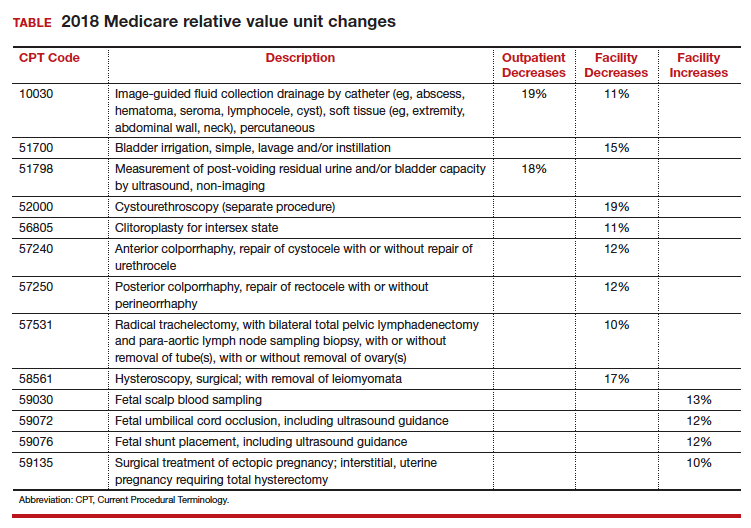
Every year, Medicare reevaluates potentially misvalued CPT codes and this year was no exception. The TABLE represents the winners and losers for codes in the outpatient and facility settings that have increased or decreased RVUs by more than 10%.
Share your thoughts! Send your Letter to the Editor to rbarbieri@frontlinemedcom.com. Please include your name and the city and state in which you practice.

Ms. Witt is an independent coding and documentation consultant and former program manager, department of coding and nomenclature, American College of Obstetricians and Gynecologists.
The author reports no financial relationships relevant to this article.
Ms. Witt is an independent coding and documentation consultant and former program manager, department of coding and nomenclature, American College of Obstetricians and Gynecologists.
The author reports no financial relationships relevant to this article.
Ms. Witt is an independent coding and documentation consultant and former program manager, department of coding and nomenclature, American College of Obstetricians and Gynecologists.
The author reports no financial relationships relevant to this article.
Another year brings changes to Current Procedural Terminology (CPT) codes (which are developed and copyrighted by the American Medical Association) in the form of additions and revisions, and payments related to resource-based relative value scale (RBRVS) revisions for selected services. As of January 1, 2018, 2 new Category I codes pertain to laparoscopic treatments for gynecologic cancer, and the 4 existing codes for colporrhaphy have been revised to include cystourethroscopy. New Category III codes include 4 for fetal magnetocardiography and 1 for transvaginal tactile imaging. Medicare also has reevaluated certain relative value units (RVUs) in outpatient and facility settings.
New and revised Category I codes
Laparoscopic treatments for gynecologic cancer. Technologic advances in performing laparoscopic procedures have allowed for more extensive laparoscopic surgery for various gynecologic cancers and, to this end, 2 new codes have been added.
First, a new code was added to capture comprehensive laparoscopic surgical staging for gynecologic cancer. This new code, 38573, Laparoscopy, surgical; with bilateral total pelvic lymphadenectomy and peri-aortic lymph node sampling, peritoneal washings, peritoneal biopsy(ies), omentectomy, and diaphragmatic washings, including diaphragmatic and other serosal biopsy(ies), when performed, may not be reported with any other code that includes lymphadenectomy, omentectomy, or hysterectomy. It is intended primarily for a stand-alone staging procedure after an initial biopsy shows a gynecologic malignancy such as ovarian cancer. This new code has been valued at 33.59 RVUs.
Second, a new code was added to capture laparoscopic debulking in conjunction with hysterectomy. The new code, 58575, Laparoscopy, surgical, total hysterectomy for resection of malignancy (tumor debulking), with omentectomy including salpingo-oophorectomy, unilateral or bilateral, when performed, has been valued at 53.62 RVUs. The open equivalent to this new code is 58953, Bilateral salpingo-oophorectomy with omentectomy, total abdominal hysterectomy and radical dissection for debulking.
Cystourethroscopy. The revisions involve no longer permitting separate reporting of 52000, Cystourethroscopy (separate procedure), with the colporrhaphy codes 57240−57265. The rationale behind this change was that surgeons were routinely performing cystoscopy at the time of these procedures and therefore it should become part of the surgical procedure. Currently the Medicare National Correct Coding Initiative (NCCI) bundles 52000 with these 4 codes, but only code 57250 allows for the use of a modifier -59 to bypass the edit if the purpose of the cystoscopy was evaluation of a distinct complaint or problem (such as evaluating patient-expressed urinary symptoms prior to the surgery that were investigated at the time of the prolapse surgery). When codes 57240, 57260, or 57265 are billed along with 52000, the cystoscopy will be denied and a modifier -59 cannot be reported to bypass this edit.
New Category III codes
The new Category III codes represent emerging technology, and it is important to report them, rather than an unlisted code, if the procedures described are performed so that data can be collected for later consideration to make these Category I CPT codes. Since these codes are not assigned relative values, the provider will need to let the payer know which existing CPT Category I code most closely represents the work involved.
Fetal magnetocardiography. The new Category III codes for fetal magnetocardiography describe essentially a fetal electrocardiogram (ECG) that would be performed to assess fetal arrhythmias by placing up to 3 leads on the mother’s abdomen. Possible comparison codes for physician work might include 59050, fetal monitoring by consultant during labor; 93000−93010, 12-lead ECG, or 93040−93042, rhythm strip up to 3 leads. However, because the equipment is very expensive, these codes would not capture practice expense and the physician would have to negotiate a reasonable reimbursement level with the payer, if the magnetocardiography was a covered service. The new codes are as follows:
- 0475T, Recording of fetal magnetic cardiac signal using at least 3 channels; patient recording and storage, data scanning with signal extraction, technical analysis and result, as well as supervision, review, and interpretation of report by a physician or other qualified health care professional
- 0476T, Recording of fetal magnetic cardiac signal using at least 3 channels; patient recording, data scanning, with raw electronic signal transfer of data and storage
- 0477T, Recording of fetal magnetic cardiac signal using at least 3 channels; signal extraction, technical analysis, and result.
Transvaginal tactile imaging. The new Category III code, 0487T, Biomechanical mapping, transvaginal, with report, describes the use of a pressure sensor probe inserted into the vaginal canal to measure and collect data on pelvic muscle strength, elasticity, tissue integrity, and tone. These data produce images in real time that are mapped to produce a report for physician review, interpretation, and report. The data allow quantification of pelvic floor dysfunction and may be useful in determining the most appropriate treatment (whether surgical or medical) for this gynecologic condition. The procedure uses a transvaginal probe like an ultrasound, so using 76830, transvaginal ultrasound, would not be unreasonable as a comparison code as a start.
Medicare relative value changes
Every year, Medicare reevaluates potentially misvalued CPT codes and this year was no exception. The TABLE represents the winners and losers for codes in the outpatient and facility settings that have increased or decreased RVUs by more than 10%.
Share your thoughts! Send your Letter to the Editor to rbarbieri@frontlinemedcom.com. Please include your name and the city and state in which you practice.
Another year brings changes to Current Procedural Terminology (CPT) codes (which are developed and copyrighted by the American Medical Association) in the form of additions and revisions, and payments related to resource-based relative value scale (RBRVS) revisions for selected services. As of January 1, 2018, 2 new Category I codes pertain to laparoscopic treatments for gynecologic cancer, and the 4 existing codes for colporrhaphy have been revised to include cystourethroscopy. New Category III codes include 4 for fetal magnetocardiography and 1 for transvaginal tactile imaging. Medicare also has reevaluated certain relative value units (RVUs) in outpatient and facility settings.
New and revised Category I codes
Laparoscopic treatments for gynecologic cancer. Technologic advances in performing laparoscopic procedures have allowed for more extensive laparoscopic surgery for various gynecologic cancers and, to this end, 2 new codes have been added.
First, a new code was added to capture comprehensive laparoscopic surgical staging for gynecologic cancer. This new code, 38573, Laparoscopy, surgical; with bilateral total pelvic lymphadenectomy and peri-aortic lymph node sampling, peritoneal washings, peritoneal biopsy(ies), omentectomy, and diaphragmatic washings, including diaphragmatic and other serosal biopsy(ies), when performed, may not be reported with any other code that includes lymphadenectomy, omentectomy, or hysterectomy. It is intended primarily for a stand-alone staging procedure after an initial biopsy shows a gynecologic malignancy such as ovarian cancer. This new code has been valued at 33.59 RVUs.
Second, a new code was added to capture laparoscopic debulking in conjunction with hysterectomy. The new code, 58575, Laparoscopy, surgical, total hysterectomy for resection of malignancy (tumor debulking), with omentectomy including salpingo-oophorectomy, unilateral or bilateral, when performed, has been valued at 53.62 RVUs. The open equivalent to this new code is 58953, Bilateral salpingo-oophorectomy with omentectomy, total abdominal hysterectomy and radical dissection for debulking.
Cystourethroscopy. The revisions involve no longer permitting separate reporting of 52000, Cystourethroscopy (separate procedure), with the colporrhaphy codes 57240−57265. The rationale behind this change was that surgeons were routinely performing cystoscopy at the time of these procedures and therefore it should become part of the surgical procedure. Currently the Medicare National Correct Coding Initiative (NCCI) bundles 52000 with these 4 codes, but only code 57250 allows for the use of a modifier -59 to bypass the edit if the purpose of the cystoscopy was evaluation of a distinct complaint or problem (such as evaluating patient-expressed urinary symptoms prior to the surgery that were investigated at the time of the prolapse surgery). When codes 57240, 57260, or 57265 are billed along with 52000, the cystoscopy will be denied and a modifier -59 cannot be reported to bypass this edit.
New Category III codes
The new Category III codes represent emerging technology, and it is important to report them, rather than an unlisted code, if the procedures described are performed so that data can be collected for later consideration to make these Category I CPT codes. Since these codes are not assigned relative values, the provider will need to let the payer know which existing CPT Category I code most closely represents the work involved.
Fetal magnetocardiography. The new Category III codes for fetal magnetocardiography describe essentially a fetal electrocardiogram (ECG) that would be performed to assess fetal arrhythmias by placing up to 3 leads on the mother’s abdomen. Possible comparison codes for physician work might include 59050, fetal monitoring by consultant during labor; 93000−93010, 12-lead ECG, or 93040−93042, rhythm strip up to 3 leads. However, because the equipment is very expensive, these codes would not capture practice expense and the physician would have to negotiate a reasonable reimbursement level with the payer, if the magnetocardiography was a covered service. The new codes are as follows:
- 0475T, Recording of fetal magnetic cardiac signal using at least 3 channels; patient recording and storage, data scanning with signal extraction, technical analysis and result, as well as supervision, review, and interpretation of report by a physician or other qualified health care professional
- 0476T, Recording of fetal magnetic cardiac signal using at least 3 channels; patient recording, data scanning, with raw electronic signal transfer of data and storage
- 0477T, Recording of fetal magnetic cardiac signal using at least 3 channels; signal extraction, technical analysis, and result.
Transvaginal tactile imaging. The new Category III code, 0487T, Biomechanical mapping, transvaginal, with report, describes the use of a pressure sensor probe inserted into the vaginal canal to measure and collect data on pelvic muscle strength, elasticity, tissue integrity, and tone. These data produce images in real time that are mapped to produce a report for physician review, interpretation, and report. The data allow quantification of pelvic floor dysfunction and may be useful in determining the most appropriate treatment (whether surgical or medical) for this gynecologic condition. The procedure uses a transvaginal probe like an ultrasound, so using 76830, transvaginal ultrasound, would not be unreasonable as a comparison code as a start.
Medicare relative value changes
Every year, Medicare reevaluates potentially misvalued CPT codes and this year was no exception. The TABLE represents the winners and losers for codes in the outpatient and facility settings that have increased or decreased RVUs by more than 10%.
Share your thoughts! Send your Letter to the Editor to rbarbieri@frontlinemedcom.com. Please include your name and the city and state in which you practice.
Alcohol dependence may accelerate aging, frontal cortical deficits
Alcoholism compounds age-associated volume deficits in the frontal cortex, independent of the additional effects of drug dependence or hepatitis C infection, suggests new research published March 14 in JAMA Psychiatry.
Edith V. Sullivan, PhD, and her coauthors reported the results of a 14-year longitudinal study that used magnetic resonance imaging to examine the brains of 116 participants with alcohol dependence and 96 age-matched controls.
They found that participants with alcohol dependence as defined by the DSM-IV had significantly greater gray matter volume deficits in their frontal, temporal, parietal, cingulate and insular cortices, compared with controls – most prominently in the frontal subregions – with the only exception being the occipital lobe. When age was taken into account, age-related volume deficits were seen in the control group in five of the six cortical regions, but the alcoholism group showed a significantly greater deficit in the precentral and superior frontal cortex.
Dr. Sullivan, of the department of psychiatry and behavioral sciences at Stanford (Calif.) University, and her coauthors said the presence of age-alcoholism interactions puts older alcohol-dependent individuals at greater risk of age-associated functional compromise, even if their excessive drinking starts later in life.
More than half of individuals in the alcoholism group (54.5%) also reported drug dependence. The imaging showed that participants with alcohol use disorder who also reported opiate or cocaine use had smaller frontal cortex volumes compared with those who were not drug users. However, the non–drug-dependent participants in the group still showed deficits in precentral, supplementary motor and medial cortices volumes, compared with controls.
“These findings in alcohol-dependent and control participants, examined 1 to 8 times or more during intervals of 1 week to 12.5 years, representing, to our knowledge, the largest and longest-studied group to date, support our study hypotheses regarding alcoholism-associated accelerated aging and cortical volume deficits independent of drug dependence or HCV infection comorbidity,” the authors wrote.
“We observed a selectivity of frontal cortex to age-alcoholism interaction beyond normal aging effects and independent of deficits related to drug dependence.”
, compared with those with alcoholism alone and compared with controls. “Thus, HCV infection, while having focal effects on frontal brain systems, targeted frontally based systems also vulnerable to chronic and extensive alcohol consumption,” the authors wrote. “Whether the compounded untoward effects of alcoholism and HCV infection on brain structure can be ameliorated with successful treatment of the infection remains to be determined.”
Dr. Sullivan and her coauthors cited several limitations. For example, non–alcohol-dependent or HCV-infected comparison groups were not available for analysis.
The study was supported by the National Institute on Alcohol Abuse and Alcoholism, and the Moldow Women’s Hope and Heal Fund. No conflicts of interest were declared.
SOURCE: Sullivan EV et al. JAMA Psychiatry. 2018 Mar 14. doi: 10.1001/jamapsychiatry.2018.0021.
With an aging population that is also showing significant increases in alcohol use and misuse, studies of the interaction between alcohol and aging and the brain are highly significant. The most compelling finding of this study is the impact of that interaction on the frontal cortex volume, because of the key role this region of the brain plays in executive function.
Deficits in frontal cortical volume associated with aging are hypothesized to also result in impulsivity and compulsivity, which opens up the possibility of greater vulnerability to alcohol use disorder later in life. So as excessive alcohol consumption contributes to this aging process, this aging process also might contribute to excessive drinking in the elderly as a form of self-medication of the negative emotional states associated with aging.
“The study ... provides compelling evidence that alcohol misuse during later adulthood could confer a greater risk of deficits in frontal lobe function beyond the deficits that typically occur with aging,” wrote George F. Koob, PhD.
Given this, it is critical that strategies be explored and implemented aimed at addressing the misuse of alcohol by older drinkers. “As Yoda might say, ‘Protect their brains, we must.’ ”
George F. Koob, PhD, is affiliated with the National Institute on Alcohol Abuse and Alcoholism at the National Institutes of Health, Rockville, Md. These comments are taken from an editorial (JAMA Psychiatry. 2018 March 14. doi: 10.1001/jamapsychiatry.2018.0009). No conflicts of interest were declared.
With an aging population that is also showing significant increases in alcohol use and misuse, studies of the interaction between alcohol and aging and the brain are highly significant. The most compelling finding of this study is the impact of that interaction on the frontal cortex volume, because of the key role this region of the brain plays in executive function.
Deficits in frontal cortical volume associated with aging are hypothesized to also result in impulsivity and compulsivity, which opens up the possibility of greater vulnerability to alcohol use disorder later in life. So as excessive alcohol consumption contributes to this aging process, this aging process also might contribute to excessive drinking in the elderly as a form of self-medication of the negative emotional states associated with aging.
“The study ... provides compelling evidence that alcohol misuse during later adulthood could confer a greater risk of deficits in frontal lobe function beyond the deficits that typically occur with aging,” wrote George F. Koob, PhD.
Given this, it is critical that strategies be explored and implemented aimed at addressing the misuse of alcohol by older drinkers. “As Yoda might say, ‘Protect their brains, we must.’ ”
George F. Koob, PhD, is affiliated with the National Institute on Alcohol Abuse and Alcoholism at the National Institutes of Health, Rockville, Md. These comments are taken from an editorial (JAMA Psychiatry. 2018 March 14. doi: 10.1001/jamapsychiatry.2018.0009). No conflicts of interest were declared.
With an aging population that is also showing significant increases in alcohol use and misuse, studies of the interaction between alcohol and aging and the brain are highly significant. The most compelling finding of this study is the impact of that interaction on the frontal cortex volume, because of the key role this region of the brain plays in executive function.
Deficits in frontal cortical volume associated with aging are hypothesized to also result in impulsivity and compulsivity, which opens up the possibility of greater vulnerability to alcohol use disorder later in life. So as excessive alcohol consumption contributes to this aging process, this aging process also might contribute to excessive drinking in the elderly as a form of self-medication of the negative emotional states associated with aging.
“The study ... provides compelling evidence that alcohol misuse during later adulthood could confer a greater risk of deficits in frontal lobe function beyond the deficits that typically occur with aging,” wrote George F. Koob, PhD.
Given this, it is critical that strategies be explored and implemented aimed at addressing the misuse of alcohol by older drinkers. “As Yoda might say, ‘Protect their brains, we must.’ ”
George F. Koob, PhD, is affiliated with the National Institute on Alcohol Abuse and Alcoholism at the National Institutes of Health, Rockville, Md. These comments are taken from an editorial (JAMA Psychiatry. 2018 March 14. doi: 10.1001/jamapsychiatry.2018.0009). No conflicts of interest were declared.
Alcoholism compounds age-associated volume deficits in the frontal cortex, independent of the additional effects of drug dependence or hepatitis C infection, suggests new research published March 14 in JAMA Psychiatry.
Edith V. Sullivan, PhD, and her coauthors reported the results of a 14-year longitudinal study that used magnetic resonance imaging to examine the brains of 116 participants with alcohol dependence and 96 age-matched controls.
They found that participants with alcohol dependence as defined by the DSM-IV had significantly greater gray matter volume deficits in their frontal, temporal, parietal, cingulate and insular cortices, compared with controls – most prominently in the frontal subregions – with the only exception being the occipital lobe. When age was taken into account, age-related volume deficits were seen in the control group in five of the six cortical regions, but the alcoholism group showed a significantly greater deficit in the precentral and superior frontal cortex.
Dr. Sullivan, of the department of psychiatry and behavioral sciences at Stanford (Calif.) University, and her coauthors said the presence of age-alcoholism interactions puts older alcohol-dependent individuals at greater risk of age-associated functional compromise, even if their excessive drinking starts later in life.
More than half of individuals in the alcoholism group (54.5%) also reported drug dependence. The imaging showed that participants with alcohol use disorder who also reported opiate or cocaine use had smaller frontal cortex volumes compared with those who were not drug users. However, the non–drug-dependent participants in the group still showed deficits in precentral, supplementary motor and medial cortices volumes, compared with controls.
“These findings in alcohol-dependent and control participants, examined 1 to 8 times or more during intervals of 1 week to 12.5 years, representing, to our knowledge, the largest and longest-studied group to date, support our study hypotheses regarding alcoholism-associated accelerated aging and cortical volume deficits independent of drug dependence or HCV infection comorbidity,” the authors wrote.
“We observed a selectivity of frontal cortex to age-alcoholism interaction beyond normal aging effects and independent of deficits related to drug dependence.”
, compared with those with alcoholism alone and compared with controls. “Thus, HCV infection, while having focal effects on frontal brain systems, targeted frontally based systems also vulnerable to chronic and extensive alcohol consumption,” the authors wrote. “Whether the compounded untoward effects of alcoholism and HCV infection on brain structure can be ameliorated with successful treatment of the infection remains to be determined.”
Dr. Sullivan and her coauthors cited several limitations. For example, non–alcohol-dependent or HCV-infected comparison groups were not available for analysis.
The study was supported by the National Institute on Alcohol Abuse and Alcoholism, and the Moldow Women’s Hope and Heal Fund. No conflicts of interest were declared.
SOURCE: Sullivan EV et al. JAMA Psychiatry. 2018 Mar 14. doi: 10.1001/jamapsychiatry.2018.0021.
Alcoholism compounds age-associated volume deficits in the frontal cortex, independent of the additional effects of drug dependence or hepatitis C infection, suggests new research published March 14 in JAMA Psychiatry.
Edith V. Sullivan, PhD, and her coauthors reported the results of a 14-year longitudinal study that used magnetic resonance imaging to examine the brains of 116 participants with alcohol dependence and 96 age-matched controls.
They found that participants with alcohol dependence as defined by the DSM-IV had significantly greater gray matter volume deficits in their frontal, temporal, parietal, cingulate and insular cortices, compared with controls – most prominently in the frontal subregions – with the only exception being the occipital lobe. When age was taken into account, age-related volume deficits were seen in the control group in five of the six cortical regions, but the alcoholism group showed a significantly greater deficit in the precentral and superior frontal cortex.
Dr. Sullivan, of the department of psychiatry and behavioral sciences at Stanford (Calif.) University, and her coauthors said the presence of age-alcoholism interactions puts older alcohol-dependent individuals at greater risk of age-associated functional compromise, even if their excessive drinking starts later in life.
More than half of individuals in the alcoholism group (54.5%) also reported drug dependence. The imaging showed that participants with alcohol use disorder who also reported opiate or cocaine use had smaller frontal cortex volumes compared with those who were not drug users. However, the non–drug-dependent participants in the group still showed deficits in precentral, supplementary motor and medial cortices volumes, compared with controls.
“These findings in alcohol-dependent and control participants, examined 1 to 8 times or more during intervals of 1 week to 12.5 years, representing, to our knowledge, the largest and longest-studied group to date, support our study hypotheses regarding alcoholism-associated accelerated aging and cortical volume deficits independent of drug dependence or HCV infection comorbidity,” the authors wrote.
“We observed a selectivity of frontal cortex to age-alcoholism interaction beyond normal aging effects and independent of deficits related to drug dependence.”
, compared with those with alcoholism alone and compared with controls. “Thus, HCV infection, while having focal effects on frontal brain systems, targeted frontally based systems also vulnerable to chronic and extensive alcohol consumption,” the authors wrote. “Whether the compounded untoward effects of alcoholism and HCV infection on brain structure can be ameliorated with successful treatment of the infection remains to be determined.”
Dr. Sullivan and her coauthors cited several limitations. For example, non–alcohol-dependent or HCV-infected comparison groups were not available for analysis.
The study was supported by the National Institute on Alcohol Abuse and Alcoholism, and the Moldow Women’s Hope and Heal Fund. No conflicts of interest were declared.
SOURCE: Sullivan EV et al. JAMA Psychiatry. 2018 Mar 14. doi: 10.1001/jamapsychiatry.2018.0021.
FROM JAMA PSYCHIATRY
Key clinical point: Older alcohol-dependent patients are at greater risk of functional compromise, even if their excessive drinking starts later in life.
Major finding: Individuals with alcohol use disorder show significantly greater deficits in the precentral and superior frontal cortex, compared with age-matched controls.
Data source: Longitudinal study of 116 participants with alcohol dependence and 96 age-matched controls.
Disclosures: The U.S. National Institute on Alcohol Abuse and Alcoholism, and the Moldow Women’s Hope and Heal Fund supported the study. No conflicts of interest were declared.
Source: Sullivan EV et al. JAMA Psychiatry. 2018 Mar 14. doi: 10.1001/jamapsychiatry.2018.0021.

FDA approves continuous glucose monitor with AI assistant
between the ages of 14 and 75 years old.
The first CGM to use artificial intelligence for this purpose, the Guardian Connect system is intended to help people with diabetes who use multiple daily injections of insulin to manage their diabetes by helping them prevent hyperglycemia and hypoglycemia, according to a statement from Medtronic.
Guardian Connect utilizes a predictive algorithm that alerts patients of significant swings in blood glucose levels up to 60 minutes prior to the event. When combined with the Guardian Sensor 3, which is placed on the abdomen to monitor blood glucose levels, the Guardian Connect system was accurate and was able to alert patients about 98.5% of hypoglycemic events, according to the results of a clinical trial. This information can also be shared and monitored with care takers and family member in real time or via text message.
Perhaps the most intriguing aspect of using the Guardian Connect system is exclusive access to the Sugar.IQ smart diabetes assistant. Utilizing artificial intelligence technology from IBM Watson Health, the Sugar.IQ assistant continually analyzes how patient’s blood glucose levels respond to factors like food intake, insulin dosages, and daily routines. The combination of continuous monitoring and real-time analysis may be able to identify patterns and lead to personalized insights that will help patients with diabetes keep their blood glucose levels under control.
“Despite proven benefits and advances in technology, only a minority of insulin-using people with diabetes currently use continuous glucose monitors,” Timothy Bailey, MD, the director of the AMCR Institute and a clinical associate professor at University of California, San Diego, said in a statement. “Newer sensors paired with intelligent algorithms that help to both predict and understand glucose excursions, particularly hypoglycemia, will make diabetes safer and more comprehensible for people who inject insulin. Greater utilization of smarter CGM systems promises to allow our patients to achieve more glycemic time-in-range and to further reduce the risk of hypoglycemia,” he said.
Similar products have been approved in the last 6 months, but they have not utilized artificial intelligence and continuous analysis.
No serious adverse events were reported in the clinical trial of Guardian Connect, but less serious adverse events were observed, including gastroenteritis, upper respiratory infection, worsening of benign prostatic hyperplasia, rash, and blisters.
Guardian Connect should be available commercially sometime this summer.
between the ages of 14 and 75 years old.
The first CGM to use artificial intelligence for this purpose, the Guardian Connect system is intended to help people with diabetes who use multiple daily injections of insulin to manage their diabetes by helping them prevent hyperglycemia and hypoglycemia, according to a statement from Medtronic.
Guardian Connect utilizes a predictive algorithm that alerts patients of significant swings in blood glucose levels up to 60 minutes prior to the event. When combined with the Guardian Sensor 3, which is placed on the abdomen to monitor blood glucose levels, the Guardian Connect system was accurate and was able to alert patients about 98.5% of hypoglycemic events, according to the results of a clinical trial. This information can also be shared and monitored with care takers and family member in real time or via text message.
Perhaps the most intriguing aspect of using the Guardian Connect system is exclusive access to the Sugar.IQ smart diabetes assistant. Utilizing artificial intelligence technology from IBM Watson Health, the Sugar.IQ assistant continually analyzes how patient’s blood glucose levels respond to factors like food intake, insulin dosages, and daily routines. The combination of continuous monitoring and real-time analysis may be able to identify patterns and lead to personalized insights that will help patients with diabetes keep their blood glucose levels under control.
“Despite proven benefits and advances in technology, only a minority of insulin-using people with diabetes currently use continuous glucose monitors,” Timothy Bailey, MD, the director of the AMCR Institute and a clinical associate professor at University of California, San Diego, said in a statement. “Newer sensors paired with intelligent algorithms that help to both predict and understand glucose excursions, particularly hypoglycemia, will make diabetes safer and more comprehensible for people who inject insulin. Greater utilization of smarter CGM systems promises to allow our patients to achieve more glycemic time-in-range and to further reduce the risk of hypoglycemia,” he said.
Similar products have been approved in the last 6 months, but they have not utilized artificial intelligence and continuous analysis.
No serious adverse events were reported in the clinical trial of Guardian Connect, but less serious adverse events were observed, including gastroenteritis, upper respiratory infection, worsening of benign prostatic hyperplasia, rash, and blisters.
Guardian Connect should be available commercially sometime this summer.
between the ages of 14 and 75 years old.
The first CGM to use artificial intelligence for this purpose, the Guardian Connect system is intended to help people with diabetes who use multiple daily injections of insulin to manage their diabetes by helping them prevent hyperglycemia and hypoglycemia, according to a statement from Medtronic.
Guardian Connect utilizes a predictive algorithm that alerts patients of significant swings in blood glucose levels up to 60 minutes prior to the event. When combined with the Guardian Sensor 3, which is placed on the abdomen to monitor blood glucose levels, the Guardian Connect system was accurate and was able to alert patients about 98.5% of hypoglycemic events, according to the results of a clinical trial. This information can also be shared and monitored with care takers and family member in real time or via text message.
Perhaps the most intriguing aspect of using the Guardian Connect system is exclusive access to the Sugar.IQ smart diabetes assistant. Utilizing artificial intelligence technology from IBM Watson Health, the Sugar.IQ assistant continually analyzes how patient’s blood glucose levels respond to factors like food intake, insulin dosages, and daily routines. The combination of continuous monitoring and real-time analysis may be able to identify patterns and lead to personalized insights that will help patients with diabetes keep their blood glucose levels under control.
“Despite proven benefits and advances in technology, only a minority of insulin-using people with diabetes currently use continuous glucose monitors,” Timothy Bailey, MD, the director of the AMCR Institute and a clinical associate professor at University of California, San Diego, said in a statement. “Newer sensors paired with intelligent algorithms that help to both predict and understand glucose excursions, particularly hypoglycemia, will make diabetes safer and more comprehensible for people who inject insulin. Greater utilization of smarter CGM systems promises to allow our patients to achieve more glycemic time-in-range and to further reduce the risk of hypoglycemia,” he said.
Similar products have been approved in the last 6 months, but they have not utilized artificial intelligence and continuous analysis.
No serious adverse events were reported in the clinical trial of Guardian Connect, but less serious adverse events were observed, including gastroenteritis, upper respiratory infection, worsening of benign prostatic hyperplasia, rash, and blisters.
Guardian Connect should be available commercially sometime this summer.
CAR T before transplant yields durable remission in B-cell malignancies
SALT LAKE CITY – Chimeric antigen receptor (CAR) T-cell therapy may be an effective bridge to hematopoietic cell transplant (HCT) for high-risk B-cell malignancies, according to a systematic analysis of patient data from the National Cancer Institute.
Additionally, patients who have received CAR T-cell therapy are likely to enter HCT with a minimal residual disease (MRD)–negative complete response, which raises the possibility of a significantly less intense conditioning regimen that could omit total body irradiation (TBI), Haneen Shalabi, DO, said at the combined annual meetings of the Center for International Blood & Marrow Transplant Research and the American Society for Blood and Marrow Transplantation.
“Patients who underwent HCT post–CAR T therapy did not have increased transplant-related morbidity or mortality,” said Dr. Shalabi, a pediatric oncologist in the hematologic diseases division of the National Cancer Institute’s pediatric oncology branch.
The combined approach also overcomes the frequent relapses seen after CAR T-cell therapy in this population. Of the 45 patients who received CAR T-cell therapy and achieved MRD-negative complete response as measured by flow cytometry, 20 did not go on to receive HCT. Of the 20 who didn’t receive HCT, 16 (80%) relapsed; 19 of the 20 (95%) had received prior HCT, said Dr. Shalabi.
However, of the 25 patients who proceeded on to receive HCT, 15 (60%) were in ongoing remission, with a median duration of 35 months (range, 11-55 months). Six patients (24%) experienced transplant-related mortality; four of these patients had no prior HCT. Ten patients (40%) experienced acute graft-versus-host disease (GVHD); two of these patients experienced grade 4 GVHD, and one experienced grade 3 GVHD.
Of the 25 patients who went on to HCT, 19 were receiving their first transplant, with a median time to transplant after CAR T-cell therapy of 57 days. Five patients (20%) had primary refractory disease. Most patients (n = 18; 72%) had TBI-based conditioning prior to their post–CAR T-cell therapy HCT. The median patient age was 15 (range, 5-30) years.
The systematic review included patients from two phase 1 studies; one was of CD19-28z CAR T-cell therapy for children and young adults with B-cell leukemia or lymphoma, and the other was of CD22-41BB CAR T-cell therapy for children and young adults with recurrent or refractory B-cell malignancies expressing CD22.
To weigh the benefit of the combined CAR T-cell therapy/HCT approach, Dr. Shalabi and her colleagues used a competing risk analysis to determine the risk of relapse post-HCT versus the risk of transplant-related mortality. Among patients undergoing their first HCT, the researchers found a 12-month cumulative incidence of relapse of 5.3% with the combined CAR T-cell therapy/HCT approach (95% confidence interval, 0.3%-22.1%). The 24-month cumulative incidence of relapse was 11.3% (95% CI, 1.7%-31.1%).
The analysis also showed the value of next-generation sequencing (NGS). “As we think about utilizing CAR T therapy as a bridge to transplant, we wanted to study the depth of CAR T–induced remission by next-gen sequencing,” Dr. Shalabi said.
Eight patients on the CD22 CAR trial had MRD analyses based on both flow cytometry and NGS. According to flow cytometry, all eight were MRD negative by 1 month; however, according to NGS, two did have detectable disease, which decreased with time. “Next-gen sequencing can identify earlier time points for relapse or ongoing remission” than flow cytometry can, she said.
An additional finding was that two-thirds of the patients who received the CD19/CD28z CAR T cells had no detectable CAR T cells when the pre-HCT conditioning regimen was initiated, said Dr. Shalabi. “CAR persistence – or lack thereof – didn’t impact post-HCT outcomes,” she said, adding that shorter-acting CAR T cells may actually be preferable when HCT is readily available as an option.
“The impact of CAR persistence peritransplant requires further analysis,” Dr. Shalabi said. It’s possible, though, that “consolidative HCT following CAR may synergistically improve event-free and overall survival for this high-risk population.”
Looking forward, Dr. Shalabi and her team are asking bigger questions: “For future directions – and this is a very big question that those in the room would probably like to know – by inducing NGS-negativity, can CAR T therapy allow for HCT conditioning deintensification, potentially reducing the risk of TRM [transplant-related mortality] and long term comorbidities?”
A future trial will explore outcomes for a conditioning regimen that omits TBI for patients who are MRD-negative by NGS, said Dr. Shalabi.
Another direction for her team’s research is to see whether introducing CAR T-cell therapy earlier in a very-high-risk population may improve outcomes; the current study population was heavily pretreated, Dr. Shalabi said.
Dr. Shalabi is employed by the National Cancer Institute. She reported no conflicts of interest.
SOURCE: Shalabi H et al. 2018 BMT Tandem Meetings, Abstract 6.
SALT LAKE CITY – Chimeric antigen receptor (CAR) T-cell therapy may be an effective bridge to hematopoietic cell transplant (HCT) for high-risk B-cell malignancies, according to a systematic analysis of patient data from the National Cancer Institute.
Additionally, patients who have received CAR T-cell therapy are likely to enter HCT with a minimal residual disease (MRD)–negative complete response, which raises the possibility of a significantly less intense conditioning regimen that could omit total body irradiation (TBI), Haneen Shalabi, DO, said at the combined annual meetings of the Center for International Blood & Marrow Transplant Research and the American Society for Blood and Marrow Transplantation.
“Patients who underwent HCT post–CAR T therapy did not have increased transplant-related morbidity or mortality,” said Dr. Shalabi, a pediatric oncologist in the hematologic diseases division of the National Cancer Institute’s pediatric oncology branch.
The combined approach also overcomes the frequent relapses seen after CAR T-cell therapy in this population. Of the 45 patients who received CAR T-cell therapy and achieved MRD-negative complete response as measured by flow cytometry, 20 did not go on to receive HCT. Of the 20 who didn’t receive HCT, 16 (80%) relapsed; 19 of the 20 (95%) had received prior HCT, said Dr. Shalabi.
However, of the 25 patients who proceeded on to receive HCT, 15 (60%) were in ongoing remission, with a median duration of 35 months (range, 11-55 months). Six patients (24%) experienced transplant-related mortality; four of these patients had no prior HCT. Ten patients (40%) experienced acute graft-versus-host disease (GVHD); two of these patients experienced grade 4 GVHD, and one experienced grade 3 GVHD.
Of the 25 patients who went on to HCT, 19 were receiving their first transplant, with a median time to transplant after CAR T-cell therapy of 57 days. Five patients (20%) had primary refractory disease. Most patients (n = 18; 72%) had TBI-based conditioning prior to their post–CAR T-cell therapy HCT. The median patient age was 15 (range, 5-30) years.
The systematic review included patients from two phase 1 studies; one was of CD19-28z CAR T-cell therapy for children and young adults with B-cell leukemia or lymphoma, and the other was of CD22-41BB CAR T-cell therapy for children and young adults with recurrent or refractory B-cell malignancies expressing CD22.
To weigh the benefit of the combined CAR T-cell therapy/HCT approach, Dr. Shalabi and her colleagues used a competing risk analysis to determine the risk of relapse post-HCT versus the risk of transplant-related mortality. Among patients undergoing their first HCT, the researchers found a 12-month cumulative incidence of relapse of 5.3% with the combined CAR T-cell therapy/HCT approach (95% confidence interval, 0.3%-22.1%). The 24-month cumulative incidence of relapse was 11.3% (95% CI, 1.7%-31.1%).
The analysis also showed the value of next-generation sequencing (NGS). “As we think about utilizing CAR T therapy as a bridge to transplant, we wanted to study the depth of CAR T–induced remission by next-gen sequencing,” Dr. Shalabi said.
Eight patients on the CD22 CAR trial had MRD analyses based on both flow cytometry and NGS. According to flow cytometry, all eight were MRD negative by 1 month; however, according to NGS, two did have detectable disease, which decreased with time. “Next-gen sequencing can identify earlier time points for relapse or ongoing remission” than flow cytometry can, she said.
An additional finding was that two-thirds of the patients who received the CD19/CD28z CAR T cells had no detectable CAR T cells when the pre-HCT conditioning regimen was initiated, said Dr. Shalabi. “CAR persistence – or lack thereof – didn’t impact post-HCT outcomes,” she said, adding that shorter-acting CAR T cells may actually be preferable when HCT is readily available as an option.
“The impact of CAR persistence peritransplant requires further analysis,” Dr. Shalabi said. It’s possible, though, that “consolidative HCT following CAR may synergistically improve event-free and overall survival for this high-risk population.”
Looking forward, Dr. Shalabi and her team are asking bigger questions: “For future directions – and this is a very big question that those in the room would probably like to know – by inducing NGS-negativity, can CAR T therapy allow for HCT conditioning deintensification, potentially reducing the risk of TRM [transplant-related mortality] and long term comorbidities?”
A future trial will explore outcomes for a conditioning regimen that omits TBI for patients who are MRD-negative by NGS, said Dr. Shalabi.
Another direction for her team’s research is to see whether introducing CAR T-cell therapy earlier in a very-high-risk population may improve outcomes; the current study population was heavily pretreated, Dr. Shalabi said.
Dr. Shalabi is employed by the National Cancer Institute. She reported no conflicts of interest.
SOURCE: Shalabi H et al. 2018 BMT Tandem Meetings, Abstract 6.
SALT LAKE CITY – Chimeric antigen receptor (CAR) T-cell therapy may be an effective bridge to hematopoietic cell transplant (HCT) for high-risk B-cell malignancies, according to a systematic analysis of patient data from the National Cancer Institute.
Additionally, patients who have received CAR T-cell therapy are likely to enter HCT with a minimal residual disease (MRD)–negative complete response, which raises the possibility of a significantly less intense conditioning regimen that could omit total body irradiation (TBI), Haneen Shalabi, DO, said at the combined annual meetings of the Center for International Blood & Marrow Transplant Research and the American Society for Blood and Marrow Transplantation.
“Patients who underwent HCT post–CAR T therapy did not have increased transplant-related morbidity or mortality,” said Dr. Shalabi, a pediatric oncologist in the hematologic diseases division of the National Cancer Institute’s pediatric oncology branch.
The combined approach also overcomes the frequent relapses seen after CAR T-cell therapy in this population. Of the 45 patients who received CAR T-cell therapy and achieved MRD-negative complete response as measured by flow cytometry, 20 did not go on to receive HCT. Of the 20 who didn’t receive HCT, 16 (80%) relapsed; 19 of the 20 (95%) had received prior HCT, said Dr. Shalabi.
However, of the 25 patients who proceeded on to receive HCT, 15 (60%) were in ongoing remission, with a median duration of 35 months (range, 11-55 months). Six patients (24%) experienced transplant-related mortality; four of these patients had no prior HCT. Ten patients (40%) experienced acute graft-versus-host disease (GVHD); two of these patients experienced grade 4 GVHD, and one experienced grade 3 GVHD.
Of the 25 patients who went on to HCT, 19 were receiving their first transplant, with a median time to transplant after CAR T-cell therapy of 57 days. Five patients (20%) had primary refractory disease. Most patients (n = 18; 72%) had TBI-based conditioning prior to their post–CAR T-cell therapy HCT. The median patient age was 15 (range, 5-30) years.
The systematic review included patients from two phase 1 studies; one was of CD19-28z CAR T-cell therapy for children and young adults with B-cell leukemia or lymphoma, and the other was of CD22-41BB CAR T-cell therapy for children and young adults with recurrent or refractory B-cell malignancies expressing CD22.
To weigh the benefit of the combined CAR T-cell therapy/HCT approach, Dr. Shalabi and her colleagues used a competing risk analysis to determine the risk of relapse post-HCT versus the risk of transplant-related mortality. Among patients undergoing their first HCT, the researchers found a 12-month cumulative incidence of relapse of 5.3% with the combined CAR T-cell therapy/HCT approach (95% confidence interval, 0.3%-22.1%). The 24-month cumulative incidence of relapse was 11.3% (95% CI, 1.7%-31.1%).
The analysis also showed the value of next-generation sequencing (NGS). “As we think about utilizing CAR T therapy as a bridge to transplant, we wanted to study the depth of CAR T–induced remission by next-gen sequencing,” Dr. Shalabi said.
Eight patients on the CD22 CAR trial had MRD analyses based on both flow cytometry and NGS. According to flow cytometry, all eight were MRD negative by 1 month; however, according to NGS, two did have detectable disease, which decreased with time. “Next-gen sequencing can identify earlier time points for relapse or ongoing remission” than flow cytometry can, she said.
An additional finding was that two-thirds of the patients who received the CD19/CD28z CAR T cells had no detectable CAR T cells when the pre-HCT conditioning regimen was initiated, said Dr. Shalabi. “CAR persistence – or lack thereof – didn’t impact post-HCT outcomes,” she said, adding that shorter-acting CAR T cells may actually be preferable when HCT is readily available as an option.
“The impact of CAR persistence peritransplant requires further analysis,” Dr. Shalabi said. It’s possible, though, that “consolidative HCT following CAR may synergistically improve event-free and overall survival for this high-risk population.”
Looking forward, Dr. Shalabi and her team are asking bigger questions: “For future directions – and this is a very big question that those in the room would probably like to know – by inducing NGS-negativity, can CAR T therapy allow for HCT conditioning deintensification, potentially reducing the risk of TRM [transplant-related mortality] and long term comorbidities?”
A future trial will explore outcomes for a conditioning regimen that omits TBI for patients who are MRD-negative by NGS, said Dr. Shalabi.
Another direction for her team’s research is to see whether introducing CAR T-cell therapy earlier in a very-high-risk population may improve outcomes; the current study population was heavily pretreated, Dr. Shalabi said.
Dr. Shalabi is employed by the National Cancer Institute. She reported no conflicts of interest.
SOURCE: Shalabi H et al. 2018 BMT Tandem Meetings, Abstract 6.
REPORTING FROM THE 2018 BMT TANDEM MEETINGS
Key clinical point:
Major finding: Of 20 patients receiving CAR T before HCT, 15 (60%) were in ongoing remission of a median 35 months.
Study details: Systematic analysis of 42 patients with B-cell malignancies receiving CAR T-cell therapy at the National Cancer Institute.
Disclosures: The study was conducted at the National Cancer Institute, where Dr. Shalabi is employed.
Source: Shalabi H et al. 2018 BMT Tandem Meetings, Abstract 6.