User login
CGIT intensifies efforts to put new technologies into the hands of clinicians
There is no change in the core mission of the AGA’s Center for GI Innovation and Technology (CGIT) since two cochairs took over the leadership last year. As always, the goal is to support the development and adoption of new technologies for the treatment of digestive disorders. However, the cochairs want to do more to identify and dismantle the obstacles that slow the process.
“Innovators face challenges at every stage, from attracting investors to refining an idea into a viable clinical tool. Even once a device has obtained regulatory approval, issues of training and reimbursement can keep a good idea from improving patient care,” explained V. Raman Muthusamy, MD, director of interventional endoscopy and general GI endoscopy, at the University of California, Los Angeles. “We want to identify these obstacles and use the CGIT resources to provide solutions.”
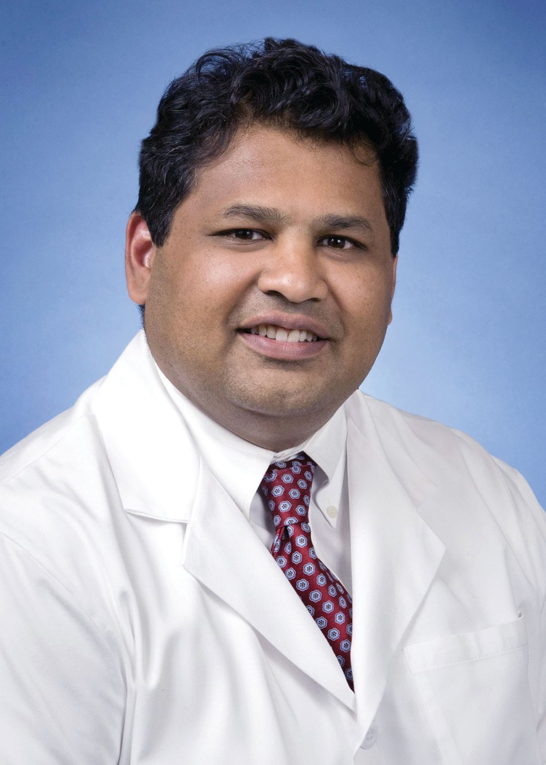
“CGIT was set up to foster collaboration between stakeholders in new technology, but we have been increasingly concerned about the roadblocks that keep important technology from getting into the hands of clinicians,” Dr. Komanduri said at the 2018 AGA Tech Summit sponsored by the AGA Center for GI Innovation and Technology. “In addition to the role that the CGIT has played in bringing together interested parties, we are taking a closer look at how we can help accelerate both the processes of development and implementation once the technology is available.”
One strategy is to expand the presence and activities of CGIT. Established in 2010, the CGIT is best known for the AGA Tech Summit, which has been an important event for bringing together innovators, investors, clinicians, and those involved in navigating regulatory issues, but Dr. Komanduri said that he and Dr. Muthusamy are working to make the summit “more of a launching point” for ideas and projects that follow the meeting.
“The AGA Tech Summit has provided an opportunity to recognize hot areas, and the goal will be to drive new ideas forward after the meeting by facilitating communication and cooperation among those who can contribute to advancing the concepts, ” Dr. Komanduri explained.
To help focus the mission of the CGIT, six areas of concentration have been identified. These are colorectal cancer screening and diagnosis, endoscope reprocessing, weight loss interventions, interventional endoscopy platforms, antireflux devices, and tissue resection technologies, according to Dr. Muthusamy. These focus areas do not preclude CGIT involvement in other types of innovations, but Dr. Muthusamy said this list helps emphasize strengths for those who do not yet consider CGIT a resource.
“We want to intersect better with the AGA members who think CGIT is not relevant to them because they are not involved in the development of new technology,” Dr. Muthusamy said. In drawing attention to these specific areas, Dr. Muthusamy said, “We plan to do more with technology updates and keep the AGA membership aware of how opportunities in clinical medicine are evolving through technological innovation.”
The mission of CGIT begins with the encouragement of new technology but it includes all the steps needed to shepherd innovations into routine clinical application. This includes helping with the strategies that will incentivize clinicians to learn the technology. While this has always been part of the CGIT mission, the cochairs are increasing their attention to eliminating delays in the process, which they recognize as a threat to fulfilling the CGIT mandate.
“There are challenges to bringing new ideas to the marketplace, and we think that pace between idea and product is too slow,” Dr. Komanduri said. “We want to increase the emphasis on finding ways to streamline the process.”
There is no change in the core mission of the AGA’s Center for GI Innovation and Technology (CGIT) since two cochairs took over the leadership last year. As always, the goal is to support the development and adoption of new technologies for the treatment of digestive disorders. However, the cochairs want to do more to identify and dismantle the obstacles that slow the process.
“Innovators face challenges at every stage, from attracting investors to refining an idea into a viable clinical tool. Even once a device has obtained regulatory approval, issues of training and reimbursement can keep a good idea from improving patient care,” explained V. Raman Muthusamy, MD, director of interventional endoscopy and general GI endoscopy, at the University of California, Los Angeles. “We want to identify these obstacles and use the CGIT resources to provide solutions.”
“CGIT was set up to foster collaboration between stakeholders in new technology, but we have been increasingly concerned about the roadblocks that keep important technology from getting into the hands of clinicians,” Dr. Komanduri said at the 2018 AGA Tech Summit sponsored by the AGA Center for GI Innovation and Technology. “In addition to the role that the CGIT has played in bringing together interested parties, we are taking a closer look at how we can help accelerate both the processes of development and implementation once the technology is available.”
One strategy is to expand the presence and activities of CGIT. Established in 2010, the CGIT is best known for the AGA Tech Summit, which has been an important event for bringing together innovators, investors, clinicians, and those involved in navigating regulatory issues, but Dr. Komanduri said that he and Dr. Muthusamy are working to make the summit “more of a launching point” for ideas and projects that follow the meeting.
“The AGA Tech Summit has provided an opportunity to recognize hot areas, and the goal will be to drive new ideas forward after the meeting by facilitating communication and cooperation among those who can contribute to advancing the concepts, ” Dr. Komanduri explained.
To help focus the mission of the CGIT, six areas of concentration have been identified. These are colorectal cancer screening and diagnosis, endoscope reprocessing, weight loss interventions, interventional endoscopy platforms, antireflux devices, and tissue resection technologies, according to Dr. Muthusamy. These focus areas do not preclude CGIT involvement in other types of innovations, but Dr. Muthusamy said this list helps emphasize strengths for those who do not yet consider CGIT a resource.
“We want to intersect better with the AGA members who think CGIT is not relevant to them because they are not involved in the development of new technology,” Dr. Muthusamy said. In drawing attention to these specific areas, Dr. Muthusamy said, “We plan to do more with technology updates and keep the AGA membership aware of how opportunities in clinical medicine are evolving through technological innovation.”
The mission of CGIT begins with the encouragement of new technology but it includes all the steps needed to shepherd innovations into routine clinical application. This includes helping with the strategies that will incentivize clinicians to learn the technology. While this has always been part of the CGIT mission, the cochairs are increasing their attention to eliminating delays in the process, which they recognize as a threat to fulfilling the CGIT mandate.
“There are challenges to bringing new ideas to the marketplace, and we think that pace between idea and product is too slow,” Dr. Komanduri said. “We want to increase the emphasis on finding ways to streamline the process.”
There is no change in the core mission of the AGA’s Center for GI Innovation and Technology (CGIT) since two cochairs took over the leadership last year. As always, the goal is to support the development and adoption of new technologies for the treatment of digestive disorders. However, the cochairs want to do more to identify and dismantle the obstacles that slow the process.
“Innovators face challenges at every stage, from attracting investors to refining an idea into a viable clinical tool. Even once a device has obtained regulatory approval, issues of training and reimbursement can keep a good idea from improving patient care,” explained V. Raman Muthusamy, MD, director of interventional endoscopy and general GI endoscopy, at the University of California, Los Angeles. “We want to identify these obstacles and use the CGIT resources to provide solutions.”
“CGIT was set up to foster collaboration between stakeholders in new technology, but we have been increasingly concerned about the roadblocks that keep important technology from getting into the hands of clinicians,” Dr. Komanduri said at the 2018 AGA Tech Summit sponsored by the AGA Center for GI Innovation and Technology. “In addition to the role that the CGIT has played in bringing together interested parties, we are taking a closer look at how we can help accelerate both the processes of development and implementation once the technology is available.”
One strategy is to expand the presence and activities of CGIT. Established in 2010, the CGIT is best known for the AGA Tech Summit, which has been an important event for bringing together innovators, investors, clinicians, and those involved in navigating regulatory issues, but Dr. Komanduri said that he and Dr. Muthusamy are working to make the summit “more of a launching point” for ideas and projects that follow the meeting.
“The AGA Tech Summit has provided an opportunity to recognize hot areas, and the goal will be to drive new ideas forward after the meeting by facilitating communication and cooperation among those who can contribute to advancing the concepts, ” Dr. Komanduri explained.
To help focus the mission of the CGIT, six areas of concentration have been identified. These are colorectal cancer screening and diagnosis, endoscope reprocessing, weight loss interventions, interventional endoscopy platforms, antireflux devices, and tissue resection technologies, according to Dr. Muthusamy. These focus areas do not preclude CGIT involvement in other types of innovations, but Dr. Muthusamy said this list helps emphasize strengths for those who do not yet consider CGIT a resource.
“We want to intersect better with the AGA members who think CGIT is not relevant to them because they are not involved in the development of new technology,” Dr. Muthusamy said. In drawing attention to these specific areas, Dr. Muthusamy said, “We plan to do more with technology updates and keep the AGA membership aware of how opportunities in clinical medicine are evolving through technological innovation.”
The mission of CGIT begins with the encouragement of new technology but it includes all the steps needed to shepherd innovations into routine clinical application. This includes helping with the strategies that will incentivize clinicians to learn the technology. While this has always been part of the CGIT mission, the cochairs are increasing their attention to eliminating delays in the process, which they recognize as a threat to fulfilling the CGIT mandate.
“There are challenges to bringing new ideas to the marketplace, and we think that pace between idea and product is too slow,” Dr. Komanduri said. “We want to increase the emphasis on finding ways to streamline the process.”
Nanotechnology breakthroughs envisioned for gastroenterology diagnostics and therapeutics
Nanotechnology is positioned to bring fundamental changes to the diagnosis and treatment of GI diseases, according to the Vadim Backman, MS, PhD, professor of biomedical engineering at the McCormick School of Engineering, Northwestern University, Chicago.
“The ways in which nanotechnology will change the field are going to be quite significant,” Dr. Backman said. He will give the Keynote Presentation at the 2018 AGA Tech Summit, sponsored by the AGA Center for GI Innovation and Technology. He described progress toward breakthroughs in diagnostics – particularly early detection of GI cancers – and therapeutics, such as nanocage transport of drugs across cell membranes. Applications have moved beyond the theoretical.

Nanotechnology is a broad term that involves manipulation of any material on a nano scale, often defined, although not strictly, as less than 100 nanometers (nm). Most bacteria measure more than 1000 nm in at least one direction. A DNA nucleotide is 2 nm. In the diagnosis and treatment of human diseases, the advantages of working on a nanoscale mean opportunities to act on the most fundamental molecular processes, including the earliest stages of pathophysiology.
In diagnosis, nanotechnology has the ability to detect genetic and epigenetic distortions that are initial steps to carcinogenesis. One set of studies has been performed with partial-wave spectroscopic microscopy capable of quantifying the properties of cellular structures on a scale as low as 20 nm. Work in this area has already successfully identified events in neoplasia development that preceded any alteration detected by conventional techniques, such as histology.
“With nanoscale sensitive optics, it is now possible to detect changes in cell and tissue structure that we have connected to the earliest steps in tumorigenesis,” Dr. Backman said. This work largely derives from progress in defining the topography of chromatin, a term that encompasses the protein, DNA, RNA, and protein composition of chromosomes. The work is relevant to all solid tumors, not just GI cancers. Based on the current status of the science, Dr. Backman foresees not just detection of the earliest events in cancer development but opportunities to tailor epigenetic therapies for personalized medicine.
In regard to other opportunities to improve diagnostics in GI diseases through nanotechnology, Dr. Backman spoke of nanoscale constructs that serve as contrast agents to visualize molecular structures and processes. These can be delivered endoscopically or systemically to provide unprecedented 3-D imaging, allowing such structures as cell receptors to be visualized and labeled.
The potential therapeutic applications of nanotechnology involve new approaches to altering disease progression. In addition to explaining the novel opportunities provided by delivering drugs on a nanoscale to alter cell processes in a way that would not be otherwise possible, Dr. Backman described how nanotechnology can be applied to influence global patterns in gene transcription. This is achieved by altering the chromatin structure.
“It is now understood that chromatin acts like software in mediating gene activities,” Dr. Backman explained. “In the case of cancer, normalization of chromatin structure can predictably modulate global patterns of gene expression. There is now evidence that modulation of chromatin structure with macrogenomic engineering may address cancer as well as other illnesses.”
In most cases, the advances described by Dr. Backman are not speculations based on the potential of the technology but concepts that are actively being developed. Partial-wave spectroscopic microscopy, which was developed in Dr. Backman’s laboratory, is permitting the nuclear nanostructure in live cells to be evaluated in real time, while progress in epigenetics is explaining which processes might be candidates for preventing or treating disease.
“Nanotechnology is positioned to advance personalized medicine and tailored therapeutics,” Dr. Backman said. “We do not yet have applications ready for regulatory approval, but they are coming.”
Nanotechnology is positioned to bring fundamental changes to the diagnosis and treatment of GI diseases, according to the Vadim Backman, MS, PhD, professor of biomedical engineering at the McCormick School of Engineering, Northwestern University, Chicago.
“The ways in which nanotechnology will change the field are going to be quite significant,” Dr. Backman said. He will give the Keynote Presentation at the 2018 AGA Tech Summit, sponsored by the AGA Center for GI Innovation and Technology. He described progress toward breakthroughs in diagnostics – particularly early detection of GI cancers – and therapeutics, such as nanocage transport of drugs across cell membranes. Applications have moved beyond the theoretical.
Nanotechnology is a broad term that involves manipulation of any material on a nano scale, often defined, although not strictly, as less than 100 nanometers (nm). Most bacteria measure more than 1000 nm in at least one direction. A DNA nucleotide is 2 nm. In the diagnosis and treatment of human diseases, the advantages of working on a nanoscale mean opportunities to act on the most fundamental molecular processes, including the earliest stages of pathophysiology.
In diagnosis, nanotechnology has the ability to detect genetic and epigenetic distortions that are initial steps to carcinogenesis. One set of studies has been performed with partial-wave spectroscopic microscopy capable of quantifying the properties of cellular structures on a scale as low as 20 nm. Work in this area has already successfully identified events in neoplasia development that preceded any alteration detected by conventional techniques, such as histology.
“With nanoscale sensitive optics, it is now possible to detect changes in cell and tissue structure that we have connected to the earliest steps in tumorigenesis,” Dr. Backman said. This work largely derives from progress in defining the topography of chromatin, a term that encompasses the protein, DNA, RNA, and protein composition of chromosomes. The work is relevant to all solid tumors, not just GI cancers. Based on the current status of the science, Dr. Backman foresees not just detection of the earliest events in cancer development but opportunities to tailor epigenetic therapies for personalized medicine.
In regard to other opportunities to improve diagnostics in GI diseases through nanotechnology, Dr. Backman spoke of nanoscale constructs that serve as contrast agents to visualize molecular structures and processes. These can be delivered endoscopically or systemically to provide unprecedented 3-D imaging, allowing such structures as cell receptors to be visualized and labeled.
The potential therapeutic applications of nanotechnology involve new approaches to altering disease progression. In addition to explaining the novel opportunities provided by delivering drugs on a nanoscale to alter cell processes in a way that would not be otherwise possible, Dr. Backman described how nanotechnology can be applied to influence global patterns in gene transcription. This is achieved by altering the chromatin structure.
“It is now understood that chromatin acts like software in mediating gene activities,” Dr. Backman explained. “In the case of cancer, normalization of chromatin structure can predictably modulate global patterns of gene expression. There is now evidence that modulation of chromatin structure with macrogenomic engineering may address cancer as well as other illnesses.”
In most cases, the advances described by Dr. Backman are not speculations based on the potential of the technology but concepts that are actively being developed. Partial-wave spectroscopic microscopy, which was developed in Dr. Backman’s laboratory, is permitting the nuclear nanostructure in live cells to be evaluated in real time, while progress in epigenetics is explaining which processes might be candidates for preventing or treating disease.
“Nanotechnology is positioned to advance personalized medicine and tailored therapeutics,” Dr. Backman said. “We do not yet have applications ready for regulatory approval, but they are coming.”
Nanotechnology is positioned to bring fundamental changes to the diagnosis and treatment of GI diseases, according to the Vadim Backman, MS, PhD, professor of biomedical engineering at the McCormick School of Engineering, Northwestern University, Chicago.
“The ways in which nanotechnology will change the field are going to be quite significant,” Dr. Backman said. He will give the Keynote Presentation at the 2018 AGA Tech Summit, sponsored by the AGA Center for GI Innovation and Technology. He described progress toward breakthroughs in diagnostics – particularly early detection of GI cancers – and therapeutics, such as nanocage transport of drugs across cell membranes. Applications have moved beyond the theoretical.
Nanotechnology is a broad term that involves manipulation of any material on a nano scale, often defined, although not strictly, as less than 100 nanometers (nm). Most bacteria measure more than 1000 nm in at least one direction. A DNA nucleotide is 2 nm. In the diagnosis and treatment of human diseases, the advantages of working on a nanoscale mean opportunities to act on the most fundamental molecular processes, including the earliest stages of pathophysiology.
In diagnosis, nanotechnology has the ability to detect genetic and epigenetic distortions that are initial steps to carcinogenesis. One set of studies has been performed with partial-wave spectroscopic microscopy capable of quantifying the properties of cellular structures on a scale as low as 20 nm. Work in this area has already successfully identified events in neoplasia development that preceded any alteration detected by conventional techniques, such as histology.
“With nanoscale sensitive optics, it is now possible to detect changes in cell and tissue structure that we have connected to the earliest steps in tumorigenesis,” Dr. Backman said. This work largely derives from progress in defining the topography of chromatin, a term that encompasses the protein, DNA, RNA, and protein composition of chromosomes. The work is relevant to all solid tumors, not just GI cancers. Based on the current status of the science, Dr. Backman foresees not just detection of the earliest events in cancer development but opportunities to tailor epigenetic therapies for personalized medicine.
In regard to other opportunities to improve diagnostics in GI diseases through nanotechnology, Dr. Backman spoke of nanoscale constructs that serve as contrast agents to visualize molecular structures and processes. These can be delivered endoscopically or systemically to provide unprecedented 3-D imaging, allowing such structures as cell receptors to be visualized and labeled.
The potential therapeutic applications of nanotechnology involve new approaches to altering disease progression. In addition to explaining the novel opportunities provided by delivering drugs on a nanoscale to alter cell processes in a way that would not be otherwise possible, Dr. Backman described how nanotechnology can be applied to influence global patterns in gene transcription. This is achieved by altering the chromatin structure.
“It is now understood that chromatin acts like software in mediating gene activities,” Dr. Backman explained. “In the case of cancer, normalization of chromatin structure can predictably modulate global patterns of gene expression. There is now evidence that modulation of chromatin structure with macrogenomic engineering may address cancer as well as other illnesses.”
In most cases, the advances described by Dr. Backman are not speculations based on the potential of the technology but concepts that are actively being developed. Partial-wave spectroscopic microscopy, which was developed in Dr. Backman’s laboratory, is permitting the nuclear nanostructure in live cells to be evaluated in real time, while progress in epigenetics is explaining which processes might be candidates for preventing or treating disease.
“Nanotechnology is positioned to advance personalized medicine and tailored therapeutics,” Dr. Backman said. “We do not yet have applications ready for regulatory approval, but they are coming.”

Effectiveness, adherence similar for nasal pillows and standard masks
according to results of a study.
In a retrospective observational study of 144 patients with obstructive sleep apnea, respiratory measures including apnea-hypopnea index (AHI), oxygen desaturation index, mean oxygen saturation, and Epworth Sleepiness scale scores did not differ between the two treatment groups at baseline and during a 12-month follow-up appointment. Treatment adherence was also similar between the two groups, reported Andrea Lanza of the Sleep Medicine Center at Niguarda Hospital in Milan, Italy, and coauthors in Sleep Medicine.
Patients received continuous positive airway pressure (CPAP) treatment between May 2012 and September 2014, and were assigned to one of two groups based on their choice of treatment. Initially, 102 opted for nasal pillows (Group P), and 42 chose the standard nasal mask (Group N). Patients who either changed masks or added a new one during titration or follow-up were assigned to a third group, Group C.
AHI did not differ significantly between groups at baseline or follow-up. In Group P, mean AHI at titration was 1.2 events per hour, compared with 1.8 in Group N and 1.9 in Group C (P = .109). At follow-up, AHI was 0.7 in Group P, 1.1 in Group N, and 0.9 in Group C (P = .172). Oxygen desaturation index and oxygen saturation also remained similar between the groups at baseline and follow-up, the investigators reported.
Additionally, long-term adherence did not differ significantly between the groups, with mean daily CPAP usage of 5.5 hours per night in Group P, 5.3 in Group N, and 5.6 in Group C. Mean usage was less than 4 hours per night for 11.6% in group P, 18.5% in group N, and 13.9% in group C, the authors added.
The frequency of side effects occurring in patients in two of the groups were similar (49% in Group P, vs. 61% in Group N; P = .212), though the nature of the side effects differed. Nostril pain or burning was reported only by patients in the nasal pillows group, and skin breakdown was reported only in the nasal mask group.
Though nasal pillows have typically been reserved for patients who do not tolerate the standard mask, the results of this study suggest that “nasal pillows could be safely prescribed as first-line interfaces,” the authors wrote. “They seem to be efficacious for CPAP titration and long-term treatment, ensuring a good rate of adherence.”
All of the authors reported having no disclosures.
SOURCE: Lanza A et al. Sleep Med. 2018 Jan;41:94-9
These results add to the body of evidence about the efficacy of nasal pillows and nasal masks. Future research should address the need for retitration when changing mask type, said Matthew R. Ebben, PhD, associate clinical professor at Cornell University, New York, in an editorial published with the study in Sleep Medicine.
“Many working in the field of sleep medicine continue to be unaware that differences in efficacy exist between mask styles, particularly in cases of moderate to severe obstructive sleep apnea,” he wrote.
“New clinical practice guidelines are needed to promote the necessity for PAP [positive airway pressure] retitration when changes in mask style are required,” he added. “Ensuring that PAP therapy is as effective as possible will reduce the need for patients and clinicians to investigate other treatment options for obstructive sleep apnea, which may be both less effective and have an inferior side effect profile compared to PAP treatment.”
These results add to the body of evidence about the efficacy of nasal pillows and nasal masks. Future research should address the need for retitration when changing mask type, said Matthew R. Ebben, PhD, associate clinical professor at Cornell University, New York, in an editorial published with the study in Sleep Medicine.
“Many working in the field of sleep medicine continue to be unaware that differences in efficacy exist between mask styles, particularly in cases of moderate to severe obstructive sleep apnea,” he wrote.
“New clinical practice guidelines are needed to promote the necessity for PAP [positive airway pressure] retitration when changes in mask style are required,” he added. “Ensuring that PAP therapy is as effective as possible will reduce the need for patients and clinicians to investigate other treatment options for obstructive sleep apnea, which may be both less effective and have an inferior side effect profile compared to PAP treatment.”
These results add to the body of evidence about the efficacy of nasal pillows and nasal masks. Future research should address the need for retitration when changing mask type, said Matthew R. Ebben, PhD, associate clinical professor at Cornell University, New York, in an editorial published with the study in Sleep Medicine.
“Many working in the field of sleep medicine continue to be unaware that differences in efficacy exist between mask styles, particularly in cases of moderate to severe obstructive sleep apnea,” he wrote.
“New clinical practice guidelines are needed to promote the necessity for PAP [positive airway pressure] retitration when changes in mask style are required,” he added. “Ensuring that PAP therapy is as effective as possible will reduce the need for patients and clinicians to investigate other treatment options for obstructive sleep apnea, which may be both less effective and have an inferior side effect profile compared to PAP treatment.”
according to results of a study.
In a retrospective observational study of 144 patients with obstructive sleep apnea, respiratory measures including apnea-hypopnea index (AHI), oxygen desaturation index, mean oxygen saturation, and Epworth Sleepiness scale scores did not differ between the two treatment groups at baseline and during a 12-month follow-up appointment. Treatment adherence was also similar between the two groups, reported Andrea Lanza of the Sleep Medicine Center at Niguarda Hospital in Milan, Italy, and coauthors in Sleep Medicine.
Patients received continuous positive airway pressure (CPAP) treatment between May 2012 and September 2014, and were assigned to one of two groups based on their choice of treatment. Initially, 102 opted for nasal pillows (Group P), and 42 chose the standard nasal mask (Group N). Patients who either changed masks or added a new one during titration or follow-up were assigned to a third group, Group C.
AHI did not differ significantly between groups at baseline or follow-up. In Group P, mean AHI at titration was 1.2 events per hour, compared with 1.8 in Group N and 1.9 in Group C (P = .109). At follow-up, AHI was 0.7 in Group P, 1.1 in Group N, and 0.9 in Group C (P = .172). Oxygen desaturation index and oxygen saturation also remained similar between the groups at baseline and follow-up, the investigators reported.
Additionally, long-term adherence did not differ significantly between the groups, with mean daily CPAP usage of 5.5 hours per night in Group P, 5.3 in Group N, and 5.6 in Group C. Mean usage was less than 4 hours per night for 11.6% in group P, 18.5% in group N, and 13.9% in group C, the authors added.
The frequency of side effects occurring in patients in two of the groups were similar (49% in Group P, vs. 61% in Group N; P = .212), though the nature of the side effects differed. Nostril pain or burning was reported only by patients in the nasal pillows group, and skin breakdown was reported only in the nasal mask group.
Though nasal pillows have typically been reserved for patients who do not tolerate the standard mask, the results of this study suggest that “nasal pillows could be safely prescribed as first-line interfaces,” the authors wrote. “They seem to be efficacious for CPAP titration and long-term treatment, ensuring a good rate of adherence.”
All of the authors reported having no disclosures.
SOURCE: Lanza A et al. Sleep Med. 2018 Jan;41:94-9
according to results of a study.
In a retrospective observational study of 144 patients with obstructive sleep apnea, respiratory measures including apnea-hypopnea index (AHI), oxygen desaturation index, mean oxygen saturation, and Epworth Sleepiness scale scores did not differ between the two treatment groups at baseline and during a 12-month follow-up appointment. Treatment adherence was also similar between the two groups, reported Andrea Lanza of the Sleep Medicine Center at Niguarda Hospital in Milan, Italy, and coauthors in Sleep Medicine.
Patients received continuous positive airway pressure (CPAP) treatment between May 2012 and September 2014, and were assigned to one of two groups based on their choice of treatment. Initially, 102 opted for nasal pillows (Group P), and 42 chose the standard nasal mask (Group N). Patients who either changed masks or added a new one during titration or follow-up were assigned to a third group, Group C.
AHI did not differ significantly between groups at baseline or follow-up. In Group P, mean AHI at titration was 1.2 events per hour, compared with 1.8 in Group N and 1.9 in Group C (P = .109). At follow-up, AHI was 0.7 in Group P, 1.1 in Group N, and 0.9 in Group C (P = .172). Oxygen desaturation index and oxygen saturation also remained similar between the groups at baseline and follow-up, the investigators reported.
Additionally, long-term adherence did not differ significantly between the groups, with mean daily CPAP usage of 5.5 hours per night in Group P, 5.3 in Group N, and 5.6 in Group C. Mean usage was less than 4 hours per night for 11.6% in group P, 18.5% in group N, and 13.9% in group C, the authors added.
The frequency of side effects occurring in patients in two of the groups were similar (49% in Group P, vs. 61% in Group N; P = .212), though the nature of the side effects differed. Nostril pain or burning was reported only by patients in the nasal pillows group, and skin breakdown was reported only in the nasal mask group.
Though nasal pillows have typically been reserved for patients who do not tolerate the standard mask, the results of this study suggest that “nasal pillows could be safely prescribed as first-line interfaces,” the authors wrote. “They seem to be efficacious for CPAP titration and long-term treatment, ensuring a good rate of adherence.”
All of the authors reported having no disclosures.
SOURCE: Lanza A et al. Sleep Med. 2018 Jan;41:94-9
FROM SLEEP MEDICINE
Key clinical point: Nasal pillows showed equal long-term efficacy as standard nasal masks in obstructive sleep apnea patients treated with continuous positive airway pressure.
Major finding: Long-term adherence did not differ significantly between the groups, with mean daily CPAP usage of 5.5 hours per night with nasal pillows and 5.3 hours per night with standard nasal masks; respiratory measures including AHI, oxygen desaturation index, and oxygen saturation also remained similar between the groups at baseline and follow-up.
Data source: A retrospective observational study of 144 OSA patients treated with nasal pillows or standard nasal masks.
Disclosures: All of the authors reported having no disclosures.
Source: Lanza A et al. Sleep Med. 2018 Jan. doi: 10.1016/j.sleep.2017/.08.024.

2018 Hidradenitis Suppurativa 4-Part Video Roundtable
Hidradenitis suppurativa (HS) is a chronic, non-contagious skin disease that causes acne-like boils to form in folds of skin, particularly in the axilla and groin areas.

Hidradenitis suppurativa (HS) is a chronic, non-contagious skin disease that causes acne-like boils to form in folds of skin, particularly in the axilla and groin areas.
Hidradenitis suppurativa (HS) is a chronic, non-contagious skin disease that causes acne-like boils to form in folds of skin, particularly in the axilla and groin areas.
High MIH case volume may up risk for adverse events in women with large uteri
ORLANDO – High case volume for surgeons performing minimally invasive hysterectomies in women with large uteri is associated with an increased rate of perioperative adverse events – but also with a decreased rate of conversion to laparotomy – according to a review of 763 procedures.
The minimally invasive hysterectomy (MIH) procedures in the study were performed by 66 surgeons and included 416 total laparoscopic hysterectomies, 196 robotic-assisted laparoscopic hysterectomies, 90 total vaginal hysterectomies, and 61 laparoscopic-assisted vaginal hysterectomies, Carol E. Bretschneider, MD, reported at the annual scientific meeting of the Society of Gynecologic Surgeons.
The mean monthly case volume was 16.4 and mean MIH volume was 23, said Dr. Bretschneider, a fellow at the Cleveland Clinic.
“The rate of postoperative adverse events was 17.8%, the rate of intraoperative adverse events was 4.2%, and the rate of conversion from a minimally invasive approach to a laparotomy was 5.5%,” she said, explaining that adverse events were defined as those greater than grade 2 on the Clavien-Dindo classification scale. “No differences were appreciated across routes [of MIH] in terms of perioperative adverse events or conversion to laparotomy,” she noted.
Even after investigators controlled for age, body mass index, uterine weight, history of laparotomy, and parity, as well as surgeon volume and operative time, they found that higher monthly MIH volume, estimated blood loss, and operative time remained significantly associated with both intraoperative adverse events (adjusted odds ratios, 1.9, 2.0, and 22.1, respectively) and postoperative adverse events (aOR, 1.3, 1.4, and 1.9, respectively), she said.
Higher BMI was associated with a lower incidence of intraoperative complications (aOR, 0.1).
“As for conversion to laparotomy, increasing surgeon volume was associated with a lower incidence of conversion (aOR, 0.4), but higher estimated blood loss and uterine weight were associated with a higher incidence of conversion (aOR, 2.6 and 7.1, respectively).”
Study subjects were women with a mean age of 47 years and mean BMI of 31 kg/m2 who underwent MIH during January 2014–June 2016 at a tertiary care referral center. Median uterine weight was 409 g, and indications for hysterectomy included fibroids, pelvic pain, abnormal uterine bleeding, and prolapse. Patients with malignancy were excluded.
“In the context of high-complexity cases, such as those for large uteri, many factors beyond surgical volume influence perioperative adverse events,” Dr. Bretschneider said, concluding that, to improve patient safety and outcomes, additional studies should be performed to explore the relationship between surgeon experience and patient factors on perioperative outcomes in the setting of common gynecologic procedures.
Dr. Bretschneider reported having no relevant disclosures.
sworcester@frontlinemedcom.com
SOURCE: Bretschneider C et al. SGS 2018, Oral Poster 12.
ORLANDO – High case volume for surgeons performing minimally invasive hysterectomies in women with large uteri is associated with an increased rate of perioperative adverse events – but also with a decreased rate of conversion to laparotomy – according to a review of 763 procedures.
The minimally invasive hysterectomy (MIH) procedures in the study were performed by 66 surgeons and included 416 total laparoscopic hysterectomies, 196 robotic-assisted laparoscopic hysterectomies, 90 total vaginal hysterectomies, and 61 laparoscopic-assisted vaginal hysterectomies, Carol E. Bretschneider, MD, reported at the annual scientific meeting of the Society of Gynecologic Surgeons.
The mean monthly case volume was 16.4 and mean MIH volume was 23, said Dr. Bretschneider, a fellow at the Cleveland Clinic.
“The rate of postoperative adverse events was 17.8%, the rate of intraoperative adverse events was 4.2%, and the rate of conversion from a minimally invasive approach to a laparotomy was 5.5%,” she said, explaining that adverse events were defined as those greater than grade 2 on the Clavien-Dindo classification scale. “No differences were appreciated across routes [of MIH] in terms of perioperative adverse events or conversion to laparotomy,” she noted.
Even after investigators controlled for age, body mass index, uterine weight, history of laparotomy, and parity, as well as surgeon volume and operative time, they found that higher monthly MIH volume, estimated blood loss, and operative time remained significantly associated with both intraoperative adverse events (adjusted odds ratios, 1.9, 2.0, and 22.1, respectively) and postoperative adverse events (aOR, 1.3, 1.4, and 1.9, respectively), she said.
Higher BMI was associated with a lower incidence of intraoperative complications (aOR, 0.1).
“As for conversion to laparotomy, increasing surgeon volume was associated with a lower incidence of conversion (aOR, 0.4), but higher estimated blood loss and uterine weight were associated with a higher incidence of conversion (aOR, 2.6 and 7.1, respectively).”
Study subjects were women with a mean age of 47 years and mean BMI of 31 kg/m2 who underwent MIH during January 2014–June 2016 at a tertiary care referral center. Median uterine weight was 409 g, and indications for hysterectomy included fibroids, pelvic pain, abnormal uterine bleeding, and prolapse. Patients with malignancy were excluded.
“In the context of high-complexity cases, such as those for large uteri, many factors beyond surgical volume influence perioperative adverse events,” Dr. Bretschneider said, concluding that, to improve patient safety and outcomes, additional studies should be performed to explore the relationship between surgeon experience and patient factors on perioperative outcomes in the setting of common gynecologic procedures.
Dr. Bretschneider reported having no relevant disclosures.
sworcester@frontlinemedcom.com
SOURCE: Bretschneider C et al. SGS 2018, Oral Poster 12.
ORLANDO – High case volume for surgeons performing minimally invasive hysterectomies in women with large uteri is associated with an increased rate of perioperative adverse events – but also with a decreased rate of conversion to laparotomy – according to a review of 763 procedures.
The minimally invasive hysterectomy (MIH) procedures in the study were performed by 66 surgeons and included 416 total laparoscopic hysterectomies, 196 robotic-assisted laparoscopic hysterectomies, 90 total vaginal hysterectomies, and 61 laparoscopic-assisted vaginal hysterectomies, Carol E. Bretschneider, MD, reported at the annual scientific meeting of the Society of Gynecologic Surgeons.
The mean monthly case volume was 16.4 and mean MIH volume was 23, said Dr. Bretschneider, a fellow at the Cleveland Clinic.
“The rate of postoperative adverse events was 17.8%, the rate of intraoperative adverse events was 4.2%, and the rate of conversion from a minimally invasive approach to a laparotomy was 5.5%,” she said, explaining that adverse events were defined as those greater than grade 2 on the Clavien-Dindo classification scale. “No differences were appreciated across routes [of MIH] in terms of perioperative adverse events or conversion to laparotomy,” she noted.
Even after investigators controlled for age, body mass index, uterine weight, history of laparotomy, and parity, as well as surgeon volume and operative time, they found that higher monthly MIH volume, estimated blood loss, and operative time remained significantly associated with both intraoperative adverse events (adjusted odds ratios, 1.9, 2.0, and 22.1, respectively) and postoperative adverse events (aOR, 1.3, 1.4, and 1.9, respectively), she said.
Higher BMI was associated with a lower incidence of intraoperative complications (aOR, 0.1).
“As for conversion to laparotomy, increasing surgeon volume was associated with a lower incidence of conversion (aOR, 0.4), but higher estimated blood loss and uterine weight were associated with a higher incidence of conversion (aOR, 2.6 and 7.1, respectively).”
Study subjects were women with a mean age of 47 years and mean BMI of 31 kg/m2 who underwent MIH during January 2014–June 2016 at a tertiary care referral center. Median uterine weight was 409 g, and indications for hysterectomy included fibroids, pelvic pain, abnormal uterine bleeding, and prolapse. Patients with malignancy were excluded.
“In the context of high-complexity cases, such as those for large uteri, many factors beyond surgical volume influence perioperative adverse events,” Dr. Bretschneider said, concluding that, to improve patient safety and outcomes, additional studies should be performed to explore the relationship between surgeon experience and patient factors on perioperative outcomes in the setting of common gynecologic procedures.
Dr. Bretschneider reported having no relevant disclosures.
sworcester@frontlinemedcom.com
SOURCE: Bretschneider C et al. SGS 2018, Oral Poster 12.
REPORTING FROM SGS 2018
Key clinical point: High case volume may increase risk for adverse events during minimally invasive hysterectomy with large uteri.
Major finding: MIH volume, estimated blood loss, and operative time were associated with intraoperative adverse events (odds ratios, 1.9, 2.0, and 22.1, respectively) and postoperative adverse events (ORs, 1.3, 1.4, and 1.9, respectively).
Study details: A retrospective cohort study of 763 women.
Disclosures: Dr. Bretschneider reported having no relevant disclosures.
Source: Bretschneider C et al. SGS 2018, Oral Poster 12.
Catching Up With Our Past CHEST Presidents
Where are they now? What have they been up to? CHEST’s Past Presidents each forged the way for the many successes of the American College of Chest Physicians, leading to enhanced patient care around the globe. Their outstanding leadership and vision are evidenced today in many of CHEST’s strategic initiatives. Let’s check in with W. Michael Alberts.
President 2005 - 2006
My year at the helm began in Montreal in 2005 and ended in Salt Lake City in 2006. The year was a blur and seemed to fly by. The inauguration was very special as my entire immediate family made the effort to attend. It was the final time that my father was able to travel. Travel was definitely one of the highlights of my Presidential year. My wife, Debra, and I made many lasting friendships and very special memories while on the road for the College.
Looking back, it is hard to believe that I have been with the University of South Florida since 1983. I came to Tampa directly from my Pulmonary and Critical Care Fellowship in San Diego. After 16 years attending at the Tampa General Hospital and the James A. Haley VA, I was named the Chief Medical Officer at the Moffitt Cancer Center in 1999. In 2015, I stepped down from that position and have been serving as the Medical Director of Moffitt’s satellite clinical location since that time. I no longer do in-patient rounding, which is a major boon to work-life balance. In addition to administrative duties , however, I continue to see outpatients two half-days a week.
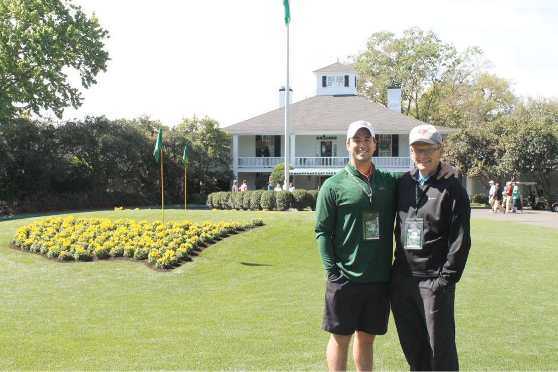
At the risk of sounding like a “Christmas letter,” let me update you on my family. Now that my wife’s father is no longer able, Debra serves as the comptroller for several family businesses. I am not sure how, but she finds time to play tennis for several teams. My son Michael recently moved to Boston from Dallas. In Texas, he was working for an investment firm focused on health care. In Boston, he manages the business development group for Shields Health Solutions. My daughter Katie is a mergers, acquisitions, and securities attorney here in Tampa, and her husband Andy is a real estate transactions attorney. We are all looking forward to the arrival of Clara Grace Peluso in June. She will be Katie and Andy’s first child and Debra and my first grandchild.
In our “abundant free time,” Debra and I enjoy spending time at our place on Sand Key near Clearwater Beach. When possible, we enjoy traveling and have developed our “bucket list.”
I look back at 2005-2006 with nothing but fondness. Serving as President of the College was both intellectually and personally fulfilling. It was certainly the highlight of my career.
Where are they now? What have they been up to? CHEST’s Past Presidents each forged the way for the many successes of the American College of Chest Physicians, leading to enhanced patient care around the globe. Their outstanding leadership and vision are evidenced today in many of CHEST’s strategic initiatives. Let’s check in with W. Michael Alberts.
President 2005 - 2006
My year at the helm began in Montreal in 2005 and ended in Salt Lake City in 2006. The year was a blur and seemed to fly by. The inauguration was very special as my entire immediate family made the effort to attend. It was the final time that my father was able to travel. Travel was definitely one of the highlights of my Presidential year. My wife, Debra, and I made many lasting friendships and very special memories while on the road for the College.
Looking back, it is hard to believe that I have been with the University of South Florida since 1983. I came to Tampa directly from my Pulmonary and Critical Care Fellowship in San Diego. After 16 years attending at the Tampa General Hospital and the James A. Haley VA, I was named the Chief Medical Officer at the Moffitt Cancer Center in 1999. In 2015, I stepped down from that position and have been serving as the Medical Director of Moffitt’s satellite clinical location since that time. I no longer do in-patient rounding, which is a major boon to work-life balance. In addition to administrative duties , however, I continue to see outpatients two half-days a week.
At the risk of sounding like a “Christmas letter,” let me update you on my family. Now that my wife’s father is no longer able, Debra serves as the comptroller for several family businesses. I am not sure how, but she finds time to play tennis for several teams. My son Michael recently moved to Boston from Dallas. In Texas, he was working for an investment firm focused on health care. In Boston, he manages the business development group for Shields Health Solutions. My daughter Katie is a mergers, acquisitions, and securities attorney here in Tampa, and her husband Andy is a real estate transactions attorney. We are all looking forward to the arrival of Clara Grace Peluso in June. She will be Katie and Andy’s first child and Debra and my first grandchild.
In our “abundant free time,” Debra and I enjoy spending time at our place on Sand Key near Clearwater Beach. When possible, we enjoy traveling and have developed our “bucket list.”
I look back at 2005-2006 with nothing but fondness. Serving as President of the College was both intellectually and personally fulfilling. It was certainly the highlight of my career.
Where are they now? What have they been up to? CHEST’s Past Presidents each forged the way for the many successes of the American College of Chest Physicians, leading to enhanced patient care around the globe. Their outstanding leadership and vision are evidenced today in many of CHEST’s strategic initiatives. Let’s check in with W. Michael Alberts.
President 2005 - 2006
My year at the helm began in Montreal in 2005 and ended in Salt Lake City in 2006. The year was a blur and seemed to fly by. The inauguration was very special as my entire immediate family made the effort to attend. It was the final time that my father was able to travel. Travel was definitely one of the highlights of my Presidential year. My wife, Debra, and I made many lasting friendships and very special memories while on the road for the College.
Looking back, it is hard to believe that I have been with the University of South Florida since 1983. I came to Tampa directly from my Pulmonary and Critical Care Fellowship in San Diego. After 16 years attending at the Tampa General Hospital and the James A. Haley VA, I was named the Chief Medical Officer at the Moffitt Cancer Center in 1999. In 2015, I stepped down from that position and have been serving as the Medical Director of Moffitt’s satellite clinical location since that time. I no longer do in-patient rounding, which is a major boon to work-life balance. In addition to administrative duties , however, I continue to see outpatients two half-days a week.
At the risk of sounding like a “Christmas letter,” let me update you on my family. Now that my wife’s father is no longer able, Debra serves as the comptroller for several family businesses. I am not sure how, but she finds time to play tennis for several teams. My son Michael recently moved to Boston from Dallas. In Texas, he was working for an investment firm focused on health care. In Boston, he manages the business development group for Shields Health Solutions. My daughter Katie is a mergers, acquisitions, and securities attorney here in Tampa, and her husband Andy is a real estate transactions attorney. We are all looking forward to the arrival of Clara Grace Peluso in June. She will be Katie and Andy’s first child and Debra and my first grandchild.
In our “abundant free time,” Debra and I enjoy spending time at our place on Sand Key near Clearwater Beach. When possible, we enjoy traveling and have developed our “bucket list.”
I look back at 2005-2006 with nothing but fondness. Serving as President of the College was both intellectually and personally fulfilling. It was certainly the highlight of my career.
EVATAR model: Endocrinology on a chip
into how female reproductive organs communicate with each other and the liver via the endocrine system.
The project was first funded in 2012 by a National Institutes of Health grant that aimed to spur development of experimental hardware, and now researchers at Northwestern University, Chicago, headed by Teresa K. Woodruff, PhD, have bioengineered a 3-D model of the female reproductive tract and the liver using living cells and tissues. The resulting model is small enough to fit in the palm of your hand.
“Cells in aggregate organs are in constant communication with each other and the conversation is provided by the endocrine system. This is a completely new era in biology helping us illuminate disease systems we could not otherwise, such as polycystic ovary disease [PCOS],” said Dr. Woodruff, who is the Thomas J. Watkins Memorial professor of obstetrics and gynecology, the vice chair of research for the department of obstetrics and gynecology, and the chief of the division of reproductive science and medicine at Northwestern University.
Until the development of EVATAR, there was no good animal or other model of female reproductive diseases, such as cancer, PCOS, and infertility.
The EVATAR 3-D system is composed of plastic cubes, each representing the target organs of the female reproductive tract. The tissue lining each cube is assembled from multiple cell lineages to create individual follicles that enclose and support oocytes, oviductal/fallopian tubes, uterine myometrium and endometrium, the cervix, and the vagina. The cubes are then connected with tiny tubes, so that the fluid representing human blood and the hormones contained therein can flow between each of the cube compartments.
The EVATAR also contains cells from the liver. Using this system, researchers can study the effects of drugs on the female reproductive system and examine the drug metabolites in the liver.
Variations on EVATAR currently in development include 3-D models of the male reproductive system, dubbed “Guy in a Cube,” she quipped.
into how female reproductive organs communicate with each other and the liver via the endocrine system.
The project was first funded in 2012 by a National Institutes of Health grant that aimed to spur development of experimental hardware, and now researchers at Northwestern University, Chicago, headed by Teresa K. Woodruff, PhD, have bioengineered a 3-D model of the female reproductive tract and the liver using living cells and tissues. The resulting model is small enough to fit in the palm of your hand.
“Cells in aggregate organs are in constant communication with each other and the conversation is provided by the endocrine system. This is a completely new era in biology helping us illuminate disease systems we could not otherwise, such as polycystic ovary disease [PCOS],” said Dr. Woodruff, who is the Thomas J. Watkins Memorial professor of obstetrics and gynecology, the vice chair of research for the department of obstetrics and gynecology, and the chief of the division of reproductive science and medicine at Northwestern University.
Until the development of EVATAR, there was no good animal or other model of female reproductive diseases, such as cancer, PCOS, and infertility.
The EVATAR 3-D system is composed of plastic cubes, each representing the target organs of the female reproductive tract. The tissue lining each cube is assembled from multiple cell lineages to create individual follicles that enclose and support oocytes, oviductal/fallopian tubes, uterine myometrium and endometrium, the cervix, and the vagina. The cubes are then connected with tiny tubes, so that the fluid representing human blood and the hormones contained therein can flow between each of the cube compartments.
The EVATAR also contains cells from the liver. Using this system, researchers can study the effects of drugs on the female reproductive system and examine the drug metabolites in the liver.
Variations on EVATAR currently in development include 3-D models of the male reproductive system, dubbed “Guy in a Cube,” she quipped.
into how female reproductive organs communicate with each other and the liver via the endocrine system.
The project was first funded in 2012 by a National Institutes of Health grant that aimed to spur development of experimental hardware, and now researchers at Northwestern University, Chicago, headed by Teresa K. Woodruff, PhD, have bioengineered a 3-D model of the female reproductive tract and the liver using living cells and tissues. The resulting model is small enough to fit in the palm of your hand.
“Cells in aggregate organs are in constant communication with each other and the conversation is provided by the endocrine system. This is a completely new era in biology helping us illuminate disease systems we could not otherwise, such as polycystic ovary disease [PCOS],” said Dr. Woodruff, who is the Thomas J. Watkins Memorial professor of obstetrics and gynecology, the vice chair of research for the department of obstetrics and gynecology, and the chief of the division of reproductive science and medicine at Northwestern University.
Until the development of EVATAR, there was no good animal or other model of female reproductive diseases, such as cancer, PCOS, and infertility.
The EVATAR 3-D system is composed of plastic cubes, each representing the target organs of the female reproductive tract. The tissue lining each cube is assembled from multiple cell lineages to create individual follicles that enclose and support oocytes, oviductal/fallopian tubes, uterine myometrium and endometrium, the cervix, and the vagina. The cubes are then connected with tiny tubes, so that the fluid representing human blood and the hormones contained therein can flow between each of the cube compartments.
The EVATAR also contains cells from the liver. Using this system, researchers can study the effects of drugs on the female reproductive system and examine the drug metabolites in the liver.
Variations on EVATAR currently in development include 3-D models of the male reproductive system, dubbed “Guy in a Cube,” she quipped.
Get Ready for CHEST 2018 in San Antonio
Have you been thinking about how great of a time you had at CHEST 2017? Or, perhaps you weren’t able to make it to CHEST 2017 and are looking forward to attending CHEST 2018? Well, we’d be happy to have you attend the annual meeting in sunny San Antonio, Texas, this fall. CHEST 2018 will occur earlier this year, from October 6-10, and we’ve got a few ways you can get involved leading up to the meeting.
CHEST 2018 Moderators
If you do not have original research to share, but believe you are qualified to moderate sessions, we have an opportunity for you! Moderating will take place on-site in San Antonio, and moderators will be recognized in the CHEST 2018 program and will receive a reduced registration rate to the meeting. See chestmeeting.chestnet.org.
CHEST Challenge 2018
Are you a US-based CHEST fellow-in-training? Compete with other programs across the country in CHEST Challenge 2018 for honor and prizes! The first round of the competition this year will consist of two parts; in addition to the traditional online quiz, there will be a number of social media challenges. The aggregate score for both of these components will be used to identify the top three-scoring teams. These top three teams will then be invited to send three fellows each to the CHEST Challenge Championship, a Jeopardy-style game show that takes place live during the CHEST Annual Meeting. http://www.chestnet.org/Hidden-Pages/CHEST-Challenge-US
CHEST Foundation Grants
We have had many talented and passionate people win our CHEST Foundation grants in research and community service. Each year, the CHEST Foundation offers grants to worthy research candidates, generous community service volunteers, and distinguished scholars in a field of expertise. Nearly 800 recipients worldwide have received more than $10 million in support and recognition of outstanding contributions to chest medicine.
How are you helping to champion lung health? The CHEST Foundation is accepting grant applications February 1 through April 9, 2018, in the following areas:
• CHEST Foundation Research Grant in Lung Cancer – $50,000 - $100,000 2-year grant*
• CHEST Foundation Research Grant in Asthma – $15,000 - $30,000 1-year grant*
• CHEST Foundation Research Grant in Pulmonary Arterial Hypertension – $25,000 - $50,000 1-year grant*
• CHEST Foundation and the Alpha-1 Foundation Research Grant in Alpha-1 Antitrypsin Deficiency – $25,000 - $50,000 1-year grant*
• CHEST Foundation Research Grant in Pulmonary Fibrosis – $25,000 - $50,000 1-year grant*
• CHEST Foundation Research Grant in Chronic Obstructive Pulmonary Disease – $30,000 1-year grant (multiple recipients selected)
• CHEST Foundation Research Grant in Venous Thromboembolism – $15,000 - $30,000 1-year grant*
• CHEST Foundation Research Grant in Nontuberculous Mycobacteria Disease – $25,000 - $50,000 1-year grant*
• CHEST Foundation Research Grant in Women’s Lung Health – $10,000 1-year grant
• CHEST Foundation Research Grant in Cystic Fibrosis – $30,000 1-year grant
• The Eli Lilly and Company Distinguished Scholar in Critical Care Medicine – $150,000 over 3 years
• CHEST Foundation Community Service Grant Honoring D. Robert McCaffree, MD, Master FCCP – $2,500- $15,000 1-year grant*
*Amount contingent on funding.
Learn more on how to apply now at chestfoundation.org/apply.
Things Happening in AprilDon’t forget to look out for CHEST 2018 registration, opening April 5. And, if you missed the first round of abstract submissions, submissions for late-breaking abstracts will open April 30. Stay updated on all things CHEST 2018 at chestmeeting.chestnet.org.
Have you been thinking about how great of a time you had at CHEST 2017? Or, perhaps you weren’t able to make it to CHEST 2017 and are looking forward to attending CHEST 2018? Well, we’d be happy to have you attend the annual meeting in sunny San Antonio, Texas, this fall. CHEST 2018 will occur earlier this year, from October 6-10, and we’ve got a few ways you can get involved leading up to the meeting.
CHEST 2018 Moderators
If you do not have original research to share, but believe you are qualified to moderate sessions, we have an opportunity for you! Moderating will take place on-site in San Antonio, and moderators will be recognized in the CHEST 2018 program and will receive a reduced registration rate to the meeting. See chestmeeting.chestnet.org.
CHEST Challenge 2018
Are you a US-based CHEST fellow-in-training? Compete with other programs across the country in CHEST Challenge 2018 for honor and prizes! The first round of the competition this year will consist of two parts; in addition to the traditional online quiz, there will be a number of social media challenges. The aggregate score for both of these components will be used to identify the top three-scoring teams. These top three teams will then be invited to send three fellows each to the CHEST Challenge Championship, a Jeopardy-style game show that takes place live during the CHEST Annual Meeting. http://www.chestnet.org/Hidden-Pages/CHEST-Challenge-US
CHEST Foundation Grants
We have had many talented and passionate people win our CHEST Foundation grants in research and community service. Each year, the CHEST Foundation offers grants to worthy research candidates, generous community service volunteers, and distinguished scholars in a field of expertise. Nearly 800 recipients worldwide have received more than $10 million in support and recognition of outstanding contributions to chest medicine.
How are you helping to champion lung health? The CHEST Foundation is accepting grant applications February 1 through April 9, 2018, in the following areas:
• CHEST Foundation Research Grant in Lung Cancer – $50,000 - $100,000 2-year grant*
• CHEST Foundation Research Grant in Asthma – $15,000 - $30,000 1-year grant*
• CHEST Foundation Research Grant in Pulmonary Arterial Hypertension – $25,000 - $50,000 1-year grant*
• CHEST Foundation and the Alpha-1 Foundation Research Grant in Alpha-1 Antitrypsin Deficiency – $25,000 - $50,000 1-year grant*
• CHEST Foundation Research Grant in Pulmonary Fibrosis – $25,000 - $50,000 1-year grant*
• CHEST Foundation Research Grant in Chronic Obstructive Pulmonary Disease – $30,000 1-year grant (multiple recipients selected)
• CHEST Foundation Research Grant in Venous Thromboembolism – $15,000 - $30,000 1-year grant*
• CHEST Foundation Research Grant in Nontuberculous Mycobacteria Disease – $25,000 - $50,000 1-year grant*
• CHEST Foundation Research Grant in Women’s Lung Health – $10,000 1-year grant
• CHEST Foundation Research Grant in Cystic Fibrosis – $30,000 1-year grant
• The Eli Lilly and Company Distinguished Scholar in Critical Care Medicine – $150,000 over 3 years
• CHEST Foundation Community Service Grant Honoring D. Robert McCaffree, MD, Master FCCP – $2,500- $15,000 1-year grant*
*Amount contingent on funding.
Learn more on how to apply now at chestfoundation.org/apply.
Things Happening in AprilDon’t forget to look out for CHEST 2018 registration, opening April 5. And, if you missed the first round of abstract submissions, submissions for late-breaking abstracts will open April 30. Stay updated on all things CHEST 2018 at chestmeeting.chestnet.org.
Have you been thinking about how great of a time you had at CHEST 2017? Or, perhaps you weren’t able to make it to CHEST 2017 and are looking forward to attending CHEST 2018? Well, we’d be happy to have you attend the annual meeting in sunny San Antonio, Texas, this fall. CHEST 2018 will occur earlier this year, from October 6-10, and we’ve got a few ways you can get involved leading up to the meeting.
CHEST 2018 Moderators
If you do not have original research to share, but believe you are qualified to moderate sessions, we have an opportunity for you! Moderating will take place on-site in San Antonio, and moderators will be recognized in the CHEST 2018 program and will receive a reduced registration rate to the meeting. See chestmeeting.chestnet.org.
CHEST Challenge 2018
Are you a US-based CHEST fellow-in-training? Compete with other programs across the country in CHEST Challenge 2018 for honor and prizes! The first round of the competition this year will consist of two parts; in addition to the traditional online quiz, there will be a number of social media challenges. The aggregate score for both of these components will be used to identify the top three-scoring teams. These top three teams will then be invited to send three fellows each to the CHEST Challenge Championship, a Jeopardy-style game show that takes place live during the CHEST Annual Meeting. http://www.chestnet.org/Hidden-Pages/CHEST-Challenge-US
CHEST Foundation Grants
We have had many talented and passionate people win our CHEST Foundation grants in research and community service. Each year, the CHEST Foundation offers grants to worthy research candidates, generous community service volunteers, and distinguished scholars in a field of expertise. Nearly 800 recipients worldwide have received more than $10 million in support and recognition of outstanding contributions to chest medicine.
How are you helping to champion lung health? The CHEST Foundation is accepting grant applications February 1 through April 9, 2018, in the following areas:
• CHEST Foundation Research Grant in Lung Cancer – $50,000 - $100,000 2-year grant*
• CHEST Foundation Research Grant in Asthma – $15,000 - $30,000 1-year grant*
• CHEST Foundation Research Grant in Pulmonary Arterial Hypertension – $25,000 - $50,000 1-year grant*
• CHEST Foundation and the Alpha-1 Foundation Research Grant in Alpha-1 Antitrypsin Deficiency – $25,000 - $50,000 1-year grant*
• CHEST Foundation Research Grant in Pulmonary Fibrosis – $25,000 - $50,000 1-year grant*
• CHEST Foundation Research Grant in Chronic Obstructive Pulmonary Disease – $30,000 1-year grant (multiple recipients selected)
• CHEST Foundation Research Grant in Venous Thromboembolism – $15,000 - $30,000 1-year grant*
• CHEST Foundation Research Grant in Nontuberculous Mycobacteria Disease – $25,000 - $50,000 1-year grant*
• CHEST Foundation Research Grant in Women’s Lung Health – $10,000 1-year grant
• CHEST Foundation Research Grant in Cystic Fibrosis – $30,000 1-year grant
• The Eli Lilly and Company Distinguished Scholar in Critical Care Medicine – $150,000 over 3 years
• CHEST Foundation Community Service Grant Honoring D. Robert McCaffree, MD, Master FCCP – $2,500- $15,000 1-year grant*
*Amount contingent on funding.
Learn more on how to apply now at chestfoundation.org/apply.
Things Happening in AprilDon’t forget to look out for CHEST 2018 registration, opening April 5. And, if you missed the first round of abstract submissions, submissions for late-breaking abstracts will open April 30. Stay updated on all things CHEST 2018 at chestmeeting.chestnet.org.
NAMDRC Update Collaboration: Now More Than Ever
Ongoing hospital mergers, acquisitions, and closings demonstrate that reimbursement maximization and cost reduction are the twin sisters of health-care system reform. This focus is not going to change in the foreseeable future, as most experts now view “health system reform” as “health financing reform.”
Reports document that more than 50% of acute care hospitals in the United States experienced negative operating margins for the federal fiscal year ending September 30, 2017. Equally alarming is the increasing number of organizations either reducing or eliminating the roles of medical directors for clinical departments. Hospital and health system executives are increasingly engaging external consultants to find ways to decrease operating costs, with the caveat of maintaining or improving quality, safety, and patient satisfaction and engagement.
Given these cost reduction pressures, what can respiratory therapy medical directors and administrative directors do to ensure that quality and safety are ensured?
We believe that quality and safety can be maintained and improved in this bottom-line-focused environment if we collaborate with stakeholders and communicate the value of respiratory care services. Following are some examples of how to reinvigorate this collaboration. The list is far from complete, but we believe it is a good starting point for making a significant difference.
Science
We recognize that much of our practice is based on levels of evidence, and we must use this evidence as a basis for our services.
In talking and working with RT administrative directors across the country, we continue to see non-value-added “treatments” being provided, such as incentive spirometry and aerosolized acetylcysteine. Not only is this a waste of resources, but, because of it, our clinical RTs are not providing therapy.
One of the best opportunities to decrease cost is to eliminate waste. These services must be eliminated. For those patients who require secretion clearance/lung expansion, we can provide evidence-based services such as oscillating positive expiratory pressure.
Protocols
Respiratory care protocols have been around for decades, but surveys indicate that only half of all RT departments utilize them. Under the guidance of NAMDRC, the AARC has been educating RTs to transition from “treatments” to evidence-based protocols. Various barriers remain, and our challenge remains to implement proven care plans in every department.
Quality Assurance
The health-care industry made the transition from “Quality Control” to “Quality Assurance” several decades ago. However, many RT administrative directors lack the knowledge and/or resources necessary to create a comprehensive QA program, much less participate in clinical research. We suggest creating a standardized model to be adopted by RT departments across the country that would measure and communicate the value of respiratory care services.
Productivity/Staffing
An area where consultants and executives often focus their cost-saving efforts is staffing. Given that 50% to 60% of operating costs are personnel, this is to be expected.
Many organizations, however, are using the wrong metrics—such as procedures, CPT codes , and billables—to project staffing FTEs. Physicians and RTs understand that these metrics are not useful and must convince consultants and executives of this. The AARC Uniform Reporting Manual, which is currently being updated, is the best guide for determining appropriate staffing.
Education
Another common step in cost control has been the significant reduction or total elimination of education budgets.
During the past 5 years, RT leaders attending the AARC Summer Forum have been polled regarding whether they received financial assistance to attend the Management Section program. Sadly, the number attending on their own dime far surpasses those receiving financial assistance.
Additionally, the RT profession is witnessing more department-based education, which, in some cases, is not education at all, but marketing, cleverly packaged in the form of CEUs.
We fully understand these changes and recognize why they have occurred. However, we suggest the need to work together to differentiate marketing from education and ensure that clinical staff receive what is needed to ensure quality care.
It is vital for us to educate our physician leaders and pulmonary and critical care fellows on the science of respiratory care. There is a significant knowledge gap, and we have a great opportunity to improve the training of fellows. It is difficult to attract active medical directors if they don’t understand the science. We believe NAMDRC can play an important role by addressing these knowledge deficits.
Ongoing hospital mergers, acquisitions, and closings demonstrate that reimbursement maximization and cost reduction are the twin sisters of health-care system reform. This focus is not going to change in the foreseeable future, as most experts now view “health system reform” as “health financing reform.”
Reports document that more than 50% of acute care hospitals in the United States experienced negative operating margins for the federal fiscal year ending September 30, 2017. Equally alarming is the increasing number of organizations either reducing or eliminating the roles of medical directors for clinical departments. Hospital and health system executives are increasingly engaging external consultants to find ways to decrease operating costs, with the caveat of maintaining or improving quality, safety, and patient satisfaction and engagement.
Given these cost reduction pressures, what can respiratory therapy medical directors and administrative directors do to ensure that quality and safety are ensured?
We believe that quality and safety can be maintained and improved in this bottom-line-focused environment if we collaborate with stakeholders and communicate the value of respiratory care services. Following are some examples of how to reinvigorate this collaboration. The list is far from complete, but we believe it is a good starting point for making a significant difference.
Science
We recognize that much of our practice is based on levels of evidence, and we must use this evidence as a basis for our services.
In talking and working with RT administrative directors across the country, we continue to see non-value-added “treatments” being provided, such as incentive spirometry and aerosolized acetylcysteine. Not only is this a waste of resources, but, because of it, our clinical RTs are not providing therapy.
One of the best opportunities to decrease cost is to eliminate waste. These services must be eliminated. For those patients who require secretion clearance/lung expansion, we can provide evidence-based services such as oscillating positive expiratory pressure.
Protocols
Respiratory care protocols have been around for decades, but surveys indicate that only half of all RT departments utilize them. Under the guidance of NAMDRC, the AARC has been educating RTs to transition from “treatments” to evidence-based protocols. Various barriers remain, and our challenge remains to implement proven care plans in every department.
Quality Assurance
The health-care industry made the transition from “Quality Control” to “Quality Assurance” several decades ago. However, many RT administrative directors lack the knowledge and/or resources necessary to create a comprehensive QA program, much less participate in clinical research. We suggest creating a standardized model to be adopted by RT departments across the country that would measure and communicate the value of respiratory care services.
Productivity/Staffing
An area where consultants and executives often focus their cost-saving efforts is staffing. Given that 50% to 60% of operating costs are personnel, this is to be expected.
Many organizations, however, are using the wrong metrics—such as procedures, CPT codes , and billables—to project staffing FTEs. Physicians and RTs understand that these metrics are not useful and must convince consultants and executives of this. The AARC Uniform Reporting Manual, which is currently being updated, is the best guide for determining appropriate staffing.
Education
Another common step in cost control has been the significant reduction or total elimination of education budgets.
During the past 5 years, RT leaders attending the AARC Summer Forum have been polled regarding whether they received financial assistance to attend the Management Section program. Sadly, the number attending on their own dime far surpasses those receiving financial assistance.
Additionally, the RT profession is witnessing more department-based education, which, in some cases, is not education at all, but marketing, cleverly packaged in the form of CEUs.
We fully understand these changes and recognize why they have occurred. However, we suggest the need to work together to differentiate marketing from education and ensure that clinical staff receive what is needed to ensure quality care.
It is vital for us to educate our physician leaders and pulmonary and critical care fellows on the science of respiratory care. There is a significant knowledge gap, and we have a great opportunity to improve the training of fellows. It is difficult to attract active medical directors if they don’t understand the science. We believe NAMDRC can play an important role by addressing these knowledge deficits.
Ongoing hospital mergers, acquisitions, and closings demonstrate that reimbursement maximization and cost reduction are the twin sisters of health-care system reform. This focus is not going to change in the foreseeable future, as most experts now view “health system reform” as “health financing reform.”
Reports document that more than 50% of acute care hospitals in the United States experienced negative operating margins for the federal fiscal year ending September 30, 2017. Equally alarming is the increasing number of organizations either reducing or eliminating the roles of medical directors for clinical departments. Hospital and health system executives are increasingly engaging external consultants to find ways to decrease operating costs, with the caveat of maintaining or improving quality, safety, and patient satisfaction and engagement.
Given these cost reduction pressures, what can respiratory therapy medical directors and administrative directors do to ensure that quality and safety are ensured?
We believe that quality and safety can be maintained and improved in this bottom-line-focused environment if we collaborate with stakeholders and communicate the value of respiratory care services. Following are some examples of how to reinvigorate this collaboration. The list is far from complete, but we believe it is a good starting point for making a significant difference.
Science
We recognize that much of our practice is based on levels of evidence, and we must use this evidence as a basis for our services.
In talking and working with RT administrative directors across the country, we continue to see non-value-added “treatments” being provided, such as incentive spirometry and aerosolized acetylcysteine. Not only is this a waste of resources, but, because of it, our clinical RTs are not providing therapy.
One of the best opportunities to decrease cost is to eliminate waste. These services must be eliminated. For those patients who require secretion clearance/lung expansion, we can provide evidence-based services such as oscillating positive expiratory pressure.
Protocols
Respiratory care protocols have been around for decades, but surveys indicate that only half of all RT departments utilize them. Under the guidance of NAMDRC, the AARC has been educating RTs to transition from “treatments” to evidence-based protocols. Various barriers remain, and our challenge remains to implement proven care plans in every department.
Quality Assurance
The health-care industry made the transition from “Quality Control” to “Quality Assurance” several decades ago. However, many RT administrative directors lack the knowledge and/or resources necessary to create a comprehensive QA program, much less participate in clinical research. We suggest creating a standardized model to be adopted by RT departments across the country that would measure and communicate the value of respiratory care services.
Productivity/Staffing
An area where consultants and executives often focus their cost-saving efforts is staffing. Given that 50% to 60% of operating costs are personnel, this is to be expected.
Many organizations, however, are using the wrong metrics—such as procedures, CPT codes , and billables—to project staffing FTEs. Physicians and RTs understand that these metrics are not useful and must convince consultants and executives of this. The AARC Uniform Reporting Manual, which is currently being updated, is the best guide for determining appropriate staffing.
Education
Another common step in cost control has been the significant reduction or total elimination of education budgets.
During the past 5 years, RT leaders attending the AARC Summer Forum have been polled regarding whether they received financial assistance to attend the Management Section program. Sadly, the number attending on their own dime far surpasses those receiving financial assistance.
Additionally, the RT profession is witnessing more department-based education, which, in some cases, is not education at all, but marketing, cleverly packaged in the form of CEUs.
We fully understand these changes and recognize why they have occurred. However, we suggest the need to work together to differentiate marketing from education and ensure that clinical staff receive what is needed to ensure quality care.
It is vital for us to educate our physician leaders and pulmonary and critical care fellows on the science of respiratory care. There is a significant knowledge gap, and we have a great opportunity to improve the training of fellows. It is difficult to attract active medical directors if they don’t understand the science. We believe NAMDRC can play an important role by addressing these knowledge deficits.
Office staff cohesiveness remains important
Recently, a lawyer and I were on the phone about a case, and he mentioned how lucky he was to have had the same staff over the course of a 25-year career, and he was afraid they’d retire before he did.
I feel the same way. My medical assistant has been with me since day one 18 years ago, my secretary since 2004. I hope they keep putting up with me until I hang up my reflex hammer.
It’s impossible to put a great team together just by talent alone. The chemistry and ability to work together are as critical as talent, if not more so, and are far more intangible and unpredictable.
I’ve been lucky that way. The three of us are cohesive. We try to keep some degree of fun in our work routine as each day goes on. My secretary’s 2-year-old daughter, who comes with her every day, adds to the family atmosphere that many patients notice. It’s not uncommon to be asked if the group of us are related. (We’re not.)
Back when I started out, it was with a large group that saw its people as replaceable, and treated them as such. As a result there was a high turnover rate of medical assistants and front office and clerical staff. This led to problems with work-flow and patient care, as there was always someone new being trained.

I can’t imagine having a better team than I have now, and will share this advice with anyone starting a practice: When you get the right people, count yourself lucky and do what you have to do to keep them. They’re worth it.
It’s a lot nicer to work with friends than strangers. It makes the drudgery of the job more interesting, the low points better, and the highs fun.
I wouldn’t have it any other way.
Dr. Block has a solo neurology practice in Scottsdale, Ariz.
Recently, a lawyer and I were on the phone about a case, and he mentioned how lucky he was to have had the same staff over the course of a 25-year career, and he was afraid they’d retire before he did.
I feel the same way. My medical assistant has been with me since day one 18 years ago, my secretary since 2004. I hope they keep putting up with me until I hang up my reflex hammer.
It’s impossible to put a great team together just by talent alone. The chemistry and ability to work together are as critical as talent, if not more so, and are far more intangible and unpredictable.
I’ve been lucky that way. The three of us are cohesive. We try to keep some degree of fun in our work routine as each day goes on. My secretary’s 2-year-old daughter, who comes with her every day, adds to the family atmosphere that many patients notice. It’s not uncommon to be asked if the group of us are related. (We’re not.)
Back when I started out, it was with a large group that saw its people as replaceable, and treated them as such. As a result there was a high turnover rate of medical assistants and front office and clerical staff. This led to problems with work-flow and patient care, as there was always someone new being trained.
I can’t imagine having a better team than I have now, and will share this advice with anyone starting a practice: When you get the right people, count yourself lucky and do what you have to do to keep them. They’re worth it.
It’s a lot nicer to work with friends than strangers. It makes the drudgery of the job more interesting, the low points better, and the highs fun.
I wouldn’t have it any other way.
Dr. Block has a solo neurology practice in Scottsdale, Ariz.
Recently, a lawyer and I were on the phone about a case, and he mentioned how lucky he was to have had the same staff over the course of a 25-year career, and he was afraid they’d retire before he did.
I feel the same way. My medical assistant has been with me since day one 18 years ago, my secretary since 2004. I hope they keep putting up with me until I hang up my reflex hammer.
It’s impossible to put a great team together just by talent alone. The chemistry and ability to work together are as critical as talent, if not more so, and are far more intangible and unpredictable.
I’ve been lucky that way. The three of us are cohesive. We try to keep some degree of fun in our work routine as each day goes on. My secretary’s 2-year-old daughter, who comes with her every day, adds to the family atmosphere that many patients notice. It’s not uncommon to be asked if the group of us are related. (We’re not.)
Back when I started out, it was with a large group that saw its people as replaceable, and treated them as such. As a result there was a high turnover rate of medical assistants and front office and clerical staff. This led to problems with work-flow and patient care, as there was always someone new being trained.
I can’t imagine having a better team than I have now, and will share this advice with anyone starting a practice: When you get the right people, count yourself lucky and do what you have to do to keep them. They’re worth it.
It’s a lot nicer to work with friends than strangers. It makes the drudgery of the job more interesting, the low points better, and the highs fun.
I wouldn’t have it any other way.
Dr. Block has a solo neurology practice in Scottsdale, Ariz.