User login
FDA OKs first-in-class HIV therapy for patients with few options
Fostemsavir is indicated for use in combination with other antiretroviral (ARV) agents in heavily treatment-experienced adults with multidrug-resistant HIV-1 infection who fail to achieve viral suppression on other regimens due to resistance, intolerance, or safety considerations.
“This approval marks a new class of antiretroviral medications that may benefit patients who have run out of HIV treatment options,” Jeff Murray, MD, deputy director of the Division of Antivirals in the FDA’s Center for Drug Evaluation and Research, said in a statement.
“The availability of new classes of antiretroviral drugs is critical for heavily treatment-experienced patients living with multidrug resistant HIV infection — helping people living with hard-to-treat HIV who are at greater risk for HIV-related complications to potentially live longer, healthier lives,” he said.
Fostemsavir 600 mg extended-release tablets are taken twice daily.
In the phase 3 BRIGHTE study, 60% of adults who added fostemsavir to optimized background ARV therapy achieved and maintained viral suppression through 96 weeks and saw clinically meaningful improvements in CD4+ T cells.
Most of the 371 participants in the study had been on anti-HIV therapy for more than 15 years (71%), had been exposed to five or more different HIV treatment regimens (85%), and/or had a history of AIDS (86%).
The most common adverse reactions with fostemsavir are nausea, fatigue, and diarrhea. Serious drug reactions included liver enzyme elevations in patients co-infected with hepatitis B or C virus and three cases of severe immune reconstitution inflammatory syndrome.
“Exciting” Advance
“There is a small group of heavily treatment-experienced adults living with HIV who are not able to maintain viral suppression with currently available medication and, without effective new options, are at great risk of progressing to AIDS,” Deborah Waterhouse, CEO of ViiV Healthcare, said in a news release.
“The approval of Rukobia is a culmination of incredibly complex research, development, and manufacturing efforts to ensure we leave no person living with HIV behind,” she said.
“As a novel HIV attachment inhibitor, fostemsavir targets the first step of the viral lifecycle offering a new mechanism of action to treat people living with HIV,” Jacob P. Lalezari, MD, chief executive officer and director of Quest Clinical Research, commented in the release.
Fostemsavir is an “exciting” advance for the heavily treatment-experienced population and “an advancement the HIV community has long been waiting for. As an activist as well as researcher, I am very grateful to ViiV Healthcare for their commitment to heavily-treatment experienced people living with HIV,” he added.
Fostemsavir was reviewed and approved under the FDA’s fast track and breakthrough therapy designations, which are intended to facilitate and expedite the development and review of new drugs to address unmet medical need in the treatment of a serious or life-threatening condition.
Full prescribing information is available online.
This article first appeared on Medscape.com.
Entrectinib results emphasize need for NTRK detection
Although fusions in the neurotrophic receptor tyrosine kinase (NTRK) gene are rare in gastrointestinal carcinomas (found in fewer than 5% of cases), they should be looked for, inasmuch as treatment with the TRK inhibitor entrectinib (Rozlytrek, Genentech/Roche) can achieve robust and durable responses, say researchers.
This point was made during several presentations at the virtual World Conference on Gastrointestinal Cancer (WCGC) 2020 on July 1.
Entrectinib and similar agents that act on NTRK fusion genes are described as tumor agnostic, in that they are biomarkers that define the cancer rather than the organ of origin.
The Food and Drug Administration last year granted accelerated approval of entrectinib for patients with locally advanced or metastatic NTRK-expressing solid tumors that have progressed following prior therapies. The drug can also be used as a first-line treatment when there are no effective therapies.
At the meeting, Manish R. Patel, MD, Department of Medicine, University of Minnesota, Minneapolis, and colleagues presented combined results from the ALKA-372-001, STARTRK-1, and STARTRK-2 studies of entrectinib.
They identified 12 gastrointestinal carcinoma patients among 74 adults with locally advanced/metastatic NTRK fusion positive, TRK inhibitor-naive solid tumors who had undergone at least 6 months of follow-up.
Many of these 12 patients had colorectal cancer (58%) or pancreatic cancer (24%).
Treatment with entrectinib elicited an overall response rate of 50%, which consisted entirely of partial responses.
The median duration of response was 12.9 months, which was largely driven by a median duration of response of 15.1 months among colorectal cancer patients, vs 10.0 months among pancreatic cancer patients and 9.3 months in the patient with cholangiocarcinoma.
The median progression-free survival was 7.1 months across the whole cohort. It was 8.0 months for pancreatic cancer patients and 12.0 months for the patient with cholangiocarcinoma.
The median overall survival was 16 months.
Dr. Patel said that this “demonstrates that entrectinib induces durable and clinically meaningful systemic responses in patients with gastrointestinal carcinomas harboring NTRK fusions.”
He noted that entrectinib is “overall very well tolerated, with very few dose interruptions or reductions, and the discontinuation rate was very low.” The majority of adverse events were of grade 1/2. The most common event was change in taste, which occurred in 37.3% of patients. There were no treatment-related deaths.
“The main take-away point from this abstract is that, though they are rare, if we identify patients with NTRK fusions during the course of the disease, we can offer them benefit from entrectinib, and I would argue that ... we should be screening patients for NTRK fusions much more frequently,” Dr. Patel added.
In the second study, a Belgian team performed immunohistochemistry (IHC) analysis followed by next-generation sequencing on archived samples of biliopancreatic cancers to determine the prevalence of NTRK fusions.
They found just one fusion among almost 150 biliary tract cancers and none in nearly 300 pancreatic adenocarcinomas.
Lead author Anne Demols, MD, PhD, Department of Gastroenterology and Gastrointestinal Oncology, CUB Hôpital Erasme, Brussels, Belgium, said the results show that, “consistent” with their low frequency in solid tumors, NTRK gene fusions are “also rare” in biliopancreatic cancers.
“Given this low frequency, testing and identification are of high clinical importance, due to possible treatment with pan-TRK inhibitors,” she said.
She added that a two-step diagnosis is recommended, in that it is both “time saving” and economical, and that next-generation sequencing is “mandatory” to confirm a positive result on IHC.
For discussant Juan W. Valle, MD, professor of medical oncology at the University of Manchester, United Kingdom, the results of the second study reinforce the take-home message of the first.
He said that “two-step diagnosis can preselect patients suitable for next-generation sequencing assay, and what we saw from the previous [study] is that the therapeutic implications make this an important diagnosis.”
Valle noted that there is an “immortal time bias” in the trial analysis, because patients had to be well enough to undergo at least 6 months of follow-up, and that “future work will focus on the best platform to use for known, as well as the identification of new, fusion partners.”
He highlighted the “improved response rate and progression-free survival” achieved with entrectinib among patients with gastrointestinal cancers harboring NTRK fusions, which will benefit patient outcomes.
Pashtoon Kasi, MD, a gastrointestinal oncologist at the University of Iowa Holden Comprehensive Cancer Center, Iowa City, commented on Twitter that, for him, the results were more than just about the impressive response rate but how “brisk, robust, and durable these tend to be.”
In his experience, even patients with stage IV disease who have responded to entrectinib have been able to undergo “secondary ‘curative’ resections.”
The ALKA-372-001, STARTRK-1 and STARTRK-2 studies were sponsored by F. Hoffmann-La Roche. The study by Demols and colleagues was funded by a research grant from Bayer Health. Dr. Patel reports relationships with Nektar Therapeutic, MSD, and Fate Therapeutics. Demols reports relationships with Bayer, Ipsen, Vifor, Servier and Roche. Dr. Valle reports relationships with numerous companies.
This article first appeared on Medscape.com.
Radiomics can identify high-risk early stage lung cancer
Radiomics, a growing area of cancer research that extracts noninvasive biomarkers from medical imaging, may be able to improve lung cancer screening by identifying patients with early stage disease at high risk for poorer outcomes.
This is the conclusion from a group of researchers who used data from the National Lung Screening Trial (NLST) to develop and validate a model based on radiomics that could identify a vulnerable high-risk group of early stage patients associated with poor outcomes. These patients would generally require aggressive follow-up and/or adjuvant therapy.
The study was published June 29 in Nature Scientific Reports.
Radiomics, also known as quantitative image features, are noninvasive biomarkers that are generated from medical imaging. An emerging translational field of research, radiomics extracts large amounts of features from radiographic medical images using data-characterization algorithms, which reflect the underlying tumor pathophysiology and heterogeneity.
The authors note that radiomics has many advantages over circulating and tissue-based biomarkers, as these quantitative image features are rapidly calculated from standard-of-care imaging and reflect the entire tumor burden – and not just a sample as is the case with tissue-based biomarkers.
“We view radiomics as a decision support tool across the cancer control continuum, whether it be screening and early detection, diagnosis, prognostication, or treatment response,” said lead author Matthew B. Schabath, PhD, associate member in cancer epidemiology at the H. Lee Moffitt Cancer Center & Research Institute in Tampa, Florida.
“Radiomic features are generated from standard-of-care imaging and validated radiomic models can provide real-time decision support information to clinicians,” he explained.
Last year, another study showed that combining radiomics and imaging may be able to determine which patients with lung cancer were most likely to respond to chemotherapy. The researchers used CT imaging of radiomic features from within and outside the lung nodule and found it could predict time to progression and overall survival, as well as response to chemotherapy, in patients with non–small cell lung cancer (NSCLC).
Anant Madabhushi, PhD, a professor of biomedical engineering and director of the Center for Computational Imaging and Personalized Diagnostics at Case Western Reserve University, Cleveland, commented that the new study is “complementary and supports the premise that radiomics both from inside and outside the tumor can tell us about outcome and treatment response.”
Dr. Madabhushi also noted his group has released several other studies along similar lines, including a study showing how radiomics can predict the benefit of adjuvant therapy in lung cancer, a study showing how radiomics can predict recurrence in early stage NSCLC, and a study showing that radiomics can predict survival and response to immunotherapy in NSCLC.
Improving current lung cancer screening
The landmark NLST showed that, as compared with chest x-rays, low-dose helical computed tomography (LDCT) was associated with a 20% relative reduction in lung cancer mortality in high-risk individuals. However, LDCT screening can lead to overdiagnosis and subsequent overtreatment of slow-growing, indolent cancers.
“Current lung cancer screening inclusion criteria in the US are largely based on the criteria used in the NLST,” Dr. Schabath told Medscape Medical News. “Though the NLST clearly demonstrated that screening LDCT is a lifesaving tool, the NLST was not designed to create public policy.”
He pointed out that fewer than 30% of Americans diagnosed with lung cancer meet the current screening entry criteria and that subsequent trials (e.g., NELSON, LUSI, or MILD) used broader and more inclusive criteria and also showed the efficacy of LDCT for early detection of lung cancer. “Thus, there should be consideration in making the lung cancer screening guidelines more inclusive,” said Dr. Schabath.
“Additionally, adjunct risk-stratification tools, such as blood-based biomarkers, could be an important complement to determine who should be part of a lung cancer screening program,” he said. “This could be particularly salient for people who have no or very few risk factors, such as never smokers.”
Pinpointing poor outcomes
In the current study, Dr. Schabath and colleagues used publicly available data and LDCT images from the NLST to generate radiomic features from screen detected, incidentally-diagnosed lung cancers. Radiomic features describing size, shape, volume, and textural characteristics were then calculated from both the intratumoral and peritumoral regions.
Patients were divided into training and test cohorts, and an external cohort of non-screen-detected lung cancer patients was used for further validation. There were no statistically significant differences between training and test cohorts for most demographics, including age, sex, smoking status, number of pack-years smoked, treatment, stage, and baseline screening result. However, self-reported chronic obstructive pulmonary disease (COPD) was significantly higher in the test cohort compared with the training group (16% vs. 7%; P = .02).
A total of 91 stable and reproducible radiomics features (peritumoral and intratumoral) were identified and 40 (26 peritumoral and 14 intratumoral) were significantly associated with overall survival in the training cohort. The features were subsequently narrowed to four, and backward elimination analyses identified a single model. Patients were then stratified into three risk-groups: low risk, intermediate risk, and high risk.
According to their model, the high-risk group had worse overall survival (hazard ratio, 9.91; 25% 2.5-year and 0% 5-year OS) as compared with the low-risk group (HR, 1.00; 93% 2.5-year and 78% 5-year OS).
The final model was validated in the test group and then replicated in the non–screen-detected patients with adenocarcinoma patients. Since the disease stage differed significantly across the risk groups, the model was stratified by stage and the authors found “compelling” results among early-stage patients, who generally have good outcomes. In this subset, the high-risk group was associated with a worse overall survival (HR, 2.63; 56% 2.5-year and 42% 5-year OS) vs. the low-risk group (HR, 1.00; 75% 2.5-year and 75% 5-year OS).
“We have ongoing studies to determine if these results are consistent in the real-world setting of lung cancer screening across multiple centers,” said Dr. Schabath. “If the NELSON, LUSI, or MILD trial data become publicly available, we will certainly pursue validating our results in those clinical trials.”
The study was funded by the National Cancer Institute. Dr. Schabath and Dr. Madabhushi have disclosed no relevant financial relationships.
This article first appeared on Medscape.com.
Intermittent fasting ‘not benign’ for patients with diabetes
stress the authors of a new viewpoint published online July 2 in JAMA.
This is because intermittent fasting in patients with type 2 diabetes has only been studied in seven small, short published trials of very different regimens, with limited evidence of benefit. In addition, some concerns arose from these studies.
Weight loss with intermittent fasting appears to be similar to that attained with caloric restriction, but in the case of those with diabetes, the best way to adjust glucose-lowering medicines to reduce the risk of hypoglycemia while practicing intermittent fasting has not been established, and there is potential for such fasting to cause glycemic variability.
The viewpoint’s lead author Benjamin D. Horne, PhD, MStat, MPH, from Intermountain Medical Center, Salt Lake City, and Stanford (Calif.) University, expanded on the issues in a podcast interview with JAMA editor in chief Howard C. Bauchner, MD.
Asked if he would advise intermittent fasting for patients with type 2 diabetes, Dr. Horne replied that he would recommend it, with caveats, “because of the safety issues – some of which are fairly benign for people who are apparently healthy but may be not quite as benign for people with type 2 diabetes.
“Things such as low blood pressure, weakness, headaches, [and] dizziness are considerations,” he continued, but “the big issue” is hypoglycemia, so caloric restriction may be a better choice for some patients with diabetes.
Dr. Horne said he likes to give patients options. “I’ve met quite a number of people who are very behind time-restricted feeding – eating during a 6- to 8-hour window,” he said. “If they are able to stay on it, they tend to really love it.”
The most popular regimen that results in some weight loss is fasting for 24 hours – with or without a 500-calorie meal – on 2 nonconsecutive days a week, the so-called 5:2 diet. And “as someone who’s in cardiovascular research,” Dr. Horne added, “the one that I’m thinking for long term is once-a-week fasting for a 24-hour period.”
Intermittent fasting: Less safe than calorie restriction in diabetes?
Patients who already have diabetes and lose weight benefit from improved glucose, blood pressure, and lipid levels, Dr. Horne and colleagues wrote.
Currently, intermittent fasting is popular in the lay press and on social media with claims of potential benefits for diabetes “that are as yet untested or unproven,” they added. In fact, “whether a patient with type 2 diabetes should engage in intermittent fasting involves a variety of concerns over safety and efficacy.”
Thus, they examined the existing evidence for the health effects and safety of intermittent fasting – defined as time-restricted feeding, or fasting on alternate days or during 1-4 days a week, with only water or also juice and bone broth, or no more than 700 calories allowed on fasting days – in patients with type 2 diabetes.
They found seven published studies of intermittent fasting in patients with type 2 diabetes, including five randomized clinical trials, of which only one study had more than 63 patients.
Intermittent fasting regimens in the studies included five fasting frequencies and most follow-up durations were 4 months or less, including 18-20 hours a day for 2 weeks; 2 days a week for 12 weeks (two studies) or for 12 months (one study); 3-4 days a week for 7-11 months; 4 days a week for 12 weeks; and 17 days in 4 months.
They all reported that intermittent fasting was tied to weight loss, and most (but not all) of the studies also found that it was associated with decreases in A1c and improved glucose levels, quality of life, and blood pressure, but not insulin resistance.
But this “heterogeneity of designs and regimens and the variance in results make it difficult to draw clinically meaningful direction,” Dr. Horne and colleagues observed.
Moreover, only one study addressed the relative safety of two intermittent fasting regimens, and it found that both regimens increased hypoglycemic events despite the use of a medication dose-change protocol.
Only one study explicitly compared intermittent fasting with caloric restriction, which found “that a twice-weekly intermittent fasting regimen improved [A1c] levels is promising,” the authors wrote.
However, that study showed only noninferiority for change in A1c level (–0.3% for intermittent fasting vs. –0.5% for caloric restriction).
The major implication, according to the viewpoint authors, is that “intermittent fasting may be less safe than caloric restriction although approximately equivalently effective.”
“Therefore,” they summarized, “until intermittent fasting is shown to be more effective than caloric restriction for reducing [A1c] or otherwise controlling diabetes, that study – and the limited other high-quality data – suggest that intermittent fasting regimens for patients with type 2 diabetes recommended by health professionals or promoted to the public should be limited to individuals for whom the risk of hypoglycemia is closely monitored and medications are carefully adjusted to ensure safety.”
Should continuous glucose monitoring to detect glycemic variability be considered?
Intermittent fasting may also bring wider fluctuations of glycemic control than simple calorie restriction, with hypoglycemia during fasting times and hyperglycemia during feeding times, which would not be reflected in A1c levels, Dr. Horne and colleagues pointed out.
“Studies have raised concern that glycemic variability leads to both microvascular (e.g., retinopathy) and macrovascular (e.g., coronary disease) complications in patients with type 2 diabetes,” they cautioned.
Therefore, “continuous glucose monitoring should be considered for studies of ... clinical interventions using intermittent fasting in patients with type 2 diabetes,” they concluded.
Dr. Horne has reported serving as principal investigator of grants for studies on intermittent fasting from the Intermountain Research and Medical Foundation. Disclosures of the other two authors are listed with the viewpoint.
A version of this article originally appeared on Medscape.com.
Lessons learned as a gastroenterologist on social media
I have always been a strong believer in meeting patients where they obtain their health information. Early in my clinical training, I realized that patients are exposed to health information through traditional media formats and, increasingly, social media, rather than brief clinical encounters. Unlike traditional media, social media allows individuals the opportunity to post information without a third-party filter. However, this opens the door for untrained individuals to spread misinformation and disinformation. In health care, this could potentially disrupt public health efforts. Even innocent mistakes like overlooking the appropriate clinical context can cause issues. Traditional media outlets also have agendas that may leave certain conditions, therapies, and other facets of health care underrepresented. My belief is that experts should therefore be trained and incentivized to be spokespeople for their own areas of expertise. Furthermore, social media provides a novel opportunity to improve health literacy while humanizing and restoring fading trust in health care.
There are several items to consider before initiating on one’s social media journey: whether you are committed to exploring the space, what one’s purpose is on social media, who the intended target audience is, which platform is most appropriate to serve that purpose and audience, and what potential pitfalls there may be.
The first question to ask oneself is whether you are prepared to devote time to cultivating a social media presence and speak or be heard publicly. Regardless of the platform, a social media presence requires consistency and audience interaction. The decision to partake can be personal; I view social media as an extension of in-person interaction, but not everyone is willing to commit to increased accessibility and visibility. Social media can still be valuable to those who choose to observe and learn rather than post.
Next is what one’s purpose is with being on social media. This can vary from peer education, boosting health literacy for patients, or using social media as a news source, networking tool, or a creative outlet. While my social media activity supports all these, my primary purpose is the distribution of accurate health information as a trained expert. When I started, I was one of few academic gastroenterologists uniquely positioned to bridge the elusive gap between the young, Gen Z crowd and academic medicine. Of similar importance is defining one’s target audience: patients, trainees, colleagues, or the general public.
Because there are numerous social media platforms, and only more to come in the future, it is critical to focus only on platforms that will serve one’s purpose and audience. Additionally, some may find more joy or agility in using one platform over the other. While I am one of the few clinicians who are adept at building communities across multiple rapidly evolving social media platforms, I will be the first to admit that it takes time to fully understand each platform with its ever-growing array of features. I find myself better at some platforms over others and, depending on my goals, I often will shift my focus from one to another.
Each platform has its pros and cons. Twitter is perhaps the most appropriate platform for starters. Easy to use with the least preparation necessary for every post, it also serves as the primary platform for academic discussion among all the popular social media platforms. Over the past few years, hundreds of gastroenterologists have become active on Twitter, which allows for ample networking opportunities and potential collaborations. The space has evolved to house various structured chats and learning opportunities as described by accounts like @MondayNightIBD, @ScopingSundays, #TracingTuesday, and @GIJournal. All major GI journals and societies are also present on Twitter and disseminating the latest information. Now a vestige of the past when text within tweets was not searchable, hashtags were used to curate discussion because searching by hashtag could reveal the latest discussion surrounding a topic and help identify others with a similar interest. Hashtags now remain relevant when crafting tweets, as the strategic inclusion of hashtags can help your content reach those who share an interest. A hashtag ontology was previously published to standardize academic conversation online in gastroenterology. Twitter also boasts features like polls that also help audiences engage.
Twitter has its disadvantages, however. Conversation is often siloed and difficult to reach audiences who don’t already follow you or others associated with you. Tweets disappear quickly in one’s feed and are often not seen by your followers. It lacks the visual appeal of other image- and video-based platforms that tend to attract more members of the general public. (Twitter lags behind these other platforms in monthly users) Other platforms like Facebook, Instagram, YouTube, LinkedIn, and TikTok have other benefits. Facebook may help foster community discussions in groups and business pages are also helpful for practice promotion. Instagram has gained popularity for educational purposes over the past 2 years, given its pairing with imagery and room for a lengthier caption. It has a variety of additional features like the temporary Instagram Stories that last 24 hours (which also allows for polling), question and answer, and livestream options. Other platforms like YouTube and TikTok have greater potential to reach audiences who otherwise would not see your content, with the former having the benefit of being highly searchable and the latter being the social media app with fastest growing popularity.
Having grown up with the Internet-based instant messaging and social media platforms, I have always enjoyed the medium as a way to connect with others. However, productive engagement on these platforms came much later. During a brief stint as part of the ABC News medical unit, I learned how Twitter was used to facilitate weekly chats around a specific topic online. I began exploring my own social media voice, which quickly gave way to live-tweeting medical conferences, hosting and participating Twitter chats myself, and guiding colleagues and professional societies to greater adoption of social media. In an attempt to introduce a divisional social media account during my fellowship, I learned of institutional barriers including antiquated policies that actively dissuaded social media use. I became increasingly involved on committees in our main GI societies after engaging in multiple research projects using social media data looking at how GI journals promote their content online, the associations between social media presence and institutional ranking, social media behavior at medical conferences, and the evolving perspectives of training program leadership regarding social media.
The pitfalls of social media remain a major concern for physicians and employers alike. First and foremost, it is important to review one’s institutional social media policy prior to starting, as individuals are ultimately held to their local policies. Not only can social media activity be a major liability for a health care employer, but also in the general public’s trust in health professionals. Protecting patient privacy and safety are of utmost concern, and physicians must be mindful not to inadvertently reveal patient identity. HIPAA violations are not limited to only naming patients by name or photo; descriptions of procedural cases and posting patient-related images such as radiographs or endoscopic images may reveal patient identity if there are unique details on these images (e.g., a radio-opaque necklace on x-ray or a particular swallowed foreign body).
Another disadvantage of social media is being approached with personal medical questions. I universally decline to answer these inquiries, citing the need to perform a comprehensive review of one’s medical chart and perform an in-person physical exam to fully assess a patient. The distinction between education and advice is subtle, yet important to recognize. Similarly, the need to uphold professionalism online is important. Short messages on social media can be misinterpreted by colleagues and the public. Not only can these interactions be potentially detrimental to one’s career, but it can further erode trust in health care if patients perceive this as fragmentation of the health care system. On platforms that encourage humor and creativity like TikTok, there have also been medical professionals and students publicly criticized and penalized for posting unprofessional content mocking patients.
With the introduction of social media influencers in recent years, some professionals have amassed followings, introducing yet another set of concerns. One is being approached with sponsorship and endorsement offers, as any agreements must be in accordance with institutional policy. As one’s following grows, there may be other concerns of safety both online and in real life. Online concerns include issues with impersonation and use of photos or written content without permission. On the surface this may not seem like a significant concern, but there have been situations where family photos are distributed to intended audiences or one’s likeness is used to endorse a product.
In addition to physical safety, another unintended consequence of social media use is its impact on one’s mental health. As social media tends to be a highlight reel, it is easy to be consumed by comparison with colleagues and their lives on social media, whether it truly reflects one’s actual life or not.
My ability to understand multiple social media platforms and anticipate a growing set of risks and concerns with using social media is what led to my involvement with multiple GI societies and appointment by my institution’s CEO to serve as the first chief medical social media officer. My desire to help other professionals with the journey also led to the formation of the Association for Healthcare Social Media, the first 501(c)(3) nonprofit professional organization devoted to health professionals on social media. There is tremendous opportunity to impact public health through social media, especially with regards to raising awareness about underrepresented conditions and presenting information that is accurate. Many barriers remain to the widespread adoption of social media by health professionals, such as the lack of financial or academic incentives. For now, there is every indication that social media is here to stay, and it will likely continue to play an important role in how we communicate with our patients.
AGA can be found online at @AmerGastroAssn (Facebook, Instagram, and Twitter) and @AGA_Gastro, @AGA_CGH, and @AGA_CMGH (Facebook and Twitter).
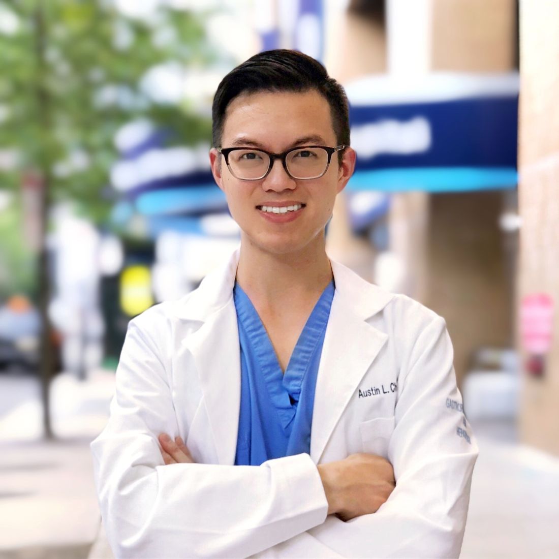
Dr. Chiang is assistant professor of medicine, division of gastroenterology & hepatology, director, endoscopic bariatric program, chief medical social media officer, Jefferson Health, Philadelphia, and president, Association for Healthcare Social Media, @austinchiangmd
I have always been a strong believer in meeting patients where they obtain their health information. Early in my clinical training, I realized that patients are exposed to health information through traditional media formats and, increasingly, social media, rather than brief clinical encounters. Unlike traditional media, social media allows individuals the opportunity to post information without a third-party filter. However, this opens the door for untrained individuals to spread misinformation and disinformation. In health care, this could potentially disrupt public health efforts. Even innocent mistakes like overlooking the appropriate clinical context can cause issues. Traditional media outlets also have agendas that may leave certain conditions, therapies, and other facets of health care underrepresented. My belief is that experts should therefore be trained and incentivized to be spokespeople for their own areas of expertise. Furthermore, social media provides a novel opportunity to improve health literacy while humanizing and restoring fading trust in health care.
There are several items to consider before initiating on one’s social media journey: whether you are committed to exploring the space, what one’s purpose is on social media, who the intended target audience is, which platform is most appropriate to serve that purpose and audience, and what potential pitfalls there may be.
The first question to ask oneself is whether you are prepared to devote time to cultivating a social media presence and speak or be heard publicly. Regardless of the platform, a social media presence requires consistency and audience interaction. The decision to partake can be personal; I view social media as an extension of in-person interaction, but not everyone is willing to commit to increased accessibility and visibility. Social media can still be valuable to those who choose to observe and learn rather than post.
Next is what one’s purpose is with being on social media. This can vary from peer education, boosting health literacy for patients, or using social media as a news source, networking tool, or a creative outlet. While my social media activity supports all these, my primary purpose is the distribution of accurate health information as a trained expert. When I started, I was one of few academic gastroenterologists uniquely positioned to bridge the elusive gap between the young, Gen Z crowd and academic medicine. Of similar importance is defining one’s target audience: patients, trainees, colleagues, or the general public.
Because there are numerous social media platforms, and only more to come in the future, it is critical to focus only on platforms that will serve one’s purpose and audience. Additionally, some may find more joy or agility in using one platform over the other. While I am one of the few clinicians who are adept at building communities across multiple rapidly evolving social media platforms, I will be the first to admit that it takes time to fully understand each platform with its ever-growing array of features. I find myself better at some platforms over others and, depending on my goals, I often will shift my focus from one to another.
Each platform has its pros and cons. Twitter is perhaps the most appropriate platform for starters. Easy to use with the least preparation necessary for every post, it also serves as the primary platform for academic discussion among all the popular social media platforms. Over the past few years, hundreds of gastroenterologists have become active on Twitter, which allows for ample networking opportunities and potential collaborations. The space has evolved to house various structured chats and learning opportunities as described by accounts like @MondayNightIBD, @ScopingSundays, #TracingTuesday, and @GIJournal. All major GI journals and societies are also present on Twitter and disseminating the latest information. Now a vestige of the past when text within tweets was not searchable, hashtags were used to curate discussion because searching by hashtag could reveal the latest discussion surrounding a topic and help identify others with a similar interest. Hashtags now remain relevant when crafting tweets, as the strategic inclusion of hashtags can help your content reach those who share an interest. A hashtag ontology was previously published to standardize academic conversation online in gastroenterology. Twitter also boasts features like polls that also help audiences engage.
Twitter has its disadvantages, however. Conversation is often siloed and difficult to reach audiences who don’t already follow you or others associated with you. Tweets disappear quickly in one’s feed and are often not seen by your followers. It lacks the visual appeal of other image- and video-based platforms that tend to attract more members of the general public. (Twitter lags behind these other platforms in monthly users) Other platforms like Facebook, Instagram, YouTube, LinkedIn, and TikTok have other benefits. Facebook may help foster community discussions in groups and business pages are also helpful for practice promotion. Instagram has gained popularity for educational purposes over the past 2 years, given its pairing with imagery and room for a lengthier caption. It has a variety of additional features like the temporary Instagram Stories that last 24 hours (which also allows for polling), question and answer, and livestream options. Other platforms like YouTube and TikTok have greater potential to reach audiences who otherwise would not see your content, with the former having the benefit of being highly searchable and the latter being the social media app with fastest growing popularity.
Having grown up with the Internet-based instant messaging and social media platforms, I have always enjoyed the medium as a way to connect with others. However, productive engagement on these platforms came much later. During a brief stint as part of the ABC News medical unit, I learned how Twitter was used to facilitate weekly chats around a specific topic online. I began exploring my own social media voice, which quickly gave way to live-tweeting medical conferences, hosting and participating Twitter chats myself, and guiding colleagues and professional societies to greater adoption of social media. In an attempt to introduce a divisional social media account during my fellowship, I learned of institutional barriers including antiquated policies that actively dissuaded social media use. I became increasingly involved on committees in our main GI societies after engaging in multiple research projects using social media data looking at how GI journals promote their content online, the associations between social media presence and institutional ranking, social media behavior at medical conferences, and the evolving perspectives of training program leadership regarding social media.
The pitfalls of social media remain a major concern for physicians and employers alike. First and foremost, it is important to review one’s institutional social media policy prior to starting, as individuals are ultimately held to their local policies. Not only can social media activity be a major liability for a health care employer, but also in the general public’s trust in health professionals. Protecting patient privacy and safety are of utmost concern, and physicians must be mindful not to inadvertently reveal patient identity. HIPAA violations are not limited to only naming patients by name or photo; descriptions of procedural cases and posting patient-related images such as radiographs or endoscopic images may reveal patient identity if there are unique details on these images (e.g., a radio-opaque necklace on x-ray or a particular swallowed foreign body).
Another disadvantage of social media is being approached with personal medical questions. I universally decline to answer these inquiries, citing the need to perform a comprehensive review of one’s medical chart and perform an in-person physical exam to fully assess a patient. The distinction between education and advice is subtle, yet important to recognize. Similarly, the need to uphold professionalism online is important. Short messages on social media can be misinterpreted by colleagues and the public. Not only can these interactions be potentially detrimental to one’s career, but it can further erode trust in health care if patients perceive this as fragmentation of the health care system. On platforms that encourage humor and creativity like TikTok, there have also been medical professionals and students publicly criticized and penalized for posting unprofessional content mocking patients.
With the introduction of social media influencers in recent years, some professionals have amassed followings, introducing yet another set of concerns. One is being approached with sponsorship and endorsement offers, as any agreements must be in accordance with institutional policy. As one’s following grows, there may be other concerns of safety both online and in real life. Online concerns include issues with impersonation and use of photos or written content without permission. On the surface this may not seem like a significant concern, but there have been situations where family photos are distributed to intended audiences or one’s likeness is used to endorse a product.
In addition to physical safety, another unintended consequence of social media use is its impact on one’s mental health. As social media tends to be a highlight reel, it is easy to be consumed by comparison with colleagues and their lives on social media, whether it truly reflects one’s actual life or not.
My ability to understand multiple social media platforms and anticipate a growing set of risks and concerns with using social media is what led to my involvement with multiple GI societies and appointment by my institution’s CEO to serve as the first chief medical social media officer. My desire to help other professionals with the journey also led to the formation of the Association for Healthcare Social Media, the first 501(c)(3) nonprofit professional organization devoted to health professionals on social media. There is tremendous opportunity to impact public health through social media, especially with regards to raising awareness about underrepresented conditions and presenting information that is accurate. Many barriers remain to the widespread adoption of social media by health professionals, such as the lack of financial or academic incentives. For now, there is every indication that social media is here to stay, and it will likely continue to play an important role in how we communicate with our patients.
AGA can be found online at @AmerGastroAssn (Facebook, Instagram, and Twitter) and @AGA_Gastro, @AGA_CGH, and @AGA_CMGH (Facebook and Twitter).
Dr. Chiang is assistant professor of medicine, division of gastroenterology & hepatology, director, endoscopic bariatric program, chief medical social media officer, Jefferson Health, Philadelphia, and president, Association for Healthcare Social Media, @austinchiangmd
Meditations in an emergency: Talking through pandemic anxiety with a pioneer of mind-body medicine
Andrew N. Wilner, MD: Welcome to Medscape. I’m Dr Andrew Wilner. Today I have a special guest, Dr James Gordon, founder and executive director of the Center for Mind-Body Medicine. Welcome, Dr Gordon.

James S. Gordon, MD: Thank you very much. It’s good to be with you.
Dr. Wilner: Thanks for joining us. We are recording this in late May 2020, in the midst of the coronavirus pandemic. Millions of people have been infected. Hundreds of thousands have died. Millions have lost their jobs. I think it’s fair to say that people are under a greater degree of stress than they’re normally accustomed to. Would you agree with that?
Dr. Gordon: I think it’s more than fair to say that everybody in the United States, and actually pretty much everyone in the world, is under extreme stress. And that compounds any stresses that they’ve experienced before in their lives. Everyone is affected.
Dr. Wilner: The mind-body medicine concept is one that you’ve pursued for decades. Tell us a little bit about the Center for Mind-Body Medicine and how that’s led to the program that you have to help us deal with the coronavirus.
Dr. Gordon: I started the Center for Mind-Body Medicine about 30 years ago. I’d been a researcher at the National Institute of Mental Health for a number of years, in private practice, and a professor at Georgetown Medical School. But I wanted to really focus on how to change and enrich medicine by making self-care, self-awareness, and group support central to all healthcare.
Western medicine is enormously powerful in certain situations, such as physical trauma, high levels of infection, congenital anomalies. But we’re not so good at working with chronic physical or psychological problems. Those are much more complex.
We’ve been discovering that what is going to make the long-term difference in conditions like type 2 diabetes, pain syndromes, hypertension, depression, and anxiety are those approaches that we can learn to do for ourselves. These are changes we can make in how we deal with stress, eat, exercise, relate to other people, and whether we find meaning and purpose in our lives.
by wars, climate-related disasters, the opioid epidemic, chronic poverty, historical trauma. We do a lot of work with indigenous people here in North America. We’ve worked in a number of communities where school shootings have traumatized everyone.
What we’ve learned over these past 25 years, and what interested me professionally as well as personally over the past 50 years, is what we’re now bringing out on an even larger scale. The kind of approaches that we’ve developed, studied, and published research on are exactly what everyone needs to include and incorporate in their daily life, as well as in their medical and health care, from now on.
Dr. Wilner: Do you have a program that’s specifically for health care providers?
Dr. Gordon: Yes. The Center for Mind-Body Medicine is primarily an educational organization rather than a service organization. Since the beginning, I’ve been focused on training health professionals. My first passion was for training physicians – I’m a physician, so there’s a feeling of fellowship there – but also health care workers and mental health professionals of every kind.
We teach health professionals a whole system, a comprehensive program of techniques of self-awareness and self-care. We teach them so that they can practice on themselves and study the underlying science, so they can then teach what they’ve learned to the patients or clients they work with. They integrate it into what they’re already doing, regardless of their specialty. At times we also offer some of the same kinds of mind-body skills groups that are the fundamental part of our training as a stand-alone intervention. You can’t really teach other people how to take care of themselves unless you’re also doing it yourself. Otherwise, it’s just a theory.
Dr. Wilner: As a neurologist, I’m interested in the mind-body system. You are a psychiatrist and understand that it’s a lot more difficult to objectify certain things. What is stress? What is happiness? What is sadness? It’s very hard to measure. You can have scales, but it requires insight on the part of the individual. So I think it’s certainly an ambitious project.
Dr. Gordon: You’re absolutely right. It requires insight. And one of the shortcomings of our medical education is that it doesn’t encourage us to look inside ourselves enough. There’s so much focus on objectivity and on data, that we’ve lost some of the subjective art of medicine.
My experience with myself, as well as with the thousands of people we’ve trained here in the United States and around the world and the many hundreds of thousands with whom they’ve worked, is that all of us have a greater capacity to understand and help ourselves than we ordinarily think or than most of us learn about in our medical education.
This work is saying to people to take a little bit of time and relax a little in order to allow yourself to come into a meditative state. And I don’t mean anything fancy by that. Meditation is just being relaxed. Moment-to-moment awareness doesn’t have to do with any particular religion or spiritual practice. It’s part of all of them. If you can get into that state, then you can begin to say, “Oh, that’s what’s going on with me. That’s why my pain is worse.”
For example, you often wonder in people with peripheral neuropathy why it becomes worse or better at certain points. I would encourage neurologists and other physicians to ask your patients, “Why do you think it’s worse?” They may say, “I don’t know, doc; that’s why I’m here.” But I would ask them to take a couple of minutes to let me know. They could think it has something to do with the fact that they had a big fight with their wife that morning, they don’t want to go to work, or whatever it is. This is part of the lost art that we need to bring back into medicine for ourselves and especially for our patients.
Dr. Wilner: Can you give me an example of some of the exercises you’d do in a class?
Dr. Gordon: All of the exercises and our entire program that we teach at the Center for Mind-Body Medicine is in this new book of mine, “The Transformation: Discovering Wholeness and Healing After Trauma.” It’s really the distillation of not just the past 25 or 30 years, but really 50 years of work.
The techniques are all pretty simple and, as we say, evidence based. There is evidence that shows how they work on us physiologically, as well as psychologically. And they’re all pretty easy to teach to anyone.
Myself and about 60 or 70 of our faculty at the Center for Mind-Body Medicine are currently leading online groups. Then several hundred of the other people we’ve trained are also leading these groups. We’re still counting it up, but we probably have between 700 and 1,000 groups going around the world, led by our faculty and by people we’ve trained.
We teach a different technique every week in these online groups. Last week, after getting people energized and focused, we did a written dialogue with an emotion. You put down the initial of your name – in my case, “J” for Jim – and create a dialogue with an emotion, such as sadness. I would write it as fast as I can.
I would say, “OK, Sadness. Why are you here? What are you doing? I don’t enjoy having you around.” And Sadness writes back to me, “But you need me.” And J says, “What do you mean I need you?” And Sadness says, “Well, your brother died 7 weeks ago, didn’t he?” And I say, “Yes, he did.” And Sadness says, “Aren’t you sad?” I say, “Yes. I’m terribly sad and grieving all the time. But I wasn’t thinking about him at this moment.” And Sadness says, “But he’s there with you all the time and that sadness is in you.” And I say, “You mean it’s in me even here, now, as I’m talking with Andrew in this interview?” And Sadness says, “Yes. You can talk about your work. But in between the words, as you take a breath, don’t you feel it in your chest?” That’s the way the dialogue goes.
Dr. Wilner: What about specifically with the coronavirus? Fear is certainly an emotion. Nobody wants to get sick and die. Nobody wants to bring this disease home to their family. People are reluctant to even go outside and you can’t shake someone’s hand. Are there precedents for this?
Dr. Gordon: There are precedents, but only relatively small groups were affected before by, for example, severe acute respiratory syndrome or H1N1, at least in the United States. But we haven’t seen a global pandemic like this since 1918. None of us was around then – or I certainly wasn’t around. So for most everyone, not only has it not happened before, but we’ve never been so globally aware of everything that’s going on and how different groups are reacting.
I’ve been reading Daniel Defoe’s book, “A Journal of the Plague Year.” It’s really very interesting. It’s about the bubonic plague in 1665 London, although he wrote it in the 1720s. Some of the same things were going on then: the enormous fear, the isolation; rich people being able to escape, poor people having nowhere to go; conspiracy theories of one kind or another, about where the plague came from or blaming a group of people for it; magical thinking that it’s just going to go away. All of those things that happened several hundred years ago are going on now.
And we’re all simultaneously aware of all those things. There’s not only the fear, which should be universal because it’s a reasonable response to this situation, but also the terrible confusion about what to do. The President is saying one thing, governors something else; Anthony Fauci is saying something else, and Deborah Birx is saying something a little bit different. There’s this tremendous confusion that overlays the fear, and I think everybody is more or less feeling these things.
So yes, a dialogue with fear is a good thing to do because it can be clarifying. What we need here is a sense of, what is it that makes sense for me to do? What precautions should I take? What precautions shouldn’t I take?
I have a 17-year-old son who lives with his mom in California. He and I were on the phone the other day. He’s a basketball player and very serious about it. He said, “I don’t want to put my life on hold.” And my response was, “If you go outside too soon, your life may be on hold for a hell of a lot longer than if you stay inside because, if you get sick, it’s serious. But you also need to start looking at the evidence and asking yourself the right questions because I can’t be there all the time and neither can your mom.”
Everybody really needs to use these kinds of tools to help themselves. The tools we teach are extremely good at bringing us back into a state of psychological and physiological balance — slow, deep breathing being a very basic one. Because it’s only in that state that we’re going to be able to make the most intelligent decisions about what to do. It’s only in that state that we’re going to be able to really look our fear in the face and find out what we should be afraid of and what we shouldn’t be afraid of.
It’s a process that’s very much integrated. We’re talking now about how to deal with the emotions. But the first part of what we do in our groups and our online trainings and webinars is teach people to just take a few deep breaths. Just take a few deep breaths in through the nose, out through the mouth, with your belly soft and relaxed. You can keep breathing this way while talking. That’s the antidote to the fight-or-flight response. We all learn about fight-or-flight in first-year physiology. We need to deal with it. We need to bring ourselves into balance. That’s the way we’re going to make the wisest decisions for ourselves and be best able to help our patients.
Dr. Wilner: As you mentioned, part of modern culture is that we now have access to all of this information worldwide. There’s a continual stream of newsfeeds, people flipping on their phones, receiving constant updates, 24/7. That’s a new phenomenon. Does that steal from us the time we had before for just breathing and synthesizing data as opposed to just acquiring it all the time?
Dr. Gordon: You’re absolutely right. It does and it’s a challenge. It can’t steal from us unless we’re letting our emotional, psychological, and physiological pockets be picked!
What we need to do is to make it our priority to come into balance. I don’t watch news all day long – a little tiny bit in the morning and in the evening, just to get a sense of what’s happening. That’s enough. And I think everybody needs to take a step back, ask if this is really what they want to be doing, and to come into balance.
The other thing that’s really important is physical activity, especially during this time. In addition to using slow, deep breathing to come into balance, physical exercise and movement of any kind is extremely good as an antidote to fight-or-flight and that shut-down, freeze-up response that we get into when we feel completely overwhelmed.
We’ve got to take it into our own hands. The media just want to sell us things. Let’s face it: They’re not here for our good. Our job as physicians and health care professionals is to really reinforce for people not only what we can do for them but what they can do for themselves.
Dr. Wilner: I’m certainly interested in learning more about mind-body medicine. For those who feel the same, where do you recommend they go to learn more?
Dr. Gordon: We have a website, cmbm.org, which features a number of webinars. I do a free webinar there every week. We have mind-body skills groups that meet once a week for 8 weeks. There are six physicians in my group and all kinds of health professionals in other groups. We have a training program that we’re bringing online. We’ve trained well over 6,000 people around the world and would love to train more. You can read about that on the website.
We’re starting to do more and more consulting with health care organizations. We’re working with the largest division of Veterans Affairs, which is in Florida, as well as in south Georgia and the Caribbean. We’re working with a large health system in Indiana and others elsewhere. In addition, we’re working with groups of physicians and mental health professionals, helping them to integrate what we have to offer into what they’re already doing.
That’s our job – to help you do your job.
Dr. Wilner: Dr Gordon, I feel more relaxed just speaking with you. Thank you for talking with me and sharing your experiences with Medscape. I look forward to learning more.
Dr. Gordon: Thank you. My pleasure.
A version of this article originally appeared on Medscape.com.
Andrew N. Wilner, MD: Welcome to Medscape. I’m Dr Andrew Wilner. Today I have a special guest, Dr James Gordon, founder and executive director of the Center for Mind-Body Medicine. Welcome, Dr Gordon.
James S. Gordon, MD: Thank you very much. It’s good to be with you.
Dr. Wilner: Thanks for joining us. We are recording this in late May 2020, in the midst of the coronavirus pandemic. Millions of people have been infected. Hundreds of thousands have died. Millions have lost their jobs. I think it’s fair to say that people are under a greater degree of stress than they’re normally accustomed to. Would you agree with that?
Dr. Gordon: I think it’s more than fair to say that everybody in the United States, and actually pretty much everyone in the world, is under extreme stress. And that compounds any stresses that they’ve experienced before in their lives. Everyone is affected.
Dr. Wilner: The mind-body medicine concept is one that you’ve pursued for decades. Tell us a little bit about the Center for Mind-Body Medicine and how that’s led to the program that you have to help us deal with the coronavirus.
Dr. Gordon: I started the Center for Mind-Body Medicine about 30 years ago. I’d been a researcher at the National Institute of Mental Health for a number of years, in private practice, and a professor at Georgetown Medical School. But I wanted to really focus on how to change and enrich medicine by making self-care, self-awareness, and group support central to all healthcare.
Western medicine is enormously powerful in certain situations, such as physical trauma, high levels of infection, congenital anomalies. But we’re not so good at working with chronic physical or psychological problems. Those are much more complex.
We’ve been discovering that what is going to make the long-term difference in conditions like type 2 diabetes, pain syndromes, hypertension, depression, and anxiety are those approaches that we can learn to do for ourselves. These are changes we can make in how we deal with stress, eat, exercise, relate to other people, and whether we find meaning and purpose in our lives.
by wars, climate-related disasters, the opioid epidemic, chronic poverty, historical trauma. We do a lot of work with indigenous people here in North America. We’ve worked in a number of communities where school shootings have traumatized everyone.
What we’ve learned over these past 25 years, and what interested me professionally as well as personally over the past 50 years, is what we’re now bringing out on an even larger scale. The kind of approaches that we’ve developed, studied, and published research on are exactly what everyone needs to include and incorporate in their daily life, as well as in their medical and health care, from now on.
Dr. Wilner: Do you have a program that’s specifically for health care providers?
Dr. Gordon: Yes. The Center for Mind-Body Medicine is primarily an educational organization rather than a service organization. Since the beginning, I’ve been focused on training health professionals. My first passion was for training physicians – I’m a physician, so there’s a feeling of fellowship there – but also health care workers and mental health professionals of every kind.
We teach health professionals a whole system, a comprehensive program of techniques of self-awareness and self-care. We teach them so that they can practice on themselves and study the underlying science, so they can then teach what they’ve learned to the patients or clients they work with. They integrate it into what they’re already doing, regardless of their specialty. At times we also offer some of the same kinds of mind-body skills groups that are the fundamental part of our training as a stand-alone intervention. You can’t really teach other people how to take care of themselves unless you’re also doing it yourself. Otherwise, it’s just a theory.
Dr. Wilner: As a neurologist, I’m interested in the mind-body system. You are a psychiatrist and understand that it’s a lot more difficult to objectify certain things. What is stress? What is happiness? What is sadness? It’s very hard to measure. You can have scales, but it requires insight on the part of the individual. So I think it’s certainly an ambitious project.
Dr. Gordon: You’re absolutely right. It requires insight. And one of the shortcomings of our medical education is that it doesn’t encourage us to look inside ourselves enough. There’s so much focus on objectivity and on data, that we’ve lost some of the subjective art of medicine.
My experience with myself, as well as with the thousands of people we’ve trained here in the United States and around the world and the many hundreds of thousands with whom they’ve worked, is that all of us have a greater capacity to understand and help ourselves than we ordinarily think or than most of us learn about in our medical education.
This work is saying to people to take a little bit of time and relax a little in order to allow yourself to come into a meditative state. And I don’t mean anything fancy by that. Meditation is just being relaxed. Moment-to-moment awareness doesn’t have to do with any particular religion or spiritual practice. It’s part of all of them. If you can get into that state, then you can begin to say, “Oh, that’s what’s going on with me. That’s why my pain is worse.”
For example, you often wonder in people with peripheral neuropathy why it becomes worse or better at certain points. I would encourage neurologists and other physicians to ask your patients, “Why do you think it’s worse?” They may say, “I don’t know, doc; that’s why I’m here.” But I would ask them to take a couple of minutes to let me know. They could think it has something to do with the fact that they had a big fight with their wife that morning, they don’t want to go to work, or whatever it is. This is part of the lost art that we need to bring back into medicine for ourselves and especially for our patients.
Dr. Wilner: Can you give me an example of some of the exercises you’d do in a class?
Dr. Gordon: All of the exercises and our entire program that we teach at the Center for Mind-Body Medicine is in this new book of mine, “The Transformation: Discovering Wholeness and Healing After Trauma.” It’s really the distillation of not just the past 25 or 30 years, but really 50 years of work.
The techniques are all pretty simple and, as we say, evidence based. There is evidence that shows how they work on us physiologically, as well as psychologically. And they’re all pretty easy to teach to anyone.
Myself and about 60 or 70 of our faculty at the Center for Mind-Body Medicine are currently leading online groups. Then several hundred of the other people we’ve trained are also leading these groups. We’re still counting it up, but we probably have between 700 and 1,000 groups going around the world, led by our faculty and by people we’ve trained.
We teach a different technique every week in these online groups. Last week, after getting people energized and focused, we did a written dialogue with an emotion. You put down the initial of your name – in my case, “J” for Jim – and create a dialogue with an emotion, such as sadness. I would write it as fast as I can.
I would say, “OK, Sadness. Why are you here? What are you doing? I don’t enjoy having you around.” And Sadness writes back to me, “But you need me.” And J says, “What do you mean I need you?” And Sadness says, “Well, your brother died 7 weeks ago, didn’t he?” And I say, “Yes, he did.” And Sadness says, “Aren’t you sad?” I say, “Yes. I’m terribly sad and grieving all the time. But I wasn’t thinking about him at this moment.” And Sadness says, “But he’s there with you all the time and that sadness is in you.” And I say, “You mean it’s in me even here, now, as I’m talking with Andrew in this interview?” And Sadness says, “Yes. You can talk about your work. But in between the words, as you take a breath, don’t you feel it in your chest?” That’s the way the dialogue goes.
Dr. Wilner: What about specifically with the coronavirus? Fear is certainly an emotion. Nobody wants to get sick and die. Nobody wants to bring this disease home to their family. People are reluctant to even go outside and you can’t shake someone’s hand. Are there precedents for this?
Dr. Gordon: There are precedents, but only relatively small groups were affected before by, for example, severe acute respiratory syndrome or H1N1, at least in the United States. But we haven’t seen a global pandemic like this since 1918. None of us was around then – or I certainly wasn’t around. So for most everyone, not only has it not happened before, but we’ve never been so globally aware of everything that’s going on and how different groups are reacting.
I’ve been reading Daniel Defoe’s book, “A Journal of the Plague Year.” It’s really very interesting. It’s about the bubonic plague in 1665 London, although he wrote it in the 1720s. Some of the same things were going on then: the enormous fear, the isolation; rich people being able to escape, poor people having nowhere to go; conspiracy theories of one kind or another, about where the plague came from or blaming a group of people for it; magical thinking that it’s just going to go away. All of those things that happened several hundred years ago are going on now.
And we’re all simultaneously aware of all those things. There’s not only the fear, which should be universal because it’s a reasonable response to this situation, but also the terrible confusion about what to do. The President is saying one thing, governors something else; Anthony Fauci is saying something else, and Deborah Birx is saying something a little bit different. There’s this tremendous confusion that overlays the fear, and I think everybody is more or less feeling these things.
So yes, a dialogue with fear is a good thing to do because it can be clarifying. What we need here is a sense of, what is it that makes sense for me to do? What precautions should I take? What precautions shouldn’t I take?
I have a 17-year-old son who lives with his mom in California. He and I were on the phone the other day. He’s a basketball player and very serious about it. He said, “I don’t want to put my life on hold.” And my response was, “If you go outside too soon, your life may be on hold for a hell of a lot longer than if you stay inside because, if you get sick, it’s serious. But you also need to start looking at the evidence and asking yourself the right questions because I can’t be there all the time and neither can your mom.”
Everybody really needs to use these kinds of tools to help themselves. The tools we teach are extremely good at bringing us back into a state of psychological and physiological balance — slow, deep breathing being a very basic one. Because it’s only in that state that we’re going to be able to make the most intelligent decisions about what to do. It’s only in that state that we’re going to be able to really look our fear in the face and find out what we should be afraid of and what we shouldn’t be afraid of.
It’s a process that’s very much integrated. We’re talking now about how to deal with the emotions. But the first part of what we do in our groups and our online trainings and webinars is teach people to just take a few deep breaths. Just take a few deep breaths in through the nose, out through the mouth, with your belly soft and relaxed. You can keep breathing this way while talking. That’s the antidote to the fight-or-flight response. We all learn about fight-or-flight in first-year physiology. We need to deal with it. We need to bring ourselves into balance. That’s the way we’re going to make the wisest decisions for ourselves and be best able to help our patients.
Dr. Wilner: As you mentioned, part of modern culture is that we now have access to all of this information worldwide. There’s a continual stream of newsfeeds, people flipping on their phones, receiving constant updates, 24/7. That’s a new phenomenon. Does that steal from us the time we had before for just breathing and synthesizing data as opposed to just acquiring it all the time?
Dr. Gordon: You’re absolutely right. It does and it’s a challenge. It can’t steal from us unless we’re letting our emotional, psychological, and physiological pockets be picked!
What we need to do is to make it our priority to come into balance. I don’t watch news all day long – a little tiny bit in the morning and in the evening, just to get a sense of what’s happening. That’s enough. And I think everybody needs to take a step back, ask if this is really what they want to be doing, and to come into balance.
The other thing that’s really important is physical activity, especially during this time. In addition to using slow, deep breathing to come into balance, physical exercise and movement of any kind is extremely good as an antidote to fight-or-flight and that shut-down, freeze-up response that we get into when we feel completely overwhelmed.
We’ve got to take it into our own hands. The media just want to sell us things. Let’s face it: They’re not here for our good. Our job as physicians and health care professionals is to really reinforce for people not only what we can do for them but what they can do for themselves.
Dr. Wilner: I’m certainly interested in learning more about mind-body medicine. For those who feel the same, where do you recommend they go to learn more?
Dr. Gordon: We have a website, cmbm.org, which features a number of webinars. I do a free webinar there every week. We have mind-body skills groups that meet once a week for 8 weeks. There are six physicians in my group and all kinds of health professionals in other groups. We have a training program that we’re bringing online. We’ve trained well over 6,000 people around the world and would love to train more. You can read about that on the website.
We’re starting to do more and more consulting with health care organizations. We’re working with the largest division of Veterans Affairs, which is in Florida, as well as in south Georgia and the Caribbean. We’re working with a large health system in Indiana and others elsewhere. In addition, we’re working with groups of physicians and mental health professionals, helping them to integrate what we have to offer into what they’re already doing.
That’s our job – to help you do your job.
Dr. Wilner: Dr Gordon, I feel more relaxed just speaking with you. Thank you for talking with me and sharing your experiences with Medscape. I look forward to learning more.
Dr. Gordon: Thank you. My pleasure.
A version of this article originally appeared on Medscape.com.
Physician leadership: Racial disparities and racism. Where do we go from here?
The destructive toll COVID-19 has caused worldwide is devastating. In the United States, the disproportionate deaths of Black, Indigenous, and Latinx people due to structural racism, amplified by economic adversity, is unacceptable. Meanwhile, the continued murder of Black people by those sworn to protect the public is abhorrent and can no longer be ignored. Black lives matter. These crises have rightly gripped our attention, and should galvanize physicians individually and collectively to use our privileged voices and relative power for justice. We must strive for engaged, passionate, and innovative leadership deliberately aimed toward antiracism and equity.
The COVID-19 pandemic has illuminated the vast inequities in our country. It has highlighted the continued poor outcomes our health and health care systems create for Black, Indigenous, and Latinx communities. It also has demonstrated clearly that we are all connected—one large community, interdependent yet rife with differential power, privilege, and oppression. We must address these racial disparities—not only in the name of justice and good health for all but also because it is a moral and ethical imperative for us as physicians—and SARS-CoV-2 clearly shows us that it is in the best interest of everyone to do so.
First step: A deep dive look at systemic racism
What is first needed is an examination and acknowledgement by medicine and health care at large of the deeply entrenched roots of systemic and institutional racism in our profession and care systems, and their disproportionate and unjust impact on the health and livelihood of communities of color. The COVID-19 pandemic is only a recent example that highlights the perpetuation of a system that harms people of color. Racism, sexism, gender discrimination, economic and social injustice, religious persecution, and violence against women and children are age-old. We have yet to see health care institutions implement system-wide intersectional and antiracist practices to address them. Mandatory implicit bias training, policies for inclusion and diversity, and position statements are necessary first steps; however, they are not a panacea. They are insufficient to create the bold changes we need. The time for words has long passed. It is time to listen, to hear the cries of anguish and outrage, to examine our privileged position, to embrace change and discomfort, and most importantly to act, and to lead in dismantling the structures around us that perpetuate racial inequity.
How can we, as physicians and leaders, join in action and make an impact?
Dr. Camara Jones, past president of the American Public Health Association, describes 3 levels of racism:
- structural or systemic
- individual or personally mediated
- internalized.
Interventions at each level are important if we are to promote equity in health and health care. This framework can help us think about the following strategic initiatives.
Continue to: 1. Commit to becoming an antiracist and engage in independent study...
1. Commit to becoming antiracist and engage in independent study. This is an important first step as it will form the foundations for interventions—one cannot facilitate change without understanding the matter at hand. This step also may be the most personally challenging step forcing all of us to wrestle with discomfort, sadness, fear, guilt, and a host of other emotional responses. Remember that great change has never been born out of comfort, and the discomfort physicians may experience while unlearning racism and learning antiracism pales in comparison to what communities of color experience daily. We must actively work to unlearn the racist and anti-Black culture that is so deeply woven into every aspect of our existence.
Learn the history that was not given to us as kids in school. Read the brilliant literary works of Black, Indigenous, and Latinx artists and scholars on dismantling racism. Expand our vocabulary and knowledge of core concepts in racism, racial justice, and equity. Examine and reflect on our day-to-day practices. Be vocal in our commitment to antiracism—the time has passed for staying silent. If you are white, facilitate conversations about race with your white colleagues; the inherent power of racism relegates it to an issue that can never be on the table, but it is time to dismantle that power. Learn what acts of meaningful and intentional alliances are and when we need to give up power or privilege to a person of color. We also need to recognize that we as physicians, while leaders in many spaces, are not leaders in the powerful racial justice grassroots movements. We should learn from these movements, follow their lead, and use our privilege to uplift racial justice in our settings.
2. Embrace the current complexities with empathy and humility, finding ways to exercise our civic responsibility to the public with compassion. During the COVID-19 pandemic we have seen the devastation that social isolation, job loss, and illness can create. Suddenly those who could never have imagined themselves without food are waiting hours in their cars for food bank donations or are finding empty shelves in stores. Those who were not safe at home were suddenly imprisoned indefinitely in unsafe situations. Those who were comfortable, well-insured, and healthy are facing an invisible health threat, insecurity, fear, anxiety, and loss. Additionally, our civic institutions are failing. Those of us who always took our right to vote for granted are being forced to stand in hours’-long lines to exercise that right; while those who have been systematically disenfranchised are enduring even greater threats to their constitutional right to exercise their political power, disallowing them to speak for their families and communities and to vote for the justice they deserve. This may be an opportunity to stop blaming victims and recognize the toll that structural and systemic contributions to inequity have created over generations.
3. Meaningfully engage with and advocate for patients. In health and health care, we must begin to engage with the communities we serve and truly listen to their needs, desires, and barriers to care, and respond accordingly. Policies that try to address the social determinants of health without that engagement, and without the acknowledgement of the structural issues that cause them, however well-intentioned, are unlikely to accomplish their goals. We need to advocate as physicians and leaders in our settings for every policy, practice, and procedure to be scrutinized using an antiracist lens. To execute this, we need to:
- ask why clinic and hospital practices are built the way they are and how to make them more reflexive and responsive to individual patient’s needs
- examine what the disproportionate impacts might be on different groups of patients from a systems-level
- be ready to dismantle and/or rebuild something that is exacerbating disparate outcomes and experiences
- advocate for change that is built upon the narratives of patients and their communities.
We should include patients in the creation of hospital policies and guidelines in order to shift power toward them and to be transparent about how the system operates in order to facilitate trust and collaboration that centers patients and communities in the systems created to serve them.
Continue to: 4. Intentionally repair and build trust...
4. Intentionally repair and build trust. To create a safe environment, we must repair what we have broken and earn the trust of communities by uplifting their voices and redistributing our power to them in changing the systems and structures that have, for generations, kept Black, Indigenous, and Latinx people oppressed. Building trust requires first owning our histories of colonization, genocide, and slavery—now turned mass incarceration, debasement, and exploitation—that has existed for centuries. We as physicians need to do an honest examination of how we have eroded the trust of the very communities we care for since our profession’s creation. We need to acknowledge, as a white-dominant profession, the medical experimentation on and exploitation of Black and Brown bodies, and how this formed the foundation for a very valid deep distrust and fear of the medical establishment. We need to recognize how our inherent racial biases continue to feed this distrust, like when we don’t treat patients’ pain adequately or make them feel like we believe and listen to their needs and concerns. We must acknowledge our complicity in perpetuating the racial inequities in health, again highlighted by the COVID-19 pandemic.
5. Increase Black, Indigenous, and Latinx representation in physician and other health care professions’ workforce. Racism impacts not only patients but also our colleagues of color. The lack of racial diversity is a symptom of racism and a representation of the continued exclusion and devaluing of physicians of color. We must recognize this legacy of exclusion and facilitate intentional recruitment, retention, inclusion, and belonging of people of color into our workforce. Tokenism, the act of symbolically including one or few people from underrepresented groups, has been a weapon used by our workforce against physicians of color, resulting in isolation, “othering,” demoralization, and other deleterious impacts. We need to reverse this history and diversify our training programs and workforce to ensure justice in our own community.
6. Design multifaceted interventions. Multilevel problems require multilevel solutions. Interventions targeted solely at one level, while helpful, are unlikely to result in the larger scale changes our society needs to implement if we are to eradicate the impact of racism on health. We have long known that it is not just “preexisting conditions” or “poor” individual behaviors that lead to negative and disparate health outcomes—these are impacted by social and structural determinants much larger and more deleterious than that. It is critically important that we allocate and redistribute resources to create safe and affordable housing; childcare and preschool facilities; healthy, available, and affordable food; equitable and affordable educational opportunities; and a clean environment to support the health of all communities—not only those with the highest tax base. It is imperative that we strive to understand the lives of our fellow human beings who have been subjected to intergenerational social injustices and oppressions that have continued to place them at the margins of society. We need to center the lived experiences of communities of color in the design of multilevel interventions, especially Black and Indigenous communities. While we as physicians cannot individually impact education, economic, or food/environment systems, we can use our power to advocate for providing resources for the patients we care for and can create strategies within the health care system to address these needs in order to achieve optimal health. Robust and equitable social structures are the foundations for health, and ensuring equitable access to them is critical to reducing disparities.
Commit to lead
We must commit to unlearning our internalized racism, rebuilding relationships with communities of color, and engaging in antiracist practices. As a profession dedicated to healing, we have an obligation to be leaders in advocating for these changes, and dismantling the inequitable structure of our health care system.
Our challenge now is to articulate solutions. While antiracism should be informed by the lived experiences of communities of color, the work of antiracism is not their responsibility. In fact, it is the responsibility of our white-dominated systems and institutions to change.
There are some solutions that are easier to enumerate because they have easily measurable outcomes or activities, such as:
- collecting data transparently
- identifying inequities in access, treatment, and care
- conducting rigorous root cause analysis of those barriers to care
- increasing diverse racial and gender representation on decision-making bodies, from board rooms to committees, from leadership teams to research participants
- redistribute power by paving the way for underrepresented colleagues to participate in clinical, administrative, educational, executive, and health policy spaces
- mentoring new leaders who come from marginalized communities.
Every patient deserves our expertise and access to high-quality care. We should review our patient panels to ensure we are taking steps personally to be just and eliminate disparities, and we should monitor the results of those efforts.
Continue to: Be open to solutions that may make us “uncomfortable”...
Be open to solutions that may make us “uncomfortable”
There are other solutions, perhaps those that would be more effective on a larger scale, which may be harder to measure using our traditional ways of inquiry or measurement. Solutions that may create discomfort, anger, or fear for those who have held their power or positions for a long time. We need to begin to engage in developing, cultivating, and valuing innovative strategies that produce equally valid knowledge, evidence, and solutions without engaging in a randomized controlled trial. We need to reinvent the way inquiry, investigation, and implementation are done, and utilize novel, justice-informed strategies that include real-world evidence to produce results that are applicable to all (not just those willing to participate in sponsored trials). Only then will we be able to provide equitable health outcomes for all.
We also must accept responsibility for the past and humbly ask communities to work with us as we struggle to eliminate racism and dehumanization of Black lives by calling out our actions or inaction, recognizing the impact of our privileged status, and stepping down or stepping aside to allow others to lead. Sometimes it is as simple as turning off the Zoom camera so others can talk. By redistributing power and focusing this work upon the narratives of marginalized communities, we can improve our system for everyone. We must lead with action within our practices and systems; become advocates within our communities, institutions, and profession; strategize and organize interventions at both structural and individual levels to first recognize and name—then change—the systems; and unlearn behaviors that perpetuate racism.
Inaction is shirking our responsibility among the medical community
Benign inaction and unintentional acquiescence with “the way things are and have always been” abdicates our responsibility as physicians to improve the health of our patients and our communities. The modern Hippocratic Oath reminds us: “I will remember that I remain a member of society, with special obligations to all my fellow human beings, those sound of mind and body as well as the infirm.” We have a professional and ethical responsibility to ensure health equity, and thus racial equity. As physicians, as healers, as leaders we must address racial inequities at all levels as we commit to improving the health of our nation. We can no longer stand silent in the face of the violence, brutality, and injustices our patients, friends, family, neighbors, communities, and society as a whole live through daily. It is unjust and inhumane to do so.
To be silent is to be complicit. As Gandhi said so long ago, we must “be the change we wish to see in the world.” And as Ijeoma Olua teaches us, “Anti-racism is the commitment to fight racism wherever you find it, including in yourself. And it’s the only way forward.”
- “So You Want to Talk about Race” Ijeoma Oluo
- “How to Be an Antiracist” Ibram X. Kendi
- “Between the World and Me” Ta-Nehisi Coates
- A conversation on race and privilege (Angela Davis and Jane Elliot) https://www.youtube.com/watch?reload=9&v=S0jf8D5WHoo
- Uncomfortable conversations with a Black man (Emmanuel Acho) https://www.youtube.com/watch?v=h8jUA7JBkF4
Antiracism – defined as the work of actively opposing racism by advocating for changes in political, economic, and social life. Antiracism tends to be an individualized approach, and set up in opposition to individual racist behaviors and impacts
Black Lives Matter – a political movement to address systemic and state violence against African Americans. Per the Black Lives Matter organizers: “In 2013, three radical Black organizers—Alicia Garza, Patrisse Cullors, and Opal Tometi—created a Black-centered political will and movement building project called BlackLivesMatter. It was in response to the acquittal of Trayvon Martin’s murderer, George Zimmerman. The project is now a member-led global network of more than 40 chapters. Members organize and build local power to intervene in violence inflicted on Black communities by the state and vigilantes. Black Lives Matter is an ideological and political intervention in a world where Black lives are systematically and intentionally targeted for demise. It is an affirmation of Black folks’ humanity, our contributions to this society, and our resilience in the face of deadly oppression.”
Implicit bias – also known as unconscious or hidden bias, implicit biases are negative associations that people unknowingly hold. They are expressed automatically, without conscious awareness. Many studies have indicated that implicit biases affect individuals’ attitudes and actions, thus creating real-world implications, even though individuals may not even be aware that those biases exist within themselves. Notably, implicit biases have been shown to trump individuals stated commitments to equality and fairness, thereby producing behavior that diverges from the explicit attitudes that many people profess.
Othering – view or treat (a person or group of people) as intrinsically different from and alien to oneself. (From https://lexico.com.)
For a full glossary of terms, visit RacialEquityTools.org (https://www.racialequitytools.org/glossary#anti-black)

Holistic HIV care broadens scope to noncommunicable diseases
Several HIV management efforts in African groups have developed differentiated service delivery models for people living with HIV who also have noncommunicable diseases, offering diagnostic and management strategies that can treat HIV patients holistically and address their range of health issues.
These efforts allow “countries with effective HIV programs to leverage lessons learned and best practices to enhance chronic noncommunicable disease” management, Miriam Rabkin, MD, said at the virtual meeting of the International AIDS conference. This approach aims to address the “growing prevalence of chronic noncommunicable diseases in low- and middle-income countries,” and the recognition that ”people living with HIV have the same or higher prevalence” of chronic noncommunicable diseases as that of others in the region where they live, said Dr. Rabkin, an epidemiologist at Columbia University in New York and director for health systems strengthening at ICAP, an international AIDS care program run at Columbia. The differentiated service delivery model derived from the premise that “one size does not fit all,” and that effective interventions must be “tailored” to the social and clinical circumstances of specific regions, she explained.
One program has focused on introducing more contemporary methods for diagnosing leukemias, lymphomas, and melanomas using flow cytometry at the Uganda National Health Laboratory Service in Kampala. This change in testing, which became available to patients starting in February 2019, has allowed diagnostics with fresh specimens that require minimal processing and results returned to referring physicians within 48 hours, a significant upgrade from the 1- to 4-week delay that was typical in the past, said Steven J. Kussick, MD, a hematopathologist and associate medical director of PhenoPath, a commercial pathology laboratory in Seattle.
The idea was to “leverage existing HIV laboratory capabilities to transform cancer diagnosis in sub-Saharan Africa,” he said during his talk at the conference. The flow cytometry approach allows an experienced pathologist like Dr. Kussick to diagnose clearcut cases in “5 seconds,” he said. The lab has already run specimens from more than 200 patients, and estimates an ability to handle specimens from about 250 patients per year at a total annual cost of roughly $60,000, an apparently sustainable operating model, said Dr. Kussick, who serves as a full-time consultant to the operation and was also instrumental in the 5-year process that created the diagnostic program. Future improvements planned for this program include bringing on-line a higher complexity diagnostic assay that’s closer to what is currently standard U.S. testing, digital imaging to facilitate consultation with remote experts, adding immunochemistry assays to allow diagnosis of solid tumors, and opening of a second laboratory in Kenya.
Another noncommunicable disease intervention in Africa that’s building on existing infrastructure for dealing with HIV infection is targeting hypertension, the most lethal risk factor globally for preventable deaths, said Jennifer Cohn, MD, senior vice president for cardiovascular health at the New York–based Resolve to Save Lives initiative. “We need to learn from what’s been done for HIV to rapidly incorporate and scale differentiated service models,” she said.
HIV and hypertension, along with diabetes, “are beginning to be recognized as ‘syndemics,’ ”synergistic pandemics, that need a holistic approach. A recent review of the topic reported that in the seven sub-Saharan countries with the highest HIV infection prevalence the percentage of adults with hypertension ranged from 20% to 24% (Curr Opin HIV AIDS. 2020 Jul;15[4]:356-60). Projections call for a “dramatic” increase in the prevalence of hypertension in both the general population and among people living with HIV, Dr. Cohn said.
As an example of the potential for combining HIV and antihypertensive care into a one-stop protocol, she cited a model program launched at Makarere University in Kampala, Uganda, that integrates HIV and antihypertensive treatment. Recent data from the program showed that among HIV-infected individuals 24% also had hypertension, and while the program lagged in putting only 28% of these hypertensive patients on a blood pressure-lowering regimen, more than three quarters of these patients on treatment successfully reached their goal blood pressure, proving the feasibility of the combined approach, Dr. Cohn said.
“Starting and scaling with differentiated service delivery models for noncommunicable diseases can help overcome barriers to uptake of care,” concluded Dr. Cohn. “As HIV cohorts age, we have to adapt and ensure we are providing quality, holistic care, including care for high impact noncommunicable diseases such as hypertension.”
Dr. Rabkin and Dr. Cohn had no disclosures.
FROM AIDS 2020
COVID-19 disruptions ‘life threatening’ for people with HIV
When the COVID-19 pandemic led to a blanket shelter-in-place order in California in March, it did more than shut down in-person visits at Ward 86, the HIV clinic for publicly insured patients at San Francisco General Hospital. It also led to a decrease in viral suppression among the clinic›s clients. By the end of June, the percentage of patients with an undetectable viral load had dropped by nearly one-third.
This is exactly what Monica Gandhi, MD, associate division chief of HIV, infectious diseases, and global medicine at the University of California, San Francisco, and medical director of the clinic, was afraid of.
“We’re profoundly worried about the impact of COVID-19 on actual treatment outcomes,” said Dr. Gandhi, cochair of the virtual International AIDS Conference (AIDS) 2020.
And it’s not just the clinic’s clients at risk. Of the 106 countries served by the Global Fund to Fight HIV, Tuberculosis, and Malaria, 85% saw disruptions in HIV programs, according to a report released last month.
These service disruptions are considerable and “life threatening,” affecting some of the people at greatest risk for HIV acquisition and poor outcomes – such as people engaged in transactional sex (40%), men who have sex with men (37%), and transgender people (31%) – the 2020 Global AIDS Update, released today by UNAIDS, reports.
“In sub-Saharan Africa alone, if there is a 6-month interruption in HIV treatment services, it will account for an additional 500,000 deaths. That doubles the number of deaths in sub-Saharan Africa alone and brings us back to 2008 mortality levels,” said Shannon Hader, MD, deputy executive director of UNAIDS. “We just can’t allow that to happen.”
In addition, 73 countries are at risk of running out of HIV medications, according to a World Health Organization report, also released today.
Quantifying the impact
The impact is not the same for all patients, said Anton Pozniak, MD, consulting physician in HIV medicine at the Chelsea and Westminster Hospital in London, and international cochair of AIDS 2020.
For some, COVID-19 has not changed much. Their viral loads remain undetectable and all they need is multimonth supplies of their antiretroviral therapy (ART) medications, he told Medscape Medical News. Still, he said he worries about the well-documented effects that social isolation is having on the mental health of these patients, and the increase in substance use associated with the pandemic.
Then there is a small group of patients with HIV who had put off starting ART before the pandemic, but now want to start, he reported.
And finally, there are the people for whom the fear of COVID-19 has crippled their ability to get care.
There are people who have decided they don’t want to come to the hospital or come to the clinic because they’re scared of getting COVID-19.
“It’s really very striking,” said Dr. Pozniak. “There are people who have decided they don’t want to come to the hospital or come to the clinic because they’re scared of getting COVID-19. We’ve offered to deliver treatment, but they don’t want the stigma of parcels of drugs arriving.”
In a study presented at the conference, four of 12 care and substance-use treatment facilities in Europe and North America – including Seattle and Philadelphia – reported patients taking longer to fill ART prescriptions. And four of the 12 also reported that clients who injected drugs and were at risk for or living with HIV were having trouble adhering to prescribed therapies. In addition, at 11 of the sites, HIV testing has either nearly or completely shut down.
Structural barriers to telemedicine
And then there are structural barriers to care – poverty, lack of transportation, lack of or slow internet access, and lack of insurance – which affect 10% to 20% of the people with HIV that Jodie Dionne-Odom, MD, sees at the 1917 Ryan White HIV clinic at the University of Alabama at Birmingham.
These are the patients she said she worries about most, the ones who, even before COVID-19, were barely managing to pay their rent, car payments, and cell phone bills.
“With COVID-19 and being at home or being laid off, those things could no longer be paid. They’ve lost their phone, they’ve lost their car,” said Dionne-Odom, chief of women’s health services for the clinic. “That’s a really significant impact, because that’s exactly the group you can’t reach by telemedicine.”
In March, when the 1917 Clinic began providing the majority of services online, these people fell off the radar, said Aadia Rana, MD, associate professor of infectious diseases at the University of Alabama at Birmingham, who also works at the clinic.
This is not for lack of trying, she explained. Staff called patients weekly to check in and reschedule appointments, but there were some they just couldn’t reach.
Although the data for the second quarter have not yet been analyzed, “I would expect that our typically close to 90% viral suppression rate is going to decrease,” she said.
This decrease is likely widespread, said Rana, who is principle investigator of the Long-Acting Therapy to Improve Treatment Success in Daily Life (LATITUDE) study.
Many of the 33 sites involved in LATITUDE shut down in the early months of the pandemic, but some are now coming back online. In fact, “we are getting all these pleas from sites around the country saying, ‘Hey, once LATITUDE is open for enrollment, we have so many people who would now be eligible’,” she told Medscape Medical News.
“Why are they now eligible and they weren’t eligible before? I’m assuming it’s because they now have a detectable viral load,” which is one of the requirements for enrollment in LATITUDE, she explained.
Impact on the LGBTI community
At the onset of the COVID-19 pandemic, Erik Lamontagne, senior economist at UNAIDS, wondered how the quarantine was affecting LGBTI people.
To find out, he and his colleagues launched a survey asking just that. He is also coprinciple investigator of the LGBT Happiness Survey, a multicountry survey of LGBTI people launched last year.
The 13,562 LGBTI respondents came from 138 countries or territories. Of the 1,140 respondents living with HIV, 26% had seen their HIV care disrupted or restricted in some way during the pandemic, and 55% of those had no more than a month’s worth of HIV medications on hand.
But the pandemic hasn’t just affected people already living with HIV, Mr. Lamontagne reported. Nine of 10 respondents were living under some form of stay-at-home order, 73% were not meeting their basic needs, 37% had missed meals as a result of economic hardship, and half of those who were still working expected to lose their jobs.
Many could not afford to quarantine, Mr. Lamontagne told Medscape Medical News. And financial resources were stretched so thin that about 1% of respondents reported engaging in transactional sex for the first time. Some reported that their economic circumstances were so dire that they couldn’t require clients to wear condoms, increasing their risk for both COVID-19 and HIV.
“What they can eat in the evening is what they can earn during the day,” Mr. Lamontagne explained.
Unfortunately, it is the people already in a situation of economic vulnerability – often those from the LGBTI community – who are most affected by COVID-19, he added.
PrEP use changing
The pandemic has also affected the use of pre-exposure prophylaxis (PrEP).
South African women taking PrEP to protect themselves from HIV during pregnancy were 2.36 times more likely to miss a clinic visit to refill their prescription after COVID-19 lockdowns began than before, data presented at the conference showed. The women cited fear of acquiring COVID-19 at the medical facility, fear of police, transportation barriers, and long clinic wait times to explain the missed visits.
A study on the use of PrEP at Fenway Health, a sexual health clinic in Boston, showed a 278% increase in unfilled PrEP prescriptions after stay-at-home orders and a 72.1% drop in new PrEP prescriptions.
It’s unclear what these data, which will be presented at the conference later this week, mean, said Douglas Krakower, MD, assistant professor of medicine and population medicine at Harvard Medical School in Boston.
“We don’t know whether this represents people having trouble accessing PrEP” out of concern about getting COVID “or concerns about financial implications,” he explained.
“They may have had hardships from unemployment or other financial constraints” and have lost insurance or are still having to pay copays, he told Medscape Medical News. Or it could just be that they’re not going out or having sex, so they’ve discontinued the medication.
“Anecdotally we’ve heard that some patients are sheltering in place and not having sex and so have chosen not to use PrEP,” he added.
It’s also possible that people are rationing pills or have moved themselves to the PrEP 2-1-1 protocol, which is used only when someone is having sex, said Dr. Krakower, citing a study showing that sexual behavior is continuing as usual during quarantine for about half the gay men in the United States.
Resilience and fragility
It’s not just people living with HIV whose routines have changed during the pandemic. A survey of HIV clinicians around the world conducted by the International Association of Providers of AIDS Care showed that 88% of HIV clinicians have been pulled away from their regular work to manage COVID-19 in their communities.
But the COVID-19 pandemic shows no signs of stopping, and clinicians are now having to re-engage with their HIV patients.
“What COVID-19 has represented for us is a looking glass to see the resilience, but also the fragility, in HIV responses, not just in the global south, but also in the global north,” José Zuniga, PhD, IAPAC president and chief executive officer, said during a preconference session on controlling the HIV epidemic.
For Dr. Dionne-Odom, reopening the 1917 Clinic in Alabama meant tracking down patients who could not participate in telemedicine. Fortunately (or unfortunately, depending on how you look at it), the clinic, which serves a population with a high level of economic insecurity, has worked to get as many phone numbers as possible for each patient. So when the clinic opened back up, staff was able to call family members, friends, and trusted contacts to bring their patients back into the clinic.
“No one wanted to reopen too quickly,” said Dr. Dionne-Odom. “But having people come in allowed us to do all the other things that are the key part of HIV care: getting them connected with a social worker and making sure they have enough food, helping them with their electricity bills and their housing issues, all the wrap-around services that are so crucial for these patients.”
This article first appeared on Medscape.com.
Epilepsy after TBI linked to worse 12-month outcomes
findings from an analysis of a large, prospective database suggest. “We found that patients essentially have a 10-times greater risk of developing posttraumatic epilepsy and seizures at 12 months [post injury] if the presenting Glasgow Coma Scale GCS) is less than 8,” said lead author John F. Burke, MD, PhD, University of California, San Francisco, in presenting the findings as part of the virtual annual meeting of the American Association of Neurological Surgeons.
Assessing risk factors
While posttraumatic epilepsy represents an estimated 20% of all cases of symptomatic epilepsy, many questions remain on those most at risk and on the long-term effects of posttraumatic epilepsy on TBI outcomes. To probe those issues, Dr. Burke and colleagues turned to the multicenter TRACK-TBI database, which has prospective, longitudinal data on more than 2,700 patients with traumatic brain injuries and is considered the largest source of prospective data on posttraumatic epilepsy.
Using the criteria of no previous epilepsy and having 12 months of follow-up, the team identified 1,493 patients with TBI. In addition, investigators identified 182 orthopedic controls (included and prospectively followed because they have injuries but not specifically head trauma) and 210 controls who are friends of the patients and who do not have injuries but allow researchers to control for socioeconomic and environmental factors.
Of the 1,493 patients with TBI, 41 (2.7%) were determined to have posttraumatic epilepsy, assessed according to a National Institute of Neurological Disorders and Stroke epilepsy screening questionnaire, which is designed to identify patients with posttraumatic epilepsy symptoms. There were no reports of epilepsy symptoms using the screening tool among the controls. Dr. Burke noted that the 2.7% was in agreement with historical reports.
In comparing patients with TBI who did and did not have posttraumatic epilepsy, no differences were observed in the groups in terms of gender, although there was a trend toward younger age among those with PTE (mean age, 35.4 years with posttraumatic injury vs. 41.5 without; P = .05).
A major risk factor for the development of posttraumatic epilepsy was presenting GCS scores. Among those with scores of less than 8, indicative of severe injury, the rate of posttraumatic epilepsy was 6% at 6 months and 12.5% at 12 months. In contrast, those with TBI presenting with GCS scores between 13 and 15, indicative of minor injury, had an incidence of posttraumatic epilepsy of 0.9% at 6 months and 1.4% at 12 months.
Imaging findings in the two groups showed that hemorrhage detected on CT imaging was associated with a significantly higher risk for posttraumatic epilepsy (P < .001).
“The main takeaway is that any hemorrhage in the brain is a major risk factor for developing seizures,” Dr. Burke said. “Whether it is subdural, epidural blood, subarachnoid or contusion, any blood confers a very [high] risk for developing seizures.”
Posttraumatic epilepsy was linked to poorer longer-term outcomes even for patients with lesser injury: Among those with TBI and GCS of 13-15, the mean Glasgow Outcome Scale Extended (GOSE) score at 12 months among those without posttraumatic epilepsy was 7, indicative of a good recovery with minor defects, whereas the mean GOSE score for those with PTE was 4.6, indicative of moderate to severe disability (P < .001).
“It was surprising to us that PTE-positive patients had a very significant decrease in GOSE, compared to PTE-negative patients,” Dr. Burke said. “There was a nearly 2-point drop in the GOSE and that was extremely significant.”
A multivariate analysis showed there was still a significant independent risk for a poor GOSE score with posttraumatic epilepsy after controlling for GCS score, head CT findings, and age (P < .001).
The authors also looked at mood outcomes using the Brief Symptom Inventory–18, which showed significant worse effect in those with posttraumatic epilepsy after multivariate adjustment (P = .01). Additionally, a highly significant worse effect in cognitive outcomes on the Rivermead cognitive metric was observed with posttraumatic epilepsy (P = .001).
“On all metrics tested, posttraumatic epilepsy worsened outcomes,” Dr. Burke said.
He noted that the study has some key limitations, including the 12-month follow-up. A previous study showed a linear increase in posttraumatic follow-up up to 30 years. “The fact that we found 41 patients at 12 months indicates there are probably more that are out there who are going to develop seizures, but because we don’t have the follow-up we can’t look at that.”
Although the screening questionnaires are effective, “the issue is these people are not being seen by an epileptologist or having scalp EEG done, and we need a more accurate way to do this,” he said. A new study, TRACK-TBI EPI, will address those limitations and a host of other issues with a 5-year follow-up.
Capturing the nuances of brain injury
Commenting on the study as a discussant, neurosurgeon Uzma Samadani, MD, PhD, of the Minneapolis Veterans Affairs Medical Center and CentraCare in Minneapolis, suggested that the future work should focus on issues including the wide-ranging mechanisms that could explain the seizure activity.
“For example, it’s known that posttraumatic epilepsy or seizures can be triggered by abnormal conductivity due to multiple different mechanisms associated with brain injury, such as endocrine dysfunction, cortical-spreading depression, and many others,” said Dr. Samadani, who has been a researcher on the TRACK-TBI study.
Factors ranging from genetic differences to comorbid conditions such as alcoholism can play a role in brain injury susceptibility, Dr. Samadani added. Furthermore, outcome measures currently available simply may not capture the unknown nuances of brain injury.
“We have to ask, are these an all-or-none phenomena, or is aberrant electrical activity after brain injury a continuum of dysfunction?” Dr. Samadani speculated.
“I would caution that we are likely underestimating the non–easily measurable consequences of brain injury,” she said. “And the better we can quantitate susceptibility, classify the nature of injury and target acute management, the less posttraumatic epilepsy/aberrant electrical activity our patients will have.”
Dr. Burke and Dr. Samadani disclosed no relevant financial relationships.
A version of this article originally appeared on Medscape.com.
FROM AANS 2020