User login
CDC updates Zika treatment guidelines for infants, children
The Centers for Disease Control and Prevention has updated its interim guidelines on treatment of infants born to mothers who may have been exposed to Zika virus during pregnancy, adding new protocols and expanding the scope of its guidance to include all children under age 18 years.
The updated guidelines, which were released Feb. 19, call for “routine care” for infants whose mothers traveled to or resided in areas with Zika virus transmission during pregnancy but did not receive Zika virus testing, provided that the infant has a normal head circumference, normal prenatal and postnatal ultrasounds, and a normal physical examination (Morb Mortal Wkly Rep. 2016;65[early release]:1-6).

If an infant’s mother has traveled to a Zika-endemic area, the first step is screening the infant for microcephaly or intracranial calcifications, either prenatally or at birth. If such defects are found, a complete physical examination and Zika virus tests should be performed on the infant.
If tests are positive or inconclusive for Zika virus infection, an additional clinical evaluation is warranted, as well as an assessment for possible long-term sequelae.
Even if no microcephaly or intracranial calcification is detected in the infant, the guidelines state that the mother should still undergo testing for Zika virus infection. If test results are negative, then “routine care” of the infant can proceed; however, positive or inconclusive results should be followed by a physical examination of the infant.
For laboratory testing of congenital Zika virus infection, infant serum should be tested for Zika virus RNA, immunoglobulin M (IgM), dengue virus IgM, and related neutralizing antibodies. If testing on a placenta or umbilical cord sample, use Zika virus immunohistochemical staining, while reverse transcription–polymerase chain reaction (RT-PCR) should be used on fixed or frozen tissue.
Acute Zika virus infections should be tested via RT-PCR for Zika virus RNA of either the serum or cerebrospinal fluid in children who have been symptomatic for less than 7 days. If symptoms have been present for 4 or more days, but Zika virus RNA has not been detected, then the serum or cerebrospinal fluid should be tested for Zika virus IgM and dengue virus IgM, along with related neutralizing antibodies.
All children under age 18 years should be considered likely for Zika infection if they have traveled to or resided in a Zika-endemic area within the previous 2 weeks and have at least two of the following known symptoms of Zika virus infection: fever, rash, conjunctivitis, and arthralgia. The same criteria apply to infants during the first 2 weeks of life if the mother has been to an area where Zika is endemic and if the infant exhibits any two symptoms of the virus.
“Arthralgia can be difficult to detect in infants and young children, and can manifest as irritability, walking with a limp (for ambulatory children), difficulty moving or refusing to move an extremity, pain on palpation, or pain with active or passive movement of the affected joint,” according to the guidelines.
NSAIDs should not be used to treat potential Zika virus infections until dengue virus infection has been definitively ruled out as the cause of illness, because of “the potential for hemorrhagic complications of dengue fever.” Further, no child under age 6 months should be prescribed NSAIDs, and no children of any age who present with acute viral illness should be given aspirin, because of associations with Reye syndrome.
Although Zika virus RNA has been isolated in breast milk, there have been no reported cases of the virus being transmitted via breastfeeding. The CDC considers breastfeeding by mothers with Zika virus infections to be safe, saying that “the benefits of breastfeeding outweigh the theoretical risks of Zika virus transmission through breast milk.”
For infants and children, the best way to avoid Zika virus infection is to avoid getting bitten by mosquitoes. The most effective ways to prevent mosquito bites are by “using air conditioning or window and door screens when indoors, wearing long-sleeved shirts and long pants, using permethrin-treated clothing and gear, and using insect repellents.” The CDC also warns against using oil of lemon eucalyptus on children under age 3 years.
“Persons with Zika virus infection should take steps to prevent mosquito bites for at least the first week of illness to decrease the risk for human-to-mosquito-to-human transmission,” the updated guidelines state.
Information on areas where Zika virus is currently prevalent, and what precautions to take when traveling to these areas, can be found on the CDC website.
The Centers for Disease Control and Prevention has updated its interim guidelines on treatment of infants born to mothers who may have been exposed to Zika virus during pregnancy, adding new protocols and expanding the scope of its guidance to include all children under age 18 years.
The updated guidelines, which were released Feb. 19, call for “routine care” for infants whose mothers traveled to or resided in areas with Zika virus transmission during pregnancy but did not receive Zika virus testing, provided that the infant has a normal head circumference, normal prenatal and postnatal ultrasounds, and a normal physical examination (Morb Mortal Wkly Rep. 2016;65[early release]:1-6).
If an infant’s mother has traveled to a Zika-endemic area, the first step is screening the infant for microcephaly or intracranial calcifications, either prenatally or at birth. If such defects are found, a complete physical examination and Zika virus tests should be performed on the infant.
If tests are positive or inconclusive for Zika virus infection, an additional clinical evaluation is warranted, as well as an assessment for possible long-term sequelae.
Even if no microcephaly or intracranial calcification is detected in the infant, the guidelines state that the mother should still undergo testing for Zika virus infection. If test results are negative, then “routine care” of the infant can proceed; however, positive or inconclusive results should be followed by a physical examination of the infant.
For laboratory testing of congenital Zika virus infection, infant serum should be tested for Zika virus RNA, immunoglobulin M (IgM), dengue virus IgM, and related neutralizing antibodies. If testing on a placenta or umbilical cord sample, use Zika virus immunohistochemical staining, while reverse transcription–polymerase chain reaction (RT-PCR) should be used on fixed or frozen tissue.
Acute Zika virus infections should be tested via RT-PCR for Zika virus RNA of either the serum or cerebrospinal fluid in children who have been symptomatic for less than 7 days. If symptoms have been present for 4 or more days, but Zika virus RNA has not been detected, then the serum or cerebrospinal fluid should be tested for Zika virus IgM and dengue virus IgM, along with related neutralizing antibodies.
All children under age 18 years should be considered likely for Zika infection if they have traveled to or resided in a Zika-endemic area within the previous 2 weeks and have at least two of the following known symptoms of Zika virus infection: fever, rash, conjunctivitis, and arthralgia. The same criteria apply to infants during the first 2 weeks of life if the mother has been to an area where Zika is endemic and if the infant exhibits any two symptoms of the virus.
“Arthralgia can be difficult to detect in infants and young children, and can manifest as irritability, walking with a limp (for ambulatory children), difficulty moving or refusing to move an extremity, pain on palpation, or pain with active or passive movement of the affected joint,” according to the guidelines.
NSAIDs should not be used to treat potential Zika virus infections until dengue virus infection has been definitively ruled out as the cause of illness, because of “the potential for hemorrhagic complications of dengue fever.” Further, no child under age 6 months should be prescribed NSAIDs, and no children of any age who present with acute viral illness should be given aspirin, because of associations with Reye syndrome.
Although Zika virus RNA has been isolated in breast milk, there have been no reported cases of the virus being transmitted via breastfeeding. The CDC considers breastfeeding by mothers with Zika virus infections to be safe, saying that “the benefits of breastfeeding outweigh the theoretical risks of Zika virus transmission through breast milk.”
For infants and children, the best way to avoid Zika virus infection is to avoid getting bitten by mosquitoes. The most effective ways to prevent mosquito bites are by “using air conditioning or window and door screens when indoors, wearing long-sleeved shirts and long pants, using permethrin-treated clothing and gear, and using insect repellents.” The CDC also warns against using oil of lemon eucalyptus on children under age 3 years.
“Persons with Zika virus infection should take steps to prevent mosquito bites for at least the first week of illness to decrease the risk for human-to-mosquito-to-human transmission,” the updated guidelines state.
Information on areas where Zika virus is currently prevalent, and what precautions to take when traveling to these areas, can be found on the CDC website.
The Centers for Disease Control and Prevention has updated its interim guidelines on treatment of infants born to mothers who may have been exposed to Zika virus during pregnancy, adding new protocols and expanding the scope of its guidance to include all children under age 18 years.
The updated guidelines, which were released Feb. 19, call for “routine care” for infants whose mothers traveled to or resided in areas with Zika virus transmission during pregnancy but did not receive Zika virus testing, provided that the infant has a normal head circumference, normal prenatal and postnatal ultrasounds, and a normal physical examination (Morb Mortal Wkly Rep. 2016;65[early release]:1-6).
If an infant’s mother has traveled to a Zika-endemic area, the first step is screening the infant for microcephaly or intracranial calcifications, either prenatally or at birth. If such defects are found, a complete physical examination and Zika virus tests should be performed on the infant.
If tests are positive or inconclusive for Zika virus infection, an additional clinical evaluation is warranted, as well as an assessment for possible long-term sequelae.
Even if no microcephaly or intracranial calcification is detected in the infant, the guidelines state that the mother should still undergo testing for Zika virus infection. If test results are negative, then “routine care” of the infant can proceed; however, positive or inconclusive results should be followed by a physical examination of the infant.
For laboratory testing of congenital Zika virus infection, infant serum should be tested for Zika virus RNA, immunoglobulin M (IgM), dengue virus IgM, and related neutralizing antibodies. If testing on a placenta or umbilical cord sample, use Zika virus immunohistochemical staining, while reverse transcription–polymerase chain reaction (RT-PCR) should be used on fixed or frozen tissue.
Acute Zika virus infections should be tested via RT-PCR for Zika virus RNA of either the serum or cerebrospinal fluid in children who have been symptomatic for less than 7 days. If symptoms have been present for 4 or more days, but Zika virus RNA has not been detected, then the serum or cerebrospinal fluid should be tested for Zika virus IgM and dengue virus IgM, along with related neutralizing antibodies.
All children under age 18 years should be considered likely for Zika infection if they have traveled to or resided in a Zika-endemic area within the previous 2 weeks and have at least two of the following known symptoms of Zika virus infection: fever, rash, conjunctivitis, and arthralgia. The same criteria apply to infants during the first 2 weeks of life if the mother has been to an area where Zika is endemic and if the infant exhibits any two symptoms of the virus.
“Arthralgia can be difficult to detect in infants and young children, and can manifest as irritability, walking with a limp (for ambulatory children), difficulty moving or refusing to move an extremity, pain on palpation, or pain with active or passive movement of the affected joint,” according to the guidelines.
NSAIDs should not be used to treat potential Zika virus infections until dengue virus infection has been definitively ruled out as the cause of illness, because of “the potential for hemorrhagic complications of dengue fever.” Further, no child under age 6 months should be prescribed NSAIDs, and no children of any age who present with acute viral illness should be given aspirin, because of associations with Reye syndrome.
Although Zika virus RNA has been isolated in breast milk, there have been no reported cases of the virus being transmitted via breastfeeding. The CDC considers breastfeeding by mothers with Zika virus infections to be safe, saying that “the benefits of breastfeeding outweigh the theoretical risks of Zika virus transmission through breast milk.”
For infants and children, the best way to avoid Zika virus infection is to avoid getting bitten by mosquitoes. The most effective ways to prevent mosquito bites are by “using air conditioning or window and door screens when indoors, wearing long-sleeved shirts and long pants, using permethrin-treated clothing and gear, and using insect repellents.” The CDC also warns against using oil of lemon eucalyptus on children under age 3 years.
“Persons with Zika virus infection should take steps to prevent mosquito bites for at least the first week of illness to decrease the risk for human-to-mosquito-to-human transmission,” the updated guidelines state.
Information on areas where Zika virus is currently prevalent, and what precautions to take when traveling to these areas, can be found on the CDC website.
FROM MMWR

Pediatric BMI increases linked to rises in blood pressure, hypertension risk
Children’s and adolescents’ risk of blood pressure increases and hypertension rose as their body mass index increased, even over a short period of a few years, according to a recent study.
“Obesity, especially severe obesity, at a young age confers an increased risk of early onset of cardiometabolic diseases such as hypertension,” wrote Emily D. Parker, Ph.D., of the HealthPartners Institute for Education and Research in Minneapolis, and her associates online (Pediatrics. 2016 Feb 19. doi: 10.1542/peds.2015-1662). “The significant adverse effect of weight gain and obesity early in life, and over a short period of time, emphasizes the importance of developing early and effective clinical and public health strategies directed at the primary prevention of overweight and obesity.”

The researchers retrospectively analyzed the health care records of 100,606 children and adolescents, aged 3-17 years, who received care from HealthPartners Medical Group in Minnesota, Kaiser Permanente Colorado, or Kaiser Permanente Northern California. All the patients had not been hypertensive within the 6 months before baseline measurements and had at least three primary care visits with blood pressure measurements between January 2007 and December 2011.
At baseline, 16% of the patients were overweight, 2% were obese, and 4% were severely obese. The majority (92%) were below the 90th percentile for their systolic blood pressure at baseline, while 4% were prehypertensive and 4% were hypertensive (at or above 95th percentile). Over a median 3.1 years of follow-up per person, 0.3% of the patients became hypertensive, translating to an incidence rate of 0.15 new cases per year.
After accounting for demographics, baseline blood pressure percentiles, year, and site, both children (aged 3-11) and adolescents with obesity were about twice as likely as children and adolescents with low healthy weights to develop hypertension (hazard ratio, 2.02 and HR, 2.20, respectively). Children and adolescents with severe obesity had more than a four times greater risk of developing hypertension (HR, 4.42 and HR, 4.46, respectively), compared with those with a low healthy weight. These were significant differences. No association appeared between those with low-normal weights at baseline and either high-normal or overweight categories during follow-up.
Forty percent of the children and 24% of the adolescents dropped from severely obese to obese during follow-up, and 45% of the children and 55% of the adolescents who were obese at baseline remained so throughout follow-up. Among children overweight at baseline, 19% became obese, 0.7% became severely obese, and 44% became a healthy weight. Among initially overweight adolescents, 13% became obese, 0.1% became severely obese, and 34% became a healthy weight.
“There was a strong association between change in BMI [body mass index] category and change in blood pressure across BMI categories in both age groups and genders,” Dr. Parker and her associates wrote. “In girls and boys 3-11 years old, both systolic blood pressure and diastolic blood pressure percentiles increased significantly when BMI increased from normal to either overweight or obese and when it increased from overweight to obese.” Similar but greater changes were seen among the adolescents, particularly among girls aged 12-17 years.
Correspondingly, children and teens who dropped from a higher to a lower BMI category had statistically significant drops in both systolic and diastolic blood pressure.
Risk of hypertension tripled for those with obesity at baseline who remained obese through follow-up (HR, 3.71 for children; HR, 3.64 for teens).
The study was funded by the National Heart, Lung, and Blood Institute. Most of the investigators had no relevant financial disclosures. Dr. Joan C. Lo has received previous research funding from Sanofi unrelated to this study.
Children’s and adolescents’ risk of blood pressure increases and hypertension rose as their body mass index increased, even over a short period of a few years, according to a recent study.
“Obesity, especially severe obesity, at a young age confers an increased risk of early onset of cardiometabolic diseases such as hypertension,” wrote Emily D. Parker, Ph.D., of the HealthPartners Institute for Education and Research in Minneapolis, and her associates online (Pediatrics. 2016 Feb 19. doi: 10.1542/peds.2015-1662). “The significant adverse effect of weight gain and obesity early in life, and over a short period of time, emphasizes the importance of developing early and effective clinical and public health strategies directed at the primary prevention of overweight and obesity.”
The researchers retrospectively analyzed the health care records of 100,606 children and adolescents, aged 3-17 years, who received care from HealthPartners Medical Group in Minnesota, Kaiser Permanente Colorado, or Kaiser Permanente Northern California. All the patients had not been hypertensive within the 6 months before baseline measurements and had at least three primary care visits with blood pressure measurements between January 2007 and December 2011.
At baseline, 16% of the patients were overweight, 2% were obese, and 4% were severely obese. The majority (92%) were below the 90th percentile for their systolic blood pressure at baseline, while 4% were prehypertensive and 4% were hypertensive (at or above 95th percentile). Over a median 3.1 years of follow-up per person, 0.3% of the patients became hypertensive, translating to an incidence rate of 0.15 new cases per year.
After accounting for demographics, baseline blood pressure percentiles, year, and site, both children (aged 3-11) and adolescents with obesity were about twice as likely as children and adolescents with low healthy weights to develop hypertension (hazard ratio, 2.02 and HR, 2.20, respectively). Children and adolescents with severe obesity had more than a four times greater risk of developing hypertension (HR, 4.42 and HR, 4.46, respectively), compared with those with a low healthy weight. These were significant differences. No association appeared between those with low-normal weights at baseline and either high-normal or overweight categories during follow-up.
Forty percent of the children and 24% of the adolescents dropped from severely obese to obese during follow-up, and 45% of the children and 55% of the adolescents who were obese at baseline remained so throughout follow-up. Among children overweight at baseline, 19% became obese, 0.7% became severely obese, and 44% became a healthy weight. Among initially overweight adolescents, 13% became obese, 0.1% became severely obese, and 34% became a healthy weight.
“There was a strong association between change in BMI [body mass index] category and change in blood pressure across BMI categories in both age groups and genders,” Dr. Parker and her associates wrote. “In girls and boys 3-11 years old, both systolic blood pressure and diastolic blood pressure percentiles increased significantly when BMI increased from normal to either overweight or obese and when it increased from overweight to obese.” Similar but greater changes were seen among the adolescents, particularly among girls aged 12-17 years.
Correspondingly, children and teens who dropped from a higher to a lower BMI category had statistically significant drops in both systolic and diastolic blood pressure.
Risk of hypertension tripled for those with obesity at baseline who remained obese through follow-up (HR, 3.71 for children; HR, 3.64 for teens).
The study was funded by the National Heart, Lung, and Blood Institute. Most of the investigators had no relevant financial disclosures. Dr. Joan C. Lo has received previous research funding from Sanofi unrelated to this study.
Children’s and adolescents’ risk of blood pressure increases and hypertension rose as their body mass index increased, even over a short period of a few years, according to a recent study.
“Obesity, especially severe obesity, at a young age confers an increased risk of early onset of cardiometabolic diseases such as hypertension,” wrote Emily D. Parker, Ph.D., of the HealthPartners Institute for Education and Research in Minneapolis, and her associates online (Pediatrics. 2016 Feb 19. doi: 10.1542/peds.2015-1662). “The significant adverse effect of weight gain and obesity early in life, and over a short period of time, emphasizes the importance of developing early and effective clinical and public health strategies directed at the primary prevention of overweight and obesity.”
The researchers retrospectively analyzed the health care records of 100,606 children and adolescents, aged 3-17 years, who received care from HealthPartners Medical Group in Minnesota, Kaiser Permanente Colorado, or Kaiser Permanente Northern California. All the patients had not been hypertensive within the 6 months before baseline measurements and had at least three primary care visits with blood pressure measurements between January 2007 and December 2011.
At baseline, 16% of the patients were overweight, 2% were obese, and 4% were severely obese. The majority (92%) were below the 90th percentile for their systolic blood pressure at baseline, while 4% were prehypertensive and 4% were hypertensive (at or above 95th percentile). Over a median 3.1 years of follow-up per person, 0.3% of the patients became hypertensive, translating to an incidence rate of 0.15 new cases per year.
After accounting for demographics, baseline blood pressure percentiles, year, and site, both children (aged 3-11) and adolescents with obesity were about twice as likely as children and adolescents with low healthy weights to develop hypertension (hazard ratio, 2.02 and HR, 2.20, respectively). Children and adolescents with severe obesity had more than a four times greater risk of developing hypertension (HR, 4.42 and HR, 4.46, respectively), compared with those with a low healthy weight. These were significant differences. No association appeared between those with low-normal weights at baseline and either high-normal or overweight categories during follow-up.
Forty percent of the children and 24% of the adolescents dropped from severely obese to obese during follow-up, and 45% of the children and 55% of the adolescents who were obese at baseline remained so throughout follow-up. Among children overweight at baseline, 19% became obese, 0.7% became severely obese, and 44% became a healthy weight. Among initially overweight adolescents, 13% became obese, 0.1% became severely obese, and 34% became a healthy weight.
“There was a strong association between change in BMI [body mass index] category and change in blood pressure across BMI categories in both age groups and genders,” Dr. Parker and her associates wrote. “In girls and boys 3-11 years old, both systolic blood pressure and diastolic blood pressure percentiles increased significantly when BMI increased from normal to either overweight or obese and when it increased from overweight to obese.” Similar but greater changes were seen among the adolescents, particularly among girls aged 12-17 years.
Correspondingly, children and teens who dropped from a higher to a lower BMI category had statistically significant drops in both systolic and diastolic blood pressure.
Risk of hypertension tripled for those with obesity at baseline who remained obese through follow-up (HR, 3.71 for children; HR, 3.64 for teens).
The study was funded by the National Heart, Lung, and Blood Institute. Most of the investigators had no relevant financial disclosures. Dr. Joan C. Lo has received previous research funding from Sanofi unrelated to this study.
FROM PEDIATRICS
Key clinical point: The risk of blood pressure increases and even hypertension rises with increasing BMI among youth aged 3-17 years.
Major finding: Incident hypertension risk doubled for children and adolescents with obesity (HR, 2.02 and HR, 2.20, respectively) and quadrupled for those with severe obesity (HR, 4.42 and HR, 4.46, respectively).
Data source: The findings are based on a retrospective cohort study of 100,606 individuals, aged 3-17 years, from one of three U.S. health systems who were tracked over a median 3.1 years.
Disclosures: The study was funded by the National Heart, Lung, and Blood Institute. Dr. Lo has received previous research funding from Sanofi.

Targeting EZH2 to treat ETP-ALL

The gene EZH2 is a driver of, and potential therapeutic target for, early T-cell precursor acute lymphoblastic leukemia (ETP-ALL), according to a new study.
A previous study, published in Nature in 2012, suggested that nearly half of ETP-ALLs have inactivating alterations in EZH2.
Loss of EZH2 function can inactivate Polycomb repressive complex 2 (PRC2), but it was not clear how PRC2 loss-of-function mutations would aid leukemia growth.
The new study, published in Cell Reports, provides some insight.
Tobias Neff, MD, of the University of Colorado Denver in Aurora, and his colleagues developed a mouse model of NRASQ61K-driven leukemia that recapitulates phenotypic and transcriptional features of ETP-ALL.
Experiments with this model revealed that inactivation of EZH2 helps accelerate leukemia development and enhances a stem-cell-related transcriptional program.
“We have 2 major features of [ETP-ALL]—stem-like cells and increased growth—and, now, we show an actor implicated in both—namely, EZH2/PRC2,” Dr Neff said.
“How exactly the stem-cell-like gene expression profile contributes to the aggressiveness of ETP-ALL is unknown, but we’ve known that these stem-like cells are associated with poor prognosis in acute leukemia.”
The researchers also found that EZH2 inactivation resulted in increased activation of STAT3 by tyrosine 705 phosphorylation. This led them to wonder whether the JAK/STAT pathway might be important in their ETP-ALL model.
The team tested the JAK1/2 inhibitor ruxolitinib in NRASQ61K cells with EZH2 deletion and observed inhibition of cell growth.
“Ruxolitinib is unlikely to treat the disease by itself, but this model will help us test possible drug combinations that could eventually benefit ETP-ALL patients,” Dr Neff said.
He and his colleagues also plan to test the activity of different drugs against other cell types with inactivated EZH2.
“In addition to our specific finding in this disease, we are excited to now have a model that allows us to explore consequences of EZH2 inactivation that may enrich our understanding of a number of other conditions with a similar set of genetic changes,” Dr Neff said.
He and his colleagues noted that EZH2 is known to be inactivated in myelodysplastic syndromes, myeloproliferative neoplasms, and other hematologic malignancies. 
The gene EZH2 is a driver of, and potential therapeutic target for, early T-cell precursor acute lymphoblastic leukemia (ETP-ALL), according to a new study.
A previous study, published in Nature in 2012, suggested that nearly half of ETP-ALLs have inactivating alterations in EZH2.
Loss of EZH2 function can inactivate Polycomb repressive complex 2 (PRC2), but it was not clear how PRC2 loss-of-function mutations would aid leukemia growth.
The new study, published in Cell Reports, provides some insight.
Tobias Neff, MD, of the University of Colorado Denver in Aurora, and his colleagues developed a mouse model of NRASQ61K-driven leukemia that recapitulates phenotypic and transcriptional features of ETP-ALL.
Experiments with this model revealed that inactivation of EZH2 helps accelerate leukemia development and enhances a stem-cell-related transcriptional program.
“We have 2 major features of [ETP-ALL]—stem-like cells and increased growth—and, now, we show an actor implicated in both—namely, EZH2/PRC2,” Dr Neff said.
“How exactly the stem-cell-like gene expression profile contributes to the aggressiveness of ETP-ALL is unknown, but we’ve known that these stem-like cells are associated with poor prognosis in acute leukemia.”
The researchers also found that EZH2 inactivation resulted in increased activation of STAT3 by tyrosine 705 phosphorylation. This led them to wonder whether the JAK/STAT pathway might be important in their ETP-ALL model.
The team tested the JAK1/2 inhibitor ruxolitinib in NRASQ61K cells with EZH2 deletion and observed inhibition of cell growth.
“Ruxolitinib is unlikely to treat the disease by itself, but this model will help us test possible drug combinations that could eventually benefit ETP-ALL patients,” Dr Neff said.
He and his colleagues also plan to test the activity of different drugs against other cell types with inactivated EZH2.
“In addition to our specific finding in this disease, we are excited to now have a model that allows us to explore consequences of EZH2 inactivation that may enrich our understanding of a number of other conditions with a similar set of genetic changes,” Dr Neff said.
He and his colleagues noted that EZH2 is known to be inactivated in myelodysplastic syndromes, myeloproliferative neoplasms, and other hematologic malignancies.
The gene EZH2 is a driver of, and potential therapeutic target for, early T-cell precursor acute lymphoblastic leukemia (ETP-ALL), according to a new study.
A previous study, published in Nature in 2012, suggested that nearly half of ETP-ALLs have inactivating alterations in EZH2.
Loss of EZH2 function can inactivate Polycomb repressive complex 2 (PRC2), but it was not clear how PRC2 loss-of-function mutations would aid leukemia growth.
The new study, published in Cell Reports, provides some insight.
Tobias Neff, MD, of the University of Colorado Denver in Aurora, and his colleagues developed a mouse model of NRASQ61K-driven leukemia that recapitulates phenotypic and transcriptional features of ETP-ALL.
Experiments with this model revealed that inactivation of EZH2 helps accelerate leukemia development and enhances a stem-cell-related transcriptional program.
“We have 2 major features of [ETP-ALL]—stem-like cells and increased growth—and, now, we show an actor implicated in both—namely, EZH2/PRC2,” Dr Neff said.
“How exactly the stem-cell-like gene expression profile contributes to the aggressiveness of ETP-ALL is unknown, but we’ve known that these stem-like cells are associated with poor prognosis in acute leukemia.”
The researchers also found that EZH2 inactivation resulted in increased activation of STAT3 by tyrosine 705 phosphorylation. This led them to wonder whether the JAK/STAT pathway might be important in their ETP-ALL model.
The team tested the JAK1/2 inhibitor ruxolitinib in NRASQ61K cells with EZH2 deletion and observed inhibition of cell growth.
“Ruxolitinib is unlikely to treat the disease by itself, but this model will help us test possible drug combinations that could eventually benefit ETP-ALL patients,” Dr Neff said.
He and his colleagues also plan to test the activity of different drugs against other cell types with inactivated EZH2.
“In addition to our specific finding in this disease, we are excited to now have a model that allows us to explore consequences of EZH2 inactivation that may enrich our understanding of a number of other conditions with a similar set of genetic changes,” Dr Neff said.
He and his colleagues noted that EZH2 is known to be inactivated in myelodysplastic syndromes, myeloproliferative neoplasms, and other hematologic malignancies.
End of life care
As the busy days of a long, gray winter drag on and an early season of Lent has begun, this column flows from reflections on life, death, and the promise of a new season of spring and green trees, hopefully coming soon – no matter what that groundhog saw.
Clinical ethics consultation often involves end of life (EOL) care. When I recall patients from my career, many memories are of patients who died. I trained with an intensivist who said he felt he was under less stress than the general pediatricians. While his patients were sicker, if one of his patients died, it was not unexpected. The presumption was that in the ICU everything possible had been done. General pediatricians rarely have a patient who dies. When they do, everyone, including the pediatrician, will ponder over whether something might have been caught earlier or something could have been done differently. But the most difficult problems in pediatric EOL care involve deciding when to stop aggressive care and let nature take its course.

I think the natural tendency for scholars is to approach difficult decisions by seeking more information. The delusion (which scholars can usually elucidate in great postmodern detail as long as it doesn’t apply to our own behavior) is that extensive information and deep reflection will lead to the best, most accurate, immutable, and nonambivalent declaration of goals, desires, values, and intents. To even make this delusion plausible, a sci-fi writer had to create a nonhuman species of Vulcans. But medical ethicists routinely apply this paradigm to existentially frightening EOL matters in a medical world full of prognostic uncertainty. Ethicists have recommended that this method of decision making be adopted by a population with low health literacy and a predilection to using Tinder.
For the internists, EOL care is mostly about specifying which modalities to use and which to limit. Over time, it became evident that a single check box, labeled Do Not Resuscitate yes/no, was insufficient to capture patient preferences. I recall once seeing a draft document with 49 boxes. It was clearly unwieldy. Most states that have adopted a POLST (Physician Orders for Life-Sustaining Treatment) paradigm have settled for forms with five to seven options. I think that if it were possible to capture advance directives better than that, eharmony.com, with its plethora of questions, would have already created an app for it. In adult medicine, we always can lament that the conversation about EOL care was too brief, but in my experience raising expectations of a more comprehensive discussion usually lowers the likelihood of people ever undertaking even an abbreviated conversation. In ethics, if you raise the bar high enough, people seem to walk under it.
Pediatrics at the EOL tends to focus discussion more on the goals of care, the likely outcomes, and the quality of life. These perspectives are then sometimes mapped, with poor reliability, onto the POLST paradigm designed for adults. Parents are usually at the bedside often enough to recognize their child’s suffering during aggressive care. Surrogate decision makers for adult EOL care occasionally lack that insight.
While advance directive documents are helpful, I don’t think there is a substitute for a physician motivated by empathy and a caring, involved surrogate decision maker at bedside. Providing information is not the key focus. The process should emphasize building trusting relationships, clear understanding, and reasonable expectations. I am reminded in those situations of an ancient quote: Medical care is “to cure sometimes, to relieve often, and to comfort always.”
Dr. Powell is a pediatric hospitalist and clinical ethics consultant living in St. Louis. Dr. Powell said he had no relevant financial disclosures or conflicts of interest. E-mail him at pdnews@frontlinemedcom.com.
As the busy days of a long, gray winter drag on and an early season of Lent has begun, this column flows from reflections on life, death, and the promise of a new season of spring and green trees, hopefully coming soon – no matter what that groundhog saw.
Clinical ethics consultation often involves end of life (EOL) care. When I recall patients from my career, many memories are of patients who died. I trained with an intensivist who said he felt he was under less stress than the general pediatricians. While his patients were sicker, if one of his patients died, it was not unexpected. The presumption was that in the ICU everything possible had been done. General pediatricians rarely have a patient who dies. When they do, everyone, including the pediatrician, will ponder over whether something might have been caught earlier or something could have been done differently. But the most difficult problems in pediatric EOL care involve deciding when to stop aggressive care and let nature take its course.
I think the natural tendency for scholars is to approach difficult decisions by seeking more information. The delusion (which scholars can usually elucidate in great postmodern detail as long as it doesn’t apply to our own behavior) is that extensive information and deep reflection will lead to the best, most accurate, immutable, and nonambivalent declaration of goals, desires, values, and intents. To even make this delusion plausible, a sci-fi writer had to create a nonhuman species of Vulcans. But medical ethicists routinely apply this paradigm to existentially frightening EOL matters in a medical world full of prognostic uncertainty. Ethicists have recommended that this method of decision making be adopted by a population with low health literacy and a predilection to using Tinder.
For the internists, EOL care is mostly about specifying which modalities to use and which to limit. Over time, it became evident that a single check box, labeled Do Not Resuscitate yes/no, was insufficient to capture patient preferences. I recall once seeing a draft document with 49 boxes. It was clearly unwieldy. Most states that have adopted a POLST (Physician Orders for Life-Sustaining Treatment) paradigm have settled for forms with five to seven options. I think that if it were possible to capture advance directives better than that, eharmony.com, with its plethora of questions, would have already created an app for it. In adult medicine, we always can lament that the conversation about EOL care was too brief, but in my experience raising expectations of a more comprehensive discussion usually lowers the likelihood of people ever undertaking even an abbreviated conversation. In ethics, if you raise the bar high enough, people seem to walk under it.
Pediatrics at the EOL tends to focus discussion more on the goals of care, the likely outcomes, and the quality of life. These perspectives are then sometimes mapped, with poor reliability, onto the POLST paradigm designed for adults. Parents are usually at the bedside often enough to recognize their child’s suffering during aggressive care. Surrogate decision makers for adult EOL care occasionally lack that insight.
While advance directive documents are helpful, I don’t think there is a substitute for a physician motivated by empathy and a caring, involved surrogate decision maker at bedside. Providing information is not the key focus. The process should emphasize building trusting relationships, clear understanding, and reasonable expectations. I am reminded in those situations of an ancient quote: Medical care is “to cure sometimes, to relieve often, and to comfort always.”
Dr. Powell is a pediatric hospitalist and clinical ethics consultant living in St. Louis. Dr. Powell said he had no relevant financial disclosures or conflicts of interest. E-mail him at pdnews@frontlinemedcom.com.
As the busy days of a long, gray winter drag on and an early season of Lent has begun, this column flows from reflections on life, death, and the promise of a new season of spring and green trees, hopefully coming soon – no matter what that groundhog saw.
Clinical ethics consultation often involves end of life (EOL) care. When I recall patients from my career, many memories are of patients who died. I trained with an intensivist who said he felt he was under less stress than the general pediatricians. While his patients were sicker, if one of his patients died, it was not unexpected. The presumption was that in the ICU everything possible had been done. General pediatricians rarely have a patient who dies. When they do, everyone, including the pediatrician, will ponder over whether something might have been caught earlier or something could have been done differently. But the most difficult problems in pediatric EOL care involve deciding when to stop aggressive care and let nature take its course.
I think the natural tendency for scholars is to approach difficult decisions by seeking more information. The delusion (which scholars can usually elucidate in great postmodern detail as long as it doesn’t apply to our own behavior) is that extensive information and deep reflection will lead to the best, most accurate, immutable, and nonambivalent declaration of goals, desires, values, and intents. To even make this delusion plausible, a sci-fi writer had to create a nonhuman species of Vulcans. But medical ethicists routinely apply this paradigm to existentially frightening EOL matters in a medical world full of prognostic uncertainty. Ethicists have recommended that this method of decision making be adopted by a population with low health literacy and a predilection to using Tinder.
For the internists, EOL care is mostly about specifying which modalities to use and which to limit. Over time, it became evident that a single check box, labeled Do Not Resuscitate yes/no, was insufficient to capture patient preferences. I recall once seeing a draft document with 49 boxes. It was clearly unwieldy. Most states that have adopted a POLST (Physician Orders for Life-Sustaining Treatment) paradigm have settled for forms with five to seven options. I think that if it were possible to capture advance directives better than that, eharmony.com, with its plethora of questions, would have already created an app for it. In adult medicine, we always can lament that the conversation about EOL care was too brief, but in my experience raising expectations of a more comprehensive discussion usually lowers the likelihood of people ever undertaking even an abbreviated conversation. In ethics, if you raise the bar high enough, people seem to walk under it.
Pediatrics at the EOL tends to focus discussion more on the goals of care, the likely outcomes, and the quality of life. These perspectives are then sometimes mapped, with poor reliability, onto the POLST paradigm designed for adults. Parents are usually at the bedside often enough to recognize their child’s suffering during aggressive care. Surrogate decision makers for adult EOL care occasionally lack that insight.
While advance directive documents are helpful, I don’t think there is a substitute for a physician motivated by empathy and a caring, involved surrogate decision maker at bedside. Providing information is not the key focus. The process should emphasize building trusting relationships, clear understanding, and reasonable expectations. I am reminded in those situations of an ancient quote: Medical care is “to cure sometimes, to relieve often, and to comfort always.”
Dr. Powell is a pediatric hospitalist and clinical ethics consultant living in St. Louis. Dr. Powell said he had no relevant financial disclosures or conflicts of interest. E-mail him at pdnews@frontlinemedcom.com.

Zika virus found in amniotic fluid
A case study conducted in Brazil revealed the presence of Zika virus in the amniotic fluid of two pregnant women, suggesting that the virus can cross the placental barrier and potentially infect the developing fetus.
Both women in the study had their amniotic fluid samples taken at 28 weeks, and later gave birth to babies with microcephaly.
The finding, published online Feb 17 in The Lancet Infectious Diseases (Lancet Infect Dis. 2016 Feb 17. doi: 10.1016/S1473-3099[16]00095-5), does not prove that Zika virus infection causes microcephaly but does suggest the biological plausibility of such a link.
In the same study, the researchers, led by Dr. Ana de Filippis of Oswaldo Cruz Institute in Rio de Janeiro, applied reverse transcription polymerase chain reaction and viral metagenomic sequencing to the viral samples, allowing them to establish that the virus was very closely related to the Zika virus that caused an outbreak in French Polynesia in 2013, and was not a recombinant strain.

The women in the study, age 27 and 35, were from the Brazilian state of Paraíba. Neither woman reported smoking, using recreational drugs or alcohol, or taking medications known to affect fetal development.
Zika virus was not found in the blood or urine of either woman when the amniotic samples were taken, though both had reported earlier symptoms consistent with Zika infection. Other infections, including HIV, dengue, chikungunya, rubella, and herpes viruses, were ruled out.
The results provide important insight into the origin of the Zika virus circulating in Brazil, the researchers wrote in their analysis. Moreover, “our group is the first, to our knowledge, to isolate the whole genome of Zika virus directly from the amniotic fluid of a pregnant woman before delivery, supporting the hypothesis that Zika virus infection could occur through transplacental transmission,” wrote Dr. de Filippis and her colleagues.
Still, little is known about the effects of Zika on the developing central nervous system, the researchers wrote. A connection between Zika virus infections and poor CNS outcomes “remains presumptive, and is based on a temporal association. New studies should be done to investigate whether the Zika virus can infect either neurological precursor cells or final differentiated cells.”
The researchers cautioned that congenital microcephaly has been associated with genetic disorders, chemical exposures, brain injury and uterine infections. Other possible contributors to the current high rate of microcephaly in Brazil, which last year was 20 times higher than in previous years, need to be investigated, they wrote.
Agencies within Brazil’s national government and the city of Rio de Janeiro funded the study, and investigators disclosed no conflicts of interest.
The temporal association between Zika virus outbreaks and microcephaly in Brazil strongly suggests that Zika virus infection during pregnancy might cause severe neurological damage in neonates. The challenge now is to provide empirical evidence for the link between Zika virus and microcephaly, and the demonstration that Zika virus can cross the placental barrier and infect the neonate strongly favors this association.
Even if all these data strongly suggest that Zika virus can cause microcephaly, the number of microcephaly cases related to Zika virus is still unknown. The next step will be to do case-control studies to estimate the potential risk of microcephaly after Zika virus infection during pregnancy, other fetal or neonatal complications, and long-term outcomes for infected symptomatic and asymptomatic neonates.
These comments were adapted from commentary by Dr. Didier Musso, Institut Louis Malardé, Tahiti, French Polynesia, and Dr. David Baud, University of Lausanne and University Hospital, Lausanne, Switzerland (Lancet Infect Dis. 2016 Feb 17. doi: 10.1016/S1473-3099[16]0096-7). Dr. Musso and Dr. Baud reported no conflicts of interest.
The temporal association between Zika virus outbreaks and microcephaly in Brazil strongly suggests that Zika virus infection during pregnancy might cause severe neurological damage in neonates. The challenge now is to provide empirical evidence for the link between Zika virus and microcephaly, and the demonstration that Zika virus can cross the placental barrier and infect the neonate strongly favors this association.
Even if all these data strongly suggest that Zika virus can cause microcephaly, the number of microcephaly cases related to Zika virus is still unknown. The next step will be to do case-control studies to estimate the potential risk of microcephaly after Zika virus infection during pregnancy, other fetal or neonatal complications, and long-term outcomes for infected symptomatic and asymptomatic neonates.
These comments were adapted from commentary by Dr. Didier Musso, Institut Louis Malardé, Tahiti, French Polynesia, and Dr. David Baud, University of Lausanne and University Hospital, Lausanne, Switzerland (Lancet Infect Dis. 2016 Feb 17. doi: 10.1016/S1473-3099[16]0096-7). Dr. Musso and Dr. Baud reported no conflicts of interest.
The temporal association between Zika virus outbreaks and microcephaly in Brazil strongly suggests that Zika virus infection during pregnancy might cause severe neurological damage in neonates. The challenge now is to provide empirical evidence for the link between Zika virus and microcephaly, and the demonstration that Zika virus can cross the placental barrier and infect the neonate strongly favors this association.
Even if all these data strongly suggest that Zika virus can cause microcephaly, the number of microcephaly cases related to Zika virus is still unknown. The next step will be to do case-control studies to estimate the potential risk of microcephaly after Zika virus infection during pregnancy, other fetal or neonatal complications, and long-term outcomes for infected symptomatic and asymptomatic neonates.
These comments were adapted from commentary by Dr. Didier Musso, Institut Louis Malardé, Tahiti, French Polynesia, and Dr. David Baud, University of Lausanne and University Hospital, Lausanne, Switzerland (Lancet Infect Dis. 2016 Feb 17. doi: 10.1016/S1473-3099[16]0096-7). Dr. Musso and Dr. Baud reported no conflicts of interest.
A case study conducted in Brazil revealed the presence of Zika virus in the amniotic fluid of two pregnant women, suggesting that the virus can cross the placental barrier and potentially infect the developing fetus.
Both women in the study had their amniotic fluid samples taken at 28 weeks, and later gave birth to babies with microcephaly.
The finding, published online Feb 17 in The Lancet Infectious Diseases (Lancet Infect Dis. 2016 Feb 17. doi: 10.1016/S1473-3099[16]00095-5), does not prove that Zika virus infection causes microcephaly but does suggest the biological plausibility of such a link.
In the same study, the researchers, led by Dr. Ana de Filippis of Oswaldo Cruz Institute in Rio de Janeiro, applied reverse transcription polymerase chain reaction and viral metagenomic sequencing to the viral samples, allowing them to establish that the virus was very closely related to the Zika virus that caused an outbreak in French Polynesia in 2013, and was not a recombinant strain.
The women in the study, age 27 and 35, were from the Brazilian state of Paraíba. Neither woman reported smoking, using recreational drugs or alcohol, or taking medications known to affect fetal development.
Zika virus was not found in the blood or urine of either woman when the amniotic samples were taken, though both had reported earlier symptoms consistent with Zika infection. Other infections, including HIV, dengue, chikungunya, rubella, and herpes viruses, were ruled out.
The results provide important insight into the origin of the Zika virus circulating in Brazil, the researchers wrote in their analysis. Moreover, “our group is the first, to our knowledge, to isolate the whole genome of Zika virus directly from the amniotic fluid of a pregnant woman before delivery, supporting the hypothesis that Zika virus infection could occur through transplacental transmission,” wrote Dr. de Filippis and her colleagues.
Still, little is known about the effects of Zika on the developing central nervous system, the researchers wrote. A connection between Zika virus infections and poor CNS outcomes “remains presumptive, and is based on a temporal association. New studies should be done to investigate whether the Zika virus can infect either neurological precursor cells or final differentiated cells.”
The researchers cautioned that congenital microcephaly has been associated with genetic disorders, chemical exposures, brain injury and uterine infections. Other possible contributors to the current high rate of microcephaly in Brazil, which last year was 20 times higher than in previous years, need to be investigated, they wrote.
Agencies within Brazil’s national government and the city of Rio de Janeiro funded the study, and investigators disclosed no conflicts of interest.
A case study conducted in Brazil revealed the presence of Zika virus in the amniotic fluid of two pregnant women, suggesting that the virus can cross the placental barrier and potentially infect the developing fetus.
Both women in the study had their amniotic fluid samples taken at 28 weeks, and later gave birth to babies with microcephaly.
The finding, published online Feb 17 in The Lancet Infectious Diseases (Lancet Infect Dis. 2016 Feb 17. doi: 10.1016/S1473-3099[16]00095-5), does not prove that Zika virus infection causes microcephaly but does suggest the biological plausibility of such a link.
In the same study, the researchers, led by Dr. Ana de Filippis of Oswaldo Cruz Institute in Rio de Janeiro, applied reverse transcription polymerase chain reaction and viral metagenomic sequencing to the viral samples, allowing them to establish that the virus was very closely related to the Zika virus that caused an outbreak in French Polynesia in 2013, and was not a recombinant strain.
The women in the study, age 27 and 35, were from the Brazilian state of Paraíba. Neither woman reported smoking, using recreational drugs or alcohol, or taking medications known to affect fetal development.
Zika virus was not found in the blood or urine of either woman when the amniotic samples were taken, though both had reported earlier symptoms consistent with Zika infection. Other infections, including HIV, dengue, chikungunya, rubella, and herpes viruses, were ruled out.
The results provide important insight into the origin of the Zika virus circulating in Brazil, the researchers wrote in their analysis. Moreover, “our group is the first, to our knowledge, to isolate the whole genome of Zika virus directly from the amniotic fluid of a pregnant woman before delivery, supporting the hypothesis that Zika virus infection could occur through transplacental transmission,” wrote Dr. de Filippis and her colleagues.
Still, little is known about the effects of Zika on the developing central nervous system, the researchers wrote. A connection between Zika virus infections and poor CNS outcomes “remains presumptive, and is based on a temporal association. New studies should be done to investigate whether the Zika virus can infect either neurological precursor cells or final differentiated cells.”
The researchers cautioned that congenital microcephaly has been associated with genetic disorders, chemical exposures, brain injury and uterine infections. Other possible contributors to the current high rate of microcephaly in Brazil, which last year was 20 times higher than in previous years, need to be investigated, they wrote.
Agencies within Brazil’s national government and the city of Rio de Janeiro funded the study, and investigators disclosed no conflicts of interest.
FROM THE LANCET INFECTIOUS DISEASES
Key clinical point: Zika virus can cross the placental barrier in pregnant women and potentially infect a fetus.
Major finding: Genetic sequencing showed virus detected in amniotic fluid corresponded 97%-100% with the strain that caused a 2013 outbreak in French Polynesia.
Data source: A case study of two women in the same region of Brazil, using amniotic samples from 28 weeks’ gestation in which Zika virus was detected and sequenced.
Disclosures: Two government agencies in Brazil sponsored the study, and investigators disclosed no conflicts of interest.

Seven myths about sex and relationships in LGBT youth
Many lesbian, gay, bisexual, and transgender (LGBT) youth face misconceptions about their sexual or gender identity. This is especially true when it comes to sex and relationships. Unfortunately, many clinicians believe these myths, and they can have devastating consequences on the health of LGBT youth.
Here are some common myths about sex and relationships in LGBT youth, and how you, as a provider, can combat them with knowledge and compassion:
Myth No. 1: Bisexual youth are promiscuous. This is a stereotype that even plagues bisexual adults. There is a persistent misconception that just because bisexuals are attracted to both sexes, they are naturally promiscuous. In fact, most bisexuals describe themselves as monogamous.1

Myth No. 2: Youth who are transgender are lesbian/gay/bisexual before transition and are straight after transition. According to the National Transgender Discrimination Survey, regardless of where they are in the transition process, 23% of transgender people identify as heterosexual, 23% identify as gay or lesbian, 25% identify as bisexual, 23% label themselves as queer, 4% describe themselves as asexual and 2% wrote in other answers.2
Myth No. 3: Gay and lesbian teens only have sex or romantic relationships with the same sex. According to the Youth Risk Behavior Survey, although 22% of lesbian and gay teens say they have sex with the same sex only, about 9% say that they have sex with both sexes.3 This shows that sexual identity does not predict sexual behavior and has important implications for the following myths.
Myth No. 4: Lesbian and bisexual girls don’t experience intimate partner violence. Because the majority of those who perpetrate intimate partner violence are men, it is tempting to assume that lesbian and bisexual teenage girls don’t experience abuse in their relationships.
Unfortunately, one study shows that 42% of lesbian and bisexual girls experienced intimate partner violence in the past, compared with 16% of heterosexual girls.4 However, this study and others do not tell us whether they have experienced abuse in their relationships with girls or with boys.
Myth No. 5: Lesbian girls can’t get gonorrhea or chlamydia or pelvic inflammatory disease (PID). About 2% of young lesbians report ever having any sexually transmitted infection (STI). A small percentage of young lesbians report having chlamydia, and this is associated with PID. It is true, however, that gonorrhea is rare among lesbians,5 but don’t forget that young lesbian women may have had sex with men.
Interestingly, the prevalence of bacterial vaginosis, a condition characterized by overgrowth of vaginal anaerobic bacteria, is higher in young women who have sex with women.6 Possible sources of transmission include digital-to-vaginal contact, oral sex, or sex toys.
Myth No. 6: Young women who have sex with women can’t get pregnant, so you don’t have to worry about birth control. Don’t forget that heterosexuals use birth control for other reasons than preventing pregnancy. Some women use birth control to help regulate periods, to ease cramping, or to treat acne. Lesbians and bisexual girls are at the same risk for these problems as are heterosexual girls, so don’t assume that they’re not interested in birth control just because they are not concerned about getting pregnant.
Also, as previously mentioned, lesbian girls may be having sex with boys, so conversations about birth control should be driven by who they are having sex with, not by how they identify.
Myth No. 7: Gay boys can’t get girls pregnant. Lesbian girls can’t get pregnant. A study by the Toronto Teen Sex Survey found that 28% of sexual minority youth report involvement in pregnancy, compared with 7% of heterosexual youth.7
Now many who are reading this may be scratching their heads. If someone finds the same sex attractive, then why are they engaging in heterosexual sex? Some studies suggest that engaging in heterosexual sex is a way to hide their true sexual orientation,8 because we live in a heterosexist and homophobic environment. After all, what better way to prove that you’re heterosexual? Another study suggests that intentionally getting pregnant or getting someone pregnant is the quickest way to parenthood, and becoming a parent can compensate for one’s identity as a sexual minority.9
So how do you overcome these persistent myths? The most important thing to do is not assume. Identity and behaviors are not the same. Always be specific when you’re asking questions about sex and relationships in LGBT youth.
The Centers for Disease Control and Prevention (CDC) recommends the following when obtaining a sexual history:
• Ask, “Are your sexual partner’s male, female, or both?”
• Ask, “When you do have sex with your partner, what do you do?” Here, you have to be very specific. Younger teenagers tend to be concrete thinkers, so don’t just ask “Are you sexually active?” Instead, try asking, “Have you ever had a penis in your mouth, vagina, or anus?” or “Do you use sex toys?”
• In terms of protection from STIs, you might ask, “Do you use condoms or a dental dam?”
• Ask, “Have you ever had an STI, and if so, how was it treated?”
• Ask, “What do you use for birth control?” either hormonal or barrier methods.
In addition to above questions, I would also ask about intimate partner violence. Often, health care providers may ask if their patient has been hit, punch, slapped, or kicked by their partners. But intimate partner violence can go beyond physical violence. It also involves emotional manipulation or birth control sabotage. Sometimes, it is better to ask if a patient has been forced to do something sexual with her partners when she didn’t want to. The patient may deny it, however, even though you highly suspect it. So it is better to remember to build a rapport, and when the patient is ready to get out of an abusive relationship, he or she will come to you for help.
Some clinicians have told me that they have a hard time asking sexual histories in LGBT youth because they’re afraid of offending them, especially when it comes to asking about sex with the opposite sex. This is a valid concern and an area of ongoing research, but I think that by making things normative, just like with any behavior, teens and young adults are more likely to disclose critical pieces of information. It is a good idea, then, to start off with “Because of homophobia, many LGBT youth may engage in heterosexual sex. Tell me, have you ever…”
By not assuming and asking specific questions, LGBT youth are more likely to tell their health care provider important information. With that information, health care providers can prevent many adverse health outcomes like teen pregnancy, STIs, and intimate partner violence. It also will give health care providers an opportunity to address the rampant stigma and discrimination that plagues this vulnerable population.
Here are some resources on sex and relationships in LGBT youth:
• The CDC 2015 STI Guidelines have a special section on STIs in men who have sex with men, women who have sex with women, and transgender men and women.
• Bedsider.org is an excellent website about birth control options and STI prevention for all sexual orientations and gender identities.
• Futures Without Violence provides resources for health care professionals to manage and prevent intimate partner violence.
References
2. National Transgender Discrimination Survey: Full Report. 2012.
3. MMWR Surveill Summ. 2011 Jun 10;60(7):1-133.
4. J Youth Adolesc. 2015 Jan;44(1):211-24.
5. Perspect Sex Reprod Health. 2008 Dec;40(4):212-7.
6. Sex Transm Dis. 2010 May;37(5):335-9.
7. Sexpress: The Toronto teen survey report. 2009.
8. Fletcher RC. Social context and social support: Exploring the lived experiences of LGBTQ youth who have been pregnant. [Master’s Project]: School of Public Health, University of Minnesota; 2011.
9. Can J Hum Sex. 2008;17(3):123-139.
Dr. Montano is an adolescent medicine fellow at Children’s Hospital of Pittsburgh of UPMC and a postdoctoral fellow in the department of pediatrics at the University of Pittsburgh. He has no relevant financial disclosures.
Many lesbian, gay, bisexual, and transgender (LGBT) youth face misconceptions about their sexual or gender identity. This is especially true when it comes to sex and relationships. Unfortunately, many clinicians believe these myths, and they can have devastating consequences on the health of LGBT youth.
Here are some common myths about sex and relationships in LGBT youth, and how you, as a provider, can combat them with knowledge and compassion:
Myth No. 1: Bisexual youth are promiscuous. This is a stereotype that even plagues bisexual adults. There is a persistent misconception that just because bisexuals are attracted to both sexes, they are naturally promiscuous. In fact, most bisexuals describe themselves as monogamous.1
Myth No. 2: Youth who are transgender are lesbian/gay/bisexual before transition and are straight after transition. According to the National Transgender Discrimination Survey, regardless of where they are in the transition process, 23% of transgender people identify as heterosexual, 23% identify as gay or lesbian, 25% identify as bisexual, 23% label themselves as queer, 4% describe themselves as asexual and 2% wrote in other answers.2
Myth No. 3: Gay and lesbian teens only have sex or romantic relationships with the same sex. According to the Youth Risk Behavior Survey, although 22% of lesbian and gay teens say they have sex with the same sex only, about 9% say that they have sex with both sexes.3 This shows that sexual identity does not predict sexual behavior and has important implications for the following myths.
Myth No. 4: Lesbian and bisexual girls don’t experience intimate partner violence. Because the majority of those who perpetrate intimate partner violence are men, it is tempting to assume that lesbian and bisexual teenage girls don’t experience abuse in their relationships.
Unfortunately, one study shows that 42% of lesbian and bisexual girls experienced intimate partner violence in the past, compared with 16% of heterosexual girls.4 However, this study and others do not tell us whether they have experienced abuse in their relationships with girls or with boys.
Myth No. 5: Lesbian girls can’t get gonorrhea or chlamydia or pelvic inflammatory disease (PID). About 2% of young lesbians report ever having any sexually transmitted infection (STI). A small percentage of young lesbians report having chlamydia, and this is associated with PID. It is true, however, that gonorrhea is rare among lesbians,5 but don’t forget that young lesbian women may have had sex with men.
Interestingly, the prevalence of bacterial vaginosis, a condition characterized by overgrowth of vaginal anaerobic bacteria, is higher in young women who have sex with women.6 Possible sources of transmission include digital-to-vaginal contact, oral sex, or sex toys.
Myth No. 6: Young women who have sex with women can’t get pregnant, so you don’t have to worry about birth control. Don’t forget that heterosexuals use birth control for other reasons than preventing pregnancy. Some women use birth control to help regulate periods, to ease cramping, or to treat acne. Lesbians and bisexual girls are at the same risk for these problems as are heterosexual girls, so don’t assume that they’re not interested in birth control just because they are not concerned about getting pregnant.
Also, as previously mentioned, lesbian girls may be having sex with boys, so conversations about birth control should be driven by who they are having sex with, not by how they identify.
Myth No. 7: Gay boys can’t get girls pregnant. Lesbian girls can’t get pregnant. A study by the Toronto Teen Sex Survey found that 28% of sexual minority youth report involvement in pregnancy, compared with 7% of heterosexual youth.7
Now many who are reading this may be scratching their heads. If someone finds the same sex attractive, then why are they engaging in heterosexual sex? Some studies suggest that engaging in heterosexual sex is a way to hide their true sexual orientation,8 because we live in a heterosexist and homophobic environment. After all, what better way to prove that you’re heterosexual? Another study suggests that intentionally getting pregnant or getting someone pregnant is the quickest way to parenthood, and becoming a parent can compensate for one’s identity as a sexual minority.9
So how do you overcome these persistent myths? The most important thing to do is not assume. Identity and behaviors are not the same. Always be specific when you’re asking questions about sex and relationships in LGBT youth.
The Centers for Disease Control and Prevention (CDC) recommends the following when obtaining a sexual history:
• Ask, “Are your sexual partner’s male, female, or both?”
• Ask, “When you do have sex with your partner, what do you do?” Here, you have to be very specific. Younger teenagers tend to be concrete thinkers, so don’t just ask “Are you sexually active?” Instead, try asking, “Have you ever had a penis in your mouth, vagina, or anus?” or “Do you use sex toys?”
• In terms of protection from STIs, you might ask, “Do you use condoms or a dental dam?”
• Ask, “Have you ever had an STI, and if so, how was it treated?”
• Ask, “What do you use for birth control?” either hormonal or barrier methods.
In addition to above questions, I would also ask about intimate partner violence. Often, health care providers may ask if their patient has been hit, punch, slapped, or kicked by their partners. But intimate partner violence can go beyond physical violence. It also involves emotional manipulation or birth control sabotage. Sometimes, it is better to ask if a patient has been forced to do something sexual with her partners when she didn’t want to. The patient may deny it, however, even though you highly suspect it. So it is better to remember to build a rapport, and when the patient is ready to get out of an abusive relationship, he or she will come to you for help.
Some clinicians have told me that they have a hard time asking sexual histories in LGBT youth because they’re afraid of offending them, especially when it comes to asking about sex with the opposite sex. This is a valid concern and an area of ongoing research, but I think that by making things normative, just like with any behavior, teens and young adults are more likely to disclose critical pieces of information. It is a good idea, then, to start off with “Because of homophobia, many LGBT youth may engage in heterosexual sex. Tell me, have you ever…”
By not assuming and asking specific questions, LGBT youth are more likely to tell their health care provider important information. With that information, health care providers can prevent many adverse health outcomes like teen pregnancy, STIs, and intimate partner violence. It also will give health care providers an opportunity to address the rampant stigma and discrimination that plagues this vulnerable population.
Here are some resources on sex and relationships in LGBT youth:
• The CDC 2015 STI Guidelines have a special section on STIs in men who have sex with men, women who have sex with women, and transgender men and women.
• Bedsider.org is an excellent website about birth control options and STI prevention for all sexual orientations and gender identities.
• Futures Without Violence provides resources for health care professionals to manage and prevent intimate partner violence.
References
2. National Transgender Discrimination Survey: Full Report. 2012.
3. MMWR Surveill Summ. 2011 Jun 10;60(7):1-133.
4. J Youth Adolesc. 2015 Jan;44(1):211-24.
5. Perspect Sex Reprod Health. 2008 Dec;40(4):212-7.
6. Sex Transm Dis. 2010 May;37(5):335-9.
7. Sexpress: The Toronto teen survey report. 2009.
8. Fletcher RC. Social context and social support: Exploring the lived experiences of LGBTQ youth who have been pregnant. [Master’s Project]: School of Public Health, University of Minnesota; 2011.
9. Can J Hum Sex. 2008;17(3):123-139.
Dr. Montano is an adolescent medicine fellow at Children’s Hospital of Pittsburgh of UPMC and a postdoctoral fellow in the department of pediatrics at the University of Pittsburgh. He has no relevant financial disclosures.
Many lesbian, gay, bisexual, and transgender (LGBT) youth face misconceptions about their sexual or gender identity. This is especially true when it comes to sex and relationships. Unfortunately, many clinicians believe these myths, and they can have devastating consequences on the health of LGBT youth.
Here are some common myths about sex and relationships in LGBT youth, and how you, as a provider, can combat them with knowledge and compassion:
Myth No. 1: Bisexual youth are promiscuous. This is a stereotype that even plagues bisexual adults. There is a persistent misconception that just because bisexuals are attracted to both sexes, they are naturally promiscuous. In fact, most bisexuals describe themselves as monogamous.1
Myth No. 2: Youth who are transgender are lesbian/gay/bisexual before transition and are straight after transition. According to the National Transgender Discrimination Survey, regardless of where they are in the transition process, 23% of transgender people identify as heterosexual, 23% identify as gay or lesbian, 25% identify as bisexual, 23% label themselves as queer, 4% describe themselves as asexual and 2% wrote in other answers.2
Myth No. 3: Gay and lesbian teens only have sex or romantic relationships with the same sex. According to the Youth Risk Behavior Survey, although 22% of lesbian and gay teens say they have sex with the same sex only, about 9% say that they have sex with both sexes.3 This shows that sexual identity does not predict sexual behavior and has important implications for the following myths.
Myth No. 4: Lesbian and bisexual girls don’t experience intimate partner violence. Because the majority of those who perpetrate intimate partner violence are men, it is tempting to assume that lesbian and bisexual teenage girls don’t experience abuse in their relationships.
Unfortunately, one study shows that 42% of lesbian and bisexual girls experienced intimate partner violence in the past, compared with 16% of heterosexual girls.4 However, this study and others do not tell us whether they have experienced abuse in their relationships with girls or with boys.
Myth No. 5: Lesbian girls can’t get gonorrhea or chlamydia or pelvic inflammatory disease (PID). About 2% of young lesbians report ever having any sexually transmitted infection (STI). A small percentage of young lesbians report having chlamydia, and this is associated with PID. It is true, however, that gonorrhea is rare among lesbians,5 but don’t forget that young lesbian women may have had sex with men.
Interestingly, the prevalence of bacterial vaginosis, a condition characterized by overgrowth of vaginal anaerobic bacteria, is higher in young women who have sex with women.6 Possible sources of transmission include digital-to-vaginal contact, oral sex, or sex toys.
Myth No. 6: Young women who have sex with women can’t get pregnant, so you don’t have to worry about birth control. Don’t forget that heterosexuals use birth control for other reasons than preventing pregnancy. Some women use birth control to help regulate periods, to ease cramping, or to treat acne. Lesbians and bisexual girls are at the same risk for these problems as are heterosexual girls, so don’t assume that they’re not interested in birth control just because they are not concerned about getting pregnant.
Also, as previously mentioned, lesbian girls may be having sex with boys, so conversations about birth control should be driven by who they are having sex with, not by how they identify.
Myth No. 7: Gay boys can’t get girls pregnant. Lesbian girls can’t get pregnant. A study by the Toronto Teen Sex Survey found that 28% of sexual minority youth report involvement in pregnancy, compared with 7% of heterosexual youth.7
Now many who are reading this may be scratching their heads. If someone finds the same sex attractive, then why are they engaging in heterosexual sex? Some studies suggest that engaging in heterosexual sex is a way to hide their true sexual orientation,8 because we live in a heterosexist and homophobic environment. After all, what better way to prove that you’re heterosexual? Another study suggests that intentionally getting pregnant or getting someone pregnant is the quickest way to parenthood, and becoming a parent can compensate for one’s identity as a sexual minority.9
So how do you overcome these persistent myths? The most important thing to do is not assume. Identity and behaviors are not the same. Always be specific when you’re asking questions about sex and relationships in LGBT youth.
The Centers for Disease Control and Prevention (CDC) recommends the following when obtaining a sexual history:
• Ask, “Are your sexual partner’s male, female, or both?”
• Ask, “When you do have sex with your partner, what do you do?” Here, you have to be very specific. Younger teenagers tend to be concrete thinkers, so don’t just ask “Are you sexually active?” Instead, try asking, “Have you ever had a penis in your mouth, vagina, or anus?” or “Do you use sex toys?”
• In terms of protection from STIs, you might ask, “Do you use condoms or a dental dam?”
• Ask, “Have you ever had an STI, and if so, how was it treated?”
• Ask, “What do you use for birth control?” either hormonal or barrier methods.
In addition to above questions, I would also ask about intimate partner violence. Often, health care providers may ask if their patient has been hit, punch, slapped, or kicked by their partners. But intimate partner violence can go beyond physical violence. It also involves emotional manipulation or birth control sabotage. Sometimes, it is better to ask if a patient has been forced to do something sexual with her partners when she didn’t want to. The patient may deny it, however, even though you highly suspect it. So it is better to remember to build a rapport, and when the patient is ready to get out of an abusive relationship, he or she will come to you for help.
Some clinicians have told me that they have a hard time asking sexual histories in LGBT youth because they’re afraid of offending them, especially when it comes to asking about sex with the opposite sex. This is a valid concern and an area of ongoing research, but I think that by making things normative, just like with any behavior, teens and young adults are more likely to disclose critical pieces of information. It is a good idea, then, to start off with “Because of homophobia, many LGBT youth may engage in heterosexual sex. Tell me, have you ever…”
By not assuming and asking specific questions, LGBT youth are more likely to tell their health care provider important information. With that information, health care providers can prevent many adverse health outcomes like teen pregnancy, STIs, and intimate partner violence. It also will give health care providers an opportunity to address the rampant stigma and discrimination that plagues this vulnerable population.
Here are some resources on sex and relationships in LGBT youth:
• The CDC 2015 STI Guidelines have a special section on STIs in men who have sex with men, women who have sex with women, and transgender men and women.
• Bedsider.org is an excellent website about birth control options and STI prevention for all sexual orientations and gender identities.
• Futures Without Violence provides resources for health care professionals to manage and prevent intimate partner violence.
References
2. National Transgender Discrimination Survey: Full Report. 2012.
3. MMWR Surveill Summ. 2011 Jun 10;60(7):1-133.
4. J Youth Adolesc. 2015 Jan;44(1):211-24.
5. Perspect Sex Reprod Health. 2008 Dec;40(4):212-7.
6. Sex Transm Dis. 2010 May;37(5):335-9.
7. Sexpress: The Toronto teen survey report. 2009.
8. Fletcher RC. Social context and social support: Exploring the lived experiences of LGBTQ youth who have been pregnant. [Master’s Project]: School of Public Health, University of Minnesota; 2011.
9. Can J Hum Sex. 2008;17(3):123-139.
Dr. Montano is an adolescent medicine fellow at Children’s Hospital of Pittsburgh of UPMC and a postdoctoral fellow in the department of pediatrics at the University of Pittsburgh. He has no relevant financial disclosures.

VIDEO: To optimize children’s oral health, take care of moms first
ATLANTA – Oral health – whether poor or good – seems to run in families. Is it behavior, biology, or both?
At the annual Pregnancy Meeting sponsored by the Society for Maternal-Fetal Medicine, Dr. Renee Samelson, a maternal-fetal medicine specialist with a certification in preventive medicine, said that taking care of moms’ teeth and gums, even before pregnancy, gives children the best shot at maintaining a healthy mouth from infancy to old age.
Watch an interview with Dr. Samelson here.
The video associated with this article is no longer available on this site. Please view all of our videos on the MDedge YouTube channel
ATLANTA – Oral health – whether poor or good – seems to run in families. Is it behavior, biology, or both?
At the annual Pregnancy Meeting sponsored by the Society for Maternal-Fetal Medicine, Dr. Renee Samelson, a maternal-fetal medicine specialist with a certification in preventive medicine, said that taking care of moms’ teeth and gums, even before pregnancy, gives children the best shot at maintaining a healthy mouth from infancy to old age.
Watch an interview with Dr. Samelson here.
The video associated with this article is no longer available on this site. Please view all of our videos on the MDedge YouTube channel
ATLANTA – Oral health – whether poor or good – seems to run in families. Is it behavior, biology, or both?
At the annual Pregnancy Meeting sponsored by the Society for Maternal-Fetal Medicine, Dr. Renee Samelson, a maternal-fetal medicine specialist with a certification in preventive medicine, said that taking care of moms’ teeth and gums, even before pregnancy, gives children the best shot at maintaining a healthy mouth from infancy to old age.
Watch an interview with Dr. Samelson here.
The video associated with this article is no longer available on this site. Please view all of our videos on the MDedge YouTube channel
AT THE PREGNANCY MEETING

Rapid growth, childhood obesity tied to fish and pregnancy
Greater risk of rapid growth in infancy and childhood obesity appeared to be tied to an increased maternal consumption of fish while pregnant, according to Nikos Stratakis of the University of Crete, Heraklion, Greece, and his associates.
The study comprised 26,184 pregnant women from Europe and the United States, during 1996-2011. Among children of women who ate fish during their pregnancy, 31% showed rapid growth between birth and 2 years, and 34.6% of those aged 4-6 years were classified as overweight or obese.
Women who ate more fish (more than 3 times per week) gave birth to offspring with higher body mass indices (BMI) at ages 2, 4, and 6 years, compared with women who rarely ate fish (once or fewer times per week). High fish intake during pregnancy was linked with a higher risk of rapid infant growth from birth to 2 years (adjusted odds ratio [aOR], 1.22) and a higher risk of offspring overweight/obesity at 4 years (aOR, 1.14) and 6 years of age (aOR, 1.22).

The cohorts provided information on child height and weight up to a maximum follow-up of 6 years. The information was obtained from clinical examinations, medical records, or parents’ questionnaires. The permissible intervals of the nominal ages were within 3 months for the first 2 years and within 6 months onwards.
“This large, multicenter study indicates that fish intake of more than 3 times/week in pregnancy is associated with increased risk of rapid growth in infancy and increased adiposity in childhood,” the researchers concluded. The findings “are in line with the fish intake limit for pregnancy proposed by the U.S. Food and Drug Administration and Environmental Protection Agency,” they wrote.
Find the full study in JAMA Pediatrics (2016 [doi:10.1001/jamapediatrics.2015.4430]).
Greater risk of rapid growth in infancy and childhood obesity appeared to be tied to an increased maternal consumption of fish while pregnant, according to Nikos Stratakis of the University of Crete, Heraklion, Greece, and his associates.
The study comprised 26,184 pregnant women from Europe and the United States, during 1996-2011. Among children of women who ate fish during their pregnancy, 31% showed rapid growth between birth and 2 years, and 34.6% of those aged 4-6 years were classified as overweight or obese.
Women who ate more fish (more than 3 times per week) gave birth to offspring with higher body mass indices (BMI) at ages 2, 4, and 6 years, compared with women who rarely ate fish (once or fewer times per week). High fish intake during pregnancy was linked with a higher risk of rapid infant growth from birth to 2 years (adjusted odds ratio [aOR], 1.22) and a higher risk of offspring overweight/obesity at 4 years (aOR, 1.14) and 6 years of age (aOR, 1.22).
The cohorts provided information on child height and weight up to a maximum follow-up of 6 years. The information was obtained from clinical examinations, medical records, or parents’ questionnaires. The permissible intervals of the nominal ages were within 3 months for the first 2 years and within 6 months onwards.
“This large, multicenter study indicates that fish intake of more than 3 times/week in pregnancy is associated with increased risk of rapid growth in infancy and increased adiposity in childhood,” the researchers concluded. The findings “are in line with the fish intake limit for pregnancy proposed by the U.S. Food and Drug Administration and Environmental Protection Agency,” they wrote.
Find the full study in JAMA Pediatrics (2016 [doi:10.1001/jamapediatrics.2015.4430]).
Greater risk of rapid growth in infancy and childhood obesity appeared to be tied to an increased maternal consumption of fish while pregnant, according to Nikos Stratakis of the University of Crete, Heraklion, Greece, and his associates.
The study comprised 26,184 pregnant women from Europe and the United States, during 1996-2011. Among children of women who ate fish during their pregnancy, 31% showed rapid growth between birth and 2 years, and 34.6% of those aged 4-6 years were classified as overweight or obese.
Women who ate more fish (more than 3 times per week) gave birth to offspring with higher body mass indices (BMI) at ages 2, 4, and 6 years, compared with women who rarely ate fish (once or fewer times per week). High fish intake during pregnancy was linked with a higher risk of rapid infant growth from birth to 2 years (adjusted odds ratio [aOR], 1.22) and a higher risk of offspring overweight/obesity at 4 years (aOR, 1.14) and 6 years of age (aOR, 1.22).
The cohorts provided information on child height and weight up to a maximum follow-up of 6 years. The information was obtained from clinical examinations, medical records, or parents’ questionnaires. The permissible intervals of the nominal ages were within 3 months for the first 2 years and within 6 months onwards.
“This large, multicenter study indicates that fish intake of more than 3 times/week in pregnancy is associated with increased risk of rapid growth in infancy and increased adiposity in childhood,” the researchers concluded. The findings “are in line with the fish intake limit for pregnancy proposed by the U.S. Food and Drug Administration and Environmental Protection Agency,” they wrote.
Find the full study in JAMA Pediatrics (2016 [doi:10.1001/jamapediatrics.2015.4430]).
FROM JAMA PEDIATRICS

Lead poisoning
Lead poisoning is a well-established cause of serious and permanent neurological, cognitive, and behavioral problems, particularly in exposed children.
Children can be exposed to lead from ingesting paint chips in their homes, when old paint is scrapped from the exterior of houses or bridges, and through the water they drink. The damage caused by lead poisoning was first recognized in the United States in the early 20th century, although lead was added to gasoline and paint until the 1970’s. Since then, regulations for lead in consumer products have become increasingly strict, and the Centers for Disease Control and Prevention’s definition of a toxic lead level has shifted from 60 micrograms/deciliter (mcg/dL) in 1970 to 5 mcg/dL in 2012. In many communities, removing lead paint up to the height of a young child is a requirement whenever an older home is sold.
Unfortunately, these regulations did not protect the families in Flint, Michigan from being exposed to high levels of lead when a change in water supply and inadequate water treatment allowed lead to enter the system from decaying water pipes. It is worth reviewing what is known about the short- and long-term consequences of lead exposure, and what lies ahead for the children of Flint.
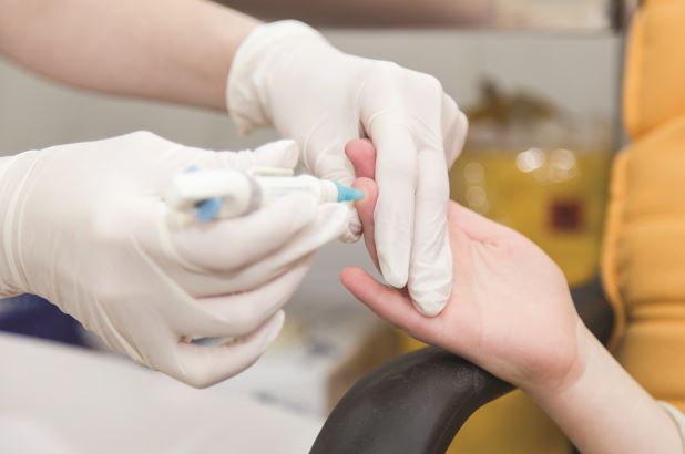
Lead is a naturally occurring element that is not metabolized, but rather absorbed, distributed to tissues, and excreted. Lead can be inhaled (with 100% absorption) and introduced through the GI tract (with about 70% absorption in children and 20% absorption in adults). GI absorption is enhanced by calcium or iron deficiency, both conditions that are relatively common, especially in poor children and can lead to pica (or eating of non-nutritious materials), further increasing the chances of lead exposure. Absorbed lead is distributed to blood (for 28-36 days), soft tissue, including the nervous system (40 days), and to bone (where it lasts for over 25 years). Blood that is retained in growing bones can be mobilized during periods of physiologic stress (such as illness, injury, or pregnancy), meaning children exposed to lead during a period of rapid bone growth are at long-term risk for acute lead poisoning from their endogenous reservoir without a new exposure. What lead is not retained by tissues is excreted by the kidneys, with adults retaining about 1% of absorbed lead, while children younger than 2 years retain over 30% of absorbed lead. So children, especially toddlers, have a greater likelihood to absorb lead from the GI tract and to retain lead in their tissues, both due to active mineralization of bone and the permeability of the blood brain barrier, primarily in children under 3 years old. This is why we are addressing what will happen to the children of Flint and not to all the residents of Flint.
Lead competitively inhibits interactions between cations and sulfhydryl groups, which are present in most human biochemical reactions. This leads to irreversible cell damage and often cell death, especially within the central nervous system. Lead exposure is associated with particular dysfunction within dopaminergic pathways within the brain, and has been associated in a dose-dependent fashion with decreased prefrontal gray matter volume. Lead poisoning also has hematologic consequences (anemia), renal consequences (interstitial nephritis), gastrointestinal symptoms (vomiting, constipation), and endocrine consequences (reversible inhibition of Vitamin D metabolism and permanently short stature). But the CNS consequences of lead exposure are particularly devastating, as they appear to have no threshold and are permanent. Their incidence is the driving force for the CDC’s lowering of the official toxic lead level and the public health efforts to screen children and educate parents about the risk of lead exposure.

So what do these serious consequences look like? People with severe lead intoxication (blood lead levels greater than 70 mcg/dL) typically present with signs of acute encephalopathy (headache, vomiting, seizures, or coma) and require intensive medical management including chelation therapy. More typically, exposed children have low but accumulating levels of lead and present with nonspecific symptoms, including lost appetite, fatigue, irritability, and insomnia, which gradually worsen.
Behavior
High levels of impulsivity, aggression, and impaired attention are the prototypical sequelae of lead poisoning (following recovery from the acute intoxication). Multiple studies have demonstrated these high levels of aggressive and impulsive behaviors in preschoolers who were exposed to lead, and these behaviors appear to continue into adolescence and adulthood. Indeed, one study found that compared with children with the lowest measurable blood lead levels (0.2-0.7 mcg/dL), those children who were in the next two quartiles had seven and twelve times the odds of meeting diagnostic criteria for conduct disorder.1 There have even been studies which correlated atmospheric lead levels (when leaded gasoline was common) with crime rates 20 years later, which supported an association between childhood lead exposure and adult criminal activity.2-4.
Multiple studies have demonstrated higher rates of inattention, distractibility, and impulsivity in lead-exposed children than would be expected given the prevalence of attention-deficit/hyperactivity disorder (ADHD) in the general population. The incidence of these symptoms goes up in a dose-dependent fashion and appears to have no threshold (so they occur at even the lowest measurable blood lead levels). In a 2006 study of nearly 5,000 children between ages 4-15 years, those with blood lead levels greater than 2 mcg/dL (still below the level the CDC deems toxic) were four times more likely to be carrying a diagnosis of ADHD and be on stimulant medication than their peers with blood lead levels less than 0.8mcg/dL.
Cognition
Closely related to impulse control and attention, the cognitive domains of intelligence and executive function are clearly damaged by lead exposure. Poor performance on tasks requiring focus, cognitive flexibility, and inhibition of automatic responses was directly associated with higher blood lead levels in a group of preschoolers with levels between 0 and 13 mcg/dL.5

IQ has been found to be so consistently diminished by increasing blood lead levels that it is used as an overall index of neurodevelopmental morbidity of lead exposure, leading to the CDC’s adoption of a lower standard definition of toxic lead levels. Even very low blood lead levels are associated with decrements in IQ: children with blood lead levels less than 7.5 mcg/dL lost an average of 3 IQ points for every 1 mcg/dL increase in blood lead levels.6 In a study of 57,000 elementary school students in 2009, Miranda et al. found that those who had a blood lead level of 4 mcg/dL at 3 years old were significantly more likely to be diagnosed with a learning disability in elementary school. Another study of 48,000 children who had a blood lead level of 5 mcg/dL were 30% more likely to fail third grade reading and math tests than their peers without measurable lead levels.
Speech and language
More recent studies have demonstrated that children with higher bone lead concentrations had poorer performance on several language-processing measures, suggesting that childhood lead exposure damages language processing and function as the young people grow. These deficits in language processing can make social development and self-regulation much more challenging in adolescence, and make school and work settings much more challenging. These findings also have implications for the utility of psychotherapy, a language-based treatment, for the other behavioral problems of lead exposure.
Motor skills
Several recent studies have assessed both fine and gross motor skills in lead-exposed children. Findings have demonstrated that balance, coordination, gross motor and fine motor skills all appear to be compromised in a dose-dependent fashion by childhood lead exposure. These findings suggest that not only are children at greater risk for accident and injury through childhood and into adulthood, a risk already increased by their compromised attention and impulse control. But they also are likely to be physically clumsy, compromising an opportunity to cultivate strengths or experience mastery when cognitive tasks may prove frustrating for them.
With deficits in such fundamental cognitive, motor, and behavioral processes, exposed children are clearly vulnerable to more than ADHD, conduct disorder, and learning disabilities. These struggles may lead to secondary vulnerabilities to anxiety or mood symptoms or substance abuse as these children grow into teenagers who face frustration at every turn. In addition to treatment for their deficits in attention and executive function, these children will ideally receive specialized supports in school and at home, to be able to master cognitive tasks, manage new social circumstances and make friends, discover their interests and talents, and generally stay on their best developmental trajectories. Lastly, the specific consequences of lead exposure will vary for any individual child, so parents will have to deal with the uncertainty of their child’s behavior and development over many years. Clearly, the children of Flint face a long road that has been substantially impacted by their lead exposure. The only good that can come from the exposure in Flint is to heighten efforts to ensure that it never happens again.
1. Environ Health Perspect. 2008 Jul;116(7):956-62.
2. Environ Res. 2000 May;83(1):1-22.
3. Environ Res. 2007 Jul;104(3):315-36.
4. Arch Pediatr Adolesc Med. 2001 May;155(5):579-82.
5. Dev Neuropsychol. 2004;26(1):513-40.
6. Environ Health Perspect. 2005 Jul;113(7):894-9.
Dr. Swick is an attending psychiatrist in the division of child psychiatry at Massachusetts General Hospital, Boston, and director of the Parenting at a Challenging Time (PACT) Program at the Vernon Cancer Center at Newton (Mass.) Wellesley Hospital. Dr. Jellinek is professor of psychiatry and of pediatrics at Harvard Medical School, Boston.
Lead poisoning is a well-established cause of serious and permanent neurological, cognitive, and behavioral problems, particularly in exposed children.
Children can be exposed to lead from ingesting paint chips in their homes, when old paint is scrapped from the exterior of houses or bridges, and through the water they drink. The damage caused by lead poisoning was first recognized in the United States in the early 20th century, although lead was added to gasoline and paint until the 1970’s. Since then, regulations for lead in consumer products have become increasingly strict, and the Centers for Disease Control and Prevention’s definition of a toxic lead level has shifted from 60 micrograms/deciliter (mcg/dL) in 1970 to 5 mcg/dL in 2012. In many communities, removing lead paint up to the height of a young child is a requirement whenever an older home is sold.
Unfortunately, these regulations did not protect the families in Flint, Michigan from being exposed to high levels of lead when a change in water supply and inadequate water treatment allowed lead to enter the system from decaying water pipes. It is worth reviewing what is known about the short- and long-term consequences of lead exposure, and what lies ahead for the children of Flint.
Lead is a naturally occurring element that is not metabolized, but rather absorbed, distributed to tissues, and excreted. Lead can be inhaled (with 100% absorption) and introduced through the GI tract (with about 70% absorption in children and 20% absorption in adults). GI absorption is enhanced by calcium or iron deficiency, both conditions that are relatively common, especially in poor children and can lead to pica (or eating of non-nutritious materials), further increasing the chances of lead exposure. Absorbed lead is distributed to blood (for 28-36 days), soft tissue, including the nervous system (40 days), and to bone (where it lasts for over 25 years). Blood that is retained in growing bones can be mobilized during periods of physiologic stress (such as illness, injury, or pregnancy), meaning children exposed to lead during a period of rapid bone growth are at long-term risk for acute lead poisoning from their endogenous reservoir without a new exposure. What lead is not retained by tissues is excreted by the kidneys, with adults retaining about 1% of absorbed lead, while children younger than 2 years retain over 30% of absorbed lead. So children, especially toddlers, have a greater likelihood to absorb lead from the GI tract and to retain lead in their tissues, both due to active mineralization of bone and the permeability of the blood brain barrier, primarily in children under 3 years old. This is why we are addressing what will happen to the children of Flint and not to all the residents of Flint.
Lead competitively inhibits interactions between cations and sulfhydryl groups, which are present in most human biochemical reactions. This leads to irreversible cell damage and often cell death, especially within the central nervous system. Lead exposure is associated with particular dysfunction within dopaminergic pathways within the brain, and has been associated in a dose-dependent fashion with decreased prefrontal gray matter volume. Lead poisoning also has hematologic consequences (anemia), renal consequences (interstitial nephritis), gastrointestinal symptoms (vomiting, constipation), and endocrine consequences (reversible inhibition of Vitamin D metabolism and permanently short stature). But the CNS consequences of lead exposure are particularly devastating, as they appear to have no threshold and are permanent. Their incidence is the driving force for the CDC’s lowering of the official toxic lead level and the public health efforts to screen children and educate parents about the risk of lead exposure.
So what do these serious consequences look like? People with severe lead intoxication (blood lead levels greater than 70 mcg/dL) typically present with signs of acute encephalopathy (headache, vomiting, seizures, or coma) and require intensive medical management including chelation therapy. More typically, exposed children have low but accumulating levels of lead and present with nonspecific symptoms, including lost appetite, fatigue, irritability, and insomnia, which gradually worsen.
Behavior
High levels of impulsivity, aggression, and impaired attention are the prototypical sequelae of lead poisoning (following recovery from the acute intoxication). Multiple studies have demonstrated these high levels of aggressive and impulsive behaviors in preschoolers who were exposed to lead, and these behaviors appear to continue into adolescence and adulthood. Indeed, one study found that compared with children with the lowest measurable blood lead levels (0.2-0.7 mcg/dL), those children who were in the next two quartiles had seven and twelve times the odds of meeting diagnostic criteria for conduct disorder.1 There have even been studies which correlated atmospheric lead levels (when leaded gasoline was common) with crime rates 20 years later, which supported an association between childhood lead exposure and adult criminal activity.2-4.
Multiple studies have demonstrated higher rates of inattention, distractibility, and impulsivity in lead-exposed children than would be expected given the prevalence of attention-deficit/hyperactivity disorder (ADHD) in the general population. The incidence of these symptoms goes up in a dose-dependent fashion and appears to have no threshold (so they occur at even the lowest measurable blood lead levels). In a 2006 study of nearly 5,000 children between ages 4-15 years, those with blood lead levels greater than 2 mcg/dL (still below the level the CDC deems toxic) were four times more likely to be carrying a diagnosis of ADHD and be on stimulant medication than their peers with blood lead levels less than 0.8mcg/dL.
Cognition
Closely related to impulse control and attention, the cognitive domains of intelligence and executive function are clearly damaged by lead exposure. Poor performance on tasks requiring focus, cognitive flexibility, and inhibition of automatic responses was directly associated with higher blood lead levels in a group of preschoolers with levels between 0 and 13 mcg/dL.5
IQ has been found to be so consistently diminished by increasing blood lead levels that it is used as an overall index of neurodevelopmental morbidity of lead exposure, leading to the CDC’s adoption of a lower standard definition of toxic lead levels. Even very low blood lead levels are associated with decrements in IQ: children with blood lead levels less than 7.5 mcg/dL lost an average of 3 IQ points for every 1 mcg/dL increase in blood lead levels.6 In a study of 57,000 elementary school students in 2009, Miranda et al. found that those who had a blood lead level of 4 mcg/dL at 3 years old were significantly more likely to be diagnosed with a learning disability in elementary school. Another study of 48,000 children who had a blood lead level of 5 mcg/dL were 30% more likely to fail third grade reading and math tests than their peers without measurable lead levels.
Speech and language
More recent studies have demonstrated that children with higher bone lead concentrations had poorer performance on several language-processing measures, suggesting that childhood lead exposure damages language processing and function as the young people grow. These deficits in language processing can make social development and self-regulation much more challenging in adolescence, and make school and work settings much more challenging. These findings also have implications for the utility of psychotherapy, a language-based treatment, for the other behavioral problems of lead exposure.
Motor skills
Several recent studies have assessed both fine and gross motor skills in lead-exposed children. Findings have demonstrated that balance, coordination, gross motor and fine motor skills all appear to be compromised in a dose-dependent fashion by childhood lead exposure. These findings suggest that not only are children at greater risk for accident and injury through childhood and into adulthood, a risk already increased by their compromised attention and impulse control. But they also are likely to be physically clumsy, compromising an opportunity to cultivate strengths or experience mastery when cognitive tasks may prove frustrating for them.
With deficits in such fundamental cognitive, motor, and behavioral processes, exposed children are clearly vulnerable to more than ADHD, conduct disorder, and learning disabilities. These struggles may lead to secondary vulnerabilities to anxiety or mood symptoms or substance abuse as these children grow into teenagers who face frustration at every turn. In addition to treatment for their deficits in attention and executive function, these children will ideally receive specialized supports in school and at home, to be able to master cognitive tasks, manage new social circumstances and make friends, discover their interests and talents, and generally stay on their best developmental trajectories. Lastly, the specific consequences of lead exposure will vary for any individual child, so parents will have to deal with the uncertainty of their child’s behavior and development over many years. Clearly, the children of Flint face a long road that has been substantially impacted by their lead exposure. The only good that can come from the exposure in Flint is to heighten efforts to ensure that it never happens again.
1. Environ Health Perspect. 2008 Jul;116(7):956-62.
2. Environ Res. 2000 May;83(1):1-22.
3. Environ Res. 2007 Jul;104(3):315-36.
4. Arch Pediatr Adolesc Med. 2001 May;155(5):579-82.
5. Dev Neuropsychol. 2004;26(1):513-40.
6. Environ Health Perspect. 2005 Jul;113(7):894-9.
Dr. Swick is an attending psychiatrist in the division of child psychiatry at Massachusetts General Hospital, Boston, and director of the Parenting at a Challenging Time (PACT) Program at the Vernon Cancer Center at Newton (Mass.) Wellesley Hospital. Dr. Jellinek is professor of psychiatry and of pediatrics at Harvard Medical School, Boston.
Lead poisoning is a well-established cause of serious and permanent neurological, cognitive, and behavioral problems, particularly in exposed children.
Children can be exposed to lead from ingesting paint chips in their homes, when old paint is scrapped from the exterior of houses or bridges, and through the water they drink. The damage caused by lead poisoning was first recognized in the United States in the early 20th century, although lead was added to gasoline and paint until the 1970’s. Since then, regulations for lead in consumer products have become increasingly strict, and the Centers for Disease Control and Prevention’s definition of a toxic lead level has shifted from 60 micrograms/deciliter (mcg/dL) in 1970 to 5 mcg/dL in 2012. In many communities, removing lead paint up to the height of a young child is a requirement whenever an older home is sold.
Unfortunately, these regulations did not protect the families in Flint, Michigan from being exposed to high levels of lead when a change in water supply and inadequate water treatment allowed lead to enter the system from decaying water pipes. It is worth reviewing what is known about the short- and long-term consequences of lead exposure, and what lies ahead for the children of Flint.
Lead is a naturally occurring element that is not metabolized, but rather absorbed, distributed to tissues, and excreted. Lead can be inhaled (with 100% absorption) and introduced through the GI tract (with about 70% absorption in children and 20% absorption in adults). GI absorption is enhanced by calcium or iron deficiency, both conditions that are relatively common, especially in poor children and can lead to pica (or eating of non-nutritious materials), further increasing the chances of lead exposure. Absorbed lead is distributed to blood (for 28-36 days), soft tissue, including the nervous system (40 days), and to bone (where it lasts for over 25 years). Blood that is retained in growing bones can be mobilized during periods of physiologic stress (such as illness, injury, or pregnancy), meaning children exposed to lead during a period of rapid bone growth are at long-term risk for acute lead poisoning from their endogenous reservoir without a new exposure. What lead is not retained by tissues is excreted by the kidneys, with adults retaining about 1% of absorbed lead, while children younger than 2 years retain over 30% of absorbed lead. So children, especially toddlers, have a greater likelihood to absorb lead from the GI tract and to retain lead in their tissues, both due to active mineralization of bone and the permeability of the blood brain barrier, primarily in children under 3 years old. This is why we are addressing what will happen to the children of Flint and not to all the residents of Flint.
Lead competitively inhibits interactions between cations and sulfhydryl groups, which are present in most human biochemical reactions. This leads to irreversible cell damage and often cell death, especially within the central nervous system. Lead exposure is associated with particular dysfunction within dopaminergic pathways within the brain, and has been associated in a dose-dependent fashion with decreased prefrontal gray matter volume. Lead poisoning also has hematologic consequences (anemia), renal consequences (interstitial nephritis), gastrointestinal symptoms (vomiting, constipation), and endocrine consequences (reversible inhibition of Vitamin D metabolism and permanently short stature). But the CNS consequences of lead exposure are particularly devastating, as they appear to have no threshold and are permanent. Their incidence is the driving force for the CDC’s lowering of the official toxic lead level and the public health efforts to screen children and educate parents about the risk of lead exposure.
So what do these serious consequences look like? People with severe lead intoxication (blood lead levels greater than 70 mcg/dL) typically present with signs of acute encephalopathy (headache, vomiting, seizures, or coma) and require intensive medical management including chelation therapy. More typically, exposed children have low but accumulating levels of lead and present with nonspecific symptoms, including lost appetite, fatigue, irritability, and insomnia, which gradually worsen.
Behavior
High levels of impulsivity, aggression, and impaired attention are the prototypical sequelae of lead poisoning (following recovery from the acute intoxication). Multiple studies have demonstrated these high levels of aggressive and impulsive behaviors in preschoolers who were exposed to lead, and these behaviors appear to continue into adolescence and adulthood. Indeed, one study found that compared with children with the lowest measurable blood lead levels (0.2-0.7 mcg/dL), those children who were in the next two quartiles had seven and twelve times the odds of meeting diagnostic criteria for conduct disorder.1 There have even been studies which correlated atmospheric lead levels (when leaded gasoline was common) with crime rates 20 years later, which supported an association between childhood lead exposure and adult criminal activity.2-4.
Multiple studies have demonstrated higher rates of inattention, distractibility, and impulsivity in lead-exposed children than would be expected given the prevalence of attention-deficit/hyperactivity disorder (ADHD) in the general population. The incidence of these symptoms goes up in a dose-dependent fashion and appears to have no threshold (so they occur at even the lowest measurable blood lead levels). In a 2006 study of nearly 5,000 children between ages 4-15 years, those with blood lead levels greater than 2 mcg/dL (still below the level the CDC deems toxic) were four times more likely to be carrying a diagnosis of ADHD and be on stimulant medication than their peers with blood lead levels less than 0.8mcg/dL.
Cognition
Closely related to impulse control and attention, the cognitive domains of intelligence and executive function are clearly damaged by lead exposure. Poor performance on tasks requiring focus, cognitive flexibility, and inhibition of automatic responses was directly associated with higher blood lead levels in a group of preschoolers with levels between 0 and 13 mcg/dL.5
IQ has been found to be so consistently diminished by increasing blood lead levels that it is used as an overall index of neurodevelopmental morbidity of lead exposure, leading to the CDC’s adoption of a lower standard definition of toxic lead levels. Even very low blood lead levels are associated with decrements in IQ: children with blood lead levels less than 7.5 mcg/dL lost an average of 3 IQ points for every 1 mcg/dL increase in blood lead levels.6 In a study of 57,000 elementary school students in 2009, Miranda et al. found that those who had a blood lead level of 4 mcg/dL at 3 years old were significantly more likely to be diagnosed with a learning disability in elementary school. Another study of 48,000 children who had a blood lead level of 5 mcg/dL were 30% more likely to fail third grade reading and math tests than their peers without measurable lead levels.
Speech and language
More recent studies have demonstrated that children with higher bone lead concentrations had poorer performance on several language-processing measures, suggesting that childhood lead exposure damages language processing and function as the young people grow. These deficits in language processing can make social development and self-regulation much more challenging in adolescence, and make school and work settings much more challenging. These findings also have implications for the utility of psychotherapy, a language-based treatment, for the other behavioral problems of lead exposure.
Motor skills
Several recent studies have assessed both fine and gross motor skills in lead-exposed children. Findings have demonstrated that balance, coordination, gross motor and fine motor skills all appear to be compromised in a dose-dependent fashion by childhood lead exposure. These findings suggest that not only are children at greater risk for accident and injury through childhood and into adulthood, a risk already increased by their compromised attention and impulse control. But they also are likely to be physically clumsy, compromising an opportunity to cultivate strengths or experience mastery when cognitive tasks may prove frustrating for them.
With deficits in such fundamental cognitive, motor, and behavioral processes, exposed children are clearly vulnerable to more than ADHD, conduct disorder, and learning disabilities. These struggles may lead to secondary vulnerabilities to anxiety or mood symptoms or substance abuse as these children grow into teenagers who face frustration at every turn. In addition to treatment for their deficits in attention and executive function, these children will ideally receive specialized supports in school and at home, to be able to master cognitive tasks, manage new social circumstances and make friends, discover their interests and talents, and generally stay on their best developmental trajectories. Lastly, the specific consequences of lead exposure will vary for any individual child, so parents will have to deal with the uncertainty of their child’s behavior and development over many years. Clearly, the children of Flint face a long road that has been substantially impacted by their lead exposure. The only good that can come from the exposure in Flint is to heighten efforts to ensure that it never happens again.
1. Environ Health Perspect. 2008 Jul;116(7):956-62.
2. Environ Res. 2000 May;83(1):1-22.
3. Environ Res. 2007 Jul;104(3):315-36.
4. Arch Pediatr Adolesc Med. 2001 May;155(5):579-82.
5. Dev Neuropsychol. 2004;26(1):513-40.
6. Environ Health Perspect. 2005 Jul;113(7):894-9.
Dr. Swick is an attending psychiatrist in the division of child psychiatry at Massachusetts General Hospital, Boston, and director of the Parenting at a Challenging Time (PACT) Program at the Vernon Cancer Center at Newton (Mass.) Wellesley Hospital. Dr. Jellinek is professor of psychiatry and of pediatrics at Harvard Medical School, Boston.
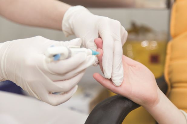
STS: Valved conduit shows right ventricular outflow durability
PHOENIX – A prosthetic conduit that contains a porcine valve showed excellent intermediate-term durability for repairing the right ventricular outflow tract in 100 teenagers and young adults at a single U.S. center.
“The Carpentier-Edwards xenograft for right ventricular outflow tract [RVOT] reconstruction provides excellent freedom from reoperation and valve dysfunction, as well as sustained improvement in right-ventricular chamber size at intermediate-term follow-up,” Dr. Heidi B. Schubmehl said at the Society of Thoracic Surgeons annual meeting.

Dr. Schubmehl reported a 92% rate of freedom from valve dysfunction with follow-up out to about 10 years, and significant reductions in right ventricular size at follow-up, compared with baseline, as measured by both echocardiography and by MRI.
The Carpentier-Edwards porcine valve and conduit “seemed to hold up better than a lot of other [prosthetic] valves,” said Dr. George M. Alfieris, director of pediatric cardiac surgery at the University of Rochester (N.Y.), and senior author for the study. In addition to the valve’s durability over approximately the first 10 years following placement, the results also showed the positive impact the valve had on right ventricular size, an important result of the repair’s efficacy, Dr. Alfieris said.
“It’s a mistake to allow the right ventricle to be under high pressure or to reach a large volume. We now focus on preserving the right ventricle,” he said in an interview. “I’ve become very concerned about preventing right ventricular dilation and preserving right ventricular function.”
Dr. Alfieris noted that his prior experience using other types of valves in the pulmonary valve and RVOT position showed those valves “did great for the first 10 years and then failed. What’s different in this series is that after 10 years, we have not seen the same dysfunction as with the prior generation of valves. I will be very interested to see what happens to them” as follow-up continues beyond 10 years. He also expressed dismay that recently the company that had been marketing the valve and conduit used in the current study, the Carpentier-Edwards, stopped selling them. He expects that as his supply of conduits runs out he’ll have to start using a different commercial valve and conduit that he believes will not perform as well or create his own conduits with a porcine valve from a different supplier.

The series of 100 patients comprised individuals aged 17 or older who received a pulmonary artery and had RVOT reconstruction at the University of Rochester during 2000-2010, Dr. Schubmehl reported. The series included 78 patients with a history of tetralogy of Fallot, 8 patients born with transposition of their great arteries, 8 patients with truncus arteriosus, and 6 patients with other congenital heart diseases. Their median age at the time they received the RVOT conduit was 24 years, 59% were men, and 99 had undergone a prior sternotomy. At the time they received the conduit, 55 had pulmonary valve insufficiency, 30 had valve stenosis, and 15 had both. Follow-up occurred an average of 7 years after conduit placement.
Two recipients died: One death occurred perioperatively in a 41-year old who had a massive cerebrovascular event, and the second death was in a 39-year old who died 2.6 years after conduit placement from respiratory failure. Two additional patients required a reintervention during follow-up, said Dr. Schubmehl, a general surgeon at the University of Rochester. One reintervention occurred after 11 years to treat endocarditis, and the second after 11 years to perform balloon valvuloplasty because of valve stenosis.
The results reported by Dr. Schubmehl for echocardiography examinations showed that the patients had a statistically significant reduction in their RVOT pressure gradient from baseline to 1-year follow-up that was sustained through their intermediate-term follow-up. Seventy-seven patients had pulmonary valve insufficiency at baseline that resolved in all patients at 1-year follow-up and remained resolved in all but one patient at extended follow-up. Nineteen patients underwent additional imaging with MRI at an average follow-up of 7 years, and these findings confirmed the echo results.
On Twitter @mitchelzoler
The intermediate-term results reported by Dr. Schubmehl using a Carpentier-Edwards conduit in the right-ventricular outflow tract are clearly better than what we have seen using other types of valves and conduits in this position. If the valve and conduit they used persists with similar performance beyond 10 years, it would be a very good option. However, what typically happens is that replacement valves look good for about 10 years and then start to fail, often with a steep failure curve. I suspect that during the next 10 years of follow-up many more of the valves they placed will start to fail. The 10- to 20-year follow-up period is critical for demonstrating long-term durability of this valve and conduit.

|
Dr. James Jaggers |
One additional potential advantage of the Carpentier-Edwards prosthesis is that the valve it contains is larger than the usual valve placed in the right ventricular outflow tract (RVOT). Failed valves increasingly are replaced by a transcatheter approach that puts a new valve inside the old, failed valve. As patients who received these replacement valves continue to survive we anticipate their need over time for a series of valve-in-valve procedures. The larger the valve at the outset, the more feasible it will be to have multiple episodes of valve-in-valve replacement.
At one time, we regarded early surgical repair of a tetralogy of Fallot defect as curative. We now know that as children with a repaired tetralogy of Fallot grow into teens and adults they require additional repairs, most often replacement of their RVOTs. This has made pulmonary valve replacement the most common surgery for adult survivors of congenital heart disease. The numbers of teen or adult patients who require a new RVOT will steadily increase as more of these children survive.
Dr. James Jaggers, professor of surgery at the University of Colorado and chief of cardiothoracic surgery at Children’s Hospital Colorado in Denver, made these comments in an interview. He had no disclosures.
The intermediate-term results reported by Dr. Schubmehl using a Carpentier-Edwards conduit in the right-ventricular outflow tract are clearly better than what we have seen using other types of valves and conduits in this position. If the valve and conduit they used persists with similar performance beyond 10 years, it would be a very good option. However, what typically happens is that replacement valves look good for about 10 years and then start to fail, often with a steep failure curve. I suspect that during the next 10 years of follow-up many more of the valves they placed will start to fail. The 10- to 20-year follow-up period is critical for demonstrating long-term durability of this valve and conduit.
|
|
Dr. James Jaggers |
One additional potential advantage of the Carpentier-Edwards prosthesis is that the valve it contains is larger than the usual valve placed in the right ventricular outflow tract (RVOT). Failed valves increasingly are replaced by a transcatheter approach that puts a new valve inside the old, failed valve. As patients who received these replacement valves continue to survive we anticipate their need over time for a series of valve-in-valve procedures. The larger the valve at the outset, the more feasible it will be to have multiple episodes of valve-in-valve replacement.
At one time, we regarded early surgical repair of a tetralogy of Fallot defect as curative. We now know that as children with a repaired tetralogy of Fallot grow into teens and adults they require additional repairs, most often replacement of their RVOTs. This has made pulmonary valve replacement the most common surgery for adult survivors of congenital heart disease. The numbers of teen or adult patients who require a new RVOT will steadily increase as more of these children survive.
Dr. James Jaggers, professor of surgery at the University of Colorado and chief of cardiothoracic surgery at Children’s Hospital Colorado in Denver, made these comments in an interview. He had no disclosures.
The intermediate-term results reported by Dr. Schubmehl using a Carpentier-Edwards conduit in the right-ventricular outflow tract are clearly better than what we have seen using other types of valves and conduits in this position. If the valve and conduit they used persists with similar performance beyond 10 years, it would be a very good option. However, what typically happens is that replacement valves look good for about 10 years and then start to fail, often with a steep failure curve. I suspect that during the next 10 years of follow-up many more of the valves they placed will start to fail. The 10- to 20-year follow-up period is critical for demonstrating long-term durability of this valve and conduit.
|
|
Dr. James Jaggers |
One additional potential advantage of the Carpentier-Edwards prosthesis is that the valve it contains is larger than the usual valve placed in the right ventricular outflow tract (RVOT). Failed valves increasingly are replaced by a transcatheter approach that puts a new valve inside the old, failed valve. As patients who received these replacement valves continue to survive we anticipate their need over time for a series of valve-in-valve procedures. The larger the valve at the outset, the more feasible it will be to have multiple episodes of valve-in-valve replacement.
At one time, we regarded early surgical repair of a tetralogy of Fallot defect as curative. We now know that as children with a repaired tetralogy of Fallot grow into teens and adults they require additional repairs, most often replacement of their RVOTs. This has made pulmonary valve replacement the most common surgery for adult survivors of congenital heart disease. The numbers of teen or adult patients who require a new RVOT will steadily increase as more of these children survive.
Dr. James Jaggers, professor of surgery at the University of Colorado and chief of cardiothoracic surgery at Children’s Hospital Colorado in Denver, made these comments in an interview. He had no disclosures.
PHOENIX – A prosthetic conduit that contains a porcine valve showed excellent intermediate-term durability for repairing the right ventricular outflow tract in 100 teenagers and young adults at a single U.S. center.
“The Carpentier-Edwards xenograft for right ventricular outflow tract [RVOT] reconstruction provides excellent freedom from reoperation and valve dysfunction, as well as sustained improvement in right-ventricular chamber size at intermediate-term follow-up,” Dr. Heidi B. Schubmehl said at the Society of Thoracic Surgeons annual meeting.
Dr. Schubmehl reported a 92% rate of freedom from valve dysfunction with follow-up out to about 10 years, and significant reductions in right ventricular size at follow-up, compared with baseline, as measured by both echocardiography and by MRI.
The Carpentier-Edwards porcine valve and conduit “seemed to hold up better than a lot of other [prosthetic] valves,” said Dr. George M. Alfieris, director of pediatric cardiac surgery at the University of Rochester (N.Y.), and senior author for the study. In addition to the valve’s durability over approximately the first 10 years following placement, the results also showed the positive impact the valve had on right ventricular size, an important result of the repair’s efficacy, Dr. Alfieris said.
“It’s a mistake to allow the right ventricle to be under high pressure or to reach a large volume. We now focus on preserving the right ventricle,” he said in an interview. “I’ve become very concerned about preventing right ventricular dilation and preserving right ventricular function.”
Dr. Alfieris noted that his prior experience using other types of valves in the pulmonary valve and RVOT position showed those valves “did great for the first 10 years and then failed. What’s different in this series is that after 10 years, we have not seen the same dysfunction as with the prior generation of valves. I will be very interested to see what happens to them” as follow-up continues beyond 10 years. He also expressed dismay that recently the company that had been marketing the valve and conduit used in the current study, the Carpentier-Edwards, stopped selling them. He expects that as his supply of conduits runs out he’ll have to start using a different commercial valve and conduit that he believes will not perform as well or create his own conduits with a porcine valve from a different supplier.
The series of 100 patients comprised individuals aged 17 or older who received a pulmonary artery and had RVOT reconstruction at the University of Rochester during 2000-2010, Dr. Schubmehl reported. The series included 78 patients with a history of tetralogy of Fallot, 8 patients born with transposition of their great arteries, 8 patients with truncus arteriosus, and 6 patients with other congenital heart diseases. Their median age at the time they received the RVOT conduit was 24 years, 59% were men, and 99 had undergone a prior sternotomy. At the time they received the conduit, 55 had pulmonary valve insufficiency, 30 had valve stenosis, and 15 had both. Follow-up occurred an average of 7 years after conduit placement.
Two recipients died: One death occurred perioperatively in a 41-year old who had a massive cerebrovascular event, and the second death was in a 39-year old who died 2.6 years after conduit placement from respiratory failure. Two additional patients required a reintervention during follow-up, said Dr. Schubmehl, a general surgeon at the University of Rochester. One reintervention occurred after 11 years to treat endocarditis, and the second after 11 years to perform balloon valvuloplasty because of valve stenosis.
The results reported by Dr. Schubmehl for echocardiography examinations showed that the patients had a statistically significant reduction in their RVOT pressure gradient from baseline to 1-year follow-up that was sustained through their intermediate-term follow-up. Seventy-seven patients had pulmonary valve insufficiency at baseline that resolved in all patients at 1-year follow-up and remained resolved in all but one patient at extended follow-up. Nineteen patients underwent additional imaging with MRI at an average follow-up of 7 years, and these findings confirmed the echo results.
On Twitter @mitchelzoler
PHOENIX – A prosthetic conduit that contains a porcine valve showed excellent intermediate-term durability for repairing the right ventricular outflow tract in 100 teenagers and young adults at a single U.S. center.
“The Carpentier-Edwards xenograft for right ventricular outflow tract [RVOT] reconstruction provides excellent freedom from reoperation and valve dysfunction, as well as sustained improvement in right-ventricular chamber size at intermediate-term follow-up,” Dr. Heidi B. Schubmehl said at the Society of Thoracic Surgeons annual meeting.
Dr. Schubmehl reported a 92% rate of freedom from valve dysfunction with follow-up out to about 10 years, and significant reductions in right ventricular size at follow-up, compared with baseline, as measured by both echocardiography and by MRI.
The Carpentier-Edwards porcine valve and conduit “seemed to hold up better than a lot of other [prosthetic] valves,” said Dr. George M. Alfieris, director of pediatric cardiac surgery at the University of Rochester (N.Y.), and senior author for the study. In addition to the valve’s durability over approximately the first 10 years following placement, the results also showed the positive impact the valve had on right ventricular size, an important result of the repair’s efficacy, Dr. Alfieris said.
“It’s a mistake to allow the right ventricle to be under high pressure or to reach a large volume. We now focus on preserving the right ventricle,” he said in an interview. “I’ve become very concerned about preventing right ventricular dilation and preserving right ventricular function.”
Dr. Alfieris noted that his prior experience using other types of valves in the pulmonary valve and RVOT position showed those valves “did great for the first 10 years and then failed. What’s different in this series is that after 10 years, we have not seen the same dysfunction as with the prior generation of valves. I will be very interested to see what happens to them” as follow-up continues beyond 10 years. He also expressed dismay that recently the company that had been marketing the valve and conduit used in the current study, the Carpentier-Edwards, stopped selling them. He expects that as his supply of conduits runs out he’ll have to start using a different commercial valve and conduit that he believes will not perform as well or create his own conduits with a porcine valve from a different supplier.
The series of 100 patients comprised individuals aged 17 or older who received a pulmonary artery and had RVOT reconstruction at the University of Rochester during 2000-2010, Dr. Schubmehl reported. The series included 78 patients with a history of tetralogy of Fallot, 8 patients born with transposition of their great arteries, 8 patients with truncus arteriosus, and 6 patients with other congenital heart diseases. Their median age at the time they received the RVOT conduit was 24 years, 59% were men, and 99 had undergone a prior sternotomy. At the time they received the conduit, 55 had pulmonary valve insufficiency, 30 had valve stenosis, and 15 had both. Follow-up occurred an average of 7 years after conduit placement.
Two recipients died: One death occurred perioperatively in a 41-year old who had a massive cerebrovascular event, and the second death was in a 39-year old who died 2.6 years after conduit placement from respiratory failure. Two additional patients required a reintervention during follow-up, said Dr. Schubmehl, a general surgeon at the University of Rochester. One reintervention occurred after 11 years to treat endocarditis, and the second after 11 years to perform balloon valvuloplasty because of valve stenosis.
The results reported by Dr. Schubmehl for echocardiography examinations showed that the patients had a statistically significant reduction in their RVOT pressure gradient from baseline to 1-year follow-up that was sustained through their intermediate-term follow-up. Seventy-seven patients had pulmonary valve insufficiency at baseline that resolved in all patients at 1-year follow-up and remained resolved in all but one patient at extended follow-up. Nineteen patients underwent additional imaging with MRI at an average follow-up of 7 years, and these findings confirmed the echo results.
On Twitter @mitchelzoler
AT THE STS ANNUAL MEETING
Key clinical point: A prosthetic conduit with a porcine valve showed excellent durability for congenital heart defect repairs at intermediate-term follow-up.
Major finding: After an average 7-year follow-up, the replacement valve and conduit had a 92% rate of freedom from valve dysfunction.
Data source: Single-center series of 100 patients.
Disclosures: Dr. Schubmehl and Dr. Alfieris had no disclosures.