User login
Nausea with pediatric functional abdominal pain may mean depression, anxiety
(FAP), reported Alexandra C. Russell, MD, and her associates at Vanderbilt University, Nashville.
In a study of 398 children with FAP and nausea and 479 with FAP alone, those with comorbid nausea had significantly more GI symptoms and depression symptoms. Anxiety symptoms were not studied. Mean age at baseline was 12 years.
The FAP plus nausea patients were 1.79-times more likely to meet criteria for a current DSM-IV anxiety disorder and 2.61-times more likely to meet DSM-IV diagnostic criteria for generalized anxiety disorder than were FAP-only patients. The FAP and nausea patients were 1.81 times more likely to meet criteria for a DSM-IV for a major depressive disorder in their lifetime than were FAP-only patients.
The suggestion that nausea may be an independent risk factor for later anxiety and depression in children with FAP “may be owing to chronic nausea being a distressing symptom that permeates multiple areas of physical and psychosocial functioning,” Dr. Russell and her colleagues surmised.
“It may be difficult for these patients to maintain good nutrition, sleep habits, exercise, and resiliency when they frequently are nauseated,” the study authors noted. “Alternatively, FAP patients with comorbid nausea may represent a phenotype with autonomic nervous system dysfunction that contributes to both nausea and emotional distress.”
Read more in Clinical Gastroenterology and Hepatology (2017 May;15[5]:706-11).
(FAP), reported Alexandra C. Russell, MD, and her associates at Vanderbilt University, Nashville.
In a study of 398 children with FAP and nausea and 479 with FAP alone, those with comorbid nausea had significantly more GI symptoms and depression symptoms. Anxiety symptoms were not studied. Mean age at baseline was 12 years.
The FAP plus nausea patients were 1.79-times more likely to meet criteria for a current DSM-IV anxiety disorder and 2.61-times more likely to meet DSM-IV diagnostic criteria for generalized anxiety disorder than were FAP-only patients. The FAP and nausea patients were 1.81 times more likely to meet criteria for a DSM-IV for a major depressive disorder in their lifetime than were FAP-only patients.
The suggestion that nausea may be an independent risk factor for later anxiety and depression in children with FAP “may be owing to chronic nausea being a distressing symptom that permeates multiple areas of physical and psychosocial functioning,” Dr. Russell and her colleagues surmised.
“It may be difficult for these patients to maintain good nutrition, sleep habits, exercise, and resiliency when they frequently are nauseated,” the study authors noted. “Alternatively, FAP patients with comorbid nausea may represent a phenotype with autonomic nervous system dysfunction that contributes to both nausea and emotional distress.”
Read more in Clinical Gastroenterology and Hepatology (2017 May;15[5]:706-11).
(FAP), reported Alexandra C. Russell, MD, and her associates at Vanderbilt University, Nashville.
In a study of 398 children with FAP and nausea and 479 with FAP alone, those with comorbid nausea had significantly more GI symptoms and depression symptoms. Anxiety symptoms were not studied. Mean age at baseline was 12 years.
The FAP plus nausea patients were 1.79-times more likely to meet criteria for a current DSM-IV anxiety disorder and 2.61-times more likely to meet DSM-IV diagnostic criteria for generalized anxiety disorder than were FAP-only patients. The FAP and nausea patients were 1.81 times more likely to meet criteria for a DSM-IV for a major depressive disorder in their lifetime than were FAP-only patients.
The suggestion that nausea may be an independent risk factor for later anxiety and depression in children with FAP “may be owing to chronic nausea being a distressing symptom that permeates multiple areas of physical and psychosocial functioning,” Dr. Russell and her colleagues surmised.
“It may be difficult for these patients to maintain good nutrition, sleep habits, exercise, and resiliency when they frequently are nauseated,” the study authors noted. “Alternatively, FAP patients with comorbid nausea may represent a phenotype with autonomic nervous system dysfunction that contributes to both nausea and emotional distress.”
Read more in Clinical Gastroenterology and Hepatology (2017 May;15[5]:706-11).
FROM CLINICAL GASTROENTEROLOGY AND HEPATOLOGY
Intravenous tPA ups mortality in children with acute ischemic stroke
BOSTON – Intravenous thrombolysis with tissue plasminogen activator (tPA) is associated with adverse outcomes, including an increased risk of death, in children with acute ischemic stroke, based on a review of cases from the 2006-2010 Nationwide Inpatient Survey.
Of 20,587 patients aged 0-17 years who were included in the survey, 198 received an intervention, including tPA in 169 patients, intra-arterial thrombectomy (IAT) in 5 patients, and both tPA and IAT in 24 patients. The overall mortality was 7.8%, but in those who received tPA it was 13.8%, compared with 7.7% in those who did not, Kathryn Ess, MD, reported at the annual meeting of the American Academy of Neurology.
Other outcomes were also worse in those who received tPA. For example, untreated patients were more likely to be discharged home than were tPA-treated patients (67.8% vs. 47.5%), and intracerebral hemorrhage was more common in treated vs. untreated patients (10.1% vs. 3.8%). Costs for treated patients averaged $200,346 vs. $123,015 for untreated patients.
Children included in the review had a mean age of 6 years, 43.9% were girls, and 47.7% were white. Treated patients were older (10 years vs. 5.9 years), and comorbidities included Moyamoya disease in 12.4% of patients, cardiac valvular disease in 6.6%, and sickle cell disease in 6.5%. Those who received tPA had a higher prevalence of procoagulable conditions (15.2% vs. 2%). Of note, the higher prevalence of intracerebral hemorrhage in treated patients was not explained by Moyamoya or sickle cell disease, as patients with those comorbidities were less likely than those without those conditions to receive treatment, Dr. Ess said.
Though limited by the retrospective study design, small numbers of treated patients, a lack of data on stroke severity or functional outcomes, and the inclusion of data from years before newer thrombectomy devices became available, the findings highlight concerns about the safety and efficacy of tPA in children with ischemic stroke, she said, noting that few studies have looked at the utility of tPA with or without IAT in the pediatric population.
“Studies of the efficacy of ischemic stroke treatment in adults can’t necessarily be extrapolated to children,” she said, adding that this is especially true given the difference in etiologies of pediatric acute ischemic stroke.
Indeed, the findings underscore “the age-old adage that children are not just little adults,” said Andrew Southerland, MD, of the University of Virginia, Charlottesville, who was the discussant for the session.
“We need prospective clinical trials in children,” he said.
Dr. Ess and Dr. Southerland reported having no relevant financial disclosures.
BOSTON – Intravenous thrombolysis with tissue plasminogen activator (tPA) is associated with adverse outcomes, including an increased risk of death, in children with acute ischemic stroke, based on a review of cases from the 2006-2010 Nationwide Inpatient Survey.
Of 20,587 patients aged 0-17 years who were included in the survey, 198 received an intervention, including tPA in 169 patients, intra-arterial thrombectomy (IAT) in 5 patients, and both tPA and IAT in 24 patients. The overall mortality was 7.8%, but in those who received tPA it was 13.8%, compared with 7.7% in those who did not, Kathryn Ess, MD, reported at the annual meeting of the American Academy of Neurology.
Other outcomes were also worse in those who received tPA. For example, untreated patients were more likely to be discharged home than were tPA-treated patients (67.8% vs. 47.5%), and intracerebral hemorrhage was more common in treated vs. untreated patients (10.1% vs. 3.8%). Costs for treated patients averaged $200,346 vs. $123,015 for untreated patients.
Children included in the review had a mean age of 6 years, 43.9% were girls, and 47.7% were white. Treated patients were older (10 years vs. 5.9 years), and comorbidities included Moyamoya disease in 12.4% of patients, cardiac valvular disease in 6.6%, and sickle cell disease in 6.5%. Those who received tPA had a higher prevalence of procoagulable conditions (15.2% vs. 2%). Of note, the higher prevalence of intracerebral hemorrhage in treated patients was not explained by Moyamoya or sickle cell disease, as patients with those comorbidities were less likely than those without those conditions to receive treatment, Dr. Ess said.
Though limited by the retrospective study design, small numbers of treated patients, a lack of data on stroke severity or functional outcomes, and the inclusion of data from years before newer thrombectomy devices became available, the findings highlight concerns about the safety and efficacy of tPA in children with ischemic stroke, she said, noting that few studies have looked at the utility of tPA with or without IAT in the pediatric population.
“Studies of the efficacy of ischemic stroke treatment in adults can’t necessarily be extrapolated to children,” she said, adding that this is especially true given the difference in etiologies of pediatric acute ischemic stroke.
Indeed, the findings underscore “the age-old adage that children are not just little adults,” said Andrew Southerland, MD, of the University of Virginia, Charlottesville, who was the discussant for the session.
“We need prospective clinical trials in children,” he said.
Dr. Ess and Dr. Southerland reported having no relevant financial disclosures.
BOSTON – Intravenous thrombolysis with tissue plasminogen activator (tPA) is associated with adverse outcomes, including an increased risk of death, in children with acute ischemic stroke, based on a review of cases from the 2006-2010 Nationwide Inpatient Survey.
Of 20,587 patients aged 0-17 years who were included in the survey, 198 received an intervention, including tPA in 169 patients, intra-arterial thrombectomy (IAT) in 5 patients, and both tPA and IAT in 24 patients. The overall mortality was 7.8%, but in those who received tPA it was 13.8%, compared with 7.7% in those who did not, Kathryn Ess, MD, reported at the annual meeting of the American Academy of Neurology.
Other outcomes were also worse in those who received tPA. For example, untreated patients were more likely to be discharged home than were tPA-treated patients (67.8% vs. 47.5%), and intracerebral hemorrhage was more common in treated vs. untreated patients (10.1% vs. 3.8%). Costs for treated patients averaged $200,346 vs. $123,015 for untreated patients.
Children included in the review had a mean age of 6 years, 43.9% were girls, and 47.7% were white. Treated patients were older (10 years vs. 5.9 years), and comorbidities included Moyamoya disease in 12.4% of patients, cardiac valvular disease in 6.6%, and sickle cell disease in 6.5%. Those who received tPA had a higher prevalence of procoagulable conditions (15.2% vs. 2%). Of note, the higher prevalence of intracerebral hemorrhage in treated patients was not explained by Moyamoya or sickle cell disease, as patients with those comorbidities were less likely than those without those conditions to receive treatment, Dr. Ess said.
Though limited by the retrospective study design, small numbers of treated patients, a lack of data on stroke severity or functional outcomes, and the inclusion of data from years before newer thrombectomy devices became available, the findings highlight concerns about the safety and efficacy of tPA in children with ischemic stroke, she said, noting that few studies have looked at the utility of tPA with or without IAT in the pediatric population.
“Studies of the efficacy of ischemic stroke treatment in adults can’t necessarily be extrapolated to children,” she said, adding that this is especially true given the difference in etiologies of pediatric acute ischemic stroke.
Indeed, the findings underscore “the age-old adage that children are not just little adults,” said Andrew Southerland, MD, of the University of Virginia, Charlottesville, who was the discussant for the session.
“We need prospective clinical trials in children,” he said.
Dr. Ess and Dr. Southerland reported having no relevant financial disclosures.
AT AAN 2017
Key clinical point:
Major finding: Mortality for pediatric acute ischemic stroke was 7.8% overall, 7.7% in those who did not receive tPA, and 13.8% in those who did receive tPA.
Data source: A retrospective review of cases from the 2006-2010 Nationwide Inpatient Sample.
Disclosures: Dr. Ess and Dr. Southerland reported having no relevant financial disclosures.
Study supports link between pediatric MS and remote viral infections
BOSTON – Prior Epstein-Barr virus (EBV) infection and prior herpes simplex virus (HSV) infection each appear to be associated with development of pediatric-onset multiple sclerosis (MS), according to findings from a large national case-control study.
Samples from 360 children with MS or clinically isolated syndrome and 496 frequency-matched controls recruited from 16 pediatric MS centers across the United States were tested for EBV, cytomegalovirus (CMV), and HSV antibodies and for 25-(OH)-vitamin D levels. After adjusting for age, sex, and race/ethnicity, evidence of a remote infection with EBV was strongly associated with higher risk of pediatric-onset MS (odds ratio, 3.6), Bardia Nourbakhsh, MD, reported at the annual meeting of the American Academy of Neurology.
“We didn’t see an association between CMV and the risk of developing pediatric MS,” he said, noting that prior studies had shown a protective effect of prior CMV.
There was a trend toward an association between lower serum vitamin D levels and the risk of developing pediatric MS, but the findings are questionable because of vitamin D supplementation started after diagnosis in most patients, he noted.
Further, analysis showed that race also played a role in the relationships between prior infections and MS.
The association between HSV-1 and -2 infection was significant only among white patients, the association between prior EBV and MS was much stronger in whites than non-whites, and the association between EBV and MS was stronger in non-Hispanics than in Hispanics, he said.
The MS risk variant HLA DRB1*1501 also played a role in the associations. The association between prior HSV-1 and -2 infection and MS risk was apparent only in DRB1-negative individuals, and, conversely, the association between prior EBV and MS risk was much stronger in those who were DRB1-positive, he said.
Patients included in the study had a mean age of 15.2 years, 64% were girls, and the mean disease duration was 354 days. Controls had a mean age of 14.3 years.
“Remote viral infections have been known as one of the most commonly cited risk factors for adult and pediatric MS,” Dr. Nourbakhsh said, noting that a prior case-control study showed these associations and that other studies suggested associations with vitamin D deficiency.
The current study was conducted in an attempt to replicate those prior findings, he said.
The results of this large study support an association between prior EBV and HSV infections and MS risk and a possible association between vitamin D deficiency and MS risk but are limited by lack of testing before disease development and by vitamin D supplementation in almost all patients after diagnosis, he said.
“In the future, hopefully, we can look further at the interaction of genes and environment and the heterogeneity of the effect of risk factors in different subpopulations,” he concluded.
Dr. Nourbakhsh reported having no disclosures.
BOSTON – Prior Epstein-Barr virus (EBV) infection and prior herpes simplex virus (HSV) infection each appear to be associated with development of pediatric-onset multiple sclerosis (MS), according to findings from a large national case-control study.
Samples from 360 children with MS or clinically isolated syndrome and 496 frequency-matched controls recruited from 16 pediatric MS centers across the United States were tested for EBV, cytomegalovirus (CMV), and HSV antibodies and for 25-(OH)-vitamin D levels. After adjusting for age, sex, and race/ethnicity, evidence of a remote infection with EBV was strongly associated with higher risk of pediatric-onset MS (odds ratio, 3.6), Bardia Nourbakhsh, MD, reported at the annual meeting of the American Academy of Neurology.
“We didn’t see an association between CMV and the risk of developing pediatric MS,” he said, noting that prior studies had shown a protective effect of prior CMV.
There was a trend toward an association between lower serum vitamin D levels and the risk of developing pediatric MS, but the findings are questionable because of vitamin D supplementation started after diagnosis in most patients, he noted.
Further, analysis showed that race also played a role in the relationships between prior infections and MS.
The association between HSV-1 and -2 infection was significant only among white patients, the association between prior EBV and MS was much stronger in whites than non-whites, and the association between EBV and MS was stronger in non-Hispanics than in Hispanics, he said.
The MS risk variant HLA DRB1*1501 also played a role in the associations. The association between prior HSV-1 and -2 infection and MS risk was apparent only in DRB1-negative individuals, and, conversely, the association between prior EBV and MS risk was much stronger in those who were DRB1-positive, he said.
Patients included in the study had a mean age of 15.2 years, 64% were girls, and the mean disease duration was 354 days. Controls had a mean age of 14.3 years.
“Remote viral infections have been known as one of the most commonly cited risk factors for adult and pediatric MS,” Dr. Nourbakhsh said, noting that a prior case-control study showed these associations and that other studies suggested associations with vitamin D deficiency.
The current study was conducted in an attempt to replicate those prior findings, he said.
The results of this large study support an association between prior EBV and HSV infections and MS risk and a possible association between vitamin D deficiency and MS risk but are limited by lack of testing before disease development and by vitamin D supplementation in almost all patients after diagnosis, he said.
“In the future, hopefully, we can look further at the interaction of genes and environment and the heterogeneity of the effect of risk factors in different subpopulations,” he concluded.
Dr. Nourbakhsh reported having no disclosures.
BOSTON – Prior Epstein-Barr virus (EBV) infection and prior herpes simplex virus (HSV) infection each appear to be associated with development of pediatric-onset multiple sclerosis (MS), according to findings from a large national case-control study.
Samples from 360 children with MS or clinically isolated syndrome and 496 frequency-matched controls recruited from 16 pediatric MS centers across the United States were tested for EBV, cytomegalovirus (CMV), and HSV antibodies and for 25-(OH)-vitamin D levels. After adjusting for age, sex, and race/ethnicity, evidence of a remote infection with EBV was strongly associated with higher risk of pediatric-onset MS (odds ratio, 3.6), Bardia Nourbakhsh, MD, reported at the annual meeting of the American Academy of Neurology.
“We didn’t see an association between CMV and the risk of developing pediatric MS,” he said, noting that prior studies had shown a protective effect of prior CMV.
There was a trend toward an association between lower serum vitamin D levels and the risk of developing pediatric MS, but the findings are questionable because of vitamin D supplementation started after diagnosis in most patients, he noted.
Further, analysis showed that race also played a role in the relationships between prior infections and MS.
The association between HSV-1 and -2 infection was significant only among white patients, the association between prior EBV and MS was much stronger in whites than non-whites, and the association between EBV and MS was stronger in non-Hispanics than in Hispanics, he said.
The MS risk variant HLA DRB1*1501 also played a role in the associations. The association between prior HSV-1 and -2 infection and MS risk was apparent only in DRB1-negative individuals, and, conversely, the association between prior EBV and MS risk was much stronger in those who were DRB1-positive, he said.
Patients included in the study had a mean age of 15.2 years, 64% were girls, and the mean disease duration was 354 days. Controls had a mean age of 14.3 years.
“Remote viral infections have been known as one of the most commonly cited risk factors for adult and pediatric MS,” Dr. Nourbakhsh said, noting that a prior case-control study showed these associations and that other studies suggested associations with vitamin D deficiency.
The current study was conducted in an attempt to replicate those prior findings, he said.
The results of this large study support an association between prior EBV and HSV infections and MS risk and a possible association between vitamin D deficiency and MS risk but are limited by lack of testing before disease development and by vitamin D supplementation in almost all patients after diagnosis, he said.
“In the future, hopefully, we can look further at the interaction of genes and environment and the heterogeneity of the effect of risk factors in different subpopulations,” he concluded.
Dr. Nourbakhsh reported having no disclosures.
Key clinical point:
Major finding: Remote infections with EBV and HSV were associated with higher risk of pediatric-onset MS (odds ratios, 3.6 and 1.5, respectively).
Data source: A study of 360 pediatric MS patients and 496 controls.
Disclosures: Dr. Nourbakhsh reported having no disclosures.
Granulomatous Cheilitis Mimicking Angioedema
To the Editor:
Granulomatous cheilitis (GC), also known as Miescher cheilitis, belongs to a larger class of diseases known as orofacial granulomatoses (OFGs), a set of diseases distinguished by their clinical and pathologic features of facial edema and granulomatous inflammation.1-3 Granulomatous cheilitis, a monosymptomatic variant of a more extensive disease known as Melkersson-Rosenthal syndrome (MRS), presents with labial swelling mimicking angioedema. Timely diagnosis of GC and MRS reduces the number of unnecessary tests, health care costs, and unnecessary patient burden. We present a case of idiopathic persistent swelling of the upper lip that was originally misdiagnosed as angioedema.
A 13-year-old white adolescent boy was referred to the allergy-immunology clinic for an alternate opinion regarding a presumed diagnosis of angioedema. He presented with prominent persistent swelling of the upper lip of 1 year’s duration associated with fissuring and discomfort while eating, which led to weight loss of more than 4.5 kg. The patient denied any history of facial asymmetry, paralysis, dental infections, or gastrointestinal tract symptoms. Additionally, he was not on any medications. His parents reported variable symptomatic worsening associated with egg ingestion, but avoiding egg did not provide any symptomatic relief. The swelling was unresponsive to multiple and prolonged courses of antihistamines and oral glucocorticoids. The patient’s medical history revealed no similar episodes of unexplained swelling, and family history was negative for angioedema. On examination, the upper lip was tender with a firm rubbery consistency. No other areas of swelling were noted. Angular cheilosis and minor labial mucosal ulcerations also were observed (Figure).
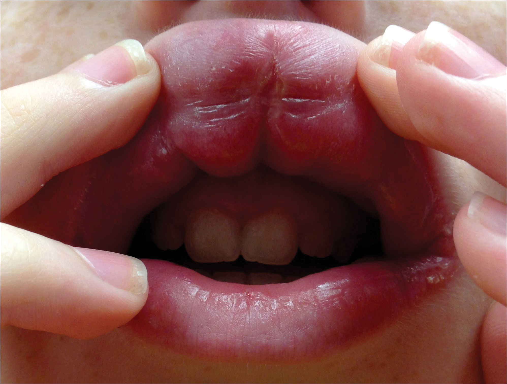
The persistent nature of the lip swelling and findings of fissures were not consistent with angioedema. Furthermore, prior laboratory studies did not reveal evidence of hereditary or acquired angioedema, and a complete blood cell count with differential was within reference range. Although the clinical suspicion for egg allergy was low, a blood test for serum-specific IgE showed a mild reactivity to egg allergen. The patient was referred to an oral surgeon for biopsy, which revealed dermal foci of noncaseating granulomas consistent with the preliminary diagnosis of GC.
Intralesional triamcinolone injections were initiated with marked improvement. Shortly after the initial improvement, however, the symptoms recurred, which necessitated several additional intralesional triamcinolone injections, again with remarkable improvement. Approximately 1.5 years later, the patient presented with recurrence of the lip swelling and admitted to having episodic diarrhea and abdominal cramps. He was referred to a pediatric gastroenterologist and a colonoscopy with biopsy confirmed Crohn disease. He was started on azathioprine followed by infliximab. A few months after this treatment was initiated, both his lip swelling and gastrointestinal tract symptoms remarkably improved. He has been maintained on this regimen and in the most recent follow-up had no recurrence of GC. He is scheduled to have another colonoscopy.
Granulomatous cheilitis is a rare chronic inflammatory condition characterized clinically by persistent lip swelling and histologically by granulomatous inflammation in the absence of systemic granulomatous disorders.4 Granulomatous cheilitis falls under the umbrella of OFGs. When it is paired with facial paralysis and fissuring of the tongue, it is specifically referred to as MRS. The prevalence of GC has historically been difficult to ascertain. In a review, an estimated incidence of 0.08% in the general population was reported with no predilection for race, sex, or age.4,5 Initially, the swelling of GC can be misdiagnosed as angioedema; therefore, it is imperative to include OFG and GC in the differential diagnosis of facial angioedema.3 Other possible diagnoses to consider include contact dermatitis, foreign-body reactions, infection, and reactions to medications such as angiotensin-converting enzyme inhibitors and nonsteroidal anti-inflammatory drugs.5 Chronic lymphedema and other granulomatous diseases also should be considered in the differential diagnosis. Isolated lymphedema of the head and neck, though rare, typically is seen following surgical or radiological interventions for cancer. Lymphatic fibrosis also can occur in the setting of chronic inflammatory skin conditions but is not typically the first presenting symptom, as was seen in our patient.6 Although granulomatous diseases such as sarcoidosis may be difficult to clinically and histologically differentiate from GC, isolated orofacial swelling in sarcoidosis is rare. If clinical suspicion for sarcoidosis does exist, however, a negative chest radiograph as well as serum calcium and angiotensin-converting enzyme levels within reference range may help differentiate GC from sarcoidosis. In our patient, the clinical suspicion for sarcoidosis was low given his clinical history, young age, and race.
The etiology of MRS and GC currently is unknown. Genetic factors, food allergies, infectious processes, and aberrant immunologic functions all have been proposed as possible mechanisms.1-3,7,8 Genetic factors, such as HLA antigen subtypes, have been investigated but have not shown a definitive correlation.8 Numerous food allergens have been suggested as causative factors in OFG via a type of delayed hypersensitivity reaction,7 with cinnamon and benzoate reported as 2 of the most cited entities.9,10 Currently, it is believed that both of these mechanisms may play an exacerbating role to an otherwise unknown disease process.7,8 The infectious process most often associated with GC is Mycobacterium tuberculosis; however, similar to genetics and food allergens, causality has not been determined.4,7 At the present time, the best evidence points to an immunologic basis of GC with the inciting event being a random influx of inflammatory cells.7,11
There is a known association between GC and Crohn disease, especially when oral lesions are present.1,9 Granulomatous cheilitis can be considered an extraintestinal manifestation of Crohn disease.Up to 20% of OFG patients eventually go on to develop Crohn disease, with some reports being even higher when OFG presents in childhood.1,9 One study proposed that both GC and Crohn disease patients shared similar histopathologic and immunopathologic features including a helper T cell (TH1)–predominant inflammatory reaction.11
The treatment of GC is challenging, with most evidence coming from sporadic case reports. Given the relatively high rate of cinnamon and benzoate hypersensitivity seen in GC patients, it has been postulated that a diet lacking in them will improve the disease. At least one study has reported positive clinical outcomes from diets lacking in cinnamon and benzoate and in fact recommended it as a potential first-line treatment.10 The mainstay of treatment, however, is corticosteroids, but continued use is discouraged due to their large side-effect profile.12 Currently, the most agreed upon treatment for patients with isolated GC is intralesional triamcinolone injections.12 Despite the robust initial response often seen with triamcinolone injections, it is not uncommon for the benefit to be short-lived, requiring additional treatments.1,5,12 Newer medical therapies that have shown promise largely are centered on anti–tumor necrosis factor α medications such as infliximab and adalimumab.13,14 It is postulated that due to the potential overlapping pathophysiology between Crohn disease and GC, there may be utility in using the same treatments.13 In situations where medical therapy fails or in extremely disfiguring cases of GC and MRS, surgical cheiloplasty is performed to reduce lip size and improve cosmetic appearance.12 In a small study, reduction cheiloplasty gave satisfactory functional and cosmetic outcomes in all 7 patients reviewed at a median follow-up of 6.5 years.15
This case emphasizes the importance of paying close attention to history and physical examination features in developing any differential diagnosis. In this patient, persistent orofacial swelling with associated mucosal ulcerations were sufficient to exclude drug-induced, idiopathic, hereditary, and acquired angioedema. The clinical history coupled with the biopsy results yielded a confident diagnosis of GC. Furthermore, similar presentations should raise concern for a subclinical inflammatory bowel disease such as Crohn disease.
- Rose AE, Leger M, Chu J, et al. Cheilitis granulomatosa. Dermatol Online J. 2011;17:15.
- Vibhute NA, Vibhute AH, Nilima DR. Cheilitis granulomatosa: a case report with review of literature. Indian J Dermatol. 2013;58:242.
- Kakimoto C, Sparks C, White AA. Melkersson-Rosenthal syndrome: a form of pseudoangioedema. Ann Allergy Asthma Immunol. 2007;99:185-189.
- McCartan BE, Healy CM, McCreary CE, et al. Characteristics of patients with orofacial granulomatosis. Oral Dis. 2011;17:696-704.
- Critchlow WA, Chang D. Cheilitis granulomatosa: a review [published online September 22, 2013]. Head Neck Pathol. 2014;8:209-213.
- Withey S, Pracy P, Vaz F, et al. Sensory deprivation as a consequence of severe head and neck lymphoedema. J Laryngol Otol. 2001;115:62-64.
- Grave B, McCullough M, Wiesenfeld D. Orofacial granulomatosis—a 20-year review. Oral Dis. 2009;15:46-51.
- Gibson J, Wray D. Human leucocyte antigen typing in orofacial. Br J Dermatol. 2000;143:1119-1121.
- Campbell H, Escudier M, Patel P, et al. Distinguishing orofacial granulomatosis from Crohn’s disease: two separate disease entities? Inflamm Bowel Dis. 2011;17:2109-2115.
- White A, Nunes C, Escudier M, et al. Improvement in orofacial granulomatosis on a cinnamon- and benzoate-free diet. Inflamm Bowel Dis. 2006;12:508-514.
- Freysdottir J, Zhang S, Tilakaratne WM, et al. Oral biopsies from patients with orofacial granulomatosis with histology resembling Crohn’s disease have a prominent Th1 environment. Inflamm Bowel Dis. 2007;13:439-445.
- Banks T, Gada S. A comprehensive review of current treatments for granulomatous cheilitis. Br J Dermatol. 2012;166:934-937.
- Peitsch WK, Kemmler N, Goerdt S, et al. Infliximab: a novel treatment option for refractory orofacial granulomatosis. Acta Derm Venereol. 2007;87:265-266.
- Ruiz Villaverde R, Sánchez Cano D. Successful treatment of granulomatous cheilitis with adalimumab. Int J Dermatol. 2012;51:118-120.
- Kruse-Lösler B, Presser D, Metze D, et al. Surgical treatment of persistent macrocheilia in patients with Melkersson-Rosenthal syndrome and cheilitis granulomatosa. Arch Dermatol. 2005;141:1085-1091.
To the Editor:
Granulomatous cheilitis (GC), also known as Miescher cheilitis, belongs to a larger class of diseases known as orofacial granulomatoses (OFGs), a set of diseases distinguished by their clinical and pathologic features of facial edema and granulomatous inflammation.1-3 Granulomatous cheilitis, a monosymptomatic variant of a more extensive disease known as Melkersson-Rosenthal syndrome (MRS), presents with labial swelling mimicking angioedema. Timely diagnosis of GC and MRS reduces the number of unnecessary tests, health care costs, and unnecessary patient burden. We present a case of idiopathic persistent swelling of the upper lip that was originally misdiagnosed as angioedema.
A 13-year-old white adolescent boy was referred to the allergy-immunology clinic for an alternate opinion regarding a presumed diagnosis of angioedema. He presented with prominent persistent swelling of the upper lip of 1 year’s duration associated with fissuring and discomfort while eating, which led to weight loss of more than 4.5 kg. The patient denied any history of facial asymmetry, paralysis, dental infections, or gastrointestinal tract symptoms. Additionally, he was not on any medications. His parents reported variable symptomatic worsening associated with egg ingestion, but avoiding egg did not provide any symptomatic relief. The swelling was unresponsive to multiple and prolonged courses of antihistamines and oral glucocorticoids. The patient’s medical history revealed no similar episodes of unexplained swelling, and family history was negative for angioedema. On examination, the upper lip was tender with a firm rubbery consistency. No other areas of swelling were noted. Angular cheilosis and minor labial mucosal ulcerations also were observed (Figure).
The persistent nature of the lip swelling and findings of fissures were not consistent with angioedema. Furthermore, prior laboratory studies did not reveal evidence of hereditary or acquired angioedema, and a complete blood cell count with differential was within reference range. Although the clinical suspicion for egg allergy was low, a blood test for serum-specific IgE showed a mild reactivity to egg allergen. The patient was referred to an oral surgeon for biopsy, which revealed dermal foci of noncaseating granulomas consistent with the preliminary diagnosis of GC.
Intralesional triamcinolone injections were initiated with marked improvement. Shortly after the initial improvement, however, the symptoms recurred, which necessitated several additional intralesional triamcinolone injections, again with remarkable improvement. Approximately 1.5 years later, the patient presented with recurrence of the lip swelling and admitted to having episodic diarrhea and abdominal cramps. He was referred to a pediatric gastroenterologist and a colonoscopy with biopsy confirmed Crohn disease. He was started on azathioprine followed by infliximab. A few months after this treatment was initiated, both his lip swelling and gastrointestinal tract symptoms remarkably improved. He has been maintained on this regimen and in the most recent follow-up had no recurrence of GC. He is scheduled to have another colonoscopy.
Granulomatous cheilitis is a rare chronic inflammatory condition characterized clinically by persistent lip swelling and histologically by granulomatous inflammation in the absence of systemic granulomatous disorders.4 Granulomatous cheilitis falls under the umbrella of OFGs. When it is paired with facial paralysis and fissuring of the tongue, it is specifically referred to as MRS. The prevalence of GC has historically been difficult to ascertain. In a review, an estimated incidence of 0.08% in the general population was reported with no predilection for race, sex, or age.4,5 Initially, the swelling of GC can be misdiagnosed as angioedema; therefore, it is imperative to include OFG and GC in the differential diagnosis of facial angioedema.3 Other possible diagnoses to consider include contact dermatitis, foreign-body reactions, infection, and reactions to medications such as angiotensin-converting enzyme inhibitors and nonsteroidal anti-inflammatory drugs.5 Chronic lymphedema and other granulomatous diseases also should be considered in the differential diagnosis. Isolated lymphedema of the head and neck, though rare, typically is seen following surgical or radiological interventions for cancer. Lymphatic fibrosis also can occur in the setting of chronic inflammatory skin conditions but is not typically the first presenting symptom, as was seen in our patient.6 Although granulomatous diseases such as sarcoidosis may be difficult to clinically and histologically differentiate from GC, isolated orofacial swelling in sarcoidosis is rare. If clinical suspicion for sarcoidosis does exist, however, a negative chest radiograph as well as serum calcium and angiotensin-converting enzyme levels within reference range may help differentiate GC from sarcoidosis. In our patient, the clinical suspicion for sarcoidosis was low given his clinical history, young age, and race.
The etiology of MRS and GC currently is unknown. Genetic factors, food allergies, infectious processes, and aberrant immunologic functions all have been proposed as possible mechanisms.1-3,7,8 Genetic factors, such as HLA antigen subtypes, have been investigated but have not shown a definitive correlation.8 Numerous food allergens have been suggested as causative factors in OFG via a type of delayed hypersensitivity reaction,7 with cinnamon and benzoate reported as 2 of the most cited entities.9,10 Currently, it is believed that both of these mechanisms may play an exacerbating role to an otherwise unknown disease process.7,8 The infectious process most often associated with GC is Mycobacterium tuberculosis; however, similar to genetics and food allergens, causality has not been determined.4,7 At the present time, the best evidence points to an immunologic basis of GC with the inciting event being a random influx of inflammatory cells.7,11
There is a known association between GC and Crohn disease, especially when oral lesions are present.1,9 Granulomatous cheilitis can be considered an extraintestinal manifestation of Crohn disease.Up to 20% of OFG patients eventually go on to develop Crohn disease, with some reports being even higher when OFG presents in childhood.1,9 One study proposed that both GC and Crohn disease patients shared similar histopathologic and immunopathologic features including a helper T cell (TH1)–predominant inflammatory reaction.11
The treatment of GC is challenging, with most evidence coming from sporadic case reports. Given the relatively high rate of cinnamon and benzoate hypersensitivity seen in GC patients, it has been postulated that a diet lacking in them will improve the disease. At least one study has reported positive clinical outcomes from diets lacking in cinnamon and benzoate and in fact recommended it as a potential first-line treatment.10 The mainstay of treatment, however, is corticosteroids, but continued use is discouraged due to their large side-effect profile.12 Currently, the most agreed upon treatment for patients with isolated GC is intralesional triamcinolone injections.12 Despite the robust initial response often seen with triamcinolone injections, it is not uncommon for the benefit to be short-lived, requiring additional treatments.1,5,12 Newer medical therapies that have shown promise largely are centered on anti–tumor necrosis factor α medications such as infliximab and adalimumab.13,14 It is postulated that due to the potential overlapping pathophysiology between Crohn disease and GC, there may be utility in using the same treatments.13 In situations where medical therapy fails or in extremely disfiguring cases of GC and MRS, surgical cheiloplasty is performed to reduce lip size and improve cosmetic appearance.12 In a small study, reduction cheiloplasty gave satisfactory functional and cosmetic outcomes in all 7 patients reviewed at a median follow-up of 6.5 years.15
This case emphasizes the importance of paying close attention to history and physical examination features in developing any differential diagnosis. In this patient, persistent orofacial swelling with associated mucosal ulcerations were sufficient to exclude drug-induced, idiopathic, hereditary, and acquired angioedema. The clinical history coupled with the biopsy results yielded a confident diagnosis of GC. Furthermore, similar presentations should raise concern for a subclinical inflammatory bowel disease such as Crohn disease.
To the Editor:
Granulomatous cheilitis (GC), also known as Miescher cheilitis, belongs to a larger class of diseases known as orofacial granulomatoses (OFGs), a set of diseases distinguished by their clinical and pathologic features of facial edema and granulomatous inflammation.1-3 Granulomatous cheilitis, a monosymptomatic variant of a more extensive disease known as Melkersson-Rosenthal syndrome (MRS), presents with labial swelling mimicking angioedema. Timely diagnosis of GC and MRS reduces the number of unnecessary tests, health care costs, and unnecessary patient burden. We present a case of idiopathic persistent swelling of the upper lip that was originally misdiagnosed as angioedema.
A 13-year-old white adolescent boy was referred to the allergy-immunology clinic for an alternate opinion regarding a presumed diagnosis of angioedema. He presented with prominent persistent swelling of the upper lip of 1 year’s duration associated with fissuring and discomfort while eating, which led to weight loss of more than 4.5 kg. The patient denied any history of facial asymmetry, paralysis, dental infections, or gastrointestinal tract symptoms. Additionally, he was not on any medications. His parents reported variable symptomatic worsening associated with egg ingestion, but avoiding egg did not provide any symptomatic relief. The swelling was unresponsive to multiple and prolonged courses of antihistamines and oral glucocorticoids. The patient’s medical history revealed no similar episodes of unexplained swelling, and family history was negative for angioedema. On examination, the upper lip was tender with a firm rubbery consistency. No other areas of swelling were noted. Angular cheilosis and minor labial mucosal ulcerations also were observed (Figure).
The persistent nature of the lip swelling and findings of fissures were not consistent with angioedema. Furthermore, prior laboratory studies did not reveal evidence of hereditary or acquired angioedema, and a complete blood cell count with differential was within reference range. Although the clinical suspicion for egg allergy was low, a blood test for serum-specific IgE showed a mild reactivity to egg allergen. The patient was referred to an oral surgeon for biopsy, which revealed dermal foci of noncaseating granulomas consistent with the preliminary diagnosis of GC.
Intralesional triamcinolone injections were initiated with marked improvement. Shortly after the initial improvement, however, the symptoms recurred, which necessitated several additional intralesional triamcinolone injections, again with remarkable improvement. Approximately 1.5 years later, the patient presented with recurrence of the lip swelling and admitted to having episodic diarrhea and abdominal cramps. He was referred to a pediatric gastroenterologist and a colonoscopy with biopsy confirmed Crohn disease. He was started on azathioprine followed by infliximab. A few months after this treatment was initiated, both his lip swelling and gastrointestinal tract symptoms remarkably improved. He has been maintained on this regimen and in the most recent follow-up had no recurrence of GC. He is scheduled to have another colonoscopy.
Granulomatous cheilitis is a rare chronic inflammatory condition characterized clinically by persistent lip swelling and histologically by granulomatous inflammation in the absence of systemic granulomatous disorders.4 Granulomatous cheilitis falls under the umbrella of OFGs. When it is paired with facial paralysis and fissuring of the tongue, it is specifically referred to as MRS. The prevalence of GC has historically been difficult to ascertain. In a review, an estimated incidence of 0.08% in the general population was reported with no predilection for race, sex, or age.4,5 Initially, the swelling of GC can be misdiagnosed as angioedema; therefore, it is imperative to include OFG and GC in the differential diagnosis of facial angioedema.3 Other possible diagnoses to consider include contact dermatitis, foreign-body reactions, infection, and reactions to medications such as angiotensin-converting enzyme inhibitors and nonsteroidal anti-inflammatory drugs.5 Chronic lymphedema and other granulomatous diseases also should be considered in the differential diagnosis. Isolated lymphedema of the head and neck, though rare, typically is seen following surgical or radiological interventions for cancer. Lymphatic fibrosis also can occur in the setting of chronic inflammatory skin conditions but is not typically the first presenting symptom, as was seen in our patient.6 Although granulomatous diseases such as sarcoidosis may be difficult to clinically and histologically differentiate from GC, isolated orofacial swelling in sarcoidosis is rare. If clinical suspicion for sarcoidosis does exist, however, a negative chest radiograph as well as serum calcium and angiotensin-converting enzyme levels within reference range may help differentiate GC from sarcoidosis. In our patient, the clinical suspicion for sarcoidosis was low given his clinical history, young age, and race.
The etiology of MRS and GC currently is unknown. Genetic factors, food allergies, infectious processes, and aberrant immunologic functions all have been proposed as possible mechanisms.1-3,7,8 Genetic factors, such as HLA antigen subtypes, have been investigated but have not shown a definitive correlation.8 Numerous food allergens have been suggested as causative factors in OFG via a type of delayed hypersensitivity reaction,7 with cinnamon and benzoate reported as 2 of the most cited entities.9,10 Currently, it is believed that both of these mechanisms may play an exacerbating role to an otherwise unknown disease process.7,8 The infectious process most often associated with GC is Mycobacterium tuberculosis; however, similar to genetics and food allergens, causality has not been determined.4,7 At the present time, the best evidence points to an immunologic basis of GC with the inciting event being a random influx of inflammatory cells.7,11
There is a known association between GC and Crohn disease, especially when oral lesions are present.1,9 Granulomatous cheilitis can be considered an extraintestinal manifestation of Crohn disease.Up to 20% of OFG patients eventually go on to develop Crohn disease, with some reports being even higher when OFG presents in childhood.1,9 One study proposed that both GC and Crohn disease patients shared similar histopathologic and immunopathologic features including a helper T cell (TH1)–predominant inflammatory reaction.11
The treatment of GC is challenging, with most evidence coming from sporadic case reports. Given the relatively high rate of cinnamon and benzoate hypersensitivity seen in GC patients, it has been postulated that a diet lacking in them will improve the disease. At least one study has reported positive clinical outcomes from diets lacking in cinnamon and benzoate and in fact recommended it as a potential first-line treatment.10 The mainstay of treatment, however, is corticosteroids, but continued use is discouraged due to their large side-effect profile.12 Currently, the most agreed upon treatment for patients with isolated GC is intralesional triamcinolone injections.12 Despite the robust initial response often seen with triamcinolone injections, it is not uncommon for the benefit to be short-lived, requiring additional treatments.1,5,12 Newer medical therapies that have shown promise largely are centered on anti–tumor necrosis factor α medications such as infliximab and adalimumab.13,14 It is postulated that due to the potential overlapping pathophysiology between Crohn disease and GC, there may be utility in using the same treatments.13 In situations where medical therapy fails or in extremely disfiguring cases of GC and MRS, surgical cheiloplasty is performed to reduce lip size and improve cosmetic appearance.12 In a small study, reduction cheiloplasty gave satisfactory functional and cosmetic outcomes in all 7 patients reviewed at a median follow-up of 6.5 years.15
This case emphasizes the importance of paying close attention to history and physical examination features in developing any differential diagnosis. In this patient, persistent orofacial swelling with associated mucosal ulcerations were sufficient to exclude drug-induced, idiopathic, hereditary, and acquired angioedema. The clinical history coupled with the biopsy results yielded a confident diagnosis of GC. Furthermore, similar presentations should raise concern for a subclinical inflammatory bowel disease such as Crohn disease.
- Rose AE, Leger M, Chu J, et al. Cheilitis granulomatosa. Dermatol Online J. 2011;17:15.
- Vibhute NA, Vibhute AH, Nilima DR. Cheilitis granulomatosa: a case report with review of literature. Indian J Dermatol. 2013;58:242.
- Kakimoto C, Sparks C, White AA. Melkersson-Rosenthal syndrome: a form of pseudoangioedema. Ann Allergy Asthma Immunol. 2007;99:185-189.
- McCartan BE, Healy CM, McCreary CE, et al. Characteristics of patients with orofacial granulomatosis. Oral Dis. 2011;17:696-704.
- Critchlow WA, Chang D. Cheilitis granulomatosa: a review [published online September 22, 2013]. Head Neck Pathol. 2014;8:209-213.
- Withey S, Pracy P, Vaz F, et al. Sensory deprivation as a consequence of severe head and neck lymphoedema. J Laryngol Otol. 2001;115:62-64.
- Grave B, McCullough M, Wiesenfeld D. Orofacial granulomatosis—a 20-year review. Oral Dis. 2009;15:46-51.
- Gibson J, Wray D. Human leucocyte antigen typing in orofacial. Br J Dermatol. 2000;143:1119-1121.
- Campbell H, Escudier M, Patel P, et al. Distinguishing orofacial granulomatosis from Crohn’s disease: two separate disease entities? Inflamm Bowel Dis. 2011;17:2109-2115.
- White A, Nunes C, Escudier M, et al. Improvement in orofacial granulomatosis on a cinnamon- and benzoate-free diet. Inflamm Bowel Dis. 2006;12:508-514.
- Freysdottir J, Zhang S, Tilakaratne WM, et al. Oral biopsies from patients with orofacial granulomatosis with histology resembling Crohn’s disease have a prominent Th1 environment. Inflamm Bowel Dis. 2007;13:439-445.
- Banks T, Gada S. A comprehensive review of current treatments for granulomatous cheilitis. Br J Dermatol. 2012;166:934-937.
- Peitsch WK, Kemmler N, Goerdt S, et al. Infliximab: a novel treatment option for refractory orofacial granulomatosis. Acta Derm Venereol. 2007;87:265-266.
- Ruiz Villaverde R, Sánchez Cano D. Successful treatment of granulomatous cheilitis with adalimumab. Int J Dermatol. 2012;51:118-120.
- Kruse-Lösler B, Presser D, Metze D, et al. Surgical treatment of persistent macrocheilia in patients with Melkersson-Rosenthal syndrome and cheilitis granulomatosa. Arch Dermatol. 2005;141:1085-1091.
- Rose AE, Leger M, Chu J, et al. Cheilitis granulomatosa. Dermatol Online J. 2011;17:15.
- Vibhute NA, Vibhute AH, Nilima DR. Cheilitis granulomatosa: a case report with review of literature. Indian J Dermatol. 2013;58:242.
- Kakimoto C, Sparks C, White AA. Melkersson-Rosenthal syndrome: a form of pseudoangioedema. Ann Allergy Asthma Immunol. 2007;99:185-189.
- McCartan BE, Healy CM, McCreary CE, et al. Characteristics of patients with orofacial granulomatosis. Oral Dis. 2011;17:696-704.
- Critchlow WA, Chang D. Cheilitis granulomatosa: a review [published online September 22, 2013]. Head Neck Pathol. 2014;8:209-213.
- Withey S, Pracy P, Vaz F, et al. Sensory deprivation as a consequence of severe head and neck lymphoedema. J Laryngol Otol. 2001;115:62-64.
- Grave B, McCullough M, Wiesenfeld D. Orofacial granulomatosis—a 20-year review. Oral Dis. 2009;15:46-51.
- Gibson J, Wray D. Human leucocyte antigen typing in orofacial. Br J Dermatol. 2000;143:1119-1121.
- Campbell H, Escudier M, Patel P, et al. Distinguishing orofacial granulomatosis from Crohn’s disease: two separate disease entities? Inflamm Bowel Dis. 2011;17:2109-2115.
- White A, Nunes C, Escudier M, et al. Improvement in orofacial granulomatosis on a cinnamon- and benzoate-free diet. Inflamm Bowel Dis. 2006;12:508-514.
- Freysdottir J, Zhang S, Tilakaratne WM, et al. Oral biopsies from patients with orofacial granulomatosis with histology resembling Crohn’s disease have a prominent Th1 environment. Inflamm Bowel Dis. 2007;13:439-445.
- Banks T, Gada S. A comprehensive review of current treatments for granulomatous cheilitis. Br J Dermatol. 2012;166:934-937.
- Peitsch WK, Kemmler N, Goerdt S, et al. Infliximab: a novel treatment option for refractory orofacial granulomatosis. Acta Derm Venereol. 2007;87:265-266.
- Ruiz Villaverde R, Sánchez Cano D. Successful treatment of granulomatous cheilitis with adalimumab. Int J Dermatol. 2012;51:118-120.
- Kruse-Lösler B, Presser D, Metze D, et al. Surgical treatment of persistent macrocheilia in patients with Melkersson-Rosenthal syndrome and cheilitis granulomatosa. Arch Dermatol. 2005;141:1085-1091.
Practice Points
- Granulomatous cheilitis (GC) is a rare diagnosis that can present as an isolated disease or in association with another disease, most commonly an inflammatory bowel disease (ie, Crohn disease).
- Often misdiagnosed as angioedema, GC can be differentiated primarily based on history and clinical examination.
- Intervention such as intralesional steroid injection is effective in the primary form; however, treatment of the underlying condition, such as Crohn disease, is needed when the 2 conditions are associated.
Drug receives rare pediatric disease designation for SCD

The US Food and Drug Administration (FDA) has granted rare pediatric disease designation to IMR-687, a product intended to treat sickle cell disease (SCD).
IMR-687 is the first SCD candidate to be designated as a drug for a rare pediatric disease, and this designation builds upon the FDA’s earlier granting of orphan designation for IMR-687.
IMR-687 is a selective inhibitor of phosphodiesterase-9 (PDE9i) in blood cells. It was specifically designed to address the underlying pathology of SCD.
In preclinical studies, IMR-687 demonstrated the ability to increase fetal globin. This prevented the polymerization of sickled hemoglobin and reduced red blood cell sickling, leukocytosis, and the occlusion of blood vessels.
Researchers presented these results at the 2016 ASH Annual Meeting.
Imara Inc., the company developing IMR-687, is conducting a phase 1a trial of the product in healthy volunteers.
If the trial has a positive outcome (results are expected this summer), Imara will initiate a phase 2a study in adults with SCD later this year. The company expects to initiate a phase 2 study in pediatric patients in 2018.
About rare pediatric disease designation
Rare pediatric disease designation is granted to drugs that show promise to treat orphan diseases affecting fewer than 200,000 patients in the US, primarily patients age 18 or younger.
The designation provides incentives to advance the development of drugs for rare disease, including access to the FDA’s expedited review and approval programs.
Under the FDA’s Rare Pediatric Disease Priority Review Voucher Program, a sponsor with rare pediatric disease designation who receives an approval of a new drug application is eligible for a voucher that can be redeemed to obtain priority review for any subsequent marketing application.
About orphan designation
The FDA grants orphan designation to products intended to treat, diagnose, or prevent diseases/disorders that affect fewer than 200,000 people in the US.
The designation provides incentives for sponsors to develop products for rare diseases. This may include tax credits toward the cost of clinical trials, prescription drug user fee waivers, and 7 years of market exclusivity if the product is approved. 
The US Food and Drug Administration (FDA) has granted rare pediatric disease designation to IMR-687, a product intended to treat sickle cell disease (SCD).
IMR-687 is the first SCD candidate to be designated as a drug for a rare pediatric disease, and this designation builds upon the FDA’s earlier granting of orphan designation for IMR-687.
IMR-687 is a selective inhibitor of phosphodiesterase-9 (PDE9i) in blood cells. It was specifically designed to address the underlying pathology of SCD.
In preclinical studies, IMR-687 demonstrated the ability to increase fetal globin. This prevented the polymerization of sickled hemoglobin and reduced red blood cell sickling, leukocytosis, and the occlusion of blood vessels.
Researchers presented these results at the 2016 ASH Annual Meeting.
Imara Inc., the company developing IMR-687, is conducting a phase 1a trial of the product in healthy volunteers.
If the trial has a positive outcome (results are expected this summer), Imara will initiate a phase 2a study in adults with SCD later this year. The company expects to initiate a phase 2 study in pediatric patients in 2018.
About rare pediatric disease designation
Rare pediatric disease designation is granted to drugs that show promise to treat orphan diseases affecting fewer than 200,000 patients in the US, primarily patients age 18 or younger.
The designation provides incentives to advance the development of drugs for rare disease, including access to the FDA’s expedited review and approval programs.
Under the FDA’s Rare Pediatric Disease Priority Review Voucher Program, a sponsor with rare pediatric disease designation who receives an approval of a new drug application is eligible for a voucher that can be redeemed to obtain priority review for any subsequent marketing application.
About orphan designation
The FDA grants orphan designation to products intended to treat, diagnose, or prevent diseases/disorders that affect fewer than 200,000 people in the US.
The designation provides incentives for sponsors to develop products for rare diseases. This may include tax credits toward the cost of clinical trials, prescription drug user fee waivers, and 7 years of market exclusivity if the product is approved.
The US Food and Drug Administration (FDA) has granted rare pediatric disease designation to IMR-687, a product intended to treat sickle cell disease (SCD).
IMR-687 is the first SCD candidate to be designated as a drug for a rare pediatric disease, and this designation builds upon the FDA’s earlier granting of orphan designation for IMR-687.
IMR-687 is a selective inhibitor of phosphodiesterase-9 (PDE9i) in blood cells. It was specifically designed to address the underlying pathology of SCD.
In preclinical studies, IMR-687 demonstrated the ability to increase fetal globin. This prevented the polymerization of sickled hemoglobin and reduced red blood cell sickling, leukocytosis, and the occlusion of blood vessels.
Researchers presented these results at the 2016 ASH Annual Meeting.
Imara Inc., the company developing IMR-687, is conducting a phase 1a trial of the product in healthy volunteers.
If the trial has a positive outcome (results are expected this summer), Imara will initiate a phase 2a study in adults with SCD later this year. The company expects to initiate a phase 2 study in pediatric patients in 2018.
About rare pediatric disease designation
Rare pediatric disease designation is granted to drugs that show promise to treat orphan diseases affecting fewer than 200,000 patients in the US, primarily patients age 18 or younger.
The designation provides incentives to advance the development of drugs for rare disease, including access to the FDA’s expedited review and approval programs.
Under the FDA’s Rare Pediatric Disease Priority Review Voucher Program, a sponsor with rare pediatric disease designation who receives an approval of a new drug application is eligible for a voucher that can be redeemed to obtain priority review for any subsequent marketing application.
About orphan designation
The FDA grants orphan designation to products intended to treat, diagnose, or prevent diseases/disorders that affect fewer than 200,000 people in the US.
The designation provides incentives for sponsors to develop products for rare diseases. This may include tax credits toward the cost of clinical trials, prescription drug user fee waivers, and 7 years of market exclusivity if the product is approved.
Asthma step-up therapy in children improves outcomes
SAN FRANCISCO – Stepping up pharmacotherapy in children with poorly controlled asthma resulted in fewer asthma-related emergency department (ED) visits and inpatient stays than maintaining current medication therapy, a study found.
Perhaps surprisingly, however, the patients with the worst outcomes were those who were told only to improve their medication adherence, said Dane Snyder, MD, a physician at Nationwide Children’s Hospital who is also in the department of pediatrics at Ohio State University, Columbus.

Asthma affects nearly 1 in 10 children, and national guidelines recommend step-up asthma pharmacotherapy in patients with poorly controlled asthma. The researchers assessed the impact of step-up therapy on inpatient care, and emergency and urgent care visits for children with poor asthma control in the Nationwide Children’s Hospital Primary Care Network in Columbus, Ohio. More than 10,000 asthma patients are treated at the 12 clinics in the network, and the study was part of a quality improvement initiative starting in July 2015.
Between August and October 2015, researchers used documentation in a standard asthma note to identify 908 patients aged 2-18 years who had poor asthma control based on their Asthma Control Test (ACT) score and history. Of these patients, 463 with a mean ACT score of 15.8 were assigned to step-up pharmacotherapy, while 445 with a mean ACT of 16.2 were not. The two groups also were similar in their use of unscheduled health care utilization in the 12 months before the study period.
Over the next 12 months, 1.3% of patients receiving step-up therapy had inpatient stays, compared with 4% in the other group, translating to a 68% lower risk of admission with step-up therapy (relative risk, 0.316; P = .01). Those receiving step-up therapy also were 37% less likely to visit the ED for asthma, with 7.1% of ED visits for those with step-up therapy and 11.2% of visits for those without it (RR, 0.634; P = .032). Visits to urgent care, however, showed no significant difference between those receiving step-up therapy (10.8%) and those who had not (10.3%).
After comparing these findings, the researchers went back and manually reviewed all 463 charts of the group who received step-up therapy to determine whether the children actually did receive a step up in therapy. Nearly a quarter (23%) of the children in the intervention group simply resumed taking their previously prescribed mediation, and 8% took allergy medication. The remaining 69% had step-up therapy.
The researchers then reanalyzed the data among those who truly had step-up pharmacotherapy, those who did not, and those who resumed taking their prior medication. The difference in inpatient admissions remained the same because none of the children who had resumed medication were admitted.
ED visits showed a more gradual distribution: 7.3% of those receiving step-up therapy, 8.4% of those who resumed taking their medication, and 11% of those with no intervention went to the ED. But these differences did not reach statistical significance.
Similarly, the differences among the three groups in urgent care visits was not statistically significant, but 15% of those who resumed taking prior medication had urgent care visits, compared with 10.3% of those with step-up therapy and 9.2% of those in the control group.
Those findings suggest that “stepping up pharmacotherapy, even in the face of controller nonadherence, can improve outcomes,” Dr. Snyder told his colleagues.
“While challenging, management changes in a large primary care network are possible,” Dr. Snyder said. He also emphasized that manually auditing bulk data, as they did with the 463 records, can be important in assessing outcomes of quality improvement measures.
The research did not use external funding, and Dr. Snyder had no disclosures.
SAN FRANCISCO – Stepping up pharmacotherapy in children with poorly controlled asthma resulted in fewer asthma-related emergency department (ED) visits and inpatient stays than maintaining current medication therapy, a study found.
Perhaps surprisingly, however, the patients with the worst outcomes were those who were told only to improve their medication adherence, said Dane Snyder, MD, a physician at Nationwide Children’s Hospital who is also in the department of pediatrics at Ohio State University, Columbus.
Asthma affects nearly 1 in 10 children, and national guidelines recommend step-up asthma pharmacotherapy in patients with poorly controlled asthma. The researchers assessed the impact of step-up therapy on inpatient care, and emergency and urgent care visits for children with poor asthma control in the Nationwide Children’s Hospital Primary Care Network in Columbus, Ohio. More than 10,000 asthma patients are treated at the 12 clinics in the network, and the study was part of a quality improvement initiative starting in July 2015.
Between August and October 2015, researchers used documentation in a standard asthma note to identify 908 patients aged 2-18 years who had poor asthma control based on their Asthma Control Test (ACT) score and history. Of these patients, 463 with a mean ACT score of 15.8 were assigned to step-up pharmacotherapy, while 445 with a mean ACT of 16.2 were not. The two groups also were similar in their use of unscheduled health care utilization in the 12 months before the study period.
Over the next 12 months, 1.3% of patients receiving step-up therapy had inpatient stays, compared with 4% in the other group, translating to a 68% lower risk of admission with step-up therapy (relative risk, 0.316; P = .01). Those receiving step-up therapy also were 37% less likely to visit the ED for asthma, with 7.1% of ED visits for those with step-up therapy and 11.2% of visits for those without it (RR, 0.634; P = .032). Visits to urgent care, however, showed no significant difference between those receiving step-up therapy (10.8%) and those who had not (10.3%).
After comparing these findings, the researchers went back and manually reviewed all 463 charts of the group who received step-up therapy to determine whether the children actually did receive a step up in therapy. Nearly a quarter (23%) of the children in the intervention group simply resumed taking their previously prescribed mediation, and 8% took allergy medication. The remaining 69% had step-up therapy.
The researchers then reanalyzed the data among those who truly had step-up pharmacotherapy, those who did not, and those who resumed taking their prior medication. The difference in inpatient admissions remained the same because none of the children who had resumed medication were admitted.
ED visits showed a more gradual distribution: 7.3% of those receiving step-up therapy, 8.4% of those who resumed taking their medication, and 11% of those with no intervention went to the ED. But these differences did not reach statistical significance.
Similarly, the differences among the three groups in urgent care visits was not statistically significant, but 15% of those who resumed taking prior medication had urgent care visits, compared with 10.3% of those with step-up therapy and 9.2% of those in the control group.
Those findings suggest that “stepping up pharmacotherapy, even in the face of controller nonadherence, can improve outcomes,” Dr. Snyder told his colleagues.
“While challenging, management changes in a large primary care network are possible,” Dr. Snyder said. He also emphasized that manually auditing bulk data, as they did with the 463 records, can be important in assessing outcomes of quality improvement measures.
The research did not use external funding, and Dr. Snyder had no disclosures.
SAN FRANCISCO – Stepping up pharmacotherapy in children with poorly controlled asthma resulted in fewer asthma-related emergency department (ED) visits and inpatient stays than maintaining current medication therapy, a study found.
Perhaps surprisingly, however, the patients with the worst outcomes were those who were told only to improve their medication adherence, said Dane Snyder, MD, a physician at Nationwide Children’s Hospital who is also in the department of pediatrics at Ohio State University, Columbus.
Asthma affects nearly 1 in 10 children, and national guidelines recommend step-up asthma pharmacotherapy in patients with poorly controlled asthma. The researchers assessed the impact of step-up therapy on inpatient care, and emergency and urgent care visits for children with poor asthma control in the Nationwide Children’s Hospital Primary Care Network in Columbus, Ohio. More than 10,000 asthma patients are treated at the 12 clinics in the network, and the study was part of a quality improvement initiative starting in July 2015.
Between August and October 2015, researchers used documentation in a standard asthma note to identify 908 patients aged 2-18 years who had poor asthma control based on their Asthma Control Test (ACT) score and history. Of these patients, 463 with a mean ACT score of 15.8 were assigned to step-up pharmacotherapy, while 445 with a mean ACT of 16.2 were not. The two groups also were similar in their use of unscheduled health care utilization in the 12 months before the study period.
Over the next 12 months, 1.3% of patients receiving step-up therapy had inpatient stays, compared with 4% in the other group, translating to a 68% lower risk of admission with step-up therapy (relative risk, 0.316; P = .01). Those receiving step-up therapy also were 37% less likely to visit the ED for asthma, with 7.1% of ED visits for those with step-up therapy and 11.2% of visits for those without it (RR, 0.634; P = .032). Visits to urgent care, however, showed no significant difference between those receiving step-up therapy (10.8%) and those who had not (10.3%).
After comparing these findings, the researchers went back and manually reviewed all 463 charts of the group who received step-up therapy to determine whether the children actually did receive a step up in therapy. Nearly a quarter (23%) of the children in the intervention group simply resumed taking their previously prescribed mediation, and 8% took allergy medication. The remaining 69% had step-up therapy.
The researchers then reanalyzed the data among those who truly had step-up pharmacotherapy, those who did not, and those who resumed taking their prior medication. The difference in inpatient admissions remained the same because none of the children who had resumed medication were admitted.
ED visits showed a more gradual distribution: 7.3% of those receiving step-up therapy, 8.4% of those who resumed taking their medication, and 11% of those with no intervention went to the ED. But these differences did not reach statistical significance.
Similarly, the differences among the three groups in urgent care visits was not statistically significant, but 15% of those who resumed taking prior medication had urgent care visits, compared with 10.3% of those with step-up therapy and 9.2% of those in the control group.
Those findings suggest that “stepping up pharmacotherapy, even in the face of controller nonadherence, can improve outcomes,” Dr. Snyder told his colleagues.
“While challenging, management changes in a large primary care network are possible,” Dr. Snyder said. He also emphasized that manually auditing bulk data, as they did with the 463 records, can be important in assessing outcomes of quality improvement measures.
The research did not use external funding, and Dr. Snyder had no disclosures.
AT PAS 2017
Key clinical point:
Major finding: Children with poorly controlled asthma receiving step-up pharmacotherapy had a 68% lower risk of inpatient admission and 37% lower risk of emergency department visits for asthma (P less than .05).
Data source: The findings are based on a nonrandomized trial of 903 children, aged 2-18 years, tracked for 12 months in the Nationwide Children’s Hospital Primary Care Network in Columbus, Ohio.
Disclosures: The research did not use external funding, and Dr. Snyder had no disclosures.
Can prenatal choline lead to prevention of Alzheimer’s?
As psychiatrists, we are the advocates for inserting the biological thread into the tapestry of understanding human behavior. Try as they may, other mental health professionals are not biologists at heart. Accordingly, psychiatrists bring important thoughtfulness to any consideration about mental health and wellness and about the treatment and prevention of problematic thoughts, feelings, and behaviors.
Throughout my career, my main focus has been on identifying strategies and treatments that can prevent mental illness. For example, I wrote a column about prevention for Clinical Psychiatry News from 2004 to 2011, and, as a member of the publication’s Editorial Advisory Board, I continue to try to steer our attention to biological aspects of prevention.

Recently, I have been seeing psychiatric articles on fetal health and mental health, and, because I am excited about the prospect of understanding fetal alcohol exposure, I feel the need to share. A recent article in the American Journal of Psychiatry was provocatively entitled, “Fetal origins of mental health: The developmental origins of health and disease hypothesis (2016. doi: 10.1176/appi.2016.16020138).
Disappointedly, the authors overlooked the biology of fetal alcohol exposure and focused on how psychosocial issues of maternal anxiety, depression, and anxiety could influence neurodevelopment, which could affect mental health outcomes after birth. Of course, I thought, “What about fetal alcohol exposure?” Meanwhile, a commentary in JAMA Psychiatry entitled “Prenatal nutritional deficiency and psychosis: Where do we go from here?” referred to prenatal choline supplementation along with other supplements (2017;74(4):349-50).
When I first stumbled upon the high prevalence of fetal alcohol exposure in low-income African American populations, it occurred to me that, since choline was involved with the psychopathology of fetal alcohol spectrum disorders and acetylcholine seemed to be involved in the psychopathology of Alzheimer’s disease, there might be a relationship between the two (Psychiatric Serv. 2015 May 1. doi: 10.1176/appi.ps.201400162). Such possible links are especially intriguing in light of the Alzheimer’s Association suggestion that Alzheimer’s disease is a “silent epidemic” among African Americans. The association notes that the prevalence among African Americans ranges from 14% to 100% higher than among whites. The problem – how to make the connection, if there were one, between the adults I was seeing and fetal alcohol exposure – proved difficult, because the time between fetal health and adult mental illness was huge. The time from fetal health and geriatric Alzheimer’s disease was even greater.
However, modern biologic science came through again. Maternal choline supplementation has been touted as a potential prenatal treatment for Down syndrome and Alzheimer’s disease (Curr Alzheimer Res. 2016;13[1]:97-106). Using mice that are genetically altered to show the development of Down syndrome and Alzheimer’s disease changes in the brain at 6 months, allowing researchers to seek prevention strategies for this pathophysiology, researchers have found that maternal choline supplementation protects against basal forebrain cholinergic neuron degeneration seen in these animals.
Thus, it would seem the problem of choline deficiency in pregnancy, most exacerbated by fetal alcohol exposure, is preventable by increasing the amount of choline available during pregnancy. So, it makes sense to increase the amount of choline in prenatal vitamins, as it appears that this biotechnical intervention not only would reduce the scourge of fetal alcohol spectrum disorders but also of Alzheimer’s disease (J Fam Med Dis Prev. 2016 Nov 29;2[6]:1-3).
Finally, the Office of Juvenile Justice and Delinquency Prevention has finally released a paper – “Fetal alcohol spectrum disorders listening session report” – from a session held in June 2013 that documents the extent of the problem in juvenile justice facilities.
Unfortunately, many of us have abdicated our role as biologists. We’ve got evidence showing the power of prenatal choline. It is time to stop counting all of the problems that stem from deficiency of choline during pregnancy and start doing something about it.
Dr. Bell is a staff psychiatrist at Jackson Park Hospital Family Medicine Clinic in Chicago, clinical psychiatrist emeritus in the department of psychiatry at the University of Illinois at Chicago, former president/CEO of Community Mental Health Council, and former director of the Institute for Juvenile Research (birthplace of child psychiatry), also in Chicago.
As psychiatrists, we are the advocates for inserting the biological thread into the tapestry of understanding human behavior. Try as they may, other mental health professionals are not biologists at heart. Accordingly, psychiatrists bring important thoughtfulness to any consideration about mental health and wellness and about the treatment and prevention of problematic thoughts, feelings, and behaviors.
Throughout my career, my main focus has been on identifying strategies and treatments that can prevent mental illness. For example, I wrote a column about prevention for Clinical Psychiatry News from 2004 to 2011, and, as a member of the publication’s Editorial Advisory Board, I continue to try to steer our attention to biological aspects of prevention.
Recently, I have been seeing psychiatric articles on fetal health and mental health, and, because I am excited about the prospect of understanding fetal alcohol exposure, I feel the need to share. A recent article in the American Journal of Psychiatry was provocatively entitled, “Fetal origins of mental health: The developmental origins of health and disease hypothesis (2016. doi: 10.1176/appi.2016.16020138).
Disappointedly, the authors overlooked the biology of fetal alcohol exposure and focused on how psychosocial issues of maternal anxiety, depression, and anxiety could influence neurodevelopment, which could affect mental health outcomes after birth. Of course, I thought, “What about fetal alcohol exposure?” Meanwhile, a commentary in JAMA Psychiatry entitled “Prenatal nutritional deficiency and psychosis: Where do we go from here?” referred to prenatal choline supplementation along with other supplements (2017;74(4):349-50).
When I first stumbled upon the high prevalence of fetal alcohol exposure in low-income African American populations, it occurred to me that, since choline was involved with the psychopathology of fetal alcohol spectrum disorders and acetylcholine seemed to be involved in the psychopathology of Alzheimer’s disease, there might be a relationship between the two (Psychiatric Serv. 2015 May 1. doi: 10.1176/appi.ps.201400162). Such possible links are especially intriguing in light of the Alzheimer’s Association suggestion that Alzheimer’s disease is a “silent epidemic” among African Americans. The association notes that the prevalence among African Americans ranges from 14% to 100% higher than among whites. The problem – how to make the connection, if there were one, between the adults I was seeing and fetal alcohol exposure – proved difficult, because the time between fetal health and adult mental illness was huge. The time from fetal health and geriatric Alzheimer’s disease was even greater.
However, modern biologic science came through again. Maternal choline supplementation has been touted as a potential prenatal treatment for Down syndrome and Alzheimer’s disease (Curr Alzheimer Res. 2016;13[1]:97-106). Using mice that are genetically altered to show the development of Down syndrome and Alzheimer’s disease changes in the brain at 6 months, allowing researchers to seek prevention strategies for this pathophysiology, researchers have found that maternal choline supplementation protects against basal forebrain cholinergic neuron degeneration seen in these animals.
Thus, it would seem the problem of choline deficiency in pregnancy, most exacerbated by fetal alcohol exposure, is preventable by increasing the amount of choline available during pregnancy. So, it makes sense to increase the amount of choline in prenatal vitamins, as it appears that this biotechnical intervention not only would reduce the scourge of fetal alcohol spectrum disorders but also of Alzheimer’s disease (J Fam Med Dis Prev. 2016 Nov 29;2[6]:1-3).
Finally, the Office of Juvenile Justice and Delinquency Prevention has finally released a paper – “Fetal alcohol spectrum disorders listening session report” – from a session held in June 2013 that documents the extent of the problem in juvenile justice facilities.
Unfortunately, many of us have abdicated our role as biologists. We’ve got evidence showing the power of prenatal choline. It is time to stop counting all of the problems that stem from deficiency of choline during pregnancy and start doing something about it.
Dr. Bell is a staff psychiatrist at Jackson Park Hospital Family Medicine Clinic in Chicago, clinical psychiatrist emeritus in the department of psychiatry at the University of Illinois at Chicago, former president/CEO of Community Mental Health Council, and former director of the Institute for Juvenile Research (birthplace of child psychiatry), also in Chicago.
As psychiatrists, we are the advocates for inserting the biological thread into the tapestry of understanding human behavior. Try as they may, other mental health professionals are not biologists at heart. Accordingly, psychiatrists bring important thoughtfulness to any consideration about mental health and wellness and about the treatment and prevention of problematic thoughts, feelings, and behaviors.
Throughout my career, my main focus has been on identifying strategies and treatments that can prevent mental illness. For example, I wrote a column about prevention for Clinical Psychiatry News from 2004 to 2011, and, as a member of the publication’s Editorial Advisory Board, I continue to try to steer our attention to biological aspects of prevention.
Recently, I have been seeing psychiatric articles on fetal health and mental health, and, because I am excited about the prospect of understanding fetal alcohol exposure, I feel the need to share. A recent article in the American Journal of Psychiatry was provocatively entitled, “Fetal origins of mental health: The developmental origins of health and disease hypothesis (2016. doi: 10.1176/appi.2016.16020138).
Disappointedly, the authors overlooked the biology of fetal alcohol exposure and focused on how psychosocial issues of maternal anxiety, depression, and anxiety could influence neurodevelopment, which could affect mental health outcomes after birth. Of course, I thought, “What about fetal alcohol exposure?” Meanwhile, a commentary in JAMA Psychiatry entitled “Prenatal nutritional deficiency and psychosis: Where do we go from here?” referred to prenatal choline supplementation along with other supplements (2017;74(4):349-50).
When I first stumbled upon the high prevalence of fetal alcohol exposure in low-income African American populations, it occurred to me that, since choline was involved with the psychopathology of fetal alcohol spectrum disorders and acetylcholine seemed to be involved in the psychopathology of Alzheimer’s disease, there might be a relationship between the two (Psychiatric Serv. 2015 May 1. doi: 10.1176/appi.ps.201400162). Such possible links are especially intriguing in light of the Alzheimer’s Association suggestion that Alzheimer’s disease is a “silent epidemic” among African Americans. The association notes that the prevalence among African Americans ranges from 14% to 100% higher than among whites. The problem – how to make the connection, if there were one, between the adults I was seeing and fetal alcohol exposure – proved difficult, because the time between fetal health and adult mental illness was huge. The time from fetal health and geriatric Alzheimer’s disease was even greater.
However, modern biologic science came through again. Maternal choline supplementation has been touted as a potential prenatal treatment for Down syndrome and Alzheimer’s disease (Curr Alzheimer Res. 2016;13[1]:97-106). Using mice that are genetically altered to show the development of Down syndrome and Alzheimer’s disease changes in the brain at 6 months, allowing researchers to seek prevention strategies for this pathophysiology, researchers have found that maternal choline supplementation protects against basal forebrain cholinergic neuron degeneration seen in these animals.
Thus, it would seem the problem of choline deficiency in pregnancy, most exacerbated by fetal alcohol exposure, is preventable by increasing the amount of choline available during pregnancy. So, it makes sense to increase the amount of choline in prenatal vitamins, as it appears that this biotechnical intervention not only would reduce the scourge of fetal alcohol spectrum disorders but also of Alzheimer’s disease (J Fam Med Dis Prev. 2016 Nov 29;2[6]:1-3).
Finally, the Office of Juvenile Justice and Delinquency Prevention has finally released a paper – “Fetal alcohol spectrum disorders listening session report” – from a session held in June 2013 that documents the extent of the problem in juvenile justice facilities.
Unfortunately, many of us have abdicated our role as biologists. We’ve got evidence showing the power of prenatal choline. It is time to stop counting all of the problems that stem from deficiency of choline during pregnancy and start doing something about it.
Dr. Bell is a staff psychiatrist at Jackson Park Hospital Family Medicine Clinic in Chicago, clinical psychiatrist emeritus in the department of psychiatry at the University of Illinois at Chicago, former president/CEO of Community Mental Health Council, and former director of the Institute for Juvenile Research (birthplace of child psychiatry), also in Chicago.
Racial, ethnic differences exist ADHD treatment of Medicaid-enrolled youth
, reported Janet R. Cummings, PhD, and her associates at the Rollins School of Public Health at Emory University, Atlanta.

Overall, quality of care for Medicaid-enrolled children receiving ADHD treatment is poor. Of those who initiated medications, 59% visited a provider within 30 days, 64% received at least two other doctor visits, and 38% received combined treatment with any psychotherapy visit. Sixty percent did not fill the ADHD prescription for enough days, 70% had no psychotherapy visit, and 42% stopped treatment.
The percentage that had any follow-up visit in the initiation phase was lower among African American children than among white children (56% vs. 61%, P less than .001), while Hispanic children were more likely than were white children to receive adequate follow-up in the initiation phase (63% vs. 61%; P less than .001) as well as in the C&M phase (71% vs. 63%; P less than .001). In children who continued medication, African American and Hispanic children were more likely than were white children to receive any psychotherapy (42% and 49% vs. 35%; P less than .001).
“The adjusted rate of discontinuing medication was 22.4% points higher (P less than .001) among African American versus white youth and 16.7% points higher (P less than .001) among Hispanic versus white youth,” Dr. Cummings and her associates said. “These findings are in line with research indicating that racial/ethnic minority parents may prefer psychosocial treatments over medication for ADHD.”
In terms of stopping treatment, the percentages were significantly higher among African American (51%) and Hispanic (45%) children than among white children at 36% (P less than .001).
“Higher rates of medication discontinuation among minority youth could be due to differences in cultural health beliefs and/or concerns about ADHD medication treatment. African American parents are less likely than white parents to conceptualize ADHD as a medical condition requiring treatment and may be less willing to administer psychotropic medication to a child due to beliefs about medication efficacy and side effects. ADHD medication is associated with an increased risk of adverse effects ... and a substantial proportion of treatment discontinuation is due to these adverse effects,” the researchers said.
Read more in Pediatrics (2017 May 16. doi: 10.1542/ peds. 2016-2444).
, reported Janet R. Cummings, PhD, and her associates at the Rollins School of Public Health at Emory University, Atlanta.
Overall, quality of care for Medicaid-enrolled children receiving ADHD treatment is poor. Of those who initiated medications, 59% visited a provider within 30 days, 64% received at least two other doctor visits, and 38% received combined treatment with any psychotherapy visit. Sixty percent did not fill the ADHD prescription for enough days, 70% had no psychotherapy visit, and 42% stopped treatment.
The percentage that had any follow-up visit in the initiation phase was lower among African American children than among white children (56% vs. 61%, P less than .001), while Hispanic children were more likely than were white children to receive adequate follow-up in the initiation phase (63% vs. 61%; P less than .001) as well as in the C&M phase (71% vs. 63%; P less than .001). In children who continued medication, African American and Hispanic children were more likely than were white children to receive any psychotherapy (42% and 49% vs. 35%; P less than .001).
“The adjusted rate of discontinuing medication was 22.4% points higher (P less than .001) among African American versus white youth and 16.7% points higher (P less than .001) among Hispanic versus white youth,” Dr. Cummings and her associates said. “These findings are in line with research indicating that racial/ethnic minority parents may prefer psychosocial treatments over medication for ADHD.”
In terms of stopping treatment, the percentages were significantly higher among African American (51%) and Hispanic (45%) children than among white children at 36% (P less than .001).
“Higher rates of medication discontinuation among minority youth could be due to differences in cultural health beliefs and/or concerns about ADHD medication treatment. African American parents are less likely than white parents to conceptualize ADHD as a medical condition requiring treatment and may be less willing to administer psychotropic medication to a child due to beliefs about medication efficacy and side effects. ADHD medication is associated with an increased risk of adverse effects ... and a substantial proportion of treatment discontinuation is due to these adverse effects,” the researchers said.
Read more in Pediatrics (2017 May 16. doi: 10.1542/ peds. 2016-2444).
, reported Janet R. Cummings, PhD, and her associates at the Rollins School of Public Health at Emory University, Atlanta.
Overall, quality of care for Medicaid-enrolled children receiving ADHD treatment is poor. Of those who initiated medications, 59% visited a provider within 30 days, 64% received at least two other doctor visits, and 38% received combined treatment with any psychotherapy visit. Sixty percent did not fill the ADHD prescription for enough days, 70% had no psychotherapy visit, and 42% stopped treatment.
The percentage that had any follow-up visit in the initiation phase was lower among African American children than among white children (56% vs. 61%, P less than .001), while Hispanic children were more likely than were white children to receive adequate follow-up in the initiation phase (63% vs. 61%; P less than .001) as well as in the C&M phase (71% vs. 63%; P less than .001). In children who continued medication, African American and Hispanic children were more likely than were white children to receive any psychotherapy (42% and 49% vs. 35%; P less than .001).
“The adjusted rate of discontinuing medication was 22.4% points higher (P less than .001) among African American versus white youth and 16.7% points higher (P less than .001) among Hispanic versus white youth,” Dr. Cummings and her associates said. “These findings are in line with research indicating that racial/ethnic minority parents may prefer psychosocial treatments over medication for ADHD.”
In terms of stopping treatment, the percentages were significantly higher among African American (51%) and Hispanic (45%) children than among white children at 36% (P less than .001).
“Higher rates of medication discontinuation among minority youth could be due to differences in cultural health beliefs and/or concerns about ADHD medication treatment. African American parents are less likely than white parents to conceptualize ADHD as a medical condition requiring treatment and may be less willing to administer psychotropic medication to a child due to beliefs about medication efficacy and side effects. ADHD medication is associated with an increased risk of adverse effects ... and a substantial proportion of treatment discontinuation is due to these adverse effects,” the researchers said.
Read more in Pediatrics (2017 May 16. doi: 10.1542/ peds. 2016-2444).
FROM PEDIATRICS
Knee injuries in youth sports bring double whammy
LAS VEGAS – Young people who sustain an intra-articular knee injury while participating in youth sports are not only at increased risk for early posttraumatic osteoarthritis 3-10 years later as young adults, but they also are more prone to develop obesity and other modifiable risk factors for osteoarthritis, according to Jackie Whittaker, PhD.
“It appears that some of these young active individuals, after sustaining an injury which is itself a risk factor for osteoarthritis, are going down a pathway where they’re developing a second risk factor for the disease. This may accelerate the rate at which they get the disease and may very well also accelerate the rate at which the disease progresses after its onset,” Dr. Whittaker of the University of Alberta, Edmonton, said at the World Congress on Osteoarthritis.

It’s well established from other studies that roughly 50% of youth who have a significant knee joint injury for which they seek medical attention go on to develop knee osteoarthritis within 10-15 years. The Alberta study focuses on the years between injury and OA onset in an effort to identify modifiable risk factors and develop interventions to delay or halt progression to the disease. There is a paucity of research regarding this crucial time period, Dr. Whittaker explained at the congress sponsored by the Osteoarthritis Research Society International.
“In Canada, we know that injury during sport and recreation is the No. 1 reason that youth between the ages of 11 and 18 seek medical attention, with an alarming one in three doing so. Knee injuries are among the most common of those injuries,” she said.
At follow-up, 29% of the group with a history of knee injury and 4% of controls already had OA as defined by MRI. The likelihood of MRI evidence of OA was 13.5-fold greater in patients who underwent knee surgery for their injury, compared with controls.
“We saw the highest risk, as expected, in patients with ACL [anterior cruciate ligament] and/or meniscal tears, but we also saw a twofold increased risk of OA in those with seemingly less severe injuries, like grade 1-3 medial and collateral ligament injuries,” said Dr. Whittaker.
“I don’t think we were really shocked that knee injury can lead to structural changes that can be associated with future symptomatic OA, but we were surprised to be seeing that as early as 3-10 years post injury. And we were also seeing greater adiposity, a higher rate of being overweight or obese, reduced physical activity, weaker knee muscular strength, and poorer performance on balance and physical function tests,” she continued.
Indeed, at follow-up the subjects with a history of a youth sports knee injury were 4.4-fold more likely to be in the top quartile of fat mass index, compared with uninjured controls, 5.7-fold more likely to be in the highest quartile for abdominal fat, 2.1 times more likely to be in the lowest quartile for total weekly physical activity, and 2.4-fold more likely to be overweight or obese by body mass index (BMI). They were significantly less aerobically fit as reflected in their performance on the 20-meter shuttle run. And they scored significantly worse on the validated Knee Injury and Osteoarthritis Outcome Score (KOOS), particularly on the knee-related quality of life, pain, and symptom subscales.
“The study is ongoing, but it has already contributed to identification of who we’re probably going to need to target for secondary prevention strategies: obviously, individuals who have torn their ACL and/or their meniscus, as was already well known, but perhaps also individuals that have had less severe injuries, those who have a high BMI or some other indicator of adiposity, and those at risk of becoming physically inactive,” Dr. Whittaker said.
She added that a key take-away message from the study, for which she is coprincipal investigator, is that reduced physical activity on the part of someone with a history of a youth sports knee injury is a big red flag. These patients need to address their modifiable risk factors for OA via physical therapy and rehabilitation, along with receiving education reinforcing the importance of lifelong musculoskeletal health.
“Reduced physical activity is a warning sign. Don’t wait for knee pain,” she emphasized.
The Alberta Youth Prevention in Early OA study is funded by the Canadian Institutes of Health Research and nonprofit organizations. Dr. Whittaker reported having no financial conflicts.
LAS VEGAS – Young people who sustain an intra-articular knee injury while participating in youth sports are not only at increased risk for early posttraumatic osteoarthritis 3-10 years later as young adults, but they also are more prone to develop obesity and other modifiable risk factors for osteoarthritis, according to Jackie Whittaker, PhD.
“It appears that some of these young active individuals, after sustaining an injury which is itself a risk factor for osteoarthritis, are going down a pathway where they’re developing a second risk factor for the disease. This may accelerate the rate at which they get the disease and may very well also accelerate the rate at which the disease progresses after its onset,” Dr. Whittaker of the University of Alberta, Edmonton, said at the World Congress on Osteoarthritis.
It’s well established from other studies that roughly 50% of youth who have a significant knee joint injury for which they seek medical attention go on to develop knee osteoarthritis within 10-15 years. The Alberta study focuses on the years between injury and OA onset in an effort to identify modifiable risk factors and develop interventions to delay or halt progression to the disease. There is a paucity of research regarding this crucial time period, Dr. Whittaker explained at the congress sponsored by the Osteoarthritis Research Society International.
“In Canada, we know that injury during sport and recreation is the No. 1 reason that youth between the ages of 11 and 18 seek medical attention, with an alarming one in three doing so. Knee injuries are among the most common of those injuries,” she said.
At follow-up, 29% of the group with a history of knee injury and 4% of controls already had OA as defined by MRI. The likelihood of MRI evidence of OA was 13.5-fold greater in patients who underwent knee surgery for their injury, compared with controls.
“We saw the highest risk, as expected, in patients with ACL [anterior cruciate ligament] and/or meniscal tears, but we also saw a twofold increased risk of OA in those with seemingly less severe injuries, like grade 1-3 medial and collateral ligament injuries,” said Dr. Whittaker.
“I don’t think we were really shocked that knee injury can lead to structural changes that can be associated with future symptomatic OA, but we were surprised to be seeing that as early as 3-10 years post injury. And we were also seeing greater adiposity, a higher rate of being overweight or obese, reduced physical activity, weaker knee muscular strength, and poorer performance on balance and physical function tests,” she continued.
Indeed, at follow-up the subjects with a history of a youth sports knee injury were 4.4-fold more likely to be in the top quartile of fat mass index, compared with uninjured controls, 5.7-fold more likely to be in the highest quartile for abdominal fat, 2.1 times more likely to be in the lowest quartile for total weekly physical activity, and 2.4-fold more likely to be overweight or obese by body mass index (BMI). They were significantly less aerobically fit as reflected in their performance on the 20-meter shuttle run. And they scored significantly worse on the validated Knee Injury and Osteoarthritis Outcome Score (KOOS), particularly on the knee-related quality of life, pain, and symptom subscales.
“The study is ongoing, but it has already contributed to identification of who we’re probably going to need to target for secondary prevention strategies: obviously, individuals who have torn their ACL and/or their meniscus, as was already well known, but perhaps also individuals that have had less severe injuries, those who have a high BMI or some other indicator of adiposity, and those at risk of becoming physically inactive,” Dr. Whittaker said.
She added that a key take-away message from the study, for which she is coprincipal investigator, is that reduced physical activity on the part of someone with a history of a youth sports knee injury is a big red flag. These patients need to address their modifiable risk factors for OA via physical therapy and rehabilitation, along with receiving education reinforcing the importance of lifelong musculoskeletal health.
“Reduced physical activity is a warning sign. Don’t wait for knee pain,” she emphasized.
The Alberta Youth Prevention in Early OA study is funded by the Canadian Institutes of Health Research and nonprofit organizations. Dr. Whittaker reported having no financial conflicts.
LAS VEGAS – Young people who sustain an intra-articular knee injury while participating in youth sports are not only at increased risk for early posttraumatic osteoarthritis 3-10 years later as young adults, but they also are more prone to develop obesity and other modifiable risk factors for osteoarthritis, according to Jackie Whittaker, PhD.
“It appears that some of these young active individuals, after sustaining an injury which is itself a risk factor for osteoarthritis, are going down a pathway where they’re developing a second risk factor for the disease. This may accelerate the rate at which they get the disease and may very well also accelerate the rate at which the disease progresses after its onset,” Dr. Whittaker of the University of Alberta, Edmonton, said at the World Congress on Osteoarthritis.
It’s well established from other studies that roughly 50% of youth who have a significant knee joint injury for which they seek medical attention go on to develop knee osteoarthritis within 10-15 years. The Alberta study focuses on the years between injury and OA onset in an effort to identify modifiable risk factors and develop interventions to delay or halt progression to the disease. There is a paucity of research regarding this crucial time period, Dr. Whittaker explained at the congress sponsored by the Osteoarthritis Research Society International.
“In Canada, we know that injury during sport and recreation is the No. 1 reason that youth between the ages of 11 and 18 seek medical attention, with an alarming one in three doing so. Knee injuries are among the most common of those injuries,” she said.
At follow-up, 29% of the group with a history of knee injury and 4% of controls already had OA as defined by MRI. The likelihood of MRI evidence of OA was 13.5-fold greater in patients who underwent knee surgery for their injury, compared with controls.
“We saw the highest risk, as expected, in patients with ACL [anterior cruciate ligament] and/or meniscal tears, but we also saw a twofold increased risk of OA in those with seemingly less severe injuries, like grade 1-3 medial and collateral ligament injuries,” said Dr. Whittaker.
“I don’t think we were really shocked that knee injury can lead to structural changes that can be associated with future symptomatic OA, but we were surprised to be seeing that as early as 3-10 years post injury. And we were also seeing greater adiposity, a higher rate of being overweight or obese, reduced physical activity, weaker knee muscular strength, and poorer performance on balance and physical function tests,” she continued.
Indeed, at follow-up the subjects with a history of a youth sports knee injury were 4.4-fold more likely to be in the top quartile of fat mass index, compared with uninjured controls, 5.7-fold more likely to be in the highest quartile for abdominal fat, 2.1 times more likely to be in the lowest quartile for total weekly physical activity, and 2.4-fold more likely to be overweight or obese by body mass index (BMI). They were significantly less aerobically fit as reflected in their performance on the 20-meter shuttle run. And they scored significantly worse on the validated Knee Injury and Osteoarthritis Outcome Score (KOOS), particularly on the knee-related quality of life, pain, and symptom subscales.
“The study is ongoing, but it has already contributed to identification of who we’re probably going to need to target for secondary prevention strategies: obviously, individuals who have torn their ACL and/or their meniscus, as was already well known, but perhaps also individuals that have had less severe injuries, those who have a high BMI or some other indicator of adiposity, and those at risk of becoming physically inactive,” Dr. Whittaker said.
She added that a key take-away message from the study, for which she is coprincipal investigator, is that reduced physical activity on the part of someone with a history of a youth sports knee injury is a big red flag. These patients need to address their modifiable risk factors for OA via physical therapy and rehabilitation, along with receiving education reinforcing the importance of lifelong musculoskeletal health.
“Reduced physical activity is a warning sign. Don’t wait for knee pain,” she emphasized.
The Alberta Youth Prevention in Early OA study is funded by the Canadian Institutes of Health Research and nonprofit organizations. Dr. Whittaker reported having no financial conflicts.
Key clinical point:
Major finding: Just 3-10 years after sustaining a knee injury while participating in youth sports, 29% of young people already have MRI evidence of knee osteoarthritis.
Data source: The Alberta Youth Prevention in Early OA study, an ongoing prospective 200-subject longitudinal cohort study of the long-term consequences of sports-related knee injury.
Disclosures: The study is funded by the Canadian Institutes of Health Research and nonprofit organizations. The presenter reported having no financial conflicts.
Infants’ responses to multiple vaccines affected by maternal antibodies
Maternal antibody concentrations continue to affect children’s immune response to their first and later vaccine doses as late as age 2 years, according to study findings.
Greater spacing between doses did not eliminate the maternal antibody effects, the research published in JAMA Pediatrics showed.
“Our analysis comprehensively models the effects of maternal antibody inhibition and infant age at vaccination on the majority of vaccine antigens contained in current global infant immunization programs and reveals that, for almost all antigens, transplacental antibody inhibits the antibody response to priming vaccinations,” wrote Merryn Voysey of the University of Oxford (England), and her associates.
Booster doses do not diminish the effect, the authors add. “These analyses further reveal the benefit of infants being older when first immunized, an association that remains after adjusting for waning maternal antibody levels” (JAMA Pediatrics. 2017 May 15. doi: 10.1001/jamapediatrics.2017.0638).
Although the blunting from maternal antibodies has been long understood for the measles vaccine, this new study showed that even conjugate vaccines have this blunting in infancy, both due to “preexisting antibodies to the polysaccharide antigen [and] from antibodies associated with carrier proteins,” the authors wrote, referring to findings related to pneumococcal conjugate vaccines.
“Therefore, prenatal immunization programs containing multicomponent vaccines have the potential to interfere with the immunogenicity of current immunization programs,” Ms. Voysey and her colleagues wrote.
The authors requested and then analyzed deidentified participant data from GlaxoSmithKline vaccine immunogenicity clinical trials in which antibody concentration data were available before and after infants’ first vaccine dose. The analysis included 7,630 infants enrolled in 32 studies in 17 countries from Europe, Africa, Latin America, East Asia, Russia, and Australia. About half were boys, and infants’ mean age at baseline was 9 weeks.
The researchers found that infants’ antibody responses were blunted for 20 of 21 antigens due to maternal antibodies they still had. The inactivated polio vaccine was affected the most: a maternal antibody twice as high as other infants’ resulted in a 20% lower antibody geometric mean ratio for type 1 and a 28% lower antibody geometric mean ratio for type 2 after vaccination.
All three vaccines in the DTaP showed inhibited responses as well. Having twice as high a maternal antibody for acellular pertussis antigens resulted in an 11% reduction in infants’ antibody for both pertussis toxoid and filamentous hemagglutinin. Antibody for pertactin was 22% lower. Double the maternal antibody for tetanus translated to 13% lower response, and diphtheria antibody was similarly 24% lower.
Even at 12-24 months, children showed a blunted response due to maternal antibodies for the acellular pertussis, inactivated polio, and diphtheria vaccines. For each additional month of age children were when first immunized, their antibody response ranged from 10%-71% greater for 18 of 21 antigens, after accounting for the influence of maternal antibodies.
“In contrast to previous reports, the effects of maternal antibodies and the infant’s age when first immunized are not only seen in response to a priming series of vaccines, but continue to affect antibody responses to booster vaccinations at ages 12 to 24 months for many antigens,” the authors reported. “This finding suggests the importance of the quality of the immune response to the first dose of antigen, regardless of subsequent doses.”
For example, delaying administration of a first vaccine dose by 2-5 weeks resulted in offsetting a two- to fivefold greater concentration of maternal pertussis antibodies. To offset similarly higher maternal antibody concentrations for the other components of the DTaP would require a delay of 3-6 weeks for diphtheria and 2-4 weeks for tetanus.
The research funding came from the National Institute for Health Research via a fellowship of one author and salary support of another. Ms. Voysey had no relevant financial disclosures. Most of her colleagues received research grants or other assistance from pharmaceutical companies.
Maternal antibody concentrations continue to affect children’s immune response to their first and later vaccine doses as late as age 2 years, according to study findings.
Greater spacing between doses did not eliminate the maternal antibody effects, the research published in JAMA Pediatrics showed.
“Our analysis comprehensively models the effects of maternal antibody inhibition and infant age at vaccination on the majority of vaccine antigens contained in current global infant immunization programs and reveals that, for almost all antigens, transplacental antibody inhibits the antibody response to priming vaccinations,” wrote Merryn Voysey of the University of Oxford (England), and her associates.
Booster doses do not diminish the effect, the authors add. “These analyses further reveal the benefit of infants being older when first immunized, an association that remains after adjusting for waning maternal antibody levels” (JAMA Pediatrics. 2017 May 15. doi: 10.1001/jamapediatrics.2017.0638).
Although the blunting from maternal antibodies has been long understood for the measles vaccine, this new study showed that even conjugate vaccines have this blunting in infancy, both due to “preexisting antibodies to the polysaccharide antigen [and] from antibodies associated with carrier proteins,” the authors wrote, referring to findings related to pneumococcal conjugate vaccines.
“Therefore, prenatal immunization programs containing multicomponent vaccines have the potential to interfere with the immunogenicity of current immunization programs,” Ms. Voysey and her colleagues wrote.
The authors requested and then analyzed deidentified participant data from GlaxoSmithKline vaccine immunogenicity clinical trials in which antibody concentration data were available before and after infants’ first vaccine dose. The analysis included 7,630 infants enrolled in 32 studies in 17 countries from Europe, Africa, Latin America, East Asia, Russia, and Australia. About half were boys, and infants’ mean age at baseline was 9 weeks.
The researchers found that infants’ antibody responses were blunted for 20 of 21 antigens due to maternal antibodies they still had. The inactivated polio vaccine was affected the most: a maternal antibody twice as high as other infants’ resulted in a 20% lower antibody geometric mean ratio for type 1 and a 28% lower antibody geometric mean ratio for type 2 after vaccination.
All three vaccines in the DTaP showed inhibited responses as well. Having twice as high a maternal antibody for acellular pertussis antigens resulted in an 11% reduction in infants’ antibody for both pertussis toxoid and filamentous hemagglutinin. Antibody for pertactin was 22% lower. Double the maternal antibody for tetanus translated to 13% lower response, and diphtheria antibody was similarly 24% lower.
Even at 12-24 months, children showed a blunted response due to maternal antibodies for the acellular pertussis, inactivated polio, and diphtheria vaccines. For each additional month of age children were when first immunized, their antibody response ranged from 10%-71% greater for 18 of 21 antigens, after accounting for the influence of maternal antibodies.
“In contrast to previous reports, the effects of maternal antibodies and the infant’s age when first immunized are not only seen in response to a priming series of vaccines, but continue to affect antibody responses to booster vaccinations at ages 12 to 24 months for many antigens,” the authors reported. “This finding suggests the importance of the quality of the immune response to the first dose of antigen, regardless of subsequent doses.”
For example, delaying administration of a first vaccine dose by 2-5 weeks resulted in offsetting a two- to fivefold greater concentration of maternal pertussis antibodies. To offset similarly higher maternal antibody concentrations for the other components of the DTaP would require a delay of 3-6 weeks for diphtheria and 2-4 weeks for tetanus.
The research funding came from the National Institute for Health Research via a fellowship of one author and salary support of another. Ms. Voysey had no relevant financial disclosures. Most of her colleagues received research grants or other assistance from pharmaceutical companies.
Maternal antibody concentrations continue to affect children’s immune response to their first and later vaccine doses as late as age 2 years, according to study findings.
Greater spacing between doses did not eliminate the maternal antibody effects, the research published in JAMA Pediatrics showed.
“Our analysis comprehensively models the effects of maternal antibody inhibition and infant age at vaccination on the majority of vaccine antigens contained in current global infant immunization programs and reveals that, for almost all antigens, transplacental antibody inhibits the antibody response to priming vaccinations,” wrote Merryn Voysey of the University of Oxford (England), and her associates.
Booster doses do not diminish the effect, the authors add. “These analyses further reveal the benefit of infants being older when first immunized, an association that remains after adjusting for waning maternal antibody levels” (JAMA Pediatrics. 2017 May 15. doi: 10.1001/jamapediatrics.2017.0638).
Although the blunting from maternal antibodies has been long understood for the measles vaccine, this new study showed that even conjugate vaccines have this blunting in infancy, both due to “preexisting antibodies to the polysaccharide antigen [and] from antibodies associated with carrier proteins,” the authors wrote, referring to findings related to pneumococcal conjugate vaccines.
“Therefore, prenatal immunization programs containing multicomponent vaccines have the potential to interfere with the immunogenicity of current immunization programs,” Ms. Voysey and her colleagues wrote.
The authors requested and then analyzed deidentified participant data from GlaxoSmithKline vaccine immunogenicity clinical trials in which antibody concentration data were available before and after infants’ first vaccine dose. The analysis included 7,630 infants enrolled in 32 studies in 17 countries from Europe, Africa, Latin America, East Asia, Russia, and Australia. About half were boys, and infants’ mean age at baseline was 9 weeks.
The researchers found that infants’ antibody responses were blunted for 20 of 21 antigens due to maternal antibodies they still had. The inactivated polio vaccine was affected the most: a maternal antibody twice as high as other infants’ resulted in a 20% lower antibody geometric mean ratio for type 1 and a 28% lower antibody geometric mean ratio for type 2 after vaccination.
All three vaccines in the DTaP showed inhibited responses as well. Having twice as high a maternal antibody for acellular pertussis antigens resulted in an 11% reduction in infants’ antibody for both pertussis toxoid and filamentous hemagglutinin. Antibody for pertactin was 22% lower. Double the maternal antibody for tetanus translated to 13% lower response, and diphtheria antibody was similarly 24% lower.
Even at 12-24 months, children showed a blunted response due to maternal antibodies for the acellular pertussis, inactivated polio, and diphtheria vaccines. For each additional month of age children were when first immunized, their antibody response ranged from 10%-71% greater for 18 of 21 antigens, after accounting for the influence of maternal antibodies.
“In contrast to previous reports, the effects of maternal antibodies and the infant’s age when first immunized are not only seen in response to a priming series of vaccines, but continue to affect antibody responses to booster vaccinations at ages 12 to 24 months for many antigens,” the authors reported. “This finding suggests the importance of the quality of the immune response to the first dose of antigen, regardless of subsequent doses.”
For example, delaying administration of a first vaccine dose by 2-5 weeks resulted in offsetting a two- to fivefold greater concentration of maternal pertussis antibodies. To offset similarly higher maternal antibody concentrations for the other components of the DTaP would require a delay of 3-6 weeks for diphtheria and 2-4 weeks for tetanus.
The research funding came from the National Institute for Health Research via a fellowship of one author and salary support of another. Ms. Voysey had no relevant financial disclosures. Most of her colleagues received research grants or other assistance from pharmaceutical companies.
FROM JAMA PEDIATRICS
Key clinical point:
Major finding: Infants’ antibody concentrations after vaccination were inhibited for 20 of 21 antigens after a first dose and for 18 of 21 antigens up to 24 months later.
Data source: The findings are based on an analysis of pre- and postimmunization antibody concentrations to 21 vaccine antigens in 7,630 infants enrolled in 32 immunogenicity clinical studies in 17 countries.
Disclosures: The research funding came from the National Institute for Health Research via a fellowship of one author and salary support of another. Ms. Voysey had no relevant financial disclosures. Most of her colleagues received research grants or other assistance from pharmaceutical companies.