User login
Nothing Is Sacred: The Need for Unceasing Questioning in Scientific Research
As we look at the trauma articles lined up in this issue of The American Journal of Orthopedics, we are reminded of one of the principle missions of our academic journals, the evaluation and dissemination of new knowledge. All 4 trauma articles offer an improvement in treatment or new perspective in the evaluation of musculoskeletal injury. As an Associate Editor of the journal, I often hear comments from reviewers like “nothing new here,” “retrospective study,” or “has been done before.” But I’m continually reminded that we can never get enough quality information in our quest to provide truth and knowledge to our readership.
Things are not always as they seem. Nothing reminds us of this as much as the revelations seen in the recent work showcased on the front page of The New York Times on August 27, 2015.1 A research group at the Center for Open Science in Charlottesville, Virginia, attempted to reproduce 100 studies published in leading psychology journals and discovered that only 35% could be verified.1 No fraud was inferred, just the “conclusion” that the results were not as definitive as originally felt to be. Interesting, shocking, or stimulating? I would contend it’s the latter. Though many things written in major and respected journals are held as sacred tenets of our craft, all should be challenged. Clinical science particularly needs to be continually refined, as one study is rarely powerful enough to be definitive.
There is nothing so true that it should not be retested. For centuries great academicians accepted and repeatedly published on the “fact” that the earth was the center of our universe. Phlebotomy was an accepted and practiced treatment for febrile illness for centuries. It is alleged to have played a significant role in the death of our country’s first president, George Washington, who succumbed to suppurative pharyngitis.2 Needless to say, we no longer hold these truths to be valid.
Even in our own recent literature, clinical solutions held to be advances, such as metal-on-metal arthroplasty, bone morphogenetic proteins (BMPs), and the aggressive arthroscopic treatment of superior labrum, anterior to posterior (SLAP) lesions, have been properly and helpfully challenged, clarifying their role in our armamentarium. Consider this issue of the journal to be a salute to the investigators and authors who honestly report their findings to us in hopes of better understanding.
But we should never be dissuaded from taking a second look, or even a third, at a clinical principle or basic science belief. In his comments to The New York Times, Brian Nosek, psychology professor at the University of Virginia and director of the Center for Open Science stated, “We see this is a call to action…to the research community to do more replication.”1 I could not agree more. Continued curiosity and constructive criticism should be encouraged. We should never be cowed into complacency because something “has already been done.” We encourage investigators to unceasingly question and work to test their hypotheses. They help us add to our fund of knowledge. Without their continued diligence we will have nothing to fill these pages. We thank them.
1. Carey B. Many psychology findings not as strong as claimed, study says. New York Times. August 27, 2015:A1. http://www.nytimes.com/2015/08/28/science/many-social-science-findings-not-as-strong-as-claimed-study-says.html?_r=0. Accessed September 11, 2015.
2. Wallenborn WM. George Washington’s terminal illness: a modern medical analysis of the last illness and death of George Washington. Papers of George Washington website. http://gwpapers.virginia.edu/history/articles/illness. Published November 5, 1997. Accessed September 11, 2015.
As we look at the trauma articles lined up in this issue of The American Journal of Orthopedics, we are reminded of one of the principle missions of our academic journals, the evaluation and dissemination of new knowledge. All 4 trauma articles offer an improvement in treatment or new perspective in the evaluation of musculoskeletal injury. As an Associate Editor of the journal, I often hear comments from reviewers like “nothing new here,” “retrospective study,” or “has been done before.” But I’m continually reminded that we can never get enough quality information in our quest to provide truth and knowledge to our readership.
Things are not always as they seem. Nothing reminds us of this as much as the revelations seen in the recent work showcased on the front page of The New York Times on August 27, 2015.1 A research group at the Center for Open Science in Charlottesville, Virginia, attempted to reproduce 100 studies published in leading psychology journals and discovered that only 35% could be verified.1 No fraud was inferred, just the “conclusion” that the results were not as definitive as originally felt to be. Interesting, shocking, or stimulating? I would contend it’s the latter. Though many things written in major and respected journals are held as sacred tenets of our craft, all should be challenged. Clinical science particularly needs to be continually refined, as one study is rarely powerful enough to be definitive.
There is nothing so true that it should not be retested. For centuries great academicians accepted and repeatedly published on the “fact” that the earth was the center of our universe. Phlebotomy was an accepted and practiced treatment for febrile illness for centuries. It is alleged to have played a significant role in the death of our country’s first president, George Washington, who succumbed to suppurative pharyngitis.2 Needless to say, we no longer hold these truths to be valid.
Even in our own recent literature, clinical solutions held to be advances, such as metal-on-metal arthroplasty, bone morphogenetic proteins (BMPs), and the aggressive arthroscopic treatment of superior labrum, anterior to posterior (SLAP) lesions, have been properly and helpfully challenged, clarifying their role in our armamentarium. Consider this issue of the journal to be a salute to the investigators and authors who honestly report their findings to us in hopes of better understanding.
But we should never be dissuaded from taking a second look, or even a third, at a clinical principle or basic science belief. In his comments to The New York Times, Brian Nosek, psychology professor at the University of Virginia and director of the Center for Open Science stated, “We see this is a call to action…to the research community to do more replication.”1 I could not agree more. Continued curiosity and constructive criticism should be encouraged. We should never be cowed into complacency because something “has already been done.” We encourage investigators to unceasingly question and work to test their hypotheses. They help us add to our fund of knowledge. Without their continued diligence we will have nothing to fill these pages. We thank them.
As we look at the trauma articles lined up in this issue of The American Journal of Orthopedics, we are reminded of one of the principle missions of our academic journals, the evaluation and dissemination of new knowledge. All 4 trauma articles offer an improvement in treatment or new perspective in the evaluation of musculoskeletal injury. As an Associate Editor of the journal, I often hear comments from reviewers like “nothing new here,” “retrospective study,” or “has been done before.” But I’m continually reminded that we can never get enough quality information in our quest to provide truth and knowledge to our readership.
Things are not always as they seem. Nothing reminds us of this as much as the revelations seen in the recent work showcased on the front page of The New York Times on August 27, 2015.1 A research group at the Center for Open Science in Charlottesville, Virginia, attempted to reproduce 100 studies published in leading psychology journals and discovered that only 35% could be verified.1 No fraud was inferred, just the “conclusion” that the results were not as definitive as originally felt to be. Interesting, shocking, or stimulating? I would contend it’s the latter. Though many things written in major and respected journals are held as sacred tenets of our craft, all should be challenged. Clinical science particularly needs to be continually refined, as one study is rarely powerful enough to be definitive.
There is nothing so true that it should not be retested. For centuries great academicians accepted and repeatedly published on the “fact” that the earth was the center of our universe. Phlebotomy was an accepted and practiced treatment for febrile illness for centuries. It is alleged to have played a significant role in the death of our country’s first president, George Washington, who succumbed to suppurative pharyngitis.2 Needless to say, we no longer hold these truths to be valid.
Even in our own recent literature, clinical solutions held to be advances, such as metal-on-metal arthroplasty, bone morphogenetic proteins (BMPs), and the aggressive arthroscopic treatment of superior labrum, anterior to posterior (SLAP) lesions, have been properly and helpfully challenged, clarifying their role in our armamentarium. Consider this issue of the journal to be a salute to the investigators and authors who honestly report their findings to us in hopes of better understanding.
But we should never be dissuaded from taking a second look, or even a third, at a clinical principle or basic science belief. In his comments to The New York Times, Brian Nosek, psychology professor at the University of Virginia and director of the Center for Open Science stated, “We see this is a call to action…to the research community to do more replication.”1 I could not agree more. Continued curiosity and constructive criticism should be encouraged. We should never be cowed into complacency because something “has already been done.” We encourage investigators to unceasingly question and work to test their hypotheses. They help us add to our fund of knowledge. Without their continued diligence we will have nothing to fill these pages. We thank them.
1. Carey B. Many psychology findings not as strong as claimed, study says. New York Times. August 27, 2015:A1. http://www.nytimes.com/2015/08/28/science/many-social-science-findings-not-as-strong-as-claimed-study-says.html?_r=0. Accessed September 11, 2015.
2. Wallenborn WM. George Washington’s terminal illness: a modern medical analysis of the last illness and death of George Washington. Papers of George Washington website. http://gwpapers.virginia.edu/history/articles/illness. Published November 5, 1997. Accessed September 11, 2015.
1. Carey B. Many psychology findings not as strong as claimed, study says. New York Times. August 27, 2015:A1. http://www.nytimes.com/2015/08/28/science/many-social-science-findings-not-as-strong-as-claimed-study-says.html?_r=0. Accessed September 11, 2015.
2. Wallenborn WM. George Washington’s terminal illness: a modern medical analysis of the last illness and death of George Washington. Papers of George Washington website. http://gwpapers.virginia.edu/history/articles/illness. Published November 5, 1997. Accessed September 11, 2015.

Medicolegal aspects of sleep apnea
Question: A patient who is a commercial truck driver has a long history of snoring and daytime sleepiness. His physical exam was remarkable for obesity, prominent extremities, a large tongue, and a prominent jaw. The treating doctor did not pursue additional work-up or treatment.
One day, the patient fell asleep while driving his truck and hit an oncoming vehicle, resulting in injuries to both patient and the other driver.
In this hypothetical scenario, which of the following statements is best?
A. The doctor may be found negligent for missing the diagnosis of acromegaly and accompanying sleep apnea, and for failing to treat and warn about driving risks.
B. The doctor may be liable to both his patient and the other injured driver.
C. The patient may be terminated from his job as a commercial truck driver, because he poses a danger to himself and the public.
D. Both A and B are correct.
E. All are correct.
Answer: D. Sleep apnea, an underdiagnosed and undertreated disabling condition, places the patient at substantial risk for injuries, chronic hypoxemia, and respiratory arrest. Excessive daytime sleepiness and fatigue may prove hazardous, particularly in those whose undivided attention is a requirement of their jobs, such as with truck drivers.
Diagnosis is established with a formal sleep study (polysomnography), and treatment with a continuous positive airway pressure (CPAP) device is usually effective. In severe or recalcitrant cases, surgical intervention (that is, uvulopalatopharyngoplasty) may be necessary.
In a recent study, Dr. Peter F. Svider of Rutgers New Jersey Medical School, Newark, and his colleagues analyzed 54 litigated sleep apnea cases, of which 33 (61%) were resolved in favor of the defendants (Otolaryngol Head Neck Surg. 2013 Dec;149[6]:947-53). Most of the cases (47) stemmed from patients who underwent surgery with perioperative complications, including death. Inadequate informed consent and monitoring, as well as inappropriate medications, were other findings.
Obstructive sleep apnea is a well-recognized complication of acromegaly with its bony and soft-tissue hypertrophy. The hypothetical situation described above is substantially modified from an actual case in which a 39-year-old man with acromegaly and sleep apnea died from cardiorespiratory arrest (Cornett v. W.O. Moss Regional Hospital, 614 So.2d 189 [La. 1993]). He had presented over the course of several years with repeated complaints of daytime sleepiness and sleeping while driving. Falling asleep in the examination room and abnormal blood gases were giveaway signs. Unfortunately, sleep apnea was left untreated.
Other litigated cases have included anoxic encephalopathy from a lost airway and inappropriate fentanyl dosing during and following aggressive surgery for mild/moderate obstructive sleep apnea, as well as cardiac arrest in a retired sailor during a routine endoscopic procedure. In the latter instance, the plaintiff alleged that there was a failure to take proper precautions in protecting the airway, given that the patient had a known case of obstructive sleep apnea.
What about other liabilities for injuries that are proximately caused by a failure to diagnose and treat?
A doctor is usually liable for negligent care only to his or her own patient, because the duty of care grows out of the doctor-patient relationship and is normally owed to the patient and no one else. However, in very limited circumstances, the duty may extend to other individuals who are family members or even total strangers.
Sleep apnea, by virtue of its sleep disturbances and resulting daytime sleepiness, poses a foreseeable risk of harm. Nonpatient third parties have successfully sued doctors for driving injuries arising out of the failure to diagnose, treat, or warn in a variety of medical conditions.
For example, the Iowa Supreme Court has held that a physician must warn a patient with newly diagnosed seizure disorder about the risks of driving (Freese v. Lemmon, 210 N.W.2d 576 [Iowa 1973]). In that case, a patient with a history of a single seizure injured a woman when he suffered a second seizure while driving.
By analogy, it seems reasonable to assume that the facts given in our hypothetical scenario may give rise to an action against the doctor not only by the patient, but by the injured nonpatient third party as well.
In many jurisdictions, sleep apnea has been accepted as a disability for purposes of the Americans with Disabilities Act (ADA). A disability is defined as a condition evincing substantial interference with a major life activity. Under the ADA, employers are required to make “reasonable accommodation” for disabled employees and cannot simply dismiss them by sole virtue of that disability.
However, qualification for the job must still be shown. One court ruled that a plaintiff with sleep apnea had failed to show that he was a qualified employee even if given his proposed “two-nap-a-day” accommodation (Jackson v. Boise Cascade Corp., 941 F. Supp. 1122 [Ala. 1996]).
Another interesting case involved an anesthesiologist who suffered from sleep apnea and who was snoring and sleeping during surgery. The hospital terminated his contract, and the anesthesiologist filed suit claiming disability discrimination. The 6th U.S. Circuit Court of Appeals affirmed the lower court’s decision in favor of the hospital, finding that the anesthesiologist was fired not because he had a disability, but because he had slept during surgical procedures (Brohm v. JH Properties, 149 F.3d 517 [6th Cir. 1998]).
Distinguishing between discharging someone for unacceptable conduct and discharging someone because of the disability, the court reasoned: “One suffering from chronic sleep deprivation may well be so tired that he cannot stay awake. But such sleep deprivation did not compel Brohm [the anesthesiologist defendant] to administer anesthetics during surgical procedures when he knew he was tired.”
On the other hand, a federal court in 2014 ordered the city of McPherson, Kan., to pay $920,000 in damages to a dismissed police officer suffering from sleep apnea. Although the city cited other factors for the termination, such as insubordination and conduct unbecoming an officer, it had focused on “sleeping on the job” as the basis for the termination. The officer reportedly experienced no further difficulties after he received medical treatment, and the city did not offer the officer an alternative to his graveyard shift or an opportunity to explain his medical condition.
Finally, a discussion of the legal aspects of sleep apnea is incomplete without noting that conducting and billing for sleep studies may occasionally be subject to abuse and/or fraud. The U.S. Department of Health & Human Services Office of Inspector General (OIG) has underscored the high utilization of sleep testing by sleep disorder clinics; in 2010 alone, Medicare reimbursement totaled some $410 million.
The OIG is targeting questionable billing practices under the False Claims Act (31 U.S.C. §§3729-3733) and for self-referrals (Stark Law). Medicare and Medicaid will only reimburse sleep studies that are reasonable and necessary. In addition, there are strict rules such as the mandatory use of properly trained and credentialed sleep technicians, and appropriate level of general physician supervision.
In its most recent prosecution, the federal government is going after the owners of a chain of sleep clinics in the San Francisco Bay area for sleep tests that were conducted in unapproved locations and/or by unlicensed technicians. In addition, the lawsuit alleges a Stark Law violation for self-referrals. The action is being taken in conjunction with a lawsuit filed by a former employee in a whistle-blower action. Under the False Claims Act, whistle-blowers who are private citizens, such as former employees, can file a qui tam action alone or in concert with the government, and they stand to collect a significant (e.g., 25%) portion of any recovery.
Dr. Tan is emeritus professor of medicine and former adjunct professor of law at the University of Hawaii, and currently directs the St. Francis International Center for Healthcare Ethics in Honolulu. This article is meant to be educational and does not constitute medical, ethical, or legal advice. Some of the articles in this series are adapted from the author’s 2006 book, “Medical Malpractice: Understanding the Law, Managing the Risk,” and his 2012 Halsbury treatise, “Medical Negligence and Professional Misconduct.” For additional information, readers may contact the author at siang@hawaii.edu.
Question: A patient who is a commercial truck driver has a long history of snoring and daytime sleepiness. His physical exam was remarkable for obesity, prominent extremities, a large tongue, and a prominent jaw. The treating doctor did not pursue additional work-up or treatment.
One day, the patient fell asleep while driving his truck and hit an oncoming vehicle, resulting in injuries to both patient and the other driver.
In this hypothetical scenario, which of the following statements is best?
A. The doctor may be found negligent for missing the diagnosis of acromegaly and accompanying sleep apnea, and for failing to treat and warn about driving risks.
B. The doctor may be liable to both his patient and the other injured driver.
C. The patient may be terminated from his job as a commercial truck driver, because he poses a danger to himself and the public.
D. Both A and B are correct.
E. All are correct.
Answer: D. Sleep apnea, an underdiagnosed and undertreated disabling condition, places the patient at substantial risk for injuries, chronic hypoxemia, and respiratory arrest. Excessive daytime sleepiness and fatigue may prove hazardous, particularly in those whose undivided attention is a requirement of their jobs, such as with truck drivers.
Diagnosis is established with a formal sleep study (polysomnography), and treatment with a continuous positive airway pressure (CPAP) device is usually effective. In severe or recalcitrant cases, surgical intervention (that is, uvulopalatopharyngoplasty) may be necessary.
In a recent study, Dr. Peter F. Svider of Rutgers New Jersey Medical School, Newark, and his colleagues analyzed 54 litigated sleep apnea cases, of which 33 (61%) were resolved in favor of the defendants (Otolaryngol Head Neck Surg. 2013 Dec;149[6]:947-53). Most of the cases (47) stemmed from patients who underwent surgery with perioperative complications, including death. Inadequate informed consent and monitoring, as well as inappropriate medications, were other findings.
Obstructive sleep apnea is a well-recognized complication of acromegaly with its bony and soft-tissue hypertrophy. The hypothetical situation described above is substantially modified from an actual case in which a 39-year-old man with acromegaly and sleep apnea died from cardiorespiratory arrest (Cornett v. W.O. Moss Regional Hospital, 614 So.2d 189 [La. 1993]). He had presented over the course of several years with repeated complaints of daytime sleepiness and sleeping while driving. Falling asleep in the examination room and abnormal blood gases were giveaway signs. Unfortunately, sleep apnea was left untreated.
Other litigated cases have included anoxic encephalopathy from a lost airway and inappropriate fentanyl dosing during and following aggressive surgery for mild/moderate obstructive sleep apnea, as well as cardiac arrest in a retired sailor during a routine endoscopic procedure. In the latter instance, the plaintiff alleged that there was a failure to take proper precautions in protecting the airway, given that the patient had a known case of obstructive sleep apnea.
What about other liabilities for injuries that are proximately caused by a failure to diagnose and treat?
A doctor is usually liable for negligent care only to his or her own patient, because the duty of care grows out of the doctor-patient relationship and is normally owed to the patient and no one else. However, in very limited circumstances, the duty may extend to other individuals who are family members or even total strangers.
Sleep apnea, by virtue of its sleep disturbances and resulting daytime sleepiness, poses a foreseeable risk of harm. Nonpatient third parties have successfully sued doctors for driving injuries arising out of the failure to diagnose, treat, or warn in a variety of medical conditions.
For example, the Iowa Supreme Court has held that a physician must warn a patient with newly diagnosed seizure disorder about the risks of driving (Freese v. Lemmon, 210 N.W.2d 576 [Iowa 1973]). In that case, a patient with a history of a single seizure injured a woman when he suffered a second seizure while driving.
By analogy, it seems reasonable to assume that the facts given in our hypothetical scenario may give rise to an action against the doctor not only by the patient, but by the injured nonpatient third party as well.
In many jurisdictions, sleep apnea has been accepted as a disability for purposes of the Americans with Disabilities Act (ADA). A disability is defined as a condition evincing substantial interference with a major life activity. Under the ADA, employers are required to make “reasonable accommodation” for disabled employees and cannot simply dismiss them by sole virtue of that disability.
However, qualification for the job must still be shown. One court ruled that a plaintiff with sleep apnea had failed to show that he was a qualified employee even if given his proposed “two-nap-a-day” accommodation (Jackson v. Boise Cascade Corp., 941 F. Supp. 1122 [Ala. 1996]).
Another interesting case involved an anesthesiologist who suffered from sleep apnea and who was snoring and sleeping during surgery. The hospital terminated his contract, and the anesthesiologist filed suit claiming disability discrimination. The 6th U.S. Circuit Court of Appeals affirmed the lower court’s decision in favor of the hospital, finding that the anesthesiologist was fired not because he had a disability, but because he had slept during surgical procedures (Brohm v. JH Properties, 149 F.3d 517 [6th Cir. 1998]).
Distinguishing between discharging someone for unacceptable conduct and discharging someone because of the disability, the court reasoned: “One suffering from chronic sleep deprivation may well be so tired that he cannot stay awake. But such sleep deprivation did not compel Brohm [the anesthesiologist defendant] to administer anesthetics during surgical procedures when he knew he was tired.”
On the other hand, a federal court in 2014 ordered the city of McPherson, Kan., to pay $920,000 in damages to a dismissed police officer suffering from sleep apnea. Although the city cited other factors for the termination, such as insubordination and conduct unbecoming an officer, it had focused on “sleeping on the job” as the basis for the termination. The officer reportedly experienced no further difficulties after he received medical treatment, and the city did not offer the officer an alternative to his graveyard shift or an opportunity to explain his medical condition.
Finally, a discussion of the legal aspects of sleep apnea is incomplete without noting that conducting and billing for sleep studies may occasionally be subject to abuse and/or fraud. The U.S. Department of Health & Human Services Office of Inspector General (OIG) has underscored the high utilization of sleep testing by sleep disorder clinics; in 2010 alone, Medicare reimbursement totaled some $410 million.
The OIG is targeting questionable billing practices under the False Claims Act (31 U.S.C. §§3729-3733) and for self-referrals (Stark Law). Medicare and Medicaid will only reimburse sleep studies that are reasonable and necessary. In addition, there are strict rules such as the mandatory use of properly trained and credentialed sleep technicians, and appropriate level of general physician supervision.
In its most recent prosecution, the federal government is going after the owners of a chain of sleep clinics in the San Francisco Bay area for sleep tests that were conducted in unapproved locations and/or by unlicensed technicians. In addition, the lawsuit alleges a Stark Law violation for self-referrals. The action is being taken in conjunction with a lawsuit filed by a former employee in a whistle-blower action. Under the False Claims Act, whistle-blowers who are private citizens, such as former employees, can file a qui tam action alone or in concert with the government, and they stand to collect a significant (e.g., 25%) portion of any recovery.
Dr. Tan is emeritus professor of medicine and former adjunct professor of law at the University of Hawaii, and currently directs the St. Francis International Center for Healthcare Ethics in Honolulu. This article is meant to be educational and does not constitute medical, ethical, or legal advice. Some of the articles in this series are adapted from the author’s 2006 book, “Medical Malpractice: Understanding the Law, Managing the Risk,” and his 2012 Halsbury treatise, “Medical Negligence and Professional Misconduct.” For additional information, readers may contact the author at siang@hawaii.edu.
Question: A patient who is a commercial truck driver has a long history of snoring and daytime sleepiness. His physical exam was remarkable for obesity, prominent extremities, a large tongue, and a prominent jaw. The treating doctor did not pursue additional work-up or treatment.
One day, the patient fell asleep while driving his truck and hit an oncoming vehicle, resulting in injuries to both patient and the other driver.
In this hypothetical scenario, which of the following statements is best?
A. The doctor may be found negligent for missing the diagnosis of acromegaly and accompanying sleep apnea, and for failing to treat and warn about driving risks.
B. The doctor may be liable to both his patient and the other injured driver.
C. The patient may be terminated from his job as a commercial truck driver, because he poses a danger to himself and the public.
D. Both A and B are correct.
E. All are correct.
Answer: D. Sleep apnea, an underdiagnosed and undertreated disabling condition, places the patient at substantial risk for injuries, chronic hypoxemia, and respiratory arrest. Excessive daytime sleepiness and fatigue may prove hazardous, particularly in those whose undivided attention is a requirement of their jobs, such as with truck drivers.
Diagnosis is established with a formal sleep study (polysomnography), and treatment with a continuous positive airway pressure (CPAP) device is usually effective. In severe or recalcitrant cases, surgical intervention (that is, uvulopalatopharyngoplasty) may be necessary.
In a recent study, Dr. Peter F. Svider of Rutgers New Jersey Medical School, Newark, and his colleagues analyzed 54 litigated sleep apnea cases, of which 33 (61%) were resolved in favor of the defendants (Otolaryngol Head Neck Surg. 2013 Dec;149[6]:947-53). Most of the cases (47) stemmed from patients who underwent surgery with perioperative complications, including death. Inadequate informed consent and monitoring, as well as inappropriate medications, were other findings.
Obstructive sleep apnea is a well-recognized complication of acromegaly with its bony and soft-tissue hypertrophy. The hypothetical situation described above is substantially modified from an actual case in which a 39-year-old man with acromegaly and sleep apnea died from cardiorespiratory arrest (Cornett v. W.O. Moss Regional Hospital, 614 So.2d 189 [La. 1993]). He had presented over the course of several years with repeated complaints of daytime sleepiness and sleeping while driving. Falling asleep in the examination room and abnormal blood gases were giveaway signs. Unfortunately, sleep apnea was left untreated.
Other litigated cases have included anoxic encephalopathy from a lost airway and inappropriate fentanyl dosing during and following aggressive surgery for mild/moderate obstructive sleep apnea, as well as cardiac arrest in a retired sailor during a routine endoscopic procedure. In the latter instance, the plaintiff alleged that there was a failure to take proper precautions in protecting the airway, given that the patient had a known case of obstructive sleep apnea.
What about other liabilities for injuries that are proximately caused by a failure to diagnose and treat?
A doctor is usually liable for negligent care only to his or her own patient, because the duty of care grows out of the doctor-patient relationship and is normally owed to the patient and no one else. However, in very limited circumstances, the duty may extend to other individuals who are family members or even total strangers.
Sleep apnea, by virtue of its sleep disturbances and resulting daytime sleepiness, poses a foreseeable risk of harm. Nonpatient third parties have successfully sued doctors for driving injuries arising out of the failure to diagnose, treat, or warn in a variety of medical conditions.
For example, the Iowa Supreme Court has held that a physician must warn a patient with newly diagnosed seizure disorder about the risks of driving (Freese v. Lemmon, 210 N.W.2d 576 [Iowa 1973]). In that case, a patient with a history of a single seizure injured a woman when he suffered a second seizure while driving.
By analogy, it seems reasonable to assume that the facts given in our hypothetical scenario may give rise to an action against the doctor not only by the patient, but by the injured nonpatient third party as well.
In many jurisdictions, sleep apnea has been accepted as a disability for purposes of the Americans with Disabilities Act (ADA). A disability is defined as a condition evincing substantial interference with a major life activity. Under the ADA, employers are required to make “reasonable accommodation” for disabled employees and cannot simply dismiss them by sole virtue of that disability.
However, qualification for the job must still be shown. One court ruled that a plaintiff with sleep apnea had failed to show that he was a qualified employee even if given his proposed “two-nap-a-day” accommodation (Jackson v. Boise Cascade Corp., 941 F. Supp. 1122 [Ala. 1996]).
Another interesting case involved an anesthesiologist who suffered from sleep apnea and who was snoring and sleeping during surgery. The hospital terminated his contract, and the anesthesiologist filed suit claiming disability discrimination. The 6th U.S. Circuit Court of Appeals affirmed the lower court’s decision in favor of the hospital, finding that the anesthesiologist was fired not because he had a disability, but because he had slept during surgical procedures (Brohm v. JH Properties, 149 F.3d 517 [6th Cir. 1998]).
Distinguishing between discharging someone for unacceptable conduct and discharging someone because of the disability, the court reasoned: “One suffering from chronic sleep deprivation may well be so tired that he cannot stay awake. But such sleep deprivation did not compel Brohm [the anesthesiologist defendant] to administer anesthetics during surgical procedures when he knew he was tired.”
On the other hand, a federal court in 2014 ordered the city of McPherson, Kan., to pay $920,000 in damages to a dismissed police officer suffering from sleep apnea. Although the city cited other factors for the termination, such as insubordination and conduct unbecoming an officer, it had focused on “sleeping on the job” as the basis for the termination. The officer reportedly experienced no further difficulties after he received medical treatment, and the city did not offer the officer an alternative to his graveyard shift or an opportunity to explain his medical condition.
Finally, a discussion of the legal aspects of sleep apnea is incomplete without noting that conducting and billing for sleep studies may occasionally be subject to abuse and/or fraud. The U.S. Department of Health & Human Services Office of Inspector General (OIG) has underscored the high utilization of sleep testing by sleep disorder clinics; in 2010 alone, Medicare reimbursement totaled some $410 million.
The OIG is targeting questionable billing practices under the False Claims Act (31 U.S.C. §§3729-3733) and for self-referrals (Stark Law). Medicare and Medicaid will only reimburse sleep studies that are reasonable and necessary. In addition, there are strict rules such as the mandatory use of properly trained and credentialed sleep technicians, and appropriate level of general physician supervision.
In its most recent prosecution, the federal government is going after the owners of a chain of sleep clinics in the San Francisco Bay area for sleep tests that were conducted in unapproved locations and/or by unlicensed technicians. In addition, the lawsuit alleges a Stark Law violation for self-referrals. The action is being taken in conjunction with a lawsuit filed by a former employee in a whistle-blower action. Under the False Claims Act, whistle-blowers who are private citizens, such as former employees, can file a qui tam action alone or in concert with the government, and they stand to collect a significant (e.g., 25%) portion of any recovery.
Dr. Tan is emeritus professor of medicine and former adjunct professor of law at the University of Hawaii, and currently directs the St. Francis International Center for Healthcare Ethics in Honolulu. This article is meant to be educational and does not constitute medical, ethical, or legal advice. Some of the articles in this series are adapted from the author’s 2006 book, “Medical Malpractice: Understanding the Law, Managing the Risk,” and his 2012 Halsbury treatise, “Medical Negligence and Professional Misconduct.” For additional information, readers may contact the author at siang@hawaii.edu.

Nail care safety
I recently went to a local salon for a manicure, and when I asked the manicurist not to cut my cuticles, she looked at me as though I was offending her. Shortly thereafter, I took a phone call that swayed my attention, and she secretly dove in and quickly started cutting my cuticles thinking I would not notice. Why is cuticle-cutting a necessary part of nail care ... and almost a rampant ritual?

The cuticle is the protective barrier surrounding the nail plate and nail folds. Biting, pulling, or improper cutting of the cuticle over time can cause long-term damage to the nail plate, such as ridging of the nail, median nail dystrophy, or permanent destruction of the nail plate. Trimming the cuticles can also break the seal that protects the surrounding skin and nails. Not only can the removal of the cuticle introduce infection, but it can also cause deformities in the nail plate itself. Infections to consider around the nail include acute or chronic paronychia, herpetic whitlow, onychomycosis, and warts. These infections can be the direct result of entry from the removal of the cuticle barrier or improperly cleaned and sterilized instruments.
Tools used to remove cuticles can transfer infections. In addition to skin infections, viruses that cause systemic infections, such as hepatitis C, can live in dry blood for up to 3 days and can be transferred on tools that have not been cleaned properly. Sterilized tools must first be cleaned and submerged in antiseptic solutions, then sterilized in an autoclave or a Food and Drug Administration–registered dry-heat sterilizer, not a UV box. UV boxes are commonly used and do not actually sterilize tools; they keep tools clean only if they have been previously sterilized.

The best way to ensure proper sterilization is to check the indicator tape or indicator color on the packaging. Autoclave tape and dry heat sterilizer strips work by changing colors when exposed to a certain temperature (and pressure for the autoclave tape) for a certain amount of time. I routinely check the sterilizing packets and immediately look up the indicator color on the Internet to ensure the color change was correct. I ask about what sterilization techniques the salon uses, and I often require salons to use my own nail care tools (which should be cleaned after every use).
Trimming or cutting cuticles is a bad habit and can be a dangerous salon ritual. Many states, such as New York and Massachusetts, do not allow manicurists to cut the cuticles given blood-borne pathogen risks and improper sanitation; however, this regulation is often loosely enforced. It also creates an endless cycle of cuticle trimming as the growing cuticle can often look frayed – and thus creates the need for them to be cut over and over again. Pushing the cuticle back may be a better option for those who prefer the cosmetic appearance of trimmed cuticles, but it still poses a portal of entry for pathogens.
Let’s educate our patients, the salons, and the regulatory boards to prevent the spread of infection and ensure safe nail care techniques.
Dr. Wesley and Dr. Talakoub are co-contributors to a monthly Aesthetic Dermatology column in Dermatology News. Dr. Talakoub is in private practice in McLean, Va. Dr. Wesley practices dermatology in Beverly Hills, Calif. This month’s column is by Dr. Talakoub.
I recently went to a local salon for a manicure, and when I asked the manicurist not to cut my cuticles, she looked at me as though I was offending her. Shortly thereafter, I took a phone call that swayed my attention, and she secretly dove in and quickly started cutting my cuticles thinking I would not notice. Why is cuticle-cutting a necessary part of nail care ... and almost a rampant ritual?
The cuticle is the protective barrier surrounding the nail plate and nail folds. Biting, pulling, or improper cutting of the cuticle over time can cause long-term damage to the nail plate, such as ridging of the nail, median nail dystrophy, or permanent destruction of the nail plate. Trimming the cuticles can also break the seal that protects the surrounding skin and nails. Not only can the removal of the cuticle introduce infection, but it can also cause deformities in the nail plate itself. Infections to consider around the nail include acute or chronic paronychia, herpetic whitlow, onychomycosis, and warts. These infections can be the direct result of entry from the removal of the cuticle barrier or improperly cleaned and sterilized instruments.
Tools used to remove cuticles can transfer infections. In addition to skin infections, viruses that cause systemic infections, such as hepatitis C, can live in dry blood for up to 3 days and can be transferred on tools that have not been cleaned properly. Sterilized tools must first be cleaned and submerged in antiseptic solutions, then sterilized in an autoclave or a Food and Drug Administration–registered dry-heat sterilizer, not a UV box. UV boxes are commonly used and do not actually sterilize tools; they keep tools clean only if they have been previously sterilized.
The best way to ensure proper sterilization is to check the indicator tape or indicator color on the packaging. Autoclave tape and dry heat sterilizer strips work by changing colors when exposed to a certain temperature (and pressure for the autoclave tape) for a certain amount of time. I routinely check the sterilizing packets and immediately look up the indicator color on the Internet to ensure the color change was correct. I ask about what sterilization techniques the salon uses, and I often require salons to use my own nail care tools (which should be cleaned after every use).
Trimming or cutting cuticles is a bad habit and can be a dangerous salon ritual. Many states, such as New York and Massachusetts, do not allow manicurists to cut the cuticles given blood-borne pathogen risks and improper sanitation; however, this regulation is often loosely enforced. It also creates an endless cycle of cuticle trimming as the growing cuticle can often look frayed – and thus creates the need for them to be cut over and over again. Pushing the cuticle back may be a better option for those who prefer the cosmetic appearance of trimmed cuticles, but it still poses a portal of entry for pathogens.
Let’s educate our patients, the salons, and the regulatory boards to prevent the spread of infection and ensure safe nail care techniques.
Dr. Wesley and Dr. Talakoub are co-contributors to a monthly Aesthetic Dermatology column in Dermatology News. Dr. Talakoub is in private practice in McLean, Va. Dr. Wesley practices dermatology in Beverly Hills, Calif. This month’s column is by Dr. Talakoub.
I recently went to a local salon for a manicure, and when I asked the manicurist not to cut my cuticles, she looked at me as though I was offending her. Shortly thereafter, I took a phone call that swayed my attention, and she secretly dove in and quickly started cutting my cuticles thinking I would not notice. Why is cuticle-cutting a necessary part of nail care ... and almost a rampant ritual?
The cuticle is the protective barrier surrounding the nail plate and nail folds. Biting, pulling, or improper cutting of the cuticle over time can cause long-term damage to the nail plate, such as ridging of the nail, median nail dystrophy, or permanent destruction of the nail plate. Trimming the cuticles can also break the seal that protects the surrounding skin and nails. Not only can the removal of the cuticle introduce infection, but it can also cause deformities in the nail plate itself. Infections to consider around the nail include acute or chronic paronychia, herpetic whitlow, onychomycosis, and warts. These infections can be the direct result of entry from the removal of the cuticle barrier or improperly cleaned and sterilized instruments.
Tools used to remove cuticles can transfer infections. In addition to skin infections, viruses that cause systemic infections, such as hepatitis C, can live in dry blood for up to 3 days and can be transferred on tools that have not been cleaned properly. Sterilized tools must first be cleaned and submerged in antiseptic solutions, then sterilized in an autoclave or a Food and Drug Administration–registered dry-heat sterilizer, not a UV box. UV boxes are commonly used and do not actually sterilize tools; they keep tools clean only if they have been previously sterilized.
The best way to ensure proper sterilization is to check the indicator tape or indicator color on the packaging. Autoclave tape and dry heat sterilizer strips work by changing colors when exposed to a certain temperature (and pressure for the autoclave tape) for a certain amount of time. I routinely check the sterilizing packets and immediately look up the indicator color on the Internet to ensure the color change was correct. I ask about what sterilization techniques the salon uses, and I often require salons to use my own nail care tools (which should be cleaned after every use).
Trimming or cutting cuticles is a bad habit and can be a dangerous salon ritual. Many states, such as New York and Massachusetts, do not allow manicurists to cut the cuticles given blood-borne pathogen risks and improper sanitation; however, this regulation is often loosely enforced. It also creates an endless cycle of cuticle trimming as the growing cuticle can often look frayed – and thus creates the need for them to be cut over and over again. Pushing the cuticle back may be a better option for those who prefer the cosmetic appearance of trimmed cuticles, but it still poses a portal of entry for pathogens.
Let’s educate our patients, the salons, and the regulatory boards to prevent the spread of infection and ensure safe nail care techniques.
Dr. Wesley and Dr. Talakoub are co-contributors to a monthly Aesthetic Dermatology column in Dermatology News. Dr. Talakoub is in private practice in McLean, Va. Dr. Wesley practices dermatology in Beverly Hills, Calif. This month’s column is by Dr. Talakoub.
Memantine for pain control in fibromyalgia
The prevalence of fibromyalgia in our clinical practices is about 2%-3%. But it may feel much higher, because some of these patients use a lot of staff and office time. Vacillations in our certainty about the diagnosis, inadequate time to differentiate new problems from the underlying one, failing to engage the patient in disease self-management, and inadequately exhausting the plethora of pharmacologic management options (however weak the data may be for each one) make care of patients with fibromyalgia challenging. Unfortunately, many of these patients are treated with chronic opioids.
So, maybe you have tried every pharmacologic option under the sun. But have you tried memantine?
Dr. Bárbara Olivan-Blázquez of the University of Zaragoza, Spain, and colleagues evaluated the efficacy of memantine for pain symptom control in fibromyalgia (Pain. 2014 Dec;155[12]:2517-25).
In this study, 63 patients with fibromyalgia were randomized to 20 mg/day of memantine or matching placebo for a total of 6 months, including an initial 1-month up-titration period. Follow-up occurred at 3 months and 6 months.
Compared with placebo, memantine decreased pain and depression at 3 months and 6 months. It also increased cognitive function and overall perceptions of function at 3 months and 6 months. More than 80% of patients completed the trial. Dizziness and headaches were the most commonly reported symptoms.
Why does it work? Memantine blocks glutamate, and glutamate may be a player in perpetuating the pain cycle for patients with fibromyalgia. Previously, memantine had been shown to be effective in other pain conditions such as complex regional pain syndrome and phantom limb pain. Memantine is well tolerated and is generically available. In my location, I was able to locate it for less than $1 per day of therapy.
So, before giving up hope or reaching for the opioid du jour, memantine may be worth a trial to decrease pain and improve function in our patients with fibromyalgia.
Dr. Ebbert is professor of medicine, a general internist at the Mayo Clinic in Rochester, Minn., and a diplomate of the American Board of Addiction Medicine. The opinions expressed are those of the author and do not necessarily represent the views and opinions of the Mayo Clinic. The opinions expressed in this article should not be used to diagnose or treat any medical condition nor should they be used as a substitute for medical advice from a qualified, board-certified practicing clinician. Dr. Ebbert has no relevant financial disclosures about this article. Follow him on Twitter @jonebbert.
The prevalence of fibromyalgia in our clinical practices is about 2%-3%. But it may feel much higher, because some of these patients use a lot of staff and office time. Vacillations in our certainty about the diagnosis, inadequate time to differentiate new problems from the underlying one, failing to engage the patient in disease self-management, and inadequately exhausting the plethora of pharmacologic management options (however weak the data may be for each one) make care of patients with fibromyalgia challenging. Unfortunately, many of these patients are treated with chronic opioids.
So, maybe you have tried every pharmacologic option under the sun. But have you tried memantine?
Dr. Bárbara Olivan-Blázquez of the University of Zaragoza, Spain, and colleagues evaluated the efficacy of memantine for pain symptom control in fibromyalgia (Pain. 2014 Dec;155[12]:2517-25).
In this study, 63 patients with fibromyalgia were randomized to 20 mg/day of memantine or matching placebo for a total of 6 months, including an initial 1-month up-titration period. Follow-up occurred at 3 months and 6 months.
Compared with placebo, memantine decreased pain and depression at 3 months and 6 months. It also increased cognitive function and overall perceptions of function at 3 months and 6 months. More than 80% of patients completed the trial. Dizziness and headaches were the most commonly reported symptoms.
Why does it work? Memantine blocks glutamate, and glutamate may be a player in perpetuating the pain cycle for patients with fibromyalgia. Previously, memantine had been shown to be effective in other pain conditions such as complex regional pain syndrome and phantom limb pain. Memantine is well tolerated and is generically available. In my location, I was able to locate it for less than $1 per day of therapy.
So, before giving up hope or reaching for the opioid du jour, memantine may be worth a trial to decrease pain and improve function in our patients with fibromyalgia.
Dr. Ebbert is professor of medicine, a general internist at the Mayo Clinic in Rochester, Minn., and a diplomate of the American Board of Addiction Medicine. The opinions expressed are those of the author and do not necessarily represent the views and opinions of the Mayo Clinic. The opinions expressed in this article should not be used to diagnose or treat any medical condition nor should they be used as a substitute for medical advice from a qualified, board-certified practicing clinician. Dr. Ebbert has no relevant financial disclosures about this article. Follow him on Twitter @jonebbert.
The prevalence of fibromyalgia in our clinical practices is about 2%-3%. But it may feel much higher, because some of these patients use a lot of staff and office time. Vacillations in our certainty about the diagnosis, inadequate time to differentiate new problems from the underlying one, failing to engage the patient in disease self-management, and inadequately exhausting the plethora of pharmacologic management options (however weak the data may be for each one) make care of patients with fibromyalgia challenging. Unfortunately, many of these patients are treated with chronic opioids.
So, maybe you have tried every pharmacologic option under the sun. But have you tried memantine?
Dr. Bárbara Olivan-Blázquez of the University of Zaragoza, Spain, and colleagues evaluated the efficacy of memantine for pain symptom control in fibromyalgia (Pain. 2014 Dec;155[12]:2517-25).
In this study, 63 patients with fibromyalgia were randomized to 20 mg/day of memantine or matching placebo for a total of 6 months, including an initial 1-month up-titration period. Follow-up occurred at 3 months and 6 months.
Compared with placebo, memantine decreased pain and depression at 3 months and 6 months. It also increased cognitive function and overall perceptions of function at 3 months and 6 months. More than 80% of patients completed the trial. Dizziness and headaches were the most commonly reported symptoms.
Why does it work? Memantine blocks glutamate, and glutamate may be a player in perpetuating the pain cycle for patients with fibromyalgia. Previously, memantine had been shown to be effective in other pain conditions such as complex regional pain syndrome and phantom limb pain. Memantine is well tolerated and is generically available. In my location, I was able to locate it for less than $1 per day of therapy.
So, before giving up hope or reaching for the opioid du jour, memantine may be worth a trial to decrease pain and improve function in our patients with fibromyalgia.
Dr. Ebbert is professor of medicine, a general internist at the Mayo Clinic in Rochester, Minn., and a diplomate of the American Board of Addiction Medicine. The opinions expressed are those of the author and do not necessarily represent the views and opinions of the Mayo Clinic. The opinions expressed in this article should not be used to diagnose or treat any medical condition nor should they be used as a substitute for medical advice from a qualified, board-certified practicing clinician. Dr. Ebbert has no relevant financial disclosures about this article. Follow him on Twitter @jonebbert.
Vulvar intraepithelial neoplasia: Changing terms and therapy trends
Vulvar intraepithelial neoplasia is a premalignant lesion of the vulva frequently encountered by gynecologic providers. There has been an increase in the incidence of VIN in younger women in recent decades thought be to be secondary to human papillomavirus infection, cigarette smoking, and sexual behavior (J Reprod Med. 2000 Aug;45[8]:613-5).
Data from the Surveillance Epidemiology and End Results (SEER) database were significant for a 411% increase in the incidence of in situ carcinoma and a 20% increase in invasive vulvar carcinoma from 1973 to 2000 (Obstet Gynecol. 2006 May;107[5]:1018-22). In addition, younger age groups are seeing an increase of in situ disease until age 49. Vulvar cancer however, continues to be a disease of older age.
Terminology
Previously, the term vulvar intraepithelial neoplasia followed the cervical intraepithelial neoplasia (CIN) designation in the 1960s. Conventions for grading these lesions have changed over time. Most recently, in 2004, the International Society for the Study of Vulvar Disease (ISSVD), composed of dermatologists, pathologists, and gynecologists, agreed to change the classification of squamous VIN from the previous VIN 1-3 classification system. The committee described VIN in two forms, “usual type” and “differentiated type” (J Reprod Med 2005;50:807-10).

In making this transition, it was recognized that VIN 1 is not in fact an oncogenic lesion and is now solely referred to as condyloma acuminatum. Grade 2 and 3 are now collectively referred to as VIN. These changes made by the ISSVD reflect the current literature on grading of VIN. In addition to VIN 1 not having any progression to malignancy, it is a diagnosis that is difficult to reproduce and may, at times, reflect reactive changes or other dermatosis. VIN 2 and 3 are not discriminated from each other in a reproducible manner and clinically have no reason for individual distinction (J Low Genit Tract Dis. 2006 Jul;10[3]:161-9).
VIN, usual type is the most common intraepithelial lesion and is historically referred to as classic VIN or Bowen’s disease. This type is associated with HPV infection and includes the formerly described warty type, basaloid type, and mixed type. The carcinogenic subtypes of HPV, 16, 18, 31, and 33 are the most common HPV subtypes responsible. It should be noted, however, that diagnosis is morphological and not based on HPV testing. Usual type is also traditionally thought to be more closely associated with risk factors such as smoking and immunocompromised states.
VIN, differentiated type is not associated with the HPV virus and is frequently found in older women. This lesion is often associated with other dermatologic conditions such as lichen sclerosis and lichen simplex chronicus. Diagnosis is also made by histology with abnormal cells being confined to the parabasal and basal portion of the rete pegs. This type also finds genetic alterations that are seen in invasive squamous cell carcinoma (Appl Immunohistochem Mol Morphol 2001;9:150-63). Differentiated type is thought to be a precursor for HPV-negative keratinizing squamous cell carcinoma of the vulva (Am J Surg Pathol. 2000 Mar;24[3]:429-41).

As awareness of this distinct form of VIN increases and more is learned about the precursors of HPV-negative squamous cell carcinoma, physicians are encouraged to closely follow up hyperplastic lesions and lichen sclerosis with biopsies and excision. The diagnosis of differentiated VIN is rarely made at present; however, this distinction by the ISSVD may improve the ability of clinicians and pathologists to recognize this HPV-negative precursor before squamous cell carcinoma is present.
The Lower Anogenital Squamous Terminology project of the College of American Pathology and the American Society for Colposcopy and Cervical Pathology advocates for more consistent terminology across lower anogenital tract lesions. This terminology applies only to HPV-related lesions (usual type) and considers the VIN 1 or condyloma accuminatum to be a low-grade lesion (LSIL), and VIN 2-3 or usual type to be high-grade lesions (HSIL) (Int J Gynecol Pathol. 2013 Jan;32[1]:76-115).
Many clinicians and pathologists have not adopted this most recent terminology; however, there is evidence that the ISSVD classification is the most clinically relevant.
Diagnosis
The majority of patients with any VIN will present with complaints of vulvar pruritus. However, women can also present with pain, burning, or dysuria, or can have an asymptomatic lesion found on pelvic exam. There are no recommended screening strategies to diagnose early VIN. Cytologic testing is complicated by the keratinization of the vulva, making this an unreliable diagnostic assessment.
On physical exam, VIN can have a heterogeneous presentation including papules, plaques, color variations, or ulcer. Differentiated type is thought to have a more defined appearance that frequently develops in the setting of other vulvar dermatosis. These are distinct, solitary lesions that are commonly raised, can have an overlying scale, and have ill-defined borders. A distinct lesion with ulceration or erosion is concerning for invasion.

Diagnosis is ultimately made by biopsy. Physicians should have a low threshold to biopsy any suspicious lesions or those unresponsive to therapy. Colposcopy is a frequent adjunct to the physical exam. Acetic acid 3%-5% soaked gauze is allowed to rest on the vulva for several minutes prior to observation with a colposcope or hand-held magnifying glass. Colposcopic findings are usually those of focal “white” epithelium. Vascular changes seen on the cervix (punctuation and mosaicism) are rarely seen on the vulva.
The entire anogenital region shares the same susceptibility to the HPV virus, thus squamous intraepithelial lesions are frequently multifocal. Physicians should have a heightened awareness of other lesions, such as cervical, vaginal, or anal, when managing a patient with VIN (Gynecol Oncol. 1995 Feb;56[2]:276-9). Appropriate cervical screening should be strictly adhered to and a thorough exam done at the time of vulvar colposcopy or exam.
Treatment
The goals of treatment include preventing carcinoma and improving symptoms while maintaining function and preserving anatomy. Treatment options for both types of VIN include excision, ablation, or medical therapy pending an evaluation of concurrent risk factors.
Premalignant disease was traditionally treated surgically. While surgical excision is still the mainstay of therapy, less aggressive techniques and medical therapy are more readily utilized. The goal of surgical excision for VIN is both diagnostic and therapeutic. When an excision for high-grade dysplasia is done (formerly VIN 3), detection of occult carcinoma was found in up to 3.2% in one large review (Gynecol Oncol. 2005;97:645-51).
Using a wide local excision to completely remove lesions with a pathologically clear margin reduces a patient’s risk of recurrence for disease compared to those excisions with positive margins (Obstet Gynecol. 1998;92:962-6). It is therefore critical that physicians carefully counsel patients who desire conservative therapy for VIN.
With any treatment, however, patients and physicians should be aware of the risk of recurrence; for vulvectomy, partial vulvectomy, local excision, and laser ablation, recurrences were seen at rates of 19%, 18%, 22%, and 23%, respectively, in a review of 3,322 patients (Gynecol Oncol. 2005;97:645-51).
CO2 laser ablation has been used for single lesions as well as multifocal or confluent disease. Many physicians advocate for its use in patients with multifocal lesions as well as those with disease around the clitoris or anus, where excisional therapy is less desirable as laser therapy results in less scarring.
A 2015 Cochrane Database Review of medical therapy for high-grade dysplasia (usual-type VIN, VIN 2/3, or high-grade VIN) found that topical imiquimod can be used as a safe and effective option for high-grade VIN. Physicians should, however, be aware of unfavorable side effects that may require dose reductions. Cidofovir may be an alternative to imiquimod pending more evidence on long-term response and progression (Cochrane Database Syst Rev. 2015 Aug 18;8:CD007924). Topical 5-fluorouracil has fallen out of favor for VIN given its significant chemical desquamation, however response rates are thought to be favorable if tolerated.
As the use of VIN terminology solidifies and information emerges on medical therapy to treat VIN, it is critical that physicians remain current when counseling and providing treatment recommendations for vulvar intraepithelial neoplasia.
Dr. Gehrig is professor and director of gynecologic oncology at the University of North Carolina at Chapel Hill. Dr. Clarke-Pearson is the chair and the Robert A. Ross Distinguished Professor of Obstetrics and Gynecology and professor in the division of gynecologic oncology at the university. Dr. Sullivan is a fellow in the division of gynecologic oncology at the university. They reported having no relevant financial disclosures. Email them at obnews@frontlinemedcom.com.
Vulvar intraepithelial neoplasia is a premalignant lesion of the vulva frequently encountered by gynecologic providers. There has been an increase in the incidence of VIN in younger women in recent decades thought be to be secondary to human papillomavirus infection, cigarette smoking, and sexual behavior (J Reprod Med. 2000 Aug;45[8]:613-5).
Data from the Surveillance Epidemiology and End Results (SEER) database were significant for a 411% increase in the incidence of in situ carcinoma and a 20% increase in invasive vulvar carcinoma from 1973 to 2000 (Obstet Gynecol. 2006 May;107[5]:1018-22). In addition, younger age groups are seeing an increase of in situ disease until age 49. Vulvar cancer however, continues to be a disease of older age.
Terminology
Previously, the term vulvar intraepithelial neoplasia followed the cervical intraepithelial neoplasia (CIN) designation in the 1960s. Conventions for grading these lesions have changed over time. Most recently, in 2004, the International Society for the Study of Vulvar Disease (ISSVD), composed of dermatologists, pathologists, and gynecologists, agreed to change the classification of squamous VIN from the previous VIN 1-3 classification system. The committee described VIN in two forms, “usual type” and “differentiated type” (J Reprod Med 2005;50:807-10).
In making this transition, it was recognized that VIN 1 is not in fact an oncogenic lesion and is now solely referred to as condyloma acuminatum. Grade 2 and 3 are now collectively referred to as VIN. These changes made by the ISSVD reflect the current literature on grading of VIN. In addition to VIN 1 not having any progression to malignancy, it is a diagnosis that is difficult to reproduce and may, at times, reflect reactive changes or other dermatosis. VIN 2 and 3 are not discriminated from each other in a reproducible manner and clinically have no reason for individual distinction (J Low Genit Tract Dis. 2006 Jul;10[3]:161-9).
VIN, usual type is the most common intraepithelial lesion and is historically referred to as classic VIN or Bowen’s disease. This type is associated with HPV infection and includes the formerly described warty type, basaloid type, and mixed type. The carcinogenic subtypes of HPV, 16, 18, 31, and 33 are the most common HPV subtypes responsible. It should be noted, however, that diagnosis is morphological and not based on HPV testing. Usual type is also traditionally thought to be more closely associated with risk factors such as smoking and immunocompromised states.
VIN, differentiated type is not associated with the HPV virus and is frequently found in older women. This lesion is often associated with other dermatologic conditions such as lichen sclerosis and lichen simplex chronicus. Diagnosis is also made by histology with abnormal cells being confined to the parabasal and basal portion of the rete pegs. This type also finds genetic alterations that are seen in invasive squamous cell carcinoma (Appl Immunohistochem Mol Morphol 2001;9:150-63). Differentiated type is thought to be a precursor for HPV-negative keratinizing squamous cell carcinoma of the vulva (Am J Surg Pathol. 2000 Mar;24[3]:429-41).
As awareness of this distinct form of VIN increases and more is learned about the precursors of HPV-negative squamous cell carcinoma, physicians are encouraged to closely follow up hyperplastic lesions and lichen sclerosis with biopsies and excision. The diagnosis of differentiated VIN is rarely made at present; however, this distinction by the ISSVD may improve the ability of clinicians and pathologists to recognize this HPV-negative precursor before squamous cell carcinoma is present.
The Lower Anogenital Squamous Terminology project of the College of American Pathology and the American Society for Colposcopy and Cervical Pathology advocates for more consistent terminology across lower anogenital tract lesions. This terminology applies only to HPV-related lesions (usual type) and considers the VIN 1 or condyloma accuminatum to be a low-grade lesion (LSIL), and VIN 2-3 or usual type to be high-grade lesions (HSIL) (Int J Gynecol Pathol. 2013 Jan;32[1]:76-115).
Many clinicians and pathologists have not adopted this most recent terminology; however, there is evidence that the ISSVD classification is the most clinically relevant.
Diagnosis
The majority of patients with any VIN will present with complaints of vulvar pruritus. However, women can also present with pain, burning, or dysuria, or can have an asymptomatic lesion found on pelvic exam. There are no recommended screening strategies to diagnose early VIN. Cytologic testing is complicated by the keratinization of the vulva, making this an unreliable diagnostic assessment.
On physical exam, VIN can have a heterogeneous presentation including papules, plaques, color variations, or ulcer. Differentiated type is thought to have a more defined appearance that frequently develops in the setting of other vulvar dermatosis. These are distinct, solitary lesions that are commonly raised, can have an overlying scale, and have ill-defined borders. A distinct lesion with ulceration or erosion is concerning for invasion.
Diagnosis is ultimately made by biopsy. Physicians should have a low threshold to biopsy any suspicious lesions or those unresponsive to therapy. Colposcopy is a frequent adjunct to the physical exam. Acetic acid 3%-5% soaked gauze is allowed to rest on the vulva for several minutes prior to observation with a colposcope or hand-held magnifying glass. Colposcopic findings are usually those of focal “white” epithelium. Vascular changes seen on the cervix (punctuation and mosaicism) are rarely seen on the vulva.
The entire anogenital region shares the same susceptibility to the HPV virus, thus squamous intraepithelial lesions are frequently multifocal. Physicians should have a heightened awareness of other lesions, such as cervical, vaginal, or anal, when managing a patient with VIN (Gynecol Oncol. 1995 Feb;56[2]:276-9). Appropriate cervical screening should be strictly adhered to and a thorough exam done at the time of vulvar colposcopy or exam.
Treatment
The goals of treatment include preventing carcinoma and improving symptoms while maintaining function and preserving anatomy. Treatment options for both types of VIN include excision, ablation, or medical therapy pending an evaluation of concurrent risk factors.
Premalignant disease was traditionally treated surgically. While surgical excision is still the mainstay of therapy, less aggressive techniques and medical therapy are more readily utilized. The goal of surgical excision for VIN is both diagnostic and therapeutic. When an excision for high-grade dysplasia is done (formerly VIN 3), detection of occult carcinoma was found in up to 3.2% in one large review (Gynecol Oncol. 2005;97:645-51).
Using a wide local excision to completely remove lesions with a pathologically clear margin reduces a patient’s risk of recurrence for disease compared to those excisions with positive margins (Obstet Gynecol. 1998;92:962-6). It is therefore critical that physicians carefully counsel patients who desire conservative therapy for VIN.
With any treatment, however, patients and physicians should be aware of the risk of recurrence; for vulvectomy, partial vulvectomy, local excision, and laser ablation, recurrences were seen at rates of 19%, 18%, 22%, and 23%, respectively, in a review of 3,322 patients (Gynecol Oncol. 2005;97:645-51).
CO2 laser ablation has been used for single lesions as well as multifocal or confluent disease. Many physicians advocate for its use in patients with multifocal lesions as well as those with disease around the clitoris or anus, where excisional therapy is less desirable as laser therapy results in less scarring.
A 2015 Cochrane Database Review of medical therapy for high-grade dysplasia (usual-type VIN, VIN 2/3, or high-grade VIN) found that topical imiquimod can be used as a safe and effective option for high-grade VIN. Physicians should, however, be aware of unfavorable side effects that may require dose reductions. Cidofovir may be an alternative to imiquimod pending more evidence on long-term response and progression (Cochrane Database Syst Rev. 2015 Aug 18;8:CD007924). Topical 5-fluorouracil has fallen out of favor for VIN given its significant chemical desquamation, however response rates are thought to be favorable if tolerated.
As the use of VIN terminology solidifies and information emerges on medical therapy to treat VIN, it is critical that physicians remain current when counseling and providing treatment recommendations for vulvar intraepithelial neoplasia.
Dr. Gehrig is professor and director of gynecologic oncology at the University of North Carolina at Chapel Hill. Dr. Clarke-Pearson is the chair and the Robert A. Ross Distinguished Professor of Obstetrics and Gynecology and professor in the division of gynecologic oncology at the university. Dr. Sullivan is a fellow in the division of gynecologic oncology at the university. They reported having no relevant financial disclosures. Email them at obnews@frontlinemedcom.com.
Vulvar intraepithelial neoplasia is a premalignant lesion of the vulva frequently encountered by gynecologic providers. There has been an increase in the incidence of VIN in younger women in recent decades thought be to be secondary to human papillomavirus infection, cigarette smoking, and sexual behavior (J Reprod Med. 2000 Aug;45[8]:613-5).
Data from the Surveillance Epidemiology and End Results (SEER) database were significant for a 411% increase in the incidence of in situ carcinoma and a 20% increase in invasive vulvar carcinoma from 1973 to 2000 (Obstet Gynecol. 2006 May;107[5]:1018-22). In addition, younger age groups are seeing an increase of in situ disease until age 49. Vulvar cancer however, continues to be a disease of older age.
Terminology
Previously, the term vulvar intraepithelial neoplasia followed the cervical intraepithelial neoplasia (CIN) designation in the 1960s. Conventions for grading these lesions have changed over time. Most recently, in 2004, the International Society for the Study of Vulvar Disease (ISSVD), composed of dermatologists, pathologists, and gynecologists, agreed to change the classification of squamous VIN from the previous VIN 1-3 classification system. The committee described VIN in two forms, “usual type” and “differentiated type” (J Reprod Med 2005;50:807-10).
In making this transition, it was recognized that VIN 1 is not in fact an oncogenic lesion and is now solely referred to as condyloma acuminatum. Grade 2 and 3 are now collectively referred to as VIN. These changes made by the ISSVD reflect the current literature on grading of VIN. In addition to VIN 1 not having any progression to malignancy, it is a diagnosis that is difficult to reproduce and may, at times, reflect reactive changes or other dermatosis. VIN 2 and 3 are not discriminated from each other in a reproducible manner and clinically have no reason for individual distinction (J Low Genit Tract Dis. 2006 Jul;10[3]:161-9).
VIN, usual type is the most common intraepithelial lesion and is historically referred to as classic VIN or Bowen’s disease. This type is associated with HPV infection and includes the formerly described warty type, basaloid type, and mixed type. The carcinogenic subtypes of HPV, 16, 18, 31, and 33 are the most common HPV subtypes responsible. It should be noted, however, that diagnosis is morphological and not based on HPV testing. Usual type is also traditionally thought to be more closely associated with risk factors such as smoking and immunocompromised states.
VIN, differentiated type is not associated with the HPV virus and is frequently found in older women. This lesion is often associated with other dermatologic conditions such as lichen sclerosis and lichen simplex chronicus. Diagnosis is also made by histology with abnormal cells being confined to the parabasal and basal portion of the rete pegs. This type also finds genetic alterations that are seen in invasive squamous cell carcinoma (Appl Immunohistochem Mol Morphol 2001;9:150-63). Differentiated type is thought to be a precursor for HPV-negative keratinizing squamous cell carcinoma of the vulva (Am J Surg Pathol. 2000 Mar;24[3]:429-41).
As awareness of this distinct form of VIN increases and more is learned about the precursors of HPV-negative squamous cell carcinoma, physicians are encouraged to closely follow up hyperplastic lesions and lichen sclerosis with biopsies and excision. The diagnosis of differentiated VIN is rarely made at present; however, this distinction by the ISSVD may improve the ability of clinicians and pathologists to recognize this HPV-negative precursor before squamous cell carcinoma is present.
The Lower Anogenital Squamous Terminology project of the College of American Pathology and the American Society for Colposcopy and Cervical Pathology advocates for more consistent terminology across lower anogenital tract lesions. This terminology applies only to HPV-related lesions (usual type) and considers the VIN 1 or condyloma accuminatum to be a low-grade lesion (LSIL), and VIN 2-3 or usual type to be high-grade lesions (HSIL) (Int J Gynecol Pathol. 2013 Jan;32[1]:76-115).
Many clinicians and pathologists have not adopted this most recent terminology; however, there is evidence that the ISSVD classification is the most clinically relevant.
Diagnosis
The majority of patients with any VIN will present with complaints of vulvar pruritus. However, women can also present with pain, burning, or dysuria, or can have an asymptomatic lesion found on pelvic exam. There are no recommended screening strategies to diagnose early VIN. Cytologic testing is complicated by the keratinization of the vulva, making this an unreliable diagnostic assessment.
On physical exam, VIN can have a heterogeneous presentation including papules, plaques, color variations, or ulcer. Differentiated type is thought to have a more defined appearance that frequently develops in the setting of other vulvar dermatosis. These are distinct, solitary lesions that are commonly raised, can have an overlying scale, and have ill-defined borders. A distinct lesion with ulceration or erosion is concerning for invasion.
Diagnosis is ultimately made by biopsy. Physicians should have a low threshold to biopsy any suspicious lesions or those unresponsive to therapy. Colposcopy is a frequent adjunct to the physical exam. Acetic acid 3%-5% soaked gauze is allowed to rest on the vulva for several minutes prior to observation with a colposcope or hand-held magnifying glass. Colposcopic findings are usually those of focal “white” epithelium. Vascular changes seen on the cervix (punctuation and mosaicism) are rarely seen on the vulva.
The entire anogenital region shares the same susceptibility to the HPV virus, thus squamous intraepithelial lesions are frequently multifocal. Physicians should have a heightened awareness of other lesions, such as cervical, vaginal, or anal, when managing a patient with VIN (Gynecol Oncol. 1995 Feb;56[2]:276-9). Appropriate cervical screening should be strictly adhered to and a thorough exam done at the time of vulvar colposcopy or exam.
Treatment
The goals of treatment include preventing carcinoma and improving symptoms while maintaining function and preserving anatomy. Treatment options for both types of VIN include excision, ablation, or medical therapy pending an evaluation of concurrent risk factors.
Premalignant disease was traditionally treated surgically. While surgical excision is still the mainstay of therapy, less aggressive techniques and medical therapy are more readily utilized. The goal of surgical excision for VIN is both diagnostic and therapeutic. When an excision for high-grade dysplasia is done (formerly VIN 3), detection of occult carcinoma was found in up to 3.2% in one large review (Gynecol Oncol. 2005;97:645-51).
Using a wide local excision to completely remove lesions with a pathologically clear margin reduces a patient’s risk of recurrence for disease compared to those excisions with positive margins (Obstet Gynecol. 1998;92:962-6). It is therefore critical that physicians carefully counsel patients who desire conservative therapy for VIN.
With any treatment, however, patients and physicians should be aware of the risk of recurrence; for vulvectomy, partial vulvectomy, local excision, and laser ablation, recurrences were seen at rates of 19%, 18%, 22%, and 23%, respectively, in a review of 3,322 patients (Gynecol Oncol. 2005;97:645-51).
CO2 laser ablation has been used for single lesions as well as multifocal or confluent disease. Many physicians advocate for its use in patients with multifocal lesions as well as those with disease around the clitoris or anus, where excisional therapy is less desirable as laser therapy results in less scarring.
A 2015 Cochrane Database Review of medical therapy for high-grade dysplasia (usual-type VIN, VIN 2/3, or high-grade VIN) found that topical imiquimod can be used as a safe and effective option for high-grade VIN. Physicians should, however, be aware of unfavorable side effects that may require dose reductions. Cidofovir may be an alternative to imiquimod pending more evidence on long-term response and progression (Cochrane Database Syst Rev. 2015 Aug 18;8:CD007924). Topical 5-fluorouracil has fallen out of favor for VIN given its significant chemical desquamation, however response rates are thought to be favorable if tolerated.
As the use of VIN terminology solidifies and information emerges on medical therapy to treat VIN, it is critical that physicians remain current when counseling and providing treatment recommendations for vulvar intraepithelial neoplasia.
Dr. Gehrig is professor and director of gynecologic oncology at the University of North Carolina at Chapel Hill. Dr. Clarke-Pearson is the chair and the Robert A. Ross Distinguished Professor of Obstetrics and Gynecology and professor in the division of gynecologic oncology at the university. Dr. Sullivan is a fellow in the division of gynecologic oncology at the university. They reported having no relevant financial disclosures. Email them at obnews@frontlinemedcom.com.

Empagliflozin’s triumph respins FDA’s diabetes drug mandate
What a difference a few weeks and the unexpected results from a home run–hitting trial made in the medical community’s take on the Food and Drug Administration’s demand to assess the cardiovascular safety of drugs for type 2 diabetes.
On August 31, the relatively ho-hum, noninferiority results from the two latest, large outcomes trials to assess the cardiovascular safety of new oral hypoglycemic drugs led to bashing of these trials by several cardiologists speaking from the main stage of the European Society of Cardiology’s (ESC) annual congress in London. On Sept. 17, less than 3 weeks later, the remarkably beneficial survival effect seen with another new oral hypoglycemic, empagliflozin (Jardiance) in a cardiovascular safety study and reported at the European Association for the Study of Diabetes (EASD) annual meeting in Stockholm and in a simultaneously-published article resulted in researchers calling the results “amazing” and audience members hailing the trial a “landmark.”
Is the biomedical field really so fickle, or did physicians just need proof of the serendipitous possibilities when running a large outcomes clinical trial using a potent drug with underexplored potential?
The back story to this attitudinal change began in 2008, when the FDA issued guidance that called on companies to collect evidence for the cardiovascular safety of new drugs that treat type 2 diabetes, a stand that launched a fleet of big studies. Reports of the results from the first two trials conducted to meet this mandate came out 2 years ago from studies of new dipeptidyl peptidase 4 (DPP-4) inhibitors, the SAVOR-TIMI-53 trial of saxagliptin (Onglyza), and the EXAMINE trial of alogliptin (Nesina). Results from the two studies were notable not only for generally showing cardiovascular safety but also for showing a small but apparently real uptick in the rate of hospitalization for heart failure associated with saxagliptin treatment.
Findings from the next two trials in the series came out in reports in June at the American Diabetes Association annual meeting in Boston, with follow-up reports on the same two studies presented at the ESC meeting on August 31. Results from the TECOS study of yet a third new DDP-4 inhibitor, sitagliptin (Januvia), showed no cardiovascular safety signals with notably no suggestion of causing any sort of heart failure problem. The fourth study, ELIXA, was the first of the FDA mandates to report on a drug from a different class, lixisenatide (Lyxumia), a glucagonlike–peptide 1 receptor agonist, and it too provided a clean outcome with no excess of cardiovascular events, compared with the control arm.
It was this steady drumbeat of neutral results showing no evidence of cardiovascular harm – aside from the problem of heart-failure exacerbation with saxagliptin – that triggered criticism of the FDA’s mandate and its consequences at ESC, specifically in remarks by Dr. Philippe Gabriel Steg, the ESC’s designated discussant for the ELIXA report.
“Are these trials a waste of resources?” asked Dr. Steg in his comments. He questioned how representative and generalizable the studies are, by enrolling patients at very high cardiovascular risk, usually patients with a recent acute coronary syndrome event. He also critiqued the trials’ relatively short follow-up, on the order of 2-3 years, saying that this is generally too brief to demonstrate a potential benefit. Most of all, he questioned launching a series of safety trials that have enrolled a total of roughly 150,000 patients in randomized, controlled trials designed to test noninferiority for cardiovascular safety against standard-treatment control arms, studies that he posited divert money and resources from investigations focused on finding new treatments and could be accomplished in a different way for a lot less money.
Dr. Steg further complained that the series of neutral results have fostered misleading beliefs about their implications. The noninferiority results “have been mistakenly interpreted as lack of efficacy,” resulting in “greater skepticism among nonspecialists about treatment of diabetes and the need to control glycemia,” he said. He also said that the noninferiority design shortchanged enrolled patients, inconveniencing them by the demands of the trial when the best they could expect was to fare no worse than control patients.
Even more striking, Dr. Steg’s critique received immediate support from the next two speakers at the meeting, Dr. Jaakko Tuomilehto, designated discussant for the TECOS sidy, and then Dr. Thomas M. MacDonald, who followed at the podium to report results from an entirely different study. “I fully agree. I think it’s a waste of resources,” said Dr. Tuomilehto.
Was it reasonable for these trialists to anticipate anything more from studies designed to confirm cardiovascular safety? “Some think the results [from these four trials] have been disappointing, others think it’s what you would expect,” commented Dr. Bernard Zinman a few weeks later in September at the EASD meeting.
I spoke with Dr. Steg soon after he lambasted the FDA-mandated cardiovascular safety trials at ESC, and he further explained to me that while he didn’t dispute the importance of better evaluating the cardiovascular safety of these oral hypoglycemic drugs, he believed this could be more efficiently assessed with a more comprehensive approach to postmarketing surveillance. He also highlighted the importance of examining cardiovascular safety in type 2 diabetes patients who better resemble real-world patients instead of in the extreme high-risk patients who have enrolled in the trials. Finally, Dr. Steg told me that he wasn’t nearly as skeptical of these trials when the FDA first announced its guidance 7 years ago, but grew increasingly dismayed as he saw how the laudable goal of collecting data on cardiovascular safety led to such profligate and questionable studies.
Attitudes shifted dramatically fewer than 3 weeks later at EASD when the empagliflozin results came out from the EMPA-REG OUTCOME study. Randomizing 7,028 patients, the study showed that treatment with either of two dosages of empagliflozin, a blocker of the sodium glucose cotransporter 2 (SGLT-2) protein in the kidney, on top of standard oral hypoglycemic and other standard lipid-lowering and antihypertensive therapies produced a “wonderful and quite profound” 38% relative risk reduction in the rate of cardiovascular death, a 32% drop in all-cause death, and a 35% cut in heart failure hospitalizations, noted trial discussant Dr. Hertzel C. Gerstein. The “unexpected” results “will open new research,” Dr. Gerstein added when speaking at EASD. “The claims that people have made that large, randomized clinical trials in patients with diabetes are not needed appear unfounded. Here is a perfect case; we need to do these trials to find lifesaving therapies.”
The empagliflozin results were so dramatic and surprising that you can’t help wondering what would have happened if the FDA had not issued its mandate for cardiovascular safety studies in 2008. Would a trial like EMPA-REG OUTCOME ever have been done? Would this effect, which the researchers suggested was likely a class effect for all SGLT-2 inhibitors, ever have been found?
The empagliflozin findings also raise doubts about the unwavering reliance cardiovascular studies have had on a primary combined outcome that marries patient survival with the incidence of nonfatal ischemic events, strokes, and MIs. The EMPA-REG OUTCOME showed a major survival benefit while simultaneously having no discernible impact on nonfatal ischemic events.
The question of what aside from the FDA’s mandate might have gotten a trial like EMPA-REG OUTCOME off the ground is especially important because the findings hint that SGLT-2 inhibitors may have treatment implications that go beyond patients with type 2 diabetes. The rapid onset of the mortality benefit seemed to point to the diuretic effect of the drug as a major mediating factor, said several at the EASD session. Might SGLT-2 inhibitors be the eagerly sought new option for managing fluid congestion in the organs of patients with acute heart failure? Aside from survival it was heart failure hospitalization where the treatment showed benefit. Better fluid management is currently a desperate need as heart failure physicians increasingly recognize that diuretic treatment alone often is inadequate for purging excess fluid from affected organs in patients with acute heart failure episodes.
Some critics of the FDA mandate weren’t willing to accept that it made EMPA-REG OUTCOME possible. A New York Times news article published on September 18 quoted Dr. Robert E. Ratner from the American Diabetes Association as saying that studies like EMPA-REG OUTCOME get launched through the initiative of drug companies acting on their own, without FDA prodding. Perhaps Dr. Ratner is correct, but which studies out there now reflect this? It certainly doesn’t seem like companies have been motivated to organize trials that assess the cardiovascular impact of oral hypoglycemic drugs outside of meeting the FDA’s requirements.
A comment that nicely summed up the game-changing impact of the empagliflozin study came from a member of the EASD audience in Stockholm, the last attendee to pose a comment from the floor as the session wrapped up on Sept. 17. “I hope everyone in the audience understands what has happened, how important this is, a real landmark study,” commented Dr. Klas Malmberg, a cardiologist and diabetes researcher at the Karolinska Institute in Stockholm.
This unexpected finding from a large outcomes study looks like it may substantially alter the way type 2 diabetes and perhaps other diseases will get treated in the future. The finding also singlehandedly morphed the FDA’s safety study mandate from “a waste of resources” to the heroic driver behind a landmark study.
On Twitter @mitchelzoler
What a difference a few weeks and the unexpected results from a home run–hitting trial made in the medical community’s take on the Food and Drug Administration’s demand to assess the cardiovascular safety of drugs for type 2 diabetes.
On August 31, the relatively ho-hum, noninferiority results from the two latest, large outcomes trials to assess the cardiovascular safety of new oral hypoglycemic drugs led to bashing of these trials by several cardiologists speaking from the main stage of the European Society of Cardiology’s (ESC) annual congress in London. On Sept. 17, less than 3 weeks later, the remarkably beneficial survival effect seen with another new oral hypoglycemic, empagliflozin (Jardiance) in a cardiovascular safety study and reported at the European Association for the Study of Diabetes (EASD) annual meeting in Stockholm and in a simultaneously-published article resulted in researchers calling the results “amazing” and audience members hailing the trial a “landmark.”
Is the biomedical field really so fickle, or did physicians just need proof of the serendipitous possibilities when running a large outcomes clinical trial using a potent drug with underexplored potential?
The back story to this attitudinal change began in 2008, when the FDA issued guidance that called on companies to collect evidence for the cardiovascular safety of new drugs that treat type 2 diabetes, a stand that launched a fleet of big studies. Reports of the results from the first two trials conducted to meet this mandate came out 2 years ago from studies of new dipeptidyl peptidase 4 (DPP-4) inhibitors, the SAVOR-TIMI-53 trial of saxagliptin (Onglyza), and the EXAMINE trial of alogliptin (Nesina). Results from the two studies were notable not only for generally showing cardiovascular safety but also for showing a small but apparently real uptick in the rate of hospitalization for heart failure associated with saxagliptin treatment.
Findings from the next two trials in the series came out in reports in June at the American Diabetes Association annual meeting in Boston, with follow-up reports on the same two studies presented at the ESC meeting on August 31. Results from the TECOS study of yet a third new DDP-4 inhibitor, sitagliptin (Januvia), showed no cardiovascular safety signals with notably no suggestion of causing any sort of heart failure problem. The fourth study, ELIXA, was the first of the FDA mandates to report on a drug from a different class, lixisenatide (Lyxumia), a glucagonlike–peptide 1 receptor agonist, and it too provided a clean outcome with no excess of cardiovascular events, compared with the control arm.
It was this steady drumbeat of neutral results showing no evidence of cardiovascular harm – aside from the problem of heart-failure exacerbation with saxagliptin – that triggered criticism of the FDA’s mandate and its consequences at ESC, specifically in remarks by Dr. Philippe Gabriel Steg, the ESC’s designated discussant for the ELIXA report.
“Are these trials a waste of resources?” asked Dr. Steg in his comments. He questioned how representative and generalizable the studies are, by enrolling patients at very high cardiovascular risk, usually patients with a recent acute coronary syndrome event. He also critiqued the trials’ relatively short follow-up, on the order of 2-3 years, saying that this is generally too brief to demonstrate a potential benefit. Most of all, he questioned launching a series of safety trials that have enrolled a total of roughly 150,000 patients in randomized, controlled trials designed to test noninferiority for cardiovascular safety against standard-treatment control arms, studies that he posited divert money and resources from investigations focused on finding new treatments and could be accomplished in a different way for a lot less money.
Dr. Steg further complained that the series of neutral results have fostered misleading beliefs about their implications. The noninferiority results “have been mistakenly interpreted as lack of efficacy,” resulting in “greater skepticism among nonspecialists about treatment of diabetes and the need to control glycemia,” he said. He also said that the noninferiority design shortchanged enrolled patients, inconveniencing them by the demands of the trial when the best they could expect was to fare no worse than control patients.
Even more striking, Dr. Steg’s critique received immediate support from the next two speakers at the meeting, Dr. Jaakko Tuomilehto, designated discussant for the TECOS sidy, and then Dr. Thomas M. MacDonald, who followed at the podium to report results from an entirely different study. “I fully agree. I think it’s a waste of resources,” said Dr. Tuomilehto.
Was it reasonable for these trialists to anticipate anything more from studies designed to confirm cardiovascular safety? “Some think the results [from these four trials] have been disappointing, others think it’s what you would expect,” commented Dr. Bernard Zinman a few weeks later in September at the EASD meeting.
I spoke with Dr. Steg soon after he lambasted the FDA-mandated cardiovascular safety trials at ESC, and he further explained to me that while he didn’t dispute the importance of better evaluating the cardiovascular safety of these oral hypoglycemic drugs, he believed this could be more efficiently assessed with a more comprehensive approach to postmarketing surveillance. He also highlighted the importance of examining cardiovascular safety in type 2 diabetes patients who better resemble real-world patients instead of in the extreme high-risk patients who have enrolled in the trials. Finally, Dr. Steg told me that he wasn’t nearly as skeptical of these trials when the FDA first announced its guidance 7 years ago, but grew increasingly dismayed as he saw how the laudable goal of collecting data on cardiovascular safety led to such profligate and questionable studies.
Attitudes shifted dramatically fewer than 3 weeks later at EASD when the empagliflozin results came out from the EMPA-REG OUTCOME study. Randomizing 7,028 patients, the study showed that treatment with either of two dosages of empagliflozin, a blocker of the sodium glucose cotransporter 2 (SGLT-2) protein in the kidney, on top of standard oral hypoglycemic and other standard lipid-lowering and antihypertensive therapies produced a “wonderful and quite profound” 38% relative risk reduction in the rate of cardiovascular death, a 32% drop in all-cause death, and a 35% cut in heart failure hospitalizations, noted trial discussant Dr. Hertzel C. Gerstein. The “unexpected” results “will open new research,” Dr. Gerstein added when speaking at EASD. “The claims that people have made that large, randomized clinical trials in patients with diabetes are not needed appear unfounded. Here is a perfect case; we need to do these trials to find lifesaving therapies.”
The empagliflozin results were so dramatic and surprising that you can’t help wondering what would have happened if the FDA had not issued its mandate for cardiovascular safety studies in 2008. Would a trial like EMPA-REG OUTCOME ever have been done? Would this effect, which the researchers suggested was likely a class effect for all SGLT-2 inhibitors, ever have been found?
The empagliflozin findings also raise doubts about the unwavering reliance cardiovascular studies have had on a primary combined outcome that marries patient survival with the incidence of nonfatal ischemic events, strokes, and MIs. The EMPA-REG OUTCOME showed a major survival benefit while simultaneously having no discernible impact on nonfatal ischemic events.
The question of what aside from the FDA’s mandate might have gotten a trial like EMPA-REG OUTCOME off the ground is especially important because the findings hint that SGLT-2 inhibitors may have treatment implications that go beyond patients with type 2 diabetes. The rapid onset of the mortality benefit seemed to point to the diuretic effect of the drug as a major mediating factor, said several at the EASD session. Might SGLT-2 inhibitors be the eagerly sought new option for managing fluid congestion in the organs of patients with acute heart failure? Aside from survival it was heart failure hospitalization where the treatment showed benefit. Better fluid management is currently a desperate need as heart failure physicians increasingly recognize that diuretic treatment alone often is inadequate for purging excess fluid from affected organs in patients with acute heart failure episodes.
Some critics of the FDA mandate weren’t willing to accept that it made EMPA-REG OUTCOME possible. A New York Times news article published on September 18 quoted Dr. Robert E. Ratner from the American Diabetes Association as saying that studies like EMPA-REG OUTCOME get launched through the initiative of drug companies acting on their own, without FDA prodding. Perhaps Dr. Ratner is correct, but which studies out there now reflect this? It certainly doesn’t seem like companies have been motivated to organize trials that assess the cardiovascular impact of oral hypoglycemic drugs outside of meeting the FDA’s requirements.
A comment that nicely summed up the game-changing impact of the empagliflozin study came from a member of the EASD audience in Stockholm, the last attendee to pose a comment from the floor as the session wrapped up on Sept. 17. “I hope everyone in the audience understands what has happened, how important this is, a real landmark study,” commented Dr. Klas Malmberg, a cardiologist and diabetes researcher at the Karolinska Institute in Stockholm.
This unexpected finding from a large outcomes study looks like it may substantially alter the way type 2 diabetes and perhaps other diseases will get treated in the future. The finding also singlehandedly morphed the FDA’s safety study mandate from “a waste of resources” to the heroic driver behind a landmark study.
On Twitter @mitchelzoler
What a difference a few weeks and the unexpected results from a home run–hitting trial made in the medical community’s take on the Food and Drug Administration’s demand to assess the cardiovascular safety of drugs for type 2 diabetes.
On August 31, the relatively ho-hum, noninferiority results from the two latest, large outcomes trials to assess the cardiovascular safety of new oral hypoglycemic drugs led to bashing of these trials by several cardiologists speaking from the main stage of the European Society of Cardiology’s (ESC) annual congress in London. On Sept. 17, less than 3 weeks later, the remarkably beneficial survival effect seen with another new oral hypoglycemic, empagliflozin (Jardiance) in a cardiovascular safety study and reported at the European Association for the Study of Diabetes (EASD) annual meeting in Stockholm and in a simultaneously-published article resulted in researchers calling the results “amazing” and audience members hailing the trial a “landmark.”
Is the biomedical field really so fickle, or did physicians just need proof of the serendipitous possibilities when running a large outcomes clinical trial using a potent drug with underexplored potential?
The back story to this attitudinal change began in 2008, when the FDA issued guidance that called on companies to collect evidence for the cardiovascular safety of new drugs that treat type 2 diabetes, a stand that launched a fleet of big studies. Reports of the results from the first two trials conducted to meet this mandate came out 2 years ago from studies of new dipeptidyl peptidase 4 (DPP-4) inhibitors, the SAVOR-TIMI-53 trial of saxagliptin (Onglyza), and the EXAMINE trial of alogliptin (Nesina). Results from the two studies were notable not only for generally showing cardiovascular safety but also for showing a small but apparently real uptick in the rate of hospitalization for heart failure associated with saxagliptin treatment.
Findings from the next two trials in the series came out in reports in June at the American Diabetes Association annual meeting in Boston, with follow-up reports on the same two studies presented at the ESC meeting on August 31. Results from the TECOS study of yet a third new DDP-4 inhibitor, sitagliptin (Januvia), showed no cardiovascular safety signals with notably no suggestion of causing any sort of heart failure problem. The fourth study, ELIXA, was the first of the FDA mandates to report on a drug from a different class, lixisenatide (Lyxumia), a glucagonlike–peptide 1 receptor agonist, and it too provided a clean outcome with no excess of cardiovascular events, compared with the control arm.
It was this steady drumbeat of neutral results showing no evidence of cardiovascular harm – aside from the problem of heart-failure exacerbation with saxagliptin – that triggered criticism of the FDA’s mandate and its consequences at ESC, specifically in remarks by Dr. Philippe Gabriel Steg, the ESC’s designated discussant for the ELIXA report.
“Are these trials a waste of resources?” asked Dr. Steg in his comments. He questioned how representative and generalizable the studies are, by enrolling patients at very high cardiovascular risk, usually patients with a recent acute coronary syndrome event. He also critiqued the trials’ relatively short follow-up, on the order of 2-3 years, saying that this is generally too brief to demonstrate a potential benefit. Most of all, he questioned launching a series of safety trials that have enrolled a total of roughly 150,000 patients in randomized, controlled trials designed to test noninferiority for cardiovascular safety against standard-treatment control arms, studies that he posited divert money and resources from investigations focused on finding new treatments and could be accomplished in a different way for a lot less money.
Dr. Steg further complained that the series of neutral results have fostered misleading beliefs about their implications. The noninferiority results “have been mistakenly interpreted as lack of efficacy,” resulting in “greater skepticism among nonspecialists about treatment of diabetes and the need to control glycemia,” he said. He also said that the noninferiority design shortchanged enrolled patients, inconveniencing them by the demands of the trial when the best they could expect was to fare no worse than control patients.
Even more striking, Dr. Steg’s critique received immediate support from the next two speakers at the meeting, Dr. Jaakko Tuomilehto, designated discussant for the TECOS sidy, and then Dr. Thomas M. MacDonald, who followed at the podium to report results from an entirely different study. “I fully agree. I think it’s a waste of resources,” said Dr. Tuomilehto.
Was it reasonable for these trialists to anticipate anything more from studies designed to confirm cardiovascular safety? “Some think the results [from these four trials] have been disappointing, others think it’s what you would expect,” commented Dr. Bernard Zinman a few weeks later in September at the EASD meeting.
I spoke with Dr. Steg soon after he lambasted the FDA-mandated cardiovascular safety trials at ESC, and he further explained to me that while he didn’t dispute the importance of better evaluating the cardiovascular safety of these oral hypoglycemic drugs, he believed this could be more efficiently assessed with a more comprehensive approach to postmarketing surveillance. He also highlighted the importance of examining cardiovascular safety in type 2 diabetes patients who better resemble real-world patients instead of in the extreme high-risk patients who have enrolled in the trials. Finally, Dr. Steg told me that he wasn’t nearly as skeptical of these trials when the FDA first announced its guidance 7 years ago, but grew increasingly dismayed as he saw how the laudable goal of collecting data on cardiovascular safety led to such profligate and questionable studies.
Attitudes shifted dramatically fewer than 3 weeks later at EASD when the empagliflozin results came out from the EMPA-REG OUTCOME study. Randomizing 7,028 patients, the study showed that treatment with either of two dosages of empagliflozin, a blocker of the sodium glucose cotransporter 2 (SGLT-2) protein in the kidney, on top of standard oral hypoglycemic and other standard lipid-lowering and antihypertensive therapies produced a “wonderful and quite profound” 38% relative risk reduction in the rate of cardiovascular death, a 32% drop in all-cause death, and a 35% cut in heart failure hospitalizations, noted trial discussant Dr. Hertzel C. Gerstein. The “unexpected” results “will open new research,” Dr. Gerstein added when speaking at EASD. “The claims that people have made that large, randomized clinical trials in patients with diabetes are not needed appear unfounded. Here is a perfect case; we need to do these trials to find lifesaving therapies.”
The empagliflozin results were so dramatic and surprising that you can’t help wondering what would have happened if the FDA had not issued its mandate for cardiovascular safety studies in 2008. Would a trial like EMPA-REG OUTCOME ever have been done? Would this effect, which the researchers suggested was likely a class effect for all SGLT-2 inhibitors, ever have been found?
The empagliflozin findings also raise doubts about the unwavering reliance cardiovascular studies have had on a primary combined outcome that marries patient survival with the incidence of nonfatal ischemic events, strokes, and MIs. The EMPA-REG OUTCOME showed a major survival benefit while simultaneously having no discernible impact on nonfatal ischemic events.
The question of what aside from the FDA’s mandate might have gotten a trial like EMPA-REG OUTCOME off the ground is especially important because the findings hint that SGLT-2 inhibitors may have treatment implications that go beyond patients with type 2 diabetes. The rapid onset of the mortality benefit seemed to point to the diuretic effect of the drug as a major mediating factor, said several at the EASD session. Might SGLT-2 inhibitors be the eagerly sought new option for managing fluid congestion in the organs of patients with acute heart failure? Aside from survival it was heart failure hospitalization where the treatment showed benefit. Better fluid management is currently a desperate need as heart failure physicians increasingly recognize that diuretic treatment alone often is inadequate for purging excess fluid from affected organs in patients with acute heart failure episodes.
Some critics of the FDA mandate weren’t willing to accept that it made EMPA-REG OUTCOME possible. A New York Times news article published on September 18 quoted Dr. Robert E. Ratner from the American Diabetes Association as saying that studies like EMPA-REG OUTCOME get launched through the initiative of drug companies acting on their own, without FDA prodding. Perhaps Dr. Ratner is correct, but which studies out there now reflect this? It certainly doesn’t seem like companies have been motivated to organize trials that assess the cardiovascular impact of oral hypoglycemic drugs outside of meeting the FDA’s requirements.
A comment that nicely summed up the game-changing impact of the empagliflozin study came from a member of the EASD audience in Stockholm, the last attendee to pose a comment from the floor as the session wrapped up on Sept. 17. “I hope everyone in the audience understands what has happened, how important this is, a real landmark study,” commented Dr. Klas Malmberg, a cardiologist and diabetes researcher at the Karolinska Institute in Stockholm.
This unexpected finding from a large outcomes study looks like it may substantially alter the way type 2 diabetes and perhaps other diseases will get treated in the future. The finding also singlehandedly morphed the FDA’s safety study mandate from “a waste of resources” to the heroic driver behind a landmark study.
On Twitter @mitchelzoler
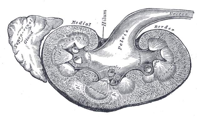
I’m having an identity crisis
I’ve described my nightmares before. But they are becoming more frequent. Night after night I would see myself in an operating room or Endo suite as a faceless wraith without a stable identity, morphing seamlessly from one specialty designation to another. Was I a vascular surgeon, a radiologist, a cardiologist, a vascular internist, a vascular specialist? This terrifying loss of awareness of my persona prompted me to make an appointment to see a psychiatrist.
As someone who has always felt comfortable with my own self-image, it was with a sense of reluctance that I confessed to this stranger that I was confused about my identity. However, once I lay down on his couch my inhibitions rapidly subsided. I blurted out, “Doctor, please help me. … I just don’t know what I am anymore. I seem to be losing my professional identity.”
He picked up my file and read it with a puzzled expression.
“Dr. Samson, I see you are a board-certified vascular surgeon. But you have intimated that you interpret your dreams as suggesting that you may not be one?”
I replied that it was only over the last few years that I had become unsure of my identity.
I told him that at times when I am introduced to someone at a cocktail party and they ask me what I do, I tell them I’m a vascular surgeon, yet they look puzzled. Then as if proud of their general knowledge they excitedly reply “Oh, I know, you treat varicose veins.”
Sighing, I would reply “Actually, you are correct but I also do things like preventing amputations and stroke, and I also manage abdominal aneurysms that if left untreated can cause sudden death.” Clearly I was trying to impress them that I don’t treat only a cosmetic nuisance but also life- and limb-threatening conditions. Nonetheless, their glazed, uncomprehending eyes remind me that most members of the lay public have no knowledge about these maladies nor that vascular surgeons are the doctors who treat them. To add insult to my already injured ego, their next comment is usually along the lines of “My cardiologist just did a full work-up and she told me that my carotid arteries and leg arteries have a cholesterol buildup and that if it gets any worse she may need to stent them. By the way what do you think of this ugly vein on my leg?”
Noticing that I had stopped talking the pyschiatrist asked “I can see that you are unhappy that patients may not know what you do, but why do you feel confused?”
“Well, a lot of what I now do doesn’t involve surgery. In fact most days I’m in an angiography suite doing stents and angioplasties or in my office prescribing medications.” He remained silent. I went on.
“I just don’t know who I am anymore. … I mean, am I a vascular surgeon or a vascular specialist? Maybe a vascular proceduralist? Or a vascular interventionalist?”
Like most of his profession, he didn’t give me an answer but rather proposed another question. “I see you are part of a private practice group, Sarasota Vascular Specialists. Doesn’t that define who you are?”
“Well,” I replied “Not really. You will notice that under that name we write ‘The Vein and Artery Experts’ because we know most people, even other physicians, may not know what that implies. Just the other day a referring doctor told me he had sent a patient with a DVT to the interventional radiologist for lytic therapy. He did not know I also provide that treatment or even that I can insert vena cava filters.”
As he had served as a high level officer in several psychiatry organizations and was academically inclined my therapist digressed and questioned me on the manner in which my specialty organization refers to itself.
“That’s the problem,” I quickly answered. “We have been all over the place. At one stage we were the International Society of Cardiovascular Surgery – then the American Association of Vascular Surgeons, and now the Society for Vascular Surgery. But some felt we should also lay claim to venous disease, and so we established the American Venous Forum.
“We even felt the need to come out with a new Journal for venous and lymphatic disease. Then some young vascular surgeons, concerned that the Peripheral Vascular Surgery Society did not convey all that we do, changed that name to the Vascular and Endovascular Surgery Society. And now I am aware that some of our leaders of the Society for Vascular Surgery are worried that the word ‘Surgery’ scares our patients and are suggesting a transition to the Society of Vascular Specialists.”
“Well then, why not just call yourself a vascular specialist?” Good point I thought as I lay there on his couch. But then it occurred to me that perhaps physicians who weren’t surgeons but who also dedicated themselves to vascular diseases could also refer to themselves as vascular specialists.
What would differentiate us? After all they can’t operate but I can. Wouldn’t this somewhat ambiguous word “specialist” further cause me an even greater identity crisis?
My brain started racing. Now I realized I was jealous of all those other specialists whose professional identity was defined by their specialty’s name. Everyone knows that a gynecologist treats only women, the orthopedist can mend broken or diseased bones, and the proctologist treats ... well, you know what!
“Maybe I’m a vasculologist?” I suggested.
He remained silent. Time seemed to expand. But in that darkened room I began to understand that he could not provide me with the answer to my identity crisis. It was up to me, my colleagues, and my specialty organization to explain to the lay public and other doctors what makes us so special. That we are indeed surgeons but that unlike any other specialty we alone can provide all forms of therapy to patients with vascular disease.
I was proud of this epiphany, and I eagerly shared it with him. He nodded approvingly. “You have made good progress but time’s up.”
As I stood to leave I noticed that he was rolling up his pant leg and staring at a bulging varicosity. He mumbled, “Hmmm … I’d better show this to my cardiologist. It may be serious.”
Dr. Samson is a clinical professor of surgery (vascular) at Florida State University Medical School, is president, Mote Vascular Foundation, and is an attending vascular surgeon, Sarasota (Fla.) Vascular Specialists. Dr. Samson also considers himself a member of his proposed American College of Vascular Surgery.
I’ve described my nightmares before. But they are becoming more frequent. Night after night I would see myself in an operating room or Endo suite as a faceless wraith without a stable identity, morphing seamlessly from one specialty designation to another. Was I a vascular surgeon, a radiologist, a cardiologist, a vascular internist, a vascular specialist? This terrifying loss of awareness of my persona prompted me to make an appointment to see a psychiatrist.
As someone who has always felt comfortable with my own self-image, it was with a sense of reluctance that I confessed to this stranger that I was confused about my identity. However, once I lay down on his couch my inhibitions rapidly subsided. I blurted out, “Doctor, please help me. … I just don’t know what I am anymore. I seem to be losing my professional identity.”
He picked up my file and read it with a puzzled expression.
“Dr. Samson, I see you are a board-certified vascular surgeon. But you have intimated that you interpret your dreams as suggesting that you may not be one?”
I replied that it was only over the last few years that I had become unsure of my identity.
I told him that at times when I am introduced to someone at a cocktail party and they ask me what I do, I tell them I’m a vascular surgeon, yet they look puzzled. Then as if proud of their general knowledge they excitedly reply “Oh, I know, you treat varicose veins.”
Sighing, I would reply “Actually, you are correct but I also do things like preventing amputations and stroke, and I also manage abdominal aneurysms that if left untreated can cause sudden death.” Clearly I was trying to impress them that I don’t treat only a cosmetic nuisance but also life- and limb-threatening conditions. Nonetheless, their glazed, uncomprehending eyes remind me that most members of the lay public have no knowledge about these maladies nor that vascular surgeons are the doctors who treat them. To add insult to my already injured ego, their next comment is usually along the lines of “My cardiologist just did a full work-up and she told me that my carotid arteries and leg arteries have a cholesterol buildup and that if it gets any worse she may need to stent them. By the way what do you think of this ugly vein on my leg?”
Noticing that I had stopped talking the pyschiatrist asked “I can see that you are unhappy that patients may not know what you do, but why do you feel confused?”
“Well, a lot of what I now do doesn’t involve surgery. In fact most days I’m in an angiography suite doing stents and angioplasties or in my office prescribing medications.” He remained silent. I went on.
“I just don’t know who I am anymore. … I mean, am I a vascular surgeon or a vascular specialist? Maybe a vascular proceduralist? Or a vascular interventionalist?”
Like most of his profession, he didn’t give me an answer but rather proposed another question. “I see you are part of a private practice group, Sarasota Vascular Specialists. Doesn’t that define who you are?”
“Well,” I replied “Not really. You will notice that under that name we write ‘The Vein and Artery Experts’ because we know most people, even other physicians, may not know what that implies. Just the other day a referring doctor told me he had sent a patient with a DVT to the interventional radiologist for lytic therapy. He did not know I also provide that treatment or even that I can insert vena cava filters.”
As he had served as a high level officer in several psychiatry organizations and was academically inclined my therapist digressed and questioned me on the manner in which my specialty organization refers to itself.
“That’s the problem,” I quickly answered. “We have been all over the place. At one stage we were the International Society of Cardiovascular Surgery – then the American Association of Vascular Surgeons, and now the Society for Vascular Surgery. But some felt we should also lay claim to venous disease, and so we established the American Venous Forum.
“We even felt the need to come out with a new Journal for venous and lymphatic disease. Then some young vascular surgeons, concerned that the Peripheral Vascular Surgery Society did not convey all that we do, changed that name to the Vascular and Endovascular Surgery Society. And now I am aware that some of our leaders of the Society for Vascular Surgery are worried that the word ‘Surgery’ scares our patients and are suggesting a transition to the Society of Vascular Specialists.”
“Well then, why not just call yourself a vascular specialist?” Good point I thought as I lay there on his couch. But then it occurred to me that perhaps physicians who weren’t surgeons but who also dedicated themselves to vascular diseases could also refer to themselves as vascular specialists.
What would differentiate us? After all they can’t operate but I can. Wouldn’t this somewhat ambiguous word “specialist” further cause me an even greater identity crisis?
My brain started racing. Now I realized I was jealous of all those other specialists whose professional identity was defined by their specialty’s name. Everyone knows that a gynecologist treats only women, the orthopedist can mend broken or diseased bones, and the proctologist treats ... well, you know what!
“Maybe I’m a vasculologist?” I suggested.
He remained silent. Time seemed to expand. But in that darkened room I began to understand that he could not provide me with the answer to my identity crisis. It was up to me, my colleagues, and my specialty organization to explain to the lay public and other doctors what makes us so special. That we are indeed surgeons but that unlike any other specialty we alone can provide all forms of therapy to patients with vascular disease.
I was proud of this epiphany, and I eagerly shared it with him. He nodded approvingly. “You have made good progress but time’s up.”
As I stood to leave I noticed that he was rolling up his pant leg and staring at a bulging varicosity. He mumbled, “Hmmm … I’d better show this to my cardiologist. It may be serious.”
Dr. Samson is a clinical professor of surgery (vascular) at Florida State University Medical School, is president, Mote Vascular Foundation, and is an attending vascular surgeon, Sarasota (Fla.) Vascular Specialists. Dr. Samson also considers himself a member of his proposed American College of Vascular Surgery.
I’ve described my nightmares before. But they are becoming more frequent. Night after night I would see myself in an operating room or Endo suite as a faceless wraith without a stable identity, morphing seamlessly from one specialty designation to another. Was I a vascular surgeon, a radiologist, a cardiologist, a vascular internist, a vascular specialist? This terrifying loss of awareness of my persona prompted me to make an appointment to see a psychiatrist.
As someone who has always felt comfortable with my own self-image, it was with a sense of reluctance that I confessed to this stranger that I was confused about my identity. However, once I lay down on his couch my inhibitions rapidly subsided. I blurted out, “Doctor, please help me. … I just don’t know what I am anymore. I seem to be losing my professional identity.”
He picked up my file and read it with a puzzled expression.
“Dr. Samson, I see you are a board-certified vascular surgeon. But you have intimated that you interpret your dreams as suggesting that you may not be one?”
I replied that it was only over the last few years that I had become unsure of my identity.
I told him that at times when I am introduced to someone at a cocktail party and they ask me what I do, I tell them I’m a vascular surgeon, yet they look puzzled. Then as if proud of their general knowledge they excitedly reply “Oh, I know, you treat varicose veins.”
Sighing, I would reply “Actually, you are correct but I also do things like preventing amputations and stroke, and I also manage abdominal aneurysms that if left untreated can cause sudden death.” Clearly I was trying to impress them that I don’t treat only a cosmetic nuisance but also life- and limb-threatening conditions. Nonetheless, their glazed, uncomprehending eyes remind me that most members of the lay public have no knowledge about these maladies nor that vascular surgeons are the doctors who treat them. To add insult to my already injured ego, their next comment is usually along the lines of “My cardiologist just did a full work-up and she told me that my carotid arteries and leg arteries have a cholesterol buildup and that if it gets any worse she may need to stent them. By the way what do you think of this ugly vein on my leg?”
Noticing that I had stopped talking the pyschiatrist asked “I can see that you are unhappy that patients may not know what you do, but why do you feel confused?”
“Well, a lot of what I now do doesn’t involve surgery. In fact most days I’m in an angiography suite doing stents and angioplasties or in my office prescribing medications.” He remained silent. I went on.
“I just don’t know who I am anymore. … I mean, am I a vascular surgeon or a vascular specialist? Maybe a vascular proceduralist? Or a vascular interventionalist?”
Like most of his profession, he didn’t give me an answer but rather proposed another question. “I see you are part of a private practice group, Sarasota Vascular Specialists. Doesn’t that define who you are?”
“Well,” I replied “Not really. You will notice that under that name we write ‘The Vein and Artery Experts’ because we know most people, even other physicians, may not know what that implies. Just the other day a referring doctor told me he had sent a patient with a DVT to the interventional radiologist for lytic therapy. He did not know I also provide that treatment or even that I can insert vena cava filters.”
As he had served as a high level officer in several psychiatry organizations and was academically inclined my therapist digressed and questioned me on the manner in which my specialty organization refers to itself.
“That’s the problem,” I quickly answered. “We have been all over the place. At one stage we were the International Society of Cardiovascular Surgery – then the American Association of Vascular Surgeons, and now the Society for Vascular Surgery. But some felt we should also lay claim to venous disease, and so we established the American Venous Forum.
“We even felt the need to come out with a new Journal for venous and lymphatic disease. Then some young vascular surgeons, concerned that the Peripheral Vascular Surgery Society did not convey all that we do, changed that name to the Vascular and Endovascular Surgery Society. And now I am aware that some of our leaders of the Society for Vascular Surgery are worried that the word ‘Surgery’ scares our patients and are suggesting a transition to the Society of Vascular Specialists.”
“Well then, why not just call yourself a vascular specialist?” Good point I thought as I lay there on his couch. But then it occurred to me that perhaps physicians who weren’t surgeons but who also dedicated themselves to vascular diseases could also refer to themselves as vascular specialists.
What would differentiate us? After all they can’t operate but I can. Wouldn’t this somewhat ambiguous word “specialist” further cause me an even greater identity crisis?
My brain started racing. Now I realized I was jealous of all those other specialists whose professional identity was defined by their specialty’s name. Everyone knows that a gynecologist treats only women, the orthopedist can mend broken or diseased bones, and the proctologist treats ... well, you know what!
“Maybe I’m a vasculologist?” I suggested.
He remained silent. Time seemed to expand. But in that darkened room I began to understand that he could not provide me with the answer to my identity crisis. It was up to me, my colleagues, and my specialty organization to explain to the lay public and other doctors what makes us so special. That we are indeed surgeons but that unlike any other specialty we alone can provide all forms of therapy to patients with vascular disease.
I was proud of this epiphany, and I eagerly shared it with him. He nodded approvingly. “You have made good progress but time’s up.”
As I stood to leave I noticed that he was rolling up his pant leg and staring at a bulging varicosity. He mumbled, “Hmmm … I’d better show this to my cardiologist. It may be serious.”
Dr. Samson is a clinical professor of surgery (vascular) at Florida State University Medical School, is president, Mote Vascular Foundation, and is an attending vascular surgeon, Sarasota (Fla.) Vascular Specialists. Dr. Samson also considers himself a member of his proposed American College of Vascular Surgery.

The registered letter runaround
Registered letters are a pain in the butt.
In the grand scheme of things, they’re a minor nuisance, albeit necessary. Documentation is everything is this job.
So here and there, I stop by the post office on the way home. It’s almost $7 a letter now, so maybe $28 a month, $336 a year. Not a huge sum.
But it’s annoying. It’s money that could be used for other expenses, and 80% of the time, the reason I have to do it is someone isn’t returning a call.
I suspect the scenario is similar in your practice. Patients are due for a follow-up MRI, MR angiography, labs, or whatever. So your staff calls them a few times. If they don’t return the call, you mail them letters. If they don’t respond, you send them registered letters. That way, even if they never respond, you can document that someone at that address got the letter.
Patients have several chances to answer the phone or call my office to schedule or refuse the test before I resort to the letter. But, for whatever reasons, sometimes they ignore them, leaving me with a trip to the post office and $7 down.
About one-third of the time we still never hear back, which is fine if that’s what they want. As long as I get the signed card, I don’t mind. Another third of the time they call to schedule, often wondering why I had to resort to a registered letter. “I got your call and other letter, so why did you send that?” And the others? They call us, usually to say they don’t want the test, or just angry that we sent them a registered letter, or (my favorite) to accuse us of harassing them. Because, you know, I enjoy making the extra trip and cost just to do that.
Unfortunately, the consequences of not doing this are (potentially) worse. In most cases, nothing would happen. But, sooner or later you risk having a medical disaster possibly occur because of whatever you were following. And then, correctly or not, they may blame you and call a lawyer. While it’s not guaranteed protection, it’s helpful to be able to prove, with a signed card, that they got your letter and chose to ignore it.
Is that worth the $7 and extra stop? Absolutely. But still, I wish people would be kind enough to just answer the phone or return a call. If they don’t want to do the study, I’m not offended. I just don’t like wasting time and money because someone won’t pick up a phone.
Dr. Block has a solo neurology practice in Scottsdale, Ariz.
Registered letters are a pain in the butt.
In the grand scheme of things, they’re a minor nuisance, albeit necessary. Documentation is everything is this job.
So here and there, I stop by the post office on the way home. It’s almost $7 a letter now, so maybe $28 a month, $336 a year. Not a huge sum.
But it’s annoying. It’s money that could be used for other expenses, and 80% of the time, the reason I have to do it is someone isn’t returning a call.
I suspect the scenario is similar in your practice. Patients are due for a follow-up MRI, MR angiography, labs, or whatever. So your staff calls them a few times. If they don’t return the call, you mail them letters. If they don’t respond, you send them registered letters. That way, even if they never respond, you can document that someone at that address got the letter.
Patients have several chances to answer the phone or call my office to schedule or refuse the test before I resort to the letter. But, for whatever reasons, sometimes they ignore them, leaving me with a trip to the post office and $7 down.
About one-third of the time we still never hear back, which is fine if that’s what they want. As long as I get the signed card, I don’t mind. Another third of the time they call to schedule, often wondering why I had to resort to a registered letter. “I got your call and other letter, so why did you send that?” And the others? They call us, usually to say they don’t want the test, or just angry that we sent them a registered letter, or (my favorite) to accuse us of harassing them. Because, you know, I enjoy making the extra trip and cost just to do that.
Unfortunately, the consequences of not doing this are (potentially) worse. In most cases, nothing would happen. But, sooner or later you risk having a medical disaster possibly occur because of whatever you were following. And then, correctly or not, they may blame you and call a lawyer. While it’s not guaranteed protection, it’s helpful to be able to prove, with a signed card, that they got your letter and chose to ignore it.
Is that worth the $7 and extra stop? Absolutely. But still, I wish people would be kind enough to just answer the phone or return a call. If they don’t want to do the study, I’m not offended. I just don’t like wasting time and money because someone won’t pick up a phone.
Dr. Block has a solo neurology practice in Scottsdale, Ariz.
Registered letters are a pain in the butt.
In the grand scheme of things, they’re a minor nuisance, albeit necessary. Documentation is everything is this job.
So here and there, I stop by the post office on the way home. It’s almost $7 a letter now, so maybe $28 a month, $336 a year. Not a huge sum.
But it’s annoying. It’s money that could be used for other expenses, and 80% of the time, the reason I have to do it is someone isn’t returning a call.
I suspect the scenario is similar in your practice. Patients are due for a follow-up MRI, MR angiography, labs, or whatever. So your staff calls them a few times. If they don’t return the call, you mail them letters. If they don’t respond, you send them registered letters. That way, even if they never respond, you can document that someone at that address got the letter.
Patients have several chances to answer the phone or call my office to schedule or refuse the test before I resort to the letter. But, for whatever reasons, sometimes they ignore them, leaving me with a trip to the post office and $7 down.
About one-third of the time we still never hear back, which is fine if that’s what they want. As long as I get the signed card, I don’t mind. Another third of the time they call to schedule, often wondering why I had to resort to a registered letter. “I got your call and other letter, so why did you send that?” And the others? They call us, usually to say they don’t want the test, or just angry that we sent them a registered letter, or (my favorite) to accuse us of harassing them. Because, you know, I enjoy making the extra trip and cost just to do that.
Unfortunately, the consequences of not doing this are (potentially) worse. In most cases, nothing would happen. But, sooner or later you risk having a medical disaster possibly occur because of whatever you were following. And then, correctly or not, they may blame you and call a lawyer. While it’s not guaranteed protection, it’s helpful to be able to prove, with a signed card, that they got your letter and chose to ignore it.
Is that worth the $7 and extra stop? Absolutely. But still, I wish people would be kind enough to just answer the phone or return a call. If they don’t want to do the study, I’m not offended. I just don’t like wasting time and money because someone won’t pick up a phone.
Dr. Block has a solo neurology practice in Scottsdale, Ariz.
Say it with a smile
“Have a nice time!”
I looked around, trying to tell where the piping, childish voice was coming from. I’d just swiped the barcode of my pass to the local lake I swim in every summer. There it was – the voice of one of the high school kids who works at the lake. She wore a wan smile.
I’ve been swimming at this lake for 35 years. No kid ever said a word to me before. “Have a good swim!” said a young man standing at the desk where, “All Children Under 12 Must Check In!”
Goodness me, I thought. The management consultants have made it to the lake.
You know who I mean. The ones who see to it that front-desk personnel always flash a bright smile and recite the corporate script. At the hardware store, the fast-food chain, the airline counter. Even the people in the auto dealer’s service department smile and murmur sweet nothings. They used to glare and growl, and make you feel like an idiot. “Whatsamattter, Bud? Dontcha know anything about cars?” Now it’s all politeness and smiles and “How may we help you, kind sir?”
And of course there’s the pharmacy. When I fill my prescription, the tech flashes a bright grin of welcome. Either that, or she has tetanus.
“Welcome to DrugTown!” she says. “May I have your name?”
I tell her. She retrieves the prescription. “Verify your address?” I do.
“Do you have a DrugTown Rewards Card?” she asks. I enter in my cellphone number, swipe my card, turn to leave.
“Be sound!” she says, still grinning. The DrugTown motto is: “Where Safe Meets Sound!”
It is easy to mock this sort of thing as formulaic and false. Insincere or not, smiling makes a difference. Some say you can actually get happier by making yourself smile. Whether that’s true or not, watching other people smile and make eye contact makes you feel good. Seeing them scowl and look away does the reverse.
This is true in doctors’ offices too. I learned this recently by being a patient.
I approached the front desk at my first visit. The lone receptionist was looking at some papers. I tried to get her attention. “Hello,” I said, “My name is ... ”
Still looking down, she shoved a clipboard across the counter. “Sign in,” she said. “And fill this out.” She handed me a sheaf of forms. “Leave it here when you’re done.” She was still looking away.
I sat in one of the waiting room chairs to work on the forms. I felt bad. As I watched the clerk ignore a succession of other patients, I asked myself why I felt so bad. First of all, it wasn’t personal; she was churlish to everyone. Second, what did this have to do with my visit? I was there to see the doctor, not his receptionist. Weren’t his skill and expertise what mattered?
True enough, but I still felt lousy. At later visits I took on the personal challenge of trying to force the clerk to make eye contact. I failed. In truth, her behavior colored my impression of the medical experience – mixed anyway – more than the medical outcome.
Sometimes I force myself to look at my own online reviews. The bad ones often focus on the alleged rudeness of my staff. It can be hard to tell from cranky patients whether their complaints are justified. But sometimes they are.
Management consultants know this. They teach employers that the customer experience has to do with more than the quality of the good or service provided. Even if the quarter-pounder is delicious, it may not taste that way if the burger-flipper is having a bad day and doesn’t know how to hide it.
So my office manager now trains our front-desk staff to be insistently cheery. This can be hard when patients are stacked three-deep, each with a form to scan, a credit card to swipe, a follow-up to book. But smile we have them do.
We don’t, however, have them recite a script when smiling. (“Make the scene! Wear sunscreen!”) We’re not up to that chapter in the customer-service handbook.
Who do you think we are? The town lake?
Dr. Rockoff practices dermatology in Brookline, Mass., and is a longtime contributor to Dermatology News. He serves on the clinical faculty at Tufts University, Boston, and has taught senior medical students and other trainees for 30 years. Write to him at dermnews@frontlinemedcom.com.
“Have a nice time!”
I looked around, trying to tell where the piping, childish voice was coming from. I’d just swiped the barcode of my pass to the local lake I swim in every summer. There it was – the voice of one of the high school kids who works at the lake. She wore a wan smile.
I’ve been swimming at this lake for 35 years. No kid ever said a word to me before. “Have a good swim!” said a young man standing at the desk where, “All Children Under 12 Must Check In!”
Goodness me, I thought. The management consultants have made it to the lake.
You know who I mean. The ones who see to it that front-desk personnel always flash a bright smile and recite the corporate script. At the hardware store, the fast-food chain, the airline counter. Even the people in the auto dealer’s service department smile and murmur sweet nothings. They used to glare and growl, and make you feel like an idiot. “Whatsamattter, Bud? Dontcha know anything about cars?” Now it’s all politeness and smiles and “How may we help you, kind sir?”
And of course there’s the pharmacy. When I fill my prescription, the tech flashes a bright grin of welcome. Either that, or she has tetanus.
“Welcome to DrugTown!” she says. “May I have your name?”
I tell her. She retrieves the prescription. “Verify your address?” I do.
“Do you have a DrugTown Rewards Card?” she asks. I enter in my cellphone number, swipe my card, turn to leave.
“Be sound!” she says, still grinning. The DrugTown motto is: “Where Safe Meets Sound!”
It is easy to mock this sort of thing as formulaic and false. Insincere or not, smiling makes a difference. Some say you can actually get happier by making yourself smile. Whether that’s true or not, watching other people smile and make eye contact makes you feel good. Seeing them scowl and look away does the reverse.
This is true in doctors’ offices too. I learned this recently by being a patient.
I approached the front desk at my first visit. The lone receptionist was looking at some papers. I tried to get her attention. “Hello,” I said, “My name is ... ”
Still looking down, she shoved a clipboard across the counter. “Sign in,” she said. “And fill this out.” She handed me a sheaf of forms. “Leave it here when you’re done.” She was still looking away.
I sat in one of the waiting room chairs to work on the forms. I felt bad. As I watched the clerk ignore a succession of other patients, I asked myself why I felt so bad. First of all, it wasn’t personal; she was churlish to everyone. Second, what did this have to do with my visit? I was there to see the doctor, not his receptionist. Weren’t his skill and expertise what mattered?
True enough, but I still felt lousy. At later visits I took on the personal challenge of trying to force the clerk to make eye contact. I failed. In truth, her behavior colored my impression of the medical experience – mixed anyway – more than the medical outcome.
Sometimes I force myself to look at my own online reviews. The bad ones often focus on the alleged rudeness of my staff. It can be hard to tell from cranky patients whether their complaints are justified. But sometimes they are.
Management consultants know this. They teach employers that the customer experience has to do with more than the quality of the good or service provided. Even if the quarter-pounder is delicious, it may not taste that way if the burger-flipper is having a bad day and doesn’t know how to hide it.
So my office manager now trains our front-desk staff to be insistently cheery. This can be hard when patients are stacked three-deep, each with a form to scan, a credit card to swipe, a follow-up to book. But smile we have them do.
We don’t, however, have them recite a script when smiling. (“Make the scene! Wear sunscreen!”) We’re not up to that chapter in the customer-service handbook.
Who do you think we are? The town lake?
Dr. Rockoff practices dermatology in Brookline, Mass., and is a longtime contributor to Dermatology News. He serves on the clinical faculty at Tufts University, Boston, and has taught senior medical students and other trainees for 30 years. Write to him at dermnews@frontlinemedcom.com.
“Have a nice time!”
I looked around, trying to tell where the piping, childish voice was coming from. I’d just swiped the barcode of my pass to the local lake I swim in every summer. There it was – the voice of one of the high school kids who works at the lake. She wore a wan smile.
I’ve been swimming at this lake for 35 years. No kid ever said a word to me before. “Have a good swim!” said a young man standing at the desk where, “All Children Under 12 Must Check In!”
Goodness me, I thought. The management consultants have made it to the lake.
You know who I mean. The ones who see to it that front-desk personnel always flash a bright smile and recite the corporate script. At the hardware store, the fast-food chain, the airline counter. Even the people in the auto dealer’s service department smile and murmur sweet nothings. They used to glare and growl, and make you feel like an idiot. “Whatsamattter, Bud? Dontcha know anything about cars?” Now it’s all politeness and smiles and “How may we help you, kind sir?”
And of course there’s the pharmacy. When I fill my prescription, the tech flashes a bright grin of welcome. Either that, or she has tetanus.
“Welcome to DrugTown!” she says. “May I have your name?”
I tell her. She retrieves the prescription. “Verify your address?” I do.
“Do you have a DrugTown Rewards Card?” she asks. I enter in my cellphone number, swipe my card, turn to leave.
“Be sound!” she says, still grinning. The DrugTown motto is: “Where Safe Meets Sound!”
It is easy to mock this sort of thing as formulaic and false. Insincere or not, smiling makes a difference. Some say you can actually get happier by making yourself smile. Whether that’s true or not, watching other people smile and make eye contact makes you feel good. Seeing them scowl and look away does the reverse.
This is true in doctors’ offices too. I learned this recently by being a patient.
I approached the front desk at my first visit. The lone receptionist was looking at some papers. I tried to get her attention. “Hello,” I said, “My name is ... ”
Still looking down, she shoved a clipboard across the counter. “Sign in,” she said. “And fill this out.” She handed me a sheaf of forms. “Leave it here when you’re done.” She was still looking away.
I sat in one of the waiting room chairs to work on the forms. I felt bad. As I watched the clerk ignore a succession of other patients, I asked myself why I felt so bad. First of all, it wasn’t personal; she was churlish to everyone. Second, what did this have to do with my visit? I was there to see the doctor, not his receptionist. Weren’t his skill and expertise what mattered?
True enough, but I still felt lousy. At later visits I took on the personal challenge of trying to force the clerk to make eye contact. I failed. In truth, her behavior colored my impression of the medical experience – mixed anyway – more than the medical outcome.
Sometimes I force myself to look at my own online reviews. The bad ones often focus on the alleged rudeness of my staff. It can be hard to tell from cranky patients whether their complaints are justified. But sometimes they are.
Management consultants know this. They teach employers that the customer experience has to do with more than the quality of the good or service provided. Even if the quarter-pounder is delicious, it may not taste that way if the burger-flipper is having a bad day and doesn’t know how to hide it.
So my office manager now trains our front-desk staff to be insistently cheery. This can be hard when patients are stacked three-deep, each with a form to scan, a credit card to swipe, a follow-up to book. But smile we have them do.
We don’t, however, have them recite a script when smiling. (“Make the scene! Wear sunscreen!”) We’re not up to that chapter in the customer-service handbook.
Who do you think we are? The town lake?
Dr. Rockoff practices dermatology in Brookline, Mass., and is a longtime contributor to Dermatology News. He serves on the clinical faculty at Tufts University, Boston, and has taught senior medical students and other trainees for 30 years. Write to him at dermnews@frontlinemedcom.com.
Prion-like transmission of neurodegenerative pathology stirs concern
Recent developments regarding the nature of transmissible pathologic proteins in a variety of common neurodegenerative diseases have started to cause clinicians and public health experts to wonder if there is cause for concern.
A group from the National Hospital for Neurology & Neurosurgery at Queen Square, London, recently published a study presenting evidence that Alzheimer’s disease may have transmissible properties similar to traditionally regarded prion diseases such as Creutzfeldt-Jakob disease (Nature. 2015 Sep 10;525:247-50. doi:10.1038/nature15369). This conclusion was based upon the observation of cerebral and vascular amyloid-beta deposition in several patients who had received cadaveric growth hormone extracts ranging from 19 to 39 years earlier and who developed iatrogenic Creutzfeldt-Jakob disease (CJD). Amyloid deposition was also identified in the pituitary glands of patients with amyloid-beta pathology also felt to be iatrogenically transmitted.
This is not the first time, however, that analogies have been drawn between neurodegenerative diseases and prion-related diseases. Perhaps inspired by the evident connectivity of affected upper and lower motor neuronal populations in amyotrophic lateral sclerosis, previous investigators have sought cell-to-cell transmission of neurodegenerative pathology and have demonstrated this for amyloid as well as tau species in Alzheimer’s disease, and for synuclein species in Parkinson’s disease.
A recent article by Dr. Stanley Prusiner of the University of California, San Francisco, and his colleagues presented evidence that brain extracts from patients with multiple system atrophy (MSA) transmitted the characteristic MSA alpha-synuclein aggregates in the brains of transgenic mice as well as in cultured human embryonic kidney cells (Proc Natl Acad Sci U S A. 2015 Aug 31. doi:10.1073/pnas.1514475112).
An emerging theme from these recent and prior studies is that neurodegenerative diseases behave a lot like prion diseases, and although they follow a much slower time course, may spread through synaptic pathways as well as between people through prion-like mechanisms. CJD, while transmissible, is not “contagious,” and public messaging should draw this distinction in regard to neurodegenerative diseases. Nonetheless, further research is urgently needed given the implications of this work. Dr. Prusiner and his associates concluded, for example, that deep brain stimulation electrodes and related equipment should not be reused from Parkinson’s disease patients for fear of spreading the synucleinopathy from one person to another. Should there be restrictions with regard to any form of tissue transplantation or blood transfusion from patients with Parkinson’s disease or Alzheimer’s disease? Questions such as these need further clarification, and given the prevalence of these procedures, such research should be highly prioritized.
Dr. Caselli is professor of neurology at the Mayo Clinic, Scottsdale, Ariz. He also serves there as associate director and clinical core director of the Alzheimer’s Disease Center.
Recent developments regarding the nature of transmissible pathologic proteins in a variety of common neurodegenerative diseases have started to cause clinicians and public health experts to wonder if there is cause for concern.
A group from the National Hospital for Neurology & Neurosurgery at Queen Square, London, recently published a study presenting evidence that Alzheimer’s disease may have transmissible properties similar to traditionally regarded prion diseases such as Creutzfeldt-Jakob disease (Nature. 2015 Sep 10;525:247-50. doi:10.1038/nature15369). This conclusion was based upon the observation of cerebral and vascular amyloid-beta deposition in several patients who had received cadaveric growth hormone extracts ranging from 19 to 39 years earlier and who developed iatrogenic Creutzfeldt-Jakob disease (CJD). Amyloid deposition was also identified in the pituitary glands of patients with amyloid-beta pathology also felt to be iatrogenically transmitted.
This is not the first time, however, that analogies have been drawn between neurodegenerative diseases and prion-related diseases. Perhaps inspired by the evident connectivity of affected upper and lower motor neuronal populations in amyotrophic lateral sclerosis, previous investigators have sought cell-to-cell transmission of neurodegenerative pathology and have demonstrated this for amyloid as well as tau species in Alzheimer’s disease, and for synuclein species in Parkinson’s disease.
A recent article by Dr. Stanley Prusiner of the University of California, San Francisco, and his colleagues presented evidence that brain extracts from patients with multiple system atrophy (MSA) transmitted the characteristic MSA alpha-synuclein aggregates in the brains of transgenic mice as well as in cultured human embryonic kidney cells (Proc Natl Acad Sci U S A. 2015 Aug 31. doi:10.1073/pnas.1514475112).
An emerging theme from these recent and prior studies is that neurodegenerative diseases behave a lot like prion diseases, and although they follow a much slower time course, may spread through synaptic pathways as well as between people through prion-like mechanisms. CJD, while transmissible, is not “contagious,” and public messaging should draw this distinction in regard to neurodegenerative diseases. Nonetheless, further research is urgently needed given the implications of this work. Dr. Prusiner and his associates concluded, for example, that deep brain stimulation electrodes and related equipment should not be reused from Parkinson’s disease patients for fear of spreading the synucleinopathy from one person to another. Should there be restrictions with regard to any form of tissue transplantation or blood transfusion from patients with Parkinson’s disease or Alzheimer’s disease? Questions such as these need further clarification, and given the prevalence of these procedures, such research should be highly prioritized.
Dr. Caselli is professor of neurology at the Mayo Clinic, Scottsdale, Ariz. He also serves there as associate director and clinical core director of the Alzheimer’s Disease Center.
Recent developments regarding the nature of transmissible pathologic proteins in a variety of common neurodegenerative diseases have started to cause clinicians and public health experts to wonder if there is cause for concern.
A group from the National Hospital for Neurology & Neurosurgery at Queen Square, London, recently published a study presenting evidence that Alzheimer’s disease may have transmissible properties similar to traditionally regarded prion diseases such as Creutzfeldt-Jakob disease (Nature. 2015 Sep 10;525:247-50. doi:10.1038/nature15369). This conclusion was based upon the observation of cerebral and vascular amyloid-beta deposition in several patients who had received cadaveric growth hormone extracts ranging from 19 to 39 years earlier and who developed iatrogenic Creutzfeldt-Jakob disease (CJD). Amyloid deposition was also identified in the pituitary glands of patients with amyloid-beta pathology also felt to be iatrogenically transmitted.
This is not the first time, however, that analogies have been drawn between neurodegenerative diseases and prion-related diseases. Perhaps inspired by the evident connectivity of affected upper and lower motor neuronal populations in amyotrophic lateral sclerosis, previous investigators have sought cell-to-cell transmission of neurodegenerative pathology and have demonstrated this for amyloid as well as tau species in Alzheimer’s disease, and for synuclein species in Parkinson’s disease.
A recent article by Dr. Stanley Prusiner of the University of California, San Francisco, and his colleagues presented evidence that brain extracts from patients with multiple system atrophy (MSA) transmitted the characteristic MSA alpha-synuclein aggregates in the brains of transgenic mice as well as in cultured human embryonic kidney cells (Proc Natl Acad Sci U S A. 2015 Aug 31. doi:10.1073/pnas.1514475112).
An emerging theme from these recent and prior studies is that neurodegenerative diseases behave a lot like prion diseases, and although they follow a much slower time course, may spread through synaptic pathways as well as between people through prion-like mechanisms. CJD, while transmissible, is not “contagious,” and public messaging should draw this distinction in regard to neurodegenerative diseases. Nonetheless, further research is urgently needed given the implications of this work. Dr. Prusiner and his associates concluded, for example, that deep brain stimulation electrodes and related equipment should not be reused from Parkinson’s disease patients for fear of spreading the synucleinopathy from one person to another. Should there be restrictions with regard to any form of tissue transplantation or blood transfusion from patients with Parkinson’s disease or Alzheimer’s disease? Questions such as these need further clarification, and given the prevalence of these procedures, such research should be highly prioritized.
Dr. Caselli is professor of neurology at the Mayo Clinic, Scottsdale, Ariz. He also serves there as associate director and clinical core director of the Alzheimer’s Disease Center.