User login
Hand Hygiene in Preventing COVID-19 Transmission
Handwashing with antimicrobial soaps or alcohol-based sanitizers is an effective measure in preventing microbial disease transmission. In the context of coronavirus disease 2019 (COVID-19) prevention, the World Health Organization and Centers for Disease Control and Prevention have recommended handwashing with soap and water after coughing/sneezing, visiting a public place, touching surfaces outside the home, and taking care of a sick person(s), as well as before and after eating. When soap and water are not available, alcohol-based sanitizers may be used.1,2
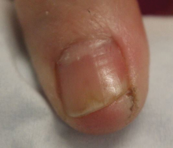
Irritant contact dermatitis (ICD) is most commonly associated with wet work and is frequently seen in health care workers in relation to hand hygiene, with survey-based studies reporting 25% to 55% of nurses affected.3-5 In a prospective study (N=102), health care workers who washed their hands more than 10 times per day were55% more likely to develop hand dermatitis.6 Frequent ICD of the hands has been reported in Chinese health care workers in association with COVID-19.7 Handwashing and/or glove wearing may be newly prioritized by workers who handle frequently touched goods and surfaces, such as flight attendants (Figure). Patients with obsessive-compulsive disorder may be another vulnerable population.8
Alcohol-based sanitizers and detergents or antimicrobials in soaps may cause ICD of the hands by denaturation of stratum corneum proteins, depletion of intercellular lipids, and decreased corneocyte cohesion. These agents alter the skin flora, with increased colonization by staphylococci and gram-negative bacilli.9 Clinical findings include xerosis, scaling, fissuring, and bleeding. Physicians may evaluate severity of ICD of the hands using the
Cleansing the hands with alcohol-based sanitizers has consistently shown equivalent or greater efficacy than antimicrobial soaps for eradication of most microbes, with exception of bacterial spores and protozoan oocysts.11 In an in vivo experiment, 70% ethanol solution was more effective in eradicating rotavirus from the fingerpads of adults than 10% povidone-iodine solution, nonmedicated soaps, and soaps containing chloroxylenol 4.8% or chlorhexidine gluconate 4%.12 Coronavirus disease 2019 is a lipophilic enveloped virus. The lipid-dissolving effects of alcohol-based sanitizers is especially effective against these kinds of viruses. An in vitro experiment showed that alcohol solutions are effective against enveloped viruses including severe acute respiratory syndrome coronavirus, Ebola virus, and Zika virus.13 There are limited data for the virucidal efficacy of non–alcohol-based sanitizers containing quaternary ammonium compounds (most commonly benzalkonium chloride) and therefore they are not recommended for protection against COVID-19. Handwashing is preferred over alcohol-based solutions when hands are visibly dirty.
Alcohol-based sanitizers typically are less likely to cause ICD than handwashing with detergent-based or antimicrobial soaps. Antimicrobial ingredients in soaps such as chlorhexidine, chloroxylenol, and triclosan are frequent culprits.11 Detergents in soap such as sodium laureth sulfate cause more skin irritation and transepidermal water loss than alcohol14; however, among health care workers, alcohol-based sanitizers often are perceived as more damaging to the skin.15 During the 2014 Ebola outbreak, use of alcohol-based sanitizers vs handwashing resulted in lower hand eczema severity index scores (n=108).16
Propensity for ICD is a limiting factor in hand hygiene adherence.17 In a double-blind randomized trial (N=54), scheduled use of an oil-containing lotion was shown to increase compliance with hand hygiene protocols in health care workers by preventing cracks, scaling, and pain.18 Using sanitizers containing humectants (eg, aloe vera gel) or moisturizers with petrolatum, liquid paraffin, glycerin, or mineral oil have all been shown to decrease the incidence of ICD in frequent handwashers.19,20 Thorough hand drying also is important in preventing dermatitis. Drying with disposable paper towels is preferred over automated air dryers to prevent aerosolization of microbes.21 Because latex has been implicated in development of ICD, use of latex-free gloves is recommended.22
Alcohol-based sanitizer is not only an effective virucidal agent but also is less likely to cause ICD, therefore promoting hand hygiene adherence. Handwashing with soap still is necessary when hands are visibly dirty but should be performed less frequently if feasible. Hand hygiene and emollient usage education is important for physicians and patients alike, particularly during the COVID-19 crisis.
- Centers for Disease Control and Prevention. Coronavirus disease 2019. how to protect yourself & others. https://www.cdc.gov/coronavirus/2019-ncov/prepare/prevention.html. Updated April 13, 2020. Accessed April 21, 2020.
- World Health Organization. Coronavirus disease (COVID-19) advice for the public. https://www.who.int/emergencies/diseases/novel-coronavirus-2019/advice-for-public. Updated March 31, 2020. Accessed April 21, 2020.
- Carøe TK, Ebbehøj NE, Bonde JPE, et al. Hand eczema and wet work: dose-response relationship and effect of leaving the profession. Contact Dermatitis. 2018;78:341-347.
- Larson E, Friedman C, Cohran J, et al. Prevalence and correlates of skin damage on the hands of nurses. Heart Lung. 1997;26:404-412.
- Lampel HP, Patel N, Boyse K, et al. Prevalence of hand dermatitis in inpatient nurses at a United States hospital. Dermatitis. 2007;18:140-142.
- Callahan A, Baron E, Fekedulegn D, et al. Winter season, frequent hand washing, and irritant patch test reactions to detergents are associated with hand dermatitis in health care workers. Dermatitis. 2013;24:170-175.
- Lan J, Song Z, Miao X, et al. Skin damage among healthcare workers managing coronavirus disease-2019 [published online March 18, 2020]. J Am Acad Dermatol. 2020;82:1215-1216.
- Katz RJ, Landau P, DeVeaugh-Geiss J, et al. Pharmacological responsiveness of dermatitis secondary to compulsive washing. Psychiatry Res. 1990;34:223-226.
- Larson EL, Hughes CA, Pyrek JD, et al. Changes in bacterial flora associated with skin damage on hands of health care personnel. Am J Infect Control. 1998;26:513-521.
- Held E, Skoet R, Johansen JD, et al. The hand eczema severity index (HECSI): a scoring system for clinical assessment of hand eczema. a study of inter- and intraobserver reliability. Br J Dermatol. 2005;152:302-307.
- Boyce JM, Pittet D, Healthcare Infection Control Practices Advisory Committee, et al. Guideline for Hand Hygiene in Health-Care Settings. Recommendations of the Healthcare Infection Control Practices Advisory Committee and the HIPAC/SHEA/APIC/IDSA Hand Hygiene Task Force. Am J Infect Control. 2002;30:S1-S46.
- Ansari SA, Sattar SA, Springthorpe VS, et al. Invivo protocol for testing efficacy of hand-washing agents against viruses and bacteria—experiments with rotavirus and Escherichi coli. Appl Environ Microbiol. 1989;55:3113-3118.
- Siddharta A, Pfaender S, Vielle NJ, et al. virucidal activity of world health organization-recommended formulations against enveloped viruses, including Zika, Ebola, and emerging coronaviruses. J Infect Dis. 2017;215:902-906.
- Pedersen LK, Held E, Johansen JD, et al. Less skin irritation from alcohol-based disinfectant than from detergent used for hand disinfection. Br J Dermatol. 2005;153:1142-1146.
- Stutz N, Becker D, Jappe U, et al. Nurses’ perceptions of the benefits and adverse effects of hand disinfection: alcohol-based hand rubs vs. hygienic handwashing: a multicentre questionnaire study with additional patch testing by the German Contact Dermatitis Research Group. Br J Dermatol. 2009;160:565-572.
- Wolfe MK, Wells E, Mitro B, et al. Seeking clearer recommendations for hand hygiene in communities facing Ebola: a randomized trial investigating the impact of six handwashing methods on skin irritation and dermatitis. PLoS One. 2016;11:e0167378.
- Pittet D, Allegranzi B, Storr J. The WHO Clean Care is Safer Care programme: field-testing to enhance sustainability and spread of hand hygiene improvements. J Infect Public Health. 2008;1:4-10.
- McCormick RD, Buchman TL, Maki DG. Double-blind, randomized trial of scheduled use of a novel barrier cream and an oil-containing lotion for protecting the hands of health care workers. Am J Infect Control. 2000;28:302-310.
- Berndt U, Wigger-Alberti W, Gabard B, et al. Efficacy of a barrier cream and its vehicle as protective measures against occupational irritant contact dermatitis. Contact Dermatitis. 2000;42:77-80.
- Kampf G, Ennen J. Regular use of a hand cream can attenuate skin dryness and roughness caused by frequent hand washing. BMC Dermatol. 2006;6:1.
- Gammon J, Hunt J. The neglected element of hand hygiene - significance of hand drying, efficiency of different methods, and clinical implication: a review. J Infect Prev. 2019;20:66-74.
- Elston DM. Letter from the editor: occupational skin disease among healthcare workers during the coronavirus (COVID-19) epidemic [published online March 18, 2020]. J Am Acad Dermatol. 2020;82:1085-1086.
Handwashing with antimicrobial soaps or alcohol-based sanitizers is an effective measure in preventing microbial disease transmission. In the context of coronavirus disease 2019 (COVID-19) prevention, the World Health Organization and Centers for Disease Control and Prevention have recommended handwashing with soap and water after coughing/sneezing, visiting a public place, touching surfaces outside the home, and taking care of a sick person(s), as well as before and after eating. When soap and water are not available, alcohol-based sanitizers may be used.1,2
Irritant contact dermatitis (ICD) is most commonly associated with wet work and is frequently seen in health care workers in relation to hand hygiene, with survey-based studies reporting 25% to 55% of nurses affected.3-5 In a prospective study (N=102), health care workers who washed their hands more than 10 times per day were55% more likely to develop hand dermatitis.6 Frequent ICD of the hands has been reported in Chinese health care workers in association with COVID-19.7 Handwashing and/or glove wearing may be newly prioritized by workers who handle frequently touched goods and surfaces, such as flight attendants (Figure). Patients with obsessive-compulsive disorder may be another vulnerable population.8
Alcohol-based sanitizers and detergents or antimicrobials in soaps may cause ICD of the hands by denaturation of stratum corneum proteins, depletion of intercellular lipids, and decreased corneocyte cohesion. These agents alter the skin flora, with increased colonization by staphylococci and gram-negative bacilli.9 Clinical findings include xerosis, scaling, fissuring, and bleeding. Physicians may evaluate severity of ICD of the hands using the
Cleansing the hands with alcohol-based sanitizers has consistently shown equivalent or greater efficacy than antimicrobial soaps for eradication of most microbes, with exception of bacterial spores and protozoan oocysts.11 In an in vivo experiment, 70% ethanol solution was more effective in eradicating rotavirus from the fingerpads of adults than 10% povidone-iodine solution, nonmedicated soaps, and soaps containing chloroxylenol 4.8% or chlorhexidine gluconate 4%.12 Coronavirus disease 2019 is a lipophilic enveloped virus. The lipid-dissolving effects of alcohol-based sanitizers is especially effective against these kinds of viruses. An in vitro experiment showed that alcohol solutions are effective against enveloped viruses including severe acute respiratory syndrome coronavirus, Ebola virus, and Zika virus.13 There are limited data for the virucidal efficacy of non–alcohol-based sanitizers containing quaternary ammonium compounds (most commonly benzalkonium chloride) and therefore they are not recommended for protection against COVID-19. Handwashing is preferred over alcohol-based solutions when hands are visibly dirty.
Alcohol-based sanitizers typically are less likely to cause ICD than handwashing with detergent-based or antimicrobial soaps. Antimicrobial ingredients in soaps such as chlorhexidine, chloroxylenol, and triclosan are frequent culprits.11 Detergents in soap such as sodium laureth sulfate cause more skin irritation and transepidermal water loss than alcohol14; however, among health care workers, alcohol-based sanitizers often are perceived as more damaging to the skin.15 During the 2014 Ebola outbreak, use of alcohol-based sanitizers vs handwashing resulted in lower hand eczema severity index scores (n=108).16
Propensity for ICD is a limiting factor in hand hygiene adherence.17 In a double-blind randomized trial (N=54), scheduled use of an oil-containing lotion was shown to increase compliance with hand hygiene protocols in health care workers by preventing cracks, scaling, and pain.18 Using sanitizers containing humectants (eg, aloe vera gel) or moisturizers with petrolatum, liquid paraffin, glycerin, or mineral oil have all been shown to decrease the incidence of ICD in frequent handwashers.19,20 Thorough hand drying also is important in preventing dermatitis. Drying with disposable paper towels is preferred over automated air dryers to prevent aerosolization of microbes.21 Because latex has been implicated in development of ICD, use of latex-free gloves is recommended.22
Alcohol-based sanitizer is not only an effective virucidal agent but also is less likely to cause ICD, therefore promoting hand hygiene adherence. Handwashing with soap still is necessary when hands are visibly dirty but should be performed less frequently if feasible. Hand hygiene and emollient usage education is important for physicians and patients alike, particularly during the COVID-19 crisis.
- Centers for Disease Control and Prevention. Coronavirus disease 2019. how to protect yourself & others. https://www.cdc.gov/coronavirus/2019-ncov/prepare/prevention.html. Updated April 13, 2020. Accessed April 21, 2020.
- World Health Organization. Coronavirus disease (COVID-19) advice for the public. https://www.who.int/emergencies/diseases/novel-coronavirus-2019/advice-for-public. Updated March 31, 2020. Accessed April 21, 2020.
- Carøe TK, Ebbehøj NE, Bonde JPE, et al. Hand eczema and wet work: dose-response relationship and effect of leaving the profession. Contact Dermatitis. 2018;78:341-347.
- Larson E, Friedman C, Cohran J, et al. Prevalence and correlates of skin damage on the hands of nurses. Heart Lung. 1997;26:404-412.
- Lampel HP, Patel N, Boyse K, et al. Prevalence of hand dermatitis in inpatient nurses at a United States hospital. Dermatitis. 2007;18:140-142.
- Callahan A, Baron E, Fekedulegn D, et al. Winter season, frequent hand washing, and irritant patch test reactions to detergents are associated with hand dermatitis in health care workers. Dermatitis. 2013;24:170-175.
- Lan J, Song Z, Miao X, et al. Skin damage among healthcare workers managing coronavirus disease-2019 [published online March 18, 2020]. J Am Acad Dermatol. 2020;82:1215-1216.
- Katz RJ, Landau P, DeVeaugh-Geiss J, et al. Pharmacological responsiveness of dermatitis secondary to compulsive washing. Psychiatry Res. 1990;34:223-226.
- Larson EL, Hughes CA, Pyrek JD, et al. Changes in bacterial flora associated with skin damage on hands of health care personnel. Am J Infect Control. 1998;26:513-521.
- Held E, Skoet R, Johansen JD, et al. The hand eczema severity index (HECSI): a scoring system for clinical assessment of hand eczema. a study of inter- and intraobserver reliability. Br J Dermatol. 2005;152:302-307.
- Boyce JM, Pittet D, Healthcare Infection Control Practices Advisory Committee, et al. Guideline for Hand Hygiene in Health-Care Settings. Recommendations of the Healthcare Infection Control Practices Advisory Committee and the HIPAC/SHEA/APIC/IDSA Hand Hygiene Task Force. Am J Infect Control. 2002;30:S1-S46.
- Ansari SA, Sattar SA, Springthorpe VS, et al. Invivo protocol for testing efficacy of hand-washing agents against viruses and bacteria—experiments with rotavirus and Escherichi coli. Appl Environ Microbiol. 1989;55:3113-3118.
- Siddharta A, Pfaender S, Vielle NJ, et al. virucidal activity of world health organization-recommended formulations against enveloped viruses, including Zika, Ebola, and emerging coronaviruses. J Infect Dis. 2017;215:902-906.
- Pedersen LK, Held E, Johansen JD, et al. Less skin irritation from alcohol-based disinfectant than from detergent used for hand disinfection. Br J Dermatol. 2005;153:1142-1146.
- Stutz N, Becker D, Jappe U, et al. Nurses’ perceptions of the benefits and adverse effects of hand disinfection: alcohol-based hand rubs vs. hygienic handwashing: a multicentre questionnaire study with additional patch testing by the German Contact Dermatitis Research Group. Br J Dermatol. 2009;160:565-572.
- Wolfe MK, Wells E, Mitro B, et al. Seeking clearer recommendations for hand hygiene in communities facing Ebola: a randomized trial investigating the impact of six handwashing methods on skin irritation and dermatitis. PLoS One. 2016;11:e0167378.
- Pittet D, Allegranzi B, Storr J. The WHO Clean Care is Safer Care programme: field-testing to enhance sustainability and spread of hand hygiene improvements. J Infect Public Health. 2008;1:4-10.
- McCormick RD, Buchman TL, Maki DG. Double-blind, randomized trial of scheduled use of a novel barrier cream and an oil-containing lotion for protecting the hands of health care workers. Am J Infect Control. 2000;28:302-310.
- Berndt U, Wigger-Alberti W, Gabard B, et al. Efficacy of a barrier cream and its vehicle as protective measures against occupational irritant contact dermatitis. Contact Dermatitis. 2000;42:77-80.
- Kampf G, Ennen J. Regular use of a hand cream can attenuate skin dryness and roughness caused by frequent hand washing. BMC Dermatol. 2006;6:1.
- Gammon J, Hunt J. The neglected element of hand hygiene - significance of hand drying, efficiency of different methods, and clinical implication: a review. J Infect Prev. 2019;20:66-74.
- Elston DM. Letter from the editor: occupational skin disease among healthcare workers during the coronavirus (COVID-19) epidemic [published online March 18, 2020]. J Am Acad Dermatol. 2020;82:1085-1086.
Practice Points
- Alcohol-based sanitizers are as or even more effective as handwashing with soap and water for preventing disease transmission of enveloped viruses such as severe acute respiratory syndrome coronavirus.
- Although perceived as more irritating, alcohol-based sanitizers are less likely to cause irritant contact dermatitis of the hands than handwashing with soap and water.
- Use of humectants, moisturizers, and/or emollients in combination with alcohol-based sanitizers allows for effective hand hygiene without irritating the skin.

More on How to Decrease Dermatology Interview Costs
To the Editor:
Ongoing concern about the high costs of dermatology residency interviews has led to several cost-saving proposals, as presented by Hussain1 in the Cutis article, “Reducing the Cost of Dermatology Residency Applications: An Applicant’s Perspective.” Additional strategies to reduce applicant costs include eliminating travel costs through video or telephone interviews, interviewing students who are visiting during their away rotation, and developing and implementing a mechanism to exempt students from participating in the Electronic Residency Application Service (
First, because applicants would be limited to 1 application to participate in the early decision program, they must realistically consider the strength of their application and weigh their chances for acceptance to that program. Programs could facilitate the process by becoming more transparent about the type of applicants that have previously matched in their program.2 If an early-decision applicant successfully matches, that applicant would be prohibited from applying to additional dermatology residency programs through
Second, early-decision actions by programs—probably by August 1, a time when most third-year medical students have completed their academic year—would be determined before ERAS releases applications to residency programs. This timeline would remove successful applicants in the early decision program from going to additional interviews and incurring the associated travel costs.
Third, early decision could be potentially beneficial to applicants who are tied to a specific geographic region for training and to programs with specific program needs, such as expertise in specific areas of dermatology research or areas of clinical need (eg, adding a dermatopathologist, plastic surgeon, internist, or a pediatrician to the residency program who now wants dermatology training) or other program needs.
Fourth, application costs could potentially be lower for early-decision applicants than through the present application process if participating institutions waived application fees. Applicants would still be responsible for submitting requested academic transcripts, letters of recommendation, and travel expenses if an on-site interview is requested by the program.
Finally, highly desirable applicants who are offered a position through early decision would result in more opportunities for other applicants to interview for the remaining available residency positions through ERAS/NRMP.
Downsides to early decision for dermatology residency include the inability of applicants to compare programs to one another through their personal experiences, such as prior rotations or interviews, and for programs to compare applicants though the interview process and away rotations. In addition, US Medical Licensing Examination Step 2 scores and Alpha Omega Alpha honor medical society status and other academic honors may not be available to programs to consider at the time of early decision. Cooperation would be needed with ERAS and NRMP to create an early decision program for dermatology residency.
One other potential consequence of the early match could involve instances of strained relationships between research fellows and their sponsoring institution or dermatology program. Research fellows often match at their research institution, and failing to early match could potentially sour the relationship between the applicant and the program, thus leading to a less productive year. However, many programs participating in an early match will probably have additional residency positions remaining in the traditional match that would be still available to the fellows.
The concept of an early-binding residency match process has the potential to save both time and money for programs and applicants. Although an early-match process would have many positive effects, there also would be inherent downsides that accompany such a system. Nonetheless, an early-match process in dermatology has the prospect of efficiently pairing applicants and programs that feel strongly about each other while simplifying the match process and reducing costs for all parties involved.
References
1. Hussain AN.
2. Weisert E, Phan M. Thoughts on reducing the cost for dermatology residency applications. DIG@UTMB blog. http://digutmb.blogspot.com/2019/12/thoughts-on-reducing-cost-for.html. Published December 23, 2019. Accessed April 17, 2020.
3. Early decision program. Association of American Medical Colleges website. https://students-residents.aamc.org/applying-medical-school/article/early-decision-program/. Accessed April 8, 2020.
Author’s Response
The early decision option for dermatology residency applications would be a welcomed addition to the process but may be complicated by 2 recent events: the coronavirus disease 2019 (COVID-19) pandemic and the change of US Medical Licensing Examination (USMLE) Step 1 score reporting to a pass/fail system.
The COVID-19 pandemic has caused remarkable economic distress and likely affects medical students more acutely given their high levels of debt. As Ryan and Wagner observed, one advantage of the early-decision option would be financial relief for certain students. If applicants successfully match during the early-decision phase, they will not need to apply to any additional dermatology programs and also can target their preliminary-year applications to the geographic region where they have already matched.
In addition, the COVID-19 pandemic may further reduce early applicants’ ability to visit programs in person. Various medical schools have curtailed away rotations, and programs may opt for virtual interviews in accordance with social distancing guidelines.1 Thus, early applicants will have even fewer opportunities to compare programs before they must make a binding decision about their residency placement. Although away rotations and interview travel are some of the largest drivers of application cost,2 reducing costs in this way might shortchange both students and programs.
Arguably, the change in USMLE Step 1 score reporting beginning in 2022 may impact residency selection for a longer period of time than the COVID-19 pandemic. Program directors cited USMLE Step 1 scores as one of the main factors determining which applicants may be invited to interview.3 The lack of numerical USMLE Step 1 scores may encourage programs to place more weight on other metrics such as USMLE Step 2 CK scores or Alpha Omega Alpha membership.4 However, as Ryan and Wagner point out, such metrics may not be available in time for early-decision applicants.
As such, future program directors will have precious little information to screen early-decision applicants and may need to conduct holistic application review. This would require increased time and manpower compared to screening based on traditional metrics but may lead to a better “fit” for an applicant with a residency.
In general, implementation of any early decision program would benefit dermatology applicants as a group by removing elite candidates from the applicant pool. According to National Resident Matching Program data, just 3% of dermatology applicants account for more than 12% of overall interviews.5 In other words, a small group of the strongest applicants receives a lion’s share of interviews, crowding out many other candidates. Removing these top-tier applicants likely would provide remaining applicants with a higher return on investment per application, and students may choose to save money by applying to fewer programs.
Adopting early-decision options within the dermatology match may be complicated given the COVID-19 pandemic and USMLE score changes but may spur positive changes in the process while also reducing the financial burden on applicants.
Aamir N. Hussain, MD, MAPP
From Northwell Health, Manhasset, New York.
The author reports no conflict of interest.
Correspondence: Aamir N. Hussain, MD, MAPP (aamir.nav.hussain@gmail.com).
References
1. Coronavirus (COVID-19) and the VSLO program. Association of American Medical Colleges website. https://students-residents.aamc.org/attending-medical-school/article/coronavirus-covid-19-and-vslo-program/. Accessed April 17, 2020.
2. Mansouri B, Walker GD, Mitchell J, et al. The cost of applying to dermatology residency: 2014 data estimates. J Am Acad Dermatol. 2016;74:754-756.
3. National Resident Matching Program, Data Release and Research Committee. Results of the 2018 NRMP Program Director Survey. Washington, DC: National Resident Matching Program; 2018. https://www.nrmp.org/wp-content/uploads/2018/07/NRMP-2018-Program-Director-Survey-for-WWW.pdf. Published June 2018. Accessed April 17, 2020.
4. Crane MA, Chang HA, Azamfirei R. Medical education takes a step in the right direction: where does that leave students? [published online March 6, 2020]. JAMA. doi:10.1001/jama.2020.2950.
5. Lee AH, Young P, Liao R, et al. I dream of Gini: quantifying inequality in otolaryngology residency interviews. Laryngoscope. 2019;129:627-633.
Triage, L&D, postpartum care during the COVID-19 pandemic
The meteoric rise in the number of test-positive and clinical cases of COVID-19 because of infection with the SARS coronavirus (SARS-CoV-2) in states and cities across the United States has added urgency to the efforts to develop protocols for hospital triage, admission, labor and delivery management, and other aspects of obstetrical care.

Emerging data suggest that, while SARS-CoV-2 is less lethal overall than the severe acute respiratory syndrome coronavirus (SARS-CoV) and Middle East respiratory syndrome coronavirus (MERS-CoV) proved to be, it is significantly more contagious. Although a severe disease, the limited worldwide data so far available (as of early May) do not indicate that pregnant women are at greater risk of severe disease, compared with the general population. However, there remains a critical need for data on maternal and perinatal outcomes in women infected with SARS-CoV-2.
Multiple physiological changes in pregnancy, from reduced cell-based immune competence to changes in respiratory tract and pulmonary function – e.g., edema of the respiratory tract, increases in secretions and oxygen consumption, elevation of the diaphragm, and decrease in functional residual capacity – have historically contributed to worse obstetric outcomes in pregnant women who have had viral pneumonias. Furthermore, limited published experience with COVID-19 in China suggests worse perinatal outcomes in some affected pregnancies, including prematurity and perinatal death.
With evolution of the pandemic and accumulation of experience, it is expected that data-driven guidelines on assessment and management of infected pregnant women will contribute to improved maternal and perinatal outcomes. What is clear now, however, is that,
Here are my recommendations, based on a currently limited body of literature on COVID-19 and other communicable viral respiratory disorders, as well my experience in the greater Detroit area, a COVID-19 hot spot.
Preparing for hospital evaluation and admission
The obstetric triage or labor and delivery (L&D) unit should be notified prior to the arrival of a patient suspected of or known to be infected with the virus. This will minimize staff exposure and allow sufficient time to prepare appropriate accommodations, equipment, and supplies for the patient’s care. Hospital infection control should be promptly notified by L&D of the expected arrival of such a patient. Placement ideally should be in a negative-pressure room, which allows outside air to flow into the room but prevents contaminated air from escaping. In the absence of a negative-pressure room, an infection isolation area should be utilized.
The patient and one accompanying support individual should wear either medical-grade masks brought from home or supplied upon entry to the hospital or homemade masks or bandanas. This will reduce the risk of viral transmission to hospital workers and other individuals encountered in the hospital prior to arriving in L&D. An ideal setup is to have separate entry areas, access corridors, and elevators for patients known or suspected to have COVID-19 infection. The patient and visitor should be expeditiously escorted to the prepared area for evaluation. Patients who are not known or suspected to be infected ideally should be tested.
Screening of patients & support individuals
Proper screening of patients and support individuals is critical to protecting both patients and staff in the L&D unit. This should include an expanded questionnaire that asks about disturbances of smell and taste and GI symptoms like loss of appetite – not only the more commonly queried symptoms of fever, shortness of breath, coughing, and exposure to someone who may have been ill.
Recent studies regarding presenting symptoms cast significant doubt, in fact, on the validity of patients with “asymptomatic COVID-19.” Over 15% of patients with confirmed infection in one published case series had solely GI symptoms and almost all had some digestive symptoms, for example, and almost 90% in another study had absent or reduced sense of smell and/or taste.1,2 In fact, the use of the term “paucisymptomatic” rather than “asymptomatic” may be most appropriate.
Support individuals also should undergo temperature screening, ideally with laser noncontact thermometers on entry to the hospital or triage.
Visitor policy
The number of visitors/support individuals should be kept to a minimum to reduce transmission risk. The actual number will be determined by hospital or state policy, but up to one visitor in the labor room appears reasonable. Very strong individual justification should be required to exceed this threshold! The visitor should not only be screened for an expanded list of symptoms, but they also should be queried for underlying illnesses (e.g., diabetes, cardiovascular disease, significant lung disease, undergoing cancer therapy) as well as for age over 65 years, each of which increase the chances of severe COVID-19 disease should infection occur. The visitor should be informed of such risks and, especially when accompanying a patient with known or suspected COVID-19, provided the option of voluntarily revoking their visitor status. A visitor with known or suspected COVID-19 infection based on testing or screening should not be allowed into the L&D unit.
In addition, institutions may be considered to have obligations to the visitor/support person beyond screening. These include instructions in proper mask usage, hand washing, and limiting the touching of surfaces to lower infection risk.
“Visitor relays” where one visitor replaces another should be strongly discouraged. Visitors should similarly not be allowed to wander around the hospital (to use phones, for instance); transiting back and forth to obtain food and coffee should be kept to a strict minimum. For visitors accompanying COVID-19–-infected women, “visitor’s plates” provided by the hospital at reasonable cost is a much-preferred arrangement for obtaining meals during the course of the hospital stay. In addition, visitors should be sent out of the room during the performance of aerosolizing procedures.
Labor and delivery management
The successful management of patients with COVID-19 requires a rigorous infection control protocol informed by guidelines from national entities, such as the Centers for Disease Control and Prevention, the Society for Maternal-Fetal Medicine, and the American College of Obstetricians and Gynecologists, and by state health departments when available.
Strict limits on the number of obstetricians and other health care workers (HCWs) entering the patient’s room should be enforced and documented to minimize risk to the HCWs attending to patients who have a positive diagnosis or who are under investigation. Only in cases of demonstrable clinical benefit should repeat visits by the same or additional HCWs be permitted. Conventional and electronic tablets present an excellent opportunity for patient follow-up visits without room entry. In our institution, this has been successfully piloted in nonpregnant patients. Obstetricians and others caring for obstetrical patients – especially those who are infected or under investigation for infection – should always wear a properly fitted N95 mask.
Because patients with COVID-19 may have or go on to develop a constellation of organ abnormalities (e.g., cardiovascular, renal, pulmonary), it is vital that a standardized panel of baseline laboratory studies be developed for pregnant patients. This will minimize the need for repeated blood draws and other testing which may increase HCW exposure.
A negative screen based on nonreport of symptoms, lack of temperature elevation, and reported nonexposure to individuals with COVID-19 symptoms still has limitations in terms of disease detection. A recent report from a tertiary care hospital in New York City found that close to one-third of pregnant patients with confirmed COVID-19 admitted over a 2-week period had no viral symptoms or instructive history on initial admission.3 This is consistent with our clinical experience. Most importantly, therefore, routine quantitative reverse transcription polymerase chain reaction testing should be performed on all patients admitted to the L&D unit.
Given the reported variability in the accuracy of polymerase chain reaction testing induced by variable effectiveness of sampling techniques, stage of infection, and inherent test accuracy issues, symptomatic patients with a negative test should first obtain clearance from infectious disease specialists before isolation precautions are discontinued. Repeat testing in 24 hours, including testing of multiple sites, may subsequently yield a positive result in persistently symptomatic patients.
Intrapartum management
As much as possible, standard obstetric indications should guide the timing and route of delivery. In the case of a COVID-19–positive patient or a patient under investigation, nonobstetric factors may bear heavily on decision making, and management flexibility is of great value. For example, in cases of severe or critical disease status, evidence suggests that early delivery regardless of gestational age can improve maternal oxygenation; this supports the liberal use of C-sections in these circumstances. In addition, shortening labor length as well as duration of hospitalization may be expected to reduce the risk of transmission to HCWs, other staff, and other patients.
High rates of cesarean delivery unsurprisingly have been reported thus far: One review of 108 case reports and series of test-positive COVID-19 pregnancies found a 92% C-section rate, and another review and meta-analysis of studies of SARS, MERS, and COVID-19 during pregnancy similarly found that the majority of patients – 84% across all coronavirus infections and 91% in COVID-19 pregnancies – were delivered by C-section.4,5 Given these high rates of cesarean deliveries, the early placement of neuraxial anesthesia while the patient is stable appears to be prudent and obviates the need for intubation, the latter of which is associated with increased aerosol generation and increased virus transmission risk.
Strict protocols for the optimal protection of staff should be observed, including proper personal protective equipment (PPE) protection. Protocols have been detailed in various guidelines and publications; they include the wearing of shoe covers, gowns, N95 masks, goggles, face shields, and two layers of gloves.
For institutions that currently do not offer routine COVID-19 testing to pregnant patients – especially those in areas of outbreaks – N95 masks and eye protection should still be provided to all HCWs involved in the intrapartum management of untested asymptomatic patients, particularly those in the active phase of labor. This protection is justified given the limitations of symptom- and history-based screening and the not-uncommon experience of the patient with a negative screen who subsequently develops the clinical syndrome.
Obstetric management of labor requires close patient contact that potentially elevates the risk of contamination and infection. During the active stage of labor, patient shouting, rapid mouth breathing, and other behaviors inherent to labor all increase the risk of aerosolization of oronasal secretions. In addition, nasal-prong oxygen administration is believed to independently increase the risk of aerosolization of secretions. The casual practice of nasal oxygen application should thus be discontinued and, where felt to be absolutely necessary, a mask should be worn on top of the prongs.
Regarding operative delivery, each participating obstetric surgeon should observe guidelines and recommendations of governing national organizations and professional groups – including the American College of Surgeons – regarding the safe conduct of operations on patients with COVID-19. Written guidelines should be tailored as needed to the performance of C-sections and readily available in L&D. Drills and simulations are generally valuable, and expertise and support should always be available in the labor room to assist with donning and doffing of PPE.
Postpartum care
Expeditious separation of the COVID-19–positive mother from her infant is recommended, including avoidance of delayed cord clamping because of insufficient evidence of benefit to the infant. Insufficient evidence exists to support vertical transmission, but the possibility of maternal-infant transmission is clinically accepted based on small case reports of infection in a neonate at 30 hours of life and in infants of mothers with suspected or confirmed COVID-19.6,7 Accordingly, it is recommended that the benefit of early infant separation should be discussed with the mother. If approved, the infant should be kept in a separate isolation area and observed.
There is no evidence of breast milk transmission of the virus. For those electing to breastfeed, the patient should be provided with a breast pump to express and store the milk for subsequent bottle feeding. For mothers who elect to room in with the infant, a separation distance of 6 feet is recommended with an intervening barrier curtain. For COVID-19–positive mothers who elect breastfeeding, meticulous hand and face washing, continuous wearing of a mask, and cleansing of the breast prior to feeding needs to be maintained.
Restrictive visiting policies of no more than one visitor should be maintained. For severely or critically ill patients with COVID-19, it has been suggested that no visitors be allowed. As with other hospitalizations of COVID-19 patients, the HCW contact should be kept at a justifiable minimum to reduce the risk of transmission.
Protecting the obstetrician and other HCWs
Protecting the health of obstetricians and other HCWs is central to any successful strategy to fight the COVID-19 epidemic. For the individual obstetrician, careful attention to national and local hospital guidelines is required as these are rapidly evolving.
Physicians and their leadership must maintain an ongoing dialogue with hospital leadership to continually upgrade and optimize infection prevention and control measures, and to uphold best practices. The experience in Wuhan, China, illustrates the effectiveness of the proper use of PPE along with population control measures to reduce infections in HCWs. Prior to understanding the mechanism of virus transmission and using protective equipment, infection rates of 3%-29% were reported among HCWs. With the meticulous utilization of mitigation strategies and population control measures – including consistent use of PPE – the rate of infection of HCWs reportedly fell to zero.
In outpatient offices, all staff and HCWs should wear masks at all times and engage in social distancing and in frequent hand sanitization. Patients should be strongly encouraged to wear masks during office visits and on all other occasions when they will be in physical proximity to other individuals outside of the home.
Reports from epidemic areas describe transmission from household sources as a significant cause of HCW infection. The information emphasizes the need for ongoing vigilance and attention to sanitization measures even when at home with one’s family. An additional benefit is reduced risk of transmission from HCWs to family members.
Dr. Bahado-Singh is professor and chair of obstetrics and gynecology at Oakland University, Rochester, Mich., and health system chair for obstetrics and gynecology at Beaumont Health System.
References
1. Luo S et al. Clin Gastroenterol Hepatol. 2020 Mar 20. doi: 10.1016/j.cgh.2020.03.043.
2. Lechien JR et al. Eur Arch Otorhinolaryngol. 2020 Apr 6. doi: 10.1007/s00405-020-05965-1.
3. Breslin N et al. Am J Obstet Gynecol MFM. 2020 Apr 9. doi: 10.1016/j.ajogmf.2020.100118.
4. Zaigham M, Andersson O. Acta Obstet Gynecol Scand. 2020 Apr 7. doi: 10.1111/aogs.13867.
5. Di Mascio D et al. Am J Obstet Gynecol MFM. 2020 Mar 25. doi: 10.1016/j.ajogmf.2020.100107.
6. Ital J. Pediatr 2020;46(1) doi: 10.1186/s13052-020-0820-x.
7. Int J Gynaecol Obstet. 2020;149(2):130-6.
*This article was updated 5/6/2020.
The meteoric rise in the number of test-positive and clinical cases of COVID-19 because of infection with the SARS coronavirus (SARS-CoV-2) in states and cities across the United States has added urgency to the efforts to develop protocols for hospital triage, admission, labor and delivery management, and other aspects of obstetrical care.
Emerging data suggest that, while SARS-CoV-2 is less lethal overall than the severe acute respiratory syndrome coronavirus (SARS-CoV) and Middle East respiratory syndrome coronavirus (MERS-CoV) proved to be, it is significantly more contagious. Although a severe disease, the limited worldwide data so far available (as of early May) do not indicate that pregnant women are at greater risk of severe disease, compared with the general population. However, there remains a critical need for data on maternal and perinatal outcomes in women infected with SARS-CoV-2.
Multiple physiological changes in pregnancy, from reduced cell-based immune competence to changes in respiratory tract and pulmonary function – e.g., edema of the respiratory tract, increases in secretions and oxygen consumption, elevation of the diaphragm, and decrease in functional residual capacity – have historically contributed to worse obstetric outcomes in pregnant women who have had viral pneumonias. Furthermore, limited published experience with COVID-19 in China suggests worse perinatal outcomes in some affected pregnancies, including prematurity and perinatal death.
With evolution of the pandemic and accumulation of experience, it is expected that data-driven guidelines on assessment and management of infected pregnant women will contribute to improved maternal and perinatal outcomes. What is clear now, however, is that,
Here are my recommendations, based on a currently limited body of literature on COVID-19 and other communicable viral respiratory disorders, as well my experience in the greater Detroit area, a COVID-19 hot spot.
Preparing for hospital evaluation and admission
The obstetric triage or labor and delivery (L&D) unit should be notified prior to the arrival of a patient suspected of or known to be infected with the virus. This will minimize staff exposure and allow sufficient time to prepare appropriate accommodations, equipment, and supplies for the patient’s care. Hospital infection control should be promptly notified by L&D of the expected arrival of such a patient. Placement ideally should be in a negative-pressure room, which allows outside air to flow into the room but prevents contaminated air from escaping. In the absence of a negative-pressure room, an infection isolation area should be utilized.
The patient and one accompanying support individual should wear either medical-grade masks brought from home or supplied upon entry to the hospital or homemade masks or bandanas. This will reduce the risk of viral transmission to hospital workers and other individuals encountered in the hospital prior to arriving in L&D. An ideal setup is to have separate entry areas, access corridors, and elevators for patients known or suspected to have COVID-19 infection. The patient and visitor should be expeditiously escorted to the prepared area for evaluation. Patients who are not known or suspected to be infected ideally should be tested.
Screening of patients & support individuals
Proper screening of patients and support individuals is critical to protecting both patients and staff in the L&D unit. This should include an expanded questionnaire that asks about disturbances of smell and taste and GI symptoms like loss of appetite – not only the more commonly queried symptoms of fever, shortness of breath, coughing, and exposure to someone who may have been ill.
Recent studies regarding presenting symptoms cast significant doubt, in fact, on the validity of patients with “asymptomatic COVID-19.” Over 15% of patients with confirmed infection in one published case series had solely GI symptoms and almost all had some digestive symptoms, for example, and almost 90% in another study had absent or reduced sense of smell and/or taste.1,2 In fact, the use of the term “paucisymptomatic” rather than “asymptomatic” may be most appropriate.
Support individuals also should undergo temperature screening, ideally with laser noncontact thermometers on entry to the hospital or triage.
Visitor policy
The number of visitors/support individuals should be kept to a minimum to reduce transmission risk. The actual number will be determined by hospital or state policy, but up to one visitor in the labor room appears reasonable. Very strong individual justification should be required to exceed this threshold! The visitor should not only be screened for an expanded list of symptoms, but they also should be queried for underlying illnesses (e.g., diabetes, cardiovascular disease, significant lung disease, undergoing cancer therapy) as well as for age over 65 years, each of which increase the chances of severe COVID-19 disease should infection occur. The visitor should be informed of such risks and, especially when accompanying a patient with known or suspected COVID-19, provided the option of voluntarily revoking their visitor status. A visitor with known or suspected COVID-19 infection based on testing or screening should not be allowed into the L&D unit.
In addition, institutions may be considered to have obligations to the visitor/support person beyond screening. These include instructions in proper mask usage, hand washing, and limiting the touching of surfaces to lower infection risk.
“Visitor relays” where one visitor replaces another should be strongly discouraged. Visitors should similarly not be allowed to wander around the hospital (to use phones, for instance); transiting back and forth to obtain food and coffee should be kept to a strict minimum. For visitors accompanying COVID-19–-infected women, “visitor’s plates” provided by the hospital at reasonable cost is a much-preferred arrangement for obtaining meals during the course of the hospital stay. In addition, visitors should be sent out of the room during the performance of aerosolizing procedures.
Labor and delivery management
The successful management of patients with COVID-19 requires a rigorous infection control protocol informed by guidelines from national entities, such as the Centers for Disease Control and Prevention, the Society for Maternal-Fetal Medicine, and the American College of Obstetricians and Gynecologists, and by state health departments when available.
Strict limits on the number of obstetricians and other health care workers (HCWs) entering the patient’s room should be enforced and documented to minimize risk to the HCWs attending to patients who have a positive diagnosis or who are under investigation. Only in cases of demonstrable clinical benefit should repeat visits by the same or additional HCWs be permitted. Conventional and electronic tablets present an excellent opportunity for patient follow-up visits without room entry. In our institution, this has been successfully piloted in nonpregnant patients. Obstetricians and others caring for obstetrical patients – especially those who are infected or under investigation for infection – should always wear a properly fitted N95 mask.
Because patients with COVID-19 may have or go on to develop a constellation of organ abnormalities (e.g., cardiovascular, renal, pulmonary), it is vital that a standardized panel of baseline laboratory studies be developed for pregnant patients. This will minimize the need for repeated blood draws and other testing which may increase HCW exposure.
A negative screen based on nonreport of symptoms, lack of temperature elevation, and reported nonexposure to individuals with COVID-19 symptoms still has limitations in terms of disease detection. A recent report from a tertiary care hospital in New York City found that close to one-third of pregnant patients with confirmed COVID-19 admitted over a 2-week period had no viral symptoms or instructive history on initial admission.3 This is consistent with our clinical experience. Most importantly, therefore, routine quantitative reverse transcription polymerase chain reaction testing should be performed on all patients admitted to the L&D unit.
Given the reported variability in the accuracy of polymerase chain reaction testing induced by variable effectiveness of sampling techniques, stage of infection, and inherent test accuracy issues, symptomatic patients with a negative test should first obtain clearance from infectious disease specialists before isolation precautions are discontinued. Repeat testing in 24 hours, including testing of multiple sites, may subsequently yield a positive result in persistently symptomatic patients.
Intrapartum management
As much as possible, standard obstetric indications should guide the timing and route of delivery. In the case of a COVID-19–positive patient or a patient under investigation, nonobstetric factors may bear heavily on decision making, and management flexibility is of great value. For example, in cases of severe or critical disease status, evidence suggests that early delivery regardless of gestational age can improve maternal oxygenation; this supports the liberal use of C-sections in these circumstances. In addition, shortening labor length as well as duration of hospitalization may be expected to reduce the risk of transmission to HCWs, other staff, and other patients.
High rates of cesarean delivery unsurprisingly have been reported thus far: One review of 108 case reports and series of test-positive COVID-19 pregnancies found a 92% C-section rate, and another review and meta-analysis of studies of SARS, MERS, and COVID-19 during pregnancy similarly found that the majority of patients – 84% across all coronavirus infections and 91% in COVID-19 pregnancies – were delivered by C-section.4,5 Given these high rates of cesarean deliveries, the early placement of neuraxial anesthesia while the patient is stable appears to be prudent and obviates the need for intubation, the latter of which is associated with increased aerosol generation and increased virus transmission risk.
Strict protocols for the optimal protection of staff should be observed, including proper personal protective equipment (PPE) protection. Protocols have been detailed in various guidelines and publications; they include the wearing of shoe covers, gowns, N95 masks, goggles, face shields, and two layers of gloves.
For institutions that currently do not offer routine COVID-19 testing to pregnant patients – especially those in areas of outbreaks – N95 masks and eye protection should still be provided to all HCWs involved in the intrapartum management of untested asymptomatic patients, particularly those in the active phase of labor. This protection is justified given the limitations of symptom- and history-based screening and the not-uncommon experience of the patient with a negative screen who subsequently develops the clinical syndrome.
Obstetric management of labor requires close patient contact that potentially elevates the risk of contamination and infection. During the active stage of labor, patient shouting, rapid mouth breathing, and other behaviors inherent to labor all increase the risk of aerosolization of oronasal secretions. In addition, nasal-prong oxygen administration is believed to independently increase the risk of aerosolization of secretions. The casual practice of nasal oxygen application should thus be discontinued and, where felt to be absolutely necessary, a mask should be worn on top of the prongs.
Regarding operative delivery, each participating obstetric surgeon should observe guidelines and recommendations of governing national organizations and professional groups – including the American College of Surgeons – regarding the safe conduct of operations on patients with COVID-19. Written guidelines should be tailored as needed to the performance of C-sections and readily available in L&D. Drills and simulations are generally valuable, and expertise and support should always be available in the labor room to assist with donning and doffing of PPE.
Postpartum care
Expeditious separation of the COVID-19–positive mother from her infant is recommended, including avoidance of delayed cord clamping because of insufficient evidence of benefit to the infant. Insufficient evidence exists to support vertical transmission, but the possibility of maternal-infant transmission is clinically accepted based on small case reports of infection in a neonate at 30 hours of life and in infants of mothers with suspected or confirmed COVID-19.6,7 Accordingly, it is recommended that the benefit of early infant separation should be discussed with the mother. If approved, the infant should be kept in a separate isolation area and observed.
There is no evidence of breast milk transmission of the virus. For those electing to breastfeed, the patient should be provided with a breast pump to express and store the milk for subsequent bottle feeding. For mothers who elect to room in with the infant, a separation distance of 6 feet is recommended with an intervening barrier curtain. For COVID-19–positive mothers who elect breastfeeding, meticulous hand and face washing, continuous wearing of a mask, and cleansing of the breast prior to feeding needs to be maintained.
Restrictive visiting policies of no more than one visitor should be maintained. For severely or critically ill patients with COVID-19, it has been suggested that no visitors be allowed. As with other hospitalizations of COVID-19 patients, the HCW contact should be kept at a justifiable minimum to reduce the risk of transmission.
Protecting the obstetrician and other HCWs
Protecting the health of obstetricians and other HCWs is central to any successful strategy to fight the COVID-19 epidemic. For the individual obstetrician, careful attention to national and local hospital guidelines is required as these are rapidly evolving.
Physicians and their leadership must maintain an ongoing dialogue with hospital leadership to continually upgrade and optimize infection prevention and control measures, and to uphold best practices. The experience in Wuhan, China, illustrates the effectiveness of the proper use of PPE along with population control measures to reduce infections in HCWs. Prior to understanding the mechanism of virus transmission and using protective equipment, infection rates of 3%-29% were reported among HCWs. With the meticulous utilization of mitigation strategies and population control measures – including consistent use of PPE – the rate of infection of HCWs reportedly fell to zero.
In outpatient offices, all staff and HCWs should wear masks at all times and engage in social distancing and in frequent hand sanitization. Patients should be strongly encouraged to wear masks during office visits and on all other occasions when they will be in physical proximity to other individuals outside of the home.
Reports from epidemic areas describe transmission from household sources as a significant cause of HCW infection. The information emphasizes the need for ongoing vigilance and attention to sanitization measures even when at home with one’s family. An additional benefit is reduced risk of transmission from HCWs to family members.
Dr. Bahado-Singh is professor and chair of obstetrics and gynecology at Oakland University, Rochester, Mich., and health system chair for obstetrics and gynecology at Beaumont Health System.
References
1. Luo S et al. Clin Gastroenterol Hepatol. 2020 Mar 20. doi: 10.1016/j.cgh.2020.03.043.
2. Lechien JR et al. Eur Arch Otorhinolaryngol. 2020 Apr 6. doi: 10.1007/s00405-020-05965-1.
3. Breslin N et al. Am J Obstet Gynecol MFM. 2020 Apr 9. doi: 10.1016/j.ajogmf.2020.100118.
4. Zaigham M, Andersson O. Acta Obstet Gynecol Scand. 2020 Apr 7. doi: 10.1111/aogs.13867.
5. Di Mascio D et al. Am J Obstet Gynecol MFM. 2020 Mar 25. doi: 10.1016/j.ajogmf.2020.100107.
6. Ital J. Pediatr 2020;46(1) doi: 10.1186/s13052-020-0820-x.
7. Int J Gynaecol Obstet. 2020;149(2):130-6.
*This article was updated 5/6/2020.
Obstetrics during the COVID-19 pandemic
The identification of the SARS coronavirus (SARS-CoV-2) and emergence of the associated infectious respiratory disease, COVID-19, in late 2019 catapulted the citizens of the world, especially those in the health care professions, into an era of considerable uncertainty. At this moment in human history, calm reassurance – founded in fact and evidence – seems its greatest need. Much of the focus within the biomedical community has been on containment, prevention, and treatment of this highly contagious and, for some, extremely virulent disease.

However, for ob.gyns on the front lines of the COVID-19 fight, there is the additional challenge of caring for at least two patients simultaneously: the mother and her unborn baby. Studies in mother-baby dyads, while being published at an incredible pace, are still quite scarce. In addition, published reports are limited by the small sample size of the patient population (many are single-case reports), lack of uniformity in the timing and types of clinical samples collected, testing delays, and varying isolation protocols in cases where the mother has confirmed SARS-CoV-2.
Five months into a pandemic that has swept the world, we still know very little about COVID-19 infection in the general population, let alone the obstetric one. We do not know if having and resolving COVID-19 infection provides any long-term protection against future disease. We do not know if vertical transmission of SARS-CoV-2 occurs. We do not know if maternal infection confers any immunologic benefit to the neonate. The list goes on.
What we do know is that taking extra precautions works. Use of personal protective equipment saves health care practitioner and patient lives. Prohibiting or restricting visitors to only one person in hospitals reduces risk of transmission to vulnerable patients.
Additionally, we know that leading with compassion is vital to easing patient – and practitioner – anxiety and stress. Most importantly, we know that people are extraordinarily resilient, especially when it comes to safeguarding the health of their families.
To address some of the major concerns that many ob.gyns. have regarding their risk of coronavirus exposure when caring for patients, we have invited Ray Bahado-Singh, MD, professor and chair of obstetrics and gynecology at Oakland University, Rochester, Mich., and health system chair for obstetrics and gynecology at Beaumont Health System, who works in a suburb of Detroit, one of our nation’s COVID-19 hot spots.
Dr. Reece, who specializes in maternal-fetal medicine, is executive vice president for medical affairs at the University of Maryland School of Medicine as well as the John Z. and Akiko K. Bowers Distinguished Professor and dean of the school of medicine. He is the medical editor of this column. He said he had no relevant financial disclosures. Contact him at obnews@mdedge.com.
The identification of the SARS coronavirus (SARS-CoV-2) and emergence of the associated infectious respiratory disease, COVID-19, in late 2019 catapulted the citizens of the world, especially those in the health care professions, into an era of considerable uncertainty. At this moment in human history, calm reassurance – founded in fact and evidence – seems its greatest need. Much of the focus within the biomedical community has been on containment, prevention, and treatment of this highly contagious and, for some, extremely virulent disease.
However, for ob.gyns on the front lines of the COVID-19 fight, there is the additional challenge of caring for at least two patients simultaneously: the mother and her unborn baby. Studies in mother-baby dyads, while being published at an incredible pace, are still quite scarce. In addition, published reports are limited by the small sample size of the patient population (many are single-case reports), lack of uniformity in the timing and types of clinical samples collected, testing delays, and varying isolation protocols in cases where the mother has confirmed SARS-CoV-2.
Five months into a pandemic that has swept the world, we still know very little about COVID-19 infection in the general population, let alone the obstetric one. We do not know if having and resolving COVID-19 infection provides any long-term protection against future disease. We do not know if vertical transmission of SARS-CoV-2 occurs. We do not know if maternal infection confers any immunologic benefit to the neonate. The list goes on.
What we do know is that taking extra precautions works. Use of personal protective equipment saves health care practitioner and patient lives. Prohibiting or restricting visitors to only one person in hospitals reduces risk of transmission to vulnerable patients.
Additionally, we know that leading with compassion is vital to easing patient – and practitioner – anxiety and stress. Most importantly, we know that people are extraordinarily resilient, especially when it comes to safeguarding the health of their families.
To address some of the major concerns that many ob.gyns. have regarding their risk of coronavirus exposure when caring for patients, we have invited Ray Bahado-Singh, MD, professor and chair of obstetrics and gynecology at Oakland University, Rochester, Mich., and health system chair for obstetrics and gynecology at Beaumont Health System, who works in a suburb of Detroit, one of our nation’s COVID-19 hot spots.
Dr. Reece, who specializes in maternal-fetal medicine, is executive vice president for medical affairs at the University of Maryland School of Medicine as well as the John Z. and Akiko K. Bowers Distinguished Professor and dean of the school of medicine. He is the medical editor of this column. He said he had no relevant financial disclosures. Contact him at obnews@mdedge.com.
Fountains of Wayne, and a hospitalist’s first day, remembered
Like many in the health care field, I have found it hard to watch the news over these past couple of months when it seems that almost every story is about COVID-19 or its repercussions. Luckily, I have two young daughters who “encourage” me to listen to the Frozen 2 soundtrack instead of putting on the evening news when I get home from work. Still, news manages to seep through my defenses. As I scrolled through some headlines recently, I learned of the death of musician Adam Schlesinger from COVID-19. He wasn’t a household name, but his death still hit me in unexpected ways.
I started internship in late June 2005, in a city (Portland, Ore.) about as different from my previous home (Dallas) as any two places can possibly be. I think the day before internship started still ranks as the most nervous of my life. I’m not sure how I slept at all that night, but somehow I did and arrived at the Portland Veterans Affairs Hospital the following morning to start my new career.
And then … nothing happened. Early on that first day, the electronic medical records crashed, and no patients were admitted during our time on “short call.” My upper level resident took care of the one or two established patients on the team (both discharged), so I ended the day with records that would not be broken during the remainder of my residency: 0 notes written, 0 patients seen. Perhaps the most successful first day that any intern, anywhere has ever had, although it prepared me quite poorly for all the subsequent days.
Since I had some time on my hands, I made the 20-minute walk to one of my new hometown’s record stores where Fountains of Wayne (FOW) was playing an acoustic in-store set. Their album from a few years prior, “Welcome Interstate Managers,” was in heavy rotation when I made the drive from Dallas to Portland. It was (and is) a great album for long drives – melodic, catchy, and (mostly) up-tempo. Adam and the band’s singer, Chris Collingwood, played several songs that night on the store’s stage. Then they headed out to the next city, and I headed back home and on to many far-busier days of residency.
We would cross paths again a decade later. I moved back to Texas and became a hospitalist. It turns out that, if you have enough hospitalists of a certain age and if enough of those hospitalists have unearned confidence in their musical ability, then a covers band will undoubtedly be formed. And so, it happened here in San Antonio. We were not selective in our song choices – we played songs from every decade of the last 50 years, bands as popular as the Beatles and as indie as the Rentals. And we played some FOW.
Our band (which will go nameless here so that our YouTube recordings are more difficult to find) played a grand total of one gig during our years of intermittent practicing. That one gig was my wedding rehearsal dinner and the penultimate song we played was “Stacy’s Mom,” which is notable for being both FOW’s biggest hit and a completely inappropriate song to play at a wedding rehearsal dinner. The crowd was probably around the same size as the one that had seen Adam and Chris play in Portland 10 years prior. I don’t think the applause we received was quite as genuine or deserved, though.
After Adam and Chris played their gig, there was an autograph session and I took home a signed poster. Last year, I decided to take it out of storage and hang it in my office. The date of the show and the first day of my physician career, a date now nearly 15 years ago, is written in psychedelic typography at the bottom. The store that I went to that day is no longer there, a victim of progress like so many other record stores across the country. Another location of the same store is still open in Portland. I hope that it and all the other small book and music stores across the country can survive this current crisis, but I know that many will not.
So, here’s to you Adam, and to all the others who have lost their lives to this terrible illness. As a small token of remembrance, I’ll be playing some Fountains of Wayne on the drive home tonight. It’s not quite the same as playing it on a cross-country drive, but hopefully, we will all be able to do that again soon.
Dr. Sehgal is a clinical associate professor of medicine in the division of general and hospital medicine at the South Texas Veterans Health Care System and UT-Health San Antonio. He is a member of the editorial advisory board for The Hospitalist.

Teledermatology Fast Facts
Due to the impact of the coronavirus disease 2019 (COVID-19) pandemic, many patients are working from home, which has led to a unique opportunity for dermatologists to step in and continue to care for their patients at home via telemedicine. With recent waivers and guidance from the Centers for Medicare & Medicaid Services (CMS), insurance coverage has been expanded for telehealth services, usually at the same level as an in-person visit. This editorial provides guidance for implementing telehealth services in your practice, and a tip sheet is available online for you to save and print. Please note that this information is changing on a day-to-day basis, so refer to the resources in the Table to get the latest updates.
Billing and Coding
The best reimbursements are for live telemedicine that emulates an outpatient visit and is billed using the same Current Procedural Terminology (CPT) codes (99201–99215). Previously, Medicare did not allow direct-to-patient visits to be billed, instead requiring a waiver for these services to be provided in underserved areas. During the COVID-19 pandemic, this requirement has been lifted, allowing all patients to be seen from any originating site (eg, the patient’s home).
Previously, the CMS had issued guidelines for telehealth visits that required that a physician-patient relationship be established in person prior to conducting telemedicine visits. These guidelines also have been waived for the duration of this public health emergency, allowing physicians to conduct new patient visits via telehealth and bill Medicare. Many commercial payors also are covering new patient visits via telehealth; however, it is best to check the patient’s plan first, as some plans may have different requirements or restrictions on allowable CPT codes and/or place of service. Prior requirements that physicians at a distant site (ie, the physician providing telemedicine services) be located at a site of clinical care also have been relaxed, thus allowing physicians to be located anywhere while providing services, even for those who are confined to their homes.
In general, commercial payors are covering telehealth visits at 100% of an in-person visit. Although COVID-19–related visits are covered by law, many payors including Aetna, Anthem, Blue Cross Blue Shield, Cigna, Emblem Health, Humana, and United Healthcare have indicated that they will waive all telehealth co-pays for a limited time, including visits not related to COVID-19. At the time of publication, only Aetna has issued a formal policy to this effect, so it is best to check with the insurer.1,2 However, it is important to note that regional and employer-specific plans may have different policies, so it is best to check with the insurance plans directly to confirm coverage and co-pay status.
Coding should be performed using the usual new/established patient visit codes for outpatients (99201–99215). A place of service (POS) code of 02 previously was used for all telehealth visits; however, the CMS is allowing offices to bill with their usual POS (generally POS 11) and modifier -95 in an updated rule that is active during this public health crisis. This change allows access to higher reimbursements, as POS 02 visits are paid at lower facility fee rates. Commercial insurers have varying policies on POS that are changing, so it is best to check with them individually.
In certain states, store-and-forward services may be billed using a GQ modifier for Medicaid; however, the remote check-in and telephone codes for Medicare do not reimburse well and generally are best avoided if a live telemedicine encounter is possible, as it provides better patient care and direct counseling capabilities, similar to an in-person visit. The CMS has indicated that it is now covering telephone visits (99441-99443) so that providers can contact patients through an audio-only device and bill for the encounter. Generally speaking, telephone visits reimburse the same or more than the virtual check-in codes (G2010/G2012) as long as the telephone encounter is more than 5-minutes long. Digital visits also are available (99421-99423), which include both store-and-forward photographs and a telephone call, but the reimbursements are similar to the telephone-only visit codes.3
Although the CMS has relaxed regulations for physicians to provide care across state lines, not all state licensing authorities have adopted similar measures, and the CMS waiver only applies to federally funded programs. It is important to check with state medical licensing authorities to see whether you are authorized to provide care if your patient is not located within the state where you hold your license at the time of the visit. Many states, but not all, have waived this requirement or have set up very expedient ways to apply for telemedicine licenses.
The CMS also released guidance that rules for documentation requirements have been temporarily relaxed,3 such that visits should be billed at a level of service consistent with either medical decision-making or total time spent by the provider, including face-to-face and non–face-to-face time spent on the patient. (Note: If billing by time, which usually is not advised, use the CMS definitions of time-based coding.) History and physical examination criteria do not have to be met.
Workflow
In general, it is best to maintain your current workflow as much as possible, with a live video encounter replacing only the patient interaction portion of the visit. You will need to maintain an infrastructure for scheduling visits, collecting co-pays (eg, over the telephone prior to the video visit), and documentation/billing.
It is best to have one device for conducting the actual video visit (eg, a laptop, tablet, or smartphone) and a separate device to use for documentation (eg, another device to access the electronic medical record). The CMS has advised that it will not enforce Health Insurance Portability and Accountability Act (HIPAA) rules,4 allowing physicians to use video conferencing and chat applications such as FaceTime, Skype, or Google Hangouts; however, patient safety is still an issue, and it is imperative to make sure you identify the patient correctly upon starting the visit. During the COVID-19 pandemic, numerous telehealth companies are offering temporary free video conferencing software that is HIPAA compliant, such as Doximity, VSee, Doxy.me, and Medweb. If you are able to go through one of these vendors, you will be able to continue conducting some telemedicine visits after the public health emergency, which may be helpful to your practice.
For some visits, such as acne patients on isotretinoin, you can write for a standing laboratory order that can be drawn at a laboratory center near your patient, and you can perform the counseling via telemedicine. For patients on isotretinoin, iPledge has issued a program update allowing the use of at-home pregnancy tests during the pandemic. The results must be communicated to the provider and documented with a time/date.5
Video Visit Tips and Pearls
Make sure to have well-defined parameters about what can be triaged via a single video visit. Suggestions include no total-body skin examinations and a limit of 1 rash or 2 lesions. Provide a disclaimer that it is not always possible to tell whether or not a lesion is concerning via a video visit, and the patient may have to come in for a biopsy at some point.
It is better to overcall via telemedicine than to undercall. Unless something is a very obvious seborrheic keratosis, skin tag, cherry angioma, or other benign lesion, it might be reasonable to tell a patient to come in for further evaluation of a worrisome lesion after things get back to normal. A static photograph from the patient can be helpful so it is clear what lesion is being examined during the current visit. If the patient has a skin cancer at a distant site in the future, there will be no doubt as to what lesion you examined. Having the capability to receive static images from the patient to serve as representative photographs of their chief concern is very helpful before the visit. Often, these images turn out to be better diagnostically than the live video itself, which can be compressed and show inaccurate colors. Some of the telemedicine vendors have this feature built-in, which is preferable. If you are asking patients to send you emails, it is better to have access to a HIPAA-compliant email inbox to avoid any potential issues down the line.
When scheduling a video visit, have your schedulers specifically tell patients that they should be on a high-speed Wi-Fi connection with good lighting in the room. You would be surprised that this is not intuitive for everyone!
Finally, most telemedicine visits are relatively short and to the point. In the beginning, start by scheduling patients every 15 to 20 minutes to allow for technical difficulties, but ultimately plan to be seeing patients at least every 10 minutes—it can be quite efficient!
- America’s Health Insurance Providers. Health insurance providers respond to coronavirus (COVID-19). https://www.ahip.org/health-insurance-providers-respond-to-coronavirus-covid-19/. Published April 22, 2020. Accessed April 23, 2020.
- Private payer coverage during COVID-19. American College of Physicians website. https://www.acponline.org/system/files/documents/clinical_information/resources/covid19/payer_chart_covid-19.pdf. Updated April 22, 2020. Accessed April 23, 2020.
- Centers for Medicare & Medicaid Services. Medicare and Medicaid programs; policy and regulatory revisions in response to the COVID-19 public health emergency. https://www.cms.gov/files/document/covid-final-ifc.pdf. Published March 26, 2020. Accessed April 23, 2020.
- Notification of enforcement discretion for telehealth remote communications during the COVID-19 nationwide public health emergency. US Department of Health and Human Services website. https://www.hhs.gov/hipaa/for-professionals/special-topics/emergency-preparedness/notification-enforcement-discretion-telehealth/index.html. Updated March 30, 2020. Accessed April 23, 2020.
- Program update. iPledge website. https://www.ipledgeprogram.com/iPledgeUI/home.u. Accessed April 23, 2020.
Due to the impact of the coronavirus disease 2019 (COVID-19) pandemic, many patients are working from home, which has led to a unique opportunity for dermatologists to step in and continue to care for their patients at home via telemedicine. With recent waivers and guidance from the Centers for Medicare & Medicaid Services (CMS), insurance coverage has been expanded for telehealth services, usually at the same level as an in-person visit. This editorial provides guidance for implementing telehealth services in your practice, and a tip sheet is available online for you to save and print. Please note that this information is changing on a day-to-day basis, so refer to the resources in the Table to get the latest updates.
Billing and Coding
The best reimbursements are for live telemedicine that emulates an outpatient visit and is billed using the same Current Procedural Terminology (CPT) codes (99201–99215). Previously, Medicare did not allow direct-to-patient visits to be billed, instead requiring a waiver for these services to be provided in underserved areas. During the COVID-19 pandemic, this requirement has been lifted, allowing all patients to be seen from any originating site (eg, the patient’s home).
Previously, the CMS had issued guidelines for telehealth visits that required that a physician-patient relationship be established in person prior to conducting telemedicine visits. These guidelines also have been waived for the duration of this public health emergency, allowing physicians to conduct new patient visits via telehealth and bill Medicare. Many commercial payors also are covering new patient visits via telehealth; however, it is best to check the patient’s plan first, as some plans may have different requirements or restrictions on allowable CPT codes and/or place of service. Prior requirements that physicians at a distant site (ie, the physician providing telemedicine services) be located at a site of clinical care also have been relaxed, thus allowing physicians to be located anywhere while providing services, even for those who are confined to their homes.
In general, commercial payors are covering telehealth visits at 100% of an in-person visit. Although COVID-19–related visits are covered by law, many payors including Aetna, Anthem, Blue Cross Blue Shield, Cigna, Emblem Health, Humana, and United Healthcare have indicated that they will waive all telehealth co-pays for a limited time, including visits not related to COVID-19. At the time of publication, only Aetna has issued a formal policy to this effect, so it is best to check with the insurer.1,2 However, it is important to note that regional and employer-specific plans may have different policies, so it is best to check with the insurance plans directly to confirm coverage and co-pay status.
Coding should be performed using the usual new/established patient visit codes for outpatients (99201–99215). A place of service (POS) code of 02 previously was used for all telehealth visits; however, the CMS is allowing offices to bill with their usual POS (generally POS 11) and modifier -95 in an updated rule that is active during this public health crisis. This change allows access to higher reimbursements, as POS 02 visits are paid at lower facility fee rates. Commercial insurers have varying policies on POS that are changing, so it is best to check with them individually.
In certain states, store-and-forward services may be billed using a GQ modifier for Medicaid; however, the remote check-in and telephone codes for Medicare do not reimburse well and generally are best avoided if a live telemedicine encounter is possible, as it provides better patient care and direct counseling capabilities, similar to an in-person visit. The CMS has indicated that it is now covering telephone visits (99441-99443) so that providers can contact patients through an audio-only device and bill for the encounter. Generally speaking, telephone visits reimburse the same or more than the virtual check-in codes (G2010/G2012) as long as the telephone encounter is more than 5-minutes long. Digital visits also are available (99421-99423), which include both store-and-forward photographs and a telephone call, but the reimbursements are similar to the telephone-only visit codes.3
Although the CMS has relaxed regulations for physicians to provide care across state lines, not all state licensing authorities have adopted similar measures, and the CMS waiver only applies to federally funded programs. It is important to check with state medical licensing authorities to see whether you are authorized to provide care if your patient is not located within the state where you hold your license at the time of the visit. Many states, but not all, have waived this requirement or have set up very expedient ways to apply for telemedicine licenses.
The CMS also released guidance that rules for documentation requirements have been temporarily relaxed,3 such that visits should be billed at a level of service consistent with either medical decision-making or total time spent by the provider, including face-to-face and non–face-to-face time spent on the patient. (Note: If billing by time, which usually is not advised, use the CMS definitions of time-based coding.) History and physical examination criteria do not have to be met.
Workflow
In general, it is best to maintain your current workflow as much as possible, with a live video encounter replacing only the patient interaction portion of the visit. You will need to maintain an infrastructure for scheduling visits, collecting co-pays (eg, over the telephone prior to the video visit), and documentation/billing.
It is best to have one device for conducting the actual video visit (eg, a laptop, tablet, or smartphone) and a separate device to use for documentation (eg, another device to access the electronic medical record). The CMS has advised that it will not enforce Health Insurance Portability and Accountability Act (HIPAA) rules,4 allowing physicians to use video conferencing and chat applications such as FaceTime, Skype, or Google Hangouts; however, patient safety is still an issue, and it is imperative to make sure you identify the patient correctly upon starting the visit. During the COVID-19 pandemic, numerous telehealth companies are offering temporary free video conferencing software that is HIPAA compliant, such as Doximity, VSee, Doxy.me, and Medweb. If you are able to go through one of these vendors, you will be able to continue conducting some telemedicine visits after the public health emergency, which may be helpful to your practice.
For some visits, such as acne patients on isotretinoin, you can write for a standing laboratory order that can be drawn at a laboratory center near your patient, and you can perform the counseling via telemedicine. For patients on isotretinoin, iPledge has issued a program update allowing the use of at-home pregnancy tests during the pandemic. The results must be communicated to the provider and documented with a time/date.5
Video Visit Tips and Pearls
Make sure to have well-defined parameters about what can be triaged via a single video visit. Suggestions include no total-body skin examinations and a limit of 1 rash or 2 lesions. Provide a disclaimer that it is not always possible to tell whether or not a lesion is concerning via a video visit, and the patient may have to come in for a biopsy at some point.
It is better to overcall via telemedicine than to undercall. Unless something is a very obvious seborrheic keratosis, skin tag, cherry angioma, or other benign lesion, it might be reasonable to tell a patient to come in for further evaluation of a worrisome lesion after things get back to normal. A static photograph from the patient can be helpful so it is clear what lesion is being examined during the current visit. If the patient has a skin cancer at a distant site in the future, there will be no doubt as to what lesion you examined. Having the capability to receive static images from the patient to serve as representative photographs of their chief concern is very helpful before the visit. Often, these images turn out to be better diagnostically than the live video itself, which can be compressed and show inaccurate colors. Some of the telemedicine vendors have this feature built-in, which is preferable. If you are asking patients to send you emails, it is better to have access to a HIPAA-compliant email inbox to avoid any potential issues down the line.
When scheduling a video visit, have your schedulers specifically tell patients that they should be on a high-speed Wi-Fi connection with good lighting in the room. You would be surprised that this is not intuitive for everyone!
Finally, most telemedicine visits are relatively short and to the point. In the beginning, start by scheduling patients every 15 to 20 minutes to allow for technical difficulties, but ultimately plan to be seeing patients at least every 10 minutes—it can be quite efficient!
- America’s Health Insurance Providers. Health insurance providers respond to coronavirus (COVID-19). https://www.ahip.org/health-insurance-providers-respond-to-coronavirus-covid-19/. Published April 22, 2020. Accessed April 23, 2020.
- Private payer coverage during COVID-19. American College of Physicians website. https://www.acponline.org/system/files/documents/clinical_information/resources/covid19/payer_chart_covid-19.pdf. Updated April 22, 2020. Accessed April 23, 2020.
- Centers for Medicare & Medicaid Services. Medicare and Medicaid programs; policy and regulatory revisions in response to the COVID-19 public health emergency. https://www.cms.gov/files/document/covid-final-ifc.pdf. Published March 26, 2020. Accessed April 23, 2020.
- Notification of enforcement discretion for telehealth remote communications during the COVID-19 nationwide public health emergency. US Department of Health and Human Services website. https://www.hhs.gov/hipaa/for-professionals/special-topics/emergency-preparedness/notification-enforcement-discretion-telehealth/index.html. Updated March 30, 2020. Accessed April 23, 2020.
- Program update. iPledge website. https://www.ipledgeprogram.com/iPledgeUI/home.u. Accessed April 23, 2020.

Doctor with a mask: Enhancing communication and empathy
Delivering a goodbye monologue to an elderly patient, I said: “Tomorrow, my colleague Dr. XYZ, who is an excellent physician, will be here in my place, and I will leave a detailed sign out for them.” I was on the last day of a 7-day-long block on hospital medicine service. Typically, when I say goodbye, some patients respond “thank you, enjoy your time,” some don’t care, and some show disappointment at the transition. This patient became uneasy, choking back tears, and said: “But, I don’t want a new doctor. You know me well. ... They don’t even allow my family in the hospital.”

That expression of anxiety, of having to build rapport with a new provider, concerns about continuity of care, and missing support of family members were not alien to me. As I instinctively took a step toward him to offer a comforting hug, an unsolicited voice in my head said, “social distancing.” I steered back, handing him a box of tissues. I continued: “You have come a long way, and things are looking good from here,” providing more details before I left the room. There was a change in my practice that week. I didn’t shake hands with my patients; I didn’t sit on any unassigned chair; I had no family members in the room asking me questions or supporting my patients. I was trying to show empathy or a smile behind a mask and protective eyewear. The business card with photograph had become more critical than ever for patients to “see” their doctor.
Moving from room to room and examining patients, it felt like the coronavirus was changing the practice of medicine beyond concerns of virus transmission, losing a patient, or putting in extra hours. I realized I was missing so-called “nonverbal communication” amid social distancing: facial expressions, social touch, and the support of family or friends to motivate or destress patients. With no visitors and curbed health care staff entries into patient’s rooms, social distancing was amounting to social isolation. My protective gear and social distancing seemed to be reducing my perceived empathy with patients, and the ability to build a good patient-physician relationship.
Amid alarms, beeps, and buzzes, patients were not only missing their families but also the familiar faces of their physicians. I needed to raise my game while embracing the “new normal” of health care. Cut to the next 13 patients: I paid more attention to voice, tone, and posture. I called patient families from the bedside instead of the office. I translated my emotions with words, loud and clear, replacing “your renal function looks better” (said without a smile) with “I am happy to see your renal function better.”
Through years of practice, I felt prepared to deal with feelings of denial, grief, anxiety, and much more, but the emotions arising as a result of this pandemic were unique. “I knew my mother was old, and this day would come,” said one of the inconsolable family members of a critically ill patient. “However, I wished to be at her side that day, not like this.” I spend my days listening to patient and family concerns about unemployment with quarantine, fears of spreading the disease to loved ones, and the possibility of medications not working.
After a long day, I went back to that first elderly patient to see if he was comfortable with the transition of care. I did a video conference with his daughter, and repeated my goodbyes. The patient smiled and said: “Doc, you deserve a break.” That day I learned about the challenges of good clinical rounding in coronavirus times, and how to overcome them. For “millennial” physicians, it is our first pandemic, and we are learning from it every day.
Driving home through empty streets, I concluded that my answers to the clinical questions asked by patients and families lean heavily on ever-changing data, and the treatments offered have yet to prove their mettle. As a result, I will continue to focus as much on the time-tested fundamentals of clinical practice: communication and empathy. I cannot allow the social distancing and the mask to hide my compassion, or take away from patient satisfaction. Shifting gears, I turned on my car radio, using music to reset my mind before attending to my now-homeschooling kids.
Dr. Saigal is a hospitalist and clinical assistant professor of medicine in the division of hospital medicine at the Ohio State University Wexner Medical Center, Columbus.
References
1. Wong CK et al. Effect of facemasks on empathy and relational continuity: A randomised controlled trial in primary care. BMC Fam Pract. 2013;14:200.
2. Little P et al. Randomised controlled trial of a brief intervention targeting predominantly nonverbal communication in general practice consultations. Br J Gen Pract. 2015;65(635):e351-6.
3. Varghese A. A doctor’s touch. TEDGlobal 2011. 2011 Jul. https://www.ted.com/talks/abraham_verghese_a_doctor_s_touch?language=en
Delivering a goodbye monologue to an elderly patient, I said: “Tomorrow, my colleague Dr. XYZ, who is an excellent physician, will be here in my place, and I will leave a detailed sign out for them.” I was on the last day of a 7-day-long block on hospital medicine service. Typically, when I say goodbye, some patients respond “thank you, enjoy your time,” some don’t care, and some show disappointment at the transition. This patient became uneasy, choking back tears, and said: “But, I don’t want a new doctor. You know me well. ... They don’t even allow my family in the hospital.”
That expression of anxiety, of having to build rapport with a new provider, concerns about continuity of care, and missing support of family members were not alien to me. As I instinctively took a step toward him to offer a comforting hug, an unsolicited voice in my head said, “social distancing.” I steered back, handing him a box of tissues. I continued: “You have come a long way, and things are looking good from here,” providing more details before I left the room. There was a change in my practice that week. I didn’t shake hands with my patients; I didn’t sit on any unassigned chair; I had no family members in the room asking me questions or supporting my patients. I was trying to show empathy or a smile behind a mask and protective eyewear. The business card with photograph had become more critical than ever for patients to “see” their doctor.
Moving from room to room and examining patients, it felt like the coronavirus was changing the practice of medicine beyond concerns of virus transmission, losing a patient, or putting in extra hours. I realized I was missing so-called “nonverbal communication” amid social distancing: facial expressions, social touch, and the support of family or friends to motivate or destress patients. With no visitors and curbed health care staff entries into patient’s rooms, social distancing was amounting to social isolation. My protective gear and social distancing seemed to be reducing my perceived empathy with patients, and the ability to build a good patient-physician relationship.
Amid alarms, beeps, and buzzes, patients were not only missing their families but also the familiar faces of their physicians. I needed to raise my game while embracing the “new normal” of health care. Cut to the next 13 patients: I paid more attention to voice, tone, and posture. I called patient families from the bedside instead of the office. I translated my emotions with words, loud and clear, replacing “your renal function looks better” (said without a smile) with “I am happy to see your renal function better.”
Through years of practice, I felt prepared to deal with feelings of denial, grief, anxiety, and much more, but the emotions arising as a result of this pandemic were unique. “I knew my mother was old, and this day would come,” said one of the inconsolable family members of a critically ill patient. “However, I wished to be at her side that day, not like this.” I spend my days listening to patient and family concerns about unemployment with quarantine, fears of spreading the disease to loved ones, and the possibility of medications not working.
After a long day, I went back to that first elderly patient to see if he was comfortable with the transition of care. I did a video conference with his daughter, and repeated my goodbyes. The patient smiled and said: “Doc, you deserve a break.” That day I learned about the challenges of good clinical rounding in coronavirus times, and how to overcome them. For “millennial” physicians, it is our first pandemic, and we are learning from it every day.
Driving home through empty streets, I concluded that my answers to the clinical questions asked by patients and families lean heavily on ever-changing data, and the treatments offered have yet to prove their mettle. As a result, I will continue to focus as much on the time-tested fundamentals of clinical practice: communication and empathy. I cannot allow the social distancing and the mask to hide my compassion, or take away from patient satisfaction. Shifting gears, I turned on my car radio, using music to reset my mind before attending to my now-homeschooling kids.
Dr. Saigal is a hospitalist and clinical assistant professor of medicine in the division of hospital medicine at the Ohio State University Wexner Medical Center, Columbus.
References
1. Wong CK et al. Effect of facemasks on empathy and relational continuity: A randomised controlled trial in primary care. BMC Fam Pract. 2013;14:200.
2. Little P et al. Randomised controlled trial of a brief intervention targeting predominantly nonverbal communication in general practice consultations. Br J Gen Pract. 2015;65(635):e351-6.
3. Varghese A. A doctor’s touch. TEDGlobal 2011. 2011 Jul. https://www.ted.com/talks/abraham_verghese_a_doctor_s_touch?language=en
Reporting for duty at the front lines of COVID-19: From your editor in chief
I am writing this in mid-April, in time for our May issue. These are unusual times. During unusual times, people rise up and do unusual and exemplary things. I firmly believe in the ability of humans to rise to the occasion and step out of their boundaries and boxes when needed. And the current COVID-19 pandemic is no exception. Our patients need us. The medical community needs us, and hematologists around the world have stepped up to help in any ways they can.

Since the beginning of the year with rumblings of the emergence of a novel SARs-CoV-2 virus in patients with influenza-like illness, the hematology community has banded together to figure out what this will mean for our patients battling malignant and nonmalignant blood disorders.
With very little published literature to go on, we have had to glean from our experience with the H1N1 influenza pandemic to develop a strategy to support hematology patients who may develop COVID-19 infection. With more questions than answers, institutions around the country and globally began to collaborate and communicate furiously with each other to learn from those who had experienced the effects of the virus before we had. We have been learning to anticipate blood supply changes, treatment modifications, and therapeutic needs for those who will inevitably get the virus.
Concern rose not just for the patients but also for the providers and clinical team who care for the hematology patients. How do we preserve and protect our workforce? A pandemic does not prevent new-diagnosis leukemia or myelodysplastic syndrome blast crisis from presenting as usual at 3 p.m. on a Friday afternoon. Who is at highest risk among the staff? If we practice social distancing what does that look like in a hematology clinic, in an infusion room? Or on the stem cell transplant in-patient unit? So many questions with minimal scientific evidence to guide our decisions.
As a sickle cell disease (SCD) specialist, I had some unique concerns. Roughly 10%-15% of the sickle cell population in the United States are supported by monthly red blood cell exchange transfusions, a lifesaving therapy to prevent recurrent stroke and to manage severe end organ damage. The vast majority of patients are on some disease-modifying therapy that requires ongoing lifelong monitoring of hematologic parameters, as well as renal and hepatic function. Most SCD patients also are members of racial minorities, live in densely populated parts of the city, and have significant social determinants of health that make adherence to social distancing mandates near impossible.
Frequent exposure to acute care for painful exacerbations of their disease, preexisting comorbidities involving the lung, heart, and kidney, and their underlying cellular and humoral immune dysfunction also put our patients at heightened risk of contracting COVID-19 infection.
So how have we handled the COVID-19 pandemic thus far? We have engaged various partners, collaborators, and colleagues to figure things out. Our institutions have established incident command operations to supervise and guide bed management, staff deployment/redeployment, and the supply chain, particularly as regards personal protective equipment; and to support physician and staff wellness. Our administrative leaders have partnered seamlessly with clinical leaders to rapidly roll out robust telemedicine strategies so that we can continue to provide ongoing medical care as best we can.
We have worked tirelessly across disciplines to develop guidance documents that are specialty specific with ways to support disease populations working with the hospitalist and acute care units to define testing, treatment, and admission and discharge criteria. We have engaged communications teams that have developed health-literate public messaging for the patients and the community about coronavirus as well as the rapidly changing public health guidelines to help #flattenthecurve.
As providers, we have reviewed our patient panels to determine who can tolerate appointment delays and who has to come in to be seen with minimal impact to health outcomes. We have read more articles in the past month than perhaps the past year; listened to more podcasts, webinars, and virtual lectures on COVID-19 and strategies to halt spread of the virus – just trying to learn more. We have engaged in social media – following COVID-19 public and private groups – to get and to offer support, as well as keep a finger on the pulse of the health community around this pandemic.
For my SCD population, I have had to decide who can tolerate simple rather than exchange transfusions for the next 3-6 months and what is the minimum number of red cell units we can safely use per red cell exchange procedure as we prepare for a possible blood supply shortage. The hematology community has worked tirelessly with national societies and numerous stakeholder groups to develop a comprehensive toolkit with regularly updated information about COVID-19 relevant to the hematology community (hematology.org/covid-19).
At a practice level, we are proactively reaching out to our hematology patients and their families to reassure them and connect them with resources and support while ensuring that they have adequate supply of their daily hematology medications with tips like using the pharmacy drive-through or home medication delivery options. The past 2 weeks in Charlotte, N.C., have been hectic with preparation. My days are long; a mixture of telemedicine visits, strategic meetings, and meetings to cascade the newest plan to the staff so that they know and are comfortable with it for the patients they take care of.
When the adrenaline from each day begins to wane, we think of our individual families; we worry about relatives far and near. We mourn the loss of loved ones or other hematologists or providers who have succumbed to the COVID-19 virus. We take a minute to think about ourselves and how this pandemic affects us individually and personally. I think about my older sister who runs a smaller hospital in the Bronx, N.Y. She is at the epicenter of the pandemic and is short-staffed in the ICU and medicine floors. Because she is an ob.gyn, she has called me for guidance on a pregnant woman with anemia and sickle cell trait. I hadn’t heard from her in 24 hours and I began to wonder – is she okay? Why didn’t she answer my call this morning? Is she sick? Did she get the information I sent to her linking her with our virtual ICU experts so she can implement a similar program for her hospital?
Next, I think of my younger sister in Long Island, N.Y., who was covering shifts as a hospitalist. She had asked me to send her some hematology tips on managing disseminated intravascular coagulopathy in COVID-19 patients as she has limited access to consultants. She sees an average of seven to nine COVID-19–positive patients and several persons under investigation per shift.
I also think of my 76-year-old mom who is upset that she cannot go to the adult center because of social distancing. So, even though I am weary, I do a FaceTime call with mom. I try to explain why it’s important for her to stay indoors. It’s only temporary, I reassure her; but I cannot say how long “temporary” is.
I pack up my bags, change out of my scrubs to head to my car thinking of my daughter who just turned 21 years old and was so excited about her college graduation in May. She had a meltdown yesterday because her university announced there will be no in-person gradation. I wonder how I can help her see the big picture and yet, why should she? She’s only 21.
Then I get a page – it’s a patient with sickle cell disease – my first COVID-19–positive patient. As I take the history and turn my computer back on to do this consult, I realize that this is what all the preparation was for. The sickle cell guidance document we had worked on over the past weeks to outline a step-by-step approach to managing a SCD patient with COVID-19 that is intentionally aligned with our institutions COVID-19 treatment protocol with specific nuances relevant to patients with red blood cell disorders, was now being put to use. I felt glad for my patient that we were prepared and had a semblance of a plan on how to approach his care.
The battle is far from over. Actually, as of my writing this, it’s just starting in my part of the country. The days will continue to be long. I continue to appreciate the beauty of the human spirit among the people we work with, the hematology community we belong to, and the patients that we serve. I am committed (as are all of you) to staying “on-duty” for as long as I can, and I’d like to take this opportunity to say to all the hematologists out there – “Thank you for your service and for reporting for duty to the front lines.”
Ifeyinwa (Ify) Osunkwo, MD, MPH, is a professor of medicine and the director of the Sickle Cell Disease Enterprise at the Levine Cancer Institute, Atrium Health, Charlotte, N.C. She is the editor in chief of Hematology News.
I am writing this in mid-April, in time for our May issue. These are unusual times. During unusual times, people rise up and do unusual and exemplary things. I firmly believe in the ability of humans to rise to the occasion and step out of their boundaries and boxes when needed. And the current COVID-19 pandemic is no exception. Our patients need us. The medical community needs us, and hematologists around the world have stepped up to help in any ways they can.
Since the beginning of the year with rumblings of the emergence of a novel SARs-CoV-2 virus in patients with influenza-like illness, the hematology community has banded together to figure out what this will mean for our patients battling malignant and nonmalignant blood disorders.
With very little published literature to go on, we have had to glean from our experience with the H1N1 influenza pandemic to develop a strategy to support hematology patients who may develop COVID-19 infection. With more questions than answers, institutions around the country and globally began to collaborate and communicate furiously with each other to learn from those who had experienced the effects of the virus before we had. We have been learning to anticipate blood supply changes, treatment modifications, and therapeutic needs for those who will inevitably get the virus.
Concern rose not just for the patients but also for the providers and clinical team who care for the hematology patients. How do we preserve and protect our workforce? A pandemic does not prevent new-diagnosis leukemia or myelodysplastic syndrome blast crisis from presenting as usual at 3 p.m. on a Friday afternoon. Who is at highest risk among the staff? If we practice social distancing what does that look like in a hematology clinic, in an infusion room? Or on the stem cell transplant in-patient unit? So many questions with minimal scientific evidence to guide our decisions.
As a sickle cell disease (SCD) specialist, I had some unique concerns. Roughly 10%-15% of the sickle cell population in the United States are supported by monthly red blood cell exchange transfusions, a lifesaving therapy to prevent recurrent stroke and to manage severe end organ damage. The vast majority of patients are on some disease-modifying therapy that requires ongoing lifelong monitoring of hematologic parameters, as well as renal and hepatic function. Most SCD patients also are members of racial minorities, live in densely populated parts of the city, and have significant social determinants of health that make adherence to social distancing mandates near impossible.
Frequent exposure to acute care for painful exacerbations of their disease, preexisting comorbidities involving the lung, heart, and kidney, and their underlying cellular and humoral immune dysfunction also put our patients at heightened risk of contracting COVID-19 infection.
So how have we handled the COVID-19 pandemic thus far? We have engaged various partners, collaborators, and colleagues to figure things out. Our institutions have established incident command operations to supervise and guide bed management, staff deployment/redeployment, and the supply chain, particularly as regards personal protective equipment; and to support physician and staff wellness. Our administrative leaders have partnered seamlessly with clinical leaders to rapidly roll out robust telemedicine strategies so that we can continue to provide ongoing medical care as best we can.
We have worked tirelessly across disciplines to develop guidance documents that are specialty specific with ways to support disease populations working with the hospitalist and acute care units to define testing, treatment, and admission and discharge criteria. We have engaged communications teams that have developed health-literate public messaging for the patients and the community about coronavirus as well as the rapidly changing public health guidelines to help #flattenthecurve.
As providers, we have reviewed our patient panels to determine who can tolerate appointment delays and who has to come in to be seen with minimal impact to health outcomes. We have read more articles in the past month than perhaps the past year; listened to more podcasts, webinars, and virtual lectures on COVID-19 and strategies to halt spread of the virus – just trying to learn more. We have engaged in social media – following COVID-19 public and private groups – to get and to offer support, as well as keep a finger on the pulse of the health community around this pandemic.
For my SCD population, I have had to decide who can tolerate simple rather than exchange transfusions for the next 3-6 months and what is the minimum number of red cell units we can safely use per red cell exchange procedure as we prepare for a possible blood supply shortage. The hematology community has worked tirelessly with national societies and numerous stakeholder groups to develop a comprehensive toolkit with regularly updated information about COVID-19 relevant to the hematology community (hematology.org/covid-19).
At a practice level, we are proactively reaching out to our hematology patients and their families to reassure them and connect them with resources and support while ensuring that they have adequate supply of their daily hematology medications with tips like using the pharmacy drive-through or home medication delivery options. The past 2 weeks in Charlotte, N.C., have been hectic with preparation. My days are long; a mixture of telemedicine visits, strategic meetings, and meetings to cascade the newest plan to the staff so that they know and are comfortable with it for the patients they take care of.
When the adrenaline from each day begins to wane, we think of our individual families; we worry about relatives far and near. We mourn the loss of loved ones or other hematologists or providers who have succumbed to the COVID-19 virus. We take a minute to think about ourselves and how this pandemic affects us individually and personally. I think about my older sister who runs a smaller hospital in the Bronx, N.Y. She is at the epicenter of the pandemic and is short-staffed in the ICU and medicine floors. Because she is an ob.gyn, she has called me for guidance on a pregnant woman with anemia and sickle cell trait. I hadn’t heard from her in 24 hours and I began to wonder – is she okay? Why didn’t she answer my call this morning? Is she sick? Did she get the information I sent to her linking her with our virtual ICU experts so she can implement a similar program for her hospital?
Next, I think of my younger sister in Long Island, N.Y., who was covering shifts as a hospitalist. She had asked me to send her some hematology tips on managing disseminated intravascular coagulopathy in COVID-19 patients as she has limited access to consultants. She sees an average of seven to nine COVID-19–positive patients and several persons under investigation per shift.
I also think of my 76-year-old mom who is upset that she cannot go to the adult center because of social distancing. So, even though I am weary, I do a FaceTime call with mom. I try to explain why it’s important for her to stay indoors. It’s only temporary, I reassure her; but I cannot say how long “temporary” is.
I pack up my bags, change out of my scrubs to head to my car thinking of my daughter who just turned 21 years old and was so excited about her college graduation in May. She had a meltdown yesterday because her university announced there will be no in-person gradation. I wonder how I can help her see the big picture and yet, why should she? She’s only 21.
Then I get a page – it’s a patient with sickle cell disease – my first COVID-19–positive patient. As I take the history and turn my computer back on to do this consult, I realize that this is what all the preparation was for. The sickle cell guidance document we had worked on over the past weeks to outline a step-by-step approach to managing a SCD patient with COVID-19 that is intentionally aligned with our institutions COVID-19 treatment protocol with specific nuances relevant to patients with red blood cell disorders, was now being put to use. I felt glad for my patient that we were prepared and had a semblance of a plan on how to approach his care.
The battle is far from over. Actually, as of my writing this, it’s just starting in my part of the country. The days will continue to be long. I continue to appreciate the beauty of the human spirit among the people we work with, the hematology community we belong to, and the patients that we serve. I am committed (as are all of you) to staying “on-duty” for as long as I can, and I’d like to take this opportunity to say to all the hematologists out there – “Thank you for your service and for reporting for duty to the front lines.”
Ifeyinwa (Ify) Osunkwo, MD, MPH, is a professor of medicine and the director of the Sickle Cell Disease Enterprise at the Levine Cancer Institute, Atrium Health, Charlotte, N.C. She is the editor in chief of Hematology News.
COVID-19: To have and to hold ... in quarantine
Tips for marriage survival during a pandemic
Most married couples vowed to stay with their partners during sickness and health, but none of us vowed to remain trapped with our loved ones behind the same four walls, all day, every day, for an unknown period of time. We didn’t sign up for this! Some romantics may be titillated by the prospect, while more independent partners may panic at the mere thought of spending all day and night with their loved ones.

Because of the swift implementation of the lifestyle-altering restrictions, couples did not have ample time to mentally and physically prepare. A lack of preparation and loss of control heightens our emotions. It can make couples more susceptible to engage in unhealthy styles of communication and destructive behaviors that are harmful to their relationships.
There are psychological reasons that “absence makes the heart grow fonder.” Distance from your partner is not just a clever way to make your partner appreciate and desire you more. It is human nature to habituate to what is part of your daily life. For instance, when your partner is away from you while on a work trip, you may find the first night or two alone relaxing; but by day 3, you begin to miss your partner’s hugs and kisses, smell, and touch. And after many days apart, you may even miss the incessant nagging that secretly motivates you. Physical distance from our partners essentially gives us the ability to long for and appreciate each other. Our brains are wired to pay more attention to things that are novel and exciting and less interested in what is in our everyday lives.
Separation gives us the ability to miss our partners, while quarantine does the complete opposite.
To avoid contemplating how to murder one’s spouse before quarantine ends, partners can strengthen their relationships by using the strategies I’ve outlined below, which are loosely based on dialectical behavior therapy (DBT). These strategies can be useful for anyone – providers and patients alike – going through these struggles.
Dialectical behavior therapy was developed by psychologist Marsha Linehan PhD, to help regulate emotions for people diagnosed with borderline personality disorder. These skills help to identify thoughts and feelings, to accept one’s inner emotional world and outward behaviors. The idea is that, once you can recognize and accept, then change is possible. The “dialectic” in dialectical behavior therapy implies that one is attempting to find a balance between acceptance and change. All of us can benefit from these skills, especially emotionally volatile couples who are trapped together in quarantine.
Radically accept what is uncertain in your lives
Radical acceptance is a practice used in DBT in situations that are out of our control, such as the COVID-19 pandemic. Radically accept that you and your partner are trapped in quarantine without attempting to place blame on our government, your spouse, your boss, and even yourself. Radical acceptance is exactly what the name implies. Accept your current situation for what it is and not what you hoped it to be.
Accept the unknown and unanswered questions such as when will this quarantine end? Will there be a summer camp? Will I get back to my office this summer? Will my children even return to school in the fall? The acceptance of what is out of your control will ultimately decrease your mental time spent worrying and obsessing about the uncertainties of your post-quarantine life and instead provide you more time to be present with your spouse.
Remain mindful during all communication with your spouse. To stay in the moment, you need to be aware of your bodily reactions to distress and notice when your heart rate increases, breathing becomes more shallow, stomach muscles tighten, and when your thoughts become more negative. Mindfulness skills enable us to use physiological changes in our body to become aware of our emotions. You can use your partner’s nonverbal body language and tone of voice to gauge that person’s emotional reactivity.
The practice of mindfulness leads to an increased emotional intelligence. The goal is to have enough self-awareness and emotional understanding of your partner and enough empathy to know when a conversation is becoming too emotionally charged and to let it go and back off. Mindfulness is not nagging your partner to remember to change the heating unit filters with a reminder of what happened years ago when this wasn’t done promptly – without first checking in to make sure your partner is emotionally ready for this type of conversation.
When we have strong emotions, we are using the more primitive parts of our brain that induce a fight or flight reaction. These emotional reactions overshadow the more advanced prefrontal region of our brain that stores our rational thoughts and reasoning skills, a concept identified by psychologist Daniel Goleman as “emotional hijacking.”
Use distress tolerance skills to deal with negative emotions
Distress tolerance is an individual’s ability to manage feelings in response to stress. Distress tolerance skills are aimed at helping one manage intense emotions without worsening a situation by engaging in behaviors that are destructive and may exacerbate the problem. The goal is to tolerate the stress while with your partner and not respond negatively or in a way that is harmful to the integrity of your relationship.
To prioritize your relationship, this may mean that you choose not to react negatively when your partner makes a passive-aggressive comment on how you spent your day during quarantine since you still have a pile of laundry on your bedroom floor and overflowing dishes in the kitchen sink. A high level of distress tolerance will enable you to not overreact or withdraw from your spouse when flooded with emotions of anger or sadness.
Distraction techniques are a type of distress tolerance skill. You can engage in activities that keep you distracted and require your full attention. When things get heated between you and your spouse during quarantine, try to obtain some distance from each other to cool down and engage in an activity that involves your full concentration.
Many of us have been surprised by our hidden talents that were discovered during the quarantine. Use the time away from your partner to distract yourself with your new passion for writing, baking, organizing, and even your newfound love of balloon artistry. Do an activity that engages your mind and provides you the necessary physical and mental time away from your partner to deescalate. You can always revisit the initial cause of the conflict when both you and your partner are not emotionally charged. You can also distract yourself with self-soothing tactics such as taking a warm bath or a reading good book. Perhaps distract yourself by giving back to others and spending time planning a drive-by surprise party for your sister’s birthday next month. It can be helpful to distract yourself by comparing yourself to others less fortunate than you or a time in your life when you and your partner were struggling much worse than now, to provide perspective. The goal is not to add to your distress but instead, provide yourself a sense of perspective.
Use interpersonal effectiveness skills to establish a healthy relationship
Be gentle in all your communications with your partner, think about your spouse’s perspective, show empathy and interest in what your partner has to say by your verbal communication or body language, such as maintaining eye contact, and offer recognitional cues, such as “uh-huh” and “oh, really.” Avoid communication that is at all invalidating. Never start a sentence with “YOU” while having heated conversations with your spouse; instead, use “I feel” statements. This type of communication avoids the blame game that gets many couples into trouble.
Instead, communicate how you feel while not necessarily blaming your spouse but rather expressing your emotions. This will ultimately lead to less defensive communication from your partner. Remember that not all communication is for the sole purpose of communicating. Much of the time, communication is used as an attempt for one partner to connect with the other partner. Couples may say that they have difficulty with communication when it is not the communication that is the issue but instead the underlying disconnect of the couple.
This disconnect usually manifests while couples are communicating, and therefore, can be misconstrued as solely a communication issue by the couple. When your partner asks you to stop staring at your phone during dinner, it is not necessarily that your spouse is attempting to control you or wants to engage in some deep conversation, but more likely a bid to try to connect with you. Your partner is attempting to tell you that he or she feels disconnected, misses you, and wants to reconnect.
Provide validation and acceptance to your partner
Focus on your partner’s strengths and accept the weaknesses. Accept that your partner is scattered, disorganized, and takes at least 20 minutes to find the phone and keys every morning. Remember that during your courtship days, you found your partner’s flighty attributes to be endearing. Do the same for your strengths and weaknesses.
Accept that the pandemic is unpredictable and that you may need to strengthen your ability to be flexible and more adaptable. This will ultimately lead to feeling less disappointment by your partner and more accepting of shortcomings. Acceptance of your imperfections will improve your sense of worth and confidence and lessen negative emotions, such as guilt, regret, and shame.

Accept the fact that, as similar as we all are, we use different methods to recharge ourselves. In contrast, your spouse needs alone time without distractions to reboot mentally and prepare for the following day. In the pre-pandemic world, if there were a mismatch in what a couple needed to feel rejuvenated, they could independently compensate and search for fulfillment outside of the home. Before stay-at-home orders were rolled out throughout the country, spouses had ample opportunities to spend time away from their partners at work, dinner with friends, or while squeezing in a 7 p.m. yoga sculpt class – barely getting home in time to kiss our children goodnight – with a few minutes to spare to engage in mundane conversation with our partners before our nighttime routine of TV commenced. Unfortunately, COVID-19 has made it very hard for couples to carve out that time for compensatory activities outside of the home.
Remember that you are a team
Remind yourself of the reason why you initially fell in love with your partner. Teammates do not keep score or compete with one another. They support each other when one player is not feeling well, and they make sacrifices for the betterment of the team.
Your marriage vows included “through sickness and health” and now should include “through quarantine.”
Dr. Abraham is a psychiatrist in private practice in Philadelphia. She has no disclosures.
Tips for marriage survival during a pandemic
Tips for marriage survival during a pandemic
Most married couples vowed to stay with their partners during sickness and health, but none of us vowed to remain trapped with our loved ones behind the same four walls, all day, every day, for an unknown period of time. We didn’t sign up for this! Some romantics may be titillated by the prospect, while more independent partners may panic at the mere thought of spending all day and night with their loved ones.
Because of the swift implementation of the lifestyle-altering restrictions, couples did not have ample time to mentally and physically prepare. A lack of preparation and loss of control heightens our emotions. It can make couples more susceptible to engage in unhealthy styles of communication and destructive behaviors that are harmful to their relationships.
There are psychological reasons that “absence makes the heart grow fonder.” Distance from your partner is not just a clever way to make your partner appreciate and desire you more. It is human nature to habituate to what is part of your daily life. For instance, when your partner is away from you while on a work trip, you may find the first night or two alone relaxing; but by day 3, you begin to miss your partner’s hugs and kisses, smell, and touch. And after many days apart, you may even miss the incessant nagging that secretly motivates you. Physical distance from our partners essentially gives us the ability to long for and appreciate each other. Our brains are wired to pay more attention to things that are novel and exciting and less interested in what is in our everyday lives.
Separation gives us the ability to miss our partners, while quarantine does the complete opposite.
To avoid contemplating how to murder one’s spouse before quarantine ends, partners can strengthen their relationships by using the strategies I’ve outlined below, which are loosely based on dialectical behavior therapy (DBT). These strategies can be useful for anyone – providers and patients alike – going through these struggles.
Dialectical behavior therapy was developed by psychologist Marsha Linehan PhD, to help regulate emotions for people diagnosed with borderline personality disorder. These skills help to identify thoughts and feelings, to accept one’s inner emotional world and outward behaviors. The idea is that, once you can recognize and accept, then change is possible. The “dialectic” in dialectical behavior therapy implies that one is attempting to find a balance between acceptance and change. All of us can benefit from these skills, especially emotionally volatile couples who are trapped together in quarantine.
Radically accept what is uncertain in your lives
Radical acceptance is a practice used in DBT in situations that are out of our control, such as the COVID-19 pandemic. Radically accept that you and your partner are trapped in quarantine without attempting to place blame on our government, your spouse, your boss, and even yourself. Radical acceptance is exactly what the name implies. Accept your current situation for what it is and not what you hoped it to be.
Accept the unknown and unanswered questions such as when will this quarantine end? Will there be a summer camp? Will I get back to my office this summer? Will my children even return to school in the fall? The acceptance of what is out of your control will ultimately decrease your mental time spent worrying and obsessing about the uncertainties of your post-quarantine life and instead provide you more time to be present with your spouse.
Remain mindful during all communication with your spouse. To stay in the moment, you need to be aware of your bodily reactions to distress and notice when your heart rate increases, breathing becomes more shallow, stomach muscles tighten, and when your thoughts become more negative. Mindfulness skills enable us to use physiological changes in our body to become aware of our emotions. You can use your partner’s nonverbal body language and tone of voice to gauge that person’s emotional reactivity.
The practice of mindfulness leads to an increased emotional intelligence. The goal is to have enough self-awareness and emotional understanding of your partner and enough empathy to know when a conversation is becoming too emotionally charged and to let it go and back off. Mindfulness is not nagging your partner to remember to change the heating unit filters with a reminder of what happened years ago when this wasn’t done promptly – without first checking in to make sure your partner is emotionally ready for this type of conversation.
When we have strong emotions, we are using the more primitive parts of our brain that induce a fight or flight reaction. These emotional reactions overshadow the more advanced prefrontal region of our brain that stores our rational thoughts and reasoning skills, a concept identified by psychologist Daniel Goleman as “emotional hijacking.”
Use distress tolerance skills to deal with negative emotions
Distress tolerance is an individual’s ability to manage feelings in response to stress. Distress tolerance skills are aimed at helping one manage intense emotions without worsening a situation by engaging in behaviors that are destructive and may exacerbate the problem. The goal is to tolerate the stress while with your partner and not respond negatively or in a way that is harmful to the integrity of your relationship.
To prioritize your relationship, this may mean that you choose not to react negatively when your partner makes a passive-aggressive comment on how you spent your day during quarantine since you still have a pile of laundry on your bedroom floor and overflowing dishes in the kitchen sink. A high level of distress tolerance will enable you to not overreact or withdraw from your spouse when flooded with emotions of anger or sadness.
Distraction techniques are a type of distress tolerance skill. You can engage in activities that keep you distracted and require your full attention. When things get heated between you and your spouse during quarantine, try to obtain some distance from each other to cool down and engage in an activity that involves your full concentration.
Many of us have been surprised by our hidden talents that were discovered during the quarantine. Use the time away from your partner to distract yourself with your new passion for writing, baking, organizing, and even your newfound love of balloon artistry. Do an activity that engages your mind and provides you the necessary physical and mental time away from your partner to deescalate. You can always revisit the initial cause of the conflict when both you and your partner are not emotionally charged. You can also distract yourself with self-soothing tactics such as taking a warm bath or a reading good book. Perhaps distract yourself by giving back to others and spending time planning a drive-by surprise party for your sister’s birthday next month. It can be helpful to distract yourself by comparing yourself to others less fortunate than you or a time in your life when you and your partner were struggling much worse than now, to provide perspective. The goal is not to add to your distress but instead, provide yourself a sense of perspective.
Use interpersonal effectiveness skills to establish a healthy relationship
Be gentle in all your communications with your partner, think about your spouse’s perspective, show empathy and interest in what your partner has to say by your verbal communication or body language, such as maintaining eye contact, and offer recognitional cues, such as “uh-huh” and “oh, really.” Avoid communication that is at all invalidating. Never start a sentence with “YOU” while having heated conversations with your spouse; instead, use “I feel” statements. This type of communication avoids the blame game that gets many couples into trouble.
Instead, communicate how you feel while not necessarily blaming your spouse but rather expressing your emotions. This will ultimately lead to less defensive communication from your partner. Remember that not all communication is for the sole purpose of communicating. Much of the time, communication is used as an attempt for one partner to connect with the other partner. Couples may say that they have difficulty with communication when it is not the communication that is the issue but instead the underlying disconnect of the couple.
This disconnect usually manifests while couples are communicating, and therefore, can be misconstrued as solely a communication issue by the couple. When your partner asks you to stop staring at your phone during dinner, it is not necessarily that your spouse is attempting to control you or wants to engage in some deep conversation, but more likely a bid to try to connect with you. Your partner is attempting to tell you that he or she feels disconnected, misses you, and wants to reconnect.
Provide validation and acceptance to your partner
Focus on your partner’s strengths and accept the weaknesses. Accept that your partner is scattered, disorganized, and takes at least 20 minutes to find the phone and keys every morning. Remember that during your courtship days, you found your partner’s flighty attributes to be endearing. Do the same for your strengths and weaknesses.
Accept that the pandemic is unpredictable and that you may need to strengthen your ability to be flexible and more adaptable. This will ultimately lead to feeling less disappointment by your partner and more accepting of shortcomings. Acceptance of your imperfections will improve your sense of worth and confidence and lessen negative emotions, such as guilt, regret, and shame.
Accept the fact that, as similar as we all are, we use different methods to recharge ourselves. In contrast, your spouse needs alone time without distractions to reboot mentally and prepare for the following day. In the pre-pandemic world, if there were a mismatch in what a couple needed to feel rejuvenated, they could independently compensate and search for fulfillment outside of the home. Before stay-at-home orders were rolled out throughout the country, spouses had ample opportunities to spend time away from their partners at work, dinner with friends, or while squeezing in a 7 p.m. yoga sculpt class – barely getting home in time to kiss our children goodnight – with a few minutes to spare to engage in mundane conversation with our partners before our nighttime routine of TV commenced. Unfortunately, COVID-19 has made it very hard for couples to carve out that time for compensatory activities outside of the home.
Remember that you are a team
Remind yourself of the reason why you initially fell in love with your partner. Teammates do not keep score or compete with one another. They support each other when one player is not feeling well, and they make sacrifices for the betterment of the team.
Your marriage vows included “through sickness and health” and now should include “through quarantine.”
Dr. Abraham is a psychiatrist in private practice in Philadelphia. She has no disclosures.
The Diagnosis and Management of Cutaneous T-Cell Lymphomas (FULL)
John Zic, MD. Let’s start by defining cutaneous T-cell lymphomas (CTCLs) and how they differ from other non-Hodgkin lymphomas. We also should discuss classification, which can be very confusing and epidemiology as it relates to the veteran population. Then I think we should dive into challenges with diagnosis and when should a VA or any provider consider mycosis fungoides (MF) and Sézary syndrome—the 2 most common variants of CTCLs.
I like to define the primary CTCLs as malignancies of the T-cell where the primary organ of involvement is the skin. However, this disease can spread to lymph nodes and visceral organs and the blood compartment in more advanced patients. Alejandro, could you provide some highlights about how CTCLs are classified?
Alejandro Ariel Gru, MD. Lymphomas are divided in the general hematology/oncology practice as Hodgkin and non-Hodgkin lymphomas. Traditionally all lymphomas that occur on the skin are non-Hodgkin lymphoma subtypes. That has specific connotations in terms of diagnosis, prognosis, and therapy. Because the T cells are one of the main residents of the subtypes of lymphocytes you encounter on the skin, most lymphomas that occur on the skin are derived of T-cell origin. B-cell lymphomas, in general, tend to be relatively uncommon or more infrequent.
There are 3 main subtypes of CTCL that present on the skin.1 MF is, by far, the most common subtype of CTCL. The disease tends to present in patients who are usually aged > 60 years and is more frequent in white males. It’s a lymphoma that is particularly relevant to the veteran population. The second subtype has many similarities to MF but shows substantial peripheral blood involvement and is referred to as Sézary syndrome. The third group is encompassed under the term CD30-positive lymphoproliferative disorders. This group includes 2 main subtypes: primary cutaneous anaplastic large-cell lymphoma and lymphomatoid papulosis. Some cases of MF develop progression to what we call large cell transformation, which implies cytologic transformation to a more aggressive lymphoma.
There are other cutaneous lymphomas that are far less common. Some are indolent and others can be more aggressive, but they represent < 5% of all CTCL subtypes.
Lauren Pinter-Brown, MD. That was a great summary about these non-Hodgkin lymphomas. In the veteran population, it’s wise to remember that there are many kinds of non-Hodgkin lymphomas. Because of the action that they have seen, some people, such as Vietnam veterans, might be more susceptible to non-Hodgkin lymphomas than others.
John Zic. That’s a good point because certainly non-Hodgkin lymphomas are listed as one of the potential disease associations with exposure to Agent Orange.
I’d like to move on to epidemiology and the incidence of MF and Sézary syndrome. An article that came out of Emory University in 2013 is one of the more up-to-date articles to examine the incidence and survival patterns of CTCL.2 The authors looked at patients from 2005 to 2008 and identified 2,273 patients in the Surveillance, Epidemiology, and End Results registry. They estimated that the incidence of MF in the US population is about 5.5 per 1,000,000 per year, which certainly makes it a rare disease. The incidence of Sézary syndrome was 0.1 per 1,000,000 per year, which comes out to about 1 per 10 million per year.
However, the MF incidence needs to be contrasted to the estimated incidence in the veteran population. In 2016, Larisa Geskin and colleagues from Columbia University and the Bronx US Department of Veterans Affairs (VA) Medical Center examined the VA database of patients with diagnoses of MF and Sézary syndrome.3 They combined them, but I have a feeling that the amount of Sézary syndrome patients was much less than those with MF. They estimated an incidence per million of 62 to 79 cases per 1,000,000 per year. The conclusion of Dr. Geskin’s study stated that the incidence of CTCL in the veteran population appears to be anywhere from 6 to 8 times higher. But if we use the most recent US incidence rates, it’s more than 10 times higher.
Those of you who have worked with veterans, either at the VA or in your private practice, do you have any ideas about why that might be?
Lauren Pinter-Brown. As you previously discussed, this is an illness of older people, and Vietnam veterans now are in their 60s and 70s. They may account for a lot of these diagnoses.
John Zic. That’s a good point. There’s quite a bit of talk about exposure to Agent Orange. But honestly, we really don’t know the cause of any of the CTCLs. We have not been able to identify a single cause. There are some risk factors. A 2014 article from the Journal of the National Cancer Institute looked at 324 cases of CTCL and compared it with 17,000 controls.4 They showed some interesting risk factors, such as body mass index (BMI) > 30 and smoking > 40 years. Similar to previous European studies, they showed that occupations like being a farmer, a painter, a woodworker, or a carpenter may carry additional risk.I wonder whether or not veterans were more likely to have some of these risk factors that this epidemiologic study picked up in addition to exposures that they may have encountered during their active-duty service. Interestingly, a decreased risk factor for developing MF was moderate physical activity. Clearly though, there are a large number of patients with CTCL in the veteran population.
I’d like to turn now to some of the challenges with diagnosis. Marianne, could you share some of your experience with early-stage disease and about how long it took them to be diagnosed?
Marianne Tawa, RN, MSN, ANP. Speaking specifically about early-stage disease, patients often share a history of waxing and waning rash that may not be particularly itchy. Confounding the picture, the distribution of early patch or plaque stage CTCL rash frequently occurs in covered areas. Many patients miss out on complete skin examinations by providers, thus early-stage CTCL may not be appreciated in a timely manner.
In certain scenarios, it may take upward of 5 to 7 years before the CTCL diagnosis is rendered. This is not because the patient delayed care. Nor is it because a skin biopsy was not performed. The progression of the disease and meeting the classic features of histology under the microscope can require clinical observation over time and repeated skin biopsies. We recommend patients refrain from topical steroid applications for 2 to 4 weeks prior to skin biopsy if we have a strong suspicion of CTCL. Many patients will report having a chronic eczematous process. Some patients may have a history of parapsoriasis, and they’re on the continuum for CTCL. That’s a common story for CTCL patients.
John Zic. What is the role of a skin biopsy in the diagnosis of CTCL? We see many patients who have had multiple skin biopsies who often wonder whether or not the diagnosis was missed by either the clinician or the pathologist.
Alejandro Ariel Gru. That is a great area of challenge in terms of pathologic diagnosis of early MF. A study led by Julia Scarisbrick, from an international registry data (PROCLIPI) on the early stages of the disease, showed a median delay of diagnosis of early MF of approximately 36 months.5 For all physicians involved in the diagnosis and care of patients with MF, the delay is probably significantly higher than that. We’ve seen patients who have lived without a diagnosis for a period of 10 or sometimes 15 years. That suggests that many cases are behaving in an indolent fashion, and patients are not progressing through the ‘natural’ stages of the disease and remain at the early stage. There also is the potential that other chronic inflammatory conditions, particularly psoriasis or parapsoriasis, can be confused with this entity. The diagnosis of certain types of parapsoriasis, can belong to the same spectrum of MF and can be treated in a similar way than patients with early stage MF are, such as phototherapy or methotrexate.
The diagnosis of MF relies on a combination of clinical, pathologic, and immunophenotypic findings where it is desired or preferred that at least 2 biopsies are done from different sides of the body. In addition to having a good clinical history that supports the diagnosis, a history of patches, plaques, and sometimes tumors in advanced stages in particular locations that are covered from the light (eg, trunk, buttocks, upper thighs, etc) combined with specific histopathologic criteria are capital to establish an accurate diagnosis.
In the biopsies, we look particularly for a lymphoid infiltrate that shows extension to the epidermis. We use the term epidermotropism to imply that these abnormal or neoplastic lymphocytes extend into the epidermis. They are also cytologically atypical. We see variations in the nucleus. In the size, we see a different character of the chromatin where they can be hyperchromatic. We also look for immunophenotypic aberrations, and particularly we analyze for patterns of expression of T-cell markers. Most cases of MF belong to a subset of T cells that are called CD4-positive or T-helper cells. We look for a patterned ratio of the CD4 and CD8 between the epidermis and an aberrant loss of the CD7 T-cell marker. Once we establish that we can see significant loss of these markers, we can tell where there is something wrong with that T-cell population, and likely belong to a neoplastic category.
In addition, we also rely on the molecular evaluation and search of a clonal population of T cells, by means of a T-cell receptor gene rearrangement study. Ideally, we like to see the establishment of a single clone of T cells that is matched in different biopsy sites. Proving that the same clone is present in 2 separate biopsies in 2 separate sites is the gold standard for diagnosis.6
John Zic. To recap, a biopsy is indicated for patients who have patches or plaques (that are slightly raised above the skin) in sun-protected areas that are fixed; rather than completely go away in the summer and come back in the winter, they are fixed if they have been present > 6 to 12 months. Many of these patients are diagnosed with eczema, psoriasis, allergic contact dermatitis, and other skin diseases before the clinician starts to think about other diagnoses, such as CTCL.
I agree that I would not rule out the diagnosis with 1 biopsy that does not show classic histologic changes. Also, I think that it’s important to alert the pathologist that you’re considering a diagnosis of T-cell lymphoma, either MF or some of the other subtypes, because that will certainly alert them to look a little closer at the infiltrating cells and perhaps do some of the other testing that was mentioned. Once we establish the MF diagnosis, staging studies may be indicated.
Lauren Pinter-Brown. Early stage would be patients with patches or plaques. Stage IA would be < 10% body surface area, and stage IB would be > 10%. I don’t perform scans for early-stage patients, but I do a very thorough physical and perform blood tests. For patients that have more advanced disease, such as tumors, erythroderma, or Sézary syndrome, I would conduct the same thorough examination and blood tests and scan the patient either with a computed tomography (CT) or a positron emission tomography (PET)/CT to detect adenopathy. We have to recognize that most of the adenopathy that is detected in these patients is peripheral, and we can feel it on physical examination.
John Zic. Do you prefer one imaging modality over the other? CT scan with IV contrast vs PET/CT?
Lauren Pinter-Brown. I tend to use PET/CT because it illuminates extranodal sites as well. I have to admit that sometimes it’s a problem to get that approved with insurance.
John Zic. In the federal system, many PET/CT scans are performed at other facilities. That would be an extra step in getting approval.
You mentioned Sézary syndrome. We should consider a diagnosis of Sézary syndrome when you have a patient with erythroderma, which means that they have > 80% of the skin covered in redness and scaling.
Lauren Pinter-Brown. The first step is to do a complete blood count (CBC) and see if there’s a lymphocytosis. Sometimes that really isn’t very sensitive, so my go-to test is flow cytometry. We are looking for an abnormal population of cells that, unlike normal T cells, often lack certain T-cell antigens. The most common would be CD7. We can confirm that this is a clone by T-cell gene rearrangement, and often in Sézary we like to compare the gene rearrangement seen in blood with what might be seen in the skin biopsy to confirm that they’re the same clone.
John Zic. That’s an excellent point. I know there are specific criteria to meet significant blood involvement. That is a topic of conversation among CTCL experts and something that might be changing over the next few years. But I think as it stands right now, having a lymphocytosis or at least an elevated CD4 count along with having a clone in the blood matching the clone in the skin are the first 2 steps in assessing blood involvement. However, the flow cytometry is very important. Not all medical centers are going to do flow cytometry—looking specifically for a drop of the CD7 or CD26 antigen among the CD4 population. But that is one of the major criteria that we look for in those patients with suspected blood involvement.
Marianne Tawa. Additionally, we would advise obtaining flow cytometry on patients that look like they have a robust skin burden with lots of patches, plaques, or tumors. We also perform lactate dehydrogenase (LDH) with staging.
John Zic. What do you usually tell patients with early-stage disease, those that have patches and plaques?
Marianne Tawa. For patients with stage IA disease, we are very optimistic about their prospects. We explain that the likelihood that early-stage disease will progress to a more advanced stage or rare variant is unlikely. This is very much a chronic disease, and the goal is to manage appropriately, palliate symptoms, and preserve quality of life (QOL).
Lauren Pinter-Brown. I often refer to a landmark paper by Youn H. Kim and colleagues that shows us that patients with IA disease who are at least treated usually have a normal lifespan.7 I encourage patients by sharing that data with them.
John Zic. Sean Whittaker and colleagues in the United Kingdom identified 5 risk factors for early-stage patients that may put them at higher risk for progressing: aged > 60 years; having a variant called folliculotropic MF; having palpable lymph nodes even if they’re reactive on biopsy, having plaques, and male sex.8 For staging of lymph nodes, what’s your usual approach when you see a patient with palpable lymph nodes?
Lauren Pinter-Brown. Many patients, particularly those with advanced skin disease, may have palpable lymph nodes that are reacting to their skin disease and on pathology would be dermatopathic. That would not change my management. I pay attention to the quality of the lymph node—if it’s very firm, if it’s > 2 cm, if it is persistent—before I biopsy. These patients have a higher incidence of wound infection after excisional biopsy. If the patient has pathologic lymph node involvement and effacement of the node with malignant cells, I would change my management. I do need to know that sort of information.
John Zic. Alejandro, as a hematopathologist can you comment on the debate about whether or not we actually do need an excisional biopsy or whether or not we can get a core lymph node biopsy to give you all the information that you need to grade it?
Alejandro Ariel Gru. There are 2 main modalities of biopsies we typically see for lymph nodes for evaluation and staging for involvement of CTCL. One is the traditional excisional biopsy that for the most part requires surgery with general anesthesia and has all the major implications that that type of procedure has. Many centers are looking at less invasive types of procedures, and needle core biopsies have become one of the most common forms of biopsy for all lymphoma subtypes. Excisional biopsies have the advantage of being able to see the whole lymph node, so you can determine and evaluate the architecture very well. Whereas needle core biopsies typically use a small needle to obtain a small piece of the tissue.
The likelihood of a successful diagnosis and accurate staging was compared recently in the British Journal of Dermatology.9 They were able to perform accurate staging in needle core biopsies of patients with MF. However, this is still a matter of debate; many people feel they are more likely to get enough information from an excisional biopsy. As we know, excisional biopsies sometimes can be hard, particularly if the large lymph node is located in an area that is difficult to access, for example, a retroperitoneal lymph node.
There are many staging categories that are used in the pathologic evaluation of lymph node involvement. On one hand, we could see the so-called dermatopathic changes, which is a reactive form of lymphadenopathy that typically happens in patients who have skin rashes and where there is no evidence of direct involvement by the disease (although there are some patients who can have T-cell clones by molecular methods). The patients who have clonal T cells perhaps might not do as well as the ones who do not. On the other hand, we have patients for whom the whole architecture of the lymph node is effaced or replaced by neoplastic malignant cells. Those patients are probably going to need more aggressive forms of therapy.10
John Zic. The type of lymph node biopsy has been a hot topic. If patients have palpable lymph nodes in the cervical, axillary, and inguinal area, I don’t know if it’s a consensus, but the recommendation right now is to consider performing a lymph node biopsy of the cervical lymph nodes first, axillary second, and inguinal lymph nodes third. That might have to do with the complication rates for those different areas.
I’d like to switch to a discussion to more advanced disease. CTCL tumors are defined as a dome-shaped nodule > 1 cm. They don’t have to be very big before we label it a tumor, and the disease is considered more advanced. For patients with a few tumors, what does your prognosis discussion sound like?
Marianne Tawa. Certainly, the prognosis discussion can become slightly more complicated when you move into the realm of tumor-stage development. This is especially true if a CTCL patient has lived with and managed indolent patches or plaques for several years. We approach these patients with optimism and with the goal of managing their tumors, whether it be with a skin-directed option, such as localized radiation or a host of approved systemic therapies. Patients presenting with or developing tumor-stage disease over time will require additional staging workup compared with early-stage disease staging practice. Patients are counseled on imaging use in tumor-stage disease and why flow cytometry may be requested to rule in or rule out accompanying peripheral blood involvement. Patients are exposed to a myriad of pictures, stories, and survival statistics from Internet research. It becomes our task to inform them of their unique presentation and tailored treatment plan, which thankfully may produce more favorable responses than those presented online.
Lauren Pinter-Brown. One thing that we focus on is the idea that a statistic regarding prognosis isn’t predictive for an individual patient. When patients go online, we caution them that many of the statistics are really old. There’s been a lot of new therapies in the past 10 years. Just looking at my patients, my feeling is that their prognosis has continued to improve over the decades that I’ve been involved in this area.
We have to take the statistics with a grain of salt, though certainly someone that has Sézary syndrome or someone that has nodal involvement or tumors is not going to fare as well as the patients that we talked about with stage I disease. However, if we all continue to do our jobs and have more and more treatment options for patients, that’s certainly going to change over time as it has with other non-Hodgkin lymphomas.
John Zic. We’ve all treated advanced patients with disease and some, of course, have died of the disease. When patients die of advanced CTCL, what are the things that lead to their demise?
Lauren Pinter-Brown. Probably the most common would be infections because their skin barrier has been broken. As the disease advances, their immune system also deteriorates. We may contribute to that sometimes with some of the therapies that we use, although we try and be judicious. First and foremost, the primary cause of death remains infection and sometimes inanition.
Marianne Tawa. I agree, infection or just the unfortunate progression of their lymphoma through the various armamentarium of treatments would be the 2 reasons.
John Zic. Let’s dive into therapy. I want to start with early stage. While, I don’t think there’s a role for systemic anticancer agents, certainly the IV agents for most patients with early-stage disease Marianne, you mentioned phototherapy. What are the types of phototherapy that you offer?
Marianne Tawa. We would start out with narrow band UVB therapy for patients with > 10% body surface area involvement. When applying topical corticosteroids to wider surface areas of the patient’s body is no longer feasible or effective, we recommend the initiation of narrow band UVB phototherapy. This is preferred because of its lessor adverse effect (AE) profile as far as nonmelanoma skin cancer risk. Patients commence narrow band UVB 3 times per week, with a goal of getting the patient into remission over a matter of months and then slowly tapering the phototherapy so that they get to a maintenance of once weekly.
Realizing that narrow band UVB may not penetrate deeper plaques or effectively reach folliculotropic variant of CTCL, we would employ PUVA, (psoralen and UVA). Patients are expected to protect their eyes with UVA glasses and remain out of the sun 24 hours following PUVA treatments. The cost of the methoxsalen can be an issue for some patients. Nonmelanoma skin cancer risks are increased in patients undergoing long-term PUVA treatments. Routine skin cancer surveillance is key.
There are monetary, time, and travel demands for patients receiving phototherapy. Thus, many CTCL patients are moving toward home-based narrow band UVB units supervised by their treating dermatologist. Other skin-directed treatment options, aside from topical corticosteroids and phototherapy, would include topical nitrogen mustard, imiquimod, and localized or total skin electron beam radiation.
John Zic. Here in Nashville, some of our veterans travel hundreds of miles to get to our center. It’s not practical for them to come here for the narrow band UVB phototherapy. Veterans can get approval through the VA Choice programs to have phototherapy performed by a local dermatologist closer to home. We also have had many veterans who choose to get home narrow band UVB phototherapy, which can be quite effective. Narrow band UVB phototherapy is among the most effective therapies for patients with generalized patches in particular, and maybe some with just a few plaques.
Medium potency topical steroids are not as helpful as superpotent topical steroids such as clobetasol, dipropionate ointment, or betamethasone dipropionate ointment. Usually, I tell patients to apply it twice a day for 8 weeks. You must be careful because these high-potency topical steroids can cause thinning of the skin, but it’s rarely seen, even in patients that may use them for 8 weeks if they’re applying them just to their patches and thin plaques. There are a few other topicals. There’s bexarotene gel, which is a topical retinoid, and mechlorethamine or nitrogen mustard gel that are available as topicals. Both of those can be helpful if patients have < 10% body surface area of patches or plaques because they can apply that at home.
Because of the excellent prognosis for patients in early stages, this is an area we want to try to avoid doing harm. For patients with advanced disease, what are some of the decisions that you think about in recommending a patient to get radiation therapy?
Lauren Pinter-Brown. I use radiation therapy sparingly and primarily for patients who either have only 1 tumor and the rest of their disease is patch and plaque or for patients who have very large tumors that are either cosmetically unacceptable or creating infection or pain. I treat people with systemic therapies primarily to prevent the formation of tumors.
John Zic. There probably is a role for total skin electron beam radiotherapy in patients who have failed multiple other skin-directed therapies and are progressing and then perhaps a role for more advanced patients who have multiple tumors where you’re trying to get some control of the disease. Are there any other situations where you might consider total skin electron beam?
Marianne Tawa. Yes, those are 2 scenarios. A third scenario would be in patients preparing for stem cell transplant. We typically do a modified 12 Gy regimen of total skin electron beam for palliation and up to 24 Gy regimen for patients who are in earnest preparing for a stem cell transplant.
John Zic. Systemic therapies also treat this disease. There are 2 oral agents. One is bexarotene capsules, a retinoid that binds to the RXR receptor and has a multitude of effects on different organ systems. It is probably the best tolerated oral agent we have. The other FDA-approved agent is vorinostat, a histone deacetylase inhibitor, but it has more gastrointestinal AEs than does bexarotene. Bexarotene has AEs as well, including hypertriglyceridemia and central hypothyroidism, which can throw a curveball to unsuspecting primary care physicians who might check thyroid function studies in these patients.
We certainly need to know about those AEs. There are many patients who have tumor-stage disease that can have radiotherapy to several tumors, then go on a drug like bexarotene capsules and may be able to maintain the remission for years. In my experience, it’s a drug that patients usually stay on. They can be weaned to a very low dose, but I’ve had several patients who come off of bexarotene only to suffer relapses.
Lauren, what are some of the things that you think about when you declare someone as having failed bexarotene or vorinostat and you’re thinking about IV therapies?
Lauren Pinter-Brown. Patient comorbidities and the particular compartment of their body that is involved are important factors. Do they have blood involvement, or not? Do they have nodal involvement, or not? Another concern is both acute and chronic toxicities that need to be discussed with the patient to determine an acceptable QOL. Finally, the schedule that you’re giving the drug. Some people may not be able to come in frequently. There are a lot of variables that go into making an individual decision at a particular time for a specific patient who will be using parenteral therapies.
John Zic. If we have a patient with advanced MF, tumors, and perhaps lymph node involvement, what are some of the systemic options that you would consider?
Lauren Pinter-Brown. With nodal involvement, an attractive option is something like IV romidepsin because we know that it treats peripheral T-cell lymphomas, which are aggressive nodal T-cell lymphomas. It’s FDA approved and also treats CTCL. Another is brentuximab vedotin if there is significant CD30 expression. It also is FDA approved for CTCL and has a long track record of treating certain peripheral T-cell lymphomas like anaplastic large cell.
John Zic. When would the stem cell transplant discussion start at your institution?
Lauren Pinter-Brown. It starts earlier for a younger patient because even though we do have lots of treatment if someone is aged 20 or30 years, I don’t really have any illusions that I have enough treatment options for them to live a normal lifespan if they have advanced disease. It’s a possibility for any patient when I see that the future options are dwindling, and that I am not going to be able to control the patient’s disease for much longer. Having said that, patients who have tumor-stage disease are among those that don’t do quite as well with allogeneic transplantation; ironically, patients with Sézary syndrome or erythroderma might do a little bit better.
John Zic. Before considering a stem cell transplant for patients with Sézary syndrome, that is erythroderma with significant blood involvement, what other treatment options would you offer?
Marianne Tawa. For low blood-burden disease, we might look at extracorporeal photopheresis as monotherapy or in combination with interferon or bexarotene. For patients with higher blood burden we might recommend low-dose alemtuzumab, especially if they have abundant CD52 expression. We also consider the newly FDA-approved anti-CCR4 antibody treatment, mogamulizumab, for patients presenting with Sézary syndrome. It is generally well tolerated but does have the potential for producing infusion reactions or drug rash.
Romidepsin has efficacy in blood, lymph node, and skin compartments. The primary considerations for patients considering romidepsin are prolonged infusion times and QOL AEs with gastrointestinal and taste disturbances and fatigue.
John Zic. Both of you have brought up an excellent point. This is a disease that while we do not have a good chance of curing, we have a pretty fair chance of controlling, especially if it’s early stage. The data from the stem cell transplant literature indicate that stem cell transplant may be one of the few modalities that we have that may offer a cure.11
Lauren Pinter-Brown. There are patients who are cured with allogeneic transplants; and the very first allogeneic transplants were performed well over 20 years ago. Many patients, even some in my practice, who were among those patients and continue to do extremely well without any evidence of disease. Sometimes when people have allogeneic transplantation, their disease relapse may be in a more indolent form that’s much easier to deal with than their original disease. Even if they’re not cured, the fact that the aggressive disease seems to be at bay may make them much easier to treat.
John Zic. Those are excellent points. You brought up photopheresis as a treatment modality for patients with evolving or early Sézary syndrome and patients with erythrodermic MF can also respond. We have a lot of experience with that at the Nashville VA medical center. We’re one of the few VA hospitals in the US that has a photopheresis unit. But the modality is available at many academic medical centers because it’s a treatment for graft-vs-host disease.
We tend to also consider photopheresis in patients who may have had an excellent response to another systemic agent. There are some data that patients who received photopheresis, after total skin electron beam therapy vs those who received chemotherapy after radiation, had a longer disease-free survival.12
I’d like to end with a discussion of something that’s very important, which is managing QOL issues for patients with CTCL. Itch is among some of the worst symptoms that can cause suffering in patients. But it is sometimes not a problem at all for patients who have a few patches or plaques. That’s one reason why they might ignore their rash. Certainly, as the disease progresses, especially those patients with erythroderma, the itch can be intractable and can have a major impact on their life. What are some approaches to managing itch at your institutions?
Lauren Pinter-Brown. One thing to be aware of is that the itch is not usually mediated by histamine, though people will often put the patients on a lot of antihistamines. I don’t find those to be the most effective treatments. I think of the itch in these patients as more of a neuropathic condition and would tend to treat more with things that you might use for neuropathy, such as gabapentin or doxepin or antidepressants. There’s a whole host of other treatments, such as aprepitant, something that I would use as an antiemetic, that might also be helpful for pruritus in this patient population.
John Zic. That’s my experience as well. I have found gabapentin to be helpful for patients with itch, though not universally.
Marianne Tawa. I consider itch a huge QOL concern for a large majority of our patients with a CTCL diagnosis. It’s on par with pain. In early-stage disease, pruritus levels improve as the cutaneous burden is reduced with skin-directed therapies such as, topical corticosteroid or phototherapy.
SSRI agents could also be considered for select patients. The antiemetic agent, aprepitant has been useful for addressing itch in a subset of our patients with Sézary syndrome. Patients will also seek out complementary modalities such acupuncture, hypnosis, and guided imagery.
John Zic. Because the disease itself affects the skin and can lead to dryness, patients often suffer with dry skin. When I trained in Chicago, that was the foundation of our treatment, making sure that patients are using a super fatted soap such as Dove (Unilever, London, United Kingdom) or Cetaphil (Galderma Laboratories; Fort Worth, TX), making sure that they’re lubricating their skin frequently with something perhaps in the wintertime as thick as petroleum jelly. And then in the summertime perhaps with Sarna lotion (Crown Laboratories; Johnson City, TN), which has menthol. It’s important to note that when the patient’s skin is infected, the itch can skyrocket. Being aware and monitoring the skin for signs of infection such as crusting and impetigo-like findings can be helpful.
I also wanted to touch on fatigue. Patients can have fatigue for many reasons. Sometimes it’s because the itch is interfering with their sleep. How do you approach managing fatigue?
Lauren Pinter-Brown. There have been many studies about cancer fatigue, and it appears that one of the cheapest and easiest modalities is for patients to walk. We often suggest that our patients go on walks, however much they can do, because that has been seen over and over again in studies of cancer fatigue to be beneficial.
John Zic. Do you have any advice for nurses that might be helping to manage patients in a cutaneous lymphoma clinic?
Marianne Tawa. As this is a rare disease, nursing encounters with patients carrying a diagnosis of CTCL in both oncology and dermatology settings may be few and far between. I recommend nurses familiarize themselves with articles published on CTCL topics found in both dermatology and oncology peer review journals. Another avenue for gaining insight and education would be through continuing education courses. Resources can also be found for nurses, patients, and caregivers through advocacy foundations such as the Cutaneous Lymphoma Foundation (www.clfoundation.org) and the Lymphoma Research Foundation (LRF@lymphma.org).
John Zic. Is there anything else that anyone would like to add to our discussion?
Lauren Pinter-Brown. One thing that we touched upon, but I was concerned that we didn’t emphasize, was the use of flow cytometry as a diagnostic tool in a patient with erythroderma. Sometimes biopsies of patients with erythroderma are not diagnostic, so clinicians need to be aware that there are other ways of diagnosing patients—nodal biopsy or flow cytometry. They should not only think of it as a staging tool but sometimes as a diagnostic tool.
Alejandro Ariel Gru. I agree. Particularly in patients who have Sézary syndrome or MF with peripheral blood involvement, sometimes the findings on the biopsy show a dissociation between how impressive the clinical presentation of the patient might be and how very few findings you might encounter on the skin biopsy. Therefore, relying on flow cytometry as a diagnostic tool is capital. Lauren, you briefly mentioned the criteria, which is looking for an abnormal CD4 to CD8 ratio of > 10%, abnormal loss of CD7, > 40%, or abnormal loss of CD26 of > 30%.
In addition, there are new markers that are now undergoing validation in the diagnosis of Sézary syndrome. One is KIR3DL2, which is a natural killer receptor that has been shown to be significantly upregulated in Sézary syndrome and appears to be both more sensitive and specific. With that also comes therapies that target the KIR3DL2 molecule.
John Zic. One of the first things we teach our dermatology residents to work up patients with erythroderma is that they shouldn’t expect the skin biopsy to help them sort out the cause of the erythroderma. As you mentioned, Lauren, the flow cytometry of peripheral blood should always be accompanied by a CBC with differential and platelets. And if the patients do have lymph nodes, consider a biopsy because sometimes that’s where you can make the firmest diagnosis of a T-cell lymphoma.
Acknowledgmentszz
The participants and Federal Practitioner would like to thank Susan Thornton, CEO of the Cutaneous Lymphoma Foundation for helping to arrange this roundtable discussion.
1. Willemze R, Cerroni L, Kempf W, et al. The 2018 update of the WHO-EORTC classification for primary cutaneous lymphomas. Blood. 2019;133(16):1703-1714.
2. Imam MH, Shenoy PJ, Flowers CR, Phillips A, Lechowicz MJ. Incidence and survival patterns of cutaneous T-cell lymphomas in the United States. Leuk Lymphoma. 2013;54(4):752-759.
3. Del Guzzo C, Levin A, Dana A, et al. The incidence of cutaneous T-Cell lymphoma in the veteran population. Abstract 133. J Invest Dermatol. 2016;136(5 suppl 1):S24.
4. Aschebrook-Kilfoy B, Cocco P, La Vecchia C, et al. Medical history, lifestyle, family history, and occupational risk factors for mycosis fungoides and Sèzary syndrome: the InterLymph Non-Hodgkin lymphoma subtypes project. J Natl Cancer Inst Monogr. 2014;48:98-105.
5. Scarisbrick JJ, Quaglino P, Prince HM, et al. The PROCLIPI international registry of early-stage mycosis fungoides identifies substantial diagnostic delay in most patients. Br J Dermatol. 2018. [Epub ahead of print.]
6. Thurber SE, Zhang B, Kim YH, Schrijver I, Zehnder J, Kohler S. T-cell clonality analysis in biopsy specimens from two different skin sites shows high specificity in the diagnosis of patients with suggested mycosis fungoides. J Am Acad Dermatol. 2007;57(5):782-790.
7. Kim YH, Jensen RA, Watanabe GL, Varghese A, Hoppe RT. Clinical stage IA (limited patch and plaque) mycosis fungoides. A long-term outcome analysis. Arch Dermatol. 1996;132(11):1309-1313.
8. Benton EC, Crichton S, Talpur R, et al. A cutaneous lymphoma international prognostic index (CLIPi) for mycosis fungoides and Sézary syndrome. Eur J Cancer. 2013; 49(13):2859-2868.
9. Battistella M, Sallé de Chou C, de Bazelaire C, et al. Lymph node image-guided core-needle biopsy for cutaneous T-cell lymphoma staging. Br J Dermatol. 2016;175(6):1397-1400.
10. Johnson WT, Mukherji R, Kartan S, Nikbakht N, Porcu P, Alpdogan O. Allogeneic hematopoietic stem cell transplantation in advanced stage mycosis fungoides and Sézary syndrome: a concise review. Chin Clin Oncol. 2019;8(1):12.
11. Johnson WT, Mukherji R, Kartan S, Nikbakht N, Porcu P, Alpdogan O. Allogeneic hematopoietic stem cell transplantation in advanced stage mycosis fungoides and Sézary syndrome: a concise review. Chin Clin Oncol. 2019;8(1):12.
12. Wilson LD, Jones GW, Kim D, et al. Experience with total skin electron beam therapy in combination with extracorporeal photopheresis in the management of patients with erythrodermic (T4) mycosis fungoides. J Am Acad Dermatol. 2000;43(1 Pt 1):54-60.
John Zic, MD. Let’s start by defining cutaneous T-cell lymphomas (CTCLs) and how they differ from other non-Hodgkin lymphomas. We also should discuss classification, which can be very confusing and epidemiology as it relates to the veteran population. Then I think we should dive into challenges with diagnosis and when should a VA or any provider consider mycosis fungoides (MF) and Sézary syndrome—the 2 most common variants of CTCLs.
I like to define the primary CTCLs as malignancies of the T-cell where the primary organ of involvement is the skin. However, this disease can spread to lymph nodes and visceral organs and the blood compartment in more advanced patients. Alejandro, could you provide some highlights about how CTCLs are classified?
Alejandro Ariel Gru, MD. Lymphomas are divided in the general hematology/oncology practice as Hodgkin and non-Hodgkin lymphomas. Traditionally all lymphomas that occur on the skin are non-Hodgkin lymphoma subtypes. That has specific connotations in terms of diagnosis, prognosis, and therapy. Because the T cells are one of the main residents of the subtypes of lymphocytes you encounter on the skin, most lymphomas that occur on the skin are derived of T-cell origin. B-cell lymphomas, in general, tend to be relatively uncommon or more infrequent.
There are 3 main subtypes of CTCL that present on the skin.1 MF is, by far, the most common subtype of CTCL. The disease tends to present in patients who are usually aged > 60 years and is more frequent in white males. It’s a lymphoma that is particularly relevant to the veteran population. The second subtype has many similarities to MF but shows substantial peripheral blood involvement and is referred to as Sézary syndrome. The third group is encompassed under the term CD30-positive lymphoproliferative disorders. This group includes 2 main subtypes: primary cutaneous anaplastic large-cell lymphoma and lymphomatoid papulosis. Some cases of MF develop progression to what we call large cell transformation, which implies cytologic transformation to a more aggressive lymphoma.
There are other cutaneous lymphomas that are far less common. Some are indolent and others can be more aggressive, but they represent < 5% of all CTCL subtypes.
Lauren Pinter-Brown, MD. That was a great summary about these non-Hodgkin lymphomas. In the veteran population, it’s wise to remember that there are many kinds of non-Hodgkin lymphomas. Because of the action that they have seen, some people, such as Vietnam veterans, might be more susceptible to non-Hodgkin lymphomas than others.
John Zic. That’s a good point because certainly non-Hodgkin lymphomas are listed as one of the potential disease associations with exposure to Agent Orange.
I’d like to move on to epidemiology and the incidence of MF and Sézary syndrome. An article that came out of Emory University in 2013 is one of the more up-to-date articles to examine the incidence and survival patterns of CTCL.2 The authors looked at patients from 2005 to 2008 and identified 2,273 patients in the Surveillance, Epidemiology, and End Results registry. They estimated that the incidence of MF in the US population is about 5.5 per 1,000,000 per year, which certainly makes it a rare disease. The incidence of Sézary syndrome was 0.1 per 1,000,000 per year, which comes out to about 1 per 10 million per year.
However, the MF incidence needs to be contrasted to the estimated incidence in the veteran population. In 2016, Larisa Geskin and colleagues from Columbia University and the Bronx US Department of Veterans Affairs (VA) Medical Center examined the VA database of patients with diagnoses of MF and Sézary syndrome.3 They combined them, but I have a feeling that the amount of Sézary syndrome patients was much less than those with MF. They estimated an incidence per million of 62 to 79 cases per 1,000,000 per year. The conclusion of Dr. Geskin’s study stated that the incidence of CTCL in the veteran population appears to be anywhere from 6 to 8 times higher. But if we use the most recent US incidence rates, it’s more than 10 times higher.
Those of you who have worked with veterans, either at the VA or in your private practice, do you have any ideas about why that might be?
Lauren Pinter-Brown. As you previously discussed, this is an illness of older people, and Vietnam veterans now are in their 60s and 70s. They may account for a lot of these diagnoses.
John Zic. That’s a good point. There’s quite a bit of talk about exposure to Agent Orange. But honestly, we really don’t know the cause of any of the CTCLs. We have not been able to identify a single cause. There are some risk factors. A 2014 article from the Journal of the National Cancer Institute looked at 324 cases of CTCL and compared it with 17,000 controls.4 They showed some interesting risk factors, such as body mass index (BMI) > 30 and smoking > 40 years. Similar to previous European studies, they showed that occupations like being a farmer, a painter, a woodworker, or a carpenter may carry additional risk.I wonder whether or not veterans were more likely to have some of these risk factors that this epidemiologic study picked up in addition to exposures that they may have encountered during their active-duty service. Interestingly, a decreased risk factor for developing MF was moderate physical activity. Clearly though, there are a large number of patients with CTCL in the veteran population.
I’d like to turn now to some of the challenges with diagnosis. Marianne, could you share some of your experience with early-stage disease and about how long it took them to be diagnosed?
Marianne Tawa, RN, MSN, ANP. Speaking specifically about early-stage disease, patients often share a history of waxing and waning rash that may not be particularly itchy. Confounding the picture, the distribution of early patch or plaque stage CTCL rash frequently occurs in covered areas. Many patients miss out on complete skin examinations by providers, thus early-stage CTCL may not be appreciated in a timely manner.
In certain scenarios, it may take upward of 5 to 7 years before the CTCL diagnosis is rendered. This is not because the patient delayed care. Nor is it because a skin biopsy was not performed. The progression of the disease and meeting the classic features of histology under the microscope can require clinical observation over time and repeated skin biopsies. We recommend patients refrain from topical steroid applications for 2 to 4 weeks prior to skin biopsy if we have a strong suspicion of CTCL. Many patients will report having a chronic eczematous process. Some patients may have a history of parapsoriasis, and they’re on the continuum for CTCL. That’s a common story for CTCL patients.
John Zic. What is the role of a skin biopsy in the diagnosis of CTCL? We see many patients who have had multiple skin biopsies who often wonder whether or not the diagnosis was missed by either the clinician or the pathologist.
Alejandro Ariel Gru. That is a great area of challenge in terms of pathologic diagnosis of early MF. A study led by Julia Scarisbrick, from an international registry data (PROCLIPI) on the early stages of the disease, showed a median delay of diagnosis of early MF of approximately 36 months.5 For all physicians involved in the diagnosis and care of patients with MF, the delay is probably significantly higher than that. We’ve seen patients who have lived without a diagnosis for a period of 10 or sometimes 15 years. That suggests that many cases are behaving in an indolent fashion, and patients are not progressing through the ‘natural’ stages of the disease and remain at the early stage. There also is the potential that other chronic inflammatory conditions, particularly psoriasis or parapsoriasis, can be confused with this entity. The diagnosis of certain types of parapsoriasis, can belong to the same spectrum of MF and can be treated in a similar way than patients with early stage MF are, such as phototherapy or methotrexate.
The diagnosis of MF relies on a combination of clinical, pathologic, and immunophenotypic findings where it is desired or preferred that at least 2 biopsies are done from different sides of the body. In addition to having a good clinical history that supports the diagnosis, a history of patches, plaques, and sometimes tumors in advanced stages in particular locations that are covered from the light (eg, trunk, buttocks, upper thighs, etc) combined with specific histopathologic criteria are capital to establish an accurate diagnosis.
In the biopsies, we look particularly for a lymphoid infiltrate that shows extension to the epidermis. We use the term epidermotropism to imply that these abnormal or neoplastic lymphocytes extend into the epidermis. They are also cytologically atypical. We see variations in the nucleus. In the size, we see a different character of the chromatin where they can be hyperchromatic. We also look for immunophenotypic aberrations, and particularly we analyze for patterns of expression of T-cell markers. Most cases of MF belong to a subset of T cells that are called CD4-positive or T-helper cells. We look for a patterned ratio of the CD4 and CD8 between the epidermis and an aberrant loss of the CD7 T-cell marker. Once we establish that we can see significant loss of these markers, we can tell where there is something wrong with that T-cell population, and likely belong to a neoplastic category.
In addition, we also rely on the molecular evaluation and search of a clonal population of T cells, by means of a T-cell receptor gene rearrangement study. Ideally, we like to see the establishment of a single clone of T cells that is matched in different biopsy sites. Proving that the same clone is present in 2 separate biopsies in 2 separate sites is the gold standard for diagnosis.6
John Zic. To recap, a biopsy is indicated for patients who have patches or plaques (that are slightly raised above the skin) in sun-protected areas that are fixed; rather than completely go away in the summer and come back in the winter, they are fixed if they have been present > 6 to 12 months. Many of these patients are diagnosed with eczema, psoriasis, allergic contact dermatitis, and other skin diseases before the clinician starts to think about other diagnoses, such as CTCL.
I agree that I would not rule out the diagnosis with 1 biopsy that does not show classic histologic changes. Also, I think that it’s important to alert the pathologist that you’re considering a diagnosis of T-cell lymphoma, either MF or some of the other subtypes, because that will certainly alert them to look a little closer at the infiltrating cells and perhaps do some of the other testing that was mentioned. Once we establish the MF diagnosis, staging studies may be indicated.
Lauren Pinter-Brown. Early stage would be patients with patches or plaques. Stage IA would be < 10% body surface area, and stage IB would be > 10%. I don’t perform scans for early-stage patients, but I do a very thorough physical and perform blood tests. For patients that have more advanced disease, such as tumors, erythroderma, or Sézary syndrome, I would conduct the same thorough examination and blood tests and scan the patient either with a computed tomography (CT) or a positron emission tomography (PET)/CT to detect adenopathy. We have to recognize that most of the adenopathy that is detected in these patients is peripheral, and we can feel it on physical examination.
John Zic. Do you prefer one imaging modality over the other? CT scan with IV contrast vs PET/CT?
Lauren Pinter-Brown. I tend to use PET/CT because it illuminates extranodal sites as well. I have to admit that sometimes it’s a problem to get that approved with insurance.
John Zic. In the federal system, many PET/CT scans are performed at other facilities. That would be an extra step in getting approval.
You mentioned Sézary syndrome. We should consider a diagnosis of Sézary syndrome when you have a patient with erythroderma, which means that they have > 80% of the skin covered in redness and scaling.
Lauren Pinter-Brown. The first step is to do a complete blood count (CBC) and see if there’s a lymphocytosis. Sometimes that really isn’t very sensitive, so my go-to test is flow cytometry. We are looking for an abnormal population of cells that, unlike normal T cells, often lack certain T-cell antigens. The most common would be CD7. We can confirm that this is a clone by T-cell gene rearrangement, and often in Sézary we like to compare the gene rearrangement seen in blood with what might be seen in the skin biopsy to confirm that they’re the same clone.
John Zic. That’s an excellent point. I know there are specific criteria to meet significant blood involvement. That is a topic of conversation among CTCL experts and something that might be changing over the next few years. But I think as it stands right now, having a lymphocytosis or at least an elevated CD4 count along with having a clone in the blood matching the clone in the skin are the first 2 steps in assessing blood involvement. However, the flow cytometry is very important. Not all medical centers are going to do flow cytometry—looking specifically for a drop of the CD7 or CD26 antigen among the CD4 population. But that is one of the major criteria that we look for in those patients with suspected blood involvement.
Marianne Tawa. Additionally, we would advise obtaining flow cytometry on patients that look like they have a robust skin burden with lots of patches, plaques, or tumors. We also perform lactate dehydrogenase (LDH) with staging.
John Zic. What do you usually tell patients with early-stage disease, those that have patches and plaques?
Marianne Tawa. For patients with stage IA disease, we are very optimistic about their prospects. We explain that the likelihood that early-stage disease will progress to a more advanced stage or rare variant is unlikely. This is very much a chronic disease, and the goal is to manage appropriately, palliate symptoms, and preserve quality of life (QOL).
Lauren Pinter-Brown. I often refer to a landmark paper by Youn H. Kim and colleagues that shows us that patients with IA disease who are at least treated usually have a normal lifespan.7 I encourage patients by sharing that data with them.
John Zic. Sean Whittaker and colleagues in the United Kingdom identified 5 risk factors for early-stage patients that may put them at higher risk for progressing: aged > 60 years; having a variant called folliculotropic MF; having palpable lymph nodes even if they’re reactive on biopsy, having plaques, and male sex.8 For staging of lymph nodes, what’s your usual approach when you see a patient with palpable lymph nodes?
Lauren Pinter-Brown. Many patients, particularly those with advanced skin disease, may have palpable lymph nodes that are reacting to their skin disease and on pathology would be dermatopathic. That would not change my management. I pay attention to the quality of the lymph node—if it’s very firm, if it’s > 2 cm, if it is persistent—before I biopsy. These patients have a higher incidence of wound infection after excisional biopsy. If the patient has pathologic lymph node involvement and effacement of the node with malignant cells, I would change my management. I do need to know that sort of information.
John Zic. Alejandro, as a hematopathologist can you comment on the debate about whether or not we actually do need an excisional biopsy or whether or not we can get a core lymph node biopsy to give you all the information that you need to grade it?
Alejandro Ariel Gru. There are 2 main modalities of biopsies we typically see for lymph nodes for evaluation and staging for involvement of CTCL. One is the traditional excisional biopsy that for the most part requires surgery with general anesthesia and has all the major implications that that type of procedure has. Many centers are looking at less invasive types of procedures, and needle core biopsies have become one of the most common forms of biopsy for all lymphoma subtypes. Excisional biopsies have the advantage of being able to see the whole lymph node, so you can determine and evaluate the architecture very well. Whereas needle core biopsies typically use a small needle to obtain a small piece of the tissue.
The likelihood of a successful diagnosis and accurate staging was compared recently in the British Journal of Dermatology.9 They were able to perform accurate staging in needle core biopsies of patients with MF. However, this is still a matter of debate; many people feel they are more likely to get enough information from an excisional biopsy. As we know, excisional biopsies sometimes can be hard, particularly if the large lymph node is located in an area that is difficult to access, for example, a retroperitoneal lymph node.
There are many staging categories that are used in the pathologic evaluation of lymph node involvement. On one hand, we could see the so-called dermatopathic changes, which is a reactive form of lymphadenopathy that typically happens in patients who have skin rashes and where there is no evidence of direct involvement by the disease (although there are some patients who can have T-cell clones by molecular methods). The patients who have clonal T cells perhaps might not do as well as the ones who do not. On the other hand, we have patients for whom the whole architecture of the lymph node is effaced or replaced by neoplastic malignant cells. Those patients are probably going to need more aggressive forms of therapy.10
John Zic. The type of lymph node biopsy has been a hot topic. If patients have palpable lymph nodes in the cervical, axillary, and inguinal area, I don’t know if it’s a consensus, but the recommendation right now is to consider performing a lymph node biopsy of the cervical lymph nodes first, axillary second, and inguinal lymph nodes third. That might have to do with the complication rates for those different areas.
I’d like to switch to a discussion to more advanced disease. CTCL tumors are defined as a dome-shaped nodule > 1 cm. They don’t have to be very big before we label it a tumor, and the disease is considered more advanced. For patients with a few tumors, what does your prognosis discussion sound like?
Marianne Tawa. Certainly, the prognosis discussion can become slightly more complicated when you move into the realm of tumor-stage development. This is especially true if a CTCL patient has lived with and managed indolent patches or plaques for several years. We approach these patients with optimism and with the goal of managing their tumors, whether it be with a skin-directed option, such as localized radiation or a host of approved systemic therapies. Patients presenting with or developing tumor-stage disease over time will require additional staging workup compared with early-stage disease staging practice. Patients are counseled on imaging use in tumor-stage disease and why flow cytometry may be requested to rule in or rule out accompanying peripheral blood involvement. Patients are exposed to a myriad of pictures, stories, and survival statistics from Internet research. It becomes our task to inform them of their unique presentation and tailored treatment plan, which thankfully may produce more favorable responses than those presented online.
Lauren Pinter-Brown. One thing that we focus on is the idea that a statistic regarding prognosis isn’t predictive for an individual patient. When patients go online, we caution them that many of the statistics are really old. There’s been a lot of new therapies in the past 10 years. Just looking at my patients, my feeling is that their prognosis has continued to improve over the decades that I’ve been involved in this area.
We have to take the statistics with a grain of salt, though certainly someone that has Sézary syndrome or someone that has nodal involvement or tumors is not going to fare as well as the patients that we talked about with stage I disease. However, if we all continue to do our jobs and have more and more treatment options for patients, that’s certainly going to change over time as it has with other non-Hodgkin lymphomas.
John Zic. We’ve all treated advanced patients with disease and some, of course, have died of the disease. When patients die of advanced CTCL, what are the things that lead to their demise?
Lauren Pinter-Brown. Probably the most common would be infections because their skin barrier has been broken. As the disease advances, their immune system also deteriorates. We may contribute to that sometimes with some of the therapies that we use, although we try and be judicious. First and foremost, the primary cause of death remains infection and sometimes inanition.
Marianne Tawa. I agree, infection or just the unfortunate progression of their lymphoma through the various armamentarium of treatments would be the 2 reasons.
John Zic. Let’s dive into therapy. I want to start with early stage. While, I don’t think there’s a role for systemic anticancer agents, certainly the IV agents for most patients with early-stage disease Marianne, you mentioned phototherapy. What are the types of phototherapy that you offer?
Marianne Tawa. We would start out with narrow band UVB therapy for patients with > 10% body surface area involvement. When applying topical corticosteroids to wider surface areas of the patient’s body is no longer feasible or effective, we recommend the initiation of narrow band UVB phototherapy. This is preferred because of its lessor adverse effect (AE) profile as far as nonmelanoma skin cancer risk. Patients commence narrow band UVB 3 times per week, with a goal of getting the patient into remission over a matter of months and then slowly tapering the phototherapy so that they get to a maintenance of once weekly.
Realizing that narrow band UVB may not penetrate deeper plaques or effectively reach folliculotropic variant of CTCL, we would employ PUVA, (psoralen and UVA). Patients are expected to protect their eyes with UVA glasses and remain out of the sun 24 hours following PUVA treatments. The cost of the methoxsalen can be an issue for some patients. Nonmelanoma skin cancer risks are increased in patients undergoing long-term PUVA treatments. Routine skin cancer surveillance is key.
There are monetary, time, and travel demands for patients receiving phototherapy. Thus, many CTCL patients are moving toward home-based narrow band UVB units supervised by their treating dermatologist. Other skin-directed treatment options, aside from topical corticosteroids and phototherapy, would include topical nitrogen mustard, imiquimod, and localized or total skin electron beam radiation.
John Zic. Here in Nashville, some of our veterans travel hundreds of miles to get to our center. It’s not practical for them to come here for the narrow band UVB phototherapy. Veterans can get approval through the VA Choice programs to have phototherapy performed by a local dermatologist closer to home. We also have had many veterans who choose to get home narrow band UVB phototherapy, which can be quite effective. Narrow band UVB phototherapy is among the most effective therapies for patients with generalized patches in particular, and maybe some with just a few plaques.
Medium potency topical steroids are not as helpful as superpotent topical steroids such as clobetasol, dipropionate ointment, or betamethasone dipropionate ointment. Usually, I tell patients to apply it twice a day for 8 weeks. You must be careful because these high-potency topical steroids can cause thinning of the skin, but it’s rarely seen, even in patients that may use them for 8 weeks if they’re applying them just to their patches and thin plaques. There are a few other topicals. There’s bexarotene gel, which is a topical retinoid, and mechlorethamine or nitrogen mustard gel that are available as topicals. Both of those can be helpful if patients have < 10% body surface area of patches or plaques because they can apply that at home.
Because of the excellent prognosis for patients in early stages, this is an area we want to try to avoid doing harm. For patients with advanced disease, what are some of the decisions that you think about in recommending a patient to get radiation therapy?
Lauren Pinter-Brown. I use radiation therapy sparingly and primarily for patients who either have only 1 tumor and the rest of their disease is patch and plaque or for patients who have very large tumors that are either cosmetically unacceptable or creating infection or pain. I treat people with systemic therapies primarily to prevent the formation of tumors.
John Zic. There probably is a role for total skin electron beam radiotherapy in patients who have failed multiple other skin-directed therapies and are progressing and then perhaps a role for more advanced patients who have multiple tumors where you’re trying to get some control of the disease. Are there any other situations where you might consider total skin electron beam?
Marianne Tawa. Yes, those are 2 scenarios. A third scenario would be in patients preparing for stem cell transplant. We typically do a modified 12 Gy regimen of total skin electron beam for palliation and up to 24 Gy regimen for patients who are in earnest preparing for a stem cell transplant.
John Zic. Systemic therapies also treat this disease. There are 2 oral agents. One is bexarotene capsules, a retinoid that binds to the RXR receptor and has a multitude of effects on different organ systems. It is probably the best tolerated oral agent we have. The other FDA-approved agent is vorinostat, a histone deacetylase inhibitor, but it has more gastrointestinal AEs than does bexarotene. Bexarotene has AEs as well, including hypertriglyceridemia and central hypothyroidism, which can throw a curveball to unsuspecting primary care physicians who might check thyroid function studies in these patients.
We certainly need to know about those AEs. There are many patients who have tumor-stage disease that can have radiotherapy to several tumors, then go on a drug like bexarotene capsules and may be able to maintain the remission for years. In my experience, it’s a drug that patients usually stay on. They can be weaned to a very low dose, but I’ve had several patients who come off of bexarotene only to suffer relapses.
Lauren, what are some of the things that you think about when you declare someone as having failed bexarotene or vorinostat and you’re thinking about IV therapies?
Lauren Pinter-Brown. Patient comorbidities and the particular compartment of their body that is involved are important factors. Do they have blood involvement, or not? Do they have nodal involvement, or not? Another concern is both acute and chronic toxicities that need to be discussed with the patient to determine an acceptable QOL. Finally, the schedule that you’re giving the drug. Some people may not be able to come in frequently. There are a lot of variables that go into making an individual decision at a particular time for a specific patient who will be using parenteral therapies.
John Zic. If we have a patient with advanced MF, tumors, and perhaps lymph node involvement, what are some of the systemic options that you would consider?
Lauren Pinter-Brown. With nodal involvement, an attractive option is something like IV romidepsin because we know that it treats peripheral T-cell lymphomas, which are aggressive nodal T-cell lymphomas. It’s FDA approved and also treats CTCL. Another is brentuximab vedotin if there is significant CD30 expression. It also is FDA approved for CTCL and has a long track record of treating certain peripheral T-cell lymphomas like anaplastic large cell.
John Zic. When would the stem cell transplant discussion start at your institution?
Lauren Pinter-Brown. It starts earlier for a younger patient because even though we do have lots of treatment if someone is aged 20 or30 years, I don’t really have any illusions that I have enough treatment options for them to live a normal lifespan if they have advanced disease. It’s a possibility for any patient when I see that the future options are dwindling, and that I am not going to be able to control the patient’s disease for much longer. Having said that, patients who have tumor-stage disease are among those that don’t do quite as well with allogeneic transplantation; ironically, patients with Sézary syndrome or erythroderma might do a little bit better.
John Zic. Before considering a stem cell transplant for patients with Sézary syndrome, that is erythroderma with significant blood involvement, what other treatment options would you offer?
Marianne Tawa. For low blood-burden disease, we might look at extracorporeal photopheresis as monotherapy or in combination with interferon or bexarotene. For patients with higher blood burden we might recommend low-dose alemtuzumab, especially if they have abundant CD52 expression. We also consider the newly FDA-approved anti-CCR4 antibody treatment, mogamulizumab, for patients presenting with Sézary syndrome. It is generally well tolerated but does have the potential for producing infusion reactions or drug rash.
Romidepsin has efficacy in blood, lymph node, and skin compartments. The primary considerations for patients considering romidepsin are prolonged infusion times and QOL AEs with gastrointestinal and taste disturbances and fatigue.
John Zic. Both of you have brought up an excellent point. This is a disease that while we do not have a good chance of curing, we have a pretty fair chance of controlling, especially if it’s early stage. The data from the stem cell transplant literature indicate that stem cell transplant may be one of the few modalities that we have that may offer a cure.11
Lauren Pinter-Brown. There are patients who are cured with allogeneic transplants; and the very first allogeneic transplants were performed well over 20 years ago. Many patients, even some in my practice, who were among those patients and continue to do extremely well without any evidence of disease. Sometimes when people have allogeneic transplantation, their disease relapse may be in a more indolent form that’s much easier to deal with than their original disease. Even if they’re not cured, the fact that the aggressive disease seems to be at bay may make them much easier to treat.
John Zic. Those are excellent points. You brought up photopheresis as a treatment modality for patients with evolving or early Sézary syndrome and patients with erythrodermic MF can also respond. We have a lot of experience with that at the Nashville VA medical center. We’re one of the few VA hospitals in the US that has a photopheresis unit. But the modality is available at many academic medical centers because it’s a treatment for graft-vs-host disease.
We tend to also consider photopheresis in patients who may have had an excellent response to another systemic agent. There are some data that patients who received photopheresis, after total skin electron beam therapy vs those who received chemotherapy after radiation, had a longer disease-free survival.12
I’d like to end with a discussion of something that’s very important, which is managing QOL issues for patients with CTCL. Itch is among some of the worst symptoms that can cause suffering in patients. But it is sometimes not a problem at all for patients who have a few patches or plaques. That’s one reason why they might ignore their rash. Certainly, as the disease progresses, especially those patients with erythroderma, the itch can be intractable and can have a major impact on their life. What are some approaches to managing itch at your institutions?
Lauren Pinter-Brown. One thing to be aware of is that the itch is not usually mediated by histamine, though people will often put the patients on a lot of antihistamines. I don’t find those to be the most effective treatments. I think of the itch in these patients as more of a neuropathic condition and would tend to treat more with things that you might use for neuropathy, such as gabapentin or doxepin or antidepressants. There’s a whole host of other treatments, such as aprepitant, something that I would use as an antiemetic, that might also be helpful for pruritus in this patient population.
John Zic. That’s my experience as well. I have found gabapentin to be helpful for patients with itch, though not universally.
Marianne Tawa. I consider itch a huge QOL concern for a large majority of our patients with a CTCL diagnosis. It’s on par with pain. In early-stage disease, pruritus levels improve as the cutaneous burden is reduced with skin-directed therapies such as, topical corticosteroid or phototherapy.
SSRI agents could also be considered for select patients. The antiemetic agent, aprepitant has been useful for addressing itch in a subset of our patients with Sézary syndrome. Patients will also seek out complementary modalities such acupuncture, hypnosis, and guided imagery.
John Zic. Because the disease itself affects the skin and can lead to dryness, patients often suffer with dry skin. When I trained in Chicago, that was the foundation of our treatment, making sure that patients are using a super fatted soap such as Dove (Unilever, London, United Kingdom) or Cetaphil (Galderma Laboratories; Fort Worth, TX), making sure that they’re lubricating their skin frequently with something perhaps in the wintertime as thick as petroleum jelly. And then in the summertime perhaps with Sarna lotion (Crown Laboratories; Johnson City, TN), which has menthol. It’s important to note that when the patient’s skin is infected, the itch can skyrocket. Being aware and monitoring the skin for signs of infection such as crusting and impetigo-like findings can be helpful.
I also wanted to touch on fatigue. Patients can have fatigue for many reasons. Sometimes it’s because the itch is interfering with their sleep. How do you approach managing fatigue?
Lauren Pinter-Brown. There have been many studies about cancer fatigue, and it appears that one of the cheapest and easiest modalities is for patients to walk. We often suggest that our patients go on walks, however much they can do, because that has been seen over and over again in studies of cancer fatigue to be beneficial.
John Zic. Do you have any advice for nurses that might be helping to manage patients in a cutaneous lymphoma clinic?
Marianne Tawa. As this is a rare disease, nursing encounters with patients carrying a diagnosis of CTCL in both oncology and dermatology settings may be few and far between. I recommend nurses familiarize themselves with articles published on CTCL topics found in both dermatology and oncology peer review journals. Another avenue for gaining insight and education would be through continuing education courses. Resources can also be found for nurses, patients, and caregivers through advocacy foundations such as the Cutaneous Lymphoma Foundation (www.clfoundation.org) and the Lymphoma Research Foundation (LRF@lymphma.org).
John Zic. Is there anything else that anyone would like to add to our discussion?
Lauren Pinter-Brown. One thing that we touched upon, but I was concerned that we didn’t emphasize, was the use of flow cytometry as a diagnostic tool in a patient with erythroderma. Sometimes biopsies of patients with erythroderma are not diagnostic, so clinicians need to be aware that there are other ways of diagnosing patients—nodal biopsy or flow cytometry. They should not only think of it as a staging tool but sometimes as a diagnostic tool.
Alejandro Ariel Gru. I agree. Particularly in patients who have Sézary syndrome or MF with peripheral blood involvement, sometimes the findings on the biopsy show a dissociation between how impressive the clinical presentation of the patient might be and how very few findings you might encounter on the skin biopsy. Therefore, relying on flow cytometry as a diagnostic tool is capital. Lauren, you briefly mentioned the criteria, which is looking for an abnormal CD4 to CD8 ratio of > 10%, abnormal loss of CD7, > 40%, or abnormal loss of CD26 of > 30%.
In addition, there are new markers that are now undergoing validation in the diagnosis of Sézary syndrome. One is KIR3DL2, which is a natural killer receptor that has been shown to be significantly upregulated in Sézary syndrome and appears to be both more sensitive and specific. With that also comes therapies that target the KIR3DL2 molecule.
John Zic. One of the first things we teach our dermatology residents to work up patients with erythroderma is that they shouldn’t expect the skin biopsy to help them sort out the cause of the erythroderma. As you mentioned, Lauren, the flow cytometry of peripheral blood should always be accompanied by a CBC with differential and platelets. And if the patients do have lymph nodes, consider a biopsy because sometimes that’s where you can make the firmest diagnosis of a T-cell lymphoma.
Acknowledgmentszz
The participants and Federal Practitioner would like to thank Susan Thornton, CEO of the Cutaneous Lymphoma Foundation for helping to arrange this roundtable discussion.
1. Willemze R, Cerroni L, Kempf W, et al. The 2018 update of the WHO-EORTC classification for primary cutaneous lymphomas. Blood. 2019;133(16):1703-1714.
2. Imam MH, Shenoy PJ, Flowers CR, Phillips A, Lechowicz MJ. Incidence and survival patterns of cutaneous T-cell lymphomas in the United States. Leuk Lymphoma. 2013;54(4):752-759.
3. Del Guzzo C, Levin A, Dana A, et al. The incidence of cutaneous T-Cell lymphoma in the veteran population. Abstract 133. J Invest Dermatol. 2016;136(5 suppl 1):S24.
4. Aschebrook-Kilfoy B, Cocco P, La Vecchia C, et al. Medical history, lifestyle, family history, and occupational risk factors for mycosis fungoides and Sèzary syndrome: the InterLymph Non-Hodgkin lymphoma subtypes project. J Natl Cancer Inst Monogr. 2014;48:98-105.
5. Scarisbrick JJ, Quaglino P, Prince HM, et al. The PROCLIPI international registry of early-stage mycosis fungoides identifies substantial diagnostic delay in most patients. Br J Dermatol. 2018. [Epub ahead of print.]
6. Thurber SE, Zhang B, Kim YH, Schrijver I, Zehnder J, Kohler S. T-cell clonality analysis in biopsy specimens from two different skin sites shows high specificity in the diagnosis of patients with suggested mycosis fungoides. J Am Acad Dermatol. 2007;57(5):782-790.
7. Kim YH, Jensen RA, Watanabe GL, Varghese A, Hoppe RT. Clinical stage IA (limited patch and plaque) mycosis fungoides. A long-term outcome analysis. Arch Dermatol. 1996;132(11):1309-1313.
8. Benton EC, Crichton S, Talpur R, et al. A cutaneous lymphoma international prognostic index (CLIPi) for mycosis fungoides and Sézary syndrome. Eur J Cancer. 2013; 49(13):2859-2868.
9. Battistella M, Sallé de Chou C, de Bazelaire C, et al. Lymph node image-guided core-needle biopsy for cutaneous T-cell lymphoma staging. Br J Dermatol. 2016;175(6):1397-1400.
10. Johnson WT, Mukherji R, Kartan S, Nikbakht N, Porcu P, Alpdogan O. Allogeneic hematopoietic stem cell transplantation in advanced stage mycosis fungoides and Sézary syndrome: a concise review. Chin Clin Oncol. 2019;8(1):12.
11. Johnson WT, Mukherji R, Kartan S, Nikbakht N, Porcu P, Alpdogan O. Allogeneic hematopoietic stem cell transplantation in advanced stage mycosis fungoides and Sézary syndrome: a concise review. Chin Clin Oncol. 2019;8(1):12.
12. Wilson LD, Jones GW, Kim D, et al. Experience with total skin electron beam therapy in combination with extracorporeal photopheresis in the management of patients with erythrodermic (T4) mycosis fungoides. J Am Acad Dermatol. 2000;43(1 Pt 1):54-60.