User login
Proposed MACRA participation threshold gets mixed reviews
Proposed changes to the Quality Payment Program (QPP) – the value-based payment program established by the Medicare Access and CHIP Reauthorization Act (MACRA) – are drawing mixed reactions from physicians.
A key component of the proposed update to the QPP is an expansion of the exemption threshold for the Merit-based Incentive Payment System (MIPS). Currently, physicians are exempt from MIPS if they bill $30,000 or less in Medicare Part B charges or have 100 or fewer Medicare patients. The proposal would increase the number of exempt physicians by raising the threshold to $90,000 or less in Part B charges or 200 or fewer Medicare patients.
Eligible clinicians and group practices that fall below the threshold can still voluntarily submit data, but they will not be scored for their participation in MIPS and will not be eligible for bonuses or face penalties for not meeting goals.
The expanded threshold proposal drew praise from the American Medical Association for exempting many small and rural practices from the requirements, but they also called on the agency to move more quickly in notifying physicians if they fall into the exempt category.
The American College of Surgeons expressed support for the changes to the threshold, adding that going forward the threshold should “be maintained at numbers no lower than that proposed for the 2018 performance period so that providers may have certainty of the criteria and their participation responsibilities from year to year.”
The American College of Cardiology supported the expansion of the low-volume threshold and encouraged the agency to review the levels annually.
But the Association of American Physicians & Surgeons suggested the volume threshold didn’t go far enough. “We ask that CMS consider a further widening of the threshold to all practices with 18 or fewer clinicians,” the group said in its comments on the proposal. “A threshold in terms of billing amounts and the number of patients creates an unhelpful incentive for physicians to turn away Medicare patients in order to qualify for the exemption.”
The group argued that larger practices can better absorb the cost of complying with MACRA regulations and basing participation on billing could create uncertainty due to fluctuations in billing from year-to-year.
Opt in option?
Several other groups sought a pathway for exempt physicians to opt into the program and take advantage of bonuses, especially those who had already prepared for the program based on the lower initial threshold.
“By raising the low-volume threshold and not offering an opt-in ability, CMS is further and needlessly delaying practices from payment based on value over volume, as well as the intent behind the establishment of virtual groups,” the American Academy of Family Physicians wrote in comments to the CMS.
The American Gastroenterological Association voiced its support for expanding the exclusion threshold and supported the idea of an opt-in, but called on the CMS to hold harmless those practices that opted in. “Clinicians and groups opting-in to the QPP should not be subject to negative payment adjustments,” AGA said in its comments to the CMS.
The American Academy of Dermatology Association supports the proposed low-volume threshold and called upon the CMS to allow those who want to participate to create a path to opt into MIPS.
The Endocrine Society supports the expansion of the threshold, but called on the CMS to provide a pathway for those who want to participate if they are otherwise excluded.
The American Osteopathic Association also called for a pathway for exempted physicians to opt in. And they cited two negative effects of expanding the threshold to a wider group of physicians. “First, it will result in wasted resources from practices that prepared for MIPS participation and are now unable to participate,” the association said in comments to the CMS. “Second, it will have the effect of dividing all practices into two levels: those that are incentivized to provide value-based care, and a significant number that are not eligible for such incentives.”
Individuals vs. groups
The American Congress of Obstetricians and Gynecologists (ACOG) supports the proposed threshold for individuals, but suggested changes for groups. “ACOG continues to believe that the threshold should only apply to individual clinicians and that CMS should develop a new, separate definition if the agency believes that groups should also have a low-volume threshold,” ACOG said in comments to the CMS. “Setting the low-volume threshold at both the individual and group level introduces unnecessary complexity into the program because other exclusions for MIPS only apply to individual clinicians.”
The Medical Group Management Association (MGMA), while supporting the general goal of reducing burden on small and solo practices, suggested it needed to be tweaked for the group setting.
“MGMA continues to question CMS’ application of the same threshold at both the clinician and group practice level,” the association said in its comments to the CMS. “This approach significantly disadvantages groups of clinicians who, in the aggregate, rarely care for Medicare patients, but include one or two members that actively participate in the program. MGMA urges CMS to extend its own logic behind setting a group practice equivalent for the non-patient-facing definition by exempting group practices when 75% or more of the national provider identifiers who bill under the group’s tax identification number meet the threshold on an individual basis.”
Expansion goes too far
But the AMGA, which represents group practices, called the new threshold “counterproductive” in its comment letter and suggested that the CMS should be lowering the threshold, not raising it.
AMGA noted that eligible clinicians who do not meet the threshold, but who are providing high-value care as defined by the QPP, are “forced to leave higher reimbursement rates on the table; these could be considerable over time.”
In addition, AMGA said that expanding the exemption decreases the money available for those who are required to participate. The CMS estimates in the proposed rule that in payment year 2019, $1.333 billion would be paid to top performers (including $500 million in exceptional performance bonus payments), but under the newest proposal, that number drops to $673 million (including $500 million in exceptional performance bonus payments). This will translate to a 0.3% bump in payment in 2020.
“The effect of eliminating of two-thirds of eligible clinicians from the MIPS formula is to financially undermine participating clinicians,” AMGA said in comments to the CMS. “They effectively lose by winning.”
Funding for Treatment Drug Courts
The Substance Abuse and Mental Health Services Administration (SAMHSA) has announced $80.8 million in grants to treatment drug court programs for people with substance use and mental disorders. “Treatment drug courts improve health and recovery outcomes, reduce the burden on the criminal justice system, and help people recover in their communities,” said Kim Johnson, PhD, director for the Center for Substance Abuse Treatment.
Related: California Opens Treatment Center for Native Youth
The grant programs include $17.8 million per year for up to 3 years to 44 existing Adult Treatment Drug courts and adult Tribal Healing to Wellness courts, which use the treatment drug court model to provide alcohol and drug treatment.
Related: IHS Funds Programs to Protect Native Youth from Substance Abuse
Another $8.2 million per year for up to 5 years will go to 20 programs to expand or enhance substance use disorder treatment services in family treatment drug courts.
Maintenance will be a new standard in MCL, doc says

Results of a phase 3 trial suggest rituximab maintenance after transplant can prolong survival in non-elderly patients with mantle cell lymphoma (MCL).
Compared to patients who did not receive maintenance, those who received rituximab every other month for 3 years after autologous transplant had superior event-free survival (EFS), progression-free survival (PFS), and overall survival (OS).
Researchers reported these results in NEJM. The study was funded by Roche and Amgen.
“We demonstrate, with this study, that treatment with immunotherapy can delay the onset of relapse and prolong the survival of patients,” said study author Steven Le Gouill, MD, PhD, of CHU de Nantes in France.*
“It is clear that the use of rituximab maintenance after chemotherapy in this type of lymphoma will become a new standard of treatment.”
Dr Le Gouill and his colleagues began this study with 299 MCL patients. Their median age was 57 (range, 27-65), and 79% were male. Most had Ann Arbor stage IV disease (84%), followed by III (10%), and II (6%).
According to MIPI, 53% of patients had low-risk disease, 27% had intermediate-risk MCL, and 19% had high-risk disease. Ninety-four percent of patients had an ECOG performance status score below 3.
Treatment
Patients first received 4 courses of induction with R-DHAP (rituximab, dexamethasone, cytarabine, and a platinum derivative [carboplatin, oxaliplatin, or cisplatin]).
The overall response rate was 94%, with 41% (n=124) achieving a complete response (CR) and 36% (n=107) achieving an unconfirmed CR.
Twenty patients who had an insufficient response then received 4 courses of R-CHOP (rituximab, cyclophosphamide, doxorubicin, vincristine, and prednisone). Eleven of these patients went on to receive an autologous hematopoietic stem cell transplant (HSCT).
In all, 257 patients (86%) underwent autologous HSCT. The conditioning regimen was R-BEAM (rituximab, carmustine, etoposide, cytarabine, and melphalan).
Sixty-five percent of patients (n=168) achieved a CR after HSCT, and 24% (n=61) had an unconfirmed CR.
Up to 3 months after HSCT, patients were randomized to observation (n=120) or to receive rituximab (375 mg/m2) every 2 months for 3 years (n=120).
The researchers said there were no significant differences between these 2 groups regarding characteristics at study enrollment or the patients’ disease status at randomization.
Results
The median follow-up from randomization was 50.2 months (range, 46.4 to 54.2). The median EFS, PFS, and OS were not reached in either group.
The 4-year EFS was 79% in the rituximab group and 61% in the observation group (P=0.001). The 4-year PFS was 83% and 64%, respectively (P<0.001). And the 4-year OS was 89% and 80%, respectively (P=0.04).
Of the 11 patients who received R-CHOP before randomization, 4 were randomized to the rituximab group and 7 to the observation group.
One patient in the rituximab group relapsed and died. The other 3 were still alive and disease-free at the final analysis.
In the observation group, 4 patients had relapsed but were alive as of the final analysis, while 3 patients had died.
There were 59 patients who did not undergo randomization (due to disease progression, death, HSCT ineligibility, toxic effects, and other reasons).
For these patients, the median PFS was 11.0 months, and the median OS was 30.6 months. ![]()
*Quote has been translated from French.
Drugs could improve treatment of CML

Preclinical research suggests 2 drugs already approved for use in the US may improve upon tyrosine kinase inhibitor (TKI) therapy in patients with chronic myeloid leukemia (CML).
The drugs are prostaglandin E1 (PGE1), which is used to treat erectile dysfunction, and misoprostol, which is used to prevent stomach ulcers.
Researchers found that each of these drugs could suppress leukemic stem cells (LSCs) and enhance the activity of imatinib in mice with CML.
Hai-Hui (Howard) Xue, MD, PhD, of University of Iowa in Iowa City, and his colleagues reported these findings in Cell Stem Cell.
“A successful treatment [for CML] is expected to kill the bulk leukemia cells and, at the same time, get rid of the leukemic stem cells,” Dr Xue said. “Potentially, that could lead to a cure.”
Therefore, Dr Xue and his colleagues set out to find drugs that could eradicate LSCs.
The researchers had previously shown that CML LSCs are “strongly dependent” on 2 transcription factors—Tcf1 and Lef1—for self-renewal, whereas normal hematopoietic stem and progenitor cells are not.
With their current research, the team found that Tcf1/Lef1 deficiency “at least partly impairs the transcriptional program” that maintains LSCs in mice and humans with CML.
So the researchers used connectivity maps to identify molecules that could replicate Tcf1/Lef1 deficiency. This screen revealed PGE1.
The team found that PGE1 inhibited the activity and self-renewal of CML LSCs. And the combination of PGE1 and imatinib could reduce leukemia growth in mouse models of CML.
When the mice received no treatment or imatinib alone, LSCs persisted. However, PGE1 enhanced the efficacy of imatinib, and mice that received this combination saw their LSCs “greatly diminished.”
The researchers then transplanted LSCs from these mice into secondary hosts and monitored their survival without administering additional treatment.
Mice that received PGE1-pretreated LSCs lived significantly longer (P<0.001) than mice that received imatinib-pretreated LSCs. And mice that received LSCs pretreated with PGE1 and imatinib lived significantly longer than mice that received PGE1-pretreated LSCs (P=0.039).
Investigating how PGE1 works to suppress LSCs, the researchers found the effect relies on a critical interaction between PGE1 and its receptor, EP4.
So the team tested misoprostol, which also interacts with EP4, in mice with CML.
The researchers found that misoprostol alone diminished LSCs, and the combination of misoprostol and imatinib “exhibited stronger effects.”
In addition, mice that received LSCs from animals previously treated with misoprostol survived longer and had a reduction in leukemia burden compared to mice that received untreated LSCs.
“We would like to be able to test these compounds in a clinical trial,” Dr Xue said. “If we could show that the combination of TKI with PGE1 or misoprostol can eliminate both the bulk tumor cells and the stem cells that keep the tumor going, that could potentially eliminate the cancer to the point where a patient would no longer need to depend on TKI.” ![]()
FDA improves public access to AE data

The US Food and Drug Administration (FDA) has launched a new search tool designed to improve access to data in the FDA’s Adverse Event Reporting System (FAERS).
FAERS includes data on adverse events (AEs) associated with drug and biologic products.
The new FAERs public dashboard allows users to search for and organize data by criteria such as drug/biological product, age of the patient, type of AE, year the AE occurred, or within a specific time frame.
The FDA intends for this tool to increase transparency by making it easier for people to see reports the agency receives.
The FDA hopes this, in turn, will spur the submission of more detailed and complete reports from consumers, healthcare professionals, and others.
The FDA uses FAERS for surveillance, such as looking for new safety concerns that might be related to a marketed product, evaluating a manufacturer’s compliance with reporting regulations, and responding to outside requests for information.
The reports in FAERS are evaluated by clinical reviewers in the FDA’s Center for Drug Evaluation and Research and Center for Biologics Evaluation and Research to monitor the safety of products after they are marketed. If a potential safety concern is identified in FAERS, further evaluation is performed.
“Our focus on safety extends beyond approval,” said Janet Woodcock, MD, director of the FDA’s Center for Drug Evaluation and Research.
“In fact, our staff spends a lot of time looking at FAERS reports received regarding approved drug and biologic products, and these reports can be very valuable components of our safety assessments. By giving people a better understanding of these data, and the associated limitations, we hope the new interface will encourage people to submit more complete reports.”
The FDA encourages healthcare professionals and consumers to report AEs or quality problems related to drugs and biologics to the FDA’s MedWatch Adverse Event Reporting Program.
In addition to the FAERS database for drugs and biologics, the FDA has AE reporting programs and databases for foods, dietary supplements, and cosmetics (CFSAN Adverse Event Reporting System [CAERS]), medical devices (Manufacturer and User Facility Device Experience [MAUDE]), and vaccines (Vaccine Adverse Event Reporting System [VAERS], which the FDA co-manages with the Centers for Disease Control and Prevention). ![]()
Anticoagulation for patients with liver cirrhosis and portal vein thrombosis
Clinical question: Should patients with liver cirrhosis with portal vein thrombosis be treated with anticoagulation?
Background: Portal vein thrombosis occurs in about 20% of patients with liver cirrhosis. Previously these patients were not often treated with anticoagulation due to concern for increased bleeding risk associated with advanced liver disease. However, restoring portal vein patency may prevent further sequelae, including intestinal infarction and portal hypertension and may also affect candidacy for liver transplantation.

Setting: Multiple sites throughout the world.
Synopsis: The authors of this meta-analysis pooled data from eight clinical trials, comprising 353 patients with liver cirrhosis and portal vein thrombosis, to assess the rates of complete and partial recanalization with anticoagulation therapy (warfarin or low molecular weight heparin) versus no therapy. The authors also assessed the rate of minor and major bleeding complications in patients who received anticoagulation, compared with those who received no therapy. Patients who received anticoagulation therapy had increased recanalization and reduced progression of thrombosis without excessive major and minor bleeding.
Bottom line: This meta-analysis suggests anticoagulation might be safe and effective in treating portal vein thrombosis in patients with cirrhosis; however, this analysis was based on nonrandomized clinical trials and did not address long-term important endpoints, such as the effect of anticoagulation on mortality.
Citation: Loffredo L, Pastori D, Farcomeni A, Violi F. Effects of anticoagulants in patients with cirrhosis and portal vein thrombosis: A systematic review and meta-analysis. Gastroenterology. 2017 May 4. E-published ahead of print.
Dr. Teixeira is a hospitalist at Ochsner Health System, New Orleans.
Clinical question: Should patients with liver cirrhosis with portal vein thrombosis be treated with anticoagulation?
Background: Portal vein thrombosis occurs in about 20% of patients with liver cirrhosis. Previously these patients were not often treated with anticoagulation due to concern for increased bleeding risk associated with advanced liver disease. However, restoring portal vein patency may prevent further sequelae, including intestinal infarction and portal hypertension and may also affect candidacy for liver transplantation.
Setting: Multiple sites throughout the world.
Synopsis: The authors of this meta-analysis pooled data from eight clinical trials, comprising 353 patients with liver cirrhosis and portal vein thrombosis, to assess the rates of complete and partial recanalization with anticoagulation therapy (warfarin or low molecular weight heparin) versus no therapy. The authors also assessed the rate of minor and major bleeding complications in patients who received anticoagulation, compared with those who received no therapy. Patients who received anticoagulation therapy had increased recanalization and reduced progression of thrombosis without excessive major and minor bleeding.
Bottom line: This meta-analysis suggests anticoagulation might be safe and effective in treating portal vein thrombosis in patients with cirrhosis; however, this analysis was based on nonrandomized clinical trials and did not address long-term important endpoints, such as the effect of anticoagulation on mortality.
Citation: Loffredo L, Pastori D, Farcomeni A, Violi F. Effects of anticoagulants in patients with cirrhosis and portal vein thrombosis: A systematic review and meta-analysis. Gastroenterology. 2017 May 4. E-published ahead of print.
Dr. Teixeira is a hospitalist at Ochsner Health System, New Orleans.
Checkmate 214: Upfront nivo/ipi bests TKI in advanced RCC
MADRID – A combination of two immune checkpoint inhibitors was superior to the tyrosine kinase inhibitor (TKI) sunitinib (Sutent) in first-line treatment of patients with advanced or metastatic renal cell carcinoma (RCC), investigators reported
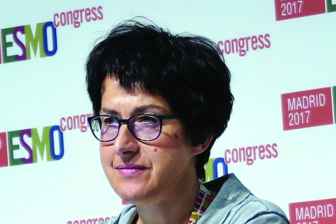
Median overall survival (OS) among 425 patients with intermediate- or poor-risk treatment-naive advanced/metastatic clear-cell RCC treated with the combination of nivolumab (Opdivo) and ipilimumab (Yervoy) was not reached after 32 months of follow-up. In contrast, the median OS for 422 patients treated with sunitinib was 26 months, reported Bernard Escudier, MD from the Institut Gustave Roussy in Villejuif, France.

Similarly tipping the balance toward the combination, the ORR was 42%, compared with 27% in the sunitinib group (P less than .0001).
“These results support the use of nivo/ipi [nivolumab/ipilimumab] as a new first-line standard of care option for patients with advanced renal cell carcinoma,” Dr. Escudier said at a briefing prior to presenting the data in a presidential symposium.
Patients with treatment-naive advanced or metastatic clear-cell RCC with measurable disease, a Karnofsky Performance Score of at least 70%, and tumor tissue available for programmed death ligand 1 (PD-L1) typing were enrolled in Checkmate 214, .
The patients were stratified by International Metastatic Renal Cell Carcinoma Database Consortium prognostic score and by region (U.S. versus Canada/Europe versus the rest of the world) and then randomly assigned to receive either 3 mg/kg nivolumab and 1 mg/kg ipilimumab every 3 weeks for four doses then 3 mg/kg nivolumab every other week or to receive 50 mg oral sunitinib once daily for 4 weeks in a 6-week cycle. Patients remained on treatment until progression or unacceptable toxicity.
The results for the coprimary endpoints are noted above.
Duration of response trended toward superior with the checkpoint inhibitor duo. At 2-year follow-up, the median duration of response was not reached with nivo/ipi, vs. 18.2 months with sunitinib. In all, 72% of patients on the combination had an ongoing response at 2 years, compared with 63% of patients on the TKI, but the upper level of the confidence interval in both trial arms had not been reached at the time of the data cutoff, so statistical significance of the difference in duration cannot be determined.
For the secondary endpoints of overall survival in the intention-to-treat population, which included 550 patients assigned to nivo/ipi and 546 to sunitinib, the ORR was 39% for patients assigned to the checkpoint inhibitors, compared with 32% for sunitinib (P = .0191). The median respective PFS numbers, however, were virtually identical at 12.4 vs. 12.3 months.
The median OS in the intention-to-treat population was not reached with the combination, versus 32.9 months with the TKI (hazard ratio, 0.68; P = .0003).
In the intermediate- or poor-risk population, PFS was significantly better with nivo/ipi among patients with PD-L1 expression in 1% or more of cells but not in patients with lower levels of PD-L1 expression.
There were more adverse events leading to discontinuation among patients on the dual checkpoint inhibitors at 22% vs. 12% with sunitinib. The most common grade 3 or greater adverse events in the combination group were fatigue and diarrhea in 4% each and rash and nausea in 2% each, while incidences of pruritus, hypothyroidism, vomiting, and hypertension occurred in fewer than 1% of patients.
In the sunitinib group, the most common grade 3 or greater events were hypertension in 16%, fatigue in 9%, palmar-plantar erythrodysesthesia syndrome in 9%, stomatitis in 3%, mucosal inflammation in 3%, and vomiting in 2%. Nausea, decreased appetite, hypothyroidism, and dysgeusia occurred in 1% or fewer of patients in this arm.
“The combination, I think, is really beneficial, because with immunotherapy we have seen that patients who respond usually have long-term benefit, and in this case high-response rate seems to be important and translates into a long-term for patients,” commented Maria de Santis, MD, from the University of Warwick, U.K., who was an invited discussant at the briefing.
“This data is clearly important and practice changing, and it challenges the former standard of care with TKI monotherapy treatment,” she added.
The study was sponsored by Bristol-Myers Squibb and Ono Pharmaceutical; Dr. Escudier disclosed honoraria from BMS. Dr. de Santis did not disclose potential conflicts of interest.
MADRID – A combination of two immune checkpoint inhibitors was superior to the tyrosine kinase inhibitor (TKI) sunitinib (Sutent) in first-line treatment of patients with advanced or metastatic renal cell carcinoma (RCC), investigators reported
Median overall survival (OS) among 425 patients with intermediate- or poor-risk treatment-naive advanced/metastatic clear-cell RCC treated with the combination of nivolumab (Opdivo) and ipilimumab (Yervoy) was not reached after 32 months of follow-up. In contrast, the median OS for 422 patients treated with sunitinib was 26 months, reported Bernard Escudier, MD from the Institut Gustave Roussy in Villejuif, France.
Similarly tipping the balance toward the combination, the ORR was 42%, compared with 27% in the sunitinib group (P less than .0001).
“These results support the use of nivo/ipi [nivolumab/ipilimumab] as a new first-line standard of care option for patients with advanced renal cell carcinoma,” Dr. Escudier said at a briefing prior to presenting the data in a presidential symposium.
Patients with treatment-naive advanced or metastatic clear-cell RCC with measurable disease, a Karnofsky Performance Score of at least 70%, and tumor tissue available for programmed death ligand 1 (PD-L1) typing were enrolled in Checkmate 214, .
The patients were stratified by International Metastatic Renal Cell Carcinoma Database Consortium prognostic score and by region (U.S. versus Canada/Europe versus the rest of the world) and then randomly assigned to receive either 3 mg/kg nivolumab and 1 mg/kg ipilimumab every 3 weeks for four doses then 3 mg/kg nivolumab every other week or to receive 50 mg oral sunitinib once daily for 4 weeks in a 6-week cycle. Patients remained on treatment until progression or unacceptable toxicity.
The results for the coprimary endpoints are noted above.
Duration of response trended toward superior with the checkpoint inhibitor duo. At 2-year follow-up, the median duration of response was not reached with nivo/ipi, vs. 18.2 months with sunitinib. In all, 72% of patients on the combination had an ongoing response at 2 years, compared with 63% of patients on the TKI, but the upper level of the confidence interval in both trial arms had not been reached at the time of the data cutoff, so statistical significance of the difference in duration cannot be determined.
For the secondary endpoints of overall survival in the intention-to-treat population, which included 550 patients assigned to nivo/ipi and 546 to sunitinib, the ORR was 39% for patients assigned to the checkpoint inhibitors, compared with 32% for sunitinib (P = .0191). The median respective PFS numbers, however, were virtually identical at 12.4 vs. 12.3 months.
The median OS in the intention-to-treat population was not reached with the combination, versus 32.9 months with the TKI (hazard ratio, 0.68; P = .0003).
In the intermediate- or poor-risk population, PFS was significantly better with nivo/ipi among patients with PD-L1 expression in 1% or more of cells but not in patients with lower levels of PD-L1 expression.
There were more adverse events leading to discontinuation among patients on the dual checkpoint inhibitors at 22% vs. 12% with sunitinib. The most common grade 3 or greater adverse events in the combination group were fatigue and diarrhea in 4% each and rash and nausea in 2% each, while incidences of pruritus, hypothyroidism, vomiting, and hypertension occurred in fewer than 1% of patients.
In the sunitinib group, the most common grade 3 or greater events were hypertension in 16%, fatigue in 9%, palmar-plantar erythrodysesthesia syndrome in 9%, stomatitis in 3%, mucosal inflammation in 3%, and vomiting in 2%. Nausea, decreased appetite, hypothyroidism, and dysgeusia occurred in 1% or fewer of patients in this arm.
“The combination, I think, is really beneficial, because with immunotherapy we have seen that patients who respond usually have long-term benefit, and in this case high-response rate seems to be important and translates into a long-term for patients,” commented Maria de Santis, MD, from the University of Warwick, U.K., who was an invited discussant at the briefing.
“This data is clearly important and practice changing, and it challenges the former standard of care with TKI monotherapy treatment,” she added.
The study was sponsored by Bristol-Myers Squibb and Ono Pharmaceutical; Dr. Escudier disclosed honoraria from BMS. Dr. de Santis did not disclose potential conflicts of interest.
AT ESMO 2017
Key clinical point: The combination of the PD-1 inhibitor nivolumab and CTLA-4 inhibitor ipilimumab was efficacious in frontline therapyfor advanced or metastatic renal cell carcinoma.
Major finding: The trial met its coprimary endpoints of overall response rate and progression-free survival, as well as its secondary endpoint of overall survival.
Data source: Randomized open-label study in 1096 patients with advanced/metastatic RCC, including 847 with intermediate- to poor-risk disease.
Disclosures: The study was sponsored by Bristol-Myers Squibb and Ono Pharmaceutical; Dr. Escudier disclosed honoraria from BMS. Dr. de Santis did not disclose potential conflicts of interest.
How to lower blood pressure in real-world primary care clinics
SAN FRANCISCO – In just 6 months, a commonsense quality improvement program increased the rate of blood pressure control from 66% to 75% among 21,035 hypertensive patients at 16 primary care clinics in northwestern South Carolina.
The American Medical Association collaborated with the Care Coordination Institute in Greenville, S.C., to develop the program, dubbed “MAP,” which stands for measuring blood pressure accurately; acting rapidly to manage uncontrolled blood pressure; and partnering with patients to promote blood pressure self-management.
With the success in South Carolina, it’s likely the program will be rolled out to other parts of the country, Dr. Egan said at the joint scientific sessions of AHA Council on Hypertension, AHA Council on Kidney in Cardiovascular Disease, and American Society of Hypertension
Twelve of the 16 practices had significant increases in BP control. In patients uncontrolled at baseline, mean BP fell from 149/85 mm Hg to 139/80 mm Hg. In controlled patients, BP fell about 10/5 mm Hg. The findings were statistically significant.
To reduce white coat hypertension and improve BP accuracy, patients who had an initial, attended BP at or above 140/90 mm Hg were put in a room by themselves for 5 minutes for three automated BP measurements, which were then averaged. Staff made sure patients were positioned properly and had the correctly sized cuff on their arm, among other measures. A program facilitator was onsite to help.
Patients who had BPs at or above 140/90 mm Hg when they were left alone were switched to drugs that have been proven to help. For white patients younger than 55 years, that meant a renin-angiotensin system blocker first and a calcium channel blocker second. For older African American patients, the calcium channel blocker came first. A diuretic was added if needed as a third step, and then spironolactone as a fourth. “That was basically the regimen in the absence of compelling indications for other agents,” Dr. Egan said.
Single-pill combinations were encouraged to help with compliance. The investigators also worked with nearby pharmacies to use generic drugs to keep costs low, and to ensure that patients could pick up all their prescriptions at one visit. Many of the patients had multiple chronic conditions and were on a half dozen or more drugs. Picking them all up at one pharmacy visit has been shown to improve adherence.
There was follow-up every 2 weeks for patients with stage 2 hypertension, at least by phone. Stage 1 patients had monthly follow-ups. Lifestyle changes were encouraged, as well as home BP monitoring. The clinics were given a few home monitors to send home with patients, and patients reported their results. If BP was elevated at home, patients were contacted and advised before their next visit.
They were also given a cookbook that matched the DASH diet with the southern palate. “We tried to make it taste good and not cost more,” Dr. Egan said. “It’s been quite popular for our patients who are willing to give it a try. It’s free on our website.”
With the intervention, “not only did [patients] take their new medications, but they also were probably taking previous medications more reliably,” he said.
It’s likely that the program reduced hypertension comorbidities, Dr. Egan noted, but there was no cost-benefit analysis.
There was no pharmaceutical industry funding. Dr. Egan disclosed research support from Boehringer.
AT JOINT HYPERTENSION 2017
FDA approves abemaciclib for advanced breast cancer
The Food and Drug Administration has approved abemaciclib (Verzenio) to be given in combination with fulvestrant, to treat patients who have hormone receptor (HR)–positive, human epidermal growth factor receptor 2 (HER2)–negative advanced or metastatic breast cancer that has progressed after taking endocrine therapy.

This is the third cyclin-dependent kinase 4/6 (CDK4/6) inhibitor approved for the treatment of advanced breast cancer. Palbociclib (Ibrance) was granted accelerated approval in February 2015, in combination with letrozole, for the treatment of HR-positive, HER2-negative advanced breast cancer as initial endocrine-based therapy in postmenopausal women. Ribociclib (Kisqali) was approved in March 2017, in combination with any aromatase inhibitor, also for the treatment of HR-positive, HER2-negative advanced breast cancer as initial endocrine-based therapy in postmenopausal women.
Approval of abemaciclib in combination with fulvestrant was based on a median progression-free survival of 16.4 months for patients taking abemaciclib with fulvestrant, compared with 9.3 months for patients taking a placebo with fulvestrant, in a randomized trial. All 669 patients in the trial had HR-positive, HER2-negative breast cancer that had progressed after treatment with endocrine therapy and had not received chemotherapy once the cancer had metastasized.
Approval of abemaciclib as a single agent was based on an overall response rate of 19.7% in a single-arm trial of 132 patients with HR-positive, HER2-negative breast cancer that had progressed after treatment with endocrine therapy and chemotherapy after the cancer metastasized.
Common side effects of abemaciclib include diarrhea, neutropenia, leukopenia, nausea, abdominal pain, infections, fatigue, anemia, decreased appetite, vomiting, and headache.
Serious side effects include diarrhea, neutropenia, elevated liver blood tests, and deep venous thrombosis/pulmonary embolism, the FDA said.
Approval was granted to Eli Lilly.
The Food and Drug Administration has approved abemaciclib (Verzenio) to be given in combination with fulvestrant, to treat patients who have hormone receptor (HR)–positive, human epidermal growth factor receptor 2 (HER2)–negative advanced or metastatic breast cancer that has progressed after taking endocrine therapy.
This is the third cyclin-dependent kinase 4/6 (CDK4/6) inhibitor approved for the treatment of advanced breast cancer. Palbociclib (Ibrance) was granted accelerated approval in February 2015, in combination with letrozole, for the treatment of HR-positive, HER2-negative advanced breast cancer as initial endocrine-based therapy in postmenopausal women. Ribociclib (Kisqali) was approved in March 2017, in combination with any aromatase inhibitor, also for the treatment of HR-positive, HER2-negative advanced breast cancer as initial endocrine-based therapy in postmenopausal women.
Approval of abemaciclib in combination with fulvestrant was based on a median progression-free survival of 16.4 months for patients taking abemaciclib with fulvestrant, compared with 9.3 months for patients taking a placebo with fulvestrant, in a randomized trial. All 669 patients in the trial had HR-positive, HER2-negative breast cancer that had progressed after treatment with endocrine therapy and had not received chemotherapy once the cancer had metastasized.
Approval of abemaciclib as a single agent was based on an overall response rate of 19.7% in a single-arm trial of 132 patients with HR-positive, HER2-negative breast cancer that had progressed after treatment with endocrine therapy and chemotherapy after the cancer metastasized.
Common side effects of abemaciclib include diarrhea, neutropenia, leukopenia, nausea, abdominal pain, infections, fatigue, anemia, decreased appetite, vomiting, and headache.
Serious side effects include diarrhea, neutropenia, elevated liver blood tests, and deep venous thrombosis/pulmonary embolism, the FDA said.
Approval was granted to Eli Lilly.
Is the patient-centered medical home on life support?
The way Edward J. Bujold, MD, sees it, the patient-centered medical home (PCMH) as a model of care has become unsustainable in the current health care landscape.
In an opinion piece published online in JAMA Internal Medicine, Dr. Bujold describes how he reinvested Medicare incentive payments to build a solo family medicine practice into a patient-centered medical home with a staff of 14 full-time and part-time employees, including an embedded psychologist, pharmacist, physical therapist, and dietitian.
“Although only about one-quarter of the patients cared for at our practice are Medicare beneficiaries, other insurers and those they insure have benefited immensely,” wrote Dr. Bujold, owner of Granite Falls (N.C.) Family Medical Care Center (JAMA Internal Med. 2017 Sept 25. doi: 10.1001/jamainternmed.2017.4651).
The bonuses from the Center for Medicare & Medicaid Services’ Meaningful Use and Physician Quality Reporting System “enabled us to develop and support the financial structure required to create the PCMH,” he noted. “For the most part, this financial support ended in December 2015. Our financial losses in 2016 are enough to jeopardize the foundation of our PCMH.”
When Congress voted to end the Medicare Sustainable Growth Rate formula, the Primary Care Incentive Payment Program authorized under the Affordable Care Act also ended.
“During 2017, a value-based payment system to replace and hopefully increase our reimbursement is to be phased in,” Dr. Bujold wrote. “The problem for practices like ours is the Medicare Access and Children’s Health Insurance Program Reauthorization Act of 2015/Merit-Based Incentive Payment System (MACRA/MIPS) does not take effect until 2019, and there is no guarantee we will receive any more money than we receive now. We may receive less.”
Citing studies from Oregon and from the Patient-Centered Primary Care Collaborative, Dr. Bujold said that for every dollar invested in primary care, the overarching health system saves about $13.
“The 80% reduction in hospital admissions for our practice is a testimony to these savings,” he stated. “The irony for me is that, 5 years ago, I generated a substantial amount of my income from the hospital portion of my practice, because I was seeing an average of five patients per day in the hospital.”
As a result of transitioning his practice to a PCMH, however, he now sees “one or two and sometimes no hospital patients per day.” At the same time, his office’s overhead cost is 65% and increasing – up from 50% when he opened his practice 31 years ago.
Dr. Bujold noted that about 60% of primary care physicians are employed by hospital organizations, which are able to charge a facility fee “above and beyond what the independent primary care physician charges in his or her office. This increases the cost of care in general by 20%-30% and the patient out-of-pocket costs by a factor of 2 or 3.”
In his view, “we have large hospital systems, the pharmaceutical industry, large insurance companies, and other moneyed players and their lobbyists with vested financial interests. The view from my practice is that these industries seem to hold all of the cards, and it is very difficult to move forward. I am more pessimistic than I was 5 years ago.”
Despite his current misgivings, Dr. Bujold ended his opinion piece by stating that the best way to “heal the health care system in the United States” is one directed by primary care and rooted in high-functioning PCMHs.
“The trajectory must be changed,” Dr. Bujold wrote. “In many areas, payers are not supporting PCMH transformation, and states are not engaging practices or working with Medicare to support transformation. Investing in high-functioning PCMH practices is the right thing to do.”
Dr. Bujold disclosed that he is employed by KPN Health as chief physician strategist and is an advisory board member of the Patient-Centered Primary Care Collaborative.
Financial support for primary care varies around the country and can also vary by payer within an individual region. Dr. Bujold describes very limited commercial-payer support for the care his practice delivers. In contrast, Rhode Island required that all payers increase the percent of total medical dollars for fully insured members paid to primary care by 1% annually from 2010 to 2014 and then sustain that increased percentage. This requirement resulted in substantial support for sustained practice changes in care delivery.
Starting in 2011, the Centers for Medicare & Medicaid Services (CMS) began programs to test new changes in primary care delivery and payment. Comprehensive Primary Care Plus, the largest such Medicare model to date, is being tested in 14 regions of the country, as well as 4 more regions starting in 2018, and is evaluated independently by region; the 5-year program is designed to strengthen primary care through multipayer-payment reform and care-delivery transformation.
The model includes two primary care practice tracks with incrementally advanced care-delivery requirements and payment options. Practices in both tracks work to improve patient access and continuity, provide care management to the patients at highest risk, engage patients and caregivers, focus on planned care and population health, and increase the comprehensiveness and coordination of care.
Primary care – and the patient-centered medical home – can be saved, but there is considerable work to be done. In our view, the delivery of comprehensive primary care requires population-based payments aligned across payers to ensure adequate support for care-delivery changes for all the patients in the practice. Policies and payment across the health care system should be aligned to support a robust primary care infrastructure that delivers on its promise of higher health care quality at lower costs.
Patients should experience the benefits of comprehensive primary care. They should expect their insurer to pay for it and their primary care practice to deliver it.
The way Edward J. Bujold, MD, sees it, the patient-centered medical home (PCMH) as a model of care has become unsustainable in the current health care landscape.
In an opinion piece published online in JAMA Internal Medicine, Dr. Bujold describes how he reinvested Medicare incentive payments to build a solo family medicine practice into a patient-centered medical home with a staff of 14 full-time and part-time employees, including an embedded psychologist, pharmacist, physical therapist, and dietitian.
“Although only about one-quarter of the patients cared for at our practice are Medicare beneficiaries, other insurers and those they insure have benefited immensely,” wrote Dr. Bujold, owner of Granite Falls (N.C.) Family Medical Care Center (JAMA Internal Med. 2017 Sept 25. doi: 10.1001/jamainternmed.2017.4651).
The bonuses from the Center for Medicare & Medicaid Services’ Meaningful Use and Physician Quality Reporting System “enabled us to develop and support the financial structure required to create the PCMH,” he noted. “For the most part, this financial support ended in December 2015. Our financial losses in 2016 are enough to jeopardize the foundation of our PCMH.”
When Congress voted to end the Medicare Sustainable Growth Rate formula, the Primary Care Incentive Payment Program authorized under the Affordable Care Act also ended.
“During 2017, a value-based payment system to replace and hopefully increase our reimbursement is to be phased in,” Dr. Bujold wrote. “The problem for practices like ours is the Medicare Access and Children’s Health Insurance Program Reauthorization Act of 2015/Merit-Based Incentive Payment System (MACRA/MIPS) does not take effect until 2019, and there is no guarantee we will receive any more money than we receive now. We may receive less.”
Citing studies from Oregon and from the Patient-Centered Primary Care Collaborative, Dr. Bujold said that for every dollar invested in primary care, the overarching health system saves about $13.
“The 80% reduction in hospital admissions for our practice is a testimony to these savings,” he stated. “The irony for me is that, 5 years ago, I generated a substantial amount of my income from the hospital portion of my practice, because I was seeing an average of five patients per day in the hospital.”
As a result of transitioning his practice to a PCMH, however, he now sees “one or two and sometimes no hospital patients per day.” At the same time, his office’s overhead cost is 65% and increasing – up from 50% when he opened his practice 31 years ago.
Dr. Bujold noted that about 60% of primary care physicians are employed by hospital organizations, which are able to charge a facility fee “above and beyond what the independent primary care physician charges in his or her office. This increases the cost of care in general by 20%-30% and the patient out-of-pocket costs by a factor of 2 or 3.”
In his view, “we have large hospital systems, the pharmaceutical industry, large insurance companies, and other moneyed players and their lobbyists with vested financial interests. The view from my practice is that these industries seem to hold all of the cards, and it is very difficult to move forward. I am more pessimistic than I was 5 years ago.”
Despite his current misgivings, Dr. Bujold ended his opinion piece by stating that the best way to “heal the health care system in the United States” is one directed by primary care and rooted in high-functioning PCMHs.
“The trajectory must be changed,” Dr. Bujold wrote. “In many areas, payers are not supporting PCMH transformation, and states are not engaging practices or working with Medicare to support transformation. Investing in high-functioning PCMH practices is the right thing to do.”
Dr. Bujold disclosed that he is employed by KPN Health as chief physician strategist and is an advisory board member of the Patient-Centered Primary Care Collaborative.
FROM JAMA INTERNAL MEDICINE