User login
Treatment-resistant depression boosts early mortality
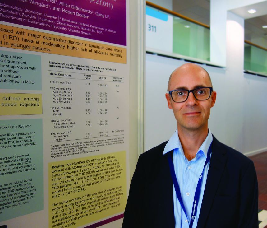
PARIS – Young adults with treatment-resistant depression have more than double the risk of all-cause mortality, compared with their peers with major depressive disorder that’s not treatment resistant, Johan Reutfors, MD, PhD, reported at the annual congress of the European College of Neuropsychopharmacology.
Across the full age spectrum of adults with treatment-resistant depression, however, the magnitude of the increased risk associated with treatment-resistant depression is less extreme than in the 18- to 29-year-olds. Yet, the moderate overall 11% increased risk of all-cause mortality in adults with treatment-resistant depression, compared with those with non–treatment-resistant major depressive disorder remains both statistically significant and clinically meaningful, observed Dr. Reutfors, a psychiatrist at the Karolinska Institute in Stockholm.
During a mean 4.1 years of follow-up, 4,662 patients died. The all-cause mortality adjusted for age, gender, and a history of substance use disorders or self-harm was 2.17-fold greater in 18- to 29-year-olds who had treatment-resistant depression, compared with those with nonresistant major depressive disorder, 1.51-fold greater in 30- to 49-year-olds with treatment-resistant depression, and 1.18-fold greater in 50- to 69-year-olds with treatment-resistant depression.
In contrast, patients aged 70 or older with treatment-resistant depression had a significant 17% lower risk of all-cause mortality than those with non-TRD major depression. In an interview, Dr. Reutfors said this apparent protective effect was probably tied to survival bias.
“If you have lived so long that perhaps during your lifetime you’ve already had many depressive episodes, maybe only the healthier ones have survived,” he explained.
The all-cause mortality in patients with treatment-resistant depression and a history of self-harm was 37% greater than in patients without treatment-resistant depression.
The causes of excess mortality in the treatment-resistant depression group were quite different in the younger and older patients. In younger patients, the increased mortality was attributed mainly to suicides and accidents. In the older group, where the degree of excess risk was more modest, a variety of fatal illnesses figured more prominently.
The study was supported by Janssen. Dr. Reutfors reported having served as a paid speaker for Eli Lilly and receiving unrestricted grant support from Schering-Plough.
AT THE ECNP CONGRESS
Key clinical point:
Major finding: Young adult Swedes with treatment-resistant major depressive disorder have an all-cause mortality nearly 2.2-fold higher than their peers with non–treatment-resistant major depression.
Data source: This retrospective cohort study used Swedish national databases to look at all-cause mortality in more than 127,000 adults under psychiatric care for major depressive disorder.
Disclosures: The study was supported by Janssen. The presenter reported having served as a paid speaker for Eli Lilly and receiving unrestricted grant support from Schering-Plough.
Delayed HIV diagnoses still substantial for some at-risk groups
HIV diagnoses are coming sooner after infection, increasing physicians’ ability to treat and prevent the spread of HIV, according to a new Centers for Disease Control and Prevention (CDC) Vital Signs report.
As HIV testing has increased, the percentage of people aware of their HIV infection has also steadily grown. As of 2014, 85% of people living with HIV in the United States were aware of their infection. This knowledge allows individuals to seek antiretroviral treatment to suppress the virus, which decreases morbidity and mortality while reducing the risk of sexual transmission to others; knowing one’s HIV status, then, is incredibly important, clinicians say, because people who are unaware that they are HIV positive account for approximately 40% of ongoing transmissions.

While HIV testing has led to a reduction of HIV infection in the total population, several groups that are at a high risk of HIV infection are not getting tested as often as they should. According to the report, 29% of men who have sex with men, 42% of intravenous drugs users, and 59% of heterosexuals did not report having been tested within the past 12 months. Of the risk groups mentioned, at least two-thirds of people in each group had seen a health care provider in the last year. Heterosexual men are at particular risk of going undiagnosed because they are less likely to see a health care provider. This has led to half of heterosexual men who have HIV going undiagnosed for 5 years or more, the report notes.
Health care providers can improve testing by discussing HIV with patients and explaining that HIV testing is a routine part of any patient’s health care. Physicians should routinely test all patients aged 13-64 years, the CDC says. Testing should be emphasized in patients in high-risk groups; these patients should be tested at least once a year. Sexually active gay and bisexual men should ideally be tested every 3-6 months. Pregnant women, or those looking to become pregnant, should be tested to as soon as possible. If a pregnant woman is in a high-risk population, she should be tested again in the third trimester.
If a patient tests positive for HIV, the CDC report says it is important for clinicians connect them with treatment options and discuss prevention of transmission. The earlier a person begins HIV treatment, the greater the benefits will be. As part of the treatment, clinicians should encourage patients to stay on antiretroviral care to reduce the viral load in the body to either very low (less than 200 copies/mL) or undetectable levels.
“HIV is being diagnosed more quickly, the number of people who have the virus under control is up, and annual infections are down. So while we celebrate our progress, we pledge to work together to end this epidemic forever,” said CDC Director Brenda Fitzgerald, MD, in a statement.
HIV diagnoses are coming sooner after infection, increasing physicians’ ability to treat and prevent the spread of HIV, according to a new Centers for Disease Control and Prevention (CDC) Vital Signs report.
As HIV testing has increased, the percentage of people aware of their HIV infection has also steadily grown. As of 2014, 85% of people living with HIV in the United States were aware of their infection. This knowledge allows individuals to seek antiretroviral treatment to suppress the virus, which decreases morbidity and mortality while reducing the risk of sexual transmission to others; knowing one’s HIV status, then, is incredibly important, clinicians say, because people who are unaware that they are HIV positive account for approximately 40% of ongoing transmissions.
While HIV testing has led to a reduction of HIV infection in the total population, several groups that are at a high risk of HIV infection are not getting tested as often as they should. According to the report, 29% of men who have sex with men, 42% of intravenous drugs users, and 59% of heterosexuals did not report having been tested within the past 12 months. Of the risk groups mentioned, at least two-thirds of people in each group had seen a health care provider in the last year. Heterosexual men are at particular risk of going undiagnosed because they are less likely to see a health care provider. This has led to half of heterosexual men who have HIV going undiagnosed for 5 years or more, the report notes.
Health care providers can improve testing by discussing HIV with patients and explaining that HIV testing is a routine part of any patient’s health care. Physicians should routinely test all patients aged 13-64 years, the CDC says. Testing should be emphasized in patients in high-risk groups; these patients should be tested at least once a year. Sexually active gay and bisexual men should ideally be tested every 3-6 months. Pregnant women, or those looking to become pregnant, should be tested to as soon as possible. If a pregnant woman is in a high-risk population, she should be tested again in the third trimester.
If a patient tests positive for HIV, the CDC report says it is important for clinicians connect them with treatment options and discuss prevention of transmission. The earlier a person begins HIV treatment, the greater the benefits will be. As part of the treatment, clinicians should encourage patients to stay on antiretroviral care to reduce the viral load in the body to either very low (less than 200 copies/mL) or undetectable levels.
“HIV is being diagnosed more quickly, the number of people who have the virus under control is up, and annual infections are down. So while we celebrate our progress, we pledge to work together to end this epidemic forever,” said CDC Director Brenda Fitzgerald, MD, in a statement.
FROM CDC VITAL SIGNS REPORT
Key clinical point:
Major finding: Approximately 15% of those living with HIV in 2015 were unaware of their infection and had a median diagnosis delay of 3 years.
Data source: Data of 39,720 individuals reported to the CDC’s National HIV Surveillance System from 50 states and the District of Columbia in 2015.
Disclosures: No conflicts of interest were reported.
Fingolimod Reduces Annualized Relapse Rate in Pediatric MS
PARIS—Among children and adolescents with multiple sclerosis (MS), patients treated with fingolimod have a lower annualized relapse rate, compared with patients treated with interferon beta-1a, according to phase III trial results presented at the Seventh Joint ECTRIMS–ACTRIMS Meeting. In addition, fingolimod results in consistently favorable MRI outcomes and may delay disability progression, compared with interferon beta-1a. Fingolimod’s safety profile in children is generally consistent with that in adults.

Between 3% and 5% of patients with MS have disease onset before age 18, but no disease-modifying therapies are FDA-approved specifically for children and adolescents with the disease. “The current treatment recommendations are based on open-label or retrospective studies,” said Tanuja Chitnis, MD, Director of the Partners Pediatric MS Center at Massachusetts General Hospital and Scientist at the Ann Romney Center for Neurological Diseases at Brigham and Women’s Hospital, in Boston.
To investigate the safety and efficacy of fingolimod versus interferon beta-1a in children and adolescents with MS, Dr. Chitnis and colleagues conducted the PARADIGMS study. “PARADIGMS is the first successfully completed global, randomized, clinical trial in pediatric MS,” said Dr. Chitnis, the principal investigator for PARADIGMS.
The PARADIGMS Study
The two-year, double-blind, double-dummy, active-controlled, parallel-group, multicenter study included children and adolescents with a confirmed diagnosis of MS. The study enrolled 215 children between the ages of 10 and 17 with an Expanded Disability Status Scale score between 0 and 5.5. The study included patients who had experienced at least one relapse in the past year or two relapses in the previous two years, or who had evidence of one or more gadolinium-enhancing lesions on MRI within six months prior to randomization.
Patients’ mean age at randomization was about 15. About two-thirds of patients were female, about two-thirds were treatment naïve, and patients were predominantly Caucasian or white. Patients had an average of 1.5 relapses in the prior 12 months and about 2.5 relapses in the prior two years.
Patients were randomized 1:1 to receive once-daily oral fingolimod (0.5 mg or 0.25 mg, dependent on patients’ body weight) or interferon beta-1a 30 μg intramuscular once weekly.
The primary end point was the frequency of relapses in patients treated up to 24 months (ie, annualized relapse rate). Secondary end points included the number of new or newly enlarged T2 lesions, gadolinium-enhancing T1 lesions, safety, and the pharmacokinetic properties of fingolimod.
Researchers conducted the study at 87 sites in more than 25 countries. Investigators designed the study with the FDA, the European Medicines Agency, and the International Pediatric MS Study Group. Basel, Switzerland-based Novartis, which markets fingolimod as Gilenya, supported the study.
Freedom From Relapse
Oral fingolimod reduced the relapse rate by 82% over a period of two years, compared with interferon beta-1a intramuscular injections, Dr. Chitnis said.
About 85% of fingolimod-treated patients were free of confirmed relapse at month 24, versus 39% of patients in the interferon beta-1a arm.
In addition, fingolimod-treated patients had a significant reduction in the number of new or newly enlarging T2 lesions and gadolinium-enhancing T1 lesions, and significantly less brain volume loss, compared with patients who received interferon beta-1a.
In a post hoc analysis, fingolimod significantly delayed confirmed disability progression, compared with interferon beta-1a.
Adverse Events
“The safety profile of fingolimod is generally consistent with that seen in the adult clinical trials,” Dr. Chitnis said. Adverse events occurred more often in the interferon beta-1a arm, whereas serious adverse events occurred more often in the fingolimod arm. The serious adverse events observed in the fingolimod arm included leukopenia in two patients, granulomatosis in one patient, hypersensitivity reaction in one patient, and four patients with seizure events, Dr. Chitnis said.
In all, 92% of fingolimod-treated patients completed the study, compared with 75% of patients in the interferon beta-1a arm. All patients had the option of transferring to an open-label fingolimod arm in an extension phase, and 171 patients entered this extension study.
—Jake Remaly
PARIS—Among children and adolescents with multiple sclerosis (MS), patients treated with fingolimod have a lower annualized relapse rate, compared with patients treated with interferon beta-1a, according to phase III trial results presented at the Seventh Joint ECTRIMS–ACTRIMS Meeting. In addition, fingolimod results in consistently favorable MRI outcomes and may delay disability progression, compared with interferon beta-1a. Fingolimod’s safety profile in children is generally consistent with that in adults.
Between 3% and 5% of patients with MS have disease onset before age 18, but no disease-modifying therapies are FDA-approved specifically for children and adolescents with the disease. “The current treatment recommendations are based on open-label or retrospective studies,” said Tanuja Chitnis, MD, Director of the Partners Pediatric MS Center at Massachusetts General Hospital and Scientist at the Ann Romney Center for Neurological Diseases at Brigham and Women’s Hospital, in Boston.
To investigate the safety and efficacy of fingolimod versus interferon beta-1a in children and adolescents with MS, Dr. Chitnis and colleagues conducted the PARADIGMS study. “PARADIGMS is the first successfully completed global, randomized, clinical trial in pediatric MS,” said Dr. Chitnis, the principal investigator for PARADIGMS.
The PARADIGMS Study
The two-year, double-blind, double-dummy, active-controlled, parallel-group, multicenter study included children and adolescents with a confirmed diagnosis of MS. The study enrolled 215 children between the ages of 10 and 17 with an Expanded Disability Status Scale score between 0 and 5.5. The study included patients who had experienced at least one relapse in the past year or two relapses in the previous two years, or who had evidence of one or more gadolinium-enhancing lesions on MRI within six months prior to randomization.
Patients’ mean age at randomization was about 15. About two-thirds of patients were female, about two-thirds were treatment naïve, and patients were predominantly Caucasian or white. Patients had an average of 1.5 relapses in the prior 12 months and about 2.5 relapses in the prior two years.
Patients were randomized 1:1 to receive once-daily oral fingolimod (0.5 mg or 0.25 mg, dependent on patients’ body weight) or interferon beta-1a 30 μg intramuscular once weekly.
The primary end point was the frequency of relapses in patients treated up to 24 months (ie, annualized relapse rate). Secondary end points included the number of new or newly enlarged T2 lesions, gadolinium-enhancing T1 lesions, safety, and the pharmacokinetic properties of fingolimod.
Researchers conducted the study at 87 sites in more than 25 countries. Investigators designed the study with the FDA, the European Medicines Agency, and the International Pediatric MS Study Group. Basel, Switzerland-based Novartis, which markets fingolimod as Gilenya, supported the study.
Freedom From Relapse
Oral fingolimod reduced the relapse rate by 82% over a period of two years, compared with interferon beta-1a intramuscular injections, Dr. Chitnis said.
About 85% of fingolimod-treated patients were free of confirmed relapse at month 24, versus 39% of patients in the interferon beta-1a arm.
In addition, fingolimod-treated patients had a significant reduction in the number of new or newly enlarging T2 lesions and gadolinium-enhancing T1 lesions, and significantly less brain volume loss, compared with patients who received interferon beta-1a.
In a post hoc analysis, fingolimod significantly delayed confirmed disability progression, compared with interferon beta-1a.
Adverse Events
“The safety profile of fingolimod is generally consistent with that seen in the adult clinical trials,” Dr. Chitnis said. Adverse events occurred more often in the interferon beta-1a arm, whereas serious adverse events occurred more often in the fingolimod arm. The serious adverse events observed in the fingolimod arm included leukopenia in two patients, granulomatosis in one patient, hypersensitivity reaction in one patient, and four patients with seizure events, Dr. Chitnis said.
In all, 92% of fingolimod-treated patients completed the study, compared with 75% of patients in the interferon beta-1a arm. All patients had the option of transferring to an open-label fingolimod arm in an extension phase, and 171 patients entered this extension study.
—Jake Remaly
Researchers identify potential gene target for AML drug development
Researchers in the United States and the United Kingdom believe they have found a new gene target that could aid in the development of more effective treatments for acute myeloid leukemia (AML).
Inhibition of the METTL3 gene allowed for the destruction of AML in human and mouse cells without damaging healthy blood cells, the researchers reported in a research letter published Nov. 27 in the journal Nature.

Using mouse cells, the researchers used CRISPR-Cas9 gene editing technology to identify RNA-modifying enzymes that are needed for the survival and proliferation of AML cells. They identified 46 potential candidate genes and further narrowed that to the METTL gene families. They next targeted METTL1, METTL3, METTL14, and METTL16 in 10 human AML cell lines and 10 cell lines from heterogeneous cancer types. METTL3 was shown to have the strongest effect.
Read the full research letter in Nature (doi: 10.1038/nature24678).
mschneider@frontlinemedcom.com
On Twitter @maryellenny
Researchers in the United States and the United Kingdom believe they have found a new gene target that could aid in the development of more effective treatments for acute myeloid leukemia (AML).
Inhibition of the METTL3 gene allowed for the destruction of AML in human and mouse cells without damaging healthy blood cells, the researchers reported in a research letter published Nov. 27 in the journal Nature.
Using mouse cells, the researchers used CRISPR-Cas9 gene editing technology to identify RNA-modifying enzymes that are needed for the survival and proliferation of AML cells. They identified 46 potential candidate genes and further narrowed that to the METTL gene families. They next targeted METTL1, METTL3, METTL14, and METTL16 in 10 human AML cell lines and 10 cell lines from heterogeneous cancer types. METTL3 was shown to have the strongest effect.
Read the full research letter in Nature (doi: 10.1038/nature24678).
mschneider@frontlinemedcom.com
On Twitter @maryellenny
FROM NATURE
Tips for Living With Tourette Syndrome
Click here to download the PDF. 
Click here to download the PDF.
Click here to download the PDF.

SABCS opening plenary to address potential of RT to transform tumors into vaccines
Silvia Formenti, MD, will open the CTRC-AACR San Antonio Breast Cancer Symposium (SABCS) with a plenary talk on the potential for radiotherapy (RT) to convert irradiated metastatic breast tumors into individualized, in situ vaccines.
“DNA damage response contributes to immune rejection of tumors, a mechanism at least in part responsible for the success of platinum and RT combinations. RT-induced cell death can evoke T-cell memory, inducing effects outside the irradiated field, defined as abscopal. In the setting of metastatic cancer, however, the occurrence of abscopal effects is extremely rare because of the established immune-suppressive microenvironment. Thus, the proimmunogenic effect of radiotherapy is best exploited in combination with immunotherapy, and the combination of RT and immune checkpoint blockade has matured to reach clinical translation,” Dr. Formenti, of Weill Cornell Medicine, New York, wrote in the abstract.
Dr. Formenti and her colleagues have translated preclinical work into clinical trials in metastatic breast cancer, as well as other tumors. Her presentation will focus on the mechanisms and on the ongoing work to determine the appropriate RT dose and fractionation to achieve abscopal responses.
Dr. Formenti will present the opening plenary on Wednesday Dec. 6 at 9 a.m. in Hall 3 of the Henry B. Gonzalez Convention Center in San Antonio.
The rest of the SABCS schedule and abstracts to be presented are available here. The symposium is sponsored by the Cancer Therapy & Research Center at the University of Texas Health Science Center at San Antonio, the American Association for Cancer Research, and Baylor College of Medicine in Houston.
Study explores why RA patients discontinue methotrexate
SAN DIEGO – Despite the well-recognized role of methotrexate in the management of rheumatoid arthritis, about 30% of patients with RA discontinue treatment with methotrexate 1-2 years after starting it, according to results from a registry study.
“We know that methotrexate is an acceptable and certainly well-characterized treatment for RA,” study author Jeffrey R. Curtis, MD, said at the annual meeting of the American College of Rheumatology. “Despite, this, though, patterns of persistence, intolerance, and inadequate response with methotrexate [MTX] are not well characterized in real-world settings. We all get the sense that there are patients who greatly dislike it.”

The researchers analyzed patients not previously treated with a biologic DMARD (bDMARD) or a targeted synthetic DMARD (tsDMARD). They compared 1,488 patients who initiated MTX monotherapy with 656 patients who initiated MTX in combination with csDMARDs during October 2001 through February 2017 and had at least one follow-up visit after treatment initiation.
The mean age of patients was 57 years, 75% were female, and 84% were white. Patients in the MTX combination therapy group were more likely to be white, better educated, and employed, but were slightly less likely to have active disease. Dr. Curtis reported that the proportion of MTX monotherapy patients who discontinued MTX was 24% at 1 year, 37% at 2 years, and 46% at 3 years, and was not significantly different from the figures for the combination MTX therapy group (P = .99). “The survival curve was overlapping and nonsignificant,” said Dr. Curtis, professor of medicine in the division of clinical immunology and rheumatology at the University of Alabama at Birmingham. “For example, at 12 months, 24.4% of people stopped methotrexate if they had started it alone, but it was 26% if they were on combination therapy. Flipping that around, 75% stayed on MTX at 1 year’s time.”
In contrast, the researchers also found that patients in the MTX monotherapy group were significantly more likely to start a bDMARD or a tsDMARD earlier than patients in the MTX combination therapy group (P less than .001). In an adjusted Cox proportional hazards model, higher risk for MTX discontinuation was associated with being disabled (hazard ratio, 1.33), being retired (HR, 1.37), or using alcohol regularly (the HR ranged from 1.22 to 2.03 depending on the mean units consumed per day). MTX discontinuation was less likely in patients with older age (HR, 0.94), longer duration of RA (HR, 0.92), or higher baseline Clinical Disease Activity Index score (HR, 0.96).
About 20% of physicians in the registry provided reasons for discontinuation of MTX. The top reasons they gave were the presence of infection and cancer (about 85% and 15%, respectively). The reasons patients gave for discontinuation of MTX were wide ranging and included unusual fatigue (about 67%), followed by stomach problems (48%), hair loss (about 36%), mental fog (about 29%), sores in the mouth (about 25%), nausea (about 19%), and diarrhea (about 19%). “Patients are telling us very different things about why this drug is being stopped,” Dr. Curtis said. “Doctors in this registry could specify a reason for stopping that would map to these reasons, and yet they didn’t.”
He acknowledged certain limitations of the study, including the potential for missing data and the possibility that some of the patients may have reinitiated MTX. “We know from other data that people may stop MTX for a period of time but then may resume it,” Dr. Curtis said. Going forward, he called for strategies “to better identify patients with suboptimal persistence to MTX and predict those most likely to not tolerate MTX, to optimize overall RA treatment.”
The study was supported by Corrona, and the analysis was funded by Pfizer. Dr. Curtis reported that he has received research grants from Amgen, Corrona, Crescendo Bioscience, and Pfizer, and consulting fees from AbbVie, Amgen, Bristol-Myers Squibb, Corrona, Myriad, Pfizer, Roche/Genentech, and UCB.
SAN DIEGO – Despite the well-recognized role of methotrexate in the management of rheumatoid arthritis, about 30% of patients with RA discontinue treatment with methotrexate 1-2 years after starting it, according to results from a registry study.
“We know that methotrexate is an acceptable and certainly well-characterized treatment for RA,” study author Jeffrey R. Curtis, MD, said at the annual meeting of the American College of Rheumatology. “Despite, this, though, patterns of persistence, intolerance, and inadequate response with methotrexate [MTX] are not well characterized in real-world settings. We all get the sense that there are patients who greatly dislike it.”
The researchers analyzed patients not previously treated with a biologic DMARD (bDMARD) or a targeted synthetic DMARD (tsDMARD). They compared 1,488 patients who initiated MTX monotherapy with 656 patients who initiated MTX in combination with csDMARDs during October 2001 through February 2017 and had at least one follow-up visit after treatment initiation.
The mean age of patients was 57 years, 75% were female, and 84% were white. Patients in the MTX combination therapy group were more likely to be white, better educated, and employed, but were slightly less likely to have active disease. Dr. Curtis reported that the proportion of MTX monotherapy patients who discontinued MTX was 24% at 1 year, 37% at 2 years, and 46% at 3 years, and was not significantly different from the figures for the combination MTX therapy group (P = .99). “The survival curve was overlapping and nonsignificant,” said Dr. Curtis, professor of medicine in the division of clinical immunology and rheumatology at the University of Alabama at Birmingham. “For example, at 12 months, 24.4% of people stopped methotrexate if they had started it alone, but it was 26% if they were on combination therapy. Flipping that around, 75% stayed on MTX at 1 year’s time.”
In contrast, the researchers also found that patients in the MTX monotherapy group were significantly more likely to start a bDMARD or a tsDMARD earlier than patients in the MTX combination therapy group (P less than .001). In an adjusted Cox proportional hazards model, higher risk for MTX discontinuation was associated with being disabled (hazard ratio, 1.33), being retired (HR, 1.37), or using alcohol regularly (the HR ranged from 1.22 to 2.03 depending on the mean units consumed per day). MTX discontinuation was less likely in patients with older age (HR, 0.94), longer duration of RA (HR, 0.92), or higher baseline Clinical Disease Activity Index score (HR, 0.96).
About 20% of physicians in the registry provided reasons for discontinuation of MTX. The top reasons they gave were the presence of infection and cancer (about 85% and 15%, respectively). The reasons patients gave for discontinuation of MTX were wide ranging and included unusual fatigue (about 67%), followed by stomach problems (48%), hair loss (about 36%), mental fog (about 29%), sores in the mouth (about 25%), nausea (about 19%), and diarrhea (about 19%). “Patients are telling us very different things about why this drug is being stopped,” Dr. Curtis said. “Doctors in this registry could specify a reason for stopping that would map to these reasons, and yet they didn’t.”
He acknowledged certain limitations of the study, including the potential for missing data and the possibility that some of the patients may have reinitiated MTX. “We know from other data that people may stop MTX for a period of time but then may resume it,” Dr. Curtis said. Going forward, he called for strategies “to better identify patients with suboptimal persistence to MTX and predict those most likely to not tolerate MTX, to optimize overall RA treatment.”
The study was supported by Corrona, and the analysis was funded by Pfizer. Dr. Curtis reported that he has received research grants from Amgen, Corrona, Crescendo Bioscience, and Pfizer, and consulting fees from AbbVie, Amgen, Bristol-Myers Squibb, Corrona, Myriad, Pfizer, Roche/Genentech, and UCB.
AT ACR 2017
Key clinical point: Nearly one-third of patients with RA discontinue methotrexate 1–2 years after initiation.
Major finding: The proportion of MTX monotherapy patients who discontinued MTX was 24% at 1 year, 37% at 2 years, and 46% at 3 years, and was not significantly different from the combination MTX therapy group (P = .99).
Study details: A registry study that compared 1,488 patients who initiated MTX monotherapy with 656 patients who initiated MTX in combination with conventional synthetic DMARDs.
Disclosures: The study was supported by Corrona, and the analysis was funded by Pfizer. Dr. Curtis reported that he has received research grants from Amgen, Corrona, Crescendo Bioscience, and Pfizer, and consulting fees from AbbVie, Amgen, Bristol-Myers Squibb, Corrona, Myriad, Pfizer, Roche/Genentech, and UCB.
Oligoclonal Bands Could Be a Valuable Criterion for the Diagnosis of MS
PARIS—Oligoclonal bands, together with symptomatic lesions disseminated in space, increase the risk of multiple sclerosis (MS), according to data presented at the Seventh Joint ECTRIMS–ACTRIMS Meeting. MRI dissemination in space (DIS) at any time plus positive oligoclonal bands should be considered as an additional criterion for MS diagnosis, according to the researchers.
Previous research has suggested that the presence of oligoclonal bands in typical clinically isolated syndromes (CIS) increases the risk of a second attack independently of MRI findings. Georgina Arrambide, MD, PhD, a neurologist at Vall d’Hebron University Hospital in Barcelona, and colleagues studied an ongoing CIS cohort to explore whether oligoclonal bands would be a valuable criterion for MS diagnosis in the context of the 2010 McDonald criteria.

An Examination of MRIs
The investigators obtained MRIs at three to five months after CIS diagnosis, at one year, and at every five years. Oligoclonal bands were determined by isoelectric focusing combined with immunoblotting. Dr. Arrambide and colleagues selected 565 patients with oligoclonal band determination and sufficient data on baseline brain MRI to assess 2010 DIS and dissemination in time (DIT) considering the symptomatic lesions. They excluded 167 participants (29.6%) who already fulfilled DIS and DIT criteria and divided the remaining 398 participants into groups with no DIS and no DIT (n = 218), DIS only (n = 164), and DIT only (n = 16).
Next, the researchers performed Cox proportional hazards regression models with 2010 McDonald as the outcome, using no DIS no DIT with no lesions (n = 107) as the reference for no DIS no DIT with one or more lesion, DIS only, and DIT only. To assess performance, Dr. Arrambide’s group selected cases with a follow-up of three or more years or a second attack within three years of the CIS (n = 305). These participants were divided into groups with no DIS and no DIT (n = 165), DIS only (n = 129), and DIT only (n = 11). The investigators classified participants with no DIS and no DIT with one or more lesion (n = 93) and DIS only according to their oligoclonal band status. They assessed sensitivity, specificity, accuracy, positive predictive value, and negative predictive value with 2010 McDonald at three years as the outcome.
Oligoclonal Bands Increased Risk of Conversion to MS
The adjusted hazard ratios of second attack were 2.8 for no DIS and no DIT with one or more lesion and negative oligoclonal bands, 6.4 for no DIS and no DIT with one or more lesion and positive oligoclonal bands, 9.7 for DIS only with negative oligoclonal bands, 14.8 for DIS only with positive oligoclonal bands, and 7.9 for DIT only. Regarding performance, specificity was 77.6 for no DIS no DIT with one or more lesion and negative oligoclonal bands, 89.1 for no DIS no DIT with one or more lesion and positive oligoclonal bands, 92.5 for DIS only and negative oligoclonal bands, 88.1 for DIS only and positive oligoclonal bands, and 97.8 for DIT only. DIS only with positive oligoclonal bands had the highest sensitivity (46.2), accuracy (64.6), and positive predictive value (83.2).
PARIS—Oligoclonal bands, together with symptomatic lesions disseminated in space, increase the risk of multiple sclerosis (MS), according to data presented at the Seventh Joint ECTRIMS–ACTRIMS Meeting. MRI dissemination in space (DIS) at any time plus positive oligoclonal bands should be considered as an additional criterion for MS diagnosis, according to the researchers.
Previous research has suggested that the presence of oligoclonal bands in typical clinically isolated syndromes (CIS) increases the risk of a second attack independently of MRI findings. Georgina Arrambide, MD, PhD, a neurologist at Vall d’Hebron University Hospital in Barcelona, and colleagues studied an ongoing CIS cohort to explore whether oligoclonal bands would be a valuable criterion for MS diagnosis in the context of the 2010 McDonald criteria.
An Examination of MRIs
The investigators obtained MRIs at three to five months after CIS diagnosis, at one year, and at every five years. Oligoclonal bands were determined by isoelectric focusing combined with immunoblotting. Dr. Arrambide and colleagues selected 565 patients with oligoclonal band determination and sufficient data on baseline brain MRI to assess 2010 DIS and dissemination in time (DIT) considering the symptomatic lesions. They excluded 167 participants (29.6%) who already fulfilled DIS and DIT criteria and divided the remaining 398 participants into groups with no DIS and no DIT (n = 218), DIS only (n = 164), and DIT only (n = 16).
Next, the researchers performed Cox proportional hazards regression models with 2010 McDonald as the outcome, using no DIS no DIT with no lesions (n = 107) as the reference for no DIS no DIT with one or more lesion, DIS only, and DIT only. To assess performance, Dr. Arrambide’s group selected cases with a follow-up of three or more years or a second attack within three years of the CIS (n = 305). These participants were divided into groups with no DIS and no DIT (n = 165), DIS only (n = 129), and DIT only (n = 11). The investigators classified participants with no DIS and no DIT with one or more lesion (n = 93) and DIS only according to their oligoclonal band status. They assessed sensitivity, specificity, accuracy, positive predictive value, and negative predictive value with 2010 McDonald at three years as the outcome.
Oligoclonal Bands Increased Risk of Conversion to MS
The adjusted hazard ratios of second attack were 2.8 for no DIS and no DIT with one or more lesion and negative oligoclonal bands, 6.4 for no DIS and no DIT with one or more lesion and positive oligoclonal bands, 9.7 for DIS only with negative oligoclonal bands, 14.8 for DIS only with positive oligoclonal bands, and 7.9 for DIT only. Regarding performance, specificity was 77.6 for no DIS no DIT with one or more lesion and negative oligoclonal bands, 89.1 for no DIS no DIT with one or more lesion and positive oligoclonal bands, 92.5 for DIS only and negative oligoclonal bands, 88.1 for DIS only and positive oligoclonal bands, and 97.8 for DIT only. DIS only with positive oligoclonal bands had the highest sensitivity (46.2), accuracy (64.6), and positive predictive value (83.2).
Does Concussion Increase the Risk of MS?
PARIS—Head trauma in adolescence, particularly if it is repeated, is associated with an increased risk of subsequent multiple sclerosis (MS), according to data described at the Seventh Joint ECTRIMS-ACTRIMS Meeting. The increased risk may result from the initiation of an autoimmune process in the CNS. This finding underscores the importance of protecting young people from head injuries, according to the researchers.
Previous studies have suggested an association between head trauma and MS risk, but they have had methodologic limitations such as retrospective data collection and small study populations. Tomas Olsson, MD, Professor of Clinical Neuroscience and Senior Physician at Karolinska Institutet in Stockholm, and colleagues used prospectively recorded data to assess whether concussion in childhood or adolescence is associated with subsequent MS risk.

The investigators used the national Swedish Patient and MS registers to identify all diagnoses of MS up to 2012 among people born in 1964 (when the Patient Register was established) or later. They identified 7,292 patients with MS and matched each with 10 people without MS by sex, year of birth, age or vital status at MS diagnosis, and region of residence. This matching resulted in a study population of 80,212. Diagnoses of concussion and control diagnoses of broken limb bones were identified using the Patient Register from birth to age 10 or from age 11 to 20. Dr. Olsson and colleagues used conditional logistic regression to examine associations between broken bones, concussions, and MS.
Concussion in adolescence was associated with an increased risk of MS, producing adjusted odds ratios of 1.22 for one diagnosis of concussion and 2.33 for more than one diagnosis of concussion, compared with none. No notable association with MS was observed for concussion in childhood, or for broken limb bones in childhood or in adolescence.
PARIS—Head trauma in adolescence, particularly if it is repeated, is associated with an increased risk of subsequent multiple sclerosis (MS), according to data described at the Seventh Joint ECTRIMS-ACTRIMS Meeting. The increased risk may result from the initiation of an autoimmune process in the CNS. This finding underscores the importance of protecting young people from head injuries, according to the researchers.
Previous studies have suggested an association between head trauma and MS risk, but they have had methodologic limitations such as retrospective data collection and small study populations. Tomas Olsson, MD, Professor of Clinical Neuroscience and Senior Physician at Karolinska Institutet in Stockholm, and colleagues used prospectively recorded data to assess whether concussion in childhood or adolescence is associated with subsequent MS risk.
The investigators used the national Swedish Patient and MS registers to identify all diagnoses of MS up to 2012 among people born in 1964 (when the Patient Register was established) or later. They identified 7,292 patients with MS and matched each with 10 people without MS by sex, year of birth, age or vital status at MS diagnosis, and region of residence. This matching resulted in a study population of 80,212. Diagnoses of concussion and control diagnoses of broken limb bones were identified using the Patient Register from birth to age 10 or from age 11 to 20. Dr. Olsson and colleagues used conditional logistic regression to examine associations between broken bones, concussions, and MS.
Concussion in adolescence was associated with an increased risk of MS, producing adjusted odds ratios of 1.22 for one diagnosis of concussion and 2.33 for more than one diagnosis of concussion, compared with none. No notable association with MS was observed for concussion in childhood, or for broken limb bones in childhood or in adolescence.

International survey sheds new light on adult atopic dermatitis
GENEVA – The prevalence of atopic dermatitis (AD) among adults ages 18-65 years varies across countries in North America, Europe, and Asia, and regionally within those countries as well, according to an unprecedented eight-country survey of roughly 90,000 subjects.
The industry-supported web-based survey included roughly 20,000 U.S. respondents along with 10,000 from each of seven other countries: Italy, Spain, France, Germany, United Kingdom, Canada, and Japan. Most prior studies have focused on pediatric AD, Laurent Eckert, PhD, observed at the annual congress of the European Academy of Dermatology and Venereology.
The prevalence of adult AD was highest in Italy (8.1%) and Spain (7.2%), followed by the United States (4.9%), France (3.6%), Canada (3.5%), United Kingdom (2.5%), Germany (2.2%), and Japan (2.1%). Other investigators had previously reported a lower figure for the United States: 3.2% versus the 4.9% found in the new international survey, noted Dr. Eckert, an epidemiologist at Sanofi in Chilly-Mazarin, France.
The United States was the only country in which the prevalence of AD was higher in men than women, albeit by the narrow margin of 5.1% versus 4.9%. The rate was similar in men and women in the United Kingdom, and significantly greater in women in the other six participating countries. For example, the male:female prevalence ratio in Canada was 3.0%:4:0%, while in Italy it was 6.0%:10.0%.
Some degree of regional variability in the prevalence of AD was seen within each country. The biggest regional differences were seen within Italy and France. “The regional variability within Italy was in accord with a previous study that showed higher rates in Mediterranean regions relative to those in a more northern, continental climate,” Dr. Eckert said.
Two-stage criteria had to be met to label a respondent as having AD in this web-based survey. The participant had to be positive on the basis of the U.K. Working Party’s diagnostic criteria for AD (Br J Dermatol. 1994 Sep;131[3]:406-16), the key element of which is an affirmative answer to the question, “In the past 12 months, did you ever have an itchy rash that was coming and going for at least 6 months?” And the subject also had to self-report having received a physician diagnosis of AD.
Dr. Eckert and his coinvestigators employed three different validated methods of assessing AD severity: Physician Global Assessment, the Patient-Oriented Eczema Measure, and the Patient-Oriented Scoring AD. These three methods yielded wide variability in the distribution of individuals labeled as having severe AD. The Physician Global Assessment categorized 3%-8% of adult AD patients as having severe disease, depending upon the country, while the Patient-Oriented Eczema Measure yielded a 9%-17% prevalence of severe disease, and Patient-Oriented Scoring AD rated 12%-21% of adults with AD as having severe disease.
“The variability is severity distribution based on the outcome measure used suggests a need for standardization of severity assessment,” Dr. Eckert said.
In most countries, the peak prevalence of adult AD occurred in the 35- to 44-year-old age group, then fell steadily. In the United States, however, the peak came a decade earlier: The prevalence was 4.5% among 18- to 24-year-olds, it was 7.2% in the 25-34 age bracket, and it declined to 6.0% at age 35-44, 3.8% at 45-54, and 2.7% among 55- to 65-year-olds.
Dr. Eckert was also first author of a new study of the burden of adult AD in the United States. The study, which analyzed health care resource utilization data from the 2013 National Health and Wellness Survey, showed that the cost burden of adult AD was comparable to that of psoriasis, although adults with AD had more emergency department visits and higher rates of asthma and other atopic comorbidities (J Am Acad Dermatol. 2017 Oct 7. pii: S0190-9622[17]32181-3. doi: 10.1016/j.jaad.2017.08.002. [Epub ahead of print]).
The international survey was supported by Sanofi, which markets dupilumab with Regeneron.
AT THE EADV CONGRESS
Key clinical point:
Major finding: The prevalence of atopic dermatitis among adults ages 18-65 ranged from a high of 8.1% in Italy to 2.1% in Japan.
Data source: A web-based survey of roughly 90,000 adults in the United States and seven other countries in North America, Europe, and Asia.
Disclosures: The survey was supported by Sanofi and presented by a company employee.