User login
FDA approval expected for CCM in heart failure patients
BOSTON – Positive results from a confirmatory trial appear to put the Optimizer by Impulse Dynamics, a cardiac contractility modulation (CCM) device for patients with function-limiting heart failure, on track for imminent U.S. marketing approval by the Food and Drug Administration. If that happens, several hundreds of thousands of U.S. heart failure patients would immediately become candidates for this treatment based on the enrolled study populations, the benefits shown, and current treatment options for advanced heart failure, experts predicted.
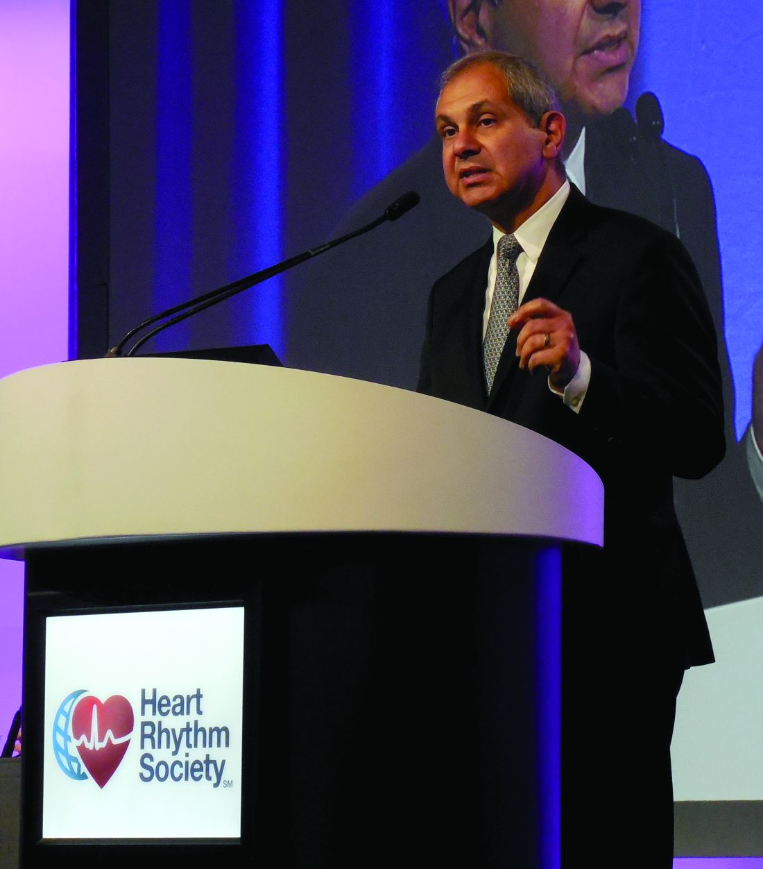
CCM “promises to meet a very large unmet need in heart failure,” William T. Abraham, MD, said as he presented the confirmatory study’s results at the annual scientific sessions of the Heart Rhythm Society. ”These patients aren’t doing well, but don’t qualify” for a heart transplant, left ventricular assist device, implantable cardioverter defibrillator, or cardiac resynchronization therapy (CRT), noted Dr. Abraham, professor and director of cardiovascular medicine at the Ohio State University in Columbus. In the months following the anticipated FDA approval, Dr. Abraham said he expects the device will be implanted in tens of thousands of U.S. heart failure patients who match the criteria of those who got the biggest benefit from CCM.
“There are few if any evidence-based treatments for patients with an ejection fraction of 35%-45%. This is an underserved population, so the potential of CCM is appropriately high,” Dr. Abraham said.
Researchers designed the study in consultation with the FDA to resolve lingering regulatory concerns following completion of three prior randomized trials with a total of nearly 650 patients. Dr. Abraham simultaneously reported the results at the meeting and published them in a report (JACC Heart Failure. 2018 May 10. doi: 10.1016/j.jchf.2018.04.010); these results from 160 patients – 74 of whom received the device and 86 of whom continued medical therapy – showed the superiority of the device for the primary endpoint of change in exercise capacity (as measured by peak oxygen uptake) and for the secondary endpoints of quality of life (as measured with the Minnesota Living With Heart Failure Questionnaire) and functional status (as measured by New York Heart Association class). The boost in exercise capacity, an average increase of 0.84 ml/kg per min in peak oxygen uptake after 24 weeks, “was similar to the improvement seen with CRT in patients with a wide QRS interval” who thereby qualified for CRT placement, Dr. Abraham said.
The CCM device also met the study’s prespecified safety endpoint of a complication rate of less than 30% – with an actual rate of 10%. “The complications were those we expect from implanted leads and pulse generators and were comparable to what happens with other implanted rhythm devices. In the context of the benefits patients received and their having no other treatment options, I see the complication rate as acceptable,” Dr. Abraham said during his report.
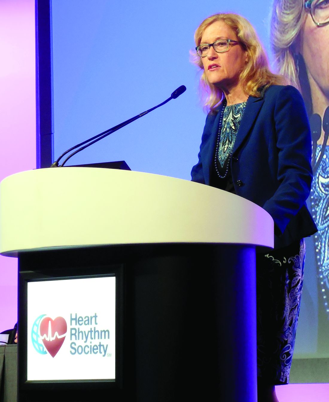
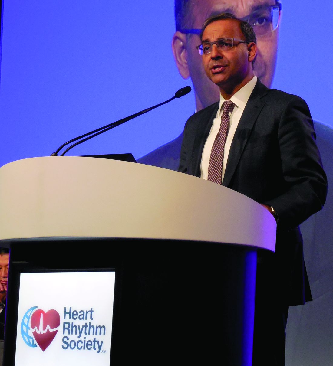
In summing up the trial’s results, Dr. Stevenson noted that “the safety endpoint was met, the primary endpoint and other functional endpoints were met, and functional endpoints are of vital importance to patients. The CCM story is not yet the CRT story,” with CRT having produced even larger effects in its pivotal trial, also led by Dr. Abraham (New Engl J Med. 2002 June 13;346[24]:1845-53), cautioned Dr. Stevenson. But in general she put a positive spin on the CCM device, saying that it “has ingenuity and innovation, and we look forward to a better understanding of which patients benefit from CCM and what we can tell them about the magnitude and duration of the benefit.”
The FIX-HF-5C trial was sponsored by Impulse Dynamics, the company developing the CCM Optimizer device. Dr. Abraham has been a consultant to Impulse Dynamics, as well as to Abbott Vascular, Medtronic, Novartis, and St. Jude Medical. Dr. Singh has been a consultant to Biotronik, Boston Scientific, Liva Nova, Medtronic, and St. Jude. Dr. Stevenson has received research funding from Abbott and Novartis. Dr. Yancy had no disclosures.
SOURCE: Abraham W et al. Heart Rhythm 2018, Abstract B-LBCT01-02.
BOSTON – Positive results from a confirmatory trial appear to put the Optimizer by Impulse Dynamics, a cardiac contractility modulation (CCM) device for patients with function-limiting heart failure, on track for imminent U.S. marketing approval by the Food and Drug Administration. If that happens, several hundreds of thousands of U.S. heart failure patients would immediately become candidates for this treatment based on the enrolled study populations, the benefits shown, and current treatment options for advanced heart failure, experts predicted.
CCM “promises to meet a very large unmet need in heart failure,” William T. Abraham, MD, said as he presented the confirmatory study’s results at the annual scientific sessions of the Heart Rhythm Society. ”These patients aren’t doing well, but don’t qualify” for a heart transplant, left ventricular assist device, implantable cardioverter defibrillator, or cardiac resynchronization therapy (CRT), noted Dr. Abraham, professor and director of cardiovascular medicine at the Ohio State University in Columbus. In the months following the anticipated FDA approval, Dr. Abraham said he expects the device will be implanted in tens of thousands of U.S. heart failure patients who match the criteria of those who got the biggest benefit from CCM.
“There are few if any evidence-based treatments for patients with an ejection fraction of 35%-45%. This is an underserved population, so the potential of CCM is appropriately high,” Dr. Abraham said.
Researchers designed the study in consultation with the FDA to resolve lingering regulatory concerns following completion of three prior randomized trials with a total of nearly 650 patients. Dr. Abraham simultaneously reported the results at the meeting and published them in a report (JACC Heart Failure. 2018 May 10. doi: 10.1016/j.jchf.2018.04.010); these results from 160 patients – 74 of whom received the device and 86 of whom continued medical therapy – showed the superiority of the device for the primary endpoint of change in exercise capacity (as measured by peak oxygen uptake) and for the secondary endpoints of quality of life (as measured with the Minnesota Living With Heart Failure Questionnaire) and functional status (as measured by New York Heart Association class). The boost in exercise capacity, an average increase of 0.84 ml/kg per min in peak oxygen uptake after 24 weeks, “was similar to the improvement seen with CRT in patients with a wide QRS interval” who thereby qualified for CRT placement, Dr. Abraham said.
The CCM device also met the study’s prespecified safety endpoint of a complication rate of less than 30% – with an actual rate of 10%. “The complications were those we expect from implanted leads and pulse generators and were comparable to what happens with other implanted rhythm devices. In the context of the benefits patients received and their having no other treatment options, I see the complication rate as acceptable,” Dr. Abraham said during his report.
In summing up the trial’s results, Dr. Stevenson noted that “the safety endpoint was met, the primary endpoint and other functional endpoints were met, and functional endpoints are of vital importance to patients. The CCM story is not yet the CRT story,” with CRT having produced even larger effects in its pivotal trial, also led by Dr. Abraham (New Engl J Med. 2002 June 13;346[24]:1845-53), cautioned Dr. Stevenson. But in general she put a positive spin on the CCM device, saying that it “has ingenuity and innovation, and we look forward to a better understanding of which patients benefit from CCM and what we can tell them about the magnitude and duration of the benefit.”
The FIX-HF-5C trial was sponsored by Impulse Dynamics, the company developing the CCM Optimizer device. Dr. Abraham has been a consultant to Impulse Dynamics, as well as to Abbott Vascular, Medtronic, Novartis, and St. Jude Medical. Dr. Singh has been a consultant to Biotronik, Boston Scientific, Liva Nova, Medtronic, and St. Jude. Dr. Stevenson has received research funding from Abbott and Novartis. Dr. Yancy had no disclosures.
SOURCE: Abraham W et al. Heart Rhythm 2018, Abstract B-LBCT01-02.
REPORTING FROM HEART RHYTHM 2018
Disease burden higher in osteoarthritis than rheumatoid arthritis
LIVERPOOL, ENGLAND – Osteoarthritis is associated with a “considerably higher disease burden” than rheumatoid arthritis 6 months after initial presentation, according to one expert’s analysis at the World Congress on Osteoarthritis.
This may partly be because of the improved treatments now available for rheumatoid arthritis, whereas there remain few treatments, and no disease-modifying therapy as yet, for osteoarthritis, Theodore Pincus, MD, suggested at the congress sponsored by the Osteoarthritis Research Society International.

“The ‘conventional’ wisdom is that ‘osteoarthritis is the most common type of arthritis,’ and ‘rheumatoid arthritis is recognized as the most crippling or disabling type of arthritis,’ ” he said, citing text from a health website and a report of the World Health Organization.
“We all know there is a lot of information on the Internet that may not be as accurate as we would like,” he observed. “We characterize this as ‘eminence-based medicine,’ ” Dr. Pincus joked, “which is defined as making the same mistakes with increasing confidence over an impressive number of years!” The alternative is, of course, evidence-based medicine, which is “the best approach,” requiring data from both clinical observations and clinical trials.
Even seemingly credible sources of health information can relay incorrect, or out-of-date, messages, such as RA being associated with worse functional status than OA. Recent observational data (RMD Open. 2017;3[1]:e000391), suggest that actually the reverse may be true, and that the disease burden seen with OA in routine care is as great as, if not greater than, RA.
Indeed, patients with OA who completed the Multi-Dimensional Health Assessment Questionnaire (MDHAQ)/Routine Assessment of Patient Index Data (RAPID3) at diagnosis at four different sites were found to have similar or worse scores for physical function, pain, and patient global assessment when compared with RA.
The MDHAQ/RAPID3 is a simple assessment tool that consists of two pages and asks patients to rate items such as their physical function in activities of daily living and levels of pain in the past week. It also asks about levels of anxiety, depression, and quality of sleep, and it includes a self-reported joint count and a patient global assessment. Scores on RAPID3 range from 0 to 30, and comprise three 0-10 scores for physical function, pain, and patient global assessment subscales in which higher scores indicate greater disease burden.
“Using this tool, we’ve been able to obtain data on patients with OA and RA for at least 30 years,” Dr. Pincus said.
One of the issues with comparing the burden of the two diseases, he noted, is that there are few places that have used the same assessment tool.
Dr. Pincus and his associates at Rush University have also shown that the disease burden in OA remains high 6 months after first visit, while greater improvement is seen in RA over this period (Osteoarthritis Cartilage. 2018;26[1]:S260. Abstract 491).
In a study of 151 patients with OA and 202 with RA, they found the composite RAPID3 scores were equally high in patients with OA and RA at their first visit (16.0 vs. 15.5, respectively) but higher in OA patients at the 6-month reassessment (14.3 vs. 11.9; P less than .004).
“We can now say that at presentation, OA and RA are similar in MDHAQ/RAPID3 scores, which were adjusted for age and BMI,” Dr. Pincus said. “Both the OA and RA patients improved, but considerably greater improvement in RA versus OA resulted in significantly poorer status for OA versus RA at 6 months.”
However, that’s not to say that OA is a worse disease than RA in every patient, Dr. Pincus was keen to point out. “Some patients with each disease have mild, moderate, or severe disease,” he stated. RA is used as benchmark for a severe disease, so these data highlight that “OA is a severe disease as well.”
This sentiment was the focus of a 2016 white paper produced by OARSI and submitted to the Food and Drug Administration, which states the case for the need to take OA more seriously and for regulatory restrictions to be removed to enable new treatments to be developed.
The prevalence of OA is at least 10-20 times higher than RA, and it’s likely that a large percentage of OA patients never get to see a rheumatologist, Dr. Pincus said. Yet the resources that go into managing RA are far greater if one excludes joint replacement.
Dr. Pincus noted that RA was not always regarded as a severe disease: 30 years ago the textbooks were stating that it had a good prognosis in the majority of cases and that patients could, by and large, use conservative regimens to manage their disease. However, real-world evidence showed that RA was associated with severe declines in function, high levels of work disability, and increased mortality, Dr. Pincus observed.
“Is osteoarthritis in 2018 where rheumatoid arthritis was in 1988, 30 years ago?” he asked rhetorically.
“The risk of long-term mortality in RA, OA, and most rheumatic disease is similar to, or greater than, hypertension, diabetes, as well as many cardiovascular and neoplastic diseases,” Dr. Pincus continued. Whereas mechanisms exist to try to log all cancer cases and compile data on the number of deaths, a rheumatic disease often is not listed anywhere on the death certificate, even as contributing to mortality, as rheumatic diseases generally are not the acute cause of death.
Functional disability and socioeconomic status are more important predictors of work disability and mortality than “any biomarker or imaging data, except x-ray.” Perhaps, Dr. Pincus said, these could also be important indicators of poor prognosis in OA and all chronic diseases?
“Physical function is a big deal,” he said. Data from a study looking at adults over the age of 50 years in the general Finnish population showed 5-year survival was significantly reduced by poorer functional capacity and less frequent physical exercise, at levels higher than smoking. Perhaps, the musculoskeletal system is more important than the other organs of the body for maintaining health, Dr. Pincus suggested.
Assessing functional status with tools such as the MDHAQ/RAPID3 is “really useful” in daily practice, Dr. Pincus said. He concluded with the words of Rudolph Virchow, who observed more than 100 years ago, that “the improvement of medicine would eventually prolong human life, but improvement of social conditions could achieve this result now and more rapidly and successfully.”
Dr. Pincus is the president of Medical History Services, which receives royalties and license fees from copyright and trademark of MDHAQ, RAPID3, or both, all of which are used to support further development of quantitative clinical measurement by both patients and physicians. He holds stock in the company and has received research funding from the company. Dr. Pincus also disclosed having a consulting agreement with Lilly.
SOURCE: Pincus T et al. Osteoarthritis Cartilage. 2018:26(1):S4. Abstract I-11.
*This story was updated 5/24/2018.
LIVERPOOL, ENGLAND – Osteoarthritis is associated with a “considerably higher disease burden” than rheumatoid arthritis 6 months after initial presentation, according to one expert’s analysis at the World Congress on Osteoarthritis.
This may partly be because of the improved treatments now available for rheumatoid arthritis, whereas there remain few treatments, and no disease-modifying therapy as yet, for osteoarthritis, Theodore Pincus, MD, suggested at the congress sponsored by the Osteoarthritis Research Society International.
“The ‘conventional’ wisdom is that ‘osteoarthritis is the most common type of arthritis,’ and ‘rheumatoid arthritis is recognized as the most crippling or disabling type of arthritis,’ ” he said, citing text from a health website and a report of the World Health Organization.
“We all know there is a lot of information on the Internet that may not be as accurate as we would like,” he observed. “We characterize this as ‘eminence-based medicine,’ ” Dr. Pincus joked, “which is defined as making the same mistakes with increasing confidence over an impressive number of years!” The alternative is, of course, evidence-based medicine, which is “the best approach,” requiring data from both clinical observations and clinical trials.
Even seemingly credible sources of health information can relay incorrect, or out-of-date, messages, such as RA being associated with worse functional status than OA. Recent observational data (RMD Open. 2017;3[1]:e000391), suggest that actually the reverse may be true, and that the disease burden seen with OA in routine care is as great as, if not greater than, RA.
Indeed, patients with OA who completed the Multi-Dimensional Health Assessment Questionnaire (MDHAQ)/Routine Assessment of Patient Index Data (RAPID3) at diagnosis at four different sites were found to have similar or worse scores for physical function, pain, and patient global assessment when compared with RA.
The MDHAQ/RAPID3 is a simple assessment tool that consists of two pages and asks patients to rate items such as their physical function in activities of daily living and levels of pain in the past week. It also asks about levels of anxiety, depression, and quality of sleep, and it includes a self-reported joint count and a patient global assessment. Scores on RAPID3 range from 0 to 30, and comprise three 0-10 scores for physical function, pain, and patient global assessment subscales in which higher scores indicate greater disease burden.
“Using this tool, we’ve been able to obtain data on patients with OA and RA for at least 30 years,” Dr. Pincus said.
One of the issues with comparing the burden of the two diseases, he noted, is that there are few places that have used the same assessment tool.
Dr. Pincus and his associates at Rush University have also shown that the disease burden in OA remains high 6 months after first visit, while greater improvement is seen in RA over this period (Osteoarthritis Cartilage. 2018;26[1]:S260. Abstract 491).
In a study of 151 patients with OA and 202 with RA, they found the composite RAPID3 scores were equally high in patients with OA and RA at their first visit (16.0 vs. 15.5, respectively) but higher in OA patients at the 6-month reassessment (14.3 vs. 11.9; P less than .004).
“We can now say that at presentation, OA and RA are similar in MDHAQ/RAPID3 scores, which were adjusted for age and BMI,” Dr. Pincus said. “Both the OA and RA patients improved, but considerably greater improvement in RA versus OA resulted in significantly poorer status for OA versus RA at 6 months.”
However, that’s not to say that OA is a worse disease than RA in every patient, Dr. Pincus was keen to point out. “Some patients with each disease have mild, moderate, or severe disease,” he stated. RA is used as benchmark for a severe disease, so these data highlight that “OA is a severe disease as well.”
This sentiment was the focus of a 2016 white paper produced by OARSI and submitted to the Food and Drug Administration, which states the case for the need to take OA more seriously and for regulatory restrictions to be removed to enable new treatments to be developed.
The prevalence of OA is at least 10-20 times higher than RA, and it’s likely that a large percentage of OA patients never get to see a rheumatologist, Dr. Pincus said. Yet the resources that go into managing RA are far greater if one excludes joint replacement.
Dr. Pincus noted that RA was not always regarded as a severe disease: 30 years ago the textbooks were stating that it had a good prognosis in the majority of cases and that patients could, by and large, use conservative regimens to manage their disease. However, real-world evidence showed that RA was associated with severe declines in function, high levels of work disability, and increased mortality, Dr. Pincus observed.
“Is osteoarthritis in 2018 where rheumatoid arthritis was in 1988, 30 years ago?” he asked rhetorically.
“The risk of long-term mortality in RA, OA, and most rheumatic disease is similar to, or greater than, hypertension, diabetes, as well as many cardiovascular and neoplastic diseases,” Dr. Pincus continued. Whereas mechanisms exist to try to log all cancer cases and compile data on the number of deaths, a rheumatic disease often is not listed anywhere on the death certificate, even as contributing to mortality, as rheumatic diseases generally are not the acute cause of death.
Functional disability and socioeconomic status are more important predictors of work disability and mortality than “any biomarker or imaging data, except x-ray.” Perhaps, Dr. Pincus said, these could also be important indicators of poor prognosis in OA and all chronic diseases?
“Physical function is a big deal,” he said. Data from a study looking at adults over the age of 50 years in the general Finnish population showed 5-year survival was significantly reduced by poorer functional capacity and less frequent physical exercise, at levels higher than smoking. Perhaps, the musculoskeletal system is more important than the other organs of the body for maintaining health, Dr. Pincus suggested.
Assessing functional status with tools such as the MDHAQ/RAPID3 is “really useful” in daily practice, Dr. Pincus said. He concluded with the words of Rudolph Virchow, who observed more than 100 years ago, that “the improvement of medicine would eventually prolong human life, but improvement of social conditions could achieve this result now and more rapidly and successfully.”
Dr. Pincus is the president of Medical History Services, which receives royalties and license fees from copyright and trademark of MDHAQ, RAPID3, or both, all of which are used to support further development of quantitative clinical measurement by both patients and physicians. He holds stock in the company and has received research funding from the company. Dr. Pincus also disclosed having a consulting agreement with Lilly.
SOURCE: Pincus T et al. Osteoarthritis Cartilage. 2018:26(1):S4. Abstract I-11.
*This story was updated 5/24/2018.
REPORTING FROM OARSI 2018
Atopic dermatitis severity reduced by topical microbiome treatment
The Beginning Assessment of Cutaneous Treatment Efficacy of Roseomonas in Atopic Dermatitis trial; BACTERiAD I/II study, an open-label phase I/II trial, first looked at the therapeutic use of R. mucosa in 10 adults aged 18 years or older. Three sucrose mixtures with increasing doses of live R. mucosa bacteria were topically applied to two body areas – the antecubital fossae and a body surface of their choice – twice per week for 2 weeks per dose. At 6 weeks, the patients stopped using the mixtures and followed a 4-week washout phase.
Treatment was found to reduce mean antecubital SCORAD (SCORing Atopic Dermatitis) scores by 59.8%. Reduction in pruritus was even more pronounced, with a mean decrease of 78.5%. Treating the hands did not improve disease severity, even in patients whose symptoms improved in other body areas. One explanation may be the increased exposure of the hands to topical antimicrobials and environmental exposures, the researchers noted.
With the success in the adult cohort, the researchers enrolled five children aged 7-17 years in the study. These patients were treated twice weekly for 16 weeks. The pediatric patients experienced a mean decrease of 70.3% in their SCORAD scores. The mean decrease in pruritis was 78.8%.
All adults who responded continued to report improved symptoms after the washout period. The pediatric patients are now being evaluated in a washout period.
Four patients did not respond; three of them had a family history of AD persisting into adulthood. “The association between these complex medical histories and the lack of clinical response suggests that differences in heritable host and/or microbial factors may impact treatment responses,” wrote Ian A. Myles, MD, and his colleagues.
“Overall, our findings suggest the safety of topical R. mucosa therapy and justify continuation of our ongoing trial to assess safety and activity in a pediatric cohort of patients with AD. These studies will additionally assess changes in host serum markers, skin metabolomics, and the skin microbiota by culture and genomic methods.”
The researchers noted that expanding to the pediatric population will deepen understanding of topical microbiome transplantation and lay the foundation for placebo-controlled trials to assess efficacy.
This work was supported by the Intramural Research Program of the National Institutes of Allergy and Infectious Diseases and the National Institutes of Health. The researchers had no disclosures.
SOURCE: Myles IA et al. JCI Insight. 2018 May 3. doi: 10.1172/jci.insight.120608.
FROM JCI INSIGHT
Key clinical point: Roseomonas mucosa reduces disease severity.
Major finding: There were reductions in SCORAD scores of 78.5% and 70.3% in the adult and pediatric cohorts, respectively.
Study details: Case study of 10 adult and 5 pediatric patients with atopic dermatitis.
Disclosures: No relevant financial disclosures were reported.
Source: Myles IA et al. JCI Insight. 2018 May 3. doi: 10.1172/jci.insight.120608.

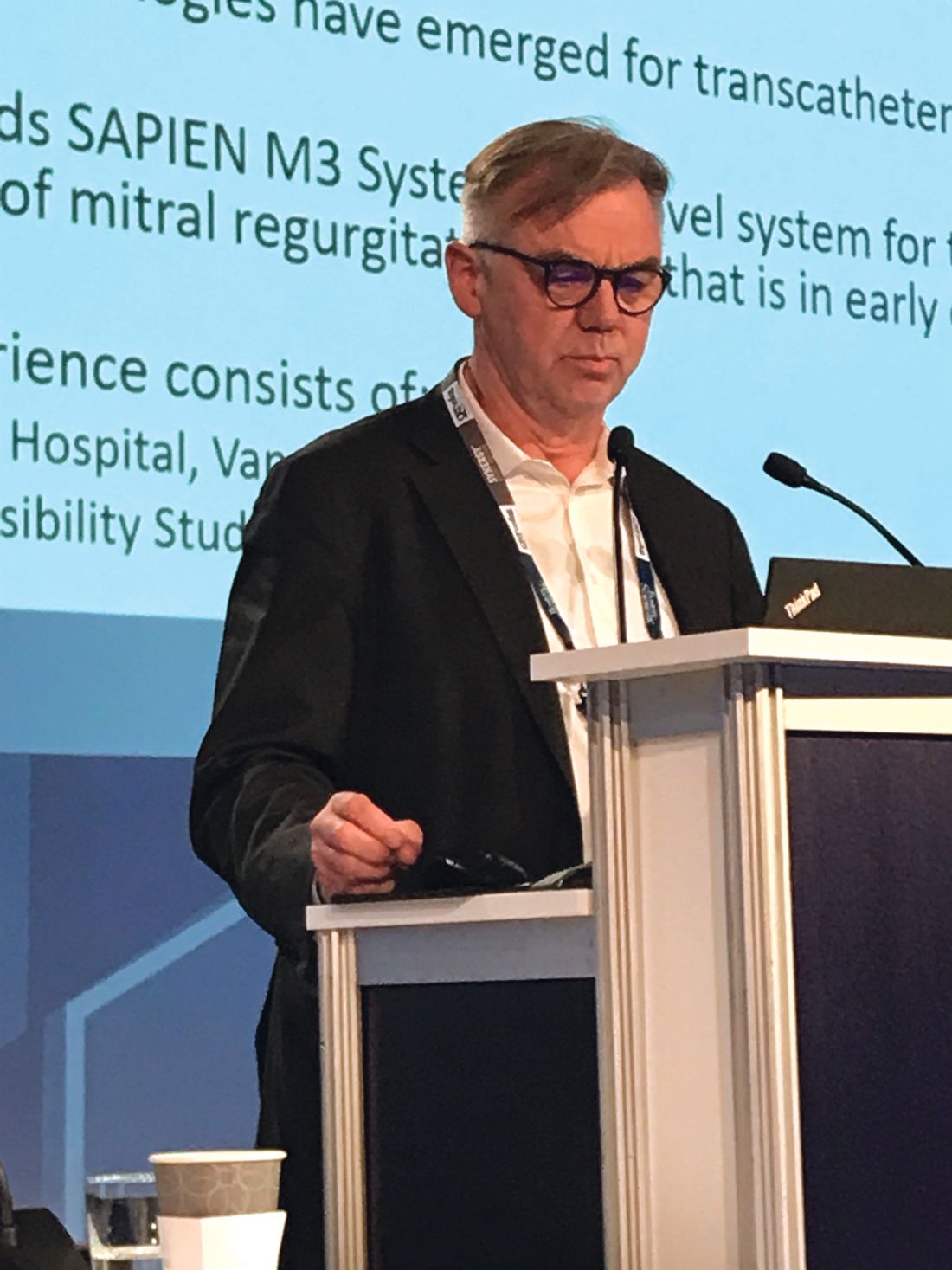
Sapien M3 mitral valve replacement data reported for first 10 patients
WASHINGTON – A novel transcatheter mitral valve replacement with a transseptally introduced docking mechanism that secures the valve with native mitral valve leaflets was found feasible and effective in an initial series of 10 patients, according to a first-in-man report at CRT 2018 sponsored by the Cardiovascular Research Institute at Washington Hospital Center.
“All patients remained hemodynamically stable throughout the procedure, and the valve was successfully implanted in all patients,” reported John G. Webb, MD, McLeod Professor of Heart Valve Intervention at the University of British Columbia, Vancouver.
The docking system “is retrievable up until the point of the final release,” Dr. Webb explained. A knitted polyethylene terephthalate skirt is employed to aid in creating a seal between the leaflets and the dock. Once the docking system is in place, the procedure “then becomes a relatively standard transcatheter transseptal valve-in-valve–type procedure” that is a “fairly easy part of the procedure at centers with transcatheter valve implantation experience.”
The very first case was performed in a 75-year-old woman with severe mitral valve insufficiency. Frail with multiple comorbidities and a left ventricular ejection fraction of 30%, the patient was not a candidate for surgery. Although Dr. Webb acknowledged that the first case “was a learning process,” he reported that the patient was discharged after a 1-night hospital stay with reassuring valve placement and function based on imaging studies.
Data was available from 10 patients from five participating centers in Canada and the United States. The mean age was 74 years, and all were New York Heart Association class III or higher. The mean left ventricular ejection fraction was 37.5%. Although the average Society of Thoracic Surgery risk score was only 4.9%, Dr. Webb noted that this underestimated the vulnerability of a population in which most had compromised renal function. Half of the 10 had severe mitral valve regurgitation prior to valve replacement, and the remainder had moderate to severe regurgitation.
“At the end of 30 days, all had mild or less insufficiency,” Dr. Webb reported. Although one patient did develop significant mitral insufficiency after discharge because of a small tear attributed to probing, it was repaired with a plug. The one technical failure occurred in a patient who required a plug during the course of valve replacement; again, the plug proved effective for preventing significant valve insufficiency.
In this series of patients, one stroke occurred 2 days after the procedure, but there were no deaths in the initial 30-day follow-up, according to Dr. Webb. Although he noted that the procedure time in the first case was 4 hours, the procedure times became shorter with experience and the second-to-last and last cases took 2.5 and 1.3 hours, respectively.
Howard C. Herrmann, MD, director of the interventional cardiology program at the University of Pennsylvania, Philadelphia, and a panelist on the symposium where these data were presented, called the results “exciting.” However, he also noted that “this is the first time that any us have had a look at this device,” so more data will be needed to understand its clinical potential.
Edwards Lifesciences sponsored this study. Dr. Webb reported financial relationships with Abbott Vascular, Edwards Lifesciences, Essential Medical, and Vivitro.
REPORTING FROM CRT 2018
Key clinical point: Initial experience with a transcatheter transseptal mitral valve replacement is encouraging.
Major finding: In the first 10 patients, technical success was achieved in 9.
Study details: A summary of first clinical experience at multiple centers.
Disclosures: Edwards Lifesciences sponsored this study. Dr. Webb reported financial relationships with Abbott Vascular, Edwards Lifesciences, Essential Medical, and Vivitro.
Checkpoint inhibitors well tolerated by NSCLC patients with preexisting autoimmune disease
For patients with non–small-cell lung cancer (NSCLC), preexisting autoimmune disease (AID) does not increase the risk of immune-related adverse events (irAEs) with checkpoint inhibitor therapy, according to investigators.
Flares of AID during therapy were generally mild, and toxicity rates were only slightly increased in patients with AID, they wrote in Journal of Clinical Oncology.
Approximately 14%-25% of patients with lung cancer also have AID, but patients with AID are typically excluded from clinical trials. “This presents a tremendous knowledge gap,” wrote lead author Giulia C. Leonardi, MD, of the Dana-Farber Cancer Institute in Boston, and coauthors.
The retrospective study comprised 56 patients with NSCLC and preexisting autoimmune disease from five academic cancer centers. Almost half of the patients had a rheumatologic disorder, 29% had a dermatologic disorder, 16% had an endocrine disorder, 11% had inflammatory bowel disease, 5% had a neurologic condition, 3% had rheumatic fever, and one patient (2%) had autoimmune hemolytic anemia.
Patients received either a programmed death-1 or programmed death-ligand 1 inhibitor as monotherapy. Median treatment time was 3.1 months, and median follow-up time after starting therapy was 17.5 months.
Eleven percent of patients with preexisting AID developed grade 3 or 4 irAEs, which is similar to 7%-15% of patients in clinical trials. Although 23% of patients experienced a flare of their preexisting AID, these flares were generally mild – no patients discontinued immunotherapy because of a flare. In contrast, 14% of patients halted therapy because of toxicity, a marginally higher number than 3%-8% of patients in trials.
Checkpoint inhibitors are now a mainstay treatment for a lung cancer, including three approved drugs: nivolumab, pembrolizumab, and atezolizumab. “Almost every patient with advanced NSCLC will likely receive a [checkpoint] inhibitor at some point over the course of their disease,” the authors noted. As checkpoint inhibitors may lead to fatal irAEs, the possible interplay between these immunotherapies and AID requires investigation.
“Our study adds to the growing body of evidence supporting the use of immunotherapy in patients with cancer with preexisting AID, albeit with close monitoring for adverse events,” the researchers concluded.
SOURCE: Leonardi GC et al. J Clin Oncol. 2018* May 20. doi: 10.1200/JCO.2017.77.0305.
*Correction, 5/22/18: An earlier version of this article misstated the year in this citation.
FROM JOURNAL OF CLINICAL ONCOLOGY
Key clinical point:
Major finding: Eleven percent of patients with autoimmune disease experienced grade 3 or 4 immune-related adverse events, compared with 7%-15% of patients without autoimmune disease.
Study details: A retrospective study of 56 patients with NSCLC and preexisting autoimmune disease from five academic cancer centers.
Disclosures: The Kaplan Research Fund and Jeni Fund, Memorial Sloan Kettering Cancer Center, the American Cancer Society, and the University of Texas MD Anderson Cancer Center sponsored the study. The researchers reported receiving financial support from Merck, Novartis, Genentech, and other companies.
Source: Leonardi GC et al. J Clin Oncol. 2018 May 20. doi: 10.1200/JCO.2017.77.0305.
IMDC model mirrors mRCC clinical outcomes
In hindsight, a widely used prognostic model for patients with metastatic renal cell carcinoma (mRCC) has been shown to be effective at stratifying patient risk, and may inform the design of future clinical trials.
By retrospectively comparing clinical outcomes with risk categories determined by the International Metastatic Renal Cell Carcinoma Database Consortium (IMDC) model, Brian I Rini, MD, of the Cleveland Clinic, and his colleagues found notable differences in progression-free survival, overall survival, and objective response rates between the different risk categories among patients with mRCC treated with sunitinib (Sutent) in a major clinical trial.
“These benchmark values can aid current and future design and interpretation of clinical trials in mRCC. Results of this analysis demonstrate clear differences in patient outcomes based on IMDC prognostic risk group,” they wrote. The report was published in Clinical Genitourinary Cancer.
They also found that the Memorial Sloan Kettering Cancer Center (MSKCC) model appeared to be similar in prognostic utility to the IMDC model, and that either model could be useful for counseling patients about prognosis and treatment options.
They based their conclusions on an analysis of data from a phase 3 clinical trial comparing sunitinib with interferon alfa in patients with mRCC. Patients in this study were grouped according to prognostic risk category by the MSKCC criteria, which overlap with the IMDC criteria in five of six areas.
In the current study, Dr. Rini and his associates applied the IMDC criteria to the same population, and derived benchmark values for outcomes by IMDC risk groups based on radiologic tumor progression measurements performed by independent reviewers on images of patients in the intention-to-treat population.
They also conducted an analysis of data from investigator measurements of tumor progression, and compared the results with the independently reviewed radiologic data for patients outcomes according to the MSKCC model.
They found that for sunitinib-treated patients in the IMDC favorable-risk group, median PFS was 14.1 months, compared with 10.7 months for those in the intermediate-risk group, 2,4 months in the poor-risk group, and 10.6 months for the combined intermediate and poor-risk groups.
The respective objective response rates were 53%, 33.7%, 11.8%, and 30.5%.
Median overall survival for favorable-risk patients was not reached, with more than 50% of patients alive at the time of data cutoff. The respective median overall survival for the intermediate-, poor-, and intermediate-plus-poor–risk groups were 23, 5.1, and 20.3 months.
“Results of this study suggest there may be significant prognostic differences between the intermediate-1 and intermediate-2 IMDC risk groups and that this should be considered when counseling patients identified to be in one of these groups,” the investigators wrote.
Medical writing for the study was supported by Pfizer. Dr. Rini and his coauthors disclosed research funding and/or consulting fees from Pfizer and other companies. Four of the coauthors are Pfizer employees and stockholders.
SOURCE: Rini B et al. Clin Genitourin Cancer. 2018 May 3. doi: 10.1016/j.clgc.2018.04.005.
FROM CLINICAL GENITOURINARY CANCER
Key clinical point: The IMDC model is prognostic for responses and survival in patients with mRCC treated with sunitinib.
Major finding: Median progression-free survival for favorable, intermediate, and poor-risk patients was 14.1, 10.7, and 2.4 months, respectively.
Study details: Retrospective analysis of 375 sunitinib-treated patients in a randomized clinical trial.
Disclosures: Medical writing for the study was supported by Pfizer. Dr. Rini and his coauthors disclosed research funding and/or consulting fees from Pfizer and other companies. Four of the coauthors are Pfizer employees and stockholders.
Source: Rini B et al. Clin Genitourin Cancer. 2018 May 3. doi: 10.1016/j.clgc.2018.04.005.
For in-stent restenosis, everolimus-eluting stents topped drug-eluting balloons
For patients with coronary artery restenosis at the site of a drug-eluting stent, placing an everolimus-eluting stent was associated with a 57% lower risk of target lesion revascularization, compared with placing a drug-eluting balloon.
At 3-year follow-up in the randomized, multicenter RIBS IV (Restenosis Intra-Stent of Drug-Eluting Stents: Drug-Eluting Balloons vs Everolimus-Eluting Stents) trial, 7.1% of patients required target lesion revascularization after EES versus 15.6% of patients after DEB (P = .015), reported Fernando Alfonso, MD, of Hospital Universitario de La Princesa, Madrid, and his associates. Consequently, the combined rate of cardiac death, myocardial infarction, and target lesion revascularization was 12.3% with EES versus 20.1% with DEB (hazard ratio, 0.57; 95% confidence interval, 0.34-0.96; P = .04). The findings were reported in JACC: Cardiovascular Interventions.
About 5%-10% of patients who receive a drug-eluting stent (DES) develop in-stent restenosis (ISR). When this happens, robust data support placing a DEB or next-generation DES, such as an EES, instead of a conventional (plain) balloon, the investigators noted. To directly compare EES versus DEB, they randomly assigned 309 patients with at least 50% lumen diameter stenosis at the DES site or involving its 5-mm edge to receive either DEB (SeQuent Please, B. Braun) with a 1.1:1 balloon-to-artery ratio (mean 18 atm [pressure]), or EES (Xience Prime, Abbott Vascular) with the same final ratio but significantly greater deployment pressure (mean 20 arm; P = .001). All patients had angina or objective evidence of ischemia without stent thrombosis, and the trial arms otherwise resembled each other clinically and demographically.
Angiography documented 100% immediate procedural success in both groups. At 1 year, rates of target lesion revascularization were 4.5% with EES and 13% with DEB, a significant difference (HR, 0.33; 95% CI, 0.14-0.79). Similarly, rates of target vessel revascularization were 8.4% and 16.2%, respectively (HR, 0.49; 95% CI, 0.25-0.97), at year 1 and 11% and 20.8%, respectively, at year 3 (HR, 0.50; 95% CI, 0.28-0.90).
Throughout the study, including at 3 years, the groups had similar rates of cardiac death (3.9% for EES vs. 3.2% for DEB), MI (2.6% vs. 4.5%), and stent thrombosis (1.3% vs. 2.6%). “Results of other composite clinical outcomes [also] were very similar,” the researchers wrote. While “both DEB and EES provide favorable long-term clinical outcomes,” patients “receiving EES benefit[ed] from a better long-term clinical outcome, mainly driven by a reduced need of target lesion and target vessel revascularization.”
Funders of the study included B. Braun and Abbott Vascular. The investigators reported having no conflicts of interest.
SOURCE: Alfonso F et al. JACC Cardiovasc Interv. 2018;11:981-91.
Three-year outcomes favored everolimus-eluting stents (EES) over drug-eluting balloons (DEBs) for treating in-stent restenosis, but this “should not be a reason to bid farewell to DEBs,” wrote Hyo-Soo Kim, MD, PhD, and Tae-Min Rhee, MD, in an editorial accompanying the study.
Both EES and DEB have excellent long-term safety, so a “simple rivalry between the [two] arms is no longer meaningful,” they wrote in JACC: Cardiovascular Interventions. “Instead, it is time to find ways to improve the efficacy of DEBs.”
The efficacy of DEB depends on the amount of antiproliferative drug delivered to the target lesion, which isn’t the case for EES, the experts noted. In past studies, independent predictors of long-term DEBs success included less than 20% residual stenosis, balloon-to-stent ratio exceeding 0.91, and inflation time above 60 seconds, they added. When all three of these criteria were met, DEBs performed as well as new-generation drug-eluting stents.
Thus, DEBs retains a treatment niche, “particularly for in-stent restenosis lesions already covered with one or more metal layers.”
Dr. Kim and Dr. Rhee are with Seoul (South Korea) National University Hospital. They reported having no conflicts of interest. These comments are from their editorial (JACC Cardiovasc Interv. 2018;11:992-4).
For patients with coronary artery restenosis at the site of a drug-eluting stent, placing an everolimus-eluting stent was associated with a 57% lower risk of target lesion revascularization, compared with placing a drug-eluting balloon.
At 3-year follow-up in the randomized, multicenter RIBS IV (Restenosis Intra-Stent of Drug-Eluting Stents: Drug-Eluting Balloons vs Everolimus-Eluting Stents) trial, 7.1% of patients required target lesion revascularization after EES versus 15.6% of patients after DEB (P = .015), reported Fernando Alfonso, MD, of Hospital Universitario de La Princesa, Madrid, and his associates. Consequently, the combined rate of cardiac death, myocardial infarction, and target lesion revascularization was 12.3% with EES versus 20.1% with DEB (hazard ratio, 0.57; 95% confidence interval, 0.34-0.96; P = .04). The findings were reported in JACC: Cardiovascular Interventions.
About 5%-10% of patients who receive a drug-eluting stent (DES) develop in-stent restenosis (ISR). When this happens, robust data support placing a DEB or next-generation DES, such as an EES, instead of a conventional (plain) balloon, the investigators noted. To directly compare EES versus DEB, they randomly assigned 309 patients with at least 50% lumen diameter stenosis at the DES site or involving its 5-mm edge to receive either DEB (SeQuent Please, B. Braun) with a 1.1:1 balloon-to-artery ratio (mean 18 atm [pressure]), or EES (Xience Prime, Abbott Vascular) with the same final ratio but significantly greater deployment pressure (mean 20 arm; P = .001). All patients had angina or objective evidence of ischemia without stent thrombosis, and the trial arms otherwise resembled each other clinically and demographically.
Angiography documented 100% immediate procedural success in both groups. At 1 year, rates of target lesion revascularization were 4.5% with EES and 13% with DEB, a significant difference (HR, 0.33; 95% CI, 0.14-0.79). Similarly, rates of target vessel revascularization were 8.4% and 16.2%, respectively (HR, 0.49; 95% CI, 0.25-0.97), at year 1 and 11% and 20.8%, respectively, at year 3 (HR, 0.50; 95% CI, 0.28-0.90).
Throughout the study, including at 3 years, the groups had similar rates of cardiac death (3.9% for EES vs. 3.2% for DEB), MI (2.6% vs. 4.5%), and stent thrombosis (1.3% vs. 2.6%). “Results of other composite clinical outcomes [also] were very similar,” the researchers wrote. While “both DEB and EES provide favorable long-term clinical outcomes,” patients “receiving EES benefit[ed] from a better long-term clinical outcome, mainly driven by a reduced need of target lesion and target vessel revascularization.”
Funders of the study included B. Braun and Abbott Vascular. The investigators reported having no conflicts of interest.
SOURCE: Alfonso F et al. JACC Cardiovasc Interv. 2018;11:981-91.
FROM JACC: CARDIOVASCULAR INTERVENTIONS
Key clinical point: Everolimus-eluting stents outperformed drug-eluting balloons for patients with restenosis of coronary arteries following initial drug-eluting stent placement.
Major finding: Three-year rates of target lesion revascularization were 7.1% with EES and 15.6% with DEB (P = .015). Consequently, the combined rate of cardiac death, myocardial infarction, and target lesion revascularization was significantly lower with EES (12.3% vs. 20.1% with DEB; P = .04).
Study details: Randomized multicenter trial of 309 patients with blinded outcomes assessment (RIBS IV).
Disclosures: Funders included B. Braun Surgical and Abbott Vascular. The investigators reported having no conflicts of interest.
Source: Alfonso F et al. JACC Cardiovasc Interv. 2018;11:981-91.
CARPREG II fine-tunes assessment of cardiac complication risk in pregnancy
A study of pregnant women with heart disease has yielded a new risk index that improves on its predecessor by integrating general, lesion-specific, and delivery-of-care variables, investigators say.
“Compared with other published risk indices, including the original CARPREG [Cardiac Disease in Pregnancy] score, CARPREG II risk index had the highest discriminative and calibrative accuracy in our study group,” investigators said in a report published in the Journal of the American College of Cardiology.
First author on the report was Candice K. Silversides, MD, division of cardiology, University of Toronto pregnancy and heart disease research program, Mount Sinai Hospital/Sinai Health System.
The widely used, original CARPREG risk index was the first to predict maternal cardiac complications based on general clinical and echocardiographic data from the baseline antepartum visit, the researchers wrote in their report.
The new index developed by Dr. Silversides and her colleagues stems from a study of pregnant women with heart disease receiving care at two large Canadian obstetric centers.
Based on analysis of 1,938 pregnancies progressing beyond 20 weeks of gestation, the investigators found that cardiac complications were overall quite common in pregnant women with heart disease, occurring in 16% of participants. However, maternal cardiac deaths or cardiac arrests were rare, they said, occurring in just 11 (0.6%) of the pregnancies.
Most complications (64%) occurred in the antepartum period, according to the report.
Looking at patient data before or after 2001, investigators found the rates of most complications were consistent over time. However, rates of pulmonary edema decreased in the post-2001 period.
Multivariate analysis of these findings revealed 10 predictors of adverse cardiac events. Those included five general factors, including previous cardiac events or arrhythmia, four lesion-specific variables including pulmonary hypertension and coronary artery disease, and one process of care variable: late pregnancy assessment.
Only 4 of those 10 factors were included in the original CARPREG index, investigators noted.
In CARPREG II, each of the 10 factors is weighted with 1-3 points, depending on risk. For example, history of prior cardiac events was associated with a higher odds ratio, and so was assigned 3 points.
The predicted risk of primary cardiac events ranges from 5% for women with a total of 0-1 points, up to 41% for women with 5 or more points.
The finding that some predictors had higher odds ratios than others reinforces the “foundational role” of clinical assessment, investigators said in the report.
“There may be other factors that affect outcomes,” they wrote. “Risk assessment for the individual patient will need to integrate risk score estimates, known lesion-specific information, and clinical judgment by an experienced physician.”
Dr. Silversides and her coauthors reported that they had no relationships to disclose relevant to the contents of their report on the study.
SOURCE: Silversides CK et al. J Am Coll Cardiol. 2018;71:2419-30.
This update and expansion of the original CARPREG scoring system is a “useful starting point” for prediction of the risk of pregnancy in women with cardiac disease, according to Uri Elkayam, MD.
However, a detailed and lesion specific evaluation is still required for a more precise determination of risk for any given patient, Dr. Elkayam cautioned in an editorial accompanying the article.
Clinicians need to thoroughly understand how the patient’s cardiac condition could be affected by hemodynamic changes during pregnancy, labor, delivery, and the postpartum period, Dr. Elkayam added in his comments.
To fully take advantage of CARPREG II, clinicians need to take into account the limitations of the scoring system, he said, including the fact that it is based on population studies.
“Although the system presented is designed to examine all adverse cardiac events that could have an impact on maternal health, it is less effective for distinguishing between mild and easily manageable events that do not have serious effects on maternal or fetal outcomes and those that may be severe, life-threatening, or require hospitalizations or early delivery,” he wrote.
Dr. Elkayam is with the department of medicine, division of cardiovascular medicine, and the department of obstetrics and gynecology, University of Southern California, Los Angeles. These comments are derived from his editorial in the Journal of the American College of Cardiology . Dr. Elkayam reported he had no relationships relevant to the contents of this paper to disclose.
A study of pregnant women with heart disease has yielded a new risk index that improves on its predecessor by integrating general, lesion-specific, and delivery-of-care variables, investigators say.
“Compared with other published risk indices, including the original CARPREG [Cardiac Disease in Pregnancy] score, CARPREG II risk index had the highest discriminative and calibrative accuracy in our study group,” investigators said in a report published in the Journal of the American College of Cardiology.
First author on the report was Candice K. Silversides, MD, division of cardiology, University of Toronto pregnancy and heart disease research program, Mount Sinai Hospital/Sinai Health System.
The widely used, original CARPREG risk index was the first to predict maternal cardiac complications based on general clinical and echocardiographic data from the baseline antepartum visit, the researchers wrote in their report.
The new index developed by Dr. Silversides and her colleagues stems from a study of pregnant women with heart disease receiving care at two large Canadian obstetric centers.
Based on analysis of 1,938 pregnancies progressing beyond 20 weeks of gestation, the investigators found that cardiac complications were overall quite common in pregnant women with heart disease, occurring in 16% of participants. However, maternal cardiac deaths or cardiac arrests were rare, they said, occurring in just 11 (0.6%) of the pregnancies.
Most complications (64%) occurred in the antepartum period, according to the report.
Looking at patient data before or after 2001, investigators found the rates of most complications were consistent over time. However, rates of pulmonary edema decreased in the post-2001 period.
Multivariate analysis of these findings revealed 10 predictors of adverse cardiac events. Those included five general factors, including previous cardiac events or arrhythmia, four lesion-specific variables including pulmonary hypertension and coronary artery disease, and one process of care variable: late pregnancy assessment.
Only 4 of those 10 factors were included in the original CARPREG index, investigators noted.
In CARPREG II, each of the 10 factors is weighted with 1-3 points, depending on risk. For example, history of prior cardiac events was associated with a higher odds ratio, and so was assigned 3 points.
The predicted risk of primary cardiac events ranges from 5% for women with a total of 0-1 points, up to 41% for women with 5 or more points.
The finding that some predictors had higher odds ratios than others reinforces the “foundational role” of clinical assessment, investigators said in the report.
“There may be other factors that affect outcomes,” they wrote. “Risk assessment for the individual patient will need to integrate risk score estimates, known lesion-specific information, and clinical judgment by an experienced physician.”
Dr. Silversides and her coauthors reported that they had no relationships to disclose relevant to the contents of their report on the study.
SOURCE: Silversides CK et al. J Am Coll Cardiol. 2018;71:2419-30.
FROM THE JOURNAL OF THE AMERICAN COLLEGE OF CARDIOLOGY
Key clinical point: A newly developed risk index enhances the ability to predict maternal cardiac complications by combining general, lesion-specific, and delivery-of-care variables.
Major finding: Investigators developed the CARPREG II risk index based on 10 predictors of maternal cardiac complications (5 general, 4 lesion-specific, and 1 related to delivery of care).
Study details: The CARPREG II Study, which included more than 2,000 prospectively enrolled consecutive pregnant women with heart disease seen at one of two large Canadian obstetric centers.
Disclosures: Authors reported that they had no relationships to disclose relevant to the contents of their report on the study.
Source: Silversides CK et al. J Am Coll Cardiol. 2018;71:2419-30.
Uvulopalatopharyngoplasty may reduce cardiac risk in sleep apnea
Surgical remodeling of the tissues of the throat using uvulopalatopharyngoplasty (UPPP) could significantly reduce the cardiac complications of obstructive sleep apnea (OSA), according to a study published in Sleep Medicine.
Researchers examined the incidence of newly diagnosed myocardial infarction, congestive heart failure, and atrial fibrillation in 192,316 patients with a new diagnosis of obstructive sleep apnea – 22,213 of whom had undergone UPPP – and 961,590 controls.
The individuals who had had UPPP had a significantly lower incidence of all three cardiovascular events, compared with those who had not undergone the procedure. The hazard ratios for myocardial infarction, congestive heart failure, and atrial fibrillation among individuals with OSA who had uvulopalatopharyngoplasty, compared with controls, were 1.002, 0.757 and 1.117, respectively. By comparison, those hazard ratios in patients with OSA who had not had UPPP, compared with controls, were 1.070, 1.165, and 1.39 for myocardial infarction, congestive heart failure, and atrial fibrillation respectively.
These figures were after accounting for confounding factors, such as age, sex, diabetes, hypertension, and dyslipidemia.
The authors wrote that the most distinctive finding of their study was that uvulopalatopharyngoplasty lowered the incidence of congestive heart failure and atrial fibrillation in patients with obstructive sleep apnea to the point that they had the same level of risk as individuals without obstructive sleep apnea.
“Prior studies have evaluated the success of UPPP based on reductions of AHI [apnea-hypopnea index], with the average success rate for the surgery being low for most patients,” wrote Heung-Man Lee, MD, PhD, then from the Guro Hospital at Korea University, Seoul, and his coauthors.
“However, the current study suggests that the effects of UPPP, regardless of the effects on AHI, can significantly reduce cardiac morbidity in patients with OSA.”
Patients without diabetes showed more benefit from UPPP in reducing the incidence of congestive heart failure, compared with those with diabetes. However, those with diabetes showed greater reductions in the risk of atrial fibrillation, compared with those without diabetes.
Similarly, the incidence of atrial fibrillation was reduced after uvulopalatopharyngoplasty but only in patients with hypertension or dyslipidemia and not in those with normal blood pressure or lipid levels.
“These differences in outcomes after UPPP are probably due to the different etiologies of cardiovascular disease,” the authors wrote. “OSA increases free fatty acid in the blood because intermittent hypoxia associated with OSA induces adipose tissue lipolysis.”
One limitation of the study was the absence of polysomnography information. The researchers relied on diagnostic codes for confirmation of OSA. They also did not have information on other sleep apnea therapies, such as CPAP or a mandibular advancing device, which may have been used in the patients who did not undergo uvulopalatopharyngoplasty.
The study was supported by the Korean Society of Otorhinolaryngology Head and Neck Surgery. No conflicts of interest were declared.
SOURCE: Lee HM et al. Sleep Med. 2018 May;45:11-16.
FROM SLEEP MEDICINE
Key clinical point: Uvulopalatopharyngoplasty might avert cardiac complications caused by obstructive sleep apnea.
Major finding: Compared with controls, the hazard ratios for MI, congestive heart failure, and atrial fibrillation 1.002, 0.757, and 1.117, respectively, among individuals with OSA who had had UPPP and 1.070, 1.165, and 1.39 among those who had not had UPPP.
Study details: Study of 192,316 patients with obstructive sleep apnea and 961,590 controls.
Disclosures: The study was supported by the Korean Society of Otorhinolaryngology–Head and Neck Surgery. No conflicts of interest were declared.
Source: Lee HM et al. Sleep Med. 2018 May;45:11-16.
8-Isoprostane levels predict OSA in children
The oxidative stress biomarker 8-isoprostane (8-IsoP) predicted obstructive sleep apnea (OSA) and disease severity in children better than the fractional concentration of exhaled nitric oxide (FENO), according to results published in Sleep Medicine.
In an analysis of 46 patients with sleep-disordered breathing and 20 controls, 8-IsoP values were also correlated with apnea hypopnea index (AHI) (r, 0.40; P = .003) and oxygen saturation, also known as SaO2, (r, –0.50; P = .001), reported Dr. Mario Berreto of the Pediatric Unit at Sant’Andrea Hospital in Rome and his coauthors.
The investigators studied 66 children aged 4.5-15.1 years, of whom 46 had sleep-disordered breathing (SDB) and were enrolled in the hospital’s Pediatric Sleep Center. The 20 healthy controls had no history of sleep problems, including snoring, apneas, and restless sleep. Exclusion criteria included acute respiratory infections in the 4 weeks preceding the study, chronic respiratory comorbidities, and therapy with corticosteroids or other anti-inflammatory drugs for at least 3 weeks.
Patients with SDB had a medical examination followed by overnight standard polysomnography (PSG), and EBC 8-IsoP and FENO measurements were collected the next morning upon waking. The SDB group also had spirometry and skin prick testing for common allergens. The children in the control group had the same tests and measurements done, except for PSG, Dr. Berreto and his colleagues wrote.
Central, obstructive, and mixed apnea events were counted according to American Academy of Sleep Medicine (AASM) criteria. AHI was defined as the average number of apnea and hypopnea events per hour of sleep. OSA was diagnosed with an AHI of one episode per hour and confirmed by the presence of SDB symptoms with AHI of one episode per hour.
Children with snoring and an AHI of less than one episode per hour were diagnosed with primary snoring (PS). Patients with an AHI greater than one episode per hour and less than five episodes per hour were diagnosed with mild OSA. Children with an AHI of greater than five episodes per hour were diagnosed with moderate to severe OSA, the authors said.
While 8-IsoP concentrations correlated with OSA severity for AHI and SaO2, FENO did not, Dr. Berreto and colleagues reported.
The difference in 8-IsoP concentrations for children with SDB and controls (mean, 39.6; P = .006) was increased when adjusted using multiple linear regression (mean, 43.2; P = .007), and the difference was even more pronounced when adjusted for all potential confounding variables (mean, 53.1; P = .008). The difference in FENO levels between SDB patients and controls was not statistically significant (mean, 1.67; P = .358) and did not change significantly when adjusted for confounding variables.
High area under the curve values were observed for 8-IsoP as a predictor of OSA (.839; 95% confidence interval, .744-.933, P = .000). The sensitivity and specificity of cutoff values of 8-IsoP concentrations above the 50th percentile were 76.5% and 78.1%, respectively.
“[It] seems that biomarkers of oxidative stress reflect OSA severity in children more closely than biomarkers of atopic-eosinophilic airway inflammation,” the authors concluded.
No disclosures or conflicts of interest were reported.
SOURCE: Barreto M et al. Sleep Medicine. 2018. doi: 10.1016/j.sleep.2018.01.011.
FROM SLEEP MEDICINE
Key clinical point: The biomarker 8-isoprostane predicted OSA and correlated with disease severity in children.
Major finding: Children with OSA had higher levels of 8-IsoP than patients with primary snoring (PS) and controls; 8-IsoP values were also correlated with apnea hypopnea index (r, 0.40; P = .003).
Study details: A single-center, cross-sectional observational study of 46 children with sleep-disordered breathing and 20 healthy controls.
Disclosures: No disclosures or conflicts of interest were reported.
Source: Barreto M et al. Sleep Medicine. 2018. doi: https://doi.org/10.1016/j.sleep.2018.01.011.