User login
Mitchel is a reporter for MDedge based in the Philadelphia area. He started with the company in 1992, when it was International Medical News Group (IMNG), and has since covered a range of medical specialties. Mitchel trained as a virologist at Roswell Park Memorial Institute in Buffalo, and then worked briefly as a researcher at Boston Children's Hospital before pivoting to journalism as a AAAS Mass Media Fellow in 1980. His first reporting job was with Science Digest magazine, and from the mid-1980s to early-1990s he was a reporter with Medical World News. @mitchelzoler
Program fosters psychotherapy in primary care practices
NEW YORK – Making primary care clinicians comfortable performing basic mental health diagnoses and management is vital for adequately treating U.S. patients with psychiatric disorders, Dr. Lawrence V. Amsel said at a psychopharmacology update held by the American Academy of Child and Adolescent Psychiatry.
In many parts of the United States there are “far fewer mental health practitioners than are needed.” Training primary care clinicians so that they are willing to do more mental health work can help address this issue, said Dr. Amsel, a clinical psychiatrist at Columbia University in New York and a faculty member of the REACH Institute, a New York–based nonprofit focused on disseminating mental health skills to primary care clinicians, teachers, parents, and others. “It’s like producing psychiatric extenders.” By consulting with a broad range of primary care clinicians, a psychiatrist can take care of a lot more kids than usual in a single psychiatric practice,” Dr. Amsel said.

But encouraging primary care providers to become more active in mental health diagnosis and management is not easy. “Most prescriptions for mental health indications are now written by primary care clinicians, but they often describe themselves as uncomfortable prescribing these medications and not adequately trained,” Dr. Amsel said in an interview.
“Their main anxiety comes from making the wrong diagnosis and then doing harm” as a consequence of their error, he explained during his talk at the meeting. Training by the REACH curriculum highlights the role of well-validated tools now available for refining assessment of a patient and boosting confidence in the diagnosis. This includes instruments like the Pediatric Symptom Checklist and the Mental Status Exam. “Reliable and validated tools are available to improve identification and assessment of mental health problems that can be used efficiently in clinical practice,” Dr. Amsel said.
Another aspect to mental health management that often troubles primary care clinicians is doubt about their knowledge and ability to safely and effectively prescribe psychiatric medications. The REACH Institute curriculum tells clinicians to focus on each patient’s primary diagnosis and treat that first, and whenever possible to use medications that are evidence based, with good supporting documentation from double-blind, randomized, controlled trials.
“We recommend that clinicians get a summary slide of the evidence that they can show to patients or family members if necessary to make clear that there is a scientific basis for the treatment and that it is based on facts and data rather than on opinion,” he said.
Training for primary care clinicians also emphasizes that management goes beyond drug treatment and also must include a psychosocial plan for each patient.
Members of the health care system have begun to “recognize that mental health is responsible for much if not most disability. Until now, this importance had not been recognized. Now that it is being recognized, I think people will develop systems that increase the capacity for identifying children with mental health issues and provide them with improved care,” Dr. Amsel said.
Dr. Amsel had no disclosures aside from his work for the REACH Institute.
On Twitter @mitchelzoler
NEW YORK – Making primary care clinicians comfortable performing basic mental health diagnoses and management is vital for adequately treating U.S. patients with psychiatric disorders, Dr. Lawrence V. Amsel said at a psychopharmacology update held by the American Academy of Child and Adolescent Psychiatry.
In many parts of the United States there are “far fewer mental health practitioners than are needed.” Training primary care clinicians so that they are willing to do more mental health work can help address this issue, said Dr. Amsel, a clinical psychiatrist at Columbia University in New York and a faculty member of the REACH Institute, a New York–based nonprofit focused on disseminating mental health skills to primary care clinicians, teachers, parents, and others. “It’s like producing psychiatric extenders.” By consulting with a broad range of primary care clinicians, a psychiatrist can take care of a lot more kids than usual in a single psychiatric practice,” Dr. Amsel said.
But encouraging primary care providers to become more active in mental health diagnosis and management is not easy. “Most prescriptions for mental health indications are now written by primary care clinicians, but they often describe themselves as uncomfortable prescribing these medications and not adequately trained,” Dr. Amsel said in an interview.
“Their main anxiety comes from making the wrong diagnosis and then doing harm” as a consequence of their error, he explained during his talk at the meeting. Training by the REACH curriculum highlights the role of well-validated tools now available for refining assessment of a patient and boosting confidence in the diagnosis. This includes instruments like the Pediatric Symptom Checklist and the Mental Status Exam. “Reliable and validated tools are available to improve identification and assessment of mental health problems that can be used efficiently in clinical practice,” Dr. Amsel said.
Another aspect to mental health management that often troubles primary care clinicians is doubt about their knowledge and ability to safely and effectively prescribe psychiatric medications. The REACH Institute curriculum tells clinicians to focus on each patient’s primary diagnosis and treat that first, and whenever possible to use medications that are evidence based, with good supporting documentation from double-blind, randomized, controlled trials.
“We recommend that clinicians get a summary slide of the evidence that they can show to patients or family members if necessary to make clear that there is a scientific basis for the treatment and that it is based on facts and data rather than on opinion,” he said.
Training for primary care clinicians also emphasizes that management goes beyond drug treatment and also must include a psychosocial plan for each patient.
Members of the health care system have begun to “recognize that mental health is responsible for much if not most disability. Until now, this importance had not been recognized. Now that it is being recognized, I think people will develop systems that increase the capacity for identifying children with mental health issues and provide them with improved care,” Dr. Amsel said.
Dr. Amsel had no disclosures aside from his work for the REACH Institute.
On Twitter @mitchelzoler
EXPERT ANALYSIS FROM THE PSYCHOPHARMACOLOGY UPDATE INSTITUTE

Emergency cardiac echocardiography accepted by Europeans
VIENNA – Rapid echocardiographic assessment has become routine for many patients who arrive at an emergency department with suspected acute heart failure, and experts consider these examinations critical for quickly getting patients on the right treatment.
Growing use and the important role for emergency echo exams prompted the European echocardiography community to issue in 2014 both recommendations and a position statement on the practice.
With their actions, European echocardiographers joined their U.S. colleagues who had earlier endorsed rapid, focused echocardiography exams. The European position also highlighted the limitations and pitfalls of emergency echo and the need for proper training.

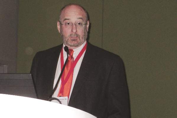
Use of limited, directed, ultrasound heart examinations on an emergency basis by physicians who are not cardiologists is “an irreversible process, but without appropriate training it may become dangerous,” Dr. Nuno Cardim said at the annual meeting of the European Association of Cardiovascular Imaging (EACVI).
A focused cardiac ultrasound (FoCUS) examination for patients with an emergency cardiac condition such as acute heart failure is not a new concept. In 2010, the American Society of Echocardiography and the American College of Emergency Physicians jointly issued a consensus statement on emergency FoCUS (J. Am. Soc. Echocardiogr. 2010;23:1225-30), and the American Society of Echocardiography followed with additional recommendations in 2013 that also dealt with nonemergency uses for FoCUS (J. Am. Soc. Echocardiogr. 2013;26:567-81).
In its 2014 position statement released last May, the EACVI directly addressed FoCUS for the first time (Eur. Heart J. Cardiovasc. Imaging 2014:15;956-60). The statement acknowledged the important role for a circumscribed, point-of-care ultrasound exam in patients undergoing cardiopulmonary resuscitation and in other critical cardiac conditions, but highlighted that a FoCUS exam does not substitute for a comprehensive echocardiographic exam, and that FoCUS should only be done by properly trained clinicians who appreciate the limits of a FoCUS exam.
The EASVI recommendations, which came out a few months later in collaboration with the Acute Cardiovascular Care Association, said that “echocardiography is now recommended (where appropriately trained practitioners are available) in the management of cardiac arrest. However, FoCUS should always be used and interpreted thoughtfully, since this fundamentally limited approach may lead to missing/misinterpretation of important findings unless the practitioner is aware of its (and their) limitations” (Eur. Heart J. Cardiovasc. Imaging 2014 [doi:10.1093/ehjci/jeu210]).
“Of course all patients with suspected acute heart failure in the emergency department should undergo an echo exam. The question is, who will do it? These are patients who are the most difficult to assess,” said Dr. Susanna Price, a member of the EACVI recommendations panel and a specialist in critical care cardiology at Royal Brompton Hospital in London.
“Without proper training, the person doing FoCUS could make a false positive diagnosis, or might miss something and make a false negative diagnosis,” said Dr. Cardim, professor and director of echocardiography and cardiac imaging at Hospital da Luz in Lisbon, and another member of the EACVI panel.
To avoid this, emergency-medicine physicians and others who often triage patients with acute heart disorders should be trained in echocardiography and especially the FoCUS exam, which aims to quickly evaluate several important abnormalities of cardiac function: pericardial effusion, cardiac tamponade, left and right ventricular size and function, and intravascular volume status. A FoCUS exam also screens for pulmonary embolism. FoCUS assesses each of these in a yes-or-no or present-or-absent way, information critical for guiding emergency management but lacking the quantitative and detailed information available with a comprehensive echocardiography exam.
“FoCUS must never substitute” for the comprehensive exam, which should always also be done, he said. FoCUS “should be used wisely and cautiously because of its limitations.”
The FoCUS exam also has equipment specifications. Ideally, clinicians should use a portable, hand-held ultrasound machine, which is larger than “pocket-sized” ultrasound devices and hence gives much better image quality compared with pocket-sized devices, Dr. Cardim said in an interview.
Dr. Cardim and Dr. Price had no disclosures.
On Twitter @mitchelzoler
VIENNA – Rapid echocardiographic assessment has become routine for many patients who arrive at an emergency department with suspected acute heart failure, and experts consider these examinations critical for quickly getting patients on the right treatment.
Growing use and the important role for emergency echo exams prompted the European echocardiography community to issue in 2014 both recommendations and a position statement on the practice.
With their actions, European echocardiographers joined their U.S. colleagues who had earlier endorsed rapid, focused echocardiography exams. The European position also highlighted the limitations and pitfalls of emergency echo and the need for proper training.
Use of limited, directed, ultrasound heart examinations on an emergency basis by physicians who are not cardiologists is “an irreversible process, but without appropriate training it may become dangerous,” Dr. Nuno Cardim said at the annual meeting of the European Association of Cardiovascular Imaging (EACVI).
A focused cardiac ultrasound (FoCUS) examination for patients with an emergency cardiac condition such as acute heart failure is not a new concept. In 2010, the American Society of Echocardiography and the American College of Emergency Physicians jointly issued a consensus statement on emergency FoCUS (J. Am. Soc. Echocardiogr. 2010;23:1225-30), and the American Society of Echocardiography followed with additional recommendations in 2013 that also dealt with nonemergency uses for FoCUS (J. Am. Soc. Echocardiogr. 2013;26:567-81).
In its 2014 position statement released last May, the EACVI directly addressed FoCUS for the first time (Eur. Heart J. Cardiovasc. Imaging 2014:15;956-60). The statement acknowledged the important role for a circumscribed, point-of-care ultrasound exam in patients undergoing cardiopulmonary resuscitation and in other critical cardiac conditions, but highlighted that a FoCUS exam does not substitute for a comprehensive echocardiographic exam, and that FoCUS should only be done by properly trained clinicians who appreciate the limits of a FoCUS exam.
The EASVI recommendations, which came out a few months later in collaboration with the Acute Cardiovascular Care Association, said that “echocardiography is now recommended (where appropriately trained practitioners are available) in the management of cardiac arrest. However, FoCUS should always be used and interpreted thoughtfully, since this fundamentally limited approach may lead to missing/misinterpretation of important findings unless the practitioner is aware of its (and their) limitations” (Eur. Heart J. Cardiovasc. Imaging 2014 [doi:10.1093/ehjci/jeu210]).
“Of course all patients with suspected acute heart failure in the emergency department should undergo an echo exam. The question is, who will do it? These are patients who are the most difficult to assess,” said Dr. Susanna Price, a member of the EACVI recommendations panel and a specialist in critical care cardiology at Royal Brompton Hospital in London.
“Without proper training, the person doing FoCUS could make a false positive diagnosis, or might miss something and make a false negative diagnosis,” said Dr. Cardim, professor and director of echocardiography and cardiac imaging at Hospital da Luz in Lisbon, and another member of the EACVI panel.
To avoid this, emergency-medicine physicians and others who often triage patients with acute heart disorders should be trained in echocardiography and especially the FoCUS exam, which aims to quickly evaluate several important abnormalities of cardiac function: pericardial effusion, cardiac tamponade, left and right ventricular size and function, and intravascular volume status. A FoCUS exam also screens for pulmonary embolism. FoCUS assesses each of these in a yes-or-no or present-or-absent way, information critical for guiding emergency management but lacking the quantitative and detailed information available with a comprehensive echocardiography exam.
“FoCUS must never substitute” for the comprehensive exam, which should always also be done, he said. FoCUS “should be used wisely and cautiously because of its limitations.”
The FoCUS exam also has equipment specifications. Ideally, clinicians should use a portable, hand-held ultrasound machine, which is larger than “pocket-sized” ultrasound devices and hence gives much better image quality compared with pocket-sized devices, Dr. Cardim said in an interview.
Dr. Cardim and Dr. Price had no disclosures.
On Twitter @mitchelzoler
EXPERT ANALYSIS FROM EUROECHO-IMAGING 2014
U.S. hospitals tout TAVR’s benefits, downplay risks
If patients seek out information online about transcatheter aortic valve replacement, they’ll read mostly about the procedure’s benefits and see much less about its risks, according to a survey of 317 U.S. hospital websites done in spring 2014.
Close examination of the information available for transcatheter aortic valve replacement (TAVR) on the websites of U.S. hospitals that perform the procedure revealed that 99% of the 262 hospitals with websites that described TAVR mentioned at least one benefit from the procedure, while 26% mentioned at least one risk, Dr. Mark D. Neuman and his associates reported in a research letter published online on Jan. 12 (JAMA Internal Medicine 2015; [doi:10.1001/jamainternmed.2014.7392]).
“Our findings suggest that web-based advertising of TAVR to the public by hospitals may understate the established risks of this procedure and provide little context for the magnitude of those risks to inform patient decision making. Hospitals may promote appropriate use of TAVR by presenting more balanced information regarding TAVR’s risks and benefits,” wrote Dr. Neuman, an anesthesiologist at the University of Pennsylvania in Philadelphia, and his coauthors.
During May-June 2014 they reviewed the websites for each of the 317 U.S. hospitals listed as sites that offer TAVR by the Society of Thoracic Surgeons and the American College of Cardiology. Fifty-five of the hospitals’ websites did not have their own English-language web page that mentioned TAVR. Of the 262 U.S. TAVR centers with a web page that described the procedure, 260 mentioned at least one benefit, most commonly the reduced degree of invasiveness of the procedure compared with open-surgery valve replacement, which appeared on 250 (95%) of the websites.
Of the 69 websites that mentioned at least one risk, they most commonly cited stroke, on 18% of the sites, followed by vascular complications, on 14%, and death, on 12%. In addition, the hospital sites supplied numerical quantification for benefits more frequently than for risks.
On Twitter @mitchelzoler
Patients often get information about their illness and possible treatments by searching on the Internet. The research letter by Dr. Neuman and his associates showed that hospital advertisements for TAVR are imbalanced. Their findings raise four significant concerns:
• Hospital websites often have the appearance of an education portal. Many of the websites for hospitals that perform TAVR bear the imprimatur of top medical centers and look like reputable health information. Patients who are referred to such pages to learn about a procedure may not be aware that they are consuming promotional materials rather than impartial educational resources.
• The regulatory environment for health care advertising in the United States is lax. Hospital advertisements may describe specific medical interventions that entail significant risks but there is no legal requirement that these risks be disclosed. Unbiased health information may be hard to find unless patients can read the medical literature.
• Patients lack a framework for evaluating what they need to know about many medical treatments and surgical procedures and, therefore, whether the information they have received is adequate. An advertisement for TAVR that fails to mention the potential for kidney injury or vascular complications is unlikely to prompt questions from patients. Poor-quality medical information is hard to recognize unless the person reading it is a clinician.
• An important shift away from medical paternalism in recent decades has meant that patient preferences are increasingly elicited and incorporated into health care decisions. Patient preferences for medical treatments are shaped by cognitive biases that may easily be exploited by online health care advertising.
An important first step toward ameliorating these concerns would be to clearly label hospital websites in a manner that allows patients to identify them as advertisements. More resources are needed to create, and direct patients to, balanced online informational tools. Clinicians should ask patients what they have learned from online medical searches and assist them in forming a complete picture of the risks and benefits of treatment options.
Finally, we must focus future attention not only on the content of health care advertising but on its impact. The risk that imbalanced information on U.S. hospital websites may negatively impact patient decision making should be an area of close scrutiny and may provide support for stricter advertising regulations.
Yael Schenker, M.D., is in the section of palliative care and medical ethics at the University of Pittsburgh. Alex John London, Ph.D., is in the department of philosophy at Carnegie Mellon University in Pittsburgh. They made these comments in an invited commentary that accompanied the report (JAMA Intern. Med. 2015 [doi:10.1001/jamainternmed.2014.7400]). They had no disclosures.
If patients seek out information online about transcatheter aortic valve replacement, they’ll read mostly about the procedure’s benefits and see much less about its risks, according to a survey of 317 U.S. hospital websites done in spring 2014.
Close examination of the information available for transcatheter aortic valve replacement (TAVR) on the websites of U.S. hospitals that perform the procedure revealed that 99% of the 262 hospitals with websites that described TAVR mentioned at least one benefit from the procedure, while 26% mentioned at least one risk, Dr. Mark D. Neuman and his associates reported in a research letter published online on Jan. 12 (JAMA Internal Medicine 2015; [doi:10.1001/jamainternmed.2014.7392]).
“Our findings suggest that web-based advertising of TAVR to the public by hospitals may understate the established risks of this procedure and provide little context for the magnitude of those risks to inform patient decision making. Hospitals may promote appropriate use of TAVR by presenting more balanced information regarding TAVR’s risks and benefits,” wrote Dr. Neuman, an anesthesiologist at the University of Pennsylvania in Philadelphia, and his coauthors.
During May-June 2014 they reviewed the websites for each of the 317 U.S. hospitals listed as sites that offer TAVR by the Society of Thoracic Surgeons and the American College of Cardiology. Fifty-five of the hospitals’ websites did not have their own English-language web page that mentioned TAVR. Of the 262 U.S. TAVR centers with a web page that described the procedure, 260 mentioned at least one benefit, most commonly the reduced degree of invasiveness of the procedure compared with open-surgery valve replacement, which appeared on 250 (95%) of the websites.
Of the 69 websites that mentioned at least one risk, they most commonly cited stroke, on 18% of the sites, followed by vascular complications, on 14%, and death, on 12%. In addition, the hospital sites supplied numerical quantification for benefits more frequently than for risks.
On Twitter @mitchelzoler
FROM JAMA INTERNAL MEDICINE
Key clinical point: Websites of U.S. hospitals that offer TAVR mention the procedure’s benefits much more often than they mention its risks.
Major finding: Benefits appeared in 99% of the websites, while risks appeared in 26% of the websites.
Data source: Survey done during May-June 2014 of 317 U.S. hospital websites.
Disclosures: Dr. Neuman and his associates had no disclosures.
Early mitral-valve repair dampens tricuspid-valve regurgitation
VIENNA – One of the best ways to prevent advanced tricuspid-valve regurgitation and need for tricuspid-valve repair may be a more aggressive approach to mitral valve repair.
“If you operate on the mitral valve early, then tricuspid regurgitation does not tend to progress,” Dr. Sunil V. Mankad said at the annual meeting of the European Association of Cardiovascular Imaging. “If you wait until the mitral valve remodels and the atrium enlarges and remodels or there is pulmonary hypertension, then tricuspid regurgitation will progress,” said Dr. Mankad, a echocardiographer at the Mayo Clinic in Rochester, Minn.

Early intervention on mitral valve prolapse has other benefits as well, he said. Mitral disease causes atrial remodeling, which can then progress to atrial fibrillation, “and once that happens it’s a game changer for the patient, even if they later undergo valve repair,” because of atrial fibrillation’s long-term risks and consequences, Dr. Mankad said in an interview.
“We believe there is also subclinical left ventricular dysfunction” in patients with mitral-valve prolapse “even if their ejection fraction is normal.” Once that happens, even if the mitral valve is repaired “the heart is not normal anymore and there is subtle left ventricular dysfunction that is not captured by just looking at ejection fraction.”
To document the impact a more aggressive approach to mitral-valve repair can have on the tricuspid valve, Dr. Mankad cited a 2011 Mayo Clinic analysis of 699 patients who underwent mitral-valve repair at Mayo for severe mitral-valve prolapse and also had some amount of tricuspid regurgitation at the time of their surgery, including 115 patients (16%) with grade 3 or higher tricuspid regurgitation. One year after surgery, the severity of tricuspid regurgitation in these patients had decreased significantly overall, and throughout follow-up only one patient required surgery for tricuspid-valve repair, 4.5 years after that patient’s mitral-valve repair (J. Thoracic Cardiovasc. Surgery 2011;142:608-13).
Dr. Mankad also cited a recent editorial written by several of his Mayo Clinic colleagues that synthesized results from the 2011 report as well as from a second Mayo report published in 2014, and a third report from a different group also published in 2014. The authors of the editorial concluded that results from all three studies showed “the performance of early correction of mitral regurgitation is important not only for its own well known benefits (preservation of survival and minimization of late heart failure risk) but also to diminish the late occurrence of functional tricuspid regurgitation (J. Thoracic Cardiovasc. Surgery 2014;148:2810-2).
Because mitral-valve repair often improves tricuspid-valve function and durability, the editorialists suggested “strongly considering” tricuspid repair for a carefully defined, select subgroup of patients. Their list included patients with tricuspid regurgitation that is worse than moderate, right-heart dysfunction, symptoms of right-heart failure, pulmonary hypertension, reduced left ventricular systolic function, cardiomyopathy, or organic tricuspid pathology.
Existing evidence supports leaving the valve alone when patients have a tricuspid regurgitation that is less than moderate when they have also undergone effective correction of degenerative mitral regurgitation. Patients like these are “unlikely ever to have difficulty with the tricuspid valve or the right ventricle,” wrote the authors of the editorial.
Dr. Mankad offered his own suggestions for identifying patients with a tricuspid valve that requires repair at the time of mitral-valve surgery.
“The evidence supports tricuspid-valve repair at the time of mitral-valve surgery if there is tricuspid annular dilatation of more than 4.0 cm measured by three-dimensional echo or greater than moderate tricuspid regurgitation. This is based on observational data and not on results from randomized control trials, but it is what I recommend,” Dr. Mankad said. “I suggest measuring the tricuspid annulus; it is quite easy to do. Directly measuring the annulus size with three-dimensional echo is pretty basic, and I think it is ready for prime time.”
Dr. Mankad had no disclosures.
On Twitter @mitchelzoler
VIENNA – One of the best ways to prevent advanced tricuspid-valve regurgitation and need for tricuspid-valve repair may be a more aggressive approach to mitral valve repair.
“If you operate on the mitral valve early, then tricuspid regurgitation does not tend to progress,” Dr. Sunil V. Mankad said at the annual meeting of the European Association of Cardiovascular Imaging. “If you wait until the mitral valve remodels and the atrium enlarges and remodels or there is pulmonary hypertension, then tricuspid regurgitation will progress,” said Dr. Mankad, a echocardiographer at the Mayo Clinic in Rochester, Minn.
Early intervention on mitral valve prolapse has other benefits as well, he said. Mitral disease causes atrial remodeling, which can then progress to atrial fibrillation, “and once that happens it’s a game changer for the patient, even if they later undergo valve repair,” because of atrial fibrillation’s long-term risks and consequences, Dr. Mankad said in an interview.
“We believe there is also subclinical left ventricular dysfunction” in patients with mitral-valve prolapse “even if their ejection fraction is normal.” Once that happens, even if the mitral valve is repaired “the heart is not normal anymore and there is subtle left ventricular dysfunction that is not captured by just looking at ejection fraction.”
To document the impact a more aggressive approach to mitral-valve repair can have on the tricuspid valve, Dr. Mankad cited a 2011 Mayo Clinic analysis of 699 patients who underwent mitral-valve repair at Mayo for severe mitral-valve prolapse and also had some amount of tricuspid regurgitation at the time of their surgery, including 115 patients (16%) with grade 3 or higher tricuspid regurgitation. One year after surgery, the severity of tricuspid regurgitation in these patients had decreased significantly overall, and throughout follow-up only one patient required surgery for tricuspid-valve repair, 4.5 years after that patient’s mitral-valve repair (J. Thoracic Cardiovasc. Surgery 2011;142:608-13).
Dr. Mankad also cited a recent editorial written by several of his Mayo Clinic colleagues that synthesized results from the 2011 report as well as from a second Mayo report published in 2014, and a third report from a different group also published in 2014. The authors of the editorial concluded that results from all three studies showed “the performance of early correction of mitral regurgitation is important not only for its own well known benefits (preservation of survival and minimization of late heart failure risk) but also to diminish the late occurrence of functional tricuspid regurgitation (J. Thoracic Cardiovasc. Surgery 2014;148:2810-2).
Because mitral-valve repair often improves tricuspid-valve function and durability, the editorialists suggested “strongly considering” tricuspid repair for a carefully defined, select subgroup of patients. Their list included patients with tricuspid regurgitation that is worse than moderate, right-heart dysfunction, symptoms of right-heart failure, pulmonary hypertension, reduced left ventricular systolic function, cardiomyopathy, or organic tricuspid pathology.
Existing evidence supports leaving the valve alone when patients have a tricuspid regurgitation that is less than moderate when they have also undergone effective correction of degenerative mitral regurgitation. Patients like these are “unlikely ever to have difficulty with the tricuspid valve or the right ventricle,” wrote the authors of the editorial.
Dr. Mankad offered his own suggestions for identifying patients with a tricuspid valve that requires repair at the time of mitral-valve surgery.
“The evidence supports tricuspid-valve repair at the time of mitral-valve surgery if there is tricuspid annular dilatation of more than 4.0 cm measured by three-dimensional echo or greater than moderate tricuspid regurgitation. This is based on observational data and not on results from randomized control trials, but it is what I recommend,” Dr. Mankad said. “I suggest measuring the tricuspid annulus; it is quite easy to do. Directly measuring the annulus size with three-dimensional echo is pretty basic, and I think it is ready for prime time.”
Dr. Mankad had no disclosures.
On Twitter @mitchelzoler
EXPERT ANALYSIS FROM EUROECHO-IMAGING 2014
3D echocardography underpins percutaneous mitral valve repair
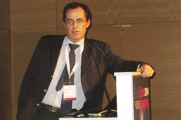
VIENNA – Three-dimensional transesophageal echocardiography “may be considered the gatekeeper for assessing the feasibility of and planning the strategy for percutaneous mitral valve repair,” Dr. Giovanni La Canna said at the annual meeting of the European Association of Association of Cardiovascular Imaging.
“Real-time, three-dimensional transesophageal echo is a new way of looking at the mitral valve,” and it supplies a highly accurate and reproducible anatomic view. It adds important additional information when selecting patients for percutaneous mitral valve repair by giving a comprehensive picture of annular dimensions and shape, intercommissural extension of target-leaflet lesions, and the extent of calcification on the annulus and leaflets, said Dr. La Canna, an echocardiographer at San Raffaele Hospital in Milan.

Three-dimensional echo aids both forms of percutaneous mitral valve repair: annuloplasty and clipping with the MitraClip system.
When performing mitral-leaflet clipping, use of 3D echo adds incremental information beyond 2D echo that aids in the selection of the site for transseptal puncture; facilitates device alignment; optimizes grasping of the leaflet with the clip; and helps in assessment of mitral-valve area and residual regurgitation, the need for additional clips, and any residual defect in the interatrial septum. For especially challenging cases, data from 2D echo can be integrated with the 3D data for the most complete imaging guidance, Dr. La Canna said.
Despite its value, 3D echo still has limitations. Resolution is currently low, parts of the imaging can “drop out” or contain artifacts, the catheter can produce an image shadow, and the image can decay during the course of the procedure, he said.
Dr. La Canna has been a consultant to Abbott Vascular, which markets the MitraClip.
On Twitter @mitchelzoler
VIENNA – Three-dimensional transesophageal echocardiography “may be considered the gatekeeper for assessing the feasibility of and planning the strategy for percutaneous mitral valve repair,” Dr. Giovanni La Canna said at the annual meeting of the European Association of Association of Cardiovascular Imaging.
“Real-time, three-dimensional transesophageal echo is a new way of looking at the mitral valve,” and it supplies a highly accurate and reproducible anatomic view. It adds important additional information when selecting patients for percutaneous mitral valve repair by giving a comprehensive picture of annular dimensions and shape, intercommissural extension of target-leaflet lesions, and the extent of calcification on the annulus and leaflets, said Dr. La Canna, an echocardiographer at San Raffaele Hospital in Milan.
Three-dimensional echo aids both forms of percutaneous mitral valve repair: annuloplasty and clipping with the MitraClip system.
When performing mitral-leaflet clipping, use of 3D echo adds incremental information beyond 2D echo that aids in the selection of the site for transseptal puncture; facilitates device alignment; optimizes grasping of the leaflet with the clip; and helps in assessment of mitral-valve area and residual regurgitation, the need for additional clips, and any residual defect in the interatrial septum. For especially challenging cases, data from 2D echo can be integrated with the 3D data for the most complete imaging guidance, Dr. La Canna said.
Despite its value, 3D echo still has limitations. Resolution is currently low, parts of the imaging can “drop out” or contain artifacts, the catheter can produce an image shadow, and the image can decay during the course of the procedure, he said.
Dr. La Canna has been a consultant to Abbott Vascular, which markets the MitraClip.
On Twitter @mitchelzoler
EXPERT ANALYSIS FROM EUROECHO-IMAGING 2014

CT Shown Most Cost Effective for Chest-Pain Assessment
VIENNA – Coronary CT followed by some form of functional stress imaging when needed now stands as the most cost-effective way to screen patients with chest pain regardless of whether the pain is chronic or acute onset.
Results from a 2011 analysis established the cost efficacy of cardiac CT followed by stress imaging for patients with positive CT results for acute chest-pain patients at low or intermediate risk for coronary disease, and no data reported since then have changed that conclusion, Dr. Thomas H. Marwick said at the annual meeting of the European Association of Cardiovascular Imaging.
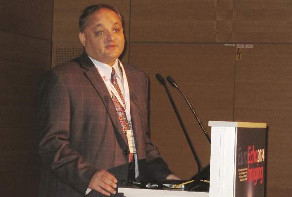
Results from a new analysis presented in a separate report at the meeting showed the superior cost effectiveness of the same approach for patients with stable chest pain who are at low or intermediate risk for having coronary disease, said Dr. Steffen E. Petersen, professor and head of the Centre for Advanced Cardiovascular Imaging at the William Harvey Research Institute in London.
“We did a cost-effectiveness analysis that took into account the cost of results that lead to additional testing and the cost when people return later with symptoms and need more testing. We compared about 15 different strategies, and found it was best to start with CT and if that was positive then do a functional test like stress echocardiography, stress single-photon emission CT (SPECT), or stress MRI. CT followed by stress echo seemed most cost effective, but using stress SPECT or MRI was not very different; they can all be used after CT,” Dr. Petersen said in an interview.
Continue for more on the analysis >>
His analysis, which modeled the costs for a man or woman aged 60 years with a 30% probability of having coronary artery disease, showed that a testing strategy of coronary CT followed by stress echo for patients with a significant coronary stenosis cost roughly £32,000 (about $50,000) for each quality-adjusted life year gained.
Choice of the follow-up, functional test for patients with stable chest pain who show at least 50% stenosis in at least one segment of their coronary vessels on CT primarily depends on other considerations, such as local experience using the various functional-imaging methods as well as how long a patient might need to wait until the stress test is done, Dr. Petersen said. For higher-risk patients who likely have significant coronary artery disease, the cost effectiveness of an initial CT examination drops below that of the alternative approach of referring the patient directly for a functional test with no initial CT. He advised clinicians to assess a patient’s pretest probability of having coronary artery disease with a tool such as the modified Diamond-Forrester model (Eur. Heart J. 2011;32:1316-30).
Dr. Petersen and his associates used already-published data from the United States, United Kingdom, and The Netherlands that documented the positive and negative predictive values of various imaging methods used alone or in tandem as well as the impact of each method on additional testing, delays while testing proceeds, and eventual patient outcomes. “We basically all agreed on our model, and then we ran the model and looked at the results,” he said.
“I think our findings would change practice in a lot of places,” he added. For patients with stable disease access to various imaging methods is usually not an issue because stable patients can receive a referral to a facility with that method available.
Continue for cost effectiveness >>
Dr. Marwick asserted that cardiac CT followed by functional testing with SPECT for patients with an indeterminate CT result remains the most cost-effective way to screen a chest-pain patient who presents at an emergency department with a history and physical findings consistent with a low or intermediate risk of having coronary disease. He and his associates documented the consistent cost effectiveness of CT followed by SPECT in patients with a coronary artery disease prevalence of anywhere from 2% to 30% in a 2011 report (J. Am. Coll. Cardiol. Img. 2011;4:549-56). A big reason why CT first is so cost effective is because of its very high negative predictive value – its ability to reliably rule out patients who do not have significant coronary disease.
“It is very hard to overcome the cost effectiveness of CT for chest pain because of its very high predictive value, and because you can use it to study patients early,” said Dr. Marwick, professor and director of the Menzies Institute for Medical Research, University of Tasmania in Hobart, Australia. “Early CT can reduce the patient’s length of stay in the emergency department while avoiding the risk of sending someone home who is having a myocardial infarction,” Dr. Marwick said in an interview. Although his analysis showed that stress SPECT was the most cost-effective option for follow-up testing of a patient with an equivocal CT result, “it could be any functional test” because they all have nearly the same performance, he added. The other options are again stress echo or stress MRI.
“If a patient has chest pain and goes directly to CT the negative predictive value is so high that more than half the patients can safely go straight home. I’m not a big fan of CT, but this is what the numbers show,” he said
Dr. Marwick acknowledged that testing a patient’s blood level of troponin using a high-sensitivity assay might soon supplant CT, but for the time being high-sensitivity troponin remain too non specific, he said. A retrospective, Swedish study reported at the 2014 annual meeting of the American College of Cardiology documented that a high-sensitivity troponin assay could rule out myocardial infarction among patients presenting with chest pain at an emergency department with a negative predictive accuracy of nearly 100%. But at that time one expert commented that proof of the clinical utility of a high-sensitivity troponin assay for emergency chest pain patients needed validation in a well-designed, prospective trial.
Dr. Petersen and Dr. Marwick had no disclosures.
VIENNA – Coronary CT followed by some form of functional stress imaging when needed now stands as the most cost-effective way to screen patients with chest pain regardless of whether the pain is chronic or acute onset.
Results from a 2011 analysis established the cost efficacy of cardiac CT followed by stress imaging for patients with positive CT results for acute chest-pain patients at low or intermediate risk for coronary disease, and no data reported since then have changed that conclusion, Dr. Thomas H. Marwick said at the annual meeting of the European Association of Cardiovascular Imaging.
Results from a new analysis presented in a separate report at the meeting showed the superior cost effectiveness of the same approach for patients with stable chest pain who are at low or intermediate risk for having coronary disease, said Dr. Steffen E. Petersen, professor and head of the Centre for Advanced Cardiovascular Imaging at the William Harvey Research Institute in London.
“We did a cost-effectiveness analysis that took into account the cost of results that lead to additional testing and the cost when people return later with symptoms and need more testing. We compared about 15 different strategies, and found it was best to start with CT and if that was positive then do a functional test like stress echocardiography, stress single-photon emission CT (SPECT), or stress MRI. CT followed by stress echo seemed most cost effective, but using stress SPECT or MRI was not very different; they can all be used after CT,” Dr. Petersen said in an interview.
Continue for more on the analysis >>
His analysis, which modeled the costs for a man or woman aged 60 years with a 30% probability of having coronary artery disease, showed that a testing strategy of coronary CT followed by stress echo for patients with a significant coronary stenosis cost roughly £32,000 (about $50,000) for each quality-adjusted life year gained.
Choice of the follow-up, functional test for patients with stable chest pain who show at least 50% stenosis in at least one segment of their coronary vessels on CT primarily depends on other considerations, such as local experience using the various functional-imaging methods as well as how long a patient might need to wait until the stress test is done, Dr. Petersen said. For higher-risk patients who likely have significant coronary artery disease, the cost effectiveness of an initial CT examination drops below that of the alternative approach of referring the patient directly for a functional test with no initial CT. He advised clinicians to assess a patient’s pretest probability of having coronary artery disease with a tool such as the modified Diamond-Forrester model (Eur. Heart J. 2011;32:1316-30).
Dr. Petersen and his associates used already-published data from the United States, United Kingdom, and The Netherlands that documented the positive and negative predictive values of various imaging methods used alone or in tandem as well as the impact of each method on additional testing, delays while testing proceeds, and eventual patient outcomes. “We basically all agreed on our model, and then we ran the model and looked at the results,” he said.
“I think our findings would change practice in a lot of places,” he added. For patients with stable disease access to various imaging methods is usually not an issue because stable patients can receive a referral to a facility with that method available.
Continue for cost effectiveness >>
Dr. Marwick asserted that cardiac CT followed by functional testing with SPECT for patients with an indeterminate CT result remains the most cost-effective way to screen a chest-pain patient who presents at an emergency department with a history and physical findings consistent with a low or intermediate risk of having coronary disease. He and his associates documented the consistent cost effectiveness of CT followed by SPECT in patients with a coronary artery disease prevalence of anywhere from 2% to 30% in a 2011 report (J. Am. Coll. Cardiol. Img. 2011;4:549-56). A big reason why CT first is so cost effective is because of its very high negative predictive value – its ability to reliably rule out patients who do not have significant coronary disease.
“It is very hard to overcome the cost effectiveness of CT for chest pain because of its very high predictive value, and because you can use it to study patients early,” said Dr. Marwick, professor and director of the Menzies Institute for Medical Research, University of Tasmania in Hobart, Australia. “Early CT can reduce the patient’s length of stay in the emergency department while avoiding the risk of sending someone home who is having a myocardial infarction,” Dr. Marwick said in an interview. Although his analysis showed that stress SPECT was the most cost-effective option for follow-up testing of a patient with an equivocal CT result, “it could be any functional test” because they all have nearly the same performance, he added. The other options are again stress echo or stress MRI.
“If a patient has chest pain and goes directly to CT the negative predictive value is so high that more than half the patients can safely go straight home. I’m not a big fan of CT, but this is what the numbers show,” he said
Dr. Marwick acknowledged that testing a patient’s blood level of troponin using a high-sensitivity assay might soon supplant CT, but for the time being high-sensitivity troponin remain too non specific, he said. A retrospective, Swedish study reported at the 2014 annual meeting of the American College of Cardiology documented that a high-sensitivity troponin assay could rule out myocardial infarction among patients presenting with chest pain at an emergency department with a negative predictive accuracy of nearly 100%. But at that time one expert commented that proof of the clinical utility of a high-sensitivity troponin assay for emergency chest pain patients needed validation in a well-designed, prospective trial.
Dr. Petersen and Dr. Marwick had no disclosures.
CT shown most cost effective for chest-pain assessment
VIENNA – Coronary CT followed by some form of functional stress imaging when needed now stands as the most cost-effective way to screen patients with chest pain regardless of whether the pain is chronic or acute onset.
Results from a 2011 analysis established the cost efficacy of cardiac CT followed by stress imaging for patients with positive CT results for acute chest-pain patients at low or intermediate risk for coronary disease, and no data reported since then have changed that conclusion, Dr. Thomas H. Marwick said at the annual meeting of the European Association of Cardiovascular Imaging.
Results from a new analysis presented in a separate report at the meeting showed the superior cost effectiveness of the same approach for patients with stable chest pain who are at low or intermediate risk for having coronary disease, said Dr. Steffen E. Petersen, professor and head of the Centre for Advanced Cardiovascular Imaging at the William Harvey Research Institute in London.

“We did a cost-effectiveness analysis that took into account the cost of results that lead to additional testing and the cost when people return later with symptoms and need more testing. We compared about 15 different strategies, and found it was best to start with CT and if that was positive then do a functional test like stress echocardiography, stress single-photon emission CT (SPECT), or stress MRI. CT followed by stress echo seemed most cost effective, but using stress SPECT or MRI was not very different; they can all be used after CT,” Dr. Petersen said in an interview.
His analysis, which modeled the costs for a man or woman aged 60 years with a 30% probability of having coronary artery disease, showed that a testing strategy of coronary CT followed by stress echo for patients with a significant coronary stenosis cost roughly £32,000 (about $50,000) for each quality-adjusted life year gained.
Choice of the follow-up, functional test for patients with stable chest pain who show at least 50% stenosis in at least one segment of their coronary vessels on CT primarily depends on other considerations, such as local experience using the various functional-imaging methods as well as how long a patient might need to wait until the stress test is done, Dr. Petersen said. For higher-risk patients who likely have significant coronary artery disease, the cost effectiveness of an initial CT examination drops below that of the alternative approach of referring the patient directly for a functional test with no initial CT. He advised clinicians to assess a patient’s pretest probability of having coronary artery disease with a tool such as the modified Diamond-Forrester model (Eur. Heart J. 2011;32:1316-30).

Dr. Petersen and his associates used already-published data from the United States, United Kingdom, and The Netherlands that documented the positive and negative predictive values of various imaging methods used alone or in tandem as well as the impact of each method on additional testing, delays while testing proceeds, and eventual patient outcomes. “We basically all agreed on our model, and then we ran the model and looked at the results,” he said.
“I think our findings would change practice in a lot of places,” he added. For patients with stable disease access to various imaging methods is usually not an issue because stable patients can receive a referral to a facility with that method available.
Dr. Marwick asserted that cardiac CT followed by functional testing with SPECT for patients with an indeterminate CT result remains the most cost-effective way to screen a chest-pain patient who presents at an emergency department with a history and physical findings consistent with a low or intermediate risk of having coronary disease. He and his associates documented the consistent cost effectiveness of CT followed by SPECT in patients with a coronary artery disease prevalence of anywhere from 2% to 30% in a 2011 report (J. Am. Coll. Cardiol. Img. 2011;4:549-56). A big reason why CT first is so cost effective is because of its very high negative predictive value – its ability to reliably rule out patients who do not have significant coronary disease.
“It is very hard to overcome the cost effectiveness of CT for chest pain because of its very high predictive value, and because you can use it to study patients early,” said Dr. Marwick, professor and director of the Menzies Institute for Medical Research, University of Tasmania in Hobart, Australia. “Early CT can reduce the patient’s length of stay in the emergency department while avoiding the risk of sending someone home who is having a myocardial infarction,” Dr. Marwick said in an interview. Although his analysis showed that stress SPECT was the most cost-effective option for follow-up testing of a patient with an equivocal CT result, “it could be any functional test” because they all have nearly the same performance, he added. The other options are again stress echo or stress MRI.
“If a patient has chest pain and goes directly to CT the negative predictive value is so high that more than half the patients can safely go straight home. I’m not a big fan of CT, but this is what the numbers show,” he said
Dr. Marwick acknowledged that testing a patient’s blood level of troponin using a high-sensitivity assay might soon supplant CT, but for the time being high-sensitivity troponin remain too non specific, he said. A retrospective, Swedish study reported at the 2014 annual meeting of the American College of Cardiology documented that a high-sensitivity troponin assay could rule out myocardial infarction among patients presenting with chest pain at an emergency department with a negative predictive accuracy of nearly 100%. But at that time one expert commented that proof of the clinical utility of a high-sensitivity troponin assay for emergency chest pain patients needed validation in a well-designed, prospective trial.
Dr. Petersen and Dr. Marwick had no disclosures.
On Twitter @mitchelzoler
VIENNA – Coronary CT followed by some form of functional stress imaging when needed now stands as the most cost-effective way to screen patients with chest pain regardless of whether the pain is chronic or acute onset.
Results from a 2011 analysis established the cost efficacy of cardiac CT followed by stress imaging for patients with positive CT results for acute chest-pain patients at low or intermediate risk for coronary disease, and no data reported since then have changed that conclusion, Dr. Thomas H. Marwick said at the annual meeting of the European Association of Cardiovascular Imaging.
Results from a new analysis presented in a separate report at the meeting showed the superior cost effectiveness of the same approach for patients with stable chest pain who are at low or intermediate risk for having coronary disease, said Dr. Steffen E. Petersen, professor and head of the Centre for Advanced Cardiovascular Imaging at the William Harvey Research Institute in London.
“We did a cost-effectiveness analysis that took into account the cost of results that lead to additional testing and the cost when people return later with symptoms and need more testing. We compared about 15 different strategies, and found it was best to start with CT and if that was positive then do a functional test like stress echocardiography, stress single-photon emission CT (SPECT), or stress MRI. CT followed by stress echo seemed most cost effective, but using stress SPECT or MRI was not very different; they can all be used after CT,” Dr. Petersen said in an interview.
His analysis, which modeled the costs for a man or woman aged 60 years with a 30% probability of having coronary artery disease, showed that a testing strategy of coronary CT followed by stress echo for patients with a significant coronary stenosis cost roughly £32,000 (about $50,000) for each quality-adjusted life year gained.
Choice of the follow-up, functional test for patients with stable chest pain who show at least 50% stenosis in at least one segment of their coronary vessels on CT primarily depends on other considerations, such as local experience using the various functional-imaging methods as well as how long a patient might need to wait until the stress test is done, Dr. Petersen said. For higher-risk patients who likely have significant coronary artery disease, the cost effectiveness of an initial CT examination drops below that of the alternative approach of referring the patient directly for a functional test with no initial CT. He advised clinicians to assess a patient’s pretest probability of having coronary artery disease with a tool such as the modified Diamond-Forrester model (Eur. Heart J. 2011;32:1316-30).
Dr. Petersen and his associates used already-published data from the United States, United Kingdom, and The Netherlands that documented the positive and negative predictive values of various imaging methods used alone or in tandem as well as the impact of each method on additional testing, delays while testing proceeds, and eventual patient outcomes. “We basically all agreed on our model, and then we ran the model and looked at the results,” he said.
“I think our findings would change practice in a lot of places,” he added. For patients with stable disease access to various imaging methods is usually not an issue because stable patients can receive a referral to a facility with that method available.
Dr. Marwick asserted that cardiac CT followed by functional testing with SPECT for patients with an indeterminate CT result remains the most cost-effective way to screen a chest-pain patient who presents at an emergency department with a history and physical findings consistent with a low or intermediate risk of having coronary disease. He and his associates documented the consistent cost effectiveness of CT followed by SPECT in patients with a coronary artery disease prevalence of anywhere from 2% to 30% in a 2011 report (J. Am. Coll. Cardiol. Img. 2011;4:549-56). A big reason why CT first is so cost effective is because of its very high negative predictive value – its ability to reliably rule out patients who do not have significant coronary disease.
“It is very hard to overcome the cost effectiveness of CT for chest pain because of its very high predictive value, and because you can use it to study patients early,” said Dr. Marwick, professor and director of the Menzies Institute for Medical Research, University of Tasmania in Hobart, Australia. “Early CT can reduce the patient’s length of stay in the emergency department while avoiding the risk of sending someone home who is having a myocardial infarction,” Dr. Marwick said in an interview. Although his analysis showed that stress SPECT was the most cost-effective option for follow-up testing of a patient with an equivocal CT result, “it could be any functional test” because they all have nearly the same performance, he added. The other options are again stress echo or stress MRI.
“If a patient has chest pain and goes directly to CT the negative predictive value is so high that more than half the patients can safely go straight home. I’m not a big fan of CT, but this is what the numbers show,” he said
Dr. Marwick acknowledged that testing a patient’s blood level of troponin using a high-sensitivity assay might soon supplant CT, but for the time being high-sensitivity troponin remain too non specific, he said. A retrospective, Swedish study reported at the 2014 annual meeting of the American College of Cardiology documented that a high-sensitivity troponin assay could rule out myocardial infarction among patients presenting with chest pain at an emergency department with a negative predictive accuracy of nearly 100%. But at that time one expert commented that proof of the clinical utility of a high-sensitivity troponin assay for emergency chest pain patients needed validation in a well-designed, prospective trial.
Dr. Petersen and Dr. Marwick had no disclosures.
On Twitter @mitchelzoler
AT EUROECHO-IMAGING 2014
Key clinical point: For both stable and acute chest-pain patients initial assessment by coronary CT produced the most cost-effective results.
Major finding: Assessing stable chest-pain patients by coronary CT followed by stress echocardiography cost £32,000 per quality-adjusted life year.
Data source: Model that ran published data from the United States, the United Kingdom, and The Netherlands.
Disclosures: Dr. Petersen and Dr. Marwick had no disclosures.

ADA’s revised diabetes 'standards' broaden statin use
Most patients with diabetes should receive at least a moderate statin dosage regardless of their cardiovascular disease risk profile, according to the American Diabetes Association’s annual update to standards for managing patients with diabetes.
“Standards of Medical Care in Diabetes–2015” also shifts the ADA’s official recommendation on assessing patients for statin treatment from a decision based on blood levels of low density lipoprotein (LDL) cholesterol to a risk-based assessment. That change brings the ADA’s position in line with the approach advocated in late 2013 by guidelines from the American College of Cardiology (ACC) and the American Heart Association (AHA) (J. Am. Coll. Cardiol. 2014;63:2889-934).
The ADA released the revised standards online Dec. 23.
The statin use recommendation is “a major change, a fairly big change in how we provide care, although not that big a change in what most patients are prescribed,” said Dr. Richard W. Grant, a primary care physician and researcher at Kaiser Permanente Northern California in Oakland and chair of the ADA’s Professional Practice Committee, the 14-member panel that produced the revised standards.

“We agreed [with the 2013 ACC and AHA lipid guidelines] that the decision to start a statin should be based on a patient’s cardiovascular disease risk, and it turns out that nearly every patient with type 2 diabetes should be on a statin,” Dr. Grant said in an interview.
The revised standards recommend a “moderate” statin dosage for patients with diabetes who are aged 40-75 years, as well as those who are older than 75 years even if they have no other cardiovascular disease risk factors (Diabetes Care 2015;38:S1-S94).
The dosage should be intensified to “high” for patients with diagnosed cardiovascular disease, and for patients aged 40-75 years with other cardiovascular disease risk factors. For patients older than 75 years with cardiovascular disease risk factors, the new revision calls for either a moderate or high dosage.
However, for patients younger than 40 years with no cardiovascular disease or risk factors, the revised standards call for no statin treatment, a moderate or high dosage for patients younger than 40 years with risk factors, and a high dosage for those with cardiovascular disease.
The ADA’s recommendation for no statin treatment of the youngest and lowest-risk patients with diabetes is somewhat at odds with the 2013 ACC and AHA recommendations. For this patient group, those recommendations said, “statin therapy should be individualized on the basis of considerations of atherosclerotic cardiovascular disease risk-reduction benefits, the potential for adverse effects and drug-drug interactions, and patient preferences.”
The new standards revision contains several other changes, including:
• The recommended goal diastolic blood pressure for patients with diabetes was revised to less than 90 mm Hg, an increase from the 80–mm Hg target that had been in place. That change follows a revision in the ADA’s 2014 standards that increased the systolic blood pressure target to less than 140 mm Hg.
Changing the diastolic target to less than 90 mm Hg was primarily a matter of following the best evidence that exists in the literature, Dr. Grant said, because only lower-grade evidence supports a target of less than 80 mm Hg.
The revised standards also note that the new targets of less than 140/90 mm Hg put the standards “ in harmonization” with the 2014 recommendations of the panel originally assembled at the Eighth Joint National Committee (JAMA 2014;311:507-20).
• The recommended blood glucose target when measured before eating is now 80-130 mg/dL, with the lower limit increased from 70 mg/dL. That change reflects new data that correlate blood glucose levels with blood levels of hemoglobin A1c.
• The revision sets the body mass index cutpoint for screening overweight or obese Asian Americans at 23 kg/m2, an increase from the prior cutpoint of 25 kg/m2.
• A new section devoted to managing patients with diabetes during pregnancy draws together information that previously had been scattered throughout the standards document, Dr. Grant explained. The section discusses gestational diabetes management, as well as managing women who had preexisting type 1 or type 2 diabetes prior to becoming pregnant.
Dr. Grant had no disclosures.
On Twitter @mitchelzoler
The efficacy of a moderate statin dosage for primary prevention of cardiovascular disease events in patients age 40-75 years with type 2 diabetes and no other risk factors was clearly established a decade ago by results from the Collaborative Atorvastatin Diabetes Study (CARDS) (Lancet 2004;364:685-96).
No prospective, randomized study has proved the efficacy of statin treatment in patients younger than 40 years with diabetes and no other risk factors; but we see increasing numbers of these patients, and they, too, are at high risk for cardiovascular disease events. I agree with the 2013 recommendation from the American College of Cardiology and American Heart Association that statin treatment should be discussed and in many cases started for these younger, lower-risk patients who still face an important cardiovascular disease risk from their diabetes alone.
Changing the target diastolic blood pressure to less than 90 mm Hg is also consistent with existing evidence. A few years ago, I wrote in an editorial that some prior blood pressure targets for patients with diabetes had been set too low (Circulation 2011;123:2776-8).
There is no evidence that patients with diabetes will benefit from a diastolic blood pressure target that is lower than less than 90 mm Hg, and an overly aggressive approach to blood pressure reduction potentially can cause adverse events. Elderly patients with diabetes often have “silent” coronary artery disease, and if their diastolic pressure goes too low, they can have inadequate coronary perfusion that will cause coronary ischemia.

|
Dr. Prakash Deedwania |
But the diastolic blood pressure target also needs individualization. Some patients, such as those with Asian ethnicity, may benefit from the greater stroke reduction achieved with more aggressive blood pressure reduction.
Aspirin use in patients with diabetes and no other cardiovascular disease risk factors has been controversial, but recent evidence from the Japanese Primary Prevention Project suggests it does not benefit patients with diabetes, even if they may also have hypertension, dyslipidemia, or both. About a third of the patients aged 60-85 years enrolled in this Japanese study had diabetes, more than 70% had dyslipidemia, and 85% had hypertension. But despite this background, daily low-dose aspirin did not reduce the incidence of atherosclerotic cardiovascular disease events during 5 years of follow-up of more than 14,000 randomized patients (JAMA 2014;312:2510-20).
Dr. Prakash C. Deedwania is professor of medicine at the University of California, San Francisco, and director of cardiology at the VA Central California Health Care System in Fresno. He made these comments in an interview. He has served as a consultant to several drug companies that market statins.
The efficacy of a moderate statin dosage for primary prevention of cardiovascular disease events in patients age 40-75 years with type 2 diabetes and no other risk factors was clearly established a decade ago by results from the Collaborative Atorvastatin Diabetes Study (CARDS) (Lancet 2004;364:685-96).
No prospective, randomized study has proved the efficacy of statin treatment in patients younger than 40 years with diabetes and no other risk factors; but we see increasing numbers of these patients, and they, too, are at high risk for cardiovascular disease events. I agree with the 2013 recommendation from the American College of Cardiology and American Heart Association that statin treatment should be discussed and in many cases started for these younger, lower-risk patients who still face an important cardiovascular disease risk from their diabetes alone.
Changing the target diastolic blood pressure to less than 90 mm Hg is also consistent with existing evidence. A few years ago, I wrote in an editorial that some prior blood pressure targets for patients with diabetes had been set too low (Circulation 2011;123:2776-8).
There is no evidence that patients with diabetes will benefit from a diastolic blood pressure target that is lower than less than 90 mm Hg, and an overly aggressive approach to blood pressure reduction potentially can cause adverse events. Elderly patients with diabetes often have “silent” coronary artery disease, and if their diastolic pressure goes too low, they can have inadequate coronary perfusion that will cause coronary ischemia.
|
|
Dr. Prakash Deedwania |
But the diastolic blood pressure target also needs individualization. Some patients, such as those with Asian ethnicity, may benefit from the greater stroke reduction achieved with more aggressive blood pressure reduction.
Aspirin use in patients with diabetes and no other cardiovascular disease risk factors has been controversial, but recent evidence from the Japanese Primary Prevention Project suggests it does not benefit patients with diabetes, even if they may also have hypertension, dyslipidemia, or both. About a third of the patients aged 60-85 years enrolled in this Japanese study had diabetes, more than 70% had dyslipidemia, and 85% had hypertension. But despite this background, daily low-dose aspirin did not reduce the incidence of atherosclerotic cardiovascular disease events during 5 years of follow-up of more than 14,000 randomized patients (JAMA 2014;312:2510-20).
Dr. Prakash C. Deedwania is professor of medicine at the University of California, San Francisco, and director of cardiology at the VA Central California Health Care System in Fresno. He made these comments in an interview. He has served as a consultant to several drug companies that market statins.
Most patients with diabetes should receive at least a moderate statin dosage regardless of their cardiovascular disease risk profile, according to the American Diabetes Association’s annual update to standards for managing patients with diabetes.
“Standards of Medical Care in Diabetes–2015” also shifts the ADA’s official recommendation on assessing patients for statin treatment from a decision based on blood levels of low density lipoprotein (LDL) cholesterol to a risk-based assessment. That change brings the ADA’s position in line with the approach advocated in late 2013 by guidelines from the American College of Cardiology (ACC) and the American Heart Association (AHA) (J. Am. Coll. Cardiol. 2014;63:2889-934).
The ADA released the revised standards online Dec. 23.
The statin use recommendation is “a major change, a fairly big change in how we provide care, although not that big a change in what most patients are prescribed,” said Dr. Richard W. Grant, a primary care physician and researcher at Kaiser Permanente Northern California in Oakland and chair of the ADA’s Professional Practice Committee, the 14-member panel that produced the revised standards.
“We agreed [with the 2013 ACC and AHA lipid guidelines] that the decision to start a statin should be based on a patient’s cardiovascular disease risk, and it turns out that nearly every patient with type 2 diabetes should be on a statin,” Dr. Grant said in an interview.
The revised standards recommend a “moderate” statin dosage for patients with diabetes who are aged 40-75 years, as well as those who are older than 75 years even if they have no other cardiovascular disease risk factors (Diabetes Care 2015;38:S1-S94).
The dosage should be intensified to “high” for patients with diagnosed cardiovascular disease, and for patients aged 40-75 years with other cardiovascular disease risk factors. For patients older than 75 years with cardiovascular disease risk factors, the new revision calls for either a moderate or high dosage.
However, for patients younger than 40 years with no cardiovascular disease or risk factors, the revised standards call for no statin treatment, a moderate or high dosage for patients younger than 40 years with risk factors, and a high dosage for those with cardiovascular disease.
The ADA’s recommendation for no statin treatment of the youngest and lowest-risk patients with diabetes is somewhat at odds with the 2013 ACC and AHA recommendations. For this patient group, those recommendations said, “statin therapy should be individualized on the basis of considerations of atherosclerotic cardiovascular disease risk-reduction benefits, the potential for adverse effects and drug-drug interactions, and patient preferences.”
The new standards revision contains several other changes, including:
• The recommended goal diastolic blood pressure for patients with diabetes was revised to less than 90 mm Hg, an increase from the 80–mm Hg target that had been in place. That change follows a revision in the ADA’s 2014 standards that increased the systolic blood pressure target to less than 140 mm Hg.
Changing the diastolic target to less than 90 mm Hg was primarily a matter of following the best evidence that exists in the literature, Dr. Grant said, because only lower-grade evidence supports a target of less than 80 mm Hg.
The revised standards also note that the new targets of less than 140/90 mm Hg put the standards “ in harmonization” with the 2014 recommendations of the panel originally assembled at the Eighth Joint National Committee (JAMA 2014;311:507-20).
• The recommended blood glucose target when measured before eating is now 80-130 mg/dL, with the lower limit increased from 70 mg/dL. That change reflects new data that correlate blood glucose levels with blood levels of hemoglobin A1c.
• The revision sets the body mass index cutpoint for screening overweight or obese Asian Americans at 23 kg/m2, an increase from the prior cutpoint of 25 kg/m2.
• A new section devoted to managing patients with diabetes during pregnancy draws together information that previously had been scattered throughout the standards document, Dr. Grant explained. The section discusses gestational diabetes management, as well as managing women who had preexisting type 1 or type 2 diabetes prior to becoming pregnant.
Dr. Grant had no disclosures.
On Twitter @mitchelzoler
FROM DIABETES CARE

When cardiologists attend meetings, do patients benefit?
Can the high-intensity care given acutely ill, high-risk U.S. patients with cardiac disease actually harm them?
Results from an unusual analysis of cardiology-meeting times seem to suggest that sobering possibility. Patient outcomes improved when thousands of high-level, American cardiologists left their practices for a few days each year to attend either of the two major U.S. heart disease meetings.
Researchers led by Dr. Anupam B. Jena of Harvard University, Boston, used Medicare data to examine mortality rates among patients hospitalized for cardiac arrest, heart failure, or acute myocardial infarction during 2002-2011. They focused on patients admitted during the annual meetings of the American College of Cardiology (usually in March) or the American Heart Association (during November).
As controls in their case-control analyses, they used data from patients admitted on similar days of the week during the 3 weeks immediately before or after these two meetings. This gave them roughly 11,000 total patients with cardiac arrest, nearly 134,000 with heart failure, and about 60,000 with acute MI – about 14% of patients in each disease category admitted during a meeting and the other 86% (controls) admitted when there was no meeting.
The results showed some statistically significant differences indicating that patients did better during the meetings, presumably when many cardiologists were away from their hospitals. These associations only occurred at teaching hospitals and among patients at high risk for inpatient mortality. The investigators saw no statistically significant differences, after adjustment, in mortality during meetings among patients treated at nonteaching hospitals or among patients with a low risk for inpatient mortality.
In analyses that adjusted for baseline differences in risk factors, the 30-day mortality rate for patients admitted to teaching hospitals with cardiac arrest was 69% during control dates and 59% during the meetings. Thirty-day mortality for patients admitted with heart failure was 25% during control dates and 18% during the meetings, researchers reported in an article published online on Dec. 22 (JAMA Intern. Med. 2014 [doi:10.1001/jamainternmed.2014.6781]).
Although 30-day mortality among patients admitted with an acute MI did not differ significantly at teaching hospitals between patients who presented during a major meeting and those who did not, the results showed that these similar mortality rates were achieved despite a statistically significant difference in the rate of percutaneous coronary interventions (PCI) that patients received: During the major meetings, 21% of the acute MI patients underwent PCI, but when a meeting was not in progress, the PCI rate jumped to 28% of all acute MI patients.
“One explanation for these findings is that the intensity of care provided during meeting dates is lower, and that for high-risk patients with cardiovascular disease, the harms of this care may unexpectedly outweigh the benefits,” Dr. Jena concluded.
It’s a remarkable and surprising finding, but can it be taken seriously? At least one expert said yes, at least seriously enough to warrant further study and consideration.
An editor’s note published with the new report suggested a plausible explanation for the findings is that “more interventions in high-risk patients with heart failure and cardiac arrest leads to higher mortality.” In her note, Dr. Rita F. Redberg, a cardiologist at the University of California, San Francisco, and editor of JAMA Internal Medicine, concluded, “It is reassuring that patient outcomes do not suffer while many cardiologists are away. More important, this analysis may help us to understand how we could lower mortality throughout the year.”
It will be interesting to see if anyone takes up the challenge to further explore this relationship and tries to find ways to apply throughout the year the protective effect of having fewer teaching-hospital cardiologists around. If a drug had this beneficial effect on mortality, the pharmaceutical industry would be all over it.
On Twitter @mitchelzoler

Hormone therapy ‘timing hypothesis’ gains ground in ELITE
CHICAGO– The “timing hypothesis” of estrogen’s ability to slow progression of atherosclerotic disease in relatively young postmenopausal women received a boost in results from a randomized trial with 643 participants.
The trial, designed about a decade ago to test the timing hypothesis, produced results showing that systemic treatment with 17beta-estradiol alone (plus topical progesterone) slowed progression of carotid intima-media thickness compared with placebo in women fewer than 6 years out from menopause, while the same treatment had no effect compared with placebo in women 10 or more years removed from menopause, Dr. Howard N. Hodis said at the American Heart Association Scientific Sessions.

The new results “support the timing hypothesis, whereby women who start hormone therapy within 6 years of menopause show a significant slowing of subclinical carotid-artery atherosclerosis, whereas women who are more than 10 years postmenopausal when starting hormone therapy show no difference from placebo,” said Dr. Hodis, professor of medicine and director of the atherosclerosis research unit at the University of Southern California, Los Angeles. The findings “are consistent with the majority of the literature” that previously reported evidence supporting a net benefit from hormone therapy when started in women who are young, and not long after they entered menopause, he added.
The ELITE (Early Versus Late Intervention Trial With Estradiol) investigators enrolled 643 postmenopausal women at the University of Southern California and stratified them into two subgroups: women fewer than 6 years removed from the onset of menopause, and women 10 or more years out from menopause. The average age of women in the younger group was about 55 years, and they averaged just under 4 years since menopause onset. Women in the older group averaged about 64 years and were an average of about 14 years removed from menopause onset.
Dr. Hodis and his associates randomized women within each of the two subgroups to daily treatment with 1 mg oral, micronized 17beta-estradiol or placebo. Women treated with estradiol who had an intact uterus also applied a micronized progesterone gel on 12 days out of every cycle, while those who received a daily placebo also applied a placebo gel. The study’s primary endpoint was the rate of change in the intima-media thickness (IMT) of the common carotid artery, measured once every 6 months. Compliance with the assigned regimens reached or exceeded 98% among all women in the study.
During 6 years of follow-up, the rate of increase in carotid IMT among the women who entered the study at least 10 years after the start of menopause tracked nearly identically between those on hormone therapy and those on placebo, with a difference between the two arms that was not statistically significant. In contrast, the women who began hormone therapy fewer than 6 years after menopause onset showed a statically significant difference in the rate of change in their carotid IMT, depending on their treatment assignment. After 6 years, the average increase in carotid IMT was roughly 40% less in women who received hormone therapy, compared with those randomized to placebo. The difference in IMT change between the older women and the younger women was also statistically significant, Dr. Hodis reported.
Dr. Hodis had no disclosures.
On Twitter@mitchelzoler
The results from the ELITE study are consistent with what we found in the Women’s Health Initiative. Last year, my collaborators from the Women’s Health Initiative and I reported that among the subgroup of women treated with conjugated equine estrogens alone, women who entered the study at age 50-59 years showed a net reduction, compared with the placebo-treated controls, in both total mortality and by a composite tally of death plus nonfatal events that included coronary heart disease, stroke, and five other adverse outcomes (JAMA 2013;310:1358-68). Women aged 60-69 years on estrogen alone had a virtually identical number of events as the placebo group, and women aged 70-79 years had a substantial excess of deaths and composite events, compared with their placebo counterparts. This age-based difference in response was statistically significant, and a remarkable pattern that we did not see in women who received with estrogen plus systemic treatment with medroxyprogesterone.
It was clear that age, as well as time from the start of menopause, played a role in the estrogen-alone arm of the Women’s Health Initiative.

|
| Whitney McKnight/Frontline Medical News Dr. JoAnn E. Manson |
These findings prompted me and my associates to formulate an algorithm and mobile app for managing menopausal symptoms and deciding whether or not a postmenopausal woman is a good candidate for hormonal therapy (Menopause 2014; 22:1-7). We do not endorse hormone therapy for any purpose other than suggesting it as an option to consider for postmenopausal women who experience moderate to severe hot flashes, night sweats, or both. Among these women, we suggest that hormone therapy is a reasonable option for women who are 10 or fewer years removed from menopause onset and if their 10-year risk for an atherosclerotic cardiovascular disease event is 5% or less as calculated by the 2013 risk calculator developed by the American Heart Association and American College of Cardiology. If these women 10 or fewer years out from their menopause start have a 6%-10% 10-year risk we also endorse hormone therapy as an option, but in a transdermal formulation. For women more than 10 years out from menopause, and for those with a greater than 10% 10-year risk, we suggest avoiding hormone therapy.
Dr. JoAnn E. Manson is professor of medicine and chief of preventive medicine at Harvard University and Brigham and Women’s Hospital, both in Boston. She had no disclosures. She made these comments in a lecture at the American Heart Association Scientific Sessions. Dr. Manson has served as lead investigator from the Women’s Health Initiative since the study began.
The results from the ELITE study are consistent with what we found in the Women’s Health Initiative. Last year, my collaborators from the Women’s Health Initiative and I reported that among the subgroup of women treated with conjugated equine estrogens alone, women who entered the study at age 50-59 years showed a net reduction, compared with the placebo-treated controls, in both total mortality and by a composite tally of death plus nonfatal events that included coronary heart disease, stroke, and five other adverse outcomes (JAMA 2013;310:1358-68). Women aged 60-69 years on estrogen alone had a virtually identical number of events as the placebo group, and women aged 70-79 years had a substantial excess of deaths and composite events, compared with their placebo counterparts. This age-based difference in response was statistically significant, and a remarkable pattern that we did not see in women who received with estrogen plus systemic treatment with medroxyprogesterone.
It was clear that age, as well as time from the start of menopause, played a role in the estrogen-alone arm of the Women’s Health Initiative.
|
|
| Whitney McKnight/Frontline Medical News Dr. JoAnn E. Manson |
These findings prompted me and my associates to formulate an algorithm and mobile app for managing menopausal symptoms and deciding whether or not a postmenopausal woman is a good candidate for hormonal therapy (Menopause 2014; 22:1-7). We do not endorse hormone therapy for any purpose other than suggesting it as an option to consider for postmenopausal women who experience moderate to severe hot flashes, night sweats, or both. Among these women, we suggest that hormone therapy is a reasonable option for women who are 10 or fewer years removed from menopause onset and if their 10-year risk for an atherosclerotic cardiovascular disease event is 5% or less as calculated by the 2013 risk calculator developed by the American Heart Association and American College of Cardiology. If these women 10 or fewer years out from their menopause start have a 6%-10% 10-year risk we also endorse hormone therapy as an option, but in a transdermal formulation. For women more than 10 years out from menopause, and for those with a greater than 10% 10-year risk, we suggest avoiding hormone therapy.
Dr. JoAnn E. Manson is professor of medicine and chief of preventive medicine at Harvard University and Brigham and Women’s Hospital, both in Boston. She had no disclosures. She made these comments in a lecture at the American Heart Association Scientific Sessions. Dr. Manson has served as lead investigator from the Women’s Health Initiative since the study began.
CHICAGO– The “timing hypothesis” of estrogen’s ability to slow progression of atherosclerotic disease in relatively young postmenopausal women received a boost in results from a randomized trial with 643 participants.
The trial, designed about a decade ago to test the timing hypothesis, produced results showing that systemic treatment with 17beta-estradiol alone (plus topical progesterone) slowed progression of carotid intima-media thickness compared with placebo in women fewer than 6 years out from menopause, while the same treatment had no effect compared with placebo in women 10 or more years removed from menopause, Dr. Howard N. Hodis said at the American Heart Association Scientific Sessions.
The new results “support the timing hypothesis, whereby women who start hormone therapy within 6 years of menopause show a significant slowing of subclinical carotid-artery atherosclerosis, whereas women who are more than 10 years postmenopausal when starting hormone therapy show no difference from placebo,” said Dr. Hodis, professor of medicine and director of the atherosclerosis research unit at the University of Southern California, Los Angeles. The findings “are consistent with the majority of the literature” that previously reported evidence supporting a net benefit from hormone therapy when started in women who are young, and not long after they entered menopause, he added.
The ELITE (Early Versus Late Intervention Trial With Estradiol) investigators enrolled 643 postmenopausal women at the University of Southern California and stratified them into two subgroups: women fewer than 6 years removed from the onset of menopause, and women 10 or more years out from menopause. The average age of women in the younger group was about 55 years, and they averaged just under 4 years since menopause onset. Women in the older group averaged about 64 years and were an average of about 14 years removed from menopause onset.
Dr. Hodis and his associates randomized women within each of the two subgroups to daily treatment with 1 mg oral, micronized 17beta-estradiol or placebo. Women treated with estradiol who had an intact uterus also applied a micronized progesterone gel on 12 days out of every cycle, while those who received a daily placebo also applied a placebo gel. The study’s primary endpoint was the rate of change in the intima-media thickness (IMT) of the common carotid artery, measured once every 6 months. Compliance with the assigned regimens reached or exceeded 98% among all women in the study.
During 6 years of follow-up, the rate of increase in carotid IMT among the women who entered the study at least 10 years after the start of menopause tracked nearly identically between those on hormone therapy and those on placebo, with a difference between the two arms that was not statistically significant. In contrast, the women who began hormone therapy fewer than 6 years after menopause onset showed a statically significant difference in the rate of change in their carotid IMT, depending on their treatment assignment. After 6 years, the average increase in carotid IMT was roughly 40% less in women who received hormone therapy, compared with those randomized to placebo. The difference in IMT change between the older women and the younger women was also statistically significant, Dr. Hodis reported.
Dr. Hodis had no disclosures.
On Twitter@mitchelzoler
AT THE AHA SCIENTIFIC SESSIONS
Key clinical point: Hormone therapy cut atherosclerotic progression, compared with placebo, in women 6 or fewer years out from menopause start, but not in those 10 or more years out.
Major finding: In women up to 6 years out from menopause, hormone therapy was linked with 40% less atherosclerotic progression than with placebo.
Data source: ELITE,a single-center, randomized study with 643 postmenopausal women.
Disclosures: Dr. Hodis had no disclosures.