User login
Mitchel is a reporter for MDedge based in the Philadelphia area. He started with the company in 1992, when it was International Medical News Group (IMNG), and has since covered a range of medical specialties. Mitchel trained as a virologist at Roswell Park Memorial Institute in Buffalo, and then worked briefly as a researcher at Boston Children's Hospital before pivoting to journalism as a AAAS Mass Media Fellow in 1980. His first reporting job was with Science Digest magazine, and from the mid-1980s to early-1990s he was a reporter with Medical World News. @mitchelzoler
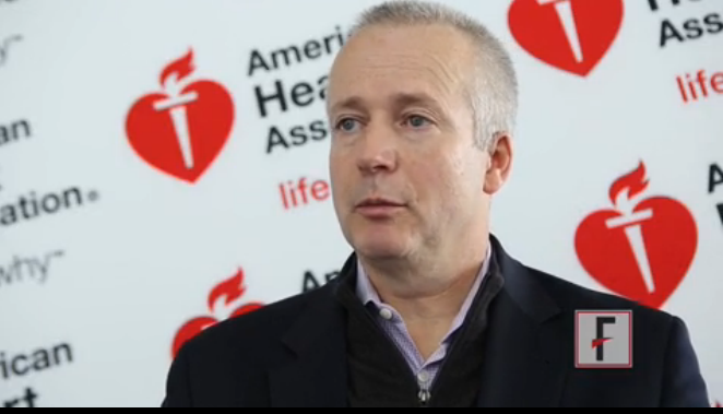
VIDEO: Focused cardiac ultrasound aids acute heart failure patients
VIENNA – Bedside echocardiography has become a key part of quickly assessing patients with acute heart failure to decide the best management strategy.
But bedside echo images often are challenging to interpret, so physicians performing an initial work-up of an acute heart failure patient need training in a basic echocardiography examination, Dr. Nuno Cardim said in an interview during the annual meeting of the European Association of Cardiovascular Imaging.
Known as Focused Cardiac Ultrasound (FoCUS), this triage-level exam is distinct from a comprehensive echocardiography assessment. Instead, FoCUS addresses some basic, yes-or-no, present-or-absent questions regarding systolic function, hypovolemia, tamponade, pleural effusion, and pulmonary embolus.
In a position paper and then in practice recommendations issued in 2014, the European Association of Cardiovascular Imaging acknowledged that use of FoCUS in acute heart failure patients was “irreversible,” said Dr. Cardim, professor and director of echocardiography and cardiac imaging at Hospital da Luz in Lisbon.
Cardiologists and echocardiography specialists need to make sure that FoCUS training is available to all physicians who might see suspected acute heart failure patients in emergency medicine settings, Dr. Cardim said.
Dr. Cardim had no disclosures.
The video associated with this article is no longer available on this site. Please view all of our videos on the MDedge YouTube channel
On Twitter @mitchelzoler
AT EUROECHO-IMAGING 2014
Open TAA repair surpasses TEVAR survival
CHICAGO – Endovascular repair of thoracic aortic aneurysms confers an immediate but short-lived survival advantage over open repair that completely disappears and then reverses 4.5 years after intervention, a study showed.
After 4.5 years, patients who underwent open repair of a thoracic aortic aneurysm had better survival than patients who underwent endovascular thoracic aneurysm repair, based on a propensity-score adjusted analysis of more than 3,000 Medicare patients.

The findings also showed a striking interhospital variability among U.S. centers performing thoracic aortic aneurysm (TAA) repair that influenced patient survival by 50%. The tertile of hospitals with the best survival outcomes collectively had a 50% reduced rate of deaths during follow-up, compared with all other hospitals, Dr. Justin M. Schaffer said at the American Heart Association scientific sessions.
“Simply by picking the right hospital, patients could reduce their mortality by half. That is pretty amazing,” said Dr. Schaffer, a cardiothoracic surgeon at Stanford (Calif.) University.
Perhaps even more notable than this undefined hospital effect was the pattern of mortality effects associated with open TAA repair, compared with thoracic endovascular aortic repair (TEVAR). For these analyses, Dr. Schaffer and his associates focused on the 1,037 Medicare beneficiaries who underwent open repair of an isolated, nonruptured TAA during 1999-2011, and 2,010 similar patients treated with TEVAR during the same period. It was relatively uncommon for patients to need repair for a TAA that was neither ruptured nor presented with another cardiac problem. This subgroup constituted 19% of all open TAA repairs on Medicare patients during those years, and 25% of all TEVARs performed.
Comparison of the TEVAR and open repair patients showed that those who underwent TEVAR were older and had consistently higher prevalence rates of a long list of comorbidities, including diabetes, chronic kidney disease, atrial fibrillation, and heart failure. The two populations also showed substantial differences for several other potential confounders. To adjust for all these, the researchers used a form of propensity scoring to balance the variability between the TEVAR and open surgery subgroups.
After adjustment, the mortality analysis showed that immediately following TEVAR, patients had a large mortality advantage, but that this immediately began to diminish such that by about 6 months after the procedure, the instantaneous advantage from TEVAR reached zero and then began tilting toward an advantage from open surgery.
Because of TEVAR’s sizable early lead the cumulative mortality numbers took awhile to reflect this. When tallied at 12 months after the procedure, cumulative survival in the TEVAR group stood at 85%, and 75% among the open surgery patients, a statistically significant advantage for TEVAR for 1-year survival.
But at 4.5 years after the procedure, the cumulative mortality rate in the TEVAR group caught up to that in the open surgery group, and following that, the TEVAR patients had a higher cumulative mortality. At 5-year follow-up, cumulative survival was 56% in the open surgery group and 55% in the TEVAR group, a statistically significant difference, Dr. Schaffer reported.
“There is a clear trade-off” between the two repair options, he said. “If a patient’s expected survival [following surgery] is limited, then TEVAR is reasonable, and if the anatomy also makes it feasible,” he said. “But for healthier patients with better expected survival, open repair is more complete, more durable, and superior.”
Although the analysis has not yet identified the clinical factors that contribute to worse survival after TEVAR, “it appears to be a higher reintervention rate,” based on preliminary assessments of the data, Dr. Schaffer said.
The analysis also showed other factors that significantly linked with mortality among all patients, regardless of whether they underwent TEVAR or open surgery. In addition to showing significant incremental risk effects from each of a range of comorbidities, the results showed that over the period 1999-2011, patients had a progressive, relative reduction of 6% fewer deaths each year, and that centers with the highest repair volumes produced 11% fewer deaths, compared with medium-volume hospitals.
Dr. Schaffer said he had no relevant financial disclosures.
On Twitter @mitchelzoler
CHICAGO – Endovascular repair of thoracic aortic aneurysms confers an immediate but short-lived survival advantage over open repair that completely disappears and then reverses 4.5 years after intervention, a study showed.
After 4.5 years, patients who underwent open repair of a thoracic aortic aneurysm had better survival than patients who underwent endovascular thoracic aneurysm repair, based on a propensity-score adjusted analysis of more than 3,000 Medicare patients.
The findings also showed a striking interhospital variability among U.S. centers performing thoracic aortic aneurysm (TAA) repair that influenced patient survival by 50%. The tertile of hospitals with the best survival outcomes collectively had a 50% reduced rate of deaths during follow-up, compared with all other hospitals, Dr. Justin M. Schaffer said at the American Heart Association scientific sessions.
“Simply by picking the right hospital, patients could reduce their mortality by half. That is pretty amazing,” said Dr. Schaffer, a cardiothoracic surgeon at Stanford (Calif.) University.
Perhaps even more notable than this undefined hospital effect was the pattern of mortality effects associated with open TAA repair, compared with thoracic endovascular aortic repair (TEVAR). For these analyses, Dr. Schaffer and his associates focused on the 1,037 Medicare beneficiaries who underwent open repair of an isolated, nonruptured TAA during 1999-2011, and 2,010 similar patients treated with TEVAR during the same period. It was relatively uncommon for patients to need repair for a TAA that was neither ruptured nor presented with another cardiac problem. This subgroup constituted 19% of all open TAA repairs on Medicare patients during those years, and 25% of all TEVARs performed.
Comparison of the TEVAR and open repair patients showed that those who underwent TEVAR were older and had consistently higher prevalence rates of a long list of comorbidities, including diabetes, chronic kidney disease, atrial fibrillation, and heart failure. The two populations also showed substantial differences for several other potential confounders. To adjust for all these, the researchers used a form of propensity scoring to balance the variability between the TEVAR and open surgery subgroups.
After adjustment, the mortality analysis showed that immediately following TEVAR, patients had a large mortality advantage, but that this immediately began to diminish such that by about 6 months after the procedure, the instantaneous advantage from TEVAR reached zero and then began tilting toward an advantage from open surgery.
Because of TEVAR’s sizable early lead the cumulative mortality numbers took awhile to reflect this. When tallied at 12 months after the procedure, cumulative survival in the TEVAR group stood at 85%, and 75% among the open surgery patients, a statistically significant advantage for TEVAR for 1-year survival.
But at 4.5 years after the procedure, the cumulative mortality rate in the TEVAR group caught up to that in the open surgery group, and following that, the TEVAR patients had a higher cumulative mortality. At 5-year follow-up, cumulative survival was 56% in the open surgery group and 55% in the TEVAR group, a statistically significant difference, Dr. Schaffer reported.
“There is a clear trade-off” between the two repair options, he said. “If a patient’s expected survival [following surgery] is limited, then TEVAR is reasonable, and if the anatomy also makes it feasible,” he said. “But for healthier patients with better expected survival, open repair is more complete, more durable, and superior.”
Although the analysis has not yet identified the clinical factors that contribute to worse survival after TEVAR, “it appears to be a higher reintervention rate,” based on preliminary assessments of the data, Dr. Schaffer said.
The analysis also showed other factors that significantly linked with mortality among all patients, regardless of whether they underwent TEVAR or open surgery. In addition to showing significant incremental risk effects from each of a range of comorbidities, the results showed that over the period 1999-2011, patients had a progressive, relative reduction of 6% fewer deaths each year, and that centers with the highest repair volumes produced 11% fewer deaths, compared with medium-volume hospitals.
Dr. Schaffer said he had no relevant financial disclosures.
On Twitter @mitchelzoler
AT THE AHA SCIENTIFIC SESSIONS
Key clinical point: Medicare patients who underwent open repair for a thoracic aortic aneurysm had better long-term survival than matched Medicare patients who had endovascular repair.
Major finding: After 5 years, survival was 56% after open thoracic aortic aneurysm repa
ir and 55% after endovascular repair.
Data source: A retrospective study of 3,047 Medicare patients who underwent repair of a nonruptured, isolated thoracic aortic aneurysm during 1999-2011.
Disclosures: Dr. Schaffer had no relevant financial disclosures.
TAVR survival improved during 2009-2012
CHICAGO – During 2009-2012, U.S. interventionalists and surgeons performing transcatheter aortic valve replacements made substantial strides in improving the safety and efficacy of the procedure, cutting first-year mortality rates by about a third, according to a review of more than 1,000 patients treated during that time.
Factors contributing to these gains included patient selection and an increased percentage of patients who emerged from their transcatheter aortic valve replacement (TAVR) procedure with mild, trace, or no paravalvular leak. Other developments that may have boosted patient survival included increased operator experience and changes in the TAVR delivery system, Dr. Nirat Beohar said at the American Heart Association scientific sessions.
“I think most important has been sizing the valve, which seemed to evolve” during the course of the 3-year period studied, said Dr. Beohar, director of the cardiac catheterization laboratory at Mount Sinai Medical Center in Miami Beach.

His analysis used data collected during the PARTNER continued-access program that allowed centers that had participated in the PARTNER trial, the first U.S. pivotal trial of the SAPIEN TAVR system marketed by Edwards, to continue to offer TAVR to patients during March 2009-January 2012, a period that mostly preceded Food and Drug Administration approval of the SAPIEN system for U.S. use in November 2011.
To assess changes in outcomes over time, Dr. Beohar and his associates divided the 1,063 patients who received TAVR through the continued-access program during this time into three tertiles: The 353 patients who underwent TAVR during March 2009-July 2010, 355 patients treated during July 2010-March 2011, and 355 patients treated during March 2011-January 2012.
Compared with patients treated during the earliest period, those treated during the most recent period were significantly older, with an average age of 86 years versus 84 years at the start of the program. The most recent patients also had a significantly reduced prevalence of comorbidities. For example, chronic obstructive pulmonary disease dropped from a 52% prevalence early to 37% during the most recent period. Patients with a porcelain aorta fell from 7% to 1%, and patients considered inoperable fell from 40% of the first tertile cohort to 8% of the patients in the most recent tertile.
All 1,063 patients in the analysis underwent TAVR using transfemoral access, but the use of fully percutaneous access (as opposed to surgical cutdown access) doubled from 31% during the first period to 62% during the most recent period.
All-cause mortality improved significantly at both 1-year and 2-year follow-up. Patients treated during the first time period, March 2009-July 2010, had a 23% mortality rate after 1 year and 35% after 2 years. During the most recent period studied, March 2011-January 2012, first-year deaths fell to 14%, and mortality was 24% after 2-year follow-up, said Dr. Beohar.
Despite this improvement in survival, the rate of nonfatal major complications during the first 30 days after treatment remained stable, occurring in 17% of patients in the first tertile, 13% in the second tertile, and 15% in the third tertile.
In contrast, the prevalence of moderate or severe paravalvular leak (PVL) following TAVR fell during the entire period studied and appeared to strongly correlate with long-term survival. The prevalence of moderate or severe PVL by 30 days after TAVR was 19% among patients in the first tertile and 10% among those in the third tertile, a statistically significant difference.
In a multivariate analysis that adjusted for several demographic and clinical factors, the appearance of moderate or severe PVL during the 30 days following TAVR was linked with a statistically significant, roughly fourfold increase in the rate of 1-year mortality, Dr. Beohar reported. Other factors that had statistically significant association with 1-year mortality were the occurrence of a nonfatal major complication, which linked with a threefold increased mortality rate; reduced renal function, which was associated with a roughly doubled mortality rate; male sex, which was tied to a nearly doubled mortality rate; and treatment with TAVR during the most recent period, which was linked with an approximately 20% reduction in mortality.
Another factor that distinguished the tertile 1 patients from those treated in tertile 3 was that the prevailing device used during the most recent period was the SAPIEN XT device, while patients treated during the first two tertile periods primarily received treatment using the original SAPIEN TAVR system.
On Twitter @mitchelzoler
CHICAGO – During 2009-2012, U.S. interventionalists and surgeons performing transcatheter aortic valve replacements made substantial strides in improving the safety and efficacy of the procedure, cutting first-year mortality rates by about a third, according to a review of more than 1,000 patients treated during that time.
Factors contributing to these gains included patient selection and an increased percentage of patients who emerged from their transcatheter aortic valve replacement (TAVR) procedure with mild, trace, or no paravalvular leak. Other developments that may have boosted patient survival included increased operator experience and changes in the TAVR delivery system, Dr. Nirat Beohar said at the American Heart Association scientific sessions.
“I think most important has been sizing the valve, which seemed to evolve” during the course of the 3-year period studied, said Dr. Beohar, director of the cardiac catheterization laboratory at Mount Sinai Medical Center in Miami Beach.
His analysis used data collected during the PARTNER continued-access program that allowed centers that had participated in the PARTNER trial, the first U.S. pivotal trial of the SAPIEN TAVR system marketed by Edwards, to continue to offer TAVR to patients during March 2009-January 2012, a period that mostly preceded Food and Drug Administration approval of the SAPIEN system for U.S. use in November 2011.
To assess changes in outcomes over time, Dr. Beohar and his associates divided the 1,063 patients who received TAVR through the continued-access program during this time into three tertiles: The 353 patients who underwent TAVR during March 2009-July 2010, 355 patients treated during July 2010-March 2011, and 355 patients treated during March 2011-January 2012.
Compared with patients treated during the earliest period, those treated during the most recent period were significantly older, with an average age of 86 years versus 84 years at the start of the program. The most recent patients also had a significantly reduced prevalence of comorbidities. For example, chronic obstructive pulmonary disease dropped from a 52% prevalence early to 37% during the most recent period. Patients with a porcelain aorta fell from 7% to 1%, and patients considered inoperable fell from 40% of the first tertile cohort to 8% of the patients in the most recent tertile.
All 1,063 patients in the analysis underwent TAVR using transfemoral access, but the use of fully percutaneous access (as opposed to surgical cutdown access) doubled from 31% during the first period to 62% during the most recent period.
All-cause mortality improved significantly at both 1-year and 2-year follow-up. Patients treated during the first time period, March 2009-July 2010, had a 23% mortality rate after 1 year and 35% after 2 years. During the most recent period studied, March 2011-January 2012, first-year deaths fell to 14%, and mortality was 24% after 2-year follow-up, said Dr. Beohar.
Despite this improvement in survival, the rate of nonfatal major complications during the first 30 days after treatment remained stable, occurring in 17% of patients in the first tertile, 13% in the second tertile, and 15% in the third tertile.
In contrast, the prevalence of moderate or severe paravalvular leak (PVL) following TAVR fell during the entire period studied and appeared to strongly correlate with long-term survival. The prevalence of moderate or severe PVL by 30 days after TAVR was 19% among patients in the first tertile and 10% among those in the third tertile, a statistically significant difference.
In a multivariate analysis that adjusted for several demographic and clinical factors, the appearance of moderate or severe PVL during the 30 days following TAVR was linked with a statistically significant, roughly fourfold increase in the rate of 1-year mortality, Dr. Beohar reported. Other factors that had statistically significant association with 1-year mortality were the occurrence of a nonfatal major complication, which linked with a threefold increased mortality rate; reduced renal function, which was associated with a roughly doubled mortality rate; male sex, which was tied to a nearly doubled mortality rate; and treatment with TAVR during the most recent period, which was linked with an approximately 20% reduction in mortality.
Another factor that distinguished the tertile 1 patients from those treated in tertile 3 was that the prevailing device used during the most recent period was the SAPIEN XT device, while patients treated during the first two tertile periods primarily received treatment using the original SAPIEN TAVR system.
On Twitter @mitchelzoler
AT THE AHA SCIENTIFIC SESSIONS
Key clinical point: During May 2009-January 2012, survival of patients undergoing transcatheter aortic valve replacement using Sapien devices showed steady improvement.
Major finding: Patients treated during March 2009-July 2010 had a 23% 1-year mortality rate, compared with a 14% rate during March 2011-January 2012.
Data source: Review of 1,063 patients who underwent transcatheter aortic valve replacement as part of the PARTNER continued access program during March 2009-January 2012.
Disclosures: The PARTNER studies and the continued access program was sponsored by Edwards, the company that markets SAPIEN TAVR devices. Dr. Beohar has been a PARTNER investigator but said he had no disclosures.

Imaging helps cut TAVR’s paravalvular leaks
VIENNA – Routine use of CT or three-dimensional echocardiography before and during transcatheter aortic valve replacement has contributed to a dramatic drop in paravalvular leaks, the procedure’s Achilles heel, based on the experience at a single, high-volume U.S. center.
In recent months, during testing of the SAPIEN 3 transcatheter aortic valve replacement (TAVR) system under development by Edwards, no patients developed moderate or severe paravalvular regurgitation during follow-up, and mild paravalvular leaks occurred at a rate of 7%-9%, Dr. Rebecca T. Hahn said at the annual meeting of the European Association of Cardiovascular Imaging. She and her associates tallied these rates recently at their institution, Columbia University Medical Center in New York, during their participation in PARTNER II, a multicenter study of the SAPIEN 3 device.
Dr. Hahn attributed these unprecedentedly low rates to three factors: preprocedure imaging with CT or 3-D echocardiography to select the best-sized valve for each patient, routine use of intraprocedural 3-D echo to guide precise valve placement and monitor for complications, and the SAPIEN 3 valve.
To put these regurgitation rates in context, in results from the pivotal CoreValve trial reported last May, the 30-day rate of moderate or severe paravalvular regurgitation was 9%, and the rate of mild paravalvular regurgitation was 36% (N. Engl. J. Med. 2014;370:1790-8). The most recent data on paravalvular leak reported from a large trial using a SAPIEN valve currently on the U.S. market was from the PARTNER II trial of inoperable patients, which documented a roughly 20% rate of moderate or severe paravalvular regurgitation using either the SAPIEN XT or the original SAPIEN system.
Dr. Hahn acknowledged that one major factor contributing to the disappearance of moderate or severe leaks following TAVR was the design of the new SAPIEN 3 TAVR valve she is working with, which features a flexible layer around the outer perimeter of the valve to better seal it into the patient’s aortic valve annulus and prevent blood from leaking through gaps around the edge.
But she also credited intraprocedural 3-D echo as a major factor in the improved outcomes, as well as the use of CT or 3-D echo to accurately size the annulus before TAVR starts so that the patient’s annulus size can be matched with the most appropriately sized replacement valve.
“Each valve has a sweet spot” for the annulus size it will fit into, and CT imaging of the annulus to measure the size, or using three-dimensional echo when CT is not good enough, is the way that TAVR heart teams now make sure they use the valve size with a sweet spot for the patient receiving the valve, said Dr. Hahn, director of invasive and valvular echocardiography at Columbia. A key task for either imaging method is to measure annulus size on the “virtual” annular plane, a plane that is challenging to identify as it is defined by only the three hinge points of the aortic valve’s leaflets. “Three-dimensional echo allows us to quantify [annular size] in ways that we couldn’t before,” she said in an interview.
During the procedure, 3-D echocardiography is the only good imaging option. In addition to clarifying unusual morphologies of the annulus and surrounding tissues and blood vessels, and giving precise information on wire and valve placement, 3-D echo allows for rapid assessment of a hemodynamic emergency if one occurs during a TAVR procedure. Three-dimensional echo also affords the best way to assess the location and severity of paravalvular regurgitation. 3-D echo “allows us to make decisions during the procedure on what to do about paravalvular regurgitation,” Dr. Hahn said.
On Twitter @mitchelzoler
AT EUROECHO-IMAGING 2014
Key clinical point: Routine CT and three-dimensional echocardiography before and during transcatheter aortic valve replacement helped dramatically cut the incidence and severity of paravalvular leaks.
Major finding: Transcatheter aortic valve replacement with the SAPIEN 3 system produced no moderate or severe paravalvular leaks and a 7%-9% rate of mild leaks.
Data source: Recent, anecdotal single-center experience in the PARTNER II trial.
Disclosures: PARTNER II is sponsored by Edwards, the company developing the SAPIEN 3 TAVR system. Dr. Hahn is an investigator in the PARTNER II trial, and has received honoraria as a speaker on behalf of Boston Scientific, St. Jude, and Philips.
Bioabsorbable-polymer coronary stent achieves noninferiority endpoint
CHICAGO– A drug-eluting coronary stent with a bioabsorbable polymer that dissolves nearly completely by 4 months after placement proved noninferior for safety and efficacy, compared with a conventional drug-eluting stent for 1 year, in a pivotal U.S. study with 1,684 patients.
If this new drug-eluting coronary stent, Synergy – which uses a bioabsorbable polymer to hold and slowly release the stent’s antirestenosis drug everolimus – were to receive Food and Drug Administration approval in 2015 based on these pivotal trial results, it would become the first such stent on the U.S. market. Some experts hailed the Synergy stent as an incremental improvement over prior devices in its combination of excellent deliverability as well as the ability of the stent’s drug-carrying polymer to vanish once it’s no longer needed, a feature that in theory could reduce the risk for late stent thrombosis and thereby reduce the necessary duration of dual-antiplatelet therapy (DAPT).

“We need to continue to evolve [stents] by improving and enhancing them and reducing the period of mandatory DAPT during the first year” following stent placement, commented Dr. Roxana Mehran, a professor of medicine and interventional cardiologist at Mount Sinai Hospital in New York. The Synergy everolimus-eluting stent with a bioabsorbable polymer is “a little step in the right direction,” she said as designated discussant for the study when its results were reported at the American Heart Association Scientific Sessions.
But others questioned the added value of a new stent that merely shows noninferiority but not superiority to comparator stents when it comes to preventing late stent thrombosis. “The major difference we’ve seen in the different bioabsorbable polymer stents is in their deliverability, which keeps getting better,” said Dr. Christoph Kaiser, professor and head of interventional cardiology at the University of Bern, Switzerland.
EVOLVE II
The EVOLVE II trial enrolled patients who required percutaneous coronary intervention (PCI) with at least one stent at 125 centers in 16 countries during November 2012–December 2013. The investigators randomized patients to PCI with either the Synergy stent or the Promus Element Plus everolimus drug-eluting coronary stent that uses a durable polymer to hold and release everolimus. A third of the patients had unstable angina, and more than a quarter had an acute myocardial infarction. Virtually all patients were on DAPT for at least 6 months, and more than 85% were still on DAPT at 12 months.
After 12 months of follow-up, the combined rate of cardiac death, myocardial infarction caused by a lesion in a stented artery, or ischemia-driven target-vessel revascularization – the trial’s primary endpoint – was 6.7% in patients who received the stent with a bioabsorbable polymer and 6.5% in the durable-polymer stent, a nonsignificant difference that met the trials prespecified definition of noninferiority, reported Dr. Dean Kereiakes, lead investigator for the study and medical director of the Christ Hospital Heart and Vascular Center in Cincinnati.
The safety analysis showed a small but statistically nonsignificant reduction in episodes of definite or probably stent thrombosis, with three such events occurring among the patients who received a bioabsorbable-polymer stent and five in patients who received a durable-polymer stent. “We would need a trial with 20,000 patients to prove superiority” of the bioabsorbable-polymer stent for an endpoint of late stent thrombosis, Dr. Kereiakes said.
The only statistically significant difference in outcomes reported in the EVOLVE II trial between the two stent types tested was in immediate procedural success, which occurred in 98.3% of patients treated with the bioabsorbable-polymer stent and in 96.9% of those treated with a durable-polymer stent, a 1.4% absolute difference.
“As a high-volume operator doing complex cases, the [Synergy] stent is more deliverable and user friendly, with a lower profile and more flexibility. It makes complicated cases easier,” Dr. Kereiakes said.

These technical advantages “will resonate with the interventional community,” commented Dr. Robert Harrington, professor and chairman of medicine and an interventional cardiologist at Stanford (Calif.) University.
Results from a second, similar study that used different bioabsorbable- and durable-polymer stents and reported at the same session showed largely similar results.
BASKET-PROVE II
The BASKET-PROVE II (Basel Stent Kosten Effektivitäts Trial-Prospective Validation Examination Part II) trial randomized 2,291 patients at any of eight centers in four European countries. Patients received a biolimus-eluting stent with a bioabsorbable-polymer (Nobori), an everolimus-eluting stent with a durable polymer (Xience Prime), or a bare-metal stent (PRO-Kinetic), and were followed for 24 months.
The study’s primary efficacy outcome was the same as in EVOLVE II, a combination of cardiac death, myocardial infarction, or need for target-vessel revascularization, but in this study, it was tallied after 24 months. The rates were 7.6% in patients who received stents with a bioabsorbable polymer, and 6.8% in those who got stents with a durable polymer, a difference that was not statistically significant, and which also met the study’s prespecified definition of noninferiority in the intention-to-treat analysis.
The study’s safety endpoint compared patients who received bioabsorbable-polymer stents with those who received bare-metal stents for rates of cardiac death, myocardial infarction, or definite or probable stent thrombosis, which occurred in 5.0% of patients who received bare-metal stents and in 3.7% of those who received stents with a bioabsorbable polymer, a difference that was not statistically significant. Notably, the rates of stent thrombosis during months 13-24 of follow-up were completely identical in patients who received the drug-eluting stent with a bioabsorbable polymer and in those who received bare-metal stents, reported Dr. Kaiser, a coprincipal investigator on the study. Concurrent with his report at the meeting, an article with the results appeared online (Circulation 2014 [doi:10.1161/CIRCULATIONAHA.114.013520/-/DC1]).
These findings “challenge the concept” that polymers are a key issue for any difference in the rate of late stent thrombosis, said Dr. Kaiser.
But another interventionalist, who cochaired the session where the results from both studies were reported, disagreed. “I think it is a step forward to get rid of the polymers. There is no use to having them” once the antirestenosis drug a stent carries is fully released, “so it is good to get rid of them,” said Dr. Stefan James, a cardiologist at Uppsala University in Sweden.
“In our registry [Swedish Coronary Angiography and Angioplasty Registry] we see that the [Synergy] stent has excellent results, with a very low stent thrombosis rate similar to what was shown” on the EVOLVE II trial. “So I think that the [bioabsorbable-polymer] stents are a step forward. After 20 years with drug-eluting stents, we are improving these devices step by step,” Dr. James said.
EVOLVE II was sponsored by Boston Scientific. Dr. Kereiakes is a consultant to and speaker for Boston Scientific, and a consultant to Abbott Vascular and Reva Medical. Dr. Mehran has received honoraria from and has been a consultant to Boston Scientific as well as to several other device and drug companies. Dr. Harrington had been an investigator for several trials that tested drugs patients take following stenting. The BASKET-PROVE II trial had no commercial sponsorship, but the study organizers received the prasugrel that patients took in the study following stenting for free from the manufacturers. Dr. Kaiser has received remuneration for being the advisory boards for Eli Lilly and Daiichi Sankyo. Dr. James had served on an advisory board for Medtronic and has received institutional research grants from Medtronic, Terumo, and Vascular Solutions.
On Twitter @mitchelzoler
CHICAGO– A drug-eluting coronary stent with a bioabsorbable polymer that dissolves nearly completely by 4 months after placement proved noninferior for safety and efficacy, compared with a conventional drug-eluting stent for 1 year, in a pivotal U.S. study with 1,684 patients.
If this new drug-eluting coronary stent, Synergy – which uses a bioabsorbable polymer to hold and slowly release the stent’s antirestenosis drug everolimus – were to receive Food and Drug Administration approval in 2015 based on these pivotal trial results, it would become the first such stent on the U.S. market. Some experts hailed the Synergy stent as an incremental improvement over prior devices in its combination of excellent deliverability as well as the ability of the stent’s drug-carrying polymer to vanish once it’s no longer needed, a feature that in theory could reduce the risk for late stent thrombosis and thereby reduce the necessary duration of dual-antiplatelet therapy (DAPT).
“We need to continue to evolve [stents] by improving and enhancing them and reducing the period of mandatory DAPT during the first year” following stent placement, commented Dr. Roxana Mehran, a professor of medicine and interventional cardiologist at Mount Sinai Hospital in New York. The Synergy everolimus-eluting stent with a bioabsorbable polymer is “a little step in the right direction,” she said as designated discussant for the study when its results were reported at the American Heart Association Scientific Sessions.
But others questioned the added value of a new stent that merely shows noninferiority but not superiority to comparator stents when it comes to preventing late stent thrombosis. “The major difference we’ve seen in the different bioabsorbable polymer stents is in their deliverability, which keeps getting better,” said Dr. Christoph Kaiser, professor and head of interventional cardiology at the University of Bern, Switzerland.
EVOLVE II
The EVOLVE II trial enrolled patients who required percutaneous coronary intervention (PCI) with at least one stent at 125 centers in 16 countries during November 2012–December 2013. The investigators randomized patients to PCI with either the Synergy stent or the Promus Element Plus everolimus drug-eluting coronary stent that uses a durable polymer to hold and release everolimus. A third of the patients had unstable angina, and more than a quarter had an acute myocardial infarction. Virtually all patients were on DAPT for at least 6 months, and more than 85% were still on DAPT at 12 months.
After 12 months of follow-up, the combined rate of cardiac death, myocardial infarction caused by a lesion in a stented artery, or ischemia-driven target-vessel revascularization – the trial’s primary endpoint – was 6.7% in patients who received the stent with a bioabsorbable polymer and 6.5% in the durable-polymer stent, a nonsignificant difference that met the trials prespecified definition of noninferiority, reported Dr. Dean Kereiakes, lead investigator for the study and medical director of the Christ Hospital Heart and Vascular Center in Cincinnati.
The safety analysis showed a small but statistically nonsignificant reduction in episodes of definite or probably stent thrombosis, with three such events occurring among the patients who received a bioabsorbable-polymer stent and five in patients who received a durable-polymer stent. “We would need a trial with 20,000 patients to prove superiority” of the bioabsorbable-polymer stent for an endpoint of late stent thrombosis, Dr. Kereiakes said.
The only statistically significant difference in outcomes reported in the EVOLVE II trial between the two stent types tested was in immediate procedural success, which occurred in 98.3% of patients treated with the bioabsorbable-polymer stent and in 96.9% of those treated with a durable-polymer stent, a 1.4% absolute difference.
“As a high-volume operator doing complex cases, the [Synergy] stent is more deliverable and user friendly, with a lower profile and more flexibility. It makes complicated cases easier,” Dr. Kereiakes said.
These technical advantages “will resonate with the interventional community,” commented Dr. Robert Harrington, professor and chairman of medicine and an interventional cardiologist at Stanford (Calif.) University.
Results from a second, similar study that used different bioabsorbable- and durable-polymer stents and reported at the same session showed largely similar results.
BASKET-PROVE II
The BASKET-PROVE II (Basel Stent Kosten Effektivitäts Trial-Prospective Validation Examination Part II) trial randomized 2,291 patients at any of eight centers in four European countries. Patients received a biolimus-eluting stent with a bioabsorbable-polymer (Nobori), an everolimus-eluting stent with a durable polymer (Xience Prime), or a bare-metal stent (PRO-Kinetic), and were followed for 24 months.
The study’s primary efficacy outcome was the same as in EVOLVE II, a combination of cardiac death, myocardial infarction, or need for target-vessel revascularization, but in this study, it was tallied after 24 months. The rates were 7.6% in patients who received stents with a bioabsorbable polymer, and 6.8% in those who got stents with a durable polymer, a difference that was not statistically significant, and which also met the study’s prespecified definition of noninferiority in the intention-to-treat analysis.
The study’s safety endpoint compared patients who received bioabsorbable-polymer stents with those who received bare-metal stents for rates of cardiac death, myocardial infarction, or definite or probable stent thrombosis, which occurred in 5.0% of patients who received bare-metal stents and in 3.7% of those who received stents with a bioabsorbable polymer, a difference that was not statistically significant. Notably, the rates of stent thrombosis during months 13-24 of follow-up were completely identical in patients who received the drug-eluting stent with a bioabsorbable polymer and in those who received bare-metal stents, reported Dr. Kaiser, a coprincipal investigator on the study. Concurrent with his report at the meeting, an article with the results appeared online (Circulation 2014 [doi:10.1161/CIRCULATIONAHA.114.013520/-/DC1]).
These findings “challenge the concept” that polymers are a key issue for any difference in the rate of late stent thrombosis, said Dr. Kaiser.
But another interventionalist, who cochaired the session where the results from both studies were reported, disagreed. “I think it is a step forward to get rid of the polymers. There is no use to having them” once the antirestenosis drug a stent carries is fully released, “so it is good to get rid of them,” said Dr. Stefan James, a cardiologist at Uppsala University in Sweden.
“In our registry [Swedish Coronary Angiography and Angioplasty Registry] we see that the [Synergy] stent has excellent results, with a very low stent thrombosis rate similar to what was shown” on the EVOLVE II trial. “So I think that the [bioabsorbable-polymer] stents are a step forward. After 20 years with drug-eluting stents, we are improving these devices step by step,” Dr. James said.
EVOLVE II was sponsored by Boston Scientific. Dr. Kereiakes is a consultant to and speaker for Boston Scientific, and a consultant to Abbott Vascular and Reva Medical. Dr. Mehran has received honoraria from and has been a consultant to Boston Scientific as well as to several other device and drug companies. Dr. Harrington had been an investigator for several trials that tested drugs patients take following stenting. The BASKET-PROVE II trial had no commercial sponsorship, but the study organizers received the prasugrel that patients took in the study following stenting for free from the manufacturers. Dr. Kaiser has received remuneration for being the advisory boards for Eli Lilly and Daiichi Sankyo. Dr. James had served on an advisory board for Medtronic and has received institutional research grants from Medtronic, Terumo, and Vascular Solutions.
On Twitter @mitchelzoler
AT THE AHA SCIENTIFIC SESSIONS
Key clinical point: A drug-eluting coronary stent with a bioabsorbable polymer proved noninferior to a conventional drug-eluting stent with a durable polymer in its pivotal U.S. trial, paving the way for marketing approval.
Major finding: The Synergy coronary stent with a bioabsorbable polymer met its prespecified noninferiority endpoints, compared with a standard drug-eluting stent.
Data source: EVOLVE II, a randomized, multicenter trial with 1,684 patients.
Disclosures: EVOLVE II was sponsored by Boston Scientific. Dr. Kereiakes is a consultant to and speaker for Boston Scientific, and a consultant to Abbott Vascular and Reva Medical.
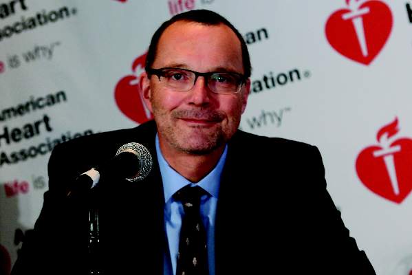
VIDEO: CT and 3D-echo imaging boost TAVR performance
VIENNA – Transcatheter aortic valve replacement has benefited enormously from the recent introduction of CT and three-dimensional echocardiography imaging into routine preprocedural planning and periprocedural guidance.
These two imaging methods have allowed clinicians to better select the right valve size for each patient, and 3D echo performed during transcatheter aortic valve replacement (TAVR) has also refined valve placement. The result has been TAVR procedures that go faster and have more accurate results while producing fewer complications, Dr. Rebecca Hahn said during an interview at the annual meeting of the European Association of Cardiovascular Imaging.
Perhaps the biggest impact of better valve-size selection and more detailed procedural guidance has been a reduced incidence and severity of paravalvular regurgitation once TAVR is complete and the replacement valve deployed. In recent months, Dr. Hahn and her associates at Columbia University Medical Center in New York have had no patients develop moderate or severe paravalvular regurgitation following TAVR, and the incidence of mild paravalvular leaks has dropped by about half, compared with when the procedure was first performed about 5 years ago, said Dr. Hahn, director of invasive and valvular echocardiography at Columbia.
The video associated with this article is no longer available on this site. Please view all of our videos on the MDedge YouTube channel
On Twitter@mitchelzoler
AT EUROECHO-IMAGING 2014

Rivaroxaban matches warfarin’s total Afib costs
CHICAGO – Patients with atrial fibrillation who start treatment with a new oral anticoagulant may spend more on their medication than if they were prescribed generic warfarin, but their overall health care costs may wind up being about the same, based on an analysis of health care expense records for more than 4,500 U.S. patients.
“Despite higher anticoagulant costs, total all-cause and atrial fibrillation–related costs remain comparable” between patients prescribed warfarin and those who received the new oral anticoagulant rivaroxaban (Xarelto), said Concetta Crivera, Pharm.D., at the American Heart Association Scientific Sessions. Higher drug costs for daily treatment with rivaroxaban were offset by reduced hospital lengths of stays and hence reduced hospitalization costs, said Dr. Crivera, director of cardiovascular health economics and outcomes research at Janssen in Raritan, N.J., the company that markets rivaroxaban along with Bayer.
“I can believe that hospitalized days would be reduced because patients treated with rivaroxaban or any of the other new oral anticoagulants don’t need to remain in the hospital while you wait for their international normalized ratio to enter the therapeutic range,” which is what happens with patients treated with warfarin, commented Dr. Jeffrey Weitz, professor of medicine and director of the Juravinski Hospital and Cancer Centre of McMaster University in Hamilton, Ont.
The new findings by Dr. Crivera “are not definitive on their own, but they add to a growing body of data that indicate that all the new oral anticoagulants are cost effective,” said Dr. Weitz, who specializes in thrombosis and anticoagulants. “The worst time for a patient on warfarin is when they start treatment. You do a disservice to patients with new-onset atrial fibrillation by using warfarin, because some patients will get into the therapeutic range but others never will. That’s why the new oral anticoagulants are better, because everybody gets into therapeutic range,” he said in an interview.
Dr. Crivera and her associates conducted a retrospective study of health records for 2,253 patients with nonvalvular atrial fibrillation (AF) who started anticoagulation treatment with rivaroxaban during the period of November 2011 (when the drug received U.S. marketing approval) through December 2012. The data came from the patient records of Humana, a U.S. HMO and insurer that covers both commercially insured patients and those covered by Medicare. The researchers matched each of the patients initiating rivaroxaban treatment with a similar patient from the Humana database with nonvalvular AF who started on warfarin during the same period. The average age of patients in the study was about 74 years, and patients in the two groups were closely matched for their demographic and clinical characteristics, including comorbidities. Data on health care use were available for patients for an average of about 4 months following their start of anticoagulant treatment.
The analysis showed that during the first months on treatment, patients prescribed rivaroxaban averaged 2.11 days of hospitalization for an AF-related episode, compared with 3.02 days for those prescribed warfarin, a statistically significant difference. Hospitalizations for any cause averaged a total of 2.71 days in the rivaroxaban group and 3.87 days in the patients on warfarin, also a significant difference, Dr. Crivera reported.
This difference in days hospitalized translated into reduced hospitalization costs, a roughly $2,000 average difference per patient in actual hospitalization costs in favor of the rivaroxaban patients for all-cause hospitalizations, and an average $1,300 difference per patient for hospitalization costs directly related to AF.
Although the rivaroxaban patients spent an average of $2,700 more per patient on pharmaceuticals for all causes, and an average $2,200 more for AF-related drugs, the total average all-cause and AF-related costs for drugs, hospitalizations, outpatient visits, and emergency department visits were similar in the two subgroups: an average total of $17,590 per patient for the rivaroxaban patients and $18,676 for warfarin patients for all causes, and an average of $7,394 for the rivaroxaban patients and $7,319 for those on warfarin for AF-related care. The between-group differences for both sets of total costs were not statistically significant.
The study was sponsored by Janssen, which along with Bayer markets rivaroxaban (Xarelto). Dr. Crivera is a Janssen employee. Dr. Weitz has been an adviser or a consultant to Janssen and Bayer and to Boehringer Ingelheim, Bristol-Myers Squibb, Daiichi Sankyo, Johnson & Johnson, Merck, Pfizer, and Portola.
On Twitter @mitchelzoler
AT THE AHA SCIENTIFIC SESSIONS
Key clinical point: Patients with atrial fibrillation who started on rivaroxaban treatment had total hospitalization and drug costs that were similar to those of patients begun on warfarin.
Major finding: Rivaroxaban-treated patients averaged $17,590 in total all-cause hospitalization and drug costs, compared with $18,676 for warfarin-treated patients.
Data source: A retrospective, matched-cohort study of 4,506 U.S. patients with atrial fibrillation and newly initiated anticoagulant treatment using health records maintained by Humana.
Disclosures: The study was sponsored by Janssen, which along with Bayer markets rivaroxaban (Xarelto). Dr. Crivera is a Janssen employee. Dr. Weitz has been an adviser or a consultant to Janssen and Bayer and to Boehringer Ingelheim, Bristol-Myers Squibb, Daiichi Sankyo, Johnson & Johnson, Merck, Pfizer, and Portola.

Polypharmacy linked to increased AFib bleeding
CHICAGO – Atrial fibrillation patients treated with an anticoagulant plus four or more other medications had significantly more bleeding complications than did those on fewer medications in a retrospective study.
“This is the first study examining polypharmacy in patients with nonvalvular atrial fibrillation,” Dr. Jonathan P. Piccini said at the American Heart Association Scientific Sessions. The main consequence is, “We should do everything we can to reduce the number of medications a patient receives,” he said.

His exploratory, retrospective analysis of data from more than 14,000 patients enrolled in an anticoagulant treatment trial showed that atrial fibrillation patients on an oral anticoagulant and taking a total of 10 or more medications concurrently had a 46% increased rate of a major bleed or clinically relevant nonmajor bleed. Patients taking five to nine medications had a 17% increased rate for this primary bleeding endpoint. Both increases were statistically significant, reported Dr. Piccini, a cardiologist at Duke University in Durham, N.C. Major bleeds alone were 90% higher in patients on at least 10 drugs and 47% higher in patients on 5-9 total drugs, compared with those on 4 or fewer, also statistically significant differences.
“Increasing medication use was associated with an increased risk of bleeding but not stroke,” he said. In addition, because the data came from a large trial that had compared the new oral anticoagulant rivaroxaban (Xarelto) with warfarin, the analysis was able to show that this polypharmacy effect was similar regardless of which anticoagulant patients took.
Polypharmacy was common among the 14,264 patients (average age, 73 years) enrolled in the trial, as 51% were on 5-9 drugs and 13% were on 10 or more drugs, with a minority of 36% forming the comparator group taking 4 or fewer medications.
The analysis used data collected from 14,264 patients enrolled in the ROCKET AF (Rivaroxaban Once Daily Oral Direct Factor Xa Inhibition Compared With Vitamin K Antagonism for Prevention of Stroke and Embolism Trial in Atrial Fibrillation) trial, which was designed as a pivotal trial comparing the safety and efficacy of the new oral anticoagulant rivaroxaban with warfarin for stroke prevention in patients with nonvalvular atrial fibrillation (N. Engl. J. Med. 2011;365:883-91). Dr. Piccini and his associates assessed various measures of bleeding as well as strokes, vascular deaths, and all-cause deaths in the patients by their number of concomitant medications based on data collected at baseline when patients entered the trial. The relative hazard rates the investigators calculated were adjusted for all “clinically important covariates,” but Dr. Piccini acknowledged that residual confounding accounted for by the adjustments likely remained.
All-cause death was also significantly increased in patients taking at least 10 (a 43% increase) or 5-9 drugs (a 25% increase), compared with patients on 4 or fewer drugs. But the rate of stroke or nonstroke embolism was not significantly increased among patients on five or more drugs. The combined rate of stroke, nonstroke embolism, and vascular death was a statistically significant 25% higher in patients on at least 10 drugs, compared with those on 4 or fewer drugs, but there was no significant increase in patients on 5-9 drugs.
The link between higher numbers of concomitant medications and an increased bleeding rate likely has two explanations, Dr. Piccini said. It could happen because the drugs themselves promote bleeding, and because the need to prescribe multiple drugs is a marker for patients who are sicker and have more comorbidities and a higher underlying risk of bleeding.
ROCKET AF was sponsored by Janssen and Bayer. Dr. Piccini has been a consultant to and has received research funding from Janssen, Bayer, and several other companies.
On Twitter @mitchelzoler
The message to physicians should be to assess patients at regular intervals to make sure they really still need all their medications, and try to reduce the number whenever possible.
One of the dangers in using a new oral anticoagulant is that patients are often also taking drugs that inhibit CYP3A4 enzymes, P-glycoprotein, or both. Among patients in Dr. Piccini’s study, 19% of those on 5-9 drugs and 26% of those on 10 or more were on at least one drug that inhibited P-glycoprotein and on at least one drug that was a CYP3A4 inhibitor. The list Dr. Piccini presented of the drugs patients received that fell in this category included amiodarone, diltiazem, verapamil, and erythromycin.
Also, these patients can be on other antithrombotic or antiplatelet drugs. We regularly assess patients’ coagulation status when they are on warfarin, but not when they are on rivaroxaban or another new oral anticoagulant. If you have patients on 10 or more drugs you need to be careful, and must also determine whether they can come off any of the drugs. You need to ask yourself, “What can I stop?” This is particularly true for patients also on aspirin, a thienopyridine, or a nonsteroidal anti-inflammatory drug, which all increase bleeding risk.
Dr. Jeffrey Weitz is professor of medicine and director of the Juravinski Hospital and Cancer Centre of McMaster University in Hamilton, Ont. He has been an adviser or consultant to Janssen, Bayer, and several drug manufacturers. He made these comments in an interview.
CHICAGO – Atrial fibrillation patients treated with an anticoagulant plus four or more other medications had significantly more bleeding complications than did those on fewer medications in a retrospective study.
“This is the first study examining polypharmacy in patients with nonvalvular atrial fibrillation,” Dr. Jonathan P. Piccini said at the American Heart Association Scientific Sessions. The main consequence is, “We should do everything we can to reduce the number of medications a patient receives,” he said.
His exploratory, retrospective analysis of data from more than 14,000 patients enrolled in an anticoagulant treatment trial showed that atrial fibrillation patients on an oral anticoagulant and taking a total of 10 or more medications concurrently had a 46% increased rate of a major bleed or clinically relevant nonmajor bleed. Patients taking five to nine medications had a 17% increased rate for this primary bleeding endpoint. Both increases were statistically significant, reported Dr. Piccini, a cardiologist at Duke University in Durham, N.C. Major bleeds alone were 90% higher in patients on at least 10 drugs and 47% higher in patients on 5-9 total drugs, compared with those on 4 or fewer, also statistically significant differences.
“Increasing medication use was associated with an increased risk of bleeding but not stroke,” he said. In addition, because the data came from a large trial that had compared the new oral anticoagulant rivaroxaban (Xarelto) with warfarin, the analysis was able to show that this polypharmacy effect was similar regardless of which anticoagulant patients took.
Polypharmacy was common among the 14,264 patients (average age, 73 years) enrolled in the trial, as 51% were on 5-9 drugs and 13% were on 10 or more drugs, with a minority of 36% forming the comparator group taking 4 or fewer medications.
The analysis used data collected from 14,264 patients enrolled in the ROCKET AF (Rivaroxaban Once Daily Oral Direct Factor Xa Inhibition Compared With Vitamin K Antagonism for Prevention of Stroke and Embolism Trial in Atrial Fibrillation) trial, which was designed as a pivotal trial comparing the safety and efficacy of the new oral anticoagulant rivaroxaban with warfarin for stroke prevention in patients with nonvalvular atrial fibrillation (N. Engl. J. Med. 2011;365:883-91). Dr. Piccini and his associates assessed various measures of bleeding as well as strokes, vascular deaths, and all-cause deaths in the patients by their number of concomitant medications based on data collected at baseline when patients entered the trial. The relative hazard rates the investigators calculated were adjusted for all “clinically important covariates,” but Dr. Piccini acknowledged that residual confounding accounted for by the adjustments likely remained.
All-cause death was also significantly increased in patients taking at least 10 (a 43% increase) or 5-9 drugs (a 25% increase), compared with patients on 4 or fewer drugs. But the rate of stroke or nonstroke embolism was not significantly increased among patients on five or more drugs. The combined rate of stroke, nonstroke embolism, and vascular death was a statistically significant 25% higher in patients on at least 10 drugs, compared with those on 4 or fewer drugs, but there was no significant increase in patients on 5-9 drugs.
The link between higher numbers of concomitant medications and an increased bleeding rate likely has two explanations, Dr. Piccini said. It could happen because the drugs themselves promote bleeding, and because the need to prescribe multiple drugs is a marker for patients who are sicker and have more comorbidities and a higher underlying risk of bleeding.
ROCKET AF was sponsored by Janssen and Bayer. Dr. Piccini has been a consultant to and has received research funding from Janssen, Bayer, and several other companies.
On Twitter @mitchelzoler
AT THE AHA SCIENTIFIC SESSIONS
Key clinical point: Among atrial fibrillation patients on an oral anticoagulant, those on at least four other medications concurrently had a significantly increased bleeding rate, compared with those on three or fewer.
Major finding: Patients on 5-9 drugs had 17% more bleeds, and those on 10 or more drugs had 46% more bleeds, than patients on fewer drugs.
Data source: A retrospective analysis of 14,264 patients enrolled in the ROCKET AF trial.
Disclosures: ROCKET AF was sponsored by Janssen and Bayer. Dr. Piccini has been a consultant to and has received research funding from Janssen, Bayer, and several other companies.
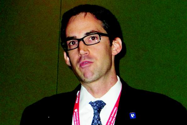
Blognosis: Participatory surveillance needs you!
One of the biggest lessons so far from the West African Ebola outbreak is the importance of early identification of infectious disease outbreaks. That’s the best way to get a quick response, and a quick response can mean the difference between an outbreak’s control or spread.
As reported from the International Meeting on Emerging Diseases and Surveillance (IMED) in early November, the Ebola experience in Africa this past year has been marked by slow responses in Liberia and Sierra Leone, followed by unprecedented Ebola spread – and quick responses in Nigeria and the Democratic Republic of the Congo, followed by declarations by the World Health Organization that Nigeria and then DRC had become Ebola-free.

Many factors play into early identification of infection outbreaks, but a new approach may be one of the best, especially once it fully catches on. It’s called participatory surveillance, and it means that everyone – or at least as many as can be persuaded – commits to regularly updating (usually weekly) a public health database on their health status.
While participatory surveillance is not being used in Africa, it’s being used elsewhere, including in the United States as the Flu Near You program. As also reported out of the same IMED meeting, Flu Near You is the best U.S. example of participatory surveillance, with a current database of about 100,000 U.S. enrollees. Ideally, the participants reply each week to a short survey that asks if they had any of 10 symptoms that can flag influenza, such as fever, headache, cough, sore throat, or fatigue.
When we first reported on Flu Near You nearly 2 years ago, it was an interesting concept just starting to roll out. Returning to Flu Near You for a second look a few weeks ago revealed that it had become a useful if still immature tool that had caught the notice of the Centers for Disease Control and Prevention (CDC).
Now it’s poised for the next step, which is where you come in: Sign up, and be active by responding to the weekly survey.
A CDC staffer in Atlanta told us that the agency already sees Flu Near You as a valuable adjunct to the flu surveillance it traditionally conducts. If the database is helpful for tracking national influenza trends with 100,000 Americans registered, imagine how much more informative it would be with 1 million, 10 million, or even more participants. The only way participatory surveillance succeeds is if people buy in and become part of the process. If enough people did that, the public health insights would soar.
Numbers are what’s holding Flu Near You back right now. Each of us has the ability to change that a little.
On Twitter @mitchelzoler
One of the biggest lessons so far from the West African Ebola outbreak is the importance of early identification of infectious disease outbreaks. That’s the best way to get a quick response, and a quick response can mean the difference between an outbreak’s control or spread.
As reported from the International Meeting on Emerging Diseases and Surveillance (IMED) in early November, the Ebola experience in Africa this past year has been marked by slow responses in Liberia and Sierra Leone, followed by unprecedented Ebola spread – and quick responses in Nigeria and the Democratic Republic of the Congo, followed by declarations by the World Health Organization that Nigeria and then DRC had become Ebola-free.
Many factors play into early identification of infection outbreaks, but a new approach may be one of the best, especially once it fully catches on. It’s called participatory surveillance, and it means that everyone – or at least as many as can be persuaded – commits to regularly updating (usually weekly) a public health database on their health status.
While participatory surveillance is not being used in Africa, it’s being used elsewhere, including in the United States as the Flu Near You program. As also reported out of the same IMED meeting, Flu Near You is the best U.S. example of participatory surveillance, with a current database of about 100,000 U.S. enrollees. Ideally, the participants reply each week to a short survey that asks if they had any of 10 symptoms that can flag influenza, such as fever, headache, cough, sore throat, or fatigue.
When we first reported on Flu Near You nearly 2 years ago, it was an interesting concept just starting to roll out. Returning to Flu Near You for a second look a few weeks ago revealed that it had become a useful if still immature tool that had caught the notice of the Centers for Disease Control and Prevention (CDC).
Now it’s poised for the next step, which is where you come in: Sign up, and be active by responding to the weekly survey.
A CDC staffer in Atlanta told us that the agency already sees Flu Near You as a valuable adjunct to the flu surveillance it traditionally conducts. If the database is helpful for tracking national influenza trends with 100,000 Americans registered, imagine how much more informative it would be with 1 million, 10 million, or even more participants. The only way participatory surveillance succeeds is if people buy in and become part of the process. If enough people did that, the public health insights would soar.
Numbers are what’s holding Flu Near You back right now. Each of us has the ability to change that a little.
On Twitter @mitchelzoler

VIDEO: Bioabsorbable polymer stents offer improved deliverability
CHICAGO – Two new types of drug-eluting, biodegradable polymer stents weren’t inferior to current drug-eluting stents, and they possibly may have caused fewer stent thrombosis episodes.
But do the new stents reduce the risk of long-term stent thrombosis? How will their ease of use affect choice of stents? And how should long-term coronary events that aren’t related to the stents themselves shape approaches to secondary prevention?
In an interview at the American Heart Association scientific sessions, Dr. Robert Harrington shared his perspectives on these questions and connected the stent studies’ results with new findings in prevention research.
The two studies of the new stents were not large enough, nor were patients followed long enough, to prove an advantage in stent thrombosis rates in a definitive way. But the lead investigator in the everolimus-eluting Synergy stent study described the stent as more flexible and deliverable than current coronary stents.
Those usability features may make that stent an attractive option for interventional cardiologists even if clinical outcomes are not significantly improved, Dr. Harrington noted.
“These technical features, such as improved deliverability, are things that will resonate with the interventional community,” he said. “It offers another option for complex cases, and that appeals greatly to interventional cardiologists who are looking for technical solutions,” said Dr. Harrington, professor and chairman of medicine at Stanford (Calif.) University.
Dr. Harrington said that he had no disclosures regarding the studied stents, but he has received grants from several drug companies that market drugs used during and after patients undergo percutaneous coronary interventions.
On Twitter @mitchelzoler
The video associated with this article is no longer available on this site. Please view all of our videos on the MDedge YouTube channel
EXPERT ANALYSIS FROM THE AHA SCIENTIFIC SESSIONS