User login
Cannabis use, childhood trauma correlated in schizophrenia patients
Childhood trauma was correlated with cannabis use in schizophrenia patients, but no significant interaction between the two factors was found, based on data from 366 individuals, including 295 with schizophrenia and 71 with schizoaffective disorder.
“Childhood trauma and cannabis consumption are among the most studied environmental risk factors for schizophrenia and are also considered to be risk-modifying factors,” wrote Grégoire Baudin of Université François-Rabelais in Tours, France, and colleagues (Schizophr Res. 2016;175:161-7).
Overall, the proportion of patients with a history of childhood trauma (CT) was not significantly different in those with and without cannabis use disorders, the researchers reported. However, CT was a significant predictor of the number of hospitalizations and of high scores on measures of excitement and emotional distress, and poor levels of function and quality of life.
The study was limited by not being representative of the schizophrenia population as a whole, the researchers noted. However, “our results emphasize the need for clinicians to systematically inquire about the traumatic history of patients with psychotic disorders, and consider trauma-focused therapy” for people with schizophrenia and for people at risk, the investigators added.
The researchers had no financial conflicts to disclose. The study was funded in part by the FondaMental Foundation, INSERM, and Investissements d’Avenir programs. Mr. Baudin has received a research grant from the FondaMental Foundation.
Find the full study here: doi: 10.1016/j.schres.2016.04.042.
Childhood trauma was correlated with cannabis use in schizophrenia patients, but no significant interaction between the two factors was found, based on data from 366 individuals, including 295 with schizophrenia and 71 with schizoaffective disorder.
“Childhood trauma and cannabis consumption are among the most studied environmental risk factors for schizophrenia and are also considered to be risk-modifying factors,” wrote Grégoire Baudin of Université François-Rabelais in Tours, France, and colleagues (Schizophr Res. 2016;175:161-7).
Overall, the proportion of patients with a history of childhood trauma (CT) was not significantly different in those with and without cannabis use disorders, the researchers reported. However, CT was a significant predictor of the number of hospitalizations and of high scores on measures of excitement and emotional distress, and poor levels of function and quality of life.
The study was limited by not being representative of the schizophrenia population as a whole, the researchers noted. However, “our results emphasize the need for clinicians to systematically inquire about the traumatic history of patients with psychotic disorders, and consider trauma-focused therapy” for people with schizophrenia and for people at risk, the investigators added.
The researchers had no financial conflicts to disclose. The study was funded in part by the FondaMental Foundation, INSERM, and Investissements d’Avenir programs. Mr. Baudin has received a research grant from the FondaMental Foundation.
Find the full study here: doi: 10.1016/j.schres.2016.04.042.
Childhood trauma was correlated with cannabis use in schizophrenia patients, but no significant interaction between the two factors was found, based on data from 366 individuals, including 295 with schizophrenia and 71 with schizoaffective disorder.
“Childhood trauma and cannabis consumption are among the most studied environmental risk factors for schizophrenia and are also considered to be risk-modifying factors,” wrote Grégoire Baudin of Université François-Rabelais in Tours, France, and colleagues (Schizophr Res. 2016;175:161-7).
Overall, the proportion of patients with a history of childhood trauma (CT) was not significantly different in those with and without cannabis use disorders, the researchers reported. However, CT was a significant predictor of the number of hospitalizations and of high scores on measures of excitement and emotional distress, and poor levels of function and quality of life.
The study was limited by not being representative of the schizophrenia population as a whole, the researchers noted. However, “our results emphasize the need for clinicians to systematically inquire about the traumatic history of patients with psychotic disorders, and consider trauma-focused therapy” for people with schizophrenia and for people at risk, the investigators added.
The researchers had no financial conflicts to disclose. The study was funded in part by the FondaMental Foundation, INSERM, and Investissements d’Avenir programs. Mr. Baudin has received a research grant from the FondaMental Foundation.
Find the full study here: doi: 10.1016/j.schres.2016.04.042.
FROM SCHIZOPHRENIA RESEARCH
Partnerships with pediatric tertiary care centers improve community ED asthma treatment
Partnerships between community emergency departments and pediatric tertiary care centers are feasible and improve care of pediatric asthma, according to Theresa A. Walls, MD, of the Children’s National Health Systems, Washington, D.C., and her associates.
A total of 724 asthma patients aged 2-17 years were included in the study. Of this group, 289 (40%) were treated at the community ED before the pediatric tertiary care center intervention and 435 (60%) were treated after the intervention. Treatment with steroids was significantly increased post intervention, with 76% of patients receiving steroids, compared with 60% of patients before the intervention.
“Because the overwhelming majority of pediatric emergency visits occur in community EDs, partnerships with these EDs can broaden the impact of quality improvement activities and should be part of future quality improvement efforts,” the investigators concluded.
Find the full study in Pediatrics (2016. doi: 10.1542/peds.2016-0088).
Dr. Walls and her group developed a quality improvement (QI) initiative with a community emergency department. One important part of the study was the use of an asthma score, which helped determine steps for ED therapy.

Dr. Walls and her group developed a quality improvement (QI) initiative with a community emergency department. One important part of the study was the use of an asthma score, which helped determine steps for ED therapy.
Dr. Walls and her group developed a quality improvement (QI) initiative with a community emergency department. One important part of the study was the use of an asthma score, which helped determine steps for ED therapy.
Partnerships between community emergency departments and pediatric tertiary care centers are feasible and improve care of pediatric asthma, according to Theresa A. Walls, MD, of the Children’s National Health Systems, Washington, D.C., and her associates.
A total of 724 asthma patients aged 2-17 years were included in the study. Of this group, 289 (40%) were treated at the community ED before the pediatric tertiary care center intervention and 435 (60%) were treated after the intervention. Treatment with steroids was significantly increased post intervention, with 76% of patients receiving steroids, compared with 60% of patients before the intervention.
“Because the overwhelming majority of pediatric emergency visits occur in community EDs, partnerships with these EDs can broaden the impact of quality improvement activities and should be part of future quality improvement efforts,” the investigators concluded.
Find the full study in Pediatrics (2016. doi: 10.1542/peds.2016-0088).
Partnerships between community emergency departments and pediatric tertiary care centers are feasible and improve care of pediatric asthma, according to Theresa A. Walls, MD, of the Children’s National Health Systems, Washington, D.C., and her associates.
A total of 724 asthma patients aged 2-17 years were included in the study. Of this group, 289 (40%) were treated at the community ED before the pediatric tertiary care center intervention and 435 (60%) were treated after the intervention. Treatment with steroids was significantly increased post intervention, with 76% of patients receiving steroids, compared with 60% of patients before the intervention.
“Because the overwhelming majority of pediatric emergency visits occur in community EDs, partnerships with these EDs can broaden the impact of quality improvement activities and should be part of future quality improvement efforts,” the investigators concluded.
Find the full study in Pediatrics (2016. doi: 10.1542/peds.2016-0088).
FROM PEDIATRICS
Skin cancer a concern in pediatric solid organ transplant recipients
As survival rates among pediatric organ transplant recipients increase, so do the rates of cutaneous malignancies later in life for this population, who are at a greater risk for skin cancers that include nonmelanoma skin cancers (NMSCs), melanoma, Kaposi sarcoma, and anogenital carcinoma, according to the authors of a literature review.
In studies, skin cancers account for 13%-55% of all cancers in pediatric organ transplant recipients (POTRs), according to Alexander L. Fogel of Stanford (Calif.) University and his coauthors. The review article provides an update on this topic, as well as information on the prevention and management of skin cancers in this population, and the differences between this group and adult organ transplant recipients (AOTRs).

NMSC is the most common type of skin cancer in the pediatric group – and the second most common type of malignancy (NMSCs are the most common type of cancer affecting adult organ transplant recipients). NMSCs typically appear an average of 12-18 years post transplantation in this population (at an average age of 26-34 years). Length of posttransplantation follow-up, sunlight exposure, fair skin, and Northern European ancestry are among the factors associated with increased risk. This type of cancer involves the lip nearly twice as often as in adult recipients: 23% vs. 12%. The pediatric cohort also experiences more nonmelanoma cancer spreading to the lymph nodes than do adults: 9% vs. 6%.
Among pediatric transplant recipients, squamous cell carcinomas appear 2.8 times more often than basal cell carcinomas, “a trend that is opposite that observed in the nontransplant population,” the authors wrote.
In one study, anogenital carcinomas accounted for 4% of posttransplant cancers in this cohort, at an average of 12 years after the transplant, at a mean age of 27 years.
Some data indicate that in adult transplant recipients, there is an association between the human papillomavirus, and anal and genital warts and posttransplant anogenital cancer, but there are little data looking at this association in the pediatric group, the authors noted.
Although melanoma and Kaposi sarcoma are also found in this cohort at rates greater than in the general population, and are associated with high mortality rates, the data are too few to draw conclusions, the authors wrote.
In 2014, 1,795 pediatric solid organ transplants were performed, accounting for 6% of all such transplants. The absolute number of pediatric transplants has remained fairly stable over 5 years, yet very little pediatric-specific literature exists for prevention and management of skin cancers post transplantation, the authors pointed out.
Changing immunosuppressive medications used in transplantation may be effective in reducing skin cancer risk, they said, noting that including rapamycin inhibitors in combination therapy has been shown to reduce the risk of developing skin cancers in some transplant patients by more than half.
The authors emphasized that regular sunscreen use and dermatologic checkups are also essential in this population, and that “the importance of regular dermatologic evaluation should be stressed to patients and their families.”
Mr. Fogel’s coauthors were Mari Miyar, MD, of the department of dermatology, Kaiser Permanente, San Jose, Calif., and Joyce Teng, MD, of the departments of dermatology and pediatrics, Stanford. The authors had no disclosures listed, and no funding source for the review was listed.
This article was updated 12/8/16.
As survival rates among pediatric organ transplant recipients increase, so do the rates of cutaneous malignancies later in life for this population, who are at a greater risk for skin cancers that include nonmelanoma skin cancers (NMSCs), melanoma, Kaposi sarcoma, and anogenital carcinoma, according to the authors of a literature review.
In studies, skin cancers account for 13%-55% of all cancers in pediatric organ transplant recipients (POTRs), according to Alexander L. Fogel of Stanford (Calif.) University and his coauthors. The review article provides an update on this topic, as well as information on the prevention and management of skin cancers in this population, and the differences between this group and adult organ transplant recipients (AOTRs).
NMSC is the most common type of skin cancer in the pediatric group – and the second most common type of malignancy (NMSCs are the most common type of cancer affecting adult organ transplant recipients). NMSCs typically appear an average of 12-18 years post transplantation in this population (at an average age of 26-34 years). Length of posttransplantation follow-up, sunlight exposure, fair skin, and Northern European ancestry are among the factors associated with increased risk. This type of cancer involves the lip nearly twice as often as in adult recipients: 23% vs. 12%. The pediatric cohort also experiences more nonmelanoma cancer spreading to the lymph nodes than do adults: 9% vs. 6%.
Among pediatric transplant recipients, squamous cell carcinomas appear 2.8 times more often than basal cell carcinomas, “a trend that is opposite that observed in the nontransplant population,” the authors wrote.
In one study, anogenital carcinomas accounted for 4% of posttransplant cancers in this cohort, at an average of 12 years after the transplant, at a mean age of 27 years.
Some data indicate that in adult transplant recipients, there is an association between the human papillomavirus, and anal and genital warts and posttransplant anogenital cancer, but there are little data looking at this association in the pediatric group, the authors noted.
Although melanoma and Kaposi sarcoma are also found in this cohort at rates greater than in the general population, and are associated with high mortality rates, the data are too few to draw conclusions, the authors wrote.
In 2014, 1,795 pediatric solid organ transplants were performed, accounting for 6% of all such transplants. The absolute number of pediatric transplants has remained fairly stable over 5 years, yet very little pediatric-specific literature exists for prevention and management of skin cancers post transplantation, the authors pointed out.
Changing immunosuppressive medications used in transplantation may be effective in reducing skin cancer risk, they said, noting that including rapamycin inhibitors in combination therapy has been shown to reduce the risk of developing skin cancers in some transplant patients by more than half.
The authors emphasized that regular sunscreen use and dermatologic checkups are also essential in this population, and that “the importance of regular dermatologic evaluation should be stressed to patients and their families.”
Mr. Fogel’s coauthors were Mari Miyar, MD, of the department of dermatology, Kaiser Permanente, San Jose, Calif., and Joyce Teng, MD, of the departments of dermatology and pediatrics, Stanford. The authors had no disclosures listed, and no funding source for the review was listed.
This article was updated 12/8/16.
As survival rates among pediatric organ transplant recipients increase, so do the rates of cutaneous malignancies later in life for this population, who are at a greater risk for skin cancers that include nonmelanoma skin cancers (NMSCs), melanoma, Kaposi sarcoma, and anogenital carcinoma, according to the authors of a literature review.
In studies, skin cancers account for 13%-55% of all cancers in pediatric organ transplant recipients (POTRs), according to Alexander L. Fogel of Stanford (Calif.) University and his coauthors. The review article provides an update on this topic, as well as information on the prevention and management of skin cancers in this population, and the differences between this group and adult organ transplant recipients (AOTRs).
NMSC is the most common type of skin cancer in the pediatric group – and the second most common type of malignancy (NMSCs are the most common type of cancer affecting adult organ transplant recipients). NMSCs typically appear an average of 12-18 years post transplantation in this population (at an average age of 26-34 years). Length of posttransplantation follow-up, sunlight exposure, fair skin, and Northern European ancestry are among the factors associated with increased risk. This type of cancer involves the lip nearly twice as often as in adult recipients: 23% vs. 12%. The pediatric cohort also experiences more nonmelanoma cancer spreading to the lymph nodes than do adults: 9% vs. 6%.
Among pediatric transplant recipients, squamous cell carcinomas appear 2.8 times more often than basal cell carcinomas, “a trend that is opposite that observed in the nontransplant population,” the authors wrote.
In one study, anogenital carcinomas accounted for 4% of posttransplant cancers in this cohort, at an average of 12 years after the transplant, at a mean age of 27 years.
Some data indicate that in adult transplant recipients, there is an association between the human papillomavirus, and anal and genital warts and posttransplant anogenital cancer, but there are little data looking at this association in the pediatric group, the authors noted.
Although melanoma and Kaposi sarcoma are also found in this cohort at rates greater than in the general population, and are associated with high mortality rates, the data are too few to draw conclusions, the authors wrote.
In 2014, 1,795 pediatric solid organ transplants were performed, accounting for 6% of all such transplants. The absolute number of pediatric transplants has remained fairly stable over 5 years, yet very little pediatric-specific literature exists for prevention and management of skin cancers post transplantation, the authors pointed out.
Changing immunosuppressive medications used in transplantation may be effective in reducing skin cancer risk, they said, noting that including rapamycin inhibitors in combination therapy has been shown to reduce the risk of developing skin cancers in some transplant patients by more than half.
The authors emphasized that regular sunscreen use and dermatologic checkups are also essential in this population, and that “the importance of regular dermatologic evaluation should be stressed to patients and their families.”
Mr. Fogel’s coauthors were Mari Miyar, MD, of the department of dermatology, Kaiser Permanente, San Jose, Calif., and Joyce Teng, MD, of the departments of dermatology and pediatrics, Stanford. The authors had no disclosures listed, and no funding source for the review was listed.
This article was updated 12/8/16.
FROM PEDIATRIC DERMATOLOGY
Key clinical point:
Major finding: Pediatric solid organ transplant recipients experience skin cancer rates between 13% and 55%.
Data source: A literature review of malignancies among pediatric organ transplant recipients.
Disclosures: The authors listed no disclosures, and no funding source for the review was listed.
Prescribing the landmark hemangioma drug: The challenges and the benefits
For Beth Drolet, MD, a pediatric dermatologist in Wisconsin, the tremendous impact oral propranolol has had on the treatment of severe infantile hemangioma is written on the faces of children diagnosed with the condition in recent years.
“You can tell which drugs the kids were on by their age,” said Dr. Drolet, professor of dermatology and pediatrics at the Medical College of Wisconsin, Milwaukee. “If they were born before 2008, before we used this medication, those kids have had multiple surgeries and are still not looking that good. But we rarely see that in the kids born after.”
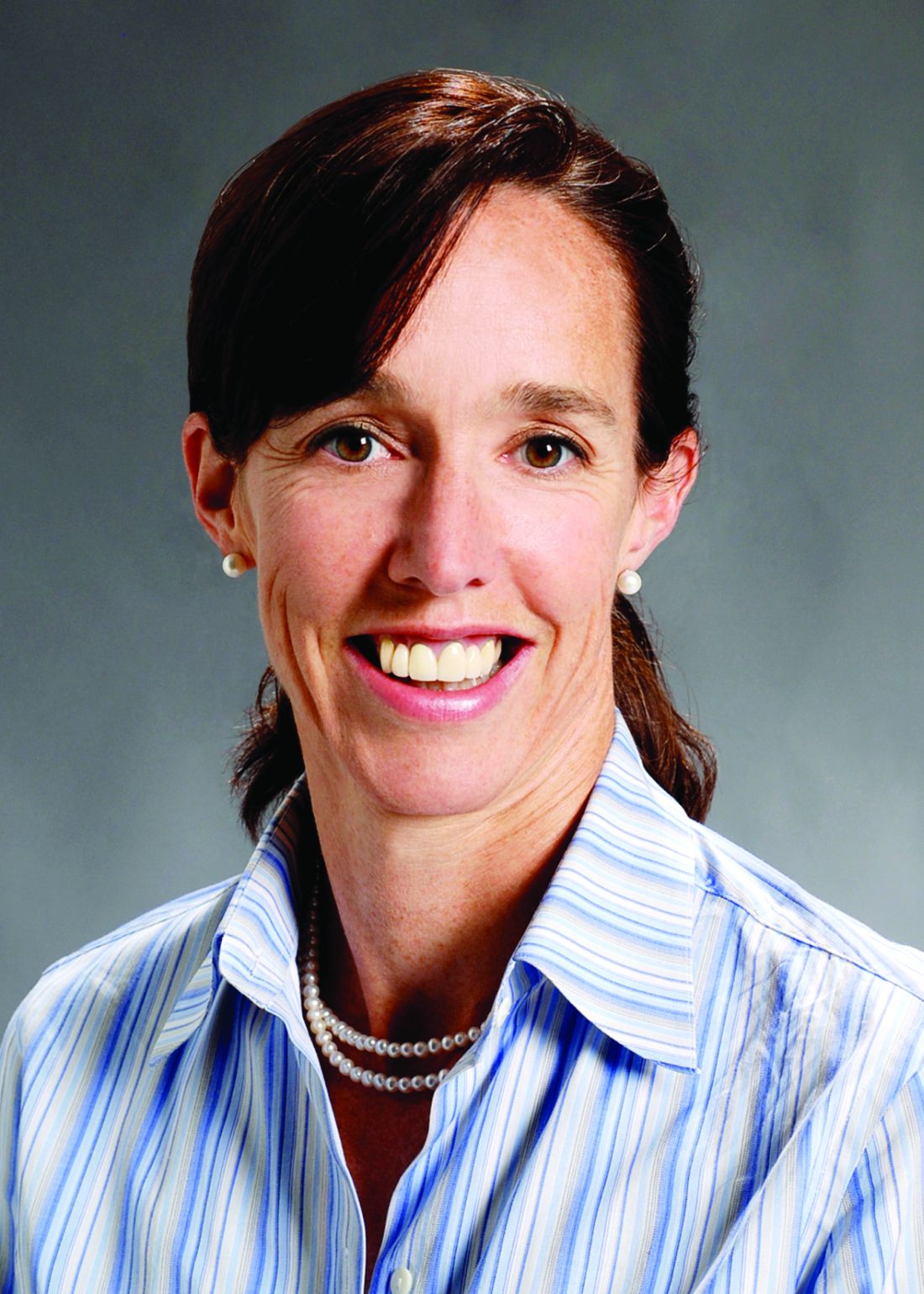
Still, it is possible for dermatologists to successfully treat their smallest patients with oral propranolol, according to Dr. Drolet and Ilona J. Frieden, MD, professor of dermatology and pediatrics at the University of California, San Francisco.
In interviews, the two pediatric dermatologists spoke about the challenges and benefits of treating hemangioma patients with oral propranolol solution, which was approved by the Food and Drug Administration in 2014 for “proliferating infantile hemangioma requiring systemic therapy.” It is the only FDA-approved systemic treatment for this indication.

The oral form of the drug was used off label to treat patients with hemangioma after a French dermatologist discovered in 2007 that it could effectively treat the condition. A topical form of propranolol is also used for hemangiomas that do not require systemic treatment.
Prior to about a decade ago, Dr. Drolet said, steroids were used to treat severe hemangiomas with limited success.
In general, infantile hemangiomas “have a natural course of gradually involuting even without treatment,” Dr. Frieden noted. But the most severe cases can produce functional impairment, scarring, and anatomic distortion.
Dr. Drolet said she considers treatment if hemangioma threatens a vital function (hearing, sight, breathing) or can lead to pain, infection, or scarring.
One challenge for dermatologists is that standard of care treatment with oral propranolol requires in-office cardiac monitoring, especially as the dose is increased over the first week or two of treatment.
“I don’t think most dermatologists are comfortable taking a heart rate and blood pressure in an infant,” said Dr. Drolet, who is director of the birthmarks and vascular anomalies section at Children’s Hospital of Wisconsin, Milwaukee. Instead, they tend to refer patients to a pediatrician or pediatric cardiologist.
Her clinic hired a cardiac nurse to train the staff in how to take heart rate and blood pressure in babies. “Partnering with cardiology was really important for us,” she commented. “We worked really closely with our pediatric cardiology team to gain that expertise for our staff to assess that. You have to be pretty comfortable with it. If you’re not, you’re going to have to find someone else.”
Another option for dermatologists, Dr. Frieden said, is to focus on heart rate alone since blood pressure in infants is difficult to measure. “It’s not FDA sanctioned, but many people seem to do that and it’s OK,” she said.
Dr. Frieden and Dr. Drolet provided the following recommendations about treating babies with oral propranolol:
• Caution parents about side effects. Cardiac side effects have been “extraordinarily rare,” Dr. Drolet said. “We have seen problems with wheezing and, very rarely, severe hypoglycemia,” which can be prevented by educating the family. While it’s uncommon for the medication alone to produce wheezing, this may occur when a respiratory infection and propranolol combine to stress the body, she noted.
In some cases, physicians prescribe albuterol for wheezing without realizing that it will interact with propranolol, she added. “One is a beta-blocker, and the other is a beta-antagonist. They completely cancel each other out.”
To prevent hypoglycemia, Dr. Frieden said she recommends that children be fed every 6 hours if they’re under 6 months old or every 8 hours if they’re over 6 months of age. And Dr. Drolet said she advises parents to stop propranolol when their infants are sick.
A major focus of an educational video provided by Dr. Drolet’s clinic is advising parents “to stop the medication if the infant is not eating regularly, vomiting, or has diarrhea. It interferes with how you respond to low blood sugar if you’re not eating,” she said. “That surprised us. Now that we’ve been teaching parents about when to call us, that’s been pretty preventable.”
Minor side effects include cold hands and feet and sleep disturbances such as sleepiness and apparent nightmares, Dr. Frieden pointed out.
• Monitor guidelines regarding safety and protocols. “Over time, we’re getting more and more expertise,” Dr. Drolet said. For example, her clinic no longer performs ECGs on babies who take the medication because research has suggested they are not needed.
• Spend time developing an education program for parents. Dr. Drolet’s clinic provides the educational video to teach parents about how oral propranolol is used. “We haven’t done that for any other drugs,” she said. “But we want to make sure we aren’t overdosing it. We’ve been very careful about our parent education to prevent that.”
Guidelines on the diagnosis and management of infantile hemangioma were published in 2015 in Pediatrics (2015 Oct;136[4]:e1060-104).
Dr. Frieden has consulted for Pierre Fabre Dermatologie, the manufacturer of the oral propranolol product, marketed as Hemangeol. Dr. Drolet has received an investigator-initiated grant from the company.
For Beth Drolet, MD, a pediatric dermatologist in Wisconsin, the tremendous impact oral propranolol has had on the treatment of severe infantile hemangioma is written on the faces of children diagnosed with the condition in recent years.
“You can tell which drugs the kids were on by their age,” said Dr. Drolet, professor of dermatology and pediatrics at the Medical College of Wisconsin, Milwaukee. “If they were born before 2008, before we used this medication, those kids have had multiple surgeries and are still not looking that good. But we rarely see that in the kids born after.”
Still, it is possible for dermatologists to successfully treat their smallest patients with oral propranolol, according to Dr. Drolet and Ilona J. Frieden, MD, professor of dermatology and pediatrics at the University of California, San Francisco.
In interviews, the two pediatric dermatologists spoke about the challenges and benefits of treating hemangioma patients with oral propranolol solution, which was approved by the Food and Drug Administration in 2014 for “proliferating infantile hemangioma requiring systemic therapy.” It is the only FDA-approved systemic treatment for this indication.
The oral form of the drug was used off label to treat patients with hemangioma after a French dermatologist discovered in 2007 that it could effectively treat the condition. A topical form of propranolol is also used for hemangiomas that do not require systemic treatment.
Prior to about a decade ago, Dr. Drolet said, steroids were used to treat severe hemangiomas with limited success.
In general, infantile hemangiomas “have a natural course of gradually involuting even without treatment,” Dr. Frieden noted. But the most severe cases can produce functional impairment, scarring, and anatomic distortion.
Dr. Drolet said she considers treatment if hemangioma threatens a vital function (hearing, sight, breathing) or can lead to pain, infection, or scarring.
One challenge for dermatologists is that standard of care treatment with oral propranolol requires in-office cardiac monitoring, especially as the dose is increased over the first week or two of treatment.
“I don’t think most dermatologists are comfortable taking a heart rate and blood pressure in an infant,” said Dr. Drolet, who is director of the birthmarks and vascular anomalies section at Children’s Hospital of Wisconsin, Milwaukee. Instead, they tend to refer patients to a pediatrician or pediatric cardiologist.
Her clinic hired a cardiac nurse to train the staff in how to take heart rate and blood pressure in babies. “Partnering with cardiology was really important for us,” she commented. “We worked really closely with our pediatric cardiology team to gain that expertise for our staff to assess that. You have to be pretty comfortable with it. If you’re not, you’re going to have to find someone else.”
Another option for dermatologists, Dr. Frieden said, is to focus on heart rate alone since blood pressure in infants is difficult to measure. “It’s not FDA sanctioned, but many people seem to do that and it’s OK,” she said.
Dr. Frieden and Dr. Drolet provided the following recommendations about treating babies with oral propranolol:
• Caution parents about side effects. Cardiac side effects have been “extraordinarily rare,” Dr. Drolet said. “We have seen problems with wheezing and, very rarely, severe hypoglycemia,” which can be prevented by educating the family. While it’s uncommon for the medication alone to produce wheezing, this may occur when a respiratory infection and propranolol combine to stress the body, she noted.
In some cases, physicians prescribe albuterol for wheezing without realizing that it will interact with propranolol, she added. “One is a beta-blocker, and the other is a beta-antagonist. They completely cancel each other out.”
To prevent hypoglycemia, Dr. Frieden said she recommends that children be fed every 6 hours if they’re under 6 months old or every 8 hours if they’re over 6 months of age. And Dr. Drolet said she advises parents to stop propranolol when their infants are sick.
A major focus of an educational video provided by Dr. Drolet’s clinic is advising parents “to stop the medication if the infant is not eating regularly, vomiting, or has diarrhea. It interferes with how you respond to low blood sugar if you’re not eating,” she said. “That surprised us. Now that we’ve been teaching parents about when to call us, that’s been pretty preventable.”
Minor side effects include cold hands and feet and sleep disturbances such as sleepiness and apparent nightmares, Dr. Frieden pointed out.
• Monitor guidelines regarding safety and protocols. “Over time, we’re getting more and more expertise,” Dr. Drolet said. For example, her clinic no longer performs ECGs on babies who take the medication because research has suggested they are not needed.
• Spend time developing an education program for parents. Dr. Drolet’s clinic provides the educational video to teach parents about how oral propranolol is used. “We haven’t done that for any other drugs,” she said. “But we want to make sure we aren’t overdosing it. We’ve been very careful about our parent education to prevent that.”
Guidelines on the diagnosis and management of infantile hemangioma were published in 2015 in Pediatrics (2015 Oct;136[4]:e1060-104).
Dr. Frieden has consulted for Pierre Fabre Dermatologie, the manufacturer of the oral propranolol product, marketed as Hemangeol. Dr. Drolet has received an investigator-initiated grant from the company.
For Beth Drolet, MD, a pediatric dermatologist in Wisconsin, the tremendous impact oral propranolol has had on the treatment of severe infantile hemangioma is written on the faces of children diagnosed with the condition in recent years.
“You can tell which drugs the kids were on by their age,” said Dr. Drolet, professor of dermatology and pediatrics at the Medical College of Wisconsin, Milwaukee. “If they were born before 2008, before we used this medication, those kids have had multiple surgeries and are still not looking that good. But we rarely see that in the kids born after.”
Still, it is possible for dermatologists to successfully treat their smallest patients with oral propranolol, according to Dr. Drolet and Ilona J. Frieden, MD, professor of dermatology and pediatrics at the University of California, San Francisco.
In interviews, the two pediatric dermatologists spoke about the challenges and benefits of treating hemangioma patients with oral propranolol solution, which was approved by the Food and Drug Administration in 2014 for “proliferating infantile hemangioma requiring systemic therapy.” It is the only FDA-approved systemic treatment for this indication.
The oral form of the drug was used off label to treat patients with hemangioma after a French dermatologist discovered in 2007 that it could effectively treat the condition. A topical form of propranolol is also used for hemangiomas that do not require systemic treatment.
Prior to about a decade ago, Dr. Drolet said, steroids were used to treat severe hemangiomas with limited success.
In general, infantile hemangiomas “have a natural course of gradually involuting even without treatment,” Dr. Frieden noted. But the most severe cases can produce functional impairment, scarring, and anatomic distortion.
Dr. Drolet said she considers treatment if hemangioma threatens a vital function (hearing, sight, breathing) or can lead to pain, infection, or scarring.
One challenge for dermatologists is that standard of care treatment with oral propranolol requires in-office cardiac monitoring, especially as the dose is increased over the first week or two of treatment.
“I don’t think most dermatologists are comfortable taking a heart rate and blood pressure in an infant,” said Dr. Drolet, who is director of the birthmarks and vascular anomalies section at Children’s Hospital of Wisconsin, Milwaukee. Instead, they tend to refer patients to a pediatrician or pediatric cardiologist.
Her clinic hired a cardiac nurse to train the staff in how to take heart rate and blood pressure in babies. “Partnering with cardiology was really important for us,” she commented. “We worked really closely with our pediatric cardiology team to gain that expertise for our staff to assess that. You have to be pretty comfortable with it. If you’re not, you’re going to have to find someone else.”
Another option for dermatologists, Dr. Frieden said, is to focus on heart rate alone since blood pressure in infants is difficult to measure. “It’s not FDA sanctioned, but many people seem to do that and it’s OK,” she said.
Dr. Frieden and Dr. Drolet provided the following recommendations about treating babies with oral propranolol:
• Caution parents about side effects. Cardiac side effects have been “extraordinarily rare,” Dr. Drolet said. “We have seen problems with wheezing and, very rarely, severe hypoglycemia,” which can be prevented by educating the family. While it’s uncommon for the medication alone to produce wheezing, this may occur when a respiratory infection and propranolol combine to stress the body, she noted.
In some cases, physicians prescribe albuterol for wheezing without realizing that it will interact with propranolol, she added. “One is a beta-blocker, and the other is a beta-antagonist. They completely cancel each other out.”
To prevent hypoglycemia, Dr. Frieden said she recommends that children be fed every 6 hours if they’re under 6 months old or every 8 hours if they’re over 6 months of age. And Dr. Drolet said she advises parents to stop propranolol when their infants are sick.
A major focus of an educational video provided by Dr. Drolet’s clinic is advising parents “to stop the medication if the infant is not eating regularly, vomiting, or has diarrhea. It interferes with how you respond to low blood sugar if you’re not eating,” she said. “That surprised us. Now that we’ve been teaching parents about when to call us, that’s been pretty preventable.”
Minor side effects include cold hands and feet and sleep disturbances such as sleepiness and apparent nightmares, Dr. Frieden pointed out.
• Monitor guidelines regarding safety and protocols. “Over time, we’re getting more and more expertise,” Dr. Drolet said. For example, her clinic no longer performs ECGs on babies who take the medication because research has suggested they are not needed.
• Spend time developing an education program for parents. Dr. Drolet’s clinic provides the educational video to teach parents about how oral propranolol is used. “We haven’t done that for any other drugs,” she said. “But we want to make sure we aren’t overdosing it. We’ve been very careful about our parent education to prevent that.”
Guidelines on the diagnosis and management of infantile hemangioma were published in 2015 in Pediatrics (2015 Oct;136[4]:e1060-104).
Dr. Frieden has consulted for Pierre Fabre Dermatologie, the manufacturer of the oral propranolol product, marketed as Hemangeol. Dr. Drolet has received an investigator-initiated grant from the company.
Study: Pretreatment ECG not always needed in babies with hemangiomas
Routine ECG screening in infants before they receive propranolol for hemangiomas is “not likely to be an effective screening tool in patients with otherwise normal physical examination and family history” and may even cause harmful delays in treatment, study authors concluded.
“As previously published guidelines suggest, it is likely that an indication-driven ECG strategy is a better approach, because there is a low incidence of ECG abnormalities that would limit propranolol use in children,” wrote Kevin B. Yarbrough, MD, a dermatologist at Phoenix Children’s Hospital, and his associates. The results “support published guidelines for propranolol initiation and are congruent with findings from other investigators” (Pediatr Dermatol. 2016 Nov;33[6]:615-20).
In the retrospective study, Dr. Yarbrough and his associates tracked 162 patients (median age, 5.2 months) who underwent routine ECG screening at several clinics before propranolol treatment for hemangiomas from 2008 to 2013. The ECGs were read as abnormal in 69 cases (43%); the most common abnormality was left ventricular hypertrophy (16 patients), followed by right ventricular hypertrophy (8), sinus bradycardia (6), and sinus tachycardia (5).
Cardiologists cleared all 69 patients for propranolol treatment, which they received. “No patients in our cohort experienced an adverse effect during treatment that could have been predicted or prevented by ECG before initiation of the propranolol,” the authors wrote.
“Routine ECG adds to the cost of treating hemangiomas and leads to unnecessary consultations and testing. Even more importantly, abnormalities detected on ECG can lead to delays in treatment initiation, which in turn can lead to greater patient morbidity, as seen in the case of our patient whose hemangioma ulcerated while awaiting cardiology consultation,” they added.
Still, they noted that ECG tests should still be performed on “infants with bradycardia or cardiac arrhythmia found during initial physical examination, a family history of congenital heart disease or arrhythmias, and a maternal history of connective tissue disease.”
Study funding information was not provided. One of the study authors reported that he was a clinical investigator for Pierre Fabre Dermatologie, the manufacturer of the oral propranolol product Hemangeol.
Routine ECG screening in infants before they receive propranolol for hemangiomas is “not likely to be an effective screening tool in patients with otherwise normal physical examination and family history” and may even cause harmful delays in treatment, study authors concluded.
“As previously published guidelines suggest, it is likely that an indication-driven ECG strategy is a better approach, because there is a low incidence of ECG abnormalities that would limit propranolol use in children,” wrote Kevin B. Yarbrough, MD, a dermatologist at Phoenix Children’s Hospital, and his associates. The results “support published guidelines for propranolol initiation and are congruent with findings from other investigators” (Pediatr Dermatol. 2016 Nov;33[6]:615-20).
In the retrospective study, Dr. Yarbrough and his associates tracked 162 patients (median age, 5.2 months) who underwent routine ECG screening at several clinics before propranolol treatment for hemangiomas from 2008 to 2013. The ECGs were read as abnormal in 69 cases (43%); the most common abnormality was left ventricular hypertrophy (16 patients), followed by right ventricular hypertrophy (8), sinus bradycardia (6), and sinus tachycardia (5).
Cardiologists cleared all 69 patients for propranolol treatment, which they received. “No patients in our cohort experienced an adverse effect during treatment that could have been predicted or prevented by ECG before initiation of the propranolol,” the authors wrote.
“Routine ECG adds to the cost of treating hemangiomas and leads to unnecessary consultations and testing. Even more importantly, abnormalities detected on ECG can lead to delays in treatment initiation, which in turn can lead to greater patient morbidity, as seen in the case of our patient whose hemangioma ulcerated while awaiting cardiology consultation,” they added.
Still, they noted that ECG tests should still be performed on “infants with bradycardia or cardiac arrhythmia found during initial physical examination, a family history of congenital heart disease or arrhythmias, and a maternal history of connective tissue disease.”
Study funding information was not provided. One of the study authors reported that he was a clinical investigator for Pierre Fabre Dermatologie, the manufacturer of the oral propranolol product Hemangeol.
Routine ECG screening in infants before they receive propranolol for hemangiomas is “not likely to be an effective screening tool in patients with otherwise normal physical examination and family history” and may even cause harmful delays in treatment, study authors concluded.
“As previously published guidelines suggest, it is likely that an indication-driven ECG strategy is a better approach, because there is a low incidence of ECG abnormalities that would limit propranolol use in children,” wrote Kevin B. Yarbrough, MD, a dermatologist at Phoenix Children’s Hospital, and his associates. The results “support published guidelines for propranolol initiation and are congruent with findings from other investigators” (Pediatr Dermatol. 2016 Nov;33[6]:615-20).
In the retrospective study, Dr. Yarbrough and his associates tracked 162 patients (median age, 5.2 months) who underwent routine ECG screening at several clinics before propranolol treatment for hemangiomas from 2008 to 2013. The ECGs were read as abnormal in 69 cases (43%); the most common abnormality was left ventricular hypertrophy (16 patients), followed by right ventricular hypertrophy (8), sinus bradycardia (6), and sinus tachycardia (5).
Cardiologists cleared all 69 patients for propranolol treatment, which they received. “No patients in our cohort experienced an adverse effect during treatment that could have been predicted or prevented by ECG before initiation of the propranolol,” the authors wrote.
“Routine ECG adds to the cost of treating hemangiomas and leads to unnecessary consultations and testing. Even more importantly, abnormalities detected on ECG can lead to delays in treatment initiation, which in turn can lead to greater patient morbidity, as seen in the case of our patient whose hemangioma ulcerated while awaiting cardiology consultation,” they added.
Still, they noted that ECG tests should still be performed on “infants with bradycardia or cardiac arrhythmia found during initial physical examination, a family history of congenital heart disease or arrhythmias, and a maternal history of connective tissue disease.”
Study funding information was not provided. One of the study authors reported that he was a clinical investigator for Pierre Fabre Dermatologie, the manufacturer of the oral propranolol product Hemangeol.
FROM PEDIATRIC DERMATOLOGY
Key clinical point: While it’s appropriate in some cases, routine ECG screening appears to be unnecessary before administering propranolol to infants to treat hemangiomas.
Major finding: All 69 infants whose screening ECGs turned up abnormalities were subsequently cleared by cardiologists.
Data source: A retrospective analysis of 162 patients with infantile hemangiomas seen at various clinics from 2008 to 2013.
Disclosures: Study funding information was not provided. One of the study authors, Alfons L. Krol, MD, reported being a clinical investigator for Pierre Fabre Dermatologie, the manufacturer of the oral propranolol product Hemangeol.
Prenatal exposure to TNF inhibitors does not increase infections in newborns
WASHINGTON – Prenatal exposure to tumor necrosis factor–inhibiting drugs does not significantly increase the risk of a serious antenatal infection in infants born to women taking the drugs for rheumatoid arthritis, according to a large database study.
Researchers from McGill University, Montreal, and the University of Alabama at Birmingham who conducted the study did find a higher rate of serious infections among infants born to users of a tumor necrosis factor inhibitor (TNFi), especially among those exposed to infliximab, but after adjustment for maternal age and other antirheumatic drugs, the risk was not statistically significant.

Infliximab is unique among the TNFi drugs in that it concentrates in cord blood, reaching levels that can exceed 150% of the maternal blood level, Dr. Vinet noted. Adalimumab concentrates similarly, although the current study did not find any significantly increased infection risk associated with that medication.
Dr. Vinet and her colleagues analyzed drug exposure in 2,455 infants born to mothers with rheumatoid arthritis (RA), who were included in the PregnAncies in RA mothers and Outcomes in offspring in the United States cohort (PAROUS) registry. This cohort is drawn from data in the national MarketScan commercial database. The infants were age- and gender-matched with more than 11,000 matched controls born to women without RA, and with no prenatal TNFi exposure. Among these drugs, she looked for exposure to adalimumab, certolizumab pegol, etanercept, golimumab, and infliximab, as well as corticosteroids and other biologic and nonbiologic disease-modifying antirheumatic drugs (DMARDs).
Two exposures were considered: drugs taken during pregnancy and drugs taken before conception but not during pregnancy. These were compared with infants of mothers with RA who didn’t take TNFi drugs, and to the control infants. Serious infections were those that required a hospitalization during the first 12 months of life; only the index incident was counted.
Among the RA cohort, 290 (12%) were exposed to a TNFi during pregnancy and 109 (4%) were born to women who had taken a TNFi before conception. The remainder of the cohort was unexposed to those medications.
The mean maternal age was 32 years and similar in all RA categories and controls.
Corticosteroid use was common in women with RA, whether they took a TNFi during pregnancy (55%), before pregnancy (44%), or not at all (26%). Nonbiologic DMARDs were given to 19% of the TNFi cohort during pregnancy and 16% before pregnancy, as well as to 15% of those who didn’t take a TNFi.
The rate of serious neonatal infection was 2% among both infants born to RA mothers who didn’t take a TNFi and those born to RA mothers who took a TNFi before conception. Control infants born to women without RA had a serious infection rate of 0.2%.
Among infants exposed to a TNFi during pregnancy, the serious infection rate was 3%; it was also 3% among those exposed only in the third trimester.
A multivariate analysis that controlled for maternal age, prepregnancy diabetes, gestational diabetes, preterm birth, and exposure to the other drug categories determined that TNFi drugs did not significantly increase the risk of a serious infection in neonates with gestational exposure (odds ratio, 1.4) or whose mothers took the drugs before conception (OR, 0.9), compared with controls. The findings were similar when the analysis was restricted to TNFi exposure in the third trimester only.
When Dr. Vinet examined each drug independently, she found numerical differences in infection rates: golimumab and certolizumab pegol, 0%; adalimumab, 2.4%; etanercept 2.7%; and infliximab, 8.3%.
Because of the relatively small number of events, this portion of the regression analysis could not control for preterm birth and gestational diabetes. But after adjusting for maternal age and in utero corticosteroid exposure, Dr. Vinet found no significant associations with serious neonatal infection and any of the TNFi drugs, including infliximab (OR, 3.5; 95% CI, 0.8-15.0).
She and her colleagues had no financial disclosures.
msullivan@frontlinemedcom.com
On Twitter @alz_gal
WASHINGTON – Prenatal exposure to tumor necrosis factor–inhibiting drugs does not significantly increase the risk of a serious antenatal infection in infants born to women taking the drugs for rheumatoid arthritis, according to a large database study.
Researchers from McGill University, Montreal, and the University of Alabama at Birmingham who conducted the study did find a higher rate of serious infections among infants born to users of a tumor necrosis factor inhibitor (TNFi), especially among those exposed to infliximab, but after adjustment for maternal age and other antirheumatic drugs, the risk was not statistically significant.
Infliximab is unique among the TNFi drugs in that it concentrates in cord blood, reaching levels that can exceed 150% of the maternal blood level, Dr. Vinet noted. Adalimumab concentrates similarly, although the current study did not find any significantly increased infection risk associated with that medication.
Dr. Vinet and her colleagues analyzed drug exposure in 2,455 infants born to mothers with rheumatoid arthritis (RA), who were included in the PregnAncies in RA mothers and Outcomes in offspring in the United States cohort (PAROUS) registry. This cohort is drawn from data in the national MarketScan commercial database. The infants were age- and gender-matched with more than 11,000 matched controls born to women without RA, and with no prenatal TNFi exposure. Among these drugs, she looked for exposure to adalimumab, certolizumab pegol, etanercept, golimumab, and infliximab, as well as corticosteroids and other biologic and nonbiologic disease-modifying antirheumatic drugs (DMARDs).
Two exposures were considered: drugs taken during pregnancy and drugs taken before conception but not during pregnancy. These were compared with infants of mothers with RA who didn’t take TNFi drugs, and to the control infants. Serious infections were those that required a hospitalization during the first 12 months of life; only the index incident was counted.
Among the RA cohort, 290 (12%) were exposed to a TNFi during pregnancy and 109 (4%) were born to women who had taken a TNFi before conception. The remainder of the cohort was unexposed to those medications.
The mean maternal age was 32 years and similar in all RA categories and controls.
Corticosteroid use was common in women with RA, whether they took a TNFi during pregnancy (55%), before pregnancy (44%), or not at all (26%). Nonbiologic DMARDs were given to 19% of the TNFi cohort during pregnancy and 16% before pregnancy, as well as to 15% of those who didn’t take a TNFi.
The rate of serious neonatal infection was 2% among both infants born to RA mothers who didn’t take a TNFi and those born to RA mothers who took a TNFi before conception. Control infants born to women without RA had a serious infection rate of 0.2%.
Among infants exposed to a TNFi during pregnancy, the serious infection rate was 3%; it was also 3% among those exposed only in the third trimester.
A multivariate analysis that controlled for maternal age, prepregnancy diabetes, gestational diabetes, preterm birth, and exposure to the other drug categories determined that TNFi drugs did not significantly increase the risk of a serious infection in neonates with gestational exposure (odds ratio, 1.4) or whose mothers took the drugs before conception (OR, 0.9), compared with controls. The findings were similar when the analysis was restricted to TNFi exposure in the third trimester only.
When Dr. Vinet examined each drug independently, she found numerical differences in infection rates: golimumab and certolizumab pegol, 0%; adalimumab, 2.4%; etanercept 2.7%; and infliximab, 8.3%.
Because of the relatively small number of events, this portion of the regression analysis could not control for preterm birth and gestational diabetes. But after adjusting for maternal age and in utero corticosteroid exposure, Dr. Vinet found no significant associations with serious neonatal infection and any of the TNFi drugs, including infliximab (OR, 3.5; 95% CI, 0.8-15.0).
She and her colleagues had no financial disclosures.
msullivan@frontlinemedcom.com
On Twitter @alz_gal
WASHINGTON – Prenatal exposure to tumor necrosis factor–inhibiting drugs does not significantly increase the risk of a serious antenatal infection in infants born to women taking the drugs for rheumatoid arthritis, according to a large database study.
Researchers from McGill University, Montreal, and the University of Alabama at Birmingham who conducted the study did find a higher rate of serious infections among infants born to users of a tumor necrosis factor inhibitor (TNFi), especially among those exposed to infliximab, but after adjustment for maternal age and other antirheumatic drugs, the risk was not statistically significant.
Infliximab is unique among the TNFi drugs in that it concentrates in cord blood, reaching levels that can exceed 150% of the maternal blood level, Dr. Vinet noted. Adalimumab concentrates similarly, although the current study did not find any significantly increased infection risk associated with that medication.
Dr. Vinet and her colleagues analyzed drug exposure in 2,455 infants born to mothers with rheumatoid arthritis (RA), who were included in the PregnAncies in RA mothers and Outcomes in offspring in the United States cohort (PAROUS) registry. This cohort is drawn from data in the national MarketScan commercial database. The infants were age- and gender-matched with more than 11,000 matched controls born to women without RA, and with no prenatal TNFi exposure. Among these drugs, she looked for exposure to adalimumab, certolizumab pegol, etanercept, golimumab, and infliximab, as well as corticosteroids and other biologic and nonbiologic disease-modifying antirheumatic drugs (DMARDs).
Two exposures were considered: drugs taken during pregnancy and drugs taken before conception but not during pregnancy. These were compared with infants of mothers with RA who didn’t take TNFi drugs, and to the control infants. Serious infections were those that required a hospitalization during the first 12 months of life; only the index incident was counted.
Among the RA cohort, 290 (12%) were exposed to a TNFi during pregnancy and 109 (4%) were born to women who had taken a TNFi before conception. The remainder of the cohort was unexposed to those medications.
The mean maternal age was 32 years and similar in all RA categories and controls.
Corticosteroid use was common in women with RA, whether they took a TNFi during pregnancy (55%), before pregnancy (44%), or not at all (26%). Nonbiologic DMARDs were given to 19% of the TNFi cohort during pregnancy and 16% before pregnancy, as well as to 15% of those who didn’t take a TNFi.
The rate of serious neonatal infection was 2% among both infants born to RA mothers who didn’t take a TNFi and those born to RA mothers who took a TNFi before conception. Control infants born to women without RA had a serious infection rate of 0.2%.
Among infants exposed to a TNFi during pregnancy, the serious infection rate was 3%; it was also 3% among those exposed only in the third trimester.
A multivariate analysis that controlled for maternal age, prepregnancy diabetes, gestational diabetes, preterm birth, and exposure to the other drug categories determined that TNFi drugs did not significantly increase the risk of a serious infection in neonates with gestational exposure (odds ratio, 1.4) or whose mothers took the drugs before conception (OR, 0.9), compared with controls. The findings were similar when the analysis was restricted to TNFi exposure in the third trimester only.
When Dr. Vinet examined each drug independently, she found numerical differences in infection rates: golimumab and certolizumab pegol, 0%; adalimumab, 2.4%; etanercept 2.7%; and infliximab, 8.3%.
Because of the relatively small number of events, this portion of the regression analysis could not control for preterm birth and gestational diabetes. But after adjusting for maternal age and in utero corticosteroid exposure, Dr. Vinet found no significant associations with serious neonatal infection and any of the TNFi drugs, including infliximab (OR, 3.5; 95% CI, 0.8-15.0).
She and her colleagues had no financial disclosures.
msullivan@frontlinemedcom.com
On Twitter @alz_gal
AT THE ACR ANNUAL MEETING
Key clinical point:
Major finding: The rates of serious neonatal infection were 2% among infants born to RA mothers without exposure to TNFi drugs and 3% among those exposed to the drugs during gestation.
Data source: The case-control study comprised 2,455 cases and more than 11,000 controls.
Disclosures: Dr. Vinet and her colleagues had no financial disclosures.
Experts share tips on minimizing the trauma of skin biopsy in children
DVDs, iPads, and toys. “Sweeties” to suck on. Buffered lidocaine, soothing talk, and a distracting “angel’s pinch.”
These are just a few of the strategies that dermatologists can use to calm children during a skin biopsy, which can be traumatic for everyone in the room. “This procedure, while minor, can be a big deal to kids,” said Robert Sidbury, MD, MPH, chief of the division of dermatology at Seattle Children’s Hospital and professor, department of pediatrics, University of Washington, Seattle. “It’s invasive. And it involves a shot and blood and discomfort, albeit relatively mild – all things that are frightening for anyone, but more so for kids.”

When a biopsy is performed in a child, “the anxiety that they bring to the situation is as much an issue as the pain,” Lawrence Eichenfield, MD, chief of pediatric and adolescent dermatology at Rady Children’s Hospital–San Diego, and professor of dermatology and pediatrics, University of California, San Diego, said in an interview.
But there are ways to lessen the intensity of the procedures for children, their parents, and medical staff, according to the two pediatric dermatologists. Here are their tips for various age groups:
Infants
Dr. Sidbury is a big fan of papooses or wraps, as long as they are not obstructive. “Babies are used to being wrapped, and it can be an atraumatic way to restrain,” he said. “If parents are comfortable, I will have them present, talking and cooing to the baby throughout. Their voices are soothing.”

Indeed, Dr. Eichenfield says he breaks his rule about allowing parents in the room for biopsies when the children are under age 7 to 8 months. “It’s more unnerving for them to be in the room, and they’re not that calming to the baby.”
Food can be another soothing strategy. Infants may suck on “sweeties,” a glucose-rich solution known as TootSweet sucrose solution, prior to and during the procedure, Dr. Sidbury said. “EMLA cream or some form of topical anesthetic can be helpful, but the provider must remain mindful of the maximum safe amounts to apply as outlined in the package insert.”
He also advises colleagues to remember the thinness of infant skin. “Biopsying ‘down to the hub,’ as one will often do in an adult with a punch biopsy, can be too deep in some places,” he said.
Toddlers and younger children
“Two-to-six-year-olds are the toughest group,” Dr. Eichenfield noted. “They’re afraid of needles, they don’t understand why they have to have the procedure, and they don’t understand that once it’s done, it’s not going to hurt.”
Shifting away their focus is ideal, he said. “Distraction is always great. They’ll sense less pain and have less anxiety if they’re busy.” Distractions like a video on DVD can be helpful, he said, as can a “counterstimulation” technique, like a firm “angel’s pinch” that prevents them from noticing an injection. “Kids are comfortable getting pinched,” he said. “Many times I’ll block their view of the procedure, too.”
Older children
If a child is over age 6 years, Dr. Eichenfield recommends asking parents about whether the child has had any difficulty while undergoing anesthesia for dental procedures. If they don’t, “you know that they’re not coming with a history of anxiety or pain that can definitely amplify their perception and concern about the procedure.”
Dr. Sidbury also recommended distractions like iPads, movie players, video games, and music. Prizes may also help: They can be given as rewards at the end of procedures.
“Try not to show the needle,” he advised. “But this does not mean surprising kids or not letting them know a shot will be involved.”
And be aware that the numbing in older children is often the hardest part. “They will realize once it stops hurting they are OK,” he pointed out. “Hence, this part should be relatively fast. Don’t linger over the child, needle in hand, explaining things. Keep the needle and sharp, scary-looking instruments covered until needed, and then keep the needle itself covered as long as possible. Just the sight of it can be a deal breaker.”
Anesthesia tips
Regardless of the age of the child, careful use of anesthesia is recommended. “I often have the parents apply a topical anesthetic at home for a few hours before their arrival,” said Bernard Cohen, MD, professor of dermatology, Johns Hopkins University, Baltimore. “I inject deeper in the subcutaneous fat first before injecting more superficially, and I try to extend the anesthetic from the first area of injection to minimize the pain.”

For his part, Dr. Sidbury recommends using EMLA or LMX cream, in advance of 1% lidocaine with buffered epinephrine injected locally. Topical EMLA works better if used liberally – albeit within specified safe limits, he said. So instead of applying a small amount and rubbing it in, a thicker layer can be applied without rubbing it in, and when possible, the area can be occluded with a dressing or other type of covering, “while you are waiting the 30-plus minutes for it to work.” Occluding the area with something like “Press ’N’ Seal” wrap that comes off easily, instead of adhesive, is a good idea, he added, since removing an adhesive dressing can be as painful as the procedure.
Like Dr. Cohen and Dr. Eichenfield, Dr. Sidbury also supports physical distraction when the lidocaine is injected, like “having the patient cough if the movement is not problematic. Or rubbing or scratching the adjacent skin to the site of shot, or the opposite arm.”
Dr. Eichenfield, Dr. Sidbury, and Dr. Cohen reported no relevant disclosures.
DVDs, iPads, and toys. “Sweeties” to suck on. Buffered lidocaine, soothing talk, and a distracting “angel’s pinch.”
These are just a few of the strategies that dermatologists can use to calm children during a skin biopsy, which can be traumatic for everyone in the room. “This procedure, while minor, can be a big deal to kids,” said Robert Sidbury, MD, MPH, chief of the division of dermatology at Seattle Children’s Hospital and professor, department of pediatrics, University of Washington, Seattle. “It’s invasive. And it involves a shot and blood and discomfort, albeit relatively mild – all things that are frightening for anyone, but more so for kids.”
When a biopsy is performed in a child, “the anxiety that they bring to the situation is as much an issue as the pain,” Lawrence Eichenfield, MD, chief of pediatric and adolescent dermatology at Rady Children’s Hospital–San Diego, and professor of dermatology and pediatrics, University of California, San Diego, said in an interview.
But there are ways to lessen the intensity of the procedures for children, their parents, and medical staff, according to the two pediatric dermatologists. Here are their tips for various age groups:
Infants
Dr. Sidbury is a big fan of papooses or wraps, as long as they are not obstructive. “Babies are used to being wrapped, and it can be an atraumatic way to restrain,” he said. “If parents are comfortable, I will have them present, talking and cooing to the baby throughout. Their voices are soothing.”
Indeed, Dr. Eichenfield says he breaks his rule about allowing parents in the room for biopsies when the children are under age 7 to 8 months. “It’s more unnerving for them to be in the room, and they’re not that calming to the baby.”
Food can be another soothing strategy. Infants may suck on “sweeties,” a glucose-rich solution known as TootSweet sucrose solution, prior to and during the procedure, Dr. Sidbury said. “EMLA cream or some form of topical anesthetic can be helpful, but the provider must remain mindful of the maximum safe amounts to apply as outlined in the package insert.”
He also advises colleagues to remember the thinness of infant skin. “Biopsying ‘down to the hub,’ as one will often do in an adult with a punch biopsy, can be too deep in some places,” he said.
Toddlers and younger children
“Two-to-six-year-olds are the toughest group,” Dr. Eichenfield noted. “They’re afraid of needles, they don’t understand why they have to have the procedure, and they don’t understand that once it’s done, it’s not going to hurt.”
Shifting away their focus is ideal, he said. “Distraction is always great. They’ll sense less pain and have less anxiety if they’re busy.” Distractions like a video on DVD can be helpful, he said, as can a “counterstimulation” technique, like a firm “angel’s pinch” that prevents them from noticing an injection. “Kids are comfortable getting pinched,” he said. “Many times I’ll block their view of the procedure, too.”
Older children
If a child is over age 6 years, Dr. Eichenfield recommends asking parents about whether the child has had any difficulty while undergoing anesthesia for dental procedures. If they don’t, “you know that they’re not coming with a history of anxiety or pain that can definitely amplify their perception and concern about the procedure.”
Dr. Sidbury also recommended distractions like iPads, movie players, video games, and music. Prizes may also help: They can be given as rewards at the end of procedures.
“Try not to show the needle,” he advised. “But this does not mean surprising kids or not letting them know a shot will be involved.”
And be aware that the numbing in older children is often the hardest part. “They will realize once it stops hurting they are OK,” he pointed out. “Hence, this part should be relatively fast. Don’t linger over the child, needle in hand, explaining things. Keep the needle and sharp, scary-looking instruments covered until needed, and then keep the needle itself covered as long as possible. Just the sight of it can be a deal breaker.”
Anesthesia tips
Regardless of the age of the child, careful use of anesthesia is recommended. “I often have the parents apply a topical anesthetic at home for a few hours before their arrival,” said Bernard Cohen, MD, professor of dermatology, Johns Hopkins University, Baltimore. “I inject deeper in the subcutaneous fat first before injecting more superficially, and I try to extend the anesthetic from the first area of injection to minimize the pain.”
For his part, Dr. Sidbury recommends using EMLA or LMX cream, in advance of 1% lidocaine with buffered epinephrine injected locally. Topical EMLA works better if used liberally – albeit within specified safe limits, he said. So instead of applying a small amount and rubbing it in, a thicker layer can be applied without rubbing it in, and when possible, the area can be occluded with a dressing or other type of covering, “while you are waiting the 30-plus minutes for it to work.” Occluding the area with something like “Press ’N’ Seal” wrap that comes off easily, instead of adhesive, is a good idea, he added, since removing an adhesive dressing can be as painful as the procedure.
Like Dr. Cohen and Dr. Eichenfield, Dr. Sidbury also supports physical distraction when the lidocaine is injected, like “having the patient cough if the movement is not problematic. Or rubbing or scratching the adjacent skin to the site of shot, or the opposite arm.”
Dr. Eichenfield, Dr. Sidbury, and Dr. Cohen reported no relevant disclosures.
DVDs, iPads, and toys. “Sweeties” to suck on. Buffered lidocaine, soothing talk, and a distracting “angel’s pinch.”
These are just a few of the strategies that dermatologists can use to calm children during a skin biopsy, which can be traumatic for everyone in the room. “This procedure, while minor, can be a big deal to kids,” said Robert Sidbury, MD, MPH, chief of the division of dermatology at Seattle Children’s Hospital and professor, department of pediatrics, University of Washington, Seattle. “It’s invasive. And it involves a shot and blood and discomfort, albeit relatively mild – all things that are frightening for anyone, but more so for kids.”
When a biopsy is performed in a child, “the anxiety that they bring to the situation is as much an issue as the pain,” Lawrence Eichenfield, MD, chief of pediatric and adolescent dermatology at Rady Children’s Hospital–San Diego, and professor of dermatology and pediatrics, University of California, San Diego, said in an interview.
But there are ways to lessen the intensity of the procedures for children, their parents, and medical staff, according to the two pediatric dermatologists. Here are their tips for various age groups:
Infants
Dr. Sidbury is a big fan of papooses or wraps, as long as they are not obstructive. “Babies are used to being wrapped, and it can be an atraumatic way to restrain,” he said. “If parents are comfortable, I will have them present, talking and cooing to the baby throughout. Their voices are soothing.”
Indeed, Dr. Eichenfield says he breaks his rule about allowing parents in the room for biopsies when the children are under age 7 to 8 months. “It’s more unnerving for them to be in the room, and they’re not that calming to the baby.”
Food can be another soothing strategy. Infants may suck on “sweeties,” a glucose-rich solution known as TootSweet sucrose solution, prior to and during the procedure, Dr. Sidbury said. “EMLA cream or some form of topical anesthetic can be helpful, but the provider must remain mindful of the maximum safe amounts to apply as outlined in the package insert.”
He also advises colleagues to remember the thinness of infant skin. “Biopsying ‘down to the hub,’ as one will often do in an adult with a punch biopsy, can be too deep in some places,” he said.
Toddlers and younger children
“Two-to-six-year-olds are the toughest group,” Dr. Eichenfield noted. “They’re afraid of needles, they don’t understand why they have to have the procedure, and they don’t understand that once it’s done, it’s not going to hurt.”
Shifting away their focus is ideal, he said. “Distraction is always great. They’ll sense less pain and have less anxiety if they’re busy.” Distractions like a video on DVD can be helpful, he said, as can a “counterstimulation” technique, like a firm “angel’s pinch” that prevents them from noticing an injection. “Kids are comfortable getting pinched,” he said. “Many times I’ll block their view of the procedure, too.”
Older children
If a child is over age 6 years, Dr. Eichenfield recommends asking parents about whether the child has had any difficulty while undergoing anesthesia for dental procedures. If they don’t, “you know that they’re not coming with a history of anxiety or pain that can definitely amplify their perception and concern about the procedure.”
Dr. Sidbury also recommended distractions like iPads, movie players, video games, and music. Prizes may also help: They can be given as rewards at the end of procedures.
“Try not to show the needle,” he advised. “But this does not mean surprising kids or not letting them know a shot will be involved.”
And be aware that the numbing in older children is often the hardest part. “They will realize once it stops hurting they are OK,” he pointed out. “Hence, this part should be relatively fast. Don’t linger over the child, needle in hand, explaining things. Keep the needle and sharp, scary-looking instruments covered until needed, and then keep the needle itself covered as long as possible. Just the sight of it can be a deal breaker.”
Anesthesia tips
Regardless of the age of the child, careful use of anesthesia is recommended. “I often have the parents apply a topical anesthetic at home for a few hours before their arrival,” said Bernard Cohen, MD, professor of dermatology, Johns Hopkins University, Baltimore. “I inject deeper in the subcutaneous fat first before injecting more superficially, and I try to extend the anesthetic from the first area of injection to minimize the pain.”
For his part, Dr. Sidbury recommends using EMLA or LMX cream, in advance of 1% lidocaine with buffered epinephrine injected locally. Topical EMLA works better if used liberally – albeit within specified safe limits, he said. So instead of applying a small amount and rubbing it in, a thicker layer can be applied without rubbing it in, and when possible, the area can be occluded with a dressing or other type of covering, “while you are waiting the 30-plus minutes for it to work.” Occluding the area with something like “Press ’N’ Seal” wrap that comes off easily, instead of adhesive, is a good idea, he added, since removing an adhesive dressing can be as painful as the procedure.
Like Dr. Cohen and Dr. Eichenfield, Dr. Sidbury also supports physical distraction when the lidocaine is injected, like “having the patient cough if the movement is not problematic. Or rubbing or scratching the adjacent skin to the site of shot, or the opposite arm.”
Dr. Eichenfield, Dr. Sidbury, and Dr. Cohen reported no relevant disclosures.
Nasal infantile hemangiomas develop most complications
Infantile hemangiomas of the nose develop more complications than those at all other body sites combined, according to a report published in Pediatric Dermatology.
In what they described as the largest study to date to assess nasal infantile hemangiomas, researchers assessed which traits are associated with complications and predict residual skin changes at the age of 5 years. “Nasal infantile hemangiomas pose an immediate risk of airway obstruction because infants are obligate nasal breathers, and may have long-term functional and psychosocial consequences if involution is incomplete or development of surrounding structures, such as nasal cartilage, is compromised,” said Maria S. Kryatova of the departments of pediatrics and dermatology, Johns Hopkins University, Baltimore, and her associates.
The investigators identified all patients younger than 18 years who had been treated at their academic referral center for nasal infantile hemangiomas between 2001 and 2014. They performed retrospective chart reviews, which included photographs, for 89 participants. The parents of 63 of these children were interviewed when the participants reached a median age of 5 years and provided comparison photographs taken at their entry into kindergarten.
Thirty-five children (39%) developed one or more complications at some time during follow-up, including airway compromise, compression, or functional impairment; ulceration; visual obstruction or ocular compression; and infection. In comparison, the Hemangioma Investigator Group has previously reported a 24% overall rate of complications at all body sites. Similarly, the proportion of study participants who received at least one type of treatment (propranolol, oral steroids, pulsed dye laser, surgery, topical timolol, intralesional corticosteroids, yttrium-aluminum-garnet laser, carbon dioxide laser, or fraxel laser) was markedly higher (80%) than that reported previously by the Hemangioma Investigator Group for all body sites (38%).
“Our study is the first to report a significant association between [the hemangioma’s location on the nose] and depth. Lesions on the nasal dorsum are unlikely to be deep, whereas nasal tip lesions are unlikely to be superficial. Deep vertical growth may be limited by underlying nasal bone in the dorsum but less so by the soft tissue of the nasal tip.” Alternatively, as suggested by other investigators, an embryologic explanation is also possible – “the fusion lines between neural crest–derived mesenchyme and ectoderm-derived nasal placodes may have different properties in the vicinity of the nasal dorsum and nasal tip that predispose them to the development of superficial and deep hemangiomas, respectively,” Ms. Kryatova and her associates reported (Ped Dermatol. 2016;33[6]:652-8).
Segmental- and indeterminate-type lesions were more likely than focal-type lesions to develop ulceration, compression, or functional obstruction, and mixed-depth hemangiomas were more likely than deep or superficial hemangiomas to ulcerate. Overall, the lesions had involuted by kindergarten age in 70% of the study participants but persisted in 30%, and most of the children with involution showed residual skin changes such as telangiectasia (14 children), fibrofatty tissue (11 children), and scarring (9 children).
These findings show that a multicenter study to expand on these conclusions and to determine the best treatment algorithm for nasal infantile hemangiomas is warranted, the investigators added.
Infantile hemangiomas of the nose develop more complications than those at all other body sites combined, according to a report published in Pediatric Dermatology.
In what they described as the largest study to date to assess nasal infantile hemangiomas, researchers assessed which traits are associated with complications and predict residual skin changes at the age of 5 years. “Nasal infantile hemangiomas pose an immediate risk of airway obstruction because infants are obligate nasal breathers, and may have long-term functional and psychosocial consequences if involution is incomplete or development of surrounding structures, such as nasal cartilage, is compromised,” said Maria S. Kryatova of the departments of pediatrics and dermatology, Johns Hopkins University, Baltimore, and her associates.
The investigators identified all patients younger than 18 years who had been treated at their academic referral center for nasal infantile hemangiomas between 2001 and 2014. They performed retrospective chart reviews, which included photographs, for 89 participants. The parents of 63 of these children were interviewed when the participants reached a median age of 5 years and provided comparison photographs taken at their entry into kindergarten.
Thirty-five children (39%) developed one or more complications at some time during follow-up, including airway compromise, compression, or functional impairment; ulceration; visual obstruction or ocular compression; and infection. In comparison, the Hemangioma Investigator Group has previously reported a 24% overall rate of complications at all body sites. Similarly, the proportion of study participants who received at least one type of treatment (propranolol, oral steroids, pulsed dye laser, surgery, topical timolol, intralesional corticosteroids, yttrium-aluminum-garnet laser, carbon dioxide laser, or fraxel laser) was markedly higher (80%) than that reported previously by the Hemangioma Investigator Group for all body sites (38%).
“Our study is the first to report a significant association between [the hemangioma’s location on the nose] and depth. Lesions on the nasal dorsum are unlikely to be deep, whereas nasal tip lesions are unlikely to be superficial. Deep vertical growth may be limited by underlying nasal bone in the dorsum but less so by the soft tissue of the nasal tip.” Alternatively, as suggested by other investigators, an embryologic explanation is also possible – “the fusion lines between neural crest–derived mesenchyme and ectoderm-derived nasal placodes may have different properties in the vicinity of the nasal dorsum and nasal tip that predispose them to the development of superficial and deep hemangiomas, respectively,” Ms. Kryatova and her associates reported (Ped Dermatol. 2016;33[6]:652-8).
Segmental- and indeterminate-type lesions were more likely than focal-type lesions to develop ulceration, compression, or functional obstruction, and mixed-depth hemangiomas were more likely than deep or superficial hemangiomas to ulcerate. Overall, the lesions had involuted by kindergarten age in 70% of the study participants but persisted in 30%, and most of the children with involution showed residual skin changes such as telangiectasia (14 children), fibrofatty tissue (11 children), and scarring (9 children).
These findings show that a multicenter study to expand on these conclusions and to determine the best treatment algorithm for nasal infantile hemangiomas is warranted, the investigators added.
Infantile hemangiomas of the nose develop more complications than those at all other body sites combined, according to a report published in Pediatric Dermatology.
In what they described as the largest study to date to assess nasal infantile hemangiomas, researchers assessed which traits are associated with complications and predict residual skin changes at the age of 5 years. “Nasal infantile hemangiomas pose an immediate risk of airway obstruction because infants are obligate nasal breathers, and may have long-term functional and psychosocial consequences if involution is incomplete or development of surrounding structures, such as nasal cartilage, is compromised,” said Maria S. Kryatova of the departments of pediatrics and dermatology, Johns Hopkins University, Baltimore, and her associates.
The investigators identified all patients younger than 18 years who had been treated at their academic referral center for nasal infantile hemangiomas between 2001 and 2014. They performed retrospective chart reviews, which included photographs, for 89 participants. The parents of 63 of these children were interviewed when the participants reached a median age of 5 years and provided comparison photographs taken at their entry into kindergarten.
Thirty-five children (39%) developed one or more complications at some time during follow-up, including airway compromise, compression, or functional impairment; ulceration; visual obstruction or ocular compression; and infection. In comparison, the Hemangioma Investigator Group has previously reported a 24% overall rate of complications at all body sites. Similarly, the proportion of study participants who received at least one type of treatment (propranolol, oral steroids, pulsed dye laser, surgery, topical timolol, intralesional corticosteroids, yttrium-aluminum-garnet laser, carbon dioxide laser, or fraxel laser) was markedly higher (80%) than that reported previously by the Hemangioma Investigator Group for all body sites (38%).
“Our study is the first to report a significant association between [the hemangioma’s location on the nose] and depth. Lesions on the nasal dorsum are unlikely to be deep, whereas nasal tip lesions are unlikely to be superficial. Deep vertical growth may be limited by underlying nasal bone in the dorsum but less so by the soft tissue of the nasal tip.” Alternatively, as suggested by other investigators, an embryologic explanation is also possible – “the fusion lines between neural crest–derived mesenchyme and ectoderm-derived nasal placodes may have different properties in the vicinity of the nasal dorsum and nasal tip that predispose them to the development of superficial and deep hemangiomas, respectively,” Ms. Kryatova and her associates reported (Ped Dermatol. 2016;33[6]:652-8).
Segmental- and indeterminate-type lesions were more likely than focal-type lesions to develop ulceration, compression, or functional obstruction, and mixed-depth hemangiomas were more likely than deep or superficial hemangiomas to ulcerate. Overall, the lesions had involuted by kindergarten age in 70% of the study participants but persisted in 30%, and most of the children with involution showed residual skin changes such as telangiectasia (14 children), fibrofatty tissue (11 children), and scarring (9 children).
These findings show that a multicenter study to expand on these conclusions and to determine the best treatment algorithm for nasal infantile hemangiomas is warranted, the investigators added.
Key clinical point: Infantile hemangiomas of the nose develop more complications than those at all other sites combined.
Major finding: Thirty-five children (39%) developed one or more complications at some time during follow-up, including airway compromise, compression, or functional impairment; lesion ulceration; visual obstruction or ocular compression; and infection.
Data source: A retrospective chart review involving 89 patients with nasal infantile hemangiomas who were followed up at 5 years of age.
Disclosures: No sponsor was cited for this study, and the authors didn’t report their financial disclosures.
The war on pain
When your peer group is dominated by folks in their early 70s, conversations at dinner parties and lobster bakes invariably morph into storytelling competitions between the survivors of recent hospitalizations and medical procedures. I try to redirect this tedious and repetitive chatter with a topic from my standard collection of conversation re-starters that includes “How about those Red Sox?” and “How’s your granddaughter’s soccer season going?” But sadly I am not always successful.
Often embedded in these tales of medical misadventure are stories of unfortunate experiences with pain medications. Sometimes the story includes a description of how prescribed pain medication created symptoms that were far worse than the pain it was intended to treat. Vomiting, constipation, and “feeling goofy” are high on the list of complaints.

These caches of unused opioids, many of which were never needed in the first place, are evidence of why our health care has become so expensive, and also represent the seeds from which the addiction epidemic has grown. Ironically, they also are collateral damage from an unsuccessful and sometimes misguided war on pain.
It isn’t clear exactly when or where the war on pain began, but I’m sure those who fired the first shots were understandably concerned that many patients with incurable and terminal conditions were suffering needlessly because their pain was being under-treated. Coincidently came the realization that the sooner we could get postoperative patients on their feet and taking deep breaths, the fewer complications we would see. And the more adequately we treated their pain, the sooner we could get those patients moving and breathing optimally.
In a good faith effort to be more “scientific” about pain management, patients were asked to rate their pain and smiley face charts appeared. Unfortunately, somewhere along the line came the mantra that not only should no patient’s pain go unmeasured, but no patient’s pain should go unmedicated.
The federal government entered the war when the Centers for Medicare & Medicaid Services issued the directive that hospitals ask patients who were being discharged if their pain had been well controlled and how often did the hospital staff do what they could to ease their pain? The answers to these questions, along with others, was collected and used in assessing a hospital’s quality of care and determining its level of reimbursement.
So far, there is insufficient data to determine how frequently this directive on pain management induced hospitals to over-prescribe medication, but it certainly hasn’t been associated with a decline in opioid abuse. It is reasonable to suspect that this salvo by the government has resulted in some collateral damage as it encouraged a steady flow of unused and unnecessary prescription narcotics out of the hospital and on to the streets.
The good news is that there has been enough concern voiced about the unintended effect of these pain management questions that the CMS has decided to eliminate financial pressure clinicians might feel to over-prescribe medications by withdrawing the questions from the patient discharge questionnaire.
The bad news is that we continue to fight the war on pain with a limited arsenal. As long as clinicians simply believe that no pain should go unmedicated, they will continue to miss opportunities to use other modalities such as counseling, physical therapy, and education that can be effective without the risk of collateral damage. Instead of asking the patient (who may not know the answer), we should be asking ourselves if we have been doing everything we could to help the patient deal with his pain. The answer is often not written on prescription pads.
Dr. Wilkoff practiced primary care pediatrics in Brunswick, Maine, for nearly 40 years. He has authored several books on behavioral pediatrics including “How to Say No to Your Toddler.”
When your peer group is dominated by folks in their early 70s, conversations at dinner parties and lobster bakes invariably morph into storytelling competitions between the survivors of recent hospitalizations and medical procedures. I try to redirect this tedious and repetitive chatter with a topic from my standard collection of conversation re-starters that includes “How about those Red Sox?” and “How’s your granddaughter’s soccer season going?” But sadly I am not always successful.
Often embedded in these tales of medical misadventure are stories of unfortunate experiences with pain medications. Sometimes the story includes a description of how prescribed pain medication created symptoms that were far worse than the pain it was intended to treat. Vomiting, constipation, and “feeling goofy” are high on the list of complaints.
These caches of unused opioids, many of which were never needed in the first place, are evidence of why our health care has become so expensive, and also represent the seeds from which the addiction epidemic has grown. Ironically, they also are collateral damage from an unsuccessful and sometimes misguided war on pain.
It isn’t clear exactly when or where the war on pain began, but I’m sure those who fired the first shots were understandably concerned that many patients with incurable and terminal conditions were suffering needlessly because their pain was being under-treated. Coincidently came the realization that the sooner we could get postoperative patients on their feet and taking deep breaths, the fewer complications we would see. And the more adequately we treated their pain, the sooner we could get those patients moving and breathing optimally.
In a good faith effort to be more “scientific” about pain management, patients were asked to rate their pain and smiley face charts appeared. Unfortunately, somewhere along the line came the mantra that not only should no patient’s pain go unmeasured, but no patient’s pain should go unmedicated.
The federal government entered the war when the Centers for Medicare & Medicaid Services issued the directive that hospitals ask patients who were being discharged if their pain had been well controlled and how often did the hospital staff do what they could to ease their pain? The answers to these questions, along with others, was collected and used in assessing a hospital’s quality of care and determining its level of reimbursement.
So far, there is insufficient data to determine how frequently this directive on pain management induced hospitals to over-prescribe medication, but it certainly hasn’t been associated with a decline in opioid abuse. It is reasonable to suspect that this salvo by the government has resulted in some collateral damage as it encouraged a steady flow of unused and unnecessary prescription narcotics out of the hospital and on to the streets.
The good news is that there has been enough concern voiced about the unintended effect of these pain management questions that the CMS has decided to eliminate financial pressure clinicians might feel to over-prescribe medications by withdrawing the questions from the patient discharge questionnaire.
The bad news is that we continue to fight the war on pain with a limited arsenal. As long as clinicians simply believe that no pain should go unmedicated, they will continue to miss opportunities to use other modalities such as counseling, physical therapy, and education that can be effective without the risk of collateral damage. Instead of asking the patient (who may not know the answer), we should be asking ourselves if we have been doing everything we could to help the patient deal with his pain. The answer is often not written on prescription pads.
Dr. Wilkoff practiced primary care pediatrics in Brunswick, Maine, for nearly 40 years. He has authored several books on behavioral pediatrics including “How to Say No to Your Toddler.”
When your peer group is dominated by folks in their early 70s, conversations at dinner parties and lobster bakes invariably morph into storytelling competitions between the survivors of recent hospitalizations and medical procedures. I try to redirect this tedious and repetitive chatter with a topic from my standard collection of conversation re-starters that includes “How about those Red Sox?” and “How’s your granddaughter’s soccer season going?” But sadly I am not always successful.
Often embedded in these tales of medical misadventure are stories of unfortunate experiences with pain medications. Sometimes the story includes a description of how prescribed pain medication created symptoms that were far worse than the pain it was intended to treat. Vomiting, constipation, and “feeling goofy” are high on the list of complaints.
These caches of unused opioids, many of which were never needed in the first place, are evidence of why our health care has become so expensive, and also represent the seeds from which the addiction epidemic has grown. Ironically, they also are collateral damage from an unsuccessful and sometimes misguided war on pain.
It isn’t clear exactly when or where the war on pain began, but I’m sure those who fired the first shots were understandably concerned that many patients with incurable and terminal conditions were suffering needlessly because their pain was being under-treated. Coincidently came the realization that the sooner we could get postoperative patients on their feet and taking deep breaths, the fewer complications we would see. And the more adequately we treated their pain, the sooner we could get those patients moving and breathing optimally.
In a good faith effort to be more “scientific” about pain management, patients were asked to rate their pain and smiley face charts appeared. Unfortunately, somewhere along the line came the mantra that not only should no patient’s pain go unmeasured, but no patient’s pain should go unmedicated.
The federal government entered the war when the Centers for Medicare & Medicaid Services issued the directive that hospitals ask patients who were being discharged if their pain had been well controlled and how often did the hospital staff do what they could to ease their pain? The answers to these questions, along with others, was collected and used in assessing a hospital’s quality of care and determining its level of reimbursement.
So far, there is insufficient data to determine how frequently this directive on pain management induced hospitals to over-prescribe medication, but it certainly hasn’t been associated with a decline in opioid abuse. It is reasonable to suspect that this salvo by the government has resulted in some collateral damage as it encouraged a steady flow of unused and unnecessary prescription narcotics out of the hospital and on to the streets.
The good news is that there has been enough concern voiced about the unintended effect of these pain management questions that the CMS has decided to eliminate financial pressure clinicians might feel to over-prescribe medications by withdrawing the questions from the patient discharge questionnaire.
The bad news is that we continue to fight the war on pain with a limited arsenal. As long as clinicians simply believe that no pain should go unmedicated, they will continue to miss opportunities to use other modalities such as counseling, physical therapy, and education that can be effective without the risk of collateral damage. Instead of asking the patient (who may not know the answer), we should be asking ourselves if we have been doing everything we could to help the patient deal with his pain. The answer is often not written on prescription pads.
Dr. Wilkoff practiced primary care pediatrics in Brunswick, Maine, for nearly 40 years. He has authored several books on behavioral pediatrics including “How to Say No to Your Toddler.”
Daily moisturizing to prevent AD found cost effective
Daily full-body moisturizing of babies from birth to 6 months of age was cost effective and may prove to be a simple preventive strategy to reduce the burden of atopic dermatitis (AD), according to a report published online on Dec. 5 in JAMA Pediatrics.
The annual cost of AD in the United States is estimated at $364 million to $3.8 billion. Preliminary studies have suggested that applying moisturizers every day for the first several months of life to babies at high risk of developing AD reduces the cumulative incidence of the disorder by approximately 50%, said Shuai Xu, MD, of the department of dermatology, Northwestern University, Chicago, and his associates.
The average amount of moisturizer needed was 3.6 g/day at birth, increasing to 6.6 g/day at age 6 months. The cost for these amounts ranged from $0.13 per ounce to $2.96 per ounce for the seven moisturizers. Petroleum jelly was the most affordable product, costing just $7.30 for a 6-month supply, and Vaniply ointment was the most expensive, costing $173.39 for a 6-month supply. The costs of Aveeno Eczema Therapy moisturizing cream, Cetaphil moisturizing cream, CeraVe moisturizing cream, Aquaphor Baby Healing ointment, and sunflower-seed oil fell between the costs of these two products.
For preventing AD, petroleum jelly was the most cost-effective product at $353 per QALY and Vaniply ointment was the least cost effective at $8,386 per QALY. All the moisturizers easily met the widely accepted threshold for cost effectiveness of $38,000 per QALY, Dr. Xu and his associates said. “Beyond the direct cost savings in preventing atopic dermatitis, preserving the skin barrier early in life for high-risk individuals may theoretically reduce the risk of developing other atopic diseases. For instance, neonatal skin barrier dysfunction is associated with food allergies at 2 years of age,” they noted.
“Furthermore, prophylactic moisturization may mitigate the risk of the occurrence of a growing list of atopic dermatitis comorbidities, which include sleep disturbances, obesity, anemia, and attention-deficit/hyperactivity disorder.”
This study was limited in that it did not include any human participants and did not measure the actual development of AD throughout childhood, but instead relied on mathematical estimates and predictions. “Larger-scale studies with longer follow-up will determine whether prophylactic moisturization simply delays the onset of atopic dermatitis or alters the actual disease course,” Dr. Xu and his associates wrote.
No sponsor was cited for this study. Dr. Xu reported being the founder and an equity owner of a website providing safe product recommendations for patients with AD, which has no financial relationships with makers of any skin products. He also reported receiving a one-time travel award from Aquaphor manufacturer Beiersdorf to present research at a medical conference. One of his coauthors reported being a consultant and/or advisor for Anacor/Pfizer, Exeltis, Galderma, Johnson & Johnson, Pierre Fabre, Regeneron, Sanofi, Theraplex, and Valeant.
Daily full-body moisturizing of babies from birth to 6 months of age was cost effective and may prove to be a simple preventive strategy to reduce the burden of atopic dermatitis (AD), according to a report published online on Dec. 5 in JAMA Pediatrics.
The annual cost of AD in the United States is estimated at $364 million to $3.8 billion. Preliminary studies have suggested that applying moisturizers every day for the first several months of life to babies at high risk of developing AD reduces the cumulative incidence of the disorder by approximately 50%, said Shuai Xu, MD, of the department of dermatology, Northwestern University, Chicago, and his associates.
The average amount of moisturizer needed was 3.6 g/day at birth, increasing to 6.6 g/day at age 6 months. The cost for these amounts ranged from $0.13 per ounce to $2.96 per ounce for the seven moisturizers. Petroleum jelly was the most affordable product, costing just $7.30 for a 6-month supply, and Vaniply ointment was the most expensive, costing $173.39 for a 6-month supply. The costs of Aveeno Eczema Therapy moisturizing cream, Cetaphil moisturizing cream, CeraVe moisturizing cream, Aquaphor Baby Healing ointment, and sunflower-seed oil fell between the costs of these two products.
For preventing AD, petroleum jelly was the most cost-effective product at $353 per QALY and Vaniply ointment was the least cost effective at $8,386 per QALY. All the moisturizers easily met the widely accepted threshold for cost effectiveness of $38,000 per QALY, Dr. Xu and his associates said. “Beyond the direct cost savings in preventing atopic dermatitis, preserving the skin barrier early in life for high-risk individuals may theoretically reduce the risk of developing other atopic diseases. For instance, neonatal skin barrier dysfunction is associated with food allergies at 2 years of age,” they noted.
“Furthermore, prophylactic moisturization may mitigate the risk of the occurrence of a growing list of atopic dermatitis comorbidities, which include sleep disturbances, obesity, anemia, and attention-deficit/hyperactivity disorder.”
This study was limited in that it did not include any human participants and did not measure the actual development of AD throughout childhood, but instead relied on mathematical estimates and predictions. “Larger-scale studies with longer follow-up will determine whether prophylactic moisturization simply delays the onset of atopic dermatitis or alters the actual disease course,” Dr. Xu and his associates wrote.
No sponsor was cited for this study. Dr. Xu reported being the founder and an equity owner of a website providing safe product recommendations for patients with AD, which has no financial relationships with makers of any skin products. He also reported receiving a one-time travel award from Aquaphor manufacturer Beiersdorf to present research at a medical conference. One of his coauthors reported being a consultant and/or advisor for Anacor/Pfizer, Exeltis, Galderma, Johnson & Johnson, Pierre Fabre, Regeneron, Sanofi, Theraplex, and Valeant.
Daily full-body moisturizing of babies from birth to 6 months of age was cost effective and may prove to be a simple preventive strategy to reduce the burden of atopic dermatitis (AD), according to a report published online on Dec. 5 in JAMA Pediatrics.
The annual cost of AD in the United States is estimated at $364 million to $3.8 billion. Preliminary studies have suggested that applying moisturizers every day for the first several months of life to babies at high risk of developing AD reduces the cumulative incidence of the disorder by approximately 50%, said Shuai Xu, MD, of the department of dermatology, Northwestern University, Chicago, and his associates.
The average amount of moisturizer needed was 3.6 g/day at birth, increasing to 6.6 g/day at age 6 months. The cost for these amounts ranged from $0.13 per ounce to $2.96 per ounce for the seven moisturizers. Petroleum jelly was the most affordable product, costing just $7.30 for a 6-month supply, and Vaniply ointment was the most expensive, costing $173.39 for a 6-month supply. The costs of Aveeno Eczema Therapy moisturizing cream, Cetaphil moisturizing cream, CeraVe moisturizing cream, Aquaphor Baby Healing ointment, and sunflower-seed oil fell between the costs of these two products.
For preventing AD, petroleum jelly was the most cost-effective product at $353 per QALY and Vaniply ointment was the least cost effective at $8,386 per QALY. All the moisturizers easily met the widely accepted threshold for cost effectiveness of $38,000 per QALY, Dr. Xu and his associates said. “Beyond the direct cost savings in preventing atopic dermatitis, preserving the skin barrier early in life for high-risk individuals may theoretically reduce the risk of developing other atopic diseases. For instance, neonatal skin barrier dysfunction is associated with food allergies at 2 years of age,” they noted.
“Furthermore, prophylactic moisturization may mitigate the risk of the occurrence of a growing list of atopic dermatitis comorbidities, which include sleep disturbances, obesity, anemia, and attention-deficit/hyperactivity disorder.”
This study was limited in that it did not include any human participants and did not measure the actual development of AD throughout childhood, but instead relied on mathematical estimates and predictions. “Larger-scale studies with longer follow-up will determine whether prophylactic moisturization simply delays the onset of atopic dermatitis or alters the actual disease course,” Dr. Xu and his associates wrote.
No sponsor was cited for this study. Dr. Xu reported being the founder and an equity owner of a website providing safe product recommendations for patients with AD, which has no financial relationships with makers of any skin products. He also reported receiving a one-time travel award from Aquaphor manufacturer Beiersdorf to present research at a medical conference. One of his coauthors reported being a consultant and/or advisor for Anacor/Pfizer, Exeltis, Galderma, Johnson & Johnson, Pierre Fabre, Regeneron, Sanofi, Theraplex, and Valeant.
FROM JAMA PEDIATRICS
Key clinical point: Daily full-body moisturizing from birth to 6 months of age was cost effective and may prove to be a simple preventive strategy for atopic dermatitis (AD).
Major finding: For preventing AD, the seven moisturizers easily met the accepted threshold for cost effectiveness of $38,000 per QALY.
Data source: A cost-effectiveness analysis based on calculations of the body surface area of hypothetical babies, the price of seven common moisturizers, and previously reported estimates of risk reduction for AD.
Disclosures: No sponsor was cited for this study. Dr. Xu reported being the founder and an equity owner of a website providing safe product recommendations for patients with atopic dermatitis, which has no financial relationships with makers of any skin products. He also reported receiving a one-time travel award from Beiersdorf to present research at a medical conference. One of his coauthors reported being a consultant and/or advisor to Anacor/Pfizer, Exeltis, Galderma, Johnson & Johnson, Pierre Fabre, Regeneron, Sanofi, Theraplex, and Valeant.