User login
More Medicaid pay leads to more pediatric participation
If you provide better Medicaid payment, pediatricians will participate.
“Office-based primary care pediatricians, who had historically been more resistant to [Medicaid] participation than either their subspecialist or safety net and institution-based peers, increased their participation in the Medicaid program nationally from before to after the 2013 to 2014 Medicaid payment increase,” noted Suk-fong S. Tang, PhD, research analyst at American Academy of Pediatrics, and colleagues.
“That we found participation increases in 4 of 5 indicators nationally and by different measures in the majority of studied states speaks to the robustness of our conclusions,” they wrote.
The researchers looked at office-based primary care pediatricians’ participation in Medicaid between 2011 to 2012 and 2015 to 2016 (prior to and following the temporary Medicaid pay increases provided by the Affordable Care Act) across 27 states and found that those accepting at least some new patients insured by Medicaid increased by 3% to 77%. Pediatricians accepting all new patients insured by Medicaid increased by 6% to 43% and those accepting these patients at least as often as new privately insured patients increased by 6% to 56%. The average percentage of patients insured by Medicaid per provider panel increased by 6% to 31%.
“In light of such findings, the cost and benefits of recruiting new participants versus incentivizing existing participants to increase their Medicaid capacity merit further investigation and the potential for both strategies to improve access should be considered,” the authors wrote.
The AAP Friends of Children Fund supported the study. The researchers reported no relevant financial conflicts of interest.
gtwachtman@frontlinemedcom.com
SOURCE: Tang S et al. Pediatrics 2018 Jan. doi: 10.1542/peds.2017-2570.
There are a number of ways to use payments as the carrot to increase pediatrician participation in Medicaid: raising rates for preventive care as with private insurance, fully covering the cost of vaccines, paying for developmental screening, and providing incentives for up-to-date immunization rates and meeting targets for developmental and lead screening.
Payments should be done as a per-member-per-month care coordination payment and claims should be simplified so they can be paid in a timely manner.
Stephen Berman, MD, of the University of Colorado, expressed his views in a commentary accompanying Dr. Suk-fong’s study (Pediatrics 2018 Jan. doi: 10.1542/peds.2017-3241 ).
There are a number of ways to use payments as the carrot to increase pediatrician participation in Medicaid: raising rates for preventive care as with private insurance, fully covering the cost of vaccines, paying for developmental screening, and providing incentives for up-to-date immunization rates and meeting targets for developmental and lead screening.
Payments should be done as a per-member-per-month care coordination payment and claims should be simplified so they can be paid in a timely manner.
Stephen Berman, MD, of the University of Colorado, expressed his views in a commentary accompanying Dr. Suk-fong’s study (Pediatrics 2018 Jan. doi: 10.1542/peds.2017-3241 ).
There are a number of ways to use payments as the carrot to increase pediatrician participation in Medicaid: raising rates for preventive care as with private insurance, fully covering the cost of vaccines, paying for developmental screening, and providing incentives for up-to-date immunization rates and meeting targets for developmental and lead screening.
Payments should be done as a per-member-per-month care coordination payment and claims should be simplified so they can be paid in a timely manner.
Stephen Berman, MD, of the University of Colorado, expressed his views in a commentary accompanying Dr. Suk-fong’s study (Pediatrics 2018 Jan. doi: 10.1542/peds.2017-3241 ).
If you provide better Medicaid payment, pediatricians will participate.
“Office-based primary care pediatricians, who had historically been more resistant to [Medicaid] participation than either their subspecialist or safety net and institution-based peers, increased their participation in the Medicaid program nationally from before to after the 2013 to 2014 Medicaid payment increase,” noted Suk-fong S. Tang, PhD, research analyst at American Academy of Pediatrics, and colleagues.
“That we found participation increases in 4 of 5 indicators nationally and by different measures in the majority of studied states speaks to the robustness of our conclusions,” they wrote.
The researchers looked at office-based primary care pediatricians’ participation in Medicaid between 2011 to 2012 and 2015 to 2016 (prior to and following the temporary Medicaid pay increases provided by the Affordable Care Act) across 27 states and found that those accepting at least some new patients insured by Medicaid increased by 3% to 77%. Pediatricians accepting all new patients insured by Medicaid increased by 6% to 43% and those accepting these patients at least as often as new privately insured patients increased by 6% to 56%. The average percentage of patients insured by Medicaid per provider panel increased by 6% to 31%.
“In light of such findings, the cost and benefits of recruiting new participants versus incentivizing existing participants to increase their Medicaid capacity merit further investigation and the potential for both strategies to improve access should be considered,” the authors wrote.
The AAP Friends of Children Fund supported the study. The researchers reported no relevant financial conflicts of interest.
gtwachtman@frontlinemedcom.com
SOURCE: Tang S et al. Pediatrics 2018 Jan. doi: 10.1542/peds.2017-2570.
If you provide better Medicaid payment, pediatricians will participate.
“Office-based primary care pediatricians, who had historically been more resistant to [Medicaid] participation than either their subspecialist or safety net and institution-based peers, increased their participation in the Medicaid program nationally from before to after the 2013 to 2014 Medicaid payment increase,” noted Suk-fong S. Tang, PhD, research analyst at American Academy of Pediatrics, and colleagues.
“That we found participation increases in 4 of 5 indicators nationally and by different measures in the majority of studied states speaks to the robustness of our conclusions,” they wrote.
The researchers looked at office-based primary care pediatricians’ participation in Medicaid between 2011 to 2012 and 2015 to 2016 (prior to and following the temporary Medicaid pay increases provided by the Affordable Care Act) across 27 states and found that those accepting at least some new patients insured by Medicaid increased by 3% to 77%. Pediatricians accepting all new patients insured by Medicaid increased by 6% to 43% and those accepting these patients at least as often as new privately insured patients increased by 6% to 56%. The average percentage of patients insured by Medicaid per provider panel increased by 6% to 31%.
“In light of such findings, the cost and benefits of recruiting new participants versus incentivizing existing participants to increase their Medicaid capacity merit further investigation and the potential for both strategies to improve access should be considered,” the authors wrote.
The AAP Friends of Children Fund supported the study. The researchers reported no relevant financial conflicts of interest.
gtwachtman@frontlinemedcom.com
SOURCE: Tang S et al. Pediatrics 2018 Jan. doi: 10.1542/peds.2017-2570.
FROM PEDIATRICS
Key clinical point: Better Medicaid pay increases physician participation in the program.
Major finding: Percentage of pediatricians accepting some Medicaid patients increased from 3% to 77% during the temporary increase.
Study details: A study of office-based primary care pediatricians’ participation in Medicaid between the periods of 2011-2012 and 2015-2016 in 27 states, before and after temporary Medicaid pay increases provided by the ACA.
Disclosures: The AAP Friends of Children Fund supported the study. The researchers reported no relevant financial conflicts of interest.
Source: Tang S et al. Pediatrics 2018 Jan. doi:10.1542/peds.2017-2570.
Children of moms with RA have higher risk of RA, too
The prevalence of rheumatoid arthritis, as well as thyroid disease and epilepsy, was significantly higher in children whose mothers had RA, according to data from a nationwide cohort study in Denmark.
RA runs in families, and many women with RA are concerned about the possible impact of their disease on a developing fetus, wrote Line R. Jølving of Odense (Denmark) University Hospital and the University of Southern Denmark in Odense and her colleagues in Arthritis Care & Research. “However, we do not have sufficient knowledge on the association between maternal RA and long-term chronic childhood outcomes, and still we do not know which specific chronic diseases to be especially aware of in children of women with RA,” they said.

Overall, the risk of RA was almost three times as high for the children of mothers with RA (hazard ratio, 2.89) as it was for the children of women without RA. In addition, the risk of thyroid disease was more than twice as high (HR, 2.19), and the risk of epilepsy was more than 50% higher (HR, 1.61). Maternal RA during pregnancy had no significant impact on children’s anxiety and personality disorders or on the presence of chronic lung disease.
Mothers with RA were generally older than were those without the condition. The children of mothers with RA were significantly more likely to be born via cesarean section, preterm, and small for gestational age, the researchers said.
The results were limited by the potential inclusion of misclassified diagnoses of child outcomes and by the lack of data on medication use. In addition, the study was not designed to determine the effect of biologic mechanisms or genes, the researchers said. Despite these limitations, “the findings in this study are relevant for pediatricians, rheumatologists, and general practitioners in order to have special awareness of early symptoms of RA, thyroid disease, and epilepsy in offspring of mothers with RA,” they said.
The researchers had no financial conflicts to disclose. The study was funded by several sources, including the Region of Southern Denmark, the University of Southern Denmark in Odense, the Center for Clinical Epidemiology, Odense University Hospital, and the Colitis-Crohn Association in Denmark.
SOURCE: Jølving L et al. Arthritis Care Res. 2017 Dec 11. doi: 10.1002/acr.23461.
The prevalence of rheumatoid arthritis, as well as thyroid disease and epilepsy, was significantly higher in children whose mothers had RA, according to data from a nationwide cohort study in Denmark.
RA runs in families, and many women with RA are concerned about the possible impact of their disease on a developing fetus, wrote Line R. Jølving of Odense (Denmark) University Hospital and the University of Southern Denmark in Odense and her colleagues in Arthritis Care & Research. “However, we do not have sufficient knowledge on the association between maternal RA and long-term chronic childhood outcomes, and still we do not know which specific chronic diseases to be especially aware of in children of women with RA,” they said.
Overall, the risk of RA was almost three times as high for the children of mothers with RA (hazard ratio, 2.89) as it was for the children of women without RA. In addition, the risk of thyroid disease was more than twice as high (HR, 2.19), and the risk of epilepsy was more than 50% higher (HR, 1.61). Maternal RA during pregnancy had no significant impact on children’s anxiety and personality disorders or on the presence of chronic lung disease.
Mothers with RA were generally older than were those without the condition. The children of mothers with RA were significantly more likely to be born via cesarean section, preterm, and small for gestational age, the researchers said.
The results were limited by the potential inclusion of misclassified diagnoses of child outcomes and by the lack of data on medication use. In addition, the study was not designed to determine the effect of biologic mechanisms or genes, the researchers said. Despite these limitations, “the findings in this study are relevant for pediatricians, rheumatologists, and general practitioners in order to have special awareness of early symptoms of RA, thyroid disease, and epilepsy in offspring of mothers with RA,” they said.
The researchers had no financial conflicts to disclose. The study was funded by several sources, including the Region of Southern Denmark, the University of Southern Denmark in Odense, the Center for Clinical Epidemiology, Odense University Hospital, and the Colitis-Crohn Association in Denmark.
SOURCE: Jølving L et al. Arthritis Care Res. 2017 Dec 11. doi: 10.1002/acr.23461.
The prevalence of rheumatoid arthritis, as well as thyroid disease and epilepsy, was significantly higher in children whose mothers had RA, according to data from a nationwide cohort study in Denmark.
RA runs in families, and many women with RA are concerned about the possible impact of their disease on a developing fetus, wrote Line R. Jølving of Odense (Denmark) University Hospital and the University of Southern Denmark in Odense and her colleagues in Arthritis Care & Research. “However, we do not have sufficient knowledge on the association between maternal RA and long-term chronic childhood outcomes, and still we do not know which specific chronic diseases to be especially aware of in children of women with RA,” they said.
Overall, the risk of RA was almost three times as high for the children of mothers with RA (hazard ratio, 2.89) as it was for the children of women without RA. In addition, the risk of thyroid disease was more than twice as high (HR, 2.19), and the risk of epilepsy was more than 50% higher (HR, 1.61). Maternal RA during pregnancy had no significant impact on children’s anxiety and personality disorders or on the presence of chronic lung disease.
Mothers with RA were generally older than were those without the condition. The children of mothers with RA were significantly more likely to be born via cesarean section, preterm, and small for gestational age, the researchers said.
The results were limited by the potential inclusion of misclassified diagnoses of child outcomes and by the lack of data on medication use. In addition, the study was not designed to determine the effect of biologic mechanisms or genes, the researchers said. Despite these limitations, “the findings in this study are relevant for pediatricians, rheumatologists, and general practitioners in order to have special awareness of early symptoms of RA, thyroid disease, and epilepsy in offspring of mothers with RA,” they said.
The researchers had no financial conflicts to disclose. The study was funded by several sources, including the Region of Southern Denmark, the University of Southern Denmark in Odense, the Center for Clinical Epidemiology, Odense University Hospital, and the Colitis-Crohn Association in Denmark.
SOURCE: Jølving L et al. Arthritis Care Res. 2017 Dec 11. doi: 10.1002/acr.23461.
FROM ARTHRITIS CARE & RESEARCH
Key clinical point: Children whose mothers had RA were significantly more likely to have RA, epilepsy, and thyroid problems than children born to mothers without RA.
Major finding: Children of mothers with RA were almost three times as likely to have RA (hazard ratio, 2.89).
Data source: A nationwide cohort study of live births in Denmark that included 2,106 children born to women with RA and 1,378,539 children born to women without RA.
Disclosures: The researchers had no financial conflicts to disclose. The study was funded by several sources, including the Region of Southern Denmark, the University of Southern Denmark in Odense, the Center for Clinical Epidemiology, Odense University Hospital, and the Colitis-Crohn Association in Denmark.
Source: Jølving L et al. Arthritis Care Res. 2017 Dec 11. doi: 10.1002/acr.23461.
Cardiosphere-derived cells may reverse Duchenne heart scarring
ANAHEIM, CALIF. – One-time infusion of cardiosphere-derived cells into the three major coronary arteries seemed to prevent and perhaps even reverse cardiac scarring, as well as improve arm function, in the open-label phase 1-2 HOPE-Duchenne trial of 25 boys with advanced Duchenne muscular dystrophy.
“Decreased scarring is really a big deal because this is counter to the natural history of Duchenne, where all the scars progress, senior investigator Ronald G. Victor, MD, said at the American Heart Association scientific sessions. “It was gratifying to see that the changes in scar coincided with improved regional systolic wall thickening, particularly in the inferior wall of the left ventricle, which is the very area disproportionately affected early and thus severely in Duchenne.”

Glucocorticoids are the only thing that help to date, prolonging ambulation by about 3 years, but with Cushingoid features and other well-known side effects.
Cardiosphere-derived cells (CDCs) – cardiac progenitor cells – have shown some promise for heart failure. They don’t seem to engraft and grow, but rather to release extracellular vesicles packed with proteins, RNA, and other bioactive molecules. “They act as a role model to get endogenous cells to do the right thing. That’s what we think,” Dr. Victor said.
The HOPE-Duchenne trial [Halt Cardiomyopathy Progression in Duchenne] randomized 12 boys with Duchenne to usual care and 13 others to usual care plus a single infusion of 75 million CDCs divided equally among the left anterior descending, circumflex, and right coronary arteries. The cells were derived from donated heart muscle. The specific CDC preparation tested was “CAP-1002” from Capricor Therapeutics, the study’s sponsor.
The boys were aged 12-22 years, with a mean age of 17.8 years. They had left ventricle scarring in at least four MRI segments; their mean left ventricle ejection fraction was just below 50%; 68% were wheel-chair bound, and all were on stable steroid regimens.
At 12 months, cardiac scarring had increased about 5% in the control group, but decreased by about 7% in the treatment arm, although with no change ejection fraction improvement (P = .03).
Skeletal muscle was assessed by mid-level and distal performance of upper limb scoring, a measure of arm function developed specifically for Duchenne. “For patients who have lost ambulation, the ability to use a joystick to drive a scooter, the ability to feed themselves and use a computer and cellphone are absolutely key to quality of life,” he said.
At 12 months, performance of upper limb scores were largely unchanged in the control group, but improved in about half of the treated boys. “A couple were quite dramatic,” Dr. Victor said. The differences were statistically significant (P = .007) when limited to subjects who started with scores below 55 points, with 58 points meaning normal function.
On the safety side, nothing unusual happened in the control arm, except a femur fracture.
One boy in the treatment arm went into ventricular fibrillation during the diagnostic angiogram. Five had periprocedural atrial fibrillation. All 13 had periprocedural troponin elevations, versus two boys in the control group over the entire course of the study. Dr. Victor didn’t report any deaths, but safety concerns are probably why Capricor is shifting to intravenous infusion for the next trial, HOPE-2.
Dr. Victor noted that troponin leaks “wax and wane” over time in Duchenne, so the transient increases in the treatment arm “were superimposed on a baseline of abnormal cardiac enzymes.”
The Food and Drug Administration has green-lighted the company’s HOPE-2 trial to start enrollment in early 2018, with a larger number of patients and intravenous CDC delivery at 3 month intervals.
Several investigators were Capricor employees. Others were consultants or reported ownership interests. Dr. Victor was the principal investigator at Cedars-Sinai, and was on the trial’s steering committee. The University of Florida, Gainesville, and Cincinnati Children’s Hospital Medical Center, Ohio, were the other two study sites.
SOURCE: Ronald Victor, MD; 2017 AHA Scientific Sessions abstract number S1177
This is a tough disease, and therapies are desperately needed.
The HOPE-Duchenne data look pretty good, and everything seems to be moving in the same direction, but 13 people treated for even 12 months doesn’t really tell us much about safety.

The pipeline for this disease is filling up, but a lot of therapies are aimed at younger patients with the hopes of keeping them functional for as long as possible. I’m glad to see a treatment aimed at more advanced disease.
In general, when the choice is made to enter a clinical trial, it may mean you can’t enter another clinical trial, and these people are in a desperate situation. We need to sort out what should be tested, not just what can be tested.
Robert M. Califf, MD, is professor of cardiology at Duke University, Durham, N.C. He was the study discussant, and wasn’t involved in the work.
This is a tough disease, and therapies are desperately needed.
The HOPE-Duchenne data look pretty good, and everything seems to be moving in the same direction, but 13 people treated for even 12 months doesn’t really tell us much about safety.
The pipeline for this disease is filling up, but a lot of therapies are aimed at younger patients with the hopes of keeping them functional for as long as possible. I’m glad to see a treatment aimed at more advanced disease.
In general, when the choice is made to enter a clinical trial, it may mean you can’t enter another clinical trial, and these people are in a desperate situation. We need to sort out what should be tested, not just what can be tested.
Robert M. Califf, MD, is professor of cardiology at Duke University, Durham, N.C. He was the study discussant, and wasn’t involved in the work.
This is a tough disease, and therapies are desperately needed.
The HOPE-Duchenne data look pretty good, and everything seems to be moving in the same direction, but 13 people treated for even 12 months doesn’t really tell us much about safety.
The pipeline for this disease is filling up, but a lot of therapies are aimed at younger patients with the hopes of keeping them functional for as long as possible. I’m glad to see a treatment aimed at more advanced disease.
In general, when the choice is made to enter a clinical trial, it may mean you can’t enter another clinical trial, and these people are in a desperate situation. We need to sort out what should be tested, not just what can be tested.
Robert M. Califf, MD, is professor of cardiology at Duke University, Durham, N.C. He was the study discussant, and wasn’t involved in the work.
ANAHEIM, CALIF. – One-time infusion of cardiosphere-derived cells into the three major coronary arteries seemed to prevent and perhaps even reverse cardiac scarring, as well as improve arm function, in the open-label phase 1-2 HOPE-Duchenne trial of 25 boys with advanced Duchenne muscular dystrophy.
“Decreased scarring is really a big deal because this is counter to the natural history of Duchenne, where all the scars progress, senior investigator Ronald G. Victor, MD, said at the American Heart Association scientific sessions. “It was gratifying to see that the changes in scar coincided with improved regional systolic wall thickening, particularly in the inferior wall of the left ventricle, which is the very area disproportionately affected early and thus severely in Duchenne.”
Glucocorticoids are the only thing that help to date, prolonging ambulation by about 3 years, but with Cushingoid features and other well-known side effects.
Cardiosphere-derived cells (CDCs) – cardiac progenitor cells – have shown some promise for heart failure. They don’t seem to engraft and grow, but rather to release extracellular vesicles packed with proteins, RNA, and other bioactive molecules. “They act as a role model to get endogenous cells to do the right thing. That’s what we think,” Dr. Victor said.
The HOPE-Duchenne trial [Halt Cardiomyopathy Progression in Duchenne] randomized 12 boys with Duchenne to usual care and 13 others to usual care plus a single infusion of 75 million CDCs divided equally among the left anterior descending, circumflex, and right coronary arteries. The cells were derived from donated heart muscle. The specific CDC preparation tested was “CAP-1002” from Capricor Therapeutics, the study’s sponsor.
The boys were aged 12-22 years, with a mean age of 17.8 years. They had left ventricle scarring in at least four MRI segments; their mean left ventricle ejection fraction was just below 50%; 68% were wheel-chair bound, and all were on stable steroid regimens.
At 12 months, cardiac scarring had increased about 5% in the control group, but decreased by about 7% in the treatment arm, although with no change ejection fraction improvement (P = .03).
Skeletal muscle was assessed by mid-level and distal performance of upper limb scoring, a measure of arm function developed specifically for Duchenne. “For patients who have lost ambulation, the ability to use a joystick to drive a scooter, the ability to feed themselves and use a computer and cellphone are absolutely key to quality of life,” he said.
At 12 months, performance of upper limb scores were largely unchanged in the control group, but improved in about half of the treated boys. “A couple were quite dramatic,” Dr. Victor said. The differences were statistically significant (P = .007) when limited to subjects who started with scores below 55 points, with 58 points meaning normal function.
On the safety side, nothing unusual happened in the control arm, except a femur fracture.
One boy in the treatment arm went into ventricular fibrillation during the diagnostic angiogram. Five had periprocedural atrial fibrillation. All 13 had periprocedural troponin elevations, versus two boys in the control group over the entire course of the study. Dr. Victor didn’t report any deaths, but safety concerns are probably why Capricor is shifting to intravenous infusion for the next trial, HOPE-2.
Dr. Victor noted that troponin leaks “wax and wane” over time in Duchenne, so the transient increases in the treatment arm “were superimposed on a baseline of abnormal cardiac enzymes.”
The Food and Drug Administration has green-lighted the company’s HOPE-2 trial to start enrollment in early 2018, with a larger number of patients and intravenous CDC delivery at 3 month intervals.
Several investigators were Capricor employees. Others were consultants or reported ownership interests. Dr. Victor was the principal investigator at Cedars-Sinai, and was on the trial’s steering committee. The University of Florida, Gainesville, and Cincinnati Children’s Hospital Medical Center, Ohio, were the other two study sites.
SOURCE: Ronald Victor, MD; 2017 AHA Scientific Sessions abstract number S1177
ANAHEIM, CALIF. – One-time infusion of cardiosphere-derived cells into the three major coronary arteries seemed to prevent and perhaps even reverse cardiac scarring, as well as improve arm function, in the open-label phase 1-2 HOPE-Duchenne trial of 25 boys with advanced Duchenne muscular dystrophy.
“Decreased scarring is really a big deal because this is counter to the natural history of Duchenne, where all the scars progress, senior investigator Ronald G. Victor, MD, said at the American Heart Association scientific sessions. “It was gratifying to see that the changes in scar coincided with improved regional systolic wall thickening, particularly in the inferior wall of the left ventricle, which is the very area disproportionately affected early and thus severely in Duchenne.”
Glucocorticoids are the only thing that help to date, prolonging ambulation by about 3 years, but with Cushingoid features and other well-known side effects.
Cardiosphere-derived cells (CDCs) – cardiac progenitor cells – have shown some promise for heart failure. They don’t seem to engraft and grow, but rather to release extracellular vesicles packed with proteins, RNA, and other bioactive molecules. “They act as a role model to get endogenous cells to do the right thing. That’s what we think,” Dr. Victor said.
The HOPE-Duchenne trial [Halt Cardiomyopathy Progression in Duchenne] randomized 12 boys with Duchenne to usual care and 13 others to usual care plus a single infusion of 75 million CDCs divided equally among the left anterior descending, circumflex, and right coronary arteries. The cells were derived from donated heart muscle. The specific CDC preparation tested was “CAP-1002” from Capricor Therapeutics, the study’s sponsor.
The boys were aged 12-22 years, with a mean age of 17.8 years. They had left ventricle scarring in at least four MRI segments; their mean left ventricle ejection fraction was just below 50%; 68% were wheel-chair bound, and all were on stable steroid regimens.
At 12 months, cardiac scarring had increased about 5% in the control group, but decreased by about 7% in the treatment arm, although with no change ejection fraction improvement (P = .03).
Skeletal muscle was assessed by mid-level and distal performance of upper limb scoring, a measure of arm function developed specifically for Duchenne. “For patients who have lost ambulation, the ability to use a joystick to drive a scooter, the ability to feed themselves and use a computer and cellphone are absolutely key to quality of life,” he said.
At 12 months, performance of upper limb scores were largely unchanged in the control group, but improved in about half of the treated boys. “A couple were quite dramatic,” Dr. Victor said. The differences were statistically significant (P = .007) when limited to subjects who started with scores below 55 points, with 58 points meaning normal function.
On the safety side, nothing unusual happened in the control arm, except a femur fracture.
One boy in the treatment arm went into ventricular fibrillation during the diagnostic angiogram. Five had periprocedural atrial fibrillation. All 13 had periprocedural troponin elevations, versus two boys in the control group over the entire course of the study. Dr. Victor didn’t report any deaths, but safety concerns are probably why Capricor is shifting to intravenous infusion for the next trial, HOPE-2.
Dr. Victor noted that troponin leaks “wax and wane” over time in Duchenne, so the transient increases in the treatment arm “were superimposed on a baseline of abnormal cardiac enzymes.”
The Food and Drug Administration has green-lighted the company’s HOPE-2 trial to start enrollment in early 2018, with a larger number of patients and intravenous CDC delivery at 3 month intervals.
Several investigators were Capricor employees. Others were consultants or reported ownership interests. Dr. Victor was the principal investigator at Cedars-Sinai, and was on the trial’s steering committee. The University of Florida, Gainesville, and Cincinnati Children’s Hospital Medical Center, Ohio, were the other two study sites.
SOURCE: Ronald Victor, MD; 2017 AHA Scientific Sessions abstract number S1177
REPORTING FROM THE AHA SCIENTIFIC SESSIONS
Key clinical point: One-time infusion of cardiosphere-derived cells (CDCs) into the three major coronary arteries seemed to prevent and perhaps even reverse cardiac scarring, as well as improve arm function, in boys with advanced Duchenne muscular dystrophy.
Major finding: At 12 months, cardiac scarring increased about 5% in the control group, but decreased about 7% in the treatment arm, although with no change in ejection fraction (P = .03).
Study details: HOPE-Duchenne, an open-label, phase 1-2 trial involving 25 boys
Disclosures: The study was funded by Capricor Therapeutics, makers of the CDC preparation tested. Several investigators were Capricor employees. Others were consultants or reported ownership interests. The senior investigator and presenter was on the trial’s steering committee.
Source: Victor R, et al. 2017 AHA Scientific Sessions abstract number S1177.
Gene mutations tied to viral skin infection in pediatric AD patients
Filaggrin (FLG) polymorphism appears to be linked with Molluscum contagiosum virus skin infection in children with atopic dermatitis (AD), reported Sara Manti, MD, of the University of Messina (Italy), and her associates.
“To the best of our knowledge, this is the first study” to describe this association, they said.
In an Italian study of 100 children with AD and 97 healthy children who served as controls, both clinical and laboratory data showed that FLG gene mutations were linked to early AD onset, a more severe clinical course of disease, and a significantly higher risk of Molluscum contagiosum virus (MCV)–associated skin infection.
The data “suggest that FLG gene may serve as a potential biomarker to screen the atopic population, identifying patients at high risk of AD, to adopt preventive measures that can restore the barrier function of the skin and reduce patients’ susceptibility to recurrent skin infection,” they wrote. “Our findings also indicate that the identified SNPs [single-nucleotide polymorphisms] might have clinically relevant implications with respect to a major MCV [M. contagiosum virus] colonization of skin, also permitting the identification of a new AD phenotype in children. However, to date, because of the existence of controversial data, we believe that further and needed insights into AD pathophysiology may also be gained by studying people with the FLG gene who do not have atopic predisposition and/or AD. Thus, a large population-based study is necessary to further validate these early interpretations and to gain detailed information about the role of FLG variants,” Dr. Manti and her coinvestigators concluded.
Read more in Manti S et al. Ann Allergy Asthma Immunol. 2017;119:446-51.
cnellist@frontlinemedcom.com
Filaggrin (FLG) polymorphism appears to be linked with Molluscum contagiosum virus skin infection in children with atopic dermatitis (AD), reported Sara Manti, MD, of the University of Messina (Italy), and her associates.
“To the best of our knowledge, this is the first study” to describe this association, they said.
In an Italian study of 100 children with AD and 97 healthy children who served as controls, both clinical and laboratory data showed that FLG gene mutations were linked to early AD onset, a more severe clinical course of disease, and a significantly higher risk of Molluscum contagiosum virus (MCV)–associated skin infection.
The data “suggest that FLG gene may serve as a potential biomarker to screen the atopic population, identifying patients at high risk of AD, to adopt preventive measures that can restore the barrier function of the skin and reduce patients’ susceptibility to recurrent skin infection,” they wrote. “Our findings also indicate that the identified SNPs [single-nucleotide polymorphisms] might have clinically relevant implications with respect to a major MCV [M. contagiosum virus] colonization of skin, also permitting the identification of a new AD phenotype in children. However, to date, because of the existence of controversial data, we believe that further and needed insights into AD pathophysiology may also be gained by studying people with the FLG gene who do not have atopic predisposition and/or AD. Thus, a large population-based study is necessary to further validate these early interpretations and to gain detailed information about the role of FLG variants,” Dr. Manti and her coinvestigators concluded.
Read more in Manti S et al. Ann Allergy Asthma Immunol. 2017;119:446-51.
cnellist@frontlinemedcom.com
Filaggrin (FLG) polymorphism appears to be linked with Molluscum contagiosum virus skin infection in children with atopic dermatitis (AD), reported Sara Manti, MD, of the University of Messina (Italy), and her associates.
“To the best of our knowledge, this is the first study” to describe this association, they said.
In an Italian study of 100 children with AD and 97 healthy children who served as controls, both clinical and laboratory data showed that FLG gene mutations were linked to early AD onset, a more severe clinical course of disease, and a significantly higher risk of Molluscum contagiosum virus (MCV)–associated skin infection.
The data “suggest that FLG gene may serve as a potential biomarker to screen the atopic population, identifying patients at high risk of AD, to adopt preventive measures that can restore the barrier function of the skin and reduce patients’ susceptibility to recurrent skin infection,” they wrote. “Our findings also indicate that the identified SNPs [single-nucleotide polymorphisms] might have clinically relevant implications with respect to a major MCV [M. contagiosum virus] colonization of skin, also permitting the identification of a new AD phenotype in children. However, to date, because of the existence of controversial data, we believe that further and needed insights into AD pathophysiology may also be gained by studying people with the FLG gene who do not have atopic predisposition and/or AD. Thus, a large population-based study is necessary to further validate these early interpretations and to gain detailed information about the role of FLG variants,” Dr. Manti and her coinvestigators concluded.
Read more in Manti S et al. Ann Allergy Asthma Immunol. 2017;119:446-51.
cnellist@frontlinemedcom.com
FROM THE ANNALS OF ALLERGY, ASTHMA & IMMUNOLOGY
Pediatric Leg Ulcers: Going Out on a Limb for the Diagnosis
Compared to the adult population with a prevalence of lower extremity ulcers reaching approximately 1% to 2%, pediatric leg ulcers are much less common and require dermatologists to think outside the box for differential diagnoses.1 Although the most common types of lower extremity ulcers in the adult population include venous leg ulcers, arterial ulcers, and diabetic foot ulcers, the etiology for pediatric ulcers is vastly different, and thus these statistics cannot be extrapolated to this younger group. Additionally, scant research has been conducted to construct a systemic algorithm for helping these patients. In 1998, Dangoisse and Song2 concluded that juvenile leg ulcers secondary to causes other than trauma are uncommon, with the infectious origin fairly frequent; however, they stated further workup should be pursued to investigate for underlying vascular, metabolic, hematologic, and immunologic disorders. They also added that an infectious etiology must be ruled out with foremost priority, and a subsequent biopsy could assist in the ultimate diagnosis.2
To further investigate pediatric leg ulcers and their unique causes, a PubMed search of articles indexed for MEDLINE published from 1995 to present was performed using the term pediatric leg ulcers. The search yielded approximately 100 relevant articles. The search generated more than 47 different causes of leg ulcers and produced unusual etiologies such as trophic ulcers of Lesch-Nyhan syndrome, ulcers secondary to disabling pansclerotic morphea of childhood, dracunculiasis, and dengue hemorrhagic fever, among others.3-6 The articles were further divided into 4 categories to better characterize the causes—hematologic, infectious, genodermatoses, and autoimmune—which are reviewed here.
Hematologic Causes
Hematologic causes predominated in this juvenile arena, with sickle cell disease specifically comprising the vast majority of causes of pediatric leg ulcers.7,8 Sickle cell disease is a chronic disease with anemia and sickling crises contributing to a myriad of health problems. In a 13-year study following 44 patients with sickle cell disease, Silva et al8 found that leg ulcers affected approximately 5% of pediatric patients; however, the authors noted that this statistic may underestimate the accurate prevalence, as the ulcers typically affect older children and their study population was a younger distribution. The lesions manifest as painful, well-demarcated ulcers with surrounding hyperpigmentation mimicking venous ulcers.9 The ulcers may be readily diagnosed if the history is known, and it is critical to maximize care of these lesions, as they may heal at least 10 times slower than venous leg ulcers and frequently recur, with the vast majority recurring in less than 1 year. Furthermore, the presence of leg ulcers in sickle cell disease may be associated with increased hemolysis and pulmonary hypertension, demonstrating the severity of disease in these patients.10 Local wound care is the mainstay of therapy including compression, leg elevation, and adjuvant wound dressings. Systemic therapies such as hydroxyurea, zinc supplementation, pentoxifylline, and transfusion therapy may be pursued in refractory cases, though an ideal systemic regimen is still under exploration.9,10 Other major hematologic abnormalities leading to leg ulcers included additional causes of anemia, such as thalassemia and hereditary spherocytosis. These patients additionally were treated with local wound care to maximize healing.11,12
Infectious Causes
Infectious causes of pediatric ulcers were much more varied with a myriad of etiologies such as ulcers from ecthyma gangrenosum caused by Pseudomonas aeruginosa to leishmaniasis and tularemia. The most commonly reported infection causing leg ulcers in the pediatric literature was Mycobacterium ulcerans, which led to the characteristic Buruli ulcer; however, this infection is likely grossly overrepresented, as more common etiologies are underreported; the geographic location for a Buruli ulcer also is important, as cases are rare in the United States.13,14 A Buruli ulcer presents as a well-defined, painless, chronic skin ulceration and most commonly affects children.15 Exposure to stagnant water in tropical climates is thought to play a role in the pathogenesis of this slow-growing, acid-fast bacillus. The bacteria produces a potent cytotoxin called mycolactone, which then induces tissue necrosis and ulceration, leading to the clinical manifestations of disease.15 The ulcers may heal spontaneously; however, up to 15% can be associated with osteomyelitis; treatment includes surgical excision and prolonged antibiotics.14 Given the numerous additional causes of pediatric leg ulcers harboring infections, it is critical to be cognizant of the travel history and immune status of the patient. The infectious cause of leg ulcers likely predominates, making a biopsy with culture necessary in any nonhealing wound in this population prior to pursuing further workup.
Genodermatoses
A number of genodermatoses also contribute to persistent wounds in the pediatric population; specifically, genodermatoses that predispose to neuropathies and decreased pain sensation, which affect the child’s ability to detect sensation in the lower extremities, can result in inadvertent trauma leading to refractory wounds. For example, hereditary, sensory, and autonomic neuropathies are rare disorders causing progressive distal sensory loss, leading to ulcerations, osteomyelitis, arthritis, and even amputation.16 Hereditary, sensory, and autonomic neuropathies are further categorized into several different types; however, the unifying theme of diminished sensation is the culprit for troublesome wounds. Therapeutic endeavors to maximize preventative care with orthotics are vital in allaying recurrent wounds in these patients. Another uncommon hereditary disorder that promotes poor wound healing is caused by an inborn error of collagen synthesis. Prolidase deficiency is an autosomal-recessive condition resulting in characteristic facies, recurrent infections, and recalcitrant leg ulcerations due to impaired collagen formation.17 More than 50% of affected patients experience leg ulcers comprised of irregular borders with prominent granulation tissue. Treatment is aimed at restoring collagen synthesis and optimizing wound healing with the use of topical proline, glycine, and even growth hormone to promote repair.18 Additional genodermatoses predisposing to leg ulcerations include Lesch-Nyhan syndrome due to self-mutilating behaviors and epidermolysis bullosa due to impaired barrier and a decreased ability to repair cutaneous defects.
Autoimmune Causes
Although a much smaller category, ulcers due to autoimmune etiologies were reported in the literature. Fibrosing disorders including morphea and scleroderma can cause extensive disease in severe cases. Disabling pansclerotic morphea of childhood can cause sclerosis that extends into muscle, fascia, and even bone, resulting in contractures and ulcerations.4 The initial areas of involvement are the arms and legs, followed by spread to the trunk and head and neck area.4 Immunosuppressant therapy is needed to halt disease progression. Pediatric cases of systemic lupus erythematosus also have been associated with digital ulcers. One case was thought to be due to vasculitis,19 and another resulted from peripheral gangrene in association with Raynaud phenomenon.20 Albeit rare, it is important to consider autoimmune connective tissue diseases when faced with recurrent wounds and to search for additional symptoms that might yield the underlying diagnosis.
Conclusion
Pediatric leg ulcers are a relatively uncommon phenomenon; however, the etiologies are vastly different than adult leg ulcers and require careful contemplation surrounding the cardinal etiology. The main categories of disease in pediatric leg ulcers after trauma include hematologic abnormalities, infection, genodermatoses, and autoimmune diseases. The evaluation requires obtaining a thorough history and physical examination, including pertinent family histories for associated inheritable disorders. If the clinical picture remains elusive and the ulceration fails conservative management, a biopsy with tissue culture may be necessary to rule out an infectious etiology.
- Morton LM, Phillips TJ. Wound healing and treating wounds: differential diagnosis and evaluation of chronic wounds. J Am Acad Dermatol. 2016;74:589-605.
- Dangoisse C, Song M. Particular aspects of ulcers in children [in French]. Rev Med Brux. 1998;18:241-244.
- Kesiktas E, Gencel E, Acarturk S. Lesch-Nyhan syndrome: reconstruction of a calcaneal defect with a sural flap. Scand J Plast Reconstr Surg Hand Surg. 2006;40:117-119.
- Kura MM, Jindal SR. Disabling pansclerotic morphea of childhood with extracutaneous manifestations. Indian J Dermatol. 2013;58:159.
- Spring M, Spearman P. Dracunculiasis: report of an imported case in the United States. Clin Infect Dis. 1997;25:749-750.
- Vitug MR, Dayrit JF, Oblepias MS, et al. Group A streptococcal septic vasculitis in a child with dengue hemorrhagic fever. Int J Dermatol. 2006;45:1458-1461.
- Adegoke SA, Adeodu OO, Adekile AD. Sickle cell disease clinical phenotypes in children from South-Western Nigeria. Niger J Clin Pract. 2015;18:95-101.
- Silva IV, Reis AF, Palaré MJ, et al. Sickle cell disease in children: chronic complications and search of predictive factors for adverse outcomes. Eur J Haematol. 2015;94:157-161.
- Trent JT, Kirsner RS. Leg ulcers in sickle cell disease. Adv Skin Wound Care. 2004;17:410-416.
- Delaney KM, Axelrod KC, Buscetta A, et al. Leg ulcers in sickle cell disease: current patterns and practices. Hemoglobin. 2013;37:325-332.
- Matta B, Abbas O, Maakaron J, et al. Leg ulcers in patients with B-thalassemia intermedia: a single centre’s experience. J Eur Acad Dermatol Venereol. 2014;28:1245-1250.
- Giraldi S, Abbage KT, Marinoni LP, et al. Leg ulcer in hereditary spherocytosis. Pediatr Dermatol. 2003;20:427-428.
- Journeau P, Fitoussi F, Jehanno P, et al. Buruli’s ulcer: three cases diagnosed and treated in France. J Pediatr Orthop B. 2003;12: 229-232.
- Raghunathan PL, Whitney EA, Asamoa K, et al. Risk factors for Buruli ulcer disease (Mycobacterium ulcerans infection): results from a case-control study in Ghana. Clin Infect Dis. 2005;40:1445-1453.
- Buruli ulcer (Mycobacterium ulcerans infection). World Health Organization website. http://www.who.int/mediacentre/factsheets/fs199/en/. Updated February 2017. Accessed December 19, 2017.
- 16. Rao AG. Painless ulcers and fissures of toes: hereditary sensory neuropathy, not leprosy. Indian J Dermatol. 2016;61:121.
- Adışen E, Erduran FB, Ezqü FS, et al. A rare cause of lower extremity ulcers: prolidase deficiency. Int J Low Extrem Wounds. 2016;15:86-91.
- Trent JT, Kirsner RS. Leg ulcers secondary to prolidase deficiency. Adv Skin Wound Care. 2004;17:468-472.
- Olivieri AN, Mellos A, Duilio C, et al. Refractory vasculitis ulcer of the toe in adolescent suffering from systemic lupus erythematosus treated successfully with hyperbaric oxygen therapy. Ital J Pediatr. 2010;36:72.
- Ziaee V, Yeganeh MH, Moradinejad MH. Peripheral gangrene: a rare presentation of systemic lupus erythematosus in a child. Am J Case Rep. 2013;14:337-340.
Compared to the adult population with a prevalence of lower extremity ulcers reaching approximately 1% to 2%, pediatric leg ulcers are much less common and require dermatologists to think outside the box for differential diagnoses.1 Although the most common types of lower extremity ulcers in the adult population include venous leg ulcers, arterial ulcers, and diabetic foot ulcers, the etiology for pediatric ulcers is vastly different, and thus these statistics cannot be extrapolated to this younger group. Additionally, scant research has been conducted to construct a systemic algorithm for helping these patients. In 1998, Dangoisse and Song2 concluded that juvenile leg ulcers secondary to causes other than trauma are uncommon, with the infectious origin fairly frequent; however, they stated further workup should be pursued to investigate for underlying vascular, metabolic, hematologic, and immunologic disorders. They also added that an infectious etiology must be ruled out with foremost priority, and a subsequent biopsy could assist in the ultimate diagnosis.2
To further investigate pediatric leg ulcers and their unique causes, a PubMed search of articles indexed for MEDLINE published from 1995 to present was performed using the term pediatric leg ulcers. The search yielded approximately 100 relevant articles. The search generated more than 47 different causes of leg ulcers and produced unusual etiologies such as trophic ulcers of Lesch-Nyhan syndrome, ulcers secondary to disabling pansclerotic morphea of childhood, dracunculiasis, and dengue hemorrhagic fever, among others.3-6 The articles were further divided into 4 categories to better characterize the causes—hematologic, infectious, genodermatoses, and autoimmune—which are reviewed here.
Hematologic Causes
Hematologic causes predominated in this juvenile arena, with sickle cell disease specifically comprising the vast majority of causes of pediatric leg ulcers.7,8 Sickle cell disease is a chronic disease with anemia and sickling crises contributing to a myriad of health problems. In a 13-year study following 44 patients with sickle cell disease, Silva et al8 found that leg ulcers affected approximately 5% of pediatric patients; however, the authors noted that this statistic may underestimate the accurate prevalence, as the ulcers typically affect older children and their study population was a younger distribution. The lesions manifest as painful, well-demarcated ulcers with surrounding hyperpigmentation mimicking venous ulcers.9 The ulcers may be readily diagnosed if the history is known, and it is critical to maximize care of these lesions, as they may heal at least 10 times slower than venous leg ulcers and frequently recur, with the vast majority recurring in less than 1 year. Furthermore, the presence of leg ulcers in sickle cell disease may be associated with increased hemolysis and pulmonary hypertension, demonstrating the severity of disease in these patients.10 Local wound care is the mainstay of therapy including compression, leg elevation, and adjuvant wound dressings. Systemic therapies such as hydroxyurea, zinc supplementation, pentoxifylline, and transfusion therapy may be pursued in refractory cases, though an ideal systemic regimen is still under exploration.9,10 Other major hematologic abnormalities leading to leg ulcers included additional causes of anemia, such as thalassemia and hereditary spherocytosis. These patients additionally were treated with local wound care to maximize healing.11,12
Infectious Causes
Infectious causes of pediatric ulcers were much more varied with a myriad of etiologies such as ulcers from ecthyma gangrenosum caused by Pseudomonas aeruginosa to leishmaniasis and tularemia. The most commonly reported infection causing leg ulcers in the pediatric literature was Mycobacterium ulcerans, which led to the characteristic Buruli ulcer; however, this infection is likely grossly overrepresented, as more common etiologies are underreported; the geographic location for a Buruli ulcer also is important, as cases are rare in the United States.13,14 A Buruli ulcer presents as a well-defined, painless, chronic skin ulceration and most commonly affects children.15 Exposure to stagnant water in tropical climates is thought to play a role in the pathogenesis of this slow-growing, acid-fast bacillus. The bacteria produces a potent cytotoxin called mycolactone, which then induces tissue necrosis and ulceration, leading to the clinical manifestations of disease.15 The ulcers may heal spontaneously; however, up to 15% can be associated with osteomyelitis; treatment includes surgical excision and prolonged antibiotics.14 Given the numerous additional causes of pediatric leg ulcers harboring infections, it is critical to be cognizant of the travel history and immune status of the patient. The infectious cause of leg ulcers likely predominates, making a biopsy with culture necessary in any nonhealing wound in this population prior to pursuing further workup.
Genodermatoses
A number of genodermatoses also contribute to persistent wounds in the pediatric population; specifically, genodermatoses that predispose to neuropathies and decreased pain sensation, which affect the child’s ability to detect sensation in the lower extremities, can result in inadvertent trauma leading to refractory wounds. For example, hereditary, sensory, and autonomic neuropathies are rare disorders causing progressive distal sensory loss, leading to ulcerations, osteomyelitis, arthritis, and even amputation.16 Hereditary, sensory, and autonomic neuropathies are further categorized into several different types; however, the unifying theme of diminished sensation is the culprit for troublesome wounds. Therapeutic endeavors to maximize preventative care with orthotics are vital in allaying recurrent wounds in these patients. Another uncommon hereditary disorder that promotes poor wound healing is caused by an inborn error of collagen synthesis. Prolidase deficiency is an autosomal-recessive condition resulting in characteristic facies, recurrent infections, and recalcitrant leg ulcerations due to impaired collagen formation.17 More than 50% of affected patients experience leg ulcers comprised of irregular borders with prominent granulation tissue. Treatment is aimed at restoring collagen synthesis and optimizing wound healing with the use of topical proline, glycine, and even growth hormone to promote repair.18 Additional genodermatoses predisposing to leg ulcerations include Lesch-Nyhan syndrome due to self-mutilating behaviors and epidermolysis bullosa due to impaired barrier and a decreased ability to repair cutaneous defects.
Autoimmune Causes
Although a much smaller category, ulcers due to autoimmune etiologies were reported in the literature. Fibrosing disorders including morphea and scleroderma can cause extensive disease in severe cases. Disabling pansclerotic morphea of childhood can cause sclerosis that extends into muscle, fascia, and even bone, resulting in contractures and ulcerations.4 The initial areas of involvement are the arms and legs, followed by spread to the trunk and head and neck area.4 Immunosuppressant therapy is needed to halt disease progression. Pediatric cases of systemic lupus erythematosus also have been associated with digital ulcers. One case was thought to be due to vasculitis,19 and another resulted from peripheral gangrene in association with Raynaud phenomenon.20 Albeit rare, it is important to consider autoimmune connective tissue diseases when faced with recurrent wounds and to search for additional symptoms that might yield the underlying diagnosis.
Conclusion
Pediatric leg ulcers are a relatively uncommon phenomenon; however, the etiologies are vastly different than adult leg ulcers and require careful contemplation surrounding the cardinal etiology. The main categories of disease in pediatric leg ulcers after trauma include hematologic abnormalities, infection, genodermatoses, and autoimmune diseases. The evaluation requires obtaining a thorough history and physical examination, including pertinent family histories for associated inheritable disorders. If the clinical picture remains elusive and the ulceration fails conservative management, a biopsy with tissue culture may be necessary to rule out an infectious etiology.
Compared to the adult population with a prevalence of lower extremity ulcers reaching approximately 1% to 2%, pediatric leg ulcers are much less common and require dermatologists to think outside the box for differential diagnoses.1 Although the most common types of lower extremity ulcers in the adult population include venous leg ulcers, arterial ulcers, and diabetic foot ulcers, the etiology for pediatric ulcers is vastly different, and thus these statistics cannot be extrapolated to this younger group. Additionally, scant research has been conducted to construct a systemic algorithm for helping these patients. In 1998, Dangoisse and Song2 concluded that juvenile leg ulcers secondary to causes other than trauma are uncommon, with the infectious origin fairly frequent; however, they stated further workup should be pursued to investigate for underlying vascular, metabolic, hematologic, and immunologic disorders. They also added that an infectious etiology must be ruled out with foremost priority, and a subsequent biopsy could assist in the ultimate diagnosis.2
To further investigate pediatric leg ulcers and their unique causes, a PubMed search of articles indexed for MEDLINE published from 1995 to present was performed using the term pediatric leg ulcers. The search yielded approximately 100 relevant articles. The search generated more than 47 different causes of leg ulcers and produced unusual etiologies such as trophic ulcers of Lesch-Nyhan syndrome, ulcers secondary to disabling pansclerotic morphea of childhood, dracunculiasis, and dengue hemorrhagic fever, among others.3-6 The articles were further divided into 4 categories to better characterize the causes—hematologic, infectious, genodermatoses, and autoimmune—which are reviewed here.
Hematologic Causes
Hematologic causes predominated in this juvenile arena, with sickle cell disease specifically comprising the vast majority of causes of pediatric leg ulcers.7,8 Sickle cell disease is a chronic disease with anemia and sickling crises contributing to a myriad of health problems. In a 13-year study following 44 patients with sickle cell disease, Silva et al8 found that leg ulcers affected approximately 5% of pediatric patients; however, the authors noted that this statistic may underestimate the accurate prevalence, as the ulcers typically affect older children and their study population was a younger distribution. The lesions manifest as painful, well-demarcated ulcers with surrounding hyperpigmentation mimicking venous ulcers.9 The ulcers may be readily diagnosed if the history is known, and it is critical to maximize care of these lesions, as they may heal at least 10 times slower than venous leg ulcers and frequently recur, with the vast majority recurring in less than 1 year. Furthermore, the presence of leg ulcers in sickle cell disease may be associated with increased hemolysis and pulmonary hypertension, demonstrating the severity of disease in these patients.10 Local wound care is the mainstay of therapy including compression, leg elevation, and adjuvant wound dressings. Systemic therapies such as hydroxyurea, zinc supplementation, pentoxifylline, and transfusion therapy may be pursued in refractory cases, though an ideal systemic regimen is still under exploration.9,10 Other major hematologic abnormalities leading to leg ulcers included additional causes of anemia, such as thalassemia and hereditary spherocytosis. These patients additionally were treated with local wound care to maximize healing.11,12
Infectious Causes
Infectious causes of pediatric ulcers were much more varied with a myriad of etiologies such as ulcers from ecthyma gangrenosum caused by Pseudomonas aeruginosa to leishmaniasis and tularemia. The most commonly reported infection causing leg ulcers in the pediatric literature was Mycobacterium ulcerans, which led to the characteristic Buruli ulcer; however, this infection is likely grossly overrepresented, as more common etiologies are underreported; the geographic location for a Buruli ulcer also is important, as cases are rare in the United States.13,14 A Buruli ulcer presents as a well-defined, painless, chronic skin ulceration and most commonly affects children.15 Exposure to stagnant water in tropical climates is thought to play a role in the pathogenesis of this slow-growing, acid-fast bacillus. The bacteria produces a potent cytotoxin called mycolactone, which then induces tissue necrosis and ulceration, leading to the clinical manifestations of disease.15 The ulcers may heal spontaneously; however, up to 15% can be associated with osteomyelitis; treatment includes surgical excision and prolonged antibiotics.14 Given the numerous additional causes of pediatric leg ulcers harboring infections, it is critical to be cognizant of the travel history and immune status of the patient. The infectious cause of leg ulcers likely predominates, making a biopsy with culture necessary in any nonhealing wound in this population prior to pursuing further workup.
Genodermatoses
A number of genodermatoses also contribute to persistent wounds in the pediatric population; specifically, genodermatoses that predispose to neuropathies and decreased pain sensation, which affect the child’s ability to detect sensation in the lower extremities, can result in inadvertent trauma leading to refractory wounds. For example, hereditary, sensory, and autonomic neuropathies are rare disorders causing progressive distal sensory loss, leading to ulcerations, osteomyelitis, arthritis, and even amputation.16 Hereditary, sensory, and autonomic neuropathies are further categorized into several different types; however, the unifying theme of diminished sensation is the culprit for troublesome wounds. Therapeutic endeavors to maximize preventative care with orthotics are vital in allaying recurrent wounds in these patients. Another uncommon hereditary disorder that promotes poor wound healing is caused by an inborn error of collagen synthesis. Prolidase deficiency is an autosomal-recessive condition resulting in characteristic facies, recurrent infections, and recalcitrant leg ulcerations due to impaired collagen formation.17 More than 50% of affected patients experience leg ulcers comprised of irregular borders with prominent granulation tissue. Treatment is aimed at restoring collagen synthesis and optimizing wound healing with the use of topical proline, glycine, and even growth hormone to promote repair.18 Additional genodermatoses predisposing to leg ulcerations include Lesch-Nyhan syndrome due to self-mutilating behaviors and epidermolysis bullosa due to impaired barrier and a decreased ability to repair cutaneous defects.
Autoimmune Causes
Although a much smaller category, ulcers due to autoimmune etiologies were reported in the literature. Fibrosing disorders including morphea and scleroderma can cause extensive disease in severe cases. Disabling pansclerotic morphea of childhood can cause sclerosis that extends into muscle, fascia, and even bone, resulting in contractures and ulcerations.4 The initial areas of involvement are the arms and legs, followed by spread to the trunk and head and neck area.4 Immunosuppressant therapy is needed to halt disease progression. Pediatric cases of systemic lupus erythematosus also have been associated with digital ulcers. One case was thought to be due to vasculitis,19 and another resulted from peripheral gangrene in association with Raynaud phenomenon.20 Albeit rare, it is important to consider autoimmune connective tissue diseases when faced with recurrent wounds and to search for additional symptoms that might yield the underlying diagnosis.
Conclusion
Pediatric leg ulcers are a relatively uncommon phenomenon; however, the etiologies are vastly different than adult leg ulcers and require careful contemplation surrounding the cardinal etiology. The main categories of disease in pediatric leg ulcers after trauma include hematologic abnormalities, infection, genodermatoses, and autoimmune diseases. The evaluation requires obtaining a thorough history and physical examination, including pertinent family histories for associated inheritable disorders. If the clinical picture remains elusive and the ulceration fails conservative management, a biopsy with tissue culture may be necessary to rule out an infectious etiology.
- Morton LM, Phillips TJ. Wound healing and treating wounds: differential diagnosis and evaluation of chronic wounds. J Am Acad Dermatol. 2016;74:589-605.
- Dangoisse C, Song M. Particular aspects of ulcers in children [in French]. Rev Med Brux. 1998;18:241-244.
- Kesiktas E, Gencel E, Acarturk S. Lesch-Nyhan syndrome: reconstruction of a calcaneal defect with a sural flap. Scand J Plast Reconstr Surg Hand Surg. 2006;40:117-119.
- Kura MM, Jindal SR. Disabling pansclerotic morphea of childhood with extracutaneous manifestations. Indian J Dermatol. 2013;58:159.
- Spring M, Spearman P. Dracunculiasis: report of an imported case in the United States. Clin Infect Dis. 1997;25:749-750.
- Vitug MR, Dayrit JF, Oblepias MS, et al. Group A streptococcal septic vasculitis in a child with dengue hemorrhagic fever. Int J Dermatol. 2006;45:1458-1461.
- Adegoke SA, Adeodu OO, Adekile AD. Sickle cell disease clinical phenotypes in children from South-Western Nigeria. Niger J Clin Pract. 2015;18:95-101.
- Silva IV, Reis AF, Palaré MJ, et al. Sickle cell disease in children: chronic complications and search of predictive factors for adverse outcomes. Eur J Haematol. 2015;94:157-161.
- Trent JT, Kirsner RS. Leg ulcers in sickle cell disease. Adv Skin Wound Care. 2004;17:410-416.
- Delaney KM, Axelrod KC, Buscetta A, et al. Leg ulcers in sickle cell disease: current patterns and practices. Hemoglobin. 2013;37:325-332.
- Matta B, Abbas O, Maakaron J, et al. Leg ulcers in patients with B-thalassemia intermedia: a single centre’s experience. J Eur Acad Dermatol Venereol. 2014;28:1245-1250.
- Giraldi S, Abbage KT, Marinoni LP, et al. Leg ulcer in hereditary spherocytosis. Pediatr Dermatol. 2003;20:427-428.
- Journeau P, Fitoussi F, Jehanno P, et al. Buruli’s ulcer: three cases diagnosed and treated in France. J Pediatr Orthop B. 2003;12: 229-232.
- Raghunathan PL, Whitney EA, Asamoa K, et al. Risk factors for Buruli ulcer disease (Mycobacterium ulcerans infection): results from a case-control study in Ghana. Clin Infect Dis. 2005;40:1445-1453.
- Buruli ulcer (Mycobacterium ulcerans infection). World Health Organization website. http://www.who.int/mediacentre/factsheets/fs199/en/. Updated February 2017. Accessed December 19, 2017.
- 16. Rao AG. Painless ulcers and fissures of toes: hereditary sensory neuropathy, not leprosy. Indian J Dermatol. 2016;61:121.
- Adışen E, Erduran FB, Ezqü FS, et al. A rare cause of lower extremity ulcers: prolidase deficiency. Int J Low Extrem Wounds. 2016;15:86-91.
- Trent JT, Kirsner RS. Leg ulcers secondary to prolidase deficiency. Adv Skin Wound Care. 2004;17:468-472.
- Olivieri AN, Mellos A, Duilio C, et al. Refractory vasculitis ulcer of the toe in adolescent suffering from systemic lupus erythematosus treated successfully with hyperbaric oxygen therapy. Ital J Pediatr. 2010;36:72.
- Ziaee V, Yeganeh MH, Moradinejad MH. Peripheral gangrene: a rare presentation of systemic lupus erythematosus in a child. Am J Case Rep. 2013;14:337-340.
- Morton LM, Phillips TJ. Wound healing and treating wounds: differential diagnosis and evaluation of chronic wounds. J Am Acad Dermatol. 2016;74:589-605.
- Dangoisse C, Song M. Particular aspects of ulcers in children [in French]. Rev Med Brux. 1998;18:241-244.
- Kesiktas E, Gencel E, Acarturk S. Lesch-Nyhan syndrome: reconstruction of a calcaneal defect with a sural flap. Scand J Plast Reconstr Surg Hand Surg. 2006;40:117-119.
- Kura MM, Jindal SR. Disabling pansclerotic morphea of childhood with extracutaneous manifestations. Indian J Dermatol. 2013;58:159.
- Spring M, Spearman P. Dracunculiasis: report of an imported case in the United States. Clin Infect Dis. 1997;25:749-750.
- Vitug MR, Dayrit JF, Oblepias MS, et al. Group A streptococcal septic vasculitis in a child with dengue hemorrhagic fever. Int J Dermatol. 2006;45:1458-1461.
- Adegoke SA, Adeodu OO, Adekile AD. Sickle cell disease clinical phenotypes in children from South-Western Nigeria. Niger J Clin Pract. 2015;18:95-101.
- Silva IV, Reis AF, Palaré MJ, et al. Sickle cell disease in children: chronic complications and search of predictive factors for adverse outcomes. Eur J Haematol. 2015;94:157-161.
- Trent JT, Kirsner RS. Leg ulcers in sickle cell disease. Adv Skin Wound Care. 2004;17:410-416.
- Delaney KM, Axelrod KC, Buscetta A, et al. Leg ulcers in sickle cell disease: current patterns and practices. Hemoglobin. 2013;37:325-332.
- Matta B, Abbas O, Maakaron J, et al. Leg ulcers in patients with B-thalassemia intermedia: a single centre’s experience. J Eur Acad Dermatol Venereol. 2014;28:1245-1250.
- Giraldi S, Abbage KT, Marinoni LP, et al. Leg ulcer in hereditary spherocytosis. Pediatr Dermatol. 2003;20:427-428.
- Journeau P, Fitoussi F, Jehanno P, et al. Buruli’s ulcer: three cases diagnosed and treated in France. J Pediatr Orthop B. 2003;12: 229-232.
- Raghunathan PL, Whitney EA, Asamoa K, et al. Risk factors for Buruli ulcer disease (Mycobacterium ulcerans infection): results from a case-control study in Ghana. Clin Infect Dis. 2005;40:1445-1453.
- Buruli ulcer (Mycobacterium ulcerans infection). World Health Organization website. http://www.who.int/mediacentre/factsheets/fs199/en/. Updated February 2017. Accessed December 19, 2017.
- 16. Rao AG. Painless ulcers and fissures of toes: hereditary sensory neuropathy, not leprosy. Indian J Dermatol. 2016;61:121.
- Adışen E, Erduran FB, Ezqü FS, et al. A rare cause of lower extremity ulcers: prolidase deficiency. Int J Low Extrem Wounds. 2016;15:86-91.
- Trent JT, Kirsner RS. Leg ulcers secondary to prolidase deficiency. Adv Skin Wound Care. 2004;17:468-472.
- Olivieri AN, Mellos A, Duilio C, et al. Refractory vasculitis ulcer of the toe in adolescent suffering from systemic lupus erythematosus treated successfully with hyperbaric oxygen therapy. Ital J Pediatr. 2010;36:72.
- Ziaee V, Yeganeh MH, Moradinejad MH. Peripheral gangrene: a rare presentation of systemic lupus erythematosus in a child. Am J Case Rep. 2013;14:337-340.

AHA: Childhood adversity strongly linked to poorer health outcomes
Research suggests an association between adverse childhood events and poorer cardiometabolic health across the lifespan, a new scientific statement from the American Heart Association declares, although it acknowledges various limitations in understanding the connection.
Studies haven’t confirmed a cause-and-effect relationship or the full extent of excess risk, and it’s not clear why some people who experience childhood trauma are more resilient than others. There’s also scant information about any link to higher cardiometabolic death rates.

For the new statement, which appears in Circulation, researchers reviewed recent systematic reviews into the links between childhood adversity – including events like violence and abuse – and cardiometabolic outcome.
By one estimate, nearly 60% of adults in the United States experienced at least one adverse childhood event. According to the statement, reviews have linked childhood adversity to higher risks of cardiac death and outcomes like heart attack and stroke. They’ve also linked adversity to risk factors like high blood pressure, obesity, and type 2 diabetes.
the statement said. However, Dr. Suglia noted that research into mortality is limited.
The level of higher risk varies by study and outcome, and no one knows if there’s a cause-and-effect link. “As you can imagine, child adversity is not an exposure that lends itself to randomized trials,” she said. “On top of that, we are talking about health effects that take many years to manifest so we rely on observational studies.”
However, “while there is a possibility that there is an alternate factor that is responsible for these associations, the evidence is consistent across different populations,” said Dr. Suglia of Emory University in Atlanta. “And in general, studies do consider alternative factors that may be associated with both child adversity and cardiovascular health, further strengthening inferences we can make from the observational studies.”
Why might the association exist? “The mechanisms that drive these associations are still not fully elucidated but we hypothesize three pathways that may mediate these associations: behavioral factors (diet, sleep, physical activity, smoking), biological (hypothalamic-pituitary-adrenal axis dysregulation, epigenetic, chronic inflammation), and mental health (posttraumatic stress disorder and depression),” she said.
The statement notes that research is limited into why some people thrive on the cardiometabolic front despite childhood adversity. “One of the recommendations is that we need to focus more on factors that could inform prevention and intervention efforts so that those that are affected by adversity in childhood are not also affected by adverse cardiovascular events,” Dr. Suglia said.
The statement also notes that there’s been only limited research into modifiers of vulnerability to the effects of childhood adversity, such as gender, race/ethnicity, genetics, and community characteristics. And it says there’s been little study of how early interventions may affect cardiometabolic outcomes: “Additional research, including longitudinal prospective studies, designed to guide and inform effective and timely individual/clinical and population-level preventive interventions is required.”
Dr. Suglia reports a research grant from the National Heart, Lung, and Blood Institute. Most of the other statement coauthors report no disclosures outside of government funding.
SOURCE: Suglia S et al. Circulation. 2017 Dec 18. doi: 10.1161/CIR.0000000000000536
Research suggests an association between adverse childhood events and poorer cardiometabolic health across the lifespan, a new scientific statement from the American Heart Association declares, although it acknowledges various limitations in understanding the connection.
Studies haven’t confirmed a cause-and-effect relationship or the full extent of excess risk, and it’s not clear why some people who experience childhood trauma are more resilient than others. There’s also scant information about any link to higher cardiometabolic death rates.
For the new statement, which appears in Circulation, researchers reviewed recent systematic reviews into the links between childhood adversity – including events like violence and abuse – and cardiometabolic outcome.
By one estimate, nearly 60% of adults in the United States experienced at least one adverse childhood event. According to the statement, reviews have linked childhood adversity to higher risks of cardiac death and outcomes like heart attack and stroke. They’ve also linked adversity to risk factors like high blood pressure, obesity, and type 2 diabetes.
the statement said. However, Dr. Suglia noted that research into mortality is limited.
The level of higher risk varies by study and outcome, and no one knows if there’s a cause-and-effect link. “As you can imagine, child adversity is not an exposure that lends itself to randomized trials,” she said. “On top of that, we are talking about health effects that take many years to manifest so we rely on observational studies.”
However, “while there is a possibility that there is an alternate factor that is responsible for these associations, the evidence is consistent across different populations,” said Dr. Suglia of Emory University in Atlanta. “And in general, studies do consider alternative factors that may be associated with both child adversity and cardiovascular health, further strengthening inferences we can make from the observational studies.”
Why might the association exist? “The mechanisms that drive these associations are still not fully elucidated but we hypothesize three pathways that may mediate these associations: behavioral factors (diet, sleep, physical activity, smoking), biological (hypothalamic-pituitary-adrenal axis dysregulation, epigenetic, chronic inflammation), and mental health (posttraumatic stress disorder and depression),” she said.
The statement notes that research is limited into why some people thrive on the cardiometabolic front despite childhood adversity. “One of the recommendations is that we need to focus more on factors that could inform prevention and intervention efforts so that those that are affected by adversity in childhood are not also affected by adverse cardiovascular events,” Dr. Suglia said.
The statement also notes that there’s been only limited research into modifiers of vulnerability to the effects of childhood adversity, such as gender, race/ethnicity, genetics, and community characteristics. And it says there’s been little study of how early interventions may affect cardiometabolic outcomes: “Additional research, including longitudinal prospective studies, designed to guide and inform effective and timely individual/clinical and population-level preventive interventions is required.”
Dr. Suglia reports a research grant from the National Heart, Lung, and Blood Institute. Most of the other statement coauthors report no disclosures outside of government funding.
SOURCE: Suglia S et al. Circulation. 2017 Dec 18. doi: 10.1161/CIR.0000000000000536
Research suggests an association between adverse childhood events and poorer cardiometabolic health across the lifespan, a new scientific statement from the American Heart Association declares, although it acknowledges various limitations in understanding the connection.
Studies haven’t confirmed a cause-and-effect relationship or the full extent of excess risk, and it’s not clear why some people who experience childhood trauma are more resilient than others. There’s also scant information about any link to higher cardiometabolic death rates.
For the new statement, which appears in Circulation, researchers reviewed recent systematic reviews into the links between childhood adversity – including events like violence and abuse – and cardiometabolic outcome.
By one estimate, nearly 60% of adults in the United States experienced at least one adverse childhood event. According to the statement, reviews have linked childhood adversity to higher risks of cardiac death and outcomes like heart attack and stroke. They’ve also linked adversity to risk factors like high blood pressure, obesity, and type 2 diabetes.
the statement said. However, Dr. Suglia noted that research into mortality is limited.
The level of higher risk varies by study and outcome, and no one knows if there’s a cause-and-effect link. “As you can imagine, child adversity is not an exposure that lends itself to randomized trials,” she said. “On top of that, we are talking about health effects that take many years to manifest so we rely on observational studies.”
However, “while there is a possibility that there is an alternate factor that is responsible for these associations, the evidence is consistent across different populations,” said Dr. Suglia of Emory University in Atlanta. “And in general, studies do consider alternative factors that may be associated with both child adversity and cardiovascular health, further strengthening inferences we can make from the observational studies.”
Why might the association exist? “The mechanisms that drive these associations are still not fully elucidated but we hypothesize three pathways that may mediate these associations: behavioral factors (diet, sleep, physical activity, smoking), biological (hypothalamic-pituitary-adrenal axis dysregulation, epigenetic, chronic inflammation), and mental health (posttraumatic stress disorder and depression),” she said.
The statement notes that research is limited into why some people thrive on the cardiometabolic front despite childhood adversity. “One of the recommendations is that we need to focus more on factors that could inform prevention and intervention efforts so that those that are affected by adversity in childhood are not also affected by adverse cardiovascular events,” Dr. Suglia said.
The statement also notes that there’s been only limited research into modifiers of vulnerability to the effects of childhood adversity, such as gender, race/ethnicity, genetics, and community characteristics. And it says there’s been little study of how early interventions may affect cardiometabolic outcomes: “Additional research, including longitudinal prospective studies, designed to guide and inform effective and timely individual/clinical and population-level preventive interventions is required.”
Dr. Suglia reports a research grant from the National Heart, Lung, and Blood Institute. Most of the other statement coauthors report no disclosures outside of government funding.
SOURCE: Suglia S et al. Circulation. 2017 Dec 18. doi: 10.1161/CIR.0000000000000536
FROM CIRCULATION
Sleepless in adolescence
One thing that constantly surprises me about adolescent sleep is that neither the teen nor the parent is as concerned about it as I am. Instead, they complain about irritability, dropping grades, anxiety, depression, obesity, oppositionality, fatigue, and even substance use – all documented effects of sleep debt.
Inadequate sleep changes the brain, resulting in thinner gray matter, less neuroplasticity, poorer higher-level cognitive abilities (attention, working memory, inhibition, judgment, decision-making), lower motivation, and poorer academic functioning. None of these are losses teens can afford!
While sleep problems are more common in those with mental health disorders, poor sleep precedes anxiety and depression more than the reverse. Sleep problems increase the risk of depression, and depression relapses. Insomnia predicts risk behaviors – drinking and driving, smoking, delinquency. Getting less than 8 hours of sleep is associated with a threefold higher risk of suicide attempts.
Despite these pervasive threats to health and development, instead of concern, I find a lot of resistance in families and teens to taking action to improve sleep.
Teens don’t believe in problems from inadequate sleep. After all, they say, their peers are “all” getting the same amount of sleep. And they are largely correct – 75% of U.S. 12th graders get less than 8 hours of sleep. But the data are clear that children aged 12-18 years need 8.25-9.25 hours of sleep.
Parents generally are not aware of how little sleep their teens are getting because they go to bed on their own. If parents do check, any teenagers worth the label can growl their way out of supervision, “promise” to shut off the lights, or feign sleep. Having the house, pantry, and electronics to themselves at night is worth the risk of a consequence, especially for those who would rather avoid interacting.
The social forces keeping teens up at night are their “life”: the hours required for homework can be the reason for inadequate sleep. In subgroups of teens, sports practices, employment, or family responsibilities may extend the day past a bedtime needed for optimal sleep.
But use of electronics – the lifeline of adolescents – is responsible for much of their sleep debt. Electronic devices both delay sleep onset and reduce sleep duration. After 9:00 p.m., 34% of children aged older than 12 years are text messaging, 44% are talking, 55% are online, and 24% are playing computer games. Use of a TV or tablet at bedtime results in reduced sleep, and increased poor quality of sleep. Three or more hours of TV result not only in difficulty falling asleep and frequent awakenings, but also sleep issues later as adults. Shooter video games result in lower sleepiness, longer sleep latency, and shorter REM sleep. Even the low level light from electronic devices alters circadian rhythm and suppresses nocturnal melatonin secretion.
Keep in mind the biological reasons teens go to bed later. One is the typical emotional hyperarousal of being a teen. But other biological forces are at work in adolescence, such as reduction in the accumulation of sleep pressure during wakefulness and delaying the melatonin release that produces sleepiness. Teens (and parents) think sleeping in on weekends takes care of inadequate weekday sleep, but this so-called “recovery sleep” tends to occur at an inappropriate time in the circadian phase and further delays melatonin production, as well as reducing sleep pressure, making it even harder to fall asleep.
In some cases, medications we prescribe – such as stimulants, theophylline, antihistamines, or anticonvulsants – are at fault for delaying or disturbing sleep. But more often it is self-administered substances that are part of the teen’s attempt to stay awake – including nicotine, alcohol, and caffeine – that produce shorter sleep duration, increased latency to sleep, more wake time during sleep, and increased daytime sleepiness; it results in a vicious cycle. Sleep disruption may explain the association of these substances with less memory consolidation, poorer academic performance, and higher rates of risk behaviors.
We adults also are a cause of teen sleep debt. We are the ones allowing the early school start times for teens, primarily to allow for after school sports programs that glorify the school and bring kudos to some at the expense of all the students. A 65-minute earlier start in 10th grade resulted in less than half of students getting 7 hours of sleep or more. The level of resulting sleepiness is equal to that of narcolepsy.

As primary care clinicians, we can and need to detect, educate about, and treat sleep debt and sleep disorders. Sleep questionnaires can help. Treatment of sleep includes coaching for: having a cool, dark room used mainly for sleep; a regular schedule 7 days per week; avoiding exercise within 2 hours of bedtime; avoiding stimulants such as caffeine, tea, nicotine, and medications at least 3 hours before bedtime; keeping to a routine with no daytime naps; and especially no media in the bedroom! For teens already not able to sleep until early morning, you can recommend that they work bedtime back or forward by 1 hour per day until hitting a time that will allow 9 hours of sleep. Alternatively, have them stay up all night to reset their biological clock. Subsequently, the sleep schedule has to stay within 1 hour for sleep and waking 7 days per week. Anxious teens, besides needing therapy, may need a soothing routine, no visible clock, and a plan to get back up for 1 hour every time it takes longer than 10 minutes to fall asleep.
If sleepy teens report adequate time in bed, then we need to understand pathologies such as obstructive sleep apnea, restless legs syndrome, menstruation-related or primary hypersomnias, and narcolepsy to diagnose and resolve the problem.
Parents may have given up protecting their teens from inadequate sleep so we as health providers need to do so.
Dr. Howard is assistant professor of pediatrics at Johns Hopkins University, Baltimore, and creator of CHADIS (www.CHADIS.com). She had no other relevant disclosures. Dr. Howard’s contribution to this publication was as a paid expert to Frontline Medical News. E-mail her at pdnews@frontlinemedcom.com.
One thing that constantly surprises me about adolescent sleep is that neither the teen nor the parent is as concerned about it as I am. Instead, they complain about irritability, dropping grades, anxiety, depression, obesity, oppositionality, fatigue, and even substance use – all documented effects of sleep debt.
Inadequate sleep changes the brain, resulting in thinner gray matter, less neuroplasticity, poorer higher-level cognitive abilities (attention, working memory, inhibition, judgment, decision-making), lower motivation, and poorer academic functioning. None of these are losses teens can afford!
While sleep problems are more common in those with mental health disorders, poor sleep precedes anxiety and depression more than the reverse. Sleep problems increase the risk of depression, and depression relapses. Insomnia predicts risk behaviors – drinking and driving, smoking, delinquency. Getting less than 8 hours of sleep is associated with a threefold higher risk of suicide attempts.
Despite these pervasive threats to health and development, instead of concern, I find a lot of resistance in families and teens to taking action to improve sleep.
Teens don’t believe in problems from inadequate sleep. After all, they say, their peers are “all” getting the same amount of sleep. And they are largely correct – 75% of U.S. 12th graders get less than 8 hours of sleep. But the data are clear that children aged 12-18 years need 8.25-9.25 hours of sleep.
Parents generally are not aware of how little sleep their teens are getting because they go to bed on their own. If parents do check, any teenagers worth the label can growl their way out of supervision, “promise” to shut off the lights, or feign sleep. Having the house, pantry, and electronics to themselves at night is worth the risk of a consequence, especially for those who would rather avoid interacting.
The social forces keeping teens up at night are their “life”: the hours required for homework can be the reason for inadequate sleep. In subgroups of teens, sports practices, employment, or family responsibilities may extend the day past a bedtime needed for optimal sleep.
But use of electronics – the lifeline of adolescents – is responsible for much of their sleep debt. Electronic devices both delay sleep onset and reduce sleep duration. After 9:00 p.m., 34% of children aged older than 12 years are text messaging, 44% are talking, 55% are online, and 24% are playing computer games. Use of a TV or tablet at bedtime results in reduced sleep, and increased poor quality of sleep. Three or more hours of TV result not only in difficulty falling asleep and frequent awakenings, but also sleep issues later as adults. Shooter video games result in lower sleepiness, longer sleep latency, and shorter REM sleep. Even the low level light from electronic devices alters circadian rhythm and suppresses nocturnal melatonin secretion.
Keep in mind the biological reasons teens go to bed later. One is the typical emotional hyperarousal of being a teen. But other biological forces are at work in adolescence, such as reduction in the accumulation of sleep pressure during wakefulness and delaying the melatonin release that produces sleepiness. Teens (and parents) think sleeping in on weekends takes care of inadequate weekday sleep, but this so-called “recovery sleep” tends to occur at an inappropriate time in the circadian phase and further delays melatonin production, as well as reducing sleep pressure, making it even harder to fall asleep.
In some cases, medications we prescribe – such as stimulants, theophylline, antihistamines, or anticonvulsants – are at fault for delaying or disturbing sleep. But more often it is self-administered substances that are part of the teen’s attempt to stay awake – including nicotine, alcohol, and caffeine – that produce shorter sleep duration, increased latency to sleep, more wake time during sleep, and increased daytime sleepiness; it results in a vicious cycle. Sleep disruption may explain the association of these substances with less memory consolidation, poorer academic performance, and higher rates of risk behaviors.
We adults also are a cause of teen sleep debt. We are the ones allowing the early school start times for teens, primarily to allow for after school sports programs that glorify the school and bring kudos to some at the expense of all the students. A 65-minute earlier start in 10th grade resulted in less than half of students getting 7 hours of sleep or more. The level of resulting sleepiness is equal to that of narcolepsy.
As primary care clinicians, we can and need to detect, educate about, and treat sleep debt and sleep disorders. Sleep questionnaires can help. Treatment of sleep includes coaching for: having a cool, dark room used mainly for sleep; a regular schedule 7 days per week; avoiding exercise within 2 hours of bedtime; avoiding stimulants such as caffeine, tea, nicotine, and medications at least 3 hours before bedtime; keeping to a routine with no daytime naps; and especially no media in the bedroom! For teens already not able to sleep until early morning, you can recommend that they work bedtime back or forward by 1 hour per day until hitting a time that will allow 9 hours of sleep. Alternatively, have them stay up all night to reset their biological clock. Subsequently, the sleep schedule has to stay within 1 hour for sleep and waking 7 days per week. Anxious teens, besides needing therapy, may need a soothing routine, no visible clock, and a plan to get back up for 1 hour every time it takes longer than 10 minutes to fall asleep.
If sleepy teens report adequate time in bed, then we need to understand pathologies such as obstructive sleep apnea, restless legs syndrome, menstruation-related or primary hypersomnias, and narcolepsy to diagnose and resolve the problem.
Parents may have given up protecting their teens from inadequate sleep so we as health providers need to do so.
Dr. Howard is assistant professor of pediatrics at Johns Hopkins University, Baltimore, and creator of CHADIS (www.CHADIS.com). She had no other relevant disclosures. Dr. Howard’s contribution to this publication was as a paid expert to Frontline Medical News. E-mail her at pdnews@frontlinemedcom.com.
One thing that constantly surprises me about adolescent sleep is that neither the teen nor the parent is as concerned about it as I am. Instead, they complain about irritability, dropping grades, anxiety, depression, obesity, oppositionality, fatigue, and even substance use – all documented effects of sleep debt.
Inadequate sleep changes the brain, resulting in thinner gray matter, less neuroplasticity, poorer higher-level cognitive abilities (attention, working memory, inhibition, judgment, decision-making), lower motivation, and poorer academic functioning. None of these are losses teens can afford!
While sleep problems are more common in those with mental health disorders, poor sleep precedes anxiety and depression more than the reverse. Sleep problems increase the risk of depression, and depression relapses. Insomnia predicts risk behaviors – drinking and driving, smoking, delinquency. Getting less than 8 hours of sleep is associated with a threefold higher risk of suicide attempts.
Despite these pervasive threats to health and development, instead of concern, I find a lot of resistance in families and teens to taking action to improve sleep.
Teens don’t believe in problems from inadequate sleep. After all, they say, their peers are “all” getting the same amount of sleep. And they are largely correct – 75% of U.S. 12th graders get less than 8 hours of sleep. But the data are clear that children aged 12-18 years need 8.25-9.25 hours of sleep.
Parents generally are not aware of how little sleep their teens are getting because they go to bed on their own. If parents do check, any teenagers worth the label can growl their way out of supervision, “promise” to shut off the lights, or feign sleep. Having the house, pantry, and electronics to themselves at night is worth the risk of a consequence, especially for those who would rather avoid interacting.
The social forces keeping teens up at night are their “life”: the hours required for homework can be the reason for inadequate sleep. In subgroups of teens, sports practices, employment, or family responsibilities may extend the day past a bedtime needed for optimal sleep.
But use of electronics – the lifeline of adolescents – is responsible for much of their sleep debt. Electronic devices both delay sleep onset and reduce sleep duration. After 9:00 p.m., 34% of children aged older than 12 years are text messaging, 44% are talking, 55% are online, and 24% are playing computer games. Use of a TV or tablet at bedtime results in reduced sleep, and increased poor quality of sleep. Three or more hours of TV result not only in difficulty falling asleep and frequent awakenings, but also sleep issues later as adults. Shooter video games result in lower sleepiness, longer sleep latency, and shorter REM sleep. Even the low level light from electronic devices alters circadian rhythm and suppresses nocturnal melatonin secretion.
Keep in mind the biological reasons teens go to bed later. One is the typical emotional hyperarousal of being a teen. But other biological forces are at work in adolescence, such as reduction in the accumulation of sleep pressure during wakefulness and delaying the melatonin release that produces sleepiness. Teens (and parents) think sleeping in on weekends takes care of inadequate weekday sleep, but this so-called “recovery sleep” tends to occur at an inappropriate time in the circadian phase and further delays melatonin production, as well as reducing sleep pressure, making it even harder to fall asleep.
In some cases, medications we prescribe – such as stimulants, theophylline, antihistamines, or anticonvulsants – are at fault for delaying or disturbing sleep. But more often it is self-administered substances that are part of the teen’s attempt to stay awake – including nicotine, alcohol, and caffeine – that produce shorter sleep duration, increased latency to sleep, more wake time during sleep, and increased daytime sleepiness; it results in a vicious cycle. Sleep disruption may explain the association of these substances with less memory consolidation, poorer academic performance, and higher rates of risk behaviors.
We adults also are a cause of teen sleep debt. We are the ones allowing the early school start times for teens, primarily to allow for after school sports programs that glorify the school and bring kudos to some at the expense of all the students. A 65-minute earlier start in 10th grade resulted in less than half of students getting 7 hours of sleep or more. The level of resulting sleepiness is equal to that of narcolepsy.
As primary care clinicians, we can and need to detect, educate about, and treat sleep debt and sleep disorders. Sleep questionnaires can help. Treatment of sleep includes coaching for: having a cool, dark room used mainly for sleep; a regular schedule 7 days per week; avoiding exercise within 2 hours of bedtime; avoiding stimulants such as caffeine, tea, nicotine, and medications at least 3 hours before bedtime; keeping to a routine with no daytime naps; and especially no media in the bedroom! For teens already not able to sleep until early morning, you can recommend that they work bedtime back or forward by 1 hour per day until hitting a time that will allow 9 hours of sleep. Alternatively, have them stay up all night to reset their biological clock. Subsequently, the sleep schedule has to stay within 1 hour for sleep and waking 7 days per week. Anxious teens, besides needing therapy, may need a soothing routine, no visible clock, and a plan to get back up for 1 hour every time it takes longer than 10 minutes to fall asleep.
If sleepy teens report adequate time in bed, then we need to understand pathologies such as obstructive sleep apnea, restless legs syndrome, menstruation-related or primary hypersomnias, and narcolepsy to diagnose and resolve the problem.
Parents may have given up protecting their teens from inadequate sleep so we as health providers need to do so.
Dr. Howard is assistant professor of pediatrics at Johns Hopkins University, Baltimore, and creator of CHADIS (www.CHADIS.com). She had no other relevant disclosures. Dr. Howard’s contribution to this publication was as a paid expert to Frontline Medical News. E-mail her at pdnews@frontlinemedcom.com.
Red Patches on a Newborn
The Diagnosis: Congenital Unilateral Nevoid Telangiectasia
Two weeks later the patches were noticeably lighter (Figures 1A and 1B). She continued to be in good health, but gynecomastia was notably present on examination (Figure 1C). At 3 months of age, all patches on the right arm, superior aspect of the chest, and superior aspect of the back had resolved, along with the gynecomastia (Figure 2).
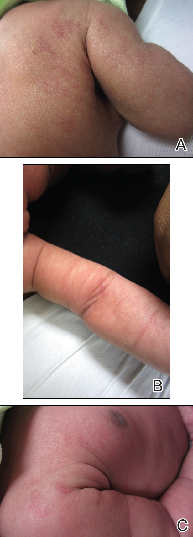
This case describes the rare condition of congenital unilateral nevoid telangiectasia (UNT). Unilateral nevoid telangiectasia is a rare cutaneous vascular condition first described by Blaschko1 in 1899. It is characterized by the presence of unilateral superficial telangiectases occurring most often in the cervical and upper thoracic dermatomes in a linear pattern.2 Females are more often affected than males (2:1 ratio), and cases of UNT are either congenital or acquired.3 Although most UNT cases are acquired and often found in females, approximately 15% of cases are congenital and are comprised largely by males. Acquired cases have been hypothesized to occur in association with hyperestrogenemic states such as pregnancy, puberty, oral contraceptive use and hormonal therapy, alcoholism, and liver disease including hepatitis B and C infections.4,5 There is conflicting evidence as to whether there is an absolute increase in the presence of estrogen and progesterone receptors in the skin, as many case reports show no increase. Instead, others hypothesize that the condition is actually a result of somatic mosaicism and that the cutaneous lesions are genetically predisposed to becoming visibly evident under conditions of elevated estrogen.2
In our case, we hypothesize that the cause was elevated maternal estrogen levels present at higher than normal levels in the fetal circulation. The presence of gynecomastia seen in our patient supports the hypothesis that increased circulating estrogen may be present in infants with UNT.
- Blaschko A. Teleangiektasien. versammlungen. Berliner Dermatologische Gesellschaft. Monatschr prakt Dermat. 1899;28:451.
- Karakas¸ M, Durdu M, Sönmezoğlu S, et al. Unilateral nevoidtelangiectasia. J Dermatol. 2004;31:109-112.
- Wenson SF, Farhana J, Sepehr A. Unilateral nevoid telangiectasia syndrome: a case report and review of the literature. Dermatol Online J. 2011;17:2.
- Hynes LR, Shenefelt PD. Unilateral nevoid telangiectasia: occurrence in two patients with hepatitis C. J Am Acad Dermatol. 1997;36(5 pt 2):819-822.
- Guedes R, Leite L. Unilateral nevoid telangiectasia: a rare disease? Indian J Dermatol. 2012;57:138-140.
The Diagnosis: Congenital Unilateral Nevoid Telangiectasia
Two weeks later the patches were noticeably lighter (Figures 1A and 1B). She continued to be in good health, but gynecomastia was notably present on examination (Figure 1C). At 3 months of age, all patches on the right arm, superior aspect of the chest, and superior aspect of the back had resolved, along with the gynecomastia (Figure 2).
This case describes the rare condition of congenital unilateral nevoid telangiectasia (UNT). Unilateral nevoid telangiectasia is a rare cutaneous vascular condition first described by Blaschko1 in 1899. It is characterized by the presence of unilateral superficial telangiectases occurring most often in the cervical and upper thoracic dermatomes in a linear pattern.2 Females are more often affected than males (2:1 ratio), and cases of UNT are either congenital or acquired.3 Although most UNT cases are acquired and often found in females, approximately 15% of cases are congenital and are comprised largely by males. Acquired cases have been hypothesized to occur in association with hyperestrogenemic states such as pregnancy, puberty, oral contraceptive use and hormonal therapy, alcoholism, and liver disease including hepatitis B and C infections.4,5 There is conflicting evidence as to whether there is an absolute increase in the presence of estrogen and progesterone receptors in the skin, as many case reports show no increase. Instead, others hypothesize that the condition is actually a result of somatic mosaicism and that the cutaneous lesions are genetically predisposed to becoming visibly evident under conditions of elevated estrogen.2
In our case, we hypothesize that the cause was elevated maternal estrogen levels present at higher than normal levels in the fetal circulation. The presence of gynecomastia seen in our patient supports the hypothesis that increased circulating estrogen may be present in infants with UNT.
The Diagnosis: Congenital Unilateral Nevoid Telangiectasia
Two weeks later the patches were noticeably lighter (Figures 1A and 1B). She continued to be in good health, but gynecomastia was notably present on examination (Figure 1C). At 3 months of age, all patches on the right arm, superior aspect of the chest, and superior aspect of the back had resolved, along with the gynecomastia (Figure 2).
This case describes the rare condition of congenital unilateral nevoid telangiectasia (UNT). Unilateral nevoid telangiectasia is a rare cutaneous vascular condition first described by Blaschko1 in 1899. It is characterized by the presence of unilateral superficial telangiectases occurring most often in the cervical and upper thoracic dermatomes in a linear pattern.2 Females are more often affected than males (2:1 ratio), and cases of UNT are either congenital or acquired.3 Although most UNT cases are acquired and often found in females, approximately 15% of cases are congenital and are comprised largely by males. Acquired cases have been hypothesized to occur in association with hyperestrogenemic states such as pregnancy, puberty, oral contraceptive use and hormonal therapy, alcoholism, and liver disease including hepatitis B and C infections.4,5 There is conflicting evidence as to whether there is an absolute increase in the presence of estrogen and progesterone receptors in the skin, as many case reports show no increase. Instead, others hypothesize that the condition is actually a result of somatic mosaicism and that the cutaneous lesions are genetically predisposed to becoming visibly evident under conditions of elevated estrogen.2
In our case, we hypothesize that the cause was elevated maternal estrogen levels present at higher than normal levels in the fetal circulation. The presence of gynecomastia seen in our patient supports the hypothesis that increased circulating estrogen may be present in infants with UNT.
- Blaschko A. Teleangiektasien. versammlungen. Berliner Dermatologische Gesellschaft. Monatschr prakt Dermat. 1899;28:451.
- Karakas¸ M, Durdu M, Sönmezoğlu S, et al. Unilateral nevoidtelangiectasia. J Dermatol. 2004;31:109-112.
- Wenson SF, Farhana J, Sepehr A. Unilateral nevoid telangiectasia syndrome: a case report and review of the literature. Dermatol Online J. 2011;17:2.
- Hynes LR, Shenefelt PD. Unilateral nevoid telangiectasia: occurrence in two patients with hepatitis C. J Am Acad Dermatol. 1997;36(5 pt 2):819-822.
- Guedes R, Leite L. Unilateral nevoid telangiectasia: a rare disease? Indian J Dermatol. 2012;57:138-140.
- Blaschko A. Teleangiektasien. versammlungen. Berliner Dermatologische Gesellschaft. Monatschr prakt Dermat. 1899;28:451.
- Karakas¸ M, Durdu M, Sönmezoğlu S, et al. Unilateral nevoidtelangiectasia. J Dermatol. 2004;31:109-112.
- Wenson SF, Farhana J, Sepehr A. Unilateral nevoid telangiectasia syndrome: a case report and review of the literature. Dermatol Online J. 2011;17:2.
- Hynes LR, Shenefelt PD. Unilateral nevoid telangiectasia: occurrence in two patients with hepatitis C. J Am Acad Dermatol. 1997;36(5 pt 2):819-822.
- Guedes R, Leite L. Unilateral nevoid telangiectasia: a rare disease? Indian J Dermatol. 2012;57:138-140.
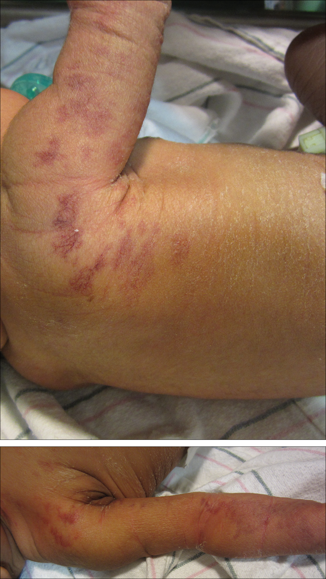
A 1-day-old female infant presented with red patches on the right arm that had been present since delivery. The patient was born to a healthy mother by spontaneous vaginal delivery without complications and with a good Apgar score. The newborn moved both arms and legs well and blood work was unremarkable. Her mother noted being healthy during pregnancy, and she had not taken any additional medications aside from prenatal vitamins. Examination of the infant revealed red blanchable reticulate patches in a dermatomal distribution extending from the posterior aspect of the right shoulder (top) down to the flexural aspect of the arm (bottom). There also were a few coalescing reticulate patches on the superior aspect of the right side of the chest and superior aspect of the right side of the back that resolved by 3 months of age.
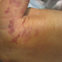
Bright Futures 4th Edition gets a clinical refresher
CHICAGO – Bracing his audience for a whirlwind tour of the many updates to the fourth edition of Bright Futures, Joseph F. Hagan Jr., MD, said that it’s still completely possible to fit Bright Futures visits into a clinic day.
“I practice primary care pediatrics,” said Dr. Hagan, a pediatrician in private practice and clinical professor of pediatrics at the University of Vermont, both in Burlington. “I said to my Bright Futures colleagues, if I didn’t think I could do this in 18 minutes, I wouldn’t ask you to do it.”
The Bright Futures framework, described by Dr. Hagan as the health prevention and disease prevention component of the medical home for children and youth, emerges in the Fourth Edition with a significant evidence-based refresher. The changes and updates are built within the existing framework and encompass surveillance and screening recommendations as well as anticipatory guidance. All content, including family handouts, has been updated, said Dr. Hagan, a coeditor of the Fourth Edition of Bright Futures. He spoke at the annual meeting of the American Academy of Pediatrics.

New clinical content
“What’s new? Maternal depression screening is new,” said Dr. Hagan, noting that the recommendation has long been under discussion. Now, supported by a 2016 United States Preventative Task Force (USPSTF) recommendation that carries a grade B level of evidence, all mothers should be screened for depression at the 1-, 2-, 4-, and 6-month Bright Futures visits.
However, he said, know your local regulations. “State mandates to do more might overrule this.” And conversely, “Just because we’re doing it universally until 6 months doesn’t mean you couldn’t selectively screen later if you have concerns.”
Safe sleep is another area with new clinical focus, he said. The new recommendation for the child to sleep in the parent’s room for “at least 6 months” draws on data from European studies showing lower mortality for children who share a room with parents during this period.
Clinicians should continue to recommend that parents not sleep with their infants in couches, chairs, or beds. As before, parents should be told not to have loose blankets, stuffed toys, or crib bumpers in their babies’ cribs. Another key message, said Dr. Hagan, is that “There is no such thing as safe ‘breast-sleeping.’ ”
Parents should be reminded not to swaddle at nap – or bedtime. The risk is that even a 2-month-old infant may be capable of wriggling over from back to front, and a swaddled infant whose hands are trapped may not be able to move to protect her airway once prone. “Swaddle for comfort, swaddle for crying, swaddle for nursing, but don’t swaddle for sleep” is the message, said Dr. Hagan.
For breast-fed babies, iron supplementation should begin at the 4-month visit. The notion is to prevent progression from iron deficiency to frank anemia, said Dr. Hagan. “We know that we screen for iron deficiency anemia … but we also know that before you’re iron deficient anemic, you’re iron deficient,” he said, and iron’s also critical to brain development. For convenience, switching from vitamin D alone to a multivitamin drop with iron at 4 months is a practical choice.
New dental health recommendations bring prevention to the pediatrician’s office. “Fluoride varnish? Do it!” said Dr. Hagan. Although the USPSTF made a 2014 grade B recommendation that primary care clinicians apply fluoride varnish to primary teeth as soon as they erupt, “It’s new to the Bright Futures periodicity schedule,” he said; parents can be assured that fluoride varnish does not cause fluorosis.
The good news for clinicians, he noted. “Once it hits the periodicity schedule, now, it’s a billable service that must be paid” under Affordable Care Act regulations, said Dr. Hagan. “Don’t let your insurer say, ‘That’s part of what you’re already being paid for.’ ” He recommends avoiding the pressure to bundle this important service. Use the discrete CPT code 99188, “Application of a fluoride varnish by a physician or other qualified health care professional.”
Although Bright Futures has updated recommendations for dyslipidemia blood screening, the USPSTF found insufficient evidence to back lipid screening for those younger than 20 years of age, citing an inability to assess the balance of benefits and harms for universal, rather than risk-based, screening. However, said Dr. Hagan, the American Academy of Pediatrics (AAP), and the National Heart, Lung, and Blood Institute (NHLBI) were looking at this issue at about the same time, and they “did a really good job of showing their work,” to show that if family history alone guided screening in the pediatric population, it “just wasn’t getting done.” And AAP and NHLBI did demonstrate evidence sufficient to support this recommendation.
Accordingly, Bright Futures recommends one screening between ages 9 and 11 years and an additional screening between ages 17 and 21. These windows are designed to bracket puberty, said Dr. Hagan, because values can be skewed during that period. “It’s billable, it’s not bundle-able, and I’d recommend that you do it,” he said.
Developmental surveillance and screening
What’s new with developmental surveillance and screening? “Well, we could argue that the milestones are something to think about, because the milestones are the cornerstone of developmental surveillance,” said Dr. Hagan. “You’re in the room with the child. You’re trained, you’re experienced, you’re smart, your gestalt tells you if their development is good or bad.” 
As important as surveillance is, though, he said, it is “nowhere near as important as screening.” Surveillance happens at every well-child visit, but there’s no substitute for formal developmental screening. For the Fourth Edition guidance and toolkit, gross motor milestones have been adjusted to reflect what’s really being seen as more parents adopt the Back to Sleep recommendations as well.
A standardized developmental screening tool is used at the 9-, 18-, and 30-month visits, and when parents or caregivers express concern about development. Autism-specific screening happens at 18 and 24 months.
“Remember this, if you remember nothing else: If the screening is positive, and you believe there’s a problem, you’re going to refer,” not just to the appropriate specialist but also for early intervention services, so time isn’t lost as the child is waiting for further evaluation and a formal diagnosis, said Dr. Hagan. This coordinated effort appropriately places the responsibility for early identification of developmental delays and disorders at the doorstep of the child’s medical home.
The federally-coordinated Birth to 5: Watch Me Thrive! effort has aggregated research-based screening tools, users’ guides targeted at a variety of audiences, and resources to help caregivers, said Dr. Hagan.
Four commonly-used tools to consider using during the visit are the Parents’ Evaluation of Developmental Status, the Ages and Stages Questionnaire, the Child Health and Development Interactive System, and the Survey of Wellbeing of Young Children. Of these, said Dr. Hagan, the latter is the only tool that’s in the public domain. However, he said, they are “all really good.”
Consider having parents fill out screening questionnaires in the waiting room before the visit, said Dr. Hagan. “I always tell my colleagues, ‘Have them start the visit without you, if you want to get it done in 18 minutes.’ ”
Two questionnaires per visit are available in the Bright Futures toolkit. One questionnaire asks developmental surveillance and risk assessment questions for selective screening. The second questionnaire asks prescreening questions to help with the anticipatory guidance part of the visit, he said. Having families do these ahead of time, said Dr. Hagan, “allows you to become more focused.”
Paying attention to practicalities can make all this go more smoothly, and maximize reimbursement as well. In his own practice, Dr. Hagan said, screening tools and questionnaires are integrated into the EHR system, so that appropriate paperwork prints automatically ahead of the visit.
It’s also worth reviewing billing practices to make sure that CPT code 96110 is used when administering screening with a standardized instrument and completing scoring and documentation. According to the Bright Futures periodicity schedule, this may be done at the 9-, 18-, and 30-month visits for developmental screening, as well as at 18 and 24 months for autism-specific screening.
Promoting lifelong health
Since the initial Bright Futures guidelines were published in the late 1990s, said Dr. Hagan, the focus has always been on seeing the child as part of the family, who, in turn, are part of the community, forming a framework that addresses the social components of child health. “If you’re not looking at the whole picture, you’re not promoting health,” he said. “It’s no big surprise that we now have a specific, called-out focus on promoting lifelong health.”

In the Fourth Edition, the theme of promoting lifelong health for families and communities is woven throughout, with social determinants of health being a specific visit priority. For example, questions about food insecurity have been drawn from the published literature and are included. Also, said Dr. Hagan, there’s specific anticipatory guidance content that’s clearly marked as addressing social determinants of health.
The fundamental importance of socioeconomic status as a social determinant of health was brought home by the Robert Wood Johnson Foundation’s Commission to Build a Healthier America, which demonstrated that, “Your ZIP code is more important to your health than your genetic code,” said Dr. Hagan. “So your work in health supervision is important, and you have been leaders in this effort.”
Research guides Bright Futures updates
The fourth edition of Bright Futures builds on health promotion themes to support the mental and physical health of children and adolescents, and has a robust framework of evidence underpinning the guidelines, said Dr. Hagan.
The goal is for clinicians to “use evidence to decide upon content of their own health supervision visits,” he explained.
The chapter of the Bright Futures guidelines that addresses the evidence and rationale for the guidelines has been expanded to better answer two questions, said Dr. Hagan: “What evidence grounds our recommendations?” and “What rationale did we use when evidence was insufficient or lacking?”
When possible, the editors of the guidelines used evidence-based sources such as recommendations from the USPSTF, the Centers for Disease Control Community Guide, and the Cochrane Collaboration.
There were many more evidence-based recommendations available to those working on the 4th edition than there had been when writing the previous edition, when, said Dr. Hagan, the USPSTF had exactly two recommendations for those under the age of 21 years. The current expanded number of USPSTF pediatric recommendations was due in part to the attention the AAP was able to bring regarding the need for evidence-based recommendations in pediatrics, he said.
When guidelines were not available, the editors also turned to high quality studies from peer reviewed publications. When such high quality evidence was lacking in a particular area, the guidelines make clear what rationale was used to formulate a given recommendation, and that some recommendations should be interpreted with a degree of caution.
And, said Dr. Hagan, even science-based guidelines will change as more data accumulates. “Don’t forget about peanuts!” he said. “It was really logical 15 years ago when we said don’t give peanut products until 1 year of age. And about 2 years ago, we found out that it really didn’t work.”
Although there are specific updates to clinical content, there also were changes made in broader strokes throughout the 4th edition. One of these shifts embeds social determinants of health in many visits. This adjustment acknowledges the growing body of knowledge that “strengths and protective factors make a difference, and risk factors make a difference” in pediatric outcomes.
A greater focus on lifelong physical and mental health is included under the general rubric of promoting lifelong health for families and communities. More emphasis is placed on promoting health for children and youth who have special health care needs as well.
Nuts-and-bolts changes in the updated 4th edition include updates for milestones of development and accompanying developmental surveillance questions, new clinical content and guidance for implementation that have been added based on strong evidence, and a variety of updates for adolescent screenings in particular.
The full 4th edition Bright Futures toolkit will be available for use in 2018.
Dr. Hagan was a coeditor of the Fourth Edition of Bright Futures.
*This article was updated on December 21, 2017
CHICAGO – Bracing his audience for a whirlwind tour of the many updates to the fourth edition of Bright Futures, Joseph F. Hagan Jr., MD, said that it’s still completely possible to fit Bright Futures visits into a clinic day.
“I practice primary care pediatrics,” said Dr. Hagan, a pediatrician in private practice and clinical professor of pediatrics at the University of Vermont, both in Burlington. “I said to my Bright Futures colleagues, if I didn’t think I could do this in 18 minutes, I wouldn’t ask you to do it.”
The Bright Futures framework, described by Dr. Hagan as the health prevention and disease prevention component of the medical home for children and youth, emerges in the Fourth Edition with a significant evidence-based refresher. The changes and updates are built within the existing framework and encompass surveillance and screening recommendations as well as anticipatory guidance. All content, including family handouts, has been updated, said Dr. Hagan, a coeditor of the Fourth Edition of Bright Futures. He spoke at the annual meeting of the American Academy of Pediatrics.
New clinical content
“What’s new? Maternal depression screening is new,” said Dr. Hagan, noting that the recommendation has long been under discussion. Now, supported by a 2016 United States Preventative Task Force (USPSTF) recommendation that carries a grade B level of evidence, all mothers should be screened for depression at the 1-, 2-, 4-, and 6-month Bright Futures visits.
However, he said, know your local regulations. “State mandates to do more might overrule this.” And conversely, “Just because we’re doing it universally until 6 months doesn’t mean you couldn’t selectively screen later if you have concerns.”
Safe sleep is another area with new clinical focus, he said. The new recommendation for the child to sleep in the parent’s room for “at least 6 months” draws on data from European studies showing lower mortality for children who share a room with parents during this period.
Clinicians should continue to recommend that parents not sleep with their infants in couches, chairs, or beds. As before, parents should be told not to have loose blankets, stuffed toys, or crib bumpers in their babies’ cribs. Another key message, said Dr. Hagan, is that “There is no such thing as safe ‘breast-sleeping.’ ”
Parents should be reminded not to swaddle at nap – or bedtime. The risk is that even a 2-month-old infant may be capable of wriggling over from back to front, and a swaddled infant whose hands are trapped may not be able to move to protect her airway once prone. “Swaddle for comfort, swaddle for crying, swaddle for nursing, but don’t swaddle for sleep” is the message, said Dr. Hagan.
For breast-fed babies, iron supplementation should begin at the 4-month visit. The notion is to prevent progression from iron deficiency to frank anemia, said Dr. Hagan. “We know that we screen for iron deficiency anemia … but we also know that before you’re iron deficient anemic, you’re iron deficient,” he said, and iron’s also critical to brain development. For convenience, switching from vitamin D alone to a multivitamin drop with iron at 4 months is a practical choice.
New dental health recommendations bring prevention to the pediatrician’s office. “Fluoride varnish? Do it!” said Dr. Hagan. Although the USPSTF made a 2014 grade B recommendation that primary care clinicians apply fluoride varnish to primary teeth as soon as they erupt, “It’s new to the Bright Futures periodicity schedule,” he said; parents can be assured that fluoride varnish does not cause fluorosis.
The good news for clinicians, he noted. “Once it hits the periodicity schedule, now, it’s a billable service that must be paid” under Affordable Care Act regulations, said Dr. Hagan. “Don’t let your insurer say, ‘That’s part of what you’re already being paid for.’ ” He recommends avoiding the pressure to bundle this important service. Use the discrete CPT code 99188, “Application of a fluoride varnish by a physician or other qualified health care professional.”
Although Bright Futures has updated recommendations for dyslipidemia blood screening, the USPSTF found insufficient evidence to back lipid screening for those younger than 20 years of age, citing an inability to assess the balance of benefits and harms for universal, rather than risk-based, screening. However, said Dr. Hagan, the American Academy of Pediatrics (AAP), and the National Heart, Lung, and Blood Institute (NHLBI) were looking at this issue at about the same time, and they “did a really good job of showing their work,” to show that if family history alone guided screening in the pediatric population, it “just wasn’t getting done.” And AAP and NHLBI did demonstrate evidence sufficient to support this recommendation.
Accordingly, Bright Futures recommends one screening between ages 9 and 11 years and an additional screening between ages 17 and 21. These windows are designed to bracket puberty, said Dr. Hagan, because values can be skewed during that period. “It’s billable, it’s not bundle-able, and I’d recommend that you do it,” he said.
Developmental surveillance and screening
What’s new with developmental surveillance and screening? “Well, we could argue that the milestones are something to think about, because the milestones are the cornerstone of developmental surveillance,” said Dr. Hagan. “You’re in the room with the child. You’re trained, you’re experienced, you’re smart, your gestalt tells you if their development is good or bad.”
As important as surveillance is, though, he said, it is “nowhere near as important as screening.” Surveillance happens at every well-child visit, but there’s no substitute for formal developmental screening. For the Fourth Edition guidance and toolkit, gross motor milestones have been adjusted to reflect what’s really being seen as more parents adopt the Back to Sleep recommendations as well.
A standardized developmental screening tool is used at the 9-, 18-, and 30-month visits, and when parents or caregivers express concern about development. Autism-specific screening happens at 18 and 24 months.
“Remember this, if you remember nothing else: If the screening is positive, and you believe there’s a problem, you’re going to refer,” not just to the appropriate specialist but also for early intervention services, so time isn’t lost as the child is waiting for further evaluation and a formal diagnosis, said Dr. Hagan. This coordinated effort appropriately places the responsibility for early identification of developmental delays and disorders at the doorstep of the child’s medical home.
The federally-coordinated Birth to 5: Watch Me Thrive! effort has aggregated research-based screening tools, users’ guides targeted at a variety of audiences, and resources to help caregivers, said Dr. Hagan.
Four commonly-used tools to consider using during the visit are the Parents’ Evaluation of Developmental Status, the Ages and Stages Questionnaire, the Child Health and Development Interactive System, and the Survey of Wellbeing of Young Children. Of these, said Dr. Hagan, the latter is the only tool that’s in the public domain. However, he said, they are “all really good.”
Consider having parents fill out screening questionnaires in the waiting room before the visit, said Dr. Hagan. “I always tell my colleagues, ‘Have them start the visit without you, if you want to get it done in 18 minutes.’ ”
Two questionnaires per visit are available in the Bright Futures toolkit. One questionnaire asks developmental surveillance and risk assessment questions for selective screening. The second questionnaire asks prescreening questions to help with the anticipatory guidance part of the visit, he said. Having families do these ahead of time, said Dr. Hagan, “allows you to become more focused.”
Paying attention to practicalities can make all this go more smoothly, and maximize reimbursement as well. In his own practice, Dr. Hagan said, screening tools and questionnaires are integrated into the EHR system, so that appropriate paperwork prints automatically ahead of the visit.
It’s also worth reviewing billing practices to make sure that CPT code 96110 is used when administering screening with a standardized instrument and completing scoring and documentation. According to the Bright Futures periodicity schedule, this may be done at the 9-, 18-, and 30-month visits for developmental screening, as well as at 18 and 24 months for autism-specific screening.
Promoting lifelong health
Since the initial Bright Futures guidelines were published in the late 1990s, said Dr. Hagan, the focus has always been on seeing the child as part of the family, who, in turn, are part of the community, forming a framework that addresses the social components of child health. “If you’re not looking at the whole picture, you’re not promoting health,” he said. “It’s no big surprise that we now have a specific, called-out focus on promoting lifelong health.”
In the Fourth Edition, the theme of promoting lifelong health for families and communities is woven throughout, with social determinants of health being a specific visit priority. For example, questions about food insecurity have been drawn from the published literature and are included. Also, said Dr. Hagan, there’s specific anticipatory guidance content that’s clearly marked as addressing social determinants of health.
The fundamental importance of socioeconomic status as a social determinant of health was brought home by the Robert Wood Johnson Foundation’s Commission to Build a Healthier America, which demonstrated that, “Your ZIP code is more important to your health than your genetic code,” said Dr. Hagan. “So your work in health supervision is important, and you have been leaders in this effort.”
Research guides Bright Futures updates
The fourth edition of Bright Futures builds on health promotion themes to support the mental and physical health of children and adolescents, and has a robust framework of evidence underpinning the guidelines, said Dr. Hagan.
The goal is for clinicians to “use evidence to decide upon content of their own health supervision visits,” he explained.
The chapter of the Bright Futures guidelines that addresses the evidence and rationale for the guidelines has been expanded to better answer two questions, said Dr. Hagan: “What evidence grounds our recommendations?” and “What rationale did we use when evidence was insufficient or lacking?”
When possible, the editors of the guidelines used evidence-based sources such as recommendations from the USPSTF, the Centers for Disease Control Community Guide, and the Cochrane Collaboration.
There were many more evidence-based recommendations available to those working on the 4th edition than there had been when writing the previous edition, when, said Dr. Hagan, the USPSTF had exactly two recommendations for those under the age of 21 years. The current expanded number of USPSTF pediatric recommendations was due in part to the attention the AAP was able to bring regarding the need for evidence-based recommendations in pediatrics, he said.
When guidelines were not available, the editors also turned to high quality studies from peer reviewed publications. When such high quality evidence was lacking in a particular area, the guidelines make clear what rationale was used to formulate a given recommendation, and that some recommendations should be interpreted with a degree of caution.
And, said Dr. Hagan, even science-based guidelines will change as more data accumulates. “Don’t forget about peanuts!” he said. “It was really logical 15 years ago when we said don’t give peanut products until 1 year of age. And about 2 years ago, we found out that it really didn’t work.”
Although there are specific updates to clinical content, there also were changes made in broader strokes throughout the 4th edition. One of these shifts embeds social determinants of health in many visits. This adjustment acknowledges the growing body of knowledge that “strengths and protective factors make a difference, and risk factors make a difference” in pediatric outcomes.
A greater focus on lifelong physical and mental health is included under the general rubric of promoting lifelong health for families and communities. More emphasis is placed on promoting health for children and youth who have special health care needs as well.
Nuts-and-bolts changes in the updated 4th edition include updates for milestones of development and accompanying developmental surveillance questions, new clinical content and guidance for implementation that have been added based on strong evidence, and a variety of updates for adolescent screenings in particular.
The full 4th edition Bright Futures toolkit will be available for use in 2018.
Dr. Hagan was a coeditor of the Fourth Edition of Bright Futures.
*This article was updated on December 21, 2017
CHICAGO – Bracing his audience for a whirlwind tour of the many updates to the fourth edition of Bright Futures, Joseph F. Hagan Jr., MD, said that it’s still completely possible to fit Bright Futures visits into a clinic day.
“I practice primary care pediatrics,” said Dr. Hagan, a pediatrician in private practice and clinical professor of pediatrics at the University of Vermont, both in Burlington. “I said to my Bright Futures colleagues, if I didn’t think I could do this in 18 minutes, I wouldn’t ask you to do it.”
The Bright Futures framework, described by Dr. Hagan as the health prevention and disease prevention component of the medical home for children and youth, emerges in the Fourth Edition with a significant evidence-based refresher. The changes and updates are built within the existing framework and encompass surveillance and screening recommendations as well as anticipatory guidance. All content, including family handouts, has been updated, said Dr. Hagan, a coeditor of the Fourth Edition of Bright Futures. He spoke at the annual meeting of the American Academy of Pediatrics.
New clinical content
“What’s new? Maternal depression screening is new,” said Dr. Hagan, noting that the recommendation has long been under discussion. Now, supported by a 2016 United States Preventative Task Force (USPSTF) recommendation that carries a grade B level of evidence, all mothers should be screened for depression at the 1-, 2-, 4-, and 6-month Bright Futures visits.
However, he said, know your local regulations. “State mandates to do more might overrule this.” And conversely, “Just because we’re doing it universally until 6 months doesn’t mean you couldn’t selectively screen later if you have concerns.”
Safe sleep is another area with new clinical focus, he said. The new recommendation for the child to sleep in the parent’s room for “at least 6 months” draws on data from European studies showing lower mortality for children who share a room with parents during this period.
Clinicians should continue to recommend that parents not sleep with their infants in couches, chairs, or beds. As before, parents should be told not to have loose blankets, stuffed toys, or crib bumpers in their babies’ cribs. Another key message, said Dr. Hagan, is that “There is no such thing as safe ‘breast-sleeping.’ ”
Parents should be reminded not to swaddle at nap – or bedtime. The risk is that even a 2-month-old infant may be capable of wriggling over from back to front, and a swaddled infant whose hands are trapped may not be able to move to protect her airway once prone. “Swaddle for comfort, swaddle for crying, swaddle for nursing, but don’t swaddle for sleep” is the message, said Dr. Hagan.
For breast-fed babies, iron supplementation should begin at the 4-month visit. The notion is to prevent progression from iron deficiency to frank anemia, said Dr. Hagan. “We know that we screen for iron deficiency anemia … but we also know that before you’re iron deficient anemic, you’re iron deficient,” he said, and iron’s also critical to brain development. For convenience, switching from vitamin D alone to a multivitamin drop with iron at 4 months is a practical choice.
New dental health recommendations bring prevention to the pediatrician’s office. “Fluoride varnish? Do it!” said Dr. Hagan. Although the USPSTF made a 2014 grade B recommendation that primary care clinicians apply fluoride varnish to primary teeth as soon as they erupt, “It’s new to the Bright Futures periodicity schedule,” he said; parents can be assured that fluoride varnish does not cause fluorosis.
The good news for clinicians, he noted. “Once it hits the periodicity schedule, now, it’s a billable service that must be paid” under Affordable Care Act regulations, said Dr. Hagan. “Don’t let your insurer say, ‘That’s part of what you’re already being paid for.’ ” He recommends avoiding the pressure to bundle this important service. Use the discrete CPT code 99188, “Application of a fluoride varnish by a physician or other qualified health care professional.”
Although Bright Futures has updated recommendations for dyslipidemia blood screening, the USPSTF found insufficient evidence to back lipid screening for those younger than 20 years of age, citing an inability to assess the balance of benefits and harms for universal, rather than risk-based, screening. However, said Dr. Hagan, the American Academy of Pediatrics (AAP), and the National Heart, Lung, and Blood Institute (NHLBI) were looking at this issue at about the same time, and they “did a really good job of showing their work,” to show that if family history alone guided screening in the pediatric population, it “just wasn’t getting done.” And AAP and NHLBI did demonstrate evidence sufficient to support this recommendation.
Accordingly, Bright Futures recommends one screening between ages 9 and 11 years and an additional screening between ages 17 and 21. These windows are designed to bracket puberty, said Dr. Hagan, because values can be skewed during that period. “It’s billable, it’s not bundle-able, and I’d recommend that you do it,” he said.
Developmental surveillance and screening
What’s new with developmental surveillance and screening? “Well, we could argue that the milestones are something to think about, because the milestones are the cornerstone of developmental surveillance,” said Dr. Hagan. “You’re in the room with the child. You’re trained, you’re experienced, you’re smart, your gestalt tells you if their development is good or bad.”
As important as surveillance is, though, he said, it is “nowhere near as important as screening.” Surveillance happens at every well-child visit, but there’s no substitute for formal developmental screening. For the Fourth Edition guidance and toolkit, gross motor milestones have been adjusted to reflect what’s really being seen as more parents adopt the Back to Sleep recommendations as well.
A standardized developmental screening tool is used at the 9-, 18-, and 30-month visits, and when parents or caregivers express concern about development. Autism-specific screening happens at 18 and 24 months.
“Remember this, if you remember nothing else: If the screening is positive, and you believe there’s a problem, you’re going to refer,” not just to the appropriate specialist but also for early intervention services, so time isn’t lost as the child is waiting for further evaluation and a formal diagnosis, said Dr. Hagan. This coordinated effort appropriately places the responsibility for early identification of developmental delays and disorders at the doorstep of the child’s medical home.
The federally-coordinated Birth to 5: Watch Me Thrive! effort has aggregated research-based screening tools, users’ guides targeted at a variety of audiences, and resources to help caregivers, said Dr. Hagan.
Four commonly-used tools to consider using during the visit are the Parents’ Evaluation of Developmental Status, the Ages and Stages Questionnaire, the Child Health and Development Interactive System, and the Survey of Wellbeing of Young Children. Of these, said Dr. Hagan, the latter is the only tool that’s in the public domain. However, he said, they are “all really good.”
Consider having parents fill out screening questionnaires in the waiting room before the visit, said Dr. Hagan. “I always tell my colleagues, ‘Have them start the visit without you, if you want to get it done in 18 minutes.’ ”
Two questionnaires per visit are available in the Bright Futures toolkit. One questionnaire asks developmental surveillance and risk assessment questions for selective screening. The second questionnaire asks prescreening questions to help with the anticipatory guidance part of the visit, he said. Having families do these ahead of time, said Dr. Hagan, “allows you to become more focused.”
Paying attention to practicalities can make all this go more smoothly, and maximize reimbursement as well. In his own practice, Dr. Hagan said, screening tools and questionnaires are integrated into the EHR system, so that appropriate paperwork prints automatically ahead of the visit.
It’s also worth reviewing billing practices to make sure that CPT code 96110 is used when administering screening with a standardized instrument and completing scoring and documentation. According to the Bright Futures periodicity schedule, this may be done at the 9-, 18-, and 30-month visits for developmental screening, as well as at 18 and 24 months for autism-specific screening.
Promoting lifelong health
Since the initial Bright Futures guidelines were published in the late 1990s, said Dr. Hagan, the focus has always been on seeing the child as part of the family, who, in turn, are part of the community, forming a framework that addresses the social components of child health. “If you’re not looking at the whole picture, you’re not promoting health,” he said. “It’s no big surprise that we now have a specific, called-out focus on promoting lifelong health.”
In the Fourth Edition, the theme of promoting lifelong health for families and communities is woven throughout, with social determinants of health being a specific visit priority. For example, questions about food insecurity have been drawn from the published literature and are included. Also, said Dr. Hagan, there’s specific anticipatory guidance content that’s clearly marked as addressing social determinants of health.
The fundamental importance of socioeconomic status as a social determinant of health was brought home by the Robert Wood Johnson Foundation’s Commission to Build a Healthier America, which demonstrated that, “Your ZIP code is more important to your health than your genetic code,” said Dr. Hagan. “So your work in health supervision is important, and you have been leaders in this effort.”
Research guides Bright Futures updates
The fourth edition of Bright Futures builds on health promotion themes to support the mental and physical health of children and adolescents, and has a robust framework of evidence underpinning the guidelines, said Dr. Hagan.
The goal is for clinicians to “use evidence to decide upon content of their own health supervision visits,” he explained.
The chapter of the Bright Futures guidelines that addresses the evidence and rationale for the guidelines has been expanded to better answer two questions, said Dr. Hagan: “What evidence grounds our recommendations?” and “What rationale did we use when evidence was insufficient or lacking?”
When possible, the editors of the guidelines used evidence-based sources such as recommendations from the USPSTF, the Centers for Disease Control Community Guide, and the Cochrane Collaboration.
There were many more evidence-based recommendations available to those working on the 4th edition than there had been when writing the previous edition, when, said Dr. Hagan, the USPSTF had exactly two recommendations for those under the age of 21 years. The current expanded number of USPSTF pediatric recommendations was due in part to the attention the AAP was able to bring regarding the need for evidence-based recommendations in pediatrics, he said.
When guidelines were not available, the editors also turned to high quality studies from peer reviewed publications. When such high quality evidence was lacking in a particular area, the guidelines make clear what rationale was used to formulate a given recommendation, and that some recommendations should be interpreted with a degree of caution.
And, said Dr. Hagan, even science-based guidelines will change as more data accumulates. “Don’t forget about peanuts!” he said. “It was really logical 15 years ago when we said don’t give peanut products until 1 year of age. And about 2 years ago, we found out that it really didn’t work.”
Although there are specific updates to clinical content, there also were changes made in broader strokes throughout the 4th edition. One of these shifts embeds social determinants of health in many visits. This adjustment acknowledges the growing body of knowledge that “strengths and protective factors make a difference, and risk factors make a difference” in pediatric outcomes.
A greater focus on lifelong physical and mental health is included under the general rubric of promoting lifelong health for families and communities. More emphasis is placed on promoting health for children and youth who have special health care needs as well.
Nuts-and-bolts changes in the updated 4th edition include updates for milestones of development and accompanying developmental surveillance questions, new clinical content and guidance for implementation that have been added based on strong evidence, and a variety of updates for adolescent screenings in particular.
The full 4th edition Bright Futures toolkit will be available for use in 2018.
Dr. Hagan was a coeditor of the Fourth Edition of Bright Futures.
*This article was updated on December 21, 2017
EXPERT ANALYSIS FROM AAP 2017
Toy stethoscopes
Many of my articles are inspired when I observe discordant things juxtaposed. As we move deep into winter, once again I am confronted with the issue of infection control in the office and on the ward. Hospitals have gowns, gloves, masks, and toy stethoscopes. My outpatient offices rarely used more than the sink. In urgent care clinic, each evening I would swab three or four throats for strep, with one or two turning positive. I thought nothing of it, other than being glad when gagging a patient that I wear glasses. In the hospital, I must gown, glove, and mask for a patient with strep throat. The variations in practice between hospitals (I’ve been credentialed in 30) do not make me confident in the evidence base for infection control practices. I mentioned the Red Book to a second-year resident last week. He said he had seen it on a shelf but never actually used it.

In medical school, I was taught that the most important part of a stethoscope is between the ears. I believe that statement is true, but in a similar way to how I choose wines. My palate can’t tell the difference between a $15 and a $50 bottle of wine, so buying more expensive wine is a waste. However, a $3 bottle of wine is clearly inferior, if not undrinkable. There are oenophiles (one a distant cousin in Norway) who have trained their palates to tell the difference in wines, just as there are audiophiles who support the sales of $1,000 stereo speakers. Some fraction of those snobs may have justification. So, if cardiologists have strong opinions on stethoscopes, I won’t begrudge them their choice of a more expensive model. Their tastes do not mean that the average person should spend that much on wine, speakers, or stethoscopes. I will assert that there was a time when I could tell a day or two in advance that my otoscope bulb was going to burn out. The color balance was wrong. I carried a pocket otoscope for a few years when rounding in the hospital, but never found it as accurate as my original one. Every craftsman gets accustomed to their best tools.
A professional should be aware of the minimum quality of tool needed to get the job done.
Toy isolation stethoscopes ($3 each retail in bulk) add nothing to my discernment of an infant with bronchiolitis who is distressed, so I consider that equipment a waste of money and polluting to the environment. I typically use my stethoscope and foam it on leaving the room. There is evidence that either foam or alcohol pads are effective1 in killing germs, but no proof that this hygiene makes a difference clinically.2 The myriad researchers who have published about stethoscope contamination have stopped at padding their academic portfolios with something easy to publish, which basically is a high school science project using agar plates. They then make insinuations about policy, without any cost-benefit analysis. They really haven’t been bothered enough to advance the science of clinical medicine and actually measure a clinical impact of these policies. It is a corruption of science created by the publish-or-perish environment.
One survey found that 45% of physicians disinfect their stethoscope annually or less. Laundering of white coats follows a similar pattern, which is why the British National Health Service banned lab coats for physicians 10 years ago. No ties or long sleeve shirts either. I am smug knowing that my sartorial sense was ahead of my time in this regard.
The quality-improvement work of Ignaz Semmelweis should be required reading for all physicians. The control chart3 he published on puerperal fever in Vienna in the 1840s is spectacular. Infection control is important. Modern medical science cannot produce a similar control chart to justify the amount of dollars spent annually on gowns, gloves, masks, and toy stethoscopes. Sad.

Dr. Powell is a pediatric hospitalist and clinical ethics consultant living in St. Louis. Email him at pdnews@frontlinemedcom.com.
References
1. Am J Infect Control. 2009 Apr;37(3):241-3.
2. J Hosp Infect. 2015 Sep;91(1):1-7.
3. https://en.wikipedia.org/wiki/Historical_mortality_rates_of_puerperal_fever
Many of my articles are inspired when I observe discordant things juxtaposed. As we move deep into winter, once again I am confronted with the issue of infection control in the office and on the ward. Hospitals have gowns, gloves, masks, and toy stethoscopes. My outpatient offices rarely used more than the sink. In urgent care clinic, each evening I would swab three or four throats for strep, with one or two turning positive. I thought nothing of it, other than being glad when gagging a patient that I wear glasses. In the hospital, I must gown, glove, and mask for a patient with strep throat. The variations in practice between hospitals (I’ve been credentialed in 30) do not make me confident in the evidence base for infection control practices. I mentioned the Red Book to a second-year resident last week. He said he had seen it on a shelf but never actually used it.
In medical school, I was taught that the most important part of a stethoscope is between the ears. I believe that statement is true, but in a similar way to how I choose wines. My palate can’t tell the difference between a $15 and a $50 bottle of wine, so buying more expensive wine is a waste. However, a $3 bottle of wine is clearly inferior, if not undrinkable. There are oenophiles (one a distant cousin in Norway) who have trained their palates to tell the difference in wines, just as there are audiophiles who support the sales of $1,000 stereo speakers. Some fraction of those snobs may have justification. So, if cardiologists have strong opinions on stethoscopes, I won’t begrudge them their choice of a more expensive model. Their tastes do not mean that the average person should spend that much on wine, speakers, or stethoscopes. I will assert that there was a time when I could tell a day or two in advance that my otoscope bulb was going to burn out. The color balance was wrong. I carried a pocket otoscope for a few years when rounding in the hospital, but never found it as accurate as my original one. Every craftsman gets accustomed to their best tools.
A professional should be aware of the minimum quality of tool needed to get the job done.
Toy isolation stethoscopes ($3 each retail in bulk) add nothing to my discernment of an infant with bronchiolitis who is distressed, so I consider that equipment a waste of money and polluting to the environment. I typically use my stethoscope and foam it on leaving the room. There is evidence that either foam or alcohol pads are effective1 in killing germs, but no proof that this hygiene makes a difference clinically.2 The myriad researchers who have published about stethoscope contamination have stopped at padding their academic portfolios with something easy to publish, which basically is a high school science project using agar plates. They then make insinuations about policy, without any cost-benefit analysis. They really haven’t been bothered enough to advance the science of clinical medicine and actually measure a clinical impact of these policies. It is a corruption of science created by the publish-or-perish environment.
One survey found that 45% of physicians disinfect their stethoscope annually or less. Laundering of white coats follows a similar pattern, which is why the British National Health Service banned lab coats for physicians 10 years ago. No ties or long sleeve shirts either. I am smug knowing that my sartorial sense was ahead of my time in this regard.
The quality-improvement work of Ignaz Semmelweis should be required reading for all physicians. The control chart3 he published on puerperal fever in Vienna in the 1840s is spectacular. Infection control is important. Modern medical science cannot produce a similar control chart to justify the amount of dollars spent annually on gowns, gloves, masks, and toy stethoscopes. Sad.
Dr. Powell is a pediatric hospitalist and clinical ethics consultant living in St. Louis. Email him at pdnews@frontlinemedcom.com.
References
1. Am J Infect Control. 2009 Apr;37(3):241-3.
2. J Hosp Infect. 2015 Sep;91(1):1-7.
3. https://en.wikipedia.org/wiki/Historical_mortality_rates_of_puerperal_fever
Many of my articles are inspired when I observe discordant things juxtaposed. As we move deep into winter, once again I am confronted with the issue of infection control in the office and on the ward. Hospitals have gowns, gloves, masks, and toy stethoscopes. My outpatient offices rarely used more than the sink. In urgent care clinic, each evening I would swab three or four throats for strep, with one or two turning positive. I thought nothing of it, other than being glad when gagging a patient that I wear glasses. In the hospital, I must gown, glove, and mask for a patient with strep throat. The variations in practice between hospitals (I’ve been credentialed in 30) do not make me confident in the evidence base for infection control practices. I mentioned the Red Book to a second-year resident last week. He said he had seen it on a shelf but never actually used it.
In medical school, I was taught that the most important part of a stethoscope is between the ears. I believe that statement is true, but in a similar way to how I choose wines. My palate can’t tell the difference between a $15 and a $50 bottle of wine, so buying more expensive wine is a waste. However, a $3 bottle of wine is clearly inferior, if not undrinkable. There are oenophiles (one a distant cousin in Norway) who have trained their palates to tell the difference in wines, just as there are audiophiles who support the sales of $1,000 stereo speakers. Some fraction of those snobs may have justification. So, if cardiologists have strong opinions on stethoscopes, I won’t begrudge them their choice of a more expensive model. Their tastes do not mean that the average person should spend that much on wine, speakers, or stethoscopes. I will assert that there was a time when I could tell a day or two in advance that my otoscope bulb was going to burn out. The color balance was wrong. I carried a pocket otoscope for a few years when rounding in the hospital, but never found it as accurate as my original one. Every craftsman gets accustomed to their best tools.
A professional should be aware of the minimum quality of tool needed to get the job done.
Toy isolation stethoscopes ($3 each retail in bulk) add nothing to my discernment of an infant with bronchiolitis who is distressed, so I consider that equipment a waste of money and polluting to the environment. I typically use my stethoscope and foam it on leaving the room. There is evidence that either foam or alcohol pads are effective1 in killing germs, but no proof that this hygiene makes a difference clinically.2 The myriad researchers who have published about stethoscope contamination have stopped at padding their academic portfolios with something easy to publish, which basically is a high school science project using agar plates. They then make insinuations about policy, without any cost-benefit analysis. They really haven’t been bothered enough to advance the science of clinical medicine and actually measure a clinical impact of these policies. It is a corruption of science created by the publish-or-perish environment.
One survey found that 45% of physicians disinfect their stethoscope annually or less. Laundering of white coats follows a similar pattern, which is why the British National Health Service banned lab coats for physicians 10 years ago. No ties or long sleeve shirts either. I am smug knowing that my sartorial sense was ahead of my time in this regard.
The quality-improvement work of Ignaz Semmelweis should be required reading for all physicians. The control chart3 he published on puerperal fever in Vienna in the 1840s is spectacular. Infection control is important. Modern medical science cannot produce a similar control chart to justify the amount of dollars spent annually on gowns, gloves, masks, and toy stethoscopes. Sad.
Dr. Powell is a pediatric hospitalist and clinical ethics consultant living in St. Louis. Email him at pdnews@frontlinemedcom.com.
References
1. Am J Infect Control. 2009 Apr;37(3):241-3.
2. J Hosp Infect. 2015 Sep;91(1):1-7.
3. https://en.wikipedia.org/wiki/Historical_mortality_rates_of_puerperal_fever