User login
Nivolumab improves survival in relapsed mesothelioma
The CONFIRM trial involved 330 previously treated patients with mesothelioma who were randomly assigned to nivolumab or placebo for 1 year or until progression or unacceptable toxicity.
Although recruitment to the study was stopped early because of the COVID-19 pandemic, enough data accrued to show that nivolumab improved overall survival by 28% over placebo, and increased PFS by 39%.
“Nivolumab was deemed a safe and effective treatment and should be considered a new treatment option for patients with relapsed mesothelioma,” said principal investigator Dean A. Fennell, MD, PhD, professor and consultant in thoracic medical oncology, University of Leicester (England).
He presented the results at the 2020 World Conference on Lung Cancer, which was rescheduled for January 2021.
Rina Hui, MD, PhD, Crown Princess Mary Cancer Centre, Westmead Hospital, Sydney, who was not involved in the study, said that these results had been a “long time coming.”
CONFIRM has added “important, encouraging data on immunotherapy in the salvage setting,” Dr. Hui said, noting that two-thirds of patients had received two or more prior lines of therapy.
Dr. Fennel noted that “a significant clinical benefit was observed in the epithelioid subtype” of the disease but not in patients with nonepithelioid disease.
However, there was “no evidence” to support programmed death–ligand 1 (PD-L1) expression as predictive of outcomes, he added, which does appear to be the case in some trials on lung cancer and other tumors.
Commenting on these observations, Dr. Hui said that PD-L1 as a predictive biomarker in mesothelioma has been “controversial,” and she emphasized that the results from CONFIRM indicate “no evidence of PD-L1 being predictive.”
However, Dr. Hui questioned the other observation that clinical benefit appeared to be seen only in the epithelioid subtype.
She emphasized that nonepithelioid disease is known to be a “more aggressive, chemoresistant subtype ... with a steep decline in the survival curves.
“Therefore, a lot of patients would not have made it to a subsequent-line clinical trial, explaining why there were only 12% in the CONFIRM study,” and so the sample size may be “too small to detect a difference in outcome,” Dr. Hui said.
Consequently, Dr. Hui said she “would not deny patients with nonepithelioid histology from considering nivolumab in the salvage setting.”
She argued that there was “no clear predictive biomarker for patient selection” emerging from the CONFIRM data.
She agreed that, in patients with mesothelioma who have progressed following platinum/pemetrexed-based chemotherapy as in the first line, “monotherapy nivolumab now can be considered as a treatment option in the second- ... or third-line setting, after second-line chemotherapy”.
However, outstanding questions remain, including whether nivolumab “provides better outcomes than second-line single agent chemotherapy or second-line gemcitabine with the [vascular endothelial growth factor receptor] inhibitor ramucirumab.”
It may also be that nivolumab plus ipilimumab might be superior to nivolumab alone in the salvage setting.
But a more fundamental question is what should be considered for salvage therapy if nivolumab and ipilimumab have already been used in the first-line setting, Dr. Hui said.
Results of first-line immunotherapy combination trials are “eagerly awaited ... to determine and develop other salvage treatments,” she commented.
Responding on Twitter, Riyaz Shah, MD, PhD, consultant medical oncologist, Maidstone and Tunbridge Wells NHS Trust in Royal Tunbridge Wells, England, echoed these comments, saying that the results were “very exciting,” but he also “can’t wait to see the first-line chemo–immunotherapy data.”
Stephen V. Liu, MD, director of thoracic oncology at Georgetown University, Washington, commented on Twitter that there was “not a lot of safety data” in the presentation and awaits their eventual publication.
He added that it is “good to have a positive trial” in relapsed mesothelioma, “though the first-line studies will decrease the eventual impact as immunotherapy becomes involved earlier in treatment.”
Details of the CONFIRM results
Relapsed mesothelioma is an “unmet need,” and, until now, “there have been no phase 3 trials which have demonstrated improved overall survival,” Dr. Fennell said in his presentation.
However, three phase 2 trials have shown that immune checkpoint targeting via PD-1 has shown useful clinical activity as a monotherapy in the relapsed setting, and one of these trials has led to approval of nivolumab in Japan for this indication.
CONFIRM was an investigator-initiated phase 3 trial in patients with relapsed mesothelioma who had received more than one prior line of therapy and had a good performance status.
Recruitment began in April 2017, and the “target sample size was 336 patients,” Dr. Fennell said, but the trial was “halted at 332 patients (in March 2020) due to the peaking of the COVID-19 pandemic in the U.K.”
“However, at the time, it was felt there were sufficient events” to justify the current analysis of the coprimary endpoints of PFS and OS, despite the latter being 59 events short of the target of 291.
Dr. Fennell said that baseline characteristics were “generally well balanced” between the nivolumab (n = 221) and placebo (n = 111) arms.
However, there were more patients with a PD-L1 tumor proportion score (TPS) of at least 1% among the patients given nivolumab, at 37% versus 29% in the placebo arm.
After a median follow-up of 17.1 months in the nivolumab arm and 14.2 months in the placebo group, overall survival was significantly longer with the active treatment, at 9.2 months versus 6.6 months with placebo (hazard ratio, 0.72; P = .018).
The proportion of patients alive at 12 months was 39.5% in the nivolumab group and 26.9% in patients given placebo. Investigator-assessed PFS was also significantly longer with nivolumab, at 3.0 months versus 1.8 months with placebo (HR, 0.61; P < .001).
The proportion of patients disease free at 12 months was 14.5% with active treatment versus 4.9% months with the placebo.
“The role for PD-L1 as a potential biomarker was assessed,” Dr. Fennell said, using the Dako 22C3 antibody, with 150 nivolumab and 84 placebo patients divided into those with a TPS <1% or ≥1%.
He noted that PD-L1 expression in the tumor “did not predict survival for patients in the CONFIRM trial,” with neither PD-L1 positive nor PD-L1 negative patients demonstrating a significant improvement in overall survival with nivolumab vs placebo.
“For histology, epithelioid mesothelioma patients benefited from nivolumab,” Dr. Fennell continued, with a hazard ratio for death of 0.71 versus placebo (P = .021). “However, for the nonepithelioid subgroup, in this immature survival analysis ... the P value was not significant,” but this was a small subgroup of patients (12% in both nivolumab and placebo groups).
The safety analysis revealed that the proportion of patients with any serious adverse events, of any grade or grade 3 or higher, was almost identical between the active and placebo arms, Dr. Fennel reported. There were five deaths (3.6%) related to a serious adverse event in the nivolumab arm and four (5.3%) in the placebo group.
This research was funded by the Stand Up to Cancer campaign for Cancer Research UK, supported by Cancer Research UK core funding at the Southampton Clinical Trials Unit, and investigator-initiated support from Bristol-Myers Squibb for free drug labeling and distribution and funding for RECIST reporting. Dr. Fennell reported relationships with Astex Therapeutics, AstraZeneca, Atara Biotherapeutics, Bayer, Boehringer Ingelheim, Bristol Myers Squibb, Clovis Oncology, Eli Lilly, Inventiva, Lab 21, Merck, and Roche. Dr. Hui reported relationships with AstraZeneca, Bristol-Myers Squibb, Eli Lilly, Merck, Novartis, Pfizer, Roche, and Seagen.
A version of this article first appeared on Medscape.com.
Screening for lung cancer in never-smokers is ‘feasible’
“Lung cancer in never-smokers is a global rising threat,” said lead researcher Pan-Chyr Yang, MD, PhD, chair professor at the National Taiwan University Hospital and academician of Academia Sinica, Taiwan.
In Taiwan, more than half of the cases of lung cancer occur in never-smokers; among female lung cancer patients, 93% are never-smokers.
The incidence of lung cancer – in particular, adenocarcinoma – is increasing in Taiwan, even though the prevalence of smoking has fallen dramatically in men in recent years and has remained low in women.
At the 2020 World Conference on Lung Cancer, which was rescheduled for January 2021, Dr. Yang presented new results that suggest “LDCT screening for never-smokers with high risk may be feasible.”
The Taiwan Lung Cancer Screening in Never-Smoker Trial (TALENT) recruited over 12,000 individuals aged 55-70 years who had never smoked or had done so more than 15 years previously and had risk factors such as a family history of the disease or passive smoke exposure, or who had regularly been exposed to frying food.
Participants underwent LDCT after chest x-ray, followed by biopsy if necessary.
These procedures detected largely invasive lung cancer in 2.6% of participants. Tumors were of stage 0-I in 95% of cases.
The lung cancer detection rate of 2.6% in TALENT in never-smokers is higher than has been found in large studies of smokers, including the 1.1% rate recorded in the NLST study and the 0.9% seen in the NELSON study.
The key factor associated with increased prevalence of lung cancer was a first-degree family history of the disease, Dr. Yang reported.
Notably, having a sister with lung cancer increased the risk for the disease by 78%. Having an affected brother doubled the risk. An increase in the number of first-degree relatives with lung cancer also significantly increased the risk.
More research needed
The TALENT study “provides new, very original evidence on lung cancer risks, and therefore lung cancer screening eligibility could be redefined in Asia, or at least in East Asia,” said the discussant for the paper, Ugo Pastorino, MD, director of thoracic surgery at IRCCS Istituto Nazionale dei Tumori Foundation, Milan.
However, he said that “more research is needed on lung cancer biology in nonsmokers.”
There is currently no follow-up or mortality data, and given the proportion of patients who underwent invasive procedures, it could be that more than 40% of those procedures were carried out in individuals with benign disease, he cautioned.
On Twitter, Stephen V. Liu, MD, director of thoracic oncology at Georgetown University, Washington, said that although family history “emerges” from the study as a potential risk factor for lung cancer, “this analysis would be much more insightful with genomic analyses of these cancers.”
Devika Das, MD, clinical assistant professor of hematology and oncology, University of Alabama at Birmingham, said that the study is “interesting,” given the rise of adenocarcinoma among never-smokers.
She agreed that further details and long-term outcomes are needed and said the key learning point was the need for a “robust” study of the biology of lung cancer in this population.
Lillian Leigh, an Australian lawyer and a lung cancer patient advocate, said the study “provides new evidence” on lung cancer risks.
“As an Asian never-smoker living with lung cancer, the TALENT trial results give me hope,” she said.
Details of TALENT findings
The TALENT study recruited individuals aged 55-70 years at 17 medical centers between February 2015 and July 2019.
Participants were required to be never-smokers or to have a smoking history of less than 10 pack-years and to have quit the habit more than 15 years previously.
They also had to have one of the following risk factors:
- Family history of lung cancer in up to third-degree relatives, in which case younger patients could be recruited.
- Environmental (passive) tobacco smoking history.
- Chronic lung disease, namely, or .
- A cooking index ≥110, defined as 2/7 × the number of days of frying per week × the number of years cooking.
- Cooking without ventilation.
The participants underwent chest x-ray. If the x-ray proved negative, the team performed standard LDCT, examined blood and urine samples for lung cancer biomarkers, and administered standard questionnaires.
Participants who were found on LDCT to have solid or part-solid nodules greater than 6 mm in diameter or pure ground-glass nodules greater than 5 mm in diameter underwent biopsy or standard follow-up.
Individuals whose initial chest x-ray was positive underwent standard contrast-enhanced chest CT prior to biopsy or standard follow-up.
Of 13,207 individuals initially screened, 12,011 were enrolled. Of those, 73.8% were women. The mean age was 61.2 years, and 93.3% were never-smokers.
Among the participants, 46.4% had a first-degree family history of lung cancer; 3.0% had a second-degree family history; and 0.5% had a third-degree family history.
Environmental tobacco exposure was recorded in 83.2% of patients. Chronic lung disease was present in 9.8%; 36.7% had a cooking index ≥110; and 1.8 cooked without ventilation.
Dr. Yang said LDCT results were positive for 17.4% of patients, and 3.4% underwent invasive procedures.
Overall, lung cancer was detected in 313 participants (2.6%). Invasive lung cancer was detected in 255 (2.1%). Of those, 17.9% had multiple primary lung cancers.
Strikingly, 96.5% of the confirmed lung cancer cases were stage 0-I. The majority were stage IA, “which is higher than in other studies that have focused on heavy smokers,” Dr. Yang said. More than half of cases (58.5%) were invasive adenocarcinomas.
The prevalence of lung cancer was significantly higher among people who had a family history of the disease, at 3.2%, vs. 2.0% in those without, at a relative risk of 1.61 (P < .001).
The prevalence was higher still in individuals who had a first-degree family history of lung cancer, at 3.3%, giving a relative risk of 1.69 in comparison with those who did not have a family history (P < .001). The findings were nonsignificant for second- and third-degree relatives.
The relative risk increased even further when the first-degree relative who had a history of lung cancer was a sister, at 1.78 (P < .001), or a brother, at 2.00 (P < .001).
The relative risk was slightly lower if the patient’s relative was the mother, at 1.43 (P = .010), and was nonsignificant if the relative was the father (P = .077).
The risk for lung cancer also increased with an increase in the number of first-degree relatives with the disease, rising from 3.1% with one relative to 4.0% with two relatives, 6.7% with three relatives, and 9.1% with at least four relatives (P < .001). A similar pattern was seen for invasive lung cancer.
The other risk factors included in the study, such as environmental tobacco exposure, chronic lung disease, and cooking index, were not significantly associated with the prevalence of lung cancer.
No funding for the study has been disclosed. Dr. Yang has received honoraria from AstraZeneca, Boehringer Ingelheim, Pfizer, Merck, Eli Lilly, Roche, GlaxoSmithKline, and ONO Pharma and has served on the advisory board of OBI Pharma, CHO Pharma, and Lin BioScience. Dr. Pastorino has disclosed no relevant financial relationships.
A version of this article first appeared on Medscape.com.
FDA grants MET inhibitor tepotinib accelerated approval for NSCLC
Tepotinib is the first once-daily oral MET inhibitor approved for this patient population, and the approval applies to both treatment-naive and previously treated patients with NSCLC.
The approval was supported by results from the ongoing phase 2 VISION trial. Tepotinib produced an overall response rate of 43% in both treatment-naive patients (n = 69) and previously treated patients (n = 83) in this trial. The median duration of response was 10.8 months and 11.1 months, respectively.
Results of the primary analysis were published in The New England Journal of Medicine last year.
Study subjects received the recommended dose of 450 mg taken as two 225-mg tablets once daily with food until disease progression or unacceptable toxicity. Adverse reactions occurring in at least 20% of patients included edema, fatigue, nausea, diarrhea, musculoskeletal pain, and dyspnea. Interstitial lung disease, hepatotoxicity, and embryo-fetal toxicity also have been reported with tepotinib.
Continued approval of tepotinib “may be contingent upon verification and description of clinical benefit in confirmatory trials,” the FDA stated in an approval announcement.
EMD Serono, the drug’s maker, also announced the approval in a press statement, calling tepotinib “an important and welcome new therapeutic option for patients with metastatic NSCLC harboring these genetic mutations.”
“METex14 skipping occurs in approximately 3% to 4% of NSCLC cases, and patients with this aggressive lung cancer are often elderly and face a poor clinical prognosis,” Paul K. Paik, MD, the VISION primary investigator and clinical director of the thoracic oncology service at Memorial Sloan Kettering Cancer Center in New York, said in the statement.
“There is a pressing need for targeted treatments that have the potential to generate durable antitumor activity and improve the lives of patients with this challenging disease,” he added.
Andrea Ferris, president and chief executive officer of the nonprofit LUNGevity Foundation, further noted the “powerful progress” made in recent years with respect to understanding genetic mutations in NSCLC.
“The availability of a new precision medicine for NSCLC with METex14 skipping alterations advances patient access to targeted treatment and underscores the importance of routine comprehensive biomarker testing for patients with this challenging cancer,” she said in the statement.
Tepotinib was approved in Japan in March 2020. The drug previously received breakthrough therapy designation and orphan drug designation from the FDA. A marketing authorization application for tepotinib was validated by the European Medicines Agency in November 2020 for a similar indication, EMD Serono reported, adding that applications “have also been submitted in Australia, Switzerland, and Canada under the FDA’s Project Orbis initiative, which provides a framework for concurrent submission and review of oncology medicines among international partners.”
Other phase 2 studies of tepotinib are ongoing. The INSIGHT 2 study is designed to test tepotinib in combination with osimertinib in MET amplified, advanced, or metastatic NSCLC with activating EGFR mutations that has progressed following first-line treatment with osimertinib. The PERSPECTIVE study is designed to test tepotinib in combination with cetuximab in patients with RAS/BRAF wild-type left-sided metastatic colorectal cancer with acquired resistance to anti-EGFR antibody targeting therapy due to MET amplification.
For more details on tepotinib, see the full prescribing information.
‘Astonishing’ 4-year survival in NSCLC with pembro plus chemo
The results are from a 4-year follow-up of 160 patients with previously untreated stage IV non–small cell lung cancer (NSCLC) taking part in the KEYNOTE-189 trial of immunotherapy with pembrolizumab plus pemetrexed–platinum chemotherapy versus chemotherapy plus placebo.
After a median follow-up of 46.3 months, the median overall survival (OS) in the intention-to-treat population was 22.0 months with the combination versus 10.6 months with chemotherapy alone (hazard ratio, 0.60).
A similar pattern was seen for progression-free survival (PFS), with patients receiving the combination having a longer median PFS, at 9.0 months versus 4.9 months with chemotherapy alone (HR, 0.50).
“Stellar data,” Riyaz Shah, MD, PhD, consultant medical oncologist, Maidstone and Tunbridge Wells NHS Trust, Royal Tunbridge Wells, England, exclaimed on Twitter.
He described the results for the programmed death-ligand 1 (PD-L1) expression subgroups as “astonishing” and singled out the performance of the combination therapy in patients with very low (<1%) tumor PD-L1 expression, showing more than 23% of patients were alive at 3 years versus just over 5% in the group given chemotherapy alone.
Charu Aggarwal, MD, MPH, Leslye M. Heisler associate professor for lung cancer excellence, Penn Medicine, Philadelphia, said the outcomes with the combination of chemotherapy and immunotherapy were “terrific.”
Sandip P. Patel, MD, medical oncologist, associate professor of medicine, University of California, San Diego, agreed that these long-term results were “very impressive.” However, he noted the “full effect” of chemotherapy plus immunotherapy has not “fully been captured in our overall cancer mortality statistics in the U.S. yet.”
The new results were presented at the 2020 World Conference on Lung Cancer, which was rescheduled for January 2021.
Previous results from KEYNOTE-189 had already demonstrated that, after a median follow-up of 10.5 months, adding pembrolizumab to chemotherapy significantly improves both OS and PFS, compared with chemotherapy alone.
The latest results show that the combination “continued to provide overall survival and progression-free survival benefit” in extended follow-up, said study presenter Jhanelle Elaine Gray, MD, chair, department of thoracic oncology, Moffitt Cancer Center, Tampa.
The 3-year OS rate with pembrolizumab plus chemotherapy, compared with chemotherapy alone was 31.3% versus 17.4%, and the estimated 3-year PFS was 11.8% versus 1.3%.
Substantial improvements were even seen in patients with tumors that had a low level of PD-L1 expression (measured as the PD-L1 tumor proportion score [TPS]).
Dr. Gray highlighted the finding that the survival benefit with pembrolizumab plus chemotherapy was seen regardless of PD-L1 expression in the tumor, with a hazard ratio versus chemotherapy alone of 0.71 in patients with a TPS ≥ 50%, 0.66 in those with a TPS of 1%-49%, and 0.52 in patients with a TPS less than 1%. A similar pattern was seen with PFS, with a hazard ratio of 0.36 in patients with a TPS of at least 50%, 0.54 in those with a TPS of 1%-49%, and 0.68 in patients with a TPS less than 1%.
In addition, overall response rate and duration of response were also improved with combination therapy, regardless of tumor PD-L1 expression.
Among 56 patients who completed 35 cycles of pembrolizumab, the objective response rate was 87.5% (with 10.7% having a complete response and 76.8% a partial response).
At the data cutoff, 45 patients were alive, 28 did not have progressive disease, and seven had started a second course of pembrolizumab.
The side effect profile of the combination was “manageable,” Dr. Gray reported.
The combination arm was associated with more grade 3-5 treatment-related adverse events than the chemotherapy alone arm, at 52.1% versus 42.1%, and more grade 3-5 immune-related adverse events and infusion reactions, at 27.7% versus 13.4%.
Events leading to treatment discontinuation were also more common with pembrolizumab plus chemotherapy than chemotherapy, at 27.4% versus 9.9%.
The combination of pembrolizumab plus pemetrexed-platinum has already become “a standard-of-care therapy for patients with newly diagnosed metastatic nonsquamous NSCLC,” Dr. Gray commented.
The study was funded by Merck. Dr. Gray disclosed relationships with Array, AstraZeneca, Boehringer Ingelheim, Bristol-Myers Squibb, Celgene, Eli Lilly, Genentech, and Merck.
A version of this article first appeared on Medscape.com.
Pandemic seems to impact lung cancer diagnosis and prognosis
The two-center study showed a 38% decrease in new lung cancer diagnoses during the pandemic. Patients diagnosed with non–small cell lung cancer (NSCLC) during the pandemic had more severe disease than patients diagnosed prepandemic, but cases of SCLC were not more severe during the pandemic. Still, the 30-day mortality rate nearly doubled for both NSCLC and SCLC patients during the pandemic.
“The prioritization of the health care system towards COVID-19 patients has led to drastic changes in cancer management that could interfere with the initial diagnosis of lung cancer, resulting in delayed treatment and worse outcomes,” said Roxana Reyes, MD, of Hospital Clínic de Barcelona. “Delay of cancer treatment is associated with increased mortality.”
Dr. Reyes and colleagues conducted a retrospective study of the impact of COVID-19 on the incidence of new lung cancer cases, disease severity, and clinical outcomes. Dr. Reyes reported the group’s findings at the 2020 World Conference on Lung Cancer (Abstract 3700), which was rescheduled for January 2021.
Study details
Dr. Reyes and colleagues compared data from two tertiary hospitals in Spain in the first 6 months of 2020 with data from the same period in 2019. Spain was one of the countries most affected by COVID-19 during the first wave of the pandemic.
The study’s primary endpoint was differences by period in the number of new lung cancer cases and disease severity. A secondary endpoint was 30-day mortality rate by period and histology.
The study included 162 patients newly diagnosed with lung cancer – 100 diagnosed before the pandemic began and 62 diagnosed during the pandemic. Overall, 68% of patients had NSCLC, and 32% had SCLC.
Baseline characteristics were similar between the prepandemic and pandemic groups, except for the proportion of nonsmokers. Twice as many patients diagnosed during the pandemic were nonsmokers (16% vs. 8%).
Differences by time period and subtype
During the pandemic, there was a 38% reduction in all lung cancer diagnoses, a 36% reduction in NSCLC diagnoses, and a 42% reduction in SCLC diagnoses.
Respiratory symptoms were more common during the pandemic for both NSCLC (30% vs. 23%) and SCLC (32% vs. 24%).
Cases of NSCLC diagnosed during the pandemic were more severe, but SCLC cases were not.
In the NSCLC cohort, symptomatic disease was more common during the pandemic (74% vs. 63%), as were advanced disease (58% vs. 46%), more than two metastatic sites (16% vs. 12%), oncologic emergencies (7% vs. 3%), hospitalization (21% vs. 18%), and death during hospitalization (44% vs. 17%).
For SCLC, symptomatic disease was less common during the pandemic (74% vs. 79%), as were advanced disease (52% vs. 67%), more than two metastatic sites (26% vs. 36%), oncologic emergencies (5% vs. 12%), hospitalization (21% vs. 33%), and death during hospitalization (0% vs. 18%).
Nevertheless, the 30-day mortality rate almost doubled during the pandemic for both NSCLC (49% vs. 25%) and SCLC (32% vs. 18%).
For both subtypes together, the median overall survival was 6.7 months during the pandemic and 7.9 months before the pandemic.
Implications and next steps
“In our descriptive study, lung cancer diagnosis is being affected during the COVID-19 pandemic,” Dr. Reyes said. “Fewer new lung cancer cases were diagnosed during COVID-19.”
Some patients with acute respiratory infections who tested negative for COVID-19 during the first 6 months of the pandemic may have had undiagnosed lung cancer, noted Matthew Peters, MD, of Concord Repatriation General Hospital and Macquarie University Hospital, both in Sydney, who was not involved in this study.
“They receive a negative result and think their problem is reduced but wonder why they still have a cough,” Dr. Peters said. “The various lockdowns and social distancing reduced the diagnosis of respiratory viral illnesses that often result in an accidental diagnosis of lung cancer. As time goes by, we will recapture harvesting of accidental diagnosis of lung cancer and provide curative treatments.”
Dr. Reyes emphasized that strategies for maintaining cancer diagnoses need to be implemented during the pandemic. She also noted that this study is ongoing, with the goal of assessing the long-term impact of COVID-19.
Dr. Reyes disclosed relationships with Roche, Bristol-Myers Squibb, and Merck Sharp & Dohme. She did not disclose funding for this study. Dr. Peters disclosed relationships with Amgen, AstraZeneca, Boehringer Ingelheim, Bristol-Myers Squibb, Eli Lilly, Merck, Novartis, Pfizer, Roche, and Takeda.
FROM WCLC 2020
COVID-19 vaccination in cancer patients: NCCN outlines priorities
Vaccination timing considerations vary based on factors such as cancer and treatment type, and reasons for delaying vaccination in the general public also apply to cancer patients (recent COVID-19 exposure, for example).
In general, however, patients with cancer should be assigned to Centers for Disease Control and Prevention priority group 1 b/c and immunized when vaccination is available to them, the guidelines state. Exceptions to this recommendation include:
- Patients undergoing hematopoietic stem cell transplant or receiving engineered cellular therapy such as chimeric antigen receptor T-cell therapy. Vaccination should be delayed for at least 3 months in these patients to maximize vaccine efficacy. Caregivers of these patients, however, should be immunized when possible.
- Patients with hematologic malignancies who are receiving intensive cytotoxic chemotherapy, such as cytarabine- or anthracycline-based regimens for acute myeloid leukemia. Vaccination in these patients should be delayed until absolute neutrophil count recovery.
- Patients undergoing major surgery. Vaccination should occur at least a few days before or after surgery.
- Patients who have experienced a severe or immediate adverse reaction to any of the ingredients in the mRNA COVID-19 vaccines.
Conversely, vaccination should occur when available in patients with hematologic malignancies and marrow failure who are expected to have limited or no recovery, patients with hematologic malignancies who are on long-term maintenance therapy, and patients with solid tumors who are receiving cytotoxic chemotherapy, targeted therapy, checkpoint inhibitors and other immunotherapy, or radiotherapy.
Caregivers, household contacts, and other close contacts who are 16 years of age and older should be vaccinated whenever they are eligible.
Unique concerns in patients with cancer
The NCCN recommendations were developed to address the unique issues and concerns with respect to patients with cancer, who have an increased risk of severe illness from SARS-CoV-2 infection. But the guidelines come with a caveat: “[t]here are limited safety and efficacy data in these patients,” the NCCN emphasized in a press statement.
“Right now, there is urgent need and limited data,” Steven Pergam, MD, co-leader of the NCCN COVID-19 Vaccination Committee, said in the statement.
“Our number one goal is helping to get the vaccine to as many people as we can,” Dr. Pergam said. “That means following existing national and regional directions for prioritizing people who are more likely to face death or severe illness from COVID-19.”
Dr. Pergam, associate professor at Fred Hutchinson Cancer Research Center in Seattle, further explained that “people receiving active cancer treatment are at greater risk for worse outcomes from COVID-19, particularly if they are older and have additional comorbidities, like immunosuppression.”
NCCN’s recommendations couldn’t have come at a better time for patients with cancer, according to Nora Disis, MD, a professor at the University of Washington in Seattle.
“The NCCN’s recommendations to prioritize COVID vaccinations for cancer patients on active treatment is an important step forward in protecting our patients from the infection,” Dr. Disis said in an interview.
“Cancer patients may be at higher risk for the complications seen with infection. In addition, cancer is a disease of older people, and a good number of our patients have the comorbidities that would predict a poorer outcome if they should become sick,” Dr. Disis added. “With the correct treatment, many patients with cancer will be long-term survivors. It is important that they be protected from infection with COVID to realize their best outcome.”
Additional vaccine considerations
The NCCN recommendations also address several other issues of importance for cancer patients, including:
- Deprioritizing other vaccines. COVID-19 vaccines should take precedence over other vaccines because data on dual vaccination are lacking. The NCCN recommends waiting 14 days after COVID-19 vaccination to deliver other vaccines.
- Vaccinating clinical trial participants. Trial leads should be consulted to prevent protocol violations or exclusions.
- Decision-making in the setting of limited vaccine availability. The NCCN noted that decisions on allocation must be made in accordance with state and local vaccine guidance but suggests prioritizing appropriate patients on active treatment, those planning to start treatment, and those who have just completed treatment. Additional risk factors for these patients, as well as other factors associated with risk for adverse COVID-19 outcomes, should also be considered. These include advanced age, comorbidities, and adverse social and demographic factors such as poverty and limited health care access.
- The need for ongoing prevention measures. Vaccines have been shown to decrease the incidence of COVID-19 and related complications, but it remains unclear whether vaccines prevent infection and subsequent transmission. This means everyone should continue following prevention recommendations, such as wearing masks and avoiding crowds.
The NCCN stressed that these recommendations are “intended to be a living document that is constantly evolving – it will be updated rapidly whenever new data comes out, as well as any potential new vaccines that may get approved in the future.” The NCCN also noted that the advisory committee will meet regularly to refine the recommendations as needed.
Dr. Pergam disclosed relationships with Chimerix Inc., Merck & Co., Global Life Technologies Inc., and Sanofi-Aventis. Dr. Disis disclosed grants from Pfizer, Bavarian Nordisk, Janssen, and Precigen. She is the founder of EpiThany and editor-in-chief of JAMA Oncology.
Model predicts acute kidney injury in cancer patients a month in advance
A model that crunches data from routine blood tests can accurately identify cancer patients who will develop acute kidney injury (AKI) up to a month before it happens, according to a cohort study.

The algorithm spotted nearly 74% of the patients who went on to develop AKI within 30 days, providing a window for intervention and possibly prevention, according to investigators.
These results were reported at the AACR Virtual Special Conference: Artificial Intelligence, Diagnosis, and Imaging (abstract PR-11).
“Cancer patients are a high-risk population for AKI due to the nature of their treatment and illness,” said presenter Lauren A. Scanlon, PhD, a data scientist at The Christie NHS Foundation Trust in Huddersfield, England. “AKI causes a huge disruption in treatment and distress for the patient, so it would be amazing if we could, say, predict the AKI before it occurs and prevent it from even happening.”
U.K. health care providers are already using an algorithm to monitor patients’ creatinine levels, comparing new values against historic ones, Dr. Scanlon explained. When that algorithm detects AKI, it issues an alert that triggers implementation of an AKI care bundle, including measures such as fluid monitoring and medication review, within 24 hours.
Taking this concept further, Dr. Scanlon and colleagues developed a random forest model, a type of machine learning algorithm, that incorporates other markers from blood tests routinely obtained for all patients, with the aim of predicting AKI up to 30 days in advance.
“Using routinely collected blood test results will ensure that the model is applicable to all our patients and can be implemented in an automated manner,” Dr. Scanlon noted.
The investigators developed and trained the model using 597,403 blood test results from 48,865 patients undergoing cancer treatment between January 2017 and May 2020.
The model assigns patients to five categories of risk for AKI in the next 30 days: very low, low, medium, high, and very high.
“We wanted the model to output in this way so that it could be used by clinicians alongside their own insight and knowledge on a case-by-case basis,” Dr. Scanlon explained.
The investigators then prospectively validated the model and its risk categories in another 9,913 patients who underwent cancer treatment between June and August 2020.
Using a model threshold of medium risk or higher, the model correctly predicted AKI in 330 (73.8%) of the 447 patients in the validation cohort who ultimately developed AKI.
“This is pretty amazing and shows that this model really is working and can correctly detect these AKIs up to 30 days before they occur, giving a huge window to put in place preventive strategies,” Dr. Scanlon said.
Among the 154 patients in whom the model incorrectly predicted AKI, 9 patients had only a single follow-up blood test and 17 patients did not have any, leaving their actual outcomes unclear.
“Given that AKI detection uses blood tests, an AKI in these patients was never confirmed,” Dr. Scanlon noted. “So this could give a potential benefit of the model that we never intended: It could reduce undiagnosed AKI by flagging those who are at risk.”
“Our next steps are to test the model through a technology clinical trial to see if putting intervention strategies in place does prevent these AKIs from taking place,” Dr. Scanlon concluded. “We are also going to move to ongoing monitoring of the model performance.”
Dr. Scanlon disclosed no conflicts of interest. The study did not receive specific funding.
A model that crunches data from routine blood tests can accurately identify cancer patients who will develop acute kidney injury (AKI) up to a month before it happens, according to a cohort study.
The algorithm spotted nearly 74% of the patients who went on to develop AKI within 30 days, providing a window for intervention and possibly prevention, according to investigators.
These results were reported at the AACR Virtual Special Conference: Artificial Intelligence, Diagnosis, and Imaging (abstract PR-11).
“Cancer patients are a high-risk population for AKI due to the nature of their treatment and illness,” said presenter Lauren A. Scanlon, PhD, a data scientist at The Christie NHS Foundation Trust in Huddersfield, England. “AKI causes a huge disruption in treatment and distress for the patient, so it would be amazing if we could, say, predict the AKI before it occurs and prevent it from even happening.”
U.K. health care providers are already using an algorithm to monitor patients’ creatinine levels, comparing new values against historic ones, Dr. Scanlon explained. When that algorithm detects AKI, it issues an alert that triggers implementation of an AKI care bundle, including measures such as fluid monitoring and medication review, within 24 hours.
Taking this concept further, Dr. Scanlon and colleagues developed a random forest model, a type of machine learning algorithm, that incorporates other markers from blood tests routinely obtained for all patients, with the aim of predicting AKI up to 30 days in advance.
“Using routinely collected blood test results will ensure that the model is applicable to all our patients and can be implemented in an automated manner,” Dr. Scanlon noted.
The investigators developed and trained the model using 597,403 blood test results from 48,865 patients undergoing cancer treatment between January 2017 and May 2020.
The model assigns patients to five categories of risk for AKI in the next 30 days: very low, low, medium, high, and very high.
“We wanted the model to output in this way so that it could be used by clinicians alongside their own insight and knowledge on a case-by-case basis,” Dr. Scanlon explained.
The investigators then prospectively validated the model and its risk categories in another 9,913 patients who underwent cancer treatment between June and August 2020.
Using a model threshold of medium risk or higher, the model correctly predicted AKI in 330 (73.8%) of the 447 patients in the validation cohort who ultimately developed AKI.
“This is pretty amazing and shows that this model really is working and can correctly detect these AKIs up to 30 days before they occur, giving a huge window to put in place preventive strategies,” Dr. Scanlon said.
Among the 154 patients in whom the model incorrectly predicted AKI, 9 patients had only a single follow-up blood test and 17 patients did not have any, leaving their actual outcomes unclear.
“Given that AKI detection uses blood tests, an AKI in these patients was never confirmed,” Dr. Scanlon noted. “So this could give a potential benefit of the model that we never intended: It could reduce undiagnosed AKI by flagging those who are at risk.”
“Our next steps are to test the model through a technology clinical trial to see if putting intervention strategies in place does prevent these AKIs from taking place,” Dr. Scanlon concluded. “We are also going to move to ongoing monitoring of the model performance.”
Dr. Scanlon disclosed no conflicts of interest. The study did not receive specific funding.
FROM AACR: AI, DIAGNOSIS, AND IMAGING 2021
AI can identify biomarkers and potentially guide therapy in NSCLC

Researchers developed deep learning models that could accurately predict a patient’s PD-L1 and EGFR mutation status without the need for a biopsy. If these models are validated in prospective trials, they could guide treatment decisions in patients with NSCLC, according to the researchers.
Wei Mu, PhD, of Moffitt Cancer Center and Research Institute in Tampa, Fla., described this research at the AACR Virtual Special Conference: Artificial Intelligence, Diagnosis, and Imaging (abstract PR-03).
Rationale
Guidelines from the National Comprehensive Cancer Network (NCCN) endorse tailored treatment for patients with NSCLC; namely, immune checkpoint inhibitors for those with PD-L1-positive tumors and EGFR tyrosine kinase inhibitors for patients with tumors harboring a mutation in EGFR.
However, the conventional approach to ascertaining tumor status for these biomarkers has disadvantages, Dr. Mu noted.
“Both require biopsy, which may fail due to insufficient quality of the tissue and, particularly for NSCLC, may increase the chance of morbidity,” Dr. Mu said.
In addition, there is room for improvement in the rigor of the biomarker assays, and there can be substantial wait times for results.
To address these issues, Dr. Mu and colleagues explored an AI radiomics approach using PET/CT scans.
“We know that EGFR mutation and positive PD-L1 expression may change the metabolism of the peritumor and intratumor microenvironment,” Dr. Mu explained. “Therefore, we had the hypothesis that they can be captured by the FDG-PET/CT images.”
Results
The investigators used FDG-PET/CT images from 837 patients with advanced NSCLC treated at four institutions. The team developed AI deep learning models that generated one score for PD-L1 positivity and another score for presence of an EGFR mutation, as well as an associated algorithm that would direct patients to the appropriate treatments depending on the scores.
Results for the PD-L1 deep learning score showed good accuracy in predicting positivity for this ligand, with an area under the curve of 0.89 in the training cohort, 0.84 in the validation cohort, and 0.82 in an external test cohort, Dr. Mu reported. All exceeded the corresponding areas under the curve for maximal standardized uptake values.
Moreover, the score was prognostic and statistically indistinguishable from PD-L1 status determined by immunohistochemistry in predicting progression-free survival.
Similarly, the EGFR deep learning score showed good accuracy in predicting mutational status, with an area under the curve of 0.86 in the training cohort, 0.83 in the validation cohort, and 0.81 in an external test cohort. It outperformed a clinical score based on sex, smoking status, tumor histology, and maximal standardized uptake value in each cohort.
The EGFR deep learning score was prognostic and statistically indistinguishable from EGFR mutational status determined by polymerase chain reaction in predicting progression-free survival.
The models showed good stability when size of the input region of interest was varied, and when different radiologists delineated the region of interest, with an intraclass correlation coefficient of 0.91.
“We developed deep learning models to predict PD-L1 status and EGFR mutation with high accuracy. Using the generated deep learning scores, we obtained a noninvasive treatment decision support tool, which may be useful as a clinical decision support tool pending validation of its clinical utility in a large prospective trial,” Dr. Mu summarized. “Using our tool, NSCLC patients could be directly offered a treatment decision without the need of biopsy.”
“In the future, we will perform a prospective observational trial to compare the results of our noninvasive treatment decision tool with molecular biomarker–based NCCN guidelines,” she said.
The investigators plan to add ALK rearrangement status and prediction of serious adverse events and cachexia to the decision support tool.
Dr. Mu disclosed no conflicts of interest. The study did not have specific funding.
FROM AACR: AI, DIAGNOSIS, AND IMAGING 2021
Lung disease raises mortality risk in older RA patients
Patients with rheumatoid arthritis–associated interstitial lung disease showed increases in overall mortality, respiratory mortality, and cancer mortality, compared with RA patients without interstitial lung disease, based on data from more than 500,000 patients in a nationwide cohort study.

RA-associated interstitial lung disease (RA-ILD) has been associated with worse survival rates as well as reduced quality of life, functional impairment, and increased health care use and costs, wrote Jeffrey A. Sparks, MD, of Brigham and Women’s Hospital, Boston, and colleagues. However, data on the incidence and prevalence of RA-ILD have been inconsistent and large studies are lacking.
In a study published online in Rheumatology, the researchers identified 509,787 RA patients aged 65 years and older from Medicare claims data. The average age of the patients was 72.6 years, and 76.2% were women.
At baseline, 10,306 (2%) of the study population had RA-ILD, and 13,372 (2.7%) developed RA-ILD over an average of 3.8 years’ follow-up per person (total of 1,873,127 person-years of follow-up). The overall incidence of RA-ILD was 7.14 per 1,000 person-years.
Overall mortality was significantly higher among RA-ILD patients than in those with RA alone in a multivariate analysis (38.7% vs. 20.7%; hazard ratio, 1.66).
In addition, RA-ILD was associated with an increased risk of respiratory mortality (HR, 4.39) and cancer mortality (HR, 1.56), compared with RA without ILD. For these hazard regression analyses, the researchers used Fine and Gray subdistribution HRs “to handle competing risks of alternative causes of mortality. For example, the risk of respiratory mortality for patients with RA-ILD, compared with RA without ILD also accounted for the competing risk of cardiovascular, cancer, infection and other types of mortality.”
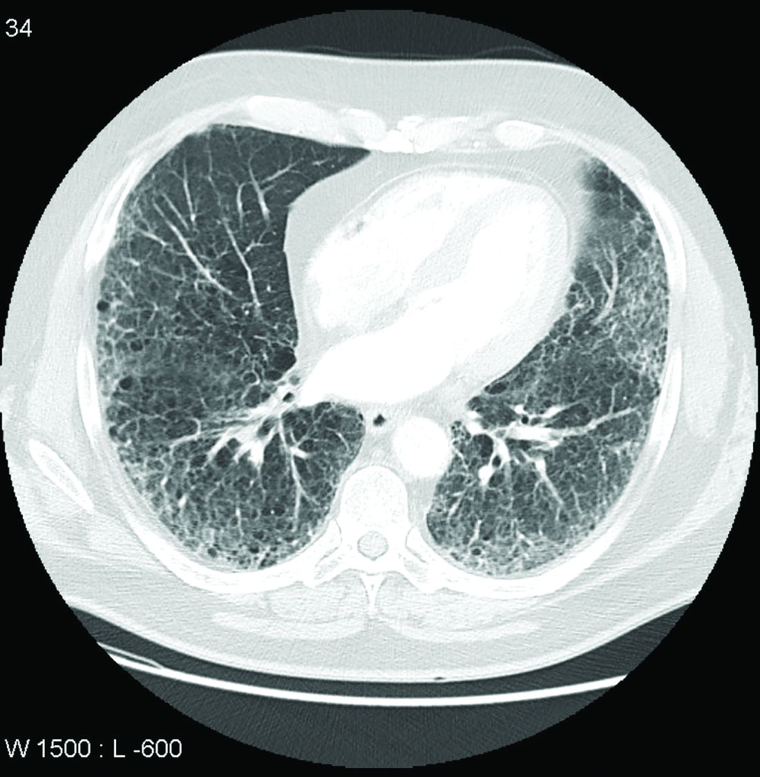
In another multivariate analysis, male gender, smoking, asthma, chronic obstructive pulmonary disorder, and medication use (specifically biologic disease-modifying antirheumatic drugs, targeted synthetic DMARDs, and glucocorticoids) were independently associated with increased incident RA-ILD at baseline. However, “the associations of RA-related medications with incident RA-ILD risk should be interpreted with caution since they may be explained by unmeasured factors, including RA disease activity, severity, comorbidities, and prior or concomitant medication use,” the researchers noted.
The study findings were limited by several factors, including the lack of data on disease activity, disease duration, disease severity, and RA-related autoantibodies, the researchers noted. However, the results support data from previous studies and were strengthened by the large sample size and data on demographics and health care use.
“Ours is the first to study the epidemiology and mortality outcomes of RA-ILD using a validated claims algorithm to identify RA and RA-ILD,” and “to quantify the mortality burden of RA-ILD and to identify a potentially novel association of RA-ILD with cancer mortality,” they noted.
The study was supported by an investigator-initiated grant from Bristol-Myers Squibb. Lead author Dr. Sparks disclosed support from the National Institute of Arthritis and Musculoskeletal and Skin Diseases, the Rheumatology Research Foundation, the Brigham Research Institute, and the R. Bruce and Joan M. Mickey Research Scholar Fund. Dr. Sparks also disclosed serving as a consultant to Bristol-Myers Squibb, Gilead, Inova Diagnostics, Janssen, Optum, and Pfizer for work unrelated to the current study. Other authors reported research funding from Bristol-Myers Squibb, involvement in a clinical trial funded by Genentech and Bristol-Myers Squibb, and receiving research support to Brigham and Women’s Hospital for other studies from AbbVie, Bayer, Bristol-Myers Squibb, Novartis, Pfizer, Roche, and Vertex.
Patients with rheumatoid arthritis–associated interstitial lung disease showed increases in overall mortality, respiratory mortality, and cancer mortality, compared with RA patients without interstitial lung disease, based on data from more than 500,000 patients in a nationwide cohort study.
RA-associated interstitial lung disease (RA-ILD) has been associated with worse survival rates as well as reduced quality of life, functional impairment, and increased health care use and costs, wrote Jeffrey A. Sparks, MD, of Brigham and Women’s Hospital, Boston, and colleagues. However, data on the incidence and prevalence of RA-ILD have been inconsistent and large studies are lacking.
In a study published online in Rheumatology, the researchers identified 509,787 RA patients aged 65 years and older from Medicare claims data. The average age of the patients was 72.6 years, and 76.2% were women.
At baseline, 10,306 (2%) of the study population had RA-ILD, and 13,372 (2.7%) developed RA-ILD over an average of 3.8 years’ follow-up per person (total of 1,873,127 person-years of follow-up). The overall incidence of RA-ILD was 7.14 per 1,000 person-years.
Overall mortality was significantly higher among RA-ILD patients than in those with RA alone in a multivariate analysis (38.7% vs. 20.7%; hazard ratio, 1.66).
In addition, RA-ILD was associated with an increased risk of respiratory mortality (HR, 4.39) and cancer mortality (HR, 1.56), compared with RA without ILD. For these hazard regression analyses, the researchers used Fine and Gray subdistribution HRs “to handle competing risks of alternative causes of mortality. For example, the risk of respiratory mortality for patients with RA-ILD, compared with RA without ILD also accounted for the competing risk of cardiovascular, cancer, infection and other types of mortality.”
In another multivariate analysis, male gender, smoking, asthma, chronic obstructive pulmonary disorder, and medication use (specifically biologic disease-modifying antirheumatic drugs, targeted synthetic DMARDs, and glucocorticoids) were independently associated with increased incident RA-ILD at baseline. However, “the associations of RA-related medications with incident RA-ILD risk should be interpreted with caution since they may be explained by unmeasured factors, including RA disease activity, severity, comorbidities, and prior or concomitant medication use,” the researchers noted.
The study findings were limited by several factors, including the lack of data on disease activity, disease duration, disease severity, and RA-related autoantibodies, the researchers noted. However, the results support data from previous studies and were strengthened by the large sample size and data on demographics and health care use.
“Ours is the first to study the epidemiology and mortality outcomes of RA-ILD using a validated claims algorithm to identify RA and RA-ILD,” and “to quantify the mortality burden of RA-ILD and to identify a potentially novel association of RA-ILD with cancer mortality,” they noted.
The study was supported by an investigator-initiated grant from Bristol-Myers Squibb. Lead author Dr. Sparks disclosed support from the National Institute of Arthritis and Musculoskeletal and Skin Diseases, the Rheumatology Research Foundation, the Brigham Research Institute, and the R. Bruce and Joan M. Mickey Research Scholar Fund. Dr. Sparks also disclosed serving as a consultant to Bristol-Myers Squibb, Gilead, Inova Diagnostics, Janssen, Optum, and Pfizer for work unrelated to the current study. Other authors reported research funding from Bristol-Myers Squibb, involvement in a clinical trial funded by Genentech and Bristol-Myers Squibb, and receiving research support to Brigham and Women’s Hospital for other studies from AbbVie, Bayer, Bristol-Myers Squibb, Novartis, Pfizer, Roche, and Vertex.
FROM RHEUMATOLOGY
Lung cancer screening during pandemic: Paused, then rebounded across patient subgroups
The COVID-19 pandemic led to a drastic reduction in lung cancer screening, but the rate of decline was similar between Whites and non-Whites and between rural and nonrural populations. All groups saw their rates of lung cancer screening (LCS) return to near prepandemic levels by June 2020, according to a new analysis of two academic and two community imaging sites in North Carolina.
The study was led by Louise Henderson, PhD, of the Lineberger Comprehensive Cancer Center, and M. Patricia Rivera, MD, FCCP, of the department of medicine, division of pulmonary disease and critical care medicine, both at the University of North Carolina at Chapel Hill. The findings appeared online in Chest.
“I am [not] surprised by the decline, but I am certainly reassured,” Abbie Begnaud, MD, FCCP, said in an interview. Dr. Begnaud is assistant professor of medicine at the University of Minnesota, Minneapolis. She was not involved in the study.

Dr. Begnaud said that the findings were similar to what she has seen at her own institution. Although the rebound in screening was good to see, it nevertheless suggests that screening is still lagging. “During the ramp-up period, they got back to nearly prepandemic levels, but you might have liked to see that the numbers were even higher. In theory, if you had several months of people who should have been getting screened who didn’t, if they were all getting caught up, you might have seen higher numbers after that,” said Dr. Begnaud.
The current winter surge in cases is likely to have long-lasting impact on lung cancer screening as well. Although she hasn’t seen a similar decline yet, Dr. Begnaud expects it’s coming. “I think we’ll see a major decline even throughout this year in screening until we are squarely out of the pandemic.” Things could be particularly challenging for resource-poor settings. “If physical resources (CT scanners) and human resources (techs, radiologists, primary care providers) are overworked, they may not have the bandwidth for ‘elective’ and preventive care,” said Dr. Begnaud.
Two previous studies looked at changes in lung cancer screening after the onset of the pandemic, but neither examined patient characteristics or risk factors. The current study included 3,688 screening exams (52.3% first-time exams), and divided them up into the pre-COVID-19 era (Jan. 1 to March 2, 2019), the beginning of the pandemic (March 3 to March 29, 2020), the shutdown period (March 30 to May 21, 2020) and the ramp-up period (May 22 to Sept. 30, 2020).
The largest reduction of screening volume occurred during the beginning of the pandemic, at –33.6% (95% confidence interval, –11.9% to –55.3%). By June, the reduction in volume was –9.1%, compared with pre-COVID-19 levels (95% CI, –4.7% to –23.0%). In the period between June and September 2020, the overall rate was similar to pre-COVID-19 levels (–15.3% change; 95% CI, –7.8% to 38.4%).
The researchers found no differences in screening changes among patient groups based on age, sex, race, smoking status, body mass index, COPD status, hypertension, or patient residence. The proportion of exams that were first-time screens was highest before the pandemic (53.8%), and declined at the beginning of the pandemic (50.7%), during shutdown (49.7%), and during the ramp-up period (48.6%). The difference between the prepandemic and ramp-up period in terms of first-time screens was statistically significant (P = .0072).
The investigators offered a couple of caveats: “Our results do not demonstrate differences in LCS volumes pre- versus during COVID among non-White patients or rural patients, both of which have persistently experienced disparities in lung cancer outcomes and other cancer screening modalities. Additionally, our results do not suggest that patients at high risk of COVID complications (i.e., patients who are obese, have COPD or hypertension) were less likely to undergo LCS.” The study demonstrated, rather, that a similar impact of the COVID-19 crisis on lung cancer screening was felt across all patient subgroups.
The study was funded by The National Cancer Institute. Dr. Rivera served as an advisory board member for Biodesix and bioAffinity Technologies, and served on an advisory research panel for Johnson & Johnson. Dr. Begnaud has no relevant financial disclosures.
The COVID-19 pandemic led to a drastic reduction in lung cancer screening, but the rate of decline was similar between Whites and non-Whites and between rural and nonrural populations. All groups saw their rates of lung cancer screening (LCS) return to near prepandemic levels by June 2020, according to a new analysis of two academic and two community imaging sites in North Carolina.
The study was led by Louise Henderson, PhD, of the Lineberger Comprehensive Cancer Center, and M. Patricia Rivera, MD, FCCP, of the department of medicine, division of pulmonary disease and critical care medicine, both at the University of North Carolina at Chapel Hill. The findings appeared online in Chest.
“I am [not] surprised by the decline, but I am certainly reassured,” Abbie Begnaud, MD, FCCP, said in an interview. Dr. Begnaud is assistant professor of medicine at the University of Minnesota, Minneapolis. She was not involved in the study.
Dr. Begnaud said that the findings were similar to what she has seen at her own institution. Although the rebound in screening was good to see, it nevertheless suggests that screening is still lagging. “During the ramp-up period, they got back to nearly prepandemic levels, but you might have liked to see that the numbers were even higher. In theory, if you had several months of people who should have been getting screened who didn’t, if they were all getting caught up, you might have seen higher numbers after that,” said Dr. Begnaud.
The current winter surge in cases is likely to have long-lasting impact on lung cancer screening as well. Although she hasn’t seen a similar decline yet, Dr. Begnaud expects it’s coming. “I think we’ll see a major decline even throughout this year in screening until we are squarely out of the pandemic.” Things could be particularly challenging for resource-poor settings. “If physical resources (CT scanners) and human resources (techs, radiologists, primary care providers) are overworked, they may not have the bandwidth for ‘elective’ and preventive care,” said Dr. Begnaud.
Two previous studies looked at changes in lung cancer screening after the onset of the pandemic, but neither examined patient characteristics or risk factors. The current study included 3,688 screening exams (52.3% first-time exams), and divided them up into the pre-COVID-19 era (Jan. 1 to March 2, 2019), the beginning of the pandemic (March 3 to March 29, 2020), the shutdown period (March 30 to May 21, 2020) and the ramp-up period (May 22 to Sept. 30, 2020).
The largest reduction of screening volume occurred during the beginning of the pandemic, at –33.6% (95% confidence interval, –11.9% to –55.3%). By June, the reduction in volume was –9.1%, compared with pre-COVID-19 levels (95% CI, –4.7% to –23.0%). In the period between June and September 2020, the overall rate was similar to pre-COVID-19 levels (–15.3% change; 95% CI, –7.8% to 38.4%).
The researchers found no differences in screening changes among patient groups based on age, sex, race, smoking status, body mass index, COPD status, hypertension, or patient residence. The proportion of exams that were first-time screens was highest before the pandemic (53.8%), and declined at the beginning of the pandemic (50.7%), during shutdown (49.7%), and during the ramp-up period (48.6%). The difference between the prepandemic and ramp-up period in terms of first-time screens was statistically significant (P = .0072).
The investigators offered a couple of caveats: “Our results do not demonstrate differences in LCS volumes pre- versus during COVID among non-White patients or rural patients, both of which have persistently experienced disparities in lung cancer outcomes and other cancer screening modalities. Additionally, our results do not suggest that patients at high risk of COVID complications (i.e., patients who are obese, have COPD or hypertension) were less likely to undergo LCS.” The study demonstrated, rather, that a similar impact of the COVID-19 crisis on lung cancer screening was felt across all patient subgroups.
The study was funded by The National Cancer Institute. Dr. Rivera served as an advisory board member for Biodesix and bioAffinity Technologies, and served on an advisory research panel for Johnson & Johnson. Dr. Begnaud has no relevant financial disclosures.
FROM CHEST