User login
Resistance exercise interventions during and following cancer treatment: a systematic review
Findings from prior systematic reviews suggest that exercise results in meaningful improvements in many clinically relevant physiologic and quality of life (QoL) outcomes during and following cancer treatment. However, the majority of exercise-cancer studies have focused upon the benefits of aerobic exercise (AE) and knowledge of the efficacy of resistance exercise (RE) alone as a supportive care intervention for cancer patients and survivors remains limited. Consequently, the purpose of this review was to provide the first systematic evaluation of the effects of RE alone upon clinically relevant physiologic and QoL outcomes during and following cancer treatment. Literature searches were conducted to identify studies examining RE interventions in cancer patients and survivors. Data were extracted on physiologic (fitness, physical function, and body composition) and QoL (fatigue, psychological well-being, and cancer-specific and global QoL outcomes. Cohen’s d effect sizes were calculated for each outcome. A total of 15 studies (6 in samples undergoing active cancer treatment and 9 in samples having completed cancer treatment) involving 1,077 participants met the inclusion criteria. Findings revealed that, on average, RE resulted in large effectsize improvements in muscular strength (d 0.86), moderate effect-size improvements in physical function (d 0.66), and small effect-size improvements in body composition (d 0.28) and QoL (d 0.25) outcomes. The effect sizes observed following RE are comparable in magnitude to the effects of exercise interventions reported in prior comprehensive reviews of the exercise cancer literature which primarily focused upon AE. Additionally, the methodologic quality of the studies was generally strong. Taken collectively, results of this systematic review suggest that RE is a promising supportive care intervention that results in meaningful improvements in clinically relevant physiologic and QoL outcomes during and following cancer treatment.
Click on the PDF icon at the top of this introduction to read the full article.
Findings from prior systematic reviews suggest that exercise results in meaningful improvements in many clinically relevant physiologic and quality of life (QoL) outcomes during and following cancer treatment. However, the majority of exercise-cancer studies have focused upon the benefits of aerobic exercise (AE) and knowledge of the efficacy of resistance exercise (RE) alone as a supportive care intervention for cancer patients and survivors remains limited. Consequently, the purpose of this review was to provide the first systematic evaluation of the effects of RE alone upon clinically relevant physiologic and QoL outcomes during and following cancer treatment. Literature searches were conducted to identify studies examining RE interventions in cancer patients and survivors. Data were extracted on physiologic (fitness, physical function, and body composition) and QoL (fatigue, psychological well-being, and cancer-specific and global QoL outcomes. Cohen’s d effect sizes were calculated for each outcome. A total of 15 studies (6 in samples undergoing active cancer treatment and 9 in samples having completed cancer treatment) involving 1,077 participants met the inclusion criteria. Findings revealed that, on average, RE resulted in large effectsize improvements in muscular strength (d 0.86), moderate effect-size improvements in physical function (d 0.66), and small effect-size improvements in body composition (d 0.28) and QoL (d 0.25) outcomes. The effect sizes observed following RE are comparable in magnitude to the effects of exercise interventions reported in prior comprehensive reviews of the exercise cancer literature which primarily focused upon AE. Additionally, the methodologic quality of the studies was generally strong. Taken collectively, results of this systematic review suggest that RE is a promising supportive care intervention that results in meaningful improvements in clinically relevant physiologic and QoL outcomes during and following cancer treatment.
Click on the PDF icon at the top of this introduction to read the full article.
Findings from prior systematic reviews suggest that exercise results in meaningful improvements in many clinically relevant physiologic and quality of life (QoL) outcomes during and following cancer treatment. However, the majority of exercise-cancer studies have focused upon the benefits of aerobic exercise (AE) and knowledge of the efficacy of resistance exercise (RE) alone as a supportive care intervention for cancer patients and survivors remains limited. Consequently, the purpose of this review was to provide the first systematic evaluation of the effects of RE alone upon clinically relevant physiologic and QoL outcomes during and following cancer treatment. Literature searches were conducted to identify studies examining RE interventions in cancer patients and survivors. Data were extracted on physiologic (fitness, physical function, and body composition) and QoL (fatigue, psychological well-being, and cancer-specific and global QoL outcomes. Cohen’s d effect sizes were calculated for each outcome. A total of 15 studies (6 in samples undergoing active cancer treatment and 9 in samples having completed cancer treatment) involving 1,077 participants met the inclusion criteria. Findings revealed that, on average, RE resulted in large effectsize improvements in muscular strength (d 0.86), moderate effect-size improvements in physical function (d 0.66), and small effect-size improvements in body composition (d 0.28) and QoL (d 0.25) outcomes. The effect sizes observed following RE are comparable in magnitude to the effects of exercise interventions reported in prior comprehensive reviews of the exercise cancer literature which primarily focused upon AE. Additionally, the methodologic quality of the studies was generally strong. Taken collectively, results of this systematic review suggest that RE is a promising supportive care intervention that results in meaningful improvements in clinically relevant physiologic and QoL outcomes during and following cancer treatment.
Click on the PDF icon at the top of this introduction to read the full article.
New imaging promises improved breast cancer utility
BRUSSELS – Researchers developing advanced imaging techniques for breast cancer patients are striving to move beyond merely documenting the size and shape of tumors to also generate information on a tumor’s physiologic characteristics and molecular composition. But these goals remain investigational, leaving the proven role of advanced imaging techniques somewhat limited.
Work is underway trying to combine the structural information obtained from MRI with metabolic imaging using a glucose tracer and positron emission tomography (PET). Investigators are also trying to expand the capabilities of PET and single-photon emission computed tomography (SPECT) with new labeled tracers that can help visualize estrogen receptors, androgen receptors, and HER2 receptors.

But even the most basic PET and SPECT analyses available today require more supporting data before they can enter routine practice. "We have to prove that it affects clinical decision making and is useful," said Dr. Elisabeth de Vries, during an imaging session at the IMPAKT 2013 Breast Cancer Conference.
Currently, the development of imaging modalities for assessing breast cancer drug targets like HER2 and estrogen receptors is "still in its infancy," said Dr. de Vries, professor and head of the department of medical oncology at the University of Groningen, the Netherlands.
The potential exists to use PET and SPECT to find and assess tumor metastases throughout a patient’s body, and to gain insight into receptor heterogeneity with a new tracer for each important breast cancer marker, but these methods still need refinement and documentation of utility, she said.
Dr. Katja Pinker-Domenig believes that the best imaging information to guide breast cancer management will come from combining information from multiple sources, such as MRI and PET or MRI and SPECT, and her group has begun to assess these couplings. Last December, they reported their experience using 3-Tesla MRI and labeled glucose in PET imaging to assess 60 patients with lesions initially detected by mammography or ultrasound and classified as categories 3-5 on the Breast Imaging Reporting and Data System (BI-RADS).
Combining the two methods resulted in a diagnostic sensitivity of 100%, a specificity of 91%, and a diagnostic accuracy of 97%, significantly better than the 97%/77%/90% rates obtained in the same patients using MRI alone, they reported at the annual meeting of the Radiological Society of North America (RSNA). Adding the metabolic PET assessment to MRI produced two false positives and unnecessary biopsies, compared with five false positives and unneeded biopsies that would have been done based on MRI only, said Dr. Pinker-Domenig, a radiologist at the Medical University of Vienna.
Another MRI enhancement she is working on adds information from MRI diffusion-weighted imaging, which can distinguish benign cells, which freely diffuse and have a high apparent diffusion coefficient, and malignant cells, which have much more restricted diffusion. In another report at the RSNA last December, Dr. Pinker-Domenig and her associates presented results from 233 patients with 279 suspicious breast lesions examined by diffusion-weighted imaging using a 3-Tesla magnetic field. Overall sensitivity was 92% and specificity was 90% compared with subsequent biopsy and histopathology.
"With diffusion-weighted imaging we can often see changes before they are visible with contrast-enhanced MRI. We can even see malignancies disappear during chemotherapy. What is thrilling about DWI is that we can see changes before we can pick them up with conventional imaging with contrast-enhanced MRI," she said.

Another way of dealing with the limitations of conventional, stand-alone MRI has been to move to higher magnetic field strengths, at 3 or 7 Tesla. Last year, Dr. Pinker-Domenig and her associates reported results using 3-Tesla MRI on 150 breast cancer patients with 99% sensitivity, 81% specificity, and a diagnostic accuracy of 93% (Eur. Radiol. 2012;22:322-30), results she called "pretty good but not sufficient." In February, the group reported at the annual meeting of the European Society of Radiology their initial experience in 25 patients using 7-Tesla MRI to image breast cancers. The boost in field strength produced only a modest uptick in sensitivity and diagnostic accuracy, but Dr. Pinker-Domenig maintained the extra cost was worth it because "the pictures are clearer," and they also likely provide more information on tumor heterogeneity, she said.
In contrast to these advances, using imaging to assess bone metastases in breast cancer patients has made little progress. "We’re not very good at looking at bone metastases with bone scans, x-rays, or PET," said Dr. Gary Cook, a radiologist at King’s College, London. These imaging modalities allow radiologists to see gross changes, but not in the detail needed for reproducible measurements. "There is a lack of sensitive, specific, reproducible, and quantifiable methods to monitor bone metastases," he said.
The conference was sponsored by the European Society for Medical Oncology.
Dr. Pinker-Domenig and Dr. Cook had no disclosures. Dr. de Vries said that she has received grant support from Genentech and Novartis.
On Twitter @mitchelzoler
BRUSSELS – Researchers developing advanced imaging techniques for breast cancer patients are striving to move beyond merely documenting the size and shape of tumors to also generate information on a tumor’s physiologic characteristics and molecular composition. But these goals remain investigational, leaving the proven role of advanced imaging techniques somewhat limited.
Work is underway trying to combine the structural information obtained from MRI with metabolic imaging using a glucose tracer and positron emission tomography (PET). Investigators are also trying to expand the capabilities of PET and single-photon emission computed tomography (SPECT) with new labeled tracers that can help visualize estrogen receptors, androgen receptors, and HER2 receptors.
But even the most basic PET and SPECT analyses available today require more supporting data before they can enter routine practice. "We have to prove that it affects clinical decision making and is useful," said Dr. Elisabeth de Vries, during an imaging session at the IMPAKT 2013 Breast Cancer Conference.
Currently, the development of imaging modalities for assessing breast cancer drug targets like HER2 and estrogen receptors is "still in its infancy," said Dr. de Vries, professor and head of the department of medical oncology at the University of Groningen, the Netherlands.
The potential exists to use PET and SPECT to find and assess tumor metastases throughout a patient’s body, and to gain insight into receptor heterogeneity with a new tracer for each important breast cancer marker, but these methods still need refinement and documentation of utility, she said.
Dr. Katja Pinker-Domenig believes that the best imaging information to guide breast cancer management will come from combining information from multiple sources, such as MRI and PET or MRI and SPECT, and her group has begun to assess these couplings. Last December, they reported their experience using 3-Tesla MRI and labeled glucose in PET imaging to assess 60 patients with lesions initially detected by mammography or ultrasound and classified as categories 3-5 on the Breast Imaging Reporting and Data System (BI-RADS).
Combining the two methods resulted in a diagnostic sensitivity of 100%, a specificity of 91%, and a diagnostic accuracy of 97%, significantly better than the 97%/77%/90% rates obtained in the same patients using MRI alone, they reported at the annual meeting of the Radiological Society of North America (RSNA). Adding the metabolic PET assessment to MRI produced two false positives and unnecessary biopsies, compared with five false positives and unneeded biopsies that would have been done based on MRI only, said Dr. Pinker-Domenig, a radiologist at the Medical University of Vienna.
Another MRI enhancement she is working on adds information from MRI diffusion-weighted imaging, which can distinguish benign cells, which freely diffuse and have a high apparent diffusion coefficient, and malignant cells, which have much more restricted diffusion. In another report at the RSNA last December, Dr. Pinker-Domenig and her associates presented results from 233 patients with 279 suspicious breast lesions examined by diffusion-weighted imaging using a 3-Tesla magnetic field. Overall sensitivity was 92% and specificity was 90% compared with subsequent biopsy and histopathology.
"With diffusion-weighted imaging we can often see changes before they are visible with contrast-enhanced MRI. We can even see malignancies disappear during chemotherapy. What is thrilling about DWI is that we can see changes before we can pick them up with conventional imaging with contrast-enhanced MRI," she said.
Another way of dealing with the limitations of conventional, stand-alone MRI has been to move to higher magnetic field strengths, at 3 or 7 Tesla. Last year, Dr. Pinker-Domenig and her associates reported results using 3-Tesla MRI on 150 breast cancer patients with 99% sensitivity, 81% specificity, and a diagnostic accuracy of 93% (Eur. Radiol. 2012;22:322-30), results she called "pretty good but not sufficient." In February, the group reported at the annual meeting of the European Society of Radiology their initial experience in 25 patients using 7-Tesla MRI to image breast cancers. The boost in field strength produced only a modest uptick in sensitivity and diagnostic accuracy, but Dr. Pinker-Domenig maintained the extra cost was worth it because "the pictures are clearer," and they also likely provide more information on tumor heterogeneity, she said.
In contrast to these advances, using imaging to assess bone metastases in breast cancer patients has made little progress. "We’re not very good at looking at bone metastases with bone scans, x-rays, or PET," said Dr. Gary Cook, a radiologist at King’s College, London. These imaging modalities allow radiologists to see gross changes, but not in the detail needed for reproducible measurements. "There is a lack of sensitive, specific, reproducible, and quantifiable methods to monitor bone metastases," he said.
The conference was sponsored by the European Society for Medical Oncology.
Dr. Pinker-Domenig and Dr. Cook had no disclosures. Dr. de Vries said that she has received grant support from Genentech and Novartis.
On Twitter @mitchelzoler
BRUSSELS – Researchers developing advanced imaging techniques for breast cancer patients are striving to move beyond merely documenting the size and shape of tumors to also generate information on a tumor’s physiologic characteristics and molecular composition. But these goals remain investigational, leaving the proven role of advanced imaging techniques somewhat limited.
Work is underway trying to combine the structural information obtained from MRI with metabolic imaging using a glucose tracer and positron emission tomography (PET). Investigators are also trying to expand the capabilities of PET and single-photon emission computed tomography (SPECT) with new labeled tracers that can help visualize estrogen receptors, androgen receptors, and HER2 receptors.
But even the most basic PET and SPECT analyses available today require more supporting data before they can enter routine practice. "We have to prove that it affects clinical decision making and is useful," said Dr. Elisabeth de Vries, during an imaging session at the IMPAKT 2013 Breast Cancer Conference.
Currently, the development of imaging modalities for assessing breast cancer drug targets like HER2 and estrogen receptors is "still in its infancy," said Dr. de Vries, professor and head of the department of medical oncology at the University of Groningen, the Netherlands.
The potential exists to use PET and SPECT to find and assess tumor metastases throughout a patient’s body, and to gain insight into receptor heterogeneity with a new tracer for each important breast cancer marker, but these methods still need refinement and documentation of utility, she said.
Dr. Katja Pinker-Domenig believes that the best imaging information to guide breast cancer management will come from combining information from multiple sources, such as MRI and PET or MRI and SPECT, and her group has begun to assess these couplings. Last December, they reported their experience using 3-Tesla MRI and labeled glucose in PET imaging to assess 60 patients with lesions initially detected by mammography or ultrasound and classified as categories 3-5 on the Breast Imaging Reporting and Data System (BI-RADS).
Combining the two methods resulted in a diagnostic sensitivity of 100%, a specificity of 91%, and a diagnostic accuracy of 97%, significantly better than the 97%/77%/90% rates obtained in the same patients using MRI alone, they reported at the annual meeting of the Radiological Society of North America (RSNA). Adding the metabolic PET assessment to MRI produced two false positives and unnecessary biopsies, compared with five false positives and unneeded biopsies that would have been done based on MRI only, said Dr. Pinker-Domenig, a radiologist at the Medical University of Vienna.
Another MRI enhancement she is working on adds information from MRI diffusion-weighted imaging, which can distinguish benign cells, which freely diffuse and have a high apparent diffusion coefficient, and malignant cells, which have much more restricted diffusion. In another report at the RSNA last December, Dr. Pinker-Domenig and her associates presented results from 233 patients with 279 suspicious breast lesions examined by diffusion-weighted imaging using a 3-Tesla magnetic field. Overall sensitivity was 92% and specificity was 90% compared with subsequent biopsy and histopathology.
"With diffusion-weighted imaging we can often see changes before they are visible with contrast-enhanced MRI. We can even see malignancies disappear during chemotherapy. What is thrilling about DWI is that we can see changes before we can pick them up with conventional imaging with contrast-enhanced MRI," she said.
Another way of dealing with the limitations of conventional, stand-alone MRI has been to move to higher magnetic field strengths, at 3 or 7 Tesla. Last year, Dr. Pinker-Domenig and her associates reported results using 3-Tesla MRI on 150 breast cancer patients with 99% sensitivity, 81% specificity, and a diagnostic accuracy of 93% (Eur. Radiol. 2012;22:322-30), results she called "pretty good but not sufficient." In February, the group reported at the annual meeting of the European Society of Radiology their initial experience in 25 patients using 7-Tesla MRI to image breast cancers. The boost in field strength produced only a modest uptick in sensitivity and diagnostic accuracy, but Dr. Pinker-Domenig maintained the extra cost was worth it because "the pictures are clearer," and they also likely provide more information on tumor heterogeneity, she said.
In contrast to these advances, using imaging to assess bone metastases in breast cancer patients has made little progress. "We’re not very good at looking at bone metastases with bone scans, x-rays, or PET," said Dr. Gary Cook, a radiologist at King’s College, London. These imaging modalities allow radiologists to see gross changes, but not in the detail needed for reproducible measurements. "There is a lack of sensitive, specific, reproducible, and quantifiable methods to monitor bone metastases," he said.
The conference was sponsored by the European Society for Medical Oncology.
Dr. Pinker-Domenig and Dr. Cook had no disclosures. Dr. de Vries said that she has received grant support from Genentech and Novartis.
On Twitter @mitchelzoler
EXPERT ANALYSIS FROM IMPAKT 2013 BREAST CANCER CONFERENCE

Intratumor heterogeneity drives need for multiple biopsies
BRUSSELS – When biopsying a primary breast cancer, once is probably not enough based on accumulating evidence of primary-tumor heterogeneity, but despite this evidence, taking multiple biopsies has not yet become routine practice.
"Intra-tumor heterogeneity is one of the causes of variation during genetic profiling" of tumors, Dr. Jorge S. Reis-Fiho said. "Taking multiple biopsies from a primary tumor and putting them all together may mitigate the effect of intra-tumor heterogeneity," but taking multiple biopsies has not yet become standard of care, he said at the conference, which was sponsored by the European Society for Medical Oncology.

"I think we should start to seriously consider taking more than one biopsy. Quite a few places already do it, and our radiologists will routinely take two or three biopsies," said Dr. Fiho, a surgical pathologist at Memorial Sloan-Kettering Cancer Center in New York. "With only a single, small biopsy of the primary tumor we may not have sufficient information to predict what the metastasis will respond to," he said in an interview.
Results from two studies reported at the meeting added to the evidence favoring multiple biopsies as a way to more precisely characterize primary tumors and target the best therapy.
In one study, Dr. Michal Jarzab and his associates took three core biopsies from the primary tumors of 26 patients and assessed them genetically for estrogen receptor, progesterone receptor, and HER2-receptor mutations. Their sample included some patients with early-stage disease and others with advanced-stage cancer. The results of the three individual biopsy tests showed significant heterogeneity in seven of the 26 tumors (27%), reported Dr. Jarzab, a researcher at the Maria Sklodowska-Curie Cancer Center and Institute of Oncology in Gliwice, Poland.
The intratumor heterogeneity in these seven patients "could negatively impact genomic assessment if done with one specimen," he said.
"If other studies report similar results" on intratumor heterogeneity, "then it could become common practice to evaluate prognostic or predictive breast cancer markers from more than one primary tumour area," Dr. Angelo Di Leo, chairman of oncology at Prato (Italy) Hospital, said in a written statement.
The second study looked at the impact of assessing one, two, or three biopsies on the variance in results from four different single- or multiple-genetic tests in biopsies collected from 51 breast cancer patients. The amount of variance depended on the gene or genes tested, but, overall, using three biopsy specimens rather than one substantially reduced the variance, reported Dr. Rosanna Lau, a researcher at the University of Texas M.D. Anderson Cancer Center in Houston.
"Pooling biopsies from a single tumor reduced outlier results and reduced variance more generally for certain genes," Dr. Lau said. "Pooling biopsies is usually preferable to a single biopsy." The results showed that "we can get a comprehensive picture of the genes being expressed in the tumor by sampling multiple areas of the tumor and pooling the samples together. This increases the precision of the assay and allows us to make more reliable predictions related to the disease," she said in a written statement.
Her study also looked at the scope of analytic variance – variance caused by technical issues – in the biopsies taken from a 20-patient subgroup and found that the extent of analytic variance also varied by gene and was generally of the same magnitude as intratumor heterogeneity. The analytic variance wasn’t helped by pooling multiple biopsies, but data-processing solutions such as normalizing and scaling reduced this part of the variance, Dr. Lau said.
Dr. Reis-Fiho, Dr. Lau, Dr. Jarzab, and Dr. Di Leo had no disclosures.
BRUSSELS – When biopsying a primary breast cancer, once is probably not enough based on accumulating evidence of primary-tumor heterogeneity, but despite this evidence, taking multiple biopsies has not yet become routine practice.
"Intra-tumor heterogeneity is one of the causes of variation during genetic profiling" of tumors, Dr. Jorge S. Reis-Fiho said. "Taking multiple biopsies from a primary tumor and putting them all together may mitigate the effect of intra-tumor heterogeneity," but taking multiple biopsies has not yet become standard of care, he said at the conference, which was sponsored by the European Society for Medical Oncology.
"I think we should start to seriously consider taking more than one biopsy. Quite a few places already do it, and our radiologists will routinely take two or three biopsies," said Dr. Fiho, a surgical pathologist at Memorial Sloan-Kettering Cancer Center in New York. "With only a single, small biopsy of the primary tumor we may not have sufficient information to predict what the metastasis will respond to," he said in an interview.
Results from two studies reported at the meeting added to the evidence favoring multiple biopsies as a way to more precisely characterize primary tumors and target the best therapy.
In one study, Dr. Michal Jarzab and his associates took three core biopsies from the primary tumors of 26 patients and assessed them genetically for estrogen receptor, progesterone receptor, and HER2-receptor mutations. Their sample included some patients with early-stage disease and others with advanced-stage cancer. The results of the three individual biopsy tests showed significant heterogeneity in seven of the 26 tumors (27%), reported Dr. Jarzab, a researcher at the Maria Sklodowska-Curie Cancer Center and Institute of Oncology in Gliwice, Poland.
The intratumor heterogeneity in these seven patients "could negatively impact genomic assessment if done with one specimen," he said.
"If other studies report similar results" on intratumor heterogeneity, "then it could become common practice to evaluate prognostic or predictive breast cancer markers from more than one primary tumour area," Dr. Angelo Di Leo, chairman of oncology at Prato (Italy) Hospital, said in a written statement.
The second study looked at the impact of assessing one, two, or three biopsies on the variance in results from four different single- or multiple-genetic tests in biopsies collected from 51 breast cancer patients. The amount of variance depended on the gene or genes tested, but, overall, using three biopsy specimens rather than one substantially reduced the variance, reported Dr. Rosanna Lau, a researcher at the University of Texas M.D. Anderson Cancer Center in Houston.
"Pooling biopsies from a single tumor reduced outlier results and reduced variance more generally for certain genes," Dr. Lau said. "Pooling biopsies is usually preferable to a single biopsy." The results showed that "we can get a comprehensive picture of the genes being expressed in the tumor by sampling multiple areas of the tumor and pooling the samples together. This increases the precision of the assay and allows us to make more reliable predictions related to the disease," she said in a written statement.
Her study also looked at the scope of analytic variance – variance caused by technical issues – in the biopsies taken from a 20-patient subgroup and found that the extent of analytic variance also varied by gene and was generally of the same magnitude as intratumor heterogeneity. The analytic variance wasn’t helped by pooling multiple biopsies, but data-processing solutions such as normalizing and scaling reduced this part of the variance, Dr. Lau said.
Dr. Reis-Fiho, Dr. Lau, Dr. Jarzab, and Dr. Di Leo had no disclosures.
BRUSSELS – When biopsying a primary breast cancer, once is probably not enough based on accumulating evidence of primary-tumor heterogeneity, but despite this evidence, taking multiple biopsies has not yet become routine practice.
"Intra-tumor heterogeneity is one of the causes of variation during genetic profiling" of tumors, Dr. Jorge S. Reis-Fiho said. "Taking multiple biopsies from a primary tumor and putting them all together may mitigate the effect of intra-tumor heterogeneity," but taking multiple biopsies has not yet become standard of care, he said at the conference, which was sponsored by the European Society for Medical Oncology.
"I think we should start to seriously consider taking more than one biopsy. Quite a few places already do it, and our radiologists will routinely take two or three biopsies," said Dr. Fiho, a surgical pathologist at Memorial Sloan-Kettering Cancer Center in New York. "With only a single, small biopsy of the primary tumor we may not have sufficient information to predict what the metastasis will respond to," he said in an interview.
Results from two studies reported at the meeting added to the evidence favoring multiple biopsies as a way to more precisely characterize primary tumors and target the best therapy.
In one study, Dr. Michal Jarzab and his associates took three core biopsies from the primary tumors of 26 patients and assessed them genetically for estrogen receptor, progesterone receptor, and HER2-receptor mutations. Their sample included some patients with early-stage disease and others with advanced-stage cancer. The results of the three individual biopsy tests showed significant heterogeneity in seven of the 26 tumors (27%), reported Dr. Jarzab, a researcher at the Maria Sklodowska-Curie Cancer Center and Institute of Oncology in Gliwice, Poland.
The intratumor heterogeneity in these seven patients "could negatively impact genomic assessment if done with one specimen," he said.
"If other studies report similar results" on intratumor heterogeneity, "then it could become common practice to evaluate prognostic or predictive breast cancer markers from more than one primary tumour area," Dr. Angelo Di Leo, chairman of oncology at Prato (Italy) Hospital, said in a written statement.
The second study looked at the impact of assessing one, two, or three biopsies on the variance in results from four different single- or multiple-genetic tests in biopsies collected from 51 breast cancer patients. The amount of variance depended on the gene or genes tested, but, overall, using three biopsy specimens rather than one substantially reduced the variance, reported Dr. Rosanna Lau, a researcher at the University of Texas M.D. Anderson Cancer Center in Houston.
"Pooling biopsies from a single tumor reduced outlier results and reduced variance more generally for certain genes," Dr. Lau said. "Pooling biopsies is usually preferable to a single biopsy." The results showed that "we can get a comprehensive picture of the genes being expressed in the tumor by sampling multiple areas of the tumor and pooling the samples together. This increases the precision of the assay and allows us to make more reliable predictions related to the disease," she said in a written statement.
Her study also looked at the scope of analytic variance – variance caused by technical issues – in the biopsies taken from a 20-patient subgroup and found that the extent of analytic variance also varied by gene and was generally of the same magnitude as intratumor heterogeneity. The analytic variance wasn’t helped by pooling multiple biopsies, but data-processing solutions such as normalizing and scaling reduced this part of the variance, Dr. Lau said.
Dr. Reis-Fiho, Dr. Lau, Dr. Jarzab, and Dr. Di Leo had no disclosures.
AT IMPAKT 2013 BREAST CANCER CONFERENCE
Major finding: Significant intratumor heterogeneity occurred in the genetic-profile results from three individual biopsies for 27% of primary breast cancers tested.
Data source: Single-center study of 26 patients with either early- or advanced-stage breast cancer.
Disclosures: Dr. Reis-Fiho, Dr. Lau, Dr. Jarzab, and Dr. Di Leo had no disclosures.

Cancer-cell reprogramming poses new treatment challenge
BRUSSELS – Metastatic breast cancer cells are even more adaptable and able to evade targeted treatment than researchers suspected as recently as a few months ago, which means they present a heightened treatment challenge that in many cases may be overcome only with multiple targeted therapies delivered at the same time.
The findings were discussed at IMPAKT 2013 Breast Cancer Conference, sponsored by the European Society for Medical Oncology.

The new wrinkle in drug resistance has been dubbed reprogramming, a way that metastatic breast cancer cells (but presumably a property shared by other advanced solid tumors as well) quickly respond to a drug that shuts down an essential cell protein, a kinase, by turning on other, alternative kinases within days of drug exposure. Many of the new, targeted therapies that have successfully treated advanced-stage breast cancer and other solid tumors are kinase inhibitors, such as trastuzumab (Herceptin) and lapatinib (Tykerb).
Using a newly-developed technique for assessing many different kinases within a cell at once, Gary Johnson, Ph.D., reported that treatment of isolated, advanced breast cancer cells in vitro with a kinase inhibitor drug produced within a week a dramatic shift in the cell’s overall kinase profile, something he calls the cell’s "kinome." Part of this reprogramming response probably occurs because of new genes that the cancer cell turns on or upregulates, and part is probably driven by epigenetic changes in the cell, said Dr. Johnson, professor and chairman of pharmacology at the University of North Carolina in Chapel Hill.
Clinicians familiar with this finding quickly recognized that the phenomenon is an important, new barrier to successful treatment in patients that will require creative solutions using rational, multidrug, or multi-sequence regimens.
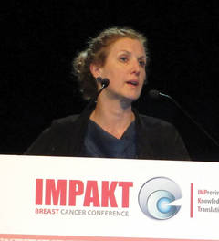
Dr. Lisa A. Carey, an oncologist who collaborates with Dr. Johnson, said she believes that reprogramming may explain her recent, frustrating results treating metastatic breast cancer patients with an investigational inhibitor of the epidermal growth factor receptor (EGFR), a tyrosine kinase.
"We gave the inhibitor to patients with triple-negative breast cancer, where the EGFR is clearly upregulated, a big, juicy target, and yet only 25% of the patients responded. Most of the time, the cancers had alternative mechanisms to keep the EGFR pathway active," Dr. Carey hypothesized based on Dr. Johnson’s recent findings. "The good news was that 25% of the time the treatment worked," said Dr. Carey, professor of hematology oncology at the University of North Carolina and medical director of the university’s Breast Center.
The new finding on breast-cancer cell reprogramming "helps us understand why the EGFR inhibitor didn’t work in most patients, it helps us understand what cancer cells do, and it helps us design our next approach. Most cancer drug development right now targets kinases," she said in an interview.
"One way to approach this is to use multiple agents at once, but if you add drugs you also add toxicity and expense. And in some patients the cell doesn’t reprogram. We need to understand how reprogramming works," with the potential to develop agents that block reprogramming instead of trying to deal with the changes that reprogramming causes. "This is an explanation of why patients don’t respond to even targeted treatments, and it gives us a way forward to potentially prevent it. Reprogramming is reproducible and potentially targetable. I think we can get around it."
"Limiting emergence of heterogeneity is important because heterogeneity is what defeats us," Dr. Larry Norton, director of the Breast Center at Memorial Sloan-Kettering Cancer Center. New York, said at the conference.

"I need to understand reprogramming of the kinome, what a treatment does to the cancer, and how it influences the next treatment I should use," said Dr. David Cameron, professor of oncology at Edinburgh University. "Patients with breast cancer will be treated with a sequence of agents. We treat patients longer, more effectively, and with more drugs, so we need to worry about what the first drug does to the cancer before we come in with a second.
Giving a clinician’s perspective on the significance of reprogramming in a talk at the meeting, Dr. Cameron said, "we will have to optimize therapy by understanding what one treatment does so we can give the next treatment at the optimal time. If we unravel the way the biology changes in the presence of treatment perhaps we can hone our treatments more effectively."
Dr. Johnson said that he has started a company, KinoDyn, to apply kinase-assessment to treatment, although as of now the company has no funding or employees. Dr. Carey, Dr. Norton, and Dr. Cameron had no disclosures.
Twitter: @mitchelzoler
BRUSSELS – Metastatic breast cancer cells are even more adaptable and able to evade targeted treatment than researchers suspected as recently as a few months ago, which means they present a heightened treatment challenge that in many cases may be overcome only with multiple targeted therapies delivered at the same time.
The findings were discussed at IMPAKT 2013 Breast Cancer Conference, sponsored by the European Society for Medical Oncology.
The new wrinkle in drug resistance has been dubbed reprogramming, a way that metastatic breast cancer cells (but presumably a property shared by other advanced solid tumors as well) quickly respond to a drug that shuts down an essential cell protein, a kinase, by turning on other, alternative kinases within days of drug exposure. Many of the new, targeted therapies that have successfully treated advanced-stage breast cancer and other solid tumors are kinase inhibitors, such as trastuzumab (Herceptin) and lapatinib (Tykerb).
Using a newly-developed technique for assessing many different kinases within a cell at once, Gary Johnson, Ph.D., reported that treatment of isolated, advanced breast cancer cells in vitro with a kinase inhibitor drug produced within a week a dramatic shift in the cell’s overall kinase profile, something he calls the cell’s "kinome." Part of this reprogramming response probably occurs because of new genes that the cancer cell turns on or upregulates, and part is probably driven by epigenetic changes in the cell, said Dr. Johnson, professor and chairman of pharmacology at the University of North Carolina in Chapel Hill.
Clinicians familiar with this finding quickly recognized that the phenomenon is an important, new barrier to successful treatment in patients that will require creative solutions using rational, multidrug, or multi-sequence regimens.
Dr. Lisa A. Carey, an oncologist who collaborates with Dr. Johnson, said she believes that reprogramming may explain her recent, frustrating results treating metastatic breast cancer patients with an investigational inhibitor of the epidermal growth factor receptor (EGFR), a tyrosine kinase.
"We gave the inhibitor to patients with triple-negative breast cancer, where the EGFR is clearly upregulated, a big, juicy target, and yet only 25% of the patients responded. Most of the time, the cancers had alternative mechanisms to keep the EGFR pathway active," Dr. Carey hypothesized based on Dr. Johnson’s recent findings. "The good news was that 25% of the time the treatment worked," said Dr. Carey, professor of hematology oncology at the University of North Carolina and medical director of the university’s Breast Center.
The new finding on breast-cancer cell reprogramming "helps us understand why the EGFR inhibitor didn’t work in most patients, it helps us understand what cancer cells do, and it helps us design our next approach. Most cancer drug development right now targets kinases," she said in an interview.
"One way to approach this is to use multiple agents at once, but if you add drugs you also add toxicity and expense. And in some patients the cell doesn’t reprogram. We need to understand how reprogramming works," with the potential to develop agents that block reprogramming instead of trying to deal with the changes that reprogramming causes. "This is an explanation of why patients don’t respond to even targeted treatments, and it gives us a way forward to potentially prevent it. Reprogramming is reproducible and potentially targetable. I think we can get around it."
"Limiting emergence of heterogeneity is important because heterogeneity is what defeats us," Dr. Larry Norton, director of the Breast Center at Memorial Sloan-Kettering Cancer Center. New York, said at the conference.
"I need to understand reprogramming of the kinome, what a treatment does to the cancer, and how it influences the next treatment I should use," said Dr. David Cameron, professor of oncology at Edinburgh University. "Patients with breast cancer will be treated with a sequence of agents. We treat patients longer, more effectively, and with more drugs, so we need to worry about what the first drug does to the cancer before we come in with a second.
Giving a clinician’s perspective on the significance of reprogramming in a talk at the meeting, Dr. Cameron said, "we will have to optimize therapy by understanding what one treatment does so we can give the next treatment at the optimal time. If we unravel the way the biology changes in the presence of treatment perhaps we can hone our treatments more effectively."
Dr. Johnson said that he has started a company, KinoDyn, to apply kinase-assessment to treatment, although as of now the company has no funding or employees. Dr. Carey, Dr. Norton, and Dr. Cameron had no disclosures.
Twitter: @mitchelzoler
BRUSSELS – Metastatic breast cancer cells are even more adaptable and able to evade targeted treatment than researchers suspected as recently as a few months ago, which means they present a heightened treatment challenge that in many cases may be overcome only with multiple targeted therapies delivered at the same time.
The findings were discussed at IMPAKT 2013 Breast Cancer Conference, sponsored by the European Society for Medical Oncology.
The new wrinkle in drug resistance has been dubbed reprogramming, a way that metastatic breast cancer cells (but presumably a property shared by other advanced solid tumors as well) quickly respond to a drug that shuts down an essential cell protein, a kinase, by turning on other, alternative kinases within days of drug exposure. Many of the new, targeted therapies that have successfully treated advanced-stage breast cancer and other solid tumors are kinase inhibitors, such as trastuzumab (Herceptin) and lapatinib (Tykerb).
Using a newly-developed technique for assessing many different kinases within a cell at once, Gary Johnson, Ph.D., reported that treatment of isolated, advanced breast cancer cells in vitro with a kinase inhibitor drug produced within a week a dramatic shift in the cell’s overall kinase profile, something he calls the cell’s "kinome." Part of this reprogramming response probably occurs because of new genes that the cancer cell turns on or upregulates, and part is probably driven by epigenetic changes in the cell, said Dr. Johnson, professor and chairman of pharmacology at the University of North Carolina in Chapel Hill.
Clinicians familiar with this finding quickly recognized that the phenomenon is an important, new barrier to successful treatment in patients that will require creative solutions using rational, multidrug, or multi-sequence regimens.
Dr. Lisa A. Carey, an oncologist who collaborates with Dr. Johnson, said she believes that reprogramming may explain her recent, frustrating results treating metastatic breast cancer patients with an investigational inhibitor of the epidermal growth factor receptor (EGFR), a tyrosine kinase.
"We gave the inhibitor to patients with triple-negative breast cancer, where the EGFR is clearly upregulated, a big, juicy target, and yet only 25% of the patients responded. Most of the time, the cancers had alternative mechanisms to keep the EGFR pathway active," Dr. Carey hypothesized based on Dr. Johnson’s recent findings. "The good news was that 25% of the time the treatment worked," said Dr. Carey, professor of hematology oncology at the University of North Carolina and medical director of the university’s Breast Center.
The new finding on breast-cancer cell reprogramming "helps us understand why the EGFR inhibitor didn’t work in most patients, it helps us understand what cancer cells do, and it helps us design our next approach. Most cancer drug development right now targets kinases," she said in an interview.
"One way to approach this is to use multiple agents at once, but if you add drugs you also add toxicity and expense. And in some patients the cell doesn’t reprogram. We need to understand how reprogramming works," with the potential to develop agents that block reprogramming instead of trying to deal with the changes that reprogramming causes. "This is an explanation of why patients don’t respond to even targeted treatments, and it gives us a way forward to potentially prevent it. Reprogramming is reproducible and potentially targetable. I think we can get around it."
"Limiting emergence of heterogeneity is important because heterogeneity is what defeats us," Dr. Larry Norton, director of the Breast Center at Memorial Sloan-Kettering Cancer Center. New York, said at the conference.
"I need to understand reprogramming of the kinome, what a treatment does to the cancer, and how it influences the next treatment I should use," said Dr. David Cameron, professor of oncology at Edinburgh University. "Patients with breast cancer will be treated with a sequence of agents. We treat patients longer, more effectively, and with more drugs, so we need to worry about what the first drug does to the cancer before we come in with a second.
Giving a clinician’s perspective on the significance of reprogramming in a talk at the meeting, Dr. Cameron said, "we will have to optimize therapy by understanding what one treatment does so we can give the next treatment at the optimal time. If we unravel the way the biology changes in the presence of treatment perhaps we can hone our treatments more effectively."
Dr. Johnson said that he has started a company, KinoDyn, to apply kinase-assessment to treatment, although as of now the company has no funding or employees. Dr. Carey, Dr. Norton, and Dr. Cameron had no disclosures.
Twitter: @mitchelzoler
EXPERT ANALYSIS FROM IMPAKT 2013 BREAST CANCER CONFERENCE
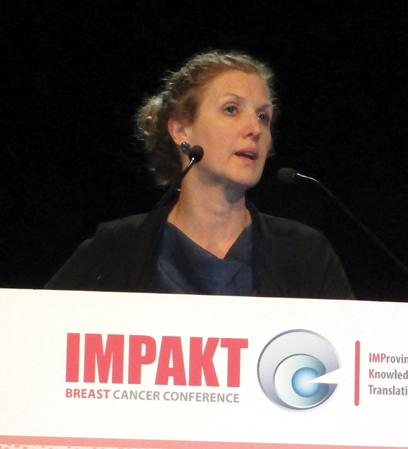
Genetic tests gauge breast-cancer recurrence risk
BRUSSELS – A pair of genetic tests each showed the ability to aid long-term prognosis estimates in patients with estrogen receptor–positive breast cancer, based on results from two retrospective analyses reported at IMPAKT 2013 Breast Cancer Conference.
The new results mean that a total of three different genetic tests have now shown added prognostic utility in retrospective analyses, bringing the field closer to actually using these tests in routine practice. The tests would prove particularly useful for patients with estrogen receptor–positive, node-negative breast cancer by helping to find those who are at low risk for long-term metastatic disease and who could safely stop treatment after 5 years of hormonal therapy, said Dr. Michael Gnant, professor of surgery at the University of Vienna.

But other experts stressed that it’s premature to start routinely using these tests.
"We’re getting to a point where we can offer to our patients genomic assays that are clinically validated and with demonstrated utility," Dr. W. Fraser Symmans said during the meeting. But, he added, while oncologists are "on the brink" of using the tests routinely, "we need to confirm reproducibility among labs, and that the test works in that setting," said Dr. Symmans, professor of pathology at the University of Texas M.D. Anderson Cancer Center in Houston.
None of the three genetic test panels were designed to predict late recurrences, noted Dr. Peter C. Dubsky, a surgeon at the University of Vienna and a collaborator with Dr. Gnant. "These data will need further validation before actually being incorporated into clinical decision-making concerning adjuvant endocrine therapy beyond 5 years," Dr. Dubsky said in a written statement.
The analysis reported by Dr. Gnant and Dr. Dubsky included formalin-fixed and paraffin-embedded tumor specimens from 1,478 patients who had been enrolled in the Austrian Breast and Colorectal Cancer Study Group (ABCSG) 8 trial, which had the primary goal of assessing two different 5-year treatment strategies in more than 3,700 postmenopausal women with estrogen receptor–positive breast cancer (J. Clin. Oncol. 2012;30:722-8).
Dr. Gnant, Dr. Dubsky, and their associates assessed the 1,478 specimens with the Prediction Analysis of Microarray test using a 58-gene classifier panel (PAM50), a test first reported in 2009 (J. Clin. Oncol. 2009;27:1160-7) that has since been commercialized as the Prosigna assay, available in Europe but not in the United States.
Using prespecified cutoffs on the PAM50 results for classifying patients as being at low, intermediate, and high risk for long term distant recurrences, the researchers tallied the rate of actual recurrences during a median follow-up of 11 years. The 15-year disease-free survival rate was 97.6% among low-risk patients, 90.9% in intermediate-risk patients, and 82.5% in high-risk patients, Dr. Gnant reported. The between-group differences were statistically significant.
The findings suggest that high-risk patients might be good candidates for extended adjuvant therapy, while low-risk patients, with a recurrence risk of less than 2% at 5-10 years and at 10-15 years following primary therapy "can be spared the side effects of extended adjuvant therapy," he said. The PAM50 risk of recurrence score "adds prognostic information beyond established clinicopathological factors."
The second analysis reported at the meeting retrospectively used medical records and specimens collected in the Arimidex, Tamoxifen Alone or in Combination (ATAC) study, which enrolled more than 6,000 postmenopausal women with localized invasive breast cancer (Lancet Oncology 2008;9:45-53). The researchers evaluated the prognostic efficacy of five different clinical, genetic, or histochemical test panels for predicting risk of distant recurrences during a median 10 years follow-up in 891 of the enrolled patients.

All five assessment panels yielded similar prognostic information during the first 5 years of follow-up. During the next 5 years, the strongest prognostic information came via the Clinical Treatment Score, which takes into account a patient’s nodal status, tumor grade and size, patient’s age, and treatment received, said Ivana Sestak, Ph.D., a statistician and epidemiologist at the University of London. The two most important prognostic features in this panel are tumor size and nodal status, she noted. Two other assessment panels – the PAM50 genetic panel and the Breast Cancer Index, a genetic test first described in 2011 – also provided statistically significant and clinically meaningful additional prediction of distant recurrences during 5-10 years of follow-up (Br. J. Cancer 2011;104:1762-9).
The other two tests Dr. Sestak and her associates evaluated did not add further prognostic information beyond what the Clinical Treatment Score provided. The uninformative tests were a set of four immunohistochemical markers (to estrogen, progesterone, HER2, and ki67), and the Oncotype Dx recurrence score – a 21-gene panel test marketed by Genomic Health.
"The PAM50 risk of recurrence score and the Breast Cancer Index may be used to identify estrogen receptor–positive breast cancer patients who are at increased risk of late recurrence and who may benefit from extended adjuvant hormonal therapy beyond 5 years," she concluded. But "none of these scores were developed to dictate treatment," she cautioned.
These findings, together with the PAM50 analysis from the ABCSG 8 study, solidify PAM50’s position as a validated prognostic tool, Dr. Gnant said.
In addition to PAM50 and the Breast Cancer Index, a third genetic test recently shown effective for predicting long-term recurrence risk in similar patients is the EndoPredict (Ann. Oncol. 2013;24:640-7).
All three assays can now be considered validated for longer-term prognosis in patients with endocrine-responsive, lymph node–negative breast cancer, Dr. Symmans said.
The conference was sponsored by the European Society for Medical Oncology.
Dr. Gnant disclosed ties to Amgen, Pfizer, Novartis, GlaxoSmithKline, Bayer, Sandoz, AstraZeneca, Genomic Health, NanoString Technologies, Sanofi-Aventis, and Roche. Dr. Sestak had no disclosures. Dr. Symmans is cofounder of, and has equity in, Nuvera Biosciences. Dr. Dubsky disclosed ties to AstraZeneca, Novartis, Sividon Diagnostica, and Pfizer.
Twitter: @mitchelzoler
The Breast Cancer Index and the PAM50 and EndoPredict tests were all developed as prognostic assays. They were not designed to, nor have they been demonstrated to, predict patient sensitivity to an endocrine treatment or to help select a class of endocrine agent to use in therapy. Despite these limitations, the data presented on all three tests suggest that they perform well for discriminating recurrence risk among patients with hormone-receptor positive, lymph-node negative breast cancer. So far, there is not good evidence that they can predict outcomes in lymph-node positive patients.

|
Mitchel L. Zoler/IMNG Medical Media
|
The patients with the greatest potential for stopping treatment after 5 years are those who also have HER2-negative disease and grade 1 or 2 disease, similar to the women enrolled in the ABCSG 8 trial.
Another attractive feature of these tests is that they can be performed by local pathology laboratories, precluding the need to send specimens to distant testing sites. The pathology community has been waiting a long time to more fully participate in the genomic diagnostic/personalized medicine arena.
The next step is to confirm the reproducibility of these tests when used by a variety of labs, essentially guaranteeing quality control. You need a critical mass of labs working together and using these tests to be sure that everyone gets clinically valid and reproducible results. I expect this to happen during the next year.
Dr. W. Fraser Symmans is professor of pathology at the University of Texas M.D. Anderson Cancer Center in Houston. He said that he is a c-founder of, and has equity in, Nuvera Biosciences. He made these comments as an invited discussant for the reports at the meeting and in an interview.
The Breast Cancer Index and the PAM50 and EndoPredict tests were all developed as prognostic assays. They were not designed to, nor have they been demonstrated to, predict patient sensitivity to an endocrine treatment or to help select a class of endocrine agent to use in therapy. Despite these limitations, the data presented on all three tests suggest that they perform well for discriminating recurrence risk among patients with hormone-receptor positive, lymph-node negative breast cancer. So far, there is not good evidence that they can predict outcomes in lymph-node positive patients.
|
|
Mitchel L. Zoler/IMNG Medical Media
|
The patients with the greatest potential for stopping treatment after 5 years are those who also have HER2-negative disease and grade 1 or 2 disease, similar to the women enrolled in the ABCSG 8 trial.
Another attractive feature of these tests is that they can be performed by local pathology laboratories, precluding the need to send specimens to distant testing sites. The pathology community has been waiting a long time to more fully participate in the genomic diagnostic/personalized medicine arena.
The next step is to confirm the reproducibility of these tests when used by a variety of labs, essentially guaranteeing quality control. You need a critical mass of labs working together and using these tests to be sure that everyone gets clinically valid and reproducible results. I expect this to happen during the next year.
Dr. W. Fraser Symmans is professor of pathology at the University of Texas M.D. Anderson Cancer Center in Houston. He said that he is a c-founder of, and has equity in, Nuvera Biosciences. He made these comments as an invited discussant for the reports at the meeting and in an interview.
The Breast Cancer Index and the PAM50 and EndoPredict tests were all developed as prognostic assays. They were not designed to, nor have they been demonstrated to, predict patient sensitivity to an endocrine treatment or to help select a class of endocrine agent to use in therapy. Despite these limitations, the data presented on all three tests suggest that they perform well for discriminating recurrence risk among patients with hormone-receptor positive, lymph-node negative breast cancer. So far, there is not good evidence that they can predict outcomes in lymph-node positive patients.
|
|
Mitchel L. Zoler/IMNG Medical Media
|
The patients with the greatest potential for stopping treatment after 5 years are those who also have HER2-negative disease and grade 1 or 2 disease, similar to the women enrolled in the ABCSG 8 trial.
Another attractive feature of these tests is that they can be performed by local pathology laboratories, precluding the need to send specimens to distant testing sites. The pathology community has been waiting a long time to more fully participate in the genomic diagnostic/personalized medicine arena.
The next step is to confirm the reproducibility of these tests when used by a variety of labs, essentially guaranteeing quality control. You need a critical mass of labs working together and using these tests to be sure that everyone gets clinically valid and reproducible results. I expect this to happen during the next year.
Dr. W. Fraser Symmans is professor of pathology at the University of Texas M.D. Anderson Cancer Center in Houston. He said that he is a c-founder of, and has equity in, Nuvera Biosciences. He made these comments as an invited discussant for the reports at the meeting and in an interview.
BRUSSELS – A pair of genetic tests each showed the ability to aid long-term prognosis estimates in patients with estrogen receptor–positive breast cancer, based on results from two retrospective analyses reported at IMPAKT 2013 Breast Cancer Conference.
The new results mean that a total of three different genetic tests have now shown added prognostic utility in retrospective analyses, bringing the field closer to actually using these tests in routine practice. The tests would prove particularly useful for patients with estrogen receptor–positive, node-negative breast cancer by helping to find those who are at low risk for long-term metastatic disease and who could safely stop treatment after 5 years of hormonal therapy, said Dr. Michael Gnant, professor of surgery at the University of Vienna.
But other experts stressed that it’s premature to start routinely using these tests.
"We’re getting to a point where we can offer to our patients genomic assays that are clinically validated and with demonstrated utility," Dr. W. Fraser Symmans said during the meeting. But, he added, while oncologists are "on the brink" of using the tests routinely, "we need to confirm reproducibility among labs, and that the test works in that setting," said Dr. Symmans, professor of pathology at the University of Texas M.D. Anderson Cancer Center in Houston.
None of the three genetic test panels were designed to predict late recurrences, noted Dr. Peter C. Dubsky, a surgeon at the University of Vienna and a collaborator with Dr. Gnant. "These data will need further validation before actually being incorporated into clinical decision-making concerning adjuvant endocrine therapy beyond 5 years," Dr. Dubsky said in a written statement.
The analysis reported by Dr. Gnant and Dr. Dubsky included formalin-fixed and paraffin-embedded tumor specimens from 1,478 patients who had been enrolled in the Austrian Breast and Colorectal Cancer Study Group (ABCSG) 8 trial, which had the primary goal of assessing two different 5-year treatment strategies in more than 3,700 postmenopausal women with estrogen receptor–positive breast cancer (J. Clin. Oncol. 2012;30:722-8).
Dr. Gnant, Dr. Dubsky, and their associates assessed the 1,478 specimens with the Prediction Analysis of Microarray test using a 58-gene classifier panel (PAM50), a test first reported in 2009 (J. Clin. Oncol. 2009;27:1160-7) that has since been commercialized as the Prosigna assay, available in Europe but not in the United States.
Using prespecified cutoffs on the PAM50 results for classifying patients as being at low, intermediate, and high risk for long term distant recurrences, the researchers tallied the rate of actual recurrences during a median follow-up of 11 years. The 15-year disease-free survival rate was 97.6% among low-risk patients, 90.9% in intermediate-risk patients, and 82.5% in high-risk patients, Dr. Gnant reported. The between-group differences were statistically significant.
The findings suggest that high-risk patients might be good candidates for extended adjuvant therapy, while low-risk patients, with a recurrence risk of less than 2% at 5-10 years and at 10-15 years following primary therapy "can be spared the side effects of extended adjuvant therapy," he said. The PAM50 risk of recurrence score "adds prognostic information beyond established clinicopathological factors."
The second analysis reported at the meeting retrospectively used medical records and specimens collected in the Arimidex, Tamoxifen Alone or in Combination (ATAC) study, which enrolled more than 6,000 postmenopausal women with localized invasive breast cancer (Lancet Oncology 2008;9:45-53). The researchers evaluated the prognostic efficacy of five different clinical, genetic, or histochemical test panels for predicting risk of distant recurrences during a median 10 years follow-up in 891 of the enrolled patients.
All five assessment panels yielded similar prognostic information during the first 5 years of follow-up. During the next 5 years, the strongest prognostic information came via the Clinical Treatment Score, which takes into account a patient’s nodal status, tumor grade and size, patient’s age, and treatment received, said Ivana Sestak, Ph.D., a statistician and epidemiologist at the University of London. The two most important prognostic features in this panel are tumor size and nodal status, she noted. Two other assessment panels – the PAM50 genetic panel and the Breast Cancer Index, a genetic test first described in 2011 – also provided statistically significant and clinically meaningful additional prediction of distant recurrences during 5-10 years of follow-up (Br. J. Cancer 2011;104:1762-9).
The other two tests Dr. Sestak and her associates evaluated did not add further prognostic information beyond what the Clinical Treatment Score provided. The uninformative tests were a set of four immunohistochemical markers (to estrogen, progesterone, HER2, and ki67), and the Oncotype Dx recurrence score – a 21-gene panel test marketed by Genomic Health.
"The PAM50 risk of recurrence score and the Breast Cancer Index may be used to identify estrogen receptor–positive breast cancer patients who are at increased risk of late recurrence and who may benefit from extended adjuvant hormonal therapy beyond 5 years," she concluded. But "none of these scores were developed to dictate treatment," she cautioned.
These findings, together with the PAM50 analysis from the ABCSG 8 study, solidify PAM50’s position as a validated prognostic tool, Dr. Gnant said.
In addition to PAM50 and the Breast Cancer Index, a third genetic test recently shown effective for predicting long-term recurrence risk in similar patients is the EndoPredict (Ann. Oncol. 2013;24:640-7).
All three assays can now be considered validated for longer-term prognosis in patients with endocrine-responsive, lymph node–negative breast cancer, Dr. Symmans said.
The conference was sponsored by the European Society for Medical Oncology.
Dr. Gnant disclosed ties to Amgen, Pfizer, Novartis, GlaxoSmithKline, Bayer, Sandoz, AstraZeneca, Genomic Health, NanoString Technologies, Sanofi-Aventis, and Roche. Dr. Sestak had no disclosures. Dr. Symmans is cofounder of, and has equity in, Nuvera Biosciences. Dr. Dubsky disclosed ties to AstraZeneca, Novartis, Sividon Diagnostica, and Pfizer.
Twitter: @mitchelzoler
BRUSSELS – A pair of genetic tests each showed the ability to aid long-term prognosis estimates in patients with estrogen receptor–positive breast cancer, based on results from two retrospective analyses reported at IMPAKT 2013 Breast Cancer Conference.
The new results mean that a total of three different genetic tests have now shown added prognostic utility in retrospective analyses, bringing the field closer to actually using these tests in routine practice. The tests would prove particularly useful for patients with estrogen receptor–positive, node-negative breast cancer by helping to find those who are at low risk for long-term metastatic disease and who could safely stop treatment after 5 years of hormonal therapy, said Dr. Michael Gnant, professor of surgery at the University of Vienna.
But other experts stressed that it’s premature to start routinely using these tests.
"We’re getting to a point where we can offer to our patients genomic assays that are clinically validated and with demonstrated utility," Dr. W. Fraser Symmans said during the meeting. But, he added, while oncologists are "on the brink" of using the tests routinely, "we need to confirm reproducibility among labs, and that the test works in that setting," said Dr. Symmans, professor of pathology at the University of Texas M.D. Anderson Cancer Center in Houston.
None of the three genetic test panels were designed to predict late recurrences, noted Dr. Peter C. Dubsky, a surgeon at the University of Vienna and a collaborator with Dr. Gnant. "These data will need further validation before actually being incorporated into clinical decision-making concerning adjuvant endocrine therapy beyond 5 years," Dr. Dubsky said in a written statement.
The analysis reported by Dr. Gnant and Dr. Dubsky included formalin-fixed and paraffin-embedded tumor specimens from 1,478 patients who had been enrolled in the Austrian Breast and Colorectal Cancer Study Group (ABCSG) 8 trial, which had the primary goal of assessing two different 5-year treatment strategies in more than 3,700 postmenopausal women with estrogen receptor–positive breast cancer (J. Clin. Oncol. 2012;30:722-8).
Dr. Gnant, Dr. Dubsky, and their associates assessed the 1,478 specimens with the Prediction Analysis of Microarray test using a 58-gene classifier panel (PAM50), a test first reported in 2009 (J. Clin. Oncol. 2009;27:1160-7) that has since been commercialized as the Prosigna assay, available in Europe but not in the United States.
Using prespecified cutoffs on the PAM50 results for classifying patients as being at low, intermediate, and high risk for long term distant recurrences, the researchers tallied the rate of actual recurrences during a median follow-up of 11 years. The 15-year disease-free survival rate was 97.6% among low-risk patients, 90.9% in intermediate-risk patients, and 82.5% in high-risk patients, Dr. Gnant reported. The between-group differences were statistically significant.
The findings suggest that high-risk patients might be good candidates for extended adjuvant therapy, while low-risk patients, with a recurrence risk of less than 2% at 5-10 years and at 10-15 years following primary therapy "can be spared the side effects of extended adjuvant therapy," he said. The PAM50 risk of recurrence score "adds prognostic information beyond established clinicopathological factors."
The second analysis reported at the meeting retrospectively used medical records and specimens collected in the Arimidex, Tamoxifen Alone or in Combination (ATAC) study, which enrolled more than 6,000 postmenopausal women with localized invasive breast cancer (Lancet Oncology 2008;9:45-53). The researchers evaluated the prognostic efficacy of five different clinical, genetic, or histochemical test panels for predicting risk of distant recurrences during a median 10 years follow-up in 891 of the enrolled patients.
All five assessment panels yielded similar prognostic information during the first 5 years of follow-up. During the next 5 years, the strongest prognostic information came via the Clinical Treatment Score, which takes into account a patient’s nodal status, tumor grade and size, patient’s age, and treatment received, said Ivana Sestak, Ph.D., a statistician and epidemiologist at the University of London. The two most important prognostic features in this panel are tumor size and nodal status, she noted. Two other assessment panels – the PAM50 genetic panel and the Breast Cancer Index, a genetic test first described in 2011 – also provided statistically significant and clinically meaningful additional prediction of distant recurrences during 5-10 years of follow-up (Br. J. Cancer 2011;104:1762-9).
The other two tests Dr. Sestak and her associates evaluated did not add further prognostic information beyond what the Clinical Treatment Score provided. The uninformative tests were a set of four immunohistochemical markers (to estrogen, progesterone, HER2, and ki67), and the Oncotype Dx recurrence score – a 21-gene panel test marketed by Genomic Health.
"The PAM50 risk of recurrence score and the Breast Cancer Index may be used to identify estrogen receptor–positive breast cancer patients who are at increased risk of late recurrence and who may benefit from extended adjuvant hormonal therapy beyond 5 years," she concluded. But "none of these scores were developed to dictate treatment," she cautioned.
These findings, together with the PAM50 analysis from the ABCSG 8 study, solidify PAM50’s position as a validated prognostic tool, Dr. Gnant said.
In addition to PAM50 and the Breast Cancer Index, a third genetic test recently shown effective for predicting long-term recurrence risk in similar patients is the EndoPredict (Ann. Oncol. 2013;24:640-7).
All three assays can now be considered validated for longer-term prognosis in patients with endocrine-responsive, lymph node–negative breast cancer, Dr. Symmans said.
The conference was sponsored by the European Society for Medical Oncology.
Dr. Gnant disclosed ties to Amgen, Pfizer, Novartis, GlaxoSmithKline, Bayer, Sandoz, AstraZeneca, Genomic Health, NanoString Technologies, Sanofi-Aventis, and Roche. Dr. Sestak had no disclosures. Dr. Symmans is cofounder of, and has equity in, Nuvera Biosciences. Dr. Dubsky disclosed ties to AstraZeneca, Novartis, Sividon Diagnostica, and Pfizer.
Twitter: @mitchelzoler
AT IMPAKT 2013 BREAST CANCER CONFERENCE
Major finding: Low-risk patients by the PAM50 test had 97.6% 15-year disease-free survival compared with 82.5% in high-risk patients.
Data source: A retrospective analysis of 1,478 women enrolled in the Austrian Breast and Colorectal Cancer Study Group (ABCSG) 8 trial.
Disclosures: Dr. Gnant disclosed ties to Amgen, Pfizer, Novartis, GlaxoSmithKline, Bayer, Sandoz, AstraZeneca, Genomic Health, NanoString Technologies, Sanofi-Aventis, and Roche. Dr. Sestak had no disclosures. Dr. Symmans is cofounder of, and has equity in, Nuvera Biosciences. Dr. Dubsky disclosed ties to AstraZeneca, Novartis, Sividon Diagnostica, and Pfizer.

Online cancer community colaunched by ex-Google executive
Two heavyweight leaders in the blend of online and medical worlds have colaunched a new online community for cancer patients and caregivers called Smart Patients that takes the goal of patient-centered services in several technological directions.
Dr. Roni Zeiger, former chief health strategist for Google, and Gilles Frydman, founder of the Association of Cancer Online Resources, introduced Smart Patients at the recent TEDMED conference in Washington. TEDMED is the medical version of the popular TED (Technology, Entertainment, and Design) conferences, where innovators and influential leaders give short talks on their activities and thoughts on the future.
Smart Patients is one of a number of online communities that connect cancer patients, helpers, and cancer survivors to share knowledge and support. What makes Smart Patients different is some of its additional features, such as a user-friendly way to search for clinical trials and the possibility of providing patient feedback on the design of future clinical trials.
The technological pedigrees of the cofounders already have brought considerable attention to Smart Patients in what might be considered a crowded field. A Google search for online cancer communities turned up the following on the first page of results alone. Frydman’s Association of Cancer Online Resources (ACOR), the granddaddy of the online sharing community when it comes to cancer, provides links to 142 online cancer communities. The American Cancer Society provides multiple online tools including a Cancer Survivors Network with chat rooms, discussion boards, private e-mail, and more. The Cancer Treatment Centers of America offers CancerCompass, where participants can share status updates, follow friends, and comment on their activities among other features. Another start-up, I Had Cancer, says it is beta testing a website that connects people battling cancer with cancer survivors.
Like some of these sites, Smart Patients lets users start and follow conversations and search for keywords to find the topics that interest them. Its clinical trials search function is based on the clinicaltrials.gov database. Smart Patients is intended to complement physician care, not replace it, according to its FAQ section, which also states that the site will include no advertising. Smart Patients plans to make money by selling anonymous data harvested from voluntary survey comments posted on the website. Potential customers include companies, researchers, and educators who want to better understand the experiences, insights, and needs of cancer patients and their caregivers. The site will follow guidelines from the Health Insurance Portability and Accountability Act (HIPAA) to ensure that individuals are not identified in these commercial transactions.
Smart Patients announced at TEDMED that it has inked partnerships with WorldOne Interactive (owner of the online physician networking site Sermo), The Bonnie J. Addario Lung Cancer Foundation, and Cancer Commons, according to news reports. Another partner is the biotechnology company Oncosec Medical, which aims to gather patient feedback before designing clinical trials of its skin cancer therapies.
Cancer was one of the first medical topics to gain traction in online sharing. With this latest entry from two power players in the field, online cancer communities may be moving to the next level.
On Twitter @sherryboschert
Smart Patients is one of a number of online communities that connect cancer patients, helpers, and cancer survivors to share knowledge and support. What makes Smart Patients different is some of its additional features, such as a user-friendly way to search for clinical trials and the possibility of providing patient feedback on the design of future clinical trials.
The technological pedigrees of the cofounders already have brought considerable attention to Smart Patients in what might be considered a crowded field. A Google search for online cancer communities turned up the following on the first page of results alone. Frydman’s Association of Cancer Online Resources (ACOR), the granddaddy of the online sharing community when it comes to cancer, provides links to 142 online cancer communities. The American Cancer Society provides multiple online tools including a Cancer Survivors Network with chat rooms, discussion boards, private e-mail, and more. The Cancer Treatment Centers of America offers CancerCompass, where participants can share status updates, follow friends, and comment on their activities among other features. Another start-up, I Had Cancer, says it is beta testing a website that connects people battling cancer with cancer survivors.
Like some of these sites, Smart Patients lets users start and follow conversations and search for keywords to find the topics that interest them. Its clinical trials search function is based on the clinicaltrials.gov database. Smart Patients is intended to complement physician care, not replace it, according to its FAQ section, which also states that the site will include no advertising. Smart Patients plans to make money by selling anonymous data harvested from voluntary survey comments posted on the website. Potential customers include companies, researchers, and educators who want to better understand the experiences, insights, and needs of cancer patients and their caregivers. The site will follow guidelines from the Health Insurance Portability and Accountability Act (HIPAA) to ensure that individuals are not identified in these commercial transactions.
Smart Patients announced at TEDMED that it has inked partnerships with WorldOne Interactive (owner of the online physician networking site Sermo), The Bonnie J. Addario Lung Cancer Foundation, and Cancer Commons, according to news reports. Another partner is the biotechnology company Oncosec Medical, which aims to gather patient feedback before designing clinical trials of its skin cancer therapies.
Cancer was one of the first medical topics to gain traction in online sharing. With this latest entry from two power players in the field, o
Two heavyweight leaders in the blend of online and medical worlds have colaunched a new online community for cancer patients and caregivers called Smart Patients that takes the goal of patient-centered services in several technological directions.
Dr. Roni Zeiger, former chief health strategist for Google, and Gilles Frydman, founder of the Association of Cancer Online Resources, introduced Smart Patients at the recent TEDMED conference in Washington. TEDMED is the medical version of the popular TED (Technology, Entertainment, and Design) conferences, where innovators and influential leaders give short talks on their activities and thoughts on the future.
Smart Patients is one of a number of online communities that connect cancer patients, helpers, and cancer survivors to share knowledge and support. What makes Smart Patients different is some of its additional features, such as a user-friendly way to search for clinical trials and the possibility of providing patient feedback on the design of future clinical trials.
The technological pedigrees of the cofounders already have brought considerable attention to Smart Patients in what might be considered a crowded field. A Google search for online cancer communities turned up the following on the first page of results alone. Frydman’s Association of Cancer Online Resources (ACOR), the granddaddy of the online sharing community when it comes to cancer, provides links to 142 online cancer communities. The American Cancer Society provides multiple online tools including a Cancer Survivors Network with chat rooms, discussion boards, private e-mail, and more. The Cancer Treatment Centers of America offers CancerCompass, where participants can share status updates, follow friends, and comment on their activities among other features. Another start-up, I Had Cancer, says it is beta testing a website that connects people battling cancer with cancer survivors.
Like some of these sites, Smart Patients lets users start and follow conversations and search for keywords to find the topics that interest them. Its clinical trials search function is based on the clinicaltrials.gov database. Smart Patients is intended to complement physician care, not replace it, according to its FAQ section, which also states that the site will include no advertising. Smart Patients plans to make money by selling anonymous data harvested from voluntary survey comments posted on the website. Potential customers include companies, researchers, and educators who want to better understand the experiences, insights, and needs of cancer patients and their caregivers. The site will follow guidelines from the Health Insurance Portability and Accountability Act (HIPAA) to ensure that individuals are not identified in these commercial transactions.
Smart Patients announced at TEDMED that it has inked partnerships with WorldOne Interactive (owner of the online physician networking site Sermo), The Bonnie J. Addario Lung Cancer Foundation, and Cancer Commons, according to news reports. Another partner is the biotechnology company Oncosec Medical, which aims to gather patient feedback before designing clinical trials of its skin cancer therapies.
Cancer was one of the first medical topics to gain traction in online sharing. With this latest entry from two power players in the field, online cancer communities may be moving to the next level.
On Twitter @sherryboschert
Two heavyweight leaders in the blend of online and medical worlds have colaunched a new online community for cancer patients and caregivers called Smart Patients that takes the goal of patient-centered services in several technological directions.
Dr. Roni Zeiger, former chief health strategist for Google, and Gilles Frydman, founder of the Association of Cancer Online Resources, introduced Smart Patients at the recent TEDMED conference in Washington. TEDMED is the medical version of the popular TED (Technology, Entertainment, and Design) conferences, where innovators and influential leaders give short talks on their activities and thoughts on the future.
Smart Patients is one of a number of online communities that connect cancer patients, helpers, and cancer survivors to share knowledge and support. What makes Smart Patients different is some of its additional features, such as a user-friendly way to search for clinical trials and the possibility of providing patient feedback on the design of future clinical trials.
The technological pedigrees of the cofounders already have brought considerable attention to Smart Patients in what might be considered a crowded field. A Google search for online cancer communities turned up the following on the first page of results alone. Frydman’s Association of Cancer Online Resources (ACOR), the granddaddy of the online sharing community when it comes to cancer, provides links to 142 online cancer communities. The American Cancer Society provides multiple online tools including a Cancer Survivors Network with chat rooms, discussion boards, private e-mail, and more. The Cancer Treatment Centers of America offers CancerCompass, where participants can share status updates, follow friends, and comment on their activities among other features. Another start-up, I Had Cancer, says it is beta testing a website that connects people battling cancer with cancer survivors.
Like some of these sites, Smart Patients lets users start and follow conversations and search for keywords to find the topics that interest them. Its clinical trials search function is based on the clinicaltrials.gov database. Smart Patients is intended to complement physician care, not replace it, according to its FAQ section, which also states that the site will include no advertising. Smart Patients plans to make money by selling anonymous data harvested from voluntary survey comments posted on the website. Potential customers include companies, researchers, and educators who want to better understand the experiences, insights, and needs of cancer patients and their caregivers. The site will follow guidelines from the Health Insurance Portability and Accountability Act (HIPAA) to ensure that individuals are not identified in these commercial transactions.
Smart Patients announced at TEDMED that it has inked partnerships with WorldOne Interactive (owner of the online physician networking site Sermo), The Bonnie J. Addario Lung Cancer Foundation, and Cancer Commons, according to news reports. Another partner is the biotechnology company Oncosec Medical, which aims to gather patient feedback before designing clinical trials of its skin cancer therapies.
Cancer was one of the first medical topics to gain traction in online sharing. With this latest entry from two power players in the field, online cancer communities may be moving to the next level.
On Twitter @sherryboschert
Smart Patients is one of a number of online communities that connect cancer patients, helpers, and cancer survivors to share knowledge and support. What makes Smart Patients different is some of its additional features, such as a user-friendly way to search for clinical trials and the possibility of providing patient feedback on the design of future clinical trials.
The technological pedigrees of the cofounders already have brought considerable attention to Smart Patients in what might be considered a crowded field. A Google search for online cancer communities turned up the following on the first page of results alone. Frydman’s Association of Cancer Online Resources (ACOR), the granddaddy of the online sharing community when it comes to cancer, provides links to 142 online cancer communities. The American Cancer Society provides multiple online tools including a Cancer Survivors Network with chat rooms, discussion boards, private e-mail, and more. The Cancer Treatment Centers of America offers CancerCompass, where participants can share status updates, follow friends, and comment on their activities among other features. Another start-up, I Had Cancer, says it is beta testing a website that connects people battling cancer with cancer survivors.
Like some of these sites, Smart Patients lets users start and follow conversations and search for keywords to find the topics that interest them. Its clinical trials search function is based on the clinicaltrials.gov database. Smart Patients is intended to complement physician care, not replace it, according to its FAQ section, which also states that the site will include no advertising. Smart Patients plans to make money by selling anonymous data harvested from voluntary survey comments posted on the website. Potential customers include companies, researchers, and educators who want to better understand the experiences, insights, and needs of cancer patients and their caregivers. The site will follow guidelines from the Health Insurance Portability and Accountability Act (HIPAA) to ensure that individuals are not identified in these commercial transactions.
Smart Patients announced at TEDMED that it has inked partnerships with WorldOne Interactive (owner of the online physician networking site Sermo), The Bonnie J. Addario Lung Cancer Foundation, and Cancer Commons, according to news reports. Another partner is the biotechnology company Oncosec Medical, which aims to gather patient feedback before designing clinical trials of its skin cancer therapies.
Cancer was one of the first medical topics to gain traction in online sharing. With this latest entry from two power players in the field, o
Smart Patients is one of a number of online communities that connect cancer patients, helpers, and cancer survivors to share knowledge and support. What makes Smart Patients different is some of its additional features, such as a user-friendly way to search for clinical trials and the possibility of providing patient feedback on the design of future clinical trials.
The technological pedigrees of the cofounders already have brought considerable attention to Smart Patients in what might be considered a crowded field. A Google search for online cancer communities turned up the following on the first page of results alone. Frydman’s Association of Cancer Online Resources (ACOR), the granddaddy of the online sharing community when it comes to cancer, provides links to 142 online cancer communities. The American Cancer Society provides multiple online tools including a Cancer Survivors Network with chat rooms, discussion boards, private e-mail, and more. The Cancer Treatment Centers of America offers CancerCompass, where participants can share status updates, follow friends, and comment on their activities among other features. Another start-up, I Had Cancer, says it is beta testing a website that connects people battling cancer with cancer survivors.
Like some of these sites, Smart Patients lets users start and follow conversations and search for keywords to find the topics that interest them. Its clinical trials search function is based on the clinicaltrials.gov database. Smart Patients is intended to complement physician care, not replace it, according to its FAQ section, which also states that the site will include no advertising. Smart Patients plans to make money by selling anonymous data harvested from voluntary survey comments posted on the website. Potential customers include companies, researchers, and educators who want to better understand the experiences, insights, and needs of cancer patients and their caregivers. The site will follow guidelines from the Health Insurance Portability and Accountability Act (HIPAA) to ensure that individuals are not identified in these commercial transactions.
Smart Patients announced at TEDMED that it has inked partnerships with WorldOne Interactive (owner of the online physician networking site Sermo), The Bonnie J. Addario Lung Cancer Foundation, and Cancer Commons, according to news reports. Another partner is the biotechnology company Oncosec Medical, which aims to gather patient feedback before designing clinical trials of its skin cancer therapies.
Cancer was one of the first medical topics to gain traction in online sharing. With this latest entry from two power players in the field, o
How I screen patients at increased risk for breast cancer
Related article: Your menopausal patient’s breast biopsy reveals atypical hyperplasia JoAnn V. Pinkerton, MD; Andrew M. Kaunitz, MD, James A. Simon, MD (Cases in Menopause, May 2013)

JoAnn V. Pinkerton, MD
Professor, Department of Obstetrics and Gynecology, and Director, Division of Midlife Women’s Health, University of Virginia. Dr. Pinkerton is a North American Menopause Society (NAMS) past president and certified menopause practitioner. She also serves on the OBG Management Board of Editors.
JoAnn V. Pinkerton, MD
Professor, Department of Obstetrics and Gynecology, and Director, Division of Midlife Women’s Health, University of Virginia. Dr. Pinkerton is a North American Menopause Society (NAMS) past president and certified menopause practitioner. She also serves on the OBG Management Board of Editors.
JoAnn V. Pinkerton, MD
Professor, Department of Obstetrics and Gynecology, and Director, Division of Midlife Women’s Health, University of Virginia. Dr. Pinkerton is a North American Menopause Society (NAMS) past president and certified menopause practitioner. She also serves on the OBG Management Board of Editors.
Related article: Your menopausal patient’s breast biopsy reveals atypical hyperplasia JoAnn V. Pinkerton, MD; Andrew M. Kaunitz, MD, James A. Simon, MD (Cases in Menopause, May 2013)
Related article: Your menopausal patient’s breast biopsy reveals atypical hyperplasia JoAnn V. Pinkerton, MD; Andrew M. Kaunitz, MD, James A. Simon, MD (Cases in Menopause, May 2013)

Smaller margins too close for comfort in breast cancer
NATIONAL HARBOR, MD. – Small surgical margins can mean big trouble for patients with breast cancer, said investigators at the annual Society of Surgical Oncology Cancer Symposium.
A retrospective study of outcomes for 2,377 women who underwent either breast-conserving therapy or mastectomy revealed that margins less than 2 mm resulted in a substantial risk of residual disease for all patients, reported Dr. Erin Garvey, a general surgery resident at Mayo Clinic Arizona in Phoenix.

"A policy of re-excision for margins less than 2 mm, coupled with a standardized multidisciplinary approach to breast cancer surgery, results in excellent re-excision and 5-year local recurrence rates. The local recurrence rate is higher, however, for those patients who complete breast-conserving therapy, thus warranting appropriate patient counseling regarding re-excision options and long-term outcome expectations," she said.
In a separate study, investigators from the University of Texas M.D. Anderson Cancer Center, Houston, reported 10-year follow-up data for women who opted for mastectomy to treat ductal carcinoma in situ (DCIS). They found that the incidence of local-regional recurrence (LRR) increased as the surgical margins shrank, and that close margins were the only independent predictor of LRR, reported Dr. Elizabeth FitzSullivan, a surgery fellow at M.D. Anderson.
"However, the local-regional recurrence rate in these patients is so low that routine postmastectomy radiation therapy is not warranted," she said.
No accord on margins
Despite multiple studies and meta-analyses, there is no standard for acceptable margin width in breast cancer, and surveys of both surgeons and radiation oncologists have shown wide variations in preferred margin widths, Dr. Garvey said.
Her group hypothesized that patients with invasive ductal carcinoma without an extensive in situ component who had surgical margins of at least 1 mm would have no evidence of residual disease on re-excision.
To test the idea, they took a retrospective look at records from a prospective database on 2,377 patients who underwent a total of 2,520 procedures from January 2000 through May 2012.
Of this group, 1,498 (63%) underwent lumpectomy, and 180 (12%) required re-excision surgery: 10% who had breast-conserving surgery alone, and 2% whose surgeries were converted to mastectomies. Of the 158 patients who had completed breast-conserving therapy following re-excision, 50 (32%) had residual disease, as did 20 of the 27 patients whose procedures were converted to mastectomies.
Of the 37% (879) who had up-front mastectomies, 2% (19) had re-excision, and of this group, 5 patients had residual disease.
In all, 40% of patients with positive margins had residual disease, compared with 38% of those with margins from 0.1 to 0.9 mm, and 33% for those with margins from 1.0 to 1.9 mm.
In univariate analysis, the presence of residual disease on re-excision did not show any significant association with age, race, menopausal status, width of the closest final margin, hormone receptor status, tumor histology, triple-negative disease, or the presence of angiolymphatic invasion. There was a trend, albeit nonsignificant, toward an association between residual disease and more than one margin narrower than 2 mm, Dr. Garvey noted.
At a median follow-up of 43 months (range, 0-140 months), 5-year local recurrence rates were 1.9% for patients who had breast-conserving therapy, and 1.1% for those who had mastectomy.
Patients who underwent breast-conserving therapy without re-excision had a 5-year local recurrence rate of 1.8%, compared with 4.3% for those who required re-excision, and 0% for those whose procedures were converted to mastectomy.
There was a nonsignificant trend toward higher local recurrence rates for breast-conserving therapy in patients who had re-excisions, which became significant when those patients who had conversion to mastectomy were excluded, with a hazard ratio compared with no re-excision of 2.56 (P = .04).
Narrower margins, larger risk
Dr. FitzSullivan and her M.D. Anderson colleagues reviewed the records of 810 women treated with mastectomy for DCIS from 1996 to 2009. They looked at the final width of histologic margins, defining disease-free margins as those of 3 mm or greater.
In all, 4 patients had positive margins, 59 had margins of 1 mm or smaller, and 35 had margins from 1.1 to 2.9 mm.

In multivariate analysis, independent predictors of close or positive margins were pathologic tumor size of 1.5 cm or greater (odds ratio, 5.11; P = .001), multicentric disease (OR, 5.44; P = .026), and the presence of necrosis (OR, 2.5; P = .003). Neither age, postmenopausal status, skin-sparing mastectomy, nor immediate breast reconstruction were significantly associated with close or positive margins, however.
None of seven patients who underwent postmastectomy radiotherapy had local-regional recurrences. Of the 803 patients who did not receive postsurgery radiation, 10-year LRR rates were 1%, consisting of 7 cases of invasive disease and 1 of DICS. Five patients had surgical management, and the remaining 3 had no further treatment.
When the researchers stratified the local recurrence rates by margin status, they saw that 5% of patients with margins of 1 mm or smaller had LRRs within 10 years, as did 3.6% of those with margins from 1.1 to 2.9 mm, compared with just 0.07% of those with disease-free margins (P less than .001). There was no difference in LRR between the two narrow-margin groups.
Among 546 patients with an intact contralateral breast, the 10-year rate of contralateral breast disease was 6.4%.
On univariate analysis, significant predictors of LRR included margin status (P = .002), multicentric disease (P = .005), and necrosis (P = .005). On multivariate analysis, however, only margin status remained significant, with an HR of 8.0 (P = .006).
Dr. FitzSullivan said that the low rate of LRR of DCIS treated with mastectomy and close surgical margins, compared with the rate of contralateral breast cancer, suggests that routine postmastectomy radiation therapy is not warranted, and should be reserved only for those patients with close or positive surgical margins that cannot be surgically excised.
Each study was internally funded. Dr. Garvey and Dr. FitzSullivan reported having no financial disclosures.
NATIONAL HARBOR, MD. – Small surgical margins can mean big trouble for patients with breast cancer, said investigators at the annual Society of Surgical Oncology Cancer Symposium.
A retrospective study of outcomes for 2,377 women who underwent either breast-conserving therapy or mastectomy revealed that margins less than 2 mm resulted in a substantial risk of residual disease for all patients, reported Dr. Erin Garvey, a general surgery resident at Mayo Clinic Arizona in Phoenix.
"A policy of re-excision for margins less than 2 mm, coupled with a standardized multidisciplinary approach to breast cancer surgery, results in excellent re-excision and 5-year local recurrence rates. The local recurrence rate is higher, however, for those patients who complete breast-conserving therapy, thus warranting appropriate patient counseling regarding re-excision options and long-term outcome expectations," she said.
In a separate study, investigators from the University of Texas M.D. Anderson Cancer Center, Houston, reported 10-year follow-up data for women who opted for mastectomy to treat ductal carcinoma in situ (DCIS). They found that the incidence of local-regional recurrence (LRR) increased as the surgical margins shrank, and that close margins were the only independent predictor of LRR, reported Dr. Elizabeth FitzSullivan, a surgery fellow at M.D. Anderson.
"However, the local-regional recurrence rate in these patients is so low that routine postmastectomy radiation therapy is not warranted," she said.
No accord on margins
Despite multiple studies and meta-analyses, there is no standard for acceptable margin width in breast cancer, and surveys of both surgeons and radiation oncologists have shown wide variations in preferred margin widths, Dr. Garvey said.
Her group hypothesized that patients with invasive ductal carcinoma without an extensive in situ component who had surgical margins of at least 1 mm would have no evidence of residual disease on re-excision.
To test the idea, they took a retrospective look at records from a prospective database on 2,377 patients who underwent a total of 2,520 procedures from January 2000 through May 2012.
Of this group, 1,498 (63%) underwent lumpectomy, and 180 (12%) required re-excision surgery: 10% who had breast-conserving surgery alone, and 2% whose surgeries were converted to mastectomies. Of the 158 patients who had completed breast-conserving therapy following re-excision, 50 (32%) had residual disease, as did 20 of the 27 patients whose procedures were converted to mastectomies.
Of the 37% (879) who had up-front mastectomies, 2% (19) had re-excision, and of this group, 5 patients had residual disease.
In all, 40% of patients with positive margins had residual disease, compared with 38% of those with margins from 0.1 to 0.9 mm, and 33% for those with margins from 1.0 to 1.9 mm.
In univariate analysis, the presence of residual disease on re-excision did not show any significant association with age, race, menopausal status, width of the closest final margin, hormone receptor status, tumor histology, triple-negative disease, or the presence of angiolymphatic invasion. There was a trend, albeit nonsignificant, toward an association between residual disease and more than one margin narrower than 2 mm, Dr. Garvey noted.
At a median follow-up of 43 months (range, 0-140 months), 5-year local recurrence rates were 1.9% for patients who had breast-conserving therapy, and 1.1% for those who had mastectomy.
Patients who underwent breast-conserving therapy without re-excision had a 5-year local recurrence rate of 1.8%, compared with 4.3% for those who required re-excision, and 0% for those whose procedures were converted to mastectomy.
There was a nonsignificant trend toward higher local recurrence rates for breast-conserving therapy in patients who had re-excisions, which became significant when those patients who had conversion to mastectomy were excluded, with a hazard ratio compared with no re-excision of 2.56 (P = .04).
Narrower margins, larger risk
Dr. FitzSullivan and her M.D. Anderson colleagues reviewed the records of 810 women treated with mastectomy for DCIS from 1996 to 2009. They looked at the final width of histologic margins, defining disease-free margins as those of 3 mm or greater.
In all, 4 patients had positive margins, 59 had margins of 1 mm or smaller, and 35 had margins from 1.1 to 2.9 mm.
In multivariate analysis, independent predictors of close or positive margins were pathologic tumor size of 1.5 cm or greater (odds ratio, 5.11; P = .001), multicentric disease (OR, 5.44; P = .026), and the presence of necrosis (OR, 2.5; P = .003). Neither age, postmenopausal status, skin-sparing mastectomy, nor immediate breast reconstruction were significantly associated with close or positive margins, however.
None of seven patients who underwent postmastectomy radiotherapy had local-regional recurrences. Of the 803 patients who did not receive postsurgery radiation, 10-year LRR rates were 1%, consisting of 7 cases of invasive disease and 1 of DICS. Five patients had surgical management, and the remaining 3 had no further treatment.
When the researchers stratified the local recurrence rates by margin status, they saw that 5% of patients with margins of 1 mm or smaller had LRRs within 10 years, as did 3.6% of those with margins from 1.1 to 2.9 mm, compared with just 0.07% of those with disease-free margins (P less than .001). There was no difference in LRR between the two narrow-margin groups.
Among 546 patients with an intact contralateral breast, the 10-year rate of contralateral breast disease was 6.4%.
On univariate analysis, significant predictors of LRR included margin status (P = .002), multicentric disease (P = .005), and necrosis (P = .005). On multivariate analysis, however, only margin status remained significant, with an HR of 8.0 (P = .006).
Dr. FitzSullivan said that the low rate of LRR of DCIS treated with mastectomy and close surgical margins, compared with the rate of contralateral breast cancer, suggests that routine postmastectomy radiation therapy is not warranted, and should be reserved only for those patients with close or positive surgical margins that cannot be surgically excised.
Each study was internally funded. Dr. Garvey and Dr. FitzSullivan reported having no financial disclosures.
NATIONAL HARBOR, MD. – Small surgical margins can mean big trouble for patients with breast cancer, said investigators at the annual Society of Surgical Oncology Cancer Symposium.
A retrospective study of outcomes for 2,377 women who underwent either breast-conserving therapy or mastectomy revealed that margins less than 2 mm resulted in a substantial risk of residual disease for all patients, reported Dr. Erin Garvey, a general surgery resident at Mayo Clinic Arizona in Phoenix.
"A policy of re-excision for margins less than 2 mm, coupled with a standardized multidisciplinary approach to breast cancer surgery, results in excellent re-excision and 5-year local recurrence rates. The local recurrence rate is higher, however, for those patients who complete breast-conserving therapy, thus warranting appropriate patient counseling regarding re-excision options and long-term outcome expectations," she said.
In a separate study, investigators from the University of Texas M.D. Anderson Cancer Center, Houston, reported 10-year follow-up data for women who opted for mastectomy to treat ductal carcinoma in situ (DCIS). They found that the incidence of local-regional recurrence (LRR) increased as the surgical margins shrank, and that close margins were the only independent predictor of LRR, reported Dr. Elizabeth FitzSullivan, a surgery fellow at M.D. Anderson.
"However, the local-regional recurrence rate in these patients is so low that routine postmastectomy radiation therapy is not warranted," she said.
No accord on margins
Despite multiple studies and meta-analyses, there is no standard for acceptable margin width in breast cancer, and surveys of both surgeons and radiation oncologists have shown wide variations in preferred margin widths, Dr. Garvey said.
Her group hypothesized that patients with invasive ductal carcinoma without an extensive in situ component who had surgical margins of at least 1 mm would have no evidence of residual disease on re-excision.
To test the idea, they took a retrospective look at records from a prospective database on 2,377 patients who underwent a total of 2,520 procedures from January 2000 through May 2012.
Of this group, 1,498 (63%) underwent lumpectomy, and 180 (12%) required re-excision surgery: 10% who had breast-conserving surgery alone, and 2% whose surgeries were converted to mastectomies. Of the 158 patients who had completed breast-conserving therapy following re-excision, 50 (32%) had residual disease, as did 20 of the 27 patients whose procedures were converted to mastectomies.
Of the 37% (879) who had up-front mastectomies, 2% (19) had re-excision, and of this group, 5 patients had residual disease.
In all, 40% of patients with positive margins had residual disease, compared with 38% of those with margins from 0.1 to 0.9 mm, and 33% for those with margins from 1.0 to 1.9 mm.
In univariate analysis, the presence of residual disease on re-excision did not show any significant association with age, race, menopausal status, width of the closest final margin, hormone receptor status, tumor histology, triple-negative disease, or the presence of angiolymphatic invasion. There was a trend, albeit nonsignificant, toward an association between residual disease and more than one margin narrower than 2 mm, Dr. Garvey noted.
At a median follow-up of 43 months (range, 0-140 months), 5-year local recurrence rates were 1.9% for patients who had breast-conserving therapy, and 1.1% for those who had mastectomy.
Patients who underwent breast-conserving therapy without re-excision had a 5-year local recurrence rate of 1.8%, compared with 4.3% for those who required re-excision, and 0% for those whose procedures were converted to mastectomy.
There was a nonsignificant trend toward higher local recurrence rates for breast-conserving therapy in patients who had re-excisions, which became significant when those patients who had conversion to mastectomy were excluded, with a hazard ratio compared with no re-excision of 2.56 (P = .04).
Narrower margins, larger risk
Dr. FitzSullivan and her M.D. Anderson colleagues reviewed the records of 810 women treated with mastectomy for DCIS from 1996 to 2009. They looked at the final width of histologic margins, defining disease-free margins as those of 3 mm or greater.
In all, 4 patients had positive margins, 59 had margins of 1 mm or smaller, and 35 had margins from 1.1 to 2.9 mm.
In multivariate analysis, independent predictors of close or positive margins were pathologic tumor size of 1.5 cm or greater (odds ratio, 5.11; P = .001), multicentric disease (OR, 5.44; P = .026), and the presence of necrosis (OR, 2.5; P = .003). Neither age, postmenopausal status, skin-sparing mastectomy, nor immediate breast reconstruction were significantly associated with close or positive margins, however.
None of seven patients who underwent postmastectomy radiotherapy had local-regional recurrences. Of the 803 patients who did not receive postsurgery radiation, 10-year LRR rates were 1%, consisting of 7 cases of invasive disease and 1 of DICS. Five patients had surgical management, and the remaining 3 had no further treatment.
When the researchers stratified the local recurrence rates by margin status, they saw that 5% of patients with margins of 1 mm or smaller had LRRs within 10 years, as did 3.6% of those with margins from 1.1 to 2.9 mm, compared with just 0.07% of those with disease-free margins (P less than .001). There was no difference in LRR between the two narrow-margin groups.
Among 546 patients with an intact contralateral breast, the 10-year rate of contralateral breast disease was 6.4%.
On univariate analysis, significant predictors of LRR included margin status (P = .002), multicentric disease (P = .005), and necrosis (P = .005). On multivariate analysis, however, only margin status remained significant, with an HR of 8.0 (P = .006).
Dr. FitzSullivan said that the low rate of LRR of DCIS treated with mastectomy and close surgical margins, compared with the rate of contralateral breast cancer, suggests that routine postmastectomy radiation therapy is not warranted, and should be reserved only for those patients with close or positive surgical margins that cannot be surgically excised.
Each study was internally funded. Dr. Garvey and Dr. FitzSullivan reported having no financial disclosures.
AT SSO 2013
Major finding: Surgical margin status was associated with an eightfold risk for local-regional recurrence of breast cancer.
Data source: Retrospective studies of data on patients treated for ductal carcinoma in situ or invasive breast cancer.
Disclosures: Each study was internally funded. Dr. Garvey and Dr. FitzSullivan reported having no financial disclosures.
Study suggests statin use decreases breast cancer mortality
WASHINGTON – Statin use – both before and after a diagnosis of breast cancer – was associated with up to a 66% reduction in the risk of dying from that cancer, a large database study has concluded.
Investigators reported that the risk reduction was consistent even after controlling for age, tumor stage and morphology, and primary treatment choice, Dr. Teemu Murtola said during an interview at the annual meeting of the American Association for Cancer Research.

"We saw that there was clearly a dose-dependent reduction in the risk of breast cancer mortality among statin users," who comprised 13% of the more than 31,000 women in the study, said Dr. Murtola, an epidemiologist at Johns Hopkins University in Baltimore. "The association held for both localized and metastatic tumors. There really appears to be something going on here, and it’s certainly a reason for further study."
During the follow-up, 6,011 women died; 3,169 deaths were due to breast cancer. The death rate among users was significantly lower than it was among nonusers (7.5% vs. 21%). In the adjusted analysis, statin users were 67% less likely to die than were nonusers with localized disease (hazard ratio, 0.33). Among those with metastatic disease, statins conferred a 48% decreased risk of death (HR, 0.52).
All of the statins investigated in the study cohort were associated with a significantly decreased risk of breast cancer death, including simvastatin (HR, 0.47), atorvastatin (HR, 0.27), fluvastatin (HR, 0.35), and pravastatin (HR, 0.50).
The retrospective study looked at statin use and breast cancer mortality among 31,114 women with breast cancer who were diagnosed in Finland during 1995-2003. All of the patients were included in the Finnish Cancer Registry. The country’s national health database also contained detailed information on statin use and other health indicators. The data on statins included the date of each prescription purchase, the numbers of refills, the dosage, and the number of pills in each refill.
Breast cancer data included the date of diagnosis, stage (local, lymph-node positive or metastatic), morphology, primary treatment choice (surgery, radiation, chemotherapy, hormonal therapy, or other), and the date and cause of death.
Follow-up started at the time of diagnosis and continued until death or the common closing date of Dec. 31, 2003, whichever came first. Median follow-up was about 3 years, but ranged from less than 1 year to 9 years.
Statin users accounted for 13% of the cohort (4,169); the remainder never used the drugs. Women who took the medications were significantly older than nonusers (64 years vs. 58 years), and more likely to have had surgery as a treatment (96% vs. 92%). They were also more likely to have undergone radiation (55% vs. 54%), but significantly less likely to have had chemotherapy (15% vs. 24%) or hormonal therapy (20% vs. 25%). Metastatic cancer was significantly less common among statin users – 4% vs. 7%. These differences may reflect an early identification of breast cancer among users, perhaps because they were visiting a physician more frequently for lipid monitoring, Dr. Murtola said. However, the multivariate analysis controlled for all these differences, including metastatic and local disease.
Dr. Murtola then investigated the mortality effects of both pre- and postdiagnostic statin use.
Compliance was high among women who started the drugs before their breast cancer diagnosis, with 85% continuing until the end of follow-up. Compared with nonusers, women who used statins for up to 490 days were 46% less likely to die of local disease and 30% less likely to die from metastatic disease.
Those who took the drugs for 491 days or more before diagnosis were 66% less likely to die from local disease and 40% less likely to die from metastatic disease.
There was no such clear, dose-dependent relationship between mortality risk and statins taken after diagnosis, Dr. Murtola said. But, he added, low compliance could have masked any benefit. "Only 14% of those who started statins after diagnosis continued to take them until the end of the follow-up period," he said. "When we limited the analysis to those who were compliant until the end of their follow-up, we saw a very similar dose-dependent risk reduction in breast cancer mortality."
In vitro studies suggest that statins could have a direct effect on breast cancer cells, he said. They seem to block the mevalonate pathway – a process involved in the manufacture of cholesterol, and also key to cell metabolism. Cells with abnormally high metabolic rates – like breast cancer cells – could be particularly vulnerable to this action, he said.
"I think that because the drugs target this pathway their impact extends beyond cholesterol. It’s certainly worth further study."
Dr. Murtola had no financial disclosures.
WASHINGTON – Statin use – both before and after a diagnosis of breast cancer – was associated with up to a 66% reduction in the risk of dying from that cancer, a large database study has concluded.
Investigators reported that the risk reduction was consistent even after controlling for age, tumor stage and morphology, and primary treatment choice, Dr. Teemu Murtola said during an interview at the annual meeting of the American Association for Cancer Research.
"We saw that there was clearly a dose-dependent reduction in the risk of breast cancer mortality among statin users," who comprised 13% of the more than 31,000 women in the study, said Dr. Murtola, an epidemiologist at Johns Hopkins University in Baltimore. "The association held for both localized and metastatic tumors. There really appears to be something going on here, and it’s certainly a reason for further study."
During the follow-up, 6,011 women died; 3,169 deaths were due to breast cancer. The death rate among users was significantly lower than it was among nonusers (7.5% vs. 21%). In the adjusted analysis, statin users were 67% less likely to die than were nonusers with localized disease (hazard ratio, 0.33). Among those with metastatic disease, statins conferred a 48% decreased risk of death (HR, 0.52).
All of the statins investigated in the study cohort were associated with a significantly decreased risk of breast cancer death, including simvastatin (HR, 0.47), atorvastatin (HR, 0.27), fluvastatin (HR, 0.35), and pravastatin (HR, 0.50).
The retrospective study looked at statin use and breast cancer mortality among 31,114 women with breast cancer who were diagnosed in Finland during 1995-2003. All of the patients were included in the Finnish Cancer Registry. The country’s national health database also contained detailed information on statin use and other health indicators. The data on statins included the date of each prescription purchase, the numbers of refills, the dosage, and the number of pills in each refill.
Breast cancer data included the date of diagnosis, stage (local, lymph-node positive or metastatic), morphology, primary treatment choice (surgery, radiation, chemotherapy, hormonal therapy, or other), and the date and cause of death.
Follow-up started at the time of diagnosis and continued until death or the common closing date of Dec. 31, 2003, whichever came first. Median follow-up was about 3 years, but ranged from less than 1 year to 9 years.
Statin users accounted for 13% of the cohort (4,169); the remainder never used the drugs. Women who took the medications were significantly older than nonusers (64 years vs. 58 years), and more likely to have had surgery as a treatment (96% vs. 92%). They were also more likely to have undergone radiation (55% vs. 54%), but significantly less likely to have had chemotherapy (15% vs. 24%) or hormonal therapy (20% vs. 25%). Metastatic cancer was significantly less common among statin users – 4% vs. 7%. These differences may reflect an early identification of breast cancer among users, perhaps because they were visiting a physician more frequently for lipid monitoring, Dr. Murtola said. However, the multivariate analysis controlled for all these differences, including metastatic and local disease.
Dr. Murtola then investigated the mortality effects of both pre- and postdiagnostic statin use.
Compliance was high among women who started the drugs before their breast cancer diagnosis, with 85% continuing until the end of follow-up. Compared with nonusers, women who used statins for up to 490 days were 46% less likely to die of local disease and 30% less likely to die from metastatic disease.
Those who took the drugs for 491 days or more before diagnosis were 66% less likely to die from local disease and 40% less likely to die from metastatic disease.
There was no such clear, dose-dependent relationship between mortality risk and statins taken after diagnosis, Dr. Murtola said. But, he added, low compliance could have masked any benefit. "Only 14% of those who started statins after diagnosis continued to take them until the end of the follow-up period," he said. "When we limited the analysis to those who were compliant until the end of their follow-up, we saw a very similar dose-dependent risk reduction in breast cancer mortality."
In vitro studies suggest that statins could have a direct effect on breast cancer cells, he said. They seem to block the mevalonate pathway – a process involved in the manufacture of cholesterol, and also key to cell metabolism. Cells with abnormally high metabolic rates – like breast cancer cells – could be particularly vulnerable to this action, he said.
"I think that because the drugs target this pathway their impact extends beyond cholesterol. It’s certainly worth further study."
Dr. Murtola had no financial disclosures.
WASHINGTON – Statin use – both before and after a diagnosis of breast cancer – was associated with up to a 66% reduction in the risk of dying from that cancer, a large database study has concluded.
Investigators reported that the risk reduction was consistent even after controlling for age, tumor stage and morphology, and primary treatment choice, Dr. Teemu Murtola said during an interview at the annual meeting of the American Association for Cancer Research.
"We saw that there was clearly a dose-dependent reduction in the risk of breast cancer mortality among statin users," who comprised 13% of the more than 31,000 women in the study, said Dr. Murtola, an epidemiologist at Johns Hopkins University in Baltimore. "The association held for both localized and metastatic tumors. There really appears to be something going on here, and it’s certainly a reason for further study."
During the follow-up, 6,011 women died; 3,169 deaths were due to breast cancer. The death rate among users was significantly lower than it was among nonusers (7.5% vs. 21%). In the adjusted analysis, statin users were 67% less likely to die than were nonusers with localized disease (hazard ratio, 0.33). Among those with metastatic disease, statins conferred a 48% decreased risk of death (HR, 0.52).
All of the statins investigated in the study cohort were associated with a significantly decreased risk of breast cancer death, including simvastatin (HR, 0.47), atorvastatin (HR, 0.27), fluvastatin (HR, 0.35), and pravastatin (HR, 0.50).
The retrospective study looked at statin use and breast cancer mortality among 31,114 women with breast cancer who were diagnosed in Finland during 1995-2003. All of the patients were included in the Finnish Cancer Registry. The country’s national health database also contained detailed information on statin use and other health indicators. The data on statins included the date of each prescription purchase, the numbers of refills, the dosage, and the number of pills in each refill.
Breast cancer data included the date of diagnosis, stage (local, lymph-node positive or metastatic), morphology, primary treatment choice (surgery, radiation, chemotherapy, hormonal therapy, or other), and the date and cause of death.
Follow-up started at the time of diagnosis and continued until death or the common closing date of Dec. 31, 2003, whichever came first. Median follow-up was about 3 years, but ranged from less than 1 year to 9 years.
Statin users accounted for 13% of the cohort (4,169); the remainder never used the drugs. Women who took the medications were significantly older than nonusers (64 years vs. 58 years), and more likely to have had surgery as a treatment (96% vs. 92%). They were also more likely to have undergone radiation (55% vs. 54%), but significantly less likely to have had chemotherapy (15% vs. 24%) or hormonal therapy (20% vs. 25%). Metastatic cancer was significantly less common among statin users – 4% vs. 7%. These differences may reflect an early identification of breast cancer among users, perhaps because they were visiting a physician more frequently for lipid monitoring, Dr. Murtola said. However, the multivariate analysis controlled for all these differences, including metastatic and local disease.
Dr. Murtola then investigated the mortality effects of both pre- and postdiagnostic statin use.
Compliance was high among women who started the drugs before their breast cancer diagnosis, with 85% continuing until the end of follow-up. Compared with nonusers, women who used statins for up to 490 days were 46% less likely to die of local disease and 30% less likely to die from metastatic disease.
Those who took the drugs for 491 days or more before diagnosis were 66% less likely to die from local disease and 40% less likely to die from metastatic disease.
There was no such clear, dose-dependent relationship between mortality risk and statins taken after diagnosis, Dr. Murtola said. But, he added, low compliance could have masked any benefit. "Only 14% of those who started statins after diagnosis continued to take them until the end of the follow-up period," he said. "When we limited the analysis to those who were compliant until the end of their follow-up, we saw a very similar dose-dependent risk reduction in breast cancer mortality."
In vitro studies suggest that statins could have a direct effect on breast cancer cells, he said. They seem to block the mevalonate pathway – a process involved in the manufacture of cholesterol, and also key to cell metabolism. Cells with abnormally high metabolic rates – like breast cancer cells – could be particularly vulnerable to this action, he said.
"I think that because the drugs target this pathway their impact extends beyond cholesterol. It’s certainly worth further study."
Dr. Murtola had no financial disclosures.
AT THE AACR ANNUAL MEETING
Major finding: Women who took statins before a diagnosis of breast cancer were up to 67% less likely to die from their disease than were those who never took the medications.
Data source: The database study comprised 31,114 women with breast cancer included in the Finnish Cancer Registry.
Disclosures: Dr. Murtola had no financial disclosures.

Supreme Court ponders patenting of human genes
Can human genes be patented? The Supreme Court is set to decide just that later this summer in a decision that could have a deep and lasting effect on medical research.
In Association for Molecular Pathology, et al. v. Myriad Genetics, the high court is considering the validity of patents held by Utah-based Myriad Genetics for the BRCA1 and BRCA2 genes, which are associated with an increased risk of breast and ovarian cancer.

Scientists at Myriad Genetics uncovered the link between the BRCA1 and BRCA2 genes and the increased risk for cancer and patented the discovery in the mid-1990s. Since then, they have held the exclusive rights to the diagnostic testing for the mutations.
The company, however, has been criticized for setting the price of its BRACAnalysis, which tests for mutations on both genes, too high. Also, critics say that, since no one else can run the test, it’s impossible to get a second opinion.
So in 2009, the Association for Molecular Pathology, the American Civil Liberties Union, and several women’s health groups filed suit against Myriad in an effort to invalidate the company’s patents. In the most recent court ruling, a federal appeals court sided with Myriad and upheld the patents.
The Supreme Court recently heard oral arguments in the case and is expected to release its decision at the end of June.
Opponents of gene patenting, including the American Medical Association, contend that Myriad doesn’t have the right to patent human genes because these are products of nature and have not been altered. The AMA, which filed an amicus brief in the case, is also opposed to the patents on ethical grounds.
"Medical innovations that provide insight into natural human biology must remain freely accessible and widely disseminated, not hidden behind a vast thicket of exclusive rights," Dr. Jeremy Lazarus, AMA president, said in a statement. "Blocking this information interferes with diagnosis and treatment of patients and inhibits new medical discoveries."
Myriad argues that its patents aren’t for the genes themselves but for the synthetic molecules based on the genes, which are created in the lab. These molecules are different from what is found in nature or the human body, according to Myriad.
On a practical level, invalidating the patents would "chill" future biomedical research, according to the Biotechnology Industry Organization (BIO). In an amicus brief in support of Myriad, the organization warned that the chilling effect would apply to nondiagnostic uses for isolated human DNA, the untapped potential of isolated nonhuman DNA, the use of RNA molecules such as microRNA, and other isolated molecules such as therapeutic proteins and antibiotics. BIO said that future advances in vaccination also could be threatened if the Myriad patent case failed. For example, without the hope of patent protection, companies might not continue to work toward making vaccines that use small pieces of DNA to trigger antibody production. This area of research, which is in the early stages, holds the potential to produce vaccines for diseases such as HIV and cancer, according to BIO.
But bioethicist Arthur Caplan, Ph.D., said that the idea that research would be stifled if patents on genes were overturned is "sheer nonsense."
"Most of the money to understand human genes came from the taxpayer, through the form of the genome mapping project," said Dr. Caplan, who is the director of the division of medical ethics at the New York University Langone Medical Center. "It wasn’t funded by private companies."
No matter what the court decides, there isn’t likely to be a major practical impact, Dr. Caplan said. Since Myriad’s 20-year patent exclusivity is about to expire, diagnostic testing on BRCA1 and BRCA2 will soon open up. So in terms of a fight about cost and access, the case is a little late, he said.
"I don’t think the case is momentous if you’re someone thinking about getting a breast cancer test because the situation is going to likely change pretty soon no matter what the court says," Dr. Caplan said.
Dr. James P. Evans, professor of genetics and medicine at the University of North Carolina at Chapel Hill, agreed.
"This is a classic example of the science having outstripped the legislative and judicial arenas," Dr. Evans said.
For starters, genome sequencing has become so routine that there is patent infringement going on "right and left," he said, which is likely to continue regardless of what the Supreme Court rules in the case. "I think the horse is out of the barn," he said.
As for research, Dr. Evans said that the removal of patents is likely to open up this area. Most of the progress in genetics so far has come from researchers who have not been seeking patents. And patents would still be available for other advances, such as the creation of new gene-sequencing platforms, he said.
mschneider@frontlinemedcom.com
On Twitter @MaryEllenNY
Opponents of gene patenting, American Medical Association,
Can human genes be patented? The Supreme Court is set to decide just that later this summer in a decision that could have a deep and lasting effect on medical research.
In Association for Molecular Pathology, et al. v. Myriad Genetics, the high court is considering the validity of patents held by Utah-based Myriad Genetics for the BRCA1 and BRCA2 genes, which are associated with an increased risk of breast and ovarian cancer.
Scientists at Myriad Genetics uncovered the link between the BRCA1 and BRCA2 genes and the increased risk for cancer and patented the discovery in the mid-1990s. Since then, they have held the exclusive rights to the diagnostic testing for the mutations.
The company, however, has been criticized for setting the price of its BRACAnalysis, which tests for mutations on both genes, too high. Also, critics say that, since no one else can run the test, it’s impossible to get a second opinion.
So in 2009, the Association for Molecular Pathology, the American Civil Liberties Union, and several women’s health groups filed suit against Myriad in an effort to invalidate the company’s patents. In the most recent court ruling, a federal appeals court sided with Myriad and upheld the patents.
The Supreme Court recently heard oral arguments in the case and is expected to release its decision at the end of June.
Opponents of gene patenting, including the American Medical Association, contend that Myriad doesn’t have the right to patent human genes because these are products of nature and have not been altered. The AMA, which filed an amicus brief in the case, is also opposed to the patents on ethical grounds.
"Medical innovations that provide insight into natural human biology must remain freely accessible and widely disseminated, not hidden behind a vast thicket of exclusive rights," Dr. Jeremy Lazarus, AMA president, said in a statement. "Blocking this information interferes with diagnosis and treatment of patients and inhibits new medical discoveries."
Myriad argues that its patents aren’t for the genes themselves but for the synthetic molecules based on the genes, which are created in the lab. These molecules are different from what is found in nature or the human body, according to Myriad.
On a practical level, invalidating the patents would "chill" future biomedical research, according to the Biotechnology Industry Organization (BIO). In an amicus brief in support of Myriad, the organization warned that the chilling effect would apply to nondiagnostic uses for isolated human DNA, the untapped potential of isolated nonhuman DNA, the use of RNA molecules such as microRNA, and other isolated molecules such as therapeutic proteins and antibiotics. BIO said that future advances in vaccination also could be threatened if the Myriad patent case failed. For example, without the hope of patent protection, companies might not continue to work toward making vaccines that use small pieces of DNA to trigger antibody production. This area of research, which is in the early stages, holds the potential to produce vaccines for diseases such as HIV and cancer, according to BIO.
But bioethicist Arthur Caplan, Ph.D., said that the idea that research would be stifled if patents on genes were overturned is "sheer nonsense."
"Most of the money to understand human genes came from the taxpayer, through the form of the genome mapping project," said Dr. Caplan, who is the director of the division of medical ethics at the New York University Langone Medical Center. "It wasn’t funded by private companies."
No matter what the court decides, there isn’t likely to be a major practical impact, Dr. Caplan said. Since Myriad’s 20-year patent exclusivity is about to expire, diagnostic testing on BRCA1 and BRCA2 will soon open up. So in terms of a fight about cost and access, the case is a little late, he said.
"I don’t think the case is momentous if you’re someone thinking about getting a breast cancer test because the situation is going to likely change pretty soon no matter what the court says," Dr. Caplan said.
Dr. James P. Evans, professor of genetics and medicine at the University of North Carolina at Chapel Hill, agreed.
"This is a classic example of the science having outstripped the legislative and judicial arenas," Dr. Evans said.
For starters, genome sequencing has become so routine that there is patent infringement going on "right and left," he said, which is likely to continue regardless of what the Supreme Court rules in the case. "I think the horse is out of the barn," he said.
As for research, Dr. Evans said that the removal of patents is likely to open up this area. Most of the progress in genetics so far has come from researchers who have not been seeking patents. And patents would still be available for other advances, such as the creation of new gene-sequencing platforms, he said.
mschneider@frontlinemedcom.com
On Twitter @MaryEllenNY
Can human genes be patented? The Supreme Court is set to decide just that later this summer in a decision that could have a deep and lasting effect on medical research.
In Association for Molecular Pathology, et al. v. Myriad Genetics, the high court is considering the validity of patents held by Utah-based Myriad Genetics for the BRCA1 and BRCA2 genes, which are associated with an increased risk of breast and ovarian cancer.
Scientists at Myriad Genetics uncovered the link between the BRCA1 and BRCA2 genes and the increased risk for cancer and patented the discovery in the mid-1990s. Since then, they have held the exclusive rights to the diagnostic testing for the mutations.
The company, however, has been criticized for setting the price of its BRACAnalysis, which tests for mutations on both genes, too high. Also, critics say that, since no one else can run the test, it’s impossible to get a second opinion.
So in 2009, the Association for Molecular Pathology, the American Civil Liberties Union, and several women’s health groups filed suit against Myriad in an effort to invalidate the company’s patents. In the most recent court ruling, a federal appeals court sided with Myriad and upheld the patents.
The Supreme Court recently heard oral arguments in the case and is expected to release its decision at the end of June.
Opponents of gene patenting, including the American Medical Association, contend that Myriad doesn’t have the right to patent human genes because these are products of nature and have not been altered. The AMA, which filed an amicus brief in the case, is also opposed to the patents on ethical grounds.
"Medical innovations that provide insight into natural human biology must remain freely accessible and widely disseminated, not hidden behind a vast thicket of exclusive rights," Dr. Jeremy Lazarus, AMA president, said in a statement. "Blocking this information interferes with diagnosis and treatment of patients and inhibits new medical discoveries."
Myriad argues that its patents aren’t for the genes themselves but for the synthetic molecules based on the genes, which are created in the lab. These molecules are different from what is found in nature or the human body, according to Myriad.
On a practical level, invalidating the patents would "chill" future biomedical research, according to the Biotechnology Industry Organization (BIO). In an amicus brief in support of Myriad, the organization warned that the chilling effect would apply to nondiagnostic uses for isolated human DNA, the untapped potential of isolated nonhuman DNA, the use of RNA molecules such as microRNA, and other isolated molecules such as therapeutic proteins and antibiotics. BIO said that future advances in vaccination also could be threatened if the Myriad patent case failed. For example, without the hope of patent protection, companies might not continue to work toward making vaccines that use small pieces of DNA to trigger antibody production. This area of research, which is in the early stages, holds the potential to produce vaccines for diseases such as HIV and cancer, according to BIO.
But bioethicist Arthur Caplan, Ph.D., said that the idea that research would be stifled if patents on genes were overturned is "sheer nonsense."
"Most of the money to understand human genes came from the taxpayer, through the form of the genome mapping project," said Dr. Caplan, who is the director of the division of medical ethics at the New York University Langone Medical Center. "It wasn’t funded by private companies."
No matter what the court decides, there isn’t likely to be a major practical impact, Dr. Caplan said. Since Myriad’s 20-year patent exclusivity is about to expire, diagnostic testing on BRCA1 and BRCA2 will soon open up. So in terms of a fight about cost and access, the case is a little late, he said.
"I don’t think the case is momentous if you’re someone thinking about getting a breast cancer test because the situation is going to likely change pretty soon no matter what the court says," Dr. Caplan said.
Dr. James P. Evans, professor of genetics and medicine at the University of North Carolina at Chapel Hill, agreed.
"This is a classic example of the science having outstripped the legislative and judicial arenas," Dr. Evans said.
For starters, genome sequencing has become so routine that there is patent infringement going on "right and left," he said, which is likely to continue regardless of what the Supreme Court rules in the case. "I think the horse is out of the barn," he said.
As for research, Dr. Evans said that the removal of patents is likely to open up this area. Most of the progress in genetics so far has come from researchers who have not been seeking patents. And patents would still be available for other advances, such as the creation of new gene-sequencing platforms, he said.
mschneider@frontlinemedcom.com
On Twitter @MaryEllenNY
Opponents of gene patenting, American Medical Association,
Opponents of gene patenting, American Medical Association,