User login
MDedge conference coverage features onsite reporting of the latest study results and expert perspectives from leading researchers.
Natalizumab bests fingolimod for relapsing-remitting MS
(RRMS). Use of natalizumab was associated with fewer new T2 lesions (0.7 vs 1.4 with fingolimod) and gadolinium-enhancing lesions (0.03 vs. 0.5, respectively) at 12 months, for example.
“The take-home message is that natalizumab showed significant superiority compared to fingolimod on the primary outcome, which was the proportion of patients reaching NEDA [no evidence of disease activity] at 12 months,” lead author Mikael Cohen, MD, said.
“The difference between both drugs was prominent on MRI parameters, especially regarding the number of gadolinium-enhancing lesions,” added Dr. Cohen, of the Department of Neurology at University Hospital Center in Nice, France.
This research was presented online as part of the 2020 American Academy of Neurology Science Highlights.
Twelve-month results
The design of the Best Escalation Strategy in MS (BEST MS) study makes it unique, Dr. Cohen said. “It was a prospective and standardized study, unlike most other publications comparing efficacy of those two drugs that were based on retrospective analysis of data registries,” he said. Although BEST MS was an open-label, real-life analysis, the neuroradiologist who analyzed MRI images was blinded to treatment arms, he added.
The multicenter study began in France in 2013, when natalizumab and fingolimod were the two most commonly used agents for active RRMS.
Dr. Cohen and colleagues assessed 230 patients with the condition. The mean age was 38 years, and 75% were women. At the discretion of the treating physician, 113 participants received natalizumab, and 117 were treated with fingolimod.
A multivariate analysis confirmed that fingolimod was associated with a lower likelihood of achieving NEDA at 12 months.
Most relapses occurred early, and the annual relapse rate favored natalizumab, the researchers noted. In addition, the number of discontinuations due to adverse events was higher in the fingolimod group.
“We are working to submit the paper for publication,” Dr. Cohen said. It has also been submitted to the ECTRIMS/ACTRIMS Joint Congress in Washington, DC, for presentation in September 2020.
More tesearch warranted
Commenting on the study, Michelle H. Cameron, MD, said the findings are difficult to interpret because “this was not a randomized controlled trial. Treatment choice was at the discretion of the providers.
“It is hard to know what biases this approach introduced – although it is reassuring that the baseline clinical and radiographic characteristics are described as similar,” said Cameron, codirector of the MS Center of Excellence West at the VA Portland Health Care System, Oregon.
In addition, the superior MRI outcomes at 12 months with natalizumab need to be backed up by clinical outcomes, she said, preferably spanning at least 2 years.
“Overall, these results seem to be consistent with the randomized controlled trials of these individual agents,” Dr. Cameron concluded.
BEST MS was an institutional study and was not funded by any pharmaceutical firm. Dr. Cohen has disclosed no relevant financial relationships. Dr. Cameron is a consultant for Greenwich Biosciences and Adamas Pharmaceuticals.
This article first appeared on Medscape.com.
(RRMS). Use of natalizumab was associated with fewer new T2 lesions (0.7 vs 1.4 with fingolimod) and gadolinium-enhancing lesions (0.03 vs. 0.5, respectively) at 12 months, for example.
“The take-home message is that natalizumab showed significant superiority compared to fingolimod on the primary outcome, which was the proportion of patients reaching NEDA [no evidence of disease activity] at 12 months,” lead author Mikael Cohen, MD, said.
“The difference between both drugs was prominent on MRI parameters, especially regarding the number of gadolinium-enhancing lesions,” added Dr. Cohen, of the Department of Neurology at University Hospital Center in Nice, France.
This research was presented online as part of the 2020 American Academy of Neurology Science Highlights.
Twelve-month results
The design of the Best Escalation Strategy in MS (BEST MS) study makes it unique, Dr. Cohen said. “It was a prospective and standardized study, unlike most other publications comparing efficacy of those two drugs that were based on retrospective analysis of data registries,” he said. Although BEST MS was an open-label, real-life analysis, the neuroradiologist who analyzed MRI images was blinded to treatment arms, he added.
The multicenter study began in France in 2013, when natalizumab and fingolimod were the two most commonly used agents for active RRMS.
Dr. Cohen and colleagues assessed 230 patients with the condition. The mean age was 38 years, and 75% were women. At the discretion of the treating physician, 113 participants received natalizumab, and 117 were treated with fingolimod.
A multivariate analysis confirmed that fingolimod was associated with a lower likelihood of achieving NEDA at 12 months.
Most relapses occurred early, and the annual relapse rate favored natalizumab, the researchers noted. In addition, the number of discontinuations due to adverse events was higher in the fingolimod group.
“We are working to submit the paper for publication,” Dr. Cohen said. It has also been submitted to the ECTRIMS/ACTRIMS Joint Congress in Washington, DC, for presentation in September 2020.
More tesearch warranted
Commenting on the study, Michelle H. Cameron, MD, said the findings are difficult to interpret because “this was not a randomized controlled trial. Treatment choice was at the discretion of the providers.
“It is hard to know what biases this approach introduced – although it is reassuring that the baseline clinical and radiographic characteristics are described as similar,” said Cameron, codirector of the MS Center of Excellence West at the VA Portland Health Care System, Oregon.
In addition, the superior MRI outcomes at 12 months with natalizumab need to be backed up by clinical outcomes, she said, preferably spanning at least 2 years.
“Overall, these results seem to be consistent with the randomized controlled trials of these individual agents,” Dr. Cameron concluded.
BEST MS was an institutional study and was not funded by any pharmaceutical firm. Dr. Cohen has disclosed no relevant financial relationships. Dr. Cameron is a consultant for Greenwich Biosciences and Adamas Pharmaceuticals.
This article first appeared on Medscape.com.
(RRMS). Use of natalizumab was associated with fewer new T2 lesions (0.7 vs 1.4 with fingolimod) and gadolinium-enhancing lesions (0.03 vs. 0.5, respectively) at 12 months, for example.
“The take-home message is that natalizumab showed significant superiority compared to fingolimod on the primary outcome, which was the proportion of patients reaching NEDA [no evidence of disease activity] at 12 months,” lead author Mikael Cohen, MD, said.
“The difference between both drugs was prominent on MRI parameters, especially regarding the number of gadolinium-enhancing lesions,” added Dr. Cohen, of the Department of Neurology at University Hospital Center in Nice, France.
This research was presented online as part of the 2020 American Academy of Neurology Science Highlights.
Twelve-month results
The design of the Best Escalation Strategy in MS (BEST MS) study makes it unique, Dr. Cohen said. “It was a prospective and standardized study, unlike most other publications comparing efficacy of those two drugs that were based on retrospective analysis of data registries,” he said. Although BEST MS was an open-label, real-life analysis, the neuroradiologist who analyzed MRI images was blinded to treatment arms, he added.
The multicenter study began in France in 2013, when natalizumab and fingolimod were the two most commonly used agents for active RRMS.
Dr. Cohen and colleagues assessed 230 patients with the condition. The mean age was 38 years, and 75% were women. At the discretion of the treating physician, 113 participants received natalizumab, and 117 were treated with fingolimod.
A multivariate analysis confirmed that fingolimod was associated with a lower likelihood of achieving NEDA at 12 months.
Most relapses occurred early, and the annual relapse rate favored natalizumab, the researchers noted. In addition, the number of discontinuations due to adverse events was higher in the fingolimod group.
“We are working to submit the paper for publication,” Dr. Cohen said. It has also been submitted to the ECTRIMS/ACTRIMS Joint Congress in Washington, DC, for presentation in September 2020.
More tesearch warranted
Commenting on the study, Michelle H. Cameron, MD, said the findings are difficult to interpret because “this was not a randomized controlled trial. Treatment choice was at the discretion of the providers.
“It is hard to know what biases this approach introduced – although it is reassuring that the baseline clinical and radiographic characteristics are described as similar,” said Cameron, codirector of the MS Center of Excellence West at the VA Portland Health Care System, Oregon.
In addition, the superior MRI outcomes at 12 months with natalizumab need to be backed up by clinical outcomes, she said, preferably spanning at least 2 years.
“Overall, these results seem to be consistent with the randomized controlled trials of these individual agents,” Dr. Cameron concluded.
BEST MS was an institutional study and was not funded by any pharmaceutical firm. Dr. Cohen has disclosed no relevant financial relationships. Dr. Cameron is a consultant for Greenwich Biosciences and Adamas Pharmaceuticals.
This article first appeared on Medscape.com.
New OS data with olaparib support ‘new era’ for ovarian cancer
Women with platinum-sensitive relapsed ovarian cancer and a BRCA mutation could see their survival extended by over a year by maintenance therapy with the PARP inhibitor olaparib (Lynparza, AstraZeneca).
The new overall survival (OS) data come from the SOLO2 study and were described as “a significant advance” in a cancer “that has a historically poor prognosis” by Richard Schilsky, MD, senior vice president and chief medical officer of the American Society of Clinical Oncology.
The results were highlighted at a presscast prior to being presented during the virtual scientific program of the 2020 ASCO annual meeting (abstract 6002).
The SOLO2 study randomly assigned almost 300 women with relapsed BRCA-related ovarian cancer that was responding to platinum-based chemotherapy to maintenance therapy with either olaparib or placebo.
Earlier results from this study showed that olaparib was associated with an investigator-assessed progression-free survival (PFS) of 19.1 months, versus just 5.5 months with placebo, as previously reported by Medscape Medical News.
New data from this trial, presented by Andrés Poveda, MD, Initia Oncology, Hospital Quironsalud, in Valencia, Spain, show that olaparib improved median OS by 12.9 months compared to placebo (51.7 months with olaparib vs. 38.8 months with placebo; hazard ratio, 0.74; P = .054).
At 5 years’ follow-up, 42.1% of women taking olaparib were alive, versus 33.2% taking placebo.
In an ASCO press release, Poveda described the improvement in median OS with olaparib as “impressive” and that it offers a “substantial benefit to our patients.
“This study helps usher in a new era of personalized medicine for women with this difficult-to-treat cancer,” he added.
Poveda told reporters that this study is “the first randomized phase 3 trial to provide overall survival data for maintenance PARP inhibitors.
“The finding that 22% of patient in the olaparib group received the study treatment for more than 5 years is unprecedented in the setting of relapsed ovarian cancer,” he added.
The new OS data were welcomed by Konstantin Zakashansky, MD, director of gynecologic oncology at Mount Sinai West, New York, who was not involved in the study.
“PARP inhibitor trials have revolutionized therapy for ovarian cancer in the front line, as well as in the recurrent setting, [with] all of the recently presented trials showing significant improvement in progression free survival,” he said.
“Overall survival data, however, which is considered the most clinically relevant endpoint in oncology trials and remains the ‘gold standard’ because of its relevance and objectivity, have been limited,” he continued.
Zakashansky recalled that when the earlier PFS data from SOLO2 were presented, “questions were raised regarding the clinical uncertainty of the benefit associated with olaparib maintenance, primarily whether the PFS benefit...would translate to a long-term overall survival benefit.”
For him, the current results “answer that question” and offer the “largest improvement in overall survival of any recurrent ovarian cancer patient trial reported to date.”
Schilsky added that the new data confirm that olaparib “should be the standard maintenance therapy for patients with BRCA-related relapsed ovarian cancer responding to platinum-based chemotherapy.”
The drug is already approved for this indication, but the new data showing a significant survival benefit are “comforting” and “good news,” he said.
Adverse events with olaparib
Treatment-emergent adverse events seen in the study were “consistent with the known tolerability profile of olaparib,” Poveda commented.
The most common events of any grade were nausea, fatigue/asthenia, and anemia. The most common event of grade ≥3 was anemia.
Adverse events leading to dose interruptions occurred in 50% of patients who received olaparib and 19% of patients who took placebo. Adverse events leading to dose reductions occurred in 28% and 3%, respectively.
Treatment discontinuation because of adverse events was reported in 17% of patients given olaparib and 3% of those in the placebo arm.
The study was funded by AstraZeneca and Merck Sharp & Dohme. Poveda reports a consulting or advisory role with AstraZeneca, Clovis Oncology, PharmaMar, Roche, and Tesaro and receiving expenses from PharmaMar. Many coauthors also report relationships with pharmaceutical companies.
This article first appeared on Medscape.com.
Women with platinum-sensitive relapsed ovarian cancer and a BRCA mutation could see their survival extended by over a year by maintenance therapy with the PARP inhibitor olaparib (Lynparza, AstraZeneca).
The new overall survival (OS) data come from the SOLO2 study and were described as “a significant advance” in a cancer “that has a historically poor prognosis” by Richard Schilsky, MD, senior vice president and chief medical officer of the American Society of Clinical Oncology.
The results were highlighted at a presscast prior to being presented during the virtual scientific program of the 2020 ASCO annual meeting (abstract 6002).
The SOLO2 study randomly assigned almost 300 women with relapsed BRCA-related ovarian cancer that was responding to platinum-based chemotherapy to maintenance therapy with either olaparib or placebo.
Earlier results from this study showed that olaparib was associated with an investigator-assessed progression-free survival (PFS) of 19.1 months, versus just 5.5 months with placebo, as previously reported by Medscape Medical News.
New data from this trial, presented by Andrés Poveda, MD, Initia Oncology, Hospital Quironsalud, in Valencia, Spain, show that olaparib improved median OS by 12.9 months compared to placebo (51.7 months with olaparib vs. 38.8 months with placebo; hazard ratio, 0.74; P = .054).
At 5 years’ follow-up, 42.1% of women taking olaparib were alive, versus 33.2% taking placebo.
In an ASCO press release, Poveda described the improvement in median OS with olaparib as “impressive” and that it offers a “substantial benefit to our patients.
“This study helps usher in a new era of personalized medicine for women with this difficult-to-treat cancer,” he added.
Poveda told reporters that this study is “the first randomized phase 3 trial to provide overall survival data for maintenance PARP inhibitors.
“The finding that 22% of patient in the olaparib group received the study treatment for more than 5 years is unprecedented in the setting of relapsed ovarian cancer,” he added.
The new OS data were welcomed by Konstantin Zakashansky, MD, director of gynecologic oncology at Mount Sinai West, New York, who was not involved in the study.
“PARP inhibitor trials have revolutionized therapy for ovarian cancer in the front line, as well as in the recurrent setting, [with] all of the recently presented trials showing significant improvement in progression free survival,” he said.
“Overall survival data, however, which is considered the most clinically relevant endpoint in oncology trials and remains the ‘gold standard’ because of its relevance and objectivity, have been limited,” he continued.
Zakashansky recalled that when the earlier PFS data from SOLO2 were presented, “questions were raised regarding the clinical uncertainty of the benefit associated with olaparib maintenance, primarily whether the PFS benefit...would translate to a long-term overall survival benefit.”
For him, the current results “answer that question” and offer the “largest improvement in overall survival of any recurrent ovarian cancer patient trial reported to date.”
Schilsky added that the new data confirm that olaparib “should be the standard maintenance therapy for patients with BRCA-related relapsed ovarian cancer responding to platinum-based chemotherapy.”
The drug is already approved for this indication, but the new data showing a significant survival benefit are “comforting” and “good news,” he said.
Adverse events with olaparib
Treatment-emergent adverse events seen in the study were “consistent with the known tolerability profile of olaparib,” Poveda commented.
The most common events of any grade were nausea, fatigue/asthenia, and anemia. The most common event of grade ≥3 was anemia.
Adverse events leading to dose interruptions occurred in 50% of patients who received olaparib and 19% of patients who took placebo. Adverse events leading to dose reductions occurred in 28% and 3%, respectively.
Treatment discontinuation because of adverse events was reported in 17% of patients given olaparib and 3% of those in the placebo arm.
The study was funded by AstraZeneca and Merck Sharp & Dohme. Poveda reports a consulting or advisory role with AstraZeneca, Clovis Oncology, PharmaMar, Roche, and Tesaro and receiving expenses from PharmaMar. Many coauthors also report relationships with pharmaceutical companies.
This article first appeared on Medscape.com.
Women with platinum-sensitive relapsed ovarian cancer and a BRCA mutation could see their survival extended by over a year by maintenance therapy with the PARP inhibitor olaparib (Lynparza, AstraZeneca).
The new overall survival (OS) data come from the SOLO2 study and were described as “a significant advance” in a cancer “that has a historically poor prognosis” by Richard Schilsky, MD, senior vice president and chief medical officer of the American Society of Clinical Oncology.
The results were highlighted at a presscast prior to being presented during the virtual scientific program of the 2020 ASCO annual meeting (abstract 6002).
The SOLO2 study randomly assigned almost 300 women with relapsed BRCA-related ovarian cancer that was responding to platinum-based chemotherapy to maintenance therapy with either olaparib or placebo.
Earlier results from this study showed that olaparib was associated with an investigator-assessed progression-free survival (PFS) of 19.1 months, versus just 5.5 months with placebo, as previously reported by Medscape Medical News.
New data from this trial, presented by Andrés Poveda, MD, Initia Oncology, Hospital Quironsalud, in Valencia, Spain, show that olaparib improved median OS by 12.9 months compared to placebo (51.7 months with olaparib vs. 38.8 months with placebo; hazard ratio, 0.74; P = .054).
At 5 years’ follow-up, 42.1% of women taking olaparib were alive, versus 33.2% taking placebo.
In an ASCO press release, Poveda described the improvement in median OS with olaparib as “impressive” and that it offers a “substantial benefit to our patients.
“This study helps usher in a new era of personalized medicine for women with this difficult-to-treat cancer,” he added.
Poveda told reporters that this study is “the first randomized phase 3 trial to provide overall survival data for maintenance PARP inhibitors.
“The finding that 22% of patient in the olaparib group received the study treatment for more than 5 years is unprecedented in the setting of relapsed ovarian cancer,” he added.
The new OS data were welcomed by Konstantin Zakashansky, MD, director of gynecologic oncology at Mount Sinai West, New York, who was not involved in the study.
“PARP inhibitor trials have revolutionized therapy for ovarian cancer in the front line, as well as in the recurrent setting, [with] all of the recently presented trials showing significant improvement in progression free survival,” he said.
“Overall survival data, however, which is considered the most clinically relevant endpoint in oncology trials and remains the ‘gold standard’ because of its relevance and objectivity, have been limited,” he continued.
Zakashansky recalled that when the earlier PFS data from SOLO2 were presented, “questions were raised regarding the clinical uncertainty of the benefit associated with olaparib maintenance, primarily whether the PFS benefit...would translate to a long-term overall survival benefit.”
For him, the current results “answer that question” and offer the “largest improvement in overall survival of any recurrent ovarian cancer patient trial reported to date.”
Schilsky added that the new data confirm that olaparib “should be the standard maintenance therapy for patients with BRCA-related relapsed ovarian cancer responding to platinum-based chemotherapy.”
The drug is already approved for this indication, but the new data showing a significant survival benefit are “comforting” and “good news,” he said.
Adverse events with olaparib
Treatment-emergent adverse events seen in the study were “consistent with the known tolerability profile of olaparib,” Poveda commented.
The most common events of any grade were nausea, fatigue/asthenia, and anemia. The most common event of grade ≥3 was anemia.
Adverse events leading to dose interruptions occurred in 50% of patients who received olaparib and 19% of patients who took placebo. Adverse events leading to dose reductions occurred in 28% and 3%, respectively.
Treatment discontinuation because of adverse events was reported in 17% of patients given olaparib and 3% of those in the placebo arm.
The study was funded by AstraZeneca and Merck Sharp & Dohme. Poveda reports a consulting or advisory role with AstraZeneca, Clovis Oncology, PharmaMar, Roche, and Tesaro and receiving expenses from PharmaMar. Many coauthors also report relationships with pharmaceutical companies.
This article first appeared on Medscape.com.
FROM ASCO 2020
Video coaching may relieve anxiety and distress for long-distance cancer caregivers
Anxiety and distress related to caring for a cancer patient who lives far away may be alleviated through an intervention that includes video-based coaching sessions with a nurse practitioner or social worker, a randomized study suggests.

About 20% of long-distance caregivers had a significant reduction in anxiety and 25% had a significant reduction in distress when they received video coaching sessions, attended oncologist visits via video, and had access to a website specifically designed for their needs.
Adding the caregiver to oncologist office visits made the patients feel better supported and didn’t add a significant amount of time to the encounter, said Sara L. Douglas, PhD, RN, of Case Western Reserve University, Cleveland.
Taken together, these results suggest that fairly simple technologies can be leveraged to help caregivers cope with psychological strains related to supporting a patient who doesn’t live nearby, Dr. Douglas said.
Distance caregivers, defined as those who live an hour or more away from the patient, can experience high rates of distress and anxiety because they lack first-hand information or may have uncertainty about the patient’s current condition, according to Dr. Douglas and colleagues.
“Caregivers’ high rates of anxiety and distress have been found to have a negative impact not only upon their own health but upon their ability to provide high quality care to the patient,” Dr. Douglas said.
With this in mind, she and her colleagues conducted a 4-month study of distance caregivers. Dr. Douglas presented results from the study at the American Society of Clinical Oncology virtual scientific program during a press briefing in advance of the meeting. This year, ASCO’s annual meeting is split into two parts. The virtual scientific program will be presented online on May 29-31, and the virtual education program will be available Aug. 8-10.
Study details
The study enrolled 441 distance caregivers of cancer patients, and Dr. Douglas presented results in 311 of those caregivers. (Data in the presentation differ from the abstract.) The caregivers were, on average, 47 years of age. Most were female (72%), white (67%), the child of the patient (63%), currently employed (81%), and new to the distance caregiver role (89%).
The caregivers were randomized to one of three study arms.
One arm received the full intervention, which consisted of four video-coaching sessions with an advanced practice nurse or social worker, videoconference office visits with the physician and patient, and access to a website with information for cancer distance caregivers. A second arm received no video coaching but had access to the website and participated in video visits with the physician and patient. The third arm, which only received access to the website, served as the study’s control group.
Results
Dr. Douglas said that the full intervention had the biggest impact on caregivers’ distress and anxiety.
Among distance caregivers who received the full intervention, 19.2% had a significant reduction in anxiety (P = .03), as measured in online surveys before and after the intervention using the PROMIS Anxiety instrument. Furthermore, 24.8% of these caregivers had a significant reduction in distress (P = .02) from preintervention to post intervention, as measured by the National Comprehensive Cancer Network Distress Thermometer. Overall, distress and anxiety scores decreased in this arm.
Distance caregivers who only had physician-patient video visits and website access had a “moderate” reduction in distress and anxiety, Dr. Douglas said. Among these caregivers, 17.3% had an improvement in anxiety from baseline, and 19.8% had an improvement in distress. Overall, distress scores decreased, but anxiety scores increased slightly in this arm.
In the control arm, 13.1% of caregivers had an improvement in anxiety from baseline, and 18% had an improvement in distress. Overall, both anxiety and distress scores increased in this arm.
“While the full intervention yielded the best results for distance caregivers, we recognize that not all health care systems have the resources to provide individualized coaching sessions to distance caregivers,” Dr. Douglas said. “Therefore, it is worth noting that videoconference office visits alone are found to be of some benefit in improving distress and anxiety in this group of cancer caregivers.”
The study results suggest videoconferencing interventions can improve the emotional well-being of remote caregivers who provide “critical support” for cancer patients, said ASCO President Howard A. “Skip” Burris III, MD.
“As COVID-19 forces separation from loved ones and increases anxiety for people with cancer and their caregivers, providing emotional support virtually is more important than ever,” Dr. Burris said in a news release highlighting the study.
This study was funded by the National Institutes of Health and Case Comprehensive Cancer Center. Dr. Douglas reported having no disclosures. Other researchers involved in the study disclosed relationships with BridgeBio Pharma, Cardinal Health, Apexigen, Roche/Genentech, Seattle Genetics, Tesaro, Array BioPharma, Abbvie, Bristol-Myers Squibb, and Celgene. A full list of Dr. Burris’s financial disclosures is available on the ASCO website.
SOURCE: Douglas SL et al. ASCO 2020, Abstract 12123.
Anxiety and distress related to caring for a cancer patient who lives far away may be alleviated through an intervention that includes video-based coaching sessions with a nurse practitioner or social worker, a randomized study suggests.
About 20% of long-distance caregivers had a significant reduction in anxiety and 25% had a significant reduction in distress when they received video coaching sessions, attended oncologist visits via video, and had access to a website specifically designed for their needs.
Adding the caregiver to oncologist office visits made the patients feel better supported and didn’t add a significant amount of time to the encounter, said Sara L. Douglas, PhD, RN, of Case Western Reserve University, Cleveland.
Taken together, these results suggest that fairly simple technologies can be leveraged to help caregivers cope with psychological strains related to supporting a patient who doesn’t live nearby, Dr. Douglas said.
Distance caregivers, defined as those who live an hour or more away from the patient, can experience high rates of distress and anxiety because they lack first-hand information or may have uncertainty about the patient’s current condition, according to Dr. Douglas and colleagues.
“Caregivers’ high rates of anxiety and distress have been found to have a negative impact not only upon their own health but upon their ability to provide high quality care to the patient,” Dr. Douglas said.
With this in mind, she and her colleagues conducted a 4-month study of distance caregivers. Dr. Douglas presented results from the study at the American Society of Clinical Oncology virtual scientific program during a press briefing in advance of the meeting. This year, ASCO’s annual meeting is split into two parts. The virtual scientific program will be presented online on May 29-31, and the virtual education program will be available Aug. 8-10.
Study details
The study enrolled 441 distance caregivers of cancer patients, and Dr. Douglas presented results in 311 of those caregivers. (Data in the presentation differ from the abstract.) The caregivers were, on average, 47 years of age. Most were female (72%), white (67%), the child of the patient (63%), currently employed (81%), and new to the distance caregiver role (89%).
The caregivers were randomized to one of three study arms.
One arm received the full intervention, which consisted of four video-coaching sessions with an advanced practice nurse or social worker, videoconference office visits with the physician and patient, and access to a website with information for cancer distance caregivers. A second arm received no video coaching but had access to the website and participated in video visits with the physician and patient. The third arm, which only received access to the website, served as the study’s control group.
Results
Dr. Douglas said that the full intervention had the biggest impact on caregivers’ distress and anxiety.
Among distance caregivers who received the full intervention, 19.2% had a significant reduction in anxiety (P = .03), as measured in online surveys before and after the intervention using the PROMIS Anxiety instrument. Furthermore, 24.8% of these caregivers had a significant reduction in distress (P = .02) from preintervention to post intervention, as measured by the National Comprehensive Cancer Network Distress Thermometer. Overall, distress and anxiety scores decreased in this arm.
Distance caregivers who only had physician-patient video visits and website access had a “moderate” reduction in distress and anxiety, Dr. Douglas said. Among these caregivers, 17.3% had an improvement in anxiety from baseline, and 19.8% had an improvement in distress. Overall, distress scores decreased, but anxiety scores increased slightly in this arm.
In the control arm, 13.1% of caregivers had an improvement in anxiety from baseline, and 18% had an improvement in distress. Overall, both anxiety and distress scores increased in this arm.
“While the full intervention yielded the best results for distance caregivers, we recognize that not all health care systems have the resources to provide individualized coaching sessions to distance caregivers,” Dr. Douglas said. “Therefore, it is worth noting that videoconference office visits alone are found to be of some benefit in improving distress and anxiety in this group of cancer caregivers.”
The study results suggest videoconferencing interventions can improve the emotional well-being of remote caregivers who provide “critical support” for cancer patients, said ASCO President Howard A. “Skip” Burris III, MD.
“As COVID-19 forces separation from loved ones and increases anxiety for people with cancer and their caregivers, providing emotional support virtually is more important than ever,” Dr. Burris said in a news release highlighting the study.
This study was funded by the National Institutes of Health and Case Comprehensive Cancer Center. Dr. Douglas reported having no disclosures. Other researchers involved in the study disclosed relationships with BridgeBio Pharma, Cardinal Health, Apexigen, Roche/Genentech, Seattle Genetics, Tesaro, Array BioPharma, Abbvie, Bristol-Myers Squibb, and Celgene. A full list of Dr. Burris’s financial disclosures is available on the ASCO website.
SOURCE: Douglas SL et al. ASCO 2020, Abstract 12123.
Anxiety and distress related to caring for a cancer patient who lives far away may be alleviated through an intervention that includes video-based coaching sessions with a nurse practitioner or social worker, a randomized study suggests.
About 20% of long-distance caregivers had a significant reduction in anxiety and 25% had a significant reduction in distress when they received video coaching sessions, attended oncologist visits via video, and had access to a website specifically designed for their needs.
Adding the caregiver to oncologist office visits made the patients feel better supported and didn’t add a significant amount of time to the encounter, said Sara L. Douglas, PhD, RN, of Case Western Reserve University, Cleveland.
Taken together, these results suggest that fairly simple technologies can be leveraged to help caregivers cope with psychological strains related to supporting a patient who doesn’t live nearby, Dr. Douglas said.
Distance caregivers, defined as those who live an hour or more away from the patient, can experience high rates of distress and anxiety because they lack first-hand information or may have uncertainty about the patient’s current condition, according to Dr. Douglas and colleagues.
“Caregivers’ high rates of anxiety and distress have been found to have a negative impact not only upon their own health but upon their ability to provide high quality care to the patient,” Dr. Douglas said.
With this in mind, she and her colleagues conducted a 4-month study of distance caregivers. Dr. Douglas presented results from the study at the American Society of Clinical Oncology virtual scientific program during a press briefing in advance of the meeting. This year, ASCO’s annual meeting is split into two parts. The virtual scientific program will be presented online on May 29-31, and the virtual education program will be available Aug. 8-10.
Study details
The study enrolled 441 distance caregivers of cancer patients, and Dr. Douglas presented results in 311 of those caregivers. (Data in the presentation differ from the abstract.) The caregivers were, on average, 47 years of age. Most were female (72%), white (67%), the child of the patient (63%), currently employed (81%), and new to the distance caregiver role (89%).
The caregivers were randomized to one of three study arms.
One arm received the full intervention, which consisted of four video-coaching sessions with an advanced practice nurse or social worker, videoconference office visits with the physician and patient, and access to a website with information for cancer distance caregivers. A second arm received no video coaching but had access to the website and participated in video visits with the physician and patient. The third arm, which only received access to the website, served as the study’s control group.
Results
Dr. Douglas said that the full intervention had the biggest impact on caregivers’ distress and anxiety.
Among distance caregivers who received the full intervention, 19.2% had a significant reduction in anxiety (P = .03), as measured in online surveys before and after the intervention using the PROMIS Anxiety instrument. Furthermore, 24.8% of these caregivers had a significant reduction in distress (P = .02) from preintervention to post intervention, as measured by the National Comprehensive Cancer Network Distress Thermometer. Overall, distress and anxiety scores decreased in this arm.
Distance caregivers who only had physician-patient video visits and website access had a “moderate” reduction in distress and anxiety, Dr. Douglas said. Among these caregivers, 17.3% had an improvement in anxiety from baseline, and 19.8% had an improvement in distress. Overall, distress scores decreased, but anxiety scores increased slightly in this arm.
In the control arm, 13.1% of caregivers had an improvement in anxiety from baseline, and 18% had an improvement in distress. Overall, both anxiety and distress scores increased in this arm.
“While the full intervention yielded the best results for distance caregivers, we recognize that not all health care systems have the resources to provide individualized coaching sessions to distance caregivers,” Dr. Douglas said. “Therefore, it is worth noting that videoconference office visits alone are found to be of some benefit in improving distress and anxiety in this group of cancer caregivers.”
The study results suggest videoconferencing interventions can improve the emotional well-being of remote caregivers who provide “critical support” for cancer patients, said ASCO President Howard A. “Skip” Burris III, MD.
“As COVID-19 forces separation from loved ones and increases anxiety for people with cancer and their caregivers, providing emotional support virtually is more important than ever,” Dr. Burris said in a news release highlighting the study.
This study was funded by the National Institutes of Health and Case Comprehensive Cancer Center. Dr. Douglas reported having no disclosures. Other researchers involved in the study disclosed relationships with BridgeBio Pharma, Cardinal Health, Apexigen, Roche/Genentech, Seattle Genetics, Tesaro, Array BioPharma, Abbvie, Bristol-Myers Squibb, and Celgene. A full list of Dr. Burris’s financial disclosures is available on the ASCO website.
SOURCE: Douglas SL et al. ASCO 2020, Abstract 12123.
FROM ASCO 2020
Incidental finding on brain MRI seen in 5% of older patients
New research shows that Knowing the expected prevalence of such incidental findings in the older general population is “extremely useful” for both researchers and clinicians, said study co-author Sarah Elisabeth Keuss, MBChB, clinical research associate, Dementia Research Centre, UCL Queen Square Institute of Neurology, London, UK.
“In research, the knowledge helps to inform study protocols regarding how to manage incidental findings and enables study participants to be appropriately informed,” said Dr. Keuss. Greater awareness also helps clinicians make decisions about whether or not to scan a patient, she said, adding that imaging is increasingly available to them. It allows clinicians to counsel patients regarding the probability of an incidental finding and balance that risk against the potential benefits of having a test.
The research is being presented online as part of the American Academy of Neurology 2020 Science Highlights. The incidental findings also were published last year in BMJ Open.
The new findings are from the first wave of data collection for the Insight 46 study, a neuroimaging substudy of the MRC National Survey of Health and Development (NSHD) 1946 British birth cohort, a broadly representative sample of the population born in mainland Britain during 1946. The research uses detailed brain imaging, cognitive testing, and blood and other biomarkers to investigate genetic and life-course factors associated with Alzheimer’s disease and cerebrovascular disease.
The current study included 502 individuals, aged about 71 years at the time of the analysis, and 49% were women. Almost all (93.8%) participants underwent 1-day MRI scans. Some 4.5% of these participants had an incidental finding of brain abnormality as per a prespecified standardized protocol.
Suspected vascular malformations were present in 1.9%, and suspected intracranial mass lesions were present in 1.5%. The single most common vascular abnormality was a suspected cerebral aneurysm, which affected 1.1% of participants.
Suspected meningiomas were the most common intracranial lesion, affecting 0.6% of study participants.
Action plan
Participants and their primary care provider were informed of findings “that were deemed to be potentially serious, or life-threatening, or could have a major impact on quality of life,” said Dr. Keuss. Relevant experts “came up with a recommended clinical action plan to help the primary care provider decide what should be the next course of action with regard to investigation or referral to another specialist,” said Dr. Keuss.
The new results are important for clinical decision-making, said Dr. Keuss. “Clinicians should consider the possibility of detecting an incidental finding whenever they’re requesting a brain scan. They should balance that risk against the possible benefits of recommending a test.”
The prevalence of incidental findings on MRI reported in the literature varies because of different methods used to review scans. “However, comparing our study with similar studies, the prevalence of the key findings with regard to aneurysms and intracranial mass lesions are very similar,” said Dr. Keuss.
Dr. Keuss and colleagues do not recommend all elderly patients get a brain scan.
“We don’t know what the long-term consequences are of being informed you have an incidental finding of an abnormality; we don’t know if it improves their outcome, and it potentially could cause anxiety,” said Dr. Keuss.
Psychological impact
The researchers have not looked at the psychological impact of negative findings on study participants, but they could do so at a later date.
“It would be very important to look into that given the potential to cause anxiety,” said Dr. Keuss. “It’s important to find out the potential negative consequences to inform researchers in future about how best to manage these findings.”
From blood tests, the analysis found that more than a third (34.6%) of participants had at least one related abnormality. The most common of these were kidney impairment (about 9%), thyroid function abnormalities (between 4% and 5%), anemia (about 4%), and low vitamin B12 levels (about 3%).
However, few of these reached the prespecified threshold for urgent action, and Dr. Keuss noted these findings were not the focus of her AAN presentation.
A strength of the study was that participants were almost the exact same age.
Important issue
Commenting on the research, David S. Liebeskind, MD, professor of neurology and director, Neurovascular Imaging Research Core, University of California, Los Angeles, said it raises “a very interesting” and “important” public health issue.
“The question is whether we do things based around individual symptomatic status, or at a larger level in terms of public health, screening the larger population to figure out who is at risk for any particular disease or disorder.”
From the standpoint of imaging technologies like MRI that show details about brain structures, experts debate whether the population should be screened “before something occurs,” said Dr. Liebeskind. “Imaging has the capacity to tell us a tremendous amount; whether this implies we should therefore image everybody is a larger public health question.”
The issue is “fraught with a lot of difficulty and complexity” as treatment paradigms tend to be “built around symptomatic status,” he said. “When we sit in the office or with a patient at the bedside, we usually focus on that individual patient and not necessarily on the larger public.”
Dr. Liebeskind noted that the question of whether to put the emphasis on the individual patient or the public at large is also being discussed during the current COVID-19 pandemic.
He wasn’t surprised that the study uncovered incidental findings in almost 5% of the sample. “If you take an 80-year-old and study their brain, a good chunk, if not half or more, will have some abnormality,” he said.
Drs. Keuss and Liebeskind have reported no relevant financial relationships.
This article first appeared on Medscape.com.
New research shows that Knowing the expected prevalence of such incidental findings in the older general population is “extremely useful” for both researchers and clinicians, said study co-author Sarah Elisabeth Keuss, MBChB, clinical research associate, Dementia Research Centre, UCL Queen Square Institute of Neurology, London, UK.
“In research, the knowledge helps to inform study protocols regarding how to manage incidental findings and enables study participants to be appropriately informed,” said Dr. Keuss. Greater awareness also helps clinicians make decisions about whether or not to scan a patient, she said, adding that imaging is increasingly available to them. It allows clinicians to counsel patients regarding the probability of an incidental finding and balance that risk against the potential benefits of having a test.
The research is being presented online as part of the American Academy of Neurology 2020 Science Highlights. The incidental findings also were published last year in BMJ Open.
The new findings are from the first wave of data collection for the Insight 46 study, a neuroimaging substudy of the MRC National Survey of Health and Development (NSHD) 1946 British birth cohort, a broadly representative sample of the population born in mainland Britain during 1946. The research uses detailed brain imaging, cognitive testing, and blood and other biomarkers to investigate genetic and life-course factors associated with Alzheimer’s disease and cerebrovascular disease.
The current study included 502 individuals, aged about 71 years at the time of the analysis, and 49% were women. Almost all (93.8%) participants underwent 1-day MRI scans. Some 4.5% of these participants had an incidental finding of brain abnormality as per a prespecified standardized protocol.
Suspected vascular malformations were present in 1.9%, and suspected intracranial mass lesions were present in 1.5%. The single most common vascular abnormality was a suspected cerebral aneurysm, which affected 1.1% of participants.
Suspected meningiomas were the most common intracranial lesion, affecting 0.6% of study participants.
Action plan
Participants and their primary care provider were informed of findings “that were deemed to be potentially serious, or life-threatening, or could have a major impact on quality of life,” said Dr. Keuss. Relevant experts “came up with a recommended clinical action plan to help the primary care provider decide what should be the next course of action with regard to investigation or referral to another specialist,” said Dr. Keuss.
The new results are important for clinical decision-making, said Dr. Keuss. “Clinicians should consider the possibility of detecting an incidental finding whenever they’re requesting a brain scan. They should balance that risk against the possible benefits of recommending a test.”
The prevalence of incidental findings on MRI reported in the literature varies because of different methods used to review scans. “However, comparing our study with similar studies, the prevalence of the key findings with regard to aneurysms and intracranial mass lesions are very similar,” said Dr. Keuss.
Dr. Keuss and colleagues do not recommend all elderly patients get a brain scan.
“We don’t know what the long-term consequences are of being informed you have an incidental finding of an abnormality; we don’t know if it improves their outcome, and it potentially could cause anxiety,” said Dr. Keuss.
Psychological impact
The researchers have not looked at the psychological impact of negative findings on study participants, but they could do so at a later date.
“It would be very important to look into that given the potential to cause anxiety,” said Dr. Keuss. “It’s important to find out the potential negative consequences to inform researchers in future about how best to manage these findings.”
From blood tests, the analysis found that more than a third (34.6%) of participants had at least one related abnormality. The most common of these were kidney impairment (about 9%), thyroid function abnormalities (between 4% and 5%), anemia (about 4%), and low vitamin B12 levels (about 3%).
However, few of these reached the prespecified threshold for urgent action, and Dr. Keuss noted these findings were not the focus of her AAN presentation.
A strength of the study was that participants were almost the exact same age.
Important issue
Commenting on the research, David S. Liebeskind, MD, professor of neurology and director, Neurovascular Imaging Research Core, University of California, Los Angeles, said it raises “a very interesting” and “important” public health issue.
“The question is whether we do things based around individual symptomatic status, or at a larger level in terms of public health, screening the larger population to figure out who is at risk for any particular disease or disorder.”
From the standpoint of imaging technologies like MRI that show details about brain structures, experts debate whether the population should be screened “before something occurs,” said Dr. Liebeskind. “Imaging has the capacity to tell us a tremendous amount; whether this implies we should therefore image everybody is a larger public health question.”
The issue is “fraught with a lot of difficulty and complexity” as treatment paradigms tend to be “built around symptomatic status,” he said. “When we sit in the office or with a patient at the bedside, we usually focus on that individual patient and not necessarily on the larger public.”
Dr. Liebeskind noted that the question of whether to put the emphasis on the individual patient or the public at large is also being discussed during the current COVID-19 pandemic.
He wasn’t surprised that the study uncovered incidental findings in almost 5% of the sample. “If you take an 80-year-old and study their brain, a good chunk, if not half or more, will have some abnormality,” he said.
Drs. Keuss and Liebeskind have reported no relevant financial relationships.
This article first appeared on Medscape.com.
New research shows that Knowing the expected prevalence of such incidental findings in the older general population is “extremely useful” for both researchers and clinicians, said study co-author Sarah Elisabeth Keuss, MBChB, clinical research associate, Dementia Research Centre, UCL Queen Square Institute of Neurology, London, UK.
“In research, the knowledge helps to inform study protocols regarding how to manage incidental findings and enables study participants to be appropriately informed,” said Dr. Keuss. Greater awareness also helps clinicians make decisions about whether or not to scan a patient, she said, adding that imaging is increasingly available to them. It allows clinicians to counsel patients regarding the probability of an incidental finding and balance that risk against the potential benefits of having a test.
The research is being presented online as part of the American Academy of Neurology 2020 Science Highlights. The incidental findings also were published last year in BMJ Open.
The new findings are from the first wave of data collection for the Insight 46 study, a neuroimaging substudy of the MRC National Survey of Health and Development (NSHD) 1946 British birth cohort, a broadly representative sample of the population born in mainland Britain during 1946. The research uses detailed brain imaging, cognitive testing, and blood and other biomarkers to investigate genetic and life-course factors associated with Alzheimer’s disease and cerebrovascular disease.
The current study included 502 individuals, aged about 71 years at the time of the analysis, and 49% were women. Almost all (93.8%) participants underwent 1-day MRI scans. Some 4.5% of these participants had an incidental finding of brain abnormality as per a prespecified standardized protocol.
Suspected vascular malformations were present in 1.9%, and suspected intracranial mass lesions were present in 1.5%. The single most common vascular abnormality was a suspected cerebral aneurysm, which affected 1.1% of participants.
Suspected meningiomas were the most common intracranial lesion, affecting 0.6% of study participants.
Action plan
Participants and their primary care provider were informed of findings “that were deemed to be potentially serious, or life-threatening, or could have a major impact on quality of life,” said Dr. Keuss. Relevant experts “came up with a recommended clinical action plan to help the primary care provider decide what should be the next course of action with regard to investigation or referral to another specialist,” said Dr. Keuss.
The new results are important for clinical decision-making, said Dr. Keuss. “Clinicians should consider the possibility of detecting an incidental finding whenever they’re requesting a brain scan. They should balance that risk against the possible benefits of recommending a test.”
The prevalence of incidental findings on MRI reported in the literature varies because of different methods used to review scans. “However, comparing our study with similar studies, the prevalence of the key findings with regard to aneurysms and intracranial mass lesions are very similar,” said Dr. Keuss.
Dr. Keuss and colleagues do not recommend all elderly patients get a brain scan.
“We don’t know what the long-term consequences are of being informed you have an incidental finding of an abnormality; we don’t know if it improves their outcome, and it potentially could cause anxiety,” said Dr. Keuss.
Psychological impact
The researchers have not looked at the psychological impact of negative findings on study participants, but they could do so at a later date.
“It would be very important to look into that given the potential to cause anxiety,” said Dr. Keuss. “It’s important to find out the potential negative consequences to inform researchers in future about how best to manage these findings.”
From blood tests, the analysis found that more than a third (34.6%) of participants had at least one related abnormality. The most common of these were kidney impairment (about 9%), thyroid function abnormalities (between 4% and 5%), anemia (about 4%), and low vitamin B12 levels (about 3%).
However, few of these reached the prespecified threshold for urgent action, and Dr. Keuss noted these findings were not the focus of her AAN presentation.
A strength of the study was that participants were almost the exact same age.
Important issue
Commenting on the research, David S. Liebeskind, MD, professor of neurology and director, Neurovascular Imaging Research Core, University of California, Los Angeles, said it raises “a very interesting” and “important” public health issue.
“The question is whether we do things based around individual symptomatic status, or at a larger level in terms of public health, screening the larger population to figure out who is at risk for any particular disease or disorder.”
From the standpoint of imaging technologies like MRI that show details about brain structures, experts debate whether the population should be screened “before something occurs,” said Dr. Liebeskind. “Imaging has the capacity to tell us a tremendous amount; whether this implies we should therefore image everybody is a larger public health question.”
The issue is “fraught with a lot of difficulty and complexity” as treatment paradigms tend to be “built around symptomatic status,” he said. “When we sit in the office or with a patient at the bedside, we usually focus on that individual patient and not necessarily on the larger public.”
Dr. Liebeskind noted that the question of whether to put the emphasis on the individual patient or the public at large is also being discussed during the current COVID-19 pandemic.
He wasn’t surprised that the study uncovered incidental findings in almost 5% of the sample. “If you take an 80-year-old and study their brain, a good chunk, if not half or more, will have some abnormality,” he said.
Drs. Keuss and Liebeskind have reported no relevant financial relationships.
This article first appeared on Medscape.com.
Depression linked to neuro dysfunction, brain lesions in MS
Depression is associated with decreased neurologic function and new brain lesions in patients with multiple sclerosis (MS), new research suggests.
In an observational study of more than 2500 patients with relapsing-remitting MS (RRMS), participants with self-reported depression were more likely to have worse scores on neuroperformance measures, such as processing speed tests, than their peers without depression.
At baseline, the group with depression also had greater odds of having at least one new contrast-enhancing lesion on MRI.
“Our results suggest that depression is not merely a reactive symptom but indicates increased risk of future MS disease activity,” the investigators note.
Lead author Jenny Feng, MD, clinical associate at the Mellen Center for MS Treatment and Research at Cleveland Clinic, added that depression should be routinely screened for in all patients with MS, something done routinely at her center.
“Every single patient that comes through the door with newly diagnosed MS, we refer to neuropsychology to screen for depression; and if there is depression, then we actively manage it because it does have an effect” on patients, she told Medscape Medical News.
“Depression isn’t just a neuropsychiatric disease,” Feng added. As shown in their study, “it may have effects on MS, especially with regards to performance in neurological function testing.”
The research is presented on AAN.com as part of the American Academy of Neurology 2020 Science Highlights. Because of the COVID-19 pandemic, the AAN had to cancel its 2020 annual meeting.
Associations Have Been “Unclear”
Although inflammatory, psychosocial, and neurodegenerative factors “have been hypothesized as etiologies” for why depression is commonly found in patients with MS,
For the current study, they assessed data from the Partners Advancing Technology and Health Solutions (MS-PATHS) database, an ongoing collaborative network of seven MS centers in the United States and three in Europe.
MS disease history and MRI data were examined, as well as 12-month scores on neuroperformance tests measuring processing speed (Symbol Digit Modalities Test), walking speed (Timed 25-Foot Walk), and manual dexterity (Nine-Hole Peg Test).
Patient-reported outcomes (PROs), as measured with the Quality of Life in Neurological Disorders (Neuro-QoL) and patient-determined disease steps, were also assessed. Depression was defined as a depression T score at baseline greater than “the 50th percentile” on the Neuro-QoL.
In the patient sample, 1333 of the participants with RRMS were classified as “not depressed” (73.7% women; mean age, 45.6 years; disease duration, 13.7 years) while 1172 were “depressed” (78.4% women; mean age, 45.9 years; disease duration, 14.3 years).
“To balance for baseline variances in the observational cohort between group with depression and group without depression, propensity score analysis was used to adjust for potential confounding factors,” the investigators report.
Worse Performance
After adjustment for baseline covariates, results showed that the depressed patients performed worse on the walking speed test (0.48; 95% confidence interval, 0.038-0.918) and processing speed test (–1.899; 95% CI, –3.548 to –0.250).
The depressed group also had increased odds at baseline of having new contrast-enhancing lesions (odds ratio, 5.89; 95% CI, 2.236-15.517). This demonstrated an “association of depression and neuroinflammatory activity” in the central nervous system, the investigators note.
At 12 months, processing speed continued to be worse in the depressed group (–1.68; 95% CI, –3.254 to –0.105).
There were trends, albeit insignificant, for decreased walking speed scores at 12 months and for decreased manual dexterity scores at both baseline and at 12 months for the participants who were depressed.
Interestingly, there were “no significant differences in PROs at month 12, despite worsening neuroperformance,” the investigators report.
“This means that patients themselves may not even realize that they were getting worse,” Feng said.
Underpowered Study?
Further results showed nonsignificant trends for increased T2 lesion volume and white matter fraction and decreased brain volume, gray matter fraction, and cortical gray matter volume at baseline and at 12 months in the depressed group.
The researchers note that study limitations include the unavailability of information on treatment compliance for depression or date of depression onset.
Feng added that because this was an observational study, other missing data included depression status for some patients at year 1 and some MRI metrics.
“So this may have been underpowered to detect some of the results. The power may have been inadequate to detect all changes,” she said.
The investigators write that future research should assess larger sample sizes with longer follow-ups and should use more advanced MRI measures, such as diffusion tensor imaging or functional MRI.
In addition, they will continue examining data from MS-PATHS. “With the newest data cut, we have new patients that we can analyze. So perhaps that can provide sufficient power to detect [more MRI] changes,” Feng said.
Unusual, Intriguing Findings
Commenting on the study for Medscape Medical News, Mark Freedman, MD, professor of neurology at the University of Ottawa and director of the Multiple Sclerosis Research Clinic at the Ottawa Hospital Research Institute, noted that he wasn’t terribly surprised” by the overall findings.
“We’ve known for years that patients who are depressed don’t do as well on our performance methods,” said Freedman, who was not involved with the research.
However, the current investigators “took a huge number of patients in this multicenter study and started using some of the statistical methods we’ve seen in the use of real-world evidence,” he noted.
“So you’re looking at some outcome measures and you have to ask yourself, ‘Why would it influence that?’ and ‘Did it happen by chance or not?’ And you ask why it is that depressed people might actually have more lesions on their MRI, which is something that is unusual,” Freedman said.
“When you start to look at this, even when you’re trying to standardize things for the differences that we know of, there are some stuff that comes out as intriguing. In general, I think those depressed patients did worse on several outcome measures that one would say, ‘That’s somewhat surprising.’ That’s why this group was very careful to not conclude absolutely that depression drives this disease. But it was consistently trending in the direction that it looks like there was more inflammatory activity in these people,” he said.
He echoed the investigators’ note that drug adherence and which depression treatment was used wasn’t controlled for; and he added that depression in the study was not based on receiving a diagnosis of clinical depression but on self-report.
Still, the patients classified as depressed “did worse. They didn’t walk as fast, which was interesting; and we know that cognitive performance is often damped because of poor concentration. But how do you get worse MRIs? This study is raising a question and [the researchers] conclude that it may be that depression might be an independent factor” for that outcome, Freedman said.
“It might be that you could get more out of a particular [MS] medicine if you pay attention to depression; and if that’s the investigators’ conclusion, and I think it is, then I certainly agree with it.”
Freedman noted that, instead of a blanket recommendation that all patients with MS should be screened for depression, he thinks clinicians, especially those at smaller centers, should focus on what’s best for treating all aspects of an individual patient.
“Don’t try to manage them if you’re not going to manage the entire picture. Looking at depression and mood and other things is very important. And if you have the capacity for an official screening, I think it’s wonderful; but not everybody does,” he said.
Feng and Freedman have disclosed no relevant financial relationships. Freedman is currently a member of the Medscape Neurology Advisory Board.
This article appeared on Medscape.com.
Depression is associated with decreased neurologic function and new brain lesions in patients with multiple sclerosis (MS), new research suggests.
In an observational study of more than 2500 patients with relapsing-remitting MS (RRMS), participants with self-reported depression were more likely to have worse scores on neuroperformance measures, such as processing speed tests, than their peers without depression.
At baseline, the group with depression also had greater odds of having at least one new contrast-enhancing lesion on MRI.
“Our results suggest that depression is not merely a reactive symptom but indicates increased risk of future MS disease activity,” the investigators note.
Lead author Jenny Feng, MD, clinical associate at the Mellen Center for MS Treatment and Research at Cleveland Clinic, added that depression should be routinely screened for in all patients with MS, something done routinely at her center.
“Every single patient that comes through the door with newly diagnosed MS, we refer to neuropsychology to screen for depression; and if there is depression, then we actively manage it because it does have an effect” on patients, she told Medscape Medical News.
“Depression isn’t just a neuropsychiatric disease,” Feng added. As shown in their study, “it may have effects on MS, especially with regards to performance in neurological function testing.”
The research is presented on AAN.com as part of the American Academy of Neurology 2020 Science Highlights. Because of the COVID-19 pandemic, the AAN had to cancel its 2020 annual meeting.
Associations Have Been “Unclear”
Although inflammatory, psychosocial, and neurodegenerative factors “have been hypothesized as etiologies” for why depression is commonly found in patients with MS,
For the current study, they assessed data from the Partners Advancing Technology and Health Solutions (MS-PATHS) database, an ongoing collaborative network of seven MS centers in the United States and three in Europe.
MS disease history and MRI data were examined, as well as 12-month scores on neuroperformance tests measuring processing speed (Symbol Digit Modalities Test), walking speed (Timed 25-Foot Walk), and manual dexterity (Nine-Hole Peg Test).
Patient-reported outcomes (PROs), as measured with the Quality of Life in Neurological Disorders (Neuro-QoL) and patient-determined disease steps, were also assessed. Depression was defined as a depression T score at baseline greater than “the 50th percentile” on the Neuro-QoL.
In the patient sample, 1333 of the participants with RRMS were classified as “not depressed” (73.7% women; mean age, 45.6 years; disease duration, 13.7 years) while 1172 were “depressed” (78.4% women; mean age, 45.9 years; disease duration, 14.3 years).
“To balance for baseline variances in the observational cohort between group with depression and group without depression, propensity score analysis was used to adjust for potential confounding factors,” the investigators report.
Worse Performance
After adjustment for baseline covariates, results showed that the depressed patients performed worse on the walking speed test (0.48; 95% confidence interval, 0.038-0.918) and processing speed test (–1.899; 95% CI, –3.548 to –0.250).
The depressed group also had increased odds at baseline of having new contrast-enhancing lesions (odds ratio, 5.89; 95% CI, 2.236-15.517). This demonstrated an “association of depression and neuroinflammatory activity” in the central nervous system, the investigators note.
At 12 months, processing speed continued to be worse in the depressed group (–1.68; 95% CI, –3.254 to –0.105).
There were trends, albeit insignificant, for decreased walking speed scores at 12 months and for decreased manual dexterity scores at both baseline and at 12 months for the participants who were depressed.
Interestingly, there were “no significant differences in PROs at month 12, despite worsening neuroperformance,” the investigators report.
“This means that patients themselves may not even realize that they were getting worse,” Feng said.
Underpowered Study?
Further results showed nonsignificant trends for increased T2 lesion volume and white matter fraction and decreased brain volume, gray matter fraction, and cortical gray matter volume at baseline and at 12 months in the depressed group.
The researchers note that study limitations include the unavailability of information on treatment compliance for depression or date of depression onset.
Feng added that because this was an observational study, other missing data included depression status for some patients at year 1 and some MRI metrics.
“So this may have been underpowered to detect some of the results. The power may have been inadequate to detect all changes,” she said.
The investigators write that future research should assess larger sample sizes with longer follow-ups and should use more advanced MRI measures, such as diffusion tensor imaging or functional MRI.
In addition, they will continue examining data from MS-PATHS. “With the newest data cut, we have new patients that we can analyze. So perhaps that can provide sufficient power to detect [more MRI] changes,” Feng said.
Unusual, Intriguing Findings
Commenting on the study for Medscape Medical News, Mark Freedman, MD, professor of neurology at the University of Ottawa and director of the Multiple Sclerosis Research Clinic at the Ottawa Hospital Research Institute, noted that he wasn’t terribly surprised” by the overall findings.
“We’ve known for years that patients who are depressed don’t do as well on our performance methods,” said Freedman, who was not involved with the research.
However, the current investigators “took a huge number of patients in this multicenter study and started using some of the statistical methods we’ve seen in the use of real-world evidence,” he noted.
“So you’re looking at some outcome measures and you have to ask yourself, ‘Why would it influence that?’ and ‘Did it happen by chance or not?’ And you ask why it is that depressed people might actually have more lesions on their MRI, which is something that is unusual,” Freedman said.
“When you start to look at this, even when you’re trying to standardize things for the differences that we know of, there are some stuff that comes out as intriguing. In general, I think those depressed patients did worse on several outcome measures that one would say, ‘That’s somewhat surprising.’ That’s why this group was very careful to not conclude absolutely that depression drives this disease. But it was consistently trending in the direction that it looks like there was more inflammatory activity in these people,” he said.
He echoed the investigators’ note that drug adherence and which depression treatment was used wasn’t controlled for; and he added that depression in the study was not based on receiving a diagnosis of clinical depression but on self-report.
Still, the patients classified as depressed “did worse. They didn’t walk as fast, which was interesting; and we know that cognitive performance is often damped because of poor concentration. But how do you get worse MRIs? This study is raising a question and [the researchers] conclude that it may be that depression might be an independent factor” for that outcome, Freedman said.
“It might be that you could get more out of a particular [MS] medicine if you pay attention to depression; and if that’s the investigators’ conclusion, and I think it is, then I certainly agree with it.”
Freedman noted that, instead of a blanket recommendation that all patients with MS should be screened for depression, he thinks clinicians, especially those at smaller centers, should focus on what’s best for treating all aspects of an individual patient.
“Don’t try to manage them if you’re not going to manage the entire picture. Looking at depression and mood and other things is very important. And if you have the capacity for an official screening, I think it’s wonderful; but not everybody does,” he said.
Feng and Freedman have disclosed no relevant financial relationships. Freedman is currently a member of the Medscape Neurology Advisory Board.
This article appeared on Medscape.com.
Depression is associated with decreased neurologic function and new brain lesions in patients with multiple sclerosis (MS), new research suggests.
In an observational study of more than 2500 patients with relapsing-remitting MS (RRMS), participants with self-reported depression were more likely to have worse scores on neuroperformance measures, such as processing speed tests, than their peers without depression.
At baseline, the group with depression also had greater odds of having at least one new contrast-enhancing lesion on MRI.
“Our results suggest that depression is not merely a reactive symptom but indicates increased risk of future MS disease activity,” the investigators note.
Lead author Jenny Feng, MD, clinical associate at the Mellen Center for MS Treatment and Research at Cleveland Clinic, added that depression should be routinely screened for in all patients with MS, something done routinely at her center.
“Every single patient that comes through the door with newly diagnosed MS, we refer to neuropsychology to screen for depression; and if there is depression, then we actively manage it because it does have an effect” on patients, she told Medscape Medical News.
“Depression isn’t just a neuropsychiatric disease,” Feng added. As shown in their study, “it may have effects on MS, especially with regards to performance in neurological function testing.”
The research is presented on AAN.com as part of the American Academy of Neurology 2020 Science Highlights. Because of the COVID-19 pandemic, the AAN had to cancel its 2020 annual meeting.
Associations Have Been “Unclear”
Although inflammatory, psychosocial, and neurodegenerative factors “have been hypothesized as etiologies” for why depression is commonly found in patients with MS,
For the current study, they assessed data from the Partners Advancing Technology and Health Solutions (MS-PATHS) database, an ongoing collaborative network of seven MS centers in the United States and three in Europe.
MS disease history and MRI data were examined, as well as 12-month scores on neuroperformance tests measuring processing speed (Symbol Digit Modalities Test), walking speed (Timed 25-Foot Walk), and manual dexterity (Nine-Hole Peg Test).
Patient-reported outcomes (PROs), as measured with the Quality of Life in Neurological Disorders (Neuro-QoL) and patient-determined disease steps, were also assessed. Depression was defined as a depression T score at baseline greater than “the 50th percentile” on the Neuro-QoL.
In the patient sample, 1333 of the participants with RRMS were classified as “not depressed” (73.7% women; mean age, 45.6 years; disease duration, 13.7 years) while 1172 were “depressed” (78.4% women; mean age, 45.9 years; disease duration, 14.3 years).
“To balance for baseline variances in the observational cohort between group with depression and group without depression, propensity score analysis was used to adjust for potential confounding factors,” the investigators report.
Worse Performance
After adjustment for baseline covariates, results showed that the depressed patients performed worse on the walking speed test (0.48; 95% confidence interval, 0.038-0.918) and processing speed test (–1.899; 95% CI, –3.548 to –0.250).
The depressed group also had increased odds at baseline of having new contrast-enhancing lesions (odds ratio, 5.89; 95% CI, 2.236-15.517). This demonstrated an “association of depression and neuroinflammatory activity” in the central nervous system, the investigators note.
At 12 months, processing speed continued to be worse in the depressed group (–1.68; 95% CI, –3.254 to –0.105).
There were trends, albeit insignificant, for decreased walking speed scores at 12 months and for decreased manual dexterity scores at both baseline and at 12 months for the participants who were depressed.
Interestingly, there were “no significant differences in PROs at month 12, despite worsening neuroperformance,” the investigators report.
“This means that patients themselves may not even realize that they were getting worse,” Feng said.
Underpowered Study?
Further results showed nonsignificant trends for increased T2 lesion volume and white matter fraction and decreased brain volume, gray matter fraction, and cortical gray matter volume at baseline and at 12 months in the depressed group.
The researchers note that study limitations include the unavailability of information on treatment compliance for depression or date of depression onset.
Feng added that because this was an observational study, other missing data included depression status for some patients at year 1 and some MRI metrics.
“So this may have been underpowered to detect some of the results. The power may have been inadequate to detect all changes,” she said.
The investigators write that future research should assess larger sample sizes with longer follow-ups and should use more advanced MRI measures, such as diffusion tensor imaging or functional MRI.
In addition, they will continue examining data from MS-PATHS. “With the newest data cut, we have new patients that we can analyze. So perhaps that can provide sufficient power to detect [more MRI] changes,” Feng said.
Unusual, Intriguing Findings
Commenting on the study for Medscape Medical News, Mark Freedman, MD, professor of neurology at the University of Ottawa and director of the Multiple Sclerosis Research Clinic at the Ottawa Hospital Research Institute, noted that he wasn’t terribly surprised” by the overall findings.
“We’ve known for years that patients who are depressed don’t do as well on our performance methods,” said Freedman, who was not involved with the research.
However, the current investigators “took a huge number of patients in this multicenter study and started using some of the statistical methods we’ve seen in the use of real-world evidence,” he noted.
“So you’re looking at some outcome measures and you have to ask yourself, ‘Why would it influence that?’ and ‘Did it happen by chance or not?’ And you ask why it is that depressed people might actually have more lesions on their MRI, which is something that is unusual,” Freedman said.
“When you start to look at this, even when you’re trying to standardize things for the differences that we know of, there are some stuff that comes out as intriguing. In general, I think those depressed patients did worse on several outcome measures that one would say, ‘That’s somewhat surprising.’ That’s why this group was very careful to not conclude absolutely that depression drives this disease. But it was consistently trending in the direction that it looks like there was more inflammatory activity in these people,” he said.
He echoed the investigators’ note that drug adherence and which depression treatment was used wasn’t controlled for; and he added that depression in the study was not based on receiving a diagnosis of clinical depression but on self-report.
Still, the patients classified as depressed “did worse. They didn’t walk as fast, which was interesting; and we know that cognitive performance is often damped because of poor concentration. But how do you get worse MRIs? This study is raising a question and [the researchers] conclude that it may be that depression might be an independent factor” for that outcome, Freedman said.
“It might be that you could get more out of a particular [MS] medicine if you pay attention to depression; and if that’s the investigators’ conclusion, and I think it is, then I certainly agree with it.”
Freedman noted that, instead of a blanket recommendation that all patients with MS should be screened for depression, he thinks clinicians, especially those at smaller centers, should focus on what’s best for treating all aspects of an individual patient.
“Don’t try to manage them if you’re not going to manage the entire picture. Looking at depression and mood and other things is very important. And if you have the capacity for an official screening, I think it’s wonderful; but not everybody does,” he said.
Feng and Freedman have disclosed no relevant financial relationships. Freedman is currently a member of the Medscape Neurology Advisory Board.
This article appeared on Medscape.com.
Blood test detects colon cancer in single-center study
Blood assay studied for colorectal cancer screening.
A blood test detected 11 of 11 cases of colorectal cancer in a study involving 354 patients, and also spotted a majority of cases – 40 out of 53 – in which participants had advanced adenomas, an investigator said.
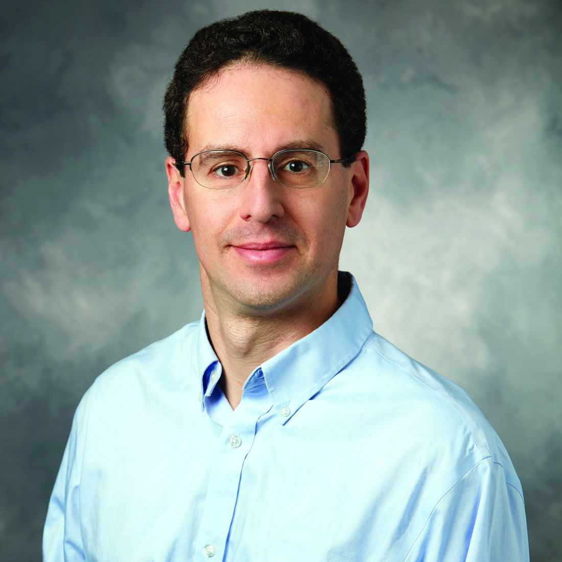
Results from a single-center study of CellMax Life’s FirstSight blood test were released as a poster as part of the annual Digestive Disease Week®, which was canceled because of COVID-19.
For a study conducted at one site, the Veterans Affairs Palo Alto (Calif.) Healthcare System, Shai Friedland, MD, and colleagues recruited 354 patients between ages 45 and 80 who were scheduled for elective colonoscopy. The researchers excluded people with a personal history of cancer or inflammatory bowel disease. They used CellMax’s FirstSight test on blood samples from the study participants.
The FirstSight test result was positive for colorectal cancer in all 11 patients in the study who were found by colonoscopy to have this condition, said Dr. Friedland, who is a professor of medicine at Stanford (Calif.) University and chief of gastroenterology at the VA Palo Alto Healthcare System. Thus, the test showed a sensitivity of 100% in this instance.
Among the 53 study participants found by colonoscopy to have advanced adenoma, 40 were positive on FirstSight; thus, so the test has a sensitivity of 75.5% for this result.
Among 79 patients who had negative colonoscopy results, meaning they were judged free of cancer or polyps, the test showed 8 as having signs of disease or growths.
“If you had a large adenoma that was removed years ago and now you have a negative colonoscopy, your score might still be high,” Dr. Friedland said in a recorded presentation for DDW. “In other words, the changes that are detectable in your blood might persist even after the polypectomy.”
He said there are plans to soon start a large-scale multicenter study of the CellMax assay.
“The blood test has the potential to fill an unmet need by giving patients a highly sensitive convenient option for colorectal cancer screening,” he said.
CellMax already is seeking to position its test as a more convenient alternative to either colonoscopy or the Cologuard screening test. Many patients put off cancer screening because of the need to take time off from work and the invasive nature of colonoscopy. Exact Sciences has used direct-to-consumer advertising to promote its Cologuard home-based test as a more convenient alternative to colonoscopy, but its product requires patients to collect their own stool samples and mail them to a lab, a process many people find off-putting.
Public health advocates, including the U.S. Preventive Services Task Force (USPSTF), have for years been pressing for wider screening of American adults for colon cancer. USPSTF is in the midst of updating its recommendations on colon cancer. In announcing its latest update of these recommendations in 2016, USPSTF said “the best screening test is the one that gets done” (JAMA. 2016;315[23]:2564-75).
USPSTF pressed for maximizing the total proportion of the eligible population, a point Dr. Friedland echoed in a CellMax press release.
“For colon cancer screening to be most effective, it is essential to detect precancerous polyps and then perform a colonoscopy to remove the polyps,” said Dr. Friedland in the CellMax press release. “Giving patients the option of getting a blood test for screening would undoubtedly increase compliance and thereby reduce mortality from colorectal cancer.”
In the DDW presentation, Dr. Friedland and colleagues also said the CellMax test showed greater sensitivity (100%) for colorectal cancer and advanced precancerous lesions (75.5%) than did Cologuard (92.3% for colorectal cancer and 42.4% for advanced precancerous lesions).
Cara Connelly, Director of Public Relations and Corporate Communications for Exact Sciences said that the company “is dedicated to getting more people screened for colorectal cancer and applaud the researchers for their efforts. We look forward to hearing more about the performance of this test in a prospective multisite study with nonsymptomatic patients.”
Naresh T. Gunaratnam, MD, a gastroenterologist and research director at Huron Gastro in Ypsilanti, Mich., said he is concerned that aggressive promotion of alternative tests may obscure the benefits of colonoscopy. Dr. Gunaratnam, a 2019 winner of the American Gastroenterological Association (AGA) Distinguished Clinician Award, has been a public critic of the marketing of colon cancer tests, which emphasize the convenience of these products. When asked by MDedge to comment on the CellMax-funded study, Dr. Gunaratnam said alternative tests do have a place for the care of patients who cannot or will not have a colonoscopy.
“But if you convince a patient who would be willing to have a colonoscopy not to, that’s a disservice,” he said.
“If you want the best test, the one that is best at finding cancers and finding polyps and the only one that can remove the polyp, that’s colonoscopy,” Dr. Gunaratnam added. “One day there may be a pill you can swallow that blows up the polyps, but we’re not there yet. We have to mechanically remove them.”
SOURCE: Friedland S et al. DDW 2020, eposter 575.
Blood assay studied for colorectal cancer screening.
Blood assay studied for colorectal cancer screening.
A blood test detected 11 of 11 cases of colorectal cancer in a study involving 354 patients, and also spotted a majority of cases – 40 out of 53 – in which participants had advanced adenomas, an investigator said.
Results from a single-center study of CellMax Life’s FirstSight blood test were released as a poster as part of the annual Digestive Disease Week®, which was canceled because of COVID-19.
For a study conducted at one site, the Veterans Affairs Palo Alto (Calif.) Healthcare System, Shai Friedland, MD, and colleagues recruited 354 patients between ages 45 and 80 who were scheduled for elective colonoscopy. The researchers excluded people with a personal history of cancer or inflammatory bowel disease. They used CellMax’s FirstSight test on blood samples from the study participants.
The FirstSight test result was positive for colorectal cancer in all 11 patients in the study who were found by colonoscopy to have this condition, said Dr. Friedland, who is a professor of medicine at Stanford (Calif.) University and chief of gastroenterology at the VA Palo Alto Healthcare System. Thus, the test showed a sensitivity of 100% in this instance.
Among the 53 study participants found by colonoscopy to have advanced adenoma, 40 were positive on FirstSight; thus, so the test has a sensitivity of 75.5% for this result.
Among 79 patients who had negative colonoscopy results, meaning they were judged free of cancer or polyps, the test showed 8 as having signs of disease or growths.
“If you had a large adenoma that was removed years ago and now you have a negative colonoscopy, your score might still be high,” Dr. Friedland said in a recorded presentation for DDW. “In other words, the changes that are detectable in your blood might persist even after the polypectomy.”
He said there are plans to soon start a large-scale multicenter study of the CellMax assay.
“The blood test has the potential to fill an unmet need by giving patients a highly sensitive convenient option for colorectal cancer screening,” he said.
CellMax already is seeking to position its test as a more convenient alternative to either colonoscopy or the Cologuard screening test. Many patients put off cancer screening because of the need to take time off from work and the invasive nature of colonoscopy. Exact Sciences has used direct-to-consumer advertising to promote its Cologuard home-based test as a more convenient alternative to colonoscopy, but its product requires patients to collect their own stool samples and mail them to a lab, a process many people find off-putting.
Public health advocates, including the U.S. Preventive Services Task Force (USPSTF), have for years been pressing for wider screening of American adults for colon cancer. USPSTF is in the midst of updating its recommendations on colon cancer. In announcing its latest update of these recommendations in 2016, USPSTF said “the best screening test is the one that gets done” (JAMA. 2016;315[23]:2564-75).
USPSTF pressed for maximizing the total proportion of the eligible population, a point Dr. Friedland echoed in a CellMax press release.
“For colon cancer screening to be most effective, it is essential to detect precancerous polyps and then perform a colonoscopy to remove the polyps,” said Dr. Friedland in the CellMax press release. “Giving patients the option of getting a blood test for screening would undoubtedly increase compliance and thereby reduce mortality from colorectal cancer.”
In the DDW presentation, Dr. Friedland and colleagues also said the CellMax test showed greater sensitivity (100%) for colorectal cancer and advanced precancerous lesions (75.5%) than did Cologuard (92.3% for colorectal cancer and 42.4% for advanced precancerous lesions).
Cara Connelly, Director of Public Relations and Corporate Communications for Exact Sciences said that the company “is dedicated to getting more people screened for colorectal cancer and applaud the researchers for their efforts. We look forward to hearing more about the performance of this test in a prospective multisite study with nonsymptomatic patients.”
Naresh T. Gunaratnam, MD, a gastroenterologist and research director at Huron Gastro in Ypsilanti, Mich., said he is concerned that aggressive promotion of alternative tests may obscure the benefits of colonoscopy. Dr. Gunaratnam, a 2019 winner of the American Gastroenterological Association (AGA) Distinguished Clinician Award, has been a public critic of the marketing of colon cancer tests, which emphasize the convenience of these products. When asked by MDedge to comment on the CellMax-funded study, Dr. Gunaratnam said alternative tests do have a place for the care of patients who cannot or will not have a colonoscopy.
“But if you convince a patient who would be willing to have a colonoscopy not to, that’s a disservice,” he said.
“If you want the best test, the one that is best at finding cancers and finding polyps and the only one that can remove the polyp, that’s colonoscopy,” Dr. Gunaratnam added. “One day there may be a pill you can swallow that blows up the polyps, but we’re not there yet. We have to mechanically remove them.”
SOURCE: Friedland S et al. DDW 2020, eposter 575.
A blood test detected 11 of 11 cases of colorectal cancer in a study involving 354 patients, and also spotted a majority of cases – 40 out of 53 – in which participants had advanced adenomas, an investigator said.
Results from a single-center study of CellMax Life’s FirstSight blood test were released as a poster as part of the annual Digestive Disease Week®, which was canceled because of COVID-19.
For a study conducted at one site, the Veterans Affairs Palo Alto (Calif.) Healthcare System, Shai Friedland, MD, and colleagues recruited 354 patients between ages 45 and 80 who were scheduled for elective colonoscopy. The researchers excluded people with a personal history of cancer or inflammatory bowel disease. They used CellMax’s FirstSight test on blood samples from the study participants.
The FirstSight test result was positive for colorectal cancer in all 11 patients in the study who were found by colonoscopy to have this condition, said Dr. Friedland, who is a professor of medicine at Stanford (Calif.) University and chief of gastroenterology at the VA Palo Alto Healthcare System. Thus, the test showed a sensitivity of 100% in this instance.
Among the 53 study participants found by colonoscopy to have advanced adenoma, 40 were positive on FirstSight; thus, so the test has a sensitivity of 75.5% for this result.
Among 79 patients who had negative colonoscopy results, meaning they were judged free of cancer or polyps, the test showed 8 as having signs of disease or growths.
“If you had a large adenoma that was removed years ago and now you have a negative colonoscopy, your score might still be high,” Dr. Friedland said in a recorded presentation for DDW. “In other words, the changes that are detectable in your blood might persist even after the polypectomy.”
He said there are plans to soon start a large-scale multicenter study of the CellMax assay.
“The blood test has the potential to fill an unmet need by giving patients a highly sensitive convenient option for colorectal cancer screening,” he said.
CellMax already is seeking to position its test as a more convenient alternative to either colonoscopy or the Cologuard screening test. Many patients put off cancer screening because of the need to take time off from work and the invasive nature of colonoscopy. Exact Sciences has used direct-to-consumer advertising to promote its Cologuard home-based test as a more convenient alternative to colonoscopy, but its product requires patients to collect their own stool samples and mail them to a lab, a process many people find off-putting.
Public health advocates, including the U.S. Preventive Services Task Force (USPSTF), have for years been pressing for wider screening of American adults for colon cancer. USPSTF is in the midst of updating its recommendations on colon cancer. In announcing its latest update of these recommendations in 2016, USPSTF said “the best screening test is the one that gets done” (JAMA. 2016;315[23]:2564-75).
USPSTF pressed for maximizing the total proportion of the eligible population, a point Dr. Friedland echoed in a CellMax press release.
“For colon cancer screening to be most effective, it is essential to detect precancerous polyps and then perform a colonoscopy to remove the polyps,” said Dr. Friedland in the CellMax press release. “Giving patients the option of getting a blood test for screening would undoubtedly increase compliance and thereby reduce mortality from colorectal cancer.”
In the DDW presentation, Dr. Friedland and colleagues also said the CellMax test showed greater sensitivity (100%) for colorectal cancer and advanced precancerous lesions (75.5%) than did Cologuard (92.3% for colorectal cancer and 42.4% for advanced precancerous lesions).
Cara Connelly, Director of Public Relations and Corporate Communications for Exact Sciences said that the company “is dedicated to getting more people screened for colorectal cancer and applaud the researchers for their efforts. We look forward to hearing more about the performance of this test in a prospective multisite study with nonsymptomatic patients.”
Naresh T. Gunaratnam, MD, a gastroenterologist and research director at Huron Gastro in Ypsilanti, Mich., said he is concerned that aggressive promotion of alternative tests may obscure the benefits of colonoscopy. Dr. Gunaratnam, a 2019 winner of the American Gastroenterological Association (AGA) Distinguished Clinician Award, has been a public critic of the marketing of colon cancer tests, which emphasize the convenience of these products. When asked by MDedge to comment on the CellMax-funded study, Dr. Gunaratnam said alternative tests do have a place for the care of patients who cannot or will not have a colonoscopy.
“But if you convince a patient who would be willing to have a colonoscopy not to, that’s a disservice,” he said.
“If you want the best test, the one that is best at finding cancers and finding polyps and the only one that can remove the polyp, that’s colonoscopy,” Dr. Gunaratnam added. “One day there may be a pill you can swallow that blows up the polyps, but we’re not there yet. We have to mechanically remove them.”
SOURCE: Friedland S et al. DDW 2020, eposter 575.
FROM DDW 2020
MRD surveillance can detect relapse before imaging in early-stage NSCLC
, according to findings from the TRACERx study.
The findings pave the way for clinical trials of MRD-driven treatment escalation, Chris Abbosh, MD, of University College London, reported during a presentation at the AACR virtual meeting I. Data in the presentation were updated from the abstract.
Dr. Abbosh and colleagues used phylogenetic circulating tumor DNA (ctDNA) profiling to assess MRD and predict relapse in patients from the TRACERx study who underwent surgery for stage I-III NSCLC.
“The approach we take is technically termed a ‘tumor-informed, personalized cell-free DNA-enrichment approach,’” Dr. Abbosh explained. “We take out the primary tumor from the patient, we multiregion sample that tumor, and submit each region for deep whole-exome sequencing.”
The researchers prioritize variants for MRD tracking based on clonality/subclonality, high copy number status, and low background sequencing noise. The researchers then construct an anchored-multiplex PCR panel against the positions of interest, which is applied to cell-free DNA in the pre- and postoperative setting.
“We’ve developed an MRD caller to go alongside this chemistry,” Dr. Abbosh said. “The main premise behind the MRD caller is that it can calculate intralibrary error rates to inform the MRD pool.”
Sensitivity and specificity
To validate their approach, Dr. Abbosh and colleagues tested the assay with low DNA input (5 ng, 10 ng) and high DNA input (30 ng, 60 ng). They found the assay to be more sensitive with higher DNA input, and variant fractions were detected down to 0.003%.
The researchers also assessed how sensitivity and specificity scale with an increasing number of variants – 50, 100, or 200 variants. When tracking 200 variants, the assay was powered to detect lower ctDNA fractions than when tracking 50 variants. On the other hand, specificity was higher with 50 variants (99.8%) than with 200 variants (99.4%).
Next, Dr. Abbosh and colleagues analyzed postoperative cell-free DNA collected at 271 time points from 37 NSCLC patients who did not relapse. This included 11 patients who developed proven second primary malignancies.
Of the 271 time points when MRD negativity was expected, MRD was not detected at 269 time points, which translates to 99.3% specificity for the assay.
Shedding, relapse, and disease-free survival
Dr. Abbosh and colleagues also found that non-adenocarcinoma histology is associated with preoperative ctDNA shedding in NSCLC. The researchers analyzed 88 early-stage preoperative samples from NSCLC patients. ctDNA was detected preoperatively in 49% of lung adenocarcinomas and 100% of lung squamous cell carcinomas.
“This finding is important when it comes to interpreting our non–small cell lung cancer relapse data from 53 TRACERx patients,” Dr. Abbosh said.
Of the 53 patients who relapsed, 42 had ctDNA detected prior to surgery and were thus considered shedders, while 11 were nonshedders. ctDNA was detectable at or before relapse in 91% (38/42) of shedders and 64% (7/11) of nonshedders.
The median time from ctDNA detection to clinical relapse was 164 days in shedders and 22 days in nonshedders. The median disease-free survival was 362 days and 640 days, respectively.
“So what these data suggest is that preoperative ctDNA detection status will be a proxy of the potential utility of ctDNA as an MRD biomarker in a clinical setting,” Dr. Abbosh explained.
Standard-of-care imaging findings in the 53 patients who relapsed further demonstrated the utility of ctDNA in this setting, Dr. Abbosh said.
All scans were divided into three categories: those showing unequivocal relapse, those with a new equivocal change (relapse, inflammation, or a nonspecific finding), and those with no evidence of relapse. Each was further categorized by preimaging MRD status.
Relapse occurred in 9 of 10 patients who were MRD positive but had a scan showing no evidence of relapse. Relapse occurred in 15 of 16 patients who were MRD positive and had scans showing new equivocal changes.
Patients with unequivocal evidence of relapse who were MRD negative at or before the scan were more likely to have a second primary cancer than to have NSCLC relapse (52% vs. 48%), which is a reflection of the specificity of the MRD assay to the primary tumor, Dr. Abbosh said.
Implications of the findings
The researchers’ findings are important because establishing an MRD-driven approach to treating early-stage NSCLC would facilitate escalation of standard-of-care treatment only for those patients at high risk for relapse, thereby overcoming a key challenge in conventional adjuvant drug-trial design, Dr. Abbosh said.
“If we take a patient population with high-risk early-stage disease who have undergone potentially curative resection of their cancer and we offer these patients adjuvant chemotherapy or adjuvant chemoradiation therapy, then we can improve 5-year survival outcomes in this population,” Dr. Abbosh said. “This is striking because, if we give the same treatment in the metastatic setting, we only see a progression-free survival benefit of a short number of months.”
This suggests a potential “vulnerability of low-burden residual cancer to systemic treatment following surgery,” he added. “So if we want to improve outcomes further in non–small cell lung cancer, we really need to focus on innovation in the early-stage space.”
Dr. Abbosh said he and colleagues demonstrated that “personalized cell-free DNA enrichment can detect low-frequency variant DNA in an accurate manner.
“We’ve shown that preoperative ctDNA shedding is associated with utility of ctDNA as an MRD biomarker and that MRD surveillance can lead to detection of relapse in advance of standard-of-care-imaging,” he said. “We feel that the field is now ready for MRD-driven adjuvant trials.”
Questions to be answered
Invited discussant Corey J. Langer, MD, of Penn Medicine in Philadelphia, outlined “fundamental questions” raised by the findings.
“We need more information on the staging and demographics of those who were MRD positive versus MRD negative,” he said.
Dr. Langer also asked about the findings for shedders versus nonshedders.
“Does this mean nonshedders fare better? This needs to addressed formally,” he said.
Another question is whether the assay “simply enables us to detect relapse sooner and increase anxiety,” or if the trajectory and outcomes in those who prove MRD positive ahead of radiographic manifestations can actually be altered.
A study comparing standard observation with early immunotherapy or chemoimmunotherapy in patients with MRD-positive radiographically occult relapse or progression – using progression-free and overall survival, along with time without symptoms of disease or relapse – would be useful, Dr. Langer said.
“A hazard ratio of 0.8 or less would be meaningful,” he added. “In this regard, there are trials looking at enhanced adjuvant treatment both in colorectal and breast cancer, and trials planned in advanced non–small cell [lung cancer].”
Dr. Langer also said it would be interesting to know if the assay can be used as an adjunct to diagnosis in frailer patients with inaccessible tumors or equivocal biopsy results or to avoid invasive procedures in patients who are stereotactic radiation candidates.
“The jury is still out on this,” he said.
TRACERx is funded by University College London in collaboration with Cancer Research UK. Dr. Abbosh disclosed relationships with AstraZeneca, Novartis, Roche Diagnostics, Bristol Myers Squibb, Achilles Therapeutics, and Archer Diagnostics. Dr. Langer reported grant/research support and/or scientific advisory work for multiple companies.
SOURCE: Abbosh C et al. AACR 2020, Abstract CT023.
, according to findings from the TRACERx study.
The findings pave the way for clinical trials of MRD-driven treatment escalation, Chris Abbosh, MD, of University College London, reported during a presentation at the AACR virtual meeting I. Data in the presentation were updated from the abstract.
Dr. Abbosh and colleagues used phylogenetic circulating tumor DNA (ctDNA) profiling to assess MRD and predict relapse in patients from the TRACERx study who underwent surgery for stage I-III NSCLC.
“The approach we take is technically termed a ‘tumor-informed, personalized cell-free DNA-enrichment approach,’” Dr. Abbosh explained. “We take out the primary tumor from the patient, we multiregion sample that tumor, and submit each region for deep whole-exome sequencing.”
The researchers prioritize variants for MRD tracking based on clonality/subclonality, high copy number status, and low background sequencing noise. The researchers then construct an anchored-multiplex PCR panel against the positions of interest, which is applied to cell-free DNA in the pre- and postoperative setting.
“We’ve developed an MRD caller to go alongside this chemistry,” Dr. Abbosh said. “The main premise behind the MRD caller is that it can calculate intralibrary error rates to inform the MRD pool.”
Sensitivity and specificity
To validate their approach, Dr. Abbosh and colleagues tested the assay with low DNA input (5 ng, 10 ng) and high DNA input (30 ng, 60 ng). They found the assay to be more sensitive with higher DNA input, and variant fractions were detected down to 0.003%.
The researchers also assessed how sensitivity and specificity scale with an increasing number of variants – 50, 100, or 200 variants. When tracking 200 variants, the assay was powered to detect lower ctDNA fractions than when tracking 50 variants. On the other hand, specificity was higher with 50 variants (99.8%) than with 200 variants (99.4%).
Next, Dr. Abbosh and colleagues analyzed postoperative cell-free DNA collected at 271 time points from 37 NSCLC patients who did not relapse. This included 11 patients who developed proven second primary malignancies.
Of the 271 time points when MRD negativity was expected, MRD was not detected at 269 time points, which translates to 99.3% specificity for the assay.
Shedding, relapse, and disease-free survival
Dr. Abbosh and colleagues also found that non-adenocarcinoma histology is associated with preoperative ctDNA shedding in NSCLC. The researchers analyzed 88 early-stage preoperative samples from NSCLC patients. ctDNA was detected preoperatively in 49% of lung adenocarcinomas and 100% of lung squamous cell carcinomas.
“This finding is important when it comes to interpreting our non–small cell lung cancer relapse data from 53 TRACERx patients,” Dr. Abbosh said.
Of the 53 patients who relapsed, 42 had ctDNA detected prior to surgery and were thus considered shedders, while 11 were nonshedders. ctDNA was detectable at or before relapse in 91% (38/42) of shedders and 64% (7/11) of nonshedders.
The median time from ctDNA detection to clinical relapse was 164 days in shedders and 22 days in nonshedders. The median disease-free survival was 362 days and 640 days, respectively.
“So what these data suggest is that preoperative ctDNA detection status will be a proxy of the potential utility of ctDNA as an MRD biomarker in a clinical setting,” Dr. Abbosh explained.
Standard-of-care imaging findings in the 53 patients who relapsed further demonstrated the utility of ctDNA in this setting, Dr. Abbosh said.
All scans were divided into three categories: those showing unequivocal relapse, those with a new equivocal change (relapse, inflammation, or a nonspecific finding), and those with no evidence of relapse. Each was further categorized by preimaging MRD status.
Relapse occurred in 9 of 10 patients who were MRD positive but had a scan showing no evidence of relapse. Relapse occurred in 15 of 16 patients who were MRD positive and had scans showing new equivocal changes.
Patients with unequivocal evidence of relapse who were MRD negative at or before the scan were more likely to have a second primary cancer than to have NSCLC relapse (52% vs. 48%), which is a reflection of the specificity of the MRD assay to the primary tumor, Dr. Abbosh said.
Implications of the findings
The researchers’ findings are important because establishing an MRD-driven approach to treating early-stage NSCLC would facilitate escalation of standard-of-care treatment only for those patients at high risk for relapse, thereby overcoming a key challenge in conventional adjuvant drug-trial design, Dr. Abbosh said.
“If we take a patient population with high-risk early-stage disease who have undergone potentially curative resection of their cancer and we offer these patients adjuvant chemotherapy or adjuvant chemoradiation therapy, then we can improve 5-year survival outcomes in this population,” Dr. Abbosh said. “This is striking because, if we give the same treatment in the metastatic setting, we only see a progression-free survival benefit of a short number of months.”
This suggests a potential “vulnerability of low-burden residual cancer to systemic treatment following surgery,” he added. “So if we want to improve outcomes further in non–small cell lung cancer, we really need to focus on innovation in the early-stage space.”
Dr. Abbosh said he and colleagues demonstrated that “personalized cell-free DNA enrichment can detect low-frequency variant DNA in an accurate manner.
“We’ve shown that preoperative ctDNA shedding is associated with utility of ctDNA as an MRD biomarker and that MRD surveillance can lead to detection of relapse in advance of standard-of-care-imaging,” he said. “We feel that the field is now ready for MRD-driven adjuvant trials.”
Questions to be answered
Invited discussant Corey J. Langer, MD, of Penn Medicine in Philadelphia, outlined “fundamental questions” raised by the findings.
“We need more information on the staging and demographics of those who were MRD positive versus MRD negative,” he said.
Dr. Langer also asked about the findings for shedders versus nonshedders.
“Does this mean nonshedders fare better? This needs to addressed formally,” he said.
Another question is whether the assay “simply enables us to detect relapse sooner and increase anxiety,” or if the trajectory and outcomes in those who prove MRD positive ahead of radiographic manifestations can actually be altered.
A study comparing standard observation with early immunotherapy or chemoimmunotherapy in patients with MRD-positive radiographically occult relapse or progression – using progression-free and overall survival, along with time without symptoms of disease or relapse – would be useful, Dr. Langer said.
“A hazard ratio of 0.8 or less would be meaningful,” he added. “In this regard, there are trials looking at enhanced adjuvant treatment both in colorectal and breast cancer, and trials planned in advanced non–small cell [lung cancer].”
Dr. Langer also said it would be interesting to know if the assay can be used as an adjunct to diagnosis in frailer patients with inaccessible tumors or equivocal biopsy results or to avoid invasive procedures in patients who are stereotactic radiation candidates.
“The jury is still out on this,” he said.
TRACERx is funded by University College London in collaboration with Cancer Research UK. Dr. Abbosh disclosed relationships with AstraZeneca, Novartis, Roche Diagnostics, Bristol Myers Squibb, Achilles Therapeutics, and Archer Diagnostics. Dr. Langer reported grant/research support and/or scientific advisory work for multiple companies.
SOURCE: Abbosh C et al. AACR 2020, Abstract CT023.
, according to findings from the TRACERx study.
The findings pave the way for clinical trials of MRD-driven treatment escalation, Chris Abbosh, MD, of University College London, reported during a presentation at the AACR virtual meeting I. Data in the presentation were updated from the abstract.
Dr. Abbosh and colleagues used phylogenetic circulating tumor DNA (ctDNA) profiling to assess MRD and predict relapse in patients from the TRACERx study who underwent surgery for stage I-III NSCLC.
“The approach we take is technically termed a ‘tumor-informed, personalized cell-free DNA-enrichment approach,’” Dr. Abbosh explained. “We take out the primary tumor from the patient, we multiregion sample that tumor, and submit each region for deep whole-exome sequencing.”
The researchers prioritize variants for MRD tracking based on clonality/subclonality, high copy number status, and low background sequencing noise. The researchers then construct an anchored-multiplex PCR panel against the positions of interest, which is applied to cell-free DNA in the pre- and postoperative setting.
“We’ve developed an MRD caller to go alongside this chemistry,” Dr. Abbosh said. “The main premise behind the MRD caller is that it can calculate intralibrary error rates to inform the MRD pool.”
Sensitivity and specificity
To validate their approach, Dr. Abbosh and colleagues tested the assay with low DNA input (5 ng, 10 ng) and high DNA input (30 ng, 60 ng). They found the assay to be more sensitive with higher DNA input, and variant fractions were detected down to 0.003%.
The researchers also assessed how sensitivity and specificity scale with an increasing number of variants – 50, 100, or 200 variants. When tracking 200 variants, the assay was powered to detect lower ctDNA fractions than when tracking 50 variants. On the other hand, specificity was higher with 50 variants (99.8%) than with 200 variants (99.4%).
Next, Dr. Abbosh and colleagues analyzed postoperative cell-free DNA collected at 271 time points from 37 NSCLC patients who did not relapse. This included 11 patients who developed proven second primary malignancies.
Of the 271 time points when MRD negativity was expected, MRD was not detected at 269 time points, which translates to 99.3% specificity for the assay.
Shedding, relapse, and disease-free survival
Dr. Abbosh and colleagues also found that non-adenocarcinoma histology is associated with preoperative ctDNA shedding in NSCLC. The researchers analyzed 88 early-stage preoperative samples from NSCLC patients. ctDNA was detected preoperatively in 49% of lung adenocarcinomas and 100% of lung squamous cell carcinomas.
“This finding is important when it comes to interpreting our non–small cell lung cancer relapse data from 53 TRACERx patients,” Dr. Abbosh said.
Of the 53 patients who relapsed, 42 had ctDNA detected prior to surgery and were thus considered shedders, while 11 were nonshedders. ctDNA was detectable at or before relapse in 91% (38/42) of shedders and 64% (7/11) of nonshedders.
The median time from ctDNA detection to clinical relapse was 164 days in shedders and 22 days in nonshedders. The median disease-free survival was 362 days and 640 days, respectively.
“So what these data suggest is that preoperative ctDNA detection status will be a proxy of the potential utility of ctDNA as an MRD biomarker in a clinical setting,” Dr. Abbosh explained.
Standard-of-care imaging findings in the 53 patients who relapsed further demonstrated the utility of ctDNA in this setting, Dr. Abbosh said.
All scans were divided into three categories: those showing unequivocal relapse, those with a new equivocal change (relapse, inflammation, or a nonspecific finding), and those with no evidence of relapse. Each was further categorized by preimaging MRD status.
Relapse occurred in 9 of 10 patients who were MRD positive but had a scan showing no evidence of relapse. Relapse occurred in 15 of 16 patients who were MRD positive and had scans showing new equivocal changes.
Patients with unequivocal evidence of relapse who were MRD negative at or before the scan were more likely to have a second primary cancer than to have NSCLC relapse (52% vs. 48%), which is a reflection of the specificity of the MRD assay to the primary tumor, Dr. Abbosh said.
Implications of the findings
The researchers’ findings are important because establishing an MRD-driven approach to treating early-stage NSCLC would facilitate escalation of standard-of-care treatment only for those patients at high risk for relapse, thereby overcoming a key challenge in conventional adjuvant drug-trial design, Dr. Abbosh said.
“If we take a patient population with high-risk early-stage disease who have undergone potentially curative resection of their cancer and we offer these patients adjuvant chemotherapy or adjuvant chemoradiation therapy, then we can improve 5-year survival outcomes in this population,” Dr. Abbosh said. “This is striking because, if we give the same treatment in the metastatic setting, we only see a progression-free survival benefit of a short number of months.”
This suggests a potential “vulnerability of low-burden residual cancer to systemic treatment following surgery,” he added. “So if we want to improve outcomes further in non–small cell lung cancer, we really need to focus on innovation in the early-stage space.”
Dr. Abbosh said he and colleagues demonstrated that “personalized cell-free DNA enrichment can detect low-frequency variant DNA in an accurate manner.
“We’ve shown that preoperative ctDNA shedding is associated with utility of ctDNA as an MRD biomarker and that MRD surveillance can lead to detection of relapse in advance of standard-of-care-imaging,” he said. “We feel that the field is now ready for MRD-driven adjuvant trials.”
Questions to be answered
Invited discussant Corey J. Langer, MD, of Penn Medicine in Philadelphia, outlined “fundamental questions” raised by the findings.
“We need more information on the staging and demographics of those who were MRD positive versus MRD negative,” he said.
Dr. Langer also asked about the findings for shedders versus nonshedders.
“Does this mean nonshedders fare better? This needs to addressed formally,” he said.
Another question is whether the assay “simply enables us to detect relapse sooner and increase anxiety,” or if the trajectory and outcomes in those who prove MRD positive ahead of radiographic manifestations can actually be altered.
A study comparing standard observation with early immunotherapy or chemoimmunotherapy in patients with MRD-positive radiographically occult relapse or progression – using progression-free and overall survival, along with time without symptoms of disease or relapse – would be useful, Dr. Langer said.
“A hazard ratio of 0.8 or less would be meaningful,” he added. “In this regard, there are trials looking at enhanced adjuvant treatment both in colorectal and breast cancer, and trials planned in advanced non–small cell [lung cancer].”
Dr. Langer also said it would be interesting to know if the assay can be used as an adjunct to diagnosis in frailer patients with inaccessible tumors or equivocal biopsy results or to avoid invasive procedures in patients who are stereotactic radiation candidates.
“The jury is still out on this,” he said.
TRACERx is funded by University College London in collaboration with Cancer Research UK. Dr. Abbosh disclosed relationships with AstraZeneca, Novartis, Roche Diagnostics, Bristol Myers Squibb, Achilles Therapeutics, and Archer Diagnostics. Dr. Langer reported grant/research support and/or scientific advisory work for multiple companies.
SOURCE: Abbosh C et al. AACR 2020, Abstract CT023.
FROM AACR 2020
Ranitidine did not show higher cancer risk than famotidine in study
People who had taken either of these drugs had a higher risk for cancer, but they also were more likely to have risk factors for this disease such as obesity or a history of smoking, an investigator said.
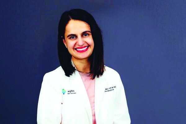
The findings from Nabeeha Mohyuddin, MD, an internal medicine resident at Allegheny Health Network in Pittsburgh, appear to be at odds with warnings from the U.S. Food and Drug Administration. The FDA in April called on manufacturers to pull all versions of ranitidine because of a problem with probable contamination by human carcinogen n-nitrosodimethylamine (NDMA). The FDA and the European Medicines Agency last year announced investigations of this NDMA contamination.
Dr. Mohyuddin and colleagues used IBM’s Explorys database, which includes data collected from EMRs from more than 40 U.S. health systems, to see if ranitidine use appeared connected to cancer diagnoses. Dr. Mohyuddin presented the findings in an abstract released as part of the annual Digestive Disease Week®, which was canceled because of COVID-19. The researchers identified records for 1.62 million users of ranitidine, 3.37 million users of famotidine, and 59.63 million people who did not use either H2 blocker.
The incidence of cancer was respectively 14.69%, 21.24%, and 5.38% for the ranitidine group, the famotidine group, and the group representing the general population without use of either H2 blocker. Among subjects without risk factors including smoking, obesity, alcohol use, family history, cirrhosis, and gastroesophageal reflux disease, ranitidine users did not have an increased risk of cancer, compared with famotidine users (odds ratio, 0.77; 95% confidence interval, 0.76-0.77; P < .001), Dr. Mohyuddin said in an email interview after her presentation.
“The results need to be interpreted with caution given that this is a retrospective study and it’s the first of its kind,” she said. “Further studies will be definitely needed on this to definitively answer the question, ‘does ranitidine have an increased risk of cancer?’ ”
People in the study who used H2 blockers tended to be older and were more likely to have other risk factors for cancers, according to the abstract:
- Of the ranitidine group, about 33.6% were older than 65, 74.4% were smokers, and 8.9% had a body mass index above 30 kg/m2.
- Of the famotidine group, about 38.3% were older than 65, 76.9% were smokers and 10.8% had a BMI above 30.
- Of the general population, about 23.9% were older than 65, 27.5% were smokers, and 1.69% were obese.
The Explorys database accounts for only 15%-19% of the entire U.S. population and that could be an explanation for why the percentage for obesity in the population seems spuriously low, Dr. Mohyuddin said in an email exchange.
Additionally, it pulls in patients through diagnosis codes, and if a different code for obesity was entered, those patients may not have been accounted for, she said.
Dhyanesh A. Patel, MD, assistant professor of medicine at Vanderbilt University, Nashville, Tenn., said it was a surprise to see the rate of cancer reported for famotidine users in the abstract presented at DDW.
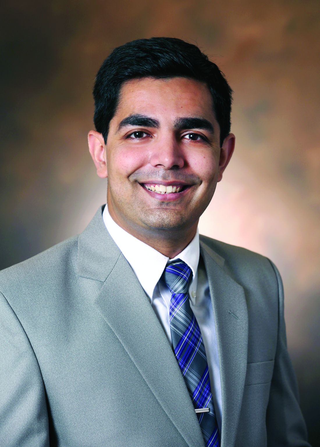
“But these databases are so prone to multiple biases that it’s really hard to tease out. You’d really have to do prospective studies where you follow these patients and then control for variables to know the true risk,” Dr. Patel said in an interview.
In many cases, for example, patients may start on H2 blockers after reporting symptoms that sound like indigestion, Patel said.
“And then the next week, they actually get an upper endoscopy and they get diagnosed with gastric cancer,” Patel said. “In the database, it may seem to be that ranitidine was started and the patient was diagnosed with gastric cancer. So now the gastric cancer gets associated with ranitidine.”
Physicians have had many options to use instead of ranitidine since the FDA last year announced its concerns about the drug, Dr. Patel said.
“Patients can easily switch over to a similar histamine blocker or you can use one of the proton pump inhibitors that haven’t been found to have this impurity in them,” he said. “Both of those are good options for patients.”
Further research
Manufacturers have been withdrawing their ranitidine products from the market since the reports of contamination surfaced last year. Several drugmakers, including Pfizer and Perrigo, have reported facing lawsuits connected with claims of cancer or increased cancer risk from ranitidine. In February 2020, a federal judicial panel opted to use a procedure, known as multidistrict litigation (MDL), to streamline the handling of these many cases. They were put before the U.S. District Court for the Southern District of Florida, labeled as In Re: Zantac (Ranitidine) NDMA Litigation.
A spokesman for FDA declined MDedge’s request for comment on Dr. Mohyuddin’s DDW presentation. He said the agency tends not to offer its views on work done by scientists outside of the FDA.
Valisure, an online pharmacy that runs quality checks on the medicines it dispenses, in September 2019 petitioned the FDA for a withdrawal of ranitidine due to concerns about NDMA. In its petition, Valisure reported finding notable levels of NDMA in ranitidine tablets, but not detecting it in testing of famotidine and several similar drugs. In an emailed statement, David Light, founder and CEO of Valisure, said the structure of Dr. Mohyuddin’s research limited its ability to detect cancer correlations because of the large and generalized study population.
“When comparing millions of people on medications which are both over-the-counter and prescription, any epidemiological impact will very likely be eclipsed by the sheer variations of exposure and a wide variety of confounding factors,” Mr. Light wrote.
There are a “vast number of variables that aren’t controlled for in such a massive and broadly defined cohort,” he added.
More focused and controlled studies will be needed to best evaluate NDMA and ranitidine, according to Mr. Light. “We are also investigating this issue with researchers at Memorial Sloan Kettering Cancer Center and plan to publish results soon,” he said.
Dr. Mohyuddin and Dr. Patel did not disclose financial conflicts in connection with ranitidine.
SOURCE: Mohyuddin N et al. DDW 2020. Abstract Tu1360.
People who had taken either of these drugs had a higher risk for cancer, but they also were more likely to have risk factors for this disease such as obesity or a history of smoking, an investigator said.
The findings from Nabeeha Mohyuddin, MD, an internal medicine resident at Allegheny Health Network in Pittsburgh, appear to be at odds with warnings from the U.S. Food and Drug Administration. The FDA in April called on manufacturers to pull all versions of ranitidine because of a problem with probable contamination by human carcinogen n-nitrosodimethylamine (NDMA). The FDA and the European Medicines Agency last year announced investigations of this NDMA contamination.
Dr. Mohyuddin and colleagues used IBM’s Explorys database, which includes data collected from EMRs from more than 40 U.S. health systems, to see if ranitidine use appeared connected to cancer diagnoses. Dr. Mohyuddin presented the findings in an abstract released as part of the annual Digestive Disease Week®, which was canceled because of COVID-19. The researchers identified records for 1.62 million users of ranitidine, 3.37 million users of famotidine, and 59.63 million people who did not use either H2 blocker.
The incidence of cancer was respectively 14.69%, 21.24%, and 5.38% for the ranitidine group, the famotidine group, and the group representing the general population without use of either H2 blocker. Among subjects without risk factors including smoking, obesity, alcohol use, family history, cirrhosis, and gastroesophageal reflux disease, ranitidine users did not have an increased risk of cancer, compared with famotidine users (odds ratio, 0.77; 95% confidence interval, 0.76-0.77; P < .001), Dr. Mohyuddin said in an email interview after her presentation.
“The results need to be interpreted with caution given that this is a retrospective study and it’s the first of its kind,” she said. “Further studies will be definitely needed on this to definitively answer the question, ‘does ranitidine have an increased risk of cancer?’ ”
People in the study who used H2 blockers tended to be older and were more likely to have other risk factors for cancers, according to the abstract:
- Of the ranitidine group, about 33.6% were older than 65, 74.4% were smokers, and 8.9% had a body mass index above 30 kg/m2.
- Of the famotidine group, about 38.3% were older than 65, 76.9% were smokers and 10.8% had a BMI above 30.
- Of the general population, about 23.9% were older than 65, 27.5% were smokers, and 1.69% were obese.
The Explorys database accounts for only 15%-19% of the entire U.S. population and that could be an explanation for why the percentage for obesity in the population seems spuriously low, Dr. Mohyuddin said in an email exchange.
Additionally, it pulls in patients through diagnosis codes, and if a different code for obesity was entered, those patients may not have been accounted for, she said.
Dhyanesh A. Patel, MD, assistant professor of medicine at Vanderbilt University, Nashville, Tenn., said it was a surprise to see the rate of cancer reported for famotidine users in the abstract presented at DDW.
“But these databases are so prone to multiple biases that it’s really hard to tease out. You’d really have to do prospective studies where you follow these patients and then control for variables to know the true risk,” Dr. Patel said in an interview.
In many cases, for example, patients may start on H2 blockers after reporting symptoms that sound like indigestion, Patel said.
“And then the next week, they actually get an upper endoscopy and they get diagnosed with gastric cancer,” Patel said. “In the database, it may seem to be that ranitidine was started and the patient was diagnosed with gastric cancer. So now the gastric cancer gets associated with ranitidine.”
Physicians have had many options to use instead of ranitidine since the FDA last year announced its concerns about the drug, Dr. Patel said.
“Patients can easily switch over to a similar histamine blocker or you can use one of the proton pump inhibitors that haven’t been found to have this impurity in them,” he said. “Both of those are good options for patients.”
Further research
Manufacturers have been withdrawing their ranitidine products from the market since the reports of contamination surfaced last year. Several drugmakers, including Pfizer and Perrigo, have reported facing lawsuits connected with claims of cancer or increased cancer risk from ranitidine. In February 2020, a federal judicial panel opted to use a procedure, known as multidistrict litigation (MDL), to streamline the handling of these many cases. They were put before the U.S. District Court for the Southern District of Florida, labeled as In Re: Zantac (Ranitidine) NDMA Litigation.
A spokesman for FDA declined MDedge’s request for comment on Dr. Mohyuddin’s DDW presentation. He said the agency tends not to offer its views on work done by scientists outside of the FDA.
Valisure, an online pharmacy that runs quality checks on the medicines it dispenses, in September 2019 petitioned the FDA for a withdrawal of ranitidine due to concerns about NDMA. In its petition, Valisure reported finding notable levels of NDMA in ranitidine tablets, but not detecting it in testing of famotidine and several similar drugs. In an emailed statement, David Light, founder and CEO of Valisure, said the structure of Dr. Mohyuddin’s research limited its ability to detect cancer correlations because of the large and generalized study population.
“When comparing millions of people on medications which are both over-the-counter and prescription, any epidemiological impact will very likely be eclipsed by the sheer variations of exposure and a wide variety of confounding factors,” Mr. Light wrote.
There are a “vast number of variables that aren’t controlled for in such a massive and broadly defined cohort,” he added.
More focused and controlled studies will be needed to best evaluate NDMA and ranitidine, according to Mr. Light. “We are also investigating this issue with researchers at Memorial Sloan Kettering Cancer Center and plan to publish results soon,” he said.
Dr. Mohyuddin and Dr. Patel did not disclose financial conflicts in connection with ranitidine.
SOURCE: Mohyuddin N et al. DDW 2020. Abstract Tu1360.
People who had taken either of these drugs had a higher risk for cancer, but they also were more likely to have risk factors for this disease such as obesity or a history of smoking, an investigator said.
The findings from Nabeeha Mohyuddin, MD, an internal medicine resident at Allegheny Health Network in Pittsburgh, appear to be at odds with warnings from the U.S. Food and Drug Administration. The FDA in April called on manufacturers to pull all versions of ranitidine because of a problem with probable contamination by human carcinogen n-nitrosodimethylamine (NDMA). The FDA and the European Medicines Agency last year announced investigations of this NDMA contamination.
Dr. Mohyuddin and colleagues used IBM’s Explorys database, which includes data collected from EMRs from more than 40 U.S. health systems, to see if ranitidine use appeared connected to cancer diagnoses. Dr. Mohyuddin presented the findings in an abstract released as part of the annual Digestive Disease Week®, which was canceled because of COVID-19. The researchers identified records for 1.62 million users of ranitidine, 3.37 million users of famotidine, and 59.63 million people who did not use either H2 blocker.
The incidence of cancer was respectively 14.69%, 21.24%, and 5.38% for the ranitidine group, the famotidine group, and the group representing the general population without use of either H2 blocker. Among subjects without risk factors including smoking, obesity, alcohol use, family history, cirrhosis, and gastroesophageal reflux disease, ranitidine users did not have an increased risk of cancer, compared with famotidine users (odds ratio, 0.77; 95% confidence interval, 0.76-0.77; P < .001), Dr. Mohyuddin said in an email interview after her presentation.
“The results need to be interpreted with caution given that this is a retrospective study and it’s the first of its kind,” she said. “Further studies will be definitely needed on this to definitively answer the question, ‘does ranitidine have an increased risk of cancer?’ ”
People in the study who used H2 blockers tended to be older and were more likely to have other risk factors for cancers, according to the abstract:
- Of the ranitidine group, about 33.6% were older than 65, 74.4% were smokers, and 8.9% had a body mass index above 30 kg/m2.
- Of the famotidine group, about 38.3% were older than 65, 76.9% were smokers and 10.8% had a BMI above 30.
- Of the general population, about 23.9% were older than 65, 27.5% were smokers, and 1.69% were obese.
The Explorys database accounts for only 15%-19% of the entire U.S. population and that could be an explanation for why the percentage for obesity in the population seems spuriously low, Dr. Mohyuddin said in an email exchange.
Additionally, it pulls in patients through diagnosis codes, and if a different code for obesity was entered, those patients may not have been accounted for, she said.
Dhyanesh A. Patel, MD, assistant professor of medicine at Vanderbilt University, Nashville, Tenn., said it was a surprise to see the rate of cancer reported for famotidine users in the abstract presented at DDW.
“But these databases are so prone to multiple biases that it’s really hard to tease out. You’d really have to do prospective studies where you follow these patients and then control for variables to know the true risk,” Dr. Patel said in an interview.
In many cases, for example, patients may start on H2 blockers after reporting symptoms that sound like indigestion, Patel said.
“And then the next week, they actually get an upper endoscopy and they get diagnosed with gastric cancer,” Patel said. “In the database, it may seem to be that ranitidine was started and the patient was diagnosed with gastric cancer. So now the gastric cancer gets associated with ranitidine.”
Physicians have had many options to use instead of ranitidine since the FDA last year announced its concerns about the drug, Dr. Patel said.
“Patients can easily switch over to a similar histamine blocker or you can use one of the proton pump inhibitors that haven’t been found to have this impurity in them,” he said. “Both of those are good options for patients.”
Further research
Manufacturers have been withdrawing their ranitidine products from the market since the reports of contamination surfaced last year. Several drugmakers, including Pfizer and Perrigo, have reported facing lawsuits connected with claims of cancer or increased cancer risk from ranitidine. In February 2020, a federal judicial panel opted to use a procedure, known as multidistrict litigation (MDL), to streamline the handling of these many cases. They were put before the U.S. District Court for the Southern District of Florida, labeled as In Re: Zantac (Ranitidine) NDMA Litigation.
A spokesman for FDA declined MDedge’s request for comment on Dr. Mohyuddin’s DDW presentation. He said the agency tends not to offer its views on work done by scientists outside of the FDA.
Valisure, an online pharmacy that runs quality checks on the medicines it dispenses, in September 2019 petitioned the FDA for a withdrawal of ranitidine due to concerns about NDMA. In its petition, Valisure reported finding notable levels of NDMA in ranitidine tablets, but not detecting it in testing of famotidine and several similar drugs. In an emailed statement, David Light, founder and CEO of Valisure, said the structure of Dr. Mohyuddin’s research limited its ability to detect cancer correlations because of the large and generalized study population.
“When comparing millions of people on medications which are both over-the-counter and prescription, any epidemiological impact will very likely be eclipsed by the sheer variations of exposure and a wide variety of confounding factors,” Mr. Light wrote.
There are a “vast number of variables that aren’t controlled for in such a massive and broadly defined cohort,” he added.
More focused and controlled studies will be needed to best evaluate NDMA and ranitidine, according to Mr. Light. “We are also investigating this issue with researchers at Memorial Sloan Kettering Cancer Center and plan to publish results soon,” he said.
Dr. Mohyuddin and Dr. Patel did not disclose financial conflicts in connection with ranitidine.
SOURCE: Mohyuddin N et al. DDW 2020. Abstract Tu1360.
FROM DDW 2020
Sleeve gastrectomy, antiobesity drugs underutilized
Despite an increasing rate of obesity in the United States, sleeve gastrectomy and postoperative antiobesity pharmacotherapy remain significantly underutilized, according to investigators.
A retrospective study involving almost 3 million adults with obesity found that only 0.94% had undergone sleeve gastrectomy, with 5.6% of those receiving weight-loss drugs after discharge, reported lead author Raj Shah, MD, of University Hospitals Cleveland Medical Center, and colleagues.
“While obesity has increased exponentially in the past decade, the trends of bariatric procedures and postoperative pharmacotherapy in this timeline is not well established,” the investigators wrote in an abstract released as part of the annual Digestive Disease Week, which was canceled because of COVID-19.

According to coauthor Abbinaya Elangovan, MD, of MetroHealth Medical Center, Cleveland, existing data suggest a practice gap.
“We know from published studies that antiobesity measures – both surgical and pharmacotherapeutic – do not match the rates of obesity,” Dr. Elangovan said. “We wanted to see how many of the morbidly obese [patients] who get bariatric surgery get started on antiobesity pharmacotherapy. We selected sleeve gastrectomy, as that is the most common bariatric procedure performed in the United States in recent times.”
The investigators began by retrospectively screening 2,717,000 individuals with a body mass index (in kg/m2) of at least 40 who entered the IBM Explorys database from 2010 to 2019. Out of this group, 25,540 individuals (0.94%) had undergone sleeve gastrectomy. Annual rates of the procedure increased from 0.06% in 2010 to 0.4% in 2019 (P < .0001).
Of the 25,540 patients who underwent sleeve gastrectomy, 1,440 (5.6%) were prescribed antiobesity medication after surgery, with about half (47%) of these prescriptions written within a year. The most common medication was phentermine (66%), followed by bupropion/naltrexone (16%) and phentermine/topiramate (14.4%).
Dr. Elangovan said that the rates of surgery and antiobesity pharmacotherapy found in the study were “sparse” compared with rates of obesity.
“[Future studies need] to find the barriers to antiobesity pharmacotherapy,” Dr. Elangovan said. “We know from some of the published studies that there are differences in provider perceptions, as well as patient populations who get the therapy.”
The present analysis showed that women, African Americans, and patients with commercial insurance were significantly more likely to receive postoperative weight-loss medications than other patient subgroups.
“I think insurance could be a potential concern,” Dr. Elangovan said. “This has been shown previously in the literature.” She also suggested that women may be accessing obesity-related health care more often than men.
Discussing steps to improve interventions for patients with obesity, Dr. Elangovan emphasized the amount of data supporting antiobesity pharmacotherapy.
“We know from studies published so far that combining pharmacotherapy with behavioral modifications has a greater percentage of success, compared to behavioral modifications by themselves,” Dr. Elangovan said.
According to Dr. Elangovan, primary care providers play a key role in connecting obese patients with the treatments they need, requiring familiarity with existing guidelines.
“It helps if practicing clinicians, especially primary care providers, are familiar with bariatric surgery criteria and institution policies,” Dr. Elangovan said. “It has been shown in some studies that limited experience in prescribing and concern for adverse reactions could affect the prescription of antiobesity pharmacotherapy. Targeted interventions such as educational programs may increase the appropriate usage of medications.”
Dr. Smith disclosed a relationship with US Endoscopy.
SOURCE: Shah R et al. DDW 2020, Abstract 791.
Despite an increasing rate of obesity in the United States, sleeve gastrectomy and postoperative antiobesity pharmacotherapy remain significantly underutilized, according to investigators.
A retrospective study involving almost 3 million adults with obesity found that only 0.94% had undergone sleeve gastrectomy, with 5.6% of those receiving weight-loss drugs after discharge, reported lead author Raj Shah, MD, of University Hospitals Cleveland Medical Center, and colleagues.
“While obesity has increased exponentially in the past decade, the trends of bariatric procedures and postoperative pharmacotherapy in this timeline is not well established,” the investigators wrote in an abstract released as part of the annual Digestive Disease Week, which was canceled because of COVID-19.
According to coauthor Abbinaya Elangovan, MD, of MetroHealth Medical Center, Cleveland, existing data suggest a practice gap.
“We know from published studies that antiobesity measures – both surgical and pharmacotherapeutic – do not match the rates of obesity,” Dr. Elangovan said. “We wanted to see how many of the morbidly obese [patients] who get bariatric surgery get started on antiobesity pharmacotherapy. We selected sleeve gastrectomy, as that is the most common bariatric procedure performed in the United States in recent times.”
The investigators began by retrospectively screening 2,717,000 individuals with a body mass index (in kg/m2) of at least 40 who entered the IBM Explorys database from 2010 to 2019. Out of this group, 25,540 individuals (0.94%) had undergone sleeve gastrectomy. Annual rates of the procedure increased from 0.06% in 2010 to 0.4% in 2019 (P < .0001).
Of the 25,540 patients who underwent sleeve gastrectomy, 1,440 (5.6%) were prescribed antiobesity medication after surgery, with about half (47%) of these prescriptions written within a year. The most common medication was phentermine (66%), followed by bupropion/naltrexone (16%) and phentermine/topiramate (14.4%).
Dr. Elangovan said that the rates of surgery and antiobesity pharmacotherapy found in the study were “sparse” compared with rates of obesity.
“[Future studies need] to find the barriers to antiobesity pharmacotherapy,” Dr. Elangovan said. “We know from some of the published studies that there are differences in provider perceptions, as well as patient populations who get the therapy.”
The present analysis showed that women, African Americans, and patients with commercial insurance were significantly more likely to receive postoperative weight-loss medications than other patient subgroups.
“I think insurance could be a potential concern,” Dr. Elangovan said. “This has been shown previously in the literature.” She also suggested that women may be accessing obesity-related health care more often than men.
Discussing steps to improve interventions for patients with obesity, Dr. Elangovan emphasized the amount of data supporting antiobesity pharmacotherapy.
“We know from studies published so far that combining pharmacotherapy with behavioral modifications has a greater percentage of success, compared to behavioral modifications by themselves,” Dr. Elangovan said.
According to Dr. Elangovan, primary care providers play a key role in connecting obese patients with the treatments they need, requiring familiarity with existing guidelines.
“It helps if practicing clinicians, especially primary care providers, are familiar with bariatric surgery criteria and institution policies,” Dr. Elangovan said. “It has been shown in some studies that limited experience in prescribing and concern for adverse reactions could affect the prescription of antiobesity pharmacotherapy. Targeted interventions such as educational programs may increase the appropriate usage of medications.”
Dr. Smith disclosed a relationship with US Endoscopy.
SOURCE: Shah R et al. DDW 2020, Abstract 791.
Despite an increasing rate of obesity in the United States, sleeve gastrectomy and postoperative antiobesity pharmacotherapy remain significantly underutilized, according to investigators.
A retrospective study involving almost 3 million adults with obesity found that only 0.94% had undergone sleeve gastrectomy, with 5.6% of those receiving weight-loss drugs after discharge, reported lead author Raj Shah, MD, of University Hospitals Cleveland Medical Center, and colleagues.
“While obesity has increased exponentially in the past decade, the trends of bariatric procedures and postoperative pharmacotherapy in this timeline is not well established,” the investigators wrote in an abstract released as part of the annual Digestive Disease Week, which was canceled because of COVID-19.
According to coauthor Abbinaya Elangovan, MD, of MetroHealth Medical Center, Cleveland, existing data suggest a practice gap.
“We know from published studies that antiobesity measures – both surgical and pharmacotherapeutic – do not match the rates of obesity,” Dr. Elangovan said. “We wanted to see how many of the morbidly obese [patients] who get bariatric surgery get started on antiobesity pharmacotherapy. We selected sleeve gastrectomy, as that is the most common bariatric procedure performed in the United States in recent times.”
The investigators began by retrospectively screening 2,717,000 individuals with a body mass index (in kg/m2) of at least 40 who entered the IBM Explorys database from 2010 to 2019. Out of this group, 25,540 individuals (0.94%) had undergone sleeve gastrectomy. Annual rates of the procedure increased from 0.06% in 2010 to 0.4% in 2019 (P < .0001).
Of the 25,540 patients who underwent sleeve gastrectomy, 1,440 (5.6%) were prescribed antiobesity medication after surgery, with about half (47%) of these prescriptions written within a year. The most common medication was phentermine (66%), followed by bupropion/naltrexone (16%) and phentermine/topiramate (14.4%).
Dr. Elangovan said that the rates of surgery and antiobesity pharmacotherapy found in the study were “sparse” compared with rates of obesity.
“[Future studies need] to find the barriers to antiobesity pharmacotherapy,” Dr. Elangovan said. “We know from some of the published studies that there are differences in provider perceptions, as well as patient populations who get the therapy.”
The present analysis showed that women, African Americans, and patients with commercial insurance were significantly more likely to receive postoperative weight-loss medications than other patient subgroups.
“I think insurance could be a potential concern,” Dr. Elangovan said. “This has been shown previously in the literature.” She also suggested that women may be accessing obesity-related health care more often than men.
Discussing steps to improve interventions for patients with obesity, Dr. Elangovan emphasized the amount of data supporting antiobesity pharmacotherapy.
“We know from studies published so far that combining pharmacotherapy with behavioral modifications has a greater percentage of success, compared to behavioral modifications by themselves,” Dr. Elangovan said.
According to Dr. Elangovan, primary care providers play a key role in connecting obese patients with the treatments they need, requiring familiarity with existing guidelines.
“It helps if practicing clinicians, especially primary care providers, are familiar with bariatric surgery criteria and institution policies,” Dr. Elangovan said. “It has been shown in some studies that limited experience in prescribing and concern for adverse reactions could affect the prescription of antiobesity pharmacotherapy. Targeted interventions such as educational programs may increase the appropriate usage of medications.”
Dr. Smith disclosed a relationship with US Endoscopy.
SOURCE: Shah R et al. DDW 2020, Abstract 791.
FROM DDW 2020
Many hydroxychloroquine COVID-19 prophylaxis trials lack ECG screening
Many planned randomized trials to test the efficacy of hydroxychloroquine or related drugs for preventing COVID-19 infection have, as of the end of April 2020, failed to include ECG assessment to either exclude people at the highest risk for possibly developing a life-threatening cardiac arrhythmia or to flag people who achieve a dangerous QTc interval on treatment, according to an analysis of the posted designs of several dozen studies.

Hydroxychloroquine, the related agent chloroquine, and azithromycin have all recently received attention as potentially effective but unproven agents for both reducing the severity and duration of established COVID-19 infection as well as possibly preventing or mitigating an incident infection. As of April 30, 155 randomized, control trials listed on a major index for pending and in-progress trials, clinicaltrials.gov, had designs that intended to randomized an overall total of more than 85,000 healthy people to receive hydroxychloroquine or chloroquine, in some cases in combination with azithromycin, to test their efficacy and safety for COVID-19 prophylaxis, Michael H. Gollob, MD, said in an article posted by the Journal of the American College of Cardiology (2020 May 11. doi: 10.1016/j.jacc.2020.05.008).
The problem is that all three agents are documented to potentially produce lengthening of the corrected QT interval (QTc), and if this happens in a person who starts treatment with a QTc on the high end, the incremental prolongation from drug treatment could push their heart rhythm into a range where their risk for a life-threatening arrhythmia becomes substantial, said Dr. Gollob, a cardiac arrhythmia researcher at Toronto General Hospital and the University of Toronto. As a consequence, he recommended excluding from these prophylaxis trials anyone with a resting QTc at baseline assessment of greater than 450 msec, as well as discontinuing treatment from anyone who develops a resting QTc of more than 480 ms while on treatment.
“Though this may seem like a conservative value for subject withdrawal from a study, this is a prudent QTc cut-off, particularly when the severity of the adverse event, sudden death, may be worse than the study endpoint” of reduced incidence of COVID-19 infection, he wrote in his opinion piece.
“We cannot provide an accurate number for elevated risk” faced by people whose QTc climbs above these thresholds, “but we know that events will occur, which is why most trials that involve QT-prolonging drugs typically have an ECG exclusion criterion of QTc greater than 450 msec,” Dr. Gollob said in an interview.
His analysis of the 155 planned randomized prophylaxis trials on clinicaltrials.gov that he examined in detail had enrollment goals that would translate into more than 85,000 uninfected people who would receive hydroxychloroquine or chloroquine plus, in come cases, azithromycin. Only six relatively small studies from among these 155 included a plan for ECG screening and monitoring in its design, he noted. “It is reasonable to estimate that among the 80,000 patients randomized to a QT-prolonging drug [without ECG screening or monitoring] there will certainly be arrhythmic events.” If some of these people were to then die from a drug-induced arrhythmic event that could have been prevented by ECG screening or monitoring, it would be a “tragedy,” Dr. Gollob said.
“It is not only inexplicable, but also inexcusable that clinical investigators would dare to include healthy individuals in a clinical trial involving QT-prolonging medications without bothering to screen their electrocardiogram,” commented Sami Viskin, MD, an electrophysiologist at Tel Aviv Sourasky Medical Center. “The fact that we needed Dr. Gollob to ring this alarm is, itself, shocking,” he said in an interview.
“ECG screening is a good option to minimize the risk. You don’t eliminate the risk, but you can minimize it,” commented Arthur Wilde, MD, a cardiac electrophysiologist and professor of medicine at the Academic Medical Center in Amsterdam. Both Dr. Viskin and Dr. Wilde agreed with the QTc interval thresholds Dr. Gollob recommended using for excluding or discontinuing study participants.
In his commentary, Dr. Gollob estimated that if 85,000 otherwise healthy adults were randomized to received a drug that can increase the QTc interval, as many as about 3,400 people (4%) in the group could statistically be expected to have an especially high vulnerability to QT prolongation because of genetic variants they might carry that collectively have roughly this prevalence. In some people of African heritage, the prevalence of genetic risk for excessive QTc lengthening can be even higher, approaching about 10%, noted Dr. Wilde.
Dr. Gollob hoped the concerns he raised will prompt the organizers of many of these studies to revise their design, and he said he already knew of one study based in Toronto that recently added an ECG-monitoring strategy in response to the concerns he raised. He expressed optimism that more studies will follow.
“It’s a real issue to have these trials designed without ECG exclusions or monitoring. I’m glad that Dr. Gollob sent this warning, because he is right. ECG monitoring during treatment is important so you can stop the treatment in time,” Dr. Wilde said. Dr. Wilde also noted that many, if not most, of the studies listed on clinicaltrials.gov may not actually launch.
In April, representatives from several cardiology societies coauthored a document of considerations when using hydroxychloroquine, chloroquine, or azithromycin to treat patients with a diagnosed COVID-19 infection, and highlighted a QTc interval of 500 msec or greater as flagging patients who should no longer receive these drugs (J Am Coll Cardiol. 2020 Apr 10. doi: 10.1016/j.jacc.2020.04.016). For patients who do not yet have COVID-19 disease and the goal from treatment is prevention the potential efficacy of these drugs is reasonable to explore, but “does not exclude the need to minimize risk to research participants, especially when enrolling healthy subjects,” Dr. Gollob said.
Dr. Gollob, Dr. Viskin, and Dr. Wilde had no relevant financial disclosures.
Many planned randomized trials to test the efficacy of hydroxychloroquine or related drugs for preventing COVID-19 infection have, as of the end of April 2020, failed to include ECG assessment to either exclude people at the highest risk for possibly developing a life-threatening cardiac arrhythmia or to flag people who achieve a dangerous QTc interval on treatment, according to an analysis of the posted designs of several dozen studies.
Hydroxychloroquine, the related agent chloroquine, and azithromycin have all recently received attention as potentially effective but unproven agents for both reducing the severity and duration of established COVID-19 infection as well as possibly preventing or mitigating an incident infection. As of April 30, 155 randomized, control trials listed on a major index for pending and in-progress trials, clinicaltrials.gov, had designs that intended to randomized an overall total of more than 85,000 healthy people to receive hydroxychloroquine or chloroquine, in some cases in combination with azithromycin, to test their efficacy and safety for COVID-19 prophylaxis, Michael H. Gollob, MD, said in an article posted by the Journal of the American College of Cardiology (2020 May 11. doi: 10.1016/j.jacc.2020.05.008).
The problem is that all three agents are documented to potentially produce lengthening of the corrected QT interval (QTc), and if this happens in a person who starts treatment with a QTc on the high end, the incremental prolongation from drug treatment could push their heart rhythm into a range where their risk for a life-threatening arrhythmia becomes substantial, said Dr. Gollob, a cardiac arrhythmia researcher at Toronto General Hospital and the University of Toronto. As a consequence, he recommended excluding from these prophylaxis trials anyone with a resting QTc at baseline assessment of greater than 450 msec, as well as discontinuing treatment from anyone who develops a resting QTc of more than 480 ms while on treatment.
“Though this may seem like a conservative value for subject withdrawal from a study, this is a prudent QTc cut-off, particularly when the severity of the adverse event, sudden death, may be worse than the study endpoint” of reduced incidence of COVID-19 infection, he wrote in his opinion piece.
“We cannot provide an accurate number for elevated risk” faced by people whose QTc climbs above these thresholds, “but we know that events will occur, which is why most trials that involve QT-prolonging drugs typically have an ECG exclusion criterion of QTc greater than 450 msec,” Dr. Gollob said in an interview.
His analysis of the 155 planned randomized prophylaxis trials on clinicaltrials.gov that he examined in detail had enrollment goals that would translate into more than 85,000 uninfected people who would receive hydroxychloroquine or chloroquine plus, in come cases, azithromycin. Only six relatively small studies from among these 155 included a plan for ECG screening and monitoring in its design, he noted. “It is reasonable to estimate that among the 80,000 patients randomized to a QT-prolonging drug [without ECG screening or monitoring] there will certainly be arrhythmic events.” If some of these people were to then die from a drug-induced arrhythmic event that could have been prevented by ECG screening or monitoring, it would be a “tragedy,” Dr. Gollob said.
“It is not only inexplicable, but also inexcusable that clinical investigators would dare to include healthy individuals in a clinical trial involving QT-prolonging medications without bothering to screen their electrocardiogram,” commented Sami Viskin, MD, an electrophysiologist at Tel Aviv Sourasky Medical Center. “The fact that we needed Dr. Gollob to ring this alarm is, itself, shocking,” he said in an interview.
“ECG screening is a good option to minimize the risk. You don’t eliminate the risk, but you can minimize it,” commented Arthur Wilde, MD, a cardiac electrophysiologist and professor of medicine at the Academic Medical Center in Amsterdam. Both Dr. Viskin and Dr. Wilde agreed with the QTc interval thresholds Dr. Gollob recommended using for excluding or discontinuing study participants.
In his commentary, Dr. Gollob estimated that if 85,000 otherwise healthy adults were randomized to received a drug that can increase the QTc interval, as many as about 3,400 people (4%) in the group could statistically be expected to have an especially high vulnerability to QT prolongation because of genetic variants they might carry that collectively have roughly this prevalence. In some people of African heritage, the prevalence of genetic risk for excessive QTc lengthening can be even higher, approaching about 10%, noted Dr. Wilde.
Dr. Gollob hoped the concerns he raised will prompt the organizers of many of these studies to revise their design, and he said he already knew of one study based in Toronto that recently added an ECG-monitoring strategy in response to the concerns he raised. He expressed optimism that more studies will follow.
“It’s a real issue to have these trials designed without ECG exclusions or monitoring. I’m glad that Dr. Gollob sent this warning, because he is right. ECG monitoring during treatment is important so you can stop the treatment in time,” Dr. Wilde said. Dr. Wilde also noted that many, if not most, of the studies listed on clinicaltrials.gov may not actually launch.
In April, representatives from several cardiology societies coauthored a document of considerations when using hydroxychloroquine, chloroquine, or azithromycin to treat patients with a diagnosed COVID-19 infection, and highlighted a QTc interval of 500 msec or greater as flagging patients who should no longer receive these drugs (J Am Coll Cardiol. 2020 Apr 10. doi: 10.1016/j.jacc.2020.04.016). For patients who do not yet have COVID-19 disease and the goal from treatment is prevention the potential efficacy of these drugs is reasonable to explore, but “does not exclude the need to minimize risk to research participants, especially when enrolling healthy subjects,” Dr. Gollob said.
Dr. Gollob, Dr. Viskin, and Dr. Wilde had no relevant financial disclosures.
Many planned randomized trials to test the efficacy of hydroxychloroquine or related drugs for preventing COVID-19 infection have, as of the end of April 2020, failed to include ECG assessment to either exclude people at the highest risk for possibly developing a life-threatening cardiac arrhythmia or to flag people who achieve a dangerous QTc interval on treatment, according to an analysis of the posted designs of several dozen studies.
Hydroxychloroquine, the related agent chloroquine, and azithromycin have all recently received attention as potentially effective but unproven agents for both reducing the severity and duration of established COVID-19 infection as well as possibly preventing or mitigating an incident infection. As of April 30, 155 randomized, control trials listed on a major index for pending and in-progress trials, clinicaltrials.gov, had designs that intended to randomized an overall total of more than 85,000 healthy people to receive hydroxychloroquine or chloroquine, in some cases in combination with azithromycin, to test their efficacy and safety for COVID-19 prophylaxis, Michael H. Gollob, MD, said in an article posted by the Journal of the American College of Cardiology (2020 May 11. doi: 10.1016/j.jacc.2020.05.008).
The problem is that all three agents are documented to potentially produce lengthening of the corrected QT interval (QTc), and if this happens in a person who starts treatment with a QTc on the high end, the incremental prolongation from drug treatment could push their heart rhythm into a range where their risk for a life-threatening arrhythmia becomes substantial, said Dr. Gollob, a cardiac arrhythmia researcher at Toronto General Hospital and the University of Toronto. As a consequence, he recommended excluding from these prophylaxis trials anyone with a resting QTc at baseline assessment of greater than 450 msec, as well as discontinuing treatment from anyone who develops a resting QTc of more than 480 ms while on treatment.
“Though this may seem like a conservative value for subject withdrawal from a study, this is a prudent QTc cut-off, particularly when the severity of the adverse event, sudden death, may be worse than the study endpoint” of reduced incidence of COVID-19 infection, he wrote in his opinion piece.
“We cannot provide an accurate number for elevated risk” faced by people whose QTc climbs above these thresholds, “but we know that events will occur, which is why most trials that involve QT-prolonging drugs typically have an ECG exclusion criterion of QTc greater than 450 msec,” Dr. Gollob said in an interview.
His analysis of the 155 planned randomized prophylaxis trials on clinicaltrials.gov that he examined in detail had enrollment goals that would translate into more than 85,000 uninfected people who would receive hydroxychloroquine or chloroquine plus, in come cases, azithromycin. Only six relatively small studies from among these 155 included a plan for ECG screening and monitoring in its design, he noted. “It is reasonable to estimate that among the 80,000 patients randomized to a QT-prolonging drug [without ECG screening or monitoring] there will certainly be arrhythmic events.” If some of these people were to then die from a drug-induced arrhythmic event that could have been prevented by ECG screening or monitoring, it would be a “tragedy,” Dr. Gollob said.
“It is not only inexplicable, but also inexcusable that clinical investigators would dare to include healthy individuals in a clinical trial involving QT-prolonging medications without bothering to screen their electrocardiogram,” commented Sami Viskin, MD, an electrophysiologist at Tel Aviv Sourasky Medical Center. “The fact that we needed Dr. Gollob to ring this alarm is, itself, shocking,” he said in an interview.
“ECG screening is a good option to minimize the risk. You don’t eliminate the risk, but you can minimize it,” commented Arthur Wilde, MD, a cardiac electrophysiologist and professor of medicine at the Academic Medical Center in Amsterdam. Both Dr. Viskin and Dr. Wilde agreed with the QTc interval thresholds Dr. Gollob recommended using for excluding or discontinuing study participants.
In his commentary, Dr. Gollob estimated that if 85,000 otherwise healthy adults were randomized to received a drug that can increase the QTc interval, as many as about 3,400 people (4%) in the group could statistically be expected to have an especially high vulnerability to QT prolongation because of genetic variants they might carry that collectively have roughly this prevalence. In some people of African heritage, the prevalence of genetic risk for excessive QTc lengthening can be even higher, approaching about 10%, noted Dr. Wilde.
Dr. Gollob hoped the concerns he raised will prompt the organizers of many of these studies to revise their design, and he said he already knew of one study based in Toronto that recently added an ECG-monitoring strategy in response to the concerns he raised. He expressed optimism that more studies will follow.
“It’s a real issue to have these trials designed without ECG exclusions or monitoring. I’m glad that Dr. Gollob sent this warning, because he is right. ECG monitoring during treatment is important so you can stop the treatment in time,” Dr. Wilde said. Dr. Wilde also noted that many, if not most, of the studies listed on clinicaltrials.gov may not actually launch.
In April, representatives from several cardiology societies coauthored a document of considerations when using hydroxychloroquine, chloroquine, or azithromycin to treat patients with a diagnosed COVID-19 infection, and highlighted a QTc interval of 500 msec or greater as flagging patients who should no longer receive these drugs (J Am Coll Cardiol. 2020 Apr 10. doi: 10.1016/j.jacc.2020.04.016). For patients who do not yet have COVID-19 disease and the goal from treatment is prevention the potential efficacy of these drugs is reasonable to explore, but “does not exclude the need to minimize risk to research participants, especially when enrolling healthy subjects,” Dr. Gollob said.
Dr. Gollob, Dr. Viskin, and Dr. Wilde had no relevant financial disclosures.
REPORTING FROM JACC