User login
Novel anti-PD-1 antibody can be given subcutaneously
based on results from an ongoing phase 1 trial.
“We assessed feasibility of monthly subcutaneous administration of PF-06801591, a humanized immunoglobulin G4 monoclonal antibody that binds to the programmed cell death [PD-1] receptor,” Melissa L. Johnson, MD, of the Sarah Cannon Research Institute, Nashville, Tenn., and colleagues wrote in JAMA Oncology. “Subcutaneous administration of an anti-PD-1 antibody in patients with advanced solid tumors appears to be feasible.”
The researchers evaluated the efficacy, safety, and pharmacokinetics of the novel anti-PD-1 therapy in a group of 40 patients with locally advanced or metastatic solid tumors. The antibody was administered in both subcutaneous and intravenous forms.
Study participants were administered the subcutaneous form of the antibody at 300 mg every 4 weeks or the intravenous form at 0.5, 1, 3, or 10 mg/kg every 3 weeks.
“Dose escalation occurred after two to four patients were enrolled per dose level, with additional patients enrolled in each cohort for further assessment,” the researchers wrote.
The primary endpoints were safety and dose-limiting adverse effects. Efficacy, immunogenicity, pharmacokinetics, and PD-1 receptor occupancy were secondary endpoints.
After analysis, Dr. Johnson and colleagues reported that the overall response rate was 18.4%. In addition, no dose-limiting toxicities were detected in both intravenous and subcutaneous groups, but grade 3 or greater adverse events were reported in 6.7% and 16% of patients treated with subcutaneous and intravenous dosage forms, respectively.
“No dose–adverse event associations were observed during intravenous dose escalation, and no serious skin toxic effects occurred with subcutaneous delivery,” they reported.
Based on the findings, the team selected the subcutaneous form (300 mg) of the therapy for additional assessment in the second half of this ongoing trial.
The study was funded by Pfizer. The authors reported financial affiliations with BerGenBio, EMD Serono, Janssen, Mirati Therapeutics, Pfizer, and several others.
SOURCE: Johnson ML et al. JAMA Oncol. 2019 May 30. doi: 10.1001/jamaoncol.2019.0836.
based on results from an ongoing phase 1 trial.
“We assessed feasibility of monthly subcutaneous administration of PF-06801591, a humanized immunoglobulin G4 monoclonal antibody that binds to the programmed cell death [PD-1] receptor,” Melissa L. Johnson, MD, of the Sarah Cannon Research Institute, Nashville, Tenn., and colleagues wrote in JAMA Oncology. “Subcutaneous administration of an anti-PD-1 antibody in patients with advanced solid tumors appears to be feasible.”
The researchers evaluated the efficacy, safety, and pharmacokinetics of the novel anti-PD-1 therapy in a group of 40 patients with locally advanced or metastatic solid tumors. The antibody was administered in both subcutaneous and intravenous forms.
Study participants were administered the subcutaneous form of the antibody at 300 mg every 4 weeks or the intravenous form at 0.5, 1, 3, or 10 mg/kg every 3 weeks.
“Dose escalation occurred after two to four patients were enrolled per dose level, with additional patients enrolled in each cohort for further assessment,” the researchers wrote.
The primary endpoints were safety and dose-limiting adverse effects. Efficacy, immunogenicity, pharmacokinetics, and PD-1 receptor occupancy were secondary endpoints.
After analysis, Dr. Johnson and colleagues reported that the overall response rate was 18.4%. In addition, no dose-limiting toxicities were detected in both intravenous and subcutaneous groups, but grade 3 or greater adverse events were reported in 6.7% and 16% of patients treated with subcutaneous and intravenous dosage forms, respectively.
“No dose–adverse event associations were observed during intravenous dose escalation, and no serious skin toxic effects occurred with subcutaneous delivery,” they reported.
Based on the findings, the team selected the subcutaneous form (300 mg) of the therapy for additional assessment in the second half of this ongoing trial.
The study was funded by Pfizer. The authors reported financial affiliations with BerGenBio, EMD Serono, Janssen, Mirati Therapeutics, Pfizer, and several others.
SOURCE: Johnson ML et al. JAMA Oncol. 2019 May 30. doi: 10.1001/jamaoncol.2019.0836.
based on results from an ongoing phase 1 trial.
“We assessed feasibility of monthly subcutaneous administration of PF-06801591, a humanized immunoglobulin G4 monoclonal antibody that binds to the programmed cell death [PD-1] receptor,” Melissa L. Johnson, MD, of the Sarah Cannon Research Institute, Nashville, Tenn., and colleagues wrote in JAMA Oncology. “Subcutaneous administration of an anti-PD-1 antibody in patients with advanced solid tumors appears to be feasible.”
The researchers evaluated the efficacy, safety, and pharmacokinetics of the novel anti-PD-1 therapy in a group of 40 patients with locally advanced or metastatic solid tumors. The antibody was administered in both subcutaneous and intravenous forms.
Study participants were administered the subcutaneous form of the antibody at 300 mg every 4 weeks or the intravenous form at 0.5, 1, 3, or 10 mg/kg every 3 weeks.
“Dose escalation occurred after two to four patients were enrolled per dose level, with additional patients enrolled in each cohort for further assessment,” the researchers wrote.
The primary endpoints were safety and dose-limiting adverse effects. Efficacy, immunogenicity, pharmacokinetics, and PD-1 receptor occupancy were secondary endpoints.
After analysis, Dr. Johnson and colleagues reported that the overall response rate was 18.4%. In addition, no dose-limiting toxicities were detected in both intravenous and subcutaneous groups, but grade 3 or greater adverse events were reported in 6.7% and 16% of patients treated with subcutaneous and intravenous dosage forms, respectively.
“No dose–adverse event associations were observed during intravenous dose escalation, and no serious skin toxic effects occurred with subcutaneous delivery,” they reported.
Based on the findings, the team selected the subcutaneous form (300 mg) of the therapy for additional assessment in the second half of this ongoing trial.
The study was funded by Pfizer. The authors reported financial affiliations with BerGenBio, EMD Serono, Janssen, Mirati Therapeutics, Pfizer, and several others.
SOURCE: Johnson ML et al. JAMA Oncol. 2019 May 30. doi: 10.1001/jamaoncol.2019.0836.
FROM JAMA ONCOLOGY
Exhibit Hall: The Place to Be for Food, Fun, Education
The Exhibit Hall is an integral part of the Vascular Annual Meeting. All members of the vascular team, as well as other attendees, will be able to see a wide-ranging array of products of interest to vascular surgeons and their teams from dozens of vendors.
The VAM website (vsweb.org/VAM19) and mobile app offer a listing of exhibitors. A real-time floor plan (vsweb.org/FloorMap) helps attendees navigate the Exhibit Hall to find the products they most want to see.
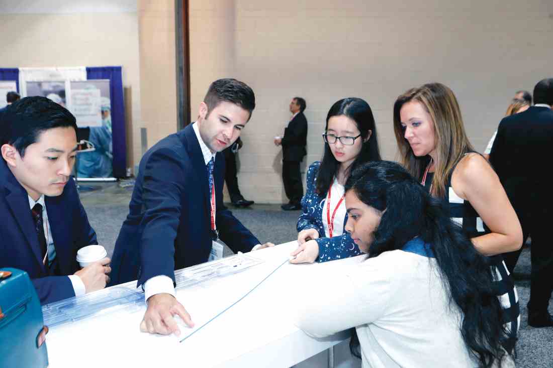
Input a keyword – such as “stents,” for example – and the website provides a listing of exhibitors who manufacture stents. Input “endovascular” and not only are companies listed but so are the Vascular and Endovascular Surgery Society and Vascular News/Charing Cross Symposium. Searches are possible for a specific vendor, as well, and can be refined.
This year’s Exhibit Hall also will include the Office Vascular Care Pavilion, of interest particularly for those clinicians who perform procedures in outpatient and office-based settings. Five vendors currently plan to exhibit at the pavilion, and four presentations for office-based providers are scheduled.
The Exhibit Hall on the lower level also hosts fun and games – literally. It’s the site for the Opening and Closing receptions, on Thursday and Friday evenings, respectively and the Scavenger Hunt. And attendees can also take advantage of non-CME learning opportunities, giveaways, training opportunities and networking potential.
The Opening Reception takes place from 5 to 6:30 p.m., coinciding with the Interactive Poster Session. The Closing Reception is from 4:30 to 5:30 p.m. Friday. It’s a great time to browse the exhibits, talk with vendors, meet up with friends and colleagues, sip on a beverage, and enjoy the food.
Participate in Scavenger Hunt
Games? That would be the Vascular Annual Meeting Scavenger Hunt, sure to be a hit with those with even a bit of a competitive streak. It’s simple and it’s fun. Just download the Mobile App to scan the QR codes found in sponsors’ booths throughout the Exhibit Hall. When a multiple-choice question appears on the display, answer it. Correct answers earn 10 points.
Vascular Live hosts innovative sessions about the latest products and development related to vascular surgery, in a theater-in-the-round setting during Thursday and Friday’s coffee breaks and lunch hours. These frequently are standing-room only, so be sure to arrive early for a good seat. (See the VAM Planner and the mobile app.)
The Exhibit Hall also is the place to be for coffee breaks, 10 a.m. and 3 p.m. Thursday and 9:30 a.m. and 3 p.m. Friday as well as lunch, at 12 p.m. Thursday and 12:15 p.m. Friday. Food stations offer box lunches – free to attendees, though tickets are required. And there are plenty of tables to permit sitting down and chatting with colleagues for a bit.
Industry participation in the VAM exhibits underwrites a significant portion of VAM, thereby allowing us to keep registration fees at a much lower rate than other industry meetings. Please support our industry partners. A complete list of exhibitors and their booth locations are found in the Connections on-site publication and in the Mobile App, VAM Planner and VAM website (vsweb.org/VAM19).
The Exhibit Hall is an integral part of the Vascular Annual Meeting. All members of the vascular team, as well as other attendees, will be able to see a wide-ranging array of products of interest to vascular surgeons and their teams from dozens of vendors.
The VAM website (vsweb.org/VAM19) and mobile app offer a listing of exhibitors. A real-time floor plan (vsweb.org/FloorMap) helps attendees navigate the Exhibit Hall to find the products they most want to see.
Input a keyword – such as “stents,” for example – and the website provides a listing of exhibitors who manufacture stents. Input “endovascular” and not only are companies listed but so are the Vascular and Endovascular Surgery Society and Vascular News/Charing Cross Symposium. Searches are possible for a specific vendor, as well, and can be refined.
This year’s Exhibit Hall also will include the Office Vascular Care Pavilion, of interest particularly for those clinicians who perform procedures in outpatient and office-based settings. Five vendors currently plan to exhibit at the pavilion, and four presentations for office-based providers are scheduled.
The Exhibit Hall on the lower level also hosts fun and games – literally. It’s the site for the Opening and Closing receptions, on Thursday and Friday evenings, respectively and the Scavenger Hunt. And attendees can also take advantage of non-CME learning opportunities, giveaways, training opportunities and networking potential.
The Opening Reception takes place from 5 to 6:30 p.m., coinciding with the Interactive Poster Session. The Closing Reception is from 4:30 to 5:30 p.m. Friday. It’s a great time to browse the exhibits, talk with vendors, meet up with friends and colleagues, sip on a beverage, and enjoy the food.
Participate in Scavenger Hunt
Games? That would be the Vascular Annual Meeting Scavenger Hunt, sure to be a hit with those with even a bit of a competitive streak. It’s simple and it’s fun. Just download the Mobile App to scan the QR codes found in sponsors’ booths throughout the Exhibit Hall. When a multiple-choice question appears on the display, answer it. Correct answers earn 10 points.
Vascular Live hosts innovative sessions about the latest products and development related to vascular surgery, in a theater-in-the-round setting during Thursday and Friday’s coffee breaks and lunch hours. These frequently are standing-room only, so be sure to arrive early for a good seat. (See the VAM Planner and the mobile app.)
The Exhibit Hall also is the place to be for coffee breaks, 10 a.m. and 3 p.m. Thursday and 9:30 a.m. and 3 p.m. Friday as well as lunch, at 12 p.m. Thursday and 12:15 p.m. Friday. Food stations offer box lunches – free to attendees, though tickets are required. And there are plenty of tables to permit sitting down and chatting with colleagues for a bit.
Industry participation in the VAM exhibits underwrites a significant portion of VAM, thereby allowing us to keep registration fees at a much lower rate than other industry meetings. Please support our industry partners. A complete list of exhibitors and their booth locations are found in the Connections on-site publication and in the Mobile App, VAM Planner and VAM website (vsweb.org/VAM19).
The Exhibit Hall is an integral part of the Vascular Annual Meeting. All members of the vascular team, as well as other attendees, will be able to see a wide-ranging array of products of interest to vascular surgeons and their teams from dozens of vendors.
The VAM website (vsweb.org/VAM19) and mobile app offer a listing of exhibitors. A real-time floor plan (vsweb.org/FloorMap) helps attendees navigate the Exhibit Hall to find the products they most want to see.
Input a keyword – such as “stents,” for example – and the website provides a listing of exhibitors who manufacture stents. Input “endovascular” and not only are companies listed but so are the Vascular and Endovascular Surgery Society and Vascular News/Charing Cross Symposium. Searches are possible for a specific vendor, as well, and can be refined.
This year’s Exhibit Hall also will include the Office Vascular Care Pavilion, of interest particularly for those clinicians who perform procedures in outpatient and office-based settings. Five vendors currently plan to exhibit at the pavilion, and four presentations for office-based providers are scheduled.
The Exhibit Hall on the lower level also hosts fun and games – literally. It’s the site for the Opening and Closing receptions, on Thursday and Friday evenings, respectively and the Scavenger Hunt. And attendees can also take advantage of non-CME learning opportunities, giveaways, training opportunities and networking potential.
The Opening Reception takes place from 5 to 6:30 p.m., coinciding with the Interactive Poster Session. The Closing Reception is from 4:30 to 5:30 p.m. Friday. It’s a great time to browse the exhibits, talk with vendors, meet up with friends and colleagues, sip on a beverage, and enjoy the food.
Participate in Scavenger Hunt
Games? That would be the Vascular Annual Meeting Scavenger Hunt, sure to be a hit with those with even a bit of a competitive streak. It’s simple and it’s fun. Just download the Mobile App to scan the QR codes found in sponsors’ booths throughout the Exhibit Hall. When a multiple-choice question appears on the display, answer it. Correct answers earn 10 points.
Vascular Live hosts innovative sessions about the latest products and development related to vascular surgery, in a theater-in-the-round setting during Thursday and Friday’s coffee breaks and lunch hours. These frequently are standing-room only, so be sure to arrive early for a good seat. (See the VAM Planner and the mobile app.)
The Exhibit Hall also is the place to be for coffee breaks, 10 a.m. and 3 p.m. Thursday and 9:30 a.m. and 3 p.m. Friday as well as lunch, at 12 p.m. Thursday and 12:15 p.m. Friday. Food stations offer box lunches – free to attendees, though tickets are required. And there are plenty of tables to permit sitting down and chatting with colleagues for a bit.
Industry participation in the VAM exhibits underwrites a significant portion of VAM, thereby allowing us to keep registration fees at a much lower rate than other industry meetings. Please support our industry partners. A complete list of exhibitors and their booth locations are found in the Connections on-site publication and in the Mobile App, VAM Planner and VAM website (vsweb.org/VAM19).
Children’s anxiety during asthma exacerbations linked to better outcomes
BALTIMORE – according to new research.
“When kids are anxious specifically during their asthma attacks, that can be a good thing because it means that they’re more vigilant,” lead author Jonathan M. Feldman, PhD, of the Albert Einstein College of Medicine’s Children’s Hospital at Montefiore and of Yeshiva University in the New York said in an interview. “They may be more likely to react during the early stages of an attack, and they may be more likely to be using self-management strategies at home and using their controller medications on a daily basis.”
He said pediatric providers can ask their patients with asthma how they feel during asthma attacks, such as whether they ever feel scared or worried.
“If a kid says no, not at all, then I would be concerned as a provider because they may not be paying attention to their asthma symptoms and they may not be taking it seriously,” Dr. Feldman said.
Past research has suggested that “illness-specific panic-fear” – the amount of anxiety someone experiences during asthma exacerbations – helps adults develop adaptive asthma management strategies, so Dr. Feldman and his colleagues examined the phenomenon as a potential protective factor in children. They shared their findings at the annual meeting of the Pediatric Academic Societies.
The research focused on Puerto Rican (n = 79) and Mexican (n = 188) children because of the substantial disparity in asthma prevalence and control between these two different Latino populations. Puerto Rican children have the highest asthma prevalence and morbidity among American children, whereas Mexican children have the lowest rates.
The 267 participants, aged 5-12 years, included 110 children from two inner-city hospitals in the New York and 157 children from two school-based health clinics and a Breathmobile in Phoenix. Nearly all the Arizona children were Mexican, and most (71%) of the Bronx children were Puerto Rican.
The authors collected the following measures at baseline and at 3, 6, 9, and 12 months follow-up: spirometry (forced expiratory volume in 1 second [FEV1]), Childhood Asthma Control Test (CACT) for children 5-11 years old, the Asthma Control Test (ACT) for 12-year-olds, adherence to inhaled corticosteroids (ICS), and acute health care utilizations (clinic sick visits, ED visits, and hospitalizations).
The authors also queried patients on four illness-specific panic-fear measures from the Childhood Asthma Symptoms Checklist: how often they felt frightened, panicky, afraid of being alone, and afraid of dying during an asthma attack (Likert 1-5 scale).
Mexican children reported higher levels of illness-specific panic-fear at the start of the study. They also tended to have lower severity of asthma, better asthma control, and better adherence to ICS, compared with Puerto Rican children.
Also at baseline, the Mexican children’s caregivers tended to be younger, poorer, and more likely to be married and to speak Spanish. The Puerto Rican caregivers, on the other hand, had a higher educational level, including 61% high school graduates, and had more depressive symptoms on the Center for Epidemiologic Studies Depression Scale (CES-D).
One-year data revealed several links between baseline reports of panic-fear and better outcomes. Mexican children who reported experiencing panic-fear at baseline were more likely to have higher FEV1 measures at 1 year of follow-up than were those who didn’t experience panic-fear (P = .02). Similarly, Puerto Rican children initially reporting panic-fear had better asthma control at 1 year, compared with those who didn’t report panic-fear (P = .007).
The researchers reported their effect sizes in terms of predicted variance in a model that accounted for the child’s age, sex, asthma duration, asthma severity, social support, acculturation, health care provider relationship, and number of family members with asthma. The model also factored in the caregiver’s age, sex, marital status, poverty level, education, and depressive symptoms.
For example, in their model, experiencing panic-fear accounted for 67% of the variance in FEV1 levels in Mexican children and 53% of the variance in asthma control in Puerto Rican children.
Less acute health care utilization also was associated with children’s baseline levels of illness-specific panic-fear. In the model, 12% of the variance in acute health care utilization among Mexican children (P = .03) and 41% of the variance among Puerto Rican children (P = .02) was explained by child-reported panic-fear. No association was seen with medication adherence.
Although caregivers’ reports of children feeling panic-fear were linked to better FEV1 outcomes in Mexican children (P = .02), the association was only slightly significant in Puerto Rican children (P = .05). Caregiver reports of children’s panic-fear were not associated with asthma control, acute health care utilization, or medication adherence.
“Providers should be aware that anxiety focused on asthma may be beneficial and facilitate adaptive asthma management strategies,” the authors concluded.
The research was funded by the National Institutes of Health. The authors reported no relevant financial disclosures.
BALTIMORE – according to new research.
“When kids are anxious specifically during their asthma attacks, that can be a good thing because it means that they’re more vigilant,” lead author Jonathan M. Feldman, PhD, of the Albert Einstein College of Medicine’s Children’s Hospital at Montefiore and of Yeshiva University in the New York said in an interview. “They may be more likely to react during the early stages of an attack, and they may be more likely to be using self-management strategies at home and using their controller medications on a daily basis.”
He said pediatric providers can ask their patients with asthma how they feel during asthma attacks, such as whether they ever feel scared or worried.
“If a kid says no, not at all, then I would be concerned as a provider because they may not be paying attention to their asthma symptoms and they may not be taking it seriously,” Dr. Feldman said.
Past research has suggested that “illness-specific panic-fear” – the amount of anxiety someone experiences during asthma exacerbations – helps adults develop adaptive asthma management strategies, so Dr. Feldman and his colleagues examined the phenomenon as a potential protective factor in children. They shared their findings at the annual meeting of the Pediatric Academic Societies.
The research focused on Puerto Rican (n = 79) and Mexican (n = 188) children because of the substantial disparity in asthma prevalence and control between these two different Latino populations. Puerto Rican children have the highest asthma prevalence and morbidity among American children, whereas Mexican children have the lowest rates.
The 267 participants, aged 5-12 years, included 110 children from two inner-city hospitals in the New York and 157 children from two school-based health clinics and a Breathmobile in Phoenix. Nearly all the Arizona children were Mexican, and most (71%) of the Bronx children were Puerto Rican.
The authors collected the following measures at baseline and at 3, 6, 9, and 12 months follow-up: spirometry (forced expiratory volume in 1 second [FEV1]), Childhood Asthma Control Test (CACT) for children 5-11 years old, the Asthma Control Test (ACT) for 12-year-olds, adherence to inhaled corticosteroids (ICS), and acute health care utilizations (clinic sick visits, ED visits, and hospitalizations).
The authors also queried patients on four illness-specific panic-fear measures from the Childhood Asthma Symptoms Checklist: how often they felt frightened, panicky, afraid of being alone, and afraid of dying during an asthma attack (Likert 1-5 scale).
Mexican children reported higher levels of illness-specific panic-fear at the start of the study. They also tended to have lower severity of asthma, better asthma control, and better adherence to ICS, compared with Puerto Rican children.
Also at baseline, the Mexican children’s caregivers tended to be younger, poorer, and more likely to be married and to speak Spanish. The Puerto Rican caregivers, on the other hand, had a higher educational level, including 61% high school graduates, and had more depressive symptoms on the Center for Epidemiologic Studies Depression Scale (CES-D).
One-year data revealed several links between baseline reports of panic-fear and better outcomes. Mexican children who reported experiencing panic-fear at baseline were more likely to have higher FEV1 measures at 1 year of follow-up than were those who didn’t experience panic-fear (P = .02). Similarly, Puerto Rican children initially reporting panic-fear had better asthma control at 1 year, compared with those who didn’t report panic-fear (P = .007).
The researchers reported their effect sizes in terms of predicted variance in a model that accounted for the child’s age, sex, asthma duration, asthma severity, social support, acculturation, health care provider relationship, and number of family members with asthma. The model also factored in the caregiver’s age, sex, marital status, poverty level, education, and depressive symptoms.
For example, in their model, experiencing panic-fear accounted for 67% of the variance in FEV1 levels in Mexican children and 53% of the variance in asthma control in Puerto Rican children.
Less acute health care utilization also was associated with children’s baseline levels of illness-specific panic-fear. In the model, 12% of the variance in acute health care utilization among Mexican children (P = .03) and 41% of the variance among Puerto Rican children (P = .02) was explained by child-reported panic-fear. No association was seen with medication adherence.
Although caregivers’ reports of children feeling panic-fear were linked to better FEV1 outcomes in Mexican children (P = .02), the association was only slightly significant in Puerto Rican children (P = .05). Caregiver reports of children’s panic-fear were not associated with asthma control, acute health care utilization, or medication adherence.
“Providers should be aware that anxiety focused on asthma may be beneficial and facilitate adaptive asthma management strategies,” the authors concluded.
The research was funded by the National Institutes of Health. The authors reported no relevant financial disclosures.
BALTIMORE – according to new research.
“When kids are anxious specifically during their asthma attacks, that can be a good thing because it means that they’re more vigilant,” lead author Jonathan M. Feldman, PhD, of the Albert Einstein College of Medicine’s Children’s Hospital at Montefiore and of Yeshiva University in the New York said in an interview. “They may be more likely to react during the early stages of an attack, and they may be more likely to be using self-management strategies at home and using their controller medications on a daily basis.”
He said pediatric providers can ask their patients with asthma how they feel during asthma attacks, such as whether they ever feel scared or worried.
“If a kid says no, not at all, then I would be concerned as a provider because they may not be paying attention to their asthma symptoms and they may not be taking it seriously,” Dr. Feldman said.
Past research has suggested that “illness-specific panic-fear” – the amount of anxiety someone experiences during asthma exacerbations – helps adults develop adaptive asthma management strategies, so Dr. Feldman and his colleagues examined the phenomenon as a potential protective factor in children. They shared their findings at the annual meeting of the Pediatric Academic Societies.
The research focused on Puerto Rican (n = 79) and Mexican (n = 188) children because of the substantial disparity in asthma prevalence and control between these two different Latino populations. Puerto Rican children have the highest asthma prevalence and morbidity among American children, whereas Mexican children have the lowest rates.
The 267 participants, aged 5-12 years, included 110 children from two inner-city hospitals in the New York and 157 children from two school-based health clinics and a Breathmobile in Phoenix. Nearly all the Arizona children were Mexican, and most (71%) of the Bronx children were Puerto Rican.
The authors collected the following measures at baseline and at 3, 6, 9, and 12 months follow-up: spirometry (forced expiratory volume in 1 second [FEV1]), Childhood Asthma Control Test (CACT) for children 5-11 years old, the Asthma Control Test (ACT) for 12-year-olds, adherence to inhaled corticosteroids (ICS), and acute health care utilizations (clinic sick visits, ED visits, and hospitalizations).
The authors also queried patients on four illness-specific panic-fear measures from the Childhood Asthma Symptoms Checklist: how often they felt frightened, panicky, afraid of being alone, and afraid of dying during an asthma attack (Likert 1-5 scale).
Mexican children reported higher levels of illness-specific panic-fear at the start of the study. They also tended to have lower severity of asthma, better asthma control, and better adherence to ICS, compared with Puerto Rican children.
Also at baseline, the Mexican children’s caregivers tended to be younger, poorer, and more likely to be married and to speak Spanish. The Puerto Rican caregivers, on the other hand, had a higher educational level, including 61% high school graduates, and had more depressive symptoms on the Center for Epidemiologic Studies Depression Scale (CES-D).
One-year data revealed several links between baseline reports of panic-fear and better outcomes. Mexican children who reported experiencing panic-fear at baseline were more likely to have higher FEV1 measures at 1 year of follow-up than were those who didn’t experience panic-fear (P = .02). Similarly, Puerto Rican children initially reporting panic-fear had better asthma control at 1 year, compared with those who didn’t report panic-fear (P = .007).
The researchers reported their effect sizes in terms of predicted variance in a model that accounted for the child’s age, sex, asthma duration, asthma severity, social support, acculturation, health care provider relationship, and number of family members with asthma. The model also factored in the caregiver’s age, sex, marital status, poverty level, education, and depressive symptoms.
For example, in their model, experiencing panic-fear accounted for 67% of the variance in FEV1 levels in Mexican children and 53% of the variance in asthma control in Puerto Rican children.
Less acute health care utilization also was associated with children’s baseline levels of illness-specific panic-fear. In the model, 12% of the variance in acute health care utilization among Mexican children (P = .03) and 41% of the variance among Puerto Rican children (P = .02) was explained by child-reported panic-fear. No association was seen with medication adherence.
Although caregivers’ reports of children feeling panic-fear were linked to better FEV1 outcomes in Mexican children (P = .02), the association was only slightly significant in Puerto Rican children (P = .05). Caregiver reports of children’s panic-fear were not associated with asthma control, acute health care utilization, or medication adherence.
“Providers should be aware that anxiety focused on asthma may be beneficial and facilitate adaptive asthma management strategies,” the authors concluded.
The research was funded by the National Institutes of Health. The authors reported no relevant financial disclosures.
REPORTING FROM PAS 2019
Can acid suppression prevent recurrent idiopathic gastroduodenal ulcer bleeding?
SAN DIEGO – However, these patients face a considerable risk of overall mortality, despite a low incidence of recurrent GI bleeding.
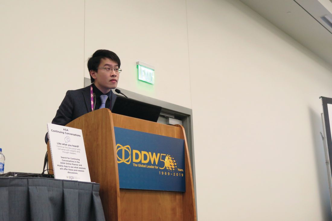
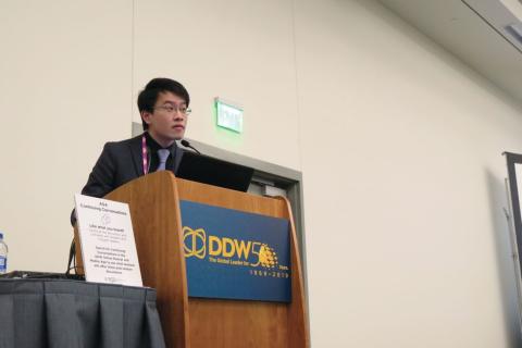
“Peptic ulcer disease is a commonly encountered condition in our daily clinical practice,” one of the study authors, Louis Lau, MD, said at the annual Digestive Disease Week. “Most of the ulcers are due to H. pylori and NSAIDs or aspirin. However, idiopathic peptic ulcers remain an area to be investigated.”
According to Dr. Lau of the division of gastroenterology and hepatology at Prince of Wales Hospital, Hong Kong, H. pylori–negative idiopathic ulcers have increased in prevalence in the Asian Pacific region, from 1%-4% in 2000 to up to 20% in recent data. These ulcers have been shown to have a bleeding rate of up to 13% within 1 year (Gastroenterology. 2005;128:1845-50) and have been associated with a mortality rate of up to 88% in 7 years of follow-up (Gastroenterology. 2009;137:525-31).
“There is no consensus for the optimal management of this particular ulcer,” he continued. “One proposed management was maintenance PPI [proton pump inhibitors]. However, we would like to look for the best optimal prevention strategy.”
Dr. Lau and associates conducted a double-blind, randomized trial at Prince of Wales Hospital to determine if there were any differences between the PPI lansoprazole and the histamine2 receptor antagonist famotidine for the prevention of recurrent bleeding in patients with H. pylori–negative idiopathic peptic ulcers. It involved a 2-year follow-up period and included a screening and end-of-study esophagogastroduodenoscopy. The drugs were repackaged into identical green capsules.
The researchers limited the analysis to 228 patients over the age 18 with H. pylori–negative idiopathic ulcers, defined as having endoscopic biopsies negative for both rapid urease test and histology for H. pylori; no exposure to aspirin, NSAIDs, or drugs of unknown nature including traditional Chinese medicine within 4 weeks; and no other causes of ulceration identified. Exclusion criteria included patients with a strong indication for maintenance therapy with PPIs, those with prior gastric surgery, pregnant patients, and those with advanced malignancy or comorbidity.
The primary endpoint was clinical GI bleeding or reduction in hemoglobin level of 2 g/dL or more and confirmation of peptic ulcers by endoscopy. The secondary endpoint was recurrent ulcer detected by end-of-study endoscopy, with or without clinical symptoms.
Of the 228 patients, 114 received lansoprazole 30 mg daily and 114 received famotidine 40 mg daily. Recurrent upper GI bleeding was suspected in 10 patients in each of the treatment groups. Ulcer confirmation via endoscopy detected one ulcer in the lansoprazole group and three in the famotidine group (0.9% vs. 2.6%, respectively; P = .62). For the secondary endpoint, there were five recurrent ulcers in the lansoprazole group and nine in the famotidine group, (4.4% vs. 7.9%, respectively; P = .27).
Dr. Lau and colleagues observed eight deaths in the lansoprazole group (7%), compared with five (4.4%) in the famotidine group (P = .39), mainly caused by pneumonia, renal failure, and other chronic medical conditions. Intention-to-treat analysis at 2 years found that both groups were comparable in terms of time to analysis of recurrent upper GI bleeding, adjusted for death as a competing risk (Gray’s test, P = .313).
“In this randomized, controlled trial, we found that both a PPI and an H2 blocker are comparable in the prevention of idiopathic ulcer recurrent bleeding,” Dr. Lau concluded. “The rebleeding rate was five times lower than a previous cohort reported with no or irregular acid suppression therapy [Gastroenterology. 2005;128[7]:1845-50]. With all this, there is still a considerable risk of recurrent ulcers and death. We postulate that there is an underlying mechanism for this type of idiopathic ulcer.”
The study was funded in part by the General Research Fund–Research Grant Council. Dr. Lau reported having no financial disclosures. Many of his coauthors reported having numerous financial ties to the pharmaceutical industry.
SOURCE: Lau L et al. DDW 2019, Abstract 322.
SAN DIEGO – However, these patients face a considerable risk of overall mortality, despite a low incidence of recurrent GI bleeding.
“Peptic ulcer disease is a commonly encountered condition in our daily clinical practice,” one of the study authors, Louis Lau, MD, said at the annual Digestive Disease Week. “Most of the ulcers are due to H. pylori and NSAIDs or aspirin. However, idiopathic peptic ulcers remain an area to be investigated.”
According to Dr. Lau of the division of gastroenterology and hepatology at Prince of Wales Hospital, Hong Kong, H. pylori–negative idiopathic ulcers have increased in prevalence in the Asian Pacific region, from 1%-4% in 2000 to up to 20% in recent data. These ulcers have been shown to have a bleeding rate of up to 13% within 1 year (Gastroenterology. 2005;128:1845-50) and have been associated with a mortality rate of up to 88% in 7 years of follow-up (Gastroenterology. 2009;137:525-31).
“There is no consensus for the optimal management of this particular ulcer,” he continued. “One proposed management was maintenance PPI [proton pump inhibitors]. However, we would like to look for the best optimal prevention strategy.”
Dr. Lau and associates conducted a double-blind, randomized trial at Prince of Wales Hospital to determine if there were any differences between the PPI lansoprazole and the histamine2 receptor antagonist famotidine for the prevention of recurrent bleeding in patients with H. pylori–negative idiopathic peptic ulcers. It involved a 2-year follow-up period and included a screening and end-of-study esophagogastroduodenoscopy. The drugs were repackaged into identical green capsules.
The researchers limited the analysis to 228 patients over the age 18 with H. pylori–negative idiopathic ulcers, defined as having endoscopic biopsies negative for both rapid urease test and histology for H. pylori; no exposure to aspirin, NSAIDs, or drugs of unknown nature including traditional Chinese medicine within 4 weeks; and no other causes of ulceration identified. Exclusion criteria included patients with a strong indication for maintenance therapy with PPIs, those with prior gastric surgery, pregnant patients, and those with advanced malignancy or comorbidity.
The primary endpoint was clinical GI bleeding or reduction in hemoglobin level of 2 g/dL or more and confirmation of peptic ulcers by endoscopy. The secondary endpoint was recurrent ulcer detected by end-of-study endoscopy, with or without clinical symptoms.
Of the 228 patients, 114 received lansoprazole 30 mg daily and 114 received famotidine 40 mg daily. Recurrent upper GI bleeding was suspected in 10 patients in each of the treatment groups. Ulcer confirmation via endoscopy detected one ulcer in the lansoprazole group and three in the famotidine group (0.9% vs. 2.6%, respectively; P = .62). For the secondary endpoint, there were five recurrent ulcers in the lansoprazole group and nine in the famotidine group, (4.4% vs. 7.9%, respectively; P = .27).
Dr. Lau and colleagues observed eight deaths in the lansoprazole group (7%), compared with five (4.4%) in the famotidine group (P = .39), mainly caused by pneumonia, renal failure, and other chronic medical conditions. Intention-to-treat analysis at 2 years found that both groups were comparable in terms of time to analysis of recurrent upper GI bleeding, adjusted for death as a competing risk (Gray’s test, P = .313).
“In this randomized, controlled trial, we found that both a PPI and an H2 blocker are comparable in the prevention of idiopathic ulcer recurrent bleeding,” Dr. Lau concluded. “The rebleeding rate was five times lower than a previous cohort reported with no or irregular acid suppression therapy [Gastroenterology. 2005;128[7]:1845-50]. With all this, there is still a considerable risk of recurrent ulcers and death. We postulate that there is an underlying mechanism for this type of idiopathic ulcer.”
The study was funded in part by the General Research Fund–Research Grant Council. Dr. Lau reported having no financial disclosures. Many of his coauthors reported having numerous financial ties to the pharmaceutical industry.
SOURCE: Lau L et al. DDW 2019, Abstract 322.
SAN DIEGO – However, these patients face a considerable risk of overall mortality, despite a low incidence of recurrent GI bleeding.
“Peptic ulcer disease is a commonly encountered condition in our daily clinical practice,” one of the study authors, Louis Lau, MD, said at the annual Digestive Disease Week. “Most of the ulcers are due to H. pylori and NSAIDs or aspirin. However, idiopathic peptic ulcers remain an area to be investigated.”
According to Dr. Lau of the division of gastroenterology and hepatology at Prince of Wales Hospital, Hong Kong, H. pylori–negative idiopathic ulcers have increased in prevalence in the Asian Pacific region, from 1%-4% in 2000 to up to 20% in recent data. These ulcers have been shown to have a bleeding rate of up to 13% within 1 year (Gastroenterology. 2005;128:1845-50) and have been associated with a mortality rate of up to 88% in 7 years of follow-up (Gastroenterology. 2009;137:525-31).
“There is no consensus for the optimal management of this particular ulcer,” he continued. “One proposed management was maintenance PPI [proton pump inhibitors]. However, we would like to look for the best optimal prevention strategy.”
Dr. Lau and associates conducted a double-blind, randomized trial at Prince of Wales Hospital to determine if there were any differences between the PPI lansoprazole and the histamine2 receptor antagonist famotidine for the prevention of recurrent bleeding in patients with H. pylori–negative idiopathic peptic ulcers. It involved a 2-year follow-up period and included a screening and end-of-study esophagogastroduodenoscopy. The drugs were repackaged into identical green capsules.
The researchers limited the analysis to 228 patients over the age 18 with H. pylori–negative idiopathic ulcers, defined as having endoscopic biopsies negative for both rapid urease test and histology for H. pylori; no exposure to aspirin, NSAIDs, or drugs of unknown nature including traditional Chinese medicine within 4 weeks; and no other causes of ulceration identified. Exclusion criteria included patients with a strong indication for maintenance therapy with PPIs, those with prior gastric surgery, pregnant patients, and those with advanced malignancy or comorbidity.
The primary endpoint was clinical GI bleeding or reduction in hemoglobin level of 2 g/dL or more and confirmation of peptic ulcers by endoscopy. The secondary endpoint was recurrent ulcer detected by end-of-study endoscopy, with or without clinical symptoms.
Of the 228 patients, 114 received lansoprazole 30 mg daily and 114 received famotidine 40 mg daily. Recurrent upper GI bleeding was suspected in 10 patients in each of the treatment groups. Ulcer confirmation via endoscopy detected one ulcer in the lansoprazole group and three in the famotidine group (0.9% vs. 2.6%, respectively; P = .62). For the secondary endpoint, there were five recurrent ulcers in the lansoprazole group and nine in the famotidine group, (4.4% vs. 7.9%, respectively; P = .27).
Dr. Lau and colleagues observed eight deaths in the lansoprazole group (7%), compared with five (4.4%) in the famotidine group (P = .39), mainly caused by pneumonia, renal failure, and other chronic medical conditions. Intention-to-treat analysis at 2 years found that both groups were comparable in terms of time to analysis of recurrent upper GI bleeding, adjusted for death as a competing risk (Gray’s test, P = .313).
“In this randomized, controlled trial, we found that both a PPI and an H2 blocker are comparable in the prevention of idiopathic ulcer recurrent bleeding,” Dr. Lau concluded. “The rebleeding rate was five times lower than a previous cohort reported with no or irregular acid suppression therapy [Gastroenterology. 2005;128[7]:1845-50]. With all this, there is still a considerable risk of recurrent ulcers and death. We postulate that there is an underlying mechanism for this type of idiopathic ulcer.”
The study was funded in part by the General Research Fund–Research Grant Council. Dr. Lau reported having no financial disclosures. Many of his coauthors reported having numerous financial ties to the pharmaceutical industry.
SOURCE: Lau L et al. DDW 2019, Abstract 322.
REPORTING FROM DDW 2019
Focus on Office-Based Care
This year’s VAM features an exhibit pavilion, special Vascular Live presentations, and a breakfast session geared specifically to clinicians who work in office and outpatient settings.
The Office Vascular Care Pavilion is in Exhibit Hall B, on the lower level of the Gaylord National Resort & Convention Center, across from the SVS Booth. Office Vascular Care Live presentations (not eligible for CME credit) include:
Thursday, June 13
12:30 to 1 p.m. OBL 101; Krishna M. Jain, MD
3 to 3:30 p.m. OBL Chatter, industry presentations by Cordis®, A Cardinal Health company, Philips and Vein Care iGuide
Sessions include: Coming to a town near you! Life Cycle of an OBL, Factors Influencing Behavior of an OBL, and Industry Partnership, sponsored by Cordis®, A Cardinal Health company
Keys to Success and Methods of Failure in Today’s OBL, David Baker, sponsored by Philips
Friday, June 14
12:30 to 1 p.m. OBL Tips and Tools; R. Clement Darling III, MD
3 to 3:30 p.m. OBL Quality and Safety; Robert G. Molnar, MD
8:30 to 9:30 a.m. Ticket Required - The SVS also has a new member section focused on the move to outpatient and office-based settings, the Section on Outpatient and Office Vascular Care (SOOVC), which will hold its first meeting Friday in National Harbor 3. Tickets are available at Registration. Prospective members are welcome.
Saturday, June 15
6:30 to 8 a.m. Breakfast Session 9 will cover “Complications in Office-Based Procedures: Their Prevention and Management.” It is being presented in collaboration with the Outpatient Endovascular and Interventional Society.
This year’s VAM features an exhibit pavilion, special Vascular Live presentations, and a breakfast session geared specifically to clinicians who work in office and outpatient settings.
The Office Vascular Care Pavilion is in Exhibit Hall B, on the lower level of the Gaylord National Resort & Convention Center, across from the SVS Booth. Office Vascular Care Live presentations (not eligible for CME credit) include:
Thursday, June 13
12:30 to 1 p.m. OBL 101; Krishna M. Jain, MD
3 to 3:30 p.m. OBL Chatter, industry presentations by Cordis®, A Cardinal Health company, Philips and Vein Care iGuide
Sessions include: Coming to a town near you! Life Cycle of an OBL, Factors Influencing Behavior of an OBL, and Industry Partnership, sponsored by Cordis®, A Cardinal Health company
Keys to Success and Methods of Failure in Today’s OBL, David Baker, sponsored by Philips
Friday, June 14
12:30 to 1 p.m. OBL Tips and Tools; R. Clement Darling III, MD
3 to 3:30 p.m. OBL Quality and Safety; Robert G. Molnar, MD
8:30 to 9:30 a.m. Ticket Required - The SVS also has a new member section focused on the move to outpatient and office-based settings, the Section on Outpatient and Office Vascular Care (SOOVC), which will hold its first meeting Friday in National Harbor 3. Tickets are available at Registration. Prospective members are welcome.
Saturday, June 15
6:30 to 8 a.m. Breakfast Session 9 will cover “Complications in Office-Based Procedures: Their Prevention and Management.” It is being presented in collaboration with the Outpatient Endovascular and Interventional Society.
This year’s VAM features an exhibit pavilion, special Vascular Live presentations, and a breakfast session geared specifically to clinicians who work in office and outpatient settings.
The Office Vascular Care Pavilion is in Exhibit Hall B, on the lower level of the Gaylord National Resort & Convention Center, across from the SVS Booth. Office Vascular Care Live presentations (not eligible for CME credit) include:
Thursday, June 13
12:30 to 1 p.m. OBL 101; Krishna M. Jain, MD
3 to 3:30 p.m. OBL Chatter, industry presentations by Cordis®, A Cardinal Health company, Philips and Vein Care iGuide
Sessions include: Coming to a town near you! Life Cycle of an OBL, Factors Influencing Behavior of an OBL, and Industry Partnership, sponsored by Cordis®, A Cardinal Health company
Keys to Success and Methods of Failure in Today’s OBL, David Baker, sponsored by Philips
Friday, June 14
12:30 to 1 p.m. OBL Tips and Tools; R. Clement Darling III, MD
3 to 3:30 p.m. OBL Quality and Safety; Robert G. Molnar, MD
8:30 to 9:30 a.m. Ticket Required - The SVS also has a new member section focused on the move to outpatient and office-based settings, the Section on Outpatient and Office Vascular Care (SOOVC), which will hold its first meeting Friday in National Harbor 3. Tickets are available at Registration. Prospective members are welcome.
Saturday, June 15
6:30 to 8 a.m. Breakfast Session 9 will cover “Complications in Office-Based Procedures: Their Prevention and Management.” It is being presented in collaboration with the Outpatient Endovascular and Interventional Society.
Reappraisal in emotion regulation unrelated to working memory in bipolar disorder
Working memory may not function properly during the reappraisal process of emotion regulation in patients with bipolar disorder, according to Dong Hun Oh of the department of psychiatry at Yonsei University, Seoul, South Korea, and associates.
For the study, published in the Journal of Affective Disorders, 43 patients with euthymic bipolar I disorder were recruited from a psychiatric outpatient clinic in Seoul and compared with 48 healthy controls. The Korean versions of the operation span task (OSPAN), emotion regulation questionnaire (K-ERQ), ruminative response scale (K-RRS), and Difficulties in Emotion Regulation Scale (K-DERS) were administered to all the patients.
In a between-group comparison of scores on the four scales measured, patients with bipolar disorder had significantly lower scores on OSPAN (P = .031), on the brooding section of the K-RRS (P =.016), and on the nonacceptance section of the K-DERS (P = .039). In a regression analysis of the interaction between working memory and the four components of emotion regulation (reappraisal, expressive suppression, brooding, and reflective pondering), a significant interaction was found in OSPAN scores for reappraisal between healthy controls and the BD group (P = .007). Brooding scores were significantly lower in the control group, but the interaction was not significant.
A simple slope analysis showed that, while working memory was worse in patients with bipolar disorder, a relationship between cognitive capacity and the efficacy of reappraisal was found only in healthy controls, the investigators noted.
“The absence of interaction between [working memory capacity] and reappraisal in euthymic [bipolar disorder] patients that we report may indicate that the positive effects of cognitive remediation or [working memory] training previously reported for healthy people, may not effectively improve [emotion regulation] for patients with [bipolar disorder],” they wrote.
Furthermore, if this is the case, cognitive interventions aimed at improving emotion regulation in patients with bipolar disorder might not work.
The investigators cited several limitations. One limitation was the small sample size; another was the use of self-administered questionnaires.
The study was supported by a faculty research grant from Yonsei University. The authors did not report any conflicts of interest.
SOURCE: Oh DH et al. J Affect Disord. 2019 Apr 8. doi: 10.1016/j.jad.2019.04.042.
Working memory may not function properly during the reappraisal process of emotion regulation in patients with bipolar disorder, according to Dong Hun Oh of the department of psychiatry at Yonsei University, Seoul, South Korea, and associates.
For the study, published in the Journal of Affective Disorders, 43 patients with euthymic bipolar I disorder were recruited from a psychiatric outpatient clinic in Seoul and compared with 48 healthy controls. The Korean versions of the operation span task (OSPAN), emotion regulation questionnaire (K-ERQ), ruminative response scale (K-RRS), and Difficulties in Emotion Regulation Scale (K-DERS) were administered to all the patients.
In a between-group comparison of scores on the four scales measured, patients with bipolar disorder had significantly lower scores on OSPAN (P = .031), on the brooding section of the K-RRS (P =.016), and on the nonacceptance section of the K-DERS (P = .039). In a regression analysis of the interaction between working memory and the four components of emotion regulation (reappraisal, expressive suppression, brooding, and reflective pondering), a significant interaction was found in OSPAN scores for reappraisal between healthy controls and the BD group (P = .007). Brooding scores were significantly lower in the control group, but the interaction was not significant.
A simple slope analysis showed that, while working memory was worse in patients with bipolar disorder, a relationship between cognitive capacity and the efficacy of reappraisal was found only in healthy controls, the investigators noted.
“The absence of interaction between [working memory capacity] and reappraisal in euthymic [bipolar disorder] patients that we report may indicate that the positive effects of cognitive remediation or [working memory] training previously reported for healthy people, may not effectively improve [emotion regulation] for patients with [bipolar disorder],” they wrote.
Furthermore, if this is the case, cognitive interventions aimed at improving emotion regulation in patients with bipolar disorder might not work.
The investigators cited several limitations. One limitation was the small sample size; another was the use of self-administered questionnaires.
The study was supported by a faculty research grant from Yonsei University. The authors did not report any conflicts of interest.
SOURCE: Oh DH et al. J Affect Disord. 2019 Apr 8. doi: 10.1016/j.jad.2019.04.042.
Working memory may not function properly during the reappraisal process of emotion regulation in patients with bipolar disorder, according to Dong Hun Oh of the department of psychiatry at Yonsei University, Seoul, South Korea, and associates.
For the study, published in the Journal of Affective Disorders, 43 patients with euthymic bipolar I disorder were recruited from a psychiatric outpatient clinic in Seoul and compared with 48 healthy controls. The Korean versions of the operation span task (OSPAN), emotion regulation questionnaire (K-ERQ), ruminative response scale (K-RRS), and Difficulties in Emotion Regulation Scale (K-DERS) were administered to all the patients.
In a between-group comparison of scores on the four scales measured, patients with bipolar disorder had significantly lower scores on OSPAN (P = .031), on the brooding section of the K-RRS (P =.016), and on the nonacceptance section of the K-DERS (P = .039). In a regression analysis of the interaction between working memory and the four components of emotion regulation (reappraisal, expressive suppression, brooding, and reflective pondering), a significant interaction was found in OSPAN scores for reappraisal between healthy controls and the BD group (P = .007). Brooding scores were significantly lower in the control group, but the interaction was not significant.
A simple slope analysis showed that, while working memory was worse in patients with bipolar disorder, a relationship between cognitive capacity and the efficacy of reappraisal was found only in healthy controls, the investigators noted.
“The absence of interaction between [working memory capacity] and reappraisal in euthymic [bipolar disorder] patients that we report may indicate that the positive effects of cognitive remediation or [working memory] training previously reported for healthy people, may not effectively improve [emotion regulation] for patients with [bipolar disorder],” they wrote.
Furthermore, if this is the case, cognitive interventions aimed at improving emotion regulation in patients with bipolar disorder might not work.
The investigators cited several limitations. One limitation was the small sample size; another was the use of self-administered questionnaires.
The study was supported by a faculty research grant from Yonsei University. The authors did not report any conflicts of interest.
SOURCE: Oh DH et al. J Affect Disord. 2019 Apr 8. doi: 10.1016/j.jad.2019.04.042.
FROM THE JOURNAL OF AFFECTIVE DISORDERS
Macrophages, Regulation, and Impaired Diabetic Wound Healing
Nonhealing wounds in patients with diabetes are a major cause of morbidity and mortality in the United States and are increasing at an alarming rate. Equally concerning, the current “standard of care” leaves 70% of diabetic wounds unhealed. Given this substantial impact on patient outcomes and health care expenditure, a critical unmet need exists for improved understanding of the pathophysiology of diabetic wounds to develop effective treatments.

This year’s SVS Foundation’s Resident Research Award is being presented to Frank M. Davis, MD, of the University of Michigan, Ann Arbor, for his research on the epigenetic regulation of the prostaglandin pathway in macrophages during type 2 diabetic wound healing. Dr. Davis, and his mentor Katherine Gallagher, MD, also from the University of Michigan, investigated how impairments in the innate immune system in patients with diabetes promote chronic inflammation and impair wound healing.
Dr. Davis will present his award-winning research in the von Liebig Forum, Thursday, June 13, discussing the role of specific epigenetic enzymes in the dictation of macrophage phenotype in wound tissue. The talk will cover how diabetes alters those enzymes to influence a deleterious phenotype that promotes inflammation and delays wound healing. “Our laboratory specifically looks at the role of monocytes/macrophages in the inflammatory phase of wound healing and how perturbation in the local environment – such as those seen in diabetes – affects monocyte/macrophage phenotype and ultimately wound healing” said Dr. Davis.

The talk will specifically cover the cyclo-oxygenase (COX)-2/prostaglandin E2 (PGE2) axis. Using both a murine model and human wound samples Dr. Davis demonstrates that PGE2 is substantially elevated in diabetic wound macrophages. Further, aberrant PGE2 production in diabetic macrophages depended on epigenetic alterations to key enzymes in the PGE2 pathway.
Specifically, MLL1, a histone methyltransferase, increased H3K4 trimethylation resulting in upregulation of PGE2 in diabetic wound macrophages. Additionally, the authors found that augmentation to miR-29b and DNA methyltransferases in diabetic macrophage result in increased COX-2 expression. Overall, the increased COX-2/PGE2 production in diabetic macrophages impairs bacterial killing, predisposing diabetic wounds to chronic infection.
“Our research provides insight into the prostaglandin E2 axis and its role in macrophage inflammation, which has previously been an unrecognized pathway leading to delayed diabetic wound healing” added Dr. Gallagher.
Finally, in his presentation, Dr. Davis will discuss translational therapies as inhibition of the PGE2 pathway through macrophage targeted nanoparticles decreased diabetic inflammation and improved healing. “Together, our results indicate the COX-2/PGE2 pathway is a critical regulator of macrophage phenotype and impaired diabetic wound healing. This work identifies therapeutic targets for negating dysregulated inflammation in diabetic wounds and identifies macrophage-targeted local therapy as an effective means of improving wound healing,” Dr. Davis concluded.
Nonhealing wounds in patients with diabetes are a major cause of morbidity and mortality in the United States and are increasing at an alarming rate. Equally concerning, the current “standard of care” leaves 70% of diabetic wounds unhealed. Given this substantial impact on patient outcomes and health care expenditure, a critical unmet need exists for improved understanding of the pathophysiology of diabetic wounds to develop effective treatments.
This year’s SVS Foundation’s Resident Research Award is being presented to Frank M. Davis, MD, of the University of Michigan, Ann Arbor, for his research on the epigenetic regulation of the prostaglandin pathway in macrophages during type 2 diabetic wound healing. Dr. Davis, and his mentor Katherine Gallagher, MD, also from the University of Michigan, investigated how impairments in the innate immune system in patients with diabetes promote chronic inflammation and impair wound healing.
Dr. Davis will present his award-winning research in the von Liebig Forum, Thursday, June 13, discussing the role of specific epigenetic enzymes in the dictation of macrophage phenotype in wound tissue. The talk will cover how diabetes alters those enzymes to influence a deleterious phenotype that promotes inflammation and delays wound healing. “Our laboratory specifically looks at the role of monocytes/macrophages in the inflammatory phase of wound healing and how perturbation in the local environment – such as those seen in diabetes – affects monocyte/macrophage phenotype and ultimately wound healing” said Dr. Davis.
The talk will specifically cover the cyclo-oxygenase (COX)-2/prostaglandin E2 (PGE2) axis. Using both a murine model and human wound samples Dr. Davis demonstrates that PGE2 is substantially elevated in diabetic wound macrophages. Further, aberrant PGE2 production in diabetic macrophages depended on epigenetic alterations to key enzymes in the PGE2 pathway.
Specifically, MLL1, a histone methyltransferase, increased H3K4 trimethylation resulting in upregulation of PGE2 in diabetic wound macrophages. Additionally, the authors found that augmentation to miR-29b and DNA methyltransferases in diabetic macrophage result in increased COX-2 expression. Overall, the increased COX-2/PGE2 production in diabetic macrophages impairs bacterial killing, predisposing diabetic wounds to chronic infection.
“Our research provides insight into the prostaglandin E2 axis and its role in macrophage inflammation, which has previously been an unrecognized pathway leading to delayed diabetic wound healing” added Dr. Gallagher.
Finally, in his presentation, Dr. Davis will discuss translational therapies as inhibition of the PGE2 pathway through macrophage targeted nanoparticles decreased diabetic inflammation and improved healing. “Together, our results indicate the COX-2/PGE2 pathway is a critical regulator of macrophage phenotype and impaired diabetic wound healing. This work identifies therapeutic targets for negating dysregulated inflammation in diabetic wounds and identifies macrophage-targeted local therapy as an effective means of improving wound healing,” Dr. Davis concluded.
Nonhealing wounds in patients with diabetes are a major cause of morbidity and mortality in the United States and are increasing at an alarming rate. Equally concerning, the current “standard of care” leaves 70% of diabetic wounds unhealed. Given this substantial impact on patient outcomes and health care expenditure, a critical unmet need exists for improved understanding of the pathophysiology of diabetic wounds to develop effective treatments.
This year’s SVS Foundation’s Resident Research Award is being presented to Frank M. Davis, MD, of the University of Michigan, Ann Arbor, for his research on the epigenetic regulation of the prostaglandin pathway in macrophages during type 2 diabetic wound healing. Dr. Davis, and his mentor Katherine Gallagher, MD, also from the University of Michigan, investigated how impairments in the innate immune system in patients with diabetes promote chronic inflammation and impair wound healing.
Dr. Davis will present his award-winning research in the von Liebig Forum, Thursday, June 13, discussing the role of specific epigenetic enzymes in the dictation of macrophage phenotype in wound tissue. The talk will cover how diabetes alters those enzymes to influence a deleterious phenotype that promotes inflammation and delays wound healing. “Our laboratory specifically looks at the role of monocytes/macrophages in the inflammatory phase of wound healing and how perturbation in the local environment – such as those seen in diabetes – affects monocyte/macrophage phenotype and ultimately wound healing” said Dr. Davis.
The talk will specifically cover the cyclo-oxygenase (COX)-2/prostaglandin E2 (PGE2) axis. Using both a murine model and human wound samples Dr. Davis demonstrates that PGE2 is substantially elevated in diabetic wound macrophages. Further, aberrant PGE2 production in diabetic macrophages depended on epigenetic alterations to key enzymes in the PGE2 pathway.
Specifically, MLL1, a histone methyltransferase, increased H3K4 trimethylation resulting in upregulation of PGE2 in diabetic wound macrophages. Additionally, the authors found that augmentation to miR-29b and DNA methyltransferases in diabetic macrophage result in increased COX-2 expression. Overall, the increased COX-2/PGE2 production in diabetic macrophages impairs bacterial killing, predisposing diabetic wounds to chronic infection.
“Our research provides insight into the prostaglandin E2 axis and its role in macrophage inflammation, which has previously been an unrecognized pathway leading to delayed diabetic wound healing” added Dr. Gallagher.
Finally, in his presentation, Dr. Davis will discuss translational therapies as inhibition of the PGE2 pathway through macrophage targeted nanoparticles decreased diabetic inflammation and improved healing. “Together, our results indicate the COX-2/PGE2 pathway is a critical regulator of macrophage phenotype and impaired diabetic wound healing. This work identifies therapeutic targets for negating dysregulated inflammation in diabetic wounds and identifies macrophage-targeted local therapy as an effective means of improving wound healing,” Dr. Davis concluded.

Some Brits snuff out TORCH screen to raise awareness of congenital syphilis
LJUBLJANA, SLOVENIA – Pediatricians in the south of England are so concerned about the recent national increase in the diagnosis of syphilis in adults and its ramifications for neonates that they’ve ditched the traditional TORCH newborn screen because the acronym doesn’t specifically remind clinicians to think about congenital syphilis, Mildred A. Iro, MD, PhD, said at the annual meeting of the European Society for Paediatric Infectious Diseases.

“ explained Dr. Iro of the University of Southampton (England).
She highlighted salient features of three recent cases of congenital syphilis managed at Southampton Children’s Hospital.
“The key message that we’d like to share is that we just need to be more aware about congenital syphilis. Retest mothers if their risk factor status changes, and test suspected infants and children,” Dr. Iro said.
As a practical matter, however, even though current guidelines recommend retesting mothers whose risk factor status becomes heightened following an initial negative syphilis serology result early in pregnancy, clinicians often are unaware that a mother’s risk status has changed. And retesting all mothers during pregnancy isn’t attractive from a cost-benefit standpoint. This makes scrupulous screening of newborns all the more important. And yet TORCH, which stands for Toxoplasmosis, Other, Rubella, Cytomegalovirus, and Herpes infections, isn’t an acronym that promotes awareness of congenital syphilis, a disease which occupies an obscure position in TORCH under the “O” for “Other” heading. That’s why the term “congenital infection screen” has become the new norm in the south of England, she explained.
However, one pediatrician who didn’t consider congenital infection screen to be an improvement in terminology over TORCH had an alternative suggestion, which struck a favorable chord with his fellow audience members: Simply change the acronym to TORCHS, with the S standing for syphilis.
Dr. Iro noted that two of the three affected children were diagnosed at age 7-8 weeks. The third wasn’t diagnosed until age 15 months, when the mother tested positive for syphilis in a subsequent pregnancy. As is typical of the disease known as “the great masquerader,” while all three of the affected children were unwell early in infancy, they presented with a wide range of symptoms. Among the more prominent features were prolonged irritability, respiratory distress, odd rashes, anemia, hepatomegaly, and tachypnea. One infant had reduced movement and pain in one arm.
All three children underwent extensive testing. None had neurosyphilis. All achieved good outcomes on standard guideline-directed therapy.
As for the mothers, they were aged 19, 21, and 23 years when diagnosed with syphilis. All were Caucasian, and antenatal blood testing was negative in all three. None were retested during pregnancy, even though two of them had a male partner or former partner who was positive for syphilis, and the partner of the third disclosed to her that he had sex with men.
At diagnosis, all three women had a strongly positive Treponema pallidum particle agglutination assay, a high rapid plasma reagin, and a positive syphilis IgM assay.
Dr. Iro reported having no financial conflicts regarding her presentation.
LJUBLJANA, SLOVENIA – Pediatricians in the south of England are so concerned about the recent national increase in the diagnosis of syphilis in adults and its ramifications for neonates that they’ve ditched the traditional TORCH newborn screen because the acronym doesn’t specifically remind clinicians to think about congenital syphilis, Mildred A. Iro, MD, PhD, said at the annual meeting of the European Society for Paediatric Infectious Diseases.
“ explained Dr. Iro of the University of Southampton (England).
She highlighted salient features of three recent cases of congenital syphilis managed at Southampton Children’s Hospital.
“The key message that we’d like to share is that we just need to be more aware about congenital syphilis. Retest mothers if their risk factor status changes, and test suspected infants and children,” Dr. Iro said.
As a practical matter, however, even though current guidelines recommend retesting mothers whose risk factor status becomes heightened following an initial negative syphilis serology result early in pregnancy, clinicians often are unaware that a mother’s risk status has changed. And retesting all mothers during pregnancy isn’t attractive from a cost-benefit standpoint. This makes scrupulous screening of newborns all the more important. And yet TORCH, which stands for Toxoplasmosis, Other, Rubella, Cytomegalovirus, and Herpes infections, isn’t an acronym that promotes awareness of congenital syphilis, a disease which occupies an obscure position in TORCH under the “O” for “Other” heading. That’s why the term “congenital infection screen” has become the new norm in the south of England, she explained.
However, one pediatrician who didn’t consider congenital infection screen to be an improvement in terminology over TORCH had an alternative suggestion, which struck a favorable chord with his fellow audience members: Simply change the acronym to TORCHS, with the S standing for syphilis.
Dr. Iro noted that two of the three affected children were diagnosed at age 7-8 weeks. The third wasn’t diagnosed until age 15 months, when the mother tested positive for syphilis in a subsequent pregnancy. As is typical of the disease known as “the great masquerader,” while all three of the affected children were unwell early in infancy, they presented with a wide range of symptoms. Among the more prominent features were prolonged irritability, respiratory distress, odd rashes, anemia, hepatomegaly, and tachypnea. One infant had reduced movement and pain in one arm.
All three children underwent extensive testing. None had neurosyphilis. All achieved good outcomes on standard guideline-directed therapy.
As for the mothers, they were aged 19, 21, and 23 years when diagnosed with syphilis. All were Caucasian, and antenatal blood testing was negative in all three. None were retested during pregnancy, even though two of them had a male partner or former partner who was positive for syphilis, and the partner of the third disclosed to her that he had sex with men.
At diagnosis, all three women had a strongly positive Treponema pallidum particle agglutination assay, a high rapid plasma reagin, and a positive syphilis IgM assay.
Dr. Iro reported having no financial conflicts regarding her presentation.
LJUBLJANA, SLOVENIA – Pediatricians in the south of England are so concerned about the recent national increase in the diagnosis of syphilis in adults and its ramifications for neonates that they’ve ditched the traditional TORCH newborn screen because the acronym doesn’t specifically remind clinicians to think about congenital syphilis, Mildred A. Iro, MD, PhD, said at the annual meeting of the European Society for Paediatric Infectious Diseases.
“ explained Dr. Iro of the University of Southampton (England).
She highlighted salient features of three recent cases of congenital syphilis managed at Southampton Children’s Hospital.
“The key message that we’d like to share is that we just need to be more aware about congenital syphilis. Retest mothers if their risk factor status changes, and test suspected infants and children,” Dr. Iro said.
As a practical matter, however, even though current guidelines recommend retesting mothers whose risk factor status becomes heightened following an initial negative syphilis serology result early in pregnancy, clinicians often are unaware that a mother’s risk status has changed. And retesting all mothers during pregnancy isn’t attractive from a cost-benefit standpoint. This makes scrupulous screening of newborns all the more important. And yet TORCH, which stands for Toxoplasmosis, Other, Rubella, Cytomegalovirus, and Herpes infections, isn’t an acronym that promotes awareness of congenital syphilis, a disease which occupies an obscure position in TORCH under the “O” for “Other” heading. That’s why the term “congenital infection screen” has become the new norm in the south of England, she explained.
However, one pediatrician who didn’t consider congenital infection screen to be an improvement in terminology over TORCH had an alternative suggestion, which struck a favorable chord with his fellow audience members: Simply change the acronym to TORCHS, with the S standing for syphilis.
Dr. Iro noted that two of the three affected children were diagnosed at age 7-8 weeks. The third wasn’t diagnosed until age 15 months, when the mother tested positive for syphilis in a subsequent pregnancy. As is typical of the disease known as “the great masquerader,” while all three of the affected children were unwell early in infancy, they presented with a wide range of symptoms. Among the more prominent features were prolonged irritability, respiratory distress, odd rashes, anemia, hepatomegaly, and tachypnea. One infant had reduced movement and pain in one arm.
All three children underwent extensive testing. None had neurosyphilis. All achieved good outcomes on standard guideline-directed therapy.
As for the mothers, they were aged 19, 21, and 23 years when diagnosed with syphilis. All were Caucasian, and antenatal blood testing was negative in all three. None were retested during pregnancy, even though two of them had a male partner or former partner who was positive for syphilis, and the partner of the third disclosed to her that he had sex with men.
At diagnosis, all three women had a strongly positive Treponema pallidum particle agglutination assay, a high rapid plasma reagin, and a positive syphilis IgM assay.
Dr. Iro reported having no financial conflicts regarding her presentation.
EXPERT ANALYSIS FROM ESPID 2019

A longing for belonging
As I watched my grandson and his team warm up for their Saturday morning lacrosse game, a long parade of mostly purple-shirted adults and children of all ages began to weave its way around the periphery of the athletic field complex. A quick reading of the hand-lettered and machine-printed shirts made it clear that I was watching a charity walk for cystic fibrosis. There must have been several hundred walkers strolling by, laughing and chatting with one another. It lent a festive atmosphere to the park. I suspect that for most of the participants this was not their first fundraising event for cystic fibrosis.

The motley mix of marchers probably included several handfuls of parents of children with cystic fibrosis. I wonder how many of those parents realized how fortunate they were. Cystic fibrosis isn’t a great diagnosis. But at least it is a diagnosis, and with the diagnosis comes a community.
Reading a front-page article on DNA testing in a recent Wall Street Journal issue had primed me to reconsider how even an unfortunate diagnosis can be extremely valuable for a family (“The Unfulfilled Promise of DNA Testing,” by Amy Dockser Marcus, May 18, 2019).The focus of the article was on the confusion and disappointment that are the predictable consequences of our current inability to accurately correlate genetic code “mistakes” with phenotypic abnormalities. Of course there have been a few successes, but we aren’t even close to the promise that many have predicted in the wake of sequencing the human genome. The family featured in the article has a ridden roller coaster ride through two failed attributions of genetic syndromes that appeared to provide their now 8-year-old daughter with a diagnosis for her epilepsy and developmental delay.
In each case, the mother had searched out other families with children who shared the same genetic code errors. She formed support groups and created foundations to promote research for these rare disorders only to learn that her daughter didn’t really fit into the phenotype exhibited by the other children. As the article indicates this mother had “found a genetic home, only to feel that she no longer belonged.” She had made “intense friendships” and for “2 years, the community was her main emotional support.” Since the second diagnosis has evaporated, she has struggled with whether to remain with that community, having already left one behind. She has been encouraged to stay involved by another mother whose son does have the diagnosis. Understandably, she is still seeking the correct diagnosis, and I suspect will form or join a new community when she finds it.
We all want to belong to a community. And with that ticket comes the opportunity to share the frustrations and difficulties unique to children with that diagnosis, and the comfort that there are other people who look, behave, and feel the way we do. We hear repeatedly about the value of diversity and how wonderful it is to be all inclusive. And certainly we should continue to be as accepting as we can of people who are different. But the truth is that we will always fall short because we seem to be hardwired to notice what is different. And the power of the longing to belong is often stronger than our will to be inclusive.

The revolution that resulted in the disappearance of the label “mental retardation” and the widespread adoption of the diagnosis of autism are examples of how a community can form around a diagnosis. But not every child who is labeled as autistic will actually fit the diagnosis. Yet even a less-than-perfect attribution can provide a place where a family and a patient can feel that they belong.
Dr. Wilkoff practiced primary care pediatrics in Brunswick, Maine for nearly 40 years. He has authored several books on behavioral pediatrics, including “How to Say No to Your Toddler.” Email him at pdnews@mdedge.com.
As I watched my grandson and his team warm up for their Saturday morning lacrosse game, a long parade of mostly purple-shirted adults and children of all ages began to weave its way around the periphery of the athletic field complex. A quick reading of the hand-lettered and machine-printed shirts made it clear that I was watching a charity walk for cystic fibrosis. There must have been several hundred walkers strolling by, laughing and chatting with one another. It lent a festive atmosphere to the park. I suspect that for most of the participants this was not their first fundraising event for cystic fibrosis.
The motley mix of marchers probably included several handfuls of parents of children with cystic fibrosis. I wonder how many of those parents realized how fortunate they were. Cystic fibrosis isn’t a great diagnosis. But at least it is a diagnosis, and with the diagnosis comes a community.
Reading a front-page article on DNA testing in a recent Wall Street Journal issue had primed me to reconsider how even an unfortunate diagnosis can be extremely valuable for a family (“The Unfulfilled Promise of DNA Testing,” by Amy Dockser Marcus, May 18, 2019).The focus of the article was on the confusion and disappointment that are the predictable consequences of our current inability to accurately correlate genetic code “mistakes” with phenotypic abnormalities. Of course there have been a few successes, but we aren’t even close to the promise that many have predicted in the wake of sequencing the human genome. The family featured in the article has a ridden roller coaster ride through two failed attributions of genetic syndromes that appeared to provide their now 8-year-old daughter with a diagnosis for her epilepsy and developmental delay.
In each case, the mother had searched out other families with children who shared the same genetic code errors. She formed support groups and created foundations to promote research for these rare disorders only to learn that her daughter didn’t really fit into the phenotype exhibited by the other children. As the article indicates this mother had “found a genetic home, only to feel that she no longer belonged.” She had made “intense friendships” and for “2 years, the community was her main emotional support.” Since the second diagnosis has evaporated, she has struggled with whether to remain with that community, having already left one behind. She has been encouraged to stay involved by another mother whose son does have the diagnosis. Understandably, she is still seeking the correct diagnosis, and I suspect will form or join a new community when she finds it.
We all want to belong to a community. And with that ticket comes the opportunity to share the frustrations and difficulties unique to children with that diagnosis, and the comfort that there are other people who look, behave, and feel the way we do. We hear repeatedly about the value of diversity and how wonderful it is to be all inclusive. And certainly we should continue to be as accepting as we can of people who are different. But the truth is that we will always fall short because we seem to be hardwired to notice what is different. And the power of the longing to belong is often stronger than our will to be inclusive.
The revolution that resulted in the disappearance of the label “mental retardation” and the widespread adoption of the diagnosis of autism are examples of how a community can form around a diagnosis. But not every child who is labeled as autistic will actually fit the diagnosis. Yet even a less-than-perfect attribution can provide a place where a family and a patient can feel that they belong.
Dr. Wilkoff practiced primary care pediatrics in Brunswick, Maine for nearly 40 years. He has authored several books on behavioral pediatrics, including “How to Say No to Your Toddler.” Email him at pdnews@mdedge.com.
As I watched my grandson and his team warm up for their Saturday morning lacrosse game, a long parade of mostly purple-shirted adults and children of all ages began to weave its way around the periphery of the athletic field complex. A quick reading of the hand-lettered and machine-printed shirts made it clear that I was watching a charity walk for cystic fibrosis. There must have been several hundred walkers strolling by, laughing and chatting with one another. It lent a festive atmosphere to the park. I suspect that for most of the participants this was not their first fundraising event for cystic fibrosis.
The motley mix of marchers probably included several handfuls of parents of children with cystic fibrosis. I wonder how many of those parents realized how fortunate they were. Cystic fibrosis isn’t a great diagnosis. But at least it is a diagnosis, and with the diagnosis comes a community.
Reading a front-page article on DNA testing in a recent Wall Street Journal issue had primed me to reconsider how even an unfortunate diagnosis can be extremely valuable for a family (“The Unfulfilled Promise of DNA Testing,” by Amy Dockser Marcus, May 18, 2019).The focus of the article was on the confusion and disappointment that are the predictable consequences of our current inability to accurately correlate genetic code “mistakes” with phenotypic abnormalities. Of course there have been a few successes, but we aren’t even close to the promise that many have predicted in the wake of sequencing the human genome. The family featured in the article has a ridden roller coaster ride through two failed attributions of genetic syndromes that appeared to provide their now 8-year-old daughter with a diagnosis for her epilepsy and developmental delay.
In each case, the mother had searched out other families with children who shared the same genetic code errors. She formed support groups and created foundations to promote research for these rare disorders only to learn that her daughter didn’t really fit into the phenotype exhibited by the other children. As the article indicates this mother had “found a genetic home, only to feel that she no longer belonged.” She had made “intense friendships” and for “2 years, the community was her main emotional support.” Since the second diagnosis has evaporated, she has struggled with whether to remain with that community, having already left one behind. She has been encouraged to stay involved by another mother whose son does have the diagnosis. Understandably, she is still seeking the correct diagnosis, and I suspect will form or join a new community when she finds it.
We all want to belong to a community. And with that ticket comes the opportunity to share the frustrations and difficulties unique to children with that diagnosis, and the comfort that there are other people who look, behave, and feel the way we do. We hear repeatedly about the value of diversity and how wonderful it is to be all inclusive. And certainly we should continue to be as accepting as we can of people who are different. But the truth is that we will always fall short because we seem to be hardwired to notice what is different. And the power of the longing to belong is often stronger than our will to be inclusive.
The revolution that resulted in the disappearance of the label “mental retardation” and the widespread adoption of the diagnosis of autism are examples of how a community can form around a diagnosis. But not every child who is labeled as autistic will actually fit the diagnosis. Yet even a less-than-perfect attribution can provide a place where a family and a patient can feel that they belong.
Dr. Wilkoff practiced primary care pediatrics in Brunswick, Maine for nearly 40 years. He has authored several books on behavioral pediatrics, including “How to Say No to Your Toddler.” Email him at pdnews@mdedge.com.

CDC creates interactive education module to improve RMSF recognition
The Centers for Disease Control and Prevention has created a first-of-its-kind interactive training module to help physicians both recognize and diagnose Rocky Mountain spotted fever (RMSF).

A record number of cases of RMSF were reported to the CDC in 2017 (6,248, up from 4,269 in 2016), but less than 1% of those cases had sufficient laboratory evidence to be confirmed. The CDC education module includes scenarios based on real cases to aid providers in recognizing RMSF and differentiating it from similar diseases. CME is available for physicians, nurse practitioners, physician assistants, veterinarians, nurses, epidemiologists, public health professionals, educators, and health communicators.
The disease initially presents with nonspecific symptoms such as fever, headache, or rash, but if left untreated, patients may require the amputation of fingers, toes, or limbs because of low blood flow; heart and lung specialty care; and ICU management. About 20% of untreated cases are fatal; half of these deaths occur within 8 days of initial presentation.
“Rocky Mountain spotted fever can be deadly if not treated early – yet cases often go unrecognized because the signs and symptoms are similar to those of many other diseases. With tickborne diseases on the rise in the U.S., this training will better equip health care providers to identify, diagnose, and treat this potentially fatal disease,” said CDC director Robert R. Redfield, MD.
Find the full press release on the CDC website.
The Centers for Disease Control and Prevention has created a first-of-its-kind interactive training module to help physicians both recognize and diagnose Rocky Mountain spotted fever (RMSF).
A record number of cases of RMSF were reported to the CDC in 2017 (6,248, up from 4,269 in 2016), but less than 1% of those cases had sufficient laboratory evidence to be confirmed. The CDC education module includes scenarios based on real cases to aid providers in recognizing RMSF and differentiating it from similar diseases. CME is available for physicians, nurse practitioners, physician assistants, veterinarians, nurses, epidemiologists, public health professionals, educators, and health communicators.
The disease initially presents with nonspecific symptoms such as fever, headache, or rash, but if left untreated, patients may require the amputation of fingers, toes, or limbs because of low blood flow; heart and lung specialty care; and ICU management. About 20% of untreated cases are fatal; half of these deaths occur within 8 days of initial presentation.
“Rocky Mountain spotted fever can be deadly if not treated early – yet cases often go unrecognized because the signs and symptoms are similar to those of many other diseases. With tickborne diseases on the rise in the U.S., this training will better equip health care providers to identify, diagnose, and treat this potentially fatal disease,” said CDC director Robert R. Redfield, MD.
Find the full press release on the CDC website.
The Centers for Disease Control and Prevention has created a first-of-its-kind interactive training module to help physicians both recognize and diagnose Rocky Mountain spotted fever (RMSF).
A record number of cases of RMSF were reported to the CDC in 2017 (6,248, up from 4,269 in 2016), but less than 1% of those cases had sufficient laboratory evidence to be confirmed. The CDC education module includes scenarios based on real cases to aid providers in recognizing RMSF and differentiating it from similar diseases. CME is available for physicians, nurse practitioners, physician assistants, veterinarians, nurses, epidemiologists, public health professionals, educators, and health communicators.
The disease initially presents with nonspecific symptoms such as fever, headache, or rash, but if left untreated, patients may require the amputation of fingers, toes, or limbs because of low blood flow; heart and lung specialty care; and ICU management. About 20% of untreated cases are fatal; half of these deaths occur within 8 days of initial presentation.
“Rocky Mountain spotted fever can be deadly if not treated early – yet cases often go unrecognized because the signs and symptoms are similar to those of many other diseases. With tickborne diseases on the rise in the U.S., this training will better equip health care providers to identify, diagnose, and treat this potentially fatal disease,” said CDC director Robert R. Redfield, MD.
Find the full press release on the CDC website.