User login
AGA journals select new editorial fellows
The AGA journals – Gastroenterology, Clinical Gastroenterology and Hepatology (CGH), and Cellular and Molecular Gastroenterology and Hepatology (CMGH) – are pleased to announce their 2019-2020 editorial fellows.
Gastroenterology
Feng Su, MD
University of Washington, Seattle
@FengSu_MD
Victoria Weis, PhD
Wake Forest School of Medicine, Winston-Salem, N.C.
CGH
Austin Chiang, MD, MPH
Sidney Kimmel Medical College of Thomas Jefferson University, Philadelphia
@AustinChiangMD
Jennifer Kolb, MD
University of Colorado Anschutz Medical Campus, Aurora
CMGH
Cambrian Liu, PhD
The Saban Research Institute, Children’s Hospital Los Angeles
Tirthadipa Pradhan-Sundd, PhD
University of Pittsburgh, Pennsylvania
@Tirthadipa
The editorial fellows will be mentored on their respective journals’ editorial processes, including peer review and the publication process from manuscript submission to acceptance. They will participate in discussions and conferences with the boards of editors and work closely with the AGA editorial staff. Additionally, the fellows will participate in AGA’s new reviewer education program and will also be offered the opportunity to contribute content to their respective journals. The newly expanded program builds on the success of the previous 2 years when Gastroenterology had an editorial fellow.
The journals’ board of editors and editorial staff congratulate the fellows and are excited to work with them over the next year.
The AGA journals – Gastroenterology, Clinical Gastroenterology and Hepatology (CGH), and Cellular and Molecular Gastroenterology and Hepatology (CMGH) – are pleased to announce their 2019-2020 editorial fellows.
Gastroenterology
Feng Su, MD
University of Washington, Seattle
@FengSu_MD
Victoria Weis, PhD
Wake Forest School of Medicine, Winston-Salem, N.C.
CGH
Austin Chiang, MD, MPH
Sidney Kimmel Medical College of Thomas Jefferson University, Philadelphia
@AustinChiangMD
Jennifer Kolb, MD
University of Colorado Anschutz Medical Campus, Aurora
CMGH
Cambrian Liu, PhD
The Saban Research Institute, Children’s Hospital Los Angeles
Tirthadipa Pradhan-Sundd, PhD
University of Pittsburgh, Pennsylvania
@Tirthadipa
The editorial fellows will be mentored on their respective journals’ editorial processes, including peer review and the publication process from manuscript submission to acceptance. They will participate in discussions and conferences with the boards of editors and work closely with the AGA editorial staff. Additionally, the fellows will participate in AGA’s new reviewer education program and will also be offered the opportunity to contribute content to their respective journals. The newly expanded program builds on the success of the previous 2 years when Gastroenterology had an editorial fellow.
The journals’ board of editors and editorial staff congratulate the fellows and are excited to work with them over the next year.
The AGA journals – Gastroenterology, Clinical Gastroenterology and Hepatology (CGH), and Cellular and Molecular Gastroenterology and Hepatology (CMGH) – are pleased to announce their 2019-2020 editorial fellows.
Gastroenterology
Feng Su, MD
University of Washington, Seattle
@FengSu_MD
Victoria Weis, PhD
Wake Forest School of Medicine, Winston-Salem, N.C.
CGH
Austin Chiang, MD, MPH
Sidney Kimmel Medical College of Thomas Jefferson University, Philadelphia
@AustinChiangMD
Jennifer Kolb, MD
University of Colorado Anschutz Medical Campus, Aurora
CMGH
Cambrian Liu, PhD
The Saban Research Institute, Children’s Hospital Los Angeles
Tirthadipa Pradhan-Sundd, PhD
University of Pittsburgh, Pennsylvania
@Tirthadipa
The editorial fellows will be mentored on their respective journals’ editorial processes, including peer review and the publication process from manuscript submission to acceptance. They will participate in discussions and conferences with the boards of editors and work closely with the AGA editorial staff. Additionally, the fellows will participate in AGA’s new reviewer education program and will also be offered the opportunity to contribute content to their respective journals. The newly expanded program builds on the success of the previous 2 years when Gastroenterology had an editorial fellow.
The journals’ board of editors and editorial staff congratulate the fellows and are excited to work with them over the next year.
Inside AGA’s meeting with FDA on fecal microbiota transplantation
AGA’s microbiome leaders recently met with representatives from FDA’s Center for Biologics Evaluation and Research (CBER) to share clinician and researcher perspectives on fecal microbiota transplantation (FMT) and understand CBER’s current thinking on the regulation of FMT for the treatment of Clostridioides difficile (C. difficile) infection. Here are the key takeaways from AGA’s discussion with CBER.
AGA made clear to FDA the needs and concerns of the clinical and research communities regarding FMT. AGA communicated clinician concerns about patient access to whole-stool FMT being restricted or perhaps eliminated once drugs containing live microbials are FDA approved. AGA’s representatives also shared concerns about the narrow inclusion criteria for current clinical trials and whether the new drugs will be as effective as whole-stool FMT for vulnerable populations such as the elderly or immunocompromised, who make up the majority of patients with C. difficile infection but are often excluded from current trials. Finally, AGA emphasized the need to encourage innovation in product development and the importance of performing controlled safety and efficacy studies on products that can be manufactured predictably and reproducibly.
All stakeholders agreed that the AGA FMT National Registry is an important effort to collect short- and long-term data on the safety and efficacy of FMT. AGA will maintain dialogue with CBER regarding data from the registry and lessons learned. Clinicians practicing FMT are strongly encouraged to participate in the FMT National Registry, which will follow short- and long-term outcomes of patients receiving FMT for up to 10 years. The registry is funded by a grant from the National Institute of Allergy and Infectious Diseases at the National Institutes of Health (award number R24 AI118629) and is a partnership of AGA, the Crohn’s &nd Colitis Foundation, the Infectious Diseases Society of America and North American Society for Pediatric Gastroenterology, Hepatology and Nutrition.
CBER is currently working on an update to the enforcement discretion policy on the use of FMT for C. difficile infection not responsive to standard therapies.Agency representatives noted that all comments will be considered as the agency finalizes the guidance. The current enforcement discretion policy has been in place since July 2013 and was most recently updated by CBER in a draft guidance in March 2016. The policy enables clinicians to use FMT for the treatment of C. difficile infection not responsive to standard therapies without having an investigational new drug (IND) application in place.
Human stool will continue to be regulated as a drug and biological product. The agency stated that human stool does not meet the definition of a tissue and FDA does not intend to change how it is currently classified.
CBER is interested in hearing ideas for novel trial designs that may help address the challenges of patient recruitment for clinical trials in C. difficile infection and other indications for FMT. AGA encourages members to share their thoughts on this topic through the AGA Community.
Following AGA’s meeting with CBER, FDA issued a safety alert because of the death of a patient who died from an FMT containing a multi-drug resistant organism. The agency has since issued additional requirements for IND holders on stool donor screening. AGA will continue to engage with FDA on this issue and share updates as they become available with all members.
Meeting participants from AGA membership included:
• Gail A. Hecht, MD, MS, AGAF, immediate past chair, AGA Center for Gut Microbiome Research and Education Scientific Advisory Board
• Colleen R. Kelly, MD, co-chair, AGA FMT National Registry Steering Committee
• Alexander Khoruts, MD, member, AGA Center for Gut Microbiome Research and Education Scientific Advisory Board
• Gary D. Wu, MD, AGAF, basic research councilor, AGA Institute Governing Board, and member, AGA FMT National Registry Steering Committee
Meeting participants from FDA/CBER included:
• Peter Marks, MD, PhD, Director, CBER
• Celia Witten, PhD, MD, Deputy Director, CBER
• Diane Maloney, JD, Associate Director for Policy, CBER
• Julie Tierney, JD, Senior Policy Advisor for Strategic Planning & Legislation, CBER
• Marion Gruber, PhD, Director, Office of Vaccines Research and Review (OVRR), CBER
• Theresa Finn, PhD, Associate Director for Policy, OVRR, CBER
• Doran Fink, MD, PhD, Deputy Director, Clinical, Division of Vaccines and Related Products Applications, OVRR, CBER
• Paul Carlson, PhD, Senior Staff Fellow, OVRR
• Lorrie McNeill, Director, Office of Communication, Outreach and Development, CBER
This meeting took place on May 6, 2019, at the FDA headquarters in Silver Spring, Md.
AGA’s microbiome leaders recently met with representatives from FDA’s Center for Biologics Evaluation and Research (CBER) to share clinician and researcher perspectives on fecal microbiota transplantation (FMT) and understand CBER’s current thinking on the regulation of FMT for the treatment of Clostridioides difficile (C. difficile) infection. Here are the key takeaways from AGA’s discussion with CBER.
AGA made clear to FDA the needs and concerns of the clinical and research communities regarding FMT. AGA communicated clinician concerns about patient access to whole-stool FMT being restricted or perhaps eliminated once drugs containing live microbials are FDA approved. AGA’s representatives also shared concerns about the narrow inclusion criteria for current clinical trials and whether the new drugs will be as effective as whole-stool FMT for vulnerable populations such as the elderly or immunocompromised, who make up the majority of patients with C. difficile infection but are often excluded from current trials. Finally, AGA emphasized the need to encourage innovation in product development and the importance of performing controlled safety and efficacy studies on products that can be manufactured predictably and reproducibly.
All stakeholders agreed that the AGA FMT National Registry is an important effort to collect short- and long-term data on the safety and efficacy of FMT. AGA will maintain dialogue with CBER regarding data from the registry and lessons learned. Clinicians practicing FMT are strongly encouraged to participate in the FMT National Registry, which will follow short- and long-term outcomes of patients receiving FMT for up to 10 years. The registry is funded by a grant from the National Institute of Allergy and Infectious Diseases at the National Institutes of Health (award number R24 AI118629) and is a partnership of AGA, the Crohn’s &nd Colitis Foundation, the Infectious Diseases Society of America and North American Society for Pediatric Gastroenterology, Hepatology and Nutrition.
CBER is currently working on an update to the enforcement discretion policy on the use of FMT for C. difficile infection not responsive to standard therapies.Agency representatives noted that all comments will be considered as the agency finalizes the guidance. The current enforcement discretion policy has been in place since July 2013 and was most recently updated by CBER in a draft guidance in March 2016. The policy enables clinicians to use FMT for the treatment of C. difficile infection not responsive to standard therapies without having an investigational new drug (IND) application in place.
Human stool will continue to be regulated as a drug and biological product. The agency stated that human stool does not meet the definition of a tissue and FDA does not intend to change how it is currently classified.
CBER is interested in hearing ideas for novel trial designs that may help address the challenges of patient recruitment for clinical trials in C. difficile infection and other indications for FMT. AGA encourages members to share their thoughts on this topic through the AGA Community.
Following AGA’s meeting with CBER, FDA issued a safety alert because of the death of a patient who died from an FMT containing a multi-drug resistant organism. The agency has since issued additional requirements for IND holders on stool donor screening. AGA will continue to engage with FDA on this issue and share updates as they become available with all members.
Meeting participants from AGA membership included:
• Gail A. Hecht, MD, MS, AGAF, immediate past chair, AGA Center for Gut Microbiome Research and Education Scientific Advisory Board
• Colleen R. Kelly, MD, co-chair, AGA FMT National Registry Steering Committee
• Alexander Khoruts, MD, member, AGA Center for Gut Microbiome Research and Education Scientific Advisory Board
• Gary D. Wu, MD, AGAF, basic research councilor, AGA Institute Governing Board, and member, AGA FMT National Registry Steering Committee
Meeting participants from FDA/CBER included:
• Peter Marks, MD, PhD, Director, CBER
• Celia Witten, PhD, MD, Deputy Director, CBER
• Diane Maloney, JD, Associate Director for Policy, CBER
• Julie Tierney, JD, Senior Policy Advisor for Strategic Planning & Legislation, CBER
• Marion Gruber, PhD, Director, Office of Vaccines Research and Review (OVRR), CBER
• Theresa Finn, PhD, Associate Director for Policy, OVRR, CBER
• Doran Fink, MD, PhD, Deputy Director, Clinical, Division of Vaccines and Related Products Applications, OVRR, CBER
• Paul Carlson, PhD, Senior Staff Fellow, OVRR
• Lorrie McNeill, Director, Office of Communication, Outreach and Development, CBER
This meeting took place on May 6, 2019, at the FDA headquarters in Silver Spring, Md.
AGA’s microbiome leaders recently met with representatives from FDA’s Center for Biologics Evaluation and Research (CBER) to share clinician and researcher perspectives on fecal microbiota transplantation (FMT) and understand CBER’s current thinking on the regulation of FMT for the treatment of Clostridioides difficile (C. difficile) infection. Here are the key takeaways from AGA’s discussion with CBER.
AGA made clear to FDA the needs and concerns of the clinical and research communities regarding FMT. AGA communicated clinician concerns about patient access to whole-stool FMT being restricted or perhaps eliminated once drugs containing live microbials are FDA approved. AGA’s representatives also shared concerns about the narrow inclusion criteria for current clinical trials and whether the new drugs will be as effective as whole-stool FMT for vulnerable populations such as the elderly or immunocompromised, who make up the majority of patients with C. difficile infection but are often excluded from current trials. Finally, AGA emphasized the need to encourage innovation in product development and the importance of performing controlled safety and efficacy studies on products that can be manufactured predictably and reproducibly.
All stakeholders agreed that the AGA FMT National Registry is an important effort to collect short- and long-term data on the safety and efficacy of FMT. AGA will maintain dialogue with CBER regarding data from the registry and lessons learned. Clinicians practicing FMT are strongly encouraged to participate in the FMT National Registry, which will follow short- and long-term outcomes of patients receiving FMT for up to 10 years. The registry is funded by a grant from the National Institute of Allergy and Infectious Diseases at the National Institutes of Health (award number R24 AI118629) and is a partnership of AGA, the Crohn’s &nd Colitis Foundation, the Infectious Diseases Society of America and North American Society for Pediatric Gastroenterology, Hepatology and Nutrition.
CBER is currently working on an update to the enforcement discretion policy on the use of FMT for C. difficile infection not responsive to standard therapies.Agency representatives noted that all comments will be considered as the agency finalizes the guidance. The current enforcement discretion policy has been in place since July 2013 and was most recently updated by CBER in a draft guidance in March 2016. The policy enables clinicians to use FMT for the treatment of C. difficile infection not responsive to standard therapies without having an investigational new drug (IND) application in place.
Human stool will continue to be regulated as a drug and biological product. The agency stated that human stool does not meet the definition of a tissue and FDA does not intend to change how it is currently classified.
CBER is interested in hearing ideas for novel trial designs that may help address the challenges of patient recruitment for clinical trials in C. difficile infection and other indications for FMT. AGA encourages members to share their thoughts on this topic through the AGA Community.
Following AGA’s meeting with CBER, FDA issued a safety alert because of the death of a patient who died from an FMT containing a multi-drug resistant organism. The agency has since issued additional requirements for IND holders on stool donor screening. AGA will continue to engage with FDA on this issue and share updates as they become available with all members.
Meeting participants from AGA membership included:
• Gail A. Hecht, MD, MS, AGAF, immediate past chair, AGA Center for Gut Microbiome Research and Education Scientific Advisory Board
• Colleen R. Kelly, MD, co-chair, AGA FMT National Registry Steering Committee
• Alexander Khoruts, MD, member, AGA Center for Gut Microbiome Research and Education Scientific Advisory Board
• Gary D. Wu, MD, AGAF, basic research councilor, AGA Institute Governing Board, and member, AGA FMT National Registry Steering Committee
Meeting participants from FDA/CBER included:
• Peter Marks, MD, PhD, Director, CBER
• Celia Witten, PhD, MD, Deputy Director, CBER
• Diane Maloney, JD, Associate Director for Policy, CBER
• Julie Tierney, JD, Senior Policy Advisor for Strategic Planning & Legislation, CBER
• Marion Gruber, PhD, Director, Office of Vaccines Research and Review (OVRR), CBER
• Theresa Finn, PhD, Associate Director for Policy, OVRR, CBER
• Doran Fink, MD, PhD, Deputy Director, Clinical, Division of Vaccines and Related Products Applications, OVRR, CBER
• Paul Carlson, PhD, Senior Staff Fellow, OVRR
• Lorrie McNeill, Director, Office of Communication, Outreach and Development, CBER
This meeting took place on May 6, 2019, at the FDA headquarters in Silver Spring, Md.
ABIM rolls out shorter, tailored MOC exams in oncology
Oncologists who specialize in treating hematologic malignancies or breast cancer will have more options for fulfilling their maintenance of certification exam requirements beginning next year.
The American Board of Internal Medicine (ABIM), in partnership with the American Society of Clinical Oncology (ASCO), will begin offering a more frequent, more specialized, and lower-stakes exam as an alternative to the current 10-year examination.
The new pathway was announced by ABIM and ASCO in May 2018 after 2 years of gathering feedback from physicians about the need for an exam that tests them in their area of practice – rather than all areas of oncology.
Now the groups are rolling out the first three assessments created under this model – general oncology, hematologic malignancies, and breast cancer. This will be followed in 2022 by exams in gastrointestinal malignancies and lung cancer/thoracic malignancies. Additional new exams will roll out in even-numbered years based on the level of physician interest.
Registration will begin on Dec. 1, 2019, for the 2020 exam, which will be offered in March and September.
Here’s how it works: Oncologists who choose the new ABIM/ASCO Medical Oncology Learning & Assessment pathway will forgo the 10-year exam in favor of shorter exams every 2 years. The new exam – which is about one-third the size of the traditional exam – is given online and can be taken at home, at the office, or at a test center.
The 2-year exam is pass/fail and physicians are allowed one retake. If they fail twice, they must take the 10-year exam.
The cost over 10 years will be the same, regardless of whether one takes the traditional exam or follows the newer path, according to ABIM.
ABIM and ASCO also have designed education around the exam. After registration, test-takers will be supplied with study materials and after the test they’ll receive additional, tailored education on questions that they got wrong.
Giving physicians feedback on how they can improve is a major change, said Richard G. Battaglia, MD, chief medical officer for ABIM. He said that one of the main messages they heard from physicians was that the exam needed to have educational value.

The other message they heard over and over, Dr. Battaglia said, is that practice tends to focus over time and that physicians want to be assessed on what they see every day.
For that reason, the big shift is in the tailored nature of the exam. All of the 2-year exams offered will contain a core module with questions that every oncologist should know, related to symptom management, disease assessment, and principles of genetics and genomics. That core module will make up 25% of the test.
The remaining 75% of the exam will be disease focused, with questions on diagnosis, testing, treatment, prognosis, and common case examples, said Jamie Von Roenn, MD, vice president of education, science, and professional development at ASCO.
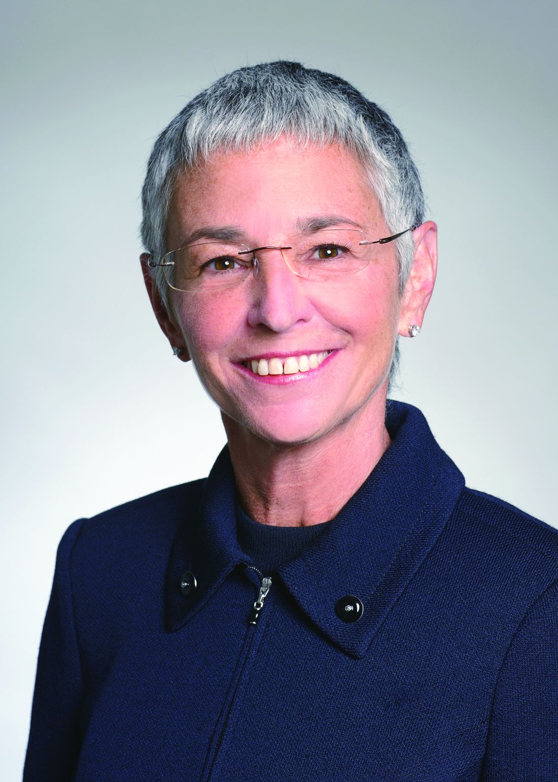
While test takers will have access to resources, such as UpToDate, during the exam, Dr. Von Roenn said don’t expect to be tested on obscure knowledge or asked about rare conditions. “All of the questions are clinical vignettes,” she said. “These are common questions that oncologists face in practice.”
The 2-year general oncology exam will follow the same overall blueprint as the traditional maintenance of certification exam, but will be “very clinically based,” Dr. Von Roenn said. “We’re not asking for things that you can look up,” she said. The exam will instead be focused on how to apply knowledge to common clinical examples.
For that reason, Dr. Von Roenn and Dr. Battaglia said physicians won’t need to spend time and money on intensive exam preparation.
“I actually suspect that people who are busy clinicians wouldn’t need to study, period,” Dr. Von Roenn said.
Richard Larson, MD, reviewed some of the materials for the hematologic malignancies exam and said the test is focused on practical scenarios.
“It’s certainly reasonable to take the test without taking a lot of time to prepare for it,” if physicians are attending annual meetings and grand rounds, and staying current on the medical literature, said Dr. Larson, professor of medicine in the department of hematology-oncology at the University of Chicago, “We’re not aiming at the zebras.”
Thomas LeBlanc, MD, who was not involved with the development of the exam, called this a welcome and overdue change.
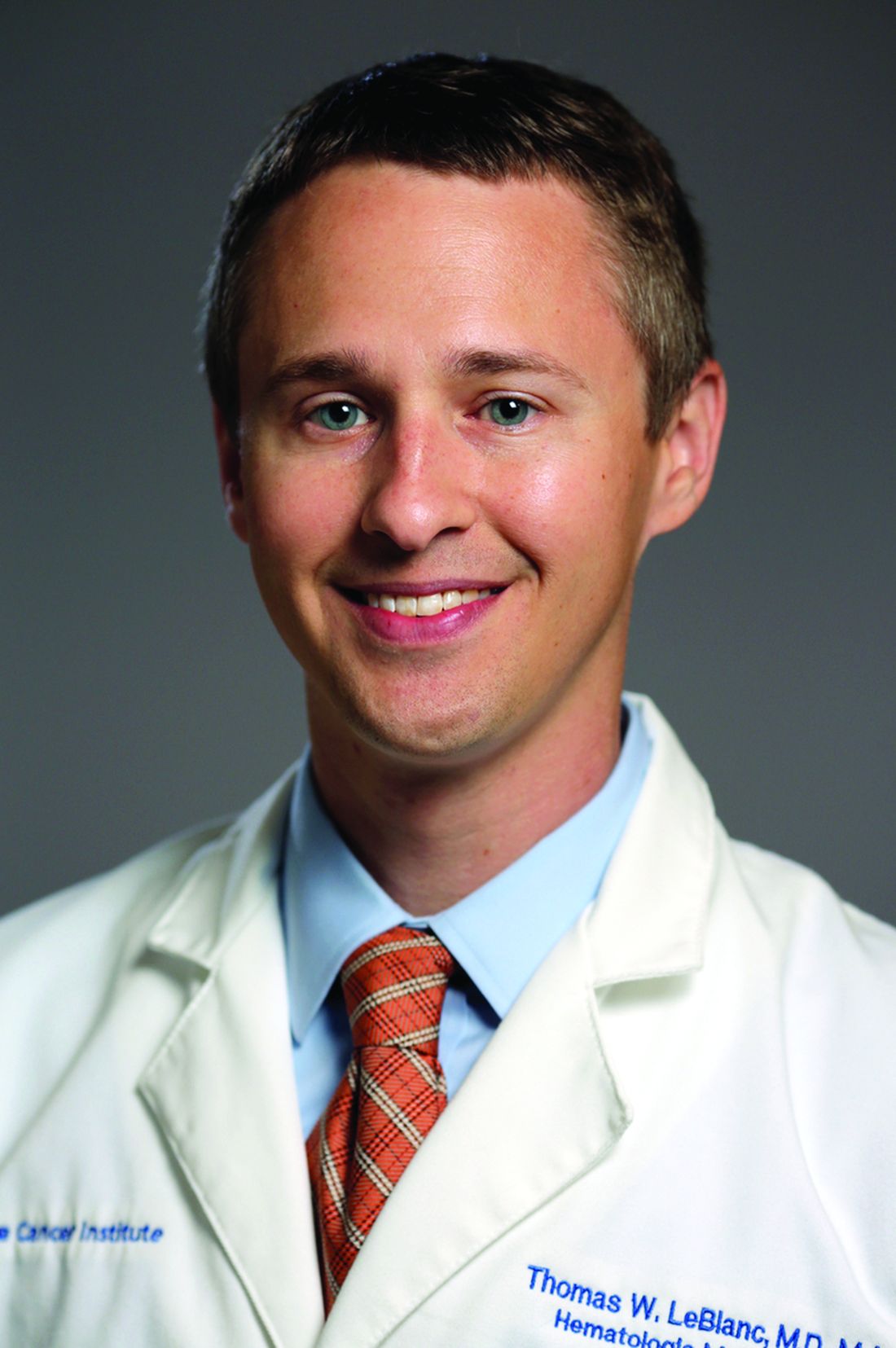
“As someone who sees and treats only patients with blood cancers, it doesn’t make sense for me to be tested on largely solid tumor knowledge, with very little of the exam content being in the area of my practice,” said Dr. LeBlanc, associate professor of medicine at Duke University, Durham, N.C. “That’s what the old examination process is like. If I haven’t kept up on how to treat colon cancer, how does that reflect anything meaningful about my abilities as a leukemia specialist?”
Dr. Battaglia encouraged any oncologist with an assessment due date coming up in the next 2 years to consider the new format. For physicians whose oncology certification has lapsed, they can regain certification by successfully passing two of the general oncology exams.
Another reason to consider trying out the new test in 2020 is that physicians can take it with “no consequence.” While normally physicians have an assessment requirement every 10 years, physicians due to pass an exam in 2020 will not be penalized if they take this shorter test option next year and are unsuccessful. They instead will get a chance to try again in 2022.
Regardless of what specialty area clinicians choose for the 2-year exam, their certificate will be in medical oncology. Opting for the 2-year exam won’t affect other certificates. Clinicians can continue to take the 10-year exam for other areas in which they are certified, Dr. Battaglia said.
Dr. Battaglia said that ABIM is looking for ways to decrease the burden on physicians who are dual boarded, and is having conversations with the specialty boards of medical oncology and hematology about how to do that, but those are early-stage discussions. “It’s on our radar screen,” he said.
The American Society of Hematology has suggested to ABIM that “longitudinal, formative assessments” are a more appropriate way to engage physicians in maintaining certification. ASH submitted comments to the American Board of Medical Specialties, which oversees ABIM, earlier this year on the best ways to overhaul maintenance of certification. While the society continues to advocate for this change, ASH officials are also working with ABIM to implement changes to the current hematology recertification assessment requirement.
“The society is looking forward to working with ABIM to ensure future hematology maintenance of certification exams are more focused on the needs of each practitioner as reflected in the mix of patients and diseases cared for in that practice,” said Alan Lichtin, MD, a hematologist at the Cleveland Clinic who serves as chair of ASH’s maintenance of certification working group. “ASH is currently exploring opportunities to work with ABIM to identify common practice profiles, with the intent to introduce an updated Knowledge Check In for hematology in 2021.”
Oncologists who specialize in treating hematologic malignancies or breast cancer will have more options for fulfilling their maintenance of certification exam requirements beginning next year.
The American Board of Internal Medicine (ABIM), in partnership with the American Society of Clinical Oncology (ASCO), will begin offering a more frequent, more specialized, and lower-stakes exam as an alternative to the current 10-year examination.
The new pathway was announced by ABIM and ASCO in May 2018 after 2 years of gathering feedback from physicians about the need for an exam that tests them in their area of practice – rather than all areas of oncology.
Now the groups are rolling out the first three assessments created under this model – general oncology, hematologic malignancies, and breast cancer. This will be followed in 2022 by exams in gastrointestinal malignancies and lung cancer/thoracic malignancies. Additional new exams will roll out in even-numbered years based on the level of physician interest.
Registration will begin on Dec. 1, 2019, for the 2020 exam, which will be offered in March and September.
Here’s how it works: Oncologists who choose the new ABIM/ASCO Medical Oncology Learning & Assessment pathway will forgo the 10-year exam in favor of shorter exams every 2 years. The new exam – which is about one-third the size of the traditional exam – is given online and can be taken at home, at the office, or at a test center.
The 2-year exam is pass/fail and physicians are allowed one retake. If they fail twice, they must take the 10-year exam.
The cost over 10 years will be the same, regardless of whether one takes the traditional exam or follows the newer path, according to ABIM.
ABIM and ASCO also have designed education around the exam. After registration, test-takers will be supplied with study materials and after the test they’ll receive additional, tailored education on questions that they got wrong.
Giving physicians feedback on how they can improve is a major change, said Richard G. Battaglia, MD, chief medical officer for ABIM. He said that one of the main messages they heard from physicians was that the exam needed to have educational value.
The other message they heard over and over, Dr. Battaglia said, is that practice tends to focus over time and that physicians want to be assessed on what they see every day.
For that reason, the big shift is in the tailored nature of the exam. All of the 2-year exams offered will contain a core module with questions that every oncologist should know, related to symptom management, disease assessment, and principles of genetics and genomics. That core module will make up 25% of the test.
The remaining 75% of the exam will be disease focused, with questions on diagnosis, testing, treatment, prognosis, and common case examples, said Jamie Von Roenn, MD, vice president of education, science, and professional development at ASCO.
While test takers will have access to resources, such as UpToDate, during the exam, Dr. Von Roenn said don’t expect to be tested on obscure knowledge or asked about rare conditions. “All of the questions are clinical vignettes,” she said. “These are common questions that oncologists face in practice.”
The 2-year general oncology exam will follow the same overall blueprint as the traditional maintenance of certification exam, but will be “very clinically based,” Dr. Von Roenn said. “We’re not asking for things that you can look up,” she said. The exam will instead be focused on how to apply knowledge to common clinical examples.
For that reason, Dr. Von Roenn and Dr. Battaglia said physicians won’t need to spend time and money on intensive exam preparation.
“I actually suspect that people who are busy clinicians wouldn’t need to study, period,” Dr. Von Roenn said.
Richard Larson, MD, reviewed some of the materials for the hematologic malignancies exam and said the test is focused on practical scenarios.
“It’s certainly reasonable to take the test without taking a lot of time to prepare for it,” if physicians are attending annual meetings and grand rounds, and staying current on the medical literature, said Dr. Larson, professor of medicine in the department of hematology-oncology at the University of Chicago, “We’re not aiming at the zebras.”
Thomas LeBlanc, MD, who was not involved with the development of the exam, called this a welcome and overdue change.
“As someone who sees and treats only patients with blood cancers, it doesn’t make sense for me to be tested on largely solid tumor knowledge, with very little of the exam content being in the area of my practice,” said Dr. LeBlanc, associate professor of medicine at Duke University, Durham, N.C. “That’s what the old examination process is like. If I haven’t kept up on how to treat colon cancer, how does that reflect anything meaningful about my abilities as a leukemia specialist?”
Dr. Battaglia encouraged any oncologist with an assessment due date coming up in the next 2 years to consider the new format. For physicians whose oncology certification has lapsed, they can regain certification by successfully passing two of the general oncology exams.
Another reason to consider trying out the new test in 2020 is that physicians can take it with “no consequence.” While normally physicians have an assessment requirement every 10 years, physicians due to pass an exam in 2020 will not be penalized if they take this shorter test option next year and are unsuccessful. They instead will get a chance to try again in 2022.
Regardless of what specialty area clinicians choose for the 2-year exam, their certificate will be in medical oncology. Opting for the 2-year exam won’t affect other certificates. Clinicians can continue to take the 10-year exam for other areas in which they are certified, Dr. Battaglia said.
Dr. Battaglia said that ABIM is looking for ways to decrease the burden on physicians who are dual boarded, and is having conversations with the specialty boards of medical oncology and hematology about how to do that, but those are early-stage discussions. “It’s on our radar screen,” he said.
The American Society of Hematology has suggested to ABIM that “longitudinal, formative assessments” are a more appropriate way to engage physicians in maintaining certification. ASH submitted comments to the American Board of Medical Specialties, which oversees ABIM, earlier this year on the best ways to overhaul maintenance of certification. While the society continues to advocate for this change, ASH officials are also working with ABIM to implement changes to the current hematology recertification assessment requirement.
“The society is looking forward to working with ABIM to ensure future hematology maintenance of certification exams are more focused on the needs of each practitioner as reflected in the mix of patients and diseases cared for in that practice,” said Alan Lichtin, MD, a hematologist at the Cleveland Clinic who serves as chair of ASH’s maintenance of certification working group. “ASH is currently exploring opportunities to work with ABIM to identify common practice profiles, with the intent to introduce an updated Knowledge Check In for hematology in 2021.”
Oncologists who specialize in treating hematologic malignancies or breast cancer will have more options for fulfilling their maintenance of certification exam requirements beginning next year.
The American Board of Internal Medicine (ABIM), in partnership with the American Society of Clinical Oncology (ASCO), will begin offering a more frequent, more specialized, and lower-stakes exam as an alternative to the current 10-year examination.
The new pathway was announced by ABIM and ASCO in May 2018 after 2 years of gathering feedback from physicians about the need for an exam that tests them in their area of practice – rather than all areas of oncology.
Now the groups are rolling out the first three assessments created under this model – general oncology, hematologic malignancies, and breast cancer. This will be followed in 2022 by exams in gastrointestinal malignancies and lung cancer/thoracic malignancies. Additional new exams will roll out in even-numbered years based on the level of physician interest.
Registration will begin on Dec. 1, 2019, for the 2020 exam, which will be offered in March and September.
Here’s how it works: Oncologists who choose the new ABIM/ASCO Medical Oncology Learning & Assessment pathway will forgo the 10-year exam in favor of shorter exams every 2 years. The new exam – which is about one-third the size of the traditional exam – is given online and can be taken at home, at the office, or at a test center.
The 2-year exam is pass/fail and physicians are allowed one retake. If they fail twice, they must take the 10-year exam.
The cost over 10 years will be the same, regardless of whether one takes the traditional exam or follows the newer path, according to ABIM.
ABIM and ASCO also have designed education around the exam. After registration, test-takers will be supplied with study materials and after the test they’ll receive additional, tailored education on questions that they got wrong.
Giving physicians feedback on how they can improve is a major change, said Richard G. Battaglia, MD, chief medical officer for ABIM. He said that one of the main messages they heard from physicians was that the exam needed to have educational value.
The other message they heard over and over, Dr. Battaglia said, is that practice tends to focus over time and that physicians want to be assessed on what they see every day.
For that reason, the big shift is in the tailored nature of the exam. All of the 2-year exams offered will contain a core module with questions that every oncologist should know, related to symptom management, disease assessment, and principles of genetics and genomics. That core module will make up 25% of the test.
The remaining 75% of the exam will be disease focused, with questions on diagnosis, testing, treatment, prognosis, and common case examples, said Jamie Von Roenn, MD, vice president of education, science, and professional development at ASCO.
While test takers will have access to resources, such as UpToDate, during the exam, Dr. Von Roenn said don’t expect to be tested on obscure knowledge or asked about rare conditions. “All of the questions are clinical vignettes,” she said. “These are common questions that oncologists face in practice.”
The 2-year general oncology exam will follow the same overall blueprint as the traditional maintenance of certification exam, but will be “very clinically based,” Dr. Von Roenn said. “We’re not asking for things that you can look up,” she said. The exam will instead be focused on how to apply knowledge to common clinical examples.
For that reason, Dr. Von Roenn and Dr. Battaglia said physicians won’t need to spend time and money on intensive exam preparation.
“I actually suspect that people who are busy clinicians wouldn’t need to study, period,” Dr. Von Roenn said.
Richard Larson, MD, reviewed some of the materials for the hematologic malignancies exam and said the test is focused on practical scenarios.
“It’s certainly reasonable to take the test without taking a lot of time to prepare for it,” if physicians are attending annual meetings and grand rounds, and staying current on the medical literature, said Dr. Larson, professor of medicine in the department of hematology-oncology at the University of Chicago, “We’re not aiming at the zebras.”
Thomas LeBlanc, MD, who was not involved with the development of the exam, called this a welcome and overdue change.
“As someone who sees and treats only patients with blood cancers, it doesn’t make sense for me to be tested on largely solid tumor knowledge, with very little of the exam content being in the area of my practice,” said Dr. LeBlanc, associate professor of medicine at Duke University, Durham, N.C. “That’s what the old examination process is like. If I haven’t kept up on how to treat colon cancer, how does that reflect anything meaningful about my abilities as a leukemia specialist?”
Dr. Battaglia encouraged any oncologist with an assessment due date coming up in the next 2 years to consider the new format. For physicians whose oncology certification has lapsed, they can regain certification by successfully passing two of the general oncology exams.
Another reason to consider trying out the new test in 2020 is that physicians can take it with “no consequence.” While normally physicians have an assessment requirement every 10 years, physicians due to pass an exam in 2020 will not be penalized if they take this shorter test option next year and are unsuccessful. They instead will get a chance to try again in 2022.
Regardless of what specialty area clinicians choose for the 2-year exam, their certificate will be in medical oncology. Opting for the 2-year exam won’t affect other certificates. Clinicians can continue to take the 10-year exam for other areas in which they are certified, Dr. Battaglia said.
Dr. Battaglia said that ABIM is looking for ways to decrease the burden on physicians who are dual boarded, and is having conversations with the specialty boards of medical oncology and hematology about how to do that, but those are early-stage discussions. “It’s on our radar screen,” he said.
The American Society of Hematology has suggested to ABIM that “longitudinal, formative assessments” are a more appropriate way to engage physicians in maintaining certification. ASH submitted comments to the American Board of Medical Specialties, which oversees ABIM, earlier this year on the best ways to overhaul maintenance of certification. While the society continues to advocate for this change, ASH officials are also working with ABIM to implement changes to the current hematology recertification assessment requirement.
“The society is looking forward to working with ABIM to ensure future hematology maintenance of certification exams are more focused on the needs of each practitioner as reflected in the mix of patients and diseases cared for in that practice,” said Alan Lichtin, MD, a hematologist at the Cleveland Clinic who serves as chair of ASH’s maintenance of certification working group. “ASH is currently exploring opportunities to work with ABIM to identify common practice profiles, with the intent to introduce an updated Knowledge Check In for hematology in 2021.”

Air pollution levels correlated with cardiorespiratory mortality, reduced life expectancy
particularly among lower-income populations, research suggests.

A study published in PLOS Medicine used vital registration and population data from across the United States for 1999-2015 to estimate the number of deaths and loss of life expectancy associated with four different models of concentrations of fine particulate matter pollution, and examine how that has changed over time.
While the current national ambient air quality standard for particle pollution is 12 mcg/m3 in almost all counties, the study found that in 1999, 59% of the 1,339 county units had concentrations above this level. At that time, the population-weighted average fine particulate matter pollution concentration for the entire country was 13.6mcg/m3. The highest level was seen in Fresno county in California, which had a fine particulate pollution concentration of 22.1 mcg/m3.
By 2015, national concentrations had declined to 8.0 mcg/m3, and the lowest observed concentration was 2.8 mcg/m3.
The investigators wrote, “Each model was applied to county-level cardiorespiratory death rates separately by sex and age group (5-year age groups from birth to 85 years and 85 years and older) because death rates vary by age group and sex, as might their associations with air pollution. From each model we estimated age-specific proportional increases in death rates (i.e. rate ratios) for each 1 mcg/m3 of PM2.5 [fine particulate matter].” The analysis revealed that fine particulate matter pollution above the lowest observed concentration of 2.8 mcg/m3 was associated with higher death rates from cardiorespiratory diseases.
Overall, researchers estimated that these higher levels contributed to 15,612 deaths from cardiorespiratory diseases in women and 14,757 deaths in men, representing 2.8% and 2.7% of all cardiorespiratory deaths, respectively. This amounted to 0.15 years of life expectancy lost in women and 0.13 years lost in men.
There was significant variation in the cost to life expectancy around the country. In the midwestern and Rocky Mountain counties in states such as New Mexico, Colorado, and Arizona, which had lower levels of air pollution, life expectancy loss was less than 0.05 years. But in southern states where the air pollution levels were highest, such as Arkansas, Oklahoma, Alabama, and around Los Angeles, the life expectancy loss was greater than 0.3 years.
“While current PM2.5 pollution is responsible for a significant mortality burden and loss of longevity, reductions in pollution since the late 1990s have benefited virtually the entire country, with the exception of 14 counties where PM2.5 increased slightly over this period,” wrote James E. Bennett, PhD, of the School of Public Health at Imperial College London and coauthors.
The primary limitation of the study is that this association between air pollution and cardiorespiratory health or life expectancy cannot be shown to be causal. Other pollutants and other environmental and behavioral factors that impact cardiorespiratory health may be significant. For example, including ozone and nitrogen dioxide levels in the models could result in different results in terms of the impact of PM2.5 on cardiorespiratory health.
The data highlighted that life expectancy loss associated with air pollution was larger in lower-income counties, those where a higher proportion of the population had a family income below the poverty line, and those where a higher proportion of the population were black or African American.
“This inequality in mortality burden occurs because lower-income counties, those with more poverty, with a greater proportion who are of black or African American race, or with a lower proportion who have graduated high school tend to have higher baseline death rates at any pollution level because of conditions associated with these covariates and hence experience a larger absolute number of deaths as a result of air pollution,” the authors wrote.
The study was funded by the U.S. Environmental Protection Agency and the Wellcome Trust. One author declared grants and personal fees from private industry, outside the submitted work.
SOURCE: Bennett JE et al. PLoS Med. 2019 Jul 23. doi: 10.1371/journal.pmed.1002856.
particularly among lower-income populations, research suggests.
A study published in PLOS Medicine used vital registration and population data from across the United States for 1999-2015 to estimate the number of deaths and loss of life expectancy associated with four different models of concentrations of fine particulate matter pollution, and examine how that has changed over time.
While the current national ambient air quality standard for particle pollution is 12 mcg/m3 in almost all counties, the study found that in 1999, 59% of the 1,339 county units had concentrations above this level. At that time, the population-weighted average fine particulate matter pollution concentration for the entire country was 13.6mcg/m3. The highest level was seen in Fresno county in California, which had a fine particulate pollution concentration of 22.1 mcg/m3.
By 2015, national concentrations had declined to 8.0 mcg/m3, and the lowest observed concentration was 2.8 mcg/m3.
The investigators wrote, “Each model was applied to county-level cardiorespiratory death rates separately by sex and age group (5-year age groups from birth to 85 years and 85 years and older) because death rates vary by age group and sex, as might their associations with air pollution. From each model we estimated age-specific proportional increases in death rates (i.e. rate ratios) for each 1 mcg/m3 of PM2.5 [fine particulate matter].” The analysis revealed that fine particulate matter pollution above the lowest observed concentration of 2.8 mcg/m3 was associated with higher death rates from cardiorespiratory diseases.
Overall, researchers estimated that these higher levels contributed to 15,612 deaths from cardiorespiratory diseases in women and 14,757 deaths in men, representing 2.8% and 2.7% of all cardiorespiratory deaths, respectively. This amounted to 0.15 years of life expectancy lost in women and 0.13 years lost in men.
There was significant variation in the cost to life expectancy around the country. In the midwestern and Rocky Mountain counties in states such as New Mexico, Colorado, and Arizona, which had lower levels of air pollution, life expectancy loss was less than 0.05 years. But in southern states where the air pollution levels were highest, such as Arkansas, Oklahoma, Alabama, and around Los Angeles, the life expectancy loss was greater than 0.3 years.
“While current PM2.5 pollution is responsible for a significant mortality burden and loss of longevity, reductions in pollution since the late 1990s have benefited virtually the entire country, with the exception of 14 counties where PM2.5 increased slightly over this period,” wrote James E. Bennett, PhD, of the School of Public Health at Imperial College London and coauthors.
The primary limitation of the study is that this association between air pollution and cardiorespiratory health or life expectancy cannot be shown to be causal. Other pollutants and other environmental and behavioral factors that impact cardiorespiratory health may be significant. For example, including ozone and nitrogen dioxide levels in the models could result in different results in terms of the impact of PM2.5 on cardiorespiratory health.
The data highlighted that life expectancy loss associated with air pollution was larger in lower-income counties, those where a higher proportion of the population had a family income below the poverty line, and those where a higher proportion of the population were black or African American.
“This inequality in mortality burden occurs because lower-income counties, those with more poverty, with a greater proportion who are of black or African American race, or with a lower proportion who have graduated high school tend to have higher baseline death rates at any pollution level because of conditions associated with these covariates and hence experience a larger absolute number of deaths as a result of air pollution,” the authors wrote.
The study was funded by the U.S. Environmental Protection Agency and the Wellcome Trust. One author declared grants and personal fees from private industry, outside the submitted work.
SOURCE: Bennett JE et al. PLoS Med. 2019 Jul 23. doi: 10.1371/journal.pmed.1002856.
particularly among lower-income populations, research suggests.
A study published in PLOS Medicine used vital registration and population data from across the United States for 1999-2015 to estimate the number of deaths and loss of life expectancy associated with four different models of concentrations of fine particulate matter pollution, and examine how that has changed over time.
While the current national ambient air quality standard for particle pollution is 12 mcg/m3 in almost all counties, the study found that in 1999, 59% of the 1,339 county units had concentrations above this level. At that time, the population-weighted average fine particulate matter pollution concentration for the entire country was 13.6mcg/m3. The highest level was seen in Fresno county in California, which had a fine particulate pollution concentration of 22.1 mcg/m3.
By 2015, national concentrations had declined to 8.0 mcg/m3, and the lowest observed concentration was 2.8 mcg/m3.
The investigators wrote, “Each model was applied to county-level cardiorespiratory death rates separately by sex and age group (5-year age groups from birth to 85 years and 85 years and older) because death rates vary by age group and sex, as might their associations with air pollution. From each model we estimated age-specific proportional increases in death rates (i.e. rate ratios) for each 1 mcg/m3 of PM2.5 [fine particulate matter].” The analysis revealed that fine particulate matter pollution above the lowest observed concentration of 2.8 mcg/m3 was associated with higher death rates from cardiorespiratory diseases.
Overall, researchers estimated that these higher levels contributed to 15,612 deaths from cardiorespiratory diseases in women and 14,757 deaths in men, representing 2.8% and 2.7% of all cardiorespiratory deaths, respectively. This amounted to 0.15 years of life expectancy lost in women and 0.13 years lost in men.
There was significant variation in the cost to life expectancy around the country. In the midwestern and Rocky Mountain counties in states such as New Mexico, Colorado, and Arizona, which had lower levels of air pollution, life expectancy loss was less than 0.05 years. But in southern states where the air pollution levels were highest, such as Arkansas, Oklahoma, Alabama, and around Los Angeles, the life expectancy loss was greater than 0.3 years.
“While current PM2.5 pollution is responsible for a significant mortality burden and loss of longevity, reductions in pollution since the late 1990s have benefited virtually the entire country, with the exception of 14 counties where PM2.5 increased slightly over this period,” wrote James E. Bennett, PhD, of the School of Public Health at Imperial College London and coauthors.
The primary limitation of the study is that this association between air pollution and cardiorespiratory health or life expectancy cannot be shown to be causal. Other pollutants and other environmental and behavioral factors that impact cardiorespiratory health may be significant. For example, including ozone and nitrogen dioxide levels in the models could result in different results in terms of the impact of PM2.5 on cardiorespiratory health.
The data highlighted that life expectancy loss associated with air pollution was larger in lower-income counties, those where a higher proportion of the population had a family income below the poverty line, and those where a higher proportion of the population were black or African American.
“This inequality in mortality burden occurs because lower-income counties, those with more poverty, with a greater proportion who are of black or African American race, or with a lower proportion who have graduated high school tend to have higher baseline death rates at any pollution level because of conditions associated with these covariates and hence experience a larger absolute number of deaths as a result of air pollution,” the authors wrote.
The study was funded by the U.S. Environmental Protection Agency and the Wellcome Trust. One author declared grants and personal fees from private industry, outside the submitted work.
SOURCE: Bennett JE et al. PLoS Med. 2019 Jul 23. doi: 10.1371/journal.pmed.1002856.
FROM PLOS MEDICINE
Key clinical point: Air pollution linked to reduced life expectancy, particularly in poorer areas.
Major finding: Fine particulate matter air pollution linked to 0.15 years of life expectancy lost in women and 0.13 years lost in men.
Study details: Population-wide data analysis for the United States.
Disclosures: The study was funded by the U.S. Environmental Protection Agency and the Wellcome Trust. One author declared grants and personal fees from private industry outside the submitted work.
Source: Bennett JE et al. PLoS Med. 2019 Jul 23. doi: 10.1371/journal.pmed.1002856.

What is the future of para-aortic lymphadenectomy for endometrial cancer?
A landmark study of advanced endometrial cancer, GOG 258, was published in the New England Journal of Medicine this summer.1 This clinical trial compared the use of carboplatin and paclitaxel chemotherapy with a combination of chemotherapy with external beam radiation, exploring the notion of “more is better.” The results of the trial revealed that the “more” (chemotherapy with external beam radiation) was no better than chemotherapy alone with respect to overall survival. These results have challenged a creeping dogma in gynecologic oncology, which has seen many providers embrace combination therapy, particularly for patients with stage III (node-positive) endometrial cancer, a group of patients who made up approximately three-quarters of GOG 258’s study population. Many have been left searching for justification of their early adoption of combination therapy before the results of a trial such as this were available. For me it also raised a slightly different question: In the light of these results, what IS the role of para-aortic lymphadenectomy in the staging of endometrial cancers? If radiation to the nodal basins is no longer part of adjuvant therapy, then

It was in the 1980s that the removal of clinically normal para-aortic lymph nodes (those residing between the renal and proximal common iliac vessels) became a part of surgical staging. This practice was endorsed by the International Federation of Gynecology and Obstetrics (FIGO) and the Gynecologic Oncology Group (GOG) surgical committee in response to findings that 11% of women with clinical stage I endometrial cancer had microscopic lymph node metastases which were discovered only with routine pathologic evaluation of these tissues. Among those with pelvic lymph node metastases (stage IIIC disease), approximately one-third also harbored disease in para-aortic nodal regions.2 Among all patients with endometrial cancer, including those with low-grade disease, only a small fraction (approximately 2%) have isolated para-aortic lymph nodes (positive para-aortic nodes, but negative pelvic nodes). However, among patients with deeply invasive higher-grade tumors, the likelihood of discovering isolated para-aortic metastases is higher at approximately 16%.3 Therefore, the dominant pattern of lymph node metastases and lymphatic dissemination of endometrial cancer appears to be via the parametrial channels laterally towards the pelvic basins, and then sequentially to the para-aortic regions. The direct lymphatic pathway to the para-aortic basins from the uterine fundus through the adnexal lymphatics misses the pelvic regions altogether and may seen logical, but actually is observed fairly infrequently.4
Over the subsequent decades, there have been debates and schools of thought regarding what is the optimal degree of lymphatic dissection for endometrial cancer staging. Some advocated for full pelvic and infrarenal para-aortic nodal dissections in all patients, including even those in the lowest risk for metastases. Others advocated for a more limited, inframesenteric para-aortic nodal dissection, although the origins of such a distinction appear to be largely arbitrary. The inferior mesenteric artery is not a physiologic landmark for lymphatic pathways, and approximately half of para-aortic metastases are located above the level of the inferior mesenteric artery. This limited sampling may have been preferred because of relative ease of dissection rather than diagnostic or therapeutic efficacy.
As the population became more obese, making para-aortic nodal dissections less feasible, and laparoscopic staging became accepted as the standard of care in endometrial cancer staging, there was a further push towards limiting the scope of lymphadenectomy. Selective algorithms, such as the so-called “Mayo clinic criteria,” were widely adopted. In this approach, gynecologic oncologists perform full pelvic and infrarenal para-aortic lymphadenectomies but only in the presence of a high-risk uterine feature such as tumor size greater than 2 cm, deep myometrial invasion, or grade 3 histology.3 While this reduced the number of para-aortic dissections being performed, it did not eliminate them, as approximately 40% of patients with endometrial cancer meet at least one of those criteria.
At this same time, we learned something else critical about the benefits, or lack thereof, of lymphadenectomy. Two landmark surgical-staging trials were published in 2009 which randomly assigned women to hysterectomy with lymphadenectomy or hysterectomy alone, and found no survival benefit for lymphadenectomy.5,6 While these trial results initially were met with noisy backlash, particularly from those who had legitimate concerns regarding study design and conclusions that reach beyond the scope of this column, ultimately their findings (that there is no therapeutic benefit to surgically removing clinically normal lymph nodes) has become largely accepted. The subsequent findings of the Laparoscopic Approach to Cancer of the Endometrium (LACE) trial further support this, as there was no difference in survival found between patients who were randomly assigned to laparoscopic versus open staging for endometrial cancer, even despite a significantly lower rate of lymphadenectomy among the laparoscopic arm.7
SLN biopsy, in which the specific nodes which drain the uterus are selectively removed, represents the most recent development in lymph node assessment for endometrial cancer. On average, only three lymph nodes are removed per patient, and para-aortic nodes infrequently are removed, because it is rare that lymphatic pathways drain directly into the aortic basins after cervical injection. Yet despite this more limited dissection of lymph nodes, especially para-aortic, with SLN biopsy, surgeons still observe similar rates of IIIC disease, compared with full lymphadenectomy, suggesting that the presence or absence of lymphatic metastases still is able to be adequately determined. SLN biopsy misses only 3% of lymphatic disease.8 What is of particular interest to practitioners of the SLN approach is that “atypical” pathways are discovered approximately 20% of the time, and nodes are harvested from locations such as the presacral space or medial to the internal iliac vessels. These nodes are in locations previously overlooked by even the most comprehensive pelvic and para-aortic lymphadenectomy. Therefore, while the para-aortic nodes may not be systematically removed with SLN biopsy, new and arguably more relevant regions are interrogated, which might explain its equivalent diagnostic virtue.
With this evolution in surgical-staging practice, what we have come to recognize is that the role of lymph node assessment is predominantly, if not exclusively, diagnostic. It can help us determine which patients are at risk for distant relapse and therefore candidates for systemic therapy (chemotherapy), versus those whose risk is predominantly of local relapse and can be adequately treated with local therapies alone, such as vaginal radiation. This brings us to the results of GOG 258. If defining the specific and complete extent of lymph node metastases (as if that was ever truly what surgeons were doing) is no longer necessary to guide the prescription and extent of external beam radiation, then lymph node dissection need only inform us of whether or not there are nodal metastases, not specifically the location of those nodal metastases. The prescription of chemotherapy is the same whether the disease is limited to the pelvic nodes or also includes the para-aortic nodes. While GOG 258 discovered more para-aortic failures among the chemotherapy-alone group, suggesting there may be some therapeutic role of radiation in preventing this, it should be noted that these para-aortic relapses did not negatively impact relapse-free survival, and these patients still can presumably be salvaged with external beam radiation to the site of para-aortic relapse.
It would seem logical that the results of GOG 258 further limit the potential role of para-aortic lymphadenectomy in women with clinical stage I disease. Perhaps para-aortic dissection can be limited to women who are at highest risk for isolated para-aortic disease, such as those with deeply invasive high-grade tumors not successfully mapped with the highly targeted sentinel node biopsy technique? Most clinicians look forward to an era in which we no longer rely on crude dissections of disease-free tissue just to prove they are disease free, but instead can utilize more sophisticated diagnostic methods to recognize disseminated disease.
Dr. Rossi is assistant professor in the division of gynecologic oncology at the University of North Carolina at Chapel Hill. Email her at obnews@mdedge.com.
References
1. N Engl J Med. 2019 Jun 13;380(24):2317-26.
2. Cancer. 1987 Oct 15;60(8 Suppl):2035-41.
3. Gynecol Oncol. 2008;109(1):11-8.
4. Int J Gynecol Cancer. 2019 Mar;29(3):613-21.
5. J Natl Cancer Inst. 2008 Dec 3;100(23):1707-16.
6. Lancet. 2009 Jan 10;373(9658):125-36.
7. JAMA. 2017 Mar 28;317(12):1224-33.
8. Lancet Oncol. 2017 Mar;18(3):384-92.
A landmark study of advanced endometrial cancer, GOG 258, was published in the New England Journal of Medicine this summer.1 This clinical trial compared the use of carboplatin and paclitaxel chemotherapy with a combination of chemotherapy with external beam radiation, exploring the notion of “more is better.” The results of the trial revealed that the “more” (chemotherapy with external beam radiation) was no better than chemotherapy alone with respect to overall survival. These results have challenged a creeping dogma in gynecologic oncology, which has seen many providers embrace combination therapy, particularly for patients with stage III (node-positive) endometrial cancer, a group of patients who made up approximately three-quarters of GOG 258’s study population. Many have been left searching for justification of their early adoption of combination therapy before the results of a trial such as this were available. For me it also raised a slightly different question: In the light of these results, what IS the role of para-aortic lymphadenectomy in the staging of endometrial cancers? If radiation to the nodal basins is no longer part of adjuvant therapy, then
It was in the 1980s that the removal of clinically normal para-aortic lymph nodes (those residing between the renal and proximal common iliac vessels) became a part of surgical staging. This practice was endorsed by the International Federation of Gynecology and Obstetrics (FIGO) and the Gynecologic Oncology Group (GOG) surgical committee in response to findings that 11% of women with clinical stage I endometrial cancer had microscopic lymph node metastases which were discovered only with routine pathologic evaluation of these tissues. Among those with pelvic lymph node metastases (stage IIIC disease), approximately one-third also harbored disease in para-aortic nodal regions.2 Among all patients with endometrial cancer, including those with low-grade disease, only a small fraction (approximately 2%) have isolated para-aortic lymph nodes (positive para-aortic nodes, but negative pelvic nodes). However, among patients with deeply invasive higher-grade tumors, the likelihood of discovering isolated para-aortic metastases is higher at approximately 16%.3 Therefore, the dominant pattern of lymph node metastases and lymphatic dissemination of endometrial cancer appears to be via the parametrial channels laterally towards the pelvic basins, and then sequentially to the para-aortic regions. The direct lymphatic pathway to the para-aortic basins from the uterine fundus through the adnexal lymphatics misses the pelvic regions altogether and may seen logical, but actually is observed fairly infrequently.4
Over the subsequent decades, there have been debates and schools of thought regarding what is the optimal degree of lymphatic dissection for endometrial cancer staging. Some advocated for full pelvic and infrarenal para-aortic nodal dissections in all patients, including even those in the lowest risk for metastases. Others advocated for a more limited, inframesenteric para-aortic nodal dissection, although the origins of such a distinction appear to be largely arbitrary. The inferior mesenteric artery is not a physiologic landmark for lymphatic pathways, and approximately half of para-aortic metastases are located above the level of the inferior mesenteric artery. This limited sampling may have been preferred because of relative ease of dissection rather than diagnostic or therapeutic efficacy.
As the population became more obese, making para-aortic nodal dissections less feasible, and laparoscopic staging became accepted as the standard of care in endometrial cancer staging, there was a further push towards limiting the scope of lymphadenectomy. Selective algorithms, such as the so-called “Mayo clinic criteria,” were widely adopted. In this approach, gynecologic oncologists perform full pelvic and infrarenal para-aortic lymphadenectomies but only in the presence of a high-risk uterine feature such as tumor size greater than 2 cm, deep myometrial invasion, or grade 3 histology.3 While this reduced the number of para-aortic dissections being performed, it did not eliminate them, as approximately 40% of patients with endometrial cancer meet at least one of those criteria.
At this same time, we learned something else critical about the benefits, or lack thereof, of lymphadenectomy. Two landmark surgical-staging trials were published in 2009 which randomly assigned women to hysterectomy with lymphadenectomy or hysterectomy alone, and found no survival benefit for lymphadenectomy.5,6 While these trial results initially were met with noisy backlash, particularly from those who had legitimate concerns regarding study design and conclusions that reach beyond the scope of this column, ultimately their findings (that there is no therapeutic benefit to surgically removing clinically normal lymph nodes) has become largely accepted. The subsequent findings of the Laparoscopic Approach to Cancer of the Endometrium (LACE) trial further support this, as there was no difference in survival found between patients who were randomly assigned to laparoscopic versus open staging for endometrial cancer, even despite a significantly lower rate of lymphadenectomy among the laparoscopic arm.7
SLN biopsy, in which the specific nodes which drain the uterus are selectively removed, represents the most recent development in lymph node assessment for endometrial cancer. On average, only three lymph nodes are removed per patient, and para-aortic nodes infrequently are removed, because it is rare that lymphatic pathways drain directly into the aortic basins after cervical injection. Yet despite this more limited dissection of lymph nodes, especially para-aortic, with SLN biopsy, surgeons still observe similar rates of IIIC disease, compared with full lymphadenectomy, suggesting that the presence or absence of lymphatic metastases still is able to be adequately determined. SLN biopsy misses only 3% of lymphatic disease.8 What is of particular interest to practitioners of the SLN approach is that “atypical” pathways are discovered approximately 20% of the time, and nodes are harvested from locations such as the presacral space or medial to the internal iliac vessels. These nodes are in locations previously overlooked by even the most comprehensive pelvic and para-aortic lymphadenectomy. Therefore, while the para-aortic nodes may not be systematically removed with SLN biopsy, new and arguably more relevant regions are interrogated, which might explain its equivalent diagnostic virtue.
With this evolution in surgical-staging practice, what we have come to recognize is that the role of lymph node assessment is predominantly, if not exclusively, diagnostic. It can help us determine which patients are at risk for distant relapse and therefore candidates for systemic therapy (chemotherapy), versus those whose risk is predominantly of local relapse and can be adequately treated with local therapies alone, such as vaginal radiation. This brings us to the results of GOG 258. If defining the specific and complete extent of lymph node metastases (as if that was ever truly what surgeons were doing) is no longer necessary to guide the prescription and extent of external beam radiation, then lymph node dissection need only inform us of whether or not there are nodal metastases, not specifically the location of those nodal metastases. The prescription of chemotherapy is the same whether the disease is limited to the pelvic nodes or also includes the para-aortic nodes. While GOG 258 discovered more para-aortic failures among the chemotherapy-alone group, suggesting there may be some therapeutic role of radiation in preventing this, it should be noted that these para-aortic relapses did not negatively impact relapse-free survival, and these patients still can presumably be salvaged with external beam radiation to the site of para-aortic relapse.
It would seem logical that the results of GOG 258 further limit the potential role of para-aortic lymphadenectomy in women with clinical stage I disease. Perhaps para-aortic dissection can be limited to women who are at highest risk for isolated para-aortic disease, such as those with deeply invasive high-grade tumors not successfully mapped with the highly targeted sentinel node biopsy technique? Most clinicians look forward to an era in which we no longer rely on crude dissections of disease-free tissue just to prove they are disease free, but instead can utilize more sophisticated diagnostic methods to recognize disseminated disease.
Dr. Rossi is assistant professor in the division of gynecologic oncology at the University of North Carolina at Chapel Hill. Email her at obnews@mdedge.com.
References
1. N Engl J Med. 2019 Jun 13;380(24):2317-26.
2. Cancer. 1987 Oct 15;60(8 Suppl):2035-41.
3. Gynecol Oncol. 2008;109(1):11-8.
4. Int J Gynecol Cancer. 2019 Mar;29(3):613-21.
5. J Natl Cancer Inst. 2008 Dec 3;100(23):1707-16.
6. Lancet. 2009 Jan 10;373(9658):125-36.
7. JAMA. 2017 Mar 28;317(12):1224-33.
8. Lancet Oncol. 2017 Mar;18(3):384-92.
A landmark study of advanced endometrial cancer, GOG 258, was published in the New England Journal of Medicine this summer.1 This clinical trial compared the use of carboplatin and paclitaxel chemotherapy with a combination of chemotherapy with external beam radiation, exploring the notion of “more is better.” The results of the trial revealed that the “more” (chemotherapy with external beam radiation) was no better than chemotherapy alone with respect to overall survival. These results have challenged a creeping dogma in gynecologic oncology, which has seen many providers embrace combination therapy, particularly for patients with stage III (node-positive) endometrial cancer, a group of patients who made up approximately three-quarters of GOG 258’s study population. Many have been left searching for justification of their early adoption of combination therapy before the results of a trial such as this were available. For me it also raised a slightly different question: In the light of these results, what IS the role of para-aortic lymphadenectomy in the staging of endometrial cancers? If radiation to the nodal basins is no longer part of adjuvant therapy, then
It was in the 1980s that the removal of clinically normal para-aortic lymph nodes (those residing between the renal and proximal common iliac vessels) became a part of surgical staging. This practice was endorsed by the International Federation of Gynecology and Obstetrics (FIGO) and the Gynecologic Oncology Group (GOG) surgical committee in response to findings that 11% of women with clinical stage I endometrial cancer had microscopic lymph node metastases which were discovered only with routine pathologic evaluation of these tissues. Among those with pelvic lymph node metastases (stage IIIC disease), approximately one-third also harbored disease in para-aortic nodal regions.2 Among all patients with endometrial cancer, including those with low-grade disease, only a small fraction (approximately 2%) have isolated para-aortic lymph nodes (positive para-aortic nodes, but negative pelvic nodes). However, among patients with deeply invasive higher-grade tumors, the likelihood of discovering isolated para-aortic metastases is higher at approximately 16%.3 Therefore, the dominant pattern of lymph node metastases and lymphatic dissemination of endometrial cancer appears to be via the parametrial channels laterally towards the pelvic basins, and then sequentially to the para-aortic regions. The direct lymphatic pathway to the para-aortic basins from the uterine fundus through the adnexal lymphatics misses the pelvic regions altogether and may seen logical, but actually is observed fairly infrequently.4
Over the subsequent decades, there have been debates and schools of thought regarding what is the optimal degree of lymphatic dissection for endometrial cancer staging. Some advocated for full pelvic and infrarenal para-aortic nodal dissections in all patients, including even those in the lowest risk for metastases. Others advocated for a more limited, inframesenteric para-aortic nodal dissection, although the origins of such a distinction appear to be largely arbitrary. The inferior mesenteric artery is not a physiologic landmark for lymphatic pathways, and approximately half of para-aortic metastases are located above the level of the inferior mesenteric artery. This limited sampling may have been preferred because of relative ease of dissection rather than diagnostic or therapeutic efficacy.
As the population became more obese, making para-aortic nodal dissections less feasible, and laparoscopic staging became accepted as the standard of care in endometrial cancer staging, there was a further push towards limiting the scope of lymphadenectomy. Selective algorithms, such as the so-called “Mayo clinic criteria,” were widely adopted. In this approach, gynecologic oncologists perform full pelvic and infrarenal para-aortic lymphadenectomies but only in the presence of a high-risk uterine feature such as tumor size greater than 2 cm, deep myometrial invasion, or grade 3 histology.3 While this reduced the number of para-aortic dissections being performed, it did not eliminate them, as approximately 40% of patients with endometrial cancer meet at least one of those criteria.
At this same time, we learned something else critical about the benefits, or lack thereof, of lymphadenectomy. Two landmark surgical-staging trials were published in 2009 which randomly assigned women to hysterectomy with lymphadenectomy or hysterectomy alone, and found no survival benefit for lymphadenectomy.5,6 While these trial results initially were met with noisy backlash, particularly from those who had legitimate concerns regarding study design and conclusions that reach beyond the scope of this column, ultimately their findings (that there is no therapeutic benefit to surgically removing clinically normal lymph nodes) has become largely accepted. The subsequent findings of the Laparoscopic Approach to Cancer of the Endometrium (LACE) trial further support this, as there was no difference in survival found between patients who were randomly assigned to laparoscopic versus open staging for endometrial cancer, even despite a significantly lower rate of lymphadenectomy among the laparoscopic arm.7
SLN biopsy, in which the specific nodes which drain the uterus are selectively removed, represents the most recent development in lymph node assessment for endometrial cancer. On average, only three lymph nodes are removed per patient, and para-aortic nodes infrequently are removed, because it is rare that lymphatic pathways drain directly into the aortic basins after cervical injection. Yet despite this more limited dissection of lymph nodes, especially para-aortic, with SLN biopsy, surgeons still observe similar rates of IIIC disease, compared with full lymphadenectomy, suggesting that the presence or absence of lymphatic metastases still is able to be adequately determined. SLN biopsy misses only 3% of lymphatic disease.8 What is of particular interest to practitioners of the SLN approach is that “atypical” pathways are discovered approximately 20% of the time, and nodes are harvested from locations such as the presacral space or medial to the internal iliac vessels. These nodes are in locations previously overlooked by even the most comprehensive pelvic and para-aortic lymphadenectomy. Therefore, while the para-aortic nodes may not be systematically removed with SLN biopsy, new and arguably more relevant regions are interrogated, which might explain its equivalent diagnostic virtue.
With this evolution in surgical-staging practice, what we have come to recognize is that the role of lymph node assessment is predominantly, if not exclusively, diagnostic. It can help us determine which patients are at risk for distant relapse and therefore candidates for systemic therapy (chemotherapy), versus those whose risk is predominantly of local relapse and can be adequately treated with local therapies alone, such as vaginal radiation. This brings us to the results of GOG 258. If defining the specific and complete extent of lymph node metastases (as if that was ever truly what surgeons were doing) is no longer necessary to guide the prescription and extent of external beam radiation, then lymph node dissection need only inform us of whether or not there are nodal metastases, not specifically the location of those nodal metastases. The prescription of chemotherapy is the same whether the disease is limited to the pelvic nodes or also includes the para-aortic nodes. While GOG 258 discovered more para-aortic failures among the chemotherapy-alone group, suggesting there may be some therapeutic role of radiation in preventing this, it should be noted that these para-aortic relapses did not negatively impact relapse-free survival, and these patients still can presumably be salvaged with external beam radiation to the site of para-aortic relapse.
It would seem logical that the results of GOG 258 further limit the potential role of para-aortic lymphadenectomy in women with clinical stage I disease. Perhaps para-aortic dissection can be limited to women who are at highest risk for isolated para-aortic disease, such as those with deeply invasive high-grade tumors not successfully mapped with the highly targeted sentinel node biopsy technique? Most clinicians look forward to an era in which we no longer rely on crude dissections of disease-free tissue just to prove they are disease free, but instead can utilize more sophisticated diagnostic methods to recognize disseminated disease.
Dr. Rossi is assistant professor in the division of gynecologic oncology at the University of North Carolina at Chapel Hill. Email her at obnews@mdedge.com.
References
1. N Engl J Med. 2019 Jun 13;380(24):2317-26.
2. Cancer. 1987 Oct 15;60(8 Suppl):2035-41.
3. Gynecol Oncol. 2008;109(1):11-8.
4. Int J Gynecol Cancer. 2019 Mar;29(3):613-21.
5. J Natl Cancer Inst. 2008 Dec 3;100(23):1707-16.
6. Lancet. 2009 Jan 10;373(9658):125-36.
7. JAMA. 2017 Mar 28;317(12):1224-33.
8. Lancet Oncol. 2017 Mar;18(3):384-92.

Administrative data may reduce costs of economic analysis
The use of administrative data may improve the accuracy and reduce costs of oncology clinical trial economic analyses, according to a retrospective study.
“We investigated whether administrative data could improve the performance of cancer trial economic analysis,” Timothy P. Hanna, MD, MSc, PhD, of Queen’s University in Kingston, Ont., and colleagues wrote in the Journal of Oncology Practice.
The researchers linked clinical trial and health administrative data to evaluate the utility of administrative data in a group of 572 patients with previously treated metastatic colorectal cancer.
The phase 3 trial assessed cetuximab plus best supportive care versus best supportive care alone in these patients. The trial data set was used to obtain information related to resource utilization, such as hospitalization or treatment costs, in addition to clinical reports, such as vital status.
Several measures were estimated by the team, including the incremental cost-effectiveness ratio (ICER) and total costs.
After analysis, the researchers found that ICER measures were comparable with the use of administrative data versus trial data alone ($164,378 vs. $211,128 per QALY added up to last contact).
“Up to trial date of last contact, vital status was in agreement for more than 96% of patients,” Dr. Hanna and colleagues reported.
These findings highlight the potential utility of administrative data in reducing the costs of collecting applicable data from clinical trials alone.
“A harmonized approach has the potential to provide more complete information on clinical trial participants at reduced cost,” they concluded.
The study was funded by the Institute for Clinical Evaluative Sciences and Queen’s University. The authors reported financial affiliations with Amgen, Janssen, Novartis, and UpToDate.
SOURCE: Hanna TP et al. J Oncol Pract. 2019 Jul 15. doi: 10.1200/JOP.18.00691.
The use of administrative data may improve the accuracy and reduce costs of oncology clinical trial economic analyses, according to a retrospective study.
“We investigated whether administrative data could improve the performance of cancer trial economic analysis,” Timothy P. Hanna, MD, MSc, PhD, of Queen’s University in Kingston, Ont., and colleagues wrote in the Journal of Oncology Practice.
The researchers linked clinical trial and health administrative data to evaluate the utility of administrative data in a group of 572 patients with previously treated metastatic colorectal cancer.
The phase 3 trial assessed cetuximab plus best supportive care versus best supportive care alone in these patients. The trial data set was used to obtain information related to resource utilization, such as hospitalization or treatment costs, in addition to clinical reports, such as vital status.
Several measures were estimated by the team, including the incremental cost-effectiveness ratio (ICER) and total costs.
After analysis, the researchers found that ICER measures were comparable with the use of administrative data versus trial data alone ($164,378 vs. $211,128 per QALY added up to last contact).
“Up to trial date of last contact, vital status was in agreement for more than 96% of patients,” Dr. Hanna and colleagues reported.
These findings highlight the potential utility of administrative data in reducing the costs of collecting applicable data from clinical trials alone.
“A harmonized approach has the potential to provide more complete information on clinical trial participants at reduced cost,” they concluded.
The study was funded by the Institute for Clinical Evaluative Sciences and Queen’s University. The authors reported financial affiliations with Amgen, Janssen, Novartis, and UpToDate.
SOURCE: Hanna TP et al. J Oncol Pract. 2019 Jul 15. doi: 10.1200/JOP.18.00691.
The use of administrative data may improve the accuracy and reduce costs of oncology clinical trial economic analyses, according to a retrospective study.
“We investigated whether administrative data could improve the performance of cancer trial economic analysis,” Timothy P. Hanna, MD, MSc, PhD, of Queen’s University in Kingston, Ont., and colleagues wrote in the Journal of Oncology Practice.
The researchers linked clinical trial and health administrative data to evaluate the utility of administrative data in a group of 572 patients with previously treated metastatic colorectal cancer.
The phase 3 trial assessed cetuximab plus best supportive care versus best supportive care alone in these patients. The trial data set was used to obtain information related to resource utilization, such as hospitalization or treatment costs, in addition to clinical reports, such as vital status.
Several measures were estimated by the team, including the incremental cost-effectiveness ratio (ICER) and total costs.
After analysis, the researchers found that ICER measures were comparable with the use of administrative data versus trial data alone ($164,378 vs. $211,128 per QALY added up to last contact).
“Up to trial date of last contact, vital status was in agreement for more than 96% of patients,” Dr. Hanna and colleagues reported.
These findings highlight the potential utility of administrative data in reducing the costs of collecting applicable data from clinical trials alone.
“A harmonized approach has the potential to provide more complete information on clinical trial participants at reduced cost,” they concluded.
The study was funded by the Institute for Clinical Evaluative Sciences and Queen’s University. The authors reported financial affiliations with Amgen, Janssen, Novartis, and UpToDate.
SOURCE: Hanna TP et al. J Oncol Pract. 2019 Jul 15. doi: 10.1200/JOP.18.00691.
FROM JOURNAL OF ONCOLOGY PRACTICE
FDA approves Otezla for treatment of Behçet’s-associated oral ulcers
The Food and Drug Administration has expanded the indication for apremilast (Otezla) to include the treatment of oral ulcers associated with Behçet’s disease in adults, according to an announcement from the manufacturer, Celgene.

FDA approval was based on results of the randomized, placebo-controlled, double-blind, phase 3 RELIEF trial, in which 207 patients with Behçet’s disease with active ulcers underwent treatment for 12 weeks with 30 mg apremilast or placebo. When measured on a visual analog scale, the reduction in pain from oral ulcers after 12 weeks in patients receiving apremilast was 42.7 points, compared with 18.7 points in the placebo group. Just over 50% of apremilast patients achieved complete response by week 12, compared with 22.3% in the placebo group.
The most common adverse events associated with apremilast during RELIEF were diarrhea, nausea, headache, and upper respiratory infection. This was consistent with apremilast’s known safety profile.
Apremilast is also indicated for treatment of patients with moderate to severe plaque psoriasis who are candidates for phototherapy or systemic therapy, and for patients with active psoriatic arthritis.
“Oral ulcers are a recurring and debilitating manifestation that affects nearly everyone living with Behçet’s disease and have an important negative impact on the quality of life for these patients. In the clinical trial, Otezla demonstrated improvements in measures of oral ulcers at week 12. Otezla has the potential to be a needed treatment option for U.S. patients and their physicians, who previously had limited options available,” Yusuf Yazici, MD, clinical associate professor in the department of medicine at New York University, said in the announcement.
The Food and Drug Administration has expanded the indication for apremilast (Otezla) to include the treatment of oral ulcers associated with Behçet’s disease in adults, according to an announcement from the manufacturer, Celgene.
FDA approval was based on results of the randomized, placebo-controlled, double-blind, phase 3 RELIEF trial, in which 207 patients with Behçet’s disease with active ulcers underwent treatment for 12 weeks with 30 mg apremilast or placebo. When measured on a visual analog scale, the reduction in pain from oral ulcers after 12 weeks in patients receiving apremilast was 42.7 points, compared with 18.7 points in the placebo group. Just over 50% of apremilast patients achieved complete response by week 12, compared with 22.3% in the placebo group.
The most common adverse events associated with apremilast during RELIEF were diarrhea, nausea, headache, and upper respiratory infection. This was consistent with apremilast’s known safety profile.
Apremilast is also indicated for treatment of patients with moderate to severe plaque psoriasis who are candidates for phototherapy or systemic therapy, and for patients with active psoriatic arthritis.
“Oral ulcers are a recurring and debilitating manifestation that affects nearly everyone living with Behçet’s disease and have an important negative impact on the quality of life for these patients. In the clinical trial, Otezla demonstrated improvements in measures of oral ulcers at week 12. Otezla has the potential to be a needed treatment option for U.S. patients and their physicians, who previously had limited options available,” Yusuf Yazici, MD, clinical associate professor in the department of medicine at New York University, said in the announcement.
The Food and Drug Administration has expanded the indication for apremilast (Otezla) to include the treatment of oral ulcers associated with Behçet’s disease in adults, according to an announcement from the manufacturer, Celgene.
FDA approval was based on results of the randomized, placebo-controlled, double-blind, phase 3 RELIEF trial, in which 207 patients with Behçet’s disease with active ulcers underwent treatment for 12 weeks with 30 mg apremilast or placebo. When measured on a visual analog scale, the reduction in pain from oral ulcers after 12 weeks in patients receiving apremilast was 42.7 points, compared with 18.7 points in the placebo group. Just over 50% of apremilast patients achieved complete response by week 12, compared with 22.3% in the placebo group.
The most common adverse events associated with apremilast during RELIEF were diarrhea, nausea, headache, and upper respiratory infection. This was consistent with apremilast’s known safety profile.
Apremilast is also indicated for treatment of patients with moderate to severe plaque psoriasis who are candidates for phototherapy or systemic therapy, and for patients with active psoriatic arthritis.
“Oral ulcers are a recurring and debilitating manifestation that affects nearly everyone living with Behçet’s disease and have an important negative impact on the quality of life for these patients. In the clinical trial, Otezla demonstrated improvements in measures of oral ulcers at week 12. Otezla has the potential to be a needed treatment option for U.S. patients and their physicians, who previously had limited options available,” Yusuf Yazici, MD, clinical associate professor in the department of medicine at New York University, said in the announcement.

Study refines ALS risk among first-degree relatives of patients with disease
, according to a recent Irish population–based study.

Among first-degree relatives of individuals with ALS whose genetic status was unknown, the lifetime risk of developing ALS was 1.4%, compared with 0.3% in the general population (P less than .001).
Overall, lifetime heritability for the 1,117 patients in the heritability study cohort was 52.3% (95% confidence interval, 42.9%-61.7%). The highest heritability was seen in mother-daughter dyads; here, the concordance rate for ALS was 2.6% and the heritability estimate was 66.2% (95% CI, 58.5%-73.9%), “pointing to a previously unrecognized sex-mediated association with disease heritability,” Marie Ryan, MD, and coauthors at Trinity College Dublin wrote in JAMA Neurology.
For the purposes of this study, heritability was defined as “the proportion of variance in liability that is attributable to additive genetic factors,” explained the investigators. Their model was applied to 1,123 Irish patients with incident ALS over a 10-year study period, drawing on a national database. Complete medical histories were available for both parents of every individual in the cohort, and the investigators identified 32 parent-offspring dyads with ALS.
In their assessment of “liability” for ALS, Dr. Ryan and colleagues accounted for age-specific and sex-specific ALS incidence rates, using a well-delineated United States reference population. They also took into account competing mortality risks that might intervene before an individual developed ALS.
Although mother-daughter dyads yielded the highest heritability estimates, heritability was also elevated among father-son dyads, at 50.1% (95% CI, 42.0%-59.4%). Opposite-sex dyads (mother-son and father-daughter) yielded similar, and lower, heritability estimates of 34%-38%. Dr. Ryan and coinvestigators acknowledged that female sex hormones might play a role in mediating risk for ALS, but the contribution and mechanism are currently unknown.
The investigators identified 674 patients in the cohort who had been tested for C9orf72; large repeat expansions in the GGGGCC segment of this gene are known to cause ALS. In all, 69 patients were C9orf72 positive, and 14 of these individuals (20.3%) reported that one parent had ALS. Among the entire cohort, 23 of the 32 patients with parents who also had ALS had C9orf72 testing, and 14 of 23 (60.9%) were positive. This discrepancy, wrote Dr. Ryan and coauthors, suggests that “genetic anticipation or pleiotropic effects may have masked the clinical phenotype in their parents.”
Parents were a mean 69.6 years old at the time of diagnosis, and offspring were a mean 52.0 years old; 56% of patients in the study cohort were male.
“[I]n our study, ALS heritability was assessed for the first time in a population in whom genetic mutations have been excluded,” wrote Dr. Ryan and coinvestigators. “We have found that, in the absence of known ALS-associated genetic mutations in the proband, first-degree relatives of individuals with ALS remain at increased risk of developing ALS compared with the general population.”
Dr. Ryan and one coauthor reported receiving funding from Science Foundation Ireland, and one coauthor reported receiving book royalties from Taylor & Francis, and fees from Cytokinetics and Wave Pharmaceuticals. The study was funded by Science Foundation Ireland and the Motor Neurone Disease Association.
SOURCE: Ryan M et al. JAMA Neurol. 2019 Jul 22. doi: 10.1001/jamaneurol.2019.2044.
, according to a recent Irish population–based study.
Among first-degree relatives of individuals with ALS whose genetic status was unknown, the lifetime risk of developing ALS was 1.4%, compared with 0.3% in the general population (P less than .001).
Overall, lifetime heritability for the 1,117 patients in the heritability study cohort was 52.3% (95% confidence interval, 42.9%-61.7%). The highest heritability was seen in mother-daughter dyads; here, the concordance rate for ALS was 2.6% and the heritability estimate was 66.2% (95% CI, 58.5%-73.9%), “pointing to a previously unrecognized sex-mediated association with disease heritability,” Marie Ryan, MD, and coauthors at Trinity College Dublin wrote in JAMA Neurology.
For the purposes of this study, heritability was defined as “the proportion of variance in liability that is attributable to additive genetic factors,” explained the investigators. Their model was applied to 1,123 Irish patients with incident ALS over a 10-year study period, drawing on a national database. Complete medical histories were available for both parents of every individual in the cohort, and the investigators identified 32 parent-offspring dyads with ALS.
In their assessment of “liability” for ALS, Dr. Ryan and colleagues accounted for age-specific and sex-specific ALS incidence rates, using a well-delineated United States reference population. They also took into account competing mortality risks that might intervene before an individual developed ALS.
Although mother-daughter dyads yielded the highest heritability estimates, heritability was also elevated among father-son dyads, at 50.1% (95% CI, 42.0%-59.4%). Opposite-sex dyads (mother-son and father-daughter) yielded similar, and lower, heritability estimates of 34%-38%. Dr. Ryan and coinvestigators acknowledged that female sex hormones might play a role in mediating risk for ALS, but the contribution and mechanism are currently unknown.
The investigators identified 674 patients in the cohort who had been tested for C9orf72; large repeat expansions in the GGGGCC segment of this gene are known to cause ALS. In all, 69 patients were C9orf72 positive, and 14 of these individuals (20.3%) reported that one parent had ALS. Among the entire cohort, 23 of the 32 patients with parents who also had ALS had C9orf72 testing, and 14 of 23 (60.9%) were positive. This discrepancy, wrote Dr. Ryan and coauthors, suggests that “genetic anticipation or pleiotropic effects may have masked the clinical phenotype in their parents.”
Parents were a mean 69.6 years old at the time of diagnosis, and offspring were a mean 52.0 years old; 56% of patients in the study cohort were male.
“[I]n our study, ALS heritability was assessed for the first time in a population in whom genetic mutations have been excluded,” wrote Dr. Ryan and coinvestigators. “We have found that, in the absence of known ALS-associated genetic mutations in the proband, first-degree relatives of individuals with ALS remain at increased risk of developing ALS compared with the general population.”
Dr. Ryan and one coauthor reported receiving funding from Science Foundation Ireland, and one coauthor reported receiving book royalties from Taylor & Francis, and fees from Cytokinetics and Wave Pharmaceuticals. The study was funded by Science Foundation Ireland and the Motor Neurone Disease Association.
SOURCE: Ryan M et al. JAMA Neurol. 2019 Jul 22. doi: 10.1001/jamaneurol.2019.2044.
, according to a recent Irish population–based study.
Among first-degree relatives of individuals with ALS whose genetic status was unknown, the lifetime risk of developing ALS was 1.4%, compared with 0.3% in the general population (P less than .001).
Overall, lifetime heritability for the 1,117 patients in the heritability study cohort was 52.3% (95% confidence interval, 42.9%-61.7%). The highest heritability was seen in mother-daughter dyads; here, the concordance rate for ALS was 2.6% and the heritability estimate was 66.2% (95% CI, 58.5%-73.9%), “pointing to a previously unrecognized sex-mediated association with disease heritability,” Marie Ryan, MD, and coauthors at Trinity College Dublin wrote in JAMA Neurology.
For the purposes of this study, heritability was defined as “the proportion of variance in liability that is attributable to additive genetic factors,” explained the investigators. Their model was applied to 1,123 Irish patients with incident ALS over a 10-year study period, drawing on a national database. Complete medical histories were available for both parents of every individual in the cohort, and the investigators identified 32 parent-offspring dyads with ALS.
In their assessment of “liability” for ALS, Dr. Ryan and colleagues accounted for age-specific and sex-specific ALS incidence rates, using a well-delineated United States reference population. They also took into account competing mortality risks that might intervene before an individual developed ALS.
Although mother-daughter dyads yielded the highest heritability estimates, heritability was also elevated among father-son dyads, at 50.1% (95% CI, 42.0%-59.4%). Opposite-sex dyads (mother-son and father-daughter) yielded similar, and lower, heritability estimates of 34%-38%. Dr. Ryan and coinvestigators acknowledged that female sex hormones might play a role in mediating risk for ALS, but the contribution and mechanism are currently unknown.
The investigators identified 674 patients in the cohort who had been tested for C9orf72; large repeat expansions in the GGGGCC segment of this gene are known to cause ALS. In all, 69 patients were C9orf72 positive, and 14 of these individuals (20.3%) reported that one parent had ALS. Among the entire cohort, 23 of the 32 patients with parents who also had ALS had C9orf72 testing, and 14 of 23 (60.9%) were positive. This discrepancy, wrote Dr. Ryan and coauthors, suggests that “genetic anticipation or pleiotropic effects may have masked the clinical phenotype in their parents.”
Parents were a mean 69.6 years old at the time of diagnosis, and offspring were a mean 52.0 years old; 56% of patients in the study cohort were male.
“[I]n our study, ALS heritability was assessed for the first time in a population in whom genetic mutations have been excluded,” wrote Dr. Ryan and coinvestigators. “We have found that, in the absence of known ALS-associated genetic mutations in the proband, first-degree relatives of individuals with ALS remain at increased risk of developing ALS compared with the general population.”
Dr. Ryan and one coauthor reported receiving funding from Science Foundation Ireland, and one coauthor reported receiving book royalties from Taylor & Francis, and fees from Cytokinetics and Wave Pharmaceuticals. The study was funded by Science Foundation Ireland and the Motor Neurone Disease Association.
SOURCE: Ryan M et al. JAMA Neurol. 2019 Jul 22. doi: 10.1001/jamaneurol.2019.2044.
FROM JAMA NEUROLOGY

A Unique Presentation of Lupus Erythematosus Tumidus in an Adolescent Boy
To the Editor:
Lupus erythematosus tumidus (LET) is a rarely diagnosed condition that was first described in 1909 by Hoffmann.1 Limited cases have been reported in the literature, with few documenting the disease in children.2 We report a unique clinical case of LET in a 14-year-old adolescent boy that was distributed solely on the hands. With slight heterogeneity in regards to clinical presentation and histopathology, there is a need for further exploration with regard to LET.
A 14-year-old adolescent boy presented to the dermatology clinic with progressive bilateral edema of 1 year’s duration with plaques and some scaling on the dorsal aspects of the digits and the nail bases predominantly on the right hand (Figure 1) and to a lesser extent on the left hand. The edema, erythema, and tenderness started in the right fifth digit; soon after the edema appeared, plaques began to form at the base of each nail bed, and the edema and erythema progressively spread to the other digits. He denied worsening of symptoms when exposed to cold temperatures. A complete review of systems was negative. The differential diagnoses included chilblain lupus erythematosus, perniosis, dermatomyositis, and polymorphous light eruption. A punch biopsy from the right fourth digit was performed.

The biopsy showed superficial and deep perivascular and periadnexal mononuclear inflammation with large amounts of interstitial mucin deposition (Figure 2). The epidermis exhibited a loose orthokeratotic scale with no signs of interface damage. A diagnosis of perniosis was entertained but was ruled out due to the lack of papillary dermal edema and large amounts of mucin. With the lack of interface change and large amounts of mucin, a diagnosis of LET was favored over chilblain lupus erythematosus, as the latter diagnosis typically demonstrates interface change. The patient was started on hydroxychloroquine 200 mg twice daily and a short course of prednisone, and improvement of the lesions/plaques was noted at follow-up 6 weeks later. Continued improvement was noted 2 years after the initial presentation. His condition recurred when the hydroxychloroquine dosage was reduced to 200 mg once daily after 1 year. The patient did not report any adverse sequelae to treatment.
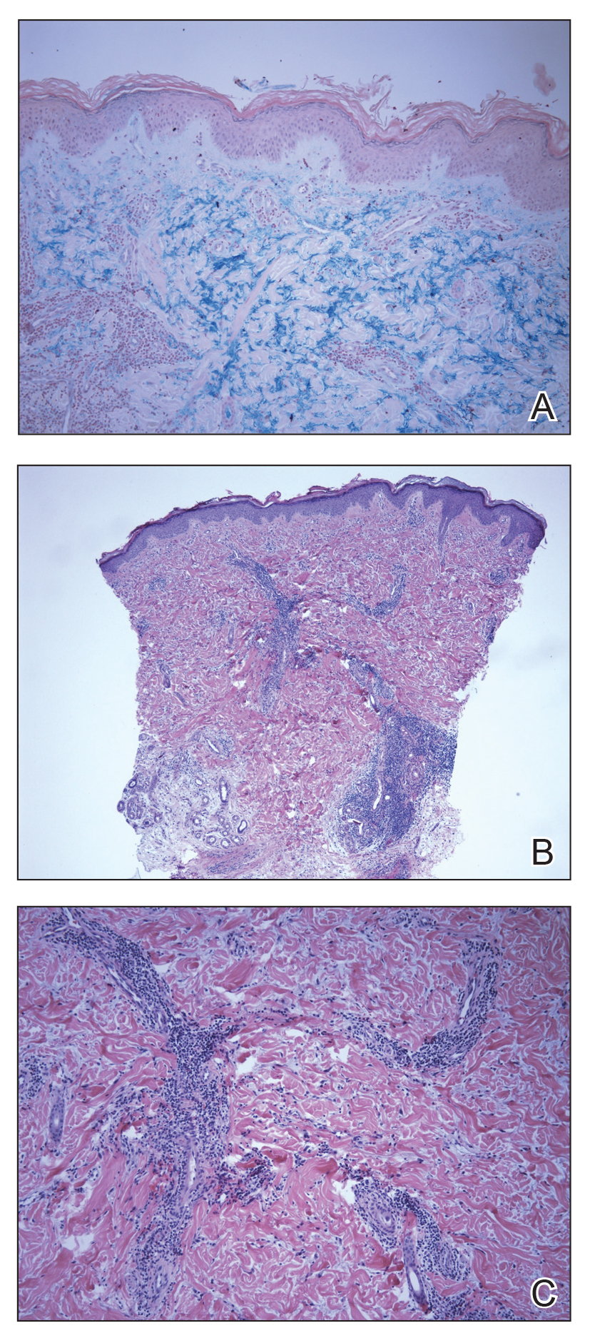
Histopathologic findings of superficial and deep perivascular and periadnexal lymphocytic infiltrates and interstitial dermal deposition of mucin in LET have remained consistent in the literature. Direct immunofluorescence has not revealed any complement or immunoglobulin deposition on the basement membrane.3,4 The epidermal characteristics are not as uniform, with the majority of cases in one review showing no epidermal changes and a minority showing minimal epidermal changes (eg, epidermal atrophy, hyperkeratosis, parakeratosis, acanthosis, spongiosis).5 When working up patients for LET, blood work usually is unremarkable, as LET rarely is associated with antinuclear antibodies or anti-Ro, anti-La, and anti-DNA antibodies.3,4 Lupus erythematosus tumidus generally is an independent process, but it has been reported to coexist with discoid lupus erythematosus and systemic lupus erythematosus in rare cases.6
The lesions of LET have been consistently described in the literature as photosensitive, erythematous, non-scarring, annular plaques and papules commonly occurring on the head/neck and other sun-exposed areas that do not cause hypopigmentation.3 Treatment of LET consists of systemic treatment with antimalarial drugs, sunscreens, and topical steroids for flares.
Lupus erythematosus tumidus is rare in children, with few case reports noted in the literature. Sonntag et al2 documented the disease in 3 children ranging from 3 to 8 years of age. Furthermore, Ruiz and Sanchez7 reported a case of LET in a 16-year-old adolescent girl. Our case is unique in that the lesions only occurred on the hands, whereas most case reports document distribution of the lesions on the head, neck, face, arms, back, and chest. Our patient’s age and the location of the lesions make it a unique clinical presentation of LET.
Reports in the literature show evidence of heterogeneity in the presentation, classification, and some of the histopathologic features of LET; however, there are minimal data on childhood LET. Further research and investigations are needed to more precisely define this condition.
Acknowledgment
The authors acknowledge Richard Schwartz, MD (Akron, Ohio), for reading the biopsy reports and assisting with photomicrographs.
- Hoffmann E. Demonstrationen: lupus erythematosus tumidus. Derm Zeitschr. 1909;16:159-160.
- Sonntag M, Lehmann P, Megahed M, et al. Lupus erythematosus tumidus in childhood. Dermatology. 2003;207:188-192.
- Schmitt V, Meuth AM, Amler S, et al. Lupus erythematosus tumidus is a separate subtype of cutaneous lupus erythematosus. Br J Dermatol. 2010;162:64-73.
- Vieira V, Del Pozo J, Yebra-Pimentel MT, et al. Lupus erythematosus tumidus: a series of 26 cases. Int J Dermatol. 2006;45:512-517.
- Kuhn A, Richter-Hintz D, Oslislo C, et al. Lupus erythematosus tumidus—a neglected subset of cutaneous lupus erythematosus: report of 40 cases. Arch Dermatol. 2000;136:1033-1041.
- Chen X, Wang S, Li L. A case report of lupus erythematosus tumidus converted from discoid lupus erythematosus. Medicine (Baltimore). 2018;97:e0375.
- Ruiz H, Sanchez J. Tumid lupus erythematosus. Am J Dermatopathol. 1999;21:356-360.
To the Editor:
Lupus erythematosus tumidus (LET) is a rarely diagnosed condition that was first described in 1909 by Hoffmann.1 Limited cases have been reported in the literature, with few documenting the disease in children.2 We report a unique clinical case of LET in a 14-year-old adolescent boy that was distributed solely on the hands. With slight heterogeneity in regards to clinical presentation and histopathology, there is a need for further exploration with regard to LET.
A 14-year-old adolescent boy presented to the dermatology clinic with progressive bilateral edema of 1 year’s duration with plaques and some scaling on the dorsal aspects of the digits and the nail bases predominantly on the right hand (Figure 1) and to a lesser extent on the left hand. The edema, erythema, and tenderness started in the right fifth digit; soon after the edema appeared, plaques began to form at the base of each nail bed, and the edema and erythema progressively spread to the other digits. He denied worsening of symptoms when exposed to cold temperatures. A complete review of systems was negative. The differential diagnoses included chilblain lupus erythematosus, perniosis, dermatomyositis, and polymorphous light eruption. A punch biopsy from the right fourth digit was performed.
The biopsy showed superficial and deep perivascular and periadnexal mononuclear inflammation with large amounts of interstitial mucin deposition (Figure 2). The epidermis exhibited a loose orthokeratotic scale with no signs of interface damage. A diagnosis of perniosis was entertained but was ruled out due to the lack of papillary dermal edema and large amounts of mucin. With the lack of interface change and large amounts of mucin, a diagnosis of LET was favored over chilblain lupus erythematosus, as the latter diagnosis typically demonstrates interface change. The patient was started on hydroxychloroquine 200 mg twice daily and a short course of prednisone, and improvement of the lesions/plaques was noted at follow-up 6 weeks later. Continued improvement was noted 2 years after the initial presentation. His condition recurred when the hydroxychloroquine dosage was reduced to 200 mg once daily after 1 year. The patient did not report any adverse sequelae to treatment.
Histopathologic findings of superficial and deep perivascular and periadnexal lymphocytic infiltrates and interstitial dermal deposition of mucin in LET have remained consistent in the literature. Direct immunofluorescence has not revealed any complement or immunoglobulin deposition on the basement membrane.3,4 The epidermal characteristics are not as uniform, with the majority of cases in one review showing no epidermal changes and a minority showing minimal epidermal changes (eg, epidermal atrophy, hyperkeratosis, parakeratosis, acanthosis, spongiosis).5 When working up patients for LET, blood work usually is unremarkable, as LET rarely is associated with antinuclear antibodies or anti-Ro, anti-La, and anti-DNA antibodies.3,4 Lupus erythematosus tumidus generally is an independent process, but it has been reported to coexist with discoid lupus erythematosus and systemic lupus erythematosus in rare cases.6
The lesions of LET have been consistently described in the literature as photosensitive, erythematous, non-scarring, annular plaques and papules commonly occurring on the head/neck and other sun-exposed areas that do not cause hypopigmentation.3 Treatment of LET consists of systemic treatment with antimalarial drugs, sunscreens, and topical steroids for flares.
Lupus erythematosus tumidus is rare in children, with few case reports noted in the literature. Sonntag et al2 documented the disease in 3 children ranging from 3 to 8 years of age. Furthermore, Ruiz and Sanchez7 reported a case of LET in a 16-year-old adolescent girl. Our case is unique in that the lesions only occurred on the hands, whereas most case reports document distribution of the lesions on the head, neck, face, arms, back, and chest. Our patient’s age and the location of the lesions make it a unique clinical presentation of LET.
Reports in the literature show evidence of heterogeneity in the presentation, classification, and some of the histopathologic features of LET; however, there are minimal data on childhood LET. Further research and investigations are needed to more precisely define this condition.
Acknowledgment
The authors acknowledge Richard Schwartz, MD (Akron, Ohio), for reading the biopsy reports and assisting with photomicrographs.
To the Editor:
Lupus erythematosus tumidus (LET) is a rarely diagnosed condition that was first described in 1909 by Hoffmann.1 Limited cases have been reported in the literature, with few documenting the disease in children.2 We report a unique clinical case of LET in a 14-year-old adolescent boy that was distributed solely on the hands. With slight heterogeneity in regards to clinical presentation and histopathology, there is a need for further exploration with regard to LET.
A 14-year-old adolescent boy presented to the dermatology clinic with progressive bilateral edema of 1 year’s duration with plaques and some scaling on the dorsal aspects of the digits and the nail bases predominantly on the right hand (Figure 1) and to a lesser extent on the left hand. The edema, erythema, and tenderness started in the right fifth digit; soon after the edema appeared, plaques began to form at the base of each nail bed, and the edema and erythema progressively spread to the other digits. He denied worsening of symptoms when exposed to cold temperatures. A complete review of systems was negative. The differential diagnoses included chilblain lupus erythematosus, perniosis, dermatomyositis, and polymorphous light eruption. A punch biopsy from the right fourth digit was performed.
The biopsy showed superficial and deep perivascular and periadnexal mononuclear inflammation with large amounts of interstitial mucin deposition (Figure 2). The epidermis exhibited a loose orthokeratotic scale with no signs of interface damage. A diagnosis of perniosis was entertained but was ruled out due to the lack of papillary dermal edema and large amounts of mucin. With the lack of interface change and large amounts of mucin, a diagnosis of LET was favored over chilblain lupus erythematosus, as the latter diagnosis typically demonstrates interface change. The patient was started on hydroxychloroquine 200 mg twice daily and a short course of prednisone, and improvement of the lesions/plaques was noted at follow-up 6 weeks later. Continued improvement was noted 2 years after the initial presentation. His condition recurred when the hydroxychloroquine dosage was reduced to 200 mg once daily after 1 year. The patient did not report any adverse sequelae to treatment.
Histopathologic findings of superficial and deep perivascular and periadnexal lymphocytic infiltrates and interstitial dermal deposition of mucin in LET have remained consistent in the literature. Direct immunofluorescence has not revealed any complement or immunoglobulin deposition on the basement membrane.3,4 The epidermal characteristics are not as uniform, with the majority of cases in one review showing no epidermal changes and a minority showing minimal epidermal changes (eg, epidermal atrophy, hyperkeratosis, parakeratosis, acanthosis, spongiosis).5 When working up patients for LET, blood work usually is unremarkable, as LET rarely is associated with antinuclear antibodies or anti-Ro, anti-La, and anti-DNA antibodies.3,4 Lupus erythematosus tumidus generally is an independent process, but it has been reported to coexist with discoid lupus erythematosus and systemic lupus erythematosus in rare cases.6
The lesions of LET have been consistently described in the literature as photosensitive, erythematous, non-scarring, annular plaques and papules commonly occurring on the head/neck and other sun-exposed areas that do not cause hypopigmentation.3 Treatment of LET consists of systemic treatment with antimalarial drugs, sunscreens, and topical steroids for flares.
Lupus erythematosus tumidus is rare in children, with few case reports noted in the literature. Sonntag et al2 documented the disease in 3 children ranging from 3 to 8 years of age. Furthermore, Ruiz and Sanchez7 reported a case of LET in a 16-year-old adolescent girl. Our case is unique in that the lesions only occurred on the hands, whereas most case reports document distribution of the lesions on the head, neck, face, arms, back, and chest. Our patient’s age and the location of the lesions make it a unique clinical presentation of LET.
Reports in the literature show evidence of heterogeneity in the presentation, classification, and some of the histopathologic features of LET; however, there are minimal data on childhood LET. Further research and investigations are needed to more precisely define this condition.
Acknowledgment
The authors acknowledge Richard Schwartz, MD (Akron, Ohio), for reading the biopsy reports and assisting with photomicrographs.
- Hoffmann E. Demonstrationen: lupus erythematosus tumidus. Derm Zeitschr. 1909;16:159-160.
- Sonntag M, Lehmann P, Megahed M, et al. Lupus erythematosus tumidus in childhood. Dermatology. 2003;207:188-192.
- Schmitt V, Meuth AM, Amler S, et al. Lupus erythematosus tumidus is a separate subtype of cutaneous lupus erythematosus. Br J Dermatol. 2010;162:64-73.
- Vieira V, Del Pozo J, Yebra-Pimentel MT, et al. Lupus erythematosus tumidus: a series of 26 cases. Int J Dermatol. 2006;45:512-517.
- Kuhn A, Richter-Hintz D, Oslislo C, et al. Lupus erythematosus tumidus—a neglected subset of cutaneous lupus erythematosus: report of 40 cases. Arch Dermatol. 2000;136:1033-1041.
- Chen X, Wang S, Li L. A case report of lupus erythematosus tumidus converted from discoid lupus erythematosus. Medicine (Baltimore). 2018;97:e0375.
- Ruiz H, Sanchez J. Tumid lupus erythematosus. Am J Dermatopathol. 1999;21:356-360.
- Hoffmann E. Demonstrationen: lupus erythematosus tumidus. Derm Zeitschr. 1909;16:159-160.
- Sonntag M, Lehmann P, Megahed M, et al. Lupus erythematosus tumidus in childhood. Dermatology. 2003;207:188-192.
- Schmitt V, Meuth AM, Amler S, et al. Lupus erythematosus tumidus is a separate subtype of cutaneous lupus erythematosus. Br J Dermatol. 2010;162:64-73.
- Vieira V, Del Pozo J, Yebra-Pimentel MT, et al. Lupus erythematosus tumidus: a series of 26 cases. Int J Dermatol. 2006;45:512-517.
- Kuhn A, Richter-Hintz D, Oslislo C, et al. Lupus erythematosus tumidus—a neglected subset of cutaneous lupus erythematosus: report of 40 cases. Arch Dermatol. 2000;136:1033-1041.
- Chen X, Wang S, Li L. A case report of lupus erythematosus tumidus converted from discoid lupus erythematosus. Medicine (Baltimore). 2018;97:e0375.
- Ruiz H, Sanchez J. Tumid lupus erythematosus. Am J Dermatopathol. 1999;21:356-360.
Practice Points
- Lupus erythematosus tumidus rarely occurs in the pediatric population.
- Lupus erythematosus tumidus is a unique subset of lupus associated with lack of interface change on histology and large amounts of mucin.
- Lesions typically present on the face and trunk but can very rarely present on the extremities and hands.

Would a universal basic income help our patients?
If you take a nighttime stroll through the downtown district in any major U.S. city – and certainly in my hometown of Baltimore – you’ll find people sleeping on the streets. Advocates talk about “the homeless mentally ill,” and it’s estimated that a quarter of homeless persons suffer from psychiatric conditions. As psychiatrists, it’s good that we care about the homeless mentally ill. As human beings, shouldn’t we also care about the addicted and indigent homeless? In a country of wealth, it continues to be a disgrace that we have people who live in tent encampments, or who literally sleep on the ground in public places with all their belongings gathered around them.

With 25 contenders for the Democratic presidential nomination, Andrew Yang has caught my attention with his platform for a universal basic income (UBI), or “freedom dividend,” for all adults. Mr. Yang’s premise is a simple one: He’d like to give every person over the age of 18 a $1,000-a-month government-supplied income, funded by a new Value Added Tax (VAT), with no stipulations. I find the concept intriguing. It is the one suggestion that might make a drastic dent in extreme poverty in our country. In theory, every adult would have enough money to afford a place to sleep.
Paul Nestadt, MD, a psychiatrist at Johns Hopkins in Baltimore, agrees with the concept of a universal basic income. “The UBI seems like a humane and egalitarian way to eliminate starvation; it provides a ‘floor’ for poverty. It is a reasonable bare minimum, especially for our patients with serious mental illness who struggle to navigate the bureaucratic steeplechase required to have their basic needs met.” Dr. Nestadt believes that some of the other presidential candidates are supporting policies that would also accomplish this goal.

So who is Andrew Yang? With no political experience, he bills himself as a technology entrepreneur who started a national education company, then went on to found Venture for America, an organization designed to create jobs. During the first round of Democratic debates, few questions were directed to him, and he was the lone gentleman on the stage without a tie. In addition to the UBI, he supports Medicare-for-all and “human-centered capitalism.” His slogan is “Humanity First,” and his website includes policy statements on a vast number of topics: everything from robocalls to wildfires to free marital counseling for all. His supporters call themselves the Yang Gang, and as of this writing, he polls at number 8 – with just 2% of the projected vote – among the 25 Democratic candidates.
While I find the prospect of a universal basic income appealing from the perspective of making a dent in extreme poverty, this is not the demographic Mr. Yang is targeting. His platform is based on the prediction that automation will continue to eliminate jobs at a rate that will devastate our people and our economy. In his book, The War on Normal People (Hachette Book Group, 2018), Yang writes about the UBI: “It’s simple, it’s fair, it’s equitable, it’s easy to understand, it benefits at least 80 percent of the population, and it will be necessary to maintain the fabric of society during the automation wave.” Mr. Yang contends that at least one-third of Americans are at risk of losing their jobs to automation.
It’s difficult to imagine that it would not be helpful to everyone’s mental health – not just those individuals with psychiatric disorders – to be freed from the worry of earning enough money to survive. While our welfare, disability, and Medicaid systems provide a safety net to many Americans, they certainly don’t cover everyone, and they engender a sense of unfairness and anger. Our current system allows that some people work hard and struggle to meet their basic needs and pay medical bills while others – usually the disabled or the poor – receive government benefits and Medicaid and/or Medicare.
In the “Making Sense” podcast hosted by Sam Harris, “A conversation with Andrew Yang,” Mr. Yang made the point that a UBI is not a new concept – economists and politicians, including Richard Nixon, have supported the idea since the 1960s. A bill proposing a basic income passed in the House of Representatives in 1971 but did not pass in the Senate. In the podcast, Mr. Yang addresses the question of whether people are responsible for their own success, and whether, as Mr. Harris puts it, “it’s just simply wrong to hand out money to people.”
“It’s not as if the truck drivers are about to get dumber and lazier overnight,” Mr. Yang responded. “It’s just that their trucks are going to start driving themselves ... it has nothing to do with their character and work ethic.” He goes on to discuss how workers who lose jobs often leave the workforce and many go on disability.
“Right now, the country’s locked in a struggle between functioning and dysfunction, between reason and unreason, and scarcity and abundance, and scarcity is winning and that’s what we have to reverse through universal basic income; it’s our best way forward.”
If we are optimistic that our government could afford to provide everyone with both a UBI and a universal health plan, such as Medicare-for-all, I still wonder if there might be a societal downside. For self-motivated individuals, a sustenance allowance is unlikely to weaken a drive to achieve. But might there be people who decide they can live on this income, who choose instead to pursue leisure activities rather than pursue education and vocation? Mr. Harris asked Mr. Yang if we would be “subsidizing all the people in their mothers’ basement playing video games.”
Mr. Yang responded, “If you’re getting a thousand dollars a month, then you’re much more likely to get out of your parents’ basement and visit friends and find things to do that are somewhat more social and external-facing. A lot of the reason a lot of these men are retreating is because there’s no real economic security or path forward for them, and they feel much better served by going online and hanging out with their friends and making measurable progress in their gaming environment.”
I like to think that an automatic income would not crush a society’s motivation and productivity, and that money provided to people would fuel education, our economy, an ability to save, and entrepreneurial endeavors, but the truth is that we just don’t know. I would love to see such an experiment done as a large-scale pilot, with the ability to undo the experiment if it fails.
Andrew Yang remains a long shot as the Democratic presidential nominee. His platform, however, is enticing, and he takes on the imminent automation crisis in a way that no one else is actively addressing. His concepts include a degree of humanity that feels welcome when our current president is tweeting that those who are unhappy here should leave. While Mr. Yang is a bit lost in the fray, I do hope his innovative spirit gains some traction.
Dr. Miller is the coauthor with Annette Hanson, MD, of “Committed: The Battle Over Involuntary Psychiatric Care” (Baltimore: Johns Hopkins University Press, 2016) and has a private practice in Baltimore.
If you take a nighttime stroll through the downtown district in any major U.S. city – and certainly in my hometown of Baltimore – you’ll find people sleeping on the streets. Advocates talk about “the homeless mentally ill,” and it’s estimated that a quarter of homeless persons suffer from psychiatric conditions. As psychiatrists, it’s good that we care about the homeless mentally ill. As human beings, shouldn’t we also care about the addicted and indigent homeless? In a country of wealth, it continues to be a disgrace that we have people who live in tent encampments, or who literally sleep on the ground in public places with all their belongings gathered around them.
With 25 contenders for the Democratic presidential nomination, Andrew Yang has caught my attention with his platform for a universal basic income (UBI), or “freedom dividend,” for all adults. Mr. Yang’s premise is a simple one: He’d like to give every person over the age of 18 a $1,000-a-month government-supplied income, funded by a new Value Added Tax (VAT), with no stipulations. I find the concept intriguing. It is the one suggestion that might make a drastic dent in extreme poverty in our country. In theory, every adult would have enough money to afford a place to sleep.
Paul Nestadt, MD, a psychiatrist at Johns Hopkins in Baltimore, agrees with the concept of a universal basic income. “The UBI seems like a humane and egalitarian way to eliminate starvation; it provides a ‘floor’ for poverty. It is a reasonable bare minimum, especially for our patients with serious mental illness who struggle to navigate the bureaucratic steeplechase required to have their basic needs met.” Dr. Nestadt believes that some of the other presidential candidates are supporting policies that would also accomplish this goal.
So who is Andrew Yang? With no political experience, he bills himself as a technology entrepreneur who started a national education company, then went on to found Venture for America, an organization designed to create jobs. During the first round of Democratic debates, few questions were directed to him, and he was the lone gentleman on the stage without a tie. In addition to the UBI, he supports Medicare-for-all and “human-centered capitalism.” His slogan is “Humanity First,” and his website includes policy statements on a vast number of topics: everything from robocalls to wildfires to free marital counseling for all. His supporters call themselves the Yang Gang, and as of this writing, he polls at number 8 – with just 2% of the projected vote – among the 25 Democratic candidates.
While I find the prospect of a universal basic income appealing from the perspective of making a dent in extreme poverty, this is not the demographic Mr. Yang is targeting. His platform is based on the prediction that automation will continue to eliminate jobs at a rate that will devastate our people and our economy. In his book, The War on Normal People (Hachette Book Group, 2018), Yang writes about the UBI: “It’s simple, it’s fair, it’s equitable, it’s easy to understand, it benefits at least 80 percent of the population, and it will be necessary to maintain the fabric of society during the automation wave.” Mr. Yang contends that at least one-third of Americans are at risk of losing their jobs to automation.
It’s difficult to imagine that it would not be helpful to everyone’s mental health – not just those individuals with psychiatric disorders – to be freed from the worry of earning enough money to survive. While our welfare, disability, and Medicaid systems provide a safety net to many Americans, they certainly don’t cover everyone, and they engender a sense of unfairness and anger. Our current system allows that some people work hard and struggle to meet their basic needs and pay medical bills while others – usually the disabled or the poor – receive government benefits and Medicaid and/or Medicare.
In the “Making Sense” podcast hosted by Sam Harris, “A conversation with Andrew Yang,” Mr. Yang made the point that a UBI is not a new concept – economists and politicians, including Richard Nixon, have supported the idea since the 1960s. A bill proposing a basic income passed in the House of Representatives in 1971 but did not pass in the Senate. In the podcast, Mr. Yang addresses the question of whether people are responsible for their own success, and whether, as Mr. Harris puts it, “it’s just simply wrong to hand out money to people.”
“It’s not as if the truck drivers are about to get dumber and lazier overnight,” Mr. Yang responded. “It’s just that their trucks are going to start driving themselves ... it has nothing to do with their character and work ethic.” He goes on to discuss how workers who lose jobs often leave the workforce and many go on disability.
“Right now, the country’s locked in a struggle between functioning and dysfunction, between reason and unreason, and scarcity and abundance, and scarcity is winning and that’s what we have to reverse through universal basic income; it’s our best way forward.”
If we are optimistic that our government could afford to provide everyone with both a UBI and a universal health plan, such as Medicare-for-all, I still wonder if there might be a societal downside. For self-motivated individuals, a sustenance allowance is unlikely to weaken a drive to achieve. But might there be people who decide they can live on this income, who choose instead to pursue leisure activities rather than pursue education and vocation? Mr. Harris asked Mr. Yang if we would be “subsidizing all the people in their mothers’ basement playing video games.”
Mr. Yang responded, “If you’re getting a thousand dollars a month, then you’re much more likely to get out of your parents’ basement and visit friends and find things to do that are somewhat more social and external-facing. A lot of the reason a lot of these men are retreating is because there’s no real economic security or path forward for them, and they feel much better served by going online and hanging out with their friends and making measurable progress in their gaming environment.”
I like to think that an automatic income would not crush a society’s motivation and productivity, and that money provided to people would fuel education, our economy, an ability to save, and entrepreneurial endeavors, but the truth is that we just don’t know. I would love to see such an experiment done as a large-scale pilot, with the ability to undo the experiment if it fails.
Andrew Yang remains a long shot as the Democratic presidential nominee. His platform, however, is enticing, and he takes on the imminent automation crisis in a way that no one else is actively addressing. His concepts include a degree of humanity that feels welcome when our current president is tweeting that those who are unhappy here should leave. While Mr. Yang is a bit lost in the fray, I do hope his innovative spirit gains some traction.
Dr. Miller is the coauthor with Annette Hanson, MD, of “Committed: The Battle Over Involuntary Psychiatric Care” (Baltimore: Johns Hopkins University Press, 2016) and has a private practice in Baltimore.
If you take a nighttime stroll through the downtown district in any major U.S. city – and certainly in my hometown of Baltimore – you’ll find people sleeping on the streets. Advocates talk about “the homeless mentally ill,” and it’s estimated that a quarter of homeless persons suffer from psychiatric conditions. As psychiatrists, it’s good that we care about the homeless mentally ill. As human beings, shouldn’t we also care about the addicted and indigent homeless? In a country of wealth, it continues to be a disgrace that we have people who live in tent encampments, or who literally sleep on the ground in public places with all their belongings gathered around them.
With 25 contenders for the Democratic presidential nomination, Andrew Yang has caught my attention with his platform for a universal basic income (UBI), or “freedom dividend,” for all adults. Mr. Yang’s premise is a simple one: He’d like to give every person over the age of 18 a $1,000-a-month government-supplied income, funded by a new Value Added Tax (VAT), with no stipulations. I find the concept intriguing. It is the one suggestion that might make a drastic dent in extreme poverty in our country. In theory, every adult would have enough money to afford a place to sleep.
Paul Nestadt, MD, a psychiatrist at Johns Hopkins in Baltimore, agrees with the concept of a universal basic income. “The UBI seems like a humane and egalitarian way to eliminate starvation; it provides a ‘floor’ for poverty. It is a reasonable bare minimum, especially for our patients with serious mental illness who struggle to navigate the bureaucratic steeplechase required to have their basic needs met.” Dr. Nestadt believes that some of the other presidential candidates are supporting policies that would also accomplish this goal.
So who is Andrew Yang? With no political experience, he bills himself as a technology entrepreneur who started a national education company, then went on to found Venture for America, an organization designed to create jobs. During the first round of Democratic debates, few questions were directed to him, and he was the lone gentleman on the stage without a tie. In addition to the UBI, he supports Medicare-for-all and “human-centered capitalism.” His slogan is “Humanity First,” and his website includes policy statements on a vast number of topics: everything from robocalls to wildfires to free marital counseling for all. His supporters call themselves the Yang Gang, and as of this writing, he polls at number 8 – with just 2% of the projected vote – among the 25 Democratic candidates.
While I find the prospect of a universal basic income appealing from the perspective of making a dent in extreme poverty, this is not the demographic Mr. Yang is targeting. His platform is based on the prediction that automation will continue to eliminate jobs at a rate that will devastate our people and our economy. In his book, The War on Normal People (Hachette Book Group, 2018), Yang writes about the UBI: “It’s simple, it’s fair, it’s equitable, it’s easy to understand, it benefits at least 80 percent of the population, and it will be necessary to maintain the fabric of society during the automation wave.” Mr. Yang contends that at least one-third of Americans are at risk of losing their jobs to automation.
It’s difficult to imagine that it would not be helpful to everyone’s mental health – not just those individuals with psychiatric disorders – to be freed from the worry of earning enough money to survive. While our welfare, disability, and Medicaid systems provide a safety net to many Americans, they certainly don’t cover everyone, and they engender a sense of unfairness and anger. Our current system allows that some people work hard and struggle to meet their basic needs and pay medical bills while others – usually the disabled or the poor – receive government benefits and Medicaid and/or Medicare.
In the “Making Sense” podcast hosted by Sam Harris, “A conversation with Andrew Yang,” Mr. Yang made the point that a UBI is not a new concept – economists and politicians, including Richard Nixon, have supported the idea since the 1960s. A bill proposing a basic income passed in the House of Representatives in 1971 but did not pass in the Senate. In the podcast, Mr. Yang addresses the question of whether people are responsible for their own success, and whether, as Mr. Harris puts it, “it’s just simply wrong to hand out money to people.”
“It’s not as if the truck drivers are about to get dumber and lazier overnight,” Mr. Yang responded. “It’s just that their trucks are going to start driving themselves ... it has nothing to do with their character and work ethic.” He goes on to discuss how workers who lose jobs often leave the workforce and many go on disability.
“Right now, the country’s locked in a struggle between functioning and dysfunction, between reason and unreason, and scarcity and abundance, and scarcity is winning and that’s what we have to reverse through universal basic income; it’s our best way forward.”
If we are optimistic that our government could afford to provide everyone with both a UBI and a universal health plan, such as Medicare-for-all, I still wonder if there might be a societal downside. For self-motivated individuals, a sustenance allowance is unlikely to weaken a drive to achieve. But might there be people who decide they can live on this income, who choose instead to pursue leisure activities rather than pursue education and vocation? Mr. Harris asked Mr. Yang if we would be “subsidizing all the people in their mothers’ basement playing video games.”
Mr. Yang responded, “If you’re getting a thousand dollars a month, then you’re much more likely to get out of your parents’ basement and visit friends and find things to do that are somewhat more social and external-facing. A lot of the reason a lot of these men are retreating is because there’s no real economic security or path forward for them, and they feel much better served by going online and hanging out with their friends and making measurable progress in their gaming environment.”
I like to think that an automatic income would not crush a society’s motivation and productivity, and that money provided to people would fuel education, our economy, an ability to save, and entrepreneurial endeavors, but the truth is that we just don’t know. I would love to see such an experiment done as a large-scale pilot, with the ability to undo the experiment if it fails.
Andrew Yang remains a long shot as the Democratic presidential nominee. His platform, however, is enticing, and he takes on the imminent automation crisis in a way that no one else is actively addressing. His concepts include a degree of humanity that feels welcome when our current president is tweeting that those who are unhappy here should leave. While Mr. Yang is a bit lost in the fray, I do hope his innovative spirit gains some traction.
Dr. Miller is the coauthor with Annette Hanson, MD, of “Committed: The Battle Over Involuntary Psychiatric Care” (Baltimore: Johns Hopkins University Press, 2016) and has a private practice in Baltimore.