User login
Nitrous oxide linked to less pain in tattoo removal
SAN DIEGO – The results of a small, single-site study suggest that nitrous oxide (NO) can play a significant role in reducing pain during laser tattoo removal.
“Nitrous oxide is a safe and effective option for patients, particularly those who have large tattoos that can’t be adequately numbed with injections or topical numbing,” the study’s lead author, Jared Mallalieu, DO, said in an interview. “NO has allowed us to treat larger tattoos – full sleeve or large back tattoos – in a single setting, which has made treatment more convenient for patients.”
Patients fared better on pain measures when they received NO, compared with topical and injectable anesthetics, according to Dr. Mallalieu, a cosmetic surgeon at the Laser Center of Maryland, Severna Park. The results were so dramatic that EMLA cream is now rarely used for patients in his clinic, although injectable lidocaine is used on smaller tattoos (smaller than 5 inches by 5 inches), he said.
The use of NO comes with challenges, however, in terms of the extra time and patient monitoring required, he said.
Dr. Mallalieu and his associates reported the results in an e-poster at the annual meeting of the American Society for Laser Medicine and Surgery.
Laser tattoo removal can be an agonizing process. “Patients describe it as being significantly more painful than getting a tattoo,” Dr. Mallalieu said. “The intense pain only lasts during the treatment,” he said, “though many patients will note some discomfort for a few hours after a treatment session.”
Most clinics use a topical cream, such as lidocaine/prilocaine (EMLA) or topical benzocaine/lidocaine/tetracaine (BLT), as an anesthetic for these procedures. “Our center has also used 1% lidocaine with epinephrine in small doses of up to 7 mg/kg,” he said. “The injections are much better than the cream.”
Sometimes the clinic uses a device that blows cold air on the skin, which “helps a little,” he added.
For the study, conducted in 2014, 23 laser tattoo removal patients were surveyed about their pain levels using a 1-10 scale, after undergoing a total of 41 single-location procedures.
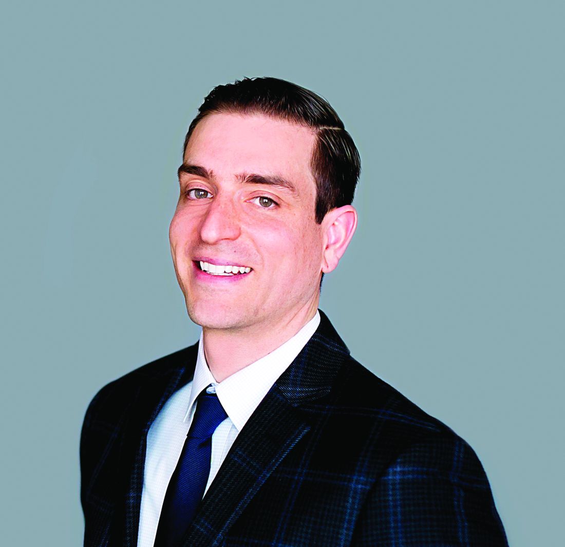
The average pain rating during the procedure was 9.1 for those treated only with lidocaine/prilocaine, 5.4 for injections of lidocaine with epinephrine alone, and 6.8 for both lidocaine/prilocaine and lidocaine with epinephrine injections.
The average pain rating for NO alone was 2.6, and was 3.6 for those who received both the injection treatment and lidocaine/prilocaine. Three of 12 NO patients reported anxiety.
Another benefit is that patients can drive after receiving NO, unlike other anesthetics, which leave patients sedated, he said. “Levels of NO are titrated to keep the patient sedated, but breathing on their own,” and patients can be easily woken up within moments of stopping the NO.
However, the use of NO requires more time to set up and more monitoring, he added. The average treatment time for procedures with NO was 27 minutes compared with 4 minutes for the other procedure, and “the patient is put on a monitor that measures pulse rate and oxygenation levels,” which not only takes more time, but requires additional staff to watch the patient. “Also, it takes about 3-5 minutes to slowly the titrate the NO to a perfect level.”
The study points out that physicians at the clinic are the only ones who perform the procedures that use NO, but at many clinics, nonphysicians perform tattoo removals.
As for cost, “NO and oxygen tanks are rather inexpensive to purchase and maintain, and there are various small units which serve to titrate the gas,” Dr. Mallalieu said. “We do charge our patients a small fee because of the added personnel and time cost associated with the procedure. As tattoo removal is considered a cosmetic procedure, insurance doesn’t come into play.”
Training to administer NO brings up the issue of what is allowed in the state, he said. Physicians can give sedation to patients, “but some states may limit the degree to which a patient can be sedated in an office. If the physician has a certified operating room, this is not a problem,” he added. “Because dentists commonly use NO, we followed the American Dental Association guidelines ... As we employ an anesthesiologist, we were quite familiar with it. That said, the administration of NO is not complex and [is] easily mastered.”
Dr. Mallalieu reported no relevant disclosures.
SAN DIEGO – The results of a small, single-site study suggest that nitrous oxide (NO) can play a significant role in reducing pain during laser tattoo removal.
“Nitrous oxide is a safe and effective option for patients, particularly those who have large tattoos that can’t be adequately numbed with injections or topical numbing,” the study’s lead author, Jared Mallalieu, DO, said in an interview. “NO has allowed us to treat larger tattoos – full sleeve or large back tattoos – in a single setting, which has made treatment more convenient for patients.”
Patients fared better on pain measures when they received NO, compared with topical and injectable anesthetics, according to Dr. Mallalieu, a cosmetic surgeon at the Laser Center of Maryland, Severna Park. The results were so dramatic that EMLA cream is now rarely used for patients in his clinic, although injectable lidocaine is used on smaller tattoos (smaller than 5 inches by 5 inches), he said.
The use of NO comes with challenges, however, in terms of the extra time and patient monitoring required, he said.
Dr. Mallalieu and his associates reported the results in an e-poster at the annual meeting of the American Society for Laser Medicine and Surgery.
Laser tattoo removal can be an agonizing process. “Patients describe it as being significantly more painful than getting a tattoo,” Dr. Mallalieu said. “The intense pain only lasts during the treatment,” he said, “though many patients will note some discomfort for a few hours after a treatment session.”
Most clinics use a topical cream, such as lidocaine/prilocaine (EMLA) or topical benzocaine/lidocaine/tetracaine (BLT), as an anesthetic for these procedures. “Our center has also used 1% lidocaine with epinephrine in small doses of up to 7 mg/kg,” he said. “The injections are much better than the cream.”
Sometimes the clinic uses a device that blows cold air on the skin, which “helps a little,” he added.
For the study, conducted in 2014, 23 laser tattoo removal patients were surveyed about their pain levels using a 1-10 scale, after undergoing a total of 41 single-location procedures.
The average pain rating during the procedure was 9.1 for those treated only with lidocaine/prilocaine, 5.4 for injections of lidocaine with epinephrine alone, and 6.8 for both lidocaine/prilocaine and lidocaine with epinephrine injections.
The average pain rating for NO alone was 2.6, and was 3.6 for those who received both the injection treatment and lidocaine/prilocaine. Three of 12 NO patients reported anxiety.
Another benefit is that patients can drive after receiving NO, unlike other anesthetics, which leave patients sedated, he said. “Levels of NO are titrated to keep the patient sedated, but breathing on their own,” and patients can be easily woken up within moments of stopping the NO.
However, the use of NO requires more time to set up and more monitoring, he added. The average treatment time for procedures with NO was 27 minutes compared with 4 minutes for the other procedure, and “the patient is put on a monitor that measures pulse rate and oxygenation levels,” which not only takes more time, but requires additional staff to watch the patient. “Also, it takes about 3-5 minutes to slowly the titrate the NO to a perfect level.”
The study points out that physicians at the clinic are the only ones who perform the procedures that use NO, but at many clinics, nonphysicians perform tattoo removals.
As for cost, “NO and oxygen tanks are rather inexpensive to purchase and maintain, and there are various small units which serve to titrate the gas,” Dr. Mallalieu said. “We do charge our patients a small fee because of the added personnel and time cost associated with the procedure. As tattoo removal is considered a cosmetic procedure, insurance doesn’t come into play.”
Training to administer NO brings up the issue of what is allowed in the state, he said. Physicians can give sedation to patients, “but some states may limit the degree to which a patient can be sedated in an office. If the physician has a certified operating room, this is not a problem,” he added. “Because dentists commonly use NO, we followed the American Dental Association guidelines ... As we employ an anesthesiologist, we were quite familiar with it. That said, the administration of NO is not complex and [is] easily mastered.”
Dr. Mallalieu reported no relevant disclosures.
SAN DIEGO – The results of a small, single-site study suggest that nitrous oxide (NO) can play a significant role in reducing pain during laser tattoo removal.
“Nitrous oxide is a safe and effective option for patients, particularly those who have large tattoos that can’t be adequately numbed with injections or topical numbing,” the study’s lead author, Jared Mallalieu, DO, said in an interview. “NO has allowed us to treat larger tattoos – full sleeve or large back tattoos – in a single setting, which has made treatment more convenient for patients.”
Patients fared better on pain measures when they received NO, compared with topical and injectable anesthetics, according to Dr. Mallalieu, a cosmetic surgeon at the Laser Center of Maryland, Severna Park. The results were so dramatic that EMLA cream is now rarely used for patients in his clinic, although injectable lidocaine is used on smaller tattoos (smaller than 5 inches by 5 inches), he said.
The use of NO comes with challenges, however, in terms of the extra time and patient monitoring required, he said.
Dr. Mallalieu and his associates reported the results in an e-poster at the annual meeting of the American Society for Laser Medicine and Surgery.
Laser tattoo removal can be an agonizing process. “Patients describe it as being significantly more painful than getting a tattoo,” Dr. Mallalieu said. “The intense pain only lasts during the treatment,” he said, “though many patients will note some discomfort for a few hours after a treatment session.”
Most clinics use a topical cream, such as lidocaine/prilocaine (EMLA) or topical benzocaine/lidocaine/tetracaine (BLT), as an anesthetic for these procedures. “Our center has also used 1% lidocaine with epinephrine in small doses of up to 7 mg/kg,” he said. “The injections are much better than the cream.”
Sometimes the clinic uses a device that blows cold air on the skin, which “helps a little,” he added.
For the study, conducted in 2014, 23 laser tattoo removal patients were surveyed about their pain levels using a 1-10 scale, after undergoing a total of 41 single-location procedures.
The average pain rating during the procedure was 9.1 for those treated only with lidocaine/prilocaine, 5.4 for injections of lidocaine with epinephrine alone, and 6.8 for both lidocaine/prilocaine and lidocaine with epinephrine injections.
The average pain rating for NO alone was 2.6, and was 3.6 for those who received both the injection treatment and lidocaine/prilocaine. Three of 12 NO patients reported anxiety.
Another benefit is that patients can drive after receiving NO, unlike other anesthetics, which leave patients sedated, he said. “Levels of NO are titrated to keep the patient sedated, but breathing on their own,” and patients can be easily woken up within moments of stopping the NO.
However, the use of NO requires more time to set up and more monitoring, he added. The average treatment time for procedures with NO was 27 minutes compared with 4 minutes for the other procedure, and “the patient is put on a monitor that measures pulse rate and oxygenation levels,” which not only takes more time, but requires additional staff to watch the patient. “Also, it takes about 3-5 minutes to slowly the titrate the NO to a perfect level.”
The study points out that physicians at the clinic are the only ones who perform the procedures that use NO, but at many clinics, nonphysicians perform tattoo removals.
As for cost, “NO and oxygen tanks are rather inexpensive to purchase and maintain, and there are various small units which serve to titrate the gas,” Dr. Mallalieu said. “We do charge our patients a small fee because of the added personnel and time cost associated with the procedure. As tattoo removal is considered a cosmetic procedure, insurance doesn’t come into play.”
Training to administer NO brings up the issue of what is allowed in the state, he said. Physicians can give sedation to patients, “but some states may limit the degree to which a patient can be sedated in an office. If the physician has a certified operating room, this is not a problem,” he added. “Because dentists commonly use NO, we followed the American Dental Association guidelines ... As we employ an anesthesiologist, we were quite familiar with it. That said, the administration of NO is not complex and [is] easily mastered.”
Dr. Mallalieu reported no relevant disclosures.
Intervention may help stem weight gain tied to antipsychotics
SAN DIEGO – Extra pounds are a perennial problem for schizophrenia patients who take antipsychotic medications with weight-boosting side effects. Now, a new randomized study finds that veterans who took part in a 12-month behavioral intervention program performed better on weight-loss measurements than those in a control group.
The difference between the two groups was far from large, with those undergoing the intervention losing an average of 1.04 centimeters in waist circumference over a year, compared with an average gain of 0.25 centimeters in the control group (P less than .001).
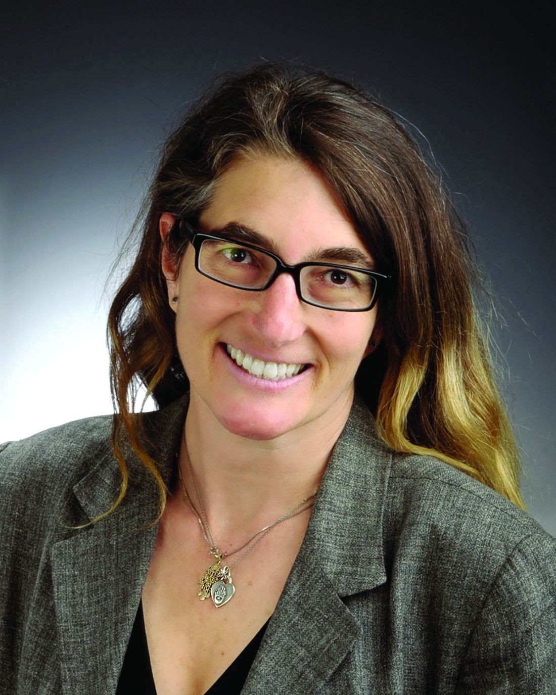
A recent meta-analysis found that weight gain is a potential side effect from prolonged use of “virtually all” antipsychotic medications, especially in those who have not taken the drugs previously (PLoS One. 2014 Apr 24;9[4]:e94112).
“Some of the most effective medications are associated with the highest weight-gain liability,” Dr. Ames said in an interview, “and patients with medication-resistant psychosis who don’t want to gain weight will resist taking these medications.”
As the meta-analysis notes, research suggests that the weight gain prompted by antipsychotics may boost mortality risk in patients with severe mental illness. The metabolic havoc linked to schizophrenia may be another factor: A 2010 summary of research notes that newer second-generation antipsychotics “generally tend to cause more problems relating to metabolic syndrome, such as obesity and type 2 diabetes mellitus,” compared with first-generation antipsychotics (FGA).
Weight gain “can be rapid and difficult to control,” the summary authors write,” adding that “the effect is worse with clozapine and olanzapine; minimal with aripiprazole and ziprasidone; and intermediate with other antipsychotics, including low-potency FGAs” (Am Fam Physician. 2010 Mar 1;81[5]:617-22).
For the new study, researchers randomly assigned 121 overweight or obese subjects with serious mental illness to either a “lifestyle balance” (LB) program (n = 62) or a usual care (UC) program (n = 59). All had become obese while taking an antipsychotic.
Subjects in the LB program met with registered dietitians for individual health coaching, weekly for 8 weeks and then monthly for up to 10 months. They also took part in 16 group nutrition and exercise classes over 2 months.
The UC group, meanwhile, met with case managers weekly for 8 weeks and then monthly for up to 10 months. They answered health questionnaires, underwent weight checks, and received self-help materials, Dr. Ames reported at the International Congress on Schizophrenia Research.
All of the participants lost weight, although waist circumference only declined in the intervention group. Body fat percentage declined in both groups, but by more (0.4% vs. 0.2%) in the intervention group, compared with UC (P = .038).
Judging by food diaries kept by 92% of the intervention group participants, their average daily caloric intake declined from 2,055 to 1,650 (P less than 0.001). The UC participants did not keep food diaries, so their caloric intake isn’t available.
Shouldn’t the intervention participants have lost more weight in light of such a large caloric decline? “Not necessarily,” Dr. Ames said. “Participants who were successful at making dietary changes began making these changes at different times throughout the study, some early and some well into the yearlong study. Decreases made in the latter part of the study would result in less weight lost. Also, exercise activity was variable, so decreased caloric intake could be offset by decreased physical activity.”
Other findings: Women did better than men at losing weight, and reducing waist circumference and body fat. There wasn’t a significant difference in the level of exercise between the groups. And researchers linked psychiatric illness insight in the LB group to greater weight loss but not in the UC group.
The study is supported by a $1.9 million VA grant for research from 2010 to 2017, Dr. Ames said.
In regard to cost-effectiveness, she said “a psychiatrist, nurse, or other mental health professional could easily weave these interventions into the care of patients in mental health settings. And the cost savings to patients by even losing just a few pounds could be enormous.”
The researchers hope to examine whether the intervention reduces the cost of medications, emergency department visits, and hospitalizations, she said.
Dr. Ames reports no relevant disclosures.
SAN DIEGO – Extra pounds are a perennial problem for schizophrenia patients who take antipsychotic medications with weight-boosting side effects. Now, a new randomized study finds that veterans who took part in a 12-month behavioral intervention program performed better on weight-loss measurements than those in a control group.
The difference between the two groups was far from large, with those undergoing the intervention losing an average of 1.04 centimeters in waist circumference over a year, compared with an average gain of 0.25 centimeters in the control group (P less than .001).
A recent meta-analysis found that weight gain is a potential side effect from prolonged use of “virtually all” antipsychotic medications, especially in those who have not taken the drugs previously (PLoS One. 2014 Apr 24;9[4]:e94112).
“Some of the most effective medications are associated with the highest weight-gain liability,” Dr. Ames said in an interview, “and patients with medication-resistant psychosis who don’t want to gain weight will resist taking these medications.”
As the meta-analysis notes, research suggests that the weight gain prompted by antipsychotics may boost mortality risk in patients with severe mental illness. The metabolic havoc linked to schizophrenia may be another factor: A 2010 summary of research notes that newer second-generation antipsychotics “generally tend to cause more problems relating to metabolic syndrome, such as obesity and type 2 diabetes mellitus,” compared with first-generation antipsychotics (FGA).
Weight gain “can be rapid and difficult to control,” the summary authors write,” adding that “the effect is worse with clozapine and olanzapine; minimal with aripiprazole and ziprasidone; and intermediate with other antipsychotics, including low-potency FGAs” (Am Fam Physician. 2010 Mar 1;81[5]:617-22).
For the new study, researchers randomly assigned 121 overweight or obese subjects with serious mental illness to either a “lifestyle balance” (LB) program (n = 62) or a usual care (UC) program (n = 59). All had become obese while taking an antipsychotic.
Subjects in the LB program met with registered dietitians for individual health coaching, weekly for 8 weeks and then monthly for up to 10 months. They also took part in 16 group nutrition and exercise classes over 2 months.
The UC group, meanwhile, met with case managers weekly for 8 weeks and then monthly for up to 10 months. They answered health questionnaires, underwent weight checks, and received self-help materials, Dr. Ames reported at the International Congress on Schizophrenia Research.
All of the participants lost weight, although waist circumference only declined in the intervention group. Body fat percentage declined in both groups, but by more (0.4% vs. 0.2%) in the intervention group, compared with UC (P = .038).
Judging by food diaries kept by 92% of the intervention group participants, their average daily caloric intake declined from 2,055 to 1,650 (P less than 0.001). The UC participants did not keep food diaries, so their caloric intake isn’t available.
Shouldn’t the intervention participants have lost more weight in light of such a large caloric decline? “Not necessarily,” Dr. Ames said. “Participants who were successful at making dietary changes began making these changes at different times throughout the study, some early and some well into the yearlong study. Decreases made in the latter part of the study would result in less weight lost. Also, exercise activity was variable, so decreased caloric intake could be offset by decreased physical activity.”
Other findings: Women did better than men at losing weight, and reducing waist circumference and body fat. There wasn’t a significant difference in the level of exercise between the groups. And researchers linked psychiatric illness insight in the LB group to greater weight loss but not in the UC group.
The study is supported by a $1.9 million VA grant for research from 2010 to 2017, Dr. Ames said.
In regard to cost-effectiveness, she said “a psychiatrist, nurse, or other mental health professional could easily weave these interventions into the care of patients in mental health settings. And the cost savings to patients by even losing just a few pounds could be enormous.”
The researchers hope to examine whether the intervention reduces the cost of medications, emergency department visits, and hospitalizations, she said.
Dr. Ames reports no relevant disclosures.
SAN DIEGO – Extra pounds are a perennial problem for schizophrenia patients who take antipsychotic medications with weight-boosting side effects. Now, a new randomized study finds that veterans who took part in a 12-month behavioral intervention program performed better on weight-loss measurements than those in a control group.
The difference between the two groups was far from large, with those undergoing the intervention losing an average of 1.04 centimeters in waist circumference over a year, compared with an average gain of 0.25 centimeters in the control group (P less than .001).
A recent meta-analysis found that weight gain is a potential side effect from prolonged use of “virtually all” antipsychotic medications, especially in those who have not taken the drugs previously (PLoS One. 2014 Apr 24;9[4]:e94112).
“Some of the most effective medications are associated with the highest weight-gain liability,” Dr. Ames said in an interview, “and patients with medication-resistant psychosis who don’t want to gain weight will resist taking these medications.”
As the meta-analysis notes, research suggests that the weight gain prompted by antipsychotics may boost mortality risk in patients with severe mental illness. The metabolic havoc linked to schizophrenia may be another factor: A 2010 summary of research notes that newer second-generation antipsychotics “generally tend to cause more problems relating to metabolic syndrome, such as obesity and type 2 diabetes mellitus,” compared with first-generation antipsychotics (FGA).
Weight gain “can be rapid and difficult to control,” the summary authors write,” adding that “the effect is worse with clozapine and olanzapine; minimal with aripiprazole and ziprasidone; and intermediate with other antipsychotics, including low-potency FGAs” (Am Fam Physician. 2010 Mar 1;81[5]:617-22).
For the new study, researchers randomly assigned 121 overweight or obese subjects with serious mental illness to either a “lifestyle balance” (LB) program (n = 62) or a usual care (UC) program (n = 59). All had become obese while taking an antipsychotic.
Subjects in the LB program met with registered dietitians for individual health coaching, weekly for 8 weeks and then monthly for up to 10 months. They also took part in 16 group nutrition and exercise classes over 2 months.
The UC group, meanwhile, met with case managers weekly for 8 weeks and then monthly for up to 10 months. They answered health questionnaires, underwent weight checks, and received self-help materials, Dr. Ames reported at the International Congress on Schizophrenia Research.
All of the participants lost weight, although waist circumference only declined in the intervention group. Body fat percentage declined in both groups, but by more (0.4% vs. 0.2%) in the intervention group, compared with UC (P = .038).
Judging by food diaries kept by 92% of the intervention group participants, their average daily caloric intake declined from 2,055 to 1,650 (P less than 0.001). The UC participants did not keep food diaries, so their caloric intake isn’t available.
Shouldn’t the intervention participants have lost more weight in light of such a large caloric decline? “Not necessarily,” Dr. Ames said. “Participants who were successful at making dietary changes began making these changes at different times throughout the study, some early and some well into the yearlong study. Decreases made in the latter part of the study would result in less weight lost. Also, exercise activity was variable, so decreased caloric intake could be offset by decreased physical activity.”
Other findings: Women did better than men at losing weight, and reducing waist circumference and body fat. There wasn’t a significant difference in the level of exercise between the groups. And researchers linked psychiatric illness insight in the LB group to greater weight loss but not in the UC group.
The study is supported by a $1.9 million VA grant for research from 2010 to 2017, Dr. Ames said.
In regard to cost-effectiveness, she said “a psychiatrist, nurse, or other mental health professional could easily weave these interventions into the care of patients in mental health settings. And the cost savings to patients by even losing just a few pounds could be enormous.”
The researchers hope to examine whether the intervention reduces the cost of medications, emergency department visits, and hospitalizations, she said.
Dr. Ames reports no relevant disclosures.
AT THE ICSR BIENNIAL MEETING
Gotta catch ’em all: Is Pokémon Go an intervention for schizophrenia?
San Diego – A 22-year-old Minnesota patient with schizophrenia tried to “catch ’em all” during last year’s Pokémon Go craze, and he ended up landing something even more important: motivation to get outside and meet people.
That’s the word from clinicians who report that the game dramatically transformed the young man’s life, coaxing him to leave his house, chat with other players, and even stop worrying so much about his movement disorder.
Could Pokémon Go become a treatment for people with mental illness who need motivation to leave their homes? It’s not clear, and the decline of the Pokémon Go phenomenon may make it difficult for researchers to find out, at least until another version sweeps the nation.
The Minnesota clinicians want to study the idea; they also want to know why it seems – based on a tiny sample – that patients with schizophrenia may have trouble tolerating the “augmented reality” built into the game.

Dr. Elmaghraby and his coauthor Suzanne Geier Jasberg, MD, an attending psychiatrist with PrairieCare Medical Group in Minneapolis, described their findings in a poster at the International Congress on Schizophrenia Research.
The Pokémon Go game appeared in the summer of 2016, and during that time, many of the young patients attending a first-episode psychosis clinic began talking about playing it, Dr. Elmaghraby said in an interview.
“They have the negative symptoms – they’re socially isolated, withdrawing from friends and families, and don’t engage with society,” she said. But the game requires users to travel around the real world in search of Pokémon characters.
“People who hadn’t left the house in many days were getting multiple steps per day by going out in the environment and engaging with other people,” she said.
The poster focuses on the 22-year-old male, who had the disorganized type of schizophrenia with auditory and visual hallucinations. He also had residual dyskinetic movements related to a previous stint on risperidone.
His thought processes were improving, but he’d had trouble leaving the house for 6 months. Then, the game coaxed him into a new phase.
“He demonstrated remarkable improvement in his negative symptoms, most notably motivation,” the clinicians wrote. “The game seemed to have a unique ability to motivate this young person to engage more robustly in social interactions.”
They also noticed that several patients, including the young man, engaged in a peculiar behavior: They turned off the “augmented reality” in the game.
Normally, Pokémon Go players keep the augmented reality feature on, allowing them to see Pokémon characters as if they’re actually nearby. Smartphone screens create the illusion by blending their live camera view of the world with images of the characters. (Think about how Dick Van Dyke appears to dance with animated penguins in “Mary Poppins,” and you’ll get the idea.)
In this augmented reality, your smartphone screen may makes it appear as if a Pokémon character is on top of the coffee cup at your desk, said report coauthor Dr. Jasberg. This feature adds to the immediacy and fun of the game.
But players can turn off this feature, eliminating the view of the world through the smartphone camera. Instead of appearing as if they’re nearby in the real world, the characters simply show up on a green screen, Dr. Jasberg said. (Players still have to go places to find them.)
The patients couldn’t explain why they preferred to turn off the feature, which is easily done, Dr. Elmaghraby said. However, they indicated that it’s not in order to preserve battery life, she said.
Dr. Elmaghraby speculates that their choices may have something to do with their underlying sensory processing dysfunction.
The clinicians hope to study how the brains of patients with schizophrenia work when they play the game with the augmented reality turned off and on. And they’re intrigued by how such games as Pokémon Go might encourage people to move and become socially engaged.
There’s been fairly little published research into the effects of the Pokémon Go craze, possibly because it erupted so recently. Several studies have examined its effects on exercise, with one analysis of college students suggesting that it especially boosted activity levels in the formerly sedentary (Int J Health Geogr. 2017 Feb 22;16[1]:8). Another study of young adults found that the increase of activity in players was moderate and vanished after 6 weeks (BMJ. 2016 Dec 13;355:i6270).
For now, Dr. Jasberg encouraged clinicians to be aware of Pokémon Go and understand that it’s a low-risk intervention. The clinicians didn’t notice any negative impacts, although it’s possible that parents may have gotten zinged by a distinctly modern phenomenon – overtaxed smartphone data plans.
The authors reported no relevant disclosures.
San Diego – A 22-year-old Minnesota patient with schizophrenia tried to “catch ’em all” during last year’s Pokémon Go craze, and he ended up landing something even more important: motivation to get outside and meet people.
That’s the word from clinicians who report that the game dramatically transformed the young man’s life, coaxing him to leave his house, chat with other players, and even stop worrying so much about his movement disorder.
Could Pokémon Go become a treatment for people with mental illness who need motivation to leave their homes? It’s not clear, and the decline of the Pokémon Go phenomenon may make it difficult for researchers to find out, at least until another version sweeps the nation.
The Minnesota clinicians want to study the idea; they also want to know why it seems – based on a tiny sample – that patients with schizophrenia may have trouble tolerating the “augmented reality” built into the game.
Dr. Elmaghraby and his coauthor Suzanne Geier Jasberg, MD, an attending psychiatrist with PrairieCare Medical Group in Minneapolis, described their findings in a poster at the International Congress on Schizophrenia Research.
The Pokémon Go game appeared in the summer of 2016, and during that time, many of the young patients attending a first-episode psychosis clinic began talking about playing it, Dr. Elmaghraby said in an interview.
“They have the negative symptoms – they’re socially isolated, withdrawing from friends and families, and don’t engage with society,” she said. But the game requires users to travel around the real world in search of Pokémon characters.
“People who hadn’t left the house in many days were getting multiple steps per day by going out in the environment and engaging with other people,” she said.
The poster focuses on the 22-year-old male, who had the disorganized type of schizophrenia with auditory and visual hallucinations. He also had residual dyskinetic movements related to a previous stint on risperidone.
His thought processes were improving, but he’d had trouble leaving the house for 6 months. Then, the game coaxed him into a new phase.
“He demonstrated remarkable improvement in his negative symptoms, most notably motivation,” the clinicians wrote. “The game seemed to have a unique ability to motivate this young person to engage more robustly in social interactions.”
They also noticed that several patients, including the young man, engaged in a peculiar behavior: They turned off the “augmented reality” in the game.
Normally, Pokémon Go players keep the augmented reality feature on, allowing them to see Pokémon characters as if they’re actually nearby. Smartphone screens create the illusion by blending their live camera view of the world with images of the characters. (Think about how Dick Van Dyke appears to dance with animated penguins in “Mary Poppins,” and you’ll get the idea.)
In this augmented reality, your smartphone screen may makes it appear as if a Pokémon character is on top of the coffee cup at your desk, said report coauthor Dr. Jasberg. This feature adds to the immediacy and fun of the game.
But players can turn off this feature, eliminating the view of the world through the smartphone camera. Instead of appearing as if they’re nearby in the real world, the characters simply show up on a green screen, Dr. Jasberg said. (Players still have to go places to find them.)
The patients couldn’t explain why they preferred to turn off the feature, which is easily done, Dr. Elmaghraby said. However, they indicated that it’s not in order to preserve battery life, she said.
Dr. Elmaghraby speculates that their choices may have something to do with their underlying sensory processing dysfunction.
The clinicians hope to study how the brains of patients with schizophrenia work when they play the game with the augmented reality turned off and on. And they’re intrigued by how such games as Pokémon Go might encourage people to move and become socially engaged.
There’s been fairly little published research into the effects of the Pokémon Go craze, possibly because it erupted so recently. Several studies have examined its effects on exercise, with one analysis of college students suggesting that it especially boosted activity levels in the formerly sedentary (Int J Health Geogr. 2017 Feb 22;16[1]:8). Another study of young adults found that the increase of activity in players was moderate and vanished after 6 weeks (BMJ. 2016 Dec 13;355:i6270).
For now, Dr. Jasberg encouraged clinicians to be aware of Pokémon Go and understand that it’s a low-risk intervention. The clinicians didn’t notice any negative impacts, although it’s possible that parents may have gotten zinged by a distinctly modern phenomenon – overtaxed smartphone data plans.
The authors reported no relevant disclosures.
San Diego – A 22-year-old Minnesota patient with schizophrenia tried to “catch ’em all” during last year’s Pokémon Go craze, and he ended up landing something even more important: motivation to get outside and meet people.
That’s the word from clinicians who report that the game dramatically transformed the young man’s life, coaxing him to leave his house, chat with other players, and even stop worrying so much about his movement disorder.
Could Pokémon Go become a treatment for people with mental illness who need motivation to leave their homes? It’s not clear, and the decline of the Pokémon Go phenomenon may make it difficult for researchers to find out, at least until another version sweeps the nation.
The Minnesota clinicians want to study the idea; they also want to know why it seems – based on a tiny sample – that patients with schizophrenia may have trouble tolerating the “augmented reality” built into the game.
Dr. Elmaghraby and his coauthor Suzanne Geier Jasberg, MD, an attending psychiatrist with PrairieCare Medical Group in Minneapolis, described their findings in a poster at the International Congress on Schizophrenia Research.
The Pokémon Go game appeared in the summer of 2016, and during that time, many of the young patients attending a first-episode psychosis clinic began talking about playing it, Dr. Elmaghraby said in an interview.
“They have the negative symptoms – they’re socially isolated, withdrawing from friends and families, and don’t engage with society,” she said. But the game requires users to travel around the real world in search of Pokémon characters.
“People who hadn’t left the house in many days were getting multiple steps per day by going out in the environment and engaging with other people,” she said.
The poster focuses on the 22-year-old male, who had the disorganized type of schizophrenia with auditory and visual hallucinations. He also had residual dyskinetic movements related to a previous stint on risperidone.
His thought processes were improving, but he’d had trouble leaving the house for 6 months. Then, the game coaxed him into a new phase.
“He demonstrated remarkable improvement in his negative symptoms, most notably motivation,” the clinicians wrote. “The game seemed to have a unique ability to motivate this young person to engage more robustly in social interactions.”
They also noticed that several patients, including the young man, engaged in a peculiar behavior: They turned off the “augmented reality” in the game.
Normally, Pokémon Go players keep the augmented reality feature on, allowing them to see Pokémon characters as if they’re actually nearby. Smartphone screens create the illusion by blending their live camera view of the world with images of the characters. (Think about how Dick Van Dyke appears to dance with animated penguins in “Mary Poppins,” and you’ll get the idea.)
In this augmented reality, your smartphone screen may makes it appear as if a Pokémon character is on top of the coffee cup at your desk, said report coauthor Dr. Jasberg. This feature adds to the immediacy and fun of the game.
But players can turn off this feature, eliminating the view of the world through the smartphone camera. Instead of appearing as if they’re nearby in the real world, the characters simply show up on a green screen, Dr. Jasberg said. (Players still have to go places to find them.)
The patients couldn’t explain why they preferred to turn off the feature, which is easily done, Dr. Elmaghraby said. However, they indicated that it’s not in order to preserve battery life, she said.
Dr. Elmaghraby speculates that their choices may have something to do with their underlying sensory processing dysfunction.
The clinicians hope to study how the brains of patients with schizophrenia work when they play the game with the augmented reality turned off and on. And they’re intrigued by how such games as Pokémon Go might encourage people to move and become socially engaged.
There’s been fairly little published research into the effects of the Pokémon Go craze, possibly because it erupted so recently. Several studies have examined its effects on exercise, with one analysis of college students suggesting that it especially boosted activity levels in the formerly sedentary (Int J Health Geogr. 2017 Feb 22;16[1]:8). Another study of young adults found that the increase of activity in players was moderate and vanished after 6 weeks (BMJ. 2016 Dec 13;355:i6270).
For now, Dr. Jasberg encouraged clinicians to be aware of Pokémon Go and understand that it’s a low-risk intervention. The clinicians didn’t notice any negative impacts, although it’s possible that parents may have gotten zinged by a distinctly modern phenomenon – overtaxed smartphone data plans.
The authors reported no relevant disclosures.
AT THE ICSR BIENNIAL MEETING
Calcium scores may assist cardiac screening in African Americans
A new study suggests calcium scores may help physicians as they navigate a wide cardiac screening guideline gap over recommendations about statin therapy in African Americans.
In a study sample of Mississippi residents, researchers found that the 2016 U.S. Preventive Services Task Force guidelines would reject statins for a quarter of the African Americans who’d be deemed statin eligible by the 2013 American College of Cardiology/American Heart Association (ACC/AHA) recommendations.
“ACC/AHA guidelines are more sensitive, but may lead to overtreatment of those who may not be at increased risk of cardiovascular disease,” said study coauthor Aferdita Spahillari, MD, a cardiology fellow at Tufts Medical Center and Beth Israel Deaconess Medical Center, Boston. In contrast, the task force guidelines “are more specific, yet they may miss individuals at higher risk who should be treated with statins,” she said.
Regardless of which screening guidelines are used, Dr. Spahillari said, “calcium scoring may aid decision making in those cases where guideline recommendations may be divergent.”
The study was presented at the annual meeting of the American College of Cardiology and published simultaneously in the JAMA Cardiology (doi: 10.1001/jamacardio.2017.0944).
The researchers sought to answer these questions: How do the ACC/AHA and task force guidelines perform at identifying at-risk African Americans? And can calcium scores help refine decisions regarding statins? According to Dr. Spahillari, the latter issue has been studied in whites but not large African American populations.
Insight could help physicians better provide preventive treatment to African Americans, who face a higher risk of cardiac problems compared with whites. In addition, “studies have shown that racial disparities exist in prescription of cardioprotective medications,” Dr. Spahillari said, “and African Americans are prescribed statins less frequently than whites.”
For the new study, researchers tracked 2,812 African American individuals aged 40-75 years (mean age 55 [SD 9.4], 65.3% female, mean body mass index 31.6 kg/m2 [SD 7.0]) in the Jackson, Miss., area. They were examined in 2000-2004, 2005-2008, and 2009-2013 and included if they didn’t show signs of prevalent subclinical and clinical atherosclerotic cardiovascular disease and weren’t on statins at baseline.
If the task force guidelines regarding 10-year risk had been in existence, 1,072 (38.1%) of the participants would have been deemed eligible for treatment. A higher number – 1,404 (49.9%) – would have been eligible under ACC/AHA guidelines (risk difference, 11.8%; 95% CI, 10.5-13.1; P less than .001).
The two sets of guidelines diverged on the eligibility of 13.8% of the total subjects: 361 (12.8%) were deemed eligible for statins by the ACC/AHA guidelines alone. (They account for 25.7% of all the statin-eligible subjects under the ACC/AHA guidelines.)
In contrast, the task force guidelines alone deemed 29 (1%) to be eligible.
Those deemed eligible for statins under both guidelines had a cardiovascular event rate of 9.6 per 1,000 patient-years (95% CI, 7.8-11.8, P = .003) over a median 10-year follow-up. The event rate for those only statin-eligible under the ACC/AHA guidelines had an event rate of 4.1 events per 1,000 patient-years (95% CI, 2.4-6.9; P = .003), which Dr. Spahillari calls “a low to intermediate rate suggesting a decreased sensitivity of the task force recommendations in identifying participants at risk of atherosclerotic cardiovascular disease.”
And what about CAC levels? Task force guidelines identified 404 of 732 (55.2%) African Americans with coronary calcium; ACC/AHA guidelines identified 507 (69.3%).
“Participants eligible for statins under the ACC/AHA guidelines who had CAC were at higher risk than those without CAC,” Dr. Spahillari said. Specifically, among those deemed eligible for statins by the ACC/AHA recommendations, the 10-year event rate per 1,000 person-years was 8.1 (95% CI, 5.9-11.1) in those with CAC and 3.1 (95% CI, 1.6-5.9) in those without CAC (P = .02).
The situation was different under the task force guidelines. CAC didn’t significantly affect the risk in those deemed eligible for statins by the task force guidelines, but it did seem to boost risk in those who weren’t statin eligible, she said.
Among those deemed not statin eligible by the task force guidelines, the event rate per 1,000 person-years was 2.8 in those with CAC (95% CI, 1.5-5.4) and 0.8 in those without (95%, CI 0.3-1.7) (P = .03).
How could CAC levels be useful for physicians? “As the role of CAC measurement is evolving, our findings support that measurement of CAC in an African-American population with an intermediate risk score would be clinically useful,” Dr. Spahillari said.
When ACC/AHA guidelines are used, she said, “the absence of CAC may reduce the number of individuals indicated for treatment with statins by ACC/AHA recommendations and temper concerns for overtreatment.”
What if a physician uses the task force guidelines in this population? “The presence of CAC may push a recommendation for statin treatment in individuals who would otherwise not be indicated for statins,” she said.
The National Heart, Lung, and Blood Institute and the National Institute on Minority Health and Health Disparities funded the study. One author reported funding from a National Institutes of Health grant. The others had no disclosures.
A new study suggests calcium scores may help physicians as they navigate a wide cardiac screening guideline gap over recommendations about statin therapy in African Americans.
In a study sample of Mississippi residents, researchers found that the 2016 U.S. Preventive Services Task Force guidelines would reject statins for a quarter of the African Americans who’d be deemed statin eligible by the 2013 American College of Cardiology/American Heart Association (ACC/AHA) recommendations.
“ACC/AHA guidelines are more sensitive, but may lead to overtreatment of those who may not be at increased risk of cardiovascular disease,” said study coauthor Aferdita Spahillari, MD, a cardiology fellow at Tufts Medical Center and Beth Israel Deaconess Medical Center, Boston. In contrast, the task force guidelines “are more specific, yet they may miss individuals at higher risk who should be treated with statins,” she said.
Regardless of which screening guidelines are used, Dr. Spahillari said, “calcium scoring may aid decision making in those cases where guideline recommendations may be divergent.”
The study was presented at the annual meeting of the American College of Cardiology and published simultaneously in the JAMA Cardiology (doi: 10.1001/jamacardio.2017.0944).
The researchers sought to answer these questions: How do the ACC/AHA and task force guidelines perform at identifying at-risk African Americans? And can calcium scores help refine decisions regarding statins? According to Dr. Spahillari, the latter issue has been studied in whites but not large African American populations.
Insight could help physicians better provide preventive treatment to African Americans, who face a higher risk of cardiac problems compared with whites. In addition, “studies have shown that racial disparities exist in prescription of cardioprotective medications,” Dr. Spahillari said, “and African Americans are prescribed statins less frequently than whites.”
For the new study, researchers tracked 2,812 African American individuals aged 40-75 years (mean age 55 [SD 9.4], 65.3% female, mean body mass index 31.6 kg/m2 [SD 7.0]) in the Jackson, Miss., area. They were examined in 2000-2004, 2005-2008, and 2009-2013 and included if they didn’t show signs of prevalent subclinical and clinical atherosclerotic cardiovascular disease and weren’t on statins at baseline.
If the task force guidelines regarding 10-year risk had been in existence, 1,072 (38.1%) of the participants would have been deemed eligible for treatment. A higher number – 1,404 (49.9%) – would have been eligible under ACC/AHA guidelines (risk difference, 11.8%; 95% CI, 10.5-13.1; P less than .001).
The two sets of guidelines diverged on the eligibility of 13.8% of the total subjects: 361 (12.8%) were deemed eligible for statins by the ACC/AHA guidelines alone. (They account for 25.7% of all the statin-eligible subjects under the ACC/AHA guidelines.)
In contrast, the task force guidelines alone deemed 29 (1%) to be eligible.
Those deemed eligible for statins under both guidelines had a cardiovascular event rate of 9.6 per 1,000 patient-years (95% CI, 7.8-11.8, P = .003) over a median 10-year follow-up. The event rate for those only statin-eligible under the ACC/AHA guidelines had an event rate of 4.1 events per 1,000 patient-years (95% CI, 2.4-6.9; P = .003), which Dr. Spahillari calls “a low to intermediate rate suggesting a decreased sensitivity of the task force recommendations in identifying participants at risk of atherosclerotic cardiovascular disease.”
And what about CAC levels? Task force guidelines identified 404 of 732 (55.2%) African Americans with coronary calcium; ACC/AHA guidelines identified 507 (69.3%).
“Participants eligible for statins under the ACC/AHA guidelines who had CAC were at higher risk than those without CAC,” Dr. Spahillari said. Specifically, among those deemed eligible for statins by the ACC/AHA recommendations, the 10-year event rate per 1,000 person-years was 8.1 (95% CI, 5.9-11.1) in those with CAC and 3.1 (95% CI, 1.6-5.9) in those without CAC (P = .02).
The situation was different under the task force guidelines. CAC didn’t significantly affect the risk in those deemed eligible for statins by the task force guidelines, but it did seem to boost risk in those who weren’t statin eligible, she said.
Among those deemed not statin eligible by the task force guidelines, the event rate per 1,000 person-years was 2.8 in those with CAC (95% CI, 1.5-5.4) and 0.8 in those without (95%, CI 0.3-1.7) (P = .03).
How could CAC levels be useful for physicians? “As the role of CAC measurement is evolving, our findings support that measurement of CAC in an African-American population with an intermediate risk score would be clinically useful,” Dr. Spahillari said.
When ACC/AHA guidelines are used, she said, “the absence of CAC may reduce the number of individuals indicated for treatment with statins by ACC/AHA recommendations and temper concerns for overtreatment.”
What if a physician uses the task force guidelines in this population? “The presence of CAC may push a recommendation for statin treatment in individuals who would otherwise not be indicated for statins,” she said.
The National Heart, Lung, and Blood Institute and the National Institute on Minority Health and Health Disparities funded the study. One author reported funding from a National Institutes of Health grant. The others had no disclosures.
A new study suggests calcium scores may help physicians as they navigate a wide cardiac screening guideline gap over recommendations about statin therapy in African Americans.
In a study sample of Mississippi residents, researchers found that the 2016 U.S. Preventive Services Task Force guidelines would reject statins for a quarter of the African Americans who’d be deemed statin eligible by the 2013 American College of Cardiology/American Heart Association (ACC/AHA) recommendations.
“ACC/AHA guidelines are more sensitive, but may lead to overtreatment of those who may not be at increased risk of cardiovascular disease,” said study coauthor Aferdita Spahillari, MD, a cardiology fellow at Tufts Medical Center and Beth Israel Deaconess Medical Center, Boston. In contrast, the task force guidelines “are more specific, yet they may miss individuals at higher risk who should be treated with statins,” she said.
Regardless of which screening guidelines are used, Dr. Spahillari said, “calcium scoring may aid decision making in those cases where guideline recommendations may be divergent.”
The study was presented at the annual meeting of the American College of Cardiology and published simultaneously in the JAMA Cardiology (doi: 10.1001/jamacardio.2017.0944).
The researchers sought to answer these questions: How do the ACC/AHA and task force guidelines perform at identifying at-risk African Americans? And can calcium scores help refine decisions regarding statins? According to Dr. Spahillari, the latter issue has been studied in whites but not large African American populations.
Insight could help physicians better provide preventive treatment to African Americans, who face a higher risk of cardiac problems compared with whites. In addition, “studies have shown that racial disparities exist in prescription of cardioprotective medications,” Dr. Spahillari said, “and African Americans are prescribed statins less frequently than whites.”
For the new study, researchers tracked 2,812 African American individuals aged 40-75 years (mean age 55 [SD 9.4], 65.3% female, mean body mass index 31.6 kg/m2 [SD 7.0]) in the Jackson, Miss., area. They were examined in 2000-2004, 2005-2008, and 2009-2013 and included if they didn’t show signs of prevalent subclinical and clinical atherosclerotic cardiovascular disease and weren’t on statins at baseline.
If the task force guidelines regarding 10-year risk had been in existence, 1,072 (38.1%) of the participants would have been deemed eligible for treatment. A higher number – 1,404 (49.9%) – would have been eligible under ACC/AHA guidelines (risk difference, 11.8%; 95% CI, 10.5-13.1; P less than .001).
The two sets of guidelines diverged on the eligibility of 13.8% of the total subjects: 361 (12.8%) were deemed eligible for statins by the ACC/AHA guidelines alone. (They account for 25.7% of all the statin-eligible subjects under the ACC/AHA guidelines.)
In contrast, the task force guidelines alone deemed 29 (1%) to be eligible.
Those deemed eligible for statins under both guidelines had a cardiovascular event rate of 9.6 per 1,000 patient-years (95% CI, 7.8-11.8, P = .003) over a median 10-year follow-up. The event rate for those only statin-eligible under the ACC/AHA guidelines had an event rate of 4.1 events per 1,000 patient-years (95% CI, 2.4-6.9; P = .003), which Dr. Spahillari calls “a low to intermediate rate suggesting a decreased sensitivity of the task force recommendations in identifying participants at risk of atherosclerotic cardiovascular disease.”
And what about CAC levels? Task force guidelines identified 404 of 732 (55.2%) African Americans with coronary calcium; ACC/AHA guidelines identified 507 (69.3%).
“Participants eligible for statins under the ACC/AHA guidelines who had CAC were at higher risk than those without CAC,” Dr. Spahillari said. Specifically, among those deemed eligible for statins by the ACC/AHA recommendations, the 10-year event rate per 1,000 person-years was 8.1 (95% CI, 5.9-11.1) in those with CAC and 3.1 (95% CI, 1.6-5.9) in those without CAC (P = .02).
The situation was different under the task force guidelines. CAC didn’t significantly affect the risk in those deemed eligible for statins by the task force guidelines, but it did seem to boost risk in those who weren’t statin eligible, she said.
Among those deemed not statin eligible by the task force guidelines, the event rate per 1,000 person-years was 2.8 in those with CAC (95% CI, 1.5-5.4) and 0.8 in those without (95%, CI 0.3-1.7) (P = .03).
How could CAC levels be useful for physicians? “As the role of CAC measurement is evolving, our findings support that measurement of CAC in an African-American population with an intermediate risk score would be clinically useful,” Dr. Spahillari said.
When ACC/AHA guidelines are used, she said, “the absence of CAC may reduce the number of individuals indicated for treatment with statins by ACC/AHA recommendations and temper concerns for overtreatment.”
What if a physician uses the task force guidelines in this population? “The presence of CAC may push a recommendation for statin treatment in individuals who would otherwise not be indicated for statins,” she said.
The National Heart, Lung, and Blood Institute and the National Institute on Minority Health and Health Disparities funded the study. One author reported funding from a National Institutes of Health grant. The others had no disclosures.
FROM ACC 17
Key clinical point: Cardiac screening guidelines diverge on statin-eligible African Americans, but calcium levels can provide treatment insight.
Major finding: U.S. Preventive Services Task Force guidelines did not recommend statins in 25.7% of 1,404 African Americans who were deemed statin eligible by American College of Cardiology/American Heart Association guidelines.
Data source: Prospective, community-based study of 2,812 African American subjects in Mississippi – aged 40-75 years, mean age 55, 65.3% female, mean body mass index 31.6 kg/m2 – tracked for median of 10 years; 1,743 underwent computed tomography.
Disclosures: The National Heart, Lung, and Blood Institute and the National Institute on Minority Health and Health Disparities funded the study. One author reported funding from a National Institutes of Health grant. The others had no disclosures.
No benefit found for routine inpatient rehab after knee replacement
A new randomized study from Australia suggests that for many patients, brief inpatient rehabilitation after knee replacement provides no benefits in several measures, compared with rehab at home.
However, “we are not concluding that there is no role for inpatient rehabilitation after knee arthroplasty,” said study coauthor Justine Naylor, PhD, of the University of New South Wales, Liverpool, Australia. “We are simply saying that for people who are otherwise well enough to be discharged directly home, inpatient rehabilitation does not provide more superior recovery across a range of outcomes.”
For the new study, conducted at two Australian hospitals during 2012-2015, researchers recruited patients aged 40 years or older with a primary diagnosis of osteoarthritis who were undergoing a primary unilateral knee arthroplasty and did not have complications such as a need for inpatient care in recovery beyond an initial 5 days after surgery.
The researchers randomly assigned 165 knee arthroplasty patients with uncomplicated cases to undergo either inpatient rehabilitation for 10 days and then recover at home for 6 weeks or a monitored 6-week home rehab program that began 2 weeks after surgery (JAMA. 2017;317[10]:1037-46).
A third group of 112 patients, 87 of whom were included in the primary analysis, were observed as they entered the home rehab protocol. They were in an initial group of 215 who declined to be randomized, mostly because they wanted to get home quickly after surgery.
The average age of all participants was 67 years, and just over two-thirds were women.
At 26 weeks, the researchers found that there was no significant difference in how the patients in the three groups fared on a 6-minute walk test (mean difference, −1.01 meters; 95% confidence interval, −25.56 to 23.55). They also found no significant difference in measurements of patient-reported pain and function (knee score mean difference, 2.06; 95% CI, −0.59 to 4.71), and quality of life (EQ-5D visual analog scale mean difference, 1.41; 95% CI, −6.42 to 3.60).
However, the patients did seem to have a preference. “Satisfaction with rehabilitation was significantly higher with inpatient rehab, average 92% vs. 83%, though both modes were well received overall,” Dr. Naylor said in an interview.
“Many patients who went to inpatient rehabilitation really enjoyed it,” she added. “We have observed that patients and carers like the convenience of inpatient rehab – a one-stop shop where patients get access to multiple clinicians, gyms, and other patients and do not have to prepare meals.”
However, she said, while “we have no doubt the inpatient rehabilitation environment is nurturing, there must also be advantages to being discharged directly home, otherwise we would have seen differences between the groups. It is possible that monitored home programs like the one provided in this study help people gain independence quickly and empower patients in their recovery.”
Dr. Naylor cautioned that inpatient rehabilitation does have potential benefits for certain patients, such as those who are the most impaired and those without someone available to care for them at home. “Future research could focus on what is best in those situations or, at least, design community-based programs [that] offer some of the perceived benefits of inpatient therapy,” she said. “In addition, we also need to know what the best rehabilitation approach is after hip arthroplasty.”
The study was funded by various sources, including a foundation grant. The study authors reported no relevant disclosures.
A new randomized study from Australia suggests that for many patients, brief inpatient rehabilitation after knee replacement provides no benefits in several measures, compared with rehab at home.
However, “we are not concluding that there is no role for inpatient rehabilitation after knee arthroplasty,” said study coauthor Justine Naylor, PhD, of the University of New South Wales, Liverpool, Australia. “We are simply saying that for people who are otherwise well enough to be discharged directly home, inpatient rehabilitation does not provide more superior recovery across a range of outcomes.”
For the new study, conducted at two Australian hospitals during 2012-2015, researchers recruited patients aged 40 years or older with a primary diagnosis of osteoarthritis who were undergoing a primary unilateral knee arthroplasty and did not have complications such as a need for inpatient care in recovery beyond an initial 5 days after surgery.
The researchers randomly assigned 165 knee arthroplasty patients with uncomplicated cases to undergo either inpatient rehabilitation for 10 days and then recover at home for 6 weeks or a monitored 6-week home rehab program that began 2 weeks after surgery (JAMA. 2017;317[10]:1037-46).
A third group of 112 patients, 87 of whom were included in the primary analysis, were observed as they entered the home rehab protocol. They were in an initial group of 215 who declined to be randomized, mostly because they wanted to get home quickly after surgery.
The average age of all participants was 67 years, and just over two-thirds were women.
At 26 weeks, the researchers found that there was no significant difference in how the patients in the three groups fared on a 6-minute walk test (mean difference, −1.01 meters; 95% confidence interval, −25.56 to 23.55). They also found no significant difference in measurements of patient-reported pain and function (knee score mean difference, 2.06; 95% CI, −0.59 to 4.71), and quality of life (EQ-5D visual analog scale mean difference, 1.41; 95% CI, −6.42 to 3.60).
However, the patients did seem to have a preference. “Satisfaction with rehabilitation was significantly higher with inpatient rehab, average 92% vs. 83%, though both modes were well received overall,” Dr. Naylor said in an interview.
“Many patients who went to inpatient rehabilitation really enjoyed it,” she added. “We have observed that patients and carers like the convenience of inpatient rehab – a one-stop shop where patients get access to multiple clinicians, gyms, and other patients and do not have to prepare meals.”
However, she said, while “we have no doubt the inpatient rehabilitation environment is nurturing, there must also be advantages to being discharged directly home, otherwise we would have seen differences between the groups. It is possible that monitored home programs like the one provided in this study help people gain independence quickly and empower patients in their recovery.”
Dr. Naylor cautioned that inpatient rehabilitation does have potential benefits for certain patients, such as those who are the most impaired and those without someone available to care for them at home. “Future research could focus on what is best in those situations or, at least, design community-based programs [that] offer some of the perceived benefits of inpatient therapy,” she said. “In addition, we also need to know what the best rehabilitation approach is after hip arthroplasty.”
The study was funded by various sources, including a foundation grant. The study authors reported no relevant disclosures.
A new randomized study from Australia suggests that for many patients, brief inpatient rehabilitation after knee replacement provides no benefits in several measures, compared with rehab at home.
However, “we are not concluding that there is no role for inpatient rehabilitation after knee arthroplasty,” said study coauthor Justine Naylor, PhD, of the University of New South Wales, Liverpool, Australia. “We are simply saying that for people who are otherwise well enough to be discharged directly home, inpatient rehabilitation does not provide more superior recovery across a range of outcomes.”
For the new study, conducted at two Australian hospitals during 2012-2015, researchers recruited patients aged 40 years or older with a primary diagnosis of osteoarthritis who were undergoing a primary unilateral knee arthroplasty and did not have complications such as a need for inpatient care in recovery beyond an initial 5 days after surgery.
The researchers randomly assigned 165 knee arthroplasty patients with uncomplicated cases to undergo either inpatient rehabilitation for 10 days and then recover at home for 6 weeks or a monitored 6-week home rehab program that began 2 weeks after surgery (JAMA. 2017;317[10]:1037-46).
A third group of 112 patients, 87 of whom were included in the primary analysis, were observed as they entered the home rehab protocol. They were in an initial group of 215 who declined to be randomized, mostly because they wanted to get home quickly after surgery.
The average age of all participants was 67 years, and just over two-thirds were women.
At 26 weeks, the researchers found that there was no significant difference in how the patients in the three groups fared on a 6-minute walk test (mean difference, −1.01 meters; 95% confidence interval, −25.56 to 23.55). They also found no significant difference in measurements of patient-reported pain and function (knee score mean difference, 2.06; 95% CI, −0.59 to 4.71), and quality of life (EQ-5D visual analog scale mean difference, 1.41; 95% CI, −6.42 to 3.60).
However, the patients did seem to have a preference. “Satisfaction with rehabilitation was significantly higher with inpatient rehab, average 92% vs. 83%, though both modes were well received overall,” Dr. Naylor said in an interview.
“Many patients who went to inpatient rehabilitation really enjoyed it,” she added. “We have observed that patients and carers like the convenience of inpatient rehab – a one-stop shop where patients get access to multiple clinicians, gyms, and other patients and do not have to prepare meals.”
However, she said, while “we have no doubt the inpatient rehabilitation environment is nurturing, there must also be advantages to being discharged directly home, otherwise we would have seen differences between the groups. It is possible that monitored home programs like the one provided in this study help people gain independence quickly and empower patients in their recovery.”
Dr. Naylor cautioned that inpatient rehabilitation does have potential benefits for certain patients, such as those who are the most impaired and those without someone available to care for them at home. “Future research could focus on what is best in those situations or, at least, design community-based programs [that] offer some of the perceived benefits of inpatient therapy,” she said. “In addition, we also need to know what the best rehabilitation approach is after hip arthroplasty.”
The study was funded by various sources, including a foundation grant. The study authors reported no relevant disclosures.
FROM JAMA
Key clinical point:
Major finding: There were no significant differences in the primary outcome of a 6-minute walk test at 26 weeks or in pain, function, and quality of life measures between patients randomized to inpatient or at-home rehab protocols.
Data source: A parallel, randomized controlled trial of 165 uncomplicated knee arthroplasty patients assigned to undergo either inpatient rehab for 10 days then in-home monitored rehab for 6 weeks or to a monitored 6-week home rehab 2 weeks after surgery. The study also included a third observational group of 112 patients (87 included in analysis) who underwent at-home rehab.
Disclosures: The study was funded by various sources, including a foundation grant. The study authors reported no relevant disclosures.
Survey: Most military family physicians wary of transgender treatments
The U.S. military now allows transgender people to serve openly, and all medical personnel are supposed to be trained in their treatment – including care related to gender transition – by June 2017.
But a new survey of 204 family physicians who work for the military shows that most aren’t willing to prescribe cross-hormone treatment to eligible adult transgender patients.
In addition, “we found that most military clinicians did not receive any formal training on transgender care during their medical education, most had not treated a patient with gender dysphoria, and most had not received sufficient training to prescribe cross-hormone therapy,” said Natasha A. Schvey, PhD, an assistant professor with the medical and clinical psychology department at the Uniformed Services University of the Health Sciences, Bethesda, Md., and lead author of a new study that summarizes the survey findings.
An estimated 12,800 transgender people serve in the U.S. military, and researchers believe that they make up a larger proportion of the military population than in the general population.
In October 2016, the Department of Defense ended its ban on service by transgender troops. A policy statement says: “Service members with a diagnosis from a military medical provider indicating that gender transition is medically necessary will be provided medical care and treatment for the diagnosed medical condition, in the same manner as other medical care and treatment.”
For the new study, Dr. Schvey and her associates surveyed 204 of the approximately 1,700 family physicians who serve the military (JAMA Intern Med. 2017 Mar 13. doi: 10.1001/jamainternmed.2017.0136). The survey respondents had all attended an annual meeting in March 2016.
Most respondents were male (62.8%) and white (85.5%); 21.7% were residents. The highest number of respondents (42.6%) worked for the U.S. Air Force.
Almost three-quarters of the respondents said that they had received no training or medical education in gender dysphoria, and 62.7% said they had never treated a transgender patient as a physician.
Just under half (47.1%) of respondents – 82 – said they would prescribe cross-hormone therapy to eligible adult patients, but only one would do so independently. The others would only do so with additional education (7.5% of all responding physicians), the help of an experienced clinician (17.2%), or both (21.8%).
The other 52.9% said they wouldn’t prescribe the hormone therapy. Eight percent of all responding physicians blamed ethical concerns, 19.5% pointed to lack of comfort, and 25.3% mentioned both, the investigators reported.
Why are so many physicians so uncomfortable?
Dr. Schvey said the survey design doesn’t allow for speculation about what the ethical concerns might be, but “the data did indicate that hours of training on transgender care were directly associated with the likelihood of prescribing cross-hormone therapy. Therefore, some of the lack of comfort may be due to lack of experience and adequate training.”
Although family physicians are generally used to treating patients with hormones for contraception and to treat perimenopausal symptoms and hypogonadism, many don’t know about dosing regimens and other information regarding their use in transgender patients, study coauthor David A. Klein, MD, assistant professor of family medicine and pediatrics at the Uniformed Services University of the Health Sciences, said in an interview. And, he noted, “some are unaware that provision of cross-hormone therapy is within their scope of care.”
Going forward, “given that greater education in transgender care is associated with increased competency,” Dr. Schvey said in an interview, “it will be important to assess the training of military physicians to ensure skill and sensitivity in treating patients who identify as transgender.”
The study had no specific funding, and the authors reported no relevant conflicts of interest.
I first learned the term transsexual from a 1972 publication by the American Medical Association titled “Human Sexuality.” I found the textbook on my father’s bookshelf as a 10-year-old and for the first time found a word to describe my inner world.
Up until 2016, transgender individuals in the military were not given appropriate medical or psychological assistance. They were forced to seek help in secret, outside the military health care system, or to await discharge proceedings.
Although the situation has improved markedly, there is still a long way to go. We need better protocols for individuals who wish to transition while active duty, for retirees, and their family members. We need to aid them in preserving their fertility, and we need to foster an environment of openness where no soldier feels like he or she is isolated from fellow service members owing to gender identity.
Although I completed my medical training as a male, today I serve as a female physician in every respect within the Department of Defense. Last month, I graduated the Army Medical Department’s Advanced Course with honors, and now I look forward to the second half of my military career being treated like any other capable military physician. My hope is that over time all clinicians gain comfort and skill in treating transgender persons.
Jamie L. Henry, MD, is with the Walter Reed National Military Medical Center in Bethesda, Md. These comments were taken from an editorial (JAMA Intern Med. 2017 Mar 13. doi: 10.1001/jamainternmed.2017.0140) accompanying the report by Dr. Schvey and her associates.
I first learned the term transsexual from a 1972 publication by the American Medical Association titled “Human Sexuality.” I found the textbook on my father’s bookshelf as a 10-year-old and for the first time found a word to describe my inner world.
Up until 2016, transgender individuals in the military were not given appropriate medical or psychological assistance. They were forced to seek help in secret, outside the military health care system, or to await discharge proceedings.
Although the situation has improved markedly, there is still a long way to go. We need better protocols for individuals who wish to transition while active duty, for retirees, and their family members. We need to aid them in preserving their fertility, and we need to foster an environment of openness where no soldier feels like he or she is isolated from fellow service members owing to gender identity.
Although I completed my medical training as a male, today I serve as a female physician in every respect within the Department of Defense. Last month, I graduated the Army Medical Department’s Advanced Course with honors, and now I look forward to the second half of my military career being treated like any other capable military physician. My hope is that over time all clinicians gain comfort and skill in treating transgender persons.
Jamie L. Henry, MD, is with the Walter Reed National Military Medical Center in Bethesda, Md. These comments were taken from an editorial (JAMA Intern Med. 2017 Mar 13. doi: 10.1001/jamainternmed.2017.0140) accompanying the report by Dr. Schvey and her associates.
I first learned the term transsexual from a 1972 publication by the American Medical Association titled “Human Sexuality.” I found the textbook on my father’s bookshelf as a 10-year-old and for the first time found a word to describe my inner world.
Up until 2016, transgender individuals in the military were not given appropriate medical or psychological assistance. They were forced to seek help in secret, outside the military health care system, or to await discharge proceedings.
Although the situation has improved markedly, there is still a long way to go. We need better protocols for individuals who wish to transition while active duty, for retirees, and their family members. We need to aid them in preserving their fertility, and we need to foster an environment of openness where no soldier feels like he or she is isolated from fellow service members owing to gender identity.
Although I completed my medical training as a male, today I serve as a female physician in every respect within the Department of Defense. Last month, I graduated the Army Medical Department’s Advanced Course with honors, and now I look forward to the second half of my military career being treated like any other capable military physician. My hope is that over time all clinicians gain comfort and skill in treating transgender persons.
Jamie L. Henry, MD, is with the Walter Reed National Military Medical Center in Bethesda, Md. These comments were taken from an editorial (JAMA Intern Med. 2017 Mar 13. doi: 10.1001/jamainternmed.2017.0140) accompanying the report by Dr. Schvey and her associates.
The U.S. military now allows transgender people to serve openly, and all medical personnel are supposed to be trained in their treatment – including care related to gender transition – by June 2017.
But a new survey of 204 family physicians who work for the military shows that most aren’t willing to prescribe cross-hormone treatment to eligible adult transgender patients.
In addition, “we found that most military clinicians did not receive any formal training on transgender care during their medical education, most had not treated a patient with gender dysphoria, and most had not received sufficient training to prescribe cross-hormone therapy,” said Natasha A. Schvey, PhD, an assistant professor with the medical and clinical psychology department at the Uniformed Services University of the Health Sciences, Bethesda, Md., and lead author of a new study that summarizes the survey findings.
An estimated 12,800 transgender people serve in the U.S. military, and researchers believe that they make up a larger proportion of the military population than in the general population.
In October 2016, the Department of Defense ended its ban on service by transgender troops. A policy statement says: “Service members with a diagnosis from a military medical provider indicating that gender transition is medically necessary will be provided medical care and treatment for the diagnosed medical condition, in the same manner as other medical care and treatment.”
For the new study, Dr. Schvey and her associates surveyed 204 of the approximately 1,700 family physicians who serve the military (JAMA Intern Med. 2017 Mar 13. doi: 10.1001/jamainternmed.2017.0136). The survey respondents had all attended an annual meeting in March 2016.
Most respondents were male (62.8%) and white (85.5%); 21.7% were residents. The highest number of respondents (42.6%) worked for the U.S. Air Force.
Almost three-quarters of the respondents said that they had received no training or medical education in gender dysphoria, and 62.7% said they had never treated a transgender patient as a physician.
Just under half (47.1%) of respondents – 82 – said they would prescribe cross-hormone therapy to eligible adult patients, but only one would do so independently. The others would only do so with additional education (7.5% of all responding physicians), the help of an experienced clinician (17.2%), or both (21.8%).
The other 52.9% said they wouldn’t prescribe the hormone therapy. Eight percent of all responding physicians blamed ethical concerns, 19.5% pointed to lack of comfort, and 25.3% mentioned both, the investigators reported.
Why are so many physicians so uncomfortable?
Dr. Schvey said the survey design doesn’t allow for speculation about what the ethical concerns might be, but “the data did indicate that hours of training on transgender care were directly associated with the likelihood of prescribing cross-hormone therapy. Therefore, some of the lack of comfort may be due to lack of experience and adequate training.”
Although family physicians are generally used to treating patients with hormones for contraception and to treat perimenopausal symptoms and hypogonadism, many don’t know about dosing regimens and other information regarding their use in transgender patients, study coauthor David A. Klein, MD, assistant professor of family medicine and pediatrics at the Uniformed Services University of the Health Sciences, said in an interview. And, he noted, “some are unaware that provision of cross-hormone therapy is within their scope of care.”
Going forward, “given that greater education in transgender care is associated with increased competency,” Dr. Schvey said in an interview, “it will be important to assess the training of military physicians to ensure skill and sensitivity in treating patients who identify as transgender.”
The study had no specific funding, and the authors reported no relevant conflicts of interest.
The U.S. military now allows transgender people to serve openly, and all medical personnel are supposed to be trained in their treatment – including care related to gender transition – by June 2017.
But a new survey of 204 family physicians who work for the military shows that most aren’t willing to prescribe cross-hormone treatment to eligible adult transgender patients.
In addition, “we found that most military clinicians did not receive any formal training on transgender care during their medical education, most had not treated a patient with gender dysphoria, and most had not received sufficient training to prescribe cross-hormone therapy,” said Natasha A. Schvey, PhD, an assistant professor with the medical and clinical psychology department at the Uniformed Services University of the Health Sciences, Bethesda, Md., and lead author of a new study that summarizes the survey findings.
An estimated 12,800 transgender people serve in the U.S. military, and researchers believe that they make up a larger proportion of the military population than in the general population.
In October 2016, the Department of Defense ended its ban on service by transgender troops. A policy statement says: “Service members with a diagnosis from a military medical provider indicating that gender transition is medically necessary will be provided medical care and treatment for the diagnosed medical condition, in the same manner as other medical care and treatment.”
For the new study, Dr. Schvey and her associates surveyed 204 of the approximately 1,700 family physicians who serve the military (JAMA Intern Med. 2017 Mar 13. doi: 10.1001/jamainternmed.2017.0136). The survey respondents had all attended an annual meeting in March 2016.
Most respondents were male (62.8%) and white (85.5%); 21.7% were residents. The highest number of respondents (42.6%) worked for the U.S. Air Force.
Almost three-quarters of the respondents said that they had received no training or medical education in gender dysphoria, and 62.7% said they had never treated a transgender patient as a physician.
Just under half (47.1%) of respondents – 82 – said they would prescribe cross-hormone therapy to eligible adult patients, but only one would do so independently. The others would only do so with additional education (7.5% of all responding physicians), the help of an experienced clinician (17.2%), or both (21.8%).
The other 52.9% said they wouldn’t prescribe the hormone therapy. Eight percent of all responding physicians blamed ethical concerns, 19.5% pointed to lack of comfort, and 25.3% mentioned both, the investigators reported.
Why are so many physicians so uncomfortable?
Dr. Schvey said the survey design doesn’t allow for speculation about what the ethical concerns might be, but “the data did indicate that hours of training on transgender care were directly associated with the likelihood of prescribing cross-hormone therapy. Therefore, some of the lack of comfort may be due to lack of experience and adequate training.”
Although family physicians are generally used to treating patients with hormones for contraception and to treat perimenopausal symptoms and hypogonadism, many don’t know about dosing regimens and other information regarding their use in transgender patients, study coauthor David A. Klein, MD, assistant professor of family medicine and pediatrics at the Uniformed Services University of the Health Sciences, said in an interview. And, he noted, “some are unaware that provision of cross-hormone therapy is within their scope of care.”
Going forward, “given that greater education in transgender care is associated with increased competency,” Dr. Schvey said in an interview, “it will be important to assess the training of military physicians to ensure skill and sensitivity in treating patients who identify as transgender.”
The study had no specific funding, and the authors reported no relevant conflicts of interest.
Barrett’s esophagus: Cancer risk is highest in first year after diagnosis
The highest risk of diagnosis with esophageal adenocarcinoma occurs in the first month after diagnosis with Barrett’s esophagus (BE).
Further, the risk of this type of esophageal cancer is lower than previously believed after that first year, according to a study of 7,932 Swedish residents who were diagnosed with BE from 2006 to 2013.

Esophageal adenocarcinoma (EA) is the most common type of esophageal cancer in the United States, and more than half of patients present with late-stage disease. “The connection between BE and EA is well known, and risk factors for both conditions remain vastly the same, including male sex, age over 50 years, central obesity, and symptomatic GERD [gastroesophageal reflux disease],” Dr. Holmberg said in an interview.
“In order to reduce mortality from esophageal cancer through surveillance and early treatment, we need to improve on BE screening programs,” he said. He also noted that, of all EA cases in Sweden, where screening for BE itself is not recommended in national guidelines, an “astonishing” 97% were diagnosed in patients with previously unknown BE.
Screening programs urge regular endoscopic surveillance in nondysplastic BE patients, but Dr. Holmberg said that their cost-effectiveness is questionable. Complicating matters, there has been conflicting research about the risk of EA that these patients actually face.
As the new study notes, patients with BE were previously thought to face a 30-125 times greater relative risk of EA than the general population did and a mean annual risk of 0.5%. But, recent studies in Europe and the U.S. have suggested that the risk is lower, with an estimated annual risk range of 0.12%-0.18%.
Dr. Holmberg said he and his colleagues launched the new study to better understand the risk in the first year after BE diagnosis.
The researchers used the Swedish National Patient Register to track 7,932 Swedish residents who were diagnosed with BE from 2006 to 2013. About two-thirds were male, and the median age at BE diagnosis was 66 years.
The findings appear in the European Journal of Cancer (2017;75:41-6). Of the patients with BE, 89 new cases of EA were diagnosed 62% of the time within a year of BE diagnosis.
Over a median follow-up of 2.13 person-years, the overall incidence of EA in the patients with BE was 1.47 (95% CI, 0.91-2.02) per 1,000 person-years. That is the equivalent of an annual risk of 0.15%.
The highest incidence rate by far was in the period of 7 to 30 days after diagnosis at 15.53 (95% CI, 4.77-26.29). The incident rates varied from 1.44 to 4.10 (95% CI, 0.00-7.38) for other periods (31-100 days, 101 days to 6 months, over 6 months to 1 year, over 1 year to 3 years, over 3 years to end of follow-up).
How should physicians interpret the study findings and look forward? “Nondysplastic BE represents almost 80% of all BE,” Dr. Holmberg said, but “nondiscriminatory long-term surveillance in nondysplastic BE is not cost-efficient.”
However, while this is “the group with the least risk of EA development, it still generates a large chunk of all EA cases. It is likely that future management of nondysplastic BE will include more risk factors than just degree of dysplasia.”
He added that “individuals with multiple risk factors, such as Caucasian males, age over 50 years, obesity, symptomatic reflux, and with long-segment nondysplastic BE would likely benefit more from more frequent surveillance or even prophylactic treatment with radio-frequency ablation compared to individuals without those risk factors.”
The study was funded by the Swedish Cancer Society and the Swedish Research Council. The authors report no relevant disclosures.
The highest risk of diagnosis with esophageal adenocarcinoma occurs in the first month after diagnosis with Barrett’s esophagus (BE).
Further, the risk of this type of esophageal cancer is lower than previously believed after that first year, according to a study of 7,932 Swedish residents who were diagnosed with BE from 2006 to 2013.
Esophageal adenocarcinoma (EA) is the most common type of esophageal cancer in the United States, and more than half of patients present with late-stage disease. “The connection between BE and EA is well known, and risk factors for both conditions remain vastly the same, including male sex, age over 50 years, central obesity, and symptomatic GERD [gastroesophageal reflux disease],” Dr. Holmberg said in an interview.
“In order to reduce mortality from esophageal cancer through surveillance and early treatment, we need to improve on BE screening programs,” he said. He also noted that, of all EA cases in Sweden, where screening for BE itself is not recommended in national guidelines, an “astonishing” 97% were diagnosed in patients with previously unknown BE.
Screening programs urge regular endoscopic surveillance in nondysplastic BE patients, but Dr. Holmberg said that their cost-effectiveness is questionable. Complicating matters, there has been conflicting research about the risk of EA that these patients actually face.
As the new study notes, patients with BE were previously thought to face a 30-125 times greater relative risk of EA than the general population did and a mean annual risk of 0.5%. But, recent studies in Europe and the U.S. have suggested that the risk is lower, with an estimated annual risk range of 0.12%-0.18%.
Dr. Holmberg said he and his colleagues launched the new study to better understand the risk in the first year after BE diagnosis.
The researchers used the Swedish National Patient Register to track 7,932 Swedish residents who were diagnosed with BE from 2006 to 2013. About two-thirds were male, and the median age at BE diagnosis was 66 years.
The findings appear in the European Journal of Cancer (2017;75:41-6). Of the patients with BE, 89 new cases of EA were diagnosed 62% of the time within a year of BE diagnosis.
Over a median follow-up of 2.13 person-years, the overall incidence of EA in the patients with BE was 1.47 (95% CI, 0.91-2.02) per 1,000 person-years. That is the equivalent of an annual risk of 0.15%.
The highest incidence rate by far was in the period of 7 to 30 days after diagnosis at 15.53 (95% CI, 4.77-26.29). The incident rates varied from 1.44 to 4.10 (95% CI, 0.00-7.38) for other periods (31-100 days, 101 days to 6 months, over 6 months to 1 year, over 1 year to 3 years, over 3 years to end of follow-up).
How should physicians interpret the study findings and look forward? “Nondysplastic BE represents almost 80% of all BE,” Dr. Holmberg said, but “nondiscriminatory long-term surveillance in nondysplastic BE is not cost-efficient.”
However, while this is “the group with the least risk of EA development, it still generates a large chunk of all EA cases. It is likely that future management of nondysplastic BE will include more risk factors than just degree of dysplasia.”
He added that “individuals with multiple risk factors, such as Caucasian males, age over 50 years, obesity, symptomatic reflux, and with long-segment nondysplastic BE would likely benefit more from more frequent surveillance or even prophylactic treatment with radio-frequency ablation compared to individuals without those risk factors.”
The study was funded by the Swedish Cancer Society and the Swedish Research Council. The authors report no relevant disclosures.
The highest risk of diagnosis with esophageal adenocarcinoma occurs in the first month after diagnosis with Barrett’s esophagus (BE).
Further, the risk of this type of esophageal cancer is lower than previously believed after that first year, according to a study of 7,932 Swedish residents who were diagnosed with BE from 2006 to 2013.
Esophageal adenocarcinoma (EA) is the most common type of esophageal cancer in the United States, and more than half of patients present with late-stage disease. “The connection between BE and EA is well known, and risk factors for both conditions remain vastly the same, including male sex, age over 50 years, central obesity, and symptomatic GERD [gastroesophageal reflux disease],” Dr. Holmberg said in an interview.
“In order to reduce mortality from esophageal cancer through surveillance and early treatment, we need to improve on BE screening programs,” he said. He also noted that, of all EA cases in Sweden, where screening for BE itself is not recommended in national guidelines, an “astonishing” 97% were diagnosed in patients with previously unknown BE.
Screening programs urge regular endoscopic surveillance in nondysplastic BE patients, but Dr. Holmberg said that their cost-effectiveness is questionable. Complicating matters, there has been conflicting research about the risk of EA that these patients actually face.
As the new study notes, patients with BE were previously thought to face a 30-125 times greater relative risk of EA than the general population did and a mean annual risk of 0.5%. But, recent studies in Europe and the U.S. have suggested that the risk is lower, with an estimated annual risk range of 0.12%-0.18%.
Dr. Holmberg said he and his colleagues launched the new study to better understand the risk in the first year after BE diagnosis.
The researchers used the Swedish National Patient Register to track 7,932 Swedish residents who were diagnosed with BE from 2006 to 2013. About two-thirds were male, and the median age at BE diagnosis was 66 years.
The findings appear in the European Journal of Cancer (2017;75:41-6). Of the patients with BE, 89 new cases of EA were diagnosed 62% of the time within a year of BE diagnosis.
Over a median follow-up of 2.13 person-years, the overall incidence of EA in the patients with BE was 1.47 (95% CI, 0.91-2.02) per 1,000 person-years. That is the equivalent of an annual risk of 0.15%.
The highest incidence rate by far was in the period of 7 to 30 days after diagnosis at 15.53 (95% CI, 4.77-26.29). The incident rates varied from 1.44 to 4.10 (95% CI, 0.00-7.38) for other periods (31-100 days, 101 days to 6 months, over 6 months to 1 year, over 1 year to 3 years, over 3 years to end of follow-up).
How should physicians interpret the study findings and look forward? “Nondysplastic BE represents almost 80% of all BE,” Dr. Holmberg said, but “nondiscriminatory long-term surveillance in nondysplastic BE is not cost-efficient.”
However, while this is “the group with the least risk of EA development, it still generates a large chunk of all EA cases. It is likely that future management of nondysplastic BE will include more risk factors than just degree of dysplasia.”
He added that “individuals with multiple risk factors, such as Caucasian males, age over 50 years, obesity, symptomatic reflux, and with long-segment nondysplastic BE would likely benefit more from more frequent surveillance or even prophylactic treatment with radio-frequency ablation compared to individuals without those risk factors.”
The study was funded by the Swedish Cancer Society and the Swedish Research Council. The authors report no relevant disclosures.
FROM THE EUROPEAN JOURNAL OF CANCER
Key clinical point: While their overall risk over the short term is low, patients with Barrett’s esophagus are especially likely to be diagnosed with esophageal adenocarcinoma in the first weeks after Barrett’s diagnosis.
Major finding: Over a median of 2.13 person-years, the annual risk of esophageal adenocarcinoma diagnosis was 0.15% in patients with Barrett’s esophagus, but the incidence rate was especially high (15.53; 95% confidence interval, 4.77-26.29) from 7 to 30 days after Barrett’s diagnosis, compared with rates of 1.44-4.10 (95% CI, 0.00-7.38) for later periods.
Data source: A population-centered analysis of 7,932 Swedish residents diagnosed with Barrett’s esophagus from 2006 to 2013.
Disclosures: The study was funded by the Swedish Cancer Society and the Swedish Research Council. The authors report no relevant disclosures.
Psoriasis and depression may pack a PsA punch
Patients with both psoriasis and major depressive disorder face an adjusted 37% greater risk of developing psoriatic arthritis (PsA) over a median follow-up of 5.1 years, a new study finds.
The findings don’t definitively prove that depression plays a role in PsA. Still, “,” study coauthor Cheryl Barnabe, MD, MSc, said in an interview.

For the new study published by the Journal of Investigative Dermatology, researchers tracked 73,447 people in the United Kingdom with psoriasis through a primary care medical records database for up to 25 years. The study statistics come from the years 1987-2012, reported Ryan T. Lewinson of the Cumming School of Medicine, in Calgary, Alta., and his associates (J Invest Dermatol. 2017. doi: 10.1016/j.jid.2016.11.032).
The median age at psoriasis diagnosis was 49.5 years (range, 20-90 years) and the median follow-up time was 5.1 years; 2% of the patients developed PsA and 7% developed major depression.
Via an unadjusted model, those with signs of depression were 1.56 times more likely to develop PsA (hazard ratio; 95% CI, 1.28-1.90; P less than .0001). In a model adjusted for factors such as age, gender, and obesity status, the extra risk of PsA was 1.37 (HR; 95% CI, 1.05-1.80; P = .021).
“The study draws into question the biological mechanisms by which depression increases the risk for developing psoriatic arthritis,” said Dr. Barnabe, an associate professor with the departments of medicine and community health sciences at the University of Calgary and a rheumatologist with Alberta Health Services.
The study notes that depression is linked to poor diet and lack of exercise, factors that could contribute to PsA. The authors also point out that researchers have linked depression to inflammation, a crucial component of both psoriasis and PsA, although they note that the study doesn’t examine systemic inflammation.
What’s next? “Mental health in chronic inflammatory diseases is not well addressed at the present time, in our system anyway, and should be a prime area of focus,” Dr. Barnabe noted. “Depression occurs at elevated rates in both psoriasis and psoriatic arthritis, and there is certainly a role for treatment to assist with disease management.”
The study authors reported no specific study funding and no relevant financial disclosures.
Patients with both psoriasis and major depressive disorder face an adjusted 37% greater risk of developing psoriatic arthritis (PsA) over a median follow-up of 5.1 years, a new study finds.
The findings don’t definitively prove that depression plays a role in PsA. Still, “,” study coauthor Cheryl Barnabe, MD, MSc, said in an interview.
For the new study published by the Journal of Investigative Dermatology, researchers tracked 73,447 people in the United Kingdom with psoriasis through a primary care medical records database for up to 25 years. The study statistics come from the years 1987-2012, reported Ryan T. Lewinson of the Cumming School of Medicine, in Calgary, Alta., and his associates (J Invest Dermatol. 2017. doi: 10.1016/j.jid.2016.11.032).
The median age at psoriasis diagnosis was 49.5 years (range, 20-90 years) and the median follow-up time was 5.1 years; 2% of the patients developed PsA and 7% developed major depression.
Via an unadjusted model, those with signs of depression were 1.56 times more likely to develop PsA (hazard ratio; 95% CI, 1.28-1.90; P less than .0001). In a model adjusted for factors such as age, gender, and obesity status, the extra risk of PsA was 1.37 (HR; 95% CI, 1.05-1.80; P = .021).
“The study draws into question the biological mechanisms by which depression increases the risk for developing psoriatic arthritis,” said Dr. Barnabe, an associate professor with the departments of medicine and community health sciences at the University of Calgary and a rheumatologist with Alberta Health Services.
The study notes that depression is linked to poor diet and lack of exercise, factors that could contribute to PsA. The authors also point out that researchers have linked depression to inflammation, a crucial component of both psoriasis and PsA, although they note that the study doesn’t examine systemic inflammation.
What’s next? “Mental health in chronic inflammatory diseases is not well addressed at the present time, in our system anyway, and should be a prime area of focus,” Dr. Barnabe noted. “Depression occurs at elevated rates in both psoriasis and psoriatic arthritis, and there is certainly a role for treatment to assist with disease management.”
The study authors reported no specific study funding and no relevant financial disclosures.
Patients with both psoriasis and major depressive disorder face an adjusted 37% greater risk of developing psoriatic arthritis (PsA) over a median follow-up of 5.1 years, a new study finds.
The findings don’t definitively prove that depression plays a role in PsA. Still, “,” study coauthor Cheryl Barnabe, MD, MSc, said in an interview.
For the new study published by the Journal of Investigative Dermatology, researchers tracked 73,447 people in the United Kingdom with psoriasis through a primary care medical records database for up to 25 years. The study statistics come from the years 1987-2012, reported Ryan T. Lewinson of the Cumming School of Medicine, in Calgary, Alta., and his associates (J Invest Dermatol. 2017. doi: 10.1016/j.jid.2016.11.032).
The median age at psoriasis diagnosis was 49.5 years (range, 20-90 years) and the median follow-up time was 5.1 years; 2% of the patients developed PsA and 7% developed major depression.
Via an unadjusted model, those with signs of depression were 1.56 times more likely to develop PsA (hazard ratio; 95% CI, 1.28-1.90; P less than .0001). In a model adjusted for factors such as age, gender, and obesity status, the extra risk of PsA was 1.37 (HR; 95% CI, 1.05-1.80; P = .021).
“The study draws into question the biological mechanisms by which depression increases the risk for developing psoriatic arthritis,” said Dr. Barnabe, an associate professor with the departments of medicine and community health sciences at the University of Calgary and a rheumatologist with Alberta Health Services.
The study notes that depression is linked to poor diet and lack of exercise, factors that could contribute to PsA. The authors also point out that researchers have linked depression to inflammation, a crucial component of both psoriasis and PsA, although they note that the study doesn’t examine systemic inflammation.
What’s next? “Mental health in chronic inflammatory diseases is not well addressed at the present time, in our system anyway, and should be a prime area of focus,” Dr. Barnabe noted. “Depression occurs at elevated rates in both psoriasis and psoriatic arthritis, and there is certainly a role for treatment to assist with disease management.”
The study authors reported no specific study funding and no relevant financial disclosures.
FROM THE JOURNAL OF INVESTIGATIVE DERMATOLOGY
Key clinical point: Depression is especially common in patients with psoriasis, and those with both conditions appear to face a higher risk of psoriatic arthritis.
Major finding: In an adjusted model, patients with psoriasis and signs of major depression were 37% more likely to develop PsA.
Data source: Retrospective study of 73,447 patients with psoriasis from the U.K. tracked for up to 25 years (median follow-up, 5.1 years).
Disclosures: The authors reported no specific study funding and no relevant financial disclosures.
Guidelines needed for outpatient opioid use after vaginal delivery
A new study of pregnant Medicaid patients in Pennsylvania found that 12% filled prescriptions for opioids within 5 days of vaginal delivery, even though fewer than one-third of the women had pain-inducing conditions.
“Our study raises the question: Why is outpatient opioid use not rare after vaginal delivery?” lead author Marian Jarlenski, PhD, MPH, said in an interview. “Outpatient opioid prescriptions after a hospitalization may be one potential pathway to opioid use disorder. I hope the study will prompt some thought about why opioids are being prescribed for women after vaginal delivery and how to best manage postdelivery pain. This is especially important in areas of the country that have extraordinarily high rates of opioid use disorder.”
A total of 12% of the women (18,131) filled an outpatient prescription for an opioid with 5 days of giving birth. Of those, just 28.2% (5,110) had one or more conditions that cause an increased level of pain after delivery, such as bilateral tubal ligation, certain kinds of lacerations, and episiotomy.
During the first 5 days after birth, the most commonly prescribed opioid was oxycodone-acetaminophen (53.3%), followed by acetaminophen-codeine (20.5%), and hydrocodone-acetaminophen (19.6%).
Of the 18,131 women with an early postdelivery opioid prescription, 14% (2,592) filled at least one other opioid prescription within 6-60 days – that’s 1.6% of all the women in the study.
“On a positive note, we saw the supply of opioid prescriptions was generally short at 3-7 days,” Dr. Jarlenski said.
The researchers linked tobacco use and a mental health condition (both adjusted odds ratio 1.3; 95% confidence interval, 1.2-1.4) to a higher risk of filling a prescription without being diagnosed with a pain-causing disorder. They also found an association between substance use disorder (not related to opioids) and a higher risk of these types of prescriptions, but only for filling a second opioid prescription in the 6-60 day period (aOR 1.4; 95% CI, 1.2-1.6).
The researchers called for national guidelines regarding the use of opioids after birth.
“Several organizations have developed opioid prescribing guidelines for chronic and acute pain,” Dr. Jarlenski said. “These guidelines are necessary because the risk of opioid use disorder and subsequent overdose events is well established. Although delivery is the most common reason for hospitalization in the United States, there are no national guidelines for outpatient opioid use after vaginal delivery.”
The study was partially supported by the University of Pittsburgh, the Pennsylvania Department of Human Services, the Building Interdisciplinary Research Careers in Women’s Health Program, and the National Institute on Drug Abuse. The researchers reported having no relevant financial disclosures.
A new study of pregnant Medicaid patients in Pennsylvania found that 12% filled prescriptions for opioids within 5 days of vaginal delivery, even though fewer than one-third of the women had pain-inducing conditions.
“Our study raises the question: Why is outpatient opioid use not rare after vaginal delivery?” lead author Marian Jarlenski, PhD, MPH, said in an interview. “Outpatient opioid prescriptions after a hospitalization may be one potential pathway to opioid use disorder. I hope the study will prompt some thought about why opioids are being prescribed for women after vaginal delivery and how to best manage postdelivery pain. This is especially important in areas of the country that have extraordinarily high rates of opioid use disorder.”
A total of 12% of the women (18,131) filled an outpatient prescription for an opioid with 5 days of giving birth. Of those, just 28.2% (5,110) had one or more conditions that cause an increased level of pain after delivery, such as bilateral tubal ligation, certain kinds of lacerations, and episiotomy.
During the first 5 days after birth, the most commonly prescribed opioid was oxycodone-acetaminophen (53.3%), followed by acetaminophen-codeine (20.5%), and hydrocodone-acetaminophen (19.6%).
Of the 18,131 women with an early postdelivery opioid prescription, 14% (2,592) filled at least one other opioid prescription within 6-60 days – that’s 1.6% of all the women in the study.
“On a positive note, we saw the supply of opioid prescriptions was generally short at 3-7 days,” Dr. Jarlenski said.
The researchers linked tobacco use and a mental health condition (both adjusted odds ratio 1.3; 95% confidence interval, 1.2-1.4) to a higher risk of filling a prescription without being diagnosed with a pain-causing disorder. They also found an association between substance use disorder (not related to opioids) and a higher risk of these types of prescriptions, but only for filling a second opioid prescription in the 6-60 day period (aOR 1.4; 95% CI, 1.2-1.6).
The researchers called for national guidelines regarding the use of opioids after birth.
“Several organizations have developed opioid prescribing guidelines for chronic and acute pain,” Dr. Jarlenski said. “These guidelines are necessary because the risk of opioid use disorder and subsequent overdose events is well established. Although delivery is the most common reason for hospitalization in the United States, there are no national guidelines for outpatient opioid use after vaginal delivery.”
The study was partially supported by the University of Pittsburgh, the Pennsylvania Department of Human Services, the Building Interdisciplinary Research Careers in Women’s Health Program, and the National Institute on Drug Abuse. The researchers reported having no relevant financial disclosures.
A new study of pregnant Medicaid patients in Pennsylvania found that 12% filled prescriptions for opioids within 5 days of vaginal delivery, even though fewer than one-third of the women had pain-inducing conditions.
“Our study raises the question: Why is outpatient opioid use not rare after vaginal delivery?” lead author Marian Jarlenski, PhD, MPH, said in an interview. “Outpatient opioid prescriptions after a hospitalization may be one potential pathway to opioid use disorder. I hope the study will prompt some thought about why opioids are being prescribed for women after vaginal delivery and how to best manage postdelivery pain. This is especially important in areas of the country that have extraordinarily high rates of opioid use disorder.”
A total of 12% of the women (18,131) filled an outpatient prescription for an opioid with 5 days of giving birth. Of those, just 28.2% (5,110) had one or more conditions that cause an increased level of pain after delivery, such as bilateral tubal ligation, certain kinds of lacerations, and episiotomy.
During the first 5 days after birth, the most commonly prescribed opioid was oxycodone-acetaminophen (53.3%), followed by acetaminophen-codeine (20.5%), and hydrocodone-acetaminophen (19.6%).
Of the 18,131 women with an early postdelivery opioid prescription, 14% (2,592) filled at least one other opioid prescription within 6-60 days – that’s 1.6% of all the women in the study.
“On a positive note, we saw the supply of opioid prescriptions was generally short at 3-7 days,” Dr. Jarlenski said.
The researchers linked tobacco use and a mental health condition (both adjusted odds ratio 1.3; 95% confidence interval, 1.2-1.4) to a higher risk of filling a prescription without being diagnosed with a pain-causing disorder. They also found an association between substance use disorder (not related to opioids) and a higher risk of these types of prescriptions, but only for filling a second opioid prescription in the 6-60 day period (aOR 1.4; 95% CI, 1.2-1.6).
The researchers called for national guidelines regarding the use of opioids after birth.
“Several organizations have developed opioid prescribing guidelines for chronic and acute pain,” Dr. Jarlenski said. “These guidelines are necessary because the risk of opioid use disorder and subsequent overdose events is well established. Although delivery is the most common reason for hospitalization in the United States, there are no national guidelines for outpatient opioid use after vaginal delivery.”
The study was partially supported by the University of Pittsburgh, the Pennsylvania Department of Human Services, the Building Interdisciplinary Research Careers in Women’s Health Program, and the National Institute on Drug Abuse. The researchers reported having no relevant financial disclosures.
FROM OBSTETRICS AND GYNECOLOGY
Key clinical point:
Major finding: A total of 12% of women on Medicaid filled opioid prescriptions within 5 days of delivery; just 28.2% of those patients had a pain-inducing condition.
Data source: A retrospective cohort study of 164,720 women enrolled in Medicaid in Pennsylvania who delivered live-born babies vaginally from 2008 to 2013.
Disclosures: The study is partially supported by the University of Pittsburgh, the Pennsylvania Department of Human Services, the Building Interdisciplinary Research Careers in Women’s Health Program, and the National Institute on Drug Abuse. The researchers reported having no relevant financial disclosures.
Updated guidelines offer insight into pediatric obesity
Among other things, the guidelines offer new insight into the role of genetics in childhood obesity, provide more extensive guidance regarding bariatric surgery in adolescents, and suggest that measurements of insulin concentrations aren’t useful barometers.
The guidelines also point to research gaps in several areas and call for more studies.
Why issue new guidelines now? “Eight years have passed since the last publication. We did a very thorough job, but there’s been an incredible amount of interest in child obesity, and more information is available,” lead author Dennis M. Styne, MD, professor of pediatrics and the Yocha Dehe endowed chair in pediatric endocrinology at the University of California at Davis, said in an interview. Indeed, recent years have produced hundreds of studies into pediatric obesity, he noted.
The 49-page guidelines are cosponsored by the European Society of Endocrinology and the Pediatric Endocrine Society (J Clin Endocrinol Metab. 2017 March;102[3]:1-49).
Pediatric obesity is of special interest to endocrinologists, Dr. Styne said. “While there’s interest from many specialists now, we are at the forefront of evaluation and treatment of complications like polycystic ovary syndrome, metabolic syndrome, dyslipidemia, and type 2 diabetes.”
The guidelines provide recommendations about a wide variety of obesity-related topics including screening, diagnosis, and treatment. The Endocrine Society commissioned two systematic reviews to support the guidelines: One examined longer randomized controlled trials into medication, surgery, lifestyle, and community-based intervention treatments. The other examined how changing body mass index levels corresponded to cardiometabolic changes.
Several updated areas in the guidelines should be of special interest to endocrinologists: guidance regarding the genetic causes of pediatric obesity, the use of weight-loss medication and surgery, and the roles of insulin tests and breast-feeding, according to Dr. Styne.
In regard to genetics, the guidelines note that research suggests 7% of patients with extreme pediatric obesity “may have rare chromosomal abnormalities and/or highly penetrant genetic mutations that drive their obesity. This percentage is likely to increase with newer methods for genetic testing.”
Dr. Styne calls this finding “remarkable.” As he put it, “we didn’t appreciate that so much in the past.”
The guidelines suggest genetic testing for patients who become obese before the age of 5 years, have a family history of extreme obesity, or show clinical signs of genetic obesity syndromes, especially extreme hyperphagia.
As for the most extreme treatments for the most obese children, the guidelines recommend against weight-loss medication outside of clinical trials and note that “increasing evidence” supports bariatric surgery in teens who haven’t been able to lose enough pounds through lifestyle modification. However, “the use of surgery requires experienced teams with resources for long-term follow-up.”
The guidelines also recommend against measuring serum insulin concentrations as part of pediatric screening for obesity. “A lot of people like to get insulin levels and think it tells them about the future of the child,” Dr. Styne said. “But it doesn’t work very well.”
In another area that reflects new information, the guidelines note that breast-feeding hasn’t been definitively shown to be effective in reducing childhood obesity. “The literature is contradictory,” Dr. Styne said, although he noted that breast-feeding still has many other benefits.
The guidelines point to research gaps in several areas, including the prevention and treatment of pediatric obesity. There’s also “uncharted territory” regarding how to “effectively transition to adult care, with continued necessary monitoring, support, and intervention.”
In regard to the best treatment programs, “we didn’t come up with an answer regarding overall effectiveness,” Dr. Styne said. “School- and community-based programs have promise, but I can’t give you the percentage of success.”
As for the overall picture of pediatric obesity in the United States, “we’re in a better situation than we were 8 years ago,” he said. “Everyone is talking about the problem, and when you talk to families, they’re more aware of it.”
Still, he said, the prevalence of obesity in kids is high, estimated at 17% of those aged 2-19 years in 2014. “That’s not a good place to be,” he said. “We still have to work harder.”
The Endocrine Society funded the guidelines. Dr. Styne reports ownership interests in Teva, Bristol Myers and Organovo. Other authors report various disclosures.
Among other things, the guidelines offer new insight into the role of genetics in childhood obesity, provide more extensive guidance regarding bariatric surgery in adolescents, and suggest that measurements of insulin concentrations aren’t useful barometers.
The guidelines also point to research gaps in several areas and call for more studies.
Why issue new guidelines now? “Eight years have passed since the last publication. We did a very thorough job, but there’s been an incredible amount of interest in child obesity, and more information is available,” lead author Dennis M. Styne, MD, professor of pediatrics and the Yocha Dehe endowed chair in pediatric endocrinology at the University of California at Davis, said in an interview. Indeed, recent years have produced hundreds of studies into pediatric obesity, he noted.
The 49-page guidelines are cosponsored by the European Society of Endocrinology and the Pediatric Endocrine Society (J Clin Endocrinol Metab. 2017 March;102[3]:1-49).
Pediatric obesity is of special interest to endocrinologists, Dr. Styne said. “While there’s interest from many specialists now, we are at the forefront of evaluation and treatment of complications like polycystic ovary syndrome, metabolic syndrome, dyslipidemia, and type 2 diabetes.”
The guidelines provide recommendations about a wide variety of obesity-related topics including screening, diagnosis, and treatment. The Endocrine Society commissioned two systematic reviews to support the guidelines: One examined longer randomized controlled trials into medication, surgery, lifestyle, and community-based intervention treatments. The other examined how changing body mass index levels corresponded to cardiometabolic changes.
Several updated areas in the guidelines should be of special interest to endocrinologists: guidance regarding the genetic causes of pediatric obesity, the use of weight-loss medication and surgery, and the roles of insulin tests and breast-feeding, according to Dr. Styne.
In regard to genetics, the guidelines note that research suggests 7% of patients with extreme pediatric obesity “may have rare chromosomal abnormalities and/or highly penetrant genetic mutations that drive their obesity. This percentage is likely to increase with newer methods for genetic testing.”
Dr. Styne calls this finding “remarkable.” As he put it, “we didn’t appreciate that so much in the past.”
The guidelines suggest genetic testing for patients who become obese before the age of 5 years, have a family history of extreme obesity, or show clinical signs of genetic obesity syndromes, especially extreme hyperphagia.
As for the most extreme treatments for the most obese children, the guidelines recommend against weight-loss medication outside of clinical trials and note that “increasing evidence” supports bariatric surgery in teens who haven’t been able to lose enough pounds through lifestyle modification. However, “the use of surgery requires experienced teams with resources for long-term follow-up.”
The guidelines also recommend against measuring serum insulin concentrations as part of pediatric screening for obesity. “A lot of people like to get insulin levels and think it tells them about the future of the child,” Dr. Styne said. “But it doesn’t work very well.”
In another area that reflects new information, the guidelines note that breast-feeding hasn’t been definitively shown to be effective in reducing childhood obesity. “The literature is contradictory,” Dr. Styne said, although he noted that breast-feeding still has many other benefits.
The guidelines point to research gaps in several areas, including the prevention and treatment of pediatric obesity. There’s also “uncharted territory” regarding how to “effectively transition to adult care, with continued necessary monitoring, support, and intervention.”
In regard to the best treatment programs, “we didn’t come up with an answer regarding overall effectiveness,” Dr. Styne said. “School- and community-based programs have promise, but I can’t give you the percentage of success.”
As for the overall picture of pediatric obesity in the United States, “we’re in a better situation than we were 8 years ago,” he said. “Everyone is talking about the problem, and when you talk to families, they’re more aware of it.”
Still, he said, the prevalence of obesity in kids is high, estimated at 17% of those aged 2-19 years in 2014. “That’s not a good place to be,” he said. “We still have to work harder.”
The Endocrine Society funded the guidelines. Dr. Styne reports ownership interests in Teva, Bristol Myers and Organovo. Other authors report various disclosures.
Among other things, the guidelines offer new insight into the role of genetics in childhood obesity, provide more extensive guidance regarding bariatric surgery in adolescents, and suggest that measurements of insulin concentrations aren’t useful barometers.
The guidelines also point to research gaps in several areas and call for more studies.
Why issue new guidelines now? “Eight years have passed since the last publication. We did a very thorough job, but there’s been an incredible amount of interest in child obesity, and more information is available,” lead author Dennis M. Styne, MD, professor of pediatrics and the Yocha Dehe endowed chair in pediatric endocrinology at the University of California at Davis, said in an interview. Indeed, recent years have produced hundreds of studies into pediatric obesity, he noted.
The 49-page guidelines are cosponsored by the European Society of Endocrinology and the Pediatric Endocrine Society (J Clin Endocrinol Metab. 2017 March;102[3]:1-49).
Pediatric obesity is of special interest to endocrinologists, Dr. Styne said. “While there’s interest from many specialists now, we are at the forefront of evaluation and treatment of complications like polycystic ovary syndrome, metabolic syndrome, dyslipidemia, and type 2 diabetes.”
The guidelines provide recommendations about a wide variety of obesity-related topics including screening, diagnosis, and treatment. The Endocrine Society commissioned two systematic reviews to support the guidelines: One examined longer randomized controlled trials into medication, surgery, lifestyle, and community-based intervention treatments. The other examined how changing body mass index levels corresponded to cardiometabolic changes.
Several updated areas in the guidelines should be of special interest to endocrinologists: guidance regarding the genetic causes of pediatric obesity, the use of weight-loss medication and surgery, and the roles of insulin tests and breast-feeding, according to Dr. Styne.
In regard to genetics, the guidelines note that research suggests 7% of patients with extreme pediatric obesity “may have rare chromosomal abnormalities and/or highly penetrant genetic mutations that drive their obesity. This percentage is likely to increase with newer methods for genetic testing.”
Dr. Styne calls this finding “remarkable.” As he put it, “we didn’t appreciate that so much in the past.”
The guidelines suggest genetic testing for patients who become obese before the age of 5 years, have a family history of extreme obesity, or show clinical signs of genetic obesity syndromes, especially extreme hyperphagia.
As for the most extreme treatments for the most obese children, the guidelines recommend against weight-loss medication outside of clinical trials and note that “increasing evidence” supports bariatric surgery in teens who haven’t been able to lose enough pounds through lifestyle modification. However, “the use of surgery requires experienced teams with resources for long-term follow-up.”
The guidelines also recommend against measuring serum insulin concentrations as part of pediatric screening for obesity. “A lot of people like to get insulin levels and think it tells them about the future of the child,” Dr. Styne said. “But it doesn’t work very well.”
In another area that reflects new information, the guidelines note that breast-feeding hasn’t been definitively shown to be effective in reducing childhood obesity. “The literature is contradictory,” Dr. Styne said, although he noted that breast-feeding still has many other benefits.
The guidelines point to research gaps in several areas, including the prevention and treatment of pediatric obesity. There’s also “uncharted territory” regarding how to “effectively transition to adult care, with continued necessary monitoring, support, and intervention.”
In regard to the best treatment programs, “we didn’t come up with an answer regarding overall effectiveness,” Dr. Styne said. “School- and community-based programs have promise, but I can’t give you the percentage of success.”
As for the overall picture of pediatric obesity in the United States, “we’re in a better situation than we were 8 years ago,” he said. “Everyone is talking about the problem, and when you talk to families, they’re more aware of it.”
Still, he said, the prevalence of obesity in kids is high, estimated at 17% of those aged 2-19 years in 2014. “That’s not a good place to be,” he said. “We still have to work harder.”
The Endocrine Society funded the guidelines. Dr. Styne reports ownership interests in Teva, Bristol Myers and Organovo. Other authors report various disclosures.