User login
AHA: Childhood adversity strongly linked to poorer health outcomes
Research suggests an association between adverse childhood events and poorer cardiometabolic health across the lifespan, a new scientific statement from the American Heart Association declares, although it acknowledges various limitations in understanding the connection.
Studies haven’t confirmed a cause-and-effect relationship or the full extent of excess risk, and it’s not clear why some people who experience childhood trauma are more resilient than others. There’s also scant information about any link to higher cardiometabolic death rates.
For the new statement, which appears in Circulation, researchers reviewed recent systematic reviews into the links between childhood adversity – including events like violence and abuse – and cardiometabolic outcome.
By one estimate, nearly 60% of adults in the United States experienced at least one adverse childhood event. According to the statement, reviews have linked childhood adversity to higher risks of cardiac death and outcomes like heart attack and stroke. They’ve also linked adversity to risk factors like high blood pressure, obesity, and type 2 diabetes.
the statement said. However, Dr. Suglia noted that research into mortality is limited.
The level of higher risk varies by study and outcome, and no one knows if there’s a cause-and-effect link. “As you can imagine, child adversity is not an exposure that lends itself to randomized trials,” she said. “On top of that, we are talking about health effects that take many years to manifest so we rely on observational studies.”
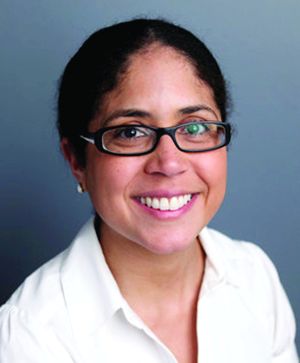
However, “while there is a possibility that there is an alternate factor that is responsible for these associations, the evidence is consistent across different populations,” said Dr. Suglia of Emory University in Atlanta. “And in general, studies do consider alternative factors that may be associated with both child adversity and cardiovascular health, further strengthening inferences we can make from the observational studies.”
Why might the association exist? “The mechanisms that drive these associations are still not fully elucidated but we hypothesize three pathways that may mediate these associations: behavioral factors (diet, sleep, physical activity, smoking), biological (hypothalamic-pituitary-adrenal axis dysregulation, epigenetic, chronic inflammation), and mental health (posttraumatic stress disorder and depression),” she said.
The statement notes that research is limited into why some people thrive on the cardiometabolic front despite childhood adversity. “One of the recommendations is that we need to focus more on factors that could inform prevention and intervention efforts so that those that are affected by adversity in childhood are not also affected by adverse cardiovascular events,” Dr. Suglia said.
The statement also notes that there’s been only limited research into modifiers of vulnerability to the effects of childhood adversity, such as gender, race/ethnicity, genetics, and community characteristics. And it says there’s been little study of how early interventions may affect cardiometabolic outcomes: “Additional research, including longitudinal prospective studies, designed to guide and inform effective and timely individual/clinical and population-level preventive interventions is required.”
Dr. Suglia reports a research grant from the National Heart, Lung, and Blood Institute. Most of the other statement coauthors report no disclosures outside of government funding.
SOURCE: Suglia S et al. Circulation. 2017 Dec 18. doi: 10.1161/CIR.0000000000000536
Research suggests an association between adverse childhood events and poorer cardiometabolic health across the lifespan, a new scientific statement from the American Heart Association declares, although it acknowledges various limitations in understanding the connection.
Studies haven’t confirmed a cause-and-effect relationship or the full extent of excess risk, and it’s not clear why some people who experience childhood trauma are more resilient than others. There’s also scant information about any link to higher cardiometabolic death rates.
For the new statement, which appears in Circulation, researchers reviewed recent systematic reviews into the links between childhood adversity – including events like violence and abuse – and cardiometabolic outcome.
By one estimate, nearly 60% of adults in the United States experienced at least one adverse childhood event. According to the statement, reviews have linked childhood adversity to higher risks of cardiac death and outcomes like heart attack and stroke. They’ve also linked adversity to risk factors like high blood pressure, obesity, and type 2 diabetes.
the statement said. However, Dr. Suglia noted that research into mortality is limited.
The level of higher risk varies by study and outcome, and no one knows if there’s a cause-and-effect link. “As you can imagine, child adversity is not an exposure that lends itself to randomized trials,” she said. “On top of that, we are talking about health effects that take many years to manifest so we rely on observational studies.”
However, “while there is a possibility that there is an alternate factor that is responsible for these associations, the evidence is consistent across different populations,” said Dr. Suglia of Emory University in Atlanta. “And in general, studies do consider alternative factors that may be associated with both child adversity and cardiovascular health, further strengthening inferences we can make from the observational studies.”
Why might the association exist? “The mechanisms that drive these associations are still not fully elucidated but we hypothesize three pathways that may mediate these associations: behavioral factors (diet, sleep, physical activity, smoking), biological (hypothalamic-pituitary-adrenal axis dysregulation, epigenetic, chronic inflammation), and mental health (posttraumatic stress disorder and depression),” she said.
The statement notes that research is limited into why some people thrive on the cardiometabolic front despite childhood adversity. “One of the recommendations is that we need to focus more on factors that could inform prevention and intervention efforts so that those that are affected by adversity in childhood are not also affected by adverse cardiovascular events,” Dr. Suglia said.
The statement also notes that there’s been only limited research into modifiers of vulnerability to the effects of childhood adversity, such as gender, race/ethnicity, genetics, and community characteristics. And it says there’s been little study of how early interventions may affect cardiometabolic outcomes: “Additional research, including longitudinal prospective studies, designed to guide and inform effective and timely individual/clinical and population-level preventive interventions is required.”
Dr. Suglia reports a research grant from the National Heart, Lung, and Blood Institute. Most of the other statement coauthors report no disclosures outside of government funding.
SOURCE: Suglia S et al. Circulation. 2017 Dec 18. doi: 10.1161/CIR.0000000000000536
Research suggests an association between adverse childhood events and poorer cardiometabolic health across the lifespan, a new scientific statement from the American Heart Association declares, although it acknowledges various limitations in understanding the connection.
Studies haven’t confirmed a cause-and-effect relationship or the full extent of excess risk, and it’s not clear why some people who experience childhood trauma are more resilient than others. There’s also scant information about any link to higher cardiometabolic death rates.
For the new statement, which appears in Circulation, researchers reviewed recent systematic reviews into the links between childhood adversity – including events like violence and abuse – and cardiometabolic outcome.
By one estimate, nearly 60% of adults in the United States experienced at least one adverse childhood event. According to the statement, reviews have linked childhood adversity to higher risks of cardiac death and outcomes like heart attack and stroke. They’ve also linked adversity to risk factors like high blood pressure, obesity, and type 2 diabetes.
the statement said. However, Dr. Suglia noted that research into mortality is limited.
The level of higher risk varies by study and outcome, and no one knows if there’s a cause-and-effect link. “As you can imagine, child adversity is not an exposure that lends itself to randomized trials,” she said. “On top of that, we are talking about health effects that take many years to manifest so we rely on observational studies.”
However, “while there is a possibility that there is an alternate factor that is responsible for these associations, the evidence is consistent across different populations,” said Dr. Suglia of Emory University in Atlanta. “And in general, studies do consider alternative factors that may be associated with both child adversity and cardiovascular health, further strengthening inferences we can make from the observational studies.”
Why might the association exist? “The mechanisms that drive these associations are still not fully elucidated but we hypothesize three pathways that may mediate these associations: behavioral factors (diet, sleep, physical activity, smoking), biological (hypothalamic-pituitary-adrenal axis dysregulation, epigenetic, chronic inflammation), and mental health (posttraumatic stress disorder and depression),” she said.
The statement notes that research is limited into why some people thrive on the cardiometabolic front despite childhood adversity. “One of the recommendations is that we need to focus more on factors that could inform prevention and intervention efforts so that those that are affected by adversity in childhood are not also affected by adverse cardiovascular events,” Dr. Suglia said.
The statement also notes that there’s been only limited research into modifiers of vulnerability to the effects of childhood adversity, such as gender, race/ethnicity, genetics, and community characteristics. And it says there’s been little study of how early interventions may affect cardiometabolic outcomes: “Additional research, including longitudinal prospective studies, designed to guide and inform effective and timely individual/clinical and population-level preventive interventions is required.”
Dr. Suglia reports a research grant from the National Heart, Lung, and Blood Institute. Most of the other statement coauthors report no disclosures outside of government funding.
SOURCE: Suglia S et al. Circulation. 2017 Dec 18. doi: 10.1161/CIR.0000000000000536
FROM CIRCULATION
New anticoagulants pose challenges for surgeons
SAN DIEGO – A new class of is now commonly seen in patients and can require new strategies for management in the surgical setting. New drugs to reverse anticoagulation agents now need to be routinely considered in advance of surgery.
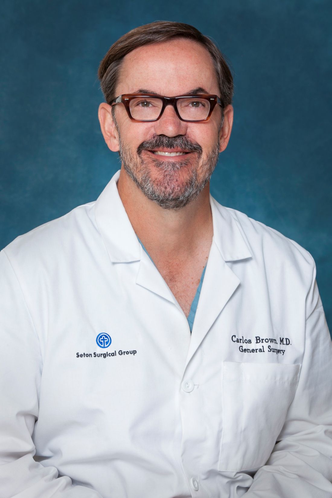
“They’re all over the place. It feels like everyone I see is on an anticoagulant, especially the new anticoagulants,” said Carlos V.R. Brown, MD, FACS, associate professor of surgery and chief of the division of acute care surgery at the University of Texas at Austin. “There’s a lot more learning that has to take place into how these medications work and how to take care of patients who use them.”
“Warfarin is slow, unpredictable, and requires monitoring and dose adjustment,” he said. In addition, interactions with food and other medications can be problematic, he said. The injectable drug heparin, meanwhile, requires monitoring and frequent dose adjustments, he said. “We’re in search of the ideal anticoagulant – one that’s oral, has a wide therapeutic window, is very predictable with rapid onset, and has minimal interaction with other food or drugs.”
Here’s the hitch, he said: “It probably doesn’t exist.”
There are now several alternatives to the old standbys on the market. One class, the direct thrombin inhibitor, is led by dabigatran etexilate (Pradaxa). Another class, the factor Xa inhibitors, includes rivaroxaban (Xarelto), apixaban (Eliquis), and edoxaban (Savaysa). From an elective surgery standpoint, Dr. Brown said in an interview, it has become important to understand how to reverse the effects of anticoagulants before a procedure.
To determine levels of the drugs, a TT (thrombin time) screening test is recommended for dabigatran and an anti-Xa test for rivaroxaban, apixaban, and edoxaban said Dr. Brown, referring to a 2017 study published in Critical Care Clinics. The paper summarized the available evidence and provided the optimal reversal strategy for bleeding patients with trauma on novel oral anticoagulants. The report also noted that newer blood thinners have a half-life of 7 or 12 hours and reach peak plasma level at 1-4 hours, depending on the medication (Crit Care Clin. 2017;33[1]135-52).
There may be no time to determine blood thinner levels in emergency situations. In those cases, patient or caregiver history about recent doses can be crucial, Dr. Brown said. “Knowing the patient’s history is going to be a key component,” he said.
Surgeons can turn to a variety of options to reverse the newer anticoagulants in an emergent setting, but only dabigatran has a Food and Drug Administration–approved reversal agent. Activated charcoal, PCC (Kcentra) and aPCC (FEIBA) can reverse dabigatran and oral factor Xa inhibitors, Dr. Brown said. Dialysis is also an option for dabigatran.
Another option to reverse dabigatran may be idarucizumab (Praxbind), a reversal agent. A 2017 industry-funded, open-label study reported successful results. It has been shown to work rapidly, Dr. Brown said, and the drug is now FDA approved (N Engl J Med. 2017 Aug 3;377[5]:431-41).
For oral factor Xa inhibitors, Dr. Brown said, andexanet alfa is now in a trial and doesn’t yet have FDA approval. “Presumably, it will provide a benefit over PCC because it’s directed at that specific medication,” he said.
Dr. Brown cautioned about the risks of reversing anticoagulants. “Any time you’re reversing an anticoagulant, the side effect is going to be clotting,” boosting the likelihood of events such as heart attack or stroke, he said. “You’re always weighing the risk versus the benefit of reversing.”
Dr. Brown has no relevant disclosures.
SAN DIEGO – A new class of is now commonly seen in patients and can require new strategies for management in the surgical setting. New drugs to reverse anticoagulation agents now need to be routinely considered in advance of surgery.
“They’re all over the place. It feels like everyone I see is on an anticoagulant, especially the new anticoagulants,” said Carlos V.R. Brown, MD, FACS, associate professor of surgery and chief of the division of acute care surgery at the University of Texas at Austin. “There’s a lot more learning that has to take place into how these medications work and how to take care of patients who use them.”
“Warfarin is slow, unpredictable, and requires monitoring and dose adjustment,” he said. In addition, interactions with food and other medications can be problematic, he said. The injectable drug heparin, meanwhile, requires monitoring and frequent dose adjustments, he said. “We’re in search of the ideal anticoagulant – one that’s oral, has a wide therapeutic window, is very predictable with rapid onset, and has minimal interaction with other food or drugs.”
Here’s the hitch, he said: “It probably doesn’t exist.”
There are now several alternatives to the old standbys on the market. One class, the direct thrombin inhibitor, is led by dabigatran etexilate (Pradaxa). Another class, the factor Xa inhibitors, includes rivaroxaban (Xarelto), apixaban (Eliquis), and edoxaban (Savaysa). From an elective surgery standpoint, Dr. Brown said in an interview, it has become important to understand how to reverse the effects of anticoagulants before a procedure.
To determine levels of the drugs, a TT (thrombin time) screening test is recommended for dabigatran and an anti-Xa test for rivaroxaban, apixaban, and edoxaban said Dr. Brown, referring to a 2017 study published in Critical Care Clinics. The paper summarized the available evidence and provided the optimal reversal strategy for bleeding patients with trauma on novel oral anticoagulants. The report also noted that newer blood thinners have a half-life of 7 or 12 hours and reach peak plasma level at 1-4 hours, depending on the medication (Crit Care Clin. 2017;33[1]135-52).
There may be no time to determine blood thinner levels in emergency situations. In those cases, patient or caregiver history about recent doses can be crucial, Dr. Brown said. “Knowing the patient’s history is going to be a key component,” he said.
Surgeons can turn to a variety of options to reverse the newer anticoagulants in an emergent setting, but only dabigatran has a Food and Drug Administration–approved reversal agent. Activated charcoal, PCC (Kcentra) and aPCC (FEIBA) can reverse dabigatran and oral factor Xa inhibitors, Dr. Brown said. Dialysis is also an option for dabigatran.
Another option to reverse dabigatran may be idarucizumab (Praxbind), a reversal agent. A 2017 industry-funded, open-label study reported successful results. It has been shown to work rapidly, Dr. Brown said, and the drug is now FDA approved (N Engl J Med. 2017 Aug 3;377[5]:431-41).
For oral factor Xa inhibitors, Dr. Brown said, andexanet alfa is now in a trial and doesn’t yet have FDA approval. “Presumably, it will provide a benefit over PCC because it’s directed at that specific medication,” he said.
Dr. Brown cautioned about the risks of reversing anticoagulants. “Any time you’re reversing an anticoagulant, the side effect is going to be clotting,” boosting the likelihood of events such as heart attack or stroke, he said. “You’re always weighing the risk versus the benefit of reversing.”
Dr. Brown has no relevant disclosures.
SAN DIEGO – A new class of is now commonly seen in patients and can require new strategies for management in the surgical setting. New drugs to reverse anticoagulation agents now need to be routinely considered in advance of surgery.
“They’re all over the place. It feels like everyone I see is on an anticoagulant, especially the new anticoagulants,” said Carlos V.R. Brown, MD, FACS, associate professor of surgery and chief of the division of acute care surgery at the University of Texas at Austin. “There’s a lot more learning that has to take place into how these medications work and how to take care of patients who use them.”
“Warfarin is slow, unpredictable, and requires monitoring and dose adjustment,” he said. In addition, interactions with food and other medications can be problematic, he said. The injectable drug heparin, meanwhile, requires monitoring and frequent dose adjustments, he said. “We’re in search of the ideal anticoagulant – one that’s oral, has a wide therapeutic window, is very predictable with rapid onset, and has minimal interaction with other food or drugs.”
Here’s the hitch, he said: “It probably doesn’t exist.”
There are now several alternatives to the old standbys on the market. One class, the direct thrombin inhibitor, is led by dabigatran etexilate (Pradaxa). Another class, the factor Xa inhibitors, includes rivaroxaban (Xarelto), apixaban (Eliquis), and edoxaban (Savaysa). From an elective surgery standpoint, Dr. Brown said in an interview, it has become important to understand how to reverse the effects of anticoagulants before a procedure.
To determine levels of the drugs, a TT (thrombin time) screening test is recommended for dabigatran and an anti-Xa test for rivaroxaban, apixaban, and edoxaban said Dr. Brown, referring to a 2017 study published in Critical Care Clinics. The paper summarized the available evidence and provided the optimal reversal strategy for bleeding patients with trauma on novel oral anticoagulants. The report also noted that newer blood thinners have a half-life of 7 or 12 hours and reach peak plasma level at 1-4 hours, depending on the medication (Crit Care Clin. 2017;33[1]135-52).
There may be no time to determine blood thinner levels in emergency situations. In those cases, patient or caregiver history about recent doses can be crucial, Dr. Brown said. “Knowing the patient’s history is going to be a key component,” he said.
Surgeons can turn to a variety of options to reverse the newer anticoagulants in an emergent setting, but only dabigatran has a Food and Drug Administration–approved reversal agent. Activated charcoal, PCC (Kcentra) and aPCC (FEIBA) can reverse dabigatran and oral factor Xa inhibitors, Dr. Brown said. Dialysis is also an option for dabigatran.
Another option to reverse dabigatran may be idarucizumab (Praxbind), a reversal agent. A 2017 industry-funded, open-label study reported successful results. It has been shown to work rapidly, Dr. Brown said, and the drug is now FDA approved (N Engl J Med. 2017 Aug 3;377[5]:431-41).
For oral factor Xa inhibitors, Dr. Brown said, andexanet alfa is now in a trial and doesn’t yet have FDA approval. “Presumably, it will provide a benefit over PCC because it’s directed at that specific medication,” he said.
Dr. Brown cautioned about the risks of reversing anticoagulants. “Any time you’re reversing an anticoagulant, the side effect is going to be clotting,” boosting the likelihood of events such as heart attack or stroke, he said. “You’re always weighing the risk versus the benefit of reversing.”
Dr. Brown has no relevant disclosures.
EXPERT OPINION FROM THE ACS CLINICAL CONGRESS
More evidence links high-potency marijuana use to first-episode psychosis
SAN DIEGO – High-potency marijuana use appears to be associated with an increased risk of a first psychotic episode, based on a case-control study conducted in Europe.
“Daily users of a strong type of cannabis face a significant increase in the probability of developing a psychotic disorder,” reported Marta Di Forti, MD, PhD, MRC, lead author of a study whose preliminary results were presented at the International Congress on Schizophrenia Research.
Dr. Di Forti spawned a media boomlet in 2015 when she and her colleagues raised the prospect of a possible association between so-called “skunk” marijuana and first psychotic episodes. In their study of subjects in London with first-time psychotic episodes and matched population controls, those who had psychotic episodes were three times (adjusted odds ratio: 2.92; 95% confidence interval, 1.52-3.45; P = .001) as likely as controls to have used “skunk” marijuana (Lancet Psychiatry. 2015 Mar;2[3]:233-8).
In the new study, Dr. Di Forti and her colleagues analyzed 1,200 first-incident cases of psychosis that were captured between the years 2010 and 2013 by the European Network of National Schizophrenia Networks Studying Gene-Environment Interactions project (EU-GEI). The researchers compared the cases to 1,300 population-based controls in five unidentified European countries and found that daily users of high-potency marijuana had the highest adjusted odds ratio (4.5-8, statistical significance not available) of a psychotic episode (Schizophr Bull. 2017 Mar:43:S30. doi: 10.1093/schbul/sbx021.078). “This effect is significant even after controlling for other drugs of abuse such as stimulants, tobacco and alcohol, and main sociodemographic confounders,” the researchers wrote in their abstract.
“The biology of cannabis-associated psychosis is still unclear,” Dr. Di Forti said in an interview. “Nevertheless, we know that THC (tetrahydrocannabinol) binds with two receptors called CB1 and CB2. They’re part of the endocannabinoid system, which from uterus onward protects our central nervous systems from insults. It activates on demand if the brain goes on hypoxia or we experience a brain injury.”
“CB1 activation leads to changes in the transmission of both GABA and glutamate. Downstream, they affect the dopamine system, which is biologically linked to psychosis.”
Dr. Di Forti dismissed the idea that people at risk for psychosis are drawn to high-potency marijuana. “Using genetic data, we’ve showed that cannabis users – both cases and controls – did not have a higher genetic load for schizophrenia than those who never used (marijuana),” she said (Lancet Psychiatry. 2015 May;2[5]:381-2).
The findings point to the importance of asking patients – and students and children – about more than just whether they have ever used marijuana. History-taking for marijuana use needs to be comparable to that performed for alcohol use, she said. “I always ask my patients for details about their past and present use but also try to understand why they use (marijuana). When possible, once I know how frequently and what type (of marijuana) they use, I can negotiate some harm-reduction strategy.”
The study is funded by the U.K.’s Medical Research Council and a European Union grant. Dr. Di Forti reports no relevant disclosures.
SAN DIEGO – High-potency marijuana use appears to be associated with an increased risk of a first psychotic episode, based on a case-control study conducted in Europe.
“Daily users of a strong type of cannabis face a significant increase in the probability of developing a psychotic disorder,” reported Marta Di Forti, MD, PhD, MRC, lead author of a study whose preliminary results were presented at the International Congress on Schizophrenia Research.
Dr. Di Forti spawned a media boomlet in 2015 when she and her colleagues raised the prospect of a possible association between so-called “skunk” marijuana and first psychotic episodes. In their study of subjects in London with first-time psychotic episodes and matched population controls, those who had psychotic episodes were three times (adjusted odds ratio: 2.92; 95% confidence interval, 1.52-3.45; P = .001) as likely as controls to have used “skunk” marijuana (Lancet Psychiatry. 2015 Mar;2[3]:233-8).
In the new study, Dr. Di Forti and her colleagues analyzed 1,200 first-incident cases of psychosis that were captured between the years 2010 and 2013 by the European Network of National Schizophrenia Networks Studying Gene-Environment Interactions project (EU-GEI). The researchers compared the cases to 1,300 population-based controls in five unidentified European countries and found that daily users of high-potency marijuana had the highest adjusted odds ratio (4.5-8, statistical significance not available) of a psychotic episode (Schizophr Bull. 2017 Mar:43:S30. doi: 10.1093/schbul/sbx021.078). “This effect is significant even after controlling for other drugs of abuse such as stimulants, tobacco and alcohol, and main sociodemographic confounders,” the researchers wrote in their abstract.
“The biology of cannabis-associated psychosis is still unclear,” Dr. Di Forti said in an interview. “Nevertheless, we know that THC (tetrahydrocannabinol) binds with two receptors called CB1 and CB2. They’re part of the endocannabinoid system, which from uterus onward protects our central nervous systems from insults. It activates on demand if the brain goes on hypoxia or we experience a brain injury.”
“CB1 activation leads to changes in the transmission of both GABA and glutamate. Downstream, they affect the dopamine system, which is biologically linked to psychosis.”
Dr. Di Forti dismissed the idea that people at risk for psychosis are drawn to high-potency marijuana. “Using genetic data, we’ve showed that cannabis users – both cases and controls – did not have a higher genetic load for schizophrenia than those who never used (marijuana),” she said (Lancet Psychiatry. 2015 May;2[5]:381-2).
The findings point to the importance of asking patients – and students and children – about more than just whether they have ever used marijuana. History-taking for marijuana use needs to be comparable to that performed for alcohol use, she said. “I always ask my patients for details about their past and present use but also try to understand why they use (marijuana). When possible, once I know how frequently and what type (of marijuana) they use, I can negotiate some harm-reduction strategy.”
The study is funded by the U.K.’s Medical Research Council and a European Union grant. Dr. Di Forti reports no relevant disclosures.
SAN DIEGO – High-potency marijuana use appears to be associated with an increased risk of a first psychotic episode, based on a case-control study conducted in Europe.
“Daily users of a strong type of cannabis face a significant increase in the probability of developing a psychotic disorder,” reported Marta Di Forti, MD, PhD, MRC, lead author of a study whose preliminary results were presented at the International Congress on Schizophrenia Research.
Dr. Di Forti spawned a media boomlet in 2015 when she and her colleagues raised the prospect of a possible association between so-called “skunk” marijuana and first psychotic episodes. In their study of subjects in London with first-time psychotic episodes and matched population controls, those who had psychotic episodes were three times (adjusted odds ratio: 2.92; 95% confidence interval, 1.52-3.45; P = .001) as likely as controls to have used “skunk” marijuana (Lancet Psychiatry. 2015 Mar;2[3]:233-8).
In the new study, Dr. Di Forti and her colleagues analyzed 1,200 first-incident cases of psychosis that were captured between the years 2010 and 2013 by the European Network of National Schizophrenia Networks Studying Gene-Environment Interactions project (EU-GEI). The researchers compared the cases to 1,300 population-based controls in five unidentified European countries and found that daily users of high-potency marijuana had the highest adjusted odds ratio (4.5-8, statistical significance not available) of a psychotic episode (Schizophr Bull. 2017 Mar:43:S30. doi: 10.1093/schbul/sbx021.078). “This effect is significant even after controlling for other drugs of abuse such as stimulants, tobacco and alcohol, and main sociodemographic confounders,” the researchers wrote in their abstract.
“The biology of cannabis-associated psychosis is still unclear,” Dr. Di Forti said in an interview. “Nevertheless, we know that THC (tetrahydrocannabinol) binds with two receptors called CB1 and CB2. They’re part of the endocannabinoid system, which from uterus onward protects our central nervous systems from insults. It activates on demand if the brain goes on hypoxia or we experience a brain injury.”
“CB1 activation leads to changes in the transmission of both GABA and glutamate. Downstream, they affect the dopamine system, which is biologically linked to psychosis.”
Dr. Di Forti dismissed the idea that people at risk for psychosis are drawn to high-potency marijuana. “Using genetic data, we’ve showed that cannabis users – both cases and controls – did not have a higher genetic load for schizophrenia than those who never used (marijuana),” she said (Lancet Psychiatry. 2015 May;2[5]:381-2).
The findings point to the importance of asking patients – and students and children – about more than just whether they have ever used marijuana. History-taking for marijuana use needs to be comparable to that performed for alcohol use, she said. “I always ask my patients for details about their past and present use but also try to understand why they use (marijuana). When possible, once I know how frequently and what type (of marijuana) they use, I can negotiate some harm-reduction strategy.”
The study is funded by the U.K.’s Medical Research Council and a European Union grant. Dr. Di Forti reports no relevant disclosures.
REPORTING FROM THE ICSR BIENNIAL MEETING
Evidence builds for long-term ineffectiveness of steroid shots for knee OA
SAN DIEGO – Real-world, nontrial research confirms the findings of a high-profile study released earlier in 2017: Corticosteroid shots are ineffective in the long term for knee osteoarthritis. In fact, researchers found a greater likelihood of a worsening condition in knees treated with the injections.
“Our findings are consistent with the latest randomized, controlled trial,” said study coauthor Jie Wei, MD, of Central South University in Changsha, China. She spoke in a plenary presentation about the study findings at the annual meeting of the American College of Rheumatology.
The use of corticosteroids for knee OA is a controversial topic. As Dr. Wei noted, there has been wide disagreement among medical societies about whether the treatment is useful in the long term for patients with pain flare-ups.
For the randomized, controlled study released in 2017, researchers tracked 140 patients aged 45 and older with inflammation of the synovial membrane. They were randomly assigned to injections of intra-articular triamcinolone or a placebo.
After 2 years of injections every 12 weeks, there was no difference in reported pain between the intervention and control groups. Also, those who received injections lost more cartilage (JAMA. 2017 May 16;317[19]:1967-75).
Researchers launched the new study to seek insight through a real-life cohort. They examined findings from the Osteoarthritis Initiative, a longitudinal study of 4,796 patients aged 45-79 at four U.S. clinics with knee OA or high risk of knee OA. Patients underwent annual examinations at baseline and annually for 4 years.
In an adjusted marginal structural analysis, knee replacement or worsening of Kellgren Lawrence grade at the tibial femoral joint was more likely in 149 injection knees than 2,191 noninjection knees (odds ratio, 5.74; 95% confidence interval, 2.01-16.42).
Knee replacement or joint space width worsening at the tibial femoral joint was also more likely in 120 injection knees than 2,112 noninjection knees (OR, 1.64; 95% CI, 0.91-2.93).
In another analysis, researchers tracked 134 injection knees (58 whose OA progressed) and 498 noninjection knees (132 whose OA progressed) for up to 8 years. After adjustment, the injection knees were more likely to have progressed (hazard ratio, 1.60; 95% CI, 1.21-2.12,).
“Several explanations may account for our study findings,” Dr. Wei said. One possibility, she said, is that corticosteroids may hurt chondrocytes by, among other things, inducing apoptosis and synovial membrane inflammation.
It’s also possible, she said, that patients may feel pain relief after injections and subsequently boost the risk of OA progression by increasing their physical activity.
“We need to know what types of physical activities may increase the OA progression,” she said. “Did patients who received steroid injection indeed increase this type of physical activity compared to subjects without steroid injection?”
Dr. Wei noted the study’s limitations, including the fact that patients who received injections had more pain at baseline, potentially indicating they had worse structural lesions that are more susceptible to progression.
The study authors reported no relevant disclosures. The National Natural Science Foundation of China funded the study. The Osteoarthritis Initiative is a partnership between the National Institutes of Health and Merck, Novartis, GlaxoSmithKline, and Pfizer.
SAN DIEGO – Real-world, nontrial research confirms the findings of a high-profile study released earlier in 2017: Corticosteroid shots are ineffective in the long term for knee osteoarthritis. In fact, researchers found a greater likelihood of a worsening condition in knees treated with the injections.
“Our findings are consistent with the latest randomized, controlled trial,” said study coauthor Jie Wei, MD, of Central South University in Changsha, China. She spoke in a plenary presentation about the study findings at the annual meeting of the American College of Rheumatology.
The use of corticosteroids for knee OA is a controversial topic. As Dr. Wei noted, there has been wide disagreement among medical societies about whether the treatment is useful in the long term for patients with pain flare-ups.
For the randomized, controlled study released in 2017, researchers tracked 140 patients aged 45 and older with inflammation of the synovial membrane. They were randomly assigned to injections of intra-articular triamcinolone or a placebo.
After 2 years of injections every 12 weeks, there was no difference in reported pain between the intervention and control groups. Also, those who received injections lost more cartilage (JAMA. 2017 May 16;317[19]:1967-75).
Researchers launched the new study to seek insight through a real-life cohort. They examined findings from the Osteoarthritis Initiative, a longitudinal study of 4,796 patients aged 45-79 at four U.S. clinics with knee OA or high risk of knee OA. Patients underwent annual examinations at baseline and annually for 4 years.
In an adjusted marginal structural analysis, knee replacement or worsening of Kellgren Lawrence grade at the tibial femoral joint was more likely in 149 injection knees than 2,191 noninjection knees (odds ratio, 5.74; 95% confidence interval, 2.01-16.42).
Knee replacement or joint space width worsening at the tibial femoral joint was also more likely in 120 injection knees than 2,112 noninjection knees (OR, 1.64; 95% CI, 0.91-2.93).
In another analysis, researchers tracked 134 injection knees (58 whose OA progressed) and 498 noninjection knees (132 whose OA progressed) for up to 8 years. After adjustment, the injection knees were more likely to have progressed (hazard ratio, 1.60; 95% CI, 1.21-2.12,).
“Several explanations may account for our study findings,” Dr. Wei said. One possibility, she said, is that corticosteroids may hurt chondrocytes by, among other things, inducing apoptosis and synovial membrane inflammation.
It’s also possible, she said, that patients may feel pain relief after injections and subsequently boost the risk of OA progression by increasing their physical activity.
“We need to know what types of physical activities may increase the OA progression,” she said. “Did patients who received steroid injection indeed increase this type of physical activity compared to subjects without steroid injection?”
Dr. Wei noted the study’s limitations, including the fact that patients who received injections had more pain at baseline, potentially indicating they had worse structural lesions that are more susceptible to progression.
The study authors reported no relevant disclosures. The National Natural Science Foundation of China funded the study. The Osteoarthritis Initiative is a partnership between the National Institutes of Health and Merck, Novartis, GlaxoSmithKline, and Pfizer.
SAN DIEGO – Real-world, nontrial research confirms the findings of a high-profile study released earlier in 2017: Corticosteroid shots are ineffective in the long term for knee osteoarthritis. In fact, researchers found a greater likelihood of a worsening condition in knees treated with the injections.
“Our findings are consistent with the latest randomized, controlled trial,” said study coauthor Jie Wei, MD, of Central South University in Changsha, China. She spoke in a plenary presentation about the study findings at the annual meeting of the American College of Rheumatology.
The use of corticosteroids for knee OA is a controversial topic. As Dr. Wei noted, there has been wide disagreement among medical societies about whether the treatment is useful in the long term for patients with pain flare-ups.
For the randomized, controlled study released in 2017, researchers tracked 140 patients aged 45 and older with inflammation of the synovial membrane. They were randomly assigned to injections of intra-articular triamcinolone or a placebo.
After 2 years of injections every 12 weeks, there was no difference in reported pain between the intervention and control groups. Also, those who received injections lost more cartilage (JAMA. 2017 May 16;317[19]:1967-75).
Researchers launched the new study to seek insight through a real-life cohort. They examined findings from the Osteoarthritis Initiative, a longitudinal study of 4,796 patients aged 45-79 at four U.S. clinics with knee OA or high risk of knee OA. Patients underwent annual examinations at baseline and annually for 4 years.
In an adjusted marginal structural analysis, knee replacement or worsening of Kellgren Lawrence grade at the tibial femoral joint was more likely in 149 injection knees than 2,191 noninjection knees (odds ratio, 5.74; 95% confidence interval, 2.01-16.42).
Knee replacement or joint space width worsening at the tibial femoral joint was also more likely in 120 injection knees than 2,112 noninjection knees (OR, 1.64; 95% CI, 0.91-2.93).
In another analysis, researchers tracked 134 injection knees (58 whose OA progressed) and 498 noninjection knees (132 whose OA progressed) for up to 8 years. After adjustment, the injection knees were more likely to have progressed (hazard ratio, 1.60; 95% CI, 1.21-2.12,).
“Several explanations may account for our study findings,” Dr. Wei said. One possibility, she said, is that corticosteroids may hurt chondrocytes by, among other things, inducing apoptosis and synovial membrane inflammation.
It’s also possible, she said, that patients may feel pain relief after injections and subsequently boost the risk of OA progression by increasing their physical activity.
“We need to know what types of physical activities may increase the OA progression,” she said. “Did patients who received steroid injection indeed increase this type of physical activity compared to subjects without steroid injection?”
Dr. Wei noted the study’s limitations, including the fact that patients who received injections had more pain at baseline, potentially indicating they had worse structural lesions that are more susceptible to progression.
The study authors reported no relevant disclosures. The National Natural Science Foundation of China funded the study. The Osteoarthritis Initiative is a partnership between the National Institutes of Health and Merck, Novartis, GlaxoSmithKline, and Pfizer.
REPORTING FROM ACR 2017
Key clinical point:
Major finding: In adjusted analysis of 134 injection knees and 498 noninjection knees tracked for up to 8 years, OA in injection knees was more likely to have progressed (HR, 1.60; 95% CI, 1.21-2.12).
Study details: Cohort analysis of data from the Osteoarthritis Initiative, which tracked patients with (or at high risk of) knee OA at four U.S. clinics.
Disclosures: The study authors reported no relevant disclosures. The National Natural Science Foundation of China funded the study. The Osteoarthritis Initiative is a partnership between the National Institutes of Health and Merck, Novartis, GlaxoSmithKline, and Pfizer.
Source: Lei G et al. ACR 2017 Abstract 1788.
Virtual reality enters the rheumatology realm
SAN DIEGO – When pain strikes arthritis patients, a beach trip may be the last thing on their minds. But a Los Angeles rheumatologist says a virtual voyage to the shore – or the fjords of Iceland or a Cirque du Soleil performance – may be just what they need to find relief without leaving their chairs.
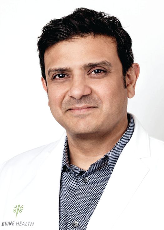
Swamy Venuturupalli, MD, has been testing virtual reality (VR) software on patients as part of a clinical feasibility study into its use to treat rheumatologic pain. It appears to be the first VR study in rheumatology.
Dr. Venuturupalli is working with gastroenterologist Brennan Spiegel, MD, a VR researcher who talked about the power of the treatment in a presentation at the annual meeting of the American College of Rheumatology.
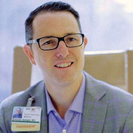
“Like a drug, we have to understand when and how to use this. It can be very powerful,” Dr. Spiegel, professor of medicine and public health at UCLA and director of Health Services Research at Cedars-Sinai Health System, said in his presentation.
“In recent years, VR technology has become increasingly affordable, immersive, flexible, and portable, enabling its use in a broad range of environments, including the inpatient medical setting,” wrote researchers in a 2017 systematic review of 11 randomized, controlled VR trials. “The capacity of VR to modulate subjective experience makes it a compelling intervention in inpatient medical settings, where VR may offer respite from the confining nature of medical wards, or where it may augment or replace analgesics in pain management” (Innov Clin Neurosci. 2017 Jan-Feb;14[1-2]:14-21).
In his ACR presentation, Dr. Spiegel said VR allows patients to escape the “psychosocial jail cell” of a hospital room. “We can put them on a helicopter and fly them over Icelandic fjords or let them sit on stage during a Cirque du Soleil performance.”
In terms of pain, he pointed to a 2017 study that he conducted with colleagues at Cedars-Sinai Medical Center. In 100 inpatients with pain scores of 3 or more on a 10-point scale, researchers compared a onetime 3-dimensional VR immersion via headset to exposure to a 2-dimensional nature video. Both interventions lasted 15 minutes.
Those in the VR group (n = 50) had greater pain improvement than did those in the 2-D group (–1.3 vs –0.6 points; P = .008), and the percentage of patients whose pain diminished was higher (65% vs. 40%; P = .01; number needed to treat = 4) (JMIR Ment Health. 2017 Jan-Mar;4[1]:e9).
Researchers reported no adverse events in the study. However, Dr. Spiegel said patients who undergo VR treatments may feel vertigo, especially if the technology is subpar, and there is a theoretical risk of seizure.
He also noted that VR can cause other negative effects. One patient had a panic attack during an immersive experience of simply throwing balls at cartoon teddy bears in a virtual environment. In this patient, “any concept of violence was enough to trigger a panic attack,” Dr. Spiegel said.
But another immersive virtual experience, putting her on a stage at Cirque du Soleil performance, was a success. “It empowered her, it made her feel brave,” Dr. Spiegel said.
He cautioned about limits of VR: Not every patient is eligible, will want to use it, or will benefit. And while the headsets used in VR have improved, he said, there’s still a way to go to make them more comfortable.
As for cost, VR equipment can be pricey. Hunter Hoffman, director of a virtual reality research center at the University of Washington, Seattle, told MIT Technology Review that the equipment for a VR pain study cost $35,000.
Dr. Venuturupalli, the rheumatologist who’s working with Dr. Spiegel, said his clinic is testing VR technology in patients with chronic pain syndromes. They’re immersed in environments such as one that simulates swimming with dolphins. “Some of my patients try to reach out and touch the dolphin in that state,” he said.
The feasibility study, now in progress, aims to enroll 20 patients and be completed by next March, Dr. Venuturupalli said. So far, the time span of improvement in patients has been variable, he said.
“Our goal is to have a full-fledged virtual reality clinic,” he said. “Like we prescribe physical therapy, we might have a VR therapy prescription that might include certain experiences. And as this technology gets cheaper and cheaper, our patients can have it at their own homes.”
Dr. Spiegel and Dr. Venuturupalli reported no relevant disclosures. Dr. Venuturupalli reports that his study is using donated VR technology from AppliedVR. The firm has created a partnership with Cedars-Sinai Medical Center.
SAN DIEGO – When pain strikes arthritis patients, a beach trip may be the last thing on their minds. But a Los Angeles rheumatologist says a virtual voyage to the shore – or the fjords of Iceland or a Cirque du Soleil performance – may be just what they need to find relief without leaving their chairs.
Swamy Venuturupalli, MD, has been testing virtual reality (VR) software on patients as part of a clinical feasibility study into its use to treat rheumatologic pain. It appears to be the first VR study in rheumatology.
Dr. Venuturupalli is working with gastroenterologist Brennan Spiegel, MD, a VR researcher who talked about the power of the treatment in a presentation at the annual meeting of the American College of Rheumatology.
“Like a drug, we have to understand when and how to use this. It can be very powerful,” Dr. Spiegel, professor of medicine and public health at UCLA and director of Health Services Research at Cedars-Sinai Health System, said in his presentation.
“In recent years, VR technology has become increasingly affordable, immersive, flexible, and portable, enabling its use in a broad range of environments, including the inpatient medical setting,” wrote researchers in a 2017 systematic review of 11 randomized, controlled VR trials. “The capacity of VR to modulate subjective experience makes it a compelling intervention in inpatient medical settings, where VR may offer respite from the confining nature of medical wards, or where it may augment or replace analgesics in pain management” (Innov Clin Neurosci. 2017 Jan-Feb;14[1-2]:14-21).
In his ACR presentation, Dr. Spiegel said VR allows patients to escape the “psychosocial jail cell” of a hospital room. “We can put them on a helicopter and fly them over Icelandic fjords or let them sit on stage during a Cirque du Soleil performance.”
In terms of pain, he pointed to a 2017 study that he conducted with colleagues at Cedars-Sinai Medical Center. In 100 inpatients with pain scores of 3 or more on a 10-point scale, researchers compared a onetime 3-dimensional VR immersion via headset to exposure to a 2-dimensional nature video. Both interventions lasted 15 minutes.
Those in the VR group (n = 50) had greater pain improvement than did those in the 2-D group (–1.3 vs –0.6 points; P = .008), and the percentage of patients whose pain diminished was higher (65% vs. 40%; P = .01; number needed to treat = 4) (JMIR Ment Health. 2017 Jan-Mar;4[1]:e9).
Researchers reported no adverse events in the study. However, Dr. Spiegel said patients who undergo VR treatments may feel vertigo, especially if the technology is subpar, and there is a theoretical risk of seizure.
He also noted that VR can cause other negative effects. One patient had a panic attack during an immersive experience of simply throwing balls at cartoon teddy bears in a virtual environment. In this patient, “any concept of violence was enough to trigger a panic attack,” Dr. Spiegel said.
But another immersive virtual experience, putting her on a stage at Cirque du Soleil performance, was a success. “It empowered her, it made her feel brave,” Dr. Spiegel said.
He cautioned about limits of VR: Not every patient is eligible, will want to use it, or will benefit. And while the headsets used in VR have improved, he said, there’s still a way to go to make them more comfortable.
As for cost, VR equipment can be pricey. Hunter Hoffman, director of a virtual reality research center at the University of Washington, Seattle, told MIT Technology Review that the equipment for a VR pain study cost $35,000.
Dr. Venuturupalli, the rheumatologist who’s working with Dr. Spiegel, said his clinic is testing VR technology in patients with chronic pain syndromes. They’re immersed in environments such as one that simulates swimming with dolphins. “Some of my patients try to reach out and touch the dolphin in that state,” he said.
The feasibility study, now in progress, aims to enroll 20 patients and be completed by next March, Dr. Venuturupalli said. So far, the time span of improvement in patients has been variable, he said.
“Our goal is to have a full-fledged virtual reality clinic,” he said. “Like we prescribe physical therapy, we might have a VR therapy prescription that might include certain experiences. And as this technology gets cheaper and cheaper, our patients can have it at their own homes.”
Dr. Spiegel and Dr. Venuturupalli reported no relevant disclosures. Dr. Venuturupalli reports that his study is using donated VR technology from AppliedVR. The firm has created a partnership with Cedars-Sinai Medical Center.
SAN DIEGO – When pain strikes arthritis patients, a beach trip may be the last thing on their minds. But a Los Angeles rheumatologist says a virtual voyage to the shore – or the fjords of Iceland or a Cirque du Soleil performance – may be just what they need to find relief without leaving their chairs.
Swamy Venuturupalli, MD, has been testing virtual reality (VR) software on patients as part of a clinical feasibility study into its use to treat rheumatologic pain. It appears to be the first VR study in rheumatology.
Dr. Venuturupalli is working with gastroenterologist Brennan Spiegel, MD, a VR researcher who talked about the power of the treatment in a presentation at the annual meeting of the American College of Rheumatology.
“Like a drug, we have to understand when and how to use this. It can be very powerful,” Dr. Spiegel, professor of medicine and public health at UCLA and director of Health Services Research at Cedars-Sinai Health System, said in his presentation.
“In recent years, VR technology has become increasingly affordable, immersive, flexible, and portable, enabling its use in a broad range of environments, including the inpatient medical setting,” wrote researchers in a 2017 systematic review of 11 randomized, controlled VR trials. “The capacity of VR to modulate subjective experience makes it a compelling intervention in inpatient medical settings, where VR may offer respite from the confining nature of medical wards, or where it may augment or replace analgesics in pain management” (Innov Clin Neurosci. 2017 Jan-Feb;14[1-2]:14-21).
In his ACR presentation, Dr. Spiegel said VR allows patients to escape the “psychosocial jail cell” of a hospital room. “We can put them on a helicopter and fly them over Icelandic fjords or let them sit on stage during a Cirque du Soleil performance.”
In terms of pain, he pointed to a 2017 study that he conducted with colleagues at Cedars-Sinai Medical Center. In 100 inpatients with pain scores of 3 or more on a 10-point scale, researchers compared a onetime 3-dimensional VR immersion via headset to exposure to a 2-dimensional nature video. Both interventions lasted 15 minutes.
Those in the VR group (n = 50) had greater pain improvement than did those in the 2-D group (–1.3 vs –0.6 points; P = .008), and the percentage of patients whose pain diminished was higher (65% vs. 40%; P = .01; number needed to treat = 4) (JMIR Ment Health. 2017 Jan-Mar;4[1]:e9).
Researchers reported no adverse events in the study. However, Dr. Spiegel said patients who undergo VR treatments may feel vertigo, especially if the technology is subpar, and there is a theoretical risk of seizure.
He also noted that VR can cause other negative effects. One patient had a panic attack during an immersive experience of simply throwing balls at cartoon teddy bears in a virtual environment. In this patient, “any concept of violence was enough to trigger a panic attack,” Dr. Spiegel said.
But another immersive virtual experience, putting her on a stage at Cirque du Soleil performance, was a success. “It empowered her, it made her feel brave,” Dr. Spiegel said.
He cautioned about limits of VR: Not every patient is eligible, will want to use it, or will benefit. And while the headsets used in VR have improved, he said, there’s still a way to go to make them more comfortable.
As for cost, VR equipment can be pricey. Hunter Hoffman, director of a virtual reality research center at the University of Washington, Seattle, told MIT Technology Review that the equipment for a VR pain study cost $35,000.
Dr. Venuturupalli, the rheumatologist who’s working with Dr. Spiegel, said his clinic is testing VR technology in patients with chronic pain syndromes. They’re immersed in environments such as one that simulates swimming with dolphins. “Some of my patients try to reach out and touch the dolphin in that state,” he said.
The feasibility study, now in progress, aims to enroll 20 patients and be completed by next March, Dr. Venuturupalli said. So far, the time span of improvement in patients has been variable, he said.
“Our goal is to have a full-fledged virtual reality clinic,” he said. “Like we prescribe physical therapy, we might have a VR therapy prescription that might include certain experiences. And as this technology gets cheaper and cheaper, our patients can have it at their own homes.”
Dr. Spiegel and Dr. Venuturupalli reported no relevant disclosures. Dr. Venuturupalli reports that his study is using donated VR technology from AppliedVR. The firm has created a partnership with Cedars-Sinai Medical Center.
REPORTING FROM ACR 2017
Inside the ‘mad rush’ for ketamine treatment
Ketamine, once best known as a pet anesthetic and party drug, is taking the United States by storm. Dozens of ketamine treatment centers are operating from coast to coast.
Big cities like Baltimore, Boston, and Phoenix have them. So do Charleston, S.C., and Boise, Idaho. Two such clinics are in sparsely populated New Mexico. And one national chain went from a pair of clinics to 10 in fewer than 2 years.
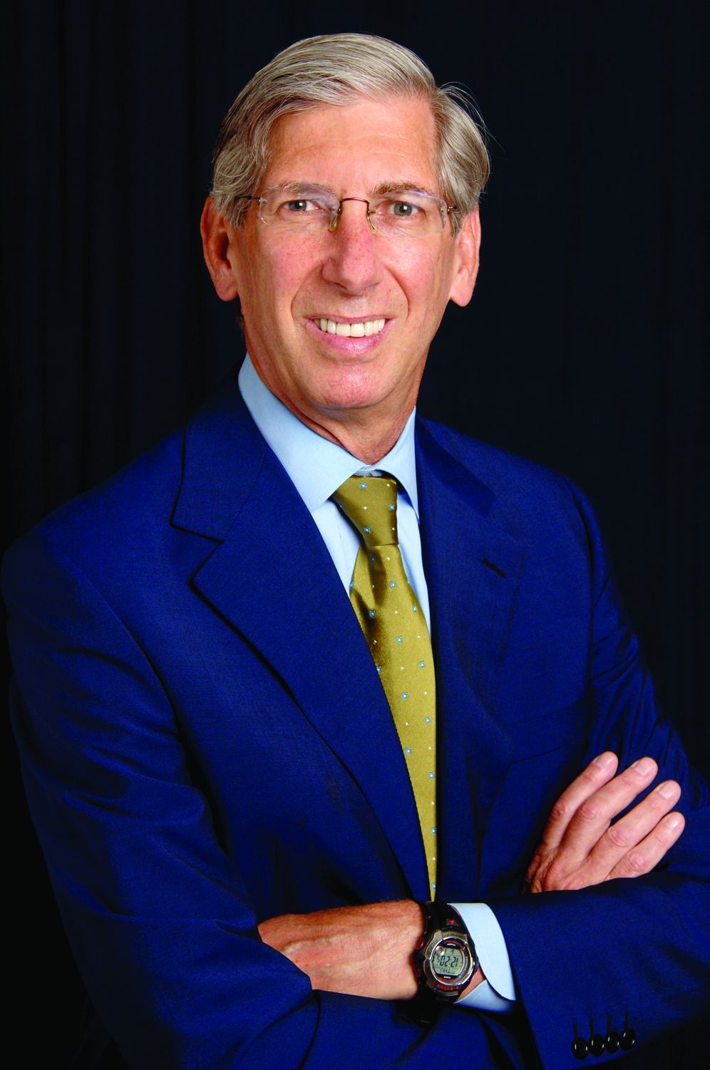
Never mind that these expensive treatments for conditions like depression are not covered by insurers or approved for this use by the Food and Drug Administration. Other questions also persist. “There is a considerable body of evidence that proves it really does work,” Dr. Lieberman said. “But we don’t know the extent of the range of conditions for which it might be effective, what the optimal frequency and concentration for dosing is, and what the long-term consequences are.”
To make matters more complicated, it’s anesthesiologists – not psychiatrists – who are leading the way toward a ketamine-infused future.
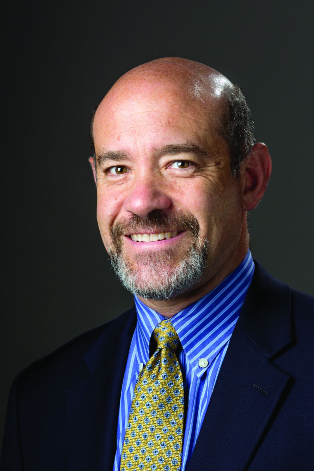
For now, however, hundreds and perhaps even thousands of patients are serving as ketamine test cases with psychiatrists only assisting remotely, if at all.
A stunningly rapid rise
Sara M. Markey, MD, is one of the rare psychiatrists in the United States who’s fully embraced ketamine treatment for mental illness.
She recalled first hearing about ketamine as an anesthetic in medical school. Best known as an anesthetic in animals, it’s also occasionally given to children and adults, although the drug’s dissociative properties have prevented widespread use.
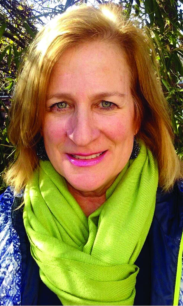
In 2006, word spread about ketamine’s use as a painkiller. “I also began hearing and reading about its potential use/efficacy in treatment-resistant depression,” said Dr. Markey, who practices in Denver. “It was difficult to find information about ketamine, and many of my colleagues were hostile to the idea of using ketamine in clinical practice.”
She persisted, however, and prescribed intranasal and oral ketamine to depressed patients with “mild success.” She also saw patients whose psychiatrists refused to consider ketamine.
In early 2016, with Steven P. Levine, MD, a New Jersey psychiatrist who pioneered ketamine use for depression, Dr. Markey opened a ketamine infusion clinic in the Mile High City.
At at that time, it was only the second in a national chain called Ketamine Treatment Centers. Now, not even 2 years later, the chain has a new name – Actify Neurotherapies – and a total of 10 clinics from San Francisco and Beverly Hills, Calif., to Palm Beach, Fla.; Raleigh, N.C.; and New York City.
“It is wonderful,” she said, “to have an opportunity to provide a medication to people that does not cause weight gain, has very few medication interactions, and which is well tolerated and generic.”
Big short-term benefits
Treatment outcomes with ketamine – which is thought to act on glutamate and N-methyl-D-aspartate receptors – can be dramatic. “Some patients describe the ketamine treatments as life saving,” said Allison F. Wells, MD, an anesthesiologist who runs a clinic in Houston.
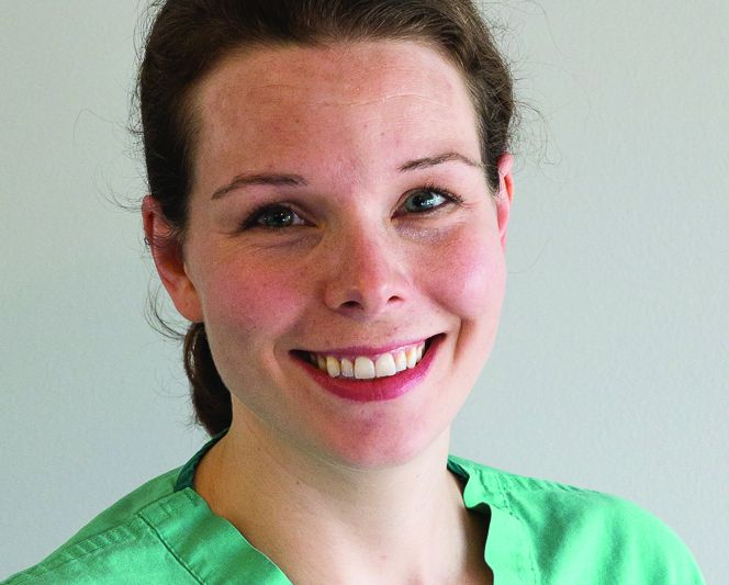
One depressed young man who tried ketamine at a Phoenix clinic in 2013 reportedly told the news site vice.com that he “felt good for a week” after his first treatment: “Not the kind of bipolar ‘good’ where I’d be manic. I just felt pleasant, and not crazy or compulsive. I felt normal for the first time in a long time.” Another depressed patient told National Public Radio that ketamine transformed his life: “I remember I was in my bathroom, and I literally fell to my knees crying because I had no anxiety; I had no depression.”
Enrique A. Abreu, DO, an anesthesiologist who offers ketamine therapy in Seattle and Portland, Ore., said he’s seen anxiety relief and a decrease in rumination in these patients. “They’re able to go back to work; a lot haven’t been able to work for a long time. And motivation is a big thing. They’re able to do things they haven’t been able to do.”
In addition, ketamine can reduce suicidal thinking, Dr. Markey said. “I am continually astounded to hear patients who come in with acute or chronic suicidal thinking report that those ideas and/or intrusive thoughts have disappeared. When they are absent, people need to be reminded of when they had them. They seem to have forgotten about them.”
‘Mystical experiences’
Dr. Markey said a retrospective analysis of about 740 patients at her chain’s clinics showed a response rate of about 75%. Other research has shown similarly high response levels.
“Multiple clinical trials suggest that a single low dose (0.5mg/kg) of IV ketamine results in a 50%-70% response rate in patients with treatment-resistant depression,” reported a 2016 clinical review. “Additional research has shown that depressed patients can experience symptom relief as early as 2 [hours], and lasting up to 2 weeks after a single administration of IV ketamine,” according to the review in Evidence Based Mental Health (2016 May;19[2]:35-8).
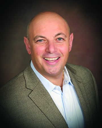
Adverse effects can include nausea and headache in patients with a history of migraine, he said. Over the long term, ketamine use can lead to incontinence and urinary urgency, he said.
As for ketamine addiction, Dr. Simelgor calls it unlikely at the lower doses that are used. However, he said, “I can’t say 100% that it won’t cause addiction.”
Who benefits? The jury’s still out
Considering its positive effects, why shouldn’t the mental health community embrace ketamine? Because, two prominent researchers say, best practices are still absent in a whole range of areas.
For example, there’s no agreement about who should undergo ketamine treatments beyond patients with treatment-resistant depression, especially those who have failed or cannot undergo electroconvulsive therapy. Ketamine therapy also is being touted by some as a treatment for a long list of other conditions from obsessive-compulsive disorder and anxiety to fibromyalgia and chronic pain disorders.
There are also limited data about dosing, making it “not possible to clarify the relative benefits and risks of doses other than 0.5 mg/kg delivered intravenously over 40 minutes,” cautioned Dr. Sanacora and Samuel T. Wilkinson, MD, also at Yale, in a 2017 commentary in JAMA (2017;318[9]:793-4).
In fact, they write, “Most published data supporting the use of ketamine as a treatment for mood disorders are based on trials that have followed up patients for just 1 week after a single administration of the drug.”
Unchartered waters
There’s also no accepted protocol beyond a typical six treatments over 2 or more weeks. This is relevant because the benefits of a series of treatments often fade away after a few weeks.
“Some patients describe the results lasting indefinitely, while most patients who respond to the treatments get to the point where they are going roughly 4-12 weeks with sustained results,” Dr. Wells said.
“When the effects start to wear off, they don’t crash,” said Dr. Abreu. Instead, he said, symptoms slowly reappear.
It’s typical for patients at Dr. Abreu’s clinic and others to return within a couple of months to go through another round of ketamine treatments. In some cases, “they continue to see us indefinitely to get them back up to where they need to be with a booster type of session,” he said.
Ketamine treatment costs vary widely, and insurers don’t cover this off-label treatment. The clinic operators quoted in this article reported a range of per-infusion costs from $350 (Dr. Markey’s clinic in Denver) to $675 (Dr. Abreu’s clinics in the Northwest).
“We have to have a talk with them: Can you afford this? This is going to take a significant amount of money every month to keep you well,” Dr. Abreu said. On the other hand, he said, the need for other medications goes away, eliminating that cost. (“They’re on [selective serotonin reputake inhibitors] usually,” he said, “but those drugs don’t work.”)
Nonpsychiatrists in forefront
At Dr. Markey’s clinic in Denver, all patients are required to see either her or a psychiatrist colleague. Some other ketamine clinics are run by psychiatrists, but that’s far from common.
Clinics often have no mental health professionals on staff and are run by anesthesiologists or other kinds of physicians.
Some clinic owners, Dr. Wells said, require patients to be under the care of a psychiatrist, neurologist, pain doctor, or other appropriate professional. “I do not intend, nor do I act, to displace psychiatrists, or the relationships our patients have with their psychiatrists, or the care they receive from their psychiatrists,” she said.
In the Northwest, Dr. Abreu said his patients take mood questionnaires, and he’s experimenting with a text-based mood monitoring system. In the Minneapolis area, anesthesiologist Dr. Simelgor is looking for a psychiatrist or psychiatrist partner for his ketamine clinic. “My thinking,” he said, “is that we need to work together.”
Still, there do not appear to be any requirements that ketamine clinic practitioners have connections to mental health professionals. Yet, as Dr. Sanacora put it: “Delivering the drug is the easiest part of the treatment. The hard part is managing the depression.”
Dr. Lieberman, Dr. Wells, Dr. Abreu, Dr. Simelgor, and Dr. Markey reported no relevant disclosures. Dr. Sanacora reported consulting fees and research contracts for multiple drug makers over the past 24 months. He holds shares in Biohaven Pharmaceuticals and is a coinventor on a U.S. patent (No. 8778979) on using glutamate agents to treat mental disorders held by Yale University.
Ketamine, once best known as a pet anesthetic and party drug, is taking the United States by storm. Dozens of ketamine treatment centers are operating from coast to coast.
Big cities like Baltimore, Boston, and Phoenix have them. So do Charleston, S.C., and Boise, Idaho. Two such clinics are in sparsely populated New Mexico. And one national chain went from a pair of clinics to 10 in fewer than 2 years.
Never mind that these expensive treatments for conditions like depression are not covered by insurers or approved for this use by the Food and Drug Administration. Other questions also persist. “There is a considerable body of evidence that proves it really does work,” Dr. Lieberman said. “But we don’t know the extent of the range of conditions for which it might be effective, what the optimal frequency and concentration for dosing is, and what the long-term consequences are.”
To make matters more complicated, it’s anesthesiologists – not psychiatrists – who are leading the way toward a ketamine-infused future.
For now, however, hundreds and perhaps even thousands of patients are serving as ketamine test cases with psychiatrists only assisting remotely, if at all.
A stunningly rapid rise
Sara M. Markey, MD, is one of the rare psychiatrists in the United States who’s fully embraced ketamine treatment for mental illness.
She recalled first hearing about ketamine as an anesthetic in medical school. Best known as an anesthetic in animals, it’s also occasionally given to children and adults, although the drug’s dissociative properties have prevented widespread use.
In 2006, word spread about ketamine’s use as a painkiller. “I also began hearing and reading about its potential use/efficacy in treatment-resistant depression,” said Dr. Markey, who practices in Denver. “It was difficult to find information about ketamine, and many of my colleagues were hostile to the idea of using ketamine in clinical practice.”
She persisted, however, and prescribed intranasal and oral ketamine to depressed patients with “mild success.” She also saw patients whose psychiatrists refused to consider ketamine.
In early 2016, with Steven P. Levine, MD, a New Jersey psychiatrist who pioneered ketamine use for depression, Dr. Markey opened a ketamine infusion clinic in the Mile High City.
At at that time, it was only the second in a national chain called Ketamine Treatment Centers. Now, not even 2 years later, the chain has a new name – Actify Neurotherapies – and a total of 10 clinics from San Francisco and Beverly Hills, Calif., to Palm Beach, Fla.; Raleigh, N.C.; and New York City.
“It is wonderful,” she said, “to have an opportunity to provide a medication to people that does not cause weight gain, has very few medication interactions, and which is well tolerated and generic.”
Big short-term benefits
Treatment outcomes with ketamine – which is thought to act on glutamate and N-methyl-D-aspartate receptors – can be dramatic. “Some patients describe the ketamine treatments as life saving,” said Allison F. Wells, MD, an anesthesiologist who runs a clinic in Houston.
One depressed young man who tried ketamine at a Phoenix clinic in 2013 reportedly told the news site vice.com that he “felt good for a week” after his first treatment: “Not the kind of bipolar ‘good’ where I’d be manic. I just felt pleasant, and not crazy or compulsive. I felt normal for the first time in a long time.” Another depressed patient told National Public Radio that ketamine transformed his life: “I remember I was in my bathroom, and I literally fell to my knees crying because I had no anxiety; I had no depression.”
Enrique A. Abreu, DO, an anesthesiologist who offers ketamine therapy in Seattle and Portland, Ore., said he’s seen anxiety relief and a decrease in rumination in these patients. “They’re able to go back to work; a lot haven’t been able to work for a long time. And motivation is a big thing. They’re able to do things they haven’t been able to do.”
In addition, ketamine can reduce suicidal thinking, Dr. Markey said. “I am continually astounded to hear patients who come in with acute or chronic suicidal thinking report that those ideas and/or intrusive thoughts have disappeared. When they are absent, people need to be reminded of when they had them. They seem to have forgotten about them.”
‘Mystical experiences’
Dr. Markey said a retrospective analysis of about 740 patients at her chain’s clinics showed a response rate of about 75%. Other research has shown similarly high response levels.
“Multiple clinical trials suggest that a single low dose (0.5mg/kg) of IV ketamine results in a 50%-70% response rate in patients with treatment-resistant depression,” reported a 2016 clinical review. “Additional research has shown that depressed patients can experience symptom relief as early as 2 [hours], and lasting up to 2 weeks after a single administration of IV ketamine,” according to the review in Evidence Based Mental Health (2016 May;19[2]:35-8).
Adverse effects can include nausea and headache in patients with a history of migraine, he said. Over the long term, ketamine use can lead to incontinence and urinary urgency, he said.
As for ketamine addiction, Dr. Simelgor calls it unlikely at the lower doses that are used. However, he said, “I can’t say 100% that it won’t cause addiction.”
Who benefits? The jury’s still out
Considering its positive effects, why shouldn’t the mental health community embrace ketamine? Because, two prominent researchers say, best practices are still absent in a whole range of areas.
For example, there’s no agreement about who should undergo ketamine treatments beyond patients with treatment-resistant depression, especially those who have failed or cannot undergo electroconvulsive therapy. Ketamine therapy also is being touted by some as a treatment for a long list of other conditions from obsessive-compulsive disorder and anxiety to fibromyalgia and chronic pain disorders.
There are also limited data about dosing, making it “not possible to clarify the relative benefits and risks of doses other than 0.5 mg/kg delivered intravenously over 40 minutes,” cautioned Dr. Sanacora and Samuel T. Wilkinson, MD, also at Yale, in a 2017 commentary in JAMA (2017;318[9]:793-4).
In fact, they write, “Most published data supporting the use of ketamine as a treatment for mood disorders are based on trials that have followed up patients for just 1 week after a single administration of the drug.”
Unchartered waters
There’s also no accepted protocol beyond a typical six treatments over 2 or more weeks. This is relevant because the benefits of a series of treatments often fade away after a few weeks.
“Some patients describe the results lasting indefinitely, while most patients who respond to the treatments get to the point where they are going roughly 4-12 weeks with sustained results,” Dr. Wells said.
“When the effects start to wear off, they don’t crash,” said Dr. Abreu. Instead, he said, symptoms slowly reappear.
It’s typical for patients at Dr. Abreu’s clinic and others to return within a couple of months to go through another round of ketamine treatments. In some cases, “they continue to see us indefinitely to get them back up to where they need to be with a booster type of session,” he said.
Ketamine treatment costs vary widely, and insurers don’t cover this off-label treatment. The clinic operators quoted in this article reported a range of per-infusion costs from $350 (Dr. Markey’s clinic in Denver) to $675 (Dr. Abreu’s clinics in the Northwest).
“We have to have a talk with them: Can you afford this? This is going to take a significant amount of money every month to keep you well,” Dr. Abreu said. On the other hand, he said, the need for other medications goes away, eliminating that cost. (“They’re on [selective serotonin reputake inhibitors] usually,” he said, “but those drugs don’t work.”)
Nonpsychiatrists in forefront
At Dr. Markey’s clinic in Denver, all patients are required to see either her or a psychiatrist colleague. Some other ketamine clinics are run by psychiatrists, but that’s far from common.
Clinics often have no mental health professionals on staff and are run by anesthesiologists or other kinds of physicians.
Some clinic owners, Dr. Wells said, require patients to be under the care of a psychiatrist, neurologist, pain doctor, or other appropriate professional. “I do not intend, nor do I act, to displace psychiatrists, or the relationships our patients have with their psychiatrists, or the care they receive from their psychiatrists,” she said.
In the Northwest, Dr. Abreu said his patients take mood questionnaires, and he’s experimenting with a text-based mood monitoring system. In the Minneapolis area, anesthesiologist Dr. Simelgor is looking for a psychiatrist or psychiatrist partner for his ketamine clinic. “My thinking,” he said, “is that we need to work together.”
Still, there do not appear to be any requirements that ketamine clinic practitioners have connections to mental health professionals. Yet, as Dr. Sanacora put it: “Delivering the drug is the easiest part of the treatment. The hard part is managing the depression.”
Dr. Lieberman, Dr. Wells, Dr. Abreu, Dr. Simelgor, and Dr. Markey reported no relevant disclosures. Dr. Sanacora reported consulting fees and research contracts for multiple drug makers over the past 24 months. He holds shares in Biohaven Pharmaceuticals and is a coinventor on a U.S. patent (No. 8778979) on using glutamate agents to treat mental disorders held by Yale University.
Ketamine, once best known as a pet anesthetic and party drug, is taking the United States by storm. Dozens of ketamine treatment centers are operating from coast to coast.
Big cities like Baltimore, Boston, and Phoenix have them. So do Charleston, S.C., and Boise, Idaho. Two such clinics are in sparsely populated New Mexico. And one national chain went from a pair of clinics to 10 in fewer than 2 years.
Never mind that these expensive treatments for conditions like depression are not covered by insurers or approved for this use by the Food and Drug Administration. Other questions also persist. “There is a considerable body of evidence that proves it really does work,” Dr. Lieberman said. “But we don’t know the extent of the range of conditions for which it might be effective, what the optimal frequency and concentration for dosing is, and what the long-term consequences are.”
To make matters more complicated, it’s anesthesiologists – not psychiatrists – who are leading the way toward a ketamine-infused future.
For now, however, hundreds and perhaps even thousands of patients are serving as ketamine test cases with psychiatrists only assisting remotely, if at all.
A stunningly rapid rise
Sara M. Markey, MD, is one of the rare psychiatrists in the United States who’s fully embraced ketamine treatment for mental illness.
She recalled first hearing about ketamine as an anesthetic in medical school. Best known as an anesthetic in animals, it’s also occasionally given to children and adults, although the drug’s dissociative properties have prevented widespread use.
In 2006, word spread about ketamine’s use as a painkiller. “I also began hearing and reading about its potential use/efficacy in treatment-resistant depression,” said Dr. Markey, who practices in Denver. “It was difficult to find information about ketamine, and many of my colleagues were hostile to the idea of using ketamine in clinical practice.”
She persisted, however, and prescribed intranasal and oral ketamine to depressed patients with “mild success.” She also saw patients whose psychiatrists refused to consider ketamine.
In early 2016, with Steven P. Levine, MD, a New Jersey psychiatrist who pioneered ketamine use for depression, Dr. Markey opened a ketamine infusion clinic in the Mile High City.
At at that time, it was only the second in a national chain called Ketamine Treatment Centers. Now, not even 2 years later, the chain has a new name – Actify Neurotherapies – and a total of 10 clinics from San Francisco and Beverly Hills, Calif., to Palm Beach, Fla.; Raleigh, N.C.; and New York City.
“It is wonderful,” she said, “to have an opportunity to provide a medication to people that does not cause weight gain, has very few medication interactions, and which is well tolerated and generic.”
Big short-term benefits
Treatment outcomes with ketamine – which is thought to act on glutamate and N-methyl-D-aspartate receptors – can be dramatic. “Some patients describe the ketamine treatments as life saving,” said Allison F. Wells, MD, an anesthesiologist who runs a clinic in Houston.
One depressed young man who tried ketamine at a Phoenix clinic in 2013 reportedly told the news site vice.com that he “felt good for a week” after his first treatment: “Not the kind of bipolar ‘good’ where I’d be manic. I just felt pleasant, and not crazy or compulsive. I felt normal for the first time in a long time.” Another depressed patient told National Public Radio that ketamine transformed his life: “I remember I was in my bathroom, and I literally fell to my knees crying because I had no anxiety; I had no depression.”
Enrique A. Abreu, DO, an anesthesiologist who offers ketamine therapy in Seattle and Portland, Ore., said he’s seen anxiety relief and a decrease in rumination in these patients. “They’re able to go back to work; a lot haven’t been able to work for a long time. And motivation is a big thing. They’re able to do things they haven’t been able to do.”
In addition, ketamine can reduce suicidal thinking, Dr. Markey said. “I am continually astounded to hear patients who come in with acute or chronic suicidal thinking report that those ideas and/or intrusive thoughts have disappeared. When they are absent, people need to be reminded of when they had them. They seem to have forgotten about them.”
‘Mystical experiences’
Dr. Markey said a retrospective analysis of about 740 patients at her chain’s clinics showed a response rate of about 75%. Other research has shown similarly high response levels.
“Multiple clinical trials suggest that a single low dose (0.5mg/kg) of IV ketamine results in a 50%-70% response rate in patients with treatment-resistant depression,” reported a 2016 clinical review. “Additional research has shown that depressed patients can experience symptom relief as early as 2 [hours], and lasting up to 2 weeks after a single administration of IV ketamine,” according to the review in Evidence Based Mental Health (2016 May;19[2]:35-8).
Adverse effects can include nausea and headache in patients with a history of migraine, he said. Over the long term, ketamine use can lead to incontinence and urinary urgency, he said.
As for ketamine addiction, Dr. Simelgor calls it unlikely at the lower doses that are used. However, he said, “I can’t say 100% that it won’t cause addiction.”
Who benefits? The jury’s still out
Considering its positive effects, why shouldn’t the mental health community embrace ketamine? Because, two prominent researchers say, best practices are still absent in a whole range of areas.
For example, there’s no agreement about who should undergo ketamine treatments beyond patients with treatment-resistant depression, especially those who have failed or cannot undergo electroconvulsive therapy. Ketamine therapy also is being touted by some as a treatment for a long list of other conditions from obsessive-compulsive disorder and anxiety to fibromyalgia and chronic pain disorders.
There are also limited data about dosing, making it “not possible to clarify the relative benefits and risks of doses other than 0.5 mg/kg delivered intravenously over 40 minutes,” cautioned Dr. Sanacora and Samuel T. Wilkinson, MD, also at Yale, in a 2017 commentary in JAMA (2017;318[9]:793-4).
In fact, they write, “Most published data supporting the use of ketamine as a treatment for mood disorders are based on trials that have followed up patients for just 1 week after a single administration of the drug.”
Unchartered waters
There’s also no accepted protocol beyond a typical six treatments over 2 or more weeks. This is relevant because the benefits of a series of treatments often fade away after a few weeks.
“Some patients describe the results lasting indefinitely, while most patients who respond to the treatments get to the point where they are going roughly 4-12 weeks with sustained results,” Dr. Wells said.
“When the effects start to wear off, they don’t crash,” said Dr. Abreu. Instead, he said, symptoms slowly reappear.
It’s typical for patients at Dr. Abreu’s clinic and others to return within a couple of months to go through another round of ketamine treatments. In some cases, “they continue to see us indefinitely to get them back up to where they need to be with a booster type of session,” he said.
Ketamine treatment costs vary widely, and insurers don’t cover this off-label treatment. The clinic operators quoted in this article reported a range of per-infusion costs from $350 (Dr. Markey’s clinic in Denver) to $675 (Dr. Abreu’s clinics in the Northwest).
“We have to have a talk with them: Can you afford this? This is going to take a significant amount of money every month to keep you well,” Dr. Abreu said. On the other hand, he said, the need for other medications goes away, eliminating that cost. (“They’re on [selective serotonin reputake inhibitors] usually,” he said, “but those drugs don’t work.”)
Nonpsychiatrists in forefront
At Dr. Markey’s clinic in Denver, all patients are required to see either her or a psychiatrist colleague. Some other ketamine clinics are run by psychiatrists, but that’s far from common.
Clinics often have no mental health professionals on staff and are run by anesthesiologists or other kinds of physicians.
Some clinic owners, Dr. Wells said, require patients to be under the care of a psychiatrist, neurologist, pain doctor, or other appropriate professional. “I do not intend, nor do I act, to displace psychiatrists, or the relationships our patients have with their psychiatrists, or the care they receive from their psychiatrists,” she said.
In the Northwest, Dr. Abreu said his patients take mood questionnaires, and he’s experimenting with a text-based mood monitoring system. In the Minneapolis area, anesthesiologist Dr. Simelgor is looking for a psychiatrist or psychiatrist partner for his ketamine clinic. “My thinking,” he said, “is that we need to work together.”
Still, there do not appear to be any requirements that ketamine clinic practitioners have connections to mental health professionals. Yet, as Dr. Sanacora put it: “Delivering the drug is the easiest part of the treatment. The hard part is managing the depression.”
Dr. Lieberman, Dr. Wells, Dr. Abreu, Dr. Simelgor, and Dr. Markey reported no relevant disclosures. Dr. Sanacora reported consulting fees and research contracts for multiple drug makers over the past 24 months. He holds shares in Biohaven Pharmaceuticals and is a coinventor on a U.S. patent (No. 8778979) on using glutamate agents to treat mental disorders held by Yale University.
Nonadherence to lupus drugs may play a role in frequent hospitalization
SAN DIEGO – New research into factors that predict which systemic lupus erythematosus patients are at high risk for hospitalization is beginning to identify the contribution of medication nonadherence to the problem.
Compared with others hospitalized for systemic lupus erythematosus (SLE), high-risk patients were an adjusted 10 percentage points less likely to show evidence of adherence to prescribed drugs, according to a study presented at the annual meeting of the American College of Rheumatology.
“Medication nonadherence remains an important problem among patients with SLE. It is a major modifiable cause to help decrease hospital admissions and readmissions and decrease risk for morbidity and mortality associated with SLE,” study coauthor Allen P. Anandarajah, MBBS, said in an interview after the ACR meeting.
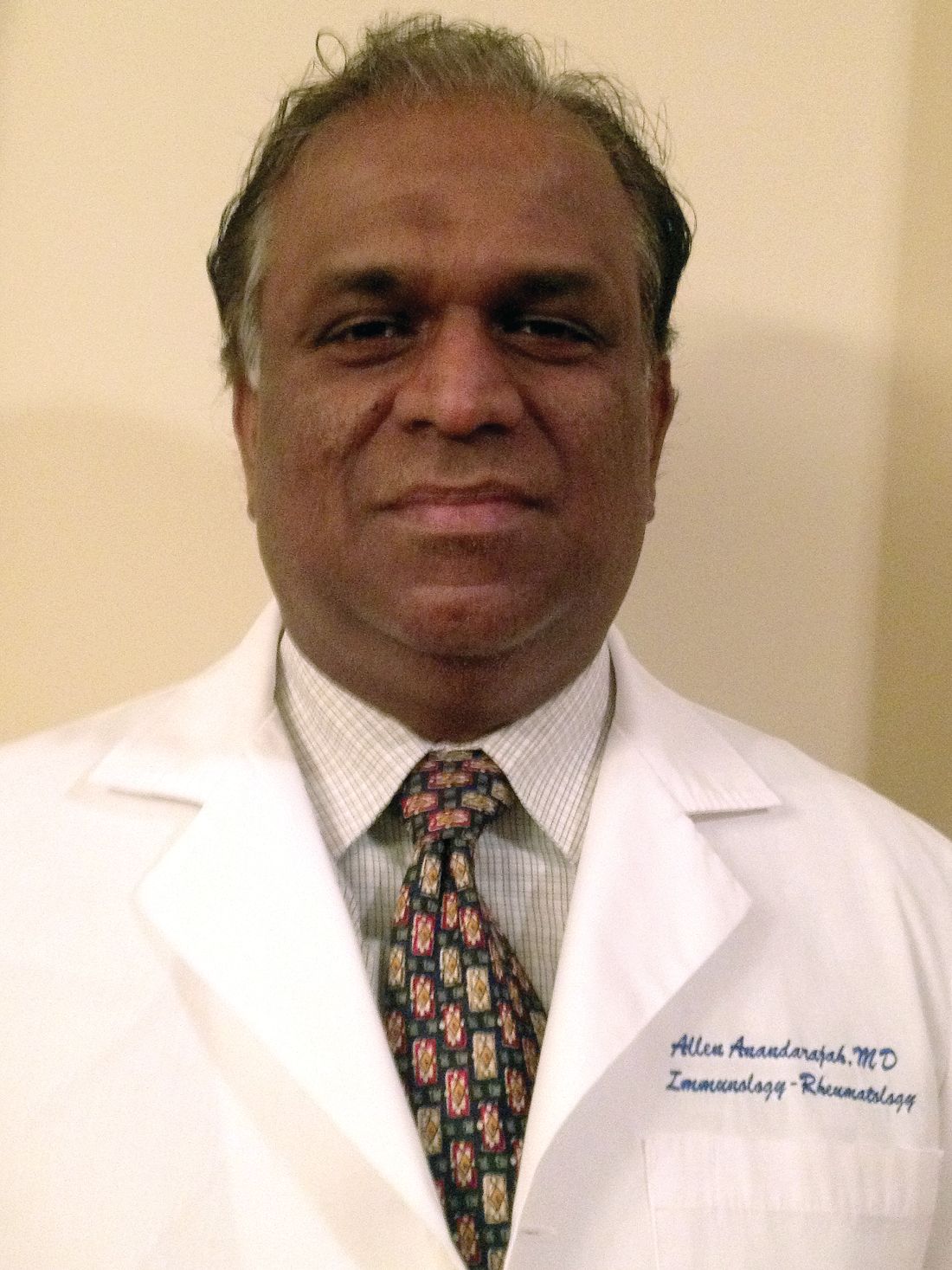
The researchers found that the average patient required $51,808 in treatment costs annually; the average stay was 8.5 days (Lupus. 2017;26[7]:756-61).
Dr. Anandarajah led another study, released at the 2016 ACR annual meeting, that found patients at high risk of hospitalization were more likely to be younger, have earlier SLE onset, and be African American (abstract 122).
As for medication nonadherence, a systematic review of 11 studies published this year found that “the percentage of nonadherent patients ranged from 43% to 75%, with studies consistently reporting that over half of patients are nonadherent” (Arthritis Care Res [Hoboken]. 2017 Nov;69[11]:1706-13).
Nonadherence is an especially significant issue “among a small group of high-risk, high-need patients,” Dr. Anandarajah said.
For the new study, the researchers aimed to better understand “if medication adherence was a risk factor for hospital admissions among SLE patients,” he said.
They identified a group of 28 high-risk patients out of 171 hospitalized SLE patients who were admitted from 2013 to 2015. Compared with other patients, the high-risk patients, who required three or more annual admissions, were younger (mean age, 39.6 vs. 47.6; P = .03), less likely to be female (82% vs. 92%; P = .09), and more likely to be African American (61% vs. 41%; P = .05).
Why might the young be less adherent? “Younger people are more likely to have difficulty with taking care of themselves when afflicted with chronic diseases due to lack of understanding of the implications of insufficiently treating their illness, poor coping skills, peer pressures about dealing with potential side effects like weight gain with steroids, and financial reasons, including lack of insurance,” he said.
As for African Americans, possible reasons for lower adherence include “cultural reasons such as a taboo about illness and misconceptions about need for continuous use of medications, lower educational levels, lack of trust in their health care providers/health care team, and socioeconomic reasons/financial issues,” he said.
The researchers linked patients to a pharmacy claims database to calculate the medication possession ratio, “an indicator of whether a patient had adequate medication supply in a given time frame,” as the study puts it. A total of 102 patients had complete pharmacy data.
The researchers found that the unadjusted mean medication possession ratio was lower in high-risk patients, compared with the others (73.4% vs. 79.9%; P = .30), and was an estimated 10 percentage points lower in an adjusted analysis that nearly reached statistical significance (P = .06).
“While it was not significant, there was a trend, and one could possibly expect a significant value with larger numbers,” Dr. Anandarajah said.
How can adherence be improved in SLE? In an interview, Michelle Petri, MD, professor of medicine and codirector of the lupus center at Johns Hopkins University, Baltimore, said she saw a major improvement in hydroxychloroquine (Plaquenil) adherence after introducing blood level testing.
“I believe rheumatologists should introduce drug monitoring for all of our important drugs: [hydroxychloroquine] (where it must be a whole blood level and not plasma), azathioprine, methotrexate, and mycophenolate,” said Dr. Petri, who praised the new research as “an excellent first study.”
Going forward, Dr. Anandarajah said his university has started a program designed to help poor, high-risk SLE patients in the Rochester area through a clinic in the inner city, coordinated care with nurses, and a series of focus-group meetings and educational programs for patients and providers. “We hope to improve compliance with outpatient visits, medication adherence, and self-management skills,” he said.
The study authors and Dr. Petri reported no relevant disclosures. No specific study funding was reported.
SAN DIEGO – New research into factors that predict which systemic lupus erythematosus patients are at high risk for hospitalization is beginning to identify the contribution of medication nonadherence to the problem.
Compared with others hospitalized for systemic lupus erythematosus (SLE), high-risk patients were an adjusted 10 percentage points less likely to show evidence of adherence to prescribed drugs, according to a study presented at the annual meeting of the American College of Rheumatology.
“Medication nonadherence remains an important problem among patients with SLE. It is a major modifiable cause to help decrease hospital admissions and readmissions and decrease risk for morbidity and mortality associated with SLE,” study coauthor Allen P. Anandarajah, MBBS, said in an interview after the ACR meeting.
The researchers found that the average patient required $51,808 in treatment costs annually; the average stay was 8.5 days (Lupus. 2017;26[7]:756-61).
Dr. Anandarajah led another study, released at the 2016 ACR annual meeting, that found patients at high risk of hospitalization were more likely to be younger, have earlier SLE onset, and be African American (abstract 122).
As for medication nonadherence, a systematic review of 11 studies published this year found that “the percentage of nonadherent patients ranged from 43% to 75%, with studies consistently reporting that over half of patients are nonadherent” (Arthritis Care Res [Hoboken]. 2017 Nov;69[11]:1706-13).
Nonadherence is an especially significant issue “among a small group of high-risk, high-need patients,” Dr. Anandarajah said.
For the new study, the researchers aimed to better understand “if medication adherence was a risk factor for hospital admissions among SLE patients,” he said.
They identified a group of 28 high-risk patients out of 171 hospitalized SLE patients who were admitted from 2013 to 2015. Compared with other patients, the high-risk patients, who required three or more annual admissions, were younger (mean age, 39.6 vs. 47.6; P = .03), less likely to be female (82% vs. 92%; P = .09), and more likely to be African American (61% vs. 41%; P = .05).
Why might the young be less adherent? “Younger people are more likely to have difficulty with taking care of themselves when afflicted with chronic diseases due to lack of understanding of the implications of insufficiently treating their illness, poor coping skills, peer pressures about dealing with potential side effects like weight gain with steroids, and financial reasons, including lack of insurance,” he said.
As for African Americans, possible reasons for lower adherence include “cultural reasons such as a taboo about illness and misconceptions about need for continuous use of medications, lower educational levels, lack of trust in their health care providers/health care team, and socioeconomic reasons/financial issues,” he said.
The researchers linked patients to a pharmacy claims database to calculate the medication possession ratio, “an indicator of whether a patient had adequate medication supply in a given time frame,” as the study puts it. A total of 102 patients had complete pharmacy data.
The researchers found that the unadjusted mean medication possession ratio was lower in high-risk patients, compared with the others (73.4% vs. 79.9%; P = .30), and was an estimated 10 percentage points lower in an adjusted analysis that nearly reached statistical significance (P = .06).
“While it was not significant, there was a trend, and one could possibly expect a significant value with larger numbers,” Dr. Anandarajah said.
How can adherence be improved in SLE? In an interview, Michelle Petri, MD, professor of medicine and codirector of the lupus center at Johns Hopkins University, Baltimore, said she saw a major improvement in hydroxychloroquine (Plaquenil) adherence after introducing blood level testing.
“I believe rheumatologists should introduce drug monitoring for all of our important drugs: [hydroxychloroquine] (where it must be a whole blood level and not plasma), azathioprine, methotrexate, and mycophenolate,” said Dr. Petri, who praised the new research as “an excellent first study.”
Going forward, Dr. Anandarajah said his university has started a program designed to help poor, high-risk SLE patients in the Rochester area through a clinic in the inner city, coordinated care with nurses, and a series of focus-group meetings and educational programs for patients and providers. “We hope to improve compliance with outpatient visits, medication adherence, and self-management skills,” he said.
The study authors and Dr. Petri reported no relevant disclosures. No specific study funding was reported.
SAN DIEGO – New research into factors that predict which systemic lupus erythematosus patients are at high risk for hospitalization is beginning to identify the contribution of medication nonadherence to the problem.
Compared with others hospitalized for systemic lupus erythematosus (SLE), high-risk patients were an adjusted 10 percentage points less likely to show evidence of adherence to prescribed drugs, according to a study presented at the annual meeting of the American College of Rheumatology.
“Medication nonadherence remains an important problem among patients with SLE. It is a major modifiable cause to help decrease hospital admissions and readmissions and decrease risk for morbidity and mortality associated with SLE,” study coauthor Allen P. Anandarajah, MBBS, said in an interview after the ACR meeting.
The researchers found that the average patient required $51,808 in treatment costs annually; the average stay was 8.5 days (Lupus. 2017;26[7]:756-61).
Dr. Anandarajah led another study, released at the 2016 ACR annual meeting, that found patients at high risk of hospitalization were more likely to be younger, have earlier SLE onset, and be African American (abstract 122).
As for medication nonadherence, a systematic review of 11 studies published this year found that “the percentage of nonadherent patients ranged from 43% to 75%, with studies consistently reporting that over half of patients are nonadherent” (Arthritis Care Res [Hoboken]. 2017 Nov;69[11]:1706-13).
Nonadherence is an especially significant issue “among a small group of high-risk, high-need patients,” Dr. Anandarajah said.
For the new study, the researchers aimed to better understand “if medication adherence was a risk factor for hospital admissions among SLE patients,” he said.
They identified a group of 28 high-risk patients out of 171 hospitalized SLE patients who were admitted from 2013 to 2015. Compared with other patients, the high-risk patients, who required three or more annual admissions, were younger (mean age, 39.6 vs. 47.6; P = .03), less likely to be female (82% vs. 92%; P = .09), and more likely to be African American (61% vs. 41%; P = .05).
Why might the young be less adherent? “Younger people are more likely to have difficulty with taking care of themselves when afflicted with chronic diseases due to lack of understanding of the implications of insufficiently treating their illness, poor coping skills, peer pressures about dealing with potential side effects like weight gain with steroids, and financial reasons, including lack of insurance,” he said.
As for African Americans, possible reasons for lower adherence include “cultural reasons such as a taboo about illness and misconceptions about need for continuous use of medications, lower educational levels, lack of trust in their health care providers/health care team, and socioeconomic reasons/financial issues,” he said.
The researchers linked patients to a pharmacy claims database to calculate the medication possession ratio, “an indicator of whether a patient had adequate medication supply in a given time frame,” as the study puts it. A total of 102 patients had complete pharmacy data.
The researchers found that the unadjusted mean medication possession ratio was lower in high-risk patients, compared with the others (73.4% vs. 79.9%; P = .30), and was an estimated 10 percentage points lower in an adjusted analysis that nearly reached statistical significance (P = .06).
“While it was not significant, there was a trend, and one could possibly expect a significant value with larger numbers,” Dr. Anandarajah said.
How can adherence be improved in SLE? In an interview, Michelle Petri, MD, professor of medicine and codirector of the lupus center at Johns Hopkins University, Baltimore, said she saw a major improvement in hydroxychloroquine (Plaquenil) adherence after introducing blood level testing.
“I believe rheumatologists should introduce drug monitoring for all of our important drugs: [hydroxychloroquine] (where it must be a whole blood level and not plasma), azathioprine, methotrexate, and mycophenolate,” said Dr. Petri, who praised the new research as “an excellent first study.”
Going forward, Dr. Anandarajah said his university has started a program designed to help poor, high-risk SLE patients in the Rochester area through a clinic in the inner city, coordinated care with nurses, and a series of focus-group meetings and educational programs for patients and providers. “We hope to improve compliance with outpatient visits, medication adherence, and self-management skills,” he said.
The study authors and Dr. Petri reported no relevant disclosures. No specific study funding was reported.
REPORTING FROM ACR 2017
Key clinical point:
Major finding: Compared with other patients hospitalized with SLE, high-risk patients had 10% lower medication adherence.
Data source: A 2-year analysis of 171 patients (28 deemed high risk) admitted for SLE at a single hospital.
Disclosures: The study authors reported no relevant disclosures. No specific study funding is reported.
Source: C. Thirukuraman et al. ACR 2017 abstract 223.
Rheumatology 911: Inside the rheumatologic emergency
SAN DIEGO – At first glance, rheumatology may seem like the perfect specialty for physicians who don’t want to be bothered by medical emergencies. But the reality can be more complicated.
As Bharat Kumar, MD, explained to an audience at the annual meeting of the American College of Rheumatology, rheumatologists will at times encounter patients in urgent need of their care due to dire medical conditions. In these situations, he said, there may be no time for careful and cautious diagnostics.
“You have to have an awareness of how you think about things,” advised Dr. Kumar, a rheumatologist/immunologist and clinical assistant professor of internal medicine at the University of Iowa, Iowa City. “During emergencies, you have to rely more on intuition to quickly get at answers.”

Q: When do rheumatologists have to deal with medical emergencies?
A: Rheumatology is considered mostly an outpatient specialty. Most of the time, rheumatologists don’t receive off-hour emergency calls.
But there are conditions in which rheumatologists have to be at the front lines in diagnosing and managing medical emergencies. These range from issues like septic arthritis to scleroderma renal crisis and vasculitis affecting vital organs such as the heart, lungs, and kidneys. These are more common at academic settings, but even rheumatologists in private practice should be aware of these conditions.
Q: How often do rheumatologists come across true emergencies in normal practice?
A: It depends on where the rheumatologist is practicing. In our academic setting, we have to see patients in the hospital several times per week.
Rarer are the emergencies that show up to clinic and require evaluation in the emergency department or hospitalization. Over the past year, that has happened perhaps three times to me.
This is likely much less in the private setting, where patients tend to be less sick and less complicated. But that is no guarantee that an emergency won’t crop up.
Q: What is the scariest emergency situation that you’ve come across?
A: It occurred when I entered a room to see a patient of mine with adult-onset Still’s disease.
She was huddled, shivering, barely answering questions. Her eyes were glazed. Her blood pressure was below 90/60 mm Hg, and her pulse was 130 beats per minute. I was petrified that she was in the midst of a cytokine storm secondary to either hemophagocytic lymphohistiocytosis (HLH) or sepsis. Given the high mortality of both, we immediately called our colleagues in the emergency department and sent her for hospitalization. It turned out that she did have HLH, and we had to pursue intensive immunosuppression to abate that cytokine storm.
It was particularly scary because there is no good way to differentiate between the two conditions, apart from going with clinical intuition.
Treating a patient who is potentially septic with immunosuppression is extremely dangerous, and ultimately, we would not have known if our intuition was correct until the infection presented itself.
Fortunately, we were correct. She recovered after 1 week of hospitalization, and we have been following her since then. But it still gives me goosebumps to think, “What if we were wrong?”
Q: Do emergencies in rheumatology tend to appear suddenly or are they more likely to occur because of a long-standing and perhaps untreated condition?
A: While it is true that uncontrolled disease activity can predispose patients to emergencies, other emergencies can occur sporadically and out of the blue.
Many times, an emergency is the first manifestation of disease. The literature is littered with cases of renal crisis being the first manifestation of systemic sclerosis. And internists are often baffled by sudden kidney failure due to previously undiagnosed lupus.
In addition, all rheumatologists have great reverence for septic arthritis and know that it can mimic gout very closely. If a swollen joint is mistaken for gout instead of septic arthritis, this can lead to worsening infection and ultimately, loss of joint function.
Q: What are some potentially dire conditions that may test the diagnostic powers of rheumatologists?
A: Rheumatologists are becoming more aware of HLH. Because it may look clinically indistinguishable from severe infection but needs to be treated with immunosuppression instead of antimicrobial therapy, rheumatologists have to keep it in mind and revisit the diagnosis often in case patients are not improving on the prescribed therapy.
Pulmonary vasculitis is another concerning condition because an otherwise negligible cough can turn into massive pulmonary hemorrhage very quickly.
Q: Do you have tips about dealing with ER doctors, primary doctors and others who may be involved with an emergency?
A: Rheumatologists think differently from other specialists. We are cognitive specialists and think more in the long term. Emergency medicine doctors are more concerned about the short term and how to deal with more immediate issues.
Signposting concerns is essential to optimizing communication. Education of other physicians is also important because more frequently than not, patients with rheumatologic diseases present very differently.
Lastly, there’s a very fine line between advocating for patients and overstepping your bounds as a consultant rheumatologist. Maintaining close collaboration and establishing clear and open lines of communication can prevent this.
Dr. Kumar has no relevant disclosures.
SAN DIEGO – At first glance, rheumatology may seem like the perfect specialty for physicians who don’t want to be bothered by medical emergencies. But the reality can be more complicated.
As Bharat Kumar, MD, explained to an audience at the annual meeting of the American College of Rheumatology, rheumatologists will at times encounter patients in urgent need of their care due to dire medical conditions. In these situations, he said, there may be no time for careful and cautious diagnostics.
“You have to have an awareness of how you think about things,” advised Dr. Kumar, a rheumatologist/immunologist and clinical assistant professor of internal medicine at the University of Iowa, Iowa City. “During emergencies, you have to rely more on intuition to quickly get at answers.”
Q: When do rheumatologists have to deal with medical emergencies?
A: Rheumatology is considered mostly an outpatient specialty. Most of the time, rheumatologists don’t receive off-hour emergency calls.
But there are conditions in which rheumatologists have to be at the front lines in diagnosing and managing medical emergencies. These range from issues like septic arthritis to scleroderma renal crisis and vasculitis affecting vital organs such as the heart, lungs, and kidneys. These are more common at academic settings, but even rheumatologists in private practice should be aware of these conditions.
Q: How often do rheumatologists come across true emergencies in normal practice?
A: It depends on where the rheumatologist is practicing. In our academic setting, we have to see patients in the hospital several times per week.
Rarer are the emergencies that show up to clinic and require evaluation in the emergency department or hospitalization. Over the past year, that has happened perhaps three times to me.
This is likely much less in the private setting, where patients tend to be less sick and less complicated. But that is no guarantee that an emergency won’t crop up.
Q: What is the scariest emergency situation that you’ve come across?
A: It occurred when I entered a room to see a patient of mine with adult-onset Still’s disease.
She was huddled, shivering, barely answering questions. Her eyes were glazed. Her blood pressure was below 90/60 mm Hg, and her pulse was 130 beats per minute. I was petrified that she was in the midst of a cytokine storm secondary to either hemophagocytic lymphohistiocytosis (HLH) or sepsis. Given the high mortality of both, we immediately called our colleagues in the emergency department and sent her for hospitalization. It turned out that she did have HLH, and we had to pursue intensive immunosuppression to abate that cytokine storm.
It was particularly scary because there is no good way to differentiate between the two conditions, apart from going with clinical intuition.
Treating a patient who is potentially septic with immunosuppression is extremely dangerous, and ultimately, we would not have known if our intuition was correct until the infection presented itself.
Fortunately, we were correct. She recovered after 1 week of hospitalization, and we have been following her since then. But it still gives me goosebumps to think, “What if we were wrong?”
Q: Do emergencies in rheumatology tend to appear suddenly or are they more likely to occur because of a long-standing and perhaps untreated condition?
A: While it is true that uncontrolled disease activity can predispose patients to emergencies, other emergencies can occur sporadically and out of the blue.
Many times, an emergency is the first manifestation of disease. The literature is littered with cases of renal crisis being the first manifestation of systemic sclerosis. And internists are often baffled by sudden kidney failure due to previously undiagnosed lupus.
In addition, all rheumatologists have great reverence for septic arthritis and know that it can mimic gout very closely. If a swollen joint is mistaken for gout instead of septic arthritis, this can lead to worsening infection and ultimately, loss of joint function.
Q: What are some potentially dire conditions that may test the diagnostic powers of rheumatologists?
A: Rheumatologists are becoming more aware of HLH. Because it may look clinically indistinguishable from severe infection but needs to be treated with immunosuppression instead of antimicrobial therapy, rheumatologists have to keep it in mind and revisit the diagnosis often in case patients are not improving on the prescribed therapy.
Pulmonary vasculitis is another concerning condition because an otherwise negligible cough can turn into massive pulmonary hemorrhage very quickly.
Q: Do you have tips about dealing with ER doctors, primary doctors and others who may be involved with an emergency?
A: Rheumatologists think differently from other specialists. We are cognitive specialists and think more in the long term. Emergency medicine doctors are more concerned about the short term and how to deal with more immediate issues.
Signposting concerns is essential to optimizing communication. Education of other physicians is also important because more frequently than not, patients with rheumatologic diseases present very differently.
Lastly, there’s a very fine line between advocating for patients and overstepping your bounds as a consultant rheumatologist. Maintaining close collaboration and establishing clear and open lines of communication can prevent this.
Dr. Kumar has no relevant disclosures.
SAN DIEGO – At first glance, rheumatology may seem like the perfect specialty for physicians who don’t want to be bothered by medical emergencies. But the reality can be more complicated.
As Bharat Kumar, MD, explained to an audience at the annual meeting of the American College of Rheumatology, rheumatologists will at times encounter patients in urgent need of their care due to dire medical conditions. In these situations, he said, there may be no time for careful and cautious diagnostics.
“You have to have an awareness of how you think about things,” advised Dr. Kumar, a rheumatologist/immunologist and clinical assistant professor of internal medicine at the University of Iowa, Iowa City. “During emergencies, you have to rely more on intuition to quickly get at answers.”
Q: When do rheumatologists have to deal with medical emergencies?
A: Rheumatology is considered mostly an outpatient specialty. Most of the time, rheumatologists don’t receive off-hour emergency calls.
But there are conditions in which rheumatologists have to be at the front lines in diagnosing and managing medical emergencies. These range from issues like septic arthritis to scleroderma renal crisis and vasculitis affecting vital organs such as the heart, lungs, and kidneys. These are more common at academic settings, but even rheumatologists in private practice should be aware of these conditions.
Q: How often do rheumatologists come across true emergencies in normal practice?
A: It depends on where the rheumatologist is practicing. In our academic setting, we have to see patients in the hospital several times per week.
Rarer are the emergencies that show up to clinic and require evaluation in the emergency department or hospitalization. Over the past year, that has happened perhaps three times to me.
This is likely much less in the private setting, where patients tend to be less sick and less complicated. But that is no guarantee that an emergency won’t crop up.
Q: What is the scariest emergency situation that you’ve come across?
A: It occurred when I entered a room to see a patient of mine with adult-onset Still’s disease.
She was huddled, shivering, barely answering questions. Her eyes were glazed. Her blood pressure was below 90/60 mm Hg, and her pulse was 130 beats per minute. I was petrified that she was in the midst of a cytokine storm secondary to either hemophagocytic lymphohistiocytosis (HLH) or sepsis. Given the high mortality of both, we immediately called our colleagues in the emergency department and sent her for hospitalization. It turned out that she did have HLH, and we had to pursue intensive immunosuppression to abate that cytokine storm.
It was particularly scary because there is no good way to differentiate between the two conditions, apart from going with clinical intuition.
Treating a patient who is potentially septic with immunosuppression is extremely dangerous, and ultimately, we would not have known if our intuition was correct until the infection presented itself.
Fortunately, we were correct. She recovered after 1 week of hospitalization, and we have been following her since then. But it still gives me goosebumps to think, “What if we were wrong?”
Q: Do emergencies in rheumatology tend to appear suddenly or are they more likely to occur because of a long-standing and perhaps untreated condition?
A: While it is true that uncontrolled disease activity can predispose patients to emergencies, other emergencies can occur sporadically and out of the blue.
Many times, an emergency is the first manifestation of disease. The literature is littered with cases of renal crisis being the first manifestation of systemic sclerosis. And internists are often baffled by sudden kidney failure due to previously undiagnosed lupus.
In addition, all rheumatologists have great reverence for septic arthritis and know that it can mimic gout very closely. If a swollen joint is mistaken for gout instead of septic arthritis, this can lead to worsening infection and ultimately, loss of joint function.
Q: What are some potentially dire conditions that may test the diagnostic powers of rheumatologists?
A: Rheumatologists are becoming more aware of HLH. Because it may look clinically indistinguishable from severe infection but needs to be treated with immunosuppression instead of antimicrobial therapy, rheumatologists have to keep it in mind and revisit the diagnosis often in case patients are not improving on the prescribed therapy.
Pulmonary vasculitis is another concerning condition because an otherwise negligible cough can turn into massive pulmonary hemorrhage very quickly.
Q: Do you have tips about dealing with ER doctors, primary doctors and others who may be involved with an emergency?
A: Rheumatologists think differently from other specialists. We are cognitive specialists and think more in the long term. Emergency medicine doctors are more concerned about the short term and how to deal with more immediate issues.
Signposting concerns is essential to optimizing communication. Education of other physicians is also important because more frequently than not, patients with rheumatologic diseases present very differently.
Lastly, there’s a very fine line between advocating for patients and overstepping your bounds as a consultant rheumatologist. Maintaining close collaboration and establishing clear and open lines of communication can prevent this.
Dr. Kumar has no relevant disclosures.
EXPERT ANALYSIS FROM ACR 2017
Survey reveals heavy EMR burden on surgical residents
SAN DIEGO – Surgical residents spend a large part of every working day in front of a computer screen, with first-year residents saying they spend an average of more than 13 hours a day on electronic medical records (EMRs).
“Residents are spending a lot of time sitting at a computer, and residents seem to be in agreement that this is time they could potentially be spending learning how to operate and care for patients, which is one of the fundamental purposes of residency training,” study lead author Edward S. Shipper III, MD, a PGY-3 general surgery resident at UT Health-San Antonio, said in an interview after he presented the study findings at the annual clinical congress of the American College of Surgeons.
Research into the EMR burden on residents is sparse. In 2015, researchers at the Medical College of Wisconsin, University of Wisconsin–Madison, and Northwestern University launched a study that they described as the first to examine changes in EMR use over time during surgical residency. The analysis of videos of patient-resident interactions in the exam room found that senior family medicine residents used EMRs more than junior residents (Fam Med. 2015;47[9]:722-26). The current study of surgical residents, however, showed the reverse: Senior residents used EMRs less than juniors.
Dr. Shipper and his colleagues analyzed survey results from 229 U.S. surgical residents who were reached via the Resident and Associate Society of the American College of Surgeons.
Of the 169 who reported demographic data, nearly half were women and 84.6% were training in general surgery, with the rest in subspecialties. The wide majority were in academic or academic-affiliated programs. Residents reported using EMRs exclusively for most clinical tasks, such as medication orders (90.8%), discharge summary (73.5%), and consultation requests (61.7%).
Only about half of those surveyed reported using EMRs exclusively for operative notes (which were often dictated) and signout/handoffs.
In terms of EMR workload per day, PGY-1 residents (n = 23) reported spending an average of 13.6 hours on the records. The average amount of time spent on EMRs per day fell to 10.8 hours for PGY-2 residents (n = 40) and dwindled to 4.6 among PGY-5 residents (n = 20). The researchers reported that the difference in daily EMR time between senior and junior residents is statistically significant.
“Whether or not you believe the specific numbers quoted by the residents, I think the message most people can agree upon is that residents are spending a lot of time during residency sitting in front of a computer,” Dr. Shipper said. “A parallel trend with the rise of the EMR is the rise of increased standards for, and tracking of, documentation requirements by the government and by insurance companies.”
Why are senior surgical residents spending less time on EMRs? “More senior residents generally have the primary responsibility of operating on the patients, and being in the operating room all day means less time spent in front of a computer,” he said.
Of the 63 open-ended responses about the use of EMRs in surgical education, 49% were negative and the rest were evenly divided between natural and positive. One resident described the records as essential to patient care because of their efficiency, while another said, “In this age of duty-hour limits, I spend most of my day in front of a computer interacting with the EHR. This significantly detracts from my educational experience.”
Dr. Shipper said that the study raises questions about how EMRs are affecting how surgical residents learn their craft. But Oren Sagher, MD, professor of neurosurgery at the University of Michigan, Ann Arbor, who has questioned the effect of EMRs on medical education (PLoS Med. 2009;6[5]:e1000069), isn’t impressed by the new research.
Dr. Sagher said an interview. “It’s well established that medical documentation usually falls mainly to the junior residents in surgery program. That was also true prior to EMRs. I would agree that EMRs do tend to take up more time than traditional paper charts did, but this finding is not earth shattering.”
The limit on duty hours would presumably push the burden of EMRs from residents to others, such as physician extenders, but it’s interesting that residents still report using EMRs for the bulk of their days, he added.
In the big picture, Dr. Sagher said, “EMRs are not optimized for the delivery of care. They appear to be mostly driven by billing concerns and safety optimization. Consequently, the people forced to use these systems are not very happy. I think this contributes to physician disenfranchisement and burnout.”
SAN DIEGO – Surgical residents spend a large part of every working day in front of a computer screen, with first-year residents saying they spend an average of more than 13 hours a day on electronic medical records (EMRs).
“Residents are spending a lot of time sitting at a computer, and residents seem to be in agreement that this is time they could potentially be spending learning how to operate and care for patients, which is one of the fundamental purposes of residency training,” study lead author Edward S. Shipper III, MD, a PGY-3 general surgery resident at UT Health-San Antonio, said in an interview after he presented the study findings at the annual clinical congress of the American College of Surgeons.
Research into the EMR burden on residents is sparse. In 2015, researchers at the Medical College of Wisconsin, University of Wisconsin–Madison, and Northwestern University launched a study that they described as the first to examine changes in EMR use over time during surgical residency. The analysis of videos of patient-resident interactions in the exam room found that senior family medicine residents used EMRs more than junior residents (Fam Med. 2015;47[9]:722-26). The current study of surgical residents, however, showed the reverse: Senior residents used EMRs less than juniors.
Dr. Shipper and his colleagues analyzed survey results from 229 U.S. surgical residents who were reached via the Resident and Associate Society of the American College of Surgeons.
Of the 169 who reported demographic data, nearly half were women and 84.6% were training in general surgery, with the rest in subspecialties. The wide majority were in academic or academic-affiliated programs. Residents reported using EMRs exclusively for most clinical tasks, such as medication orders (90.8%), discharge summary (73.5%), and consultation requests (61.7%).
Only about half of those surveyed reported using EMRs exclusively for operative notes (which were often dictated) and signout/handoffs.
In terms of EMR workload per day, PGY-1 residents (n = 23) reported spending an average of 13.6 hours on the records. The average amount of time spent on EMRs per day fell to 10.8 hours for PGY-2 residents (n = 40) and dwindled to 4.6 among PGY-5 residents (n = 20). The researchers reported that the difference in daily EMR time between senior and junior residents is statistically significant.
“Whether or not you believe the specific numbers quoted by the residents, I think the message most people can agree upon is that residents are spending a lot of time during residency sitting in front of a computer,” Dr. Shipper said. “A parallel trend with the rise of the EMR is the rise of increased standards for, and tracking of, documentation requirements by the government and by insurance companies.”
Why are senior surgical residents spending less time on EMRs? “More senior residents generally have the primary responsibility of operating on the patients, and being in the operating room all day means less time spent in front of a computer,” he said.
Of the 63 open-ended responses about the use of EMRs in surgical education, 49% were negative and the rest were evenly divided between natural and positive. One resident described the records as essential to patient care because of their efficiency, while another said, “In this age of duty-hour limits, I spend most of my day in front of a computer interacting with the EHR. This significantly detracts from my educational experience.”
Dr. Shipper said that the study raises questions about how EMRs are affecting how surgical residents learn their craft. But Oren Sagher, MD, professor of neurosurgery at the University of Michigan, Ann Arbor, who has questioned the effect of EMRs on medical education (PLoS Med. 2009;6[5]:e1000069), isn’t impressed by the new research.
Dr. Sagher said an interview. “It’s well established that medical documentation usually falls mainly to the junior residents in surgery program. That was also true prior to EMRs. I would agree that EMRs do tend to take up more time than traditional paper charts did, but this finding is not earth shattering.”
The limit on duty hours would presumably push the burden of EMRs from residents to others, such as physician extenders, but it’s interesting that residents still report using EMRs for the bulk of their days, he added.
In the big picture, Dr. Sagher said, “EMRs are not optimized for the delivery of care. They appear to be mostly driven by billing concerns and safety optimization. Consequently, the people forced to use these systems are not very happy. I think this contributes to physician disenfranchisement and burnout.”
SAN DIEGO – Surgical residents spend a large part of every working day in front of a computer screen, with first-year residents saying they spend an average of more than 13 hours a day on electronic medical records (EMRs).
“Residents are spending a lot of time sitting at a computer, and residents seem to be in agreement that this is time they could potentially be spending learning how to operate and care for patients, which is one of the fundamental purposes of residency training,” study lead author Edward S. Shipper III, MD, a PGY-3 general surgery resident at UT Health-San Antonio, said in an interview after he presented the study findings at the annual clinical congress of the American College of Surgeons.
Research into the EMR burden on residents is sparse. In 2015, researchers at the Medical College of Wisconsin, University of Wisconsin–Madison, and Northwestern University launched a study that they described as the first to examine changes in EMR use over time during surgical residency. The analysis of videos of patient-resident interactions in the exam room found that senior family medicine residents used EMRs more than junior residents (Fam Med. 2015;47[9]:722-26). The current study of surgical residents, however, showed the reverse: Senior residents used EMRs less than juniors.
Dr. Shipper and his colleagues analyzed survey results from 229 U.S. surgical residents who were reached via the Resident and Associate Society of the American College of Surgeons.
Of the 169 who reported demographic data, nearly half were women and 84.6% were training in general surgery, with the rest in subspecialties. The wide majority were in academic or academic-affiliated programs. Residents reported using EMRs exclusively for most clinical tasks, such as medication orders (90.8%), discharge summary (73.5%), and consultation requests (61.7%).
Only about half of those surveyed reported using EMRs exclusively for operative notes (which were often dictated) and signout/handoffs.
In terms of EMR workload per day, PGY-1 residents (n = 23) reported spending an average of 13.6 hours on the records. The average amount of time spent on EMRs per day fell to 10.8 hours for PGY-2 residents (n = 40) and dwindled to 4.6 among PGY-5 residents (n = 20). The researchers reported that the difference in daily EMR time between senior and junior residents is statistically significant.
“Whether or not you believe the specific numbers quoted by the residents, I think the message most people can agree upon is that residents are spending a lot of time during residency sitting in front of a computer,” Dr. Shipper said. “A parallel trend with the rise of the EMR is the rise of increased standards for, and tracking of, documentation requirements by the government and by insurance companies.”
Why are senior surgical residents spending less time on EMRs? “More senior residents generally have the primary responsibility of operating on the patients, and being in the operating room all day means less time spent in front of a computer,” he said.
Of the 63 open-ended responses about the use of EMRs in surgical education, 49% were negative and the rest were evenly divided between natural and positive. One resident described the records as essential to patient care because of their efficiency, while another said, “In this age of duty-hour limits, I spend most of my day in front of a computer interacting with the EHR. This significantly detracts from my educational experience.”
Dr. Shipper said that the study raises questions about how EMRs are affecting how surgical residents learn their craft. But Oren Sagher, MD, professor of neurosurgery at the University of Michigan, Ann Arbor, who has questioned the effect of EMRs on medical education (PLoS Med. 2009;6[5]:e1000069), isn’t impressed by the new research.
Dr. Sagher said an interview. “It’s well established that medical documentation usually falls mainly to the junior residents in surgery program. That was also true prior to EMRs. I would agree that EMRs do tend to take up more time than traditional paper charts did, but this finding is not earth shattering.”
The limit on duty hours would presumably push the burden of EMRs from residents to others, such as physician extenders, but it’s interesting that residents still report using EMRs for the bulk of their days, he added.
In the big picture, Dr. Sagher said, “EMRs are not optimized for the delivery of care. They appear to be mostly driven by billing concerns and safety optimization. Consequently, the people forced to use these systems are not very happy. I think this contributes to physician disenfranchisement and burnout.”
AT THE ACS CLINICAL CONGRESS
Hispanics trail blacks, whites in bariatric surgery rates
SAN DIEGO – A study of procedures at academic centers provides evidence that obese Hispanics in the United States undergo bariatric surgery at a much lower rate than whites and blacks. It also reveals marked regional variations in overall weight-loss surgery.
“Our findings do suggest that severely obese Hispanics are utilizing bariatric surgery much lower than other ethnic groups,” said study coauthor Ninh T. Nguyen, MD, FACS, chair of the department of surgery at the University of California Irvine Medical Center, in an interview. “Our research does not specifically address the reasons for this gap in the delivery of care. Further research will need to be done to understand the reasons and the ways to close this gap.”
According to Dr. Nguyen, the researchers undertook the study to better understand how bariatric surgery is delivered across ethnicities and geographic regions in the United States.
The researchers analyzed statistics from the Vizient health care performance database for the years 2013-2015. They focused on patients at about 120 academic centers who underwent 73,119 laparoscopic sleeve gastrectomy, laparoscopic Roux-en-Y gastric bypass, and laparoscopic adjustable gastric banding procedures. The patients were stratified by race and region.
Researchers found that bariatric procedures were performed at a much higher rate in the Northeast academic centers (2.21 per 1,000 obese persons), compared with the Midwest (0.73), South (0.50), and West (0.33).
In regard to race, the rates for blacks and whites were fairly similar in the Northeast (2.02 and 2.35 bariatric procedures per 1,000 obese persons, respectively), the South (0.59 and 0.63, respectively) and the West (0.45 and 0.43, respectively). There was a wider gap in the Midwest, with whites at 1.07 and blacks at 0.69.
Across the country, however, obese Hispanics were less likely than persons of the other two races to undergo weight-loss surgery. The gap was fairly small in the Northeast, where 1.74 per 1,000 obese Hispanics underwent weight-loss surgery, compared with rates of 2.02 and 2.35 among black and whites, respectively. But the disparity was much larger in the other parts of the country, with rates at 0.14 in the West, 0.11 in the South and 0.33 in the Midwest, compared with rates from 0.43 to 1.07 among blacks and whites.
The reasons for the surgery gap are unknown. Dr. Nguyen pointed to several possible explanations: “lack of education of obesity as a disease by the primary care providers and the need for referral to a bariatric surgeon for patients with body mass index greater than 40 kg/m2 or 35 kg/m2 with obesity-related comorbidities; poor understanding of the benefits of bariatric surgery and its low risk; lack of understanding of the urgency for treatment by the patient and provider; and hurdles in obtaining coverage for the operation by insurers.”
John Magaña Morton, MD, FACS, chief of bariatric and minimally invasive surgery at Stanford (Calif.) University School of Medicine and past president of the American Society for Metabolic and Bariatric Surgery, doesn’t think discrimination is causing the disparity.
“It’s probably a reflection of insurance status – Hispanics tend to be less insured than Caucasian or African American patients – as well as preference for patients to go to nonacademic centers,” he said.
Indeed, a Kaiser Family Foundation analysis found that 21% of the 52 million Hispanics younger than 65 years in the United States were uninsured in 2015, compared with 9% of whites and 13% of blacks. Only Native Americans/Alaska Natives had an uninsured rate as high as Hispanics.
“In terms of need [for weight-loss features], it’s certainly there for Hispanics,” said Dr. Morton. “[Hispanic patients] have high rates of obesity and diabetes, both of which are helped by bariatric surgery.”
He said about 40% of patients in his Palo Alto, Calif., practice are Hispanic, reflecting the high number in the local population.
It helps that Dr. Morton and several of his partners speak Spanish. “If you have a welcoming environment,” he said, “that can make a difference.”
The study authors and Dr. Morton report no relevant disclosures. No specific study funding is reported.
The AGA Obesity Practice Guide provides a comprehensive, multi-disciplinary process to personalize innovative obesity care for safe and effective weight management, including the use of bariatric endoscopy and surgery. Learn more at www.gastro.org/obesity.
SAN DIEGO – A study of procedures at academic centers provides evidence that obese Hispanics in the United States undergo bariatric surgery at a much lower rate than whites and blacks. It also reveals marked regional variations in overall weight-loss surgery.
“Our findings do suggest that severely obese Hispanics are utilizing bariatric surgery much lower than other ethnic groups,” said study coauthor Ninh T. Nguyen, MD, FACS, chair of the department of surgery at the University of California Irvine Medical Center, in an interview. “Our research does not specifically address the reasons for this gap in the delivery of care. Further research will need to be done to understand the reasons and the ways to close this gap.”
According to Dr. Nguyen, the researchers undertook the study to better understand how bariatric surgery is delivered across ethnicities and geographic regions in the United States.
The researchers analyzed statistics from the Vizient health care performance database for the years 2013-2015. They focused on patients at about 120 academic centers who underwent 73,119 laparoscopic sleeve gastrectomy, laparoscopic Roux-en-Y gastric bypass, and laparoscopic adjustable gastric banding procedures. The patients were stratified by race and region.
Researchers found that bariatric procedures were performed at a much higher rate in the Northeast academic centers (2.21 per 1,000 obese persons), compared with the Midwest (0.73), South (0.50), and West (0.33).
In regard to race, the rates for blacks and whites were fairly similar in the Northeast (2.02 and 2.35 bariatric procedures per 1,000 obese persons, respectively), the South (0.59 and 0.63, respectively) and the West (0.45 and 0.43, respectively). There was a wider gap in the Midwest, with whites at 1.07 and blacks at 0.69.
Across the country, however, obese Hispanics were less likely than persons of the other two races to undergo weight-loss surgery. The gap was fairly small in the Northeast, where 1.74 per 1,000 obese Hispanics underwent weight-loss surgery, compared with rates of 2.02 and 2.35 among black and whites, respectively. But the disparity was much larger in the other parts of the country, with rates at 0.14 in the West, 0.11 in the South and 0.33 in the Midwest, compared with rates from 0.43 to 1.07 among blacks and whites.
The reasons for the surgery gap are unknown. Dr. Nguyen pointed to several possible explanations: “lack of education of obesity as a disease by the primary care providers and the need for referral to a bariatric surgeon for patients with body mass index greater than 40 kg/m2 or 35 kg/m2 with obesity-related comorbidities; poor understanding of the benefits of bariatric surgery and its low risk; lack of understanding of the urgency for treatment by the patient and provider; and hurdles in obtaining coverage for the operation by insurers.”
John Magaña Morton, MD, FACS, chief of bariatric and minimally invasive surgery at Stanford (Calif.) University School of Medicine and past president of the American Society for Metabolic and Bariatric Surgery, doesn’t think discrimination is causing the disparity.
“It’s probably a reflection of insurance status – Hispanics tend to be less insured than Caucasian or African American patients – as well as preference for patients to go to nonacademic centers,” he said.
Indeed, a Kaiser Family Foundation analysis found that 21% of the 52 million Hispanics younger than 65 years in the United States were uninsured in 2015, compared with 9% of whites and 13% of blacks. Only Native Americans/Alaska Natives had an uninsured rate as high as Hispanics.
“In terms of need [for weight-loss features], it’s certainly there for Hispanics,” said Dr. Morton. “[Hispanic patients] have high rates of obesity and diabetes, both of which are helped by bariatric surgery.”
He said about 40% of patients in his Palo Alto, Calif., practice are Hispanic, reflecting the high number in the local population.
It helps that Dr. Morton and several of his partners speak Spanish. “If you have a welcoming environment,” he said, “that can make a difference.”
The study authors and Dr. Morton report no relevant disclosures. No specific study funding is reported.
The AGA Obesity Practice Guide provides a comprehensive, multi-disciplinary process to personalize innovative obesity care for safe and effective weight management, including the use of bariatric endoscopy and surgery. Learn more at www.gastro.org/obesity.
SAN DIEGO – A study of procedures at academic centers provides evidence that obese Hispanics in the United States undergo bariatric surgery at a much lower rate than whites and blacks. It also reveals marked regional variations in overall weight-loss surgery.
“Our findings do suggest that severely obese Hispanics are utilizing bariatric surgery much lower than other ethnic groups,” said study coauthor Ninh T. Nguyen, MD, FACS, chair of the department of surgery at the University of California Irvine Medical Center, in an interview. “Our research does not specifically address the reasons for this gap in the delivery of care. Further research will need to be done to understand the reasons and the ways to close this gap.”
According to Dr. Nguyen, the researchers undertook the study to better understand how bariatric surgery is delivered across ethnicities and geographic regions in the United States.
The researchers analyzed statistics from the Vizient health care performance database for the years 2013-2015. They focused on patients at about 120 academic centers who underwent 73,119 laparoscopic sleeve gastrectomy, laparoscopic Roux-en-Y gastric bypass, and laparoscopic adjustable gastric banding procedures. The patients were stratified by race and region.
Researchers found that bariatric procedures were performed at a much higher rate in the Northeast academic centers (2.21 per 1,000 obese persons), compared with the Midwest (0.73), South (0.50), and West (0.33).
In regard to race, the rates for blacks and whites were fairly similar in the Northeast (2.02 and 2.35 bariatric procedures per 1,000 obese persons, respectively), the South (0.59 and 0.63, respectively) and the West (0.45 and 0.43, respectively). There was a wider gap in the Midwest, with whites at 1.07 and blacks at 0.69.
Across the country, however, obese Hispanics were less likely than persons of the other two races to undergo weight-loss surgery. The gap was fairly small in the Northeast, where 1.74 per 1,000 obese Hispanics underwent weight-loss surgery, compared with rates of 2.02 and 2.35 among black and whites, respectively. But the disparity was much larger in the other parts of the country, with rates at 0.14 in the West, 0.11 in the South and 0.33 in the Midwest, compared with rates from 0.43 to 1.07 among blacks and whites.
The reasons for the surgery gap are unknown. Dr. Nguyen pointed to several possible explanations: “lack of education of obesity as a disease by the primary care providers and the need for referral to a bariatric surgeon for patients with body mass index greater than 40 kg/m2 or 35 kg/m2 with obesity-related comorbidities; poor understanding of the benefits of bariatric surgery and its low risk; lack of understanding of the urgency for treatment by the patient and provider; and hurdles in obtaining coverage for the operation by insurers.”
John Magaña Morton, MD, FACS, chief of bariatric and minimally invasive surgery at Stanford (Calif.) University School of Medicine and past president of the American Society for Metabolic and Bariatric Surgery, doesn’t think discrimination is causing the disparity.
“It’s probably a reflection of insurance status – Hispanics tend to be less insured than Caucasian or African American patients – as well as preference for patients to go to nonacademic centers,” he said.
Indeed, a Kaiser Family Foundation analysis found that 21% of the 52 million Hispanics younger than 65 years in the United States were uninsured in 2015, compared with 9% of whites and 13% of blacks. Only Native Americans/Alaska Natives had an uninsured rate as high as Hispanics.
“In terms of need [for weight-loss features], it’s certainly there for Hispanics,” said Dr. Morton. “[Hispanic patients] have high rates of obesity and diabetes, both of which are helped by bariatric surgery.”
He said about 40% of patients in his Palo Alto, Calif., practice are Hispanic, reflecting the high number in the local population.
It helps that Dr. Morton and several of his partners speak Spanish. “If you have a welcoming environment,” he said, “that can make a difference.”
The study authors and Dr. Morton report no relevant disclosures. No specific study funding is reported.
The AGA Obesity Practice Guide provides a comprehensive, multi-disciplinary process to personalize innovative obesity care for safe and effective weight management, including the use of bariatric endoscopy and surgery. Learn more at www.gastro.org/obesity.
AT THE ACS CLINICAL CONGRESS
Key clinical point: At academic centers, obese Hispanics undergo bariatric surgery at a much lower rate than blacks and whites. U.S. regions outside the Northeast have lower rates of weight-loss procedures overall.
Major finding: Outside the Northeast, the bariatric surgery rate per 1,000 obese people is much lower for Hispanics (range, 0.11-0.33) than for blacks and whites (range, 0.43-1.07).
Data source: Analysis of 73,119 bariatric procedures from 2013-2015 at about 120 academic centers.
Disclosures: The study authors report no relevant disclosures. No specific study funding is reported.