User login
Rituximab fails to eliminate meningeal inflammation in progressive MS
Researchers are heading back to the drawing board after a tiny phase 1 trial of intrathecal rituximab in certain patients with progressive multiple sclerosis failed to eliminate inflammation in the meninges.
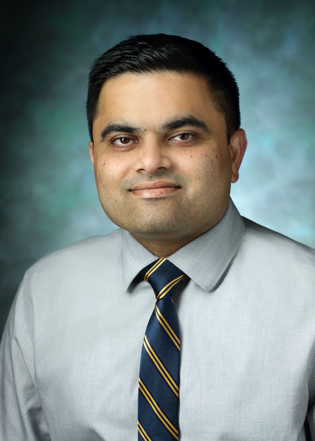
The study investigators had hoped the treatment would significantly help progressive MS patients with leptomeningeal inflammation, which has been linked to worsening disease. However, “this paradigm seems to have not gotten rid of the meningeal inflammation significantly. We need a better approach,” said Pavan Bhargava, MD, of Johns Hopkins University, Baltimore, who spoke in an interview in advance of presenting the study findings at a meeting held by the Americas Committee for Treatment and Research in Multiple Sclerosis.
The new study aimed to test the value of targeting immune cell follicles with doses of rituximab given intrathecally – straight into the spinal fluid – to greatly boost the penetration of the drug into the meninges. When given intravenously, Dr. Bhargava said, only a minuscule amount of rituximab makes it to the brain.
Dr. Bhargava’s team screened 36 patients with progressive MS with MRI scans and found that 15 showed signs of meningeal inflammation. Of those, 11 agreed to take part in the study, and 8 fit the criteria.
The participants (median age 55.5 years; five were female) received two intrathecal treatments of 25 mg of rituximab 2 weeks apart, were monitored via follow-up clinical evaluations and lab tests at weeks 2, 8, 24, and 48, and received lumbar punctures and MRI scans at weeks 8 and 24.
“The main outcome was safety,” he said. “Most patients tolerated the rituximab, and the only side effect occurred in one patient who had abnormal sensations in the leg for 35-45 minutes. Otherwise, the medication was safe.”
The researchers detected no change in the meningeal inflammatory lesions before and after the treatment at 24 weeks.
But the tiny dose, much smaller than the typical two doses of 1,000 mg of IV rituximab over 2 weeks, completely depleted B cells in the blood of almost all patients, Dr. Bhargava said. “It was likely leaking out into the blood and clearing the B cells.” Peripheral blood B cells decreased from a median of 18.55% at baseline to 0.1% at week 2 and 8 with an increase to 3.6% at 24 weeks.
This effect was not as robust in spinal fluid, he said, where B cells were not fully depleted.
The findings suggest more cycles of treatment may be needed to target types of immune cells other than B cells, he noted. Or perhaps another drug would work better if it has a different mechanism and can penetrate more effectively.
The next step, he said, is to screen alternative drug approaches in animal models with meningeal inflammation that’s similar to that found in humans.
The study was funded by the International Progressive MS Alliance and the Race to Erase MS. Dr. Bhargava reported no relevant disclosures.
SOURCE: Bhargava P et al. ACTRIMS Forum 2018 Abstract P027.
Researchers are heading back to the drawing board after a tiny phase 1 trial of intrathecal rituximab in certain patients with progressive multiple sclerosis failed to eliminate inflammation in the meninges.
The study investigators had hoped the treatment would significantly help progressive MS patients with leptomeningeal inflammation, which has been linked to worsening disease. However, “this paradigm seems to have not gotten rid of the meningeal inflammation significantly. We need a better approach,” said Pavan Bhargava, MD, of Johns Hopkins University, Baltimore, who spoke in an interview in advance of presenting the study findings at a meeting held by the Americas Committee for Treatment and Research in Multiple Sclerosis.
The new study aimed to test the value of targeting immune cell follicles with doses of rituximab given intrathecally – straight into the spinal fluid – to greatly boost the penetration of the drug into the meninges. When given intravenously, Dr. Bhargava said, only a minuscule amount of rituximab makes it to the brain.
Dr. Bhargava’s team screened 36 patients with progressive MS with MRI scans and found that 15 showed signs of meningeal inflammation. Of those, 11 agreed to take part in the study, and 8 fit the criteria.
The participants (median age 55.5 years; five were female) received two intrathecal treatments of 25 mg of rituximab 2 weeks apart, were monitored via follow-up clinical evaluations and lab tests at weeks 2, 8, 24, and 48, and received lumbar punctures and MRI scans at weeks 8 and 24.
“The main outcome was safety,” he said. “Most patients tolerated the rituximab, and the only side effect occurred in one patient who had abnormal sensations in the leg for 35-45 minutes. Otherwise, the medication was safe.”
The researchers detected no change in the meningeal inflammatory lesions before and after the treatment at 24 weeks.
But the tiny dose, much smaller than the typical two doses of 1,000 mg of IV rituximab over 2 weeks, completely depleted B cells in the blood of almost all patients, Dr. Bhargava said. “It was likely leaking out into the blood and clearing the B cells.” Peripheral blood B cells decreased from a median of 18.55% at baseline to 0.1% at week 2 and 8 with an increase to 3.6% at 24 weeks.
This effect was not as robust in spinal fluid, he said, where B cells were not fully depleted.
The findings suggest more cycles of treatment may be needed to target types of immune cells other than B cells, he noted. Or perhaps another drug would work better if it has a different mechanism and can penetrate more effectively.
The next step, he said, is to screen alternative drug approaches in animal models with meningeal inflammation that’s similar to that found in humans.
The study was funded by the International Progressive MS Alliance and the Race to Erase MS. Dr. Bhargava reported no relevant disclosures.
SOURCE: Bhargava P et al. ACTRIMS Forum 2018 Abstract P027.
Researchers are heading back to the drawing board after a tiny phase 1 trial of intrathecal rituximab in certain patients with progressive multiple sclerosis failed to eliminate inflammation in the meninges.
The study investigators had hoped the treatment would significantly help progressive MS patients with leptomeningeal inflammation, which has been linked to worsening disease. However, “this paradigm seems to have not gotten rid of the meningeal inflammation significantly. We need a better approach,” said Pavan Bhargava, MD, of Johns Hopkins University, Baltimore, who spoke in an interview in advance of presenting the study findings at a meeting held by the Americas Committee for Treatment and Research in Multiple Sclerosis.
The new study aimed to test the value of targeting immune cell follicles with doses of rituximab given intrathecally – straight into the spinal fluid – to greatly boost the penetration of the drug into the meninges. When given intravenously, Dr. Bhargava said, only a minuscule amount of rituximab makes it to the brain.
Dr. Bhargava’s team screened 36 patients with progressive MS with MRI scans and found that 15 showed signs of meningeal inflammation. Of those, 11 agreed to take part in the study, and 8 fit the criteria.
The participants (median age 55.5 years; five were female) received two intrathecal treatments of 25 mg of rituximab 2 weeks apart, were monitored via follow-up clinical evaluations and lab tests at weeks 2, 8, 24, and 48, and received lumbar punctures and MRI scans at weeks 8 and 24.
“The main outcome was safety,” he said. “Most patients tolerated the rituximab, and the only side effect occurred in one patient who had abnormal sensations in the leg for 35-45 minutes. Otherwise, the medication was safe.”
The researchers detected no change in the meningeal inflammatory lesions before and after the treatment at 24 weeks.
But the tiny dose, much smaller than the typical two doses of 1,000 mg of IV rituximab over 2 weeks, completely depleted B cells in the blood of almost all patients, Dr. Bhargava said. “It was likely leaking out into the blood and clearing the B cells.” Peripheral blood B cells decreased from a median of 18.55% at baseline to 0.1% at week 2 and 8 with an increase to 3.6% at 24 weeks.
This effect was not as robust in spinal fluid, he said, where B cells were not fully depleted.
The findings suggest more cycles of treatment may be needed to target types of immune cells other than B cells, he noted. Or perhaps another drug would work better if it has a different mechanism and can penetrate more effectively.
The next step, he said, is to screen alternative drug approaches in animal models with meningeal inflammation that’s similar to that found in humans.
The study was funded by the International Progressive MS Alliance and the Race to Erase MS. Dr. Bhargava reported no relevant disclosures.
SOURCE: Bhargava P et al. ACTRIMS Forum 2018 Abstract P027.
FROM ACTRIMS FORUM 2018
Key clinical point:
Major finding: No change in leptomeningeal inflammation was seen after two intrathecal doses of 25 mg of rituximab administered over 2 weeks.
Data source: Prospective study of eight patients with progressive MS and signs of meningeal inflammation.
Disclosures: The study was funded by the International Progressive MS Alliance and the Race to Erase MS. Study presenter Pavan Bhargava, MD, reported no relevant disclosures.
Source: Bhargava P et al. ACTRIMS Forum 2018 Abstract P027.
Barancik Prize winner to discuss MS biology advances
Two decades ago, multiple sclerosis researchers like the University of Cambridge’s, Robin Franklin, PhD, understood the crucial role of regeneration of the myelin coating on nerve fibers, and they knew the process went awry in MS patients. But, he recalled, “we knew very little about how remyelination came about and the processes that orchestrated it.”
Now, thanks in part to the work of Dr. Franklin’s team, research into remyelination is making tremendous advances. “We’ve begun to understand the mechanisms that are operating, and that is fundamental to devising new therapies,” he said in an interview. “You don’t develop effective new therapies unless you understand the underlying biology. Now, we’re on the brink of that biology leading to regenerative medicine in MS.”

Dr. Franklin is the winner of the international prize, which recognizes exceptional MS research and is administered by the National MS Society.
In recent years, Dr. Franklin and his colleagues have published multiple studies that reveal new details about the workings of myelin regeneration, especially the biology by which oligodendrocyte progenitor cells become new oligodendrocytes.
“There are simple steps in that process,” Dr. Franklin said. “What we’ve been able to do is decorate that simple model with a whole host of mechanisms that allow remyelination to take place.”
In 2011, Franklin and his colleagues published a study in rodents identifying the RXRg gene as a key promoter of remyelination, making it a potential target for therapy (Nat Neurosci. 2011;14:45-53).
“There’s still a lot more to be learned,” Dr. Franklin said, “but we’ve learned a sufficient amount that we can engage in a meaningful way with the pharmaceutical industry and MS clinicians to make regenerative medicines an effective component of the MS therapeutic armory.”
Dr. Franklin has no relevant disclosures.
Two decades ago, multiple sclerosis researchers like the University of Cambridge’s, Robin Franklin, PhD, understood the crucial role of regeneration of the myelin coating on nerve fibers, and they knew the process went awry in MS patients. But, he recalled, “we knew very little about how remyelination came about and the processes that orchestrated it.”
Now, thanks in part to the work of Dr. Franklin’s team, research into remyelination is making tremendous advances. “We’ve begun to understand the mechanisms that are operating, and that is fundamental to devising new therapies,” he said in an interview. “You don’t develop effective new therapies unless you understand the underlying biology. Now, we’re on the brink of that biology leading to regenerative medicine in MS.”
Dr. Franklin is the winner of the international prize, which recognizes exceptional MS research and is administered by the National MS Society.
In recent years, Dr. Franklin and his colleagues have published multiple studies that reveal new details about the workings of myelin regeneration, especially the biology by which oligodendrocyte progenitor cells become new oligodendrocytes.
“There are simple steps in that process,” Dr. Franklin said. “What we’ve been able to do is decorate that simple model with a whole host of mechanisms that allow remyelination to take place.”
In 2011, Franklin and his colleagues published a study in rodents identifying the RXRg gene as a key promoter of remyelination, making it a potential target for therapy (Nat Neurosci. 2011;14:45-53).
“There’s still a lot more to be learned,” Dr. Franklin said, “but we’ve learned a sufficient amount that we can engage in a meaningful way with the pharmaceutical industry and MS clinicians to make regenerative medicines an effective component of the MS therapeutic armory.”
Dr. Franklin has no relevant disclosures.
Two decades ago, multiple sclerosis researchers like the University of Cambridge’s, Robin Franklin, PhD, understood the crucial role of regeneration of the myelin coating on nerve fibers, and they knew the process went awry in MS patients. But, he recalled, “we knew very little about how remyelination came about and the processes that orchestrated it.”
Now, thanks in part to the work of Dr. Franklin’s team, research into remyelination is making tremendous advances. “We’ve begun to understand the mechanisms that are operating, and that is fundamental to devising new therapies,” he said in an interview. “You don’t develop effective new therapies unless you understand the underlying biology. Now, we’re on the brink of that biology leading to regenerative medicine in MS.”
Dr. Franklin is the winner of the international prize, which recognizes exceptional MS research and is administered by the National MS Society.
In recent years, Dr. Franklin and his colleagues have published multiple studies that reveal new details about the workings of myelin regeneration, especially the biology by which oligodendrocyte progenitor cells become new oligodendrocytes.
“There are simple steps in that process,” Dr. Franklin said. “What we’ve been able to do is decorate that simple model with a whole host of mechanisms that allow remyelination to take place.”
In 2011, Franklin and his colleagues published a study in rodents identifying the RXRg gene as a key promoter of remyelination, making it a potential target for therapy (Nat Neurosci. 2011;14:45-53).
“There’s still a lot more to be learned,” Dr. Franklin said, “but we’ve learned a sufficient amount that we can engage in a meaningful way with the pharmaceutical industry and MS clinicians to make regenerative medicines an effective component of the MS therapeutic armory.”
Dr. Franklin has no relevant disclosures.
FROM ACTRIMS FORUM 2018
Researcher’s talk will tackle aging and gut bacteria in MS
Germs, genes, and aging each play a role in the development of multiple sclerosis (MS), but researchers are still figuring out how they’re connected. Now, a speaker at a meeting held by the Americas Committee for Treatment and Research in Multiple Sclerosis will shed light on recent findings that offer new insight into a specific type of germ – gut bacteria.
Suhayl Dhib-Jalbut, MD, ACTRIMS’s former president, will highlight his team’s investigation into the links between aging and alterations in gut microbiota in MS during the Kenneth P. Johnson Memorial Lecture on Feb. 1 at the meeting in San Diego.
Dr. Dhib-Jalbut’s presentation will focus on findings of a study by his team that was published late last year in Proceedings of the National Academy of Sciences of the U.S.A. (Proc Natl Acad Sci U S A. 2017 Oct 31;114[44]:E9318-27).
As the study explains, Dr. Dhib-Jalbut and colleagues triggered spontaneous experimental autoimmune encephalomyelitis (EAE) in transgenic mice by disrupting gut bacteria during adolescence and early young adulthood. But the process of aging in the mice past early young adulthood suppressed EAE onset by boosting immunologic tolerance.
The findings in this animal model offer insight into why the incidence of MS peaks at ages 20-40 years in humans, he said. “The implication is that one can perhaps manipulate gut bacteria with antibiotics or other treatments to impact the course of MS.”
Germs, genes, and aging each play a role in the development of multiple sclerosis (MS), but researchers are still figuring out how they’re connected. Now, a speaker at a meeting held by the Americas Committee for Treatment and Research in Multiple Sclerosis will shed light on recent findings that offer new insight into a specific type of germ – gut bacteria.
Suhayl Dhib-Jalbut, MD, ACTRIMS’s former president, will highlight his team’s investigation into the links between aging and alterations in gut microbiota in MS during the Kenneth P. Johnson Memorial Lecture on Feb. 1 at the meeting in San Diego.
Dr. Dhib-Jalbut’s presentation will focus on findings of a study by his team that was published late last year in Proceedings of the National Academy of Sciences of the U.S.A. (Proc Natl Acad Sci U S A. 2017 Oct 31;114[44]:E9318-27).
As the study explains, Dr. Dhib-Jalbut and colleagues triggered spontaneous experimental autoimmune encephalomyelitis (EAE) in transgenic mice by disrupting gut bacteria during adolescence and early young adulthood. But the process of aging in the mice past early young adulthood suppressed EAE onset by boosting immunologic tolerance.
The findings in this animal model offer insight into why the incidence of MS peaks at ages 20-40 years in humans, he said. “The implication is that one can perhaps manipulate gut bacteria with antibiotics or other treatments to impact the course of MS.”
Germs, genes, and aging each play a role in the development of multiple sclerosis (MS), but researchers are still figuring out how they’re connected. Now, a speaker at a meeting held by the Americas Committee for Treatment and Research in Multiple Sclerosis will shed light on recent findings that offer new insight into a specific type of germ – gut bacteria.
Suhayl Dhib-Jalbut, MD, ACTRIMS’s former president, will highlight his team’s investigation into the links between aging and alterations in gut microbiota in MS during the Kenneth P. Johnson Memorial Lecture on Feb. 1 at the meeting in San Diego.
Dr. Dhib-Jalbut’s presentation will focus on findings of a study by his team that was published late last year in Proceedings of the National Academy of Sciences of the U.S.A. (Proc Natl Acad Sci U S A. 2017 Oct 31;114[44]:E9318-27).
As the study explains, Dr. Dhib-Jalbut and colleagues triggered spontaneous experimental autoimmune encephalomyelitis (EAE) in transgenic mice by disrupting gut bacteria during adolescence and early young adulthood. But the process of aging in the mice past early young adulthood suppressed EAE onset by boosting immunologic tolerance.
The findings in this animal model offer insight into why the incidence of MS peaks at ages 20-40 years in humans, he said. “The implication is that one can perhaps manipulate gut bacteria with antibiotics or other treatments to impact the course of MS.”
REPORTING FROM ACTRIMS FORUM 2018
ACTRIMS Forum 2018 highlights MS therapeutic targets
Current and future therapeutic targets will be the major focus of a meeting held by the Americas Committee for Treatment and Research in Multiple Sclerosis (ACTRIMS), according to the meeting’s scientific program committee chair.
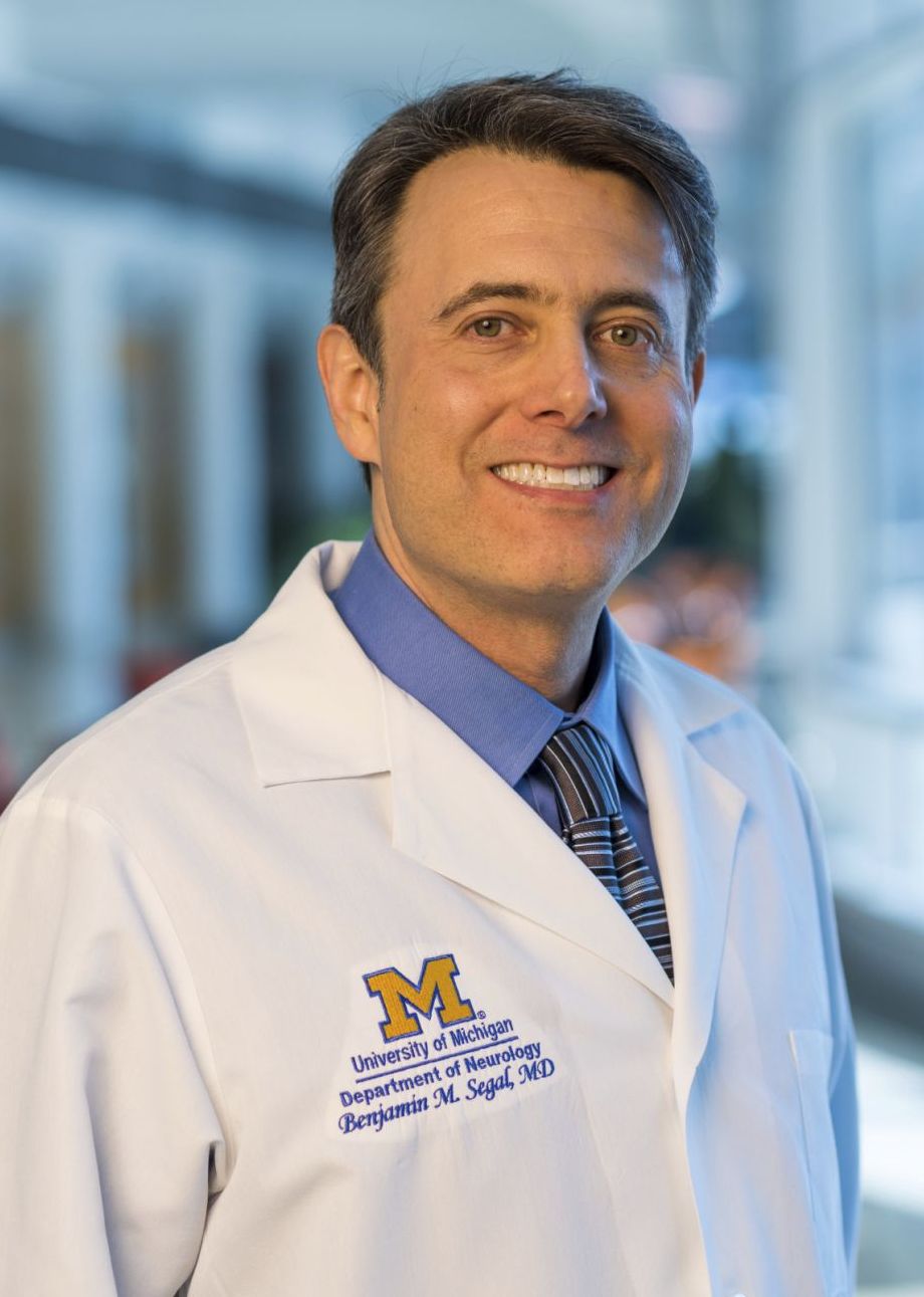
As many as 1,000 attendees are expected at the ACTRIMS Forum in San Diego, Feb. 1-3, Dr. Segal said, and about 300 abstracts will be presented. “The idea is for the meeting to be academically rigorous and have a speaker roster composed of the thought leaders in the field to make it akin to more of a keystone symposium.”
The program begins on Feb. 1 with presentations from young investigators about Emerging Concepts in MS. The other sessions will feature discussions about the blood-brain barrier, lymphocytes, cutting-edge developments in MS research, microglia and macrophages, astrocytes, and oligodendrocytes and their precursors.
“The sessions will begin with an overview of the particular target and its role in MS,” Dr. Segal said. “For example, the blood-brain barrier session starts with an overview of the different cell types that maintain the barrier’s integrity and the different ways they could prevent inflammatory cells from entering the central nervous system and causing lesions.”
One presentation in that session, he said, will discuss lessons from research into the mechanism of action of natalizumab (Tysabri). Another presentation focuses on new adhesion molecules – a crucial component of the inflammatory system – that have been discovered in animal models and may lead to new blocking therapies, he said.
The discussion about lymphocytes – white blood cells – will include presentations about new and emerging therapies that deplete them in different ways, he said. “One talk is about alemtuzumab (Lemtrada), which blocks all lymphocytes and can make you susceptible to other autoimmune disorders. What does that mean about how MS is similar to or different than other autoimmune diseases?”
On the myelin front, he said, “there will be a number of talks about how we identify remyelinating agents for clinical trials and how can we enhance precursors of myelination and reconstitute the damaged myelin.”
Another session will look at astrocytes, the glial support cells that are attracting newfound attention. Researchers have known that they’re stimulated in MS, and now new research is suggesting they can cause damage, he said. “They might be a completely new target. There’s evidence that they can have a neuroprotective effect, but it depends on how they’re stimulated.”
Are we on the cusp of being able to reverse damage from MS? “The answers are highly speculative. Research is so unpredictable and clinical trials do take a while,” he said. Still, “we understand repair pathways better than we ever have, and there’s lots of exciting work being done in animal models. We’re closer than we ever have been, and I do believe those drugs will be tested in clinical trials, hopefully within the next 5-10 years.”
Dr. Segal disclosed receiving an investigator-initiated grant from Genentech and support from Mallinckrodt for a clinical trial.
Current and future therapeutic targets will be the major focus of a meeting held by the Americas Committee for Treatment and Research in Multiple Sclerosis (ACTRIMS), according to the meeting’s scientific program committee chair.
As many as 1,000 attendees are expected at the ACTRIMS Forum in San Diego, Feb. 1-3, Dr. Segal said, and about 300 abstracts will be presented. “The idea is for the meeting to be academically rigorous and have a speaker roster composed of the thought leaders in the field to make it akin to more of a keystone symposium.”
The program begins on Feb. 1 with presentations from young investigators about Emerging Concepts in MS. The other sessions will feature discussions about the blood-brain barrier, lymphocytes, cutting-edge developments in MS research, microglia and macrophages, astrocytes, and oligodendrocytes and their precursors.
“The sessions will begin with an overview of the particular target and its role in MS,” Dr. Segal said. “For example, the blood-brain barrier session starts with an overview of the different cell types that maintain the barrier’s integrity and the different ways they could prevent inflammatory cells from entering the central nervous system and causing lesions.”
One presentation in that session, he said, will discuss lessons from research into the mechanism of action of natalizumab (Tysabri). Another presentation focuses on new adhesion molecules – a crucial component of the inflammatory system – that have been discovered in animal models and may lead to new blocking therapies, he said.
The discussion about lymphocytes – white blood cells – will include presentations about new and emerging therapies that deplete them in different ways, he said. “One talk is about alemtuzumab (Lemtrada), which blocks all lymphocytes and can make you susceptible to other autoimmune disorders. What does that mean about how MS is similar to or different than other autoimmune diseases?”
On the myelin front, he said, “there will be a number of talks about how we identify remyelinating agents for clinical trials and how can we enhance precursors of myelination and reconstitute the damaged myelin.”
Another session will look at astrocytes, the glial support cells that are attracting newfound attention. Researchers have known that they’re stimulated in MS, and now new research is suggesting they can cause damage, he said. “They might be a completely new target. There’s evidence that they can have a neuroprotective effect, but it depends on how they’re stimulated.”
Are we on the cusp of being able to reverse damage from MS? “The answers are highly speculative. Research is so unpredictable and clinical trials do take a while,” he said. Still, “we understand repair pathways better than we ever have, and there’s lots of exciting work being done in animal models. We’re closer than we ever have been, and I do believe those drugs will be tested in clinical trials, hopefully within the next 5-10 years.”
Dr. Segal disclosed receiving an investigator-initiated grant from Genentech and support from Mallinckrodt for a clinical trial.
Current and future therapeutic targets will be the major focus of a meeting held by the Americas Committee for Treatment and Research in Multiple Sclerosis (ACTRIMS), according to the meeting’s scientific program committee chair.
As many as 1,000 attendees are expected at the ACTRIMS Forum in San Diego, Feb. 1-3, Dr. Segal said, and about 300 abstracts will be presented. “The idea is for the meeting to be academically rigorous and have a speaker roster composed of the thought leaders in the field to make it akin to more of a keystone symposium.”
The program begins on Feb. 1 with presentations from young investigators about Emerging Concepts in MS. The other sessions will feature discussions about the blood-brain barrier, lymphocytes, cutting-edge developments in MS research, microglia and macrophages, astrocytes, and oligodendrocytes and their precursors.
“The sessions will begin with an overview of the particular target and its role in MS,” Dr. Segal said. “For example, the blood-brain barrier session starts with an overview of the different cell types that maintain the barrier’s integrity and the different ways they could prevent inflammatory cells from entering the central nervous system and causing lesions.”
One presentation in that session, he said, will discuss lessons from research into the mechanism of action of natalizumab (Tysabri). Another presentation focuses on new adhesion molecules – a crucial component of the inflammatory system – that have been discovered in animal models and may lead to new blocking therapies, he said.
The discussion about lymphocytes – white blood cells – will include presentations about new and emerging therapies that deplete them in different ways, he said. “One talk is about alemtuzumab (Lemtrada), which blocks all lymphocytes and can make you susceptible to other autoimmune disorders. What does that mean about how MS is similar to or different than other autoimmune diseases?”
On the myelin front, he said, “there will be a number of talks about how we identify remyelinating agents for clinical trials and how can we enhance precursors of myelination and reconstitute the damaged myelin.”
Another session will look at astrocytes, the glial support cells that are attracting newfound attention. Researchers have known that they’re stimulated in MS, and now new research is suggesting they can cause damage, he said. “They might be a completely new target. There’s evidence that they can have a neuroprotective effect, but it depends on how they’re stimulated.”
Are we on the cusp of being able to reverse damage from MS? “The answers are highly speculative. Research is so unpredictable and clinical trials do take a while,” he said. Still, “we understand repair pathways better than we ever have, and there’s lots of exciting work being done in animal models. We’re closer than we ever have been, and I do believe those drugs will be tested in clinical trials, hopefully within the next 5-10 years.”
Dr. Segal disclosed receiving an investigator-initiated grant from Genentech and support from Mallinckrodt for a clinical trial.
FROM ACTRIMS FORUM 2018
Listen up: Acoustic device useful for diabetic foot ulcers
The Food & Drug Administration has approved the marketing of a device that uses acoustic shock waves to boost wound closure in patients with diabetic foot ulcers (DFUs), an especially stubborn and dangerous condition.
The treatment is experimental, and only limited research into its effectiveness has been published. Still, representatives of its manufacturer say the device, known as dermaPACE, has produced promising results as a secondary treatment in stubborn cases.

A wound care specialist said in an interview that the shock wave technology appears to hold promise.
“A shortcoming in the field of wound care is that providers are typically not trained in a standardized fashion on when and how to a perform meticulous excisional sharp debridement of a wound,” said Bill Tettelbach, MD, systems medical director of Wound Care & Hyperbaric Medicine Services at Intermountain Healthcare in Salt Lake City. “In the majority of cases, the better the debridement, the more rapidly the patient will obtain wound closure.”
This new therapy may provide a benefit as a secondary treatment, especially when the patient cannot tolerate extensive sharp debridement, he said. It also could potentially improve biofilm penetration of antimicrobial topical treatments, he said.
DFUs are believed to affect as many as 1 in 4 people with diabetes over the course of their lifetimes. A 2014 report estimated that care of these wounds costs insurers as much as $13 billion a year in the U.S. alone (Diabetes Care. 2014 Mar;37[3]:651-8).
Treatment options include debridement and, in more extreme cases, hyperbaric oxygen treatment. Amputation can be required if treatment is unsuccessful.
According to Mr. Stegagno, the shock wave device is about the size of a desktop computer from a decade ago. A high-voltage generator box is connected to a handheld therapy head and delivers an acoustic pulse to the patient. The system “is like a spark plug that you see in your automobile,” he said. “It’s pretty much the same technology as lithotripsy, just downsized significantly. The key part is a highly focused, high-energy pulse.”
In a news release, the FDA said it examined the results of two studies of patients with diabetes who received usual DFU care along with either the shock wave therapy or a sham therapy. A total of 336 patients took part in the multicenter, randomized, double-blind studies.
According to the FDA, the studies found a 44% wound closure rate at 24 weeks in patients who had undergone 1-7 shock wave treatments, compared with the 30% wound closure rate in those who received the sham treatment.
Side effects included pain while the device was applied, bruising and numbness, migraines, nausea, fainting, wound infection, fever, and infection beyond the wound such as cellulitis and osteomyelitis.
“There were no meaningful statistical differences in the adverse event rates between the dermaPACE-treated patients and the sham-control group,” Mr. Stegagno said. “There were no issues regarding the tolerability of the treatment, which suggests that a second course of treatment, if needed, is a clinically viable option.”
Mr. Stegagno said the FDA expressed concern about “increased incidences of osteomyelitis at later points in the trials, particularly at the 10-week mark and later.” In response to the agency’s concerns, warning statements were added to labeling, he said.
According to Mr. Stegagno, only one study into the shock wave treatment for DFU has been published, although research has been released through posters and abstracts. The small published study favorably compared shock wave therapy with hyperbaric oxygen therapy. (Diabetes Res Clin Pract. 2011 May;92[2]:187-93)
“Sanuwave will be sponsoring additional studies later this year in the [United States] as follow-on studies to the just-completed DFU trials,” Mr. Stegagno said.
The FDA says the device is intended to be used in adults aged 22 and up with certain types of chronic DFUs. The Sanuwave company says patients should be treated with 4-8 applications over 2-10 weeks.
The shock wave process appears to boost healing through a process that leads to inflammatory responses and oxygenation, Mr. Stegagno said, by first creating an initial compression phase that “squeezes the cell and creates a microtrauma.”
“The cell wakes up and says, ‘Something just punched me,’ ” he said. “This tissue and cellular disruption is believed to initiate the cellular signaling for growth factors and other proteins noted in studies.”
The effects of negative pressure also play a role in stimulation of the wound, he said.
The shock wave therapy will cost an estimated $3,000-$4,000 per protocol of 8 treatments, said Kevin A. Richardson II, the CEO and chairman of the board at Sanuwave, in an interview. The initial plan is for the company to place the devices with doctors while the firm still owns the machines, he said.
The FDA approved the marketing of the device as part of its de novo premarket review pathway, which allows certain new types of devices to be approved when approved similar devices don’t yet exist for the purposes of comparison.
Mr. Stegagno and Mr. Richardson work for Sanuwave. Dr. Tettelbach reported no relevant disclosures.
The Food & Drug Administration has approved the marketing of a device that uses acoustic shock waves to boost wound closure in patients with diabetic foot ulcers (DFUs), an especially stubborn and dangerous condition.
The treatment is experimental, and only limited research into its effectiveness has been published. Still, representatives of its manufacturer say the device, known as dermaPACE, has produced promising results as a secondary treatment in stubborn cases.
A wound care specialist said in an interview that the shock wave technology appears to hold promise.
“A shortcoming in the field of wound care is that providers are typically not trained in a standardized fashion on when and how to a perform meticulous excisional sharp debridement of a wound,” said Bill Tettelbach, MD, systems medical director of Wound Care & Hyperbaric Medicine Services at Intermountain Healthcare in Salt Lake City. “In the majority of cases, the better the debridement, the more rapidly the patient will obtain wound closure.”
This new therapy may provide a benefit as a secondary treatment, especially when the patient cannot tolerate extensive sharp debridement, he said. It also could potentially improve biofilm penetration of antimicrobial topical treatments, he said.
DFUs are believed to affect as many as 1 in 4 people with diabetes over the course of their lifetimes. A 2014 report estimated that care of these wounds costs insurers as much as $13 billion a year in the U.S. alone (Diabetes Care. 2014 Mar;37[3]:651-8).
Treatment options include debridement and, in more extreme cases, hyperbaric oxygen treatment. Amputation can be required if treatment is unsuccessful.
According to Mr. Stegagno, the shock wave device is about the size of a desktop computer from a decade ago. A high-voltage generator box is connected to a handheld therapy head and delivers an acoustic pulse to the patient. The system “is like a spark plug that you see in your automobile,” he said. “It’s pretty much the same technology as lithotripsy, just downsized significantly. The key part is a highly focused, high-energy pulse.”
In a news release, the FDA said it examined the results of two studies of patients with diabetes who received usual DFU care along with either the shock wave therapy or a sham therapy. A total of 336 patients took part in the multicenter, randomized, double-blind studies.
According to the FDA, the studies found a 44% wound closure rate at 24 weeks in patients who had undergone 1-7 shock wave treatments, compared with the 30% wound closure rate in those who received the sham treatment.
Side effects included pain while the device was applied, bruising and numbness, migraines, nausea, fainting, wound infection, fever, and infection beyond the wound such as cellulitis and osteomyelitis.
“There were no meaningful statistical differences in the adverse event rates between the dermaPACE-treated patients and the sham-control group,” Mr. Stegagno said. “There were no issues regarding the tolerability of the treatment, which suggests that a second course of treatment, if needed, is a clinically viable option.”
Mr. Stegagno said the FDA expressed concern about “increased incidences of osteomyelitis at later points in the trials, particularly at the 10-week mark and later.” In response to the agency’s concerns, warning statements were added to labeling, he said.
According to Mr. Stegagno, only one study into the shock wave treatment for DFU has been published, although research has been released through posters and abstracts. The small published study favorably compared shock wave therapy with hyperbaric oxygen therapy. (Diabetes Res Clin Pract. 2011 May;92[2]:187-93)
“Sanuwave will be sponsoring additional studies later this year in the [United States] as follow-on studies to the just-completed DFU trials,” Mr. Stegagno said.
The FDA says the device is intended to be used in adults aged 22 and up with certain types of chronic DFUs. The Sanuwave company says patients should be treated with 4-8 applications over 2-10 weeks.
The shock wave process appears to boost healing through a process that leads to inflammatory responses and oxygenation, Mr. Stegagno said, by first creating an initial compression phase that “squeezes the cell and creates a microtrauma.”
“The cell wakes up and says, ‘Something just punched me,’ ” he said. “This tissue and cellular disruption is believed to initiate the cellular signaling for growth factors and other proteins noted in studies.”
The effects of negative pressure also play a role in stimulation of the wound, he said.
The shock wave therapy will cost an estimated $3,000-$4,000 per protocol of 8 treatments, said Kevin A. Richardson II, the CEO and chairman of the board at Sanuwave, in an interview. The initial plan is for the company to place the devices with doctors while the firm still owns the machines, he said.
The FDA approved the marketing of the device as part of its de novo premarket review pathway, which allows certain new types of devices to be approved when approved similar devices don’t yet exist for the purposes of comparison.
Mr. Stegagno and Mr. Richardson work for Sanuwave. Dr. Tettelbach reported no relevant disclosures.
The Food & Drug Administration has approved the marketing of a device that uses acoustic shock waves to boost wound closure in patients with diabetic foot ulcers (DFUs), an especially stubborn and dangerous condition.
The treatment is experimental, and only limited research into its effectiveness has been published. Still, representatives of its manufacturer say the device, known as dermaPACE, has produced promising results as a secondary treatment in stubborn cases.
A wound care specialist said in an interview that the shock wave technology appears to hold promise.
“A shortcoming in the field of wound care is that providers are typically not trained in a standardized fashion on when and how to a perform meticulous excisional sharp debridement of a wound,” said Bill Tettelbach, MD, systems medical director of Wound Care & Hyperbaric Medicine Services at Intermountain Healthcare in Salt Lake City. “In the majority of cases, the better the debridement, the more rapidly the patient will obtain wound closure.”
This new therapy may provide a benefit as a secondary treatment, especially when the patient cannot tolerate extensive sharp debridement, he said. It also could potentially improve biofilm penetration of antimicrobial topical treatments, he said.
DFUs are believed to affect as many as 1 in 4 people with diabetes over the course of their lifetimes. A 2014 report estimated that care of these wounds costs insurers as much as $13 billion a year in the U.S. alone (Diabetes Care. 2014 Mar;37[3]:651-8).
Treatment options include debridement and, in more extreme cases, hyperbaric oxygen treatment. Amputation can be required if treatment is unsuccessful.
According to Mr. Stegagno, the shock wave device is about the size of a desktop computer from a decade ago. A high-voltage generator box is connected to a handheld therapy head and delivers an acoustic pulse to the patient. The system “is like a spark plug that you see in your automobile,” he said. “It’s pretty much the same technology as lithotripsy, just downsized significantly. The key part is a highly focused, high-energy pulse.”
In a news release, the FDA said it examined the results of two studies of patients with diabetes who received usual DFU care along with either the shock wave therapy or a sham therapy. A total of 336 patients took part in the multicenter, randomized, double-blind studies.
According to the FDA, the studies found a 44% wound closure rate at 24 weeks in patients who had undergone 1-7 shock wave treatments, compared with the 30% wound closure rate in those who received the sham treatment.
Side effects included pain while the device was applied, bruising and numbness, migraines, nausea, fainting, wound infection, fever, and infection beyond the wound such as cellulitis and osteomyelitis.
“There were no meaningful statistical differences in the adverse event rates between the dermaPACE-treated patients and the sham-control group,” Mr. Stegagno said. “There were no issues regarding the tolerability of the treatment, which suggests that a second course of treatment, if needed, is a clinically viable option.”
Mr. Stegagno said the FDA expressed concern about “increased incidences of osteomyelitis at later points in the trials, particularly at the 10-week mark and later.” In response to the agency’s concerns, warning statements were added to labeling, he said.
According to Mr. Stegagno, only one study into the shock wave treatment for DFU has been published, although research has been released through posters and abstracts. The small published study favorably compared shock wave therapy with hyperbaric oxygen therapy. (Diabetes Res Clin Pract. 2011 May;92[2]:187-93)
“Sanuwave will be sponsoring additional studies later this year in the [United States] as follow-on studies to the just-completed DFU trials,” Mr. Stegagno said.
The FDA says the device is intended to be used in adults aged 22 and up with certain types of chronic DFUs. The Sanuwave company says patients should be treated with 4-8 applications over 2-10 weeks.
The shock wave process appears to boost healing through a process that leads to inflammatory responses and oxygenation, Mr. Stegagno said, by first creating an initial compression phase that “squeezes the cell and creates a microtrauma.”
“The cell wakes up and says, ‘Something just punched me,’ ” he said. “This tissue and cellular disruption is believed to initiate the cellular signaling for growth factors and other proteins noted in studies.”
The effects of negative pressure also play a role in stimulation of the wound, he said.
The shock wave therapy will cost an estimated $3,000-$4,000 per protocol of 8 treatments, said Kevin A. Richardson II, the CEO and chairman of the board at Sanuwave, in an interview. The initial plan is for the company to place the devices with doctors while the firm still owns the machines, he said.
The FDA approved the marketing of the device as part of its de novo premarket review pathway, which allows certain new types of devices to be approved when approved similar devices don’t yet exist for the purposes of comparison.
Mr. Stegagno and Mr. Richardson work for Sanuwave. Dr. Tettelbach reported no relevant disclosures.
Study: Test carcinoembryonic antigen in colon cancer after surgery
A new study challenges the practice of measuring carcinoembryonic antigen (CEA) in patients with colon cancer prior to surgery, as is currently recommended by some guidelines. Researchers found that the levels do have predictive value about risk of recurrence when tested after surgery, but they challenge its use before.
“Consistent with the literature, our data show that postoperative CEA is more informative than preoperative CEA,” the study authors wrote. “Emphasis should be placed on postoperative CEA, and in the setting of modern high-quality imaging, we question the utility of measuring preoperative CEA.”
The study, which was published online in JAMA Oncology, noted that testing for CEA – a colorectal cancer tumor marker – had more value prior to the current era of imaging because it could indicate the need to widen a search for metastases.
“In the era of high-quality computed tomography (CT), the utility of measuring preoperative CEA is less obvious because an elevated preoperative CEA with a normal CT scan does not preclude surgery with curative intent,” wrote Tsuyoshi Konishi, MD, of Memorial Sloan-Kettering Cancer Center in New York and his coauthors.
CEA levels can return to normal after colon cancer surgery – but not always. According to the study, the National Comprehensive Cancer Network, the American Society of Clinical Oncology, and the European Group on Tumor Markers recommend its measurement prior to surgery in patients with nonmetastatic cancer.
For the new study, the authors sought to understand the predictive value of various CEA levels both before and after surgery.
The researchers retrospectively tracked 1,027 patients at a single center with stage I-III colon cancer (50.4% male; median age, 64 years). (Another 221 patients had been removed because of exclusion criteria, such as recent malignancy treatment and preoperative chemotherapy or radiation treatment.)
Nearly 70% of the 1,027 patients had normal preoperative CEA levels, and the rest had elevated levels.
Of the 312 patients with elevated preoperative CEA levels, 113 were lost to follow-up postoperative CEA tests. In the remaining patients, CEA levels returned to normal in 71%.
Patients were followed for a median of 38 months; 10.3% had recurrences, and 4.6% died. The 3-year recurrence-free survival rate was 88.4%.
Researchers found that those with normal preoperative CEA levels were more likely to reach recurrence-free survival at 3 years (89.7%) than were those who had elevated preoperative CEA levels (82.3%; P = .05). The higher level wasn’t much different than the recurrence-free survival level in those whose CEA levels had normalized after the operation (87.9%; P = .86).
Those with elevated CEA levels after surgery had a lower likelihood of reaching recurrence-free survival at 3 years (74.5%) than did those who had normal postoperative CEA levels (89.4%).
The study also linked abnormal postoperative CEA levels to shorter recurrence-free survival (hazard ratio, 2.0; 95% confidence interval, 1.1-3.5). Normalized postoperative CEA levels, however, were not linked to shorter recurrence-free survival (HR, 0.77; 95% CI, 0.45-1.30).
The researchers noted that they didn’t control for factors such as smoking (which increases CEA levels) and disease like gastritis and diabetes, which can produce misleading CEA levels. “Rather, this study is pragmatic,” the researchers wrote, “and represents what is likely to be seen in real-world colorectal cancer care.”
Moving forward, the researchers wrote, “patients with elevated postoperative CEA tend to experience recurrence early, which might justify a risk-adjusted and individualized surveillance strategy.”
The study authors report no relevant disclosures. The National Institutes of Health/National Cancer Institute funded the study in part.
SOURCE: Konishi T et al. JAMA Oncol. 2017. doi: 10.1001/jamaoncol.2017.4420.
Multiple organizations recommend that patients with nonmetastatic colorectal cancer undergo carcinoembryonic antigen (CEA) measurement prior to surgery, and there’s talk that it should be added to the staging system of the American Joint Committee on Cancer.
This new study, however, provides a challenge to the use of preoperative CEA tests in this population. As the researchers report, patients with normalized CEA levels postsurgery – even those with abnormal levels before surgery – were more likely to reach recurrence-free survival for 3 years.
The findings spotlight the value of real-world evidence and real-world data rather than statistics gathered during a controlled clinical trial.
There are limitations to this study, such as generalizability (it was performed at a single center), loss of patients to CEA follow-up postsurgery, and timing (the study period began a decade ago).
Still, the findings, once confirmed, could change clinical practice, and they point to the potential use of CEA as a biomarker early-warning sign that a tumor may recur.
Rebecca Anne Miksad, MD, MPH, is with Harvard Medical School in Boston (leave of absence from 2017 to 2018) and Neal J. Meropol, MD, is with Case Western Reserve University in Cleveland. They disclose employment by Flatiron Health. These comments are taken from their editorial accompanying the study by Konishi et al. in JAMA Oncology, doi:10.1001/jamaoncol.2017.4420.
Multiple organizations recommend that patients with nonmetastatic colorectal cancer undergo carcinoembryonic antigen (CEA) measurement prior to surgery, and there’s talk that it should be added to the staging system of the American Joint Committee on Cancer.
This new study, however, provides a challenge to the use of preoperative CEA tests in this population. As the researchers report, patients with normalized CEA levels postsurgery – even those with abnormal levels before surgery – were more likely to reach recurrence-free survival for 3 years.
The findings spotlight the value of real-world evidence and real-world data rather than statistics gathered during a controlled clinical trial.
There are limitations to this study, such as generalizability (it was performed at a single center), loss of patients to CEA follow-up postsurgery, and timing (the study period began a decade ago).
Still, the findings, once confirmed, could change clinical practice, and they point to the potential use of CEA as a biomarker early-warning sign that a tumor may recur.
Rebecca Anne Miksad, MD, MPH, is with Harvard Medical School in Boston (leave of absence from 2017 to 2018) and Neal J. Meropol, MD, is with Case Western Reserve University in Cleveland. They disclose employment by Flatiron Health. These comments are taken from their editorial accompanying the study by Konishi et al. in JAMA Oncology, doi:10.1001/jamaoncol.2017.4420.
Multiple organizations recommend that patients with nonmetastatic colorectal cancer undergo carcinoembryonic antigen (CEA) measurement prior to surgery, and there’s talk that it should be added to the staging system of the American Joint Committee on Cancer.
This new study, however, provides a challenge to the use of preoperative CEA tests in this population. As the researchers report, patients with normalized CEA levels postsurgery – even those with abnormal levels before surgery – were more likely to reach recurrence-free survival for 3 years.
The findings spotlight the value of real-world evidence and real-world data rather than statistics gathered during a controlled clinical trial.
There are limitations to this study, such as generalizability (it was performed at a single center), loss of patients to CEA follow-up postsurgery, and timing (the study period began a decade ago).
Still, the findings, once confirmed, could change clinical practice, and they point to the potential use of CEA as a biomarker early-warning sign that a tumor may recur.
Rebecca Anne Miksad, MD, MPH, is with Harvard Medical School in Boston (leave of absence from 2017 to 2018) and Neal J. Meropol, MD, is with Case Western Reserve University in Cleveland. They disclose employment by Flatiron Health. These comments are taken from their editorial accompanying the study by Konishi et al. in JAMA Oncology, doi:10.1001/jamaoncol.2017.4420.
A new study challenges the practice of measuring carcinoembryonic antigen (CEA) in patients with colon cancer prior to surgery, as is currently recommended by some guidelines. Researchers found that the levels do have predictive value about risk of recurrence when tested after surgery, but they challenge its use before.
“Consistent with the literature, our data show that postoperative CEA is more informative than preoperative CEA,” the study authors wrote. “Emphasis should be placed on postoperative CEA, and in the setting of modern high-quality imaging, we question the utility of measuring preoperative CEA.”
The study, which was published online in JAMA Oncology, noted that testing for CEA – a colorectal cancer tumor marker – had more value prior to the current era of imaging because it could indicate the need to widen a search for metastases.
“In the era of high-quality computed tomography (CT), the utility of measuring preoperative CEA is less obvious because an elevated preoperative CEA with a normal CT scan does not preclude surgery with curative intent,” wrote Tsuyoshi Konishi, MD, of Memorial Sloan-Kettering Cancer Center in New York and his coauthors.
CEA levels can return to normal after colon cancer surgery – but not always. According to the study, the National Comprehensive Cancer Network, the American Society of Clinical Oncology, and the European Group on Tumor Markers recommend its measurement prior to surgery in patients with nonmetastatic cancer.
For the new study, the authors sought to understand the predictive value of various CEA levels both before and after surgery.
The researchers retrospectively tracked 1,027 patients at a single center with stage I-III colon cancer (50.4% male; median age, 64 years). (Another 221 patients had been removed because of exclusion criteria, such as recent malignancy treatment and preoperative chemotherapy or radiation treatment.)
Nearly 70% of the 1,027 patients had normal preoperative CEA levels, and the rest had elevated levels.
Of the 312 patients with elevated preoperative CEA levels, 113 were lost to follow-up postoperative CEA tests. In the remaining patients, CEA levels returned to normal in 71%.
Patients were followed for a median of 38 months; 10.3% had recurrences, and 4.6% died. The 3-year recurrence-free survival rate was 88.4%.
Researchers found that those with normal preoperative CEA levels were more likely to reach recurrence-free survival at 3 years (89.7%) than were those who had elevated preoperative CEA levels (82.3%; P = .05). The higher level wasn’t much different than the recurrence-free survival level in those whose CEA levels had normalized after the operation (87.9%; P = .86).
Those with elevated CEA levels after surgery had a lower likelihood of reaching recurrence-free survival at 3 years (74.5%) than did those who had normal postoperative CEA levels (89.4%).
The study also linked abnormal postoperative CEA levels to shorter recurrence-free survival (hazard ratio, 2.0; 95% confidence interval, 1.1-3.5). Normalized postoperative CEA levels, however, were not linked to shorter recurrence-free survival (HR, 0.77; 95% CI, 0.45-1.30).
The researchers noted that they didn’t control for factors such as smoking (which increases CEA levels) and disease like gastritis and diabetes, which can produce misleading CEA levels. “Rather, this study is pragmatic,” the researchers wrote, “and represents what is likely to be seen in real-world colorectal cancer care.”
Moving forward, the researchers wrote, “patients with elevated postoperative CEA tend to experience recurrence early, which might justify a risk-adjusted and individualized surveillance strategy.”
The study authors report no relevant disclosures. The National Institutes of Health/National Cancer Institute funded the study in part.
SOURCE: Konishi T et al. JAMA Oncol. 2017. doi: 10.1001/jamaoncol.2017.4420.
A new study challenges the practice of measuring carcinoembryonic antigen (CEA) in patients with colon cancer prior to surgery, as is currently recommended by some guidelines. Researchers found that the levels do have predictive value about risk of recurrence when tested after surgery, but they challenge its use before.
“Consistent with the literature, our data show that postoperative CEA is more informative than preoperative CEA,” the study authors wrote. “Emphasis should be placed on postoperative CEA, and in the setting of modern high-quality imaging, we question the utility of measuring preoperative CEA.”
The study, which was published online in JAMA Oncology, noted that testing for CEA – a colorectal cancer tumor marker – had more value prior to the current era of imaging because it could indicate the need to widen a search for metastases.
“In the era of high-quality computed tomography (CT), the utility of measuring preoperative CEA is less obvious because an elevated preoperative CEA with a normal CT scan does not preclude surgery with curative intent,” wrote Tsuyoshi Konishi, MD, of Memorial Sloan-Kettering Cancer Center in New York and his coauthors.
CEA levels can return to normal after colon cancer surgery – but not always. According to the study, the National Comprehensive Cancer Network, the American Society of Clinical Oncology, and the European Group on Tumor Markers recommend its measurement prior to surgery in patients with nonmetastatic cancer.
For the new study, the authors sought to understand the predictive value of various CEA levels both before and after surgery.
The researchers retrospectively tracked 1,027 patients at a single center with stage I-III colon cancer (50.4% male; median age, 64 years). (Another 221 patients had been removed because of exclusion criteria, such as recent malignancy treatment and preoperative chemotherapy or radiation treatment.)
Nearly 70% of the 1,027 patients had normal preoperative CEA levels, and the rest had elevated levels.
Of the 312 patients with elevated preoperative CEA levels, 113 were lost to follow-up postoperative CEA tests. In the remaining patients, CEA levels returned to normal in 71%.
Patients were followed for a median of 38 months; 10.3% had recurrences, and 4.6% died. The 3-year recurrence-free survival rate was 88.4%.
Researchers found that those with normal preoperative CEA levels were more likely to reach recurrence-free survival at 3 years (89.7%) than were those who had elevated preoperative CEA levels (82.3%; P = .05). The higher level wasn’t much different than the recurrence-free survival level in those whose CEA levels had normalized after the operation (87.9%; P = .86).
Those with elevated CEA levels after surgery had a lower likelihood of reaching recurrence-free survival at 3 years (74.5%) than did those who had normal postoperative CEA levels (89.4%).
The study also linked abnormal postoperative CEA levels to shorter recurrence-free survival (hazard ratio, 2.0; 95% confidence interval, 1.1-3.5). Normalized postoperative CEA levels, however, were not linked to shorter recurrence-free survival (HR, 0.77; 95% CI, 0.45-1.30).
The researchers noted that they didn’t control for factors such as smoking (which increases CEA levels) and disease like gastritis and diabetes, which can produce misleading CEA levels. “Rather, this study is pragmatic,” the researchers wrote, “and represents what is likely to be seen in real-world colorectal cancer care.”
Moving forward, the researchers wrote, “patients with elevated postoperative CEA tend to experience recurrence early, which might justify a risk-adjusted and individualized surveillance strategy.”
The study authors report no relevant disclosures. The National Institutes of Health/National Cancer Institute funded the study in part.
SOURCE: Konishi T et al. JAMA Oncol. 2017. doi: 10.1001/jamaoncol.2017.4420.
FROM JAMA ONCOLOGY
Key clinical point: Measurement of CEA levels in patients with colon cancer appears to have value postoperatively but not preoperatively.
Major finding: Patients with abnormal postoperative CEA levels were less likely to reach 3-year recurrence-free survival than were those with normal levels (74.5% vs. 89.4%).
Study details: Retrospective, single-center study of 1,027 patients with stage I-III colon cancer (50.4% male; median age, 64 years).
Disclosures: The study authors report no relevant disclosures. The National Institutes of Health/National Cancer Institute funded the study in part.
Source: Konishi T et al. JAMA Oncol. 2017. doi: 10.1001/jamaoncol.2017.4420.
Online education program linked to less anxiety, medication use in colonoscopies
An online education program appeared to dampen anxiety levels in colonoscopy patients and even seemed to reduce the burden of the procedure in terms of time and medication used, a study showed.
“The program was very well received and generally improved patient’s anxiety,” said lead author Siddhartha Parker, MD, a gastroenterologist at Dartmouth-Hitchcock Medical Center, Lebanon, N.H. “Only 4% of patients described feeling more anxious after watching the program whereas 58% felt less anxious.”
“Patients who are afraid or anxious may require more sedation, experience more pain, and, most importantly, may be less likely to have a procedure done,” he said. “Some patients don’t schedule at all, while others may schedule and then cancel last minute or no-show an appointment, which has its own implications and costs to the health care system.”
In terms of anxiety prevention, he said, “primarily we rely on nurses and doctors to discuss the need for screening and provide reassurance about low risks and real benefits.”
For the current study, published in the Journal of Clinical Gastroenterology, researchers tested a Web-based colonoscopy education program from a company called Emmi Solutions.
The program is designed to provide details about the colonoscopy process. “It covers what to expect before, during, and after the procedure including possible findings (e.g., polyps),” Dr. Parker said. “It also covers risks and benefits with some tips on recovery after the procedure. I think having some dedicated time to understand the procedure is most helpful. It is rare that a patient gets 15-20 minutes to review any topic with their doctor, and the Emmi program has the added benefit of visual cues.”
The researchers recruited and randomly assigned 51 patients to a control group and 52 patients to the intervention. All were scheduled to undergo colonoscopies within at least 2 weeks.
The groups were similar: Most patients were white (100% of the control group, 89% of the intervention group), and most were female; the average age was 48 years The intervention group had more education, at 69% with college degrees vs. 43% in the control group (P = .01).
Patients in the control group received usual colonoscopy education materials, while those in the intervention group were also directed to the education website.
All participants took a survey immediately before their procedures and received a $10 gift card as reimbursement for their participation.
Researchers found that patients in the intervention group scored higher than did those in the control group on questions about colonoscopy knowledge (82% of questions correct vs. 74%, P less than .001).
Also, the education website may have convinced its viewers to worry less about the procedure and more about its ultimate findings. Compared with the control group, those in the intervention group were more likely to say they were most concerned about the findings (38.5% vs. 21.6%) and less likely to say they were more concerned about both the findings and the procedure (38.5% vs. 56.9%). Roughly 20% of those in both groups said they were most concerned about the procedure.
“Some patients report being more concerned about findings, e.g., cancer, before the program but more anxious about complications after the program,” Dr. Parker said. “I think that is a reasonable outcome as asymptomatic patients should have very little concern about findings, given the goal is to find and remove polyps.”
Researchers also tracked the use of sedatives during the procedure. Those in the intervention group required less midazolam (Versed) (average dose of 3.66 mg vs. 4.46 mg, P = .004). They also required less fentanyl (164 mcg vs. 186 mcg), but this finding was not statistically significant (P = .063).
“There was also a trend, not quite statistically significant, to improved prep quality,” Dr. Parker said. (In the intervention vs. the control group, 96% vs. 88% of subjects were rated as good or excellent in terms of prep quality, P = .27).
“This is important because a poor or inadequate prep leads to a need for a repeat procedure to ensure adequate screening,” he said. “If prep quality improves, that means fewer unnecessary repeated procedures.”
The researchers reported study limitations such as multiple endoscopists and sedation nurses and the variation in education levels of the participants.
The cost of the interactive program was not available.
According to Dr. Parker, Dartmouth-Hitchcock Medical Center now offers the education program to all colonoscopy patients, and a modified version is available for those undergoing upper endoscopies.
“Not all patients have adequate computer resources, and some patients simply choose not to watch,” he said. “But my impression is that those who watched the program are more relaxed during the informed consent process. However, we are not actively tracking outcomes such as procedure time or medication usage.”
No more studies of the program are planned at the institution, he said.
One of the study authors is an employee of Emmi Solutions, which provided the program for free for use in the study. The other authors reported no relevant disclosures. No study funding was reported.
SOURCE: Parker S et al. J Clin Gastroenterol. doi: 10.1097/MCG.0000000000000958
An online education program appeared to dampen anxiety levels in colonoscopy patients and even seemed to reduce the burden of the procedure in terms of time and medication used, a study showed.
“The program was very well received and generally improved patient’s anxiety,” said lead author Siddhartha Parker, MD, a gastroenterologist at Dartmouth-Hitchcock Medical Center, Lebanon, N.H. “Only 4% of patients described feeling more anxious after watching the program whereas 58% felt less anxious.”
“Patients who are afraid or anxious may require more sedation, experience more pain, and, most importantly, may be less likely to have a procedure done,” he said. “Some patients don’t schedule at all, while others may schedule and then cancel last minute or no-show an appointment, which has its own implications and costs to the health care system.”
In terms of anxiety prevention, he said, “primarily we rely on nurses and doctors to discuss the need for screening and provide reassurance about low risks and real benefits.”
For the current study, published in the Journal of Clinical Gastroenterology, researchers tested a Web-based colonoscopy education program from a company called Emmi Solutions.
The program is designed to provide details about the colonoscopy process. “It covers what to expect before, during, and after the procedure including possible findings (e.g., polyps),” Dr. Parker said. “It also covers risks and benefits with some tips on recovery after the procedure. I think having some dedicated time to understand the procedure is most helpful. It is rare that a patient gets 15-20 minutes to review any topic with their doctor, and the Emmi program has the added benefit of visual cues.”
The researchers recruited and randomly assigned 51 patients to a control group and 52 patients to the intervention. All were scheduled to undergo colonoscopies within at least 2 weeks.
The groups were similar: Most patients were white (100% of the control group, 89% of the intervention group), and most were female; the average age was 48 years The intervention group had more education, at 69% with college degrees vs. 43% in the control group (P = .01).
Patients in the control group received usual colonoscopy education materials, while those in the intervention group were also directed to the education website.
All participants took a survey immediately before their procedures and received a $10 gift card as reimbursement for their participation.
Researchers found that patients in the intervention group scored higher than did those in the control group on questions about colonoscopy knowledge (82% of questions correct vs. 74%, P less than .001).
Also, the education website may have convinced its viewers to worry less about the procedure and more about its ultimate findings. Compared with the control group, those in the intervention group were more likely to say they were most concerned about the findings (38.5% vs. 21.6%) and less likely to say they were more concerned about both the findings and the procedure (38.5% vs. 56.9%). Roughly 20% of those in both groups said they were most concerned about the procedure.
“Some patients report being more concerned about findings, e.g., cancer, before the program but more anxious about complications after the program,” Dr. Parker said. “I think that is a reasonable outcome as asymptomatic patients should have very little concern about findings, given the goal is to find and remove polyps.”
Researchers also tracked the use of sedatives during the procedure. Those in the intervention group required less midazolam (Versed) (average dose of 3.66 mg vs. 4.46 mg, P = .004). They also required less fentanyl (164 mcg vs. 186 mcg), but this finding was not statistically significant (P = .063).
“There was also a trend, not quite statistically significant, to improved prep quality,” Dr. Parker said. (In the intervention vs. the control group, 96% vs. 88% of subjects were rated as good or excellent in terms of prep quality, P = .27).
“This is important because a poor or inadequate prep leads to a need for a repeat procedure to ensure adequate screening,” he said. “If prep quality improves, that means fewer unnecessary repeated procedures.”
The researchers reported study limitations such as multiple endoscopists and sedation nurses and the variation in education levels of the participants.
The cost of the interactive program was not available.
According to Dr. Parker, Dartmouth-Hitchcock Medical Center now offers the education program to all colonoscopy patients, and a modified version is available for those undergoing upper endoscopies.
“Not all patients have adequate computer resources, and some patients simply choose not to watch,” he said. “But my impression is that those who watched the program are more relaxed during the informed consent process. However, we are not actively tracking outcomes such as procedure time or medication usage.”
No more studies of the program are planned at the institution, he said.
One of the study authors is an employee of Emmi Solutions, which provided the program for free for use in the study. The other authors reported no relevant disclosures. No study funding was reported.
SOURCE: Parker S et al. J Clin Gastroenterol. doi: 10.1097/MCG.0000000000000958
An online education program appeared to dampen anxiety levels in colonoscopy patients and even seemed to reduce the burden of the procedure in terms of time and medication used, a study showed.
“The program was very well received and generally improved patient’s anxiety,” said lead author Siddhartha Parker, MD, a gastroenterologist at Dartmouth-Hitchcock Medical Center, Lebanon, N.H. “Only 4% of patients described feeling more anxious after watching the program whereas 58% felt less anxious.”
“Patients who are afraid or anxious may require more sedation, experience more pain, and, most importantly, may be less likely to have a procedure done,” he said. “Some patients don’t schedule at all, while others may schedule and then cancel last minute or no-show an appointment, which has its own implications and costs to the health care system.”
In terms of anxiety prevention, he said, “primarily we rely on nurses and doctors to discuss the need for screening and provide reassurance about low risks and real benefits.”
For the current study, published in the Journal of Clinical Gastroenterology, researchers tested a Web-based colonoscopy education program from a company called Emmi Solutions.
The program is designed to provide details about the colonoscopy process. “It covers what to expect before, during, and after the procedure including possible findings (e.g., polyps),” Dr. Parker said. “It also covers risks and benefits with some tips on recovery after the procedure. I think having some dedicated time to understand the procedure is most helpful. It is rare that a patient gets 15-20 minutes to review any topic with their doctor, and the Emmi program has the added benefit of visual cues.”
The researchers recruited and randomly assigned 51 patients to a control group and 52 patients to the intervention. All were scheduled to undergo colonoscopies within at least 2 weeks.
The groups were similar: Most patients were white (100% of the control group, 89% of the intervention group), and most were female; the average age was 48 years The intervention group had more education, at 69% with college degrees vs. 43% in the control group (P = .01).
Patients in the control group received usual colonoscopy education materials, while those in the intervention group were also directed to the education website.
All participants took a survey immediately before their procedures and received a $10 gift card as reimbursement for their participation.
Researchers found that patients in the intervention group scored higher than did those in the control group on questions about colonoscopy knowledge (82% of questions correct vs. 74%, P less than .001).
Also, the education website may have convinced its viewers to worry less about the procedure and more about its ultimate findings. Compared with the control group, those in the intervention group were more likely to say they were most concerned about the findings (38.5% vs. 21.6%) and less likely to say they were more concerned about both the findings and the procedure (38.5% vs. 56.9%). Roughly 20% of those in both groups said they were most concerned about the procedure.
“Some patients report being more concerned about findings, e.g., cancer, before the program but more anxious about complications after the program,” Dr. Parker said. “I think that is a reasonable outcome as asymptomatic patients should have very little concern about findings, given the goal is to find and remove polyps.”
Researchers also tracked the use of sedatives during the procedure. Those in the intervention group required less midazolam (Versed) (average dose of 3.66 mg vs. 4.46 mg, P = .004). They also required less fentanyl (164 mcg vs. 186 mcg), but this finding was not statistically significant (P = .063).
“There was also a trend, not quite statistically significant, to improved prep quality,” Dr. Parker said. (In the intervention vs. the control group, 96% vs. 88% of subjects were rated as good or excellent in terms of prep quality, P = .27).
“This is important because a poor or inadequate prep leads to a need for a repeat procedure to ensure adequate screening,” he said. “If prep quality improves, that means fewer unnecessary repeated procedures.”
The researchers reported study limitations such as multiple endoscopists and sedation nurses and the variation in education levels of the participants.
The cost of the interactive program was not available.
According to Dr. Parker, Dartmouth-Hitchcock Medical Center now offers the education program to all colonoscopy patients, and a modified version is available for those undergoing upper endoscopies.
“Not all patients have adequate computer resources, and some patients simply choose not to watch,” he said. “But my impression is that those who watched the program are more relaxed during the informed consent process. However, we are not actively tracking outcomes such as procedure time or medication usage.”
No more studies of the program are planned at the institution, he said.
One of the study authors is an employee of Emmi Solutions, which provided the program for free for use in the study. The other authors reported no relevant disclosures. No study funding was reported.
SOURCE: Parker S et al. J Clin Gastroenterol. doi: 10.1097/MCG.0000000000000958
FROM THE JOURNAL OF CLINICAL GASTROENTEROLOGY
Key clinical point:
Major finding: Among program users, 58% reported being less anxious and required less midazolam during their procedures (average dose of 3.66 mg vs. 4.46 mg, P = .004).
Study details: A randomized, prospective study of 51 patients (control group) and 52 patients assigned to visit a website for a multimedia education program prior to colonoscopy.
Disclosures: One study author is an employee of the company that provided the program for free. The other study authors reported no relevant disclosures. No study funding was reported.
Source: Parker S et al. J Clin Gastroenterology. doi: 10.1097/MCG.0000000000000958.
Contraceptive use appears low in teen girls on teratogenic medications
SAN DIEGO – A majority of U.S. teenage girls on Medicaid who were prescribed teratogenic medications for rheumatic diseases did not receive or fill prescriptions for contraception, and some became pregnant while taking the medication, a study showed.
The analysis of a Medicaid database of paid claims from 12 unidentified U.S. states during 2013-2015 found a pregnancy rate of 7.6% in 4,853 females aged 15-19 who were prescribed at least one of eight teratogenic medications, mostly labeled as category D or X: cyclophosphamide, enalapril, leflunomide, lisinopril, losartan, methotrexate, mycophenolate, and cyclosporine (category C). This rate varied from 2.2% in 15-year-olds to 13.6% in 19-year-olds.

The number of filled prescriptions for contraception was low despite the associated risk of the teratogenic medications, said Dr. Hays, a third-year pediatric rheumatology fellow at the Medical University of South Carolina in Charleston.
“Each teratogenic medication carries a different risk,” she said. “Certain teratogenic medications carry a greater risk than other teratogenic medications. The associated risk may vary based on the timing and duration of use.”
“The risks and benefits of using a teratogenic medication for treatment should be discussed with teens and women of reproductive age,” Dr. Hays said. “Disease activity should be low, and the teratogenic medication should be replaced with a safer medication prior to pregnancy if one is available. Highly effective forms of contraception should be offered to patients who are at risk for unintended pregnancy and those who are not trying to conceive.”
In addition to rheumatic diseases, the eight teratogenic medications cited in the study are used to treat other conditions such as inflammatory bowel disease, hypertension, kidney disease, and malignancy, Dr. Hays said.
The study population was made up of 45.0% whites, 36.6% blacks, 4.8% Hispanics, and 13.6% others.
About 10% were defined as having a specific rheumatic disease, mainly systemic lupus erythematosus (70%), Dr. Hays said. “However, more than 10% had a rheumatic condition of some kind. Generalizations in ICD-9 coding made it difficult to identify specific diseases in some cases.”
The study did not examine birth defect outcomes in the babies born to the females in the cohort, but Dr. Hays said researchers are considering whether to track these numbers via claims data. The study also was not able to capture the use of nonprescribed contraception, such as condoms.
Moving forward, Dr. Hays said the researchers “are planning to also look at pregnancy and contraception rates in age-matched teens not taking teratogenic medications using the same Medicaid claims database. We are also hoping to research patient and provider perspectives pertaining to teratogenic medication use/risk and contraception.”
In addition, researchers hope to examine how commonly the individual drugs were prescribed and link them to contraception use and pregnancy, she said.
The study authors had no relevant financial disclosures, and the study had no external funding. The Medical University of South Carolina provided funding for database access.
SOURCE: Hays K et al. ACR 2017 Abstract 1813.
SAN DIEGO – A majority of U.S. teenage girls on Medicaid who were prescribed teratogenic medications for rheumatic diseases did not receive or fill prescriptions for contraception, and some became pregnant while taking the medication, a study showed.
The analysis of a Medicaid database of paid claims from 12 unidentified U.S. states during 2013-2015 found a pregnancy rate of 7.6% in 4,853 females aged 15-19 who were prescribed at least one of eight teratogenic medications, mostly labeled as category D or X: cyclophosphamide, enalapril, leflunomide, lisinopril, losartan, methotrexate, mycophenolate, and cyclosporine (category C). This rate varied from 2.2% in 15-year-olds to 13.6% in 19-year-olds.
The number of filled prescriptions for contraception was low despite the associated risk of the teratogenic medications, said Dr. Hays, a third-year pediatric rheumatology fellow at the Medical University of South Carolina in Charleston.
“Each teratogenic medication carries a different risk,” she said. “Certain teratogenic medications carry a greater risk than other teratogenic medications. The associated risk may vary based on the timing and duration of use.”
“The risks and benefits of using a teratogenic medication for treatment should be discussed with teens and women of reproductive age,” Dr. Hays said. “Disease activity should be low, and the teratogenic medication should be replaced with a safer medication prior to pregnancy if one is available. Highly effective forms of contraception should be offered to patients who are at risk for unintended pregnancy and those who are not trying to conceive.”
In addition to rheumatic diseases, the eight teratogenic medications cited in the study are used to treat other conditions such as inflammatory bowel disease, hypertension, kidney disease, and malignancy, Dr. Hays said.
The study population was made up of 45.0% whites, 36.6% blacks, 4.8% Hispanics, and 13.6% others.
About 10% were defined as having a specific rheumatic disease, mainly systemic lupus erythematosus (70%), Dr. Hays said. “However, more than 10% had a rheumatic condition of some kind. Generalizations in ICD-9 coding made it difficult to identify specific diseases in some cases.”
The study did not examine birth defect outcomes in the babies born to the females in the cohort, but Dr. Hays said researchers are considering whether to track these numbers via claims data. The study also was not able to capture the use of nonprescribed contraception, such as condoms.
Moving forward, Dr. Hays said the researchers “are planning to also look at pregnancy and contraception rates in age-matched teens not taking teratogenic medications using the same Medicaid claims database. We are also hoping to research patient and provider perspectives pertaining to teratogenic medication use/risk and contraception.”
In addition, researchers hope to examine how commonly the individual drugs were prescribed and link them to contraception use and pregnancy, she said.
The study authors had no relevant financial disclosures, and the study had no external funding. The Medical University of South Carolina provided funding for database access.
SOURCE: Hays K et al. ACR 2017 Abstract 1813.
SAN DIEGO – A majority of U.S. teenage girls on Medicaid who were prescribed teratogenic medications for rheumatic diseases did not receive or fill prescriptions for contraception, and some became pregnant while taking the medication, a study showed.
The analysis of a Medicaid database of paid claims from 12 unidentified U.S. states during 2013-2015 found a pregnancy rate of 7.6% in 4,853 females aged 15-19 who were prescribed at least one of eight teratogenic medications, mostly labeled as category D or X: cyclophosphamide, enalapril, leflunomide, lisinopril, losartan, methotrexate, mycophenolate, and cyclosporine (category C). This rate varied from 2.2% in 15-year-olds to 13.6% in 19-year-olds.
The number of filled prescriptions for contraception was low despite the associated risk of the teratogenic medications, said Dr. Hays, a third-year pediatric rheumatology fellow at the Medical University of South Carolina in Charleston.
“Each teratogenic medication carries a different risk,” she said. “Certain teratogenic medications carry a greater risk than other teratogenic medications. The associated risk may vary based on the timing and duration of use.”
“The risks and benefits of using a teratogenic medication for treatment should be discussed with teens and women of reproductive age,” Dr. Hays said. “Disease activity should be low, and the teratogenic medication should be replaced with a safer medication prior to pregnancy if one is available. Highly effective forms of contraception should be offered to patients who are at risk for unintended pregnancy and those who are not trying to conceive.”
In addition to rheumatic diseases, the eight teratogenic medications cited in the study are used to treat other conditions such as inflammatory bowel disease, hypertension, kidney disease, and malignancy, Dr. Hays said.
The study population was made up of 45.0% whites, 36.6% blacks, 4.8% Hispanics, and 13.6% others.
About 10% were defined as having a specific rheumatic disease, mainly systemic lupus erythematosus (70%), Dr. Hays said. “However, more than 10% had a rheumatic condition of some kind. Generalizations in ICD-9 coding made it difficult to identify specific diseases in some cases.”
The study did not examine birth defect outcomes in the babies born to the females in the cohort, but Dr. Hays said researchers are considering whether to track these numbers via claims data. The study also was not able to capture the use of nonprescribed contraception, such as condoms.
Moving forward, Dr. Hays said the researchers “are planning to also look at pregnancy and contraception rates in age-matched teens not taking teratogenic medications using the same Medicaid claims database. We are also hoping to research patient and provider perspectives pertaining to teratogenic medication use/risk and contraception.”
In addition, researchers hope to examine how commonly the individual drugs were prescribed and link them to contraception use and pregnancy, she said.
The study authors had no relevant financial disclosures, and the study had no external funding. The Medical University of South Carolina provided funding for database access.
SOURCE: Hays K et al. ACR 2017 Abstract 1813.
REPORTING FROM ACR 2017
Key clinical point:
Major finding: A total of 7.6% of teen girls taking teratogenic medications became pregnant during a 3-year period.
Study details: An analysis of 4,853 females aged 15-19 on Medicaid who were taking teratogenic medications.
Disclosures: The study authors had no relevant financial disclosures. The study had no external funding. The Medical University of South Carolina provided funding for database access.
Source: Hays K et al. ACR 2017 Abstract 1813.
Path CR signals good outcomes in treated high-risk breast cancers
REPORTING FROM SABCS 2017
SAN ANTONIO – Pathological complete response (pCR) is an accurate predictor of both event-free and distant recurrence-free survival in patients treated for stage 2 or 3 breast cancers with high risk for recurrence, according to investigators in the adaptive I-SPY2 trial.
Among 746 patients with various types of breast cancer evaluated in an intention-to-treat analysis, 3-year event-free survival (EFS) for those who had a pCR after chemotherapy and definitive surgery was 94%, compared with 76% for those who did not have a pCR.
Similarly, 3-year distant recurrence-free survival (DRFS) was 95% for patients who had a pCR after first-line therapy, compared with 79% for patients who did not, reported Douglas Yee, MD, of the Masonic Cancer Center at the University of Minnesota in Minneapolis.
“We find that path CR is a strong predictor of event-free survival and distance recurrence-free survival in the setting of a multiagent platform trial,” he said at the San Antonio Breast Cancer Symposium.
He explained that pCR is predictive of survival across all tumor subsets – regardless of hormone receptor status, HER2 status, and 70-gene signature (MammaPrint) status – and that pCR as a clinical trial endpoint allows investigators to rapidly evaluate novel therapeutic combinations, with the potential for identifying more effective and ideally less toxic regimens.
I-SPY 2 is a phase 2, adaptively randomized neoadjuvant clinical trial with a shared control arm in which patients receive standard neoadjuvant chemotherapy, and up to four simultaneous investigational arms. The primary endpoint is pCR, defined as no residual invasive cancer in the breast or lymph nodes, assessed according to the Residual Cancer Burden method. The goal is to match therapies with the most responsive breast cancer subtypes.
In the current analysis, the investigators looked at data on 245 patients with hormone receptor-negative and HER2– tumors (HR–/HER2–), 275 with HR-positive/HER2-negative tumors, 77 with HR–/HER2+ tumors, and 149 with tumors positive for both HR and HER2. Respective rates of pCR in these groups were 41%, 18%, 68% and 39%.
As noted, both EFS and DRFS were significantly better for patients with pCRs after initial therapy, with hazard ratios of 0.20 (P less than .001) for each comparison.
The predictive value of both EFS and DRFS held true for three of four subtypes studied, including triple-negative breast cancer (hazard ratios 0.17 and 0.16, P less than .001 for each), HR+/HER2– (HRs 0.21, P = .016 for EFS, and 0.22, P = .024 for DRFS), and HR–/HER2+ (HRs 0.10, and 0.14, P less than .001 for each).
However, among patients with HR+/HER2+ (triple-positive) breast cancers, EFS and DRFS rates were high for both patients with pCRs and those without, and the differences did not reach statistical significance. In these patients, the 3-year EFS rates were 96% for pCR and 87% for non pCR (HR, 0.26, P = .054), and the 3-year DRFS was 97% vs. 92% (HR, 0.19, P = 0.80).
“We believe that achieving path CR doing any therapy for any subtype is a sufficient endpoint,” Dr. Yee said. “Given that, we need to develop minimally-invasive techniques such as MRI and biopsy to identify path CR prior to definitive surgery. We need to validate robust MRI and tissue predictors of path CR, and if we can do that, we can begin to de-escalate toxic therapies,” he said.
The converse is also true: For patients who do not experience a path CR, MRI and tissue predictors could help to identify early in the course of therapy those patients who may need alternative treatments, he added.
“I think that this very clearly validates path CR as an incredibly powerful biomarker,” Eric P. Winer, MD, of Dana-Farber Cancer Institute in Boston, said in the question-and-response session following Dr. Yee’s presentation.
Dr. Winer emphasized, however, “that this does not mean that we can use path CR in comparing treatment A and treatment B in a randomized trial to determine that one is better in terms of long-term outcome.”
I-SPY2 is sponsored by the Quantum Leap Healthcare Collaborative, with funding support from the William K Bowes Foundation, Give Breast Cancer the Boot, University of California San Francisco, The Biomarkers consortium, Quintiles, the Breast Cancer Research Foundation, and Safeway. Dr. Yee reported having no relevant disclosures.
op@frontlinemedcom.com
SOURCE: Yee, D et al. SABCS Abstract GS3-08.
REPORTING FROM SABCS 2017
SAN ANTONIO – Pathological complete response (pCR) is an accurate predictor of both event-free and distant recurrence-free survival in patients treated for stage 2 or 3 breast cancers with high risk for recurrence, according to investigators in the adaptive I-SPY2 trial.
Among 746 patients with various types of breast cancer evaluated in an intention-to-treat analysis, 3-year event-free survival (EFS) for those who had a pCR after chemotherapy and definitive surgery was 94%, compared with 76% for those who did not have a pCR.
Similarly, 3-year distant recurrence-free survival (DRFS) was 95% for patients who had a pCR after first-line therapy, compared with 79% for patients who did not, reported Douglas Yee, MD, of the Masonic Cancer Center at the University of Minnesota in Minneapolis.
“We find that path CR is a strong predictor of event-free survival and distance recurrence-free survival in the setting of a multiagent platform trial,” he said at the San Antonio Breast Cancer Symposium.
He explained that pCR is predictive of survival across all tumor subsets – regardless of hormone receptor status, HER2 status, and 70-gene signature (MammaPrint) status – and that pCR as a clinical trial endpoint allows investigators to rapidly evaluate novel therapeutic combinations, with the potential for identifying more effective and ideally less toxic regimens.
I-SPY 2 is a phase 2, adaptively randomized neoadjuvant clinical trial with a shared control arm in which patients receive standard neoadjuvant chemotherapy, and up to four simultaneous investigational arms. The primary endpoint is pCR, defined as no residual invasive cancer in the breast or lymph nodes, assessed according to the Residual Cancer Burden method. The goal is to match therapies with the most responsive breast cancer subtypes.
In the current analysis, the investigators looked at data on 245 patients with hormone receptor-negative and HER2– tumors (HR–/HER2–), 275 with HR-positive/HER2-negative tumors, 77 with HR–/HER2+ tumors, and 149 with tumors positive for both HR and HER2. Respective rates of pCR in these groups were 41%, 18%, 68% and 39%.
As noted, both EFS and DRFS were significantly better for patients with pCRs after initial therapy, with hazard ratios of 0.20 (P less than .001) for each comparison.
The predictive value of both EFS and DRFS held true for three of four subtypes studied, including triple-negative breast cancer (hazard ratios 0.17 and 0.16, P less than .001 for each), HR+/HER2– (HRs 0.21, P = .016 for EFS, and 0.22, P = .024 for DRFS), and HR–/HER2+ (HRs 0.10, and 0.14, P less than .001 for each).
However, among patients with HR+/HER2+ (triple-positive) breast cancers, EFS and DRFS rates were high for both patients with pCRs and those without, and the differences did not reach statistical significance. In these patients, the 3-year EFS rates were 96% for pCR and 87% for non pCR (HR, 0.26, P = .054), and the 3-year DRFS was 97% vs. 92% (HR, 0.19, P = 0.80).
“We believe that achieving path CR doing any therapy for any subtype is a sufficient endpoint,” Dr. Yee said. “Given that, we need to develop minimally-invasive techniques such as MRI and biopsy to identify path CR prior to definitive surgery. We need to validate robust MRI and tissue predictors of path CR, and if we can do that, we can begin to de-escalate toxic therapies,” he said.
The converse is also true: For patients who do not experience a path CR, MRI and tissue predictors could help to identify early in the course of therapy those patients who may need alternative treatments, he added.
“I think that this very clearly validates path CR as an incredibly powerful biomarker,” Eric P. Winer, MD, of Dana-Farber Cancer Institute in Boston, said in the question-and-response session following Dr. Yee’s presentation.
Dr. Winer emphasized, however, “that this does not mean that we can use path CR in comparing treatment A and treatment B in a randomized trial to determine that one is better in terms of long-term outcome.”
I-SPY2 is sponsored by the Quantum Leap Healthcare Collaborative, with funding support from the William K Bowes Foundation, Give Breast Cancer the Boot, University of California San Francisco, The Biomarkers consortium, Quintiles, the Breast Cancer Research Foundation, and Safeway. Dr. Yee reported having no relevant disclosures.
op@frontlinemedcom.com
SOURCE: Yee, D et al. SABCS Abstract GS3-08.
REPORTING FROM SABCS 2017
SAN ANTONIO – Pathological complete response (pCR) is an accurate predictor of both event-free and distant recurrence-free survival in patients treated for stage 2 or 3 breast cancers with high risk for recurrence, according to investigators in the adaptive I-SPY2 trial.
Among 746 patients with various types of breast cancer evaluated in an intention-to-treat analysis, 3-year event-free survival (EFS) for those who had a pCR after chemotherapy and definitive surgery was 94%, compared with 76% for those who did not have a pCR.
Similarly, 3-year distant recurrence-free survival (DRFS) was 95% for patients who had a pCR after first-line therapy, compared with 79% for patients who did not, reported Douglas Yee, MD, of the Masonic Cancer Center at the University of Minnesota in Minneapolis.
“We find that path CR is a strong predictor of event-free survival and distance recurrence-free survival in the setting of a multiagent platform trial,” he said at the San Antonio Breast Cancer Symposium.
He explained that pCR is predictive of survival across all tumor subsets – regardless of hormone receptor status, HER2 status, and 70-gene signature (MammaPrint) status – and that pCR as a clinical trial endpoint allows investigators to rapidly evaluate novel therapeutic combinations, with the potential for identifying more effective and ideally less toxic regimens.
I-SPY 2 is a phase 2, adaptively randomized neoadjuvant clinical trial with a shared control arm in which patients receive standard neoadjuvant chemotherapy, and up to four simultaneous investigational arms. The primary endpoint is pCR, defined as no residual invasive cancer in the breast or lymph nodes, assessed according to the Residual Cancer Burden method. The goal is to match therapies with the most responsive breast cancer subtypes.
In the current analysis, the investigators looked at data on 245 patients with hormone receptor-negative and HER2– tumors (HR–/HER2–), 275 with HR-positive/HER2-negative tumors, 77 with HR–/HER2+ tumors, and 149 with tumors positive for both HR and HER2. Respective rates of pCR in these groups were 41%, 18%, 68% and 39%.
As noted, both EFS and DRFS were significantly better for patients with pCRs after initial therapy, with hazard ratios of 0.20 (P less than .001) for each comparison.
The predictive value of both EFS and DRFS held true for three of four subtypes studied, including triple-negative breast cancer (hazard ratios 0.17 and 0.16, P less than .001 for each), HR+/HER2– (HRs 0.21, P = .016 for EFS, and 0.22, P = .024 for DRFS), and HR–/HER2+ (HRs 0.10, and 0.14, P less than .001 for each).
However, among patients with HR+/HER2+ (triple-positive) breast cancers, EFS and DRFS rates were high for both patients with pCRs and those without, and the differences did not reach statistical significance. In these patients, the 3-year EFS rates were 96% for pCR and 87% for non pCR (HR, 0.26, P = .054), and the 3-year DRFS was 97% vs. 92% (HR, 0.19, P = 0.80).
“We believe that achieving path CR doing any therapy for any subtype is a sufficient endpoint,” Dr. Yee said. “Given that, we need to develop minimally-invasive techniques such as MRI and biopsy to identify path CR prior to definitive surgery. We need to validate robust MRI and tissue predictors of path CR, and if we can do that, we can begin to de-escalate toxic therapies,” he said.
The converse is also true: For patients who do not experience a path CR, MRI and tissue predictors could help to identify early in the course of therapy those patients who may need alternative treatments, he added.
“I think that this very clearly validates path CR as an incredibly powerful biomarker,” Eric P. Winer, MD, of Dana-Farber Cancer Institute in Boston, said in the question-and-response session following Dr. Yee’s presentation.
Dr. Winer emphasized, however, “that this does not mean that we can use path CR in comparing treatment A and treatment B in a randomized trial to determine that one is better in terms of long-term outcome.”
I-SPY2 is sponsored by the Quantum Leap Healthcare Collaborative, with funding support from the William K Bowes Foundation, Give Breast Cancer the Boot, University of California San Francisco, The Biomarkers consortium, Quintiles, the Breast Cancer Research Foundation, and Safeway. Dr. Yee reported having no relevant disclosures.
op@frontlinemedcom.com
SOURCE: Yee, D et al. SABCS Abstract GS3-08.
Cannabidiol linked to reduction in psychotic symptoms in schizophrenia
FROM THE AMERICAN JOURNAL OF PSYCHIATRY
Patients with schizophrenia showed signs of improvement in positive psychotic symptoms and clinician-rated improvement after treatment with cannabidiol, a component of cannabis thought to counteract the effects of tetrahydrocannabinol (THC), an industry-funded phase 2 study reported.
According to the authors, the study is only the second to examine the use of cannabidiol in schizophrenia. “Because cannabidiol acts in a way different from conventional antipsychotic medication, it may represent a new class of treatment for schizophrenia,” wrote the study authors, including several employees of the drugmaker that helped to fund the research.
The study, led by Philip McGuire, FMedSci., of King’s College London, was published online Dec. 15 in the American Journal of Psychiatry.
Researchers have long been interested in the relationship between aspects of schizophrenia – especially psychosis – and cannabis. There’s been less focus on the effects of individual cannabinoids – components of cannabis such as THC (which is psychoactive), cannabinol (which is linked to sleep), and cannabidiol.
Researchers have linked cannabidiol to anxiety relief, and it’s been used to treat a wide variety of conditions, particularly epilepsy.
For the current study, a randomized, double-blind parallel-group trial, researchers assigned patients with schizophrenia or related psychotic disorders to receive 1,000 mg/day of cannabidiol (n = 43) or placebo (n = 45) for 6 weeks. The patients took the drug (10 mL of a 100-mg/mL oral solution) or a matching placebo twice a day in divided doses. They continued to take their existing antipsychotic medication; the study did not accept anyone taking more than one medication for that purpose.
The patients, aged 18-65, were at 15 hospitals in the United Kingdom, Romania, and Poland; 93% of the subjects were white, and 58% were male. The subjects were allowed to continue the use of alcohol or cannabis.
Eighty-three patients finished the trial after two withdrew because of adverse events and three withdrew consent. The intention-to-treat analysis involved 86 patients: 42 who took cannabidiol (including 3 who took it for 21 or fewer days) and 44 who took the placebo. At the end of treatment, the researchers found that the positive score on the Positive and Negative Syndrome Scale (PANSS) fell from baseline by a mean –1.7 points in the placebo group and –3.2 points in the cannabidiol group, a difference of –1.4 points (P = .019). PANSS total and general scores also fell in both groups, as did the Scale for the Assessment of Negative Symptoms score, but the differences between the groups were not statistically significant. PANSS negative scores fell by about the same level in both groups.
At the end of treatment, clinicians were more likely to rate subjects in the cannabidiol group as improved on the Clinical Global Impressions (CGI) Scale (P = 0.018), compared with those in the placebo group.
About 35% of patients in both groups reported adverse events, including gastrointestinal problems (21% of those in the cannabidiol group and 6.7% of those in the placebo group). Researchers determined that just 10 patients in each group had treatment-related adverse events.
The mechanism of action of cannabidiol is unclear. Its effects “do not appear to depend on dopamine D2 receptor antagonism,” the investigators said. Theories about its mechanism of action include inhibition of the fatty acid amide hydrolase, inhibition of adenosine reuptake, TRPV1 and 5-hydroxytryptamine 1A receptor agonism, and D2High partial agonism. “Because [cannabidiol] acts in a way different from conventional antipsychotic medication, it may represent a new class of treatment for schizophrenia,” the authors wrote.
The investigators acknowledged that the study has limitations. For example, nearly all the participants were white. And although research suggests that cannabis use is very common in patients with schizophrenia, the study authors reported that only three patients tested positive for THC at baseline. “Because so few patients tested positive for THC, it was not possible to assess whether the effects of cannabidiol were influenced by cannabis use,” the authors wrote.
GW Research funded the trial and supplied the investigational medicinal product. Other study funders included the National Institute for Health Research Biomedical Research Center at South London, Maudsley NHS Foundation Trust, and King’s College London. Four of the authors are employees of GW Pharmaceuticals and hold shares in the company; one of the four is a cosignatory on a patent for the use of cannabinoids in combination with aripiprazole. Two of the other authors reported various disclosures, including funding from GW Pharmaceuticals. Another two study authors reported no relevant disclosures.
cpnews@frontlinemedcom.com
SOURCE: McGuire P et al. Am J Psychiatry. 2017 Dec 15. doi: 10.1176/appi.ajp.2017.17030325
FROM THE AMERICAN JOURNAL OF PSYCHIATRY
Patients with schizophrenia showed signs of improvement in positive psychotic symptoms and clinician-rated improvement after treatment with cannabidiol, a component of cannabis thought to counteract the effects of tetrahydrocannabinol (THC), an industry-funded phase 2 study reported.
According to the authors, the study is only the second to examine the use of cannabidiol in schizophrenia. “Because cannabidiol acts in a way different from conventional antipsychotic medication, it may represent a new class of treatment for schizophrenia,” wrote the study authors, including several employees of the drugmaker that helped to fund the research.
The study, led by Philip McGuire, FMedSci., of King’s College London, was published online Dec. 15 in the American Journal of Psychiatry.
Researchers have long been interested in the relationship between aspects of schizophrenia – especially psychosis – and cannabis. There’s been less focus on the effects of individual cannabinoids – components of cannabis such as THC (which is psychoactive), cannabinol (which is linked to sleep), and cannabidiol.
Researchers have linked cannabidiol to anxiety relief, and it’s been used to treat a wide variety of conditions, particularly epilepsy.
For the current study, a randomized, double-blind parallel-group trial, researchers assigned patients with schizophrenia or related psychotic disorders to receive 1,000 mg/day of cannabidiol (n = 43) or placebo (n = 45) for 6 weeks. The patients took the drug (10 mL of a 100-mg/mL oral solution) or a matching placebo twice a day in divided doses. They continued to take their existing antipsychotic medication; the study did not accept anyone taking more than one medication for that purpose.
The patients, aged 18-65, were at 15 hospitals in the United Kingdom, Romania, and Poland; 93% of the subjects were white, and 58% were male. The subjects were allowed to continue the use of alcohol or cannabis.
Eighty-three patients finished the trial after two withdrew because of adverse events and three withdrew consent. The intention-to-treat analysis involved 86 patients: 42 who took cannabidiol (including 3 who took it for 21 or fewer days) and 44 who took the placebo. At the end of treatment, the researchers found that the positive score on the Positive and Negative Syndrome Scale (PANSS) fell from baseline by a mean –1.7 points in the placebo group and –3.2 points in the cannabidiol group, a difference of –1.4 points (P = .019). PANSS total and general scores also fell in both groups, as did the Scale for the Assessment of Negative Symptoms score, but the differences between the groups were not statistically significant. PANSS negative scores fell by about the same level in both groups.
At the end of treatment, clinicians were more likely to rate subjects in the cannabidiol group as improved on the Clinical Global Impressions (CGI) Scale (P = 0.018), compared with those in the placebo group.
About 35% of patients in both groups reported adverse events, including gastrointestinal problems (21% of those in the cannabidiol group and 6.7% of those in the placebo group). Researchers determined that just 10 patients in each group had treatment-related adverse events.
The mechanism of action of cannabidiol is unclear. Its effects “do not appear to depend on dopamine D2 receptor antagonism,” the investigators said. Theories about its mechanism of action include inhibition of the fatty acid amide hydrolase, inhibition of adenosine reuptake, TRPV1 and 5-hydroxytryptamine 1A receptor agonism, and D2High partial agonism. “Because [cannabidiol] acts in a way different from conventional antipsychotic medication, it may represent a new class of treatment for schizophrenia,” the authors wrote.
The investigators acknowledged that the study has limitations. For example, nearly all the participants were white. And although research suggests that cannabis use is very common in patients with schizophrenia, the study authors reported that only three patients tested positive for THC at baseline. “Because so few patients tested positive for THC, it was not possible to assess whether the effects of cannabidiol were influenced by cannabis use,” the authors wrote.
GW Research funded the trial and supplied the investigational medicinal product. Other study funders included the National Institute for Health Research Biomedical Research Center at South London, Maudsley NHS Foundation Trust, and King’s College London. Four of the authors are employees of GW Pharmaceuticals and hold shares in the company; one of the four is a cosignatory on a patent for the use of cannabinoids in combination with aripiprazole. Two of the other authors reported various disclosures, including funding from GW Pharmaceuticals. Another two study authors reported no relevant disclosures.
cpnews@frontlinemedcom.com
SOURCE: McGuire P et al. Am J Psychiatry. 2017 Dec 15. doi: 10.1176/appi.ajp.2017.17030325
FROM THE AMERICAN JOURNAL OF PSYCHIATRY
Patients with schizophrenia showed signs of improvement in positive psychotic symptoms and clinician-rated improvement after treatment with cannabidiol, a component of cannabis thought to counteract the effects of tetrahydrocannabinol (THC), an industry-funded phase 2 study reported.
According to the authors, the study is only the second to examine the use of cannabidiol in schizophrenia. “Because cannabidiol acts in a way different from conventional antipsychotic medication, it may represent a new class of treatment for schizophrenia,” wrote the study authors, including several employees of the drugmaker that helped to fund the research.
The study, led by Philip McGuire, FMedSci., of King’s College London, was published online Dec. 15 in the American Journal of Psychiatry.
Researchers have long been interested in the relationship between aspects of schizophrenia – especially psychosis – and cannabis. There’s been less focus on the effects of individual cannabinoids – components of cannabis such as THC (which is psychoactive), cannabinol (which is linked to sleep), and cannabidiol.
Researchers have linked cannabidiol to anxiety relief, and it’s been used to treat a wide variety of conditions, particularly epilepsy.
For the current study, a randomized, double-blind parallel-group trial, researchers assigned patients with schizophrenia or related psychotic disorders to receive 1,000 mg/day of cannabidiol (n = 43) or placebo (n = 45) for 6 weeks. The patients took the drug (10 mL of a 100-mg/mL oral solution) or a matching placebo twice a day in divided doses. They continued to take their existing antipsychotic medication; the study did not accept anyone taking more than one medication for that purpose.
The patients, aged 18-65, were at 15 hospitals in the United Kingdom, Romania, and Poland; 93% of the subjects were white, and 58% were male. The subjects were allowed to continue the use of alcohol or cannabis.
Eighty-three patients finished the trial after two withdrew because of adverse events and three withdrew consent. The intention-to-treat analysis involved 86 patients: 42 who took cannabidiol (including 3 who took it for 21 or fewer days) and 44 who took the placebo. At the end of treatment, the researchers found that the positive score on the Positive and Negative Syndrome Scale (PANSS) fell from baseline by a mean –1.7 points in the placebo group and –3.2 points in the cannabidiol group, a difference of –1.4 points (P = .019). PANSS total and general scores also fell in both groups, as did the Scale for the Assessment of Negative Symptoms score, but the differences between the groups were not statistically significant. PANSS negative scores fell by about the same level in both groups.
At the end of treatment, clinicians were more likely to rate subjects in the cannabidiol group as improved on the Clinical Global Impressions (CGI) Scale (P = 0.018), compared with those in the placebo group.
About 35% of patients in both groups reported adverse events, including gastrointestinal problems (21% of those in the cannabidiol group and 6.7% of those in the placebo group). Researchers determined that just 10 patients in each group had treatment-related adverse events.
The mechanism of action of cannabidiol is unclear. Its effects “do not appear to depend on dopamine D2 receptor antagonism,” the investigators said. Theories about its mechanism of action include inhibition of the fatty acid amide hydrolase, inhibition of adenosine reuptake, TRPV1 and 5-hydroxytryptamine 1A receptor agonism, and D2High partial agonism. “Because [cannabidiol] acts in a way different from conventional antipsychotic medication, it may represent a new class of treatment for schizophrenia,” the authors wrote.
The investigators acknowledged that the study has limitations. For example, nearly all the participants were white. And although research suggests that cannabis use is very common in patients with schizophrenia, the study authors reported that only three patients tested positive for THC at baseline. “Because so few patients tested positive for THC, it was not possible to assess whether the effects of cannabidiol were influenced by cannabis use,” the authors wrote.
GW Research funded the trial and supplied the investigational medicinal product. Other study funders included the National Institute for Health Research Biomedical Research Center at South London, Maudsley NHS Foundation Trust, and King’s College London. Four of the authors are employees of GW Pharmaceuticals and hold shares in the company; one of the four is a cosignatory on a patent for the use of cannabinoids in combination with aripiprazole. Two of the other authors reported various disclosures, including funding from GW Pharmaceuticals. Another two study authors reported no relevant disclosures.
cpnews@frontlinemedcom.com
SOURCE: McGuire P et al. Am J Psychiatry. 2017 Dec 15. doi: 10.1176/appi.ajp.2017.17030325