User login
Subcutaneous Tortuous Nodules on the Posterior Lower Extremity
The Diagnosis: Plexiform Neurofibroma as a Manifestation of Neurofibromatosis Type I
Physical examination revealed a large 10×8-cm subcutaneous nodule that was boggy and resembled a bag of worms on palpation. It was covered by slightly hyperpigmented skin. He also had numerous (>20) café au lait spots measuring 2 to 3 cm across the body and several others on the axillae. There were no gross eye findings. Otherwise the examination was unremarkable on the rest of the body. The patient’s paternal grandfather and aunt had similar macules and multiple nodules. The patient had mild to moderate learning difficulties. He was subsequently referred for genetic and ophthalmology evaluation.
Plexiform neurofibromas are usually benign nerve sheath tumors that are elongated and are multinodular, forming when the tumor involves either multiple trunks of a plexus or multiple fascicles of a large nerve such as the sciatic. Some plexiform neurofibromas resemble a bag of worms; others produce a massive ropy enlargement of the nerve.1,2 Plexiform neurofibromas are associated with cases of neurofibromatosis type I (NFI) and are themselves one of the diagnostic criteria for NFI.1
Plexiform neurofibromas are benign tumors that are the result of a genetic mutation in which loss of heterozygosity occurs, as is the case with the other predominant neoplasms of NFI, that results in unrestricted cell growth.3,4 Some patients have a loss of heterozygosity of this tumor suppression gene with overgrowth of neurofibromatosis on a Blaschko segment. One study in mouse models showed that stromal mast cells were involved in promoting inflammation and increasing tumor growth by mediation of mitogenic signals involved in vascular ingrowth, collagen deposition, and cellular proliferation.5 Plexiform neurofibromas are a presenting feature in 30% of NFI cases within the first year of life. They are extensive nerve sheath tumors with an unpredictable growth pattern that can involve multiple fascicles (ie, large nerves and their branches). Five percent become malignant and the transformation is often heralded by rapid growth and pain.6 If malignant transformation is suspected, biopsy is diagnostic. Magnetic resonance imaging with and without contrast can categorize them into 3 growth categories: superficial, displacing, and invasive.7 Because plexiform neurofibromas are rare tumors, it previously was common practice to delay surgical intervention until disfigurement or disability arose. Complete surgical resection at more advanced stages is nearly impossible given the networklike growth pattern that commonly encapsulates vital structures.8,9 Therefore, surgery has been used in the past for debulking the large growths that eventually will recur. A study of 9 small superficial plexiform neurofibromas in children aged 3 to 15 years documented treatment with early surgical resection, which showed complete resection and no relapse at 4 years. This study showed a promising strategy to prevent future extension of these fast-growing tumors into vital structures.8 There also are current clinical trials investigating sirolimus and peginterferon alfa-2b in patients with more invasive plexiform neurofibromas that are unable to undergo surgical resection due to encapsulation or proximity to essential anatomical structures (registered at www.clinicaltrials.gov with the identifiers NCT00652990 and NCT00678951, respectively).
Pain, development of a neurologic deficit, or enlargement of a preexisting plexiform neurofibroma may signal a malignant peripheral nerve sheath tumor (MPNST) and require immediate evaluation.10 Examination by magnetic resonance imaging and positron emission tomography is useful in distinguishing benign and MPNSTs,8,11,12 but definitive differentiation can only be made by histologic examination of the tumor. Complete surgical excision, when possible, is the only treatment that offers the possibility of cure of MPNSTs. Adjuvant chemotherapy or radiotherapy also is sometimes used, though benefit has not been clearly established.8,9,13,14
Death certificate and population-based studies have shown that approximately 10% of patients with NFI have a reduced life expectancy due to MPNSTs; indeed, these tumors arising from plexiform neurofibromas are the main cause of death in adults with NFI. In 2003, Mautner et al7 studied 50 individuals with NFI. The objective was to establish magnetic resonance imaging criteria for MPNST and to test their usefulness in detecting early malignant change in plexiform neurofibromas. This study found that MPNST in patients with NFI frequently showed inhomogeneous contrast enhancement. This inhomogeneity was due to necrosis and hemorrhage, as shown by macroscopic and histologic analysis of amputated limbs in 2 patients within the study. The investigators found it to be possible to detect malignant transformation at an early stage in patients with no overt clinical signs of progression.7 Careful follow-up will determine how frequently early malignancy can be detected and if it is worthwhile carrying out magnetic resonance imaging at defined intervals.2,7,10,15,16
- Friedman JM. Neurofibromatosis 1. In: Pagon RA, ed. GeneReviews. Seattle, WA: University of Washington, Seattle; 1993. http://www.ncbi.nlm.nih.gov/books/NBK1109/. Updated September 4, 2014. Accessed April 6, 2015.
- Friedman JM, Riccardi VM. Clinical epidemiological features. In: Friedman JM, Gutmann DH, MacCollin M, et al, eds. Neurofibromatosis: Phenotype, Natural History, and Pathogenesis. Baltimore, MD: Johns Hopkins University Press; 1999:29-86.
- Bausch B, Borozdin W, Mautner VF, et al; European-American Phaeochromocytoma Registry Study Group. Germline NF1 mutational spectra and loss-of-heterozygosity analyses in patients with phaeochromocytoma and neurofibromatosis type 1. J Clin Endocrinol Metab. 2007;92:2784-2792.
- Bottillo I, Ahlquist T, Brekke H, et al. Germline and somatic NF1 mutations in sporadic and NF1-associated malignant peripheral nerve sheath tumours. J Pathol. 2009;217:693-701.
- Staser K, Yang FC, Clapp DW. Pathogenesis of plexiform neurofibroma: tumor-stromal/hematopoietic interactions in tumor progression. Ann Rev Pathol. 2012;7:469-495.
- Murphey MD, Smith WS, Smith SE, et al. From the archives of the AFIP: imaging of musculoskeletal neurogenic tumors: radiologic-pathologic correlation. Radiographics. 1999;19:1253-1280.
- Mautner VF, Friedrich RE, von Deimling A, et al. Malignant peripheral nerve sheath tumours in neurofibromatosis type 1: MRI supports the diagnosis of malignant plexiform neurofibroma. Neuroradiology. 2003;45:618-625.
- Friedrich RE, Schmelzle R, Hartmann M, et al. Resection of small plexiform neurofibromas in neurofibromatosis type 1 children. World J Surg Oncol. 2005;3:6.
- Gottfried ON, Viskochil DH, Fults DW, et al. Molecular, genetic, and cellular pathogenesis of neurofibromas and surgical implications. Neurosurgery. 2006;58:1-16.
- Valeyrie-Allanore L, Ismaïli N, Bastuji-Garin S, et al. Symptoms associated with malignancy of peripheral nerve sheath tumours: a retrospective study of 69 patients with neurofibromatosis 1. Br J Dermatol. 2005;153:79-82.
- Bensaid B, Giammarile F, Mognetti T, et al. Utility of 18 FDG positron emission tomography in detection of sarcomatous transformation in neurofibromatosis type 1. Ann Dermatol Venereol. 2007;134:735-741.
- Ferner RE, Lucas JD, O’Doherty MJ, et al. Evaluation of (18)fluorodeoxyglucose positron emission tomography ((18)FDG PET) in the detection of malignant peripheral nerve sheath tumours arising from within plexiform neurofibromas in neurofibromatosis 1. J Neurol Neurosurg Psychiatry. 2000;68:3353-3357.
- Baujat B, Krastinova-Lolov D, Blumen M, et al. Radiofrequency in the treatment of craniofacial plexiform neurofibromatosis: a pilot study. Plast Reconstr Surg. 2006;117:1261-1268.
- Hummel T, Anyane-Yeboa A, Mo J, et al. Response of NF1-related plexiform neurofibroma to high-dose carboplatin. Pediatr Blood Cancer. 2011;56:488-490.
- Feldmann R, Schuierer G, Wessel A, et al. Development of MRI T2 hyperintensities and cognitive functioning in patients with neurofibromatosis type 1. Acta Paediatr. 2010;99:1657-1660.
- Blazo MA, Lewis RA, Chintagumpala MM, et al. Outcomes of systematic screening for optic pathway tumors in children with neurofibromatosis type 1. Am J Med Genet A. 2004;127A:224-229.
The Diagnosis: Plexiform Neurofibroma as a Manifestation of Neurofibromatosis Type I
Physical examination revealed a large 10×8-cm subcutaneous nodule that was boggy and resembled a bag of worms on palpation. It was covered by slightly hyperpigmented skin. He also had numerous (>20) café au lait spots measuring 2 to 3 cm across the body and several others on the axillae. There were no gross eye findings. Otherwise the examination was unremarkable on the rest of the body. The patient’s paternal grandfather and aunt had similar macules and multiple nodules. The patient had mild to moderate learning difficulties. He was subsequently referred for genetic and ophthalmology evaluation.
Plexiform neurofibromas are usually benign nerve sheath tumors that are elongated and are multinodular, forming when the tumor involves either multiple trunks of a plexus or multiple fascicles of a large nerve such as the sciatic. Some plexiform neurofibromas resemble a bag of worms; others produce a massive ropy enlargement of the nerve.1,2 Plexiform neurofibromas are associated with cases of neurofibromatosis type I (NFI) and are themselves one of the diagnostic criteria for NFI.1
Plexiform neurofibromas are benign tumors that are the result of a genetic mutation in which loss of heterozygosity occurs, as is the case with the other predominant neoplasms of NFI, that results in unrestricted cell growth.3,4 Some patients have a loss of heterozygosity of this tumor suppression gene with overgrowth of neurofibromatosis on a Blaschko segment. One study in mouse models showed that stromal mast cells were involved in promoting inflammation and increasing tumor growth by mediation of mitogenic signals involved in vascular ingrowth, collagen deposition, and cellular proliferation.5 Plexiform neurofibromas are a presenting feature in 30% of NFI cases within the first year of life. They are extensive nerve sheath tumors with an unpredictable growth pattern that can involve multiple fascicles (ie, large nerves and their branches). Five percent become malignant and the transformation is often heralded by rapid growth and pain.6 If malignant transformation is suspected, biopsy is diagnostic. Magnetic resonance imaging with and without contrast can categorize them into 3 growth categories: superficial, displacing, and invasive.7 Because plexiform neurofibromas are rare tumors, it previously was common practice to delay surgical intervention until disfigurement or disability arose. Complete surgical resection at more advanced stages is nearly impossible given the networklike growth pattern that commonly encapsulates vital structures.8,9 Therefore, surgery has been used in the past for debulking the large growths that eventually will recur. A study of 9 small superficial plexiform neurofibromas in children aged 3 to 15 years documented treatment with early surgical resection, which showed complete resection and no relapse at 4 years. This study showed a promising strategy to prevent future extension of these fast-growing tumors into vital structures.8 There also are current clinical trials investigating sirolimus and peginterferon alfa-2b in patients with more invasive plexiform neurofibromas that are unable to undergo surgical resection due to encapsulation or proximity to essential anatomical structures (registered at www.clinicaltrials.gov with the identifiers NCT00652990 and NCT00678951, respectively).
Pain, development of a neurologic deficit, or enlargement of a preexisting plexiform neurofibroma may signal a malignant peripheral nerve sheath tumor (MPNST) and require immediate evaluation.10 Examination by magnetic resonance imaging and positron emission tomography is useful in distinguishing benign and MPNSTs,8,11,12 but definitive differentiation can only be made by histologic examination of the tumor. Complete surgical excision, when possible, is the only treatment that offers the possibility of cure of MPNSTs. Adjuvant chemotherapy or radiotherapy also is sometimes used, though benefit has not been clearly established.8,9,13,14
Death certificate and population-based studies have shown that approximately 10% of patients with NFI have a reduced life expectancy due to MPNSTs; indeed, these tumors arising from plexiform neurofibromas are the main cause of death in adults with NFI. In 2003, Mautner et al7 studied 50 individuals with NFI. The objective was to establish magnetic resonance imaging criteria for MPNST and to test their usefulness in detecting early malignant change in plexiform neurofibromas. This study found that MPNST in patients with NFI frequently showed inhomogeneous contrast enhancement. This inhomogeneity was due to necrosis and hemorrhage, as shown by macroscopic and histologic analysis of amputated limbs in 2 patients within the study. The investigators found it to be possible to detect malignant transformation at an early stage in patients with no overt clinical signs of progression.7 Careful follow-up will determine how frequently early malignancy can be detected and if it is worthwhile carrying out magnetic resonance imaging at defined intervals.2,7,10,15,16
The Diagnosis: Plexiform Neurofibroma as a Manifestation of Neurofibromatosis Type I
Physical examination revealed a large 10×8-cm subcutaneous nodule that was boggy and resembled a bag of worms on palpation. It was covered by slightly hyperpigmented skin. He also had numerous (>20) café au lait spots measuring 2 to 3 cm across the body and several others on the axillae. There were no gross eye findings. Otherwise the examination was unremarkable on the rest of the body. The patient’s paternal grandfather and aunt had similar macules and multiple nodules. The patient had mild to moderate learning difficulties. He was subsequently referred for genetic and ophthalmology evaluation.
Plexiform neurofibromas are usually benign nerve sheath tumors that are elongated and are multinodular, forming when the tumor involves either multiple trunks of a plexus or multiple fascicles of a large nerve such as the sciatic. Some plexiform neurofibromas resemble a bag of worms; others produce a massive ropy enlargement of the nerve.1,2 Plexiform neurofibromas are associated with cases of neurofibromatosis type I (NFI) and are themselves one of the diagnostic criteria for NFI.1
Plexiform neurofibromas are benign tumors that are the result of a genetic mutation in which loss of heterozygosity occurs, as is the case with the other predominant neoplasms of NFI, that results in unrestricted cell growth.3,4 Some patients have a loss of heterozygosity of this tumor suppression gene with overgrowth of neurofibromatosis on a Blaschko segment. One study in mouse models showed that stromal mast cells were involved in promoting inflammation and increasing tumor growth by mediation of mitogenic signals involved in vascular ingrowth, collagen deposition, and cellular proliferation.5 Plexiform neurofibromas are a presenting feature in 30% of NFI cases within the first year of life. They are extensive nerve sheath tumors with an unpredictable growth pattern that can involve multiple fascicles (ie, large nerves and their branches). Five percent become malignant and the transformation is often heralded by rapid growth and pain.6 If malignant transformation is suspected, biopsy is diagnostic. Magnetic resonance imaging with and without contrast can categorize them into 3 growth categories: superficial, displacing, and invasive.7 Because plexiform neurofibromas are rare tumors, it previously was common practice to delay surgical intervention until disfigurement or disability arose. Complete surgical resection at more advanced stages is nearly impossible given the networklike growth pattern that commonly encapsulates vital structures.8,9 Therefore, surgery has been used in the past for debulking the large growths that eventually will recur. A study of 9 small superficial plexiform neurofibromas in children aged 3 to 15 years documented treatment with early surgical resection, which showed complete resection and no relapse at 4 years. This study showed a promising strategy to prevent future extension of these fast-growing tumors into vital structures.8 There also are current clinical trials investigating sirolimus and peginterferon alfa-2b in patients with more invasive plexiform neurofibromas that are unable to undergo surgical resection due to encapsulation or proximity to essential anatomical structures (registered at www.clinicaltrials.gov with the identifiers NCT00652990 and NCT00678951, respectively).
Pain, development of a neurologic deficit, or enlargement of a preexisting plexiform neurofibroma may signal a malignant peripheral nerve sheath tumor (MPNST) and require immediate evaluation.10 Examination by magnetic resonance imaging and positron emission tomography is useful in distinguishing benign and MPNSTs,8,11,12 but definitive differentiation can only be made by histologic examination of the tumor. Complete surgical excision, when possible, is the only treatment that offers the possibility of cure of MPNSTs. Adjuvant chemotherapy or radiotherapy also is sometimes used, though benefit has not been clearly established.8,9,13,14
Death certificate and population-based studies have shown that approximately 10% of patients with NFI have a reduced life expectancy due to MPNSTs; indeed, these tumors arising from plexiform neurofibromas are the main cause of death in adults with NFI. In 2003, Mautner et al7 studied 50 individuals with NFI. The objective was to establish magnetic resonance imaging criteria for MPNST and to test their usefulness in detecting early malignant change in plexiform neurofibromas. This study found that MPNST in patients with NFI frequently showed inhomogeneous contrast enhancement. This inhomogeneity was due to necrosis and hemorrhage, as shown by macroscopic and histologic analysis of amputated limbs in 2 patients within the study. The investigators found it to be possible to detect malignant transformation at an early stage in patients with no overt clinical signs of progression.7 Careful follow-up will determine how frequently early malignancy can be detected and if it is worthwhile carrying out magnetic resonance imaging at defined intervals.2,7,10,15,16
- Friedman JM. Neurofibromatosis 1. In: Pagon RA, ed. GeneReviews. Seattle, WA: University of Washington, Seattle; 1993. http://www.ncbi.nlm.nih.gov/books/NBK1109/. Updated September 4, 2014. Accessed April 6, 2015.
- Friedman JM, Riccardi VM. Clinical epidemiological features. In: Friedman JM, Gutmann DH, MacCollin M, et al, eds. Neurofibromatosis: Phenotype, Natural History, and Pathogenesis. Baltimore, MD: Johns Hopkins University Press; 1999:29-86.
- Bausch B, Borozdin W, Mautner VF, et al; European-American Phaeochromocytoma Registry Study Group. Germline NF1 mutational spectra and loss-of-heterozygosity analyses in patients with phaeochromocytoma and neurofibromatosis type 1. J Clin Endocrinol Metab. 2007;92:2784-2792.
- Bottillo I, Ahlquist T, Brekke H, et al. Germline and somatic NF1 mutations in sporadic and NF1-associated malignant peripheral nerve sheath tumours. J Pathol. 2009;217:693-701.
- Staser K, Yang FC, Clapp DW. Pathogenesis of plexiform neurofibroma: tumor-stromal/hematopoietic interactions in tumor progression. Ann Rev Pathol. 2012;7:469-495.
- Murphey MD, Smith WS, Smith SE, et al. From the archives of the AFIP: imaging of musculoskeletal neurogenic tumors: radiologic-pathologic correlation. Radiographics. 1999;19:1253-1280.
- Mautner VF, Friedrich RE, von Deimling A, et al. Malignant peripheral nerve sheath tumours in neurofibromatosis type 1: MRI supports the diagnosis of malignant plexiform neurofibroma. Neuroradiology. 2003;45:618-625.
- Friedrich RE, Schmelzle R, Hartmann M, et al. Resection of small plexiform neurofibromas in neurofibromatosis type 1 children. World J Surg Oncol. 2005;3:6.
- Gottfried ON, Viskochil DH, Fults DW, et al. Molecular, genetic, and cellular pathogenesis of neurofibromas and surgical implications. Neurosurgery. 2006;58:1-16.
- Valeyrie-Allanore L, Ismaïli N, Bastuji-Garin S, et al. Symptoms associated with malignancy of peripheral nerve sheath tumours: a retrospective study of 69 patients with neurofibromatosis 1. Br J Dermatol. 2005;153:79-82.
- Bensaid B, Giammarile F, Mognetti T, et al. Utility of 18 FDG positron emission tomography in detection of sarcomatous transformation in neurofibromatosis type 1. Ann Dermatol Venereol. 2007;134:735-741.
- Ferner RE, Lucas JD, O’Doherty MJ, et al. Evaluation of (18)fluorodeoxyglucose positron emission tomography ((18)FDG PET) in the detection of malignant peripheral nerve sheath tumours arising from within plexiform neurofibromas in neurofibromatosis 1. J Neurol Neurosurg Psychiatry. 2000;68:3353-3357.
- Baujat B, Krastinova-Lolov D, Blumen M, et al. Radiofrequency in the treatment of craniofacial plexiform neurofibromatosis: a pilot study. Plast Reconstr Surg. 2006;117:1261-1268.
- Hummel T, Anyane-Yeboa A, Mo J, et al. Response of NF1-related plexiform neurofibroma to high-dose carboplatin. Pediatr Blood Cancer. 2011;56:488-490.
- Feldmann R, Schuierer G, Wessel A, et al. Development of MRI T2 hyperintensities and cognitive functioning in patients with neurofibromatosis type 1. Acta Paediatr. 2010;99:1657-1660.
- Blazo MA, Lewis RA, Chintagumpala MM, et al. Outcomes of systematic screening for optic pathway tumors in children with neurofibromatosis type 1. Am J Med Genet A. 2004;127A:224-229.
- Friedman JM. Neurofibromatosis 1. In: Pagon RA, ed. GeneReviews. Seattle, WA: University of Washington, Seattle; 1993. http://www.ncbi.nlm.nih.gov/books/NBK1109/. Updated September 4, 2014. Accessed April 6, 2015.
- Friedman JM, Riccardi VM. Clinical epidemiological features. In: Friedman JM, Gutmann DH, MacCollin M, et al, eds. Neurofibromatosis: Phenotype, Natural History, and Pathogenesis. Baltimore, MD: Johns Hopkins University Press; 1999:29-86.
- Bausch B, Borozdin W, Mautner VF, et al; European-American Phaeochromocytoma Registry Study Group. Germline NF1 mutational spectra and loss-of-heterozygosity analyses in patients with phaeochromocytoma and neurofibromatosis type 1. J Clin Endocrinol Metab. 2007;92:2784-2792.
- Bottillo I, Ahlquist T, Brekke H, et al. Germline and somatic NF1 mutations in sporadic and NF1-associated malignant peripheral nerve sheath tumours. J Pathol. 2009;217:693-701.
- Staser K, Yang FC, Clapp DW. Pathogenesis of plexiform neurofibroma: tumor-stromal/hematopoietic interactions in tumor progression. Ann Rev Pathol. 2012;7:469-495.
- Murphey MD, Smith WS, Smith SE, et al. From the archives of the AFIP: imaging of musculoskeletal neurogenic tumors: radiologic-pathologic correlation. Radiographics. 1999;19:1253-1280.
- Mautner VF, Friedrich RE, von Deimling A, et al. Malignant peripheral nerve sheath tumours in neurofibromatosis type 1: MRI supports the diagnosis of malignant plexiform neurofibroma. Neuroradiology. 2003;45:618-625.
- Friedrich RE, Schmelzle R, Hartmann M, et al. Resection of small plexiform neurofibromas in neurofibromatosis type 1 children. World J Surg Oncol. 2005;3:6.
- Gottfried ON, Viskochil DH, Fults DW, et al. Molecular, genetic, and cellular pathogenesis of neurofibromas and surgical implications. Neurosurgery. 2006;58:1-16.
- Valeyrie-Allanore L, Ismaïli N, Bastuji-Garin S, et al. Symptoms associated with malignancy of peripheral nerve sheath tumours: a retrospective study of 69 patients with neurofibromatosis 1. Br J Dermatol. 2005;153:79-82.
- Bensaid B, Giammarile F, Mognetti T, et al. Utility of 18 FDG positron emission tomography in detection of sarcomatous transformation in neurofibromatosis type 1. Ann Dermatol Venereol. 2007;134:735-741.
- Ferner RE, Lucas JD, O’Doherty MJ, et al. Evaluation of (18)fluorodeoxyglucose positron emission tomography ((18)FDG PET) in the detection of malignant peripheral nerve sheath tumours arising from within plexiform neurofibromas in neurofibromatosis 1. J Neurol Neurosurg Psychiatry. 2000;68:3353-3357.
- Baujat B, Krastinova-Lolov D, Blumen M, et al. Radiofrequency in the treatment of craniofacial plexiform neurofibromatosis: a pilot study. Plast Reconstr Surg. 2006;117:1261-1268.
- Hummel T, Anyane-Yeboa A, Mo J, et al. Response of NF1-related plexiform neurofibroma to high-dose carboplatin. Pediatr Blood Cancer. 2011;56:488-490.
- Feldmann R, Schuierer G, Wessel A, et al. Development of MRI T2 hyperintensities and cognitive functioning in patients with neurofibromatosis type 1. Acta Paediatr. 2010;99:1657-1660.
- Blazo MA, Lewis RA, Chintagumpala MM, et al. Outcomes of systematic screening for optic pathway tumors in children with neurofibromatosis type 1. Am J Med Genet A. 2004;127A:224-229.
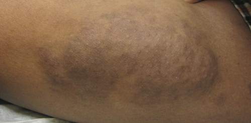
An 11-year-old boy presented for evaluation of a 10×8-cm tortuous lesion on the right posterior leg. Although it had been present since birth, the patient’s mother reported recent growth of the lesion. The lesion was noted to occasionally become irritated and pruritic. The patient’s history was remarkable for asthma.
Epic fail
A couple of years ago, “epic fail” was the phrase my teenage son used as I unsuccessfully attempted to beat him in a game we were playing. At the time, I thought to myself it was a harsh, but accurate assessment of my performance. And I was certainly motivated to practice on my own so that the next time, things would be different.
That same phrase came to mind as I read an October 2015 online article published in Cancer Epidemiology, Biomarkers & Prevention titled “Quality of Physician Communication about Human Papillomavirus Vaccine: Findings from a National Survey” (Nov:24[11];1673). The article describes well the poor performance of the medical community – primarily pediatricians and family physicians – in providing this vaccine. Another source, the most recent National Immunization Survey–Teen 2014, reports another alarming trend: HPV vaccine series initiation and completion continues to lag far behind what it should be.

It came as no surprise to me that the journal article clearly showed what I have suspected for some time and what has been hinted at by previous studies. The epic failure in providing what is essentially a cancer-prevention vaccine to the recommended population of 11- to 12-year-old boys and girls lies not at the feet of the antivaccine movement or hesitant parents. Rather, the failure belongs to us.
The article describes findings from an online survey sent to 2,368 pediatricians and family physicians in 2014. Respondents (776) self-reported their own performance on strength of endorsement (saying the vaccine is important), timeliness (recommending it at ages 11 and 12 years), consistency (recommending it routinely versus using a risk-based approach), and urgency (recommending same-day vaccination).
More than one-quarter stated they did not strongly endorse the HPV vaccine, and a similar number reported they did not recommend it be given at 11-12 years of age. Amazingly, 59% stated they used a risk-based approach versus a routine approach to recommending the HPV vaccine, and only half of the respondents recommended giving the vaccine at the current encounter when discussing the HPV vaccine. Because this is self-reported data, these results represent a best-case scenario because respondents would be unlikely to paint an unflattering picture of their own performance.
Clearly, we have a major problem with physicians struggling with their own discomfort in talking about the HPV vaccine and who erroneously believe parents do not value it. The physician’s lack of competency in communicating effectively – overtly and covertly – leads to a lack of an affirmative recommendation that is so important in any preventive intervention. We are at risk of being the generation of pediatricians and family physicians who collectively failed to protect our patients from a preventable cause of cancer. Only we can fix what is wrong with us. Only we can turn around this epic failure.
Physicians and other providers of medical care to adolescents can access resources to help themselves improve their provision of the HPV vaccine to their patients. One of the best collections of resources can be found online at the American Academy of Pediatrics' Champion Toolkit. This includes material from the Centers for Disease Control and Prevention and AAP as well as some illuminating video vignettes that illustrate the do's and don'ts of communicating with families about HPV vaccination. This must become part of our mission!
Dr. Terk is a pediatrician in Keller, Tx., and is the immediate past president of the Texas Pediatric Society.
*Updated 1/26/2016
A couple of years ago, “epic fail” was the phrase my teenage son used as I unsuccessfully attempted to beat him in a game we were playing. At the time, I thought to myself it was a harsh, but accurate assessment of my performance. And I was certainly motivated to practice on my own so that the next time, things would be different.
That same phrase came to mind as I read an October 2015 online article published in Cancer Epidemiology, Biomarkers & Prevention titled “Quality of Physician Communication about Human Papillomavirus Vaccine: Findings from a National Survey” (Nov:24[11];1673). The article describes well the poor performance of the medical community – primarily pediatricians and family physicians – in providing this vaccine. Another source, the most recent National Immunization Survey–Teen 2014, reports another alarming trend: HPV vaccine series initiation and completion continues to lag far behind what it should be.
It came as no surprise to me that the journal article clearly showed what I have suspected for some time and what has been hinted at by previous studies. The epic failure in providing what is essentially a cancer-prevention vaccine to the recommended population of 11- to 12-year-old boys and girls lies not at the feet of the antivaccine movement or hesitant parents. Rather, the failure belongs to us.
The article describes findings from an online survey sent to 2,368 pediatricians and family physicians in 2014. Respondents (776) self-reported their own performance on strength of endorsement (saying the vaccine is important), timeliness (recommending it at ages 11 and 12 years), consistency (recommending it routinely versus using a risk-based approach), and urgency (recommending same-day vaccination).
More than one-quarter stated they did not strongly endorse the HPV vaccine, and a similar number reported they did not recommend it be given at 11-12 years of age. Amazingly, 59% stated they used a risk-based approach versus a routine approach to recommending the HPV vaccine, and only half of the respondents recommended giving the vaccine at the current encounter when discussing the HPV vaccine. Because this is self-reported data, these results represent a best-case scenario because respondents would be unlikely to paint an unflattering picture of their own performance.
Clearly, we have a major problem with physicians struggling with their own discomfort in talking about the HPV vaccine and who erroneously believe parents do not value it. The physician’s lack of competency in communicating effectively – overtly and covertly – leads to a lack of an affirmative recommendation that is so important in any preventive intervention. We are at risk of being the generation of pediatricians and family physicians who collectively failed to protect our patients from a preventable cause of cancer. Only we can fix what is wrong with us. Only we can turn around this epic failure.
Physicians and other providers of medical care to adolescents can access resources to help themselves improve their provision of the HPV vaccine to their patients. One of the best collections of resources can be found online at the American Academy of Pediatrics' Champion Toolkit. This includes material from the Centers for Disease Control and Prevention and AAP as well as some illuminating video vignettes that illustrate the do's and don'ts of communicating with families about HPV vaccination. This must become part of our mission!
Dr. Terk is a pediatrician in Keller, Tx., and is the immediate past president of the Texas Pediatric Society.
*Updated 1/26/2016
A couple of years ago, “epic fail” was the phrase my teenage son used as I unsuccessfully attempted to beat him in a game we were playing. At the time, I thought to myself it was a harsh, but accurate assessment of my performance. And I was certainly motivated to practice on my own so that the next time, things would be different.
That same phrase came to mind as I read an October 2015 online article published in Cancer Epidemiology, Biomarkers & Prevention titled “Quality of Physician Communication about Human Papillomavirus Vaccine: Findings from a National Survey” (Nov:24[11];1673). The article describes well the poor performance of the medical community – primarily pediatricians and family physicians – in providing this vaccine. Another source, the most recent National Immunization Survey–Teen 2014, reports another alarming trend: HPV vaccine series initiation and completion continues to lag far behind what it should be.
It came as no surprise to me that the journal article clearly showed what I have suspected for some time and what has been hinted at by previous studies. The epic failure in providing what is essentially a cancer-prevention vaccine to the recommended population of 11- to 12-year-old boys and girls lies not at the feet of the antivaccine movement or hesitant parents. Rather, the failure belongs to us.
The article describes findings from an online survey sent to 2,368 pediatricians and family physicians in 2014. Respondents (776) self-reported their own performance on strength of endorsement (saying the vaccine is important), timeliness (recommending it at ages 11 and 12 years), consistency (recommending it routinely versus using a risk-based approach), and urgency (recommending same-day vaccination).
More than one-quarter stated they did not strongly endorse the HPV vaccine, and a similar number reported they did not recommend it be given at 11-12 years of age. Amazingly, 59% stated they used a risk-based approach versus a routine approach to recommending the HPV vaccine, and only half of the respondents recommended giving the vaccine at the current encounter when discussing the HPV vaccine. Because this is self-reported data, these results represent a best-case scenario because respondents would be unlikely to paint an unflattering picture of their own performance.
Clearly, we have a major problem with physicians struggling with their own discomfort in talking about the HPV vaccine and who erroneously believe parents do not value it. The physician’s lack of competency in communicating effectively – overtly and covertly – leads to a lack of an affirmative recommendation that is so important in any preventive intervention. We are at risk of being the generation of pediatricians and family physicians who collectively failed to protect our patients from a preventable cause of cancer. Only we can fix what is wrong with us. Only we can turn around this epic failure.
Physicians and other providers of medical care to adolescents can access resources to help themselves improve their provision of the HPV vaccine to their patients. One of the best collections of resources can be found online at the American Academy of Pediatrics' Champion Toolkit. This includes material from the Centers for Disease Control and Prevention and AAP as well as some illuminating video vignettes that illustrate the do's and don'ts of communicating with families about HPV vaccination. This must become part of our mission!
Dr. Terk is a pediatrician in Keller, Tx., and is the immediate past president of the Texas Pediatric Society.
*Updated 1/26/2016

Maternal immunization during pregnancy: lessons learned, and emerging opportunities
Pediatricians and our teams are the immunization experts. We educate, advocate, and incorporate vaccines into much of our daily routine. As such, we recognize the importance of working with our colleagues in family medicine, internal medicine, and obstetrics to optimize immunization programs for high-risk individuals, including pregnant women. Recent advances in vaccine recommendations during pregnancy are a result of the collaborative efforts of the health care providers for these women, and from systematic evaluation of immunization programs, vaccine pregnancy registries, and disease epidemiology.
Vaccinating women during pregnancy should be considered when a vaccine is known to be safe and when the following apply:

• The risk of severe infection is high during or augmented by pregnancy.
• The specific infection during pregnancy threatens the fetus.
• Maternal protection against infection benefits the newborn.
• Passive transfer of antibody from mother to fetus benefits the newborn.
Examples of safe vaccines immediately come to mind that fulfill one of more of these criteria, yet substantial obstacles exist even where safety and effectiveness data are robust. Because clinical vaccine trials traditionally exclude pregnant women, safety and effectiveness data for this group and their newborns are limited and often must come through experience. In a climate of increased vaccine hesitancy in general, both among providers and patients, vaccine delivery can be fragmented and particularly difficult to streamline. Additional obstacles that exist for any immunization program, including one that targets pregnant women specifically, are immunization delivery logistics and cost.
One of the major success stories of maternal immunization that is easily forgotten or overlooked in developed parts of the world is in the prevention of maternal and neonatal tetanus (MNT). A review of recent history reminds us that between the years 2000 and 2014, 35 countries were finally successful in eliminating MNT, including China, Turkey, Egypt, and South Africa. In addition, 24 of 36 states in the country of India, 30 of 34 provinces of Indonesia, and most of Ethiopia have met with success. This has been accomplished through aggressive tetanus vaccination programs, and through education programs targeted at optimal umbilical cord stump care after delivery.

In the United States, the Advisory Committee on Immunization Practices recommends that all pregnant women should receive inactivated influenza vaccine and Tdap vaccine. In addition, several other vaccines are recommended under certain circumstances. Live attenuated vaccines are considered contraindicated, although yellow fever vaccine is an exception during epidemics, or when travel to a highly endemic area during pregnancy cannot be avoided.
Influenza vaccine administered during pregnancy reduces maternal morbidity and mortality. Moreover, safety and benefits for the fetus are clearly documented. Both retrospective cohort analysis studies and randomized controlled trials have consistently demonstrated lower risk of preterm birth and lower risk for delivering newborns small for gestational age among women who received inactivated influenza vaccine during pregnancy. The benefit extends to term healthy infants who are less likely to be hospitalized during the first months of life if their mother was vaccinated against influenza during pregnancy. Because the mother was immunized during pregnancy, it reduces her risk of infection, thereby reducing the potential that the newborn will be exposed to a mother who is contagious. Perhaps more importantly, infants born at or near term have the benefit of transplacental antibody endowment from their mother, including vaccine induced anti-influenza antibodies. This passive protection is expected to last for several months. Active immunization against influenza during infancy begins at 6 months of age.
Tdap vaccine is also recommended during each pregnancy. In the United States, MNT is eliminated. Here, as in other developed countries, Tdap is administered to reduce infant pertussis morbidity. Pertussis remains endemic to the United States, and infants who develop whooping cough during the first 2-3 months of life are at the highest risk for morbidity and mortality.
Historically, one-third of infants infected with pertussis were infected by their mother, although more recent evidence suggests that older siblings are at least as likely a source. Looking back to the paradigm for protection against influenza infection in the first few months of life, it becomes clear why the Advisory Committee on Immunization Practices of the Centers for Disease Control and Prevention has recommended Tdap vaccine for all mothers during each pregnancy.
The first goal is to prevent pertussis in the mother so that she will not transmit the infection to her newborn. The additional goal, and the rationale for vaccinating pregnant women during every pregnancy, is to optimize levels of anti-pertussis antibody in the mother, so that the transplacental endowment to the infant is as robust as possible.
Serologic studies have demonstrated that Tdap vaccine induces high anti-pertussis antibodies when administered during pregnancy, but that the half-life of those antibodies is brief. When Tdap is given during pregnancy, and the infant is born at or near term, the antibody transfer to the infant is expected to provide passive protection for several months. Maternal immunization during pregnancy thereby reduces the risk that the mother will develop pertussis and transmit it to her newborn, while at the same time allows a degree of passive immunoprotection to the infant during the most vulnerable period of life. Active immunization against pertussis in the infant begins between 6-8 weeks of age.
Another infection that is exceedingly common during the first several months of life is respiratory syncytial virus (RSV). RSV remains the most common reason for infant hospitalization in the United States and other developed countries. The source of the virus can be any other person with a mild or moderate respiratory tract infection as the virus is ubiquitous, and can re-infect individuals throughout their lifetime. The first infection, however, is the worst. It is estimated that between 3% and 4% of the U.S. birth cohort is hospitalized with RSV. With a U.S. birth cohort of about 4 million, the result is 120,000-160,000 infant admissions annually.
The RSV epidemic is seasonal, fairly predictable, and dreaded by primary care pediatricians and hospitalists alike. Lower respiratory tract infection with RSV, in the form of RSV bronchiolitis, presents all too commonly in the young infant with cough, coryza, tachypnea, and wheezing. When the work of breathing increases, and the cough symptoms predominate, the infant is unable to feed efficiently. Hospitalizations may be for dehydration, concerns for impending respiratory failure, or for the administration of supplemental oxygen or other respiratory support. No specific therapeutic interventions reliably reduce the symptoms or the length of hospital stay, nor do they reduce the possibility that intensive care with mechanical ventilation may be required. Treatments are only supportive. An effective vaccine remains elusive.
All other common infections that once resulted in high rates of hospitalization in the first year of life are now substantially reduced through vaccination. Why not this one? The development of a safe and effective vaccine to prevent infant RSV infection or to reduce RSV-associated hospitalizations is especially challenging for multiple reasons.
Some of these reasons have met with substantial advances quite recently, including the discovery of antigen structures needed to induce neutralizing antibody responses. There also are challenges specific to the infant group we need most to protect. Infant RSV infection itself confers only modest protection against subsequent infection. Repeated infections over time are necessary for protection against illness when re-exposed. A vaccine that is able to induce a response similar to natural infection would therefore require multiple doses, presumably over time (the so called ‘primary series’) before a substantial clinical benefit would be expected. This is particularly important because most RSV-associated hospitalizations occur during the first several months of life, reducing the timeline for which a protective vaccine series could be administered.
The challenges are parallel to the issues described earlier for protection against both influenza and pertussis. Infant protection against both of these infections are now addressed, at least to start with, by vaccinating the mother during her pregnancy. In the infant, the pertussis vaccine primary series is then initiated between 6 and 8 weeks of age, and the influenza series initiated at 6 months of age. It is the passive protection, in the form of transplacental maternal antibody, that offers the interim protection during the highest-risk first months of life.
For infants at very high risk of serious RSV infection, passive antibody protection is already administered in the form of the pharmacobiologic medication palivizumab. Its half-life dictates monthly injections for those eligible, and its cost precludes its use for any but the highest risk infants (those born prematurely, and those with chronic lung disease and/or congenital heart disease). This strategy, however, has proven effective in preventing RSV-associated hospitalization in every group in which it has been studied. This “proof of concept” strongly suggests that if the right RSV vaccine is given to women during pregnancy to induce a robust neutralizing anti-RSV antibody response, and that antibody is transferred to the fetus prior to birth, the newborn will benefit from protection against RSV for a period of time.
Several questions emerge. If RSV is ubiquitous, and can re-infect individuals throughout their lifetime, then some women will be infected during their pregnancy. Do their infants benefit? As a re-infection, the maternal symptoms would be expected to be mild, but the infection could boost the women’s natural immunity with a robust anamnestic antibody response. This possibility has not been studied systematically, but might help to explain why some healthy term infants exposed to RSV develop little or no symptoms, while others (mothers who have not recently had a natural RSV infection) develop severe illness requiring hospitalization.
There are data to support the contention that term infants born to mothers with higher naturally occurring anti-RSV neutralizing antibodies benefit from those antibodies. In a large prospective cohort study performed in Kenya, cord blood anti-RSV antibody concentrations correlated directly with the length of time before the infant’s first RSV infection. It’s therefore logical to conclude that administering an effective RSV vaccine during pregnancy could augment that natural antibody response, be transferred to the infant at birth, and offer protection against RSV when exposed.
Several candidate vaccines for study already exist and have undergone phase I testing in nonpregnant adults. Once safety is demonstrated, the next step is to identify the vaccine formulation resulting in the most robust anti-RSV neutralizing antibody concentrations. Such a candidate vaccine will be chosen for future phase III trials during pregnancy. Safety, and maternal/cord blood RSV antibody titers will be of interest during that clinical trial, but the rates and timing of RSV infection and RSV-associated hospitalizations among the infants born to those mothers will be the most instructive.
Ideally, a candidate RSV vaccine shown to be as safe and as effective during pregnancy as inactivated influenza vaccines and/or Tdap vaccines would be implemented immediately and universally. Unfortunately, substantial vaccine hesitancy for the use of influenza and Tdap vaccines continues among pregnant patients and their providers. Acceptance of an RSV vaccine for use during pregnancy will not come easily, or immediately. As with all of our successful vaccine programs, launching such an effort will require education, patience, and careful post-licensure documentation of the impact that the intervention has in the real world.
Dr. Domachowske is professor of pediatrics and professor of microbiology and immunology at the State University of New York Upstate Medical University, Syracuse, N.Y. Dr. Domachowske is performing clinical trials and has grants in the area of RSV prevention with Astra Zeneca, Regeneron, and Glaxo Smith Kline.
Pediatricians and our teams are the immunization experts. We educate, advocate, and incorporate vaccines into much of our daily routine. As such, we recognize the importance of working with our colleagues in family medicine, internal medicine, and obstetrics to optimize immunization programs for high-risk individuals, including pregnant women. Recent advances in vaccine recommendations during pregnancy are a result of the collaborative efforts of the health care providers for these women, and from systematic evaluation of immunization programs, vaccine pregnancy registries, and disease epidemiology.
Vaccinating women during pregnancy should be considered when a vaccine is known to be safe and when the following apply:
• The risk of severe infection is high during or augmented by pregnancy.
• The specific infection during pregnancy threatens the fetus.
• Maternal protection against infection benefits the newborn.
• Passive transfer of antibody from mother to fetus benefits the newborn.
Examples of safe vaccines immediately come to mind that fulfill one of more of these criteria, yet substantial obstacles exist even where safety and effectiveness data are robust. Because clinical vaccine trials traditionally exclude pregnant women, safety and effectiveness data for this group and their newborns are limited and often must come through experience. In a climate of increased vaccine hesitancy in general, both among providers and patients, vaccine delivery can be fragmented and particularly difficult to streamline. Additional obstacles that exist for any immunization program, including one that targets pregnant women specifically, are immunization delivery logistics and cost.
One of the major success stories of maternal immunization that is easily forgotten or overlooked in developed parts of the world is in the prevention of maternal and neonatal tetanus (MNT). A review of recent history reminds us that between the years 2000 and 2014, 35 countries were finally successful in eliminating MNT, including China, Turkey, Egypt, and South Africa. In addition, 24 of 36 states in the country of India, 30 of 34 provinces of Indonesia, and most of Ethiopia have met with success. This has been accomplished through aggressive tetanus vaccination programs, and through education programs targeted at optimal umbilical cord stump care after delivery.
In the United States, the Advisory Committee on Immunization Practices recommends that all pregnant women should receive inactivated influenza vaccine and Tdap vaccine. In addition, several other vaccines are recommended under certain circumstances. Live attenuated vaccines are considered contraindicated, although yellow fever vaccine is an exception during epidemics, or when travel to a highly endemic area during pregnancy cannot be avoided.
Influenza vaccine administered during pregnancy reduces maternal morbidity and mortality. Moreover, safety and benefits for the fetus are clearly documented. Both retrospective cohort analysis studies and randomized controlled trials have consistently demonstrated lower risk of preterm birth and lower risk for delivering newborns small for gestational age among women who received inactivated influenza vaccine during pregnancy. The benefit extends to term healthy infants who are less likely to be hospitalized during the first months of life if their mother was vaccinated against influenza during pregnancy. Because the mother was immunized during pregnancy, it reduces her risk of infection, thereby reducing the potential that the newborn will be exposed to a mother who is contagious. Perhaps more importantly, infants born at or near term have the benefit of transplacental antibody endowment from their mother, including vaccine induced anti-influenza antibodies. This passive protection is expected to last for several months. Active immunization against influenza during infancy begins at 6 months of age.
Tdap vaccine is also recommended during each pregnancy. In the United States, MNT is eliminated. Here, as in other developed countries, Tdap is administered to reduce infant pertussis morbidity. Pertussis remains endemic to the United States, and infants who develop whooping cough during the first 2-3 months of life are at the highest risk for morbidity and mortality.
Historically, one-third of infants infected with pertussis were infected by their mother, although more recent evidence suggests that older siblings are at least as likely a source. Looking back to the paradigm for protection against influenza infection in the first few months of life, it becomes clear why the Advisory Committee on Immunization Practices of the Centers for Disease Control and Prevention has recommended Tdap vaccine for all mothers during each pregnancy.
The first goal is to prevent pertussis in the mother so that she will not transmit the infection to her newborn. The additional goal, and the rationale for vaccinating pregnant women during every pregnancy, is to optimize levels of anti-pertussis antibody in the mother, so that the transplacental endowment to the infant is as robust as possible.
Serologic studies have demonstrated that Tdap vaccine induces high anti-pertussis antibodies when administered during pregnancy, but that the half-life of those antibodies is brief. When Tdap is given during pregnancy, and the infant is born at or near term, the antibody transfer to the infant is expected to provide passive protection for several months. Maternal immunization during pregnancy thereby reduces the risk that the mother will develop pertussis and transmit it to her newborn, while at the same time allows a degree of passive immunoprotection to the infant during the most vulnerable period of life. Active immunization against pertussis in the infant begins between 6-8 weeks of age.
Another infection that is exceedingly common during the first several months of life is respiratory syncytial virus (RSV). RSV remains the most common reason for infant hospitalization in the United States and other developed countries. The source of the virus can be any other person with a mild or moderate respiratory tract infection as the virus is ubiquitous, and can re-infect individuals throughout their lifetime. The first infection, however, is the worst. It is estimated that between 3% and 4% of the U.S. birth cohort is hospitalized with RSV. With a U.S. birth cohort of about 4 million, the result is 120,000-160,000 infant admissions annually.
The RSV epidemic is seasonal, fairly predictable, and dreaded by primary care pediatricians and hospitalists alike. Lower respiratory tract infection with RSV, in the form of RSV bronchiolitis, presents all too commonly in the young infant with cough, coryza, tachypnea, and wheezing. When the work of breathing increases, and the cough symptoms predominate, the infant is unable to feed efficiently. Hospitalizations may be for dehydration, concerns for impending respiratory failure, or for the administration of supplemental oxygen or other respiratory support. No specific therapeutic interventions reliably reduce the symptoms or the length of hospital stay, nor do they reduce the possibility that intensive care with mechanical ventilation may be required. Treatments are only supportive. An effective vaccine remains elusive.
All other common infections that once resulted in high rates of hospitalization in the first year of life are now substantially reduced through vaccination. Why not this one? The development of a safe and effective vaccine to prevent infant RSV infection or to reduce RSV-associated hospitalizations is especially challenging for multiple reasons.
Some of these reasons have met with substantial advances quite recently, including the discovery of antigen structures needed to induce neutralizing antibody responses. There also are challenges specific to the infant group we need most to protect. Infant RSV infection itself confers only modest protection against subsequent infection. Repeated infections over time are necessary for protection against illness when re-exposed. A vaccine that is able to induce a response similar to natural infection would therefore require multiple doses, presumably over time (the so called ‘primary series’) before a substantial clinical benefit would be expected. This is particularly important because most RSV-associated hospitalizations occur during the first several months of life, reducing the timeline for which a protective vaccine series could be administered.
The challenges are parallel to the issues described earlier for protection against both influenza and pertussis. Infant protection against both of these infections are now addressed, at least to start with, by vaccinating the mother during her pregnancy. In the infant, the pertussis vaccine primary series is then initiated between 6 and 8 weeks of age, and the influenza series initiated at 6 months of age. It is the passive protection, in the form of transplacental maternal antibody, that offers the interim protection during the highest-risk first months of life.
For infants at very high risk of serious RSV infection, passive antibody protection is already administered in the form of the pharmacobiologic medication palivizumab. Its half-life dictates monthly injections for those eligible, and its cost precludes its use for any but the highest risk infants (those born prematurely, and those with chronic lung disease and/or congenital heart disease). This strategy, however, has proven effective in preventing RSV-associated hospitalization in every group in which it has been studied. This “proof of concept” strongly suggests that if the right RSV vaccine is given to women during pregnancy to induce a robust neutralizing anti-RSV antibody response, and that antibody is transferred to the fetus prior to birth, the newborn will benefit from protection against RSV for a period of time.
Several questions emerge. If RSV is ubiquitous, and can re-infect individuals throughout their lifetime, then some women will be infected during their pregnancy. Do their infants benefit? As a re-infection, the maternal symptoms would be expected to be mild, but the infection could boost the women’s natural immunity with a robust anamnestic antibody response. This possibility has not been studied systematically, but might help to explain why some healthy term infants exposed to RSV develop little or no symptoms, while others (mothers who have not recently had a natural RSV infection) develop severe illness requiring hospitalization.
There are data to support the contention that term infants born to mothers with higher naturally occurring anti-RSV neutralizing antibodies benefit from those antibodies. In a large prospective cohort study performed in Kenya, cord blood anti-RSV antibody concentrations correlated directly with the length of time before the infant’s first RSV infection. It’s therefore logical to conclude that administering an effective RSV vaccine during pregnancy could augment that natural antibody response, be transferred to the infant at birth, and offer protection against RSV when exposed.
Several candidate vaccines for study already exist and have undergone phase I testing in nonpregnant adults. Once safety is demonstrated, the next step is to identify the vaccine formulation resulting in the most robust anti-RSV neutralizing antibody concentrations. Such a candidate vaccine will be chosen for future phase III trials during pregnancy. Safety, and maternal/cord blood RSV antibody titers will be of interest during that clinical trial, but the rates and timing of RSV infection and RSV-associated hospitalizations among the infants born to those mothers will be the most instructive.
Ideally, a candidate RSV vaccine shown to be as safe and as effective during pregnancy as inactivated influenza vaccines and/or Tdap vaccines would be implemented immediately and universally. Unfortunately, substantial vaccine hesitancy for the use of influenza and Tdap vaccines continues among pregnant patients and their providers. Acceptance of an RSV vaccine for use during pregnancy will not come easily, or immediately. As with all of our successful vaccine programs, launching such an effort will require education, patience, and careful post-licensure documentation of the impact that the intervention has in the real world.
Dr. Domachowske is professor of pediatrics and professor of microbiology and immunology at the State University of New York Upstate Medical University, Syracuse, N.Y. Dr. Domachowske is performing clinical trials and has grants in the area of RSV prevention with Astra Zeneca, Regeneron, and Glaxo Smith Kline.
Pediatricians and our teams are the immunization experts. We educate, advocate, and incorporate vaccines into much of our daily routine. As such, we recognize the importance of working with our colleagues in family medicine, internal medicine, and obstetrics to optimize immunization programs for high-risk individuals, including pregnant women. Recent advances in vaccine recommendations during pregnancy are a result of the collaborative efforts of the health care providers for these women, and from systematic evaluation of immunization programs, vaccine pregnancy registries, and disease epidemiology.
Vaccinating women during pregnancy should be considered when a vaccine is known to be safe and when the following apply:
• The risk of severe infection is high during or augmented by pregnancy.
• The specific infection during pregnancy threatens the fetus.
• Maternal protection against infection benefits the newborn.
• Passive transfer of antibody from mother to fetus benefits the newborn.
Examples of safe vaccines immediately come to mind that fulfill one of more of these criteria, yet substantial obstacles exist even where safety and effectiveness data are robust. Because clinical vaccine trials traditionally exclude pregnant women, safety and effectiveness data for this group and their newborns are limited and often must come through experience. In a climate of increased vaccine hesitancy in general, both among providers and patients, vaccine delivery can be fragmented and particularly difficult to streamline. Additional obstacles that exist for any immunization program, including one that targets pregnant women specifically, are immunization delivery logistics and cost.
One of the major success stories of maternal immunization that is easily forgotten or overlooked in developed parts of the world is in the prevention of maternal and neonatal tetanus (MNT). A review of recent history reminds us that between the years 2000 and 2014, 35 countries were finally successful in eliminating MNT, including China, Turkey, Egypt, and South Africa. In addition, 24 of 36 states in the country of India, 30 of 34 provinces of Indonesia, and most of Ethiopia have met with success. This has been accomplished through aggressive tetanus vaccination programs, and through education programs targeted at optimal umbilical cord stump care after delivery.
In the United States, the Advisory Committee on Immunization Practices recommends that all pregnant women should receive inactivated influenza vaccine and Tdap vaccine. In addition, several other vaccines are recommended under certain circumstances. Live attenuated vaccines are considered contraindicated, although yellow fever vaccine is an exception during epidemics, or when travel to a highly endemic area during pregnancy cannot be avoided.
Influenza vaccine administered during pregnancy reduces maternal morbidity and mortality. Moreover, safety and benefits for the fetus are clearly documented. Both retrospective cohort analysis studies and randomized controlled trials have consistently demonstrated lower risk of preterm birth and lower risk for delivering newborns small for gestational age among women who received inactivated influenza vaccine during pregnancy. The benefit extends to term healthy infants who are less likely to be hospitalized during the first months of life if their mother was vaccinated against influenza during pregnancy. Because the mother was immunized during pregnancy, it reduces her risk of infection, thereby reducing the potential that the newborn will be exposed to a mother who is contagious. Perhaps more importantly, infants born at or near term have the benefit of transplacental antibody endowment from their mother, including vaccine induced anti-influenza antibodies. This passive protection is expected to last for several months. Active immunization against influenza during infancy begins at 6 months of age.
Tdap vaccine is also recommended during each pregnancy. In the United States, MNT is eliminated. Here, as in other developed countries, Tdap is administered to reduce infant pertussis morbidity. Pertussis remains endemic to the United States, and infants who develop whooping cough during the first 2-3 months of life are at the highest risk for morbidity and mortality.
Historically, one-third of infants infected with pertussis were infected by their mother, although more recent evidence suggests that older siblings are at least as likely a source. Looking back to the paradigm for protection against influenza infection in the first few months of life, it becomes clear why the Advisory Committee on Immunization Practices of the Centers for Disease Control and Prevention has recommended Tdap vaccine for all mothers during each pregnancy.
The first goal is to prevent pertussis in the mother so that she will not transmit the infection to her newborn. The additional goal, and the rationale for vaccinating pregnant women during every pregnancy, is to optimize levels of anti-pertussis antibody in the mother, so that the transplacental endowment to the infant is as robust as possible.
Serologic studies have demonstrated that Tdap vaccine induces high anti-pertussis antibodies when administered during pregnancy, but that the half-life of those antibodies is brief. When Tdap is given during pregnancy, and the infant is born at or near term, the antibody transfer to the infant is expected to provide passive protection for several months. Maternal immunization during pregnancy thereby reduces the risk that the mother will develop pertussis and transmit it to her newborn, while at the same time allows a degree of passive immunoprotection to the infant during the most vulnerable period of life. Active immunization against pertussis in the infant begins between 6-8 weeks of age.
Another infection that is exceedingly common during the first several months of life is respiratory syncytial virus (RSV). RSV remains the most common reason for infant hospitalization in the United States and other developed countries. The source of the virus can be any other person with a mild or moderate respiratory tract infection as the virus is ubiquitous, and can re-infect individuals throughout their lifetime. The first infection, however, is the worst. It is estimated that between 3% and 4% of the U.S. birth cohort is hospitalized with RSV. With a U.S. birth cohort of about 4 million, the result is 120,000-160,000 infant admissions annually.
The RSV epidemic is seasonal, fairly predictable, and dreaded by primary care pediatricians and hospitalists alike. Lower respiratory tract infection with RSV, in the form of RSV bronchiolitis, presents all too commonly in the young infant with cough, coryza, tachypnea, and wheezing. When the work of breathing increases, and the cough symptoms predominate, the infant is unable to feed efficiently. Hospitalizations may be for dehydration, concerns for impending respiratory failure, or for the administration of supplemental oxygen or other respiratory support. No specific therapeutic interventions reliably reduce the symptoms or the length of hospital stay, nor do they reduce the possibility that intensive care with mechanical ventilation may be required. Treatments are only supportive. An effective vaccine remains elusive.
All other common infections that once resulted in high rates of hospitalization in the first year of life are now substantially reduced through vaccination. Why not this one? The development of a safe and effective vaccine to prevent infant RSV infection or to reduce RSV-associated hospitalizations is especially challenging for multiple reasons.
Some of these reasons have met with substantial advances quite recently, including the discovery of antigen structures needed to induce neutralizing antibody responses. There also are challenges specific to the infant group we need most to protect. Infant RSV infection itself confers only modest protection against subsequent infection. Repeated infections over time are necessary for protection against illness when re-exposed. A vaccine that is able to induce a response similar to natural infection would therefore require multiple doses, presumably over time (the so called ‘primary series’) before a substantial clinical benefit would be expected. This is particularly important because most RSV-associated hospitalizations occur during the first several months of life, reducing the timeline for which a protective vaccine series could be administered.
The challenges are parallel to the issues described earlier for protection against both influenza and pertussis. Infant protection against both of these infections are now addressed, at least to start with, by vaccinating the mother during her pregnancy. In the infant, the pertussis vaccine primary series is then initiated between 6 and 8 weeks of age, and the influenza series initiated at 6 months of age. It is the passive protection, in the form of transplacental maternal antibody, that offers the interim protection during the highest-risk first months of life.
For infants at very high risk of serious RSV infection, passive antibody protection is already administered in the form of the pharmacobiologic medication palivizumab. Its half-life dictates monthly injections for those eligible, and its cost precludes its use for any but the highest risk infants (those born prematurely, and those with chronic lung disease and/or congenital heart disease). This strategy, however, has proven effective in preventing RSV-associated hospitalization in every group in which it has been studied. This “proof of concept” strongly suggests that if the right RSV vaccine is given to women during pregnancy to induce a robust neutralizing anti-RSV antibody response, and that antibody is transferred to the fetus prior to birth, the newborn will benefit from protection against RSV for a period of time.
Several questions emerge. If RSV is ubiquitous, and can re-infect individuals throughout their lifetime, then some women will be infected during their pregnancy. Do their infants benefit? As a re-infection, the maternal symptoms would be expected to be mild, but the infection could boost the women’s natural immunity with a robust anamnestic antibody response. This possibility has not been studied systematically, but might help to explain why some healthy term infants exposed to RSV develop little or no symptoms, while others (mothers who have not recently had a natural RSV infection) develop severe illness requiring hospitalization.
There are data to support the contention that term infants born to mothers with higher naturally occurring anti-RSV neutralizing antibodies benefit from those antibodies. In a large prospective cohort study performed in Kenya, cord blood anti-RSV antibody concentrations correlated directly with the length of time before the infant’s first RSV infection. It’s therefore logical to conclude that administering an effective RSV vaccine during pregnancy could augment that natural antibody response, be transferred to the infant at birth, and offer protection against RSV when exposed.
Several candidate vaccines for study already exist and have undergone phase I testing in nonpregnant adults. Once safety is demonstrated, the next step is to identify the vaccine formulation resulting in the most robust anti-RSV neutralizing antibody concentrations. Such a candidate vaccine will be chosen for future phase III trials during pregnancy. Safety, and maternal/cord blood RSV antibody titers will be of interest during that clinical trial, but the rates and timing of RSV infection and RSV-associated hospitalizations among the infants born to those mothers will be the most instructive.
Ideally, a candidate RSV vaccine shown to be as safe and as effective during pregnancy as inactivated influenza vaccines and/or Tdap vaccines would be implemented immediately and universally. Unfortunately, substantial vaccine hesitancy for the use of influenza and Tdap vaccines continues among pregnant patients and their providers. Acceptance of an RSV vaccine for use during pregnancy will not come easily, or immediately. As with all of our successful vaccine programs, launching such an effort will require education, patience, and careful post-licensure documentation of the impact that the intervention has in the real world.
Dr. Domachowske is professor of pediatrics and professor of microbiology and immunology at the State University of New York Upstate Medical University, Syracuse, N.Y. Dr. Domachowske is performing clinical trials and has grants in the area of RSV prevention with Astra Zeneca, Regeneron, and Glaxo Smith Kline.

AAP says newborn pain management plans essential for all providers
Repeated exposure to physical pain can have both short-term and long-term adverse effects in newborns, yet providers too inconsistently assess and adequately manage pain in neonates, the American Academy of Pediatrics said in an updated policy statement on preventing and managing procedural pain in these infants.
“Neonates are frequently subjected to painful procedures, with the most immature infants receiving the highest number of painful events,” wrote the AAP Committee on Fetus and Newborn and the AAP Section on Anesthesiology and Pain Medicine. Premature infants also are at the highest risk for long-term sequelae, the committees noted. “These sequelae include physiologic instability; altered brain development; and abnormal neurodevelopment, somatosensory, and stress response systems, which can persist into childhood” (Pediatrics. 2016 Jan 25. doi: 10.1542/peds.2015-4271).
The statement nicely summarizes current evidence on the evaluation and management of pain in neonates, as well as potential short-term and long-term negative effects, said Dr. Clay T. Jones, a neonatal hospitalist at Newton-Wellesley Hospital in Newton, Mass., who was not involved in drafting the statement.
“It is a nice testament to how far we’ve come since the days of performing major surgeries in newborns without the use of analgesic medications, but there is still considerable room for improvement,” Dr. Jones said in an interview. “Although we’ve learned a lot about pain in this population over the past few decades, the authors go to great lengths to make it clear that the optimal way to treat pain hasn’t been established yet, and that the evaluation of risk versus benefit in regards to management using pharmaceutical agents is ongoing.”
The statement described the challenges of effective pain management, including foremost the newborn’s inability to communicate and the dearth of information about effective pain assessments and management in this population.
“Contextual factors such as gestational age and behavioral state may play a significant role in pain assessment and are beginning to be included in some assessment tools,” the committees wrote. “New and emerging technologies to measure pain responses, such as near-infrared spectroscopy, amplitude-integrated electroencephalography, functional MRI, skin conductance, and heart rate variability assessment, are being investigated.”
In the meantime, however, all facilities caring for newborns should implement a pain prevention program that takes advantage of what pain management strategies do exist. In addition to reviewing these strategies, the statement recommended all providers implement and use pain assessment and management plans that include both pharmacologic and nonpharmacologic therapies for major procedures and routine minor interventions that might still cause pain.
“Where this report might have the largest impact is in the outpatient offices of pediatric medical providers and in newborn nurseries,” Dr. Jones said. “It is not uncommon for young infants to undergo painful medical procedures, such as heel sticks and circumcisions, without a systematic approach to pain evaluation and inconsistent efforts to prevent or reduce pain.”
Yet there is no excuse for this lack of a consistent approach, he said.
“Nonpharmaceutical interventions are safe, easy to implement, [and] inexpensive, and there is good evidence that they reduce physiologic surrogate markers for pain such as blood pressure, heart rate, and respiratory rate,” Dr. Jones said.
The statement’s first recommendation is that all institutions have written guidelines for a stepwise plan of pain prevention and treatment in newborns. This plan also should include use of currently available and validated neonatal assessment tools “before, during, and after painful procedures to monitor the effectiveness of pain relief interventions.”
Another recommendation urged pediatricians and other neonatal health care providers and family members to receive ongoing education about recognizing and assessing pain in newborns and then managing it as much as current knowledge and evidence allow. The committees called for more research into pain assessment tools and pain prevention and amelioration strategies, including pharmacologic options.
The nonpharmacologic strategies shown to be effective for reducing pain during short-term mild or moderately painful procedures include facilitated tucking, sensorial stimulation, nonnutritive sucking, and breastfeeding or providing expressed human milk.
“Anything we can do to ease pain will improve a baby’s quality of life,” said Dr. Nathan Boonstra of Blank Children’s Hospital pediatric clinic in Des Moines, Iowa, who was not involved in writing the statement.
“Pediatricians should always be judicious when deciding to draw blood, but when we need to, we should reflexively think about what can keep the procedure’s pain at a minimum,” he said in an interview. “Many of my patients’ mothers instinctively want to breastfeed to help their newborns through something painful, and their instinct serves them well.”
The policy statement mentioned oral sucrose and/or glucose solutions as effective analgesic options for mild to moderately painful procedures, but recommended these be prescribed and tracked as medication.
This strategy carries risks and other drawbacks, however, Dr. Thomas M Seman, a pediatrician in group practice in Danvers, Mass., said in an interview
“Having sucrose or glucose solutions in the office can be dangerous because of the risk of overuse and hyperglycemia as well as the cost of these items,” said Dr. Seman, who was not involved with drafting the AAP statement. His office policy primarily focuses on the parents holding their children and talking, singing, or humming to them during procedures, followed by feeding and/or acetaminophen, he said.
“The other medications used are prohibitive for a number of reasons,” said Dr. Seman, although he added that most of the procedures described in the statement are performed on premature infants in a NICU with only a few done in private practices.
For example, opioids such as fentanyl and morphine are most frequently used for persistent pain, yet the data on appropriate dosing and long-term effects in the newborn period are “woefully lacking and/or conflicting,” the statement noted. The evidence for benzodiazepines, often used for sedation in the neonatal intensive care, shows little additional analgesic benefit, but these agents can potentiate the risk of respiratory depression and hypotension associated with opioid use. Caution should particularly be exercised before using methadone, ketamine, propofol, and dexmedetomidine because so little is known about safe and effective dosing of these medications in neonates. They also carry various potential risks ranging from neurotoxicity to bradycardia, desaturations, and prolonged hypotension.
While NSAIDs are not recommended at this young age, oral or intravenous acetaminophen has sufficient preliminary safety and efficacy to be considered for postoperative pain relief, according to the statement.
The AAP did not report disclosures for committee members. Dr. Boonstra, Dr. Jones, and Dr. Seman had no relevant financial disclosures.
Repeated exposure to physical pain can have both short-term and long-term adverse effects in newborns, yet providers too inconsistently assess and adequately manage pain in neonates, the American Academy of Pediatrics said in an updated policy statement on preventing and managing procedural pain in these infants.
“Neonates are frequently subjected to painful procedures, with the most immature infants receiving the highest number of painful events,” wrote the AAP Committee on Fetus and Newborn and the AAP Section on Anesthesiology and Pain Medicine. Premature infants also are at the highest risk for long-term sequelae, the committees noted. “These sequelae include physiologic instability; altered brain development; and abnormal neurodevelopment, somatosensory, and stress response systems, which can persist into childhood” (Pediatrics. 2016 Jan 25. doi: 10.1542/peds.2015-4271).
The statement nicely summarizes current evidence on the evaluation and management of pain in neonates, as well as potential short-term and long-term negative effects, said Dr. Clay T. Jones, a neonatal hospitalist at Newton-Wellesley Hospital in Newton, Mass., who was not involved in drafting the statement.
“It is a nice testament to how far we’ve come since the days of performing major surgeries in newborns without the use of analgesic medications, but there is still considerable room for improvement,” Dr. Jones said in an interview. “Although we’ve learned a lot about pain in this population over the past few decades, the authors go to great lengths to make it clear that the optimal way to treat pain hasn’t been established yet, and that the evaluation of risk versus benefit in regards to management using pharmaceutical agents is ongoing.”
The statement described the challenges of effective pain management, including foremost the newborn’s inability to communicate and the dearth of information about effective pain assessments and management in this population.
“Contextual factors such as gestational age and behavioral state may play a significant role in pain assessment and are beginning to be included in some assessment tools,” the committees wrote. “New and emerging technologies to measure pain responses, such as near-infrared spectroscopy, amplitude-integrated electroencephalography, functional MRI, skin conductance, and heart rate variability assessment, are being investigated.”
In the meantime, however, all facilities caring for newborns should implement a pain prevention program that takes advantage of what pain management strategies do exist. In addition to reviewing these strategies, the statement recommended all providers implement and use pain assessment and management plans that include both pharmacologic and nonpharmacologic therapies for major procedures and routine minor interventions that might still cause pain.
“Where this report might have the largest impact is in the outpatient offices of pediatric medical providers and in newborn nurseries,” Dr. Jones said. “It is not uncommon for young infants to undergo painful medical procedures, such as heel sticks and circumcisions, without a systematic approach to pain evaluation and inconsistent efforts to prevent or reduce pain.”
Yet there is no excuse for this lack of a consistent approach, he said.
“Nonpharmaceutical interventions are safe, easy to implement, [and] inexpensive, and there is good evidence that they reduce physiologic surrogate markers for pain such as blood pressure, heart rate, and respiratory rate,” Dr. Jones said.
The statement’s first recommendation is that all institutions have written guidelines for a stepwise plan of pain prevention and treatment in newborns. This plan also should include use of currently available and validated neonatal assessment tools “before, during, and after painful procedures to monitor the effectiveness of pain relief interventions.”
Another recommendation urged pediatricians and other neonatal health care providers and family members to receive ongoing education about recognizing and assessing pain in newborns and then managing it as much as current knowledge and evidence allow. The committees called for more research into pain assessment tools and pain prevention and amelioration strategies, including pharmacologic options.
The nonpharmacologic strategies shown to be effective for reducing pain during short-term mild or moderately painful procedures include facilitated tucking, sensorial stimulation, nonnutritive sucking, and breastfeeding or providing expressed human milk.
“Anything we can do to ease pain will improve a baby’s quality of life,” said Dr. Nathan Boonstra of Blank Children’s Hospital pediatric clinic in Des Moines, Iowa, who was not involved in writing the statement.
“Pediatricians should always be judicious when deciding to draw blood, but when we need to, we should reflexively think about what can keep the procedure’s pain at a minimum,” he said in an interview. “Many of my patients’ mothers instinctively want to breastfeed to help their newborns through something painful, and their instinct serves them well.”
The policy statement mentioned oral sucrose and/or glucose solutions as effective analgesic options for mild to moderately painful procedures, but recommended these be prescribed and tracked as medication.
This strategy carries risks and other drawbacks, however, Dr. Thomas M Seman, a pediatrician in group practice in Danvers, Mass., said in an interview
“Having sucrose or glucose solutions in the office can be dangerous because of the risk of overuse and hyperglycemia as well as the cost of these items,” said Dr. Seman, who was not involved with drafting the AAP statement. His office policy primarily focuses on the parents holding their children and talking, singing, or humming to them during procedures, followed by feeding and/or acetaminophen, he said.
“The other medications used are prohibitive for a number of reasons,” said Dr. Seman, although he added that most of the procedures described in the statement are performed on premature infants in a NICU with only a few done in private practices.
For example, opioids such as fentanyl and morphine are most frequently used for persistent pain, yet the data on appropriate dosing and long-term effects in the newborn period are “woefully lacking and/or conflicting,” the statement noted. The evidence for benzodiazepines, often used for sedation in the neonatal intensive care, shows little additional analgesic benefit, but these agents can potentiate the risk of respiratory depression and hypotension associated with opioid use. Caution should particularly be exercised before using methadone, ketamine, propofol, and dexmedetomidine because so little is known about safe and effective dosing of these medications in neonates. They also carry various potential risks ranging from neurotoxicity to bradycardia, desaturations, and prolonged hypotension.
While NSAIDs are not recommended at this young age, oral or intravenous acetaminophen has sufficient preliminary safety and efficacy to be considered for postoperative pain relief, according to the statement.
The AAP did not report disclosures for committee members. Dr. Boonstra, Dr. Jones, and Dr. Seman had no relevant financial disclosures.
Repeated exposure to physical pain can have both short-term and long-term adverse effects in newborns, yet providers too inconsistently assess and adequately manage pain in neonates, the American Academy of Pediatrics said in an updated policy statement on preventing and managing procedural pain in these infants.
“Neonates are frequently subjected to painful procedures, with the most immature infants receiving the highest number of painful events,” wrote the AAP Committee on Fetus and Newborn and the AAP Section on Anesthesiology and Pain Medicine. Premature infants also are at the highest risk for long-term sequelae, the committees noted. “These sequelae include physiologic instability; altered brain development; and abnormal neurodevelopment, somatosensory, and stress response systems, which can persist into childhood” (Pediatrics. 2016 Jan 25. doi: 10.1542/peds.2015-4271).
The statement nicely summarizes current evidence on the evaluation and management of pain in neonates, as well as potential short-term and long-term negative effects, said Dr. Clay T. Jones, a neonatal hospitalist at Newton-Wellesley Hospital in Newton, Mass., who was not involved in drafting the statement.
“It is a nice testament to how far we’ve come since the days of performing major surgeries in newborns without the use of analgesic medications, but there is still considerable room for improvement,” Dr. Jones said in an interview. “Although we’ve learned a lot about pain in this population over the past few decades, the authors go to great lengths to make it clear that the optimal way to treat pain hasn’t been established yet, and that the evaluation of risk versus benefit in regards to management using pharmaceutical agents is ongoing.”
The statement described the challenges of effective pain management, including foremost the newborn’s inability to communicate and the dearth of information about effective pain assessments and management in this population.
“Contextual factors such as gestational age and behavioral state may play a significant role in pain assessment and are beginning to be included in some assessment tools,” the committees wrote. “New and emerging technologies to measure pain responses, such as near-infrared spectroscopy, amplitude-integrated electroencephalography, functional MRI, skin conductance, and heart rate variability assessment, are being investigated.”
In the meantime, however, all facilities caring for newborns should implement a pain prevention program that takes advantage of what pain management strategies do exist. In addition to reviewing these strategies, the statement recommended all providers implement and use pain assessment and management plans that include both pharmacologic and nonpharmacologic therapies for major procedures and routine minor interventions that might still cause pain.
“Where this report might have the largest impact is in the outpatient offices of pediatric medical providers and in newborn nurseries,” Dr. Jones said. “It is not uncommon for young infants to undergo painful medical procedures, such as heel sticks and circumcisions, without a systematic approach to pain evaluation and inconsistent efforts to prevent or reduce pain.”
Yet there is no excuse for this lack of a consistent approach, he said.
“Nonpharmaceutical interventions are safe, easy to implement, [and] inexpensive, and there is good evidence that they reduce physiologic surrogate markers for pain such as blood pressure, heart rate, and respiratory rate,” Dr. Jones said.
The statement’s first recommendation is that all institutions have written guidelines for a stepwise plan of pain prevention and treatment in newborns. This plan also should include use of currently available and validated neonatal assessment tools “before, during, and after painful procedures to monitor the effectiveness of pain relief interventions.”
Another recommendation urged pediatricians and other neonatal health care providers and family members to receive ongoing education about recognizing and assessing pain in newborns and then managing it as much as current knowledge and evidence allow. The committees called for more research into pain assessment tools and pain prevention and amelioration strategies, including pharmacologic options.
The nonpharmacologic strategies shown to be effective for reducing pain during short-term mild or moderately painful procedures include facilitated tucking, sensorial stimulation, nonnutritive sucking, and breastfeeding or providing expressed human milk.
“Anything we can do to ease pain will improve a baby’s quality of life,” said Dr. Nathan Boonstra of Blank Children’s Hospital pediatric clinic in Des Moines, Iowa, who was not involved in writing the statement.
“Pediatricians should always be judicious when deciding to draw blood, but when we need to, we should reflexively think about what can keep the procedure’s pain at a minimum,” he said in an interview. “Many of my patients’ mothers instinctively want to breastfeed to help their newborns through something painful, and their instinct serves them well.”
The policy statement mentioned oral sucrose and/or glucose solutions as effective analgesic options for mild to moderately painful procedures, but recommended these be prescribed and tracked as medication.
This strategy carries risks and other drawbacks, however, Dr. Thomas M Seman, a pediatrician in group practice in Danvers, Mass., said in an interview
“Having sucrose or glucose solutions in the office can be dangerous because of the risk of overuse and hyperglycemia as well as the cost of these items,” said Dr. Seman, who was not involved with drafting the AAP statement. His office policy primarily focuses on the parents holding their children and talking, singing, or humming to them during procedures, followed by feeding and/or acetaminophen, he said.
“The other medications used are prohibitive for a number of reasons,” said Dr. Seman, although he added that most of the procedures described in the statement are performed on premature infants in a NICU with only a few done in private practices.
For example, opioids such as fentanyl and morphine are most frequently used for persistent pain, yet the data on appropriate dosing and long-term effects in the newborn period are “woefully lacking and/or conflicting,” the statement noted. The evidence for benzodiazepines, often used for sedation in the neonatal intensive care, shows little additional analgesic benefit, but these agents can potentiate the risk of respiratory depression and hypotension associated with opioid use. Caution should particularly be exercised before using methadone, ketamine, propofol, and dexmedetomidine because so little is known about safe and effective dosing of these medications in neonates. They also carry various potential risks ranging from neurotoxicity to bradycardia, desaturations, and prolonged hypotension.
While NSAIDs are not recommended at this young age, oral or intravenous acetaminophen has sufficient preliminary safety and efficacy to be considered for postoperative pain relief, according to the statement.
The AAP did not report disclosures for committee members. Dr. Boonstra, Dr. Jones, and Dr. Seman had no relevant financial disclosures.
FROM PEDIATRICS
2 new USPSTF draft recommendations—what you need to know

Ivacaftor appears safe for young children with cystic fibrosis
Ivacaftor, a cystic fibrosis transmembrane conductance regulator potentiator, appears generally safe for young children with cystic fibrosis and a CFTR gating mutation, an open-label, single-arm study shows.
The study, published online Jan. 20, is the first to assess ivacaftor in children aged 2-5 years, suggests that the drug is well tolerated, somewhat improves sweat chloride and nutritional measures, and also might improve pancreatic function, reported Dr. Jane C. Davies of the National Heart & Lung Institute of Imperial College London, and her associates. “Reported side effects are similar to those in the general cystic fibrosis population, although children with previous cystic fibrosis liver disease might have transient rises in transaminase concentrations,” they added.

Ivacaftor is approved by the Food and Drug Administration for children aged 6 years and older with cystic fibrosis, and it should be even more beneficial if given before patients develop infections and inflammation as a result of cystic fibrosis transmembrane conductance regulator (CFTR) dysfunction, the researchers noted.
The investigators enrolled children aged 2 to 5 years with cystic fibrosis and a CFTR gating mutation on at least one allele from 15 hospitals in the United States, the United Kingdom, and Canada. To establish short-term safety and pharmacokinetics of ivacaftor and its metabolites, nine patients received an oral dose of 50 mg (if they weighed less than 14 kg) or 75 mg (if they weighed 14 kg or more) every 12 hours for 4 days.
To assess longer-term safety, 34 patients received these doses for 24 weeks, followed by an open-label extension study that remains underway (Lancet Respir Med. 2016 Jan 20. doi: 10.1016/S2213-2600[15]00545-7).
The pharmacokinetic analyses indicated that exposure was similar to that reported in adults, the researchers reported. The median Cmin was 536 ng per mL for the 50-mg dose and 580 ng per mL for the 75-mg dose, and median ivacaftor AUC values were 9,840 ng×h/mL and 10,200 ngxh/mL, respectively. The most common adverse events over 6 weeks of treatment included cough (56% of patients) and vomiting (29%). In addition five (15%) patients had liver function test results that were more than eight-fold higher than the upper limit of normal. As a result, four had the study drug interrupted and one discontinued. Hypertransaminasemia was the only serious adverse event considered related to ivacaftor, and the only adverse event leading to treatment discontinuation. By week 24, sweat chloride levels had dropped an average of 47 mmol per L from baseline (standard deviation, 26.2, P less than .0001), weight had dropped by a z score factor of 0.2 (P less than .0001), and BMI had fallen by a factor of 0.4 (P less than .0001).
“Unfortunately, this study could not generate meaningful data on pulmonary function, because only three patients produced research-quality measurements, and we did not provide specific training or quality control in preschool lung function testing,” the investigators said. The results “confirmed a safe and tolerable dose of an acceptable formulation of ivacaftor in children aged 2 to 5 years with a CFTR gating mutation, although liver function seems to require closer monitoring in this age range than in adults, particularly among those with a history of elevated [liver function tests].”
Vertex Pharmaceuticals funded the study. Dr. Davies reported serving on advisory boards for Proteostasis, Pharmaxis, Pulmocide, Novartis, and Vertex Pharmaceuticals, and participating in educational activities for which her institution received payment. Seven coinvestigators also reported financial relationships with Vertex, including three who reported employment with the company.
The study results are “groundbreaking” for cystic fibrosis care in young children, Dr. Sophie Yammine, Dr. Philipp Latzin, and Dr. Florian Singer wrote in an accompanying editorial. “Targeted treatment of this basic defect has potential for both prevention of damage and functional improvement of affected organs,” they wrote.
However, they said that many unknowns remain, “such as the earliest age of possible application, data for natural fluctuation of new outcome variables, and other points that have been reviewed previously. “In any case, the results published by Davies and colleagues are good news for young children with cystic fibrosis and their families, who often have an insufficient amount of advocacy,” they wrote.
Dr. Yammine and Dr. Latzin are affiliated with University Children’s Hospital Bern in Switzerland. Dr. Latzin is also at University Children’s Hospital Basel in Switzerland, and Dr. Singer is with University Children’s Hospital Zurich in Switzerland.
The study results are “groundbreaking” for cystic fibrosis care in young children, Dr. Sophie Yammine, Dr. Philipp Latzin, and Dr. Florian Singer wrote in an accompanying editorial. “Targeted treatment of this basic defect has potential for both prevention of damage and functional improvement of affected organs,” they wrote.
However, they said that many unknowns remain, “such as the earliest age of possible application, data for natural fluctuation of new outcome variables, and other points that have been reviewed previously. “In any case, the results published by Davies and colleagues are good news for young children with cystic fibrosis and their families, who often have an insufficient amount of advocacy,” they wrote.
Dr. Yammine and Dr. Latzin are affiliated with University Children’s Hospital Bern in Switzerland. Dr. Latzin is also at University Children’s Hospital Basel in Switzerland, and Dr. Singer is with University Children’s Hospital Zurich in Switzerland.
The study results are “groundbreaking” for cystic fibrosis care in young children, Dr. Sophie Yammine, Dr. Philipp Latzin, and Dr. Florian Singer wrote in an accompanying editorial. “Targeted treatment of this basic defect has potential for both prevention of damage and functional improvement of affected organs,” they wrote.
However, they said that many unknowns remain, “such as the earliest age of possible application, data for natural fluctuation of new outcome variables, and other points that have been reviewed previously. “In any case, the results published by Davies and colleagues are good news for young children with cystic fibrosis and their families, who often have an insufficient amount of advocacy,” they wrote.
Dr. Yammine and Dr. Latzin are affiliated with University Children’s Hospital Bern in Switzerland. Dr. Latzin is also at University Children’s Hospital Basel in Switzerland, and Dr. Singer is with University Children’s Hospital Zurich in Switzerland.
Ivacaftor, a cystic fibrosis transmembrane conductance regulator potentiator, appears generally safe for young children with cystic fibrosis and a CFTR gating mutation, an open-label, single-arm study shows.
The study, published online Jan. 20, is the first to assess ivacaftor in children aged 2-5 years, suggests that the drug is well tolerated, somewhat improves sweat chloride and nutritional measures, and also might improve pancreatic function, reported Dr. Jane C. Davies of the National Heart & Lung Institute of Imperial College London, and her associates. “Reported side effects are similar to those in the general cystic fibrosis population, although children with previous cystic fibrosis liver disease might have transient rises in transaminase concentrations,” they added.
Ivacaftor is approved by the Food and Drug Administration for children aged 6 years and older with cystic fibrosis, and it should be even more beneficial if given before patients develop infections and inflammation as a result of cystic fibrosis transmembrane conductance regulator (CFTR) dysfunction, the researchers noted.
The investigators enrolled children aged 2 to 5 years with cystic fibrosis and a CFTR gating mutation on at least one allele from 15 hospitals in the United States, the United Kingdom, and Canada. To establish short-term safety and pharmacokinetics of ivacaftor and its metabolites, nine patients received an oral dose of 50 mg (if they weighed less than 14 kg) or 75 mg (if they weighed 14 kg or more) every 12 hours for 4 days.
To assess longer-term safety, 34 patients received these doses for 24 weeks, followed by an open-label extension study that remains underway (Lancet Respir Med. 2016 Jan 20. doi: 10.1016/S2213-2600[15]00545-7).
The pharmacokinetic analyses indicated that exposure was similar to that reported in adults, the researchers reported. The median Cmin was 536 ng per mL for the 50-mg dose and 580 ng per mL for the 75-mg dose, and median ivacaftor AUC values were 9,840 ng×h/mL and 10,200 ngxh/mL, respectively. The most common adverse events over 6 weeks of treatment included cough (56% of patients) and vomiting (29%). In addition five (15%) patients had liver function test results that were more than eight-fold higher than the upper limit of normal. As a result, four had the study drug interrupted and one discontinued. Hypertransaminasemia was the only serious adverse event considered related to ivacaftor, and the only adverse event leading to treatment discontinuation. By week 24, sweat chloride levels had dropped an average of 47 mmol per L from baseline (standard deviation, 26.2, P less than .0001), weight had dropped by a z score factor of 0.2 (P less than .0001), and BMI had fallen by a factor of 0.4 (P less than .0001).
“Unfortunately, this study could not generate meaningful data on pulmonary function, because only three patients produced research-quality measurements, and we did not provide specific training or quality control in preschool lung function testing,” the investigators said. The results “confirmed a safe and tolerable dose of an acceptable formulation of ivacaftor in children aged 2 to 5 years with a CFTR gating mutation, although liver function seems to require closer monitoring in this age range than in adults, particularly among those with a history of elevated [liver function tests].”
Vertex Pharmaceuticals funded the study. Dr. Davies reported serving on advisory boards for Proteostasis, Pharmaxis, Pulmocide, Novartis, and Vertex Pharmaceuticals, and participating in educational activities for which her institution received payment. Seven coinvestigators also reported financial relationships with Vertex, including three who reported employment with the company.
Ivacaftor, a cystic fibrosis transmembrane conductance regulator potentiator, appears generally safe for young children with cystic fibrosis and a CFTR gating mutation, an open-label, single-arm study shows.
The study, published online Jan. 20, is the first to assess ivacaftor in children aged 2-5 years, suggests that the drug is well tolerated, somewhat improves sweat chloride and nutritional measures, and also might improve pancreatic function, reported Dr. Jane C. Davies of the National Heart & Lung Institute of Imperial College London, and her associates. “Reported side effects are similar to those in the general cystic fibrosis population, although children with previous cystic fibrosis liver disease might have transient rises in transaminase concentrations,” they added.
Ivacaftor is approved by the Food and Drug Administration for children aged 6 years and older with cystic fibrosis, and it should be even more beneficial if given before patients develop infections and inflammation as a result of cystic fibrosis transmembrane conductance regulator (CFTR) dysfunction, the researchers noted.
The investigators enrolled children aged 2 to 5 years with cystic fibrosis and a CFTR gating mutation on at least one allele from 15 hospitals in the United States, the United Kingdom, and Canada. To establish short-term safety and pharmacokinetics of ivacaftor and its metabolites, nine patients received an oral dose of 50 mg (if they weighed less than 14 kg) or 75 mg (if they weighed 14 kg or more) every 12 hours for 4 days.
To assess longer-term safety, 34 patients received these doses for 24 weeks, followed by an open-label extension study that remains underway (Lancet Respir Med. 2016 Jan 20. doi: 10.1016/S2213-2600[15]00545-7).
The pharmacokinetic analyses indicated that exposure was similar to that reported in adults, the researchers reported. The median Cmin was 536 ng per mL for the 50-mg dose and 580 ng per mL for the 75-mg dose, and median ivacaftor AUC values were 9,840 ng×h/mL and 10,200 ngxh/mL, respectively. The most common adverse events over 6 weeks of treatment included cough (56% of patients) and vomiting (29%). In addition five (15%) patients had liver function test results that were more than eight-fold higher than the upper limit of normal. As a result, four had the study drug interrupted and one discontinued. Hypertransaminasemia was the only serious adverse event considered related to ivacaftor, and the only adverse event leading to treatment discontinuation. By week 24, sweat chloride levels had dropped an average of 47 mmol per L from baseline (standard deviation, 26.2, P less than .0001), weight had dropped by a z score factor of 0.2 (P less than .0001), and BMI had fallen by a factor of 0.4 (P less than .0001).
“Unfortunately, this study could not generate meaningful data on pulmonary function, because only three patients produced research-quality measurements, and we did not provide specific training or quality control in preschool lung function testing,” the investigators said. The results “confirmed a safe and tolerable dose of an acceptable formulation of ivacaftor in children aged 2 to 5 years with a CFTR gating mutation, although liver function seems to require closer monitoring in this age range than in adults, particularly among those with a history of elevated [liver function tests].”
Vertex Pharmaceuticals funded the study. Dr. Davies reported serving on advisory boards for Proteostasis, Pharmaxis, Pulmocide, Novartis, and Vertex Pharmaceuticals, and participating in educational activities for which her institution received payment. Seven coinvestigators also reported financial relationships with Vertex, including three who reported employment with the company.
Key clinical point: Ivacaftor appeared generally safe and well tolerated in young children with cystic fibrosis and a CFTR gating mutation.
Major finding: The only serious adverse event considered related to treatment was elevated transaminase levels on liver function tests.
Data source: A phase III, single-arm study of 9 children treated in part 1 (4 days) and 34 children treated in part 2 (24 weeks).
Disclosures: Vertex Pharmaceuticals funded the study. Dr. Davies reported serving on advisory boards for Proteostasis, Pharmaxis, Pulmocide, Novartis, and Vertex Pharmaceuticals, and participating in educational activities for which her institution received payment. Seven coinvestigators also reported financial relationships with Vertex, including three who reported employment with the company.

Caring for gender-nonconforming youth in a primary care setting – Part 2
Gender identity typically develops in early childhood, and by age 4 years, most children consistently refer to themselves as a girl or a boy.1 For the majority of children, natal sex or sex assigned at birth, aligns with gender identity (a person’s innate sense of feeling male, female, or somewhere in between). However, this is not always the case. Gender identity can be understood as a spectrum with youth identifying as a gender that aligns with their natal sex (cisgender), is opposite of their natal sex (transgender), no gender (agender), or somewhere in between (genderqueer). The distress that can result from an incongruence between natal sex and gender identity is called gender dysphoria. Youth with gender dysphoria are at increased risk for a number of conditions, including suicide and self-harm. Early identification and appropriate care of these youth can reduce these risks. This month’s column will briefly review assessment of these youth in the pediatric setting.
Many youth who have a gender-nonconforming identity in childhood will not go on to have one in adulthood.2,3 Those who have a consistent, insistent, and persistent nonconforming identity are more likely to have this identity persist into adulthood. Youth who experience increased gender dysphoria with the onset of puberty rarely have this subside.

As it can be difficult to predict the trajectory of gender identity from childhood to adolescence, the approach to the prepubertal and pubertal gender nonconforming patient is different. It is important to note that research suggests that gender identity is innate and cannot be changed with interventions. The goals of care for gender-nonconforming (GN) youth include providing a safe environment where youth can explore their identities, and individualizing treatment to meet the needs of each patient and family.
Care for prepubertal GN youth
For parents:
Have you noticed, or are you concerned about your child’s:
• Preference or rejection of particular toys/games?
• Hair and clothing preferences or rejections?
• Preferred (if any) gender of playmates?
Has your child ever expressed:
• A desire to be or insistence that they are the other gender?
• A dislike of their sexual anatomy?
• A desire for primary (penis, vagina) or secondary (periods, facial hair) sex characteristics of the other gender?
Are you concerned about bullying ?
Do you have any concerns about your child’s mood or concerns for self-harm?
For children:
• Do you feel more like a girl, boy, neither, both?
• How would you like to play, cut your hair, dress?
• What name or pronoun (she for girl, he for boy) fits you?4
The goal for prepubertal youth with nonconforming identities is to ensure that they are safe at home, school, and at play. Some youth may express a desire to “transition” or live as their identified gender by changing their name and dressing as their identified gender. Some youth and families may choose to transition only in certain settings (at home, but not at school). Some youth and families may want a safe space where the child can grow, develop, and continue to explore their identity without transitioning. Mental health providers trained in the care of GN youth can help patients and families decide if transition is appropriate for them and support them with the process and timing of transitioning. For youth who experience depression, anxiety, bullying, or thoughts of self-harm related to their gender identity, care by an experienced mental health provider is essential. It is important to recognize that each patient and family will need an individualized approach based on their needs.
Care for pubertal GN youth

The development of secondary sex characteristics can be particularly distressing for GN youth. Some youth may first experience gender dysphoria at this time. This distress combined with the psychosocial stressors of adolescent development can lead to depression, anxiety, suicidal ideation, self-harm, and other risk taking behaviors. Visits with pubertal GN youth, as with any adolescent, should include confidential time alone with the medical provider to discuss any concerns. Youth should be informed that information will be kept confidential, but parents will need to be notified of any safety concerns (such as suicidality or self-harm). As with prepubertal youth, a history related to hair and clothing preferences; distress related to genital anatomy; and the desire to be the other gender should be obtained. A pubertal history and any related symptoms of distress also should be obtained.
DO
• Ask preferred name and pronoun.
• Perform confidential strength and risk assessment.
• Assess for family and social support.
• Refer to appropriate mental health and transgender providers.
DON’T
• Assume names and pronouns.
• Interview patient only with parent in the room.
• Disclose identity without patient consent.
• Dismiss parents as sources of support.
• Refer for reparative therapy.4
Youth who are suspected to have a diagnosis of gender dysphoria should be referred to mental health and medical providers with experience caring for transgender youth. These specialists can work with patients and families, and determine when and if youth are eligible for puberty blocking therapy with GnRH analogues and/or hormone therapy. GnRH analogues, if appropriate, can be prescribed after patients have reached sexual maturity rating stage 2. The rationale for this treatment is to prevent the development of unwanted secondary sex characteristics while giving the youth a chance to continue with psychotherapy and explore their gender identity.5 Hormone therapy, if appropriate, can be prescribed a few years later under the care of a transgender specialist and mental health provider.
Summary
It is normal to experiment with gender roles and expression in childhood. Providing a safe space to do this is important.
Individuals who have a persistent, consistent, and insistent gender-nonconforming identification and who have increased distress with puberty are unlikely to have this subside.
Pediatricians can assess for gender dysphoria and screen for related mood disorders and behaviors in the primary care setting. Appropriate referral to trained professionals is important.
Care should be individualized and focused on the health and safety of the patient.
Resources
For health care professionals
• World Professional Association for Transgender Health: Standards of care on care of transgender patients and provider directory. www.wpath.org• Physicians for Reproductive Health’s adolescent reproductive and sexual health education program (ARSHEP): Best practices for adolescent and reproductive health: Module on caring for transgender adolescent patients. prh.org/teen-reproductive-health/arshep-downloads/
For patients and families
• Family Acceptance Project: familyproject.sfsu.edu/
• Healthychildren.org: Parenting website supported by the American Academy of Pediatrics. Links to articles on gender nonconforming and transgender children; gender identity development in children. www.healthychildren.org
References
1. Caring for Your School Age Child: Ages 5-12 by the American Academy of Pediatrics (New York: Bantam Books, 1995).
2. Dev Psychol. 2008 Jan;44(1):34-45.
3. J Am Acad Child and Adolesc Psychiatry. 2008;47(12):1413-23
4. Caring for Transgender Adolescent Patients. Physicians for Reproductive Health’s Adolescent Reproductive and Sexual Health Education Program (ARSHEP): Best practices for adolescent and reproductive health: prh.org/teen-reproductive-health/arshep-downloads/
5. World Professional Association of Transgender Health, Standards of Care for the Health of Transsexual, Transgender, and Gender-Nonconforming People, 7th Edition (International Journal of Transgenderism. 2011;13:165-232)
Dr. Chelvakumar is an attending physician in the division of adolescent medicine at Nationwide Children’s Hospital and an assistant professor of clinical pediatrics at the Ohio State University, both in Columbus.
Gender identity typically develops in early childhood, and by age 4 years, most children consistently refer to themselves as a girl or a boy.1 For the majority of children, natal sex or sex assigned at birth, aligns with gender identity (a person’s innate sense of feeling male, female, or somewhere in between). However, this is not always the case. Gender identity can be understood as a spectrum with youth identifying as a gender that aligns with their natal sex (cisgender), is opposite of their natal sex (transgender), no gender (agender), or somewhere in between (genderqueer). The distress that can result from an incongruence between natal sex and gender identity is called gender dysphoria. Youth with gender dysphoria are at increased risk for a number of conditions, including suicide and self-harm. Early identification and appropriate care of these youth can reduce these risks. This month’s column will briefly review assessment of these youth in the pediatric setting.
Many youth who have a gender-nonconforming identity in childhood will not go on to have one in adulthood.2,3 Those who have a consistent, insistent, and persistent nonconforming identity are more likely to have this identity persist into adulthood. Youth who experience increased gender dysphoria with the onset of puberty rarely have this subside.
As it can be difficult to predict the trajectory of gender identity from childhood to adolescence, the approach to the prepubertal and pubertal gender nonconforming patient is different. It is important to note that research suggests that gender identity is innate and cannot be changed with interventions. The goals of care for gender-nonconforming (GN) youth include providing a safe environment where youth can explore their identities, and individualizing treatment to meet the needs of each patient and family.
Care for prepubertal GN youth
For parents:
Have you noticed, or are you concerned about your child’s:
• Preference or rejection of particular toys/games?
• Hair and clothing preferences or rejections?
• Preferred (if any) gender of playmates?
Has your child ever expressed:
• A desire to be or insistence that they are the other gender?
• A dislike of their sexual anatomy?
• A desire for primary (penis, vagina) or secondary (periods, facial hair) sex characteristics of the other gender?
Are you concerned about bullying ?
Do you have any concerns about your child’s mood or concerns for self-harm?
For children:
• Do you feel more like a girl, boy, neither, both?
• How would you like to play, cut your hair, dress?
• What name or pronoun (she for girl, he for boy) fits you?4
The goal for prepubertal youth with nonconforming identities is to ensure that they are safe at home, school, and at play. Some youth may express a desire to “transition” or live as their identified gender by changing their name and dressing as their identified gender. Some youth and families may choose to transition only in certain settings (at home, but not at school). Some youth and families may want a safe space where the child can grow, develop, and continue to explore their identity without transitioning. Mental health providers trained in the care of GN youth can help patients and families decide if transition is appropriate for them and support them with the process and timing of transitioning. For youth who experience depression, anxiety, bullying, or thoughts of self-harm related to their gender identity, care by an experienced mental health provider is essential. It is important to recognize that each patient and family will need an individualized approach based on their needs.
Care for pubertal GN youth
The development of secondary sex characteristics can be particularly distressing for GN youth. Some youth may first experience gender dysphoria at this time. This distress combined with the psychosocial stressors of adolescent development can lead to depression, anxiety, suicidal ideation, self-harm, and other risk taking behaviors. Visits with pubertal GN youth, as with any adolescent, should include confidential time alone with the medical provider to discuss any concerns. Youth should be informed that information will be kept confidential, but parents will need to be notified of any safety concerns (such as suicidality or self-harm). As with prepubertal youth, a history related to hair and clothing preferences; distress related to genital anatomy; and the desire to be the other gender should be obtained. A pubertal history and any related symptoms of distress also should be obtained.
DO
• Ask preferred name and pronoun.
• Perform confidential strength and risk assessment.
• Assess for family and social support.
• Refer to appropriate mental health and transgender providers.
DON’T
• Assume names and pronouns.
• Interview patient only with parent in the room.
• Disclose identity without patient consent.
• Dismiss parents as sources of support.
• Refer for reparative therapy.4
Youth who are suspected to have a diagnosis of gender dysphoria should be referred to mental health and medical providers with experience caring for transgender youth. These specialists can work with patients and families, and determine when and if youth are eligible for puberty blocking therapy with GnRH analogues and/or hormone therapy. GnRH analogues, if appropriate, can be prescribed after patients have reached sexual maturity rating stage 2. The rationale for this treatment is to prevent the development of unwanted secondary sex characteristics while giving the youth a chance to continue with psychotherapy and explore their gender identity.5 Hormone therapy, if appropriate, can be prescribed a few years later under the care of a transgender specialist and mental health provider.
Summary
It is normal to experiment with gender roles and expression in childhood. Providing a safe space to do this is important.
Individuals who have a persistent, consistent, and insistent gender-nonconforming identification and who have increased distress with puberty are unlikely to have this subside.
Pediatricians can assess for gender dysphoria and screen for related mood disorders and behaviors in the primary care setting. Appropriate referral to trained professionals is important.
Care should be individualized and focused on the health and safety of the patient.
Resources
For health care professionals
• World Professional Association for Transgender Health: Standards of care on care of transgender patients and provider directory. www.wpath.org• Physicians for Reproductive Health’s adolescent reproductive and sexual health education program (ARSHEP): Best practices for adolescent and reproductive health: Module on caring for transgender adolescent patients. prh.org/teen-reproductive-health/arshep-downloads/
For patients and families
• Family Acceptance Project: familyproject.sfsu.edu/
• Healthychildren.org: Parenting website supported by the American Academy of Pediatrics. Links to articles on gender nonconforming and transgender children; gender identity development in children. www.healthychildren.org
References
1. Caring for Your School Age Child: Ages 5-12 by the American Academy of Pediatrics (New York: Bantam Books, 1995).
2. Dev Psychol. 2008 Jan;44(1):34-45.
3. J Am Acad Child and Adolesc Psychiatry. 2008;47(12):1413-23
4. Caring for Transgender Adolescent Patients. Physicians for Reproductive Health’s Adolescent Reproductive and Sexual Health Education Program (ARSHEP): Best practices for adolescent and reproductive health: prh.org/teen-reproductive-health/arshep-downloads/
5. World Professional Association of Transgender Health, Standards of Care for the Health of Transsexual, Transgender, and Gender-Nonconforming People, 7th Edition (International Journal of Transgenderism. 2011;13:165-232)
Dr. Chelvakumar is an attending physician in the division of adolescent medicine at Nationwide Children’s Hospital and an assistant professor of clinical pediatrics at the Ohio State University, both in Columbus.
Gender identity typically develops in early childhood, and by age 4 years, most children consistently refer to themselves as a girl or a boy.1 For the majority of children, natal sex or sex assigned at birth, aligns with gender identity (a person’s innate sense of feeling male, female, or somewhere in between). However, this is not always the case. Gender identity can be understood as a spectrum with youth identifying as a gender that aligns with their natal sex (cisgender), is opposite of their natal sex (transgender), no gender (agender), or somewhere in between (genderqueer). The distress that can result from an incongruence between natal sex and gender identity is called gender dysphoria. Youth with gender dysphoria are at increased risk for a number of conditions, including suicide and self-harm. Early identification and appropriate care of these youth can reduce these risks. This month’s column will briefly review assessment of these youth in the pediatric setting.
Many youth who have a gender-nonconforming identity in childhood will not go on to have one in adulthood.2,3 Those who have a consistent, insistent, and persistent nonconforming identity are more likely to have this identity persist into adulthood. Youth who experience increased gender dysphoria with the onset of puberty rarely have this subside.
As it can be difficult to predict the trajectory of gender identity from childhood to adolescence, the approach to the prepubertal and pubertal gender nonconforming patient is different. It is important to note that research suggests that gender identity is innate and cannot be changed with interventions. The goals of care for gender-nonconforming (GN) youth include providing a safe environment where youth can explore their identities, and individualizing treatment to meet the needs of each patient and family.
Care for prepubertal GN youth
For parents:
Have you noticed, or are you concerned about your child’s:
• Preference or rejection of particular toys/games?
• Hair and clothing preferences or rejections?
• Preferred (if any) gender of playmates?
Has your child ever expressed:
• A desire to be or insistence that they are the other gender?
• A dislike of their sexual anatomy?
• A desire for primary (penis, vagina) or secondary (periods, facial hair) sex characteristics of the other gender?
Are you concerned about bullying ?
Do you have any concerns about your child’s mood or concerns for self-harm?
For children:
• Do you feel more like a girl, boy, neither, both?
• How would you like to play, cut your hair, dress?
• What name or pronoun (she for girl, he for boy) fits you?4
The goal for prepubertal youth with nonconforming identities is to ensure that they are safe at home, school, and at play. Some youth may express a desire to “transition” or live as their identified gender by changing their name and dressing as their identified gender. Some youth and families may choose to transition only in certain settings (at home, but not at school). Some youth and families may want a safe space where the child can grow, develop, and continue to explore their identity without transitioning. Mental health providers trained in the care of GN youth can help patients and families decide if transition is appropriate for them and support them with the process and timing of transitioning. For youth who experience depression, anxiety, bullying, or thoughts of self-harm related to their gender identity, care by an experienced mental health provider is essential. It is important to recognize that each patient and family will need an individualized approach based on their needs.
Care for pubertal GN youth
The development of secondary sex characteristics can be particularly distressing for GN youth. Some youth may first experience gender dysphoria at this time. This distress combined with the psychosocial stressors of adolescent development can lead to depression, anxiety, suicidal ideation, self-harm, and other risk taking behaviors. Visits with pubertal GN youth, as with any adolescent, should include confidential time alone with the medical provider to discuss any concerns. Youth should be informed that information will be kept confidential, but parents will need to be notified of any safety concerns (such as suicidality or self-harm). As with prepubertal youth, a history related to hair and clothing preferences; distress related to genital anatomy; and the desire to be the other gender should be obtained. A pubertal history and any related symptoms of distress also should be obtained.
DO
• Ask preferred name and pronoun.
• Perform confidential strength and risk assessment.
• Assess for family and social support.
• Refer to appropriate mental health and transgender providers.
DON’T
• Assume names and pronouns.
• Interview patient only with parent in the room.
• Disclose identity without patient consent.
• Dismiss parents as sources of support.
• Refer for reparative therapy.4
Youth who are suspected to have a diagnosis of gender dysphoria should be referred to mental health and medical providers with experience caring for transgender youth. These specialists can work with patients and families, and determine when and if youth are eligible for puberty blocking therapy with GnRH analogues and/or hormone therapy. GnRH analogues, if appropriate, can be prescribed after patients have reached sexual maturity rating stage 2. The rationale for this treatment is to prevent the development of unwanted secondary sex characteristics while giving the youth a chance to continue with psychotherapy and explore their gender identity.5 Hormone therapy, if appropriate, can be prescribed a few years later under the care of a transgender specialist and mental health provider.
Summary
It is normal to experiment with gender roles and expression in childhood. Providing a safe space to do this is important.
Individuals who have a persistent, consistent, and insistent gender-nonconforming identification and who have increased distress with puberty are unlikely to have this subside.
Pediatricians can assess for gender dysphoria and screen for related mood disorders and behaviors in the primary care setting. Appropriate referral to trained professionals is important.
Care should be individualized and focused on the health and safety of the patient.
Resources
For health care professionals
• World Professional Association for Transgender Health: Standards of care on care of transgender patients and provider directory. www.wpath.org• Physicians for Reproductive Health’s adolescent reproductive and sexual health education program (ARSHEP): Best practices for adolescent and reproductive health: Module on caring for transgender adolescent patients. prh.org/teen-reproductive-health/arshep-downloads/
For patients and families
• Family Acceptance Project: familyproject.sfsu.edu/
• Healthychildren.org: Parenting website supported by the American Academy of Pediatrics. Links to articles on gender nonconforming and transgender children; gender identity development in children. www.healthychildren.org
References
1. Caring for Your School Age Child: Ages 5-12 by the American Academy of Pediatrics (New York: Bantam Books, 1995).
2. Dev Psychol. 2008 Jan;44(1):34-45.
3. J Am Acad Child and Adolesc Psychiatry. 2008;47(12):1413-23
4. Caring for Transgender Adolescent Patients. Physicians for Reproductive Health’s Adolescent Reproductive and Sexual Health Education Program (ARSHEP): Best practices for adolescent and reproductive health: prh.org/teen-reproductive-health/arshep-downloads/
5. World Professional Association of Transgender Health, Standards of Care for the Health of Transsexual, Transgender, and Gender-Nonconforming People, 7th Edition (International Journal of Transgenderism. 2011;13:165-232)
Dr. Chelvakumar is an attending physician in the division of adolescent medicine at Nationwide Children’s Hospital and an assistant professor of clinical pediatrics at the Ohio State University, both in Columbus.

Obesity linked to VTE in kids

Photo by Matthew Lester
A single-center, retrospective study has revealed an association between obesity and venous thromboembolism (VTE) in children and adolescents.
While obesity is a well-established risk factor for VTE in adults, previous studies in pediatric populations have yielded mixed results.
The new study, however, showed that obesity, as determined by body mass index (BMI), was a statistically significant predictor of VTE in juveniles.
The research was published in Hospital Pediatrics.
“This is important because the incidence of pediatric VTE has increased dramatically over the last 20 years, and childhood obesity remains highly prevalent in the United States,” said study author Elizabeth Halvorson, MD, of Wake Forest Baptist Medical Center in Winston-Salem, North Carolina.
For this study, she and her colleagues conducted a retrospective chart review of inpatients at Wake Forest Baptist’s Brenner Children’s Hospital between January 2000 and September 2012.
The researchers identified 88 patients, ages 2 to 18, who had confirmed cases of VTE. The team compared these patients to control subjects (2 controls per case) matched by age, gender, and the presence of a central venous catheter.
Of the 88 patients with VTE, 33 (37.5%) were obese, although most of them had known risk factors for VTE in addition to obesity.
In univariate analysis, the researchers found a statistically significant association between VTE and obesity, or increased BMI z score (P=0.002).
In a multivariate analysis, obesity remained a significant predictor of VTE. The odds ratio (OR) was 3.1 (P=0.007).
Other factors were significant predictors of VTE as well, including bacteremia (OR: 4.9; P=0.02), a stay in the intensive care unit (OR: 2.5; P=0.02), and the use of oral contraceptives (OR: 17.4; P<0.001).
“Our study presents data from a single institution with a relatively small sample size,” Dr Halvorson noted. “But it does demonstrate an association between obesity and VTE in children, which should be explored further in larger future studies.” 
Photo by Matthew Lester
A single-center, retrospective study has revealed an association between obesity and venous thromboembolism (VTE) in children and adolescents.
While obesity is a well-established risk factor for VTE in adults, previous studies in pediatric populations have yielded mixed results.
The new study, however, showed that obesity, as determined by body mass index (BMI), was a statistically significant predictor of VTE in juveniles.
The research was published in Hospital Pediatrics.
“This is important because the incidence of pediatric VTE has increased dramatically over the last 20 years, and childhood obesity remains highly prevalent in the United States,” said study author Elizabeth Halvorson, MD, of Wake Forest Baptist Medical Center in Winston-Salem, North Carolina.
For this study, she and her colleagues conducted a retrospective chart review of inpatients at Wake Forest Baptist’s Brenner Children’s Hospital between January 2000 and September 2012.
The researchers identified 88 patients, ages 2 to 18, who had confirmed cases of VTE. The team compared these patients to control subjects (2 controls per case) matched by age, gender, and the presence of a central venous catheter.
Of the 88 patients with VTE, 33 (37.5%) were obese, although most of them had known risk factors for VTE in addition to obesity.
In univariate analysis, the researchers found a statistically significant association between VTE and obesity, or increased BMI z score (P=0.002).
In a multivariate analysis, obesity remained a significant predictor of VTE. The odds ratio (OR) was 3.1 (P=0.007).
Other factors were significant predictors of VTE as well, including bacteremia (OR: 4.9; P=0.02), a stay in the intensive care unit (OR: 2.5; P=0.02), and the use of oral contraceptives (OR: 17.4; P<0.001).
“Our study presents data from a single institution with a relatively small sample size,” Dr Halvorson noted. “But it does demonstrate an association between obesity and VTE in children, which should be explored further in larger future studies.”
Photo by Matthew Lester
A single-center, retrospective study has revealed an association between obesity and venous thromboembolism (VTE) in children and adolescents.
While obesity is a well-established risk factor for VTE in adults, previous studies in pediatric populations have yielded mixed results.
The new study, however, showed that obesity, as determined by body mass index (BMI), was a statistically significant predictor of VTE in juveniles.
The research was published in Hospital Pediatrics.
“This is important because the incidence of pediatric VTE has increased dramatically over the last 20 years, and childhood obesity remains highly prevalent in the United States,” said study author Elizabeth Halvorson, MD, of Wake Forest Baptist Medical Center in Winston-Salem, North Carolina.
For this study, she and her colleagues conducted a retrospective chart review of inpatients at Wake Forest Baptist’s Brenner Children’s Hospital between January 2000 and September 2012.
The researchers identified 88 patients, ages 2 to 18, who had confirmed cases of VTE. The team compared these patients to control subjects (2 controls per case) matched by age, gender, and the presence of a central venous catheter.
Of the 88 patients with VTE, 33 (37.5%) were obese, although most of them had known risk factors for VTE in addition to obesity.
In univariate analysis, the researchers found a statistically significant association between VTE and obesity, or increased BMI z score (P=0.002).
In a multivariate analysis, obesity remained a significant predictor of VTE. The odds ratio (OR) was 3.1 (P=0.007).
Other factors were significant predictors of VTE as well, including bacteremia (OR: 4.9; P=0.02), a stay in the intensive care unit (OR: 2.5; P=0.02), and the use of oral contraceptives (OR: 17.4; P<0.001).
“Our study presents data from a single institution with a relatively small sample size,” Dr Halvorson noted. “But it does demonstrate an association between obesity and VTE in children, which should be explored further in larger future studies.”
Single and double dose of MenACWY-CRM series are immunogenic
Both single-dose and two-dose meningococcal CRM-conjugate vaccine series (MenACWY-CRM) were immunogenic, but antibody responses were greater in patients after two doses, especially for patients aged 2-5 years old, according to a new study.
The multicenter, randomized, observer-blind, placebo-controlled study, conducted in the United States between October 2012 and May 2014, included 713 subjects who were up to date with their routine childhood immunizations. The subjects were broken into two age groups: children aged 2-5 years and children aged 6-10 years. The two age groups were randomized 1:1 to receive two doses of MenACWY-CRM or one dose of placebo followed by one dose of MenACWY-CRM, 2 months apart. Immunogenicity was measured using serum bactericidal activity with human complement.

The researchers’ primary objective was to assess the noninferiority and superiority of antibody responses of the two doses, compared with the single dose against Neisseria meningitidis serogroups A,C, W, and Y, at 1-month after the last vaccination. Among the exclusion criteria for participation in the study were previous or suspected disease caused by N. meningitidis or immunization with any investigational or licensed vaccines containing meningococcal antigens.
Noninferiority of two doses of MenACWY-CRM, compared with a single dose, was demonstrated for all four serogroups. Additionally, at 1 month after the last vaccination, the superiority of receiving the two doses over the single dose was demonstrated for serogroups C and Y, for patients in the 2- to 5-year-old age cohort, while the superiority of two doses over one dose was demonstrated for the serogroup Y, for patients in the 6- to 10-year-old cohort.
The safety profile of the vaccine was similar in both groups. Medically attended adverse events that were considered to have been “possibly or probably related to the study vaccine” occurred in five subjects (as six events) in the 2- to 5-year-old age cohort and three subjects (as three events) in the 6- to 10-year-age cohort. One subject in the cohort aged 6-10 years withdrew from the study, because of vomiting and diarrhea, “which were considered to be unrelated to the study vaccine.” Eight serious adverse events were reported by five subjects, with one of the events “considered to be possibly related to the study vaccine.” That event was idiopathic thrombocytopenia, which occurred in a 6-year-old who received the single dose.
“[B]oth the single-dose and the two-dose primary series of MenACWY-CRM are immunogenic and have an acceptable safety profile in 2- to 10-year-old children. The two-dose primary series induced a higher immune response than [did] a single dose, although this difference in response decreases over time,” Dr. William Johnston Jr. of Alabama Clinical Therapeutics in Birmingham and his colleagues said.
Read the study in the Pediatric Infectious Disease Journal (doi: 10.1097/INF.0000000000000931).
Both single-dose and two-dose meningococcal CRM-conjugate vaccine series (MenACWY-CRM) were immunogenic, but antibody responses were greater in patients after two doses, especially for patients aged 2-5 years old, according to a new study.
The multicenter, randomized, observer-blind, placebo-controlled study, conducted in the United States between October 2012 and May 2014, included 713 subjects who were up to date with their routine childhood immunizations. The subjects were broken into two age groups: children aged 2-5 years and children aged 6-10 years. The two age groups were randomized 1:1 to receive two doses of MenACWY-CRM or one dose of placebo followed by one dose of MenACWY-CRM, 2 months apart. Immunogenicity was measured using serum bactericidal activity with human complement.
The researchers’ primary objective was to assess the noninferiority and superiority of antibody responses of the two doses, compared with the single dose against Neisseria meningitidis serogroups A,C, W, and Y, at 1-month after the last vaccination. Among the exclusion criteria for participation in the study were previous or suspected disease caused by N. meningitidis or immunization with any investigational or licensed vaccines containing meningococcal antigens.
Noninferiority of two doses of MenACWY-CRM, compared with a single dose, was demonstrated for all four serogroups. Additionally, at 1 month after the last vaccination, the superiority of receiving the two doses over the single dose was demonstrated for serogroups C and Y, for patients in the 2- to 5-year-old age cohort, while the superiority of two doses over one dose was demonstrated for the serogroup Y, for patients in the 6- to 10-year-old cohort.
The safety profile of the vaccine was similar in both groups. Medically attended adverse events that were considered to have been “possibly or probably related to the study vaccine” occurred in five subjects (as six events) in the 2- to 5-year-old age cohort and three subjects (as three events) in the 6- to 10-year-age cohort. One subject in the cohort aged 6-10 years withdrew from the study, because of vomiting and diarrhea, “which were considered to be unrelated to the study vaccine.” Eight serious adverse events were reported by five subjects, with one of the events “considered to be possibly related to the study vaccine.” That event was idiopathic thrombocytopenia, which occurred in a 6-year-old who received the single dose.
“[B]oth the single-dose and the two-dose primary series of MenACWY-CRM are immunogenic and have an acceptable safety profile in 2- to 10-year-old children. The two-dose primary series induced a higher immune response than [did] a single dose, although this difference in response decreases over time,” Dr. William Johnston Jr. of Alabama Clinical Therapeutics in Birmingham and his colleagues said.
Read the study in the Pediatric Infectious Disease Journal (doi: 10.1097/INF.0000000000000931).
Both single-dose and two-dose meningococcal CRM-conjugate vaccine series (MenACWY-CRM) were immunogenic, but antibody responses were greater in patients after two doses, especially for patients aged 2-5 years old, according to a new study.
The multicenter, randomized, observer-blind, placebo-controlled study, conducted in the United States between October 2012 and May 2014, included 713 subjects who were up to date with their routine childhood immunizations. The subjects were broken into two age groups: children aged 2-5 years and children aged 6-10 years. The two age groups were randomized 1:1 to receive two doses of MenACWY-CRM or one dose of placebo followed by one dose of MenACWY-CRM, 2 months apart. Immunogenicity was measured using serum bactericidal activity with human complement.
The researchers’ primary objective was to assess the noninferiority and superiority of antibody responses of the two doses, compared with the single dose against Neisseria meningitidis serogroups A,C, W, and Y, at 1-month after the last vaccination. Among the exclusion criteria for participation in the study were previous or suspected disease caused by N. meningitidis or immunization with any investigational or licensed vaccines containing meningococcal antigens.
Noninferiority of two doses of MenACWY-CRM, compared with a single dose, was demonstrated for all four serogroups. Additionally, at 1 month after the last vaccination, the superiority of receiving the two doses over the single dose was demonstrated for serogroups C and Y, for patients in the 2- to 5-year-old age cohort, while the superiority of two doses over one dose was demonstrated for the serogroup Y, for patients in the 6- to 10-year-old cohort.
The safety profile of the vaccine was similar in both groups. Medically attended adverse events that were considered to have been “possibly or probably related to the study vaccine” occurred in five subjects (as six events) in the 2- to 5-year-old age cohort and three subjects (as three events) in the 6- to 10-year-age cohort. One subject in the cohort aged 6-10 years withdrew from the study, because of vomiting and diarrhea, “which were considered to be unrelated to the study vaccine.” Eight serious adverse events were reported by five subjects, with one of the events “considered to be possibly related to the study vaccine.” That event was idiopathic thrombocytopenia, which occurred in a 6-year-old who received the single dose.
“[B]oth the single-dose and the two-dose primary series of MenACWY-CRM are immunogenic and have an acceptable safety profile in 2- to 10-year-old children. The two-dose primary series induced a higher immune response than [did] a single dose, although this difference in response decreases over time,” Dr. William Johnston Jr. of Alabama Clinical Therapeutics in Birmingham and his colleagues said.
Read the study in the Pediatric Infectious Disease Journal (doi: 10.1097/INF.0000000000000931).
FROM THE PEDIATRIC INFECTIOUS DISEASE JOURNAL

Drug approved to treat ALL in EU
The European Commission has granted marketing authorization for pegaspargase (Oncaspar) to be used as part of combination antineoplastic therapy for pediatric and adult patients with acute lymphoblastic leukemia (ALL).
The approval means the drug can be marketed for this indication in the 28 member countries of the European Union (EU), as well as Iceland, Liechtenstein, and Norway.
Pegaspargase was already approved for use in Argentina, Belarus, Germany, Kazakhstan, Poland, Russia, Ukraine, and the US.
“Oncaspar has been used as an integral component of the treatment regimen for pediatric and adult patients with ALL for many years, in Europe and worldwide,” said Martin Schrappe, of Schleswig-Holstein University Hospital in Kiel, Germany.
“Today’s marketing authorization will ensure that more patients across the EU will benefit from access to Oncaspar as part of a standard of care regimen.”
The drug is being developed by Baxalta Incorporated.
First-line ALL
Researchers have evaluated the safety and effectiveness of pegaspargase in a study of 118 pediatric patients (ages 1 to 9) with newly diagnosed ALL. The patients were randomized 1:1 to pegaspargase or native E coli L-asparaginase, both as part of combination therapy.
Asparagine depletion (magnitude and duration) was similar between the 2 treatment arms. Event-free survival rates were also similar (about 80% in both arms), but the study was not designed to evaluate differences in event-free survival.
Grade 3/4 adverse events occurring in the pegaspargase and native E coli L-asparaginase arms, respectively, were abnormal liver tests (5% and 8%), elevated transaminases (3% and 7%), hyperbilirubinemia (2% and 2%), hyperglycemia (5% and 3%), central nervous system thrombosis (3% and 3%), coagulopathy (2% and 5%), pancreatitis (2% and 2%), and clinical allergic reactions to asparaginase (2% and 0%).
Previously treated ALL
Researchers have evaluated the effectiveness of pegaspargase in 4 open-label studies of patients with a history of prior clinical allergic reaction to asparaginase. The studies enrolled a total of 42 patients with multiply relapsed acute leukemia (39 with ALL).
Patients received pegaspargase as a single agent or as part of multi-agent chemotherapy. The re-induction response rate was 50%—36% complete responses and 14% partial responses. Three responses occurred in patients who received single-agent pegaspargase.
Adverse event information on pegaspargase in relapsed ALL has been compiled from 5 clinical trials. The studies enrolled a total of 174 patients with relapsed ALL who received pegaspargase as a single agent or as part of combination therapy.
Sixty-two of the patients had prior hypersensitivity reactions to asparaginase, and 112 did not. Allergic reactions to pegaspargase occurred in 32% of previously hypersensitive patients and 10% of non-hypersensitive patients.
The most common adverse events observed in patients who received pegaspargase were clinical allergic reactions, elevated transaminases, hyperbilirubinemia, and coagulopathies.
The most common serious adverse events due to pegaspargase were thrombosis (4%), hyperglycemia requiring insulin therapy (3%), and pancreatitis (1%).
For more details on these trials and pegaspargase in general, see the product information.
The European Commission has granted marketing authorization for pegaspargase (Oncaspar) to be used as part of combination antineoplastic therapy for pediatric and adult patients with acute lymphoblastic leukemia (ALL).
The approval means the drug can be marketed for this indication in the 28 member countries of the European Union (EU), as well as Iceland, Liechtenstein, and Norway.
Pegaspargase was already approved for use in Argentina, Belarus, Germany, Kazakhstan, Poland, Russia, Ukraine, and the US.
“Oncaspar has been used as an integral component of the treatment regimen for pediatric and adult patients with ALL for many years, in Europe and worldwide,” said Martin Schrappe, of Schleswig-Holstein University Hospital in Kiel, Germany.
“Today’s marketing authorization will ensure that more patients across the EU will benefit from access to Oncaspar as part of a standard of care regimen.”
The drug is being developed by Baxalta Incorporated.
First-line ALL
Researchers have evaluated the safety and effectiveness of pegaspargase in a study of 118 pediatric patients (ages 1 to 9) with newly diagnosed ALL. The patients were randomized 1:1 to pegaspargase or native E coli L-asparaginase, both as part of combination therapy.
Asparagine depletion (magnitude and duration) was similar between the 2 treatment arms. Event-free survival rates were also similar (about 80% in both arms), but the study was not designed to evaluate differences in event-free survival.
Grade 3/4 adverse events occurring in the pegaspargase and native E coli L-asparaginase arms, respectively, were abnormal liver tests (5% and 8%), elevated transaminases (3% and 7%), hyperbilirubinemia (2% and 2%), hyperglycemia (5% and 3%), central nervous system thrombosis (3% and 3%), coagulopathy (2% and 5%), pancreatitis (2% and 2%), and clinical allergic reactions to asparaginase (2% and 0%).
Previously treated ALL
Researchers have evaluated the effectiveness of pegaspargase in 4 open-label studies of patients with a history of prior clinical allergic reaction to asparaginase. The studies enrolled a total of 42 patients with multiply relapsed acute leukemia (39 with ALL).
Patients received pegaspargase as a single agent or as part of multi-agent chemotherapy. The re-induction response rate was 50%—36% complete responses and 14% partial responses. Three responses occurred in patients who received single-agent pegaspargase.
Adverse event information on pegaspargase in relapsed ALL has been compiled from 5 clinical trials. The studies enrolled a total of 174 patients with relapsed ALL who received pegaspargase as a single agent or as part of combination therapy.
Sixty-two of the patients had prior hypersensitivity reactions to asparaginase, and 112 did not. Allergic reactions to pegaspargase occurred in 32% of previously hypersensitive patients and 10% of non-hypersensitive patients.
The most common adverse events observed in patients who received pegaspargase were clinical allergic reactions, elevated transaminases, hyperbilirubinemia, and coagulopathies.
The most common serious adverse events due to pegaspargase were thrombosis (4%), hyperglycemia requiring insulin therapy (3%), and pancreatitis (1%).
For more details on these trials and pegaspargase in general, see the product information.
The European Commission has granted marketing authorization for pegaspargase (Oncaspar) to be used as part of combination antineoplastic therapy for pediatric and adult patients with acute lymphoblastic leukemia (ALL).
The approval means the drug can be marketed for this indication in the 28 member countries of the European Union (EU), as well as Iceland, Liechtenstein, and Norway.
Pegaspargase was already approved for use in Argentina, Belarus, Germany, Kazakhstan, Poland, Russia, Ukraine, and the US.
“Oncaspar has been used as an integral component of the treatment regimen for pediatric and adult patients with ALL for many years, in Europe and worldwide,” said Martin Schrappe, of Schleswig-Holstein University Hospital in Kiel, Germany.
“Today’s marketing authorization will ensure that more patients across the EU will benefit from access to Oncaspar as part of a standard of care regimen.”
The drug is being developed by Baxalta Incorporated.
First-line ALL
Researchers have evaluated the safety and effectiveness of pegaspargase in a study of 118 pediatric patients (ages 1 to 9) with newly diagnosed ALL. The patients were randomized 1:1 to pegaspargase or native E coli L-asparaginase, both as part of combination therapy.
Asparagine depletion (magnitude and duration) was similar between the 2 treatment arms. Event-free survival rates were also similar (about 80% in both arms), but the study was not designed to evaluate differences in event-free survival.
Grade 3/4 adverse events occurring in the pegaspargase and native E coli L-asparaginase arms, respectively, were abnormal liver tests (5% and 8%), elevated transaminases (3% and 7%), hyperbilirubinemia (2% and 2%), hyperglycemia (5% and 3%), central nervous system thrombosis (3% and 3%), coagulopathy (2% and 5%), pancreatitis (2% and 2%), and clinical allergic reactions to asparaginase (2% and 0%).
Previously treated ALL
Researchers have evaluated the effectiveness of pegaspargase in 4 open-label studies of patients with a history of prior clinical allergic reaction to asparaginase. The studies enrolled a total of 42 patients with multiply relapsed acute leukemia (39 with ALL).
Patients received pegaspargase as a single agent or as part of multi-agent chemotherapy. The re-induction response rate was 50%—36% complete responses and 14% partial responses. Three responses occurred in patients who received single-agent pegaspargase.
Adverse event information on pegaspargase in relapsed ALL has been compiled from 5 clinical trials. The studies enrolled a total of 174 patients with relapsed ALL who received pegaspargase as a single agent or as part of combination therapy.
Sixty-two of the patients had prior hypersensitivity reactions to asparaginase, and 112 did not. Allergic reactions to pegaspargase occurred in 32% of previously hypersensitive patients and 10% of non-hypersensitive patients.
The most common adverse events observed in patients who received pegaspargase were clinical allergic reactions, elevated transaminases, hyperbilirubinemia, and coagulopathies.
The most common serious adverse events due to pegaspargase were thrombosis (4%), hyperglycemia requiring insulin therapy (3%), and pancreatitis (1%).
For more details on these trials and pegaspargase in general, see the product information.