User login
CDC: Screen women for alcohol, birth control use
An estimated 3.3 million U.S. women aged 15-44 years risk conceiving children with fetal alcohol spectrum disorders by using alcohol but not birth control.
The finding has officials at the Centers for Disease Control and Prevention urging physicians to screen this group for concomitant drinking and nonuse of contraception. The data come from an analysis of 4,303 nonpregnant, nonsterile women ages 15-44 years from the 2011-2013 National Survey of Family Growth, conducted by the CDC (MMWR. 2016;65:1-7.).
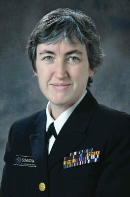
“Alcohol can permanently harm a developing baby before a woman knows she is pregnant,” Dr. Anne Schuchat, the CDC’s principal deputy director, said during a media briefing on Feb. 2. “About half of all pregnancies in the United States are unplanned, and even if planned, most women won’t know they are pregnant for the first month or so, when they might still be drinking. The risk is real. Why take the chance?”
Fetal alcohol spectrum disorders (FASD) can include physical, behavioral, and intellectual disabilities that can last for a child’s lifetime. Dr, Schuchat said the CDC estimates that as many as 1 in 20 U.S. schoolchildren may have FASD. Currently, there are no data on what amounts of alcohol are safe for a woman to drink at any stage of pregnancy.
“Not drinking alcohol is one of the best things you can do to ensure the health of your baby,” Dr. Schuchat said.
For the study, a woman was considered at risk for an alcohol-exposed pregnancy during the past month if she was nonsterile and had sex with a nonsterile male, drank any alcohol, and did not use contraception in the past month. The CDC found the weighted prevalence of alcohol-exposed pregnancy risk among U.S. women aged 15-44 years was 7.3%.

During a 1-month period, approximately 3.3 million U.S. women were at risk for an alcohol-exposed pregnancy. The highest risk group – at 10.4% – were women aged 25-29 years. The lowest risk group were those aged 15-20 years, at 2.2%.
Neither race nor ethnicity were found to be risk factors, although the risk for an alcohol-exposed pregnancy was higher among married and cohabitating women at 11.7% and 13.6% respectively, compared with their single counterparts (2.3%).
The study also found that three-quarters of women who want to get pregnant as soon as possible do not stop drinking alcohol after discontinuing contraception.
Physicians and other health care providers should advise women who want to become pregnant to stop drinking alcohol as soon as they stop using birth control, Dr. Schuchat said.
She added that physicians should screen all adults for alcohol use, not just women, even though that is not currently standard practice. “We think it should be more common to do on a regular basis.” Dr. Schuchat said the federal government requires most health plans to cover alcohol screening without cost to the patient.
The CDC recommends that physicians:
• Screen all adult female patients for alcohol use annually.
• Advise women to cease all alcohol intake if there is any chance at all that she could be pregnant.
• Counsel, refer, and follow-up with patients who need additional support to not drink while pregnant.
• Use correct billing codes to be reimbursed for screening and counseling.
The American College of Obstetricians and Gynecologists, which recommends that women completely abstain from alcohol during pregnancy, praised the CDC’s guidance that physicians routinely screen women regarding their alcohol use.
Dr. Mark S. DeFrancesco, ACOG president, said the other important message from the CDC report is that physicians should counsel women about contraception use.
“As the CDC notes, roughly half of all pregnancies in the United States are unintended. In many cases of unintended pregnancy, women inadvertently expose their fetuses to alcohol and its teratogenic effects prior to discovering that they are pregnant,” he said in statement. “This is just another reason why it’s so important that health care providers counsel women about how to prevent unintended pregnancy through use of the contraceptive method that is right for them. There are many benefits to helping women become pregnant only when they are ready, and avoiding alcohol exposure is one of them.”
On Twitter @whitneymcknight
An estimated 3.3 million U.S. women aged 15-44 years risk conceiving children with fetal alcohol spectrum disorders by using alcohol but not birth control.
The finding has officials at the Centers for Disease Control and Prevention urging physicians to screen this group for concomitant drinking and nonuse of contraception. The data come from an analysis of 4,303 nonpregnant, nonsterile women ages 15-44 years from the 2011-2013 National Survey of Family Growth, conducted by the CDC (MMWR. 2016;65:1-7.).
“Alcohol can permanently harm a developing baby before a woman knows she is pregnant,” Dr. Anne Schuchat, the CDC’s principal deputy director, said during a media briefing on Feb. 2. “About half of all pregnancies in the United States are unplanned, and even if planned, most women won’t know they are pregnant for the first month or so, when they might still be drinking. The risk is real. Why take the chance?”
Fetal alcohol spectrum disorders (FASD) can include physical, behavioral, and intellectual disabilities that can last for a child’s lifetime. Dr, Schuchat said the CDC estimates that as many as 1 in 20 U.S. schoolchildren may have FASD. Currently, there are no data on what amounts of alcohol are safe for a woman to drink at any stage of pregnancy.
“Not drinking alcohol is one of the best things you can do to ensure the health of your baby,” Dr. Schuchat said.
For the study, a woman was considered at risk for an alcohol-exposed pregnancy during the past month if she was nonsterile and had sex with a nonsterile male, drank any alcohol, and did not use contraception in the past month. The CDC found the weighted prevalence of alcohol-exposed pregnancy risk among U.S. women aged 15-44 years was 7.3%.
During a 1-month period, approximately 3.3 million U.S. women were at risk for an alcohol-exposed pregnancy. The highest risk group – at 10.4% – were women aged 25-29 years. The lowest risk group were those aged 15-20 years, at 2.2%.
Neither race nor ethnicity were found to be risk factors, although the risk for an alcohol-exposed pregnancy was higher among married and cohabitating women at 11.7% and 13.6% respectively, compared with their single counterparts (2.3%).
The study also found that three-quarters of women who want to get pregnant as soon as possible do not stop drinking alcohol after discontinuing contraception.
Physicians and other health care providers should advise women who want to become pregnant to stop drinking alcohol as soon as they stop using birth control, Dr. Schuchat said.
She added that physicians should screen all adults for alcohol use, not just women, even though that is not currently standard practice. “We think it should be more common to do on a regular basis.” Dr. Schuchat said the federal government requires most health plans to cover alcohol screening without cost to the patient.
The CDC recommends that physicians:
• Screen all adult female patients for alcohol use annually.
• Advise women to cease all alcohol intake if there is any chance at all that she could be pregnant.
• Counsel, refer, and follow-up with patients who need additional support to not drink while pregnant.
• Use correct billing codes to be reimbursed for screening and counseling.
The American College of Obstetricians and Gynecologists, which recommends that women completely abstain from alcohol during pregnancy, praised the CDC’s guidance that physicians routinely screen women regarding their alcohol use.
Dr. Mark S. DeFrancesco, ACOG president, said the other important message from the CDC report is that physicians should counsel women about contraception use.
“As the CDC notes, roughly half of all pregnancies in the United States are unintended. In many cases of unintended pregnancy, women inadvertently expose their fetuses to alcohol and its teratogenic effects prior to discovering that they are pregnant,” he said in statement. “This is just another reason why it’s so important that health care providers counsel women about how to prevent unintended pregnancy through use of the contraceptive method that is right for them. There are many benefits to helping women become pregnant only when they are ready, and avoiding alcohol exposure is one of them.”
On Twitter @whitneymcknight
An estimated 3.3 million U.S. women aged 15-44 years risk conceiving children with fetal alcohol spectrum disorders by using alcohol but not birth control.
The finding has officials at the Centers for Disease Control and Prevention urging physicians to screen this group for concomitant drinking and nonuse of contraception. The data come from an analysis of 4,303 nonpregnant, nonsterile women ages 15-44 years from the 2011-2013 National Survey of Family Growth, conducted by the CDC (MMWR. 2016;65:1-7.).
“Alcohol can permanently harm a developing baby before a woman knows she is pregnant,” Dr. Anne Schuchat, the CDC’s principal deputy director, said during a media briefing on Feb. 2. “About half of all pregnancies in the United States are unplanned, and even if planned, most women won’t know they are pregnant for the first month or so, when they might still be drinking. The risk is real. Why take the chance?”
Fetal alcohol spectrum disorders (FASD) can include physical, behavioral, and intellectual disabilities that can last for a child’s lifetime. Dr, Schuchat said the CDC estimates that as many as 1 in 20 U.S. schoolchildren may have FASD. Currently, there are no data on what amounts of alcohol are safe for a woman to drink at any stage of pregnancy.
“Not drinking alcohol is one of the best things you can do to ensure the health of your baby,” Dr. Schuchat said.
For the study, a woman was considered at risk for an alcohol-exposed pregnancy during the past month if she was nonsterile and had sex with a nonsterile male, drank any alcohol, and did not use contraception in the past month. The CDC found the weighted prevalence of alcohol-exposed pregnancy risk among U.S. women aged 15-44 years was 7.3%.
During a 1-month period, approximately 3.3 million U.S. women were at risk for an alcohol-exposed pregnancy. The highest risk group – at 10.4% – were women aged 25-29 years. The lowest risk group were those aged 15-20 years, at 2.2%.
Neither race nor ethnicity were found to be risk factors, although the risk for an alcohol-exposed pregnancy was higher among married and cohabitating women at 11.7% and 13.6% respectively, compared with their single counterparts (2.3%).
The study also found that three-quarters of women who want to get pregnant as soon as possible do not stop drinking alcohol after discontinuing contraception.
Physicians and other health care providers should advise women who want to become pregnant to stop drinking alcohol as soon as they stop using birth control, Dr. Schuchat said.
She added that physicians should screen all adults for alcohol use, not just women, even though that is not currently standard practice. “We think it should be more common to do on a regular basis.” Dr. Schuchat said the federal government requires most health plans to cover alcohol screening without cost to the patient.
The CDC recommends that physicians:
• Screen all adult female patients for alcohol use annually.
• Advise women to cease all alcohol intake if there is any chance at all that she could be pregnant.
• Counsel, refer, and follow-up with patients who need additional support to not drink while pregnant.
• Use correct billing codes to be reimbursed for screening and counseling.
The American College of Obstetricians and Gynecologists, which recommends that women completely abstain from alcohol during pregnancy, praised the CDC’s guidance that physicians routinely screen women regarding their alcohol use.
Dr. Mark S. DeFrancesco, ACOG president, said the other important message from the CDC report is that physicians should counsel women about contraception use.
“As the CDC notes, roughly half of all pregnancies in the United States are unintended. In many cases of unintended pregnancy, women inadvertently expose their fetuses to alcohol and its teratogenic effects prior to discovering that they are pregnant,” he said in statement. “This is just another reason why it’s so important that health care providers counsel women about how to prevent unintended pregnancy through use of the contraceptive method that is right for them. There are many benefits to helping women become pregnant only when they are ready, and avoiding alcohol exposure is one of them.”
On Twitter @whitneymcknight
FROM THE MMWR
Key clinical point: The CDC advises physicians to screen women for alcohol use and provide contraception counseling.
Major finding: A total of 3.3 million women aged 15-44 years risk conceiving a child with FASD by using alcohol and having unprotected sex.
Data source: Data on 4,303 nonpregnant, nonsterile women aged 15-44 years from the 2011-2013 National Survey of Family Growth.
Disclosures: The researchers did not report having any financial disclosures.
Dravet Syndrome Incidence Is Twice That of Prior Estimate
NATIONAL HARBOR, MD—The incidence of Dravet syndrome in the US is double that of a prior estimate, according to research presented at the 2015 Child Neurology Society Annual Meeting. In addition, genetic testing should be considered in children who have two or more prolonged febrile seizures by age 12 months, researchers said. “Increased awareness of this disorder could lead to earlier diagnosis and, overall, to improved seizure control,” said Yvonne Wu, MD, MPH, a child neurologist and epidemiologist at the University of California, San Francisco.

The incidence of Dravet syndrome in the US had been estimated to be about 1 in 40,000 based on the National Collaborative Perinatal Project initiated in 1959. Of the 40,000 prospective births in that study, one baby developed Dravet syndrome. More recent studies in Europe looked at the incidence of Dravet syndrome due to SCN1A mutations. Those studies found that the incidence of the epileptic encephalopathy ranged from about one in 22,000 to one in 41,000.
To determine the incidence of Dravet syndrome in the US in the current era of genetic testing, Dr. Wu and colleagues studied a retrospective, population-based cohort of all infants born at Kaiser Permanente Northern California between January 1, 2007, and June 30, 2010. The investigators performed an electronic search for patients who received at least two diagnoses of seizure by age 12 months, and who still were prescribed anticonvulsants at age 24 months. The researchers then performed a detailed chart review to identify patients who met at least four of five criteria for clinical Dravet syndrome: children were normal prior to seizure onset; had at least two seizures, febrile or afebrile, by 12 months; had myoclonic, hemiclonic, or generalized tonic-clonic seizure type; had at least two prolonged seizures (ie, that lasted more than 10 minutes); and had refractory epilepsy requiring multiple anticonvulsants after age 24 months.
The investigators excluded patients who had epilepsy due to other reasons, such as brain malformation or tuberous sclerosis, and patients who had seizures prior to age 1 month.
The study population included 125,000 births. The investigators identified eight cases of clinical Dravet syndrome, yielding an incidence of one per 15,700. All eight patients obtained SCN1A genetic sequencing as part of their routine clinical care. Seven had a gene mutation identified, and the mutation was thought to be pathogenic in six of the cases.
Researchers may have missed cases if children with Dravet syndrome presented after 12 months or left Kaiser Permanente.
“There is some evidence, and hopefully there will be more over time, that making an earlier, correct diagnosis may lead to improved seizure control due to the use of more appropriate anticonvulsant therapies,” Dr. Wu said. “For instance, lamotrigine and carbamazepine should be avoided since they may exacerbate seizures, and other medications, such as stiripentol, topiramate, and valproate, may be more efficacious.” In addition, new therapies for Dravet syndrome may be on the horizon. Cannabidiol is in phase III trials, and a large trial of fenfluramine is planned, Dr. Wu said.
—Jake Remaly
Suggested Reading
Wu YW, Sullivan J, McDaniel SS, et al. Incidence of Dravet syndrome in a US population. Pediatrics. 2015;136(5):e1310-1315.
NATIONAL HARBOR, MD—The incidence of Dravet syndrome in the US is double that of a prior estimate, according to research presented at the 2015 Child Neurology Society Annual Meeting. In addition, genetic testing should be considered in children who have two or more prolonged febrile seizures by age 12 months, researchers said. “Increased awareness of this disorder could lead to earlier diagnosis and, overall, to improved seizure control,” said Yvonne Wu, MD, MPH, a child neurologist and epidemiologist at the University of California, San Francisco.
The incidence of Dravet syndrome in the US had been estimated to be about 1 in 40,000 based on the National Collaborative Perinatal Project initiated in 1959. Of the 40,000 prospective births in that study, one baby developed Dravet syndrome. More recent studies in Europe looked at the incidence of Dravet syndrome due to SCN1A mutations. Those studies found that the incidence of the epileptic encephalopathy ranged from about one in 22,000 to one in 41,000.
To determine the incidence of Dravet syndrome in the US in the current era of genetic testing, Dr. Wu and colleagues studied a retrospective, population-based cohort of all infants born at Kaiser Permanente Northern California between January 1, 2007, and June 30, 2010. The investigators performed an electronic search for patients who received at least two diagnoses of seizure by age 12 months, and who still were prescribed anticonvulsants at age 24 months. The researchers then performed a detailed chart review to identify patients who met at least four of five criteria for clinical Dravet syndrome: children were normal prior to seizure onset; had at least two seizures, febrile or afebrile, by 12 months; had myoclonic, hemiclonic, or generalized tonic-clonic seizure type; had at least two prolonged seizures (ie, that lasted more than 10 minutes); and had refractory epilepsy requiring multiple anticonvulsants after age 24 months.
The investigators excluded patients who had epilepsy due to other reasons, such as brain malformation or tuberous sclerosis, and patients who had seizures prior to age 1 month.
The study population included 125,000 births. The investigators identified eight cases of clinical Dravet syndrome, yielding an incidence of one per 15,700. All eight patients obtained SCN1A genetic sequencing as part of their routine clinical care. Seven had a gene mutation identified, and the mutation was thought to be pathogenic in six of the cases.
Researchers may have missed cases if children with Dravet syndrome presented after 12 months or left Kaiser Permanente.
“There is some evidence, and hopefully there will be more over time, that making an earlier, correct diagnosis may lead to improved seizure control due to the use of more appropriate anticonvulsant therapies,” Dr. Wu said. “For instance, lamotrigine and carbamazepine should be avoided since they may exacerbate seizures, and other medications, such as stiripentol, topiramate, and valproate, may be more efficacious.” In addition, new therapies for Dravet syndrome may be on the horizon. Cannabidiol is in phase III trials, and a large trial of fenfluramine is planned, Dr. Wu said.
—Jake Remaly
NATIONAL HARBOR, MD—The incidence of Dravet syndrome in the US is double that of a prior estimate, according to research presented at the 2015 Child Neurology Society Annual Meeting. In addition, genetic testing should be considered in children who have two or more prolonged febrile seizures by age 12 months, researchers said. “Increased awareness of this disorder could lead to earlier diagnosis and, overall, to improved seizure control,” said Yvonne Wu, MD, MPH, a child neurologist and epidemiologist at the University of California, San Francisco.
The incidence of Dravet syndrome in the US had been estimated to be about 1 in 40,000 based on the National Collaborative Perinatal Project initiated in 1959. Of the 40,000 prospective births in that study, one baby developed Dravet syndrome. More recent studies in Europe looked at the incidence of Dravet syndrome due to SCN1A mutations. Those studies found that the incidence of the epileptic encephalopathy ranged from about one in 22,000 to one in 41,000.
To determine the incidence of Dravet syndrome in the US in the current era of genetic testing, Dr. Wu and colleagues studied a retrospective, population-based cohort of all infants born at Kaiser Permanente Northern California between January 1, 2007, and June 30, 2010. The investigators performed an electronic search for patients who received at least two diagnoses of seizure by age 12 months, and who still were prescribed anticonvulsants at age 24 months. The researchers then performed a detailed chart review to identify patients who met at least four of five criteria for clinical Dravet syndrome: children were normal prior to seizure onset; had at least two seizures, febrile or afebrile, by 12 months; had myoclonic, hemiclonic, or generalized tonic-clonic seizure type; had at least two prolonged seizures (ie, that lasted more than 10 minutes); and had refractory epilepsy requiring multiple anticonvulsants after age 24 months.
The investigators excluded patients who had epilepsy due to other reasons, such as brain malformation or tuberous sclerosis, and patients who had seizures prior to age 1 month.
The study population included 125,000 births. The investigators identified eight cases of clinical Dravet syndrome, yielding an incidence of one per 15,700. All eight patients obtained SCN1A genetic sequencing as part of their routine clinical care. Seven had a gene mutation identified, and the mutation was thought to be pathogenic in six of the cases.
Researchers may have missed cases if children with Dravet syndrome presented after 12 months or left Kaiser Permanente.
“There is some evidence, and hopefully there will be more over time, that making an earlier, correct diagnosis may lead to improved seizure control due to the use of more appropriate anticonvulsant therapies,” Dr. Wu said. “For instance, lamotrigine and carbamazepine should be avoided since they may exacerbate seizures, and other medications, such as stiripentol, topiramate, and valproate, may be more efficacious.” In addition, new therapies for Dravet syndrome may be on the horizon. Cannabidiol is in phase III trials, and a large trial of fenfluramine is planned, Dr. Wu said.
—Jake Remaly
Suggested Reading
Wu YW, Sullivan J, McDaniel SS, et al. Incidence of Dravet syndrome in a US population. Pediatrics. 2015;136(5):e1310-1315.
Suggested Reading
Wu YW, Sullivan J, McDaniel SS, et al. Incidence of Dravet syndrome in a US population. Pediatrics. 2015;136(5):e1310-1315.

Growing ADHD drug selection aids individualized therapy
NEW YORK – One way to individualize selection from the growing list of long-acting stimulants for attention-deficit/hyperactivity disorder is to consider when each provides peak activity, according to an update on these medications at a psychopharmacology update held by the American Academy of Child and Adolescent Psychiatry.
If the immediate-release component of a given long-acting stimulant formation is relatively high, “that is a particularly good medication for early-morning problems, such as a math class in the first period,” said Dr. Laurence L. Greenhill, professor in the division of child and adolescent psychiatry at Columbia University in New York. “On the other hand, if the math class is in the eighth period and there are a lot of behavioral problems, then those agents with less immediate release may be a better choice.”

The list of long-acting stimulants, each with a unique formulation and pattern of stimulant release, has grown substantially over the last 2 years with two new agents released within the last 4 months, according to Dr. Greenhill. The latest regulatory approval in this class was for Adzenys XR-ODT (amphetamine), which is being marketed as the first extended-release orally disintegrating tablet for ADHD. FDA approval was granted on Jan. 28, which was the day before Dr. Greenhill spoke and just 3 months after approval of Dynavel XR, a long-acting amphetamine oral suspension.
All of the long-acting stimulants employ amphetamine, methylphenidate, or dexmethylphenidate as their active agent, but they employ different strategies to provide active drug over the 8-12 hours most claim as their duration of activity. Oral therapies, which can now be delivered in capsule, chewable, and liquid formulations, employ a large array of technologies to slow release of the active ingredient over each dosing interval. Skin patches provide yet another option for drug delivery.
By themselves, the differences in formulations may have their own relevance to drug selection. For example, liquid drugs can be sprinkled on food, while a patch may be the best option for children with difficulty swallowing pills. But Dr. Greenhill suggested that patterns of drug release could be important for individualizing drug selection according to the time of day that symptoms peak. For some, such as dexmethylphenidate XR (Focalin XR), up to 50% of the active ingredient is available in the immediate-release component with the remaining drug provided in delayed release. For others, such as methylphenidate XR (Concerta), only 20% is immediate release. These have obvious implications if timing of symptom control is important.
Because of the potential complications from stimulant drugs, Dr. Greenhill advocated employing these agents within the framework of published guidelines, including one from the AACAP, specific to the management of ADHD in children. Of specific risks, cardiac events are widely considered the most worrisome. According to Dr. Greenhill, a baseline history should include a focus on any cardiac abnormalities or syncope episodes. Although he said that routine electrocardiograms are not necessary, the patient’s pulse and blood pressure should be rechecked at least every 6 months. Referral to a cardiologist is appropriate for persistent abnormalities in elevated blood pressure or in patients with tachycardia.
New evidence, however, has refuted the previously reported association between long-acting stimulants and tics, Dr. Greenhill said. He cited a recently published meta-analysis of 22 randomized, placebo-controlled trials that found no significant no association between psychostimulant use and either new onset or worsening of tics (J Am Acad Child Adolesc Psychiatry. 2015 Sep;54[9]:728-36).
“The rate of worsening of new-onset tics in the stimulant group was, in fact, less than the rate of tics in the placebo group,” reported Dr. Greenhill, who characterized this as a take-home clinical pearl. “The bottom line is that practitioners can tell parents that there is no evidence that these medications will worsen tics.”
Nevertheless, to avoid the appearance of a temporal association between stimulants and tics, Dr. Greenhill suggested that children who discontinued stimulant therapy because of a tic should be rechallenged at a time of low stress.
Overall, ADHD patients without an adequate response to one stimulant should be tried on another, and these therapies can be combined with adjunctive drugs and behavioral treatments. But Dr. Greenhill suggested matching drug release patterns to the timing of ADHD symptoms is one way to guide the initial choice of long-acting agent.
Dr. Greenhill reported financial relationship with Hoffmann-LaRoche and Shire.
NEW YORK – One way to individualize selection from the growing list of long-acting stimulants for attention-deficit/hyperactivity disorder is to consider when each provides peak activity, according to an update on these medications at a psychopharmacology update held by the American Academy of Child and Adolescent Psychiatry.
If the immediate-release component of a given long-acting stimulant formation is relatively high, “that is a particularly good medication for early-morning problems, such as a math class in the first period,” said Dr. Laurence L. Greenhill, professor in the division of child and adolescent psychiatry at Columbia University in New York. “On the other hand, if the math class is in the eighth period and there are a lot of behavioral problems, then those agents with less immediate release may be a better choice.”
The list of long-acting stimulants, each with a unique formulation and pattern of stimulant release, has grown substantially over the last 2 years with two new agents released within the last 4 months, according to Dr. Greenhill. The latest regulatory approval in this class was for Adzenys XR-ODT (amphetamine), which is being marketed as the first extended-release orally disintegrating tablet for ADHD. FDA approval was granted on Jan. 28, which was the day before Dr. Greenhill spoke and just 3 months after approval of Dynavel XR, a long-acting amphetamine oral suspension.
All of the long-acting stimulants employ amphetamine, methylphenidate, or dexmethylphenidate as their active agent, but they employ different strategies to provide active drug over the 8-12 hours most claim as their duration of activity. Oral therapies, which can now be delivered in capsule, chewable, and liquid formulations, employ a large array of technologies to slow release of the active ingredient over each dosing interval. Skin patches provide yet another option for drug delivery.
By themselves, the differences in formulations may have their own relevance to drug selection. For example, liquid drugs can be sprinkled on food, while a patch may be the best option for children with difficulty swallowing pills. But Dr. Greenhill suggested that patterns of drug release could be important for individualizing drug selection according to the time of day that symptoms peak. For some, such as dexmethylphenidate XR (Focalin XR), up to 50% of the active ingredient is available in the immediate-release component with the remaining drug provided in delayed release. For others, such as methylphenidate XR (Concerta), only 20% is immediate release. These have obvious implications if timing of symptom control is important.
Because of the potential complications from stimulant drugs, Dr. Greenhill advocated employing these agents within the framework of published guidelines, including one from the AACAP, specific to the management of ADHD in children. Of specific risks, cardiac events are widely considered the most worrisome. According to Dr. Greenhill, a baseline history should include a focus on any cardiac abnormalities or syncope episodes. Although he said that routine electrocardiograms are not necessary, the patient’s pulse and blood pressure should be rechecked at least every 6 months. Referral to a cardiologist is appropriate for persistent abnormalities in elevated blood pressure or in patients with tachycardia.
New evidence, however, has refuted the previously reported association between long-acting stimulants and tics, Dr. Greenhill said. He cited a recently published meta-analysis of 22 randomized, placebo-controlled trials that found no significant no association between psychostimulant use and either new onset or worsening of tics (J Am Acad Child Adolesc Psychiatry. 2015 Sep;54[9]:728-36).
“The rate of worsening of new-onset tics in the stimulant group was, in fact, less than the rate of tics in the placebo group,” reported Dr. Greenhill, who characterized this as a take-home clinical pearl. “The bottom line is that practitioners can tell parents that there is no evidence that these medications will worsen tics.”
Nevertheless, to avoid the appearance of a temporal association between stimulants and tics, Dr. Greenhill suggested that children who discontinued stimulant therapy because of a tic should be rechallenged at a time of low stress.
Overall, ADHD patients without an adequate response to one stimulant should be tried on another, and these therapies can be combined with adjunctive drugs and behavioral treatments. But Dr. Greenhill suggested matching drug release patterns to the timing of ADHD symptoms is one way to guide the initial choice of long-acting agent.
Dr. Greenhill reported financial relationship with Hoffmann-LaRoche and Shire.
NEW YORK – One way to individualize selection from the growing list of long-acting stimulants for attention-deficit/hyperactivity disorder is to consider when each provides peak activity, according to an update on these medications at a psychopharmacology update held by the American Academy of Child and Adolescent Psychiatry.
If the immediate-release component of a given long-acting stimulant formation is relatively high, “that is a particularly good medication for early-morning problems, such as a math class in the first period,” said Dr. Laurence L. Greenhill, professor in the division of child and adolescent psychiatry at Columbia University in New York. “On the other hand, if the math class is in the eighth period and there are a lot of behavioral problems, then those agents with less immediate release may be a better choice.”
The list of long-acting stimulants, each with a unique formulation and pattern of stimulant release, has grown substantially over the last 2 years with two new agents released within the last 4 months, according to Dr. Greenhill. The latest regulatory approval in this class was for Adzenys XR-ODT (amphetamine), which is being marketed as the first extended-release orally disintegrating tablet for ADHD. FDA approval was granted on Jan. 28, which was the day before Dr. Greenhill spoke and just 3 months after approval of Dynavel XR, a long-acting amphetamine oral suspension.
All of the long-acting stimulants employ amphetamine, methylphenidate, or dexmethylphenidate as their active agent, but they employ different strategies to provide active drug over the 8-12 hours most claim as their duration of activity. Oral therapies, which can now be delivered in capsule, chewable, and liquid formulations, employ a large array of technologies to slow release of the active ingredient over each dosing interval. Skin patches provide yet another option for drug delivery.
By themselves, the differences in formulations may have their own relevance to drug selection. For example, liquid drugs can be sprinkled on food, while a patch may be the best option for children with difficulty swallowing pills. But Dr. Greenhill suggested that patterns of drug release could be important for individualizing drug selection according to the time of day that symptoms peak. For some, such as dexmethylphenidate XR (Focalin XR), up to 50% of the active ingredient is available in the immediate-release component with the remaining drug provided in delayed release. For others, such as methylphenidate XR (Concerta), only 20% is immediate release. These have obvious implications if timing of symptom control is important.
Because of the potential complications from stimulant drugs, Dr. Greenhill advocated employing these agents within the framework of published guidelines, including one from the AACAP, specific to the management of ADHD in children. Of specific risks, cardiac events are widely considered the most worrisome. According to Dr. Greenhill, a baseline history should include a focus on any cardiac abnormalities or syncope episodes. Although he said that routine electrocardiograms are not necessary, the patient’s pulse and blood pressure should be rechecked at least every 6 months. Referral to a cardiologist is appropriate for persistent abnormalities in elevated blood pressure or in patients with tachycardia.
New evidence, however, has refuted the previously reported association between long-acting stimulants and tics, Dr. Greenhill said. He cited a recently published meta-analysis of 22 randomized, placebo-controlled trials that found no significant no association between psychostimulant use and either new onset or worsening of tics (J Am Acad Child Adolesc Psychiatry. 2015 Sep;54[9]:728-36).
“The rate of worsening of new-onset tics in the stimulant group was, in fact, less than the rate of tics in the placebo group,” reported Dr. Greenhill, who characterized this as a take-home clinical pearl. “The bottom line is that practitioners can tell parents that there is no evidence that these medications will worsen tics.”
Nevertheless, to avoid the appearance of a temporal association between stimulants and tics, Dr. Greenhill suggested that children who discontinued stimulant therapy because of a tic should be rechallenged at a time of low stress.
Overall, ADHD patients without an adequate response to one stimulant should be tried on another, and these therapies can be combined with adjunctive drugs and behavioral treatments. But Dr. Greenhill suggested matching drug release patterns to the timing of ADHD symptoms is one way to guide the initial choice of long-acting agent.
Dr. Greenhill reported financial relationship with Hoffmann-LaRoche and Shire.
EXPERT ANALYSIS FROM THE PSYCHOPHARMACOLOGY UPDATE INSTITUTE

WHO Declares "Public Health Emergency" for Microcephaly Linked to Zika Virus
The World Health Organization has declared a “public health emergency of international concern” related to the clusters of microcephaly and other neurological complications reported in Brazil and earlier in French Polynesia.

Though there is a strong association between these cases and the Zika virus, a causal link still has not been scientifically proven, according to the WHO.
The WHO’s emergency declaration clears the way for the international health community to move forward with a coordinated response. Dr. Margaret Chan, WHO Director-General, said her organization plans to take a number of precautionary measures, including improving surveillance and detection of infections, congenital malformations, and neurological complications. They will also work with countries to intensify control of mosquito populations and help expedite the development of diagnostic tests and vaccines to protect at-risk populations.
The recommendations came after a Feb. 1 meeting of the International Health Regulations Emergency Committee, which Dr. Chan convened last week in response to the Zika virus outbreak and the observed increase in neurological disorders and neonatal malformations.
The group of 18 experts advised that the clusters of microcephaly and other complications constitute an “extraordinary event and a public health threat to other parts of the world.” The group did not recommend any restrictions on travel or trade with areas where the Zika virus transmission is ongoing, however.
“At present, the most important protective measures are the control of mosquito populations and the prevention of mosquito bites in at-risk individuals, especially pregnant women,” Dr. Chan said during a press briefing.
Dr. Chan said it’s unclear how long it will take to determine if Zika virus is causing the uptick in microcephaly and other congenital malformations and neurological abnormalities, but health officials are working to set up case-control studies that are scheduled to start in the next 2 weeks.
The World Health Organization has declared a “public health emergency of international concern” related to the clusters of microcephaly and other neurological complications reported in Brazil and earlier in French Polynesia.
Though there is a strong association between these cases and the Zika virus, a causal link still has not been scientifically proven, according to the WHO.
The WHO’s emergency declaration clears the way for the international health community to move forward with a coordinated response. Dr. Margaret Chan, WHO Director-General, said her organization plans to take a number of precautionary measures, including improving surveillance and detection of infections, congenital malformations, and neurological complications. They will also work with countries to intensify control of mosquito populations and help expedite the development of diagnostic tests and vaccines to protect at-risk populations.
The recommendations came after a Feb. 1 meeting of the International Health Regulations Emergency Committee, which Dr. Chan convened last week in response to the Zika virus outbreak and the observed increase in neurological disorders and neonatal malformations.
The group of 18 experts advised that the clusters of microcephaly and other complications constitute an “extraordinary event and a public health threat to other parts of the world.” The group did not recommend any restrictions on travel or trade with areas where the Zika virus transmission is ongoing, however.
“At present, the most important protective measures are the control of mosquito populations and the prevention of mosquito bites in at-risk individuals, especially pregnant women,” Dr. Chan said during a press briefing.
Dr. Chan said it’s unclear how long it will take to determine if Zika virus is causing the uptick in microcephaly and other congenital malformations and neurological abnormalities, but health officials are working to set up case-control studies that are scheduled to start in the next 2 weeks.
The World Health Organization has declared a “public health emergency of international concern” related to the clusters of microcephaly and other neurological complications reported in Brazil and earlier in French Polynesia.
Though there is a strong association between these cases and the Zika virus, a causal link still has not been scientifically proven, according to the WHO.
The WHO’s emergency declaration clears the way for the international health community to move forward with a coordinated response. Dr. Margaret Chan, WHO Director-General, said her organization plans to take a number of precautionary measures, including improving surveillance and detection of infections, congenital malformations, and neurological complications. They will also work with countries to intensify control of mosquito populations and help expedite the development of diagnostic tests and vaccines to protect at-risk populations.
The recommendations came after a Feb. 1 meeting of the International Health Regulations Emergency Committee, which Dr. Chan convened last week in response to the Zika virus outbreak and the observed increase in neurological disorders and neonatal malformations.
The group of 18 experts advised that the clusters of microcephaly and other complications constitute an “extraordinary event and a public health threat to other parts of the world.” The group did not recommend any restrictions on travel or trade with areas where the Zika virus transmission is ongoing, however.
“At present, the most important protective measures are the control of mosquito populations and the prevention of mosquito bites in at-risk individuals, especially pregnant women,” Dr. Chan said during a press briefing.
Dr. Chan said it’s unclear how long it will take to determine if Zika virus is causing the uptick in microcephaly and other congenital malformations and neurological abnormalities, but health officials are working to set up case-control studies that are scheduled to start in the next 2 weeks.

WHO declares ‘public health emergency’ for microcephaly linked to Zika virus
The World Health Organization has declared a “public health emergency of international concern” related to the clusters of microcephaly and other neurological complications reported in Brazil and earlier in French Polynesia.
Though there is a strong association between these cases and the Zika virus, a causal link still has not been scientifically proven, according to the WHO.
The WHO’s emergency declaration clears the way for the international health community to move forward with a coordinated response. Dr. Margaret Chan, WHO Director-General, said her organization plans to take a number of precautionary measures, including improving surveillance and detection of infections, congenital malformations, and neurological complications. They will also work with countries to intensify control of mosquito populations and help expedite the development of diagnostic tests and vaccines to protect at-risk populations.
The recommendations came after a Feb. 1 meeting of the International Health Regulations Emergency Committee, which Dr. Chan convened last week in response to the Zika virus outbreak and the observed increase in neurological disorders and neonatal malformations.
The group of 18 experts advised that the clusters of microcephaly and other complications constitute an “extraordinary event and a public health threat to other parts of the world.” The group did not recommend any restrictions on travel or trade with areas where the Zika virus transmission is ongoing, however.
“At present, the most important protective measures are the control of mosquito populations and the prevention of mosquito bites in at-risk individuals, especially pregnant women,” Dr. Chan said during a press briefing.
Dr. Chan said it’s unclear how long it will take to determine if Zika virus is causing the uptick in microcephaly and other congenital malformations and neurological abnormalities, but health officials are working to set up case-control studies that are scheduled to start in the next 2 weeks.
mschneider@frontlinemedcom.com
On Twitter @maryellenny
The World Health Organization has declared a “public health emergency of international concern” related to the clusters of microcephaly and other neurological complications reported in Brazil and earlier in French Polynesia.
Though there is a strong association between these cases and the Zika virus, a causal link still has not been scientifically proven, according to the WHO.
The WHO’s emergency declaration clears the way for the international health community to move forward with a coordinated response. Dr. Margaret Chan, WHO Director-General, said her organization plans to take a number of precautionary measures, including improving surveillance and detection of infections, congenital malformations, and neurological complications. They will also work with countries to intensify control of mosquito populations and help expedite the development of diagnostic tests and vaccines to protect at-risk populations.
The recommendations came after a Feb. 1 meeting of the International Health Regulations Emergency Committee, which Dr. Chan convened last week in response to the Zika virus outbreak and the observed increase in neurological disorders and neonatal malformations.
The group of 18 experts advised that the clusters of microcephaly and other complications constitute an “extraordinary event and a public health threat to other parts of the world.” The group did not recommend any restrictions on travel or trade with areas where the Zika virus transmission is ongoing, however.
“At present, the most important protective measures are the control of mosquito populations and the prevention of mosquito bites in at-risk individuals, especially pregnant women,” Dr. Chan said during a press briefing.
Dr. Chan said it’s unclear how long it will take to determine if Zika virus is causing the uptick in microcephaly and other congenital malformations and neurological abnormalities, but health officials are working to set up case-control studies that are scheduled to start in the next 2 weeks.
mschneider@frontlinemedcom.com
On Twitter @maryellenny
The World Health Organization has declared a “public health emergency of international concern” related to the clusters of microcephaly and other neurological complications reported in Brazil and earlier in French Polynesia.
Though there is a strong association between these cases and the Zika virus, a causal link still has not been scientifically proven, according to the WHO.
The WHO’s emergency declaration clears the way for the international health community to move forward with a coordinated response. Dr. Margaret Chan, WHO Director-General, said her organization plans to take a number of precautionary measures, including improving surveillance and detection of infections, congenital malformations, and neurological complications. They will also work with countries to intensify control of mosquito populations and help expedite the development of diagnostic tests and vaccines to protect at-risk populations.
The recommendations came after a Feb. 1 meeting of the International Health Regulations Emergency Committee, which Dr. Chan convened last week in response to the Zika virus outbreak and the observed increase in neurological disorders and neonatal malformations.
The group of 18 experts advised that the clusters of microcephaly and other complications constitute an “extraordinary event and a public health threat to other parts of the world.” The group did not recommend any restrictions on travel or trade with areas where the Zika virus transmission is ongoing, however.
“At present, the most important protective measures are the control of mosquito populations and the prevention of mosquito bites in at-risk individuals, especially pregnant women,” Dr. Chan said during a press briefing.
Dr. Chan said it’s unclear how long it will take to determine if Zika virus is causing the uptick in microcephaly and other congenital malformations and neurological abnormalities, but health officials are working to set up case-control studies that are scheduled to start in the next 2 weeks.
mschneider@frontlinemedcom.com
On Twitter @maryellenny
No increased death risk from vaccination in adolescents, young adults
Risk of death is not increased in people aged 9-26 years old in the 30 days after receiving any vaccination, according to Natalie L. McCarthy of the Centers for Disease Control and Prevention and her associates.
Among nearly 2.2 million enrollees of the Vaccine Safety Database, 1,100 deaths occurred within 12 months after vaccination, and 76 (7%) occurred within 30 days of vaccination. Of these deaths, 59 had medical records which could be confirmed, with 33 due to external causes such as suicide and accidents, 1 due to an unknown cause, and 25 due to non-external causes such as neoplasm, circulatory disease, and respiratory disease.

The relative risk for influenza vaccination and any vaccination due to non-external sources was 0.44 and 0.57, and the relative risk for the influenza vaccine and all vaccines due to all causes of death was 0.42 and 0.72. The relative risk for the 4-valent human papillomavirus (4vHPV) vaccine from non-external sources was 1.28, and 1.12 from all sources. No vaccine had a significant association with death.
“This research should reassure the public with regard to the safety of 4vHPV vaccine, as well as other vaccines routinely administered to individuals 9 to 26 years of age,” the investigators concluded.
Find the full study in Pediatrics (2016;137[3]:e20152970).
Risk of death is not increased in people aged 9-26 years old in the 30 days after receiving any vaccination, according to Natalie L. McCarthy of the Centers for Disease Control and Prevention and her associates.
Among nearly 2.2 million enrollees of the Vaccine Safety Database, 1,100 deaths occurred within 12 months after vaccination, and 76 (7%) occurred within 30 days of vaccination. Of these deaths, 59 had medical records which could be confirmed, with 33 due to external causes such as suicide and accidents, 1 due to an unknown cause, and 25 due to non-external causes such as neoplasm, circulatory disease, and respiratory disease.
The relative risk for influenza vaccination and any vaccination due to non-external sources was 0.44 and 0.57, and the relative risk for the influenza vaccine and all vaccines due to all causes of death was 0.42 and 0.72. The relative risk for the 4-valent human papillomavirus (4vHPV) vaccine from non-external sources was 1.28, and 1.12 from all sources. No vaccine had a significant association with death.
“This research should reassure the public with regard to the safety of 4vHPV vaccine, as well as other vaccines routinely administered to individuals 9 to 26 years of age,” the investigators concluded.
Find the full study in Pediatrics (2016;137[3]:e20152970).
Risk of death is not increased in people aged 9-26 years old in the 30 days after receiving any vaccination, according to Natalie L. McCarthy of the Centers for Disease Control and Prevention and her associates.
Among nearly 2.2 million enrollees of the Vaccine Safety Database, 1,100 deaths occurred within 12 months after vaccination, and 76 (7%) occurred within 30 days of vaccination. Of these deaths, 59 had medical records which could be confirmed, with 33 due to external causes such as suicide and accidents, 1 due to an unknown cause, and 25 due to non-external causes such as neoplasm, circulatory disease, and respiratory disease.
The relative risk for influenza vaccination and any vaccination due to non-external sources was 0.44 and 0.57, and the relative risk for the influenza vaccine and all vaccines due to all causes of death was 0.42 and 0.72. The relative risk for the 4-valent human papillomavirus (4vHPV) vaccine from non-external sources was 1.28, and 1.12 from all sources. No vaccine had a significant association with death.
“This research should reassure the public with regard to the safety of 4vHPV vaccine, as well as other vaccines routinely administered to individuals 9 to 26 years of age,” the investigators concluded.
Find the full study in Pediatrics (2016;137[3]:e20152970).
FROM PEDIATRICS

Appendicitis, antibiotics, and surgery: An evolving trilogy
Appendicitis is the most common surgical emergency in children. It is seen at all ages; however, it is less common in infants and toddlers younger than 4 years of age and peaks at an incidence of 25/100,000 in children 12- to 18-years-old. Fortunately, appendicitis is rarely fatal but can be associated with significant morbidity, especially in young children in whom the diagnosis is often delayed and perforation is more common. Reducing morbidity requires early diagnosis and optimizing management such that perforation and associated peritonitis are prevented.
The classical signs and symptoms of appendicitis are periumbilical pain migrating to the right lower quadrant, nausea, and low-grade fever. Presentation may vary if the location of the appendix is atypical, but primarily is age associated. In young children, abdominal distension, hip pain with or without limp, and fever are commonplace. In older children, right lower quadrant abdominal pain that intensifies with coughing or movement is frequent. Localized tenderness also appears to be age related; right lower quadrant tenderness and rebound are more often found in older children and adolescents, whereas younger children have more diffuse signs.

When I started my career, abdominal x-rays would be performed in search of a fecalith. However, such studies were of low sensitivity, and clinical acumen had a primary role in the decision to take the child to the operating room. In the current era, ultrasound and CT scan provide reasonable sensitivity and specificity. Ultrasound criteria include a diameter greater than 6 mm, concentric rings (target sign), an appendicolith, high echogenicity, obstruction of the lumen, and fluid surrounding the appendix.
As the pathogenesis of appendicitis represents occlusion of the appendiceal lumen, followed by overgrowth or translocation of bowel flora resulting in inflammation of the wall of the appendix, anaerobes and gram-negative gut flora represent the most important pathogens. In advanced cases, necrosis and gangrene of the appendix result with progression to rupture and peritonitis.
The traditional management was early surgical intervention to reduce the risk of perforation and peritonitis with acceptance of high rates of negative abdominal explorations as an acceptable consequence. Today, the approach to management of appendicitis is undergoing reevaluation. Early antimicrobial treatment has become routine in the management of nonperforated, perforated, or abscessed appendicitis. However, the question being asked is, “Do all children with uncomplicated appendicitis need appendectomy, or is antibiotic management sufficient?”
P. Salminen et al. reported on the results of a randomized clinical trial in 530 patients aged 18-60 years, comparing antimicrobial treatment alone with early appendectomy. Among 273 patients in the surgical group, all but 1 underwent successful appendectomy, resulting in a success rate of 99.6% (95% CI, 98.0%-100.0%). In the antibiotic group, 186 of 256 patients (70%) treated with antibiotics did not require surgery; 70 (27%) underwent appendectomy within 1 year of initial presentation for appendicitis (JAMA. 2015 Jun 16;313[23]:2340-8). There were no intraabdominal abscesses or other major complications associated with delayed appendectomy in patients randomized to antibiotic treatment. The authors concluded that among patients with CT-proven, uncomplicated appendicitis, antibiotic treatment did not meet the prespecified criterion for noninferiority, compared with appendectomy. However, most patients randomized to antibiotics for uncomplicated appendicitis did not require appendectomy during the 1-year follow-up period.
J.A. Horst et al. reviewed published reports of medical management of appendicitis in children (Ann Emerg Med. 2015 Aug;66[2]:119-22). They concluded that medical management of uncomplicated appendicitis in a select low-risk pediatric population is safe and does not result in significant morbidity. The arguments against a nonoperative approach include the risk of recurrent appendicitis, including the anxiety associated with any recurrences of abdominal pain, the risk of antibiotic-related complications, the potential for increased duration of hospitalization, and the relatively low morbidity of appendectomy in children. Factors associated with failed antibiotic management included fecaliths, fluid collections, or an appendiceal diameter greater than 1.1 cm on CT scan. The investigators concluded such children are poor candidates for nonsurgical management.
The bottom line is that antimicrobial therapy, in the absence of surgery, can be effective. Certainly in remote settings where surgery is not readily available, antimicrobial therapy with fluid and electrolyte management and close observation can be used in children with uncomplicated appendicitis with few failures and relatively few children requiring subsequent appendectomy. In more complicated cases with evidence of fecalith, or appendiceal abscess or phlegm, initial antimicrobial therapy reduces the acute inflammation and urgent need for surgery, but persistent inflammation of the appendix is often observed and appendectomy, either acutely or after improvement following antimicrobial therapy, appears indicated. Many different antimicrobial regimens have proven effective; ceftriaxone and metronidazole are associated with low rates of complications, offer an opportunity for once-daily therapy, and are cost effective, compared with other once-daily regimens.
Dr. Pelton is chief of pediatric infectious disease and coordinator of the maternal-child HIV program at Boston Medical Center.
Appendicitis is the most common surgical emergency in children. It is seen at all ages; however, it is less common in infants and toddlers younger than 4 years of age and peaks at an incidence of 25/100,000 in children 12- to 18-years-old. Fortunately, appendicitis is rarely fatal but can be associated with significant morbidity, especially in young children in whom the diagnosis is often delayed and perforation is more common. Reducing morbidity requires early diagnosis and optimizing management such that perforation and associated peritonitis are prevented.
The classical signs and symptoms of appendicitis are periumbilical pain migrating to the right lower quadrant, nausea, and low-grade fever. Presentation may vary if the location of the appendix is atypical, but primarily is age associated. In young children, abdominal distension, hip pain with or without limp, and fever are commonplace. In older children, right lower quadrant abdominal pain that intensifies with coughing or movement is frequent. Localized tenderness also appears to be age related; right lower quadrant tenderness and rebound are more often found in older children and adolescents, whereas younger children have more diffuse signs.
When I started my career, abdominal x-rays would be performed in search of a fecalith. However, such studies were of low sensitivity, and clinical acumen had a primary role in the decision to take the child to the operating room. In the current era, ultrasound and CT scan provide reasonable sensitivity and specificity. Ultrasound criteria include a diameter greater than 6 mm, concentric rings (target sign), an appendicolith, high echogenicity, obstruction of the lumen, and fluid surrounding the appendix.
As the pathogenesis of appendicitis represents occlusion of the appendiceal lumen, followed by overgrowth or translocation of bowel flora resulting in inflammation of the wall of the appendix, anaerobes and gram-negative gut flora represent the most important pathogens. In advanced cases, necrosis and gangrene of the appendix result with progression to rupture and peritonitis.
The traditional management was early surgical intervention to reduce the risk of perforation and peritonitis with acceptance of high rates of negative abdominal explorations as an acceptable consequence. Today, the approach to management of appendicitis is undergoing reevaluation. Early antimicrobial treatment has become routine in the management of nonperforated, perforated, or abscessed appendicitis. However, the question being asked is, “Do all children with uncomplicated appendicitis need appendectomy, or is antibiotic management sufficient?”
P. Salminen et al. reported on the results of a randomized clinical trial in 530 patients aged 18-60 years, comparing antimicrobial treatment alone with early appendectomy. Among 273 patients in the surgical group, all but 1 underwent successful appendectomy, resulting in a success rate of 99.6% (95% CI, 98.0%-100.0%). In the antibiotic group, 186 of 256 patients (70%) treated with antibiotics did not require surgery; 70 (27%) underwent appendectomy within 1 year of initial presentation for appendicitis (JAMA. 2015 Jun 16;313[23]:2340-8). There were no intraabdominal abscesses or other major complications associated with delayed appendectomy in patients randomized to antibiotic treatment. The authors concluded that among patients with CT-proven, uncomplicated appendicitis, antibiotic treatment did not meet the prespecified criterion for noninferiority, compared with appendectomy. However, most patients randomized to antibiotics for uncomplicated appendicitis did not require appendectomy during the 1-year follow-up period.
J.A. Horst et al. reviewed published reports of medical management of appendicitis in children (Ann Emerg Med. 2015 Aug;66[2]:119-22). They concluded that medical management of uncomplicated appendicitis in a select low-risk pediatric population is safe and does not result in significant morbidity. The arguments against a nonoperative approach include the risk of recurrent appendicitis, including the anxiety associated with any recurrences of abdominal pain, the risk of antibiotic-related complications, the potential for increased duration of hospitalization, and the relatively low morbidity of appendectomy in children. Factors associated with failed antibiotic management included fecaliths, fluid collections, or an appendiceal diameter greater than 1.1 cm on CT scan. The investigators concluded such children are poor candidates for nonsurgical management.
The bottom line is that antimicrobial therapy, in the absence of surgery, can be effective. Certainly in remote settings where surgery is not readily available, antimicrobial therapy with fluid and electrolyte management and close observation can be used in children with uncomplicated appendicitis with few failures and relatively few children requiring subsequent appendectomy. In more complicated cases with evidence of fecalith, or appendiceal abscess or phlegm, initial antimicrobial therapy reduces the acute inflammation and urgent need for surgery, but persistent inflammation of the appendix is often observed and appendectomy, either acutely or after improvement following antimicrobial therapy, appears indicated. Many different antimicrobial regimens have proven effective; ceftriaxone and metronidazole are associated with low rates of complications, offer an opportunity for once-daily therapy, and are cost effective, compared with other once-daily regimens.
Dr. Pelton is chief of pediatric infectious disease and coordinator of the maternal-child HIV program at Boston Medical Center.
Appendicitis is the most common surgical emergency in children. It is seen at all ages; however, it is less common in infants and toddlers younger than 4 years of age and peaks at an incidence of 25/100,000 in children 12- to 18-years-old. Fortunately, appendicitis is rarely fatal but can be associated with significant morbidity, especially in young children in whom the diagnosis is often delayed and perforation is more common. Reducing morbidity requires early diagnosis and optimizing management such that perforation and associated peritonitis are prevented.
The classical signs and symptoms of appendicitis are periumbilical pain migrating to the right lower quadrant, nausea, and low-grade fever. Presentation may vary if the location of the appendix is atypical, but primarily is age associated. In young children, abdominal distension, hip pain with or without limp, and fever are commonplace. In older children, right lower quadrant abdominal pain that intensifies with coughing or movement is frequent. Localized tenderness also appears to be age related; right lower quadrant tenderness and rebound are more often found in older children and adolescents, whereas younger children have more diffuse signs.
When I started my career, abdominal x-rays would be performed in search of a fecalith. However, such studies were of low sensitivity, and clinical acumen had a primary role in the decision to take the child to the operating room. In the current era, ultrasound and CT scan provide reasonable sensitivity and specificity. Ultrasound criteria include a diameter greater than 6 mm, concentric rings (target sign), an appendicolith, high echogenicity, obstruction of the lumen, and fluid surrounding the appendix.
As the pathogenesis of appendicitis represents occlusion of the appendiceal lumen, followed by overgrowth or translocation of bowel flora resulting in inflammation of the wall of the appendix, anaerobes and gram-negative gut flora represent the most important pathogens. In advanced cases, necrosis and gangrene of the appendix result with progression to rupture and peritonitis.
The traditional management was early surgical intervention to reduce the risk of perforation and peritonitis with acceptance of high rates of negative abdominal explorations as an acceptable consequence. Today, the approach to management of appendicitis is undergoing reevaluation. Early antimicrobial treatment has become routine in the management of nonperforated, perforated, or abscessed appendicitis. However, the question being asked is, “Do all children with uncomplicated appendicitis need appendectomy, or is antibiotic management sufficient?”
P. Salminen et al. reported on the results of a randomized clinical trial in 530 patients aged 18-60 years, comparing antimicrobial treatment alone with early appendectomy. Among 273 patients in the surgical group, all but 1 underwent successful appendectomy, resulting in a success rate of 99.6% (95% CI, 98.0%-100.0%). In the antibiotic group, 186 of 256 patients (70%) treated with antibiotics did not require surgery; 70 (27%) underwent appendectomy within 1 year of initial presentation for appendicitis (JAMA. 2015 Jun 16;313[23]:2340-8). There were no intraabdominal abscesses or other major complications associated with delayed appendectomy in patients randomized to antibiotic treatment. The authors concluded that among patients with CT-proven, uncomplicated appendicitis, antibiotic treatment did not meet the prespecified criterion for noninferiority, compared with appendectomy. However, most patients randomized to antibiotics for uncomplicated appendicitis did not require appendectomy during the 1-year follow-up period.
J.A. Horst et al. reviewed published reports of medical management of appendicitis in children (Ann Emerg Med. 2015 Aug;66[2]:119-22). They concluded that medical management of uncomplicated appendicitis in a select low-risk pediatric population is safe and does not result in significant morbidity. The arguments against a nonoperative approach include the risk of recurrent appendicitis, including the anxiety associated with any recurrences of abdominal pain, the risk of antibiotic-related complications, the potential for increased duration of hospitalization, and the relatively low morbidity of appendectomy in children. Factors associated with failed antibiotic management included fecaliths, fluid collections, or an appendiceal diameter greater than 1.1 cm on CT scan. The investigators concluded such children are poor candidates for nonsurgical management.
The bottom line is that antimicrobial therapy, in the absence of surgery, can be effective. Certainly in remote settings where surgery is not readily available, antimicrobial therapy with fluid and electrolyte management and close observation can be used in children with uncomplicated appendicitis with few failures and relatively few children requiring subsequent appendectomy. In more complicated cases with evidence of fecalith, or appendiceal abscess or phlegm, initial antimicrobial therapy reduces the acute inflammation and urgent need for surgery, but persistent inflammation of the appendix is often observed and appendectomy, either acutely or after improvement following antimicrobial therapy, appears indicated. Many different antimicrobial regimens have proven effective; ceftriaxone and metronidazole are associated with low rates of complications, offer an opportunity for once-daily therapy, and are cost effective, compared with other once-daily regimens.
Dr. Pelton is chief of pediatric infectious disease and coordinator of the maternal-child HIV program at Boston Medical Center.

Follow-up care for adolescent depression is inadequate
Follow-up primary care for adolescents with depression is extremely poor, with 19% overall and 40% of those given antidepressants receiving no follow up at all, according to an analysis of electronic health records for 4,612 patients attending three large and highly regarded primary-care settings, which was published online Feb. 1 in JAMA Pediatrics.
“Current standards of care recommend that adolescents identified with depression symptoms receive further assessment, initiate antidepressant medication and/or psychotherapy treatment, and are monitored for changes in symptoms, especially following an antidepressant prescription. Evidence from this study suggests that quality of care in routine practice diverges from these standards,” said Briannon C. O’Connor, Ph.D., of New York University Child Study Center, and her associates.

They analyzed EHR data from two unnamed health maintenance organizations in the western United States and a large network of community health centers in the Northeast. The researchers identified 4,612 adolescents with moderately severe symptoms of depression. At the index visit to their primary care physicians, 47% of these patients were diagnosed as having major depression and 24% as having other/unspecified depression. The most frequently used screen for depression was the Patient Health Questionnaire.
During the 3 months following these index visits, symptom monitoring was documented in 32% (1,486) of the adolescents overall. This means that two-thirds of patients had no symptom monitoring, which is considered basic care even for patients with mild depressive symptoms.
In addition, 19% of adolescents with newly identified depression had no follow-up visits at all with their primary care physicians.
“These findings raise concerns that many adolescents with depression receive an unacceptable level of care, particularly striking because more than half of adolescent suicide completers have chronic, unremitting depression,” the investigators said (JAMA Pediatr. 2016 Feb 1. doi: 10.1001/jamapediatrics.2015.4158).Treatment was initiated in 64% of the adolescents: antidepressants only in 19%, psychotherapy only in 29%, and combined antidepressants and psychotherapy in 16%. Fully 40% of the nearly 900 adolescents who were prescribed antidepressants showed no follow-up visits at all during the 3 months after the index visit. “Current black box warnings highlight the risk for increased suicidality for youth prescribed antidepressants and recommend patients are ‘monitored appropriately and observed closely … especially during the initial few months,’ ” Dr. O’Connor and her associates noted.
These findings are particularly troubling because the participating sites “are often looked to as leaders in cutting-edge care that routinely use quality-improvement initiatives focused on adolescent behavioral health care. Thus, [the study results], as discouraging as they are, may overstate the quality of care in other settings,” the investigators added.
This study was supported by the Agency for Healthcare Research and Quality and the Centers for Medicare & Medicaid Services. Dr. O’Connor and her associates reported having no relevant financial disclosures.
Follow-up primary care for adolescents with depression is extremely poor, with 19% overall and 40% of those given antidepressants receiving no follow up at all, according to an analysis of electronic health records for 4,612 patients attending three large and highly regarded primary-care settings, which was published online Feb. 1 in JAMA Pediatrics.
“Current standards of care recommend that adolescents identified with depression symptoms receive further assessment, initiate antidepressant medication and/or psychotherapy treatment, and are monitored for changes in symptoms, especially following an antidepressant prescription. Evidence from this study suggests that quality of care in routine practice diverges from these standards,” said Briannon C. O’Connor, Ph.D., of New York University Child Study Center, and her associates.
They analyzed EHR data from two unnamed health maintenance organizations in the western United States and a large network of community health centers in the Northeast. The researchers identified 4,612 adolescents with moderately severe symptoms of depression. At the index visit to their primary care physicians, 47% of these patients were diagnosed as having major depression and 24% as having other/unspecified depression. The most frequently used screen for depression was the Patient Health Questionnaire.
During the 3 months following these index visits, symptom monitoring was documented in 32% (1,486) of the adolescents overall. This means that two-thirds of patients had no symptom monitoring, which is considered basic care even for patients with mild depressive symptoms.
In addition, 19% of adolescents with newly identified depression had no follow-up visits at all with their primary care physicians.
“These findings raise concerns that many adolescents with depression receive an unacceptable level of care, particularly striking because more than half of adolescent suicide completers have chronic, unremitting depression,” the investigators said (JAMA Pediatr. 2016 Feb 1. doi: 10.1001/jamapediatrics.2015.4158).Treatment was initiated in 64% of the adolescents: antidepressants only in 19%, psychotherapy only in 29%, and combined antidepressants and psychotherapy in 16%. Fully 40% of the nearly 900 adolescents who were prescribed antidepressants showed no follow-up visits at all during the 3 months after the index visit. “Current black box warnings highlight the risk for increased suicidality for youth prescribed antidepressants and recommend patients are ‘monitored appropriately and observed closely … especially during the initial few months,’ ” Dr. O’Connor and her associates noted.
These findings are particularly troubling because the participating sites “are often looked to as leaders in cutting-edge care that routinely use quality-improvement initiatives focused on adolescent behavioral health care. Thus, [the study results], as discouraging as they are, may overstate the quality of care in other settings,” the investigators added.
This study was supported by the Agency for Healthcare Research and Quality and the Centers for Medicare & Medicaid Services. Dr. O’Connor and her associates reported having no relevant financial disclosures.
Follow-up primary care for adolescents with depression is extremely poor, with 19% overall and 40% of those given antidepressants receiving no follow up at all, according to an analysis of electronic health records for 4,612 patients attending three large and highly regarded primary-care settings, which was published online Feb. 1 in JAMA Pediatrics.
“Current standards of care recommend that adolescents identified with depression symptoms receive further assessment, initiate antidepressant medication and/or psychotherapy treatment, and are monitored for changes in symptoms, especially following an antidepressant prescription. Evidence from this study suggests that quality of care in routine practice diverges from these standards,” said Briannon C. O’Connor, Ph.D., of New York University Child Study Center, and her associates.
They analyzed EHR data from two unnamed health maintenance organizations in the western United States and a large network of community health centers in the Northeast. The researchers identified 4,612 adolescents with moderately severe symptoms of depression. At the index visit to their primary care physicians, 47% of these patients were diagnosed as having major depression and 24% as having other/unspecified depression. The most frequently used screen for depression was the Patient Health Questionnaire.
During the 3 months following these index visits, symptom monitoring was documented in 32% (1,486) of the adolescents overall. This means that two-thirds of patients had no symptom monitoring, which is considered basic care even for patients with mild depressive symptoms.
In addition, 19% of adolescents with newly identified depression had no follow-up visits at all with their primary care physicians.
“These findings raise concerns that many adolescents with depression receive an unacceptable level of care, particularly striking because more than half of adolescent suicide completers have chronic, unremitting depression,” the investigators said (JAMA Pediatr. 2016 Feb 1. doi: 10.1001/jamapediatrics.2015.4158).Treatment was initiated in 64% of the adolescents: antidepressants only in 19%, psychotherapy only in 29%, and combined antidepressants and psychotherapy in 16%. Fully 40% of the nearly 900 adolescents who were prescribed antidepressants showed no follow-up visits at all during the 3 months after the index visit. “Current black box warnings highlight the risk for increased suicidality for youth prescribed antidepressants and recommend patients are ‘monitored appropriately and observed closely … especially during the initial few months,’ ” Dr. O’Connor and her associates noted.
These findings are particularly troubling because the participating sites “are often looked to as leaders in cutting-edge care that routinely use quality-improvement initiatives focused on adolescent behavioral health care. Thus, [the study results], as discouraging as they are, may overstate the quality of care in other settings,” the investigators added.
This study was supported by the Agency for Healthcare Research and Quality and the Centers for Medicare & Medicaid Services. Dr. O’Connor and her associates reported having no relevant financial disclosures.
FROM JAMA PEDIATRICS
Key clinical point: Follow-up care for adolescents with depression was very poor in three highly regarded primary care settings.
Major finding: 854 of 4,612 (19%) adolescents with depression had no follow-up care whatsoever, including 356 (40%) of those who were prescribed an antidepressant.
Data source: A retrospective analysis of electronic health records for 4,612 adolescents with depression attending three large primary health care systems.
Disclosures: This study was supported by the Agency for Healthcare Research and Quality and the Centers for Medicare & Medicaid Services. Dr. O’Connor and her associates reported having no relevant financial disclosures.

Standard BMI inadequate for ALL patients

Photo by Bill Branson
New research suggests that body mass index (BMI) is an inadequate method for estimating changes in body fat and obesity in children with acute lymphoblastic leukemia (ALL).
Investigators found a discrepancy between BMI and body composition in this population, and the cause of this appeared to be increases in body fat with simultaneous loss of lean muscle mass during treatment.
The team reported these findings in Leukemia & Lymphoma.
With previous work, the investigators found that obese children diagnosed with high-risk ALL had a 50% greater risk of their disease recurring compared with children who were not obese.
“In my lab, we’ve seen a direct interaction between fat cells and leukemia cells that may help explain this increased risk of disease relapse,” said study author Steven Mittelman, MD, PhD, of Children’s Hospital Los Angeles in California.
“It appears that the fat cells ‘protect’ leukemia cells, making them less susceptible to chemotherapy and making an accurate measure of body fat essential.”
To determine if BMI accurately reflects body fat in ALL, the investigators analyzed 50 patients. They were predominantly Hispanic, between the ages of 10 to 21, and had newly diagnosed high-risk B-precursor ALL or T-cell ALL.
The team measured the percentage of total body fat and lean muscle mass at the time of diagnosis, at the end of induction, and at the end of delayed intensification. They also calculated BMI Z-score—a measure of how a given child’s BMI deviates from a population of children of the same age and sex—at these time points.
The investigators said sarcopenic obesity—gain in body fat percentage with loss of lean muscle mass—was “surprisingly common” during ALL treatment.
And sarcopenic obesity resulted in poor correlation between changes in BMI Z-score and body fat percentage overall (r=-0.05), within the time points (r=0.02), and within patients (r=-0.09, all not significant). BMI Z-score and body fat percentage changed in opposite directions in more than 50% of interval assessments.
“We found that change in BMI did not reflect changes in body fat or obesity,” said Etan Orgel, MD, of Children’s Hospital Los Angeles.
“In some patients, reaching a ‘healthy’ BMI was due solely to loss of muscle even while body fat continued to rise. Based on these results, we believe that evaluation of obesity in patients with leukemia should include direct measures of body composition.” 
Photo by Bill Branson
New research suggests that body mass index (BMI) is an inadequate method for estimating changes in body fat and obesity in children with acute lymphoblastic leukemia (ALL).
Investigators found a discrepancy between BMI and body composition in this population, and the cause of this appeared to be increases in body fat with simultaneous loss of lean muscle mass during treatment.
The team reported these findings in Leukemia & Lymphoma.
With previous work, the investigators found that obese children diagnosed with high-risk ALL had a 50% greater risk of their disease recurring compared with children who were not obese.
“In my lab, we’ve seen a direct interaction between fat cells and leukemia cells that may help explain this increased risk of disease relapse,” said study author Steven Mittelman, MD, PhD, of Children’s Hospital Los Angeles in California.
“It appears that the fat cells ‘protect’ leukemia cells, making them less susceptible to chemotherapy and making an accurate measure of body fat essential.”
To determine if BMI accurately reflects body fat in ALL, the investigators analyzed 50 patients. They were predominantly Hispanic, between the ages of 10 to 21, and had newly diagnosed high-risk B-precursor ALL or T-cell ALL.
The team measured the percentage of total body fat and lean muscle mass at the time of diagnosis, at the end of induction, and at the end of delayed intensification. They also calculated BMI Z-score—a measure of how a given child’s BMI deviates from a population of children of the same age and sex—at these time points.
The investigators said sarcopenic obesity—gain in body fat percentage with loss of lean muscle mass—was “surprisingly common” during ALL treatment.
And sarcopenic obesity resulted in poor correlation between changes in BMI Z-score and body fat percentage overall (r=-0.05), within the time points (r=0.02), and within patients (r=-0.09, all not significant). BMI Z-score and body fat percentage changed in opposite directions in more than 50% of interval assessments.
“We found that change in BMI did not reflect changes in body fat or obesity,” said Etan Orgel, MD, of Children’s Hospital Los Angeles.
“In some patients, reaching a ‘healthy’ BMI was due solely to loss of muscle even while body fat continued to rise. Based on these results, we believe that evaluation of obesity in patients with leukemia should include direct measures of body composition.”
Photo by Bill Branson
New research suggests that body mass index (BMI) is an inadequate method for estimating changes in body fat and obesity in children with acute lymphoblastic leukemia (ALL).
Investigators found a discrepancy between BMI and body composition in this population, and the cause of this appeared to be increases in body fat with simultaneous loss of lean muscle mass during treatment.
The team reported these findings in Leukemia & Lymphoma.
With previous work, the investigators found that obese children diagnosed with high-risk ALL had a 50% greater risk of their disease recurring compared with children who were not obese.
“In my lab, we’ve seen a direct interaction between fat cells and leukemia cells that may help explain this increased risk of disease relapse,” said study author Steven Mittelman, MD, PhD, of Children’s Hospital Los Angeles in California.
“It appears that the fat cells ‘protect’ leukemia cells, making them less susceptible to chemotherapy and making an accurate measure of body fat essential.”
To determine if BMI accurately reflects body fat in ALL, the investigators analyzed 50 patients. They were predominantly Hispanic, between the ages of 10 to 21, and had newly diagnosed high-risk B-precursor ALL or T-cell ALL.
The team measured the percentage of total body fat and lean muscle mass at the time of diagnosis, at the end of induction, and at the end of delayed intensification. They also calculated BMI Z-score—a measure of how a given child’s BMI deviates from a population of children of the same age and sex—at these time points.
The investigators said sarcopenic obesity—gain in body fat percentage with loss of lean muscle mass—was “surprisingly common” during ALL treatment.
And sarcopenic obesity resulted in poor correlation between changes in BMI Z-score and body fat percentage overall (r=-0.05), within the time points (r=0.02), and within patients (r=-0.09, all not significant). BMI Z-score and body fat percentage changed in opposite directions in more than 50% of interval assessments.
“We found that change in BMI did not reflect changes in body fat or obesity,” said Etan Orgel, MD, of Children’s Hospital Los Angeles.
“In some patients, reaching a ‘healthy’ BMI was due solely to loss of muscle even while body fat continued to rise. Based on these results, we believe that evaluation of obesity in patients with leukemia should include direct measures of body composition.”
Case Report: Hypertension in a Pediatric Patient With Repeat Aortic Coarctation Repair
Introduction
Coarctation of the aorta comprises approximately 5% to 8% of congenital heart defects and is often associated with valvular malformations.1 These defects are typically diagnosed early and are managed with surgical repair, balloon angioplasty, or endovascular stent placement. However, as the following case illustrates, complications can occur in this population despite early intervention.
Case
A 15-year-old male adolescent presented to the pediatric ED after repeated blood pressure (BP) checks by the school nurse revealed consistently elevated systolic and diastolic pressures. The patient’s hypertension was associated with symptoms of intermittent headache and light-headedness. His medical history was remarkable for a congenital aortic coarctation and a bicuspid aortic valve. The patient had undergone a subclavian flap repair prior to 1 month of age, followed by a balloon dilatation 1 year later for recurrent coarctation. The rest of the patient’s medical history was unremarkable, including normal renal function. He denied illicit drug or alcohol use, sexual activity, or trauma.
On evaluation, the patient’s cardiac examination revealed a regular rate and rhythm with normally split S2; there were no rubs, murmurs, or gallops on auscultation. He had normal and equal pulses of the upper and lower extremities bilaterally. The patient presented without cyanosis. He was alert and oriented with normal upper and lower extremity reflexes. The neurological examination, including cranial nerve, strength, and gait testing, was unremarkable. The gastrointestinal examination showed a soft, nondistended abdomen, with no pulsatile masses. There was no abnormal swelling of his extremities. Although the physical examination findings were unremarkable, the patient’s vital signs were concerning as BP in his right upper extremity was as high as 208/110 mm Hg, while BP in his right leg was 130/68 mm Hg.
The patient followed up with his cardiologist, who ordered cardiac magnetic resonance imaging (MRI). The MRI showed mild narrowing of the distal aortic arch with a minimal and clinically insignificant pressure gradient. Based on the MRI findings, the patient was referred to a pediatric nephrologist, who performed a 24-hour ambulatory BP evaluation. The results of this study showed the patient to have systolic hypertension at the 95th percentile for his age and height. Based on the patient’s athletic predilection, β-blockers were avoided, and he was instead started on the angiotensin-converting enzyme (ACE) inhibitor lisinopril, along with annual follow-up cardiac evaluation.
Discussion
The authors’ initial concern for this patient was the possibility of a recurrent coarctation causing a significant pressure gradient between the upper and lower extremities with associated symptoms. A review of the literature demonstrates such an occurrence is not uncommon in this patient population, especially in patients with a history of early intervention (ie, within the first year of life).2
Causes and Incidence
One of the factors believed to contribute to recurrent coarctation is insufficient growth versus retraction of the manipulated tissues over time. The rates of recurrence vary based on the initial technique used for repair. These recurrences have been found to be approximately 6% in patients who had subclavian flap repairs; 31% for those who had balloon angioplasty alone; and approximately 20% in patients who had aortic stenting.3-5 As seen in this case, balloon angioplasty is usually performed in patients requiring revascularization. However, up to 32% of these patients will require further intervention due to subsequent recurrence.6
Evaluation
Although emergency physicians (EPs) have numerous diagnostic modalities available to evaluate patients with suspected aortic coarctation, as long as the patient is in no acute distress, much of the work-up can be performed on an outpatient basis—in conjunction with the primary- and subspecialty-care team. Regarding appropriate imaging modalities, echocardiography with Doppler or 3D reconstruction of MR angiogram can be useful in detecting both anatomical abnormalities as well as the associated gradient dysfunction; computed tomography can be used for assessing the anatomy.7 All of these modalities can also be used to evaluate late-term complications of aortic coarctation pathology, including aortic aneurysms. To help ensure good outcome, the EP should always keep the possibility of recurrence in the differential when evaluating these patients, regardless of the number of previous interventions attempted.
Hypertension
As this case illustrates, patients with a history of coarctation repair often develop high BP. Unfortunately, up to 23% of these patients will go on to have BP above the 95th percentile.5 Moreover, a significant number of patients in this population will also suffer from exercise-induced hypertension, even when at-rest BP is controlled with antihypertensive medications.8
β-blockers, angiotensin-receptor blockers, and ACE inhibitors are considered the first-line medications for hypertension in adults and adult-sized patients with this condition.9
Since a high proportion of patients as young as age 7 years may develop high BP postrepair,10 the EP should discuss the initiation of an antihypertensive agent with the patient’s care team prior to discharge. It is also important to keep in mind that elevated BP is present to a significant degree even in patients without recurrent obstruction. The negative sequelae associated with uncontrolled hypertension is well known, and patients with congenital anatomical anomalies are at higher risk for such negative outcomes.
Conclusion
This case illustrates a common presentation of a teenaged patient with a chronic medical condition due to a corrected congenital cardiac defect. It also demonstrates the unique and early opportunity the EP has to evaluate and provide appropriate intervention for patients with potentially life-threatening diseases.
Patients with a history of corrective vascular surgery due to congenital heart malformations are an at-risk population. Therefore, during evaluation, the EP should always keep in mind that that these patients have a higher prevalence of related abnormalities at earlier ages than the general population. Steps initiated in the ED prior to discharge, in collaboration with the patient’s primary- and specialty-care team, can assist in expediting appropriate outpatient management of any sequelae. If a patient does not have a cardiologist, a referral to one should always be made prior to discharge.
Dr Smith is a postgraduate year 3 resident in the department of emergency medicine at Alpert Medical School of Brown University, Providence, Rhode Island. Dr Merritt is an assistant professor and pediatric emergency medicine attending in the department of emergency medicine, Brown Alpert Medical School, Providence, Rhode Island.
- Hypertension in a Pediatric Patient With Repeat Aortic Coarctation Repair
- Saxena A. Recurrent coarctation: interventional techniques and results. World J Pediatr Congenit Heart Surg. 2015;6(2):257-265.
- Uchytil B, Ceryny J, Nicovsky J, et al. Surgery for coarctation of the aorta: long-term post-operative results. Scripta Medica. 2003;76(6):347-356.
- Jahangiri M, Shinebourne EA, Zurakowski D, Rigby ML, Redington AN, Lincoln C. Subclavian flap angioplasty: does the arch look after itself? J Thorac Cardiovasc Surg. 2000;120(2):224-229.
- Rao PS, Thapar MK, Galal O, Wilson AD. Follow-up results of balloon angioplasty of native coarctation in neonates and infants. Am Heart J. 1990;120(6 Pt 1):1310-1304.
- Holzer R, Qureshi S, Ghasemi A, et al. Stenting of aortic coarctation: acute, intermediate, and long-term results of a prospective multi-institutional registry--Congenital Cardiovascular Interventional Study Consortium (CCISC). Catheter Cardiovasc Interv. 2010;76(4):553-563.
- Yetman AT, Nykanen D, McCrindle BW, et al. Balloon angioplasty of recurrent coarctation: a 12-year review. J Am Coll Cardiol. 1997;30(3):811-816.
- Bashore TM, Granger CB, Jackson KP, Patel MR. Heart disease. In: Current Medical Diagnosis and Treatment 2016. Papadakis MA, McPhee SJ. The McGraw-Hill Companies, Inc: New York; 2010:322,323
- Correia AS, Gonçalves A, Paiva M, et al. Long-term follow-up after aortic coarctation repair: the unsolved issue of exercise-induced hypertension. Rev Port Cardiol. 2013;32(11):879-883.
- Warnes CA, Williams RG, Bashore TM, et al; American College of Cardiology/American Heart Association Task Force on Practice Guidelines. ACC/AHA 2008 guidelines for the management of adults with congenital heart disease. Circulation. 2008;111(23):e766,e767. Available at: http://circ.ahajournals.org/content/118/23/e714.full.pdf. Accessed January 12, 2016.
- O’Sullivan JJ, Derrick G, Darnell R. Prevalence of hypertension in children after early repair of coarctation of the aorta: a cohort study using casual and 24 hour blood pressure measurement. Heart. 2002;88(2):163-166.
Introduction
Coarctation of the aorta comprises approximately 5% to 8% of congenital heart defects and is often associated with valvular malformations.1 These defects are typically diagnosed early and are managed with surgical repair, balloon angioplasty, or endovascular stent placement. However, as the following case illustrates, complications can occur in this population despite early intervention.
Case
A 15-year-old male adolescent presented to the pediatric ED after repeated blood pressure (BP) checks by the school nurse revealed consistently elevated systolic and diastolic pressures. The patient’s hypertension was associated with symptoms of intermittent headache and light-headedness. His medical history was remarkable for a congenital aortic coarctation and a bicuspid aortic valve. The patient had undergone a subclavian flap repair prior to 1 month of age, followed by a balloon dilatation 1 year later for recurrent coarctation. The rest of the patient’s medical history was unremarkable, including normal renal function. He denied illicit drug or alcohol use, sexual activity, or trauma.
On evaluation, the patient’s cardiac examination revealed a regular rate and rhythm with normally split S2; there were no rubs, murmurs, or gallops on auscultation. He had normal and equal pulses of the upper and lower extremities bilaterally. The patient presented without cyanosis. He was alert and oriented with normal upper and lower extremity reflexes. The neurological examination, including cranial nerve, strength, and gait testing, was unremarkable. The gastrointestinal examination showed a soft, nondistended abdomen, with no pulsatile masses. There was no abnormal swelling of his extremities. Although the physical examination findings were unremarkable, the patient’s vital signs were concerning as BP in his right upper extremity was as high as 208/110 mm Hg, while BP in his right leg was 130/68 mm Hg.
The patient followed up with his cardiologist, who ordered cardiac magnetic resonance imaging (MRI). The MRI showed mild narrowing of the distal aortic arch with a minimal and clinically insignificant pressure gradient. Based on the MRI findings, the patient was referred to a pediatric nephrologist, who performed a 24-hour ambulatory BP evaluation. The results of this study showed the patient to have systolic hypertension at the 95th percentile for his age and height. Based on the patient’s athletic predilection, β-blockers were avoided, and he was instead started on the angiotensin-converting enzyme (ACE) inhibitor lisinopril, along with annual follow-up cardiac evaluation.
Discussion
The authors’ initial concern for this patient was the possibility of a recurrent coarctation causing a significant pressure gradient between the upper and lower extremities with associated symptoms. A review of the literature demonstrates such an occurrence is not uncommon in this patient population, especially in patients with a history of early intervention (ie, within the first year of life).2
Causes and Incidence
One of the factors believed to contribute to recurrent coarctation is insufficient growth versus retraction of the manipulated tissues over time. The rates of recurrence vary based on the initial technique used for repair. These recurrences have been found to be approximately 6% in patients who had subclavian flap repairs; 31% for those who had balloon angioplasty alone; and approximately 20% in patients who had aortic stenting.3-5 As seen in this case, balloon angioplasty is usually performed in patients requiring revascularization. However, up to 32% of these patients will require further intervention due to subsequent recurrence.6
Evaluation
Although emergency physicians (EPs) have numerous diagnostic modalities available to evaluate patients with suspected aortic coarctation, as long as the patient is in no acute distress, much of the work-up can be performed on an outpatient basis—in conjunction with the primary- and subspecialty-care team. Regarding appropriate imaging modalities, echocardiography with Doppler or 3D reconstruction of MR angiogram can be useful in detecting both anatomical abnormalities as well as the associated gradient dysfunction; computed tomography can be used for assessing the anatomy.7 All of these modalities can also be used to evaluate late-term complications of aortic coarctation pathology, including aortic aneurysms. To help ensure good outcome, the EP should always keep the possibility of recurrence in the differential when evaluating these patients, regardless of the number of previous interventions attempted.
Hypertension
As this case illustrates, patients with a history of coarctation repair often develop high BP. Unfortunately, up to 23% of these patients will go on to have BP above the 95th percentile.5 Moreover, a significant number of patients in this population will also suffer from exercise-induced hypertension, even when at-rest BP is controlled with antihypertensive medications.8
β-blockers, angiotensin-receptor blockers, and ACE inhibitors are considered the first-line medications for hypertension in adults and adult-sized patients with this condition.9
Since a high proportion of patients as young as age 7 years may develop high BP postrepair,10 the EP should discuss the initiation of an antihypertensive agent with the patient’s care team prior to discharge. It is also important to keep in mind that elevated BP is present to a significant degree even in patients without recurrent obstruction. The negative sequelae associated with uncontrolled hypertension is well known, and patients with congenital anatomical anomalies are at higher risk for such negative outcomes.
Conclusion
This case illustrates a common presentation of a teenaged patient with a chronic medical condition due to a corrected congenital cardiac defect. It also demonstrates the unique and early opportunity the EP has to evaluate and provide appropriate intervention for patients with potentially life-threatening diseases.
Patients with a history of corrective vascular surgery due to congenital heart malformations are an at-risk population. Therefore, during evaluation, the EP should always keep in mind that that these patients have a higher prevalence of related abnormalities at earlier ages than the general population. Steps initiated in the ED prior to discharge, in collaboration with the patient’s primary- and specialty-care team, can assist in expediting appropriate outpatient management of any sequelae. If a patient does not have a cardiologist, a referral to one should always be made prior to discharge.
Dr Smith is a postgraduate year 3 resident in the department of emergency medicine at Alpert Medical School of Brown University, Providence, Rhode Island. Dr Merritt is an assistant professor and pediatric emergency medicine attending in the department of emergency medicine, Brown Alpert Medical School, Providence, Rhode Island.
Introduction
Coarctation of the aorta comprises approximately 5% to 8% of congenital heart defects and is often associated with valvular malformations.1 These defects are typically diagnosed early and are managed with surgical repair, balloon angioplasty, or endovascular stent placement. However, as the following case illustrates, complications can occur in this population despite early intervention.
Case
A 15-year-old male adolescent presented to the pediatric ED after repeated blood pressure (BP) checks by the school nurse revealed consistently elevated systolic and diastolic pressures. The patient’s hypertension was associated with symptoms of intermittent headache and light-headedness. His medical history was remarkable for a congenital aortic coarctation and a bicuspid aortic valve. The patient had undergone a subclavian flap repair prior to 1 month of age, followed by a balloon dilatation 1 year later for recurrent coarctation. The rest of the patient’s medical history was unremarkable, including normal renal function. He denied illicit drug or alcohol use, sexual activity, or trauma.
On evaluation, the patient’s cardiac examination revealed a regular rate and rhythm with normally split S2; there were no rubs, murmurs, or gallops on auscultation. He had normal and equal pulses of the upper and lower extremities bilaterally. The patient presented without cyanosis. He was alert and oriented with normal upper and lower extremity reflexes. The neurological examination, including cranial nerve, strength, and gait testing, was unremarkable. The gastrointestinal examination showed a soft, nondistended abdomen, with no pulsatile masses. There was no abnormal swelling of his extremities. Although the physical examination findings were unremarkable, the patient’s vital signs were concerning as BP in his right upper extremity was as high as 208/110 mm Hg, while BP in his right leg was 130/68 mm Hg.
The patient followed up with his cardiologist, who ordered cardiac magnetic resonance imaging (MRI). The MRI showed mild narrowing of the distal aortic arch with a minimal and clinically insignificant pressure gradient. Based on the MRI findings, the patient was referred to a pediatric nephrologist, who performed a 24-hour ambulatory BP evaluation. The results of this study showed the patient to have systolic hypertension at the 95th percentile for his age and height. Based on the patient’s athletic predilection, β-blockers were avoided, and he was instead started on the angiotensin-converting enzyme (ACE) inhibitor lisinopril, along with annual follow-up cardiac evaluation.
Discussion
The authors’ initial concern for this patient was the possibility of a recurrent coarctation causing a significant pressure gradient between the upper and lower extremities with associated symptoms. A review of the literature demonstrates such an occurrence is not uncommon in this patient population, especially in patients with a history of early intervention (ie, within the first year of life).2
Causes and Incidence
One of the factors believed to contribute to recurrent coarctation is insufficient growth versus retraction of the manipulated tissues over time. The rates of recurrence vary based on the initial technique used for repair. These recurrences have been found to be approximately 6% in patients who had subclavian flap repairs; 31% for those who had balloon angioplasty alone; and approximately 20% in patients who had aortic stenting.3-5 As seen in this case, balloon angioplasty is usually performed in patients requiring revascularization. However, up to 32% of these patients will require further intervention due to subsequent recurrence.6
Evaluation
Although emergency physicians (EPs) have numerous diagnostic modalities available to evaluate patients with suspected aortic coarctation, as long as the patient is in no acute distress, much of the work-up can be performed on an outpatient basis—in conjunction with the primary- and subspecialty-care team. Regarding appropriate imaging modalities, echocardiography with Doppler or 3D reconstruction of MR angiogram can be useful in detecting both anatomical abnormalities as well as the associated gradient dysfunction; computed tomography can be used for assessing the anatomy.7 All of these modalities can also be used to evaluate late-term complications of aortic coarctation pathology, including aortic aneurysms. To help ensure good outcome, the EP should always keep the possibility of recurrence in the differential when evaluating these patients, regardless of the number of previous interventions attempted.
Hypertension
As this case illustrates, patients with a history of coarctation repair often develop high BP. Unfortunately, up to 23% of these patients will go on to have BP above the 95th percentile.5 Moreover, a significant number of patients in this population will also suffer from exercise-induced hypertension, even when at-rest BP is controlled with antihypertensive medications.8
β-blockers, angiotensin-receptor blockers, and ACE inhibitors are considered the first-line medications for hypertension in adults and adult-sized patients with this condition.9
Since a high proportion of patients as young as age 7 years may develop high BP postrepair,10 the EP should discuss the initiation of an antihypertensive agent with the patient’s care team prior to discharge. It is also important to keep in mind that elevated BP is present to a significant degree even in patients without recurrent obstruction. The negative sequelae associated with uncontrolled hypertension is well known, and patients with congenital anatomical anomalies are at higher risk for such negative outcomes.
Conclusion
This case illustrates a common presentation of a teenaged patient with a chronic medical condition due to a corrected congenital cardiac defect. It also demonstrates the unique and early opportunity the EP has to evaluate and provide appropriate intervention for patients with potentially life-threatening diseases.
Patients with a history of corrective vascular surgery due to congenital heart malformations are an at-risk population. Therefore, during evaluation, the EP should always keep in mind that that these patients have a higher prevalence of related abnormalities at earlier ages than the general population. Steps initiated in the ED prior to discharge, in collaboration with the patient’s primary- and specialty-care team, can assist in expediting appropriate outpatient management of any sequelae. If a patient does not have a cardiologist, a referral to one should always be made prior to discharge.
Dr Smith is a postgraduate year 3 resident in the department of emergency medicine at Alpert Medical School of Brown University, Providence, Rhode Island. Dr Merritt is an assistant professor and pediatric emergency medicine attending in the department of emergency medicine, Brown Alpert Medical School, Providence, Rhode Island.
- Hypertension in a Pediatric Patient With Repeat Aortic Coarctation Repair
- Saxena A. Recurrent coarctation: interventional techniques and results. World J Pediatr Congenit Heart Surg. 2015;6(2):257-265.
- Uchytil B, Ceryny J, Nicovsky J, et al. Surgery for coarctation of the aorta: long-term post-operative results. Scripta Medica. 2003;76(6):347-356.
- Jahangiri M, Shinebourne EA, Zurakowski D, Rigby ML, Redington AN, Lincoln C. Subclavian flap angioplasty: does the arch look after itself? J Thorac Cardiovasc Surg. 2000;120(2):224-229.
- Rao PS, Thapar MK, Galal O, Wilson AD. Follow-up results of balloon angioplasty of native coarctation in neonates and infants. Am Heart J. 1990;120(6 Pt 1):1310-1304.
- Holzer R, Qureshi S, Ghasemi A, et al. Stenting of aortic coarctation: acute, intermediate, and long-term results of a prospective multi-institutional registry--Congenital Cardiovascular Interventional Study Consortium (CCISC). Catheter Cardiovasc Interv. 2010;76(4):553-563.
- Yetman AT, Nykanen D, McCrindle BW, et al. Balloon angioplasty of recurrent coarctation: a 12-year review. J Am Coll Cardiol. 1997;30(3):811-816.
- Bashore TM, Granger CB, Jackson KP, Patel MR. Heart disease. In: Current Medical Diagnosis and Treatment 2016. Papadakis MA, McPhee SJ. The McGraw-Hill Companies, Inc: New York; 2010:322,323
- Correia AS, Gonçalves A, Paiva M, et al. Long-term follow-up after aortic coarctation repair: the unsolved issue of exercise-induced hypertension. Rev Port Cardiol. 2013;32(11):879-883.
- Warnes CA, Williams RG, Bashore TM, et al; American College of Cardiology/American Heart Association Task Force on Practice Guidelines. ACC/AHA 2008 guidelines for the management of adults with congenital heart disease. Circulation. 2008;111(23):e766,e767. Available at: http://circ.ahajournals.org/content/118/23/e714.full.pdf. Accessed January 12, 2016.
- O’Sullivan JJ, Derrick G, Darnell R. Prevalence of hypertension in children after early repair of coarctation of the aorta: a cohort study using casual and 24 hour blood pressure measurement. Heart. 2002;88(2):163-166.
- Hypertension in a Pediatric Patient With Repeat Aortic Coarctation Repair
- Saxena A. Recurrent coarctation: interventional techniques and results. World J Pediatr Congenit Heart Surg. 2015;6(2):257-265.
- Uchytil B, Ceryny J, Nicovsky J, et al. Surgery for coarctation of the aorta: long-term post-operative results. Scripta Medica. 2003;76(6):347-356.
- Jahangiri M, Shinebourne EA, Zurakowski D, Rigby ML, Redington AN, Lincoln C. Subclavian flap angioplasty: does the arch look after itself? J Thorac Cardiovasc Surg. 2000;120(2):224-229.
- Rao PS, Thapar MK, Galal O, Wilson AD. Follow-up results of balloon angioplasty of native coarctation in neonates and infants. Am Heart J. 1990;120(6 Pt 1):1310-1304.
- Holzer R, Qureshi S, Ghasemi A, et al. Stenting of aortic coarctation: acute, intermediate, and long-term results of a prospective multi-institutional registry--Congenital Cardiovascular Interventional Study Consortium (CCISC). Catheter Cardiovasc Interv. 2010;76(4):553-563.
- Yetman AT, Nykanen D, McCrindle BW, et al. Balloon angioplasty of recurrent coarctation: a 12-year review. J Am Coll Cardiol. 1997;30(3):811-816.
- Bashore TM, Granger CB, Jackson KP, Patel MR. Heart disease. In: Current Medical Diagnosis and Treatment 2016. Papadakis MA, McPhee SJ. The McGraw-Hill Companies, Inc: New York; 2010:322,323
- Correia AS, Gonçalves A, Paiva M, et al. Long-term follow-up after aortic coarctation repair: the unsolved issue of exercise-induced hypertension. Rev Port Cardiol. 2013;32(11):879-883.
- Warnes CA, Williams RG, Bashore TM, et al; American College of Cardiology/American Heart Association Task Force on Practice Guidelines. ACC/AHA 2008 guidelines for the management of adults with congenital heart disease. Circulation. 2008;111(23):e766,e767. Available at: http://circ.ahajournals.org/content/118/23/e714.full.pdf. Accessed January 12, 2016.
- O’Sullivan JJ, Derrick G, Darnell R. Prevalence of hypertension in children after early repair of coarctation of the aorta: a cohort study using casual and 24 hour blood pressure measurement. Heart. 2002;88(2):163-166.