User login
Ambulatory blood pressure rules hypertension diagnosis and follow-up
Evidence is becoming overwhelming that ambulatory blood pressure monitoring is the only reliable way to measure blood pressure for both diagnosing hypertension and following patients once they are diagnosed.
Office-based blood pressure measurement is out, be it a one-off reading or a cluster of sequential readings during a single office visit. Ambulatory blood pressure monitoring (ABPM) increasingly is the standard of care.
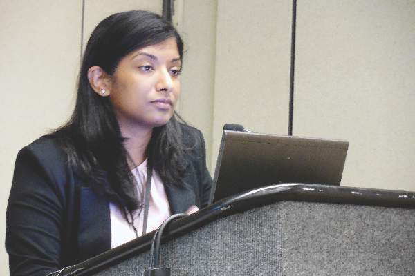
One recent nail in the coffin of office-based measurement came in a modestly-sized but revealing study reported by Dr. Joyce P. Samuel, a pediatric hypertension specialist at the University of Texas in Houston. She reported her experience directly comparing ambulatory and carefully-done office-based blood pressure measurement in a presentation at the annual meeting of the Pediatric Academic Societies in Baltimore.
Dr. Samuel followed 40 patients age 9-21 years whom she had previously diagnosed with essential hypertension (children with a systolic blood pressure at or above the 95th percentile for sex, age, and height), and repeatedly measured their blood pressures by both ambulatory and office-based readings at 2-week intervals as she searched for the best combination of antihypertensive drugs for each patient. She sent patients home for 24 hours of blood-pressure monitoring with an ambulatory device, and when they returned to her office the next day, she performed an office-based measurement using meticulous technique: Each child was seated and calm, measured on the right arm, with four measurements taken sequentially at about 1 minute intervals with the first reading discarded and the remaining three averaged.
Over the course of several months, she collected 173 paired ambulatory and office-based systolic blood pressure readings from individual patients. Substantial differences between the two forms of measurement were remarkably common. In 20% of the pairs, the ambulatory systolic reading was at least 10 mm Hg higher than the office-based reading, and for some pairs the differences ran as high as 30 mm Hg. In an additional 32% of the paired readings, office-based systolic pressure ran at least 10 mm Hg higher and in some cases as much as 35 mm Hg higher than the ambulatory reading.
Dr. Samuel also analyzed her findings a different way to assess the clinical consequences of these differences based on whether a child’s systolic pressure identified the patient as normotensive, hypertensive, or prehypertensive (a systolic pressure at the 90-94th percentile for the child’s age, sex and height). She found that the diagnoses matched for only 49% of the paired measurements. In 24% of the paired readings, ABPM identified children with hypertension that was not seen with concurrent office-based measurement, cases of masked hypertension. In 17% of the pairs, office-based measurement diagnosed hypertension that was not confirmed by ABPM, cases of white-coat hypertension. The remaining 10% of pairs were mismatched by showing normotensive with one method and prehypertensive with the other method. Dr. Samuel searched for any consistent patterns in these differences and found none. The disparate results with ambulatory and office-based measurements seemed almost random, with no correlation with age, sex, race, the medications patients received, or how many times a patient had already undergone dual blood-pressure monitoring. Individual patients had no meaningful differences between some of their paired measurements but had a meaningful disparity for others.
“We were unable to predict discrepancies,” said Dr. Samuels.
“You can’t get around it, you need ambulatory blood pressure monitoring to make the best diagnosis” of hypertension, she told me. “We need to push to make ambulatory monitoring more available. I am moving toward believing that ambulatory blood pressure monitoring must be routinely done on everyone. This is what the data suggest.”
It’s also where medicine is headed. In 2015, the U.S. Preventative Services Task Force (USPSTF) issued new recommendations for hypertension screening in adults aged 18 years or older, indicating that there was “convincing evidence that ABPM is the best method for diagnosing hypertension,” and the agency further recommended that ABPM is “the reference standard for confirming the diagnosis of hypertension.” Another endorsement of ambulatory blood pressure monitoring came out last year from the International Society for Chronobiology.
Recommendations are not yet as evolved for children. The USPSTF last weighed in on screening kids for hypertension in 2013, and said the evidence as of then was “insufficient” to assess the benefits and harms of screening for hypertension in children and adolescents. That document endorsed careful office-based blood pressure measurement, which highlights how recently expert sentiment has shifted on the issue of measurement. In response to the USPSTF 2013 statement, the American Academy of Pediatrics noted that it continued to back recommendations that are more than a decade old from the National High Blood Pressure Education Program that called for hypertension screening in children starting when they are 3 years old. Neither of these two groups has made any recent statement about the preferred method to measure blood pressure.
On Twitter @mitchelzoler
Evidence is becoming overwhelming that ambulatory blood pressure monitoring is the only reliable way to measure blood pressure for both diagnosing hypertension and following patients once they are diagnosed.
Office-based blood pressure measurement is out, be it a one-off reading or a cluster of sequential readings during a single office visit. Ambulatory blood pressure monitoring (ABPM) increasingly is the standard of care.
One recent nail in the coffin of office-based measurement came in a modestly-sized but revealing study reported by Dr. Joyce P. Samuel, a pediatric hypertension specialist at the University of Texas in Houston. She reported her experience directly comparing ambulatory and carefully-done office-based blood pressure measurement in a presentation at the annual meeting of the Pediatric Academic Societies in Baltimore.
Dr. Samuel followed 40 patients age 9-21 years whom she had previously diagnosed with essential hypertension (children with a systolic blood pressure at or above the 95th percentile for sex, age, and height), and repeatedly measured their blood pressures by both ambulatory and office-based readings at 2-week intervals as she searched for the best combination of antihypertensive drugs for each patient. She sent patients home for 24 hours of blood-pressure monitoring with an ambulatory device, and when they returned to her office the next day, she performed an office-based measurement using meticulous technique: Each child was seated and calm, measured on the right arm, with four measurements taken sequentially at about 1 minute intervals with the first reading discarded and the remaining three averaged.
Over the course of several months, she collected 173 paired ambulatory and office-based systolic blood pressure readings from individual patients. Substantial differences between the two forms of measurement were remarkably common. In 20% of the pairs, the ambulatory systolic reading was at least 10 mm Hg higher than the office-based reading, and for some pairs the differences ran as high as 30 mm Hg. In an additional 32% of the paired readings, office-based systolic pressure ran at least 10 mm Hg higher and in some cases as much as 35 mm Hg higher than the ambulatory reading.
Dr. Samuel also analyzed her findings a different way to assess the clinical consequences of these differences based on whether a child’s systolic pressure identified the patient as normotensive, hypertensive, or prehypertensive (a systolic pressure at the 90-94th percentile for the child’s age, sex and height). She found that the diagnoses matched for only 49% of the paired measurements. In 24% of the paired readings, ABPM identified children with hypertension that was not seen with concurrent office-based measurement, cases of masked hypertension. In 17% of the pairs, office-based measurement diagnosed hypertension that was not confirmed by ABPM, cases of white-coat hypertension. The remaining 10% of pairs were mismatched by showing normotensive with one method and prehypertensive with the other method. Dr. Samuel searched for any consistent patterns in these differences and found none. The disparate results with ambulatory and office-based measurements seemed almost random, with no correlation with age, sex, race, the medications patients received, or how many times a patient had already undergone dual blood-pressure monitoring. Individual patients had no meaningful differences between some of their paired measurements but had a meaningful disparity for others.
“We were unable to predict discrepancies,” said Dr. Samuels.
“You can’t get around it, you need ambulatory blood pressure monitoring to make the best diagnosis” of hypertension, she told me. “We need to push to make ambulatory monitoring more available. I am moving toward believing that ambulatory blood pressure monitoring must be routinely done on everyone. This is what the data suggest.”
It’s also where medicine is headed. In 2015, the U.S. Preventative Services Task Force (USPSTF) issued new recommendations for hypertension screening in adults aged 18 years or older, indicating that there was “convincing evidence that ABPM is the best method for diagnosing hypertension,” and the agency further recommended that ABPM is “the reference standard for confirming the diagnosis of hypertension.” Another endorsement of ambulatory blood pressure monitoring came out last year from the International Society for Chronobiology.
Recommendations are not yet as evolved for children. The USPSTF last weighed in on screening kids for hypertension in 2013, and said the evidence as of then was “insufficient” to assess the benefits and harms of screening for hypertension in children and adolescents. That document endorsed careful office-based blood pressure measurement, which highlights how recently expert sentiment has shifted on the issue of measurement. In response to the USPSTF 2013 statement, the American Academy of Pediatrics noted that it continued to back recommendations that are more than a decade old from the National High Blood Pressure Education Program that called for hypertension screening in children starting when they are 3 years old. Neither of these two groups has made any recent statement about the preferred method to measure blood pressure.
On Twitter @mitchelzoler
Evidence is becoming overwhelming that ambulatory blood pressure monitoring is the only reliable way to measure blood pressure for both diagnosing hypertension and following patients once they are diagnosed.
Office-based blood pressure measurement is out, be it a one-off reading or a cluster of sequential readings during a single office visit. Ambulatory blood pressure monitoring (ABPM) increasingly is the standard of care.
One recent nail in the coffin of office-based measurement came in a modestly-sized but revealing study reported by Dr. Joyce P. Samuel, a pediatric hypertension specialist at the University of Texas in Houston. She reported her experience directly comparing ambulatory and carefully-done office-based blood pressure measurement in a presentation at the annual meeting of the Pediatric Academic Societies in Baltimore.
Dr. Samuel followed 40 patients age 9-21 years whom she had previously diagnosed with essential hypertension (children with a systolic blood pressure at or above the 95th percentile for sex, age, and height), and repeatedly measured their blood pressures by both ambulatory and office-based readings at 2-week intervals as she searched for the best combination of antihypertensive drugs for each patient. She sent patients home for 24 hours of blood-pressure monitoring with an ambulatory device, and when they returned to her office the next day, she performed an office-based measurement using meticulous technique: Each child was seated and calm, measured on the right arm, with four measurements taken sequentially at about 1 minute intervals with the first reading discarded and the remaining three averaged.
Over the course of several months, she collected 173 paired ambulatory and office-based systolic blood pressure readings from individual patients. Substantial differences between the two forms of measurement were remarkably common. In 20% of the pairs, the ambulatory systolic reading was at least 10 mm Hg higher than the office-based reading, and for some pairs the differences ran as high as 30 mm Hg. In an additional 32% of the paired readings, office-based systolic pressure ran at least 10 mm Hg higher and in some cases as much as 35 mm Hg higher than the ambulatory reading.
Dr. Samuel also analyzed her findings a different way to assess the clinical consequences of these differences based on whether a child’s systolic pressure identified the patient as normotensive, hypertensive, or prehypertensive (a systolic pressure at the 90-94th percentile for the child’s age, sex and height). She found that the diagnoses matched for only 49% of the paired measurements. In 24% of the paired readings, ABPM identified children with hypertension that was not seen with concurrent office-based measurement, cases of masked hypertension. In 17% of the pairs, office-based measurement diagnosed hypertension that was not confirmed by ABPM, cases of white-coat hypertension. The remaining 10% of pairs were mismatched by showing normotensive with one method and prehypertensive with the other method. Dr. Samuel searched for any consistent patterns in these differences and found none. The disparate results with ambulatory and office-based measurements seemed almost random, with no correlation with age, sex, race, the medications patients received, or how many times a patient had already undergone dual blood-pressure monitoring. Individual patients had no meaningful differences between some of their paired measurements but had a meaningful disparity for others.
“We were unable to predict discrepancies,” said Dr. Samuels.
“You can’t get around it, you need ambulatory blood pressure monitoring to make the best diagnosis” of hypertension, she told me. “We need to push to make ambulatory monitoring more available. I am moving toward believing that ambulatory blood pressure monitoring must be routinely done on everyone. This is what the data suggest.”
It’s also where medicine is headed. In 2015, the U.S. Preventative Services Task Force (USPSTF) issued new recommendations for hypertension screening in adults aged 18 years or older, indicating that there was “convincing evidence that ABPM is the best method for diagnosing hypertension,” and the agency further recommended that ABPM is “the reference standard for confirming the diagnosis of hypertension.” Another endorsement of ambulatory blood pressure monitoring came out last year from the International Society for Chronobiology.
Recommendations are not yet as evolved for children. The USPSTF last weighed in on screening kids for hypertension in 2013, and said the evidence as of then was “insufficient” to assess the benefits and harms of screening for hypertension in children and adolescents. That document endorsed careful office-based blood pressure measurement, which highlights how recently expert sentiment has shifted on the issue of measurement. In response to the USPSTF 2013 statement, the American Academy of Pediatrics noted that it continued to back recommendations that are more than a decade old from the National High Blood Pressure Education Program that called for hypertension screening in children starting when they are 3 years old. Neither of these two groups has made any recent statement about the preferred method to measure blood pressure.
On Twitter @mitchelzoler
VIDEO: Transgender care becoming an increasingly important component of pediatric care
BALTIMORE – With a growing number of youth coming out as transgender, it’s becoming increasingly important for both the current and upcoming generation of pediatricians to know how to provide the best possible health care to this population.
“It’s now very common for pediatricians to be approached by parents or teens, and asked for help or support with a transition,” explained Dr. Daniel E. Shumer of the University of Michigan in Ann Arbor. He added, “I think it’s incumbent upon pediatricians to figure out how to provide that education to trainees so that the next generation of pediatricians is able to provide excellent care for transgender kids and their families.”
In an interview at the annual meeting of the Pediatric Academic Societies, Dr. Shumer elaborated on the importance of training new pediatricians on how to handle LGBT patients, exactly what such training should entail, and why it’s equally important for current pediatricians to educate themselves on this very issue.
Dr. Shumer did not report any relevant financial disclosures.
Go to LGBT Youth Consult on our site for articles on the nature of sexuality and gender identity and how they affect health, advice on how to talk with your patients about these topics, and how to make your office a safe place for LGBT youth.
The video associated with this article is no longer available on this site. Please view all of our videos on the MDedge YouTube channel
BALTIMORE – With a growing number of youth coming out as transgender, it’s becoming increasingly important for both the current and upcoming generation of pediatricians to know how to provide the best possible health care to this population.
“It’s now very common for pediatricians to be approached by parents or teens, and asked for help or support with a transition,” explained Dr. Daniel E. Shumer of the University of Michigan in Ann Arbor. He added, “I think it’s incumbent upon pediatricians to figure out how to provide that education to trainees so that the next generation of pediatricians is able to provide excellent care for transgender kids and their families.”
In an interview at the annual meeting of the Pediatric Academic Societies, Dr. Shumer elaborated on the importance of training new pediatricians on how to handle LGBT patients, exactly what such training should entail, and why it’s equally important for current pediatricians to educate themselves on this very issue.
Dr. Shumer did not report any relevant financial disclosures.
Go to LGBT Youth Consult on our site for articles on the nature of sexuality and gender identity and how they affect health, advice on how to talk with your patients about these topics, and how to make your office a safe place for LGBT youth.
The video associated with this article is no longer available on this site. Please view all of our videos on the MDedge YouTube channel
BALTIMORE – With a growing number of youth coming out as transgender, it’s becoming increasingly important for both the current and upcoming generation of pediatricians to know how to provide the best possible health care to this population.
“It’s now very common for pediatricians to be approached by parents or teens, and asked for help or support with a transition,” explained Dr. Daniel E. Shumer of the University of Michigan in Ann Arbor. He added, “I think it’s incumbent upon pediatricians to figure out how to provide that education to trainees so that the next generation of pediatricians is able to provide excellent care for transgender kids and their families.”
In an interview at the annual meeting of the Pediatric Academic Societies, Dr. Shumer elaborated on the importance of training new pediatricians on how to handle LGBT patients, exactly what such training should entail, and why it’s equally important for current pediatricians to educate themselves on this very issue.
Dr. Shumer did not report any relevant financial disclosures.
Go to LGBT Youth Consult on our site for articles on the nature of sexuality and gender identity and how they affect health, advice on how to talk with your patients about these topics, and how to make your office a safe place for LGBT youth.
The video associated with this article is no longer available on this site. Please view all of our videos on the MDedge YouTube channel
EXPERT ANALYSIS FROM THE PAS ANNUAL MEETING

Predictors of abnormal longitudinal patterns of lung-function growth and decline
When it comes to predictive demographic and clinical factors associated with abnormal patterns of lung growth and decline in those with persistent, mild-to-moderate asthma in childhood, male sex and childhood levels of lung function as assessed by the forced expiratory volume in 1 second (FEV1) make all the difference, according to the results of a study published in the New England Journal of Medicine.
“Determinants of abnormal patterns of FEV1 growth and decline are multifactorial and complex, and identification of factors associated with the timing of a decline from the maximal level requires longitudinal data,” said author Michael McGeachie, Ph.D., of Brigham and Women’s Hospital, Boston, and his colleagues.

To identify these determinants, particularly in those with asthma, Dr. McGeachie and colleagues analyzed longitudinal data from a subset of participants from the Childhood Asthma Management Program (CAMP) cohort who were followed from enrollment at the age of 5 to 12 years, into the third decade of life. The trajectory of lung growth and the decline from maximum growth in this large cohort of persons who had persistent, mild-to-moderate asthma in childhood were compared against those from persons without asthma who were participants in the third National Health and Nutrition Examination Survey. (N Engl J Med. 2016;374:1842-52).
Data from 684 participants from the CAMP cohort were assessed and 25% had normal lung-function growth without an early decline, 26% had normal growth and an early decline, 23% had reduced growth without an early decline, and 26% had reduced growth and an early decline. Results of the multinomial logistic-regression analysis of risk factors for abnormal patterns of lung growth and decline showed that the 26% of participants with reduced growth and an early decline had lower FEV1 lung function at enrollment and were more likely to be male, compared with those who had normal growth.
Additional study results indicated that 18% of the participants who had reduced lung-function growth, with or without an early decline, met the case definition for chronic obstructive pulmonary disease (COPD) based on the postbronchodilator spirometric criteria at an age of less than 30 years.
Based on their data, Dr. McGeachie and his colleagues said that detection of an abnormal trajectory by means of early and ongoing serial FEV1 monitoring may help identify children and young adults at risk for abnormal lung-function growth that could lead to chronic airflow obstruction in adulthood.
Funding for this project was provided by grants from the Parker B. Francis Foundation, the National Institutes of Health, the National Human Genome Research Institute, and the Human Frontier Science Program Organization. Dr. McGeachie reported grant support from one of the funding sources during the conduct of the study. Nineteen coauthors reported they had nothing to disclose and the remainder either reported grant support or ties to industry sources.
When it comes to predictive demographic and clinical factors associated with abnormal patterns of lung growth and decline in those with persistent, mild-to-moderate asthma in childhood, male sex and childhood levels of lung function as assessed by the forced expiratory volume in 1 second (FEV1) make all the difference, according to the results of a study published in the New England Journal of Medicine.
“Determinants of abnormal patterns of FEV1 growth and decline are multifactorial and complex, and identification of factors associated with the timing of a decline from the maximal level requires longitudinal data,” said author Michael McGeachie, Ph.D., of Brigham and Women’s Hospital, Boston, and his colleagues.
To identify these determinants, particularly in those with asthma, Dr. McGeachie and colleagues analyzed longitudinal data from a subset of participants from the Childhood Asthma Management Program (CAMP) cohort who were followed from enrollment at the age of 5 to 12 years, into the third decade of life. The trajectory of lung growth and the decline from maximum growth in this large cohort of persons who had persistent, mild-to-moderate asthma in childhood were compared against those from persons without asthma who were participants in the third National Health and Nutrition Examination Survey. (N Engl J Med. 2016;374:1842-52).
Data from 684 participants from the CAMP cohort were assessed and 25% had normal lung-function growth without an early decline, 26% had normal growth and an early decline, 23% had reduced growth without an early decline, and 26% had reduced growth and an early decline. Results of the multinomial logistic-regression analysis of risk factors for abnormal patterns of lung growth and decline showed that the 26% of participants with reduced growth and an early decline had lower FEV1 lung function at enrollment and were more likely to be male, compared with those who had normal growth.
Additional study results indicated that 18% of the participants who had reduced lung-function growth, with or without an early decline, met the case definition for chronic obstructive pulmonary disease (COPD) based on the postbronchodilator spirometric criteria at an age of less than 30 years.
Based on their data, Dr. McGeachie and his colleagues said that detection of an abnormal trajectory by means of early and ongoing serial FEV1 monitoring may help identify children and young adults at risk for abnormal lung-function growth that could lead to chronic airflow obstruction in adulthood.
Funding for this project was provided by grants from the Parker B. Francis Foundation, the National Institutes of Health, the National Human Genome Research Institute, and the Human Frontier Science Program Organization. Dr. McGeachie reported grant support from one of the funding sources during the conduct of the study. Nineteen coauthors reported they had nothing to disclose and the remainder either reported grant support or ties to industry sources.
When it comes to predictive demographic and clinical factors associated with abnormal patterns of lung growth and decline in those with persistent, mild-to-moderate asthma in childhood, male sex and childhood levels of lung function as assessed by the forced expiratory volume in 1 second (FEV1) make all the difference, according to the results of a study published in the New England Journal of Medicine.
“Determinants of abnormal patterns of FEV1 growth and decline are multifactorial and complex, and identification of factors associated with the timing of a decline from the maximal level requires longitudinal data,” said author Michael McGeachie, Ph.D., of Brigham and Women’s Hospital, Boston, and his colleagues.
To identify these determinants, particularly in those with asthma, Dr. McGeachie and colleagues analyzed longitudinal data from a subset of participants from the Childhood Asthma Management Program (CAMP) cohort who were followed from enrollment at the age of 5 to 12 years, into the third decade of life. The trajectory of lung growth and the decline from maximum growth in this large cohort of persons who had persistent, mild-to-moderate asthma in childhood were compared against those from persons without asthma who were participants in the third National Health and Nutrition Examination Survey. (N Engl J Med. 2016;374:1842-52).
Data from 684 participants from the CAMP cohort were assessed and 25% had normal lung-function growth without an early decline, 26% had normal growth and an early decline, 23% had reduced growth without an early decline, and 26% had reduced growth and an early decline. Results of the multinomial logistic-regression analysis of risk factors for abnormal patterns of lung growth and decline showed that the 26% of participants with reduced growth and an early decline had lower FEV1 lung function at enrollment and were more likely to be male, compared with those who had normal growth.
Additional study results indicated that 18% of the participants who had reduced lung-function growth, with or without an early decline, met the case definition for chronic obstructive pulmonary disease (COPD) based on the postbronchodilator spirometric criteria at an age of less than 30 years.
Based on their data, Dr. McGeachie and his colleagues said that detection of an abnormal trajectory by means of early and ongoing serial FEV1 monitoring may help identify children and young adults at risk for abnormal lung-function growth that could lead to chronic airflow obstruction in adulthood.
Funding for this project was provided by grants from the Parker B. Francis Foundation, the National Institutes of Health, the National Human Genome Research Institute, and the Human Frontier Science Program Organization. Dr. McGeachie reported grant support from one of the funding sources during the conduct of the study. Nineteen coauthors reported they had nothing to disclose and the remainder either reported grant support or ties to industry sources.
Key clinical point: Abnormal longitudinal patterns of lung-function growth in young asthma patients are associated with specific risk factors and may be related to development of COPD.
Major finding: Those with reduced lung growth and an early decline had a lower forced expiratory volume in 1 second lung function in childhood and were more likely to be male.
Data sources: A subset of participants from the Childhood Asthma Management Program cohort.
Disclosures: Funding for this project was provided by grants from the Parker B. Francis Foundation, the National Institutes of Health, the National Human Genome Research Institute, and the Human Frontier Science Program Organization. Dr. McGeachie reported grant support from one of the funding sources during the conduct of the study. Nineteen coauthors reported they had nothing to disclose and the remainder either reported grant support or ties to industry sources.

Infantile hemangiomas clear faster with Nd:YAG treatment
BOSTON – Using a 650-microsecond, pulsed 1064nm neodymium-doped yttrium aluminum garnet (Nd:YAG) laser to treat infantile hemangiomas can yield high clearance rates after one treatment, without adverse reactions, according to a study presented at the annual meeting of the American Society for Laser Medicine and Surgery.
“[Hemangiomas] have historically been treated with a bunch of different devices; most commonly, the pulsed dye laser and also propranolol, either orally or topically,” said Dr. David J. Goldberg of Skin Laser & Surgery Specialists of New York and New Jersey. While use of an Nd:YAG laser for treating infantile hemangiomas is not exactly new, “what’s new about this is it was not a millisecond Nd:YAG laser; it was a microsecond Nd:YAG laser, [which] generally makes it much safer for treatment in general.”
The study enrolled 250 children between age 1 month and 4 months at the time of recruitment; they were followed for one year. They received a single treatment with an Nd:YAG 1064nm laser using a 650 microsecond pulse duration, a spot size of 2-3 millimeters, and a fluence of 42-64 joules per square centimeter. Photographs were taken for evaluation immediately before and after each treatment, and 30 and 180 days after each treatment.
Treatments lasted 3-5 minutes each, covering an area no larger than three square centimeters. Infants who started treatment in the first six weeks of life had the best results, according to Dr. Goldberg. Treated areas immediately turned gray after each treatment on all subjects, and all subjects showed “substantial improvement” in hemangioma size after only a single treatment, based on photographic evaluation taken 6 months after treatment. Over 75% of hemangiomas were either completely or almost completely (over 75% clearance) eliminated over the 1-year follow-up period.
There was no downtime or adverse events experienced by any patients following treatments. Overall, parents of the children who received treatment were “highly satisfied” with the results.
“The big takeaway is that you may not need multiple treatments [and] you may not need to use a pulse dye laser – one treatment may be enough,” Dr. Goldberg explained. “This [Nd:YAG] device is also a very small device that you can put into a suitcase, so it’s not that big device that we’re used to using.”
Dr. Goldberg did not report any relevant financial disclosures.
BOSTON – Using a 650-microsecond, pulsed 1064nm neodymium-doped yttrium aluminum garnet (Nd:YAG) laser to treat infantile hemangiomas can yield high clearance rates after one treatment, without adverse reactions, according to a study presented at the annual meeting of the American Society for Laser Medicine and Surgery.
“[Hemangiomas] have historically been treated with a bunch of different devices; most commonly, the pulsed dye laser and also propranolol, either orally or topically,” said Dr. David J. Goldberg of Skin Laser & Surgery Specialists of New York and New Jersey. While use of an Nd:YAG laser for treating infantile hemangiomas is not exactly new, “what’s new about this is it was not a millisecond Nd:YAG laser; it was a microsecond Nd:YAG laser, [which] generally makes it much safer for treatment in general.”
The study enrolled 250 children between age 1 month and 4 months at the time of recruitment; they were followed for one year. They received a single treatment with an Nd:YAG 1064nm laser using a 650 microsecond pulse duration, a spot size of 2-3 millimeters, and a fluence of 42-64 joules per square centimeter. Photographs were taken for evaluation immediately before and after each treatment, and 30 and 180 days after each treatment.
Treatments lasted 3-5 minutes each, covering an area no larger than three square centimeters. Infants who started treatment in the first six weeks of life had the best results, according to Dr. Goldberg. Treated areas immediately turned gray after each treatment on all subjects, and all subjects showed “substantial improvement” in hemangioma size after only a single treatment, based on photographic evaluation taken 6 months after treatment. Over 75% of hemangiomas were either completely or almost completely (over 75% clearance) eliminated over the 1-year follow-up period.
There was no downtime or adverse events experienced by any patients following treatments. Overall, parents of the children who received treatment were “highly satisfied” with the results.
“The big takeaway is that you may not need multiple treatments [and] you may not need to use a pulse dye laser – one treatment may be enough,” Dr. Goldberg explained. “This [Nd:YAG] device is also a very small device that you can put into a suitcase, so it’s not that big device that we’re used to using.”
Dr. Goldberg did not report any relevant financial disclosures.
BOSTON – Using a 650-microsecond, pulsed 1064nm neodymium-doped yttrium aluminum garnet (Nd:YAG) laser to treat infantile hemangiomas can yield high clearance rates after one treatment, without adverse reactions, according to a study presented at the annual meeting of the American Society for Laser Medicine and Surgery.
“[Hemangiomas] have historically been treated with a bunch of different devices; most commonly, the pulsed dye laser and also propranolol, either orally or topically,” said Dr. David J. Goldberg of Skin Laser & Surgery Specialists of New York and New Jersey. While use of an Nd:YAG laser for treating infantile hemangiomas is not exactly new, “what’s new about this is it was not a millisecond Nd:YAG laser; it was a microsecond Nd:YAG laser, [which] generally makes it much safer for treatment in general.”
The study enrolled 250 children between age 1 month and 4 months at the time of recruitment; they were followed for one year. They received a single treatment with an Nd:YAG 1064nm laser using a 650 microsecond pulse duration, a spot size of 2-3 millimeters, and a fluence of 42-64 joules per square centimeter. Photographs were taken for evaluation immediately before and after each treatment, and 30 and 180 days after each treatment.
Treatments lasted 3-5 minutes each, covering an area no larger than three square centimeters. Infants who started treatment in the first six weeks of life had the best results, according to Dr. Goldberg. Treated areas immediately turned gray after each treatment on all subjects, and all subjects showed “substantial improvement” in hemangioma size after only a single treatment, based on photographic evaluation taken 6 months after treatment. Over 75% of hemangiomas were either completely or almost completely (over 75% clearance) eliminated over the 1-year follow-up period.
There was no downtime or adverse events experienced by any patients following treatments. Overall, parents of the children who received treatment were “highly satisfied” with the results.
“The big takeaway is that you may not need multiple treatments [and] you may not need to use a pulse dye laser – one treatment may be enough,” Dr. Goldberg explained. “This [Nd:YAG] device is also a very small device that you can put into a suitcase, so it’s not that big device that we’re used to using.”
Dr. Goldberg did not report any relevant financial disclosures.
AT LASER 2016
Key clinical point: Infantile hemangiomas can be effectively treated with a 650-microsecond pulsed Nd:YAG 1064nm laser, with no evidence of adverse skin effects.
Major finding: Substantial improvement in hemangiomas were seen after one treatment administered in 5 minutes or less.
Data source: The study enrolled 250 infants with hemangiomas aged 1 to 4 months at baseline, and were followed for one year.
Disclosures: Dr. Goldberg did not report any relevant financial disclosures.
Target Fortification of Breast Milk Boosts Preemie Growth
BALTIMORE – For a premature infant, breast milk straight from Mom or a donor may not be the ideal meal.
The problem is that breast milk is often unbalanced in its content of protein, fat, and carbohydrates, and for a premature neonate fed by a nasogastric tube, unfortified breast milk can provide too little of one nutrient or too much of another. Conventional fortification of breast milk adds a standardized cocktail of nutrients that doesn’t provide an ideal balance for optimal growth.
Frequent analysis of breast milk’s nutrient content and target fortification to tweak the milk to a more ideal nutrient balance may be a way around this problem, and results from a single-center, randomized trial with 85 premature infants showed that this approach led to significantly better infant weights when measured at 36 weeks postmenstrual age, Dr. Christoph Fusch reported at the annual meeting of the Pediatric Academic Societies.

Target fortification of breast milk resulted in an average weight at 36 weeks postmenstrual age of 2,510 g among 42 infants in the intervention group, compared with an average weight of 2,280 g among 43 control infants who received breast milk that underwent routine fortification with a commercial product – a statistically significant difference for the study’s primary endpoint, said Dr. Fusch, professor of pediatrics at McMaster University and director of the neonatal intensive care unit at McMaster Children’s Hospital, both in Hamilton, Ont.
Baseline weights in the two groups of premature infants, who were born at an average of 27 weeks’ gestation and began the study at an average age of about 30 weeks’ postmenstrual age, were 970 g in the controls and 960 g in the infants who received target fortification.
The benefit from target fortification was even more dramatic in the 50% of infants fed on breast milk with the lowest protein content. In this subgroup, the 21 infants on target fortification weighed an average of 2,540 g at 36 weeks postmenstrual age, compared with an average weight of 2,170 g among 21 control infants.
“If you use standard fortification you never get [the nutrients] right. Too many nutrients will be outside [the ideal range] and those babies have a high risk of not growing,” Dr. Fusch noted in an interview.
One of the key steps in the study was measuring the protein, fat, and carbohydrate content of the breast milk that each infant received 3 days a week and using those data to guide fortification. Assessment of each breast milk specimen takes about 2 minutes, after which a technician adds the required levels of freshly prepared nutrient supplements to produce a final protein content of 3 g/dL, fat at 4.3 g/dL, and carbohydrate at 8.5 g/dL. The researchers set these levels based on an assumed daily intake of 150 mL/kg of fortified breast milk.
Dr. Fusch and his associates plan additional analyses of the data they collected in this study to look at the impact of target fortification on infant length and head circumference, metabolic responses, body composition, and neurodevelopment out to 18 months of age. They also have launched a larger, multicenter study with a similar design at 10 sites in Europe and 8 sites in North America.
The infants who stand to gain the most from target fortification are those fed donor breast milk, an increasingly popular option when breast milk from the infant’s mother is not available, but also a flawed option because this milk is usually low in protein, said Dr. Fusch, who also holds the Jack Sinclair Chair in Neonatology at the university. Target fortification is not an issue for term infants who feed normally because they usually cry to demand more milk when they lack specific nutrients. Premature infants are much more vulnerable when fed by a nasogastric tube because they will become sated simply by milk volume and then stop eating, even when they ingest an unbalanced nutrient supply.
BALTIMORE – For a premature infant, breast milk straight from Mom or a donor may not be the ideal meal.
The problem is that breast milk is often unbalanced in its content of protein, fat, and carbohydrates, and for a premature neonate fed by a nasogastric tube, unfortified breast milk can provide too little of one nutrient or too much of another. Conventional fortification of breast milk adds a standardized cocktail of nutrients that doesn’t provide an ideal balance for optimal growth.
Frequent analysis of breast milk’s nutrient content and target fortification to tweak the milk to a more ideal nutrient balance may be a way around this problem, and results from a single-center, randomized trial with 85 premature infants showed that this approach led to significantly better infant weights when measured at 36 weeks postmenstrual age, Dr. Christoph Fusch reported at the annual meeting of the Pediatric Academic Societies.
Target fortification of breast milk resulted in an average weight at 36 weeks postmenstrual age of 2,510 g among 42 infants in the intervention group, compared with an average weight of 2,280 g among 43 control infants who received breast milk that underwent routine fortification with a commercial product – a statistically significant difference for the study’s primary endpoint, said Dr. Fusch, professor of pediatrics at McMaster University and director of the neonatal intensive care unit at McMaster Children’s Hospital, both in Hamilton, Ont.
Baseline weights in the two groups of premature infants, who were born at an average of 27 weeks’ gestation and began the study at an average age of about 30 weeks’ postmenstrual age, were 970 g in the controls and 960 g in the infants who received target fortification.
The benefit from target fortification was even more dramatic in the 50% of infants fed on breast milk with the lowest protein content. In this subgroup, the 21 infants on target fortification weighed an average of 2,540 g at 36 weeks postmenstrual age, compared with an average weight of 2,170 g among 21 control infants.
“If you use standard fortification you never get [the nutrients] right. Too many nutrients will be outside [the ideal range] and those babies have a high risk of not growing,” Dr. Fusch noted in an interview.
One of the key steps in the study was measuring the protein, fat, and carbohydrate content of the breast milk that each infant received 3 days a week and using those data to guide fortification. Assessment of each breast milk specimen takes about 2 minutes, after which a technician adds the required levels of freshly prepared nutrient supplements to produce a final protein content of 3 g/dL, fat at 4.3 g/dL, and carbohydrate at 8.5 g/dL. The researchers set these levels based on an assumed daily intake of 150 mL/kg of fortified breast milk.
Dr. Fusch and his associates plan additional analyses of the data they collected in this study to look at the impact of target fortification on infant length and head circumference, metabolic responses, body composition, and neurodevelopment out to 18 months of age. They also have launched a larger, multicenter study with a similar design at 10 sites in Europe and 8 sites in North America.
The infants who stand to gain the most from target fortification are those fed donor breast milk, an increasingly popular option when breast milk from the infant’s mother is not available, but also a flawed option because this milk is usually low in protein, said Dr. Fusch, who also holds the Jack Sinclair Chair in Neonatology at the university. Target fortification is not an issue for term infants who feed normally because they usually cry to demand more milk when they lack specific nutrients. Premature infants are much more vulnerable when fed by a nasogastric tube because they will become sated simply by milk volume and then stop eating, even when they ingest an unbalanced nutrient supply.
BALTIMORE – For a premature infant, breast milk straight from Mom or a donor may not be the ideal meal.
The problem is that breast milk is often unbalanced in its content of protein, fat, and carbohydrates, and for a premature neonate fed by a nasogastric tube, unfortified breast milk can provide too little of one nutrient or too much of another. Conventional fortification of breast milk adds a standardized cocktail of nutrients that doesn’t provide an ideal balance for optimal growth.
Frequent analysis of breast milk’s nutrient content and target fortification to tweak the milk to a more ideal nutrient balance may be a way around this problem, and results from a single-center, randomized trial with 85 premature infants showed that this approach led to significantly better infant weights when measured at 36 weeks postmenstrual age, Dr. Christoph Fusch reported at the annual meeting of the Pediatric Academic Societies.
Target fortification of breast milk resulted in an average weight at 36 weeks postmenstrual age of 2,510 g among 42 infants in the intervention group, compared with an average weight of 2,280 g among 43 control infants who received breast milk that underwent routine fortification with a commercial product – a statistically significant difference for the study’s primary endpoint, said Dr. Fusch, professor of pediatrics at McMaster University and director of the neonatal intensive care unit at McMaster Children’s Hospital, both in Hamilton, Ont.
Baseline weights in the two groups of premature infants, who were born at an average of 27 weeks’ gestation and began the study at an average age of about 30 weeks’ postmenstrual age, were 970 g in the controls and 960 g in the infants who received target fortification.
The benefit from target fortification was even more dramatic in the 50% of infants fed on breast milk with the lowest protein content. In this subgroup, the 21 infants on target fortification weighed an average of 2,540 g at 36 weeks postmenstrual age, compared with an average weight of 2,170 g among 21 control infants.
“If you use standard fortification you never get [the nutrients] right. Too many nutrients will be outside [the ideal range] and those babies have a high risk of not growing,” Dr. Fusch noted in an interview.
One of the key steps in the study was measuring the protein, fat, and carbohydrate content of the breast milk that each infant received 3 days a week and using those data to guide fortification. Assessment of each breast milk specimen takes about 2 minutes, after which a technician adds the required levels of freshly prepared nutrient supplements to produce a final protein content of 3 g/dL, fat at 4.3 g/dL, and carbohydrate at 8.5 g/dL. The researchers set these levels based on an assumed daily intake of 150 mL/kg of fortified breast milk.
Dr. Fusch and his associates plan additional analyses of the data they collected in this study to look at the impact of target fortification on infant length and head circumference, metabolic responses, body composition, and neurodevelopment out to 18 months of age. They also have launched a larger, multicenter study with a similar design at 10 sites in Europe and 8 sites in North America.
The infants who stand to gain the most from target fortification are those fed donor breast milk, an increasingly popular option when breast milk from the infant’s mother is not available, but also a flawed option because this milk is usually low in protein, said Dr. Fusch, who also holds the Jack Sinclair Chair in Neonatology at the university. Target fortification is not an issue for term infants who feed normally because they usually cry to demand more milk when they lack specific nutrients. Premature infants are much more vulnerable when fed by a nasogastric tube because they will become sated simply by milk volume and then stop eating, even when they ingest an unbalanced nutrient supply.
AT THE PAS ANNUAL MEETING

Target fortification of breast milk boosts preemie growth
BALTIMORE – For a premature infant, breast milk straight from Mom or a donor may not be the ideal meal.
The problem is that breast milk is often unbalanced in its content of protein, fat, and carbohydrates, and for a premature neonate fed by a nasogastric tube, unfortified breast milk can provide too little of one nutrient or too much of another. Conventional fortification of breast milk adds a standardized cocktail of nutrients that doesn’t provide an ideal balance for optimal growth.
Frequent analysis of breast milk’s nutrient content and target fortification to tweak the milk to a more ideal nutrient balance may be a way around this problem, and results from a single-center, randomized trial with 85 premature infants showed that this approach led to significantly better infant weights when measured at 36 weeks postmenstrual age, Dr. Christoph Fusch reported at the annual meeting of the Pediatric Academic Societies.

Target fortification of breast milk resulted in an average weight at 36 weeks postmenstrual age of 2,510 g among 42 infants in the intervention group, compared with an average weight of 2,280 g among 43 control infants who received breast milk that underwent routine fortification with a commercial product – a statistically significant difference for the study’s primary endpoint, said Dr. Fusch, professor of pediatrics at McMaster University and director of the neonatal intensive care unit at McMaster Children’s Hospital, both in Hamilton, Ont.
Baseline weights in the two groups of premature infants, who were born at an average of 27 weeks’ gestation and began the study at an average age of about 30 weeks’ postmenstrual age, were 970 g in the controls and 960 g in the infants who received target fortification.
The benefit from target fortification was even more dramatic in the 50% of infants fed on breast milk with the lowest protein content. In this subgroup, the 21 infants on target fortification weighed an average of 2,540 g at 36 weeks postmenstrual age, compared with an average weight of 2,170 g among 21 control infants.
“If you use standard fortification you never get [the nutrients] right. Too many nutrients will be outside [the ideal range] and those babies have a high risk of not growing,” Dr. Fusch noted in an interview.
One of the key steps in the study was measuring the protein, fat, and carbohydrate content of the breast milk that each infant received 3 days a week and using those data to guide fortification. Assessment of each breast milk specimen takes about 2 minutes, after which a technician adds the required levels of freshly prepared nutrient supplements to produce a final protein content of 3 g/dL, fat at 4.3 g/dL, and carbohydrate at 8.5 g/dL. The researchers set these levels based on an assumed daily intake of 150 mL/kg of fortified breast milk.
Dr. Fusch and his associates plan additional analyses of the data they collected in this study to look at the impact of target fortification on infant length and head circumference, metabolic responses, body composition, and neurodevelopment out to 18 months of age. They also have launched a larger, multicenter study with a similar design at 10 sites in Europe and 8 sites in North America.
The infants who stand to gain the most from target fortification are those fed donor breast milk, an increasingly popular option when breast milk from the infant’s mother is not available, but also a flawed option because this milk is usually low in protein, said Dr. Fusch, who also holds the Jack Sinclair Chair in Neonatology at the university. Target fortification is not an issue for term infants who feed normally because they usually cry to demand more milk when they lack specific nutrients. Premature infants are much more vulnerable when fed by a nasogastric tube because they will become sated simply by milk volume and then stop eating, even when they ingest an unbalanced nutrient supply.
On Twitter @mitchelzoler
BALTIMORE – For a premature infant, breast milk straight from Mom or a donor may not be the ideal meal.
The problem is that breast milk is often unbalanced in its content of protein, fat, and carbohydrates, and for a premature neonate fed by a nasogastric tube, unfortified breast milk can provide too little of one nutrient or too much of another. Conventional fortification of breast milk adds a standardized cocktail of nutrients that doesn’t provide an ideal balance for optimal growth.
Frequent analysis of breast milk’s nutrient content and target fortification to tweak the milk to a more ideal nutrient balance may be a way around this problem, and results from a single-center, randomized trial with 85 premature infants showed that this approach led to significantly better infant weights when measured at 36 weeks postmenstrual age, Dr. Christoph Fusch reported at the annual meeting of the Pediatric Academic Societies.
Target fortification of breast milk resulted in an average weight at 36 weeks postmenstrual age of 2,510 g among 42 infants in the intervention group, compared with an average weight of 2,280 g among 43 control infants who received breast milk that underwent routine fortification with a commercial product – a statistically significant difference for the study’s primary endpoint, said Dr. Fusch, professor of pediatrics at McMaster University and director of the neonatal intensive care unit at McMaster Children’s Hospital, both in Hamilton, Ont.
Baseline weights in the two groups of premature infants, who were born at an average of 27 weeks’ gestation and began the study at an average age of about 30 weeks’ postmenstrual age, were 970 g in the controls and 960 g in the infants who received target fortification.
The benefit from target fortification was even more dramatic in the 50% of infants fed on breast milk with the lowest protein content. In this subgroup, the 21 infants on target fortification weighed an average of 2,540 g at 36 weeks postmenstrual age, compared with an average weight of 2,170 g among 21 control infants.
“If you use standard fortification you never get [the nutrients] right. Too many nutrients will be outside [the ideal range] and those babies have a high risk of not growing,” Dr. Fusch noted in an interview.
One of the key steps in the study was measuring the protein, fat, and carbohydrate content of the breast milk that each infant received 3 days a week and using those data to guide fortification. Assessment of each breast milk specimen takes about 2 minutes, after which a technician adds the required levels of freshly prepared nutrient supplements to produce a final protein content of 3 g/dL, fat at 4.3 g/dL, and carbohydrate at 8.5 g/dL. The researchers set these levels based on an assumed daily intake of 150 mL/kg of fortified breast milk.
Dr. Fusch and his associates plan additional analyses of the data they collected in this study to look at the impact of target fortification on infant length and head circumference, metabolic responses, body composition, and neurodevelopment out to 18 months of age. They also have launched a larger, multicenter study with a similar design at 10 sites in Europe and 8 sites in North America.
The infants who stand to gain the most from target fortification are those fed donor breast milk, an increasingly popular option when breast milk from the infant’s mother is not available, but also a flawed option because this milk is usually low in protein, said Dr. Fusch, who also holds the Jack Sinclair Chair in Neonatology at the university. Target fortification is not an issue for term infants who feed normally because they usually cry to demand more milk when they lack specific nutrients. Premature infants are much more vulnerable when fed by a nasogastric tube because they will become sated simply by milk volume and then stop eating, even when they ingest an unbalanced nutrient supply.
On Twitter @mitchelzoler
BALTIMORE – For a premature infant, breast milk straight from Mom or a donor may not be the ideal meal.
The problem is that breast milk is often unbalanced in its content of protein, fat, and carbohydrates, and for a premature neonate fed by a nasogastric tube, unfortified breast milk can provide too little of one nutrient or too much of another. Conventional fortification of breast milk adds a standardized cocktail of nutrients that doesn’t provide an ideal balance for optimal growth.
Frequent analysis of breast milk’s nutrient content and target fortification to tweak the milk to a more ideal nutrient balance may be a way around this problem, and results from a single-center, randomized trial with 85 premature infants showed that this approach led to significantly better infant weights when measured at 36 weeks postmenstrual age, Dr. Christoph Fusch reported at the annual meeting of the Pediatric Academic Societies.
Target fortification of breast milk resulted in an average weight at 36 weeks postmenstrual age of 2,510 g among 42 infants in the intervention group, compared with an average weight of 2,280 g among 43 control infants who received breast milk that underwent routine fortification with a commercial product – a statistically significant difference for the study’s primary endpoint, said Dr. Fusch, professor of pediatrics at McMaster University and director of the neonatal intensive care unit at McMaster Children’s Hospital, both in Hamilton, Ont.
Baseline weights in the two groups of premature infants, who were born at an average of 27 weeks’ gestation and began the study at an average age of about 30 weeks’ postmenstrual age, were 970 g in the controls and 960 g in the infants who received target fortification.
The benefit from target fortification was even more dramatic in the 50% of infants fed on breast milk with the lowest protein content. In this subgroup, the 21 infants on target fortification weighed an average of 2,540 g at 36 weeks postmenstrual age, compared with an average weight of 2,170 g among 21 control infants.
“If you use standard fortification you never get [the nutrients] right. Too many nutrients will be outside [the ideal range] and those babies have a high risk of not growing,” Dr. Fusch noted in an interview.
One of the key steps in the study was measuring the protein, fat, and carbohydrate content of the breast milk that each infant received 3 days a week and using those data to guide fortification. Assessment of each breast milk specimen takes about 2 minutes, after which a technician adds the required levels of freshly prepared nutrient supplements to produce a final protein content of 3 g/dL, fat at 4.3 g/dL, and carbohydrate at 8.5 g/dL. The researchers set these levels based on an assumed daily intake of 150 mL/kg of fortified breast milk.
Dr. Fusch and his associates plan additional analyses of the data they collected in this study to look at the impact of target fortification on infant length and head circumference, metabolic responses, body composition, and neurodevelopment out to 18 months of age. They also have launched a larger, multicenter study with a similar design at 10 sites in Europe and 8 sites in North America.
The infants who stand to gain the most from target fortification are those fed donor breast milk, an increasingly popular option when breast milk from the infant’s mother is not available, but also a flawed option because this milk is usually low in protein, said Dr. Fusch, who also holds the Jack Sinclair Chair in Neonatology at the university. Target fortification is not an issue for term infants who feed normally because they usually cry to demand more milk when they lack specific nutrients. Premature infants are much more vulnerable when fed by a nasogastric tube because they will become sated simply by milk volume and then stop eating, even when they ingest an unbalanced nutrient supply.
On Twitter @mitchelzoler
AT THE PAS ANNUAL MEETING
Key clinical point: Premature infants fed breast milk that had undergone target fortification showed significantly better growth to 36 weeks postmenstrual age, compared with control infants fed with conventionally fortified breast milk.
Major finding: Average weight after intervention was 2,510 g in infants fed target-fortified breast milk and 2,280 g in controls.
Data source: Single-center, randomized controlled study with 85 premature infants.
Disclosures: Dr. Fusch had no disclosures.

VIDEO: Preemies on early caffeine not benefited, possibly harmed
BALTIMORE – Early initiation of caffeine treatment in premature neonates on mechanical ventilation did not cut the time to when these babies could successfully wean off the ventilator, according to findings of a single-center, randomized controlled study of 83 children.
The results also showed an “unexpected” trend toward increased mortality among the neonates who received early caffeine treatment, Dr. Cynthia M. Amaro reported at the annual meeting of the Pediatric Academic Societies. This signal of elevated mortality with caffeine treatment prompted the study’s data and safety monitoring board to prematurely stop the trial, limiting enrollment to just 75% of the number originally planned in the study’s design, thereby raising questions about the reliability of the primary-endpoint finding that early caffeine treatment did not result in the benefit of a reduced time to extubation.
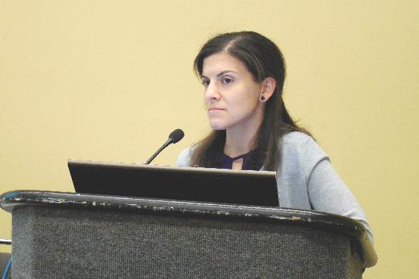
Dr. Amaro said that she and her associates ran the study to address what had emerged as a significant area of doubt in routine U.S. practice on how to best use caffeine treatment in this neonatal population following publication of findings from the landmark Caffeine for Apnea of Prematurity (CAP) Trial (N Engl J Med. 2006 May 18;354[20]:2112-21). Results from the CAP Trial had shown in nearly 2,000 randomized, premature infants that treatment with caffeine led to significantly fewer episodes of bronchopulmonary dysplasia as well as quicker time to extubation of mechanical ventilation. Caffeine or other methylxanthines stimulate an infant’s respiratory center to allow faster extubation.
Ever since that publication a decade ago, “clinicians have been using caffeine earlier and more liberally, without really good data to support its early use in mechanically-ventilated preterm babies,” explained Dr. Amaro, a neonatologist at the University of Miami and Holtz Children’s Hospital in Miami.
Based on the new findings from the study she reported, “we are now not routinely initiating caffeine in mechanically ventilated preterm babies and just using caffeine immediately before extubation to treat apnea of prematurity. This returns caffeine treatment to the way it was used in the CAP Trial,” she said. “Further studies are needed before we can say what is best for early treatment of these preterm babies,” Dr. Amaro said in a video interview.
Her report led to a flurry of comments during the question period, with several pediatricians voicing concern about the reliability of results from a study that followed only 83 patients because of its premature termination.
“The data and safety monitoring board’s decision is a big issue,” said Dr. Carl E. Hunt, a pediatrician at the Uniformed Services University of the Health Sciences in Bethesda, Md. “There is a literature that shows results of studies can be very different when they stop early. It’s unfortunate because we don’t have other prospective data, and it may now be hard to do a large randomized, controlled trial” of early caffeine treatment, Dr. Hunt said.
While Dr. Amaro conceded that premature termination limited her study’s size, she also asserted that her analyses confirmed the validity of the finding of no benefit from early caffeine treatment. “We projected to full enrollment, and there still was no difference in the time to first successful extubation,” she said.
Her study enrolled preterm infants during January 2013–December 2015 born at 23-30 weeks’ gestation who required mechanical ventilation during their first 5 days. Randomization assigned 41 infants to receive a 20-mg/kg bolus of caffeine, followed by a maintenance dosage of 5 mg/kg that continued until extubation, while 42 patients received placebo and did not get caffeine until just before attempted extubation. The bolus and maintenance caffeine dosages tested were identical to those used in the CAP Trial.
The researchers defined successful extubation as keeping a child off restart of mechanical ventilation for more than 24 hours. The average gestational age of the enrolled neonates was 26 weeks, their average weight was 700 g, and intubation started an average of 3 hours after delivery.
The study’s primary endpoint, age at first successful extubation, was an average of 24 days among the neonates treated with caffeine and 20 days in those on placebo, Dr. Amaro reported. Mortality occurred at an average of 30 days after delivery in the caffeine recipients and after an average of 10 days in the controls. The incidence of death was 22% in those on early caffeine and 12% among those in the placebo group, an excess of four deaths in the intervention arm that was not statically significant.
A recent review of more than 29,000 matched very-low-birth-weight infants managed in routine practice showed that neonates who received early caffeine had an adjusted mortality risk that was 23% higher than that of matched infants not receiving early caffeine, Dr. Amaro noted (J Pediatrics. 2014 May;164[5]:992-8).
The incidence of bronchopulmonary dysplasia also did not show a statistically significant difference between the two study arms, 46% among those on early caffeine and 53% in the placebo group. Patients on early caffeine also had higher rates of necrotizing enterocolitis, more episodes of necrotizing enterocolitis requiring surgery, and more intraventricular hemorrhages, but none of these differences reached statistical significance.
The video associated with this article is no longer available on this site. Please view all of our videos on the MDedge YouTube channel
On Twitter @mitchelzoler
Clinicians need to be very cautious starting caffeine to treat premature neonates on mechanical ventilation with the goal of reducing the time to extubation. The findings from Dr. Amaro and her associates show that we do not really know whether starting caffeine treatment early is the right thing to do.
Because their study stopped sooner than planned, I don’t believe the number of enrolled patients was large enough to provide clear guidance on when to start treatment with caffeine or another methylxanthine. We still don’t know the answer. In addition, the suggestion of excess mortality with caffeine treatment gives us no reassurance that caffeine is safe in this setting. However, as was clear in the questions and comments from some members of the audience that heard this report, some people continue to believe that early treatment with caffeine of premature infants on mechanical ventilation is appropriate.
As a result of the equivocal results in Dr. Amaro’s study, the best option is probably to go back to using the protocol tested and shown safe and effective in the 2006 report from the Caffeine for Apnea of Prematurity Trial (N Engl J Med. 2006 May 18;354[20]:2112-21). Ideally, researchers will soon run a large, randomized trial that can better address the questions that Dr. Amaro and her associates attempted to answer with their study.
Dr. Clifford W. Bogue is professor of pediatrics at Yale University, New Haven, Conn., and chaired the session where Dr. Amaro presented her report. He had no relevant disclosures. He made these comments in an interview.
Clinicians need to be very cautious starting caffeine to treat premature neonates on mechanical ventilation with the goal of reducing the time to extubation. The findings from Dr. Amaro and her associates show that we do not really know whether starting caffeine treatment early is the right thing to do.
Because their study stopped sooner than planned, I don’t believe the number of enrolled patients was large enough to provide clear guidance on when to start treatment with caffeine or another methylxanthine. We still don’t know the answer. In addition, the suggestion of excess mortality with caffeine treatment gives us no reassurance that caffeine is safe in this setting. However, as was clear in the questions and comments from some members of the audience that heard this report, some people continue to believe that early treatment with caffeine of premature infants on mechanical ventilation is appropriate.
As a result of the equivocal results in Dr. Amaro’s study, the best option is probably to go back to using the protocol tested and shown safe and effective in the 2006 report from the Caffeine for Apnea of Prematurity Trial (N Engl J Med. 2006 May 18;354[20]:2112-21). Ideally, researchers will soon run a large, randomized trial that can better address the questions that Dr. Amaro and her associates attempted to answer with their study.
Dr. Clifford W. Bogue is professor of pediatrics at Yale University, New Haven, Conn., and chaired the session where Dr. Amaro presented her report. He had no relevant disclosures. He made these comments in an interview.
Clinicians need to be very cautious starting caffeine to treat premature neonates on mechanical ventilation with the goal of reducing the time to extubation. The findings from Dr. Amaro and her associates show that we do not really know whether starting caffeine treatment early is the right thing to do.
Because their study stopped sooner than planned, I don’t believe the number of enrolled patients was large enough to provide clear guidance on when to start treatment with caffeine or another methylxanthine. We still don’t know the answer. In addition, the suggestion of excess mortality with caffeine treatment gives us no reassurance that caffeine is safe in this setting. However, as was clear in the questions and comments from some members of the audience that heard this report, some people continue to believe that early treatment with caffeine of premature infants on mechanical ventilation is appropriate.
As a result of the equivocal results in Dr. Amaro’s study, the best option is probably to go back to using the protocol tested and shown safe and effective in the 2006 report from the Caffeine for Apnea of Prematurity Trial (N Engl J Med. 2006 May 18;354[20]:2112-21). Ideally, researchers will soon run a large, randomized trial that can better address the questions that Dr. Amaro and her associates attempted to answer with their study.
Dr. Clifford W. Bogue is professor of pediatrics at Yale University, New Haven, Conn., and chaired the session where Dr. Amaro presented her report. He had no relevant disclosures. He made these comments in an interview.
BALTIMORE – Early initiation of caffeine treatment in premature neonates on mechanical ventilation did not cut the time to when these babies could successfully wean off the ventilator, according to findings of a single-center, randomized controlled study of 83 children.
The results also showed an “unexpected” trend toward increased mortality among the neonates who received early caffeine treatment, Dr. Cynthia M. Amaro reported at the annual meeting of the Pediatric Academic Societies. This signal of elevated mortality with caffeine treatment prompted the study’s data and safety monitoring board to prematurely stop the trial, limiting enrollment to just 75% of the number originally planned in the study’s design, thereby raising questions about the reliability of the primary-endpoint finding that early caffeine treatment did not result in the benefit of a reduced time to extubation.
Dr. Amaro said that she and her associates ran the study to address what had emerged as a significant area of doubt in routine U.S. practice on how to best use caffeine treatment in this neonatal population following publication of findings from the landmark Caffeine for Apnea of Prematurity (CAP) Trial (N Engl J Med. 2006 May 18;354[20]:2112-21). Results from the CAP Trial had shown in nearly 2,000 randomized, premature infants that treatment with caffeine led to significantly fewer episodes of bronchopulmonary dysplasia as well as quicker time to extubation of mechanical ventilation. Caffeine or other methylxanthines stimulate an infant’s respiratory center to allow faster extubation.
Ever since that publication a decade ago, “clinicians have been using caffeine earlier and more liberally, without really good data to support its early use in mechanically-ventilated preterm babies,” explained Dr. Amaro, a neonatologist at the University of Miami and Holtz Children’s Hospital in Miami.
Based on the new findings from the study she reported, “we are now not routinely initiating caffeine in mechanically ventilated preterm babies and just using caffeine immediately before extubation to treat apnea of prematurity. This returns caffeine treatment to the way it was used in the CAP Trial,” she said. “Further studies are needed before we can say what is best for early treatment of these preterm babies,” Dr. Amaro said in a video interview.
Her report led to a flurry of comments during the question period, with several pediatricians voicing concern about the reliability of results from a study that followed only 83 patients because of its premature termination.
“The data and safety monitoring board’s decision is a big issue,” said Dr. Carl E. Hunt, a pediatrician at the Uniformed Services University of the Health Sciences in Bethesda, Md. “There is a literature that shows results of studies can be very different when they stop early. It’s unfortunate because we don’t have other prospective data, and it may now be hard to do a large randomized, controlled trial” of early caffeine treatment, Dr. Hunt said.
While Dr. Amaro conceded that premature termination limited her study’s size, she also asserted that her analyses confirmed the validity of the finding of no benefit from early caffeine treatment. “We projected to full enrollment, and there still was no difference in the time to first successful extubation,” she said.
Her study enrolled preterm infants during January 2013–December 2015 born at 23-30 weeks’ gestation who required mechanical ventilation during their first 5 days. Randomization assigned 41 infants to receive a 20-mg/kg bolus of caffeine, followed by a maintenance dosage of 5 mg/kg that continued until extubation, while 42 patients received placebo and did not get caffeine until just before attempted extubation. The bolus and maintenance caffeine dosages tested were identical to those used in the CAP Trial.
The researchers defined successful extubation as keeping a child off restart of mechanical ventilation for more than 24 hours. The average gestational age of the enrolled neonates was 26 weeks, their average weight was 700 g, and intubation started an average of 3 hours after delivery.
The study’s primary endpoint, age at first successful extubation, was an average of 24 days among the neonates treated with caffeine and 20 days in those on placebo, Dr. Amaro reported. Mortality occurred at an average of 30 days after delivery in the caffeine recipients and after an average of 10 days in the controls. The incidence of death was 22% in those on early caffeine and 12% among those in the placebo group, an excess of four deaths in the intervention arm that was not statically significant.
A recent review of more than 29,000 matched very-low-birth-weight infants managed in routine practice showed that neonates who received early caffeine had an adjusted mortality risk that was 23% higher than that of matched infants not receiving early caffeine, Dr. Amaro noted (J Pediatrics. 2014 May;164[5]:992-8).
The incidence of bronchopulmonary dysplasia also did not show a statistically significant difference between the two study arms, 46% among those on early caffeine and 53% in the placebo group. Patients on early caffeine also had higher rates of necrotizing enterocolitis, more episodes of necrotizing enterocolitis requiring surgery, and more intraventricular hemorrhages, but none of these differences reached statistical significance.
The video associated with this article is no longer available on this site. Please view all of our videos on the MDedge YouTube channel
On Twitter @mitchelzoler
BALTIMORE – Early initiation of caffeine treatment in premature neonates on mechanical ventilation did not cut the time to when these babies could successfully wean off the ventilator, according to findings of a single-center, randomized controlled study of 83 children.
The results also showed an “unexpected” trend toward increased mortality among the neonates who received early caffeine treatment, Dr. Cynthia M. Amaro reported at the annual meeting of the Pediatric Academic Societies. This signal of elevated mortality with caffeine treatment prompted the study’s data and safety monitoring board to prematurely stop the trial, limiting enrollment to just 75% of the number originally planned in the study’s design, thereby raising questions about the reliability of the primary-endpoint finding that early caffeine treatment did not result in the benefit of a reduced time to extubation.
Dr. Amaro said that she and her associates ran the study to address what had emerged as a significant area of doubt in routine U.S. practice on how to best use caffeine treatment in this neonatal population following publication of findings from the landmark Caffeine for Apnea of Prematurity (CAP) Trial (N Engl J Med. 2006 May 18;354[20]:2112-21). Results from the CAP Trial had shown in nearly 2,000 randomized, premature infants that treatment with caffeine led to significantly fewer episodes of bronchopulmonary dysplasia as well as quicker time to extubation of mechanical ventilation. Caffeine or other methylxanthines stimulate an infant’s respiratory center to allow faster extubation.
Ever since that publication a decade ago, “clinicians have been using caffeine earlier and more liberally, without really good data to support its early use in mechanically-ventilated preterm babies,” explained Dr. Amaro, a neonatologist at the University of Miami and Holtz Children’s Hospital in Miami.
Based on the new findings from the study she reported, “we are now not routinely initiating caffeine in mechanically ventilated preterm babies and just using caffeine immediately before extubation to treat apnea of prematurity. This returns caffeine treatment to the way it was used in the CAP Trial,” she said. “Further studies are needed before we can say what is best for early treatment of these preterm babies,” Dr. Amaro said in a video interview.
Her report led to a flurry of comments during the question period, with several pediatricians voicing concern about the reliability of results from a study that followed only 83 patients because of its premature termination.
“The data and safety monitoring board’s decision is a big issue,” said Dr. Carl E. Hunt, a pediatrician at the Uniformed Services University of the Health Sciences in Bethesda, Md. “There is a literature that shows results of studies can be very different when they stop early. It’s unfortunate because we don’t have other prospective data, and it may now be hard to do a large randomized, controlled trial” of early caffeine treatment, Dr. Hunt said.
While Dr. Amaro conceded that premature termination limited her study’s size, she also asserted that her analyses confirmed the validity of the finding of no benefit from early caffeine treatment. “We projected to full enrollment, and there still was no difference in the time to first successful extubation,” she said.
Her study enrolled preterm infants during January 2013–December 2015 born at 23-30 weeks’ gestation who required mechanical ventilation during their first 5 days. Randomization assigned 41 infants to receive a 20-mg/kg bolus of caffeine, followed by a maintenance dosage of 5 mg/kg that continued until extubation, while 42 patients received placebo and did not get caffeine until just before attempted extubation. The bolus and maintenance caffeine dosages tested were identical to those used in the CAP Trial.
The researchers defined successful extubation as keeping a child off restart of mechanical ventilation for more than 24 hours. The average gestational age of the enrolled neonates was 26 weeks, their average weight was 700 g, and intubation started an average of 3 hours after delivery.
The study’s primary endpoint, age at first successful extubation, was an average of 24 days among the neonates treated with caffeine and 20 days in those on placebo, Dr. Amaro reported. Mortality occurred at an average of 30 days after delivery in the caffeine recipients and after an average of 10 days in the controls. The incidence of death was 22% in those on early caffeine and 12% among those in the placebo group, an excess of four deaths in the intervention arm that was not statically significant.
A recent review of more than 29,000 matched very-low-birth-weight infants managed in routine practice showed that neonates who received early caffeine had an adjusted mortality risk that was 23% higher than that of matched infants not receiving early caffeine, Dr. Amaro noted (J Pediatrics. 2014 May;164[5]:992-8).
The incidence of bronchopulmonary dysplasia also did not show a statistically significant difference between the two study arms, 46% among those on early caffeine and 53% in the placebo group. Patients on early caffeine also had higher rates of necrotizing enterocolitis, more episodes of necrotizing enterocolitis requiring surgery, and more intraventricular hemorrhages, but none of these differences reached statistical significance.
The video associated with this article is no longer available on this site. Please view all of our videos on the MDedge YouTube channel
On Twitter @mitchelzoler
AT THE PAS ANNUAL MEETING
Key clinical point: Early caffeine treatment of premature neonates on mechanical ventilation did not reduce the time to extubation and produced a trend toward increased mortality.
Major finding: The age of first successful extubation averaged 24 days in infants receiving caffeine and 20 days among controls.
Data source: Single-center, randomized, controlled study of 83 premature neonates.
Disclosures: Dr. Amaro had no disclosures.
Summer colds
Most viral infections in summer months are caused by enteroviruses. We studied illnesses in about 400 kids aged 4-18 years seen in private pediatric practice and were surprised by what we found.
Our impression was that summer colds lasted for a shorter time span than winter colds. What we found was that the median duration of illness was about 8 days. Among the various syndromes, the most common was stomatitis (viral blisters in the throat), accounting for 58% of all cases seen. A flulike illness with fever, myalgias, and malaise was second most common (28% of cases), followed by hand/foot/mouth syndrome (8%), pleurodynia (3%), fever with viral rash (3%), and aseptic meningitis (1%). Most of the cases occurred among children 4-12 years old.

The most prevalent symptoms were fever, headache, sore throat, tiredness, muscle aches, and crankiness. Fever was present in about 85% of cases of children with stomatitis, in 95% of cases with myalgias and malaise, but in only 50% of cases of hand/foot/mouth. Headache was very common as well, occurring in about 40% of children with stomatitis, 70% of children with myalgias and malaise, and in 30% of children with hand/foot/mouth.
Illness within a household was quite common. About 50% of the children who came for care had a sibling or parent ill with a summer cold. However, while the symptoms of the family members often were the same as the child who presented for care, that was not always the case. As anticipated, most illness within a household occurred within a 2-week time span. Hand/foot/mouth was most easily recognized by parents to have spread among their children. When a parent became ill, it was almost always the mother because she was almost always the primary parent caretaker.
Summer colds took a toll on families in terms of loss of work by parents. Most of the children were ill enough to stay out of day care or school for about 2-4 days. Virtually all the children with hand/foot/mouth and stomatitis with classic viral blister lesions had a single visit to the pediatric practice, and very limited or no tests done or medications prescribed other than acetaminophen or ibuprofen. But for the children with higher fevers without hand/foot/mouth or stomatitis, the costs of care escalated as tests were much more often performed (CBC, chest x-ray), and medications prescribed (antibiotics for uncertain diagnosis in the context of high fever), and occasional referrals made to the emergency department for further work-up (100% of cases of aseptic meningitis and 50% of cases of pleurodynia).
Overall, summer colds are not so insignificant as presumed at first glance. What interests me now is why summer colds so infrequently are followed by an acute otitis media or sinusitis, whereas winter colds caused by respiratory syncytial virus, influenza, and rhinoviruses are followed by an acute otitis media in about one-third of cases. A new study is underway!
Dr. Pichichero, a specialist in pediatric infectious diseases, is director of the Research Institute, Rochester (N.Y.) General Hospital. He is also a pediatrician at Legacy Pediatrics in Rochester. He has no disclosures.
Most viral infections in summer months are caused by enteroviruses. We studied illnesses in about 400 kids aged 4-18 years seen in private pediatric practice and were surprised by what we found.
Our impression was that summer colds lasted for a shorter time span than winter colds. What we found was that the median duration of illness was about 8 days. Among the various syndromes, the most common was stomatitis (viral blisters in the throat), accounting for 58% of all cases seen. A flulike illness with fever, myalgias, and malaise was second most common (28% of cases), followed by hand/foot/mouth syndrome (8%), pleurodynia (3%), fever with viral rash (3%), and aseptic meningitis (1%). Most of the cases occurred among children 4-12 years old.
The most prevalent symptoms were fever, headache, sore throat, tiredness, muscle aches, and crankiness. Fever was present in about 85% of cases of children with stomatitis, in 95% of cases with myalgias and malaise, but in only 50% of cases of hand/foot/mouth. Headache was very common as well, occurring in about 40% of children with stomatitis, 70% of children with myalgias and malaise, and in 30% of children with hand/foot/mouth.
Illness within a household was quite common. About 50% of the children who came for care had a sibling or parent ill with a summer cold. However, while the symptoms of the family members often were the same as the child who presented for care, that was not always the case. As anticipated, most illness within a household occurred within a 2-week time span. Hand/foot/mouth was most easily recognized by parents to have spread among their children. When a parent became ill, it was almost always the mother because she was almost always the primary parent caretaker.
Summer colds took a toll on families in terms of loss of work by parents. Most of the children were ill enough to stay out of day care or school for about 2-4 days. Virtually all the children with hand/foot/mouth and stomatitis with classic viral blister lesions had a single visit to the pediatric practice, and very limited or no tests done or medications prescribed other than acetaminophen or ibuprofen. But for the children with higher fevers without hand/foot/mouth or stomatitis, the costs of care escalated as tests were much more often performed (CBC, chest x-ray), and medications prescribed (antibiotics for uncertain diagnosis in the context of high fever), and occasional referrals made to the emergency department for further work-up (100% of cases of aseptic meningitis and 50% of cases of pleurodynia).
Overall, summer colds are not so insignificant as presumed at first glance. What interests me now is why summer colds so infrequently are followed by an acute otitis media or sinusitis, whereas winter colds caused by respiratory syncytial virus, influenza, and rhinoviruses are followed by an acute otitis media in about one-third of cases. A new study is underway!
Dr. Pichichero, a specialist in pediatric infectious diseases, is director of the Research Institute, Rochester (N.Y.) General Hospital. He is also a pediatrician at Legacy Pediatrics in Rochester. He has no disclosures.
Most viral infections in summer months are caused by enteroviruses. We studied illnesses in about 400 kids aged 4-18 years seen in private pediatric practice and were surprised by what we found.
Our impression was that summer colds lasted for a shorter time span than winter colds. What we found was that the median duration of illness was about 8 days. Among the various syndromes, the most common was stomatitis (viral blisters in the throat), accounting for 58% of all cases seen. A flulike illness with fever, myalgias, and malaise was second most common (28% of cases), followed by hand/foot/mouth syndrome (8%), pleurodynia (3%), fever with viral rash (3%), and aseptic meningitis (1%). Most of the cases occurred among children 4-12 years old.
The most prevalent symptoms were fever, headache, sore throat, tiredness, muscle aches, and crankiness. Fever was present in about 85% of cases of children with stomatitis, in 95% of cases with myalgias and malaise, but in only 50% of cases of hand/foot/mouth. Headache was very common as well, occurring in about 40% of children with stomatitis, 70% of children with myalgias and malaise, and in 30% of children with hand/foot/mouth.
Illness within a household was quite common. About 50% of the children who came for care had a sibling or parent ill with a summer cold. However, while the symptoms of the family members often were the same as the child who presented for care, that was not always the case. As anticipated, most illness within a household occurred within a 2-week time span. Hand/foot/mouth was most easily recognized by parents to have spread among their children. When a parent became ill, it was almost always the mother because she was almost always the primary parent caretaker.
Summer colds took a toll on families in terms of loss of work by parents. Most of the children were ill enough to stay out of day care or school for about 2-4 days. Virtually all the children with hand/foot/mouth and stomatitis with classic viral blister lesions had a single visit to the pediatric practice, and very limited or no tests done or medications prescribed other than acetaminophen or ibuprofen. But for the children with higher fevers without hand/foot/mouth or stomatitis, the costs of care escalated as tests were much more often performed (CBC, chest x-ray), and medications prescribed (antibiotics for uncertain diagnosis in the context of high fever), and occasional referrals made to the emergency department for further work-up (100% of cases of aseptic meningitis and 50% of cases of pleurodynia).
Overall, summer colds are not so insignificant as presumed at first glance. What interests me now is why summer colds so infrequently are followed by an acute otitis media or sinusitis, whereas winter colds caused by respiratory syncytial virus, influenza, and rhinoviruses are followed by an acute otitis media in about one-third of cases. A new study is underway!
Dr. Pichichero, a specialist in pediatric infectious diseases, is director of the Research Institute, Rochester (N.Y.) General Hospital. He is also a pediatrician at Legacy Pediatrics in Rochester. He has no disclosures.

Sports safety
The National Athletic Trainers Association and the American Medical Society for Sports Medicine have developed a new program they call Collaborative Solutions for Safety in Sports, with the goal of establishing a suite of safety rules, policies, and possibly laws to protect high school athletes from injury (“School Athletes Often Lack Adequate Protection” by Jane Brody, New York Times, April 18, 2016).
After its second meeting, Dr. Jonathan Drezner, director of the Center for Sports Cardiology at the University of Washington, Seattle, said that the collaborative hopes that eventually every high school in the country will have an athletic trainer at every practice and game; an emergency action plan to respond appropriately to an athlete in distress; a publicly accessible automated external defibrillator (AED) and a school-based program in its use; and climatization policies to prevent heat injury and heat stroke.

I suspect that in communities in which the athletic facilities are located on a single campus that these guideline might be achievable. But here in Brunswick, Maine, and all of the other communities that I am familiar with, having a trainer at every practice and game is logistically impractical and financially unsustainable.
For example, on a given weekday afternoon in the spring here in Brunswick, there may be boys and girls varsity and junior varsity lacrosse, baseball, and softball practices or games on fields scattered around town – many of which are miles apart. The track team may be running on the roads and in the woods, who knows where. Staffing all of these events might be a financial windfall for the athletic trainers, if they could be found. But the money just isn’t there. Although the 50 high school athletes who died last year according to the National Athletic Trainers Association is 50 too many, I doubt that having a trainer at every practice and game would be a cost-effective solution.

When I opened my practice, Brunswick’s only pediatrician eagerly relinquished the job of school physician. Along with doing annual physical assessments on the bus drivers, I was expected to attend all of the home varsity football games. Trotting out on the field to evaluate the bumped and bruised players provided good visibility and helped build my practice. But I wondered who was going to attend to the junior varsity players and to the soccer players, who in my experience were more likely to be injured than the varsity football players. I certainly didn’t have the time. Nor could I be in five places at once.
After a couple of years, the athletic director and I hatched a plan to ask the school board to require that the coaches in every sport be certified in CPR. Our plan was quickly adopted, in part because a 15-year-old in a neighboring town had recently suffered a cardiac event during a track practice. Later, it was discovered that she had short QT syndrome, but there was an unfortunate delay in finding someone skilled in CPR.
In the 35 years since the CPR requirement was initially adopted, there has not been a single player who required resuscitation. However, last year one of the track coaches was running with a friend at dawn on a rural road when his friend dropped, pulseless. The coach’s school-required CPR training saved the man’s life.
While having a trainer at every high school practice and game is an unrealistic goal, educating coaches and players on how to identify and manage an athlete in distress makes a lot of sense. But the collaborative’s recommendation that is my personal favorite is having AEDs publicly accessible at games and practices. As a grandparent who spends a lot of time watching his grandchildren compete, I want the equipment available should I get a little too excited.
Dr. Wilkoff practiced primary care pediatrics in Brunswick, Maine, for nearly 40 years. He has authored several books on behavioral pediatrics, including “How to Say No to Your Toddler.”
The National Athletic Trainers Association and the American Medical Society for Sports Medicine have developed a new program they call Collaborative Solutions for Safety in Sports, with the goal of establishing a suite of safety rules, policies, and possibly laws to protect high school athletes from injury (“School Athletes Often Lack Adequate Protection” by Jane Brody, New York Times, April 18, 2016).
After its second meeting, Dr. Jonathan Drezner, director of the Center for Sports Cardiology at the University of Washington, Seattle, said that the collaborative hopes that eventually every high school in the country will have an athletic trainer at every practice and game; an emergency action plan to respond appropriately to an athlete in distress; a publicly accessible automated external defibrillator (AED) and a school-based program in its use; and climatization policies to prevent heat injury and heat stroke.
I suspect that in communities in which the athletic facilities are located on a single campus that these guideline might be achievable. But here in Brunswick, Maine, and all of the other communities that I am familiar with, having a trainer at every practice and game is logistically impractical and financially unsustainable.
For example, on a given weekday afternoon in the spring here in Brunswick, there may be boys and girls varsity and junior varsity lacrosse, baseball, and softball practices or games on fields scattered around town – many of which are miles apart. The track team may be running on the roads and in the woods, who knows where. Staffing all of these events might be a financial windfall for the athletic trainers, if they could be found. But the money just isn’t there. Although the 50 high school athletes who died last year according to the National Athletic Trainers Association is 50 too many, I doubt that having a trainer at every practice and game would be a cost-effective solution.
When I opened my practice, Brunswick’s only pediatrician eagerly relinquished the job of school physician. Along with doing annual physical assessments on the bus drivers, I was expected to attend all of the home varsity football games. Trotting out on the field to evaluate the bumped and bruised players provided good visibility and helped build my practice. But I wondered who was going to attend to the junior varsity players and to the soccer players, who in my experience were more likely to be injured than the varsity football players. I certainly didn’t have the time. Nor could I be in five places at once.
After a couple of years, the athletic director and I hatched a plan to ask the school board to require that the coaches in every sport be certified in CPR. Our plan was quickly adopted, in part because a 15-year-old in a neighboring town had recently suffered a cardiac event during a track practice. Later, it was discovered that she had short QT syndrome, but there was an unfortunate delay in finding someone skilled in CPR.
In the 35 years since the CPR requirement was initially adopted, there has not been a single player who required resuscitation. However, last year one of the track coaches was running with a friend at dawn on a rural road when his friend dropped, pulseless. The coach’s school-required CPR training saved the man’s life.
While having a trainer at every high school practice and game is an unrealistic goal, educating coaches and players on how to identify and manage an athlete in distress makes a lot of sense. But the collaborative’s recommendation that is my personal favorite is having AEDs publicly accessible at games and practices. As a grandparent who spends a lot of time watching his grandchildren compete, I want the equipment available should I get a little too excited.
Dr. Wilkoff practiced primary care pediatrics in Brunswick, Maine, for nearly 40 years. He has authored several books on behavioral pediatrics, including “How to Say No to Your Toddler.”
The National Athletic Trainers Association and the American Medical Society for Sports Medicine have developed a new program they call Collaborative Solutions for Safety in Sports, with the goal of establishing a suite of safety rules, policies, and possibly laws to protect high school athletes from injury (“School Athletes Often Lack Adequate Protection” by Jane Brody, New York Times, April 18, 2016).
After its second meeting, Dr. Jonathan Drezner, director of the Center for Sports Cardiology at the University of Washington, Seattle, said that the collaborative hopes that eventually every high school in the country will have an athletic trainer at every practice and game; an emergency action plan to respond appropriately to an athlete in distress; a publicly accessible automated external defibrillator (AED) and a school-based program in its use; and climatization policies to prevent heat injury and heat stroke.
I suspect that in communities in which the athletic facilities are located on a single campus that these guideline might be achievable. But here in Brunswick, Maine, and all of the other communities that I am familiar with, having a trainer at every practice and game is logistically impractical and financially unsustainable.
For example, on a given weekday afternoon in the spring here in Brunswick, there may be boys and girls varsity and junior varsity lacrosse, baseball, and softball practices or games on fields scattered around town – many of which are miles apart. The track team may be running on the roads and in the woods, who knows where. Staffing all of these events might be a financial windfall for the athletic trainers, if they could be found. But the money just isn’t there. Although the 50 high school athletes who died last year according to the National Athletic Trainers Association is 50 too many, I doubt that having a trainer at every practice and game would be a cost-effective solution.
When I opened my practice, Brunswick’s only pediatrician eagerly relinquished the job of school physician. Along with doing annual physical assessments on the bus drivers, I was expected to attend all of the home varsity football games. Trotting out on the field to evaluate the bumped and bruised players provided good visibility and helped build my practice. But I wondered who was going to attend to the junior varsity players and to the soccer players, who in my experience were more likely to be injured than the varsity football players. I certainly didn’t have the time. Nor could I be in five places at once.
After a couple of years, the athletic director and I hatched a plan to ask the school board to require that the coaches in every sport be certified in CPR. Our plan was quickly adopted, in part because a 15-year-old in a neighboring town had recently suffered a cardiac event during a track practice. Later, it was discovered that she had short QT syndrome, but there was an unfortunate delay in finding someone skilled in CPR.
In the 35 years since the CPR requirement was initially adopted, there has not been a single player who required resuscitation. However, last year one of the track coaches was running with a friend at dawn on a rural road when his friend dropped, pulseless. The coach’s school-required CPR training saved the man’s life.
While having a trainer at every high school practice and game is an unrealistic goal, educating coaches and players on how to identify and manage an athlete in distress makes a lot of sense. But the collaborative’s recommendation that is my personal favorite is having AEDs publicly accessible at games and practices. As a grandparent who spends a lot of time watching his grandchildren compete, I want the equipment available should I get a little too excited.
Dr. Wilkoff practiced primary care pediatrics in Brunswick, Maine, for nearly 40 years. He has authored several books on behavioral pediatrics, including “How to Say No to Your Toddler.”

Which ECG criteria are best for screening adolescent athletes?
CHICAGO – Results of the first-ever large study of ECG-based criteria for use in sports preparticipation screening in adolescent athletes demonstrate that the so-called Refined criteria decisively outperform the European Society of Cardiology and Seattle criteria, Dr. Aneil Malhotra reported at the annual meeting of the American College of Cardiology.
All three sets of 12-lead ECG criteria maintained an overall 92% sensitivity for the detection of serious cardiac pathology. However, the Refined criteria provided a major boost in specificity – and a markedly lower false positive rate, according to Dr. Malhotra of St. George’s, University of London.
The Refined criteria performed particularly well in the black adolescent athletes. That’s important because of black athletes’ historically high false positive rates with other ECG criteria, which lead to the time and expense of further testing, needless anxiety for the athletes and their families, and susceptibility to erroneous disqualification, he added.
The ESC recommendations for ECG interpretation in athletes were published in 2010 (Eur Heart J. 2010 Jan;31[2]:243-59). Because of their high false positive rates and the fact that the ESC criteria were derived exclusively from white athletes, an international consensus panel subsequently issued the Seattle criteria, which were developed based upon data from both black and white athletes (Br J Sports Med. 2013 Feb;47[3]:122-4).
While the Seattle criteria performed better than the ESC criteria, many sports cardiologists felt there was further room for improvement in setting criteria that differentiate between physiologic ECG patterns and those indicative of serious cardiac disease. Thus, another international expert consensus panel came up with the Refined criteria. And while the Refined criteria clearly outperformed the ESC and Seattle criteria when applied to a large group of young adult athletes (Circulation. 2014 Apr 22;129[16]:1637-49), the fact is that all three sets of criteria were developed with data from adult athletes. None had been tested in adolescent athletes, even though they are vastly more numerous than were adult athletes in every country. It was the need to validate the applicability of the adult-based criteria in adolescent athletes that was the impetus for Dr. Malhotra’s retrospective study.
He and his coinvestigators applied all three sets of criteria to a unique data set: 10,156 elite adolescent soccer players in the English Football Association’s program for the development of soccer professionals. The cohort consisted of 9,262 white and 894 black players, making this the largest-ever study conducted in adolescent black athletes. The subjects averaged just over 16 years in age and trained on average for more than 12 hours per week.
The English Football Association’s preparticipation screening program included a full history and physical examination, a 12-lead ECG, and – most importantly – an echocardiogram for every athlete, which enabled investigators to validate the ECG findings. The cost of echocardiography in young English footballers is covered by Cardiac Risk in the Young, a charitable organization.
Application of the ESC criteria resulted in an abnormal ECG requiring further investigation in 12.9% of the white athletes. With the Seattle criteria, this rate dropped to 4.2%. With the Refined criteria, it fell further to 2.8%.
In black adolescent athletes, the abnormal ECG rate was 16.2% with the ESC criteria, 5.8% with the Seattle criteria, and 3.8% using the Refined criteria.
A total of 36 cases of cardiac conditions predisposing to sudden cardiac death were identified in the overall cohort. Thirty-three of the 36 affected athletes were correctly identified by all three sets of ECG criteria, including all 28 cases of Wolf-Parkinson-White syndrome and the three cases of hypertrophic cardiomyopathy.
Armed with the true positive rates, the investigators were able to determine that the false positive ECG rates were 12.6% in whites and 15.5% in blacks with the ESC criteria, 3.9% in whites and 5.3% in blacks with the Seattle criteria, and 2.5% in whites and 3.1% in blacks by the Refined criteria.
The positive predictive value of the 12-lead ECG was more than fourfold higher with the Refined as compared with the ESC criteria: 11% versus 2.5% with the ESC and 7.5% with the Seattle criteria, Dr. Malhotra continued.
He explained that the Refined criteria differ from the other two sets of criteria as follows: There are a series of normal ECG findings in the adolescent athletes that are unrelated to training, and that experts agree don’t warrant further evaluation, such as incomplete right bundle branch block or sinus bradycardia. And there are well established ECG abnormalities that necessitate further testing. The Refined criteria establish an evidence-based set of borderline ECG findings that are deemed normal in isolation: atrial enlargement, axis deviation, and complete bundle branch block. Only if two or more of these findings are present is the ECG deemed abnormal.
The Refined criteria have been adopted by the sports cardiologists who perform preparticipation screening for the English Football Association and other UK athletic organizations. But Dr. Malhotra and his coinvestigators also tested a further refinement of the Refined criteria in the adolescent soccer players and found it further improved the results. The refinement involved incorporation of one element of the Seattle criteria, which deems T wave inversion in the V1 and V2 leads as normal in all athletes.
When Dr. Malhotra and colleagues applied this additional refinement to the study cohort of more than 10,000 elite adolescent soccer players, they found the updated Refined criteria reduced the overall abnormal ECG rate from 2.9% to 2.1% while maintaining a specificity in excess of 96% for the detection of serious cardiac pathology. These updated Refined criteria will soon be routinely used in preparticipation screening for the Football Association’s youth development program, he added.
Session co-chair Dr. John A. Spertus was favorably impressed.
“This is really terrific work. Congratulations. I love studies that declare and define waste in effort and resources, and worry for patients, and can eliminate that,” said Dr. Spertus, professor of medicine and director of cardiovascular education and outcomes research at the Mid America Heart Institute and University of Missouri, Kansas City.
The American Academy of Pediatrics and other U.S. medical societies do not routinely recommend a preparticipation ECG at this time, but rather a preparticipation physical evaluation involving a targeted history and physical exam available at www.aap.org by searching “ppe-4 forms.”
This study was funded by Cardiac Risk in the Young and the English Football Association. Dr. Malhotra reported having no financial conflicts of interest.
CHICAGO – Results of the first-ever large study of ECG-based criteria for use in sports preparticipation screening in adolescent athletes demonstrate that the so-called Refined criteria decisively outperform the European Society of Cardiology and Seattle criteria, Dr. Aneil Malhotra reported at the annual meeting of the American College of Cardiology.
All three sets of 12-lead ECG criteria maintained an overall 92% sensitivity for the detection of serious cardiac pathology. However, the Refined criteria provided a major boost in specificity – and a markedly lower false positive rate, according to Dr. Malhotra of St. George’s, University of London.
The Refined criteria performed particularly well in the black adolescent athletes. That’s important because of black athletes’ historically high false positive rates with other ECG criteria, which lead to the time and expense of further testing, needless anxiety for the athletes and their families, and susceptibility to erroneous disqualification, he added.
The ESC recommendations for ECG interpretation in athletes were published in 2010 (Eur Heart J. 2010 Jan;31[2]:243-59). Because of their high false positive rates and the fact that the ESC criteria were derived exclusively from white athletes, an international consensus panel subsequently issued the Seattle criteria, which were developed based upon data from both black and white athletes (Br J Sports Med. 2013 Feb;47[3]:122-4).
While the Seattle criteria performed better than the ESC criteria, many sports cardiologists felt there was further room for improvement in setting criteria that differentiate between physiologic ECG patterns and those indicative of serious cardiac disease. Thus, another international expert consensus panel came up with the Refined criteria. And while the Refined criteria clearly outperformed the ESC and Seattle criteria when applied to a large group of young adult athletes (Circulation. 2014 Apr 22;129[16]:1637-49), the fact is that all three sets of criteria were developed with data from adult athletes. None had been tested in adolescent athletes, even though they are vastly more numerous than were adult athletes in every country. It was the need to validate the applicability of the adult-based criteria in adolescent athletes that was the impetus for Dr. Malhotra’s retrospective study.
He and his coinvestigators applied all three sets of criteria to a unique data set: 10,156 elite adolescent soccer players in the English Football Association’s program for the development of soccer professionals. The cohort consisted of 9,262 white and 894 black players, making this the largest-ever study conducted in adolescent black athletes. The subjects averaged just over 16 years in age and trained on average for more than 12 hours per week.
The English Football Association’s preparticipation screening program included a full history and physical examination, a 12-lead ECG, and – most importantly – an echocardiogram for every athlete, which enabled investigators to validate the ECG findings. The cost of echocardiography in young English footballers is covered by Cardiac Risk in the Young, a charitable organization.
Application of the ESC criteria resulted in an abnormal ECG requiring further investigation in 12.9% of the white athletes. With the Seattle criteria, this rate dropped to 4.2%. With the Refined criteria, it fell further to 2.8%.
In black adolescent athletes, the abnormal ECG rate was 16.2% with the ESC criteria, 5.8% with the Seattle criteria, and 3.8% using the Refined criteria.
A total of 36 cases of cardiac conditions predisposing to sudden cardiac death were identified in the overall cohort. Thirty-three of the 36 affected athletes were correctly identified by all three sets of ECG criteria, including all 28 cases of Wolf-Parkinson-White syndrome and the three cases of hypertrophic cardiomyopathy.
Armed with the true positive rates, the investigators were able to determine that the false positive ECG rates were 12.6% in whites and 15.5% in blacks with the ESC criteria, 3.9% in whites and 5.3% in blacks with the Seattle criteria, and 2.5% in whites and 3.1% in blacks by the Refined criteria.
The positive predictive value of the 12-lead ECG was more than fourfold higher with the Refined as compared with the ESC criteria: 11% versus 2.5% with the ESC and 7.5% with the Seattle criteria, Dr. Malhotra continued.
He explained that the Refined criteria differ from the other two sets of criteria as follows: There are a series of normal ECG findings in the adolescent athletes that are unrelated to training, and that experts agree don’t warrant further evaluation, such as incomplete right bundle branch block or sinus bradycardia. And there are well established ECG abnormalities that necessitate further testing. The Refined criteria establish an evidence-based set of borderline ECG findings that are deemed normal in isolation: atrial enlargement, axis deviation, and complete bundle branch block. Only if two or more of these findings are present is the ECG deemed abnormal.
The Refined criteria have been adopted by the sports cardiologists who perform preparticipation screening for the English Football Association and other UK athletic organizations. But Dr. Malhotra and his coinvestigators also tested a further refinement of the Refined criteria in the adolescent soccer players and found it further improved the results. The refinement involved incorporation of one element of the Seattle criteria, which deems T wave inversion in the V1 and V2 leads as normal in all athletes.
When Dr. Malhotra and colleagues applied this additional refinement to the study cohort of more than 10,000 elite adolescent soccer players, they found the updated Refined criteria reduced the overall abnormal ECG rate from 2.9% to 2.1% while maintaining a specificity in excess of 96% for the detection of serious cardiac pathology. These updated Refined criteria will soon be routinely used in preparticipation screening for the Football Association’s youth development program, he added.
Session co-chair Dr. John A. Spertus was favorably impressed.
“This is really terrific work. Congratulations. I love studies that declare and define waste in effort and resources, and worry for patients, and can eliminate that,” said Dr. Spertus, professor of medicine and director of cardiovascular education and outcomes research at the Mid America Heart Institute and University of Missouri, Kansas City.
The American Academy of Pediatrics and other U.S. medical societies do not routinely recommend a preparticipation ECG at this time, but rather a preparticipation physical evaluation involving a targeted history and physical exam available at www.aap.org by searching “ppe-4 forms.”
This study was funded by Cardiac Risk in the Young and the English Football Association. Dr. Malhotra reported having no financial conflicts of interest.
CHICAGO – Results of the first-ever large study of ECG-based criteria for use in sports preparticipation screening in adolescent athletes demonstrate that the so-called Refined criteria decisively outperform the European Society of Cardiology and Seattle criteria, Dr. Aneil Malhotra reported at the annual meeting of the American College of Cardiology.
All three sets of 12-lead ECG criteria maintained an overall 92% sensitivity for the detection of serious cardiac pathology. However, the Refined criteria provided a major boost in specificity – and a markedly lower false positive rate, according to Dr. Malhotra of St. George’s, University of London.
The Refined criteria performed particularly well in the black adolescent athletes. That’s important because of black athletes’ historically high false positive rates with other ECG criteria, which lead to the time and expense of further testing, needless anxiety for the athletes and their families, and susceptibility to erroneous disqualification, he added.
The ESC recommendations for ECG interpretation in athletes were published in 2010 (Eur Heart J. 2010 Jan;31[2]:243-59). Because of their high false positive rates and the fact that the ESC criteria were derived exclusively from white athletes, an international consensus panel subsequently issued the Seattle criteria, which were developed based upon data from both black and white athletes (Br J Sports Med. 2013 Feb;47[3]:122-4).
While the Seattle criteria performed better than the ESC criteria, many sports cardiologists felt there was further room for improvement in setting criteria that differentiate between physiologic ECG patterns and those indicative of serious cardiac disease. Thus, another international expert consensus panel came up with the Refined criteria. And while the Refined criteria clearly outperformed the ESC and Seattle criteria when applied to a large group of young adult athletes (Circulation. 2014 Apr 22;129[16]:1637-49), the fact is that all three sets of criteria were developed with data from adult athletes. None had been tested in adolescent athletes, even though they are vastly more numerous than were adult athletes in every country. It was the need to validate the applicability of the adult-based criteria in adolescent athletes that was the impetus for Dr. Malhotra’s retrospective study.
He and his coinvestigators applied all three sets of criteria to a unique data set: 10,156 elite adolescent soccer players in the English Football Association’s program for the development of soccer professionals. The cohort consisted of 9,262 white and 894 black players, making this the largest-ever study conducted in adolescent black athletes. The subjects averaged just over 16 years in age and trained on average for more than 12 hours per week.
The English Football Association’s preparticipation screening program included a full history and physical examination, a 12-lead ECG, and – most importantly – an echocardiogram for every athlete, which enabled investigators to validate the ECG findings. The cost of echocardiography in young English footballers is covered by Cardiac Risk in the Young, a charitable organization.
Application of the ESC criteria resulted in an abnormal ECG requiring further investigation in 12.9% of the white athletes. With the Seattle criteria, this rate dropped to 4.2%. With the Refined criteria, it fell further to 2.8%.
In black adolescent athletes, the abnormal ECG rate was 16.2% with the ESC criteria, 5.8% with the Seattle criteria, and 3.8% using the Refined criteria.
A total of 36 cases of cardiac conditions predisposing to sudden cardiac death were identified in the overall cohort. Thirty-three of the 36 affected athletes were correctly identified by all three sets of ECG criteria, including all 28 cases of Wolf-Parkinson-White syndrome and the three cases of hypertrophic cardiomyopathy.
Armed with the true positive rates, the investigators were able to determine that the false positive ECG rates were 12.6% in whites and 15.5% in blacks with the ESC criteria, 3.9% in whites and 5.3% in blacks with the Seattle criteria, and 2.5% in whites and 3.1% in blacks by the Refined criteria.
The positive predictive value of the 12-lead ECG was more than fourfold higher with the Refined as compared with the ESC criteria: 11% versus 2.5% with the ESC and 7.5% with the Seattle criteria, Dr. Malhotra continued.
He explained that the Refined criteria differ from the other two sets of criteria as follows: There are a series of normal ECG findings in the adolescent athletes that are unrelated to training, and that experts agree don’t warrant further evaluation, such as incomplete right bundle branch block or sinus bradycardia. And there are well established ECG abnormalities that necessitate further testing. The Refined criteria establish an evidence-based set of borderline ECG findings that are deemed normal in isolation: atrial enlargement, axis deviation, and complete bundle branch block. Only if two or more of these findings are present is the ECG deemed abnormal.
The Refined criteria have been adopted by the sports cardiologists who perform preparticipation screening for the English Football Association and other UK athletic organizations. But Dr. Malhotra and his coinvestigators also tested a further refinement of the Refined criteria in the adolescent soccer players and found it further improved the results. The refinement involved incorporation of one element of the Seattle criteria, which deems T wave inversion in the V1 and V2 leads as normal in all athletes.
When Dr. Malhotra and colleagues applied this additional refinement to the study cohort of more than 10,000 elite adolescent soccer players, they found the updated Refined criteria reduced the overall abnormal ECG rate from 2.9% to 2.1% while maintaining a specificity in excess of 96% for the detection of serious cardiac pathology. These updated Refined criteria will soon be routinely used in preparticipation screening for the Football Association’s youth development program, he added.
Session co-chair Dr. John A. Spertus was favorably impressed.
“This is really terrific work. Congratulations. I love studies that declare and define waste in effort and resources, and worry for patients, and can eliminate that,” said Dr. Spertus, professor of medicine and director of cardiovascular education and outcomes research at the Mid America Heart Institute and University of Missouri, Kansas City.
The American Academy of Pediatrics and other U.S. medical societies do not routinely recommend a preparticipation ECG at this time, but rather a preparticipation physical evaluation involving a targeted history and physical exam available at www.aap.org by searching “ppe-4 forms.”
This study was funded by Cardiac Risk in the Young and the English Football Association. Dr. Malhotra reported having no financial conflicts of interest.
AT ACC 16
Key clinical point: So-called Refined criteria outperform Seattle and ESC criteria for detection of cardiac abnormalities in teen elite athletes.
Major finding: The overall abnormal screening ECG rate was 13.2% with the ESC criteria, 4.3% with the Seattle criteria, and 2.9% with the Refined criteria.
Data source: A retrospective study that applied three different sets of 12-lead ECG interpretation criteria to preparticipation screening of more than 10,000 elite adolescent United Kingdom soccer players.
Disclosures: This study was funded by Cardiac Risk in the Young and the English Football Association. The presenter reported having no financial conflicts of interest.