User login
Early intensive prophylaxis provides better QoL in hemophilia
ORLANDO – Boys with hemophilia have a better quality of life when they are managed with intensive bleeding prophylaxis with clotting factor concentrates rather than on-demand therapy, results of an international study suggest.
An analysis of pooled data from six studies conducted in eight countries showed that boys with severe hemophilia who received limited on-demand factor replacement had significantly lower health-related quality of life (HRQoL) scores than did boys treated prophylactically, reported Victoria Price, MB, of Dalhousie University, Halifax, N.S., and colleagues.

“Early initiation of intensive prophylaxis has the greatest impact on HRQoL in boys with severe hemophilia. HRQoL in boys with severe hemophilia exposed to early initiation of intensive prophylaxis is comparable to boys with mild hemophilia receiving on-demand therapy,” they wrote in a scientific poster presented at the World Federation of Hemophilia World Congress.
In countries where patients have access to safe clotting factor concentrates, prophylaxis has become the standard of care for management of hemophilia A and B, because it has been shown to reduce the frequency of bleeds. But because of the small sample sizes in most studies of hemophilia, it has been difficult to determine whether prophylaxis actually has a significant effect on HRQoL, the investigators noted.
To get an answer to that question, they looked at data from six studies in boys younger than 18 that measured HRQoL using the same standard instrument, the Canadian Haemophilia Outcomes–Kids’ Life Assessment Tool (CHO-KLAT). The studies were conducted in Brazil, Canada, China, France, Germany, the Netherlands, Spain, and the United Kingdom.
Patients in the studies were grouped into one of five categories:
• Early initiation with intensive prophylaxis (Germany, the Netherlands, Spain, UK)
• Gradual initiation with intensive prophylaxis (Canada, France)
• Late initiation with limited prophylaxis (Brazil, China)
• On-demand with good access to factor (Canada and European nations)
• On-demand with variable or limited access to factor (Brazil and China).
Data on a total of 254 boys with severe hemophilia were analyzed and compared with estimated scores from boys with mild hemophilia in the pooled dataset.
Of the boys with severe hemophilia, 220 (86.6% had hemophilia A, and 34 (13.4%) had hemophilia B. They ranged in age from 4.4 to 17.9 years; 21 of the boys were younger than 7 years.
The investigators found that in a linear regression model, patients with early initiation and intensive prophylaxis had the highest CHO-KLAT scores, followed by those who had gradual initiation with intensive prophylaxis (P = .003), late initiation/limited prophylaxis (P = .005), on-demand with good access (P = .008) and, lastly, on-demand with limited access to factor (P less than .001)
The authors noted that the differences by treatment group in the analysis were defined by expert consensus, and may not reflect changes in treatment protocols over the last 10 years. The studies were published between 2006 and 2016.
“Despite this limitation, important differences were observed between groups,” they wrote.
The studies were supported by grants from the Canadian Haemophilia Society and the Society of Haemophilia Clinic Directors of Canada, Bayer, Baxalta, and CSL Behring. The authors did not report conflict of interest disclosures.
ORLANDO – Boys with hemophilia have a better quality of life when they are managed with intensive bleeding prophylaxis with clotting factor concentrates rather than on-demand therapy, results of an international study suggest.
An analysis of pooled data from six studies conducted in eight countries showed that boys with severe hemophilia who received limited on-demand factor replacement had significantly lower health-related quality of life (HRQoL) scores than did boys treated prophylactically, reported Victoria Price, MB, of Dalhousie University, Halifax, N.S., and colleagues.
“Early initiation of intensive prophylaxis has the greatest impact on HRQoL in boys with severe hemophilia. HRQoL in boys with severe hemophilia exposed to early initiation of intensive prophylaxis is comparable to boys with mild hemophilia receiving on-demand therapy,” they wrote in a scientific poster presented at the World Federation of Hemophilia World Congress.
In countries where patients have access to safe clotting factor concentrates, prophylaxis has become the standard of care for management of hemophilia A and B, because it has been shown to reduce the frequency of bleeds. But because of the small sample sizes in most studies of hemophilia, it has been difficult to determine whether prophylaxis actually has a significant effect on HRQoL, the investigators noted.
To get an answer to that question, they looked at data from six studies in boys younger than 18 that measured HRQoL using the same standard instrument, the Canadian Haemophilia Outcomes–Kids’ Life Assessment Tool (CHO-KLAT). The studies were conducted in Brazil, Canada, China, France, Germany, the Netherlands, Spain, and the United Kingdom.
Patients in the studies were grouped into one of five categories:
• Early initiation with intensive prophylaxis (Germany, the Netherlands, Spain, UK)
• Gradual initiation with intensive prophylaxis (Canada, France)
• Late initiation with limited prophylaxis (Brazil, China)
• On-demand with good access to factor (Canada and European nations)
• On-demand with variable or limited access to factor (Brazil and China).
Data on a total of 254 boys with severe hemophilia were analyzed and compared with estimated scores from boys with mild hemophilia in the pooled dataset.
Of the boys with severe hemophilia, 220 (86.6% had hemophilia A, and 34 (13.4%) had hemophilia B. They ranged in age from 4.4 to 17.9 years; 21 of the boys were younger than 7 years.
The investigators found that in a linear regression model, patients with early initiation and intensive prophylaxis had the highest CHO-KLAT scores, followed by those who had gradual initiation with intensive prophylaxis (P = .003), late initiation/limited prophylaxis (P = .005), on-demand with good access (P = .008) and, lastly, on-demand with limited access to factor (P less than .001)
The authors noted that the differences by treatment group in the analysis were defined by expert consensus, and may not reflect changes in treatment protocols over the last 10 years. The studies were published between 2006 and 2016.
“Despite this limitation, important differences were observed between groups,” they wrote.
The studies were supported by grants from the Canadian Haemophilia Society and the Society of Haemophilia Clinic Directors of Canada, Bayer, Baxalta, and CSL Behring. The authors did not report conflict of interest disclosures.
ORLANDO – Boys with hemophilia have a better quality of life when they are managed with intensive bleeding prophylaxis with clotting factor concentrates rather than on-demand therapy, results of an international study suggest.
An analysis of pooled data from six studies conducted in eight countries showed that boys with severe hemophilia who received limited on-demand factor replacement had significantly lower health-related quality of life (HRQoL) scores than did boys treated prophylactically, reported Victoria Price, MB, of Dalhousie University, Halifax, N.S., and colleagues.
“Early initiation of intensive prophylaxis has the greatest impact on HRQoL in boys with severe hemophilia. HRQoL in boys with severe hemophilia exposed to early initiation of intensive prophylaxis is comparable to boys with mild hemophilia receiving on-demand therapy,” they wrote in a scientific poster presented at the World Federation of Hemophilia World Congress.
In countries where patients have access to safe clotting factor concentrates, prophylaxis has become the standard of care for management of hemophilia A and B, because it has been shown to reduce the frequency of bleeds. But because of the small sample sizes in most studies of hemophilia, it has been difficult to determine whether prophylaxis actually has a significant effect on HRQoL, the investigators noted.
To get an answer to that question, they looked at data from six studies in boys younger than 18 that measured HRQoL using the same standard instrument, the Canadian Haemophilia Outcomes–Kids’ Life Assessment Tool (CHO-KLAT). The studies were conducted in Brazil, Canada, China, France, Germany, the Netherlands, Spain, and the United Kingdom.
Patients in the studies were grouped into one of five categories:
• Early initiation with intensive prophylaxis (Germany, the Netherlands, Spain, UK)
• Gradual initiation with intensive prophylaxis (Canada, France)
• Late initiation with limited prophylaxis (Brazil, China)
• On-demand with good access to factor (Canada and European nations)
• On-demand with variable or limited access to factor (Brazil and China).
Data on a total of 254 boys with severe hemophilia were analyzed and compared with estimated scores from boys with mild hemophilia in the pooled dataset.
Of the boys with severe hemophilia, 220 (86.6% had hemophilia A, and 34 (13.4%) had hemophilia B. They ranged in age from 4.4 to 17.9 years; 21 of the boys were younger than 7 years.
The investigators found that in a linear regression model, patients with early initiation and intensive prophylaxis had the highest CHO-KLAT scores, followed by those who had gradual initiation with intensive prophylaxis (P = .003), late initiation/limited prophylaxis (P = .005), on-demand with good access (P = .008) and, lastly, on-demand with limited access to factor (P less than .001)
The authors noted that the differences by treatment group in the analysis were defined by expert consensus, and may not reflect changes in treatment protocols over the last 10 years. The studies were published between 2006 and 2016.
“Despite this limitation, important differences were observed between groups,” they wrote.
The studies were supported by grants from the Canadian Haemophilia Society and the Society of Haemophilia Clinic Directors of Canada, Bayer, Baxalta, and CSL Behring. The authors did not report conflict of interest disclosures.
AT WFH 2016 WORLD CONGRESS
Key clinical point: Early initiation of intensive bleeding prophylaxis was associated with significantly better health-related quality of life (HRQoL).
Major finding: Boys with severe hemophilia started early on intensive prophylaxis had HRQoL comparable to that of boys with mild hemophilia with good access to factor.
Data source: Pooled analysis of six studies comprising 254 boys with severe hemophilia.
Disclosures: The studies were supported by grants from the Canadian Haemophilia Society and the Society of Haemophilia Clinic Directors of Canada, Bayer, Baxalta, and CSL Behring. The authors did not report conflict of interest disclosures.

Too many infants with congenital hypothyroidism go undetected, untreated
DENVER – An alarming percentage of infants born in Utah from 2006 to 2015 with primary congenital hypothyroidism were either lost to follow-up or inadequately treated.
If such a thing can happen in Utah with its highly functioning public health system, it probably can happen in the rest of the United States as well, Joel Ehrenkranz, MD, said at the American Thyroid Association annual meeting. “We just have not looked for it yet.”
Screening for and treating congenital hypothyroidism in infants is one of the great public health successes of the 20th century in the United States. It deserves to have the same level of importance as eradication of polio and smallpox in this country, noted Dr. Ehrenkranz, an endocrinologist in private practice in Glenwood Springs, Colo. At the time of this research, Dr. Ehrenkranz was with Intermountain Healthcare in Murray, Utah.
The American Academy of Pediatrics recommends diagnosis of congenital hypothyroidism by the 14th day of life and that the baby be biochemically euthyroid by week 6 (Pediatrics. 2006 June. doi: 10.1542/peds.2006-0915), he said.
The cohort included 4,394 children from birth to 24 months of age. The screening test was done by a third-generation bioluminescence serum TSH assay, not dried blood blot. Of these infants, 2% (82 babies) had a TSH level greater than or equal to 20 mIU/L at their initial test. That TSH was still high by day 14 in 42 infants (23 girls). But of all the babies with primary congenital hypothyroidism, 50% had a delayed diagnosis, he reported.
Twelve children (15%) were never retested; 34% reached AAP goals of having a TSH level less than 5 mIU/L within 28 days after starting treatment; half of the children with primary congenital hypothyropidism did not meet the treatment goal: “They were inadequately treated,” he said.
Of particular interest were 16 infants who had a TSH level of less than 20 mIU/L but on retesting had one of 20 or higher. Three of these infants had multiple TSH levels greater than 20, perhaps representing a subset with a late onset form of the disorder.
“We are not doing as well as we could do,” he said; 50% of affected babies have a delayed diagnosis with consequences of a delayed maturation of the pituitary thyroid axis. Logistics are a challenge. And 50% of babies did not meet treatment guidelines.
In comparison to the state of screening and management in Utah, moderator Alex S. Stagnaro-Green, MD, noted that many pediatricians and endocrinologists operate under the presumption that screening for primary congenital hypothyroidism is “a well-oiled machine and that these babies are being taken care of.”
Utah has a very functional public health infrastructure. Of note, Utah’s birth rate is the highest in the country. The birth rates in several of its counties rival the highest rates found in the world, according to Dr. Ehrenkranz.
“So I think we have a very significant problem nationwide that just hasn’t been looked at,” he noted in response to the question. He undertook looking at newborn TSH levels in the first place as part of a project with the Food and Drug Administration. It was only then that he and his associates were struck by how many babies had low serum TSH levels, he said.
He had no relevant financial relationships to disclose.
DENVER – An alarming percentage of infants born in Utah from 2006 to 2015 with primary congenital hypothyroidism were either lost to follow-up or inadequately treated.
If such a thing can happen in Utah with its highly functioning public health system, it probably can happen in the rest of the United States as well, Joel Ehrenkranz, MD, said at the American Thyroid Association annual meeting. “We just have not looked for it yet.”
Screening for and treating congenital hypothyroidism in infants is one of the great public health successes of the 20th century in the United States. It deserves to have the same level of importance as eradication of polio and smallpox in this country, noted Dr. Ehrenkranz, an endocrinologist in private practice in Glenwood Springs, Colo. At the time of this research, Dr. Ehrenkranz was with Intermountain Healthcare in Murray, Utah.
The American Academy of Pediatrics recommends diagnosis of congenital hypothyroidism by the 14th day of life and that the baby be biochemically euthyroid by week 6 (Pediatrics. 2006 June. doi: 10.1542/peds.2006-0915), he said.
The cohort included 4,394 children from birth to 24 months of age. The screening test was done by a third-generation bioluminescence serum TSH assay, not dried blood blot. Of these infants, 2% (82 babies) had a TSH level greater than or equal to 20 mIU/L at their initial test. That TSH was still high by day 14 in 42 infants (23 girls). But of all the babies with primary congenital hypothyroidism, 50% had a delayed diagnosis, he reported.
Twelve children (15%) were never retested; 34% reached AAP goals of having a TSH level less than 5 mIU/L within 28 days after starting treatment; half of the children with primary congenital hypothyropidism did not meet the treatment goal: “They were inadequately treated,” he said.
Of particular interest were 16 infants who had a TSH level of less than 20 mIU/L but on retesting had one of 20 or higher. Three of these infants had multiple TSH levels greater than 20, perhaps representing a subset with a late onset form of the disorder.
“We are not doing as well as we could do,” he said; 50% of affected babies have a delayed diagnosis with consequences of a delayed maturation of the pituitary thyroid axis. Logistics are a challenge. And 50% of babies did not meet treatment guidelines.
In comparison to the state of screening and management in Utah, moderator Alex S. Stagnaro-Green, MD, noted that many pediatricians and endocrinologists operate under the presumption that screening for primary congenital hypothyroidism is “a well-oiled machine and that these babies are being taken care of.”
Utah has a very functional public health infrastructure. Of note, Utah’s birth rate is the highest in the country. The birth rates in several of its counties rival the highest rates found in the world, according to Dr. Ehrenkranz.
“So I think we have a very significant problem nationwide that just hasn’t been looked at,” he noted in response to the question. He undertook looking at newborn TSH levels in the first place as part of a project with the Food and Drug Administration. It was only then that he and his associates were struck by how many babies had low serum TSH levels, he said.
He had no relevant financial relationships to disclose.
DENVER – An alarming percentage of infants born in Utah from 2006 to 2015 with primary congenital hypothyroidism were either lost to follow-up or inadequately treated.
If such a thing can happen in Utah with its highly functioning public health system, it probably can happen in the rest of the United States as well, Joel Ehrenkranz, MD, said at the American Thyroid Association annual meeting. “We just have not looked for it yet.”
Screening for and treating congenital hypothyroidism in infants is one of the great public health successes of the 20th century in the United States. It deserves to have the same level of importance as eradication of polio and smallpox in this country, noted Dr. Ehrenkranz, an endocrinologist in private practice in Glenwood Springs, Colo. At the time of this research, Dr. Ehrenkranz was with Intermountain Healthcare in Murray, Utah.
The American Academy of Pediatrics recommends diagnosis of congenital hypothyroidism by the 14th day of life and that the baby be biochemically euthyroid by week 6 (Pediatrics. 2006 June. doi: 10.1542/peds.2006-0915), he said.
The cohort included 4,394 children from birth to 24 months of age. The screening test was done by a third-generation bioluminescence serum TSH assay, not dried blood blot. Of these infants, 2% (82 babies) had a TSH level greater than or equal to 20 mIU/L at their initial test. That TSH was still high by day 14 in 42 infants (23 girls). But of all the babies with primary congenital hypothyroidism, 50% had a delayed diagnosis, he reported.
Twelve children (15%) were never retested; 34% reached AAP goals of having a TSH level less than 5 mIU/L within 28 days after starting treatment; half of the children with primary congenital hypothyropidism did not meet the treatment goal: “They were inadequately treated,” he said.
Of particular interest were 16 infants who had a TSH level of less than 20 mIU/L but on retesting had one of 20 or higher. Three of these infants had multiple TSH levels greater than 20, perhaps representing a subset with a late onset form of the disorder.
“We are not doing as well as we could do,” he said; 50% of affected babies have a delayed diagnosis with consequences of a delayed maturation of the pituitary thyroid axis. Logistics are a challenge. And 50% of babies did not meet treatment guidelines.
In comparison to the state of screening and management in Utah, moderator Alex S. Stagnaro-Green, MD, noted that many pediatricians and endocrinologists operate under the presumption that screening for primary congenital hypothyroidism is “a well-oiled machine and that these babies are being taken care of.”
Utah has a very functional public health infrastructure. Of note, Utah’s birth rate is the highest in the country. The birth rates in several of its counties rival the highest rates found in the world, according to Dr. Ehrenkranz.
“So I think we have a very significant problem nationwide that just hasn’t been looked at,” he noted in response to the question. He undertook looking at newborn TSH levels in the first place as part of a project with the Food and Drug Administration. It was only then that he and his associates were struck by how many babies had low serum TSH levels, he said.
He had no relevant financial relationships to disclose.
AT THE ATA ANNUAL MEETING
Key clinical point: A large percentage of babies with congenital hypothyroidism are falling through the cracks in Utah, and likely throughout the United States.
Major finding: Almost 2% of 4,395 babies had TSH levels equal to or above 20 mIU/L when assessed after birth; of those, a significant share were lost to follow-up or inadequately treated.
Data source: A review of TSH measurements in all babies born in Utah between 2006 and 2015.
Disclosures: Dr. Ehrenkranz had no relevant financial disclosures.
Children having an anaphylactic attack at school may not get proper treatment
Children experiencing an anaphylactic event at school may frequently encounter staff members who are not permitted to administer potentially life-saving epinephrine, according to recent survey study.
Of 6,574 surveys submitted by schools, there were 1,140 anaphylactic events reported in 736 schools.
A total of 6,088 schools provided data on staff training for recognizing anaphylaxis recognition: 30% provided training for all staff, 28% for most staff, 37% for the school nurse and select staff, and 2% for just the school nurse. Of 6,053 schools providing data on who is permitted to administer epinephrine to treat anaphylaxis, 22% permitted all staff, 16% permitted most staff, 55% permitted the school nurse and select trained staff, and 3% permitted the school nurse only, reported Martha V. White, MD, Institute for Asthma and Allergy, Wheaton, Md., and her associates (Pediatr Allerg Immunol Pulmonol. 2016. doi: 10.1089/ped.2016.0675).

These findings “suggest that there may be an opportunity to improve school staff training programs. Only 58.6% of schools surveyed trained all or most staff members to recognize the signs and symptoms of anaphylaxis. Similarly,only 37% of responding schools permitted all or most staff to administer epinephrine. ... School policies should be designed to allow for prompt administration of epinephrine during the early stages of an anaphylactic attack,” Dr. White and her associates concluded.
Among students having events for whom grade information was available, 33% occurred in elementary school students, 19% occurred in middle school students, and 45% occurred in high school students. In 1,049 anaphylactic events, the allergy history was known: 68% of events occurred in students with known allergies and 25% were in students with no known allergies.
When triggers were identified (in 78% of cases), food was the most common trigger, occurring in 60%, followed by insect bites or stings in 8%; environmental, medication, or health related triggers in 9%; and latex in 1%.
Data on use of epinephrine autoinjectors (EAIs) was available in 1,059 cases. EAIs were administered in 76% of anaphylactic events, were not administered in 23% cases, and it was unknown whether EAIs were given in the remaining 1%.
This study was supported by Mylan Specialty. Dr. White has served as a consultant for Mylan and Merck, and has received grants, fees, or support from numerous pharmaceutical companies. Christopher Herrem, PhD, is a paid employee of Mylan and may hold stock within the company. The remaining authors reported that they had no conflicts of interest.
Children experiencing an anaphylactic event at school may frequently encounter staff members who are not permitted to administer potentially life-saving epinephrine, according to recent survey study.
Of 6,574 surveys submitted by schools, there were 1,140 anaphylactic events reported in 736 schools.
A total of 6,088 schools provided data on staff training for recognizing anaphylaxis recognition: 30% provided training for all staff, 28% for most staff, 37% for the school nurse and select staff, and 2% for just the school nurse. Of 6,053 schools providing data on who is permitted to administer epinephrine to treat anaphylaxis, 22% permitted all staff, 16% permitted most staff, 55% permitted the school nurse and select trained staff, and 3% permitted the school nurse only, reported Martha V. White, MD, Institute for Asthma and Allergy, Wheaton, Md., and her associates (Pediatr Allerg Immunol Pulmonol. 2016. doi: 10.1089/ped.2016.0675).
These findings “suggest that there may be an opportunity to improve school staff training programs. Only 58.6% of schools surveyed trained all or most staff members to recognize the signs and symptoms of anaphylaxis. Similarly,only 37% of responding schools permitted all or most staff to administer epinephrine. ... School policies should be designed to allow for prompt administration of epinephrine during the early stages of an anaphylactic attack,” Dr. White and her associates concluded.
Among students having events for whom grade information was available, 33% occurred in elementary school students, 19% occurred in middle school students, and 45% occurred in high school students. In 1,049 anaphylactic events, the allergy history was known: 68% of events occurred in students with known allergies and 25% were in students with no known allergies.
When triggers were identified (in 78% of cases), food was the most common trigger, occurring in 60%, followed by insect bites or stings in 8%; environmental, medication, or health related triggers in 9%; and latex in 1%.
Data on use of epinephrine autoinjectors (EAIs) was available in 1,059 cases. EAIs were administered in 76% of anaphylactic events, were not administered in 23% cases, and it was unknown whether EAIs were given in the remaining 1%.
This study was supported by Mylan Specialty. Dr. White has served as a consultant for Mylan and Merck, and has received grants, fees, or support from numerous pharmaceutical companies. Christopher Herrem, PhD, is a paid employee of Mylan and may hold stock within the company. The remaining authors reported that they had no conflicts of interest.
Children experiencing an anaphylactic event at school may frequently encounter staff members who are not permitted to administer potentially life-saving epinephrine, according to recent survey study.
Of 6,574 surveys submitted by schools, there were 1,140 anaphylactic events reported in 736 schools.
A total of 6,088 schools provided data on staff training for recognizing anaphylaxis recognition: 30% provided training for all staff, 28% for most staff, 37% for the school nurse and select staff, and 2% for just the school nurse. Of 6,053 schools providing data on who is permitted to administer epinephrine to treat anaphylaxis, 22% permitted all staff, 16% permitted most staff, 55% permitted the school nurse and select trained staff, and 3% permitted the school nurse only, reported Martha V. White, MD, Institute for Asthma and Allergy, Wheaton, Md., and her associates (Pediatr Allerg Immunol Pulmonol. 2016. doi: 10.1089/ped.2016.0675).
These findings “suggest that there may be an opportunity to improve school staff training programs. Only 58.6% of schools surveyed trained all or most staff members to recognize the signs and symptoms of anaphylaxis. Similarly,only 37% of responding schools permitted all or most staff to administer epinephrine. ... School policies should be designed to allow for prompt administration of epinephrine during the early stages of an anaphylactic attack,” Dr. White and her associates concluded.
Among students having events for whom grade information was available, 33% occurred in elementary school students, 19% occurred in middle school students, and 45% occurred in high school students. In 1,049 anaphylactic events, the allergy history was known: 68% of events occurred in students with known allergies and 25% were in students with no known allergies.
When triggers were identified (in 78% of cases), food was the most common trigger, occurring in 60%, followed by insect bites or stings in 8%; environmental, medication, or health related triggers in 9%; and latex in 1%.
Data on use of epinephrine autoinjectors (EAIs) was available in 1,059 cases. EAIs were administered in 76% of anaphylactic events, were not administered in 23% cases, and it was unknown whether EAIs were given in the remaining 1%.
This study was supported by Mylan Specialty. Dr. White has served as a consultant for Mylan and Merck, and has received grants, fees, or support from numerous pharmaceutical companies. Christopher Herrem, PhD, is a paid employee of Mylan and may hold stock within the company. The remaining authors reported that they had no conflicts of interest.
FROM PEDIATRIC ALLERGY, IMMUNOLOGY, AND PULMONOLOGY
Key clinical point: School staff members are often not allowed to administer potentially life-saving epinephrine to children having an anaphylactic event.
Major finding: Of 6,053 schools providing data on who is permitted to administer epinephrine to treat anaphylaxis, 22% permitted all staff, 16% permitted most staff, 55% permitted the school nurse and select trained staff, and 3% permitted the school nurse only.
Data source: A survey of 6,574 schools.
Disclosures: This study was supported by Mylan Specialty. Dr. White has served as a consultant for Mylan and Merck and has received grants, fees, or support from numerous pharmaceutical companies. Christopher Herrem, PhD, is a paid employee of Mylan and may hold stock within the company. The remaining authors reported having no conflicts of interest.

Parenting: Tips on discussing a tough but important topic
It seems like the field of psychiatry has been all over the map when it comes to viewing the importance of parenting with regard to child behavioral problems and disorders. For decades, we heard that parents, particularly mothers, were to blame for everything from childhood autism to excessive temper tantrums.1 Then, parenting somehow got somewhat pushed aside as the genetic and biological underpinnings of behavior became increasingly appreciated. For a while, parenting was nearly relegated to epiphenomenon status – that is, an almost irrelevant reaction to genetically driven child behavior.
More recently, it appears that some semblance of balance has been restored with parenting behavior being appreciated as critically important in the development of a child, but in the context of many other mutually interacting factors.2 There also is a far greater understanding that child behavior and parent behavior is very much a two-way street.

These more nuanced and neuroscience-backed perspectives, however, don’t make bringing up the subject of parenting any easier. In part because of how seriously most mothers, fathers, and other caretakers take their job as a parent, it can be easy to put parents on the defensive, especially when one of their children is struggling behaviorally. At the same time, taking the easy way out by giving boilerplate advice, or even avoiding the topic of parenting completely, is a huge missed opportunity to engage families who often are desperately seeking some guidance.
Case summary
Emily is a healthy 6-year-old girl who comes in with her single mother and her two younger siblings for an annual exam. Her mother proudly reports that she is doing great at school, but seems reluctant to say much about her home life. The mother seems somewhat frazzled, and the interview is difficult because the three siblings are arguing with each other. After Emily and her sister fight over reading the same book, the mother suddenly and quite loudly says, “Can you just let me talk for 1 second!”
Discussion
Pediatricians often have strong suspicions that parents are struggling with a child’s behavior but can have trouble knowing how exactly to bring up the subject of parenting. Some specific suggestions for having productive discussions on parenting include the following:
• Think about the statements embedded in your questions. A screening question about parenting such as, “Can you tell me about the areas of parenting that you are most proud of and the areas where you feel you need the most help?” helps a parent understand that you assume that no parent is perfect and that everyone has areas of strength and weakness.
• Compliment when you can. Related to the above, find those areas of positive parenting, even if it involves effort more than results, and communicate that you have noticed them. This can make talking about the weaknesses a little easier to hear for the parent.
• Frame the issue in terms of surpluses rather than deficits. Instead of coming from the perspective that a parent is deficient in their basic parenting skills, reframe the challenge as someone needing “superparent” skills to manage multiple or more challenging children. The often-heard statements that “kids don’t come with instruction books” or “you need to earn a license to drive a car but not raise a child” are almost cliché these days, but still convey to parents that you understand how difficult parenting can be. In some cases, it may be appropriate to disclose some parenting challenges you have experienced firsthand.
• Get details. Before launching into specific recommendations, ask yourself if you are able to really see the issue a parent is describing. Rather than reviewing a laundry list of sleep hygiene recommendations, for example, it can be very worthwhile to ask, “How exactly does bedtime work at your home?” Getting all the details can not only build empathy, but allow you to really see specific areas for improvement. If you can’t paint a picture of how a scene might really look at this patient’s home, there likely is more to learn.
Of course, one of the key challenges here is time. Really giving these parenting concerns the time they deserve usually means going beyond the precious few minutes pediatricians have for a well visit. In these instances, it may be worth scheduling a future appointment that is exclusively devoted to this issue. Alternatively, a referral can be made to a therapist, counselor, or parent “coach” to give a family greater opportunity to work 1:1 with a professional. When you do this, be clear that you are looking for a therapist to work with the whole family, ideally using many of the evidence-based techniques that have been shown to be effective. A list of manual-based treatments as well as some books that parents could read on their own to address oppositional-defiant behavior is available, including a guide for families from the American Academy from Child and Adolescent Psychiatry.3
Case follow-up
The pediatrician finds another book to satisfy the younger sibling and says to the mother, “I’m glad to see that at least they are fighting over a book. That’s great that you have taught them to like reading.” They commiserate about how difficult it is to raise three young children as a single parent, and the mother then begins to open up about Emily’s defiant and disrespectful behavior at home that the mother blames on herself. The pediatrician offers a referral to see a local family therapist, which the mother gratefully accepts.
References
1. Am J Orthopsychiatry. 1985 Jul;55(3):345-53.
2. Child Adolesc Psychiatr Clin N Am. 2016 Apr;25(2):167-78.
3. American Academy of Child and Adolescent Psychiatry. (2009). Oppositional Defiant Disorder: A Guide for Families.
Dr. Rettew is associate professor of psychiatry and pediatrics at the University of Vermont, Burlington. Email him at pdnews@frontlinemedcom.com.
It seems like the field of psychiatry has been all over the map when it comes to viewing the importance of parenting with regard to child behavioral problems and disorders. For decades, we heard that parents, particularly mothers, were to blame for everything from childhood autism to excessive temper tantrums.1 Then, parenting somehow got somewhat pushed aside as the genetic and biological underpinnings of behavior became increasingly appreciated. For a while, parenting was nearly relegated to epiphenomenon status – that is, an almost irrelevant reaction to genetically driven child behavior.
More recently, it appears that some semblance of balance has been restored with parenting behavior being appreciated as critically important in the development of a child, but in the context of many other mutually interacting factors.2 There also is a far greater understanding that child behavior and parent behavior is very much a two-way street.
These more nuanced and neuroscience-backed perspectives, however, don’t make bringing up the subject of parenting any easier. In part because of how seriously most mothers, fathers, and other caretakers take their job as a parent, it can be easy to put parents on the defensive, especially when one of their children is struggling behaviorally. At the same time, taking the easy way out by giving boilerplate advice, or even avoiding the topic of parenting completely, is a huge missed opportunity to engage families who often are desperately seeking some guidance.
Case summary
Emily is a healthy 6-year-old girl who comes in with her single mother and her two younger siblings for an annual exam. Her mother proudly reports that she is doing great at school, but seems reluctant to say much about her home life. The mother seems somewhat frazzled, and the interview is difficult because the three siblings are arguing with each other. After Emily and her sister fight over reading the same book, the mother suddenly and quite loudly says, “Can you just let me talk for 1 second!”
Discussion
Pediatricians often have strong suspicions that parents are struggling with a child’s behavior but can have trouble knowing how exactly to bring up the subject of parenting. Some specific suggestions for having productive discussions on parenting include the following:
• Think about the statements embedded in your questions. A screening question about parenting such as, “Can you tell me about the areas of parenting that you are most proud of and the areas where you feel you need the most help?” helps a parent understand that you assume that no parent is perfect and that everyone has areas of strength and weakness.
• Compliment when you can. Related to the above, find those areas of positive parenting, even if it involves effort more than results, and communicate that you have noticed them. This can make talking about the weaknesses a little easier to hear for the parent.
• Frame the issue in terms of surpluses rather than deficits. Instead of coming from the perspective that a parent is deficient in their basic parenting skills, reframe the challenge as someone needing “superparent” skills to manage multiple or more challenging children. The often-heard statements that “kids don’t come with instruction books” or “you need to earn a license to drive a car but not raise a child” are almost cliché these days, but still convey to parents that you understand how difficult parenting can be. In some cases, it may be appropriate to disclose some parenting challenges you have experienced firsthand.
• Get details. Before launching into specific recommendations, ask yourself if you are able to really see the issue a parent is describing. Rather than reviewing a laundry list of sleep hygiene recommendations, for example, it can be very worthwhile to ask, “How exactly does bedtime work at your home?” Getting all the details can not only build empathy, but allow you to really see specific areas for improvement. If you can’t paint a picture of how a scene might really look at this patient’s home, there likely is more to learn.
Of course, one of the key challenges here is time. Really giving these parenting concerns the time they deserve usually means going beyond the precious few minutes pediatricians have for a well visit. In these instances, it may be worth scheduling a future appointment that is exclusively devoted to this issue. Alternatively, a referral can be made to a therapist, counselor, or parent “coach” to give a family greater opportunity to work 1:1 with a professional. When you do this, be clear that you are looking for a therapist to work with the whole family, ideally using many of the evidence-based techniques that have been shown to be effective. A list of manual-based treatments as well as some books that parents could read on their own to address oppositional-defiant behavior is available, including a guide for families from the American Academy from Child and Adolescent Psychiatry.3
Case follow-up
The pediatrician finds another book to satisfy the younger sibling and says to the mother, “I’m glad to see that at least they are fighting over a book. That’s great that you have taught them to like reading.” They commiserate about how difficult it is to raise three young children as a single parent, and the mother then begins to open up about Emily’s defiant and disrespectful behavior at home that the mother blames on herself. The pediatrician offers a referral to see a local family therapist, which the mother gratefully accepts.
References
1. Am J Orthopsychiatry. 1985 Jul;55(3):345-53.
2. Child Adolesc Psychiatr Clin N Am. 2016 Apr;25(2):167-78.
3. American Academy of Child and Adolescent Psychiatry. (2009). Oppositional Defiant Disorder: A Guide for Families.
Dr. Rettew is associate professor of psychiatry and pediatrics at the University of Vermont, Burlington. Email him at pdnews@frontlinemedcom.com.
It seems like the field of psychiatry has been all over the map when it comes to viewing the importance of parenting with regard to child behavioral problems and disorders. For decades, we heard that parents, particularly mothers, were to blame for everything from childhood autism to excessive temper tantrums.1 Then, parenting somehow got somewhat pushed aside as the genetic and biological underpinnings of behavior became increasingly appreciated. For a while, parenting was nearly relegated to epiphenomenon status – that is, an almost irrelevant reaction to genetically driven child behavior.
More recently, it appears that some semblance of balance has been restored with parenting behavior being appreciated as critically important in the development of a child, but in the context of many other mutually interacting factors.2 There also is a far greater understanding that child behavior and parent behavior is very much a two-way street.
These more nuanced and neuroscience-backed perspectives, however, don’t make bringing up the subject of parenting any easier. In part because of how seriously most mothers, fathers, and other caretakers take their job as a parent, it can be easy to put parents on the defensive, especially when one of their children is struggling behaviorally. At the same time, taking the easy way out by giving boilerplate advice, or even avoiding the topic of parenting completely, is a huge missed opportunity to engage families who often are desperately seeking some guidance.
Case summary
Emily is a healthy 6-year-old girl who comes in with her single mother and her two younger siblings for an annual exam. Her mother proudly reports that she is doing great at school, but seems reluctant to say much about her home life. The mother seems somewhat frazzled, and the interview is difficult because the three siblings are arguing with each other. After Emily and her sister fight over reading the same book, the mother suddenly and quite loudly says, “Can you just let me talk for 1 second!”
Discussion
Pediatricians often have strong suspicions that parents are struggling with a child’s behavior but can have trouble knowing how exactly to bring up the subject of parenting. Some specific suggestions for having productive discussions on parenting include the following:
• Think about the statements embedded in your questions. A screening question about parenting such as, “Can you tell me about the areas of parenting that you are most proud of and the areas where you feel you need the most help?” helps a parent understand that you assume that no parent is perfect and that everyone has areas of strength and weakness.
• Compliment when you can. Related to the above, find those areas of positive parenting, even if it involves effort more than results, and communicate that you have noticed them. This can make talking about the weaknesses a little easier to hear for the parent.
• Frame the issue in terms of surpluses rather than deficits. Instead of coming from the perspective that a parent is deficient in their basic parenting skills, reframe the challenge as someone needing “superparent” skills to manage multiple or more challenging children. The often-heard statements that “kids don’t come with instruction books” or “you need to earn a license to drive a car but not raise a child” are almost cliché these days, but still convey to parents that you understand how difficult parenting can be. In some cases, it may be appropriate to disclose some parenting challenges you have experienced firsthand.
• Get details. Before launching into specific recommendations, ask yourself if you are able to really see the issue a parent is describing. Rather than reviewing a laundry list of sleep hygiene recommendations, for example, it can be very worthwhile to ask, “How exactly does bedtime work at your home?” Getting all the details can not only build empathy, but allow you to really see specific areas for improvement. If you can’t paint a picture of how a scene might really look at this patient’s home, there likely is more to learn.
Of course, one of the key challenges here is time. Really giving these parenting concerns the time they deserve usually means going beyond the precious few minutes pediatricians have for a well visit. In these instances, it may be worth scheduling a future appointment that is exclusively devoted to this issue. Alternatively, a referral can be made to a therapist, counselor, or parent “coach” to give a family greater opportunity to work 1:1 with a professional. When you do this, be clear that you are looking for a therapist to work with the whole family, ideally using many of the evidence-based techniques that have been shown to be effective. A list of manual-based treatments as well as some books that parents could read on their own to address oppositional-defiant behavior is available, including a guide for families from the American Academy from Child and Adolescent Psychiatry.3
Case follow-up
The pediatrician finds another book to satisfy the younger sibling and says to the mother, “I’m glad to see that at least they are fighting over a book. That’s great that you have taught them to like reading.” They commiserate about how difficult it is to raise three young children as a single parent, and the mother then begins to open up about Emily’s defiant and disrespectful behavior at home that the mother blames on herself. The pediatrician offers a referral to see a local family therapist, which the mother gratefully accepts.
References
1. Am J Orthopsychiatry. 1985 Jul;55(3):345-53.
2. Child Adolesc Psychiatr Clin N Am. 2016 Apr;25(2):167-78.
3. American Academy of Child and Adolescent Psychiatry. (2009). Oppositional Defiant Disorder: A Guide for Families.
Dr. Rettew is associate professor of psychiatry and pediatrics at the University of Vermont, Burlington. Email him at pdnews@frontlinemedcom.com.

New-onset pediatric AD phenotype differs from adult AD
The skin phenotype of new-onset pediatric atopic dermatitis differs substantially from that in adult AD, according to an assessment of biopsy findings in infants and children.
The study findings have important therapeutic implications, especially in light of the fact that much of the work in this area has been based on adult biomarkers, reflecting “decades of disease activity and chronic use of immunosuppressants in adults,” the investigators reported. Little is known about alterations in early lesions in children, which limits the advancement of targeted therapies, Hitokazu Esaki, MD, of the Icahn School of Medicine at Mount Sinai in New York City, and colleagues reported online in the Journal of Allergy and Clinical Immunology (2016 Sep 22. doi: 10.1016/j.jaci.2016.07.013).
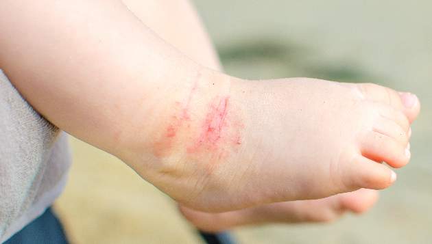
To characterize early pediatric AD skin phenotype, the investigators assessed lesional and nonlesional biopsies from 19 children under age 5 years (mean, 1.3 years) within 6 months of moderate to severe disease onset, as well as those from age-matched controls and adults, and found that, compared with adult AD, early AD involves comparable or greater epidermal hyperplasia and cellular infiltration, similar strong activation of Th2 and Th22 axes, and some Th1 skewing.
In addition, early AD involves significantly higher induction of Th17-related cytokines, compared with adult AD. Expression of filaggrin – an abundant barrier differentiation protein – was similar in AD and healthy children, whereas down-regulation is characteristic in adult AD, the investigators noted.
Nonlesional skin biopsies from the children showed both higher levels of inflammation and epidermal proliferation markers, they said.
The “surprising findings” of an early multicytokine response in new-onset pediatric AD, characterized by marked Th17, Th9, Th2, and Th22 activation, suggest that targeting of multiple cytokine axes may be needed in children with early-onset AD, one of the lead authors on the study, Emma Guttman-Yassky, MD, also of the Icahn School of Medicine at Mount Sinai, said in an interview.
Dr. Guttman-Yassky, who noted that the study was conducted in close collaboration with Amy S. Paller, MD, of Northwestern University, Chicago, explained that early AD, compared with adult AD, involves differential immune skewing and barrier responses with features that are in some ways comparable to those of psoriasis – particularly with respect to the consistently higher levels of Th17-related mediators in childhood AD, as psoriasis is considered a Th17-centered disease.
Further, the findings with respect to filaggrin represent another important aspect of the study, she said, noting that they represent a possible challenge to the notion that filaggrin is integral to disease elicitation and instigation of the “atopic march.”
The study findings may suggest novel targets for pediatric AD, and they also suggest a need for early immune intervention, not only to treat the AD, but also to prevent the atopic march, she said.
“These findings are likely to result in both different understanding of AD onset and distinct treatment approaches for infants and children,” she and her colleagues concluded.
This work was funded by a research grant from the LEO Foundation. Individual authors were supported by grants from the National Center for Advancing Translational Sciences and the National Institutes of Health Clinical and Translational Science Award program. Dr. Esaki reported having no disclosures. Dr. Guttman-Yassky reported financial relationships with numerous pharmaceutical companies.
Atopic dermatitis (AD) is the most common chronic inflammatory skin disease. Severe AD places a huge burden on patients, their families, and society in terms of health care dollars spent and lost work days. Considering the prevalence of AD, both families and dermatologists find it understandably frustrating that we have limited and often ineffective tools to treat severe AD in children. Change may be around the corner.
This study by Esaki et al. sheds critical light on the pathogenesis of early onset AD in children and we hope it will set the stage to revolutionize the treatment of AD using the paradigm of psoriasis as a model. Using lesional and nonlesional biopsies from 19 children under age 5 obtained during the first 6 months of onset of AD, Esaki et al. have demonstrated that children with AD have a multicytokine inflammatory infiltrate with Th17 predominance. This sets the stage for biologics focused on the Th17 pathway in these children, although multimodal therapy to address different cytokines may ultimately be required.
The investigators also found that children with AD had similar filaggrin expression compared to control children, implying that atopic dermatitis is at its heart an immunologic disorder rather than a barrier defect although we will likely continue to learn more about this fine balance.
As pediatric dermatologists on the front line caring for patients with severe AD, we welcome further studies and especially look forward to effective treatments for our patients who might finally experience relief of itch, clear skin, and a good night’s sleep.
A. Yasmine Kirkorian, MD, and Kalyani Marathe, MD, are pediatric dermatologists at Children’s National Health System, in the departments of dermatology and pediatrics at George Washington University, Washington, DC. They are on the editorial advisory board of Dermatology News. They had no disclosures.
Atopic dermatitis (AD) is the most common chronic inflammatory skin disease. Severe AD places a huge burden on patients, their families, and society in terms of health care dollars spent and lost work days. Considering the prevalence of AD, both families and dermatologists find it understandably frustrating that we have limited and often ineffective tools to treat severe AD in children. Change may be around the corner.
This study by Esaki et al. sheds critical light on the pathogenesis of early onset AD in children and we hope it will set the stage to revolutionize the treatment of AD using the paradigm of psoriasis as a model. Using lesional and nonlesional biopsies from 19 children under age 5 obtained during the first 6 months of onset of AD, Esaki et al. have demonstrated that children with AD have a multicytokine inflammatory infiltrate with Th17 predominance. This sets the stage for biologics focused on the Th17 pathway in these children, although multimodal therapy to address different cytokines may ultimately be required.
The investigators also found that children with AD had similar filaggrin expression compared to control children, implying that atopic dermatitis is at its heart an immunologic disorder rather than a barrier defect although we will likely continue to learn more about this fine balance.
As pediatric dermatologists on the front line caring for patients with severe AD, we welcome further studies and especially look forward to effective treatments for our patients who might finally experience relief of itch, clear skin, and a good night’s sleep.
A. Yasmine Kirkorian, MD, and Kalyani Marathe, MD, are pediatric dermatologists at Children’s National Health System, in the departments of dermatology and pediatrics at George Washington University, Washington, DC. They are on the editorial advisory board of Dermatology News. They had no disclosures.
Atopic dermatitis (AD) is the most common chronic inflammatory skin disease. Severe AD places a huge burden on patients, their families, and society in terms of health care dollars spent and lost work days. Considering the prevalence of AD, both families and dermatologists find it understandably frustrating that we have limited and often ineffective tools to treat severe AD in children. Change may be around the corner.
This study by Esaki et al. sheds critical light on the pathogenesis of early onset AD in children and we hope it will set the stage to revolutionize the treatment of AD using the paradigm of psoriasis as a model. Using lesional and nonlesional biopsies from 19 children under age 5 obtained during the first 6 months of onset of AD, Esaki et al. have demonstrated that children with AD have a multicytokine inflammatory infiltrate with Th17 predominance. This sets the stage for biologics focused on the Th17 pathway in these children, although multimodal therapy to address different cytokines may ultimately be required.
The investigators also found that children with AD had similar filaggrin expression compared to control children, implying that atopic dermatitis is at its heart an immunologic disorder rather than a barrier defect although we will likely continue to learn more about this fine balance.
As pediatric dermatologists on the front line caring for patients with severe AD, we welcome further studies and especially look forward to effective treatments for our patients who might finally experience relief of itch, clear skin, and a good night’s sleep.
A. Yasmine Kirkorian, MD, and Kalyani Marathe, MD, are pediatric dermatologists at Children’s National Health System, in the departments of dermatology and pediatrics at George Washington University, Washington, DC. They are on the editorial advisory board of Dermatology News. They had no disclosures.
The skin phenotype of new-onset pediatric atopic dermatitis differs substantially from that in adult AD, according to an assessment of biopsy findings in infants and children.
The study findings have important therapeutic implications, especially in light of the fact that much of the work in this area has been based on adult biomarkers, reflecting “decades of disease activity and chronic use of immunosuppressants in adults,” the investigators reported. Little is known about alterations in early lesions in children, which limits the advancement of targeted therapies, Hitokazu Esaki, MD, of the Icahn School of Medicine at Mount Sinai in New York City, and colleagues reported online in the Journal of Allergy and Clinical Immunology (2016 Sep 22. doi: 10.1016/j.jaci.2016.07.013).
To characterize early pediatric AD skin phenotype, the investigators assessed lesional and nonlesional biopsies from 19 children under age 5 years (mean, 1.3 years) within 6 months of moderate to severe disease onset, as well as those from age-matched controls and adults, and found that, compared with adult AD, early AD involves comparable or greater epidermal hyperplasia and cellular infiltration, similar strong activation of Th2 and Th22 axes, and some Th1 skewing.
In addition, early AD involves significantly higher induction of Th17-related cytokines, compared with adult AD. Expression of filaggrin – an abundant barrier differentiation protein – was similar in AD and healthy children, whereas down-regulation is characteristic in adult AD, the investigators noted.
Nonlesional skin biopsies from the children showed both higher levels of inflammation and epidermal proliferation markers, they said.
The “surprising findings” of an early multicytokine response in new-onset pediatric AD, characterized by marked Th17, Th9, Th2, and Th22 activation, suggest that targeting of multiple cytokine axes may be needed in children with early-onset AD, one of the lead authors on the study, Emma Guttman-Yassky, MD, also of the Icahn School of Medicine at Mount Sinai, said in an interview.
Dr. Guttman-Yassky, who noted that the study was conducted in close collaboration with Amy S. Paller, MD, of Northwestern University, Chicago, explained that early AD, compared with adult AD, involves differential immune skewing and barrier responses with features that are in some ways comparable to those of psoriasis – particularly with respect to the consistently higher levels of Th17-related mediators in childhood AD, as psoriasis is considered a Th17-centered disease.
Further, the findings with respect to filaggrin represent another important aspect of the study, she said, noting that they represent a possible challenge to the notion that filaggrin is integral to disease elicitation and instigation of the “atopic march.”
The study findings may suggest novel targets for pediatric AD, and they also suggest a need for early immune intervention, not only to treat the AD, but also to prevent the atopic march, she said.
“These findings are likely to result in both different understanding of AD onset and distinct treatment approaches for infants and children,” she and her colleagues concluded.
This work was funded by a research grant from the LEO Foundation. Individual authors were supported by grants from the National Center for Advancing Translational Sciences and the National Institutes of Health Clinical and Translational Science Award program. Dr. Esaki reported having no disclosures. Dr. Guttman-Yassky reported financial relationships with numerous pharmaceutical companies.
The skin phenotype of new-onset pediatric atopic dermatitis differs substantially from that in adult AD, according to an assessment of biopsy findings in infants and children.
The study findings have important therapeutic implications, especially in light of the fact that much of the work in this area has been based on adult biomarkers, reflecting “decades of disease activity and chronic use of immunosuppressants in adults,” the investigators reported. Little is known about alterations in early lesions in children, which limits the advancement of targeted therapies, Hitokazu Esaki, MD, of the Icahn School of Medicine at Mount Sinai in New York City, and colleagues reported online in the Journal of Allergy and Clinical Immunology (2016 Sep 22. doi: 10.1016/j.jaci.2016.07.013).
To characterize early pediatric AD skin phenotype, the investigators assessed lesional and nonlesional biopsies from 19 children under age 5 years (mean, 1.3 years) within 6 months of moderate to severe disease onset, as well as those from age-matched controls and adults, and found that, compared with adult AD, early AD involves comparable or greater epidermal hyperplasia and cellular infiltration, similar strong activation of Th2 and Th22 axes, and some Th1 skewing.
In addition, early AD involves significantly higher induction of Th17-related cytokines, compared with adult AD. Expression of filaggrin – an abundant barrier differentiation protein – was similar in AD and healthy children, whereas down-regulation is characteristic in adult AD, the investigators noted.
Nonlesional skin biopsies from the children showed both higher levels of inflammation and epidermal proliferation markers, they said.
The “surprising findings” of an early multicytokine response in new-onset pediatric AD, characterized by marked Th17, Th9, Th2, and Th22 activation, suggest that targeting of multiple cytokine axes may be needed in children with early-onset AD, one of the lead authors on the study, Emma Guttman-Yassky, MD, also of the Icahn School of Medicine at Mount Sinai, said in an interview.
Dr. Guttman-Yassky, who noted that the study was conducted in close collaboration with Amy S. Paller, MD, of Northwestern University, Chicago, explained that early AD, compared with adult AD, involves differential immune skewing and barrier responses with features that are in some ways comparable to those of psoriasis – particularly with respect to the consistently higher levels of Th17-related mediators in childhood AD, as psoriasis is considered a Th17-centered disease.
Further, the findings with respect to filaggrin represent another important aspect of the study, she said, noting that they represent a possible challenge to the notion that filaggrin is integral to disease elicitation and instigation of the “atopic march.”
The study findings may suggest novel targets for pediatric AD, and they also suggest a need for early immune intervention, not only to treat the AD, but also to prevent the atopic march, she said.
“These findings are likely to result in both different understanding of AD onset and distinct treatment approaches for infants and children,” she and her colleagues concluded.
This work was funded by a research grant from the LEO Foundation. Individual authors were supported by grants from the National Center for Advancing Translational Sciences and the National Institutes of Health Clinical and Translational Science Award program. Dr. Esaki reported having no disclosures. Dr. Guttman-Yassky reported financial relationships with numerous pharmaceutical companies.
FROM THE JOURNAL OF ALLERGY AND CLINICAL IMMUNOLOGY
Key clinical point: The skin phenotype of new-onset pediatric atopic dermatitis differs substantially from that in adult AD, which has important therapeutic implications, according to a study of biopsy findings in infants and children.
Major finding: Early AD involves significantly higher induction of Th17-related cytokines, compared with adult AD.
Data source: An analysis of biopsies from 19 children with AD.
Disclosures: This work was funded by a research grant from the LEO Foundation. Individual authors were supported by grants from the National Center for Advancing Translational Sciences and the National Institutes of Health Clinical and Translational Science Award program. Dr. Esaki reported having no disclosures. Dr. Guttman-Yassky reported financial relationships with numerous pharmaceutical companies.
OMA publishes pediatric obesity treatment guide
The Pediatric Obesity Algorithm – a free state-of-the-science management review from the Obesity Medicine Association – should help clinicians weed through the options for overweight kids, according to Texas pediatrician and lead author Suzanne Cuda, MD.
The group put the latest thinking into one place to serve as a handy guide, and plans to update it every 2 years. The idea was to present choices, not push particular approaches. “Many of my colleagues have expressed concern [that] they do not have the resources to provide the kind of support these kids need,” said Dr. Cuda, director of the weight management clinic at the Children’s Hospital of San Antonio and associate professor of pediatrics at the Baylor College of Medicine. Clinicians can find the guide at www.PediatricObesityAlgorithm.org.

The effort also will help clinicians prepare for American Board of Obesity Medicine certification, since it was “designed to address the content on the exam. A lot of tools out there for children [emphasize] prevention. Our starting point was children who are already overweight,“ she said.
The document covers risk factors, differential diagnoses, assessment, diet, appropriate activity levels, medications, surgery, comorbidity management, and other issues, often broken down by age and body mass index.
Nothing is particularly controversial, although some clinicians are reluctant to move beyond diet and exercise for kids, Dr. Cuda said.
The Obesity Medicine Association (OMA) was aware of that, and so was careful to note, for instance, which obesity medications are approved for pediatric use – orlistat (Xenical), metformin, and phentermine – and their real-world effect.
“We didn’t cover all the drugs out there” because many haven’t been tested in children, Dr. Cuda said. The group also highlights antiseizure and other drugs that put on weight.
The guide covers birth to adulthood. There can be signs of problems even before the first birthday, such as weight above the 95th percentile. In those cases, evidence supports exclusive breast feeding for as long as possible, and no more than 24 ounces per day in formula-fed children, with no cereal or media watching.
OMA hasn’t submitted its work for endorsement by other groups, so other associations haven’t signed onto it. “We didn’t want to prolong putting it out there to go through that whole process,” Dr. Cuda said.
Even so, some organizations are aware of the contents and support the effort. “It aligns with the resources we have already developed on this topic. These are core competencies ... all physicians should have, regardless of whether they are certified in obesity medicine,” said an American Academy of Pediatrics staff member.
Until recently, OMA was known as the American Society of Bariatric Physicians. It rebranded itself to avoid being mistaken for a bariatric surgery group.
Dr. Cuda and the other authors had no disclosures. There was no industry funding for the work.
The Pediatric Obesity Algorithm – a free state-of-the-science management review from the Obesity Medicine Association – should help clinicians weed through the options for overweight kids, according to Texas pediatrician and lead author Suzanne Cuda, MD.
The group put the latest thinking into one place to serve as a handy guide, and plans to update it every 2 years. The idea was to present choices, not push particular approaches. “Many of my colleagues have expressed concern [that] they do not have the resources to provide the kind of support these kids need,” said Dr. Cuda, director of the weight management clinic at the Children’s Hospital of San Antonio and associate professor of pediatrics at the Baylor College of Medicine. Clinicians can find the guide at www.PediatricObesityAlgorithm.org.
The effort also will help clinicians prepare for American Board of Obesity Medicine certification, since it was “designed to address the content on the exam. A lot of tools out there for children [emphasize] prevention. Our starting point was children who are already overweight,“ she said.
The document covers risk factors, differential diagnoses, assessment, diet, appropriate activity levels, medications, surgery, comorbidity management, and other issues, often broken down by age and body mass index.
Nothing is particularly controversial, although some clinicians are reluctant to move beyond diet and exercise for kids, Dr. Cuda said.
The Obesity Medicine Association (OMA) was aware of that, and so was careful to note, for instance, which obesity medications are approved for pediatric use – orlistat (Xenical), metformin, and phentermine – and their real-world effect.
“We didn’t cover all the drugs out there” because many haven’t been tested in children, Dr. Cuda said. The group also highlights antiseizure and other drugs that put on weight.
The guide covers birth to adulthood. There can be signs of problems even before the first birthday, such as weight above the 95th percentile. In those cases, evidence supports exclusive breast feeding for as long as possible, and no more than 24 ounces per day in formula-fed children, with no cereal or media watching.
OMA hasn’t submitted its work for endorsement by other groups, so other associations haven’t signed onto it. “We didn’t want to prolong putting it out there to go through that whole process,” Dr. Cuda said.
Even so, some organizations are aware of the contents and support the effort. “It aligns with the resources we have already developed on this topic. These are core competencies ... all physicians should have, regardless of whether they are certified in obesity medicine,” said an American Academy of Pediatrics staff member.
Until recently, OMA was known as the American Society of Bariatric Physicians. It rebranded itself to avoid being mistaken for a bariatric surgery group.
Dr. Cuda and the other authors had no disclosures. There was no industry funding for the work.
The Pediatric Obesity Algorithm – a free state-of-the-science management review from the Obesity Medicine Association – should help clinicians weed through the options for overweight kids, according to Texas pediatrician and lead author Suzanne Cuda, MD.
The group put the latest thinking into one place to serve as a handy guide, and plans to update it every 2 years. The idea was to present choices, not push particular approaches. “Many of my colleagues have expressed concern [that] they do not have the resources to provide the kind of support these kids need,” said Dr. Cuda, director of the weight management clinic at the Children’s Hospital of San Antonio and associate professor of pediatrics at the Baylor College of Medicine. Clinicians can find the guide at www.PediatricObesityAlgorithm.org.
The effort also will help clinicians prepare for American Board of Obesity Medicine certification, since it was “designed to address the content on the exam. A lot of tools out there for children [emphasize] prevention. Our starting point was children who are already overweight,“ she said.
The document covers risk factors, differential diagnoses, assessment, diet, appropriate activity levels, medications, surgery, comorbidity management, and other issues, often broken down by age and body mass index.
Nothing is particularly controversial, although some clinicians are reluctant to move beyond diet and exercise for kids, Dr. Cuda said.
The Obesity Medicine Association (OMA) was aware of that, and so was careful to note, for instance, which obesity medications are approved for pediatric use – orlistat (Xenical), metformin, and phentermine – and their real-world effect.
“We didn’t cover all the drugs out there” because many haven’t been tested in children, Dr. Cuda said. The group also highlights antiseizure and other drugs that put on weight.
The guide covers birth to adulthood. There can be signs of problems even before the first birthday, such as weight above the 95th percentile. In those cases, evidence supports exclusive breast feeding for as long as possible, and no more than 24 ounces per day in formula-fed children, with no cereal or media watching.
OMA hasn’t submitted its work for endorsement by other groups, so other associations haven’t signed onto it. “We didn’t want to prolong putting it out there to go through that whole process,” Dr. Cuda said.
Even so, some organizations are aware of the contents and support the effort. “It aligns with the resources we have already developed on this topic. These are core competencies ... all physicians should have, regardless of whether they are certified in obesity medicine,” said an American Academy of Pediatrics staff member.
Until recently, OMA was known as the American Society of Bariatric Physicians. It rebranded itself to avoid being mistaken for a bariatric surgery group.
Dr. Cuda and the other authors had no disclosures. There was no industry funding for the work.
FROM THE OBESITY MEDICINE ASSOCIATION

Prenatal Tdap vaccination prevents occurrence, reduces severity of pertussis in infants
Prenatal Tdap vaccination prevents the occurrence of and reduces the severity of pertussis in infants, two retrospective cohort studies showed.
In 2012, the Advisory Committee on Immunization Practices recommended that pregnant women receive a Tdap vaccination during their third trimester of pregnancy to optimize the transfer of pertussis antibodies to the fetus. Since the committee’s recommendation, no studies to evaluate the effectiveness of this strategy have been conducted in the United States, Kathleen Winter of the California Department of Public Health and her associates reported (Clin. Infect. Dis. 2016. doi: 10.1093/cid/ciw634) .

Therefore, Ms. Winter and her associates conducted two separate retrospective cohort studies: one to compare the effectiveness of prenatal versus postpartum Tdap vaccination in preventing pertussis and the second to investigate the effectiveness of prenatal Tdap vaccination on pertussis severity in infants.
For the comparison study, researchers identified 42,941 mothers who were vaccinated during pregnancy and 31,563 mothers who were vaccinated following delivery. The stage of pregnancy at the time of vaccination was documented for 42,218 of the mothers vaccinated prenatally, and 77% were vaccinated during the recommended window of 27-36 weeks’ gestation. For the remaining mothers, 14% received vaccinations before 27 weeks’ gestation, and 9% were vaccinated after 36 weeks’ gestation. Infants whose mothers received the Tdap vaccine at any point during pregnancy were less likely to develop pertussis before 8 weeks of age (odds ratio, 0.36; 95% confidence interval, 0.15-0.89) or 12 weeks of age (OR, 0.47; 95% CI, 0.24-0.92).
Moreover, infants whose mothers received the vaccine during 27-36 weeks’ gestation were less likely to develop pertussis than were infants whose mothers were vaccinated during pregnancy but outside the 27- to 36-week time frame (OR, 0.22; 95% CI, 0.08-0.63).
Overall, Tdap vaccination during 27-36 weeks’ gestation was 85% more effective in reducing pertussis in infants younger than 8 weeks old and 72% more effective in preventing pertussis in infants younger than 12 weeks old, compared with postpartum vaccination.
In a companion paper, researchers described the results of a separate retrospective cohort study, the “first known study demonstrating that prenatal Tdap vaccination reduces severity of disease in infants who are infected with pertussis,” according to Ms. Winter and her associates (Clin. Infect. Dis. 2016. doi: 10.1093/cid/ciw633).
For this study, the researchers identified 420 infants born between January 2011 and December 2015 who reported with pertussis at less than 63 days of age and had known maternal vaccination status. Of those 420 infants, only 49 mothers (12%) received Tdap vaccination during pregnancy, and only 14 received Tdap during the recommended window of 27-36 weeks’ gestation.
Infants born to mothers who received the Tdap vaccine during pregnancy were significantly less likely to be hospitalized when they developed pertussis (OR, 0.4; 95% CI, 0.2-0.9), were less likely to be admitted to the intensive care unit (OR, 0.5; 95% CI, 0.2-1.2), and had shorter hospital stays (median, 3 days vs. 6 days; P = .019). Infants born to vaccinated mothers also were older when they developed pertussis and were less likely to display common pertussis symptoms: paroxysmal cough, apnea, cyanosis, and whoop.
“Prenatal Tdap vaccination was 58% effective in preventing hospitalizations in infants infected with pertussis,” the researchers wrote, adding that “prenatal Tdap vaccination of mothers is a critical strategy for reducing the morbidity and mortality from pertussis.”
On Twitter @jessnicolecraig
The two papers by Ms. Winter and her colleagues are remarkably important and of high clinical interest. In 2012, recognizing the significant morbidity and mortality of pertussis among infants in the first 2-3 months of life and the lack of a newborn vaccination for pertussis, the Advisory Committee on Immunization Practices (ACIP) recommended maternal vaccination at 27-36 weeks’ gestation to stem the tide of increasing pertussis infections in this vulnerable age group. The recommendation was based on immunogenicity studies that showed maternal vaccination at 27-36 weeks’ gestation boosted maternal antibodies to pertussis antigens in the acellular vaccines used and that the antibodies crossed the placenta in sufficient amounts such that protection in the newborn could be anticipated up to at least 3 months of age.

|
Dr. Michael E. Pichichero |
Winter et al. showed in these two companion papers that the ACIP recommendation was effective and excellent results have been achieved in California. I would expect the same results across the United States.
While this research is novel in the United States, the results could have been anticipated because two prior studies from the United Kingdom and Australia demonstrated the effectiveness of maternal Tdap vaccination. Although the study design was retrospective (a weakness, compared with a prospective study design), the findings are convincing.
More vaccines are being studied for possible maternal use during pregnancy. The hesitation of the “unknown risks” of maternal vaccination slowly are disappearing as more success stories like this one provide confidence in terms of the lack of side effects and the substantial benefit.
Dr. Michael E. Pichichero is a clinical professor in the department of pediatrics at the University of Rochester (N.Y.), a research professor at the Rochester Institute of Technology, and the director of research at Rochester General Hospital Research Institute. Dr. Pichichero said he had no relevant financial disclosures.
The two papers by Ms. Winter and her colleagues are remarkably important and of high clinical interest. In 2012, recognizing the significant morbidity and mortality of pertussis among infants in the first 2-3 months of life and the lack of a newborn vaccination for pertussis, the Advisory Committee on Immunization Practices (ACIP) recommended maternal vaccination at 27-36 weeks’ gestation to stem the tide of increasing pertussis infections in this vulnerable age group. The recommendation was based on immunogenicity studies that showed maternal vaccination at 27-36 weeks’ gestation boosted maternal antibodies to pertussis antigens in the acellular vaccines used and that the antibodies crossed the placenta in sufficient amounts such that protection in the newborn could be anticipated up to at least 3 months of age.
|
|
Dr. Michael E. Pichichero |
Winter et al. showed in these two companion papers that the ACIP recommendation was effective and excellent results have been achieved in California. I would expect the same results across the United States.
While this research is novel in the United States, the results could have been anticipated because two prior studies from the United Kingdom and Australia demonstrated the effectiveness of maternal Tdap vaccination. Although the study design was retrospective (a weakness, compared with a prospective study design), the findings are convincing.
More vaccines are being studied for possible maternal use during pregnancy. The hesitation of the “unknown risks” of maternal vaccination slowly are disappearing as more success stories like this one provide confidence in terms of the lack of side effects and the substantial benefit.
Dr. Michael E. Pichichero is a clinical professor in the department of pediatrics at the University of Rochester (N.Y.), a research professor at the Rochester Institute of Technology, and the director of research at Rochester General Hospital Research Institute. Dr. Pichichero said he had no relevant financial disclosures.
The two papers by Ms. Winter and her colleagues are remarkably important and of high clinical interest. In 2012, recognizing the significant morbidity and mortality of pertussis among infants in the first 2-3 months of life and the lack of a newborn vaccination for pertussis, the Advisory Committee on Immunization Practices (ACIP) recommended maternal vaccination at 27-36 weeks’ gestation to stem the tide of increasing pertussis infections in this vulnerable age group. The recommendation was based on immunogenicity studies that showed maternal vaccination at 27-36 weeks’ gestation boosted maternal antibodies to pertussis antigens in the acellular vaccines used and that the antibodies crossed the placenta in sufficient amounts such that protection in the newborn could be anticipated up to at least 3 months of age.
|
|
Dr. Michael E. Pichichero |
Winter et al. showed in these two companion papers that the ACIP recommendation was effective and excellent results have been achieved in California. I would expect the same results across the United States.
While this research is novel in the United States, the results could have been anticipated because two prior studies from the United Kingdom and Australia demonstrated the effectiveness of maternal Tdap vaccination. Although the study design was retrospective (a weakness, compared with a prospective study design), the findings are convincing.
More vaccines are being studied for possible maternal use during pregnancy. The hesitation of the “unknown risks” of maternal vaccination slowly are disappearing as more success stories like this one provide confidence in terms of the lack of side effects and the substantial benefit.
Dr. Michael E. Pichichero is a clinical professor in the department of pediatrics at the University of Rochester (N.Y.), a research professor at the Rochester Institute of Technology, and the director of research at Rochester General Hospital Research Institute. Dr. Pichichero said he had no relevant financial disclosures.
Prenatal Tdap vaccination prevents the occurrence of and reduces the severity of pertussis in infants, two retrospective cohort studies showed.
In 2012, the Advisory Committee on Immunization Practices recommended that pregnant women receive a Tdap vaccination during their third trimester of pregnancy to optimize the transfer of pertussis antibodies to the fetus. Since the committee’s recommendation, no studies to evaluate the effectiveness of this strategy have been conducted in the United States, Kathleen Winter of the California Department of Public Health and her associates reported (Clin. Infect. Dis. 2016. doi: 10.1093/cid/ciw634) .
Therefore, Ms. Winter and her associates conducted two separate retrospective cohort studies: one to compare the effectiveness of prenatal versus postpartum Tdap vaccination in preventing pertussis and the second to investigate the effectiveness of prenatal Tdap vaccination on pertussis severity in infants.
For the comparison study, researchers identified 42,941 mothers who were vaccinated during pregnancy and 31,563 mothers who were vaccinated following delivery. The stage of pregnancy at the time of vaccination was documented for 42,218 of the mothers vaccinated prenatally, and 77% were vaccinated during the recommended window of 27-36 weeks’ gestation. For the remaining mothers, 14% received vaccinations before 27 weeks’ gestation, and 9% were vaccinated after 36 weeks’ gestation. Infants whose mothers received the Tdap vaccine at any point during pregnancy were less likely to develop pertussis before 8 weeks of age (odds ratio, 0.36; 95% confidence interval, 0.15-0.89) or 12 weeks of age (OR, 0.47; 95% CI, 0.24-0.92).
Moreover, infants whose mothers received the vaccine during 27-36 weeks’ gestation were less likely to develop pertussis than were infants whose mothers were vaccinated during pregnancy but outside the 27- to 36-week time frame (OR, 0.22; 95% CI, 0.08-0.63).
Overall, Tdap vaccination during 27-36 weeks’ gestation was 85% more effective in reducing pertussis in infants younger than 8 weeks old and 72% more effective in preventing pertussis in infants younger than 12 weeks old, compared with postpartum vaccination.
In a companion paper, researchers described the results of a separate retrospective cohort study, the “first known study demonstrating that prenatal Tdap vaccination reduces severity of disease in infants who are infected with pertussis,” according to Ms. Winter and her associates (Clin. Infect. Dis. 2016. doi: 10.1093/cid/ciw633).
For this study, the researchers identified 420 infants born between January 2011 and December 2015 who reported with pertussis at less than 63 days of age and had known maternal vaccination status. Of those 420 infants, only 49 mothers (12%) received Tdap vaccination during pregnancy, and only 14 received Tdap during the recommended window of 27-36 weeks’ gestation.
Infants born to mothers who received the Tdap vaccine during pregnancy were significantly less likely to be hospitalized when they developed pertussis (OR, 0.4; 95% CI, 0.2-0.9), were less likely to be admitted to the intensive care unit (OR, 0.5; 95% CI, 0.2-1.2), and had shorter hospital stays (median, 3 days vs. 6 days; P = .019). Infants born to vaccinated mothers also were older when they developed pertussis and were less likely to display common pertussis symptoms: paroxysmal cough, apnea, cyanosis, and whoop.
“Prenatal Tdap vaccination was 58% effective in preventing hospitalizations in infants infected with pertussis,” the researchers wrote, adding that “prenatal Tdap vaccination of mothers is a critical strategy for reducing the morbidity and mortality from pertussis.”
On Twitter @jessnicolecraig
Prenatal Tdap vaccination prevents the occurrence of and reduces the severity of pertussis in infants, two retrospective cohort studies showed.
In 2012, the Advisory Committee on Immunization Practices recommended that pregnant women receive a Tdap vaccination during their third trimester of pregnancy to optimize the transfer of pertussis antibodies to the fetus. Since the committee’s recommendation, no studies to evaluate the effectiveness of this strategy have been conducted in the United States, Kathleen Winter of the California Department of Public Health and her associates reported (Clin. Infect. Dis. 2016. doi: 10.1093/cid/ciw634) .
Therefore, Ms. Winter and her associates conducted two separate retrospective cohort studies: one to compare the effectiveness of prenatal versus postpartum Tdap vaccination in preventing pertussis and the second to investigate the effectiveness of prenatal Tdap vaccination on pertussis severity in infants.
For the comparison study, researchers identified 42,941 mothers who were vaccinated during pregnancy and 31,563 mothers who were vaccinated following delivery. The stage of pregnancy at the time of vaccination was documented for 42,218 of the mothers vaccinated prenatally, and 77% were vaccinated during the recommended window of 27-36 weeks’ gestation. For the remaining mothers, 14% received vaccinations before 27 weeks’ gestation, and 9% were vaccinated after 36 weeks’ gestation. Infants whose mothers received the Tdap vaccine at any point during pregnancy were less likely to develop pertussis before 8 weeks of age (odds ratio, 0.36; 95% confidence interval, 0.15-0.89) or 12 weeks of age (OR, 0.47; 95% CI, 0.24-0.92).
Moreover, infants whose mothers received the vaccine during 27-36 weeks’ gestation were less likely to develop pertussis than were infants whose mothers were vaccinated during pregnancy but outside the 27- to 36-week time frame (OR, 0.22; 95% CI, 0.08-0.63).
Overall, Tdap vaccination during 27-36 weeks’ gestation was 85% more effective in reducing pertussis in infants younger than 8 weeks old and 72% more effective in preventing pertussis in infants younger than 12 weeks old, compared with postpartum vaccination.
In a companion paper, researchers described the results of a separate retrospective cohort study, the “first known study demonstrating that prenatal Tdap vaccination reduces severity of disease in infants who are infected with pertussis,” according to Ms. Winter and her associates (Clin. Infect. Dis. 2016. doi: 10.1093/cid/ciw633).
For this study, the researchers identified 420 infants born between January 2011 and December 2015 who reported with pertussis at less than 63 days of age and had known maternal vaccination status. Of those 420 infants, only 49 mothers (12%) received Tdap vaccination during pregnancy, and only 14 received Tdap during the recommended window of 27-36 weeks’ gestation.
Infants born to mothers who received the Tdap vaccine during pregnancy were significantly less likely to be hospitalized when they developed pertussis (OR, 0.4; 95% CI, 0.2-0.9), were less likely to be admitted to the intensive care unit (OR, 0.5; 95% CI, 0.2-1.2), and had shorter hospital stays (median, 3 days vs. 6 days; P = .019). Infants born to vaccinated mothers also were older when they developed pertussis and were less likely to display common pertussis symptoms: paroxysmal cough, apnea, cyanosis, and whoop.
“Prenatal Tdap vaccination was 58% effective in preventing hospitalizations in infants infected with pertussis,” the researchers wrote, adding that “prenatal Tdap vaccination of mothers is a critical strategy for reducing the morbidity and mortality from pertussis.”
On Twitter @jessnicolecraig
FROM CLINICAL INFECTIOUS DISEASES
Key clinical point: Prenatal Tdap vaccination prevents the occurrence of and reduces the severity of pertussis in infants.
Major finding: Tdap vaccination during 27-36 weeks’ gestation was 85% more effective in reducing pertussis in infants younger than 8 weeks old, compared with postpartum Tdap vaccination.
Data source: Two retrospective cohort studies.
Disclosures: The California Department of Public Health Immunization Branch funded this study. One investigator reported receiving financial compensation from GlaxoSmithKline and serving as a speaker for Sanofi Pasteur.

Reduced rotavirus detection after vaccine licensure tied to herd immunity
Rotavirus detection was found to be diminished after rotavirus vaccine licensure, which is consistent with herd immunity, according to Harvey W. Kaufman, MD, and Zhen Chen.
During the 11-year period, 276,949 specimens were submitted for rotavirus antigen detection. In the prevaccine period, the laboratory performed an average of 31,800 tests for rotavirus antigen detection annually, of which 21% were positive. During the postvaccine period, an average of 20,981 annual tests were performed, with only 6% having a positive result, which represents an 82% reduction in the total number of positive results and a 73% reduction in the positivity rate (P less than .001). In the transition period, the positivity rate for rotavirus antigen detection was 14%, which represents a 27% reduction in the positive number and a 32% reduction in the positivity rate, compared with the prevaccine period (P less than .001).

The study noted that the positivity rate during each of the three periods was nearly identical for boys and girls, with 21.1% for boys and 20.9% for girls during the prevaccine period, 9.8% for boys and 9.6% for girls in the transition period, and 4.9% for boys and 4.6% for girls in the postvaccine period.
“The data support the notion of herd immunity in children unlikely to have been vaccinated,” the researchers concluded. “Although the postvaccination period featured alternating years of higher and lower positivity, the peak seasons have a lower positivity rate than in the prevaccination period.”
Find the full study in Pediatrics (2016 Sept. 23. doi: 10.1542/peds.2016-1173).
Rotavirus detection was found to be diminished after rotavirus vaccine licensure, which is consistent with herd immunity, according to Harvey W. Kaufman, MD, and Zhen Chen.
During the 11-year period, 276,949 specimens were submitted for rotavirus antigen detection. In the prevaccine period, the laboratory performed an average of 31,800 tests for rotavirus antigen detection annually, of which 21% were positive. During the postvaccine period, an average of 20,981 annual tests were performed, with only 6% having a positive result, which represents an 82% reduction in the total number of positive results and a 73% reduction in the positivity rate (P less than .001). In the transition period, the positivity rate for rotavirus antigen detection was 14%, which represents a 27% reduction in the positive number and a 32% reduction in the positivity rate, compared with the prevaccine period (P less than .001).
The study noted that the positivity rate during each of the three periods was nearly identical for boys and girls, with 21.1% for boys and 20.9% for girls during the prevaccine period, 9.8% for boys and 9.6% for girls in the transition period, and 4.9% for boys and 4.6% for girls in the postvaccine period.
“The data support the notion of herd immunity in children unlikely to have been vaccinated,” the researchers concluded. “Although the postvaccination period featured alternating years of higher and lower positivity, the peak seasons have a lower positivity rate than in the prevaccination period.”
Find the full study in Pediatrics (2016 Sept. 23. doi: 10.1542/peds.2016-1173).
Rotavirus detection was found to be diminished after rotavirus vaccine licensure, which is consistent with herd immunity, according to Harvey W. Kaufman, MD, and Zhen Chen.
During the 11-year period, 276,949 specimens were submitted for rotavirus antigen detection. In the prevaccine period, the laboratory performed an average of 31,800 tests for rotavirus antigen detection annually, of which 21% were positive. During the postvaccine period, an average of 20,981 annual tests were performed, with only 6% having a positive result, which represents an 82% reduction in the total number of positive results and a 73% reduction in the positivity rate (P less than .001). In the transition period, the positivity rate for rotavirus antigen detection was 14%, which represents a 27% reduction in the positive number and a 32% reduction in the positivity rate, compared with the prevaccine period (P less than .001).
The study noted that the positivity rate during each of the three periods was nearly identical for boys and girls, with 21.1% for boys and 20.9% for girls during the prevaccine period, 9.8% for boys and 9.6% for girls in the transition period, and 4.9% for boys and 4.6% for girls in the postvaccine period.
“The data support the notion of herd immunity in children unlikely to have been vaccinated,” the researchers concluded. “Although the postvaccination period featured alternating years of higher and lower positivity, the peak seasons have a lower positivity rate than in the prevaccination period.”
Find the full study in Pediatrics (2016 Sept. 23. doi: 10.1542/peds.2016-1173).
FROM PEDIATRICS

RAS mutations have contradictory roles in ALL

Photo by Debbie Vogel
New research has revealed relapse-specific mutations in pediatric acute lymphoblastic leukemia (ALL) and suggests that mutations in the RAS family may drive both resistance and sensitivity to treatment.
Specifically, the study showed that KRAS-mutant ALL cells were resistant to methotrexate but exhibited increased sensitivity to vincristine.
Hossein Khiabanian, PhD, of Rutgers Cancer Institute of New Jersey, and his colleagues reported these findings in PNAS.
The researchers performed whole-exome and whole-genome sequencing on samples from 55 pediatric patients with relapsed ALL, identified specific genomic changes, and validated these findings in 279 additional samples.
“We found that ALL relapse emerges from small, often clinically undetectable populations of cancer cells that are only partially genetically similar to the dominant leukemic population at diagnosis,” Dr Khiabanian said. “We also identified numerous new mutations in genes involved in drug resistance that are specific to relapsed ALL.”
In the first 55 patients (33 T-cell ALLs and 22 B-cell precursor ALLs), the researchers identified 27 recurrently mutated genes whose mutations were preferentially selected or retained at the time of relapse.
The team said 23 (85%) of these mutated genes were not previously implicated in ALL relapse—HTR3A, MED12, USP9X, CACNA1H, TENM3, AACS, SAMD4A, ANO5, PAPPA, NAALADL2, HIST3H2A, FZD7, TBX15, NEB, GREB1L, PLXNA4, SGK223, TSC1, PTPRG, FGF10, SYCP2, TRPM3, and EYS.
The researchers found mutations in the same genes when they analyzed 49 paired diagnosis and relapse B-cell precursor ALL samples as well as an additional 230 relapsed B-cell precursor ALL samples. In addition, the analyses revealed mutations in NT5C2, NR3C1, CREBBP, KMT2D, JAK2, JAK3, and TP53.
The team also noted that some patient samples showed retention or emergence of RAS mutant clones at relapse. In other patients, RAS mutant clones that were present at diagnosis were replaced by RAS wild-type populations at relapse.
The researchers said this suggests a role for both positive and negative selection evolutionary pressures in clonal evolution of RAS-mutant leukemia.
To investigate this further, they analyzed mouse and human wild-type and mutant RAS isogenic leukemia cells. In these experiments, KRAS-mutant cells showed increased sensitivity to vincristine and increased resistance to methotrexate.
“These results highlight how drug therapy can impact the evolution of leukemia and show a previously unrecognized role of RAS mutations as causes of both sensitivity and resistance to chemotherapy,” Dr Khiabanian said.
“Early identification of these mutations, as well as other genetic alterations that have been shown to induce therapeutic resistance in leukemia patients, is pertinent in guiding precision medicine treatment strategies and prevention of relapsed disease—a goal that is now being pursued in my lab at Rutgers.” 
Photo by Debbie Vogel
New research has revealed relapse-specific mutations in pediatric acute lymphoblastic leukemia (ALL) and suggests that mutations in the RAS family may drive both resistance and sensitivity to treatment.
Specifically, the study showed that KRAS-mutant ALL cells were resistant to methotrexate but exhibited increased sensitivity to vincristine.
Hossein Khiabanian, PhD, of Rutgers Cancer Institute of New Jersey, and his colleagues reported these findings in PNAS.
The researchers performed whole-exome and whole-genome sequencing on samples from 55 pediatric patients with relapsed ALL, identified specific genomic changes, and validated these findings in 279 additional samples.
“We found that ALL relapse emerges from small, often clinically undetectable populations of cancer cells that are only partially genetically similar to the dominant leukemic population at diagnosis,” Dr Khiabanian said. “We also identified numerous new mutations in genes involved in drug resistance that are specific to relapsed ALL.”
In the first 55 patients (33 T-cell ALLs and 22 B-cell precursor ALLs), the researchers identified 27 recurrently mutated genes whose mutations were preferentially selected or retained at the time of relapse.
The team said 23 (85%) of these mutated genes were not previously implicated in ALL relapse—HTR3A, MED12, USP9X, CACNA1H, TENM3, AACS, SAMD4A, ANO5, PAPPA, NAALADL2, HIST3H2A, FZD7, TBX15, NEB, GREB1L, PLXNA4, SGK223, TSC1, PTPRG, FGF10, SYCP2, TRPM3, and EYS.
The researchers found mutations in the same genes when they analyzed 49 paired diagnosis and relapse B-cell precursor ALL samples as well as an additional 230 relapsed B-cell precursor ALL samples. In addition, the analyses revealed mutations in NT5C2, NR3C1, CREBBP, KMT2D, JAK2, JAK3, and TP53.
The team also noted that some patient samples showed retention or emergence of RAS mutant clones at relapse. In other patients, RAS mutant clones that were present at diagnosis were replaced by RAS wild-type populations at relapse.
The researchers said this suggests a role for both positive and negative selection evolutionary pressures in clonal evolution of RAS-mutant leukemia.
To investigate this further, they analyzed mouse and human wild-type and mutant RAS isogenic leukemia cells. In these experiments, KRAS-mutant cells showed increased sensitivity to vincristine and increased resistance to methotrexate.
“These results highlight how drug therapy can impact the evolution of leukemia and show a previously unrecognized role of RAS mutations as causes of both sensitivity and resistance to chemotherapy,” Dr Khiabanian said.
“Early identification of these mutations, as well as other genetic alterations that have been shown to induce therapeutic resistance in leukemia patients, is pertinent in guiding precision medicine treatment strategies and prevention of relapsed disease—a goal that is now being pursued in my lab at Rutgers.”
Photo by Debbie Vogel
New research has revealed relapse-specific mutations in pediatric acute lymphoblastic leukemia (ALL) and suggests that mutations in the RAS family may drive both resistance and sensitivity to treatment.
Specifically, the study showed that KRAS-mutant ALL cells were resistant to methotrexate but exhibited increased sensitivity to vincristine.
Hossein Khiabanian, PhD, of Rutgers Cancer Institute of New Jersey, and his colleagues reported these findings in PNAS.
The researchers performed whole-exome and whole-genome sequencing on samples from 55 pediatric patients with relapsed ALL, identified specific genomic changes, and validated these findings in 279 additional samples.
“We found that ALL relapse emerges from small, often clinically undetectable populations of cancer cells that are only partially genetically similar to the dominant leukemic population at diagnosis,” Dr Khiabanian said. “We also identified numerous new mutations in genes involved in drug resistance that are specific to relapsed ALL.”
In the first 55 patients (33 T-cell ALLs and 22 B-cell precursor ALLs), the researchers identified 27 recurrently mutated genes whose mutations were preferentially selected or retained at the time of relapse.
The team said 23 (85%) of these mutated genes were not previously implicated in ALL relapse—HTR3A, MED12, USP9X, CACNA1H, TENM3, AACS, SAMD4A, ANO5, PAPPA, NAALADL2, HIST3H2A, FZD7, TBX15, NEB, GREB1L, PLXNA4, SGK223, TSC1, PTPRG, FGF10, SYCP2, TRPM3, and EYS.
The researchers found mutations in the same genes when they analyzed 49 paired diagnosis and relapse B-cell precursor ALL samples as well as an additional 230 relapsed B-cell precursor ALL samples. In addition, the analyses revealed mutations in NT5C2, NR3C1, CREBBP, KMT2D, JAK2, JAK3, and TP53.
The team also noted that some patient samples showed retention or emergence of RAS mutant clones at relapse. In other patients, RAS mutant clones that were present at diagnosis were replaced by RAS wild-type populations at relapse.
The researchers said this suggests a role for both positive and negative selection evolutionary pressures in clonal evolution of RAS-mutant leukemia.
To investigate this further, they analyzed mouse and human wild-type and mutant RAS isogenic leukemia cells. In these experiments, KRAS-mutant cells showed increased sensitivity to vincristine and increased resistance to methotrexate.
“These results highlight how drug therapy can impact the evolution of leukemia and show a previously unrecognized role of RAS mutations as causes of both sensitivity and resistance to chemotherapy,” Dr Khiabanian said.
“Early identification of these mutations, as well as other genetic alterations that have been shown to induce therapeutic resistance in leukemia patients, is pertinent in guiding precision medicine treatment strategies and prevention of relapsed disease—a goal that is now being pursued in my lab at Rutgers.”
Back pain: Let’s get it straight!
Back pain is a common complaint among adolescents. But there are several misconceptions about back pain that lead to excessive referrals and unnecessary imaging.
One of the most common of these misconceptions is that back pain in adolescents is caused by their carrying a heavy backpack. When this was researched, carrying a heavy backpack had a weak association with back pain,1,2 but because so many relate the two, the American Academy of Pediatrics came out with recommendations limiting the weight to 20% of the child’s body weight.3

Another misconception regarding back pain in children is that the cause is likely serious and needs a prompt work-up. A study followed 560 children who were diagnosed with low back pain and idiopathic scoliosis. In only 9% of cases was an underlying pathologic condition found: 29 patients had spondylolysis or spondylolisthesis, 9 had Scheuermann’s kyphosis, 5 had a syrinx, 2 had a herniated disk, 1 had hydromyelia, 1 had a tethered cord, and 1 had an intraspinal tumor.4
As with all diagnoses, a detailed history is very important. Is the pain acute or chronic? Does the pain radiate? Is there nighttime pain? Where exactly is the pain? Commonly, with acute onset there is an association of new activity or trauma. A good social history is also important because complaints of back pain are high on the list with psychosomatic issues as well.
The physical exam should include inspection of the back with the patient standing, bent forward, and in a hyperextended position. Identifying curvatures of the spine and pinpointing what increases the pain are helpful in getting the correct diagnosis. Middle back pain associated with an excessively rounded back, or kyphosis of the thoracic spine, is known as Scheuermann’s kyphosis. This is due to the vertebrae becoming wedged.5
Body habitus also is important to note. Adolescents with large breasts may complain of upper back pain especially, if they are not wearing a supportive bra. Weak abdominal muscles can also give rise to back pain, most commonly in the lower back. Tight hamstrings can cause posterior rotation of the pelvis, which can result in lumbosacral pain.
More worrisome causes of back pain are spondylosis or stress fractures; these are usually caused by a sports trauma such as gymnastics or diving, but also can be caused by a rapid growth spurt.1 Pain is usually mild initially. Spondylolisthesis is the forward movement of one vertebra on another. This causes pain in the lumbosacral area, with radiation into the lower extremities and weakness.4
Infection and tumor are rare causes of low back pain. Both can cause nighttime pain. Infection in the intervertebral disk space, or diskitis, is more common in younger children. Pain can be described in the back or the abdominal area.1 Limping or refusal to walk are also noted.
Tumors, although rare, are what parents worry the most about. Osteoid osteoma is the most common form, and should be considered whenever a scoliosis is of new onset or advances quickly.6
A work-up for acute low back pain associated with minor trauma and no radiation or limitation to movement can be done conservatively. If the physical exam reveals muscle imbalances or tightness and abdominal weakness, referral to physical therapy is warranted.
If low back pain is associated with abnormal findings, then an x-ray should be done. Anteroposterior and lateral views are usually sufficient. Oblique views should be added if there is suspicion of a stress fracture.
Referral to an orthopedist is warranted if there are any bowel or bladder changes, significant weakness, and/or sensory changes. A bone scan, CT scan and MRI are more definitive tests. The bone scan is not very sensitive (61%) but has a high specificity (80%). CT scans show soft tissue, but no marrow elements are seen, so it is not as helpful with a herniated disk.7
In this ever-changing medical arena, taking a detailed history and doing a good exam will not only save a lot of time but also decrease the number of unnecessary referrals, which is the name of the game!
References
1. J Pediatr Orthop. 2006 May-Jun;26(3):358-63.
2. Pediatrics. 2003 Apr;111(4 Pt 1):822-8.
3. American Academy of Pediatrics. Backpack safety.
4. J Bone Joint Surg Am. 1997 Mar;79(3):364-8.
6. J Paediatr Child Health. 2000 Jun;36(3):208-12.
7. Skeletal Radiol. 2005 Feb;34(2):63-73.
Dr. Pearce is a pediatrician in Frankfort, Ill.
Back pain is a common complaint among adolescents. But there are several misconceptions about back pain that lead to excessive referrals and unnecessary imaging.
One of the most common of these misconceptions is that back pain in adolescents is caused by their carrying a heavy backpack. When this was researched, carrying a heavy backpack had a weak association with back pain,1,2 but because so many relate the two, the American Academy of Pediatrics came out with recommendations limiting the weight to 20% of the child’s body weight.3
Another misconception regarding back pain in children is that the cause is likely serious and needs a prompt work-up. A study followed 560 children who were diagnosed with low back pain and idiopathic scoliosis. In only 9% of cases was an underlying pathologic condition found: 29 patients had spondylolysis or spondylolisthesis, 9 had Scheuermann’s kyphosis, 5 had a syrinx, 2 had a herniated disk, 1 had hydromyelia, 1 had a tethered cord, and 1 had an intraspinal tumor.4
As with all diagnoses, a detailed history is very important. Is the pain acute or chronic? Does the pain radiate? Is there nighttime pain? Where exactly is the pain? Commonly, with acute onset there is an association of new activity or trauma. A good social history is also important because complaints of back pain are high on the list with psychosomatic issues as well.
The physical exam should include inspection of the back with the patient standing, bent forward, and in a hyperextended position. Identifying curvatures of the spine and pinpointing what increases the pain are helpful in getting the correct diagnosis. Middle back pain associated with an excessively rounded back, or kyphosis of the thoracic spine, is known as Scheuermann’s kyphosis. This is due to the vertebrae becoming wedged.5
Body habitus also is important to note. Adolescents with large breasts may complain of upper back pain especially, if they are not wearing a supportive bra. Weak abdominal muscles can also give rise to back pain, most commonly in the lower back. Tight hamstrings can cause posterior rotation of the pelvis, which can result in lumbosacral pain.
More worrisome causes of back pain are spondylosis or stress fractures; these are usually caused by a sports trauma such as gymnastics or diving, but also can be caused by a rapid growth spurt.1 Pain is usually mild initially. Spondylolisthesis is the forward movement of one vertebra on another. This causes pain in the lumbosacral area, with radiation into the lower extremities and weakness.4
Infection and tumor are rare causes of low back pain. Both can cause nighttime pain. Infection in the intervertebral disk space, or diskitis, is more common in younger children. Pain can be described in the back or the abdominal area.1 Limping or refusal to walk are also noted.
Tumors, although rare, are what parents worry the most about. Osteoid osteoma is the most common form, and should be considered whenever a scoliosis is of new onset or advances quickly.6
A work-up for acute low back pain associated with minor trauma and no radiation or limitation to movement can be done conservatively. If the physical exam reveals muscle imbalances or tightness and abdominal weakness, referral to physical therapy is warranted.
If low back pain is associated with abnormal findings, then an x-ray should be done. Anteroposterior and lateral views are usually sufficient. Oblique views should be added if there is suspicion of a stress fracture.
Referral to an orthopedist is warranted if there are any bowel or bladder changes, significant weakness, and/or sensory changes. A bone scan, CT scan and MRI are more definitive tests. The bone scan is not very sensitive (61%) but has a high specificity (80%). CT scans show soft tissue, but no marrow elements are seen, so it is not as helpful with a herniated disk.7
In this ever-changing medical arena, taking a detailed history and doing a good exam will not only save a lot of time but also decrease the number of unnecessary referrals, which is the name of the game!
References
1. J Pediatr Orthop. 2006 May-Jun;26(3):358-63.
2. Pediatrics. 2003 Apr;111(4 Pt 1):822-8.
3. American Academy of Pediatrics. Backpack safety.
4. J Bone Joint Surg Am. 1997 Mar;79(3):364-8.
6. J Paediatr Child Health. 2000 Jun;36(3):208-12.
7. Skeletal Radiol. 2005 Feb;34(2):63-73.
Dr. Pearce is a pediatrician in Frankfort, Ill.
Back pain is a common complaint among adolescents. But there are several misconceptions about back pain that lead to excessive referrals and unnecessary imaging.
One of the most common of these misconceptions is that back pain in adolescents is caused by their carrying a heavy backpack. When this was researched, carrying a heavy backpack had a weak association with back pain,1,2 but because so many relate the two, the American Academy of Pediatrics came out with recommendations limiting the weight to 20% of the child’s body weight.3
Another misconception regarding back pain in children is that the cause is likely serious and needs a prompt work-up. A study followed 560 children who were diagnosed with low back pain and idiopathic scoliosis. In only 9% of cases was an underlying pathologic condition found: 29 patients had spondylolysis or spondylolisthesis, 9 had Scheuermann’s kyphosis, 5 had a syrinx, 2 had a herniated disk, 1 had hydromyelia, 1 had a tethered cord, and 1 had an intraspinal tumor.4
As with all diagnoses, a detailed history is very important. Is the pain acute or chronic? Does the pain radiate? Is there nighttime pain? Where exactly is the pain? Commonly, with acute onset there is an association of new activity or trauma. A good social history is also important because complaints of back pain are high on the list with psychosomatic issues as well.
The physical exam should include inspection of the back with the patient standing, bent forward, and in a hyperextended position. Identifying curvatures of the spine and pinpointing what increases the pain are helpful in getting the correct diagnosis. Middle back pain associated with an excessively rounded back, or kyphosis of the thoracic spine, is known as Scheuermann’s kyphosis. This is due to the vertebrae becoming wedged.5
Body habitus also is important to note. Adolescents with large breasts may complain of upper back pain especially, if they are not wearing a supportive bra. Weak abdominal muscles can also give rise to back pain, most commonly in the lower back. Tight hamstrings can cause posterior rotation of the pelvis, which can result in lumbosacral pain.
More worrisome causes of back pain are spondylosis or stress fractures; these are usually caused by a sports trauma such as gymnastics or diving, but also can be caused by a rapid growth spurt.1 Pain is usually mild initially. Spondylolisthesis is the forward movement of one vertebra on another. This causes pain in the lumbosacral area, with radiation into the lower extremities and weakness.4
Infection and tumor are rare causes of low back pain. Both can cause nighttime pain. Infection in the intervertebral disk space, or diskitis, is more common in younger children. Pain can be described in the back or the abdominal area.1 Limping or refusal to walk are also noted.
Tumors, although rare, are what parents worry the most about. Osteoid osteoma is the most common form, and should be considered whenever a scoliosis is of new onset or advances quickly.6
A work-up for acute low back pain associated with minor trauma and no radiation or limitation to movement can be done conservatively. If the physical exam reveals muscle imbalances or tightness and abdominal weakness, referral to physical therapy is warranted.
If low back pain is associated with abnormal findings, then an x-ray should be done. Anteroposterior and lateral views are usually sufficient. Oblique views should be added if there is suspicion of a stress fracture.
Referral to an orthopedist is warranted if there are any bowel or bladder changes, significant weakness, and/or sensory changes. A bone scan, CT scan and MRI are more definitive tests. The bone scan is not very sensitive (61%) but has a high specificity (80%). CT scans show soft tissue, but no marrow elements are seen, so it is not as helpful with a herniated disk.7
In this ever-changing medical arena, taking a detailed history and doing a good exam will not only save a lot of time but also decrease the number of unnecessary referrals, which is the name of the game!
References
1. J Pediatr Orthop. 2006 May-Jun;26(3):358-63.
2. Pediatrics. 2003 Apr;111(4 Pt 1):822-8.
3. American Academy of Pediatrics. Backpack safety.
4. J Bone Joint Surg Am. 1997 Mar;79(3):364-8.
6. J Paediatr Child Health. 2000 Jun;36(3):208-12.
7. Skeletal Radiol. 2005 Feb;34(2):63-73.
Dr. Pearce is a pediatrician in Frankfort, Ill.