User login
Test-and-treat approach best in Crohn's disease
In Crohn’s disease that is no longer responsive to infliximab, a testing-based strategy for regaining treatment efficacy is more cost-effective than is empiric dose escalation, wrote Dr. Fernando S. Velayos and colleagues. The study was published in the June issue of Clinical Gastroenterology and Hepatology.
In a decision analytical model conducted from the perspective of a third-party payer, Dr. Velayos of the University of California, San Francisco, looked at two cohorts of Crohn’s patients with loss of response to infliximab.
"The model included only infliximab and not other TNF antagonists given that, first, infliximab drug and antibody concentrations are the only commercial tests currently available in the United States, and, second, sufficient published data exist for infliximab to populate the necessary efficacy and cost inputs," he noted.
Quality-adjusted life years (QALYs) and costs were calculated based on a 1-year time horizon; costs included treatment and health state costs, but not indirect costs such as time missed from work, wrote the authors.
The base case in this model was a 35-year-old man weighing 70 kg with moderate to severe active ileocolonic Crohn’s disease who had achieved remission after infliximab induction, but experienced Crohn’s-like symptoms during maintenance. Additionally, the model assumed that nonbiologic therapies had already been exhausted, that the patient did not have Clostridium difficile, and that surgery was not warranted.
Each cohort was then subjected to one of two strategies: testing-based treatment or empiric treatment.
In the former strategy, all patients receive testing to detect antibodies to infliximab and quantify serum infliximab concentration.
Patients who had antibodies to infliximab were then switched to a different TNF antagonist, adalimumab.
Patients who lacked infliximab antibodies and did have a therapeutic serum concentration of the drug, on the other hand, underwent computerized tomography enterography and/or colonoscopy.
A finding of active inflammation meant surgery, and a finding of no active inflammation meant continuation of infliximab "for symptoms presumed caused by some other disease process that does not clearly justify immediate surgery or change in Crohn’s therapy."
The empiric strategy, meanwhile, followed guidance from the World Congress of Gastroenterology, whereby nonresponders first underwent an increase in the infliximab dose; if they still failed, they were switched to adalimumab.
Further failure meant an increased adalimumab dose, and, finally, if Crohn’s symptoms were still present, the patient proceeded to surgery.
Dr. Velayos found that the testing strategy yielded similar QALYs compared with the empiric strategy, at 0.801 versus 0.800, respectively. However, the testing approach to treatment was less expensive, at $31,870, compared with $37,266 for the empiric strategy.
The researchers also found that although the proportion of patients with response and in remission was similar at 1 year for both the testing and empiric cohort, "this was achieved differently."
Specifically, patients in the testing group underwent more surgery (48% versus 34% in the empiric group) and had substantially lower use of high-dose biological therapy (41% versus 54%).
In addition, more patients in the testing strategy were managed without biological therapy (34% versus 27%, respectively).
Moreover, "Even in situations in which the empiric strategy was not dominated by the testing strategy and thus an incremental cost-efficiency ratio was calculated, the estimates ranged between $500,000 to more than $5 million per QALY gained, well in excess of the $50,000 to $100,000 per QALY considered to be a reasonable cost-effectiveness threshold."
Several authors, including Dr. Velayos, disclosed ties with pharmaceutical companies. The researchers said the study was funded by Prometheus Laboratories, maker of diagnostic laboratory tests for use in Crohn’s disease and other conditions.
FROM CLINICAL GASTROENTEROLOGY AND HEPATOLOGY
Major finding: A test-and-treat approach to refractory Crohn’s disease was less expensive, at $31,870, compared with $37,266 for an empiric approach.
Data source: A decision analysis comparing the cost effectiveness of a testing-based algorithm with an empiric algorithm.
Disclosures: Several authors, including Dr. Velayos, disclosed ties with pharmaceutical companies. The researchers said the study was funded by Prometheus Laboratories, maker of diagnostic laboratory tests for use in Crohn’s disease and other conditions.
Universal screening doesn't pay in celiac disease
Serologic screening for celiac disease for symptomatic and high-risk children is more cost effective than is universal screening, at least when it comes to future bone disease.
Indeed, the current standard of practice of selective screening is also associated with greater quality-adjusted life year (QALY) gains over universal screening, reported Dr. K.T. Park in the June issue of Clinical Gastroenterology and Hepatology.
In light of "ongoing clinical concern that current practice of celiac disease screening misses a considerable proportion of asymptomatic celiac disease patients," Dr. Park of Stanford (Calif.) University and colleagues developed a decision analytic Markov model of 12-year-old cohorts (1,000 male and 1,000 female) with population-based prevalence of celiac disease in North America (Clin. Gastroenterol. Hepatol. 2013 June [doi:10.1016/j.cgh.2012.12.037]).
They used hip bone and vertebral fractures as clinical endpoints to assess the cost effectiveness of either universal serologic screening for celiac disease or selective screening in only symptomatic or high-risk children.
"Suboptimal bone health in the form of nontraumatic fractures is an established risk factor for celiac disease patients who are nonadherent to a gluten-free diet, or have undiagnosed subclinical disease," they explained.
Selective screening – the current standard of care – included screening of high-risk children, such as those with type 1 diabetes mellitus; Down, Turner, and Williams syndromes; IgA deficiency; systemic lupus; autoimmune thyroiditis; and those with a first-degree and/or second-degree relative with celiac disease.
Selective screening also included screening of symptomatic children exhibiting diarrhea, abdominal pain, bloating, and other irritable bowel–like symptoms, as well as poor growth, wasting, failure to thrive, or anemia.
In the model, "Any positive serologic screens required diagnostic confirmation via endoscopic duodenal mucosal biopsies," wrote the authors.
"Once a celiac disease patient started lifelong therapy by maintaining a strict gluten-free diet, patients were subject to natural adherence and nonadherence rates reported in the literature."
Additionally, the investigators’ model assumed the development of deteriorating bone disease among celiac patients, and calculated this at the same rate as the non–celiac population with comparable bone demineralization found in the literature.
They specifically focused on the development of hip and vertebral fractures, which "carry the highest morbidity rate in terms of progressing to long-term disability."
Cost estimates for procedures were derived from the Centers for Medicare and Medicaid Services for 2012.
Dr. Park found that for males, universal screening accrues a lifetime average cost of $8,532, with an associated QALY-gained of 25.511.
In contrast, selective screening had lower costs of $8,472, as well as a higher QALY-gained of 25.515.
Similarly, for females, universal screening carried lifetime average cost of $11,383 with an associated QALY-gained of 25.74; selective screening was cheaper, at $11,328, and had a slightly higher QALY-gained, of 25.75.
"Adopting the universal serologic screening strategy, where virtually every preadolescent child would be screened for celiac disease as part of his/her routine blood work in the primary care setting, would be more expensive and fails to increase the long-term quality of life of the population as a whole" in terms of bone health, wrote the investigators.
Indeed, "the universal serologic screening strategy introduces potential harm from unnecessary endoscopic evaluations of healthy individuals if serologic screening is falsely positive."
On the other hand, they cautioned that cost and quality of life assessments that use endpoints other than fracture, including anemia, infertility, or malignancy, "could change the cost effectiveness of universal screening for celiac disease."
The authors disclosed that Dr. Park was supported by a grant from the National Institutes of Health. They stated that they have no conflicts of interest.
Cost-effective screening for any disease
necessitates the ability to detect disease at a stage such that low-cost
intervention could be initiated that avoids undesirable outcomes. In this
carefully constructed decision analysis model from Park et al., two strategies
were studied: screening without symptoms at all in 12-year-old children and
selective screening in those who were symptomatic or at high risk. In general,
evaluation of a symptomatic patient is not usually considered screening
although in this model, the symptoms are of celiac disease and not those of the
study endpoint, complications of osteoporosis.
The inference from this study is that population-based
screening is not cost effective, and a strategy that investigates those at-risk
persons with hereditary diseases such as Down, Turner, and Williams syndromes,
first-degree relatives with celiac disease, or those with symptoms of celiac disease
is cost effective. There are important considerations not taken into account by
this model, for instance those who are tissue transglutaminase (tTG) positive
but biopsy negative are patients who are at higher risk of eventually
developing small bowel abnormalities, though this at most might affect 10% of
these patients.
In addition, the cost of potential complications of
untreated celiac disease other than bone disease, such as lymphoma, seizures,
or peripheral neuropathy, are not accounted for in this analysis due to the
difficulty of quantifying them but would likely increase the cost-effectiveness
of screening. Finally, as the course of osteoporosis in untreated celiac
disease is unknown, it is not possible to model the benefits of earlier
treatment. Ultimately, the only way to really test these strategies is a
carefully constructed clinical trial of these two strategies, but this model
certainly suggests that current practices may end up being the appropriate
ones.
Kenneth K. Wang, M.D., is the Van Cleve Professor of
Gastroenterology Research at the Mayo Clinic, Rochester, Minn. He had no
relevant disclosures.
Serologic screening for celiac disease for symptomatic and high-risk children is more cost effective than is universal screening, at least when it comes to future bone disease.
Indeed, the current standard of practice of selective screening is also associated with greater quality-adjusted life year (QALY) gains over universal screening, reported Dr. K.T. Park in the June issue of Clinical Gastroenterology and Hepatology.
In light of "ongoing clinical concern that current practice of celiac disease screening misses a considerable proportion of asymptomatic celiac disease patients," Dr. Park of Stanford (Calif.) University and colleagues developed a decision analytic Markov model of 12-year-old cohorts (1,000 male and 1,000 female) with population-based prevalence of celiac disease in North America (Clin. Gastroenterol. Hepatol. 2013 June [doi:10.1016/j.cgh.2012.12.037]).
They used hip bone and vertebral fractures as clinical endpoints to assess the cost effectiveness of either universal serologic screening for celiac disease or selective screening in only symptomatic or high-risk children.
"Suboptimal bone health in the form of nontraumatic fractures is an established risk factor for celiac disease patients who are nonadherent to a gluten-free diet, or have undiagnosed subclinical disease," they explained.
Selective screening – the current standard of care – included screening of high-risk children, such as those with type 1 diabetes mellitus; Down, Turner, and Williams syndromes; IgA deficiency; systemic lupus; autoimmune thyroiditis; and those with a first-degree and/or second-degree relative with celiac disease.
Selective screening also included screening of symptomatic children exhibiting diarrhea, abdominal pain, bloating, and other irritable bowel–like symptoms, as well as poor growth, wasting, failure to thrive, or anemia.
In the model, "Any positive serologic screens required diagnostic confirmation via endoscopic duodenal mucosal biopsies," wrote the authors.
"Once a celiac disease patient started lifelong therapy by maintaining a strict gluten-free diet, patients were subject to natural adherence and nonadherence rates reported in the literature."
Additionally, the investigators’ model assumed the development of deteriorating bone disease among celiac patients, and calculated this at the same rate as the non–celiac population with comparable bone demineralization found in the literature.
They specifically focused on the development of hip and vertebral fractures, which "carry the highest morbidity rate in terms of progressing to long-term disability."
Cost estimates for procedures were derived from the Centers for Medicare and Medicaid Services for 2012.
Dr. Park found that for males, universal screening accrues a lifetime average cost of $8,532, with an associated QALY-gained of 25.511.
In contrast, selective screening had lower costs of $8,472, as well as a higher QALY-gained of 25.515.
Similarly, for females, universal screening carried lifetime average cost of $11,383 with an associated QALY-gained of 25.74; selective screening was cheaper, at $11,328, and had a slightly higher QALY-gained, of 25.75.
"Adopting the universal serologic screening strategy, where virtually every preadolescent child would be screened for celiac disease as part of his/her routine blood work in the primary care setting, would be more expensive and fails to increase the long-term quality of life of the population as a whole" in terms of bone health, wrote the investigators.
Indeed, "the universal serologic screening strategy introduces potential harm from unnecessary endoscopic evaluations of healthy individuals if serologic screening is falsely positive."
On the other hand, they cautioned that cost and quality of life assessments that use endpoints other than fracture, including anemia, infertility, or malignancy, "could change the cost effectiveness of universal screening for celiac disease."
The authors disclosed that Dr. Park was supported by a grant from the National Institutes of Health. They stated that they have no conflicts of interest.
FROM CLINICAL GASTROENTEROLOGY AND HEPATOLOGY
Major finding: Universal screening for celiac disease among adolescents is not cost effective, nor is it associated with any gain in quality of life.
Data source: A Markov model of 1,000 12-year old males and 1,000 12-year old females.
Disclosures: The authors disclosed that Dr. Park was supported by a grant from the National Institutes of Health. They stated that they have no conflicts of interest.
Anti-TNFs for ulcerative colitis up sepsis risk after some proctocolectomies
PHOENIX – Among patients with ulcerative colitis, preoperative therapy targeting tumor necrosis factor increases the risk of postoperative complications after two-stage restorative proctocolectomy procedures but not after three-stage ones, a study has shown.
A team at the Cleveland Clinic retrospectively assessed outcomes in more than 500 patients who underwent a restorative proctocolectomy for medically refractory ulcerative colitis during a recent 5-year period. Overall, 28% were receiving an agent that targets tumor necrosis factor (TNF) before their surgery.
The main results, reported at the annual meeting of the American Society of Colon and Rectal Surgeons, showed that among patients having an initial total proctocolectomy (TPC) with ileoanal pouch–anal anastomosis, preoperative anti-TNF therapy more than doubled the risk of pelvic sepsis in the subsequent year.
In contrast, among patients having an initial subtotal colectomy (STC) with end ileostomy, preoperative anti-TNF therapy did not significantly affect the risk of complications overall, or in the subset who went on to have completion proctectomy and ileoanal pouch–anal anastomosis.
"Considering the lack of [a randomized controlled trial], our conclusion is that preoperative exposure to biologics is associated with an increased risk of pelvic sepsis after TPC with ileoanal pouch–anal anastomosis," commented lead investigator Dr. Jinyu Gu, a colorectal surgeon at the Cleveland Clinic. "This risk can be mitigated by the performance of initial STC."
The study’s senior investigator, Dr. P. Ravi Kiran, noted the importance of studying late complications in this population.

"We consciously decided to include patients who developed complications up to 1 year after surgery because quite often, these patients will not manifest their pelvic sepsis until the stoma is closed," he explained. "Some of the previous data from our institution have also shown that if someone were to get pelvic complications or septic complications that resemble Crohn’s disease, it is unlikely that it is really the disease that does it within 1 year after surgery; it is usually septic complications that manifest in a delayed fashion. ... A problem with any study that does not include patients for a prolonged follow-up is that you cannot really know what are the long-term complications because the presence of a stoma sometimes keeps the pelvic infection hidden."
"We, as colorectal surgeons, have been questioning what to do with patients who are on anti-TNF therapy when we operate on them," session comoderator Dr. Janice Rafferty, chief of the division of colon and rectal surgery at the University of Cincinnati, commented in an interview.
"I think this tells us that if we do a pouch procedure on them, and they are on anti-TNF therapy, their risk for pelvic sepsis is higher than if they had a three-stage procedure and we get them off of anti-TNF therapy," she said. "That probably supports what most colorectal surgeons suspect and want to do, but I think the gastroenterologists are currently pushing us, saying there is not a lot of evidence to say that they have a worse outcome."
Session comoderator Dr. Bruce Robb of Indiana University in Indianapolis agreed and noted that the findings support a recent shift toward multistage procedures in this population.

"For a long time, we were talking about two- versus one-stage procedures, and now we are going back, I think, especially with the advent of laparoscopy; people are much happier to do a three-stage procedure than they were even 10 years ago," he said. "This study sort of validates a change in practice pattern that’s already in place."
Dr. Gu’s team retrospectively assessed outcomes in 588 patients who underwent a restorative proctocolectomy for medically refractory ulcerative colitis between 2006 and 2010. Patients with complicated colitis, colitis-associated neoplasia, and Crohn’s disease were excluded.
The investigators assessed the rates of a variety of postoperative complications: pelvic sepsis, leaking of the colorectal stump, wound infection, postoperative hemorrhage, thromboembolism, urinary tract infection, and pneumonia.
Patients were defined as receiving anti-TNF therapy preoperatively if they had received at least 12 weeks of infliximab (Remicade) or at least 4 weeks of adalimumab (Humira) or certolizumab (Cimzia).
Of the 181 patients whose initial surgery was TPC with ileoanal pouch–anal anastomosis, 14% were receiving anti-TNF therapy preoperatively.
Within this group, the 30-day rate of complications did not differ significantly between patients who were and were not receiving preoperative anti-TNF therapy.
But the cumulative 1-year rate of pelvic sepsis was twice as high in patients receiving anti-TNF therapy (32% vs. 16%, P = .012). In adjusted analyses, these patients still had a more than doubling of the risk of pelvic sepsis (hazard ratio, 2.62; P = .027).
Of the 407 patients whose initial surgery was STC with end ileostomy, 35% were receiving anti-TNF agents preoperatively.
Within this group, patients taking anti-TNF agents preoperatively did not have an elevated risk of any of the complications studied at either 30 days or 1 year. The findings were similar among the subset who went on to have a completion proctectomy and ileoanal pouch–anal anastomosis.
Dr. Gu and Dr. Kiran both disclosed no relevant financial conflicts.
PHOENIX – Among patients with ulcerative colitis, preoperative therapy targeting tumor necrosis factor increases the risk of postoperative complications after two-stage restorative proctocolectomy procedures but not after three-stage ones, a study has shown.
A team at the Cleveland Clinic retrospectively assessed outcomes in more than 500 patients who underwent a restorative proctocolectomy for medically refractory ulcerative colitis during a recent 5-year period. Overall, 28% were receiving an agent that targets tumor necrosis factor (TNF) before their surgery.
The main results, reported at the annual meeting of the American Society of Colon and Rectal Surgeons, showed that among patients having an initial total proctocolectomy (TPC) with ileoanal pouch–anal anastomosis, preoperative anti-TNF therapy more than doubled the risk of pelvic sepsis in the subsequent year.
In contrast, among patients having an initial subtotal colectomy (STC) with end ileostomy, preoperative anti-TNF therapy did not significantly affect the risk of complications overall, or in the subset who went on to have completion proctectomy and ileoanal pouch–anal anastomosis.
"Considering the lack of [a randomized controlled trial], our conclusion is that preoperative exposure to biologics is associated with an increased risk of pelvic sepsis after TPC with ileoanal pouch–anal anastomosis," commented lead investigator Dr. Jinyu Gu, a colorectal surgeon at the Cleveland Clinic. "This risk can be mitigated by the performance of initial STC."
The study’s senior investigator, Dr. P. Ravi Kiran, noted the importance of studying late complications in this population.
"We consciously decided to include patients who developed complications up to 1 year after surgery because quite often, these patients will not manifest their pelvic sepsis until the stoma is closed," he explained. "Some of the previous data from our institution have also shown that if someone were to get pelvic complications or septic complications that resemble Crohn’s disease, it is unlikely that it is really the disease that does it within 1 year after surgery; it is usually septic complications that manifest in a delayed fashion. ... A problem with any study that does not include patients for a prolonged follow-up is that you cannot really know what are the long-term complications because the presence of a stoma sometimes keeps the pelvic infection hidden."
"We, as colorectal surgeons, have been questioning what to do with patients who are on anti-TNF therapy when we operate on them," session comoderator Dr. Janice Rafferty, chief of the division of colon and rectal surgery at the University of Cincinnati, commented in an interview.
"I think this tells us that if we do a pouch procedure on them, and they are on anti-TNF therapy, their risk for pelvic sepsis is higher than if they had a three-stage procedure and we get them off of anti-TNF therapy," she said. "That probably supports what most colorectal surgeons suspect and want to do, but I think the gastroenterologists are currently pushing us, saying there is not a lot of evidence to say that they have a worse outcome."
Session comoderator Dr. Bruce Robb of Indiana University in Indianapolis agreed and noted that the findings support a recent shift toward multistage procedures in this population.
"For a long time, we were talking about two- versus one-stage procedures, and now we are going back, I think, especially with the advent of laparoscopy; people are much happier to do a three-stage procedure than they were even 10 years ago," he said. "This study sort of validates a change in practice pattern that’s already in place."
Dr. Gu’s team retrospectively assessed outcomes in 588 patients who underwent a restorative proctocolectomy for medically refractory ulcerative colitis between 2006 and 2010. Patients with complicated colitis, colitis-associated neoplasia, and Crohn’s disease were excluded.
The investigators assessed the rates of a variety of postoperative complications: pelvic sepsis, leaking of the colorectal stump, wound infection, postoperative hemorrhage, thromboembolism, urinary tract infection, and pneumonia.
Patients were defined as receiving anti-TNF therapy preoperatively if they had received at least 12 weeks of infliximab (Remicade) or at least 4 weeks of adalimumab (Humira) or certolizumab (Cimzia).
Of the 181 patients whose initial surgery was TPC with ileoanal pouch–anal anastomosis, 14% were receiving anti-TNF therapy preoperatively.
Within this group, the 30-day rate of complications did not differ significantly between patients who were and were not receiving preoperative anti-TNF therapy.
But the cumulative 1-year rate of pelvic sepsis was twice as high in patients receiving anti-TNF therapy (32% vs. 16%, P = .012). In adjusted analyses, these patients still had a more than doubling of the risk of pelvic sepsis (hazard ratio, 2.62; P = .027).
Of the 407 patients whose initial surgery was STC with end ileostomy, 35% were receiving anti-TNF agents preoperatively.
Within this group, patients taking anti-TNF agents preoperatively did not have an elevated risk of any of the complications studied at either 30 days or 1 year. The findings were similar among the subset who went on to have a completion proctectomy and ileoanal pouch–anal anastomosis.
Dr. Gu and Dr. Kiran both disclosed no relevant financial conflicts.
AT THE ASCRS ANNUAL MEETING
Major finding: Preoperative anti-TNF therapy increased the risk of sepsis after initial total proctocolectomy with ileoanal pouch–anal anastomosis (HR, 2.62). In contrast, it did not increase the risk of any complications after initial subtotal colectomy with end ileostomy.
Data source: A retrospective cohort study of 588 patients with ulcerative colitis
Disclosures: Dr. Gu and Dr. Kiran disclosed no relevant financial conflicts.

Hint of prolonged response to vedoluzimab seen in Crohn's
ORLANDO – In patients with Crohn’s disease, a response to the investigational monoclonal antibody vedoluzimab within 6 weeks of initiating therapy was predictive of a continued response to the drug, even at lower doses.
Among patients in the GEMINI II trial with a documented response to vedoluzimab after 6 weeks, 32% of those who were then randomized to receive the drug once every 8 weeks for an additional 46 weeks had a corticosteroid-free clinical remission of Crohn’s disease (CD), as did 29% of those who continued to receive the same dose every 4 weeks and 16% of those on placebo, said Dr. William Sandborn at the annual Digestive Disease Week.
However, the rate of durable clinical remissions, defined as clinical remissions at 80% or more of study visits, was comparable for both dosing groups and the placebo group.

"Patients who had a clinical response to vedolizumab by week 6 then went on to have stable clinical remission rates throughout the maintenance phase and significantly higher clinical remission rates than placebo by week 52," said Dr. Sandborn of the University of California, San Diego.
Vedolizumab is an investigational, gut-selective monoclonal antibody targeting the alpha-4 beta-7 integrin. In GEMINI II, the drug was shown to be more effective than placebo for induction and maintenance therapy of CD.
For this analysis, the researchers dug deeper into the data from GEMINI II and looked at maintenance-phase outcomes for those patients who had a clinical response to the drug by week 6 of the trial.
Patients in the trial were adults 18-80 years old with a diagnosis of CD at least 3 months before study entry, moderate to severe CD as determined by a CD Activity Index (CDAI) score of 220-450 at screening, and either intolerance of or an inadequate response to purine antimetabolites or anti–tumor necrosis factor (anti-TNF) agents.
A clinical response to vedolizumab was defined as at least a 70-point decline in CDAI score from baseline value at week 6 following two induction doses of therapy. Patients were randomized on a 1:1:1 basis to receive vedolizumab via infusion every 8 weeks, the same dose every 4 weeks, or placebo until week 52.
Patients who did not have a clinical response by week 6 were treated with open-label vedolizumab at the 300-mg dose every 8 weeks until week 52, and were assessed with those patients who had been on placebo throughout the induction and maintenance phases.
CDAI scores among 153 patients on placebo stabilized at 26 weeks, but continued to decline through week 52 among patients on vedolizumab at both dosing frequencies (154 patients in each dosing group). Rates of clinical remission (CDAI score of 150 or lower) remained relatively stable among patients on vedolizumab, but declined among those on placebo.
A corticosteroid-free remission was seen at week 52 in 32% of patients on the 8-week schedule and in 16% of those on placebo (P = .015). The remission rate was 29% for those on the 4-week schedule (P vs. placebo = .045).
In addition, 21% of those on vedolizumab every 8 weeks had durable clinical remissions, as did 16% of those on the every-4-week dose and 14% of those on placebo. There were no statistically significant differences among the three groups.
In the question-and-answer session following presentation of the results, an attendee commented that "it’s a little disturbing that the more frequent dose seemed to be numerically inferior to the less-frequent dose at virtually every measured outcome."
Dr. Sandborn said that the investigators have extensively examined that question and determined that "there’s noise around the measurements, but you couldn’t draw any firm statistical conclusions."
The study was funded by Millennium/Takeda. Dr. Sandborn disclosed serving as a consultant and receiving grant and research support from the combined companies.
ORLANDO – In patients with Crohn’s disease, a response to the investigational monoclonal antibody vedoluzimab within 6 weeks of initiating therapy was predictive of a continued response to the drug, even at lower doses.
Among patients in the GEMINI II trial with a documented response to vedoluzimab after 6 weeks, 32% of those who were then randomized to receive the drug once every 8 weeks for an additional 46 weeks had a corticosteroid-free clinical remission of Crohn’s disease (CD), as did 29% of those who continued to receive the same dose every 4 weeks and 16% of those on placebo, said Dr. William Sandborn at the annual Digestive Disease Week.
However, the rate of durable clinical remissions, defined as clinical remissions at 80% or more of study visits, was comparable for both dosing groups and the placebo group.
"Patients who had a clinical response to vedolizumab by week 6 then went on to have stable clinical remission rates throughout the maintenance phase and significantly higher clinical remission rates than placebo by week 52," said Dr. Sandborn of the University of California, San Diego.
Vedolizumab is an investigational, gut-selective monoclonal antibody targeting the alpha-4 beta-7 integrin. In GEMINI II, the drug was shown to be more effective than placebo for induction and maintenance therapy of CD.
For this analysis, the researchers dug deeper into the data from GEMINI II and looked at maintenance-phase outcomes for those patients who had a clinical response to the drug by week 6 of the trial.
Patients in the trial were adults 18-80 years old with a diagnosis of CD at least 3 months before study entry, moderate to severe CD as determined by a CD Activity Index (CDAI) score of 220-450 at screening, and either intolerance of or an inadequate response to purine antimetabolites or anti–tumor necrosis factor (anti-TNF) agents.
A clinical response to vedolizumab was defined as at least a 70-point decline in CDAI score from baseline value at week 6 following two induction doses of therapy. Patients were randomized on a 1:1:1 basis to receive vedolizumab via infusion every 8 weeks, the same dose every 4 weeks, or placebo until week 52.
Patients who did not have a clinical response by week 6 were treated with open-label vedolizumab at the 300-mg dose every 8 weeks until week 52, and were assessed with those patients who had been on placebo throughout the induction and maintenance phases.
CDAI scores among 153 patients on placebo stabilized at 26 weeks, but continued to decline through week 52 among patients on vedolizumab at both dosing frequencies (154 patients in each dosing group). Rates of clinical remission (CDAI score of 150 or lower) remained relatively stable among patients on vedolizumab, but declined among those on placebo.
A corticosteroid-free remission was seen at week 52 in 32% of patients on the 8-week schedule and in 16% of those on placebo (P = .015). The remission rate was 29% for those on the 4-week schedule (P vs. placebo = .045).
In addition, 21% of those on vedolizumab every 8 weeks had durable clinical remissions, as did 16% of those on the every-4-week dose and 14% of those on placebo. There were no statistically significant differences among the three groups.
In the question-and-answer session following presentation of the results, an attendee commented that "it’s a little disturbing that the more frequent dose seemed to be numerically inferior to the less-frequent dose at virtually every measured outcome."
Dr. Sandborn said that the investigators have extensively examined that question and determined that "there’s noise around the measurements, but you couldn’t draw any firm statistical conclusions."
The study was funded by Millennium/Takeda. Dr. Sandborn disclosed serving as a consultant and receiving grant and research support from the combined companies.
AT DDW 2013
Major finding: Among patients with a documented response to vedoluzimab after 6 weeks, 32% of those who received it every 8 weeks had a clinical remission at week 52, compared with 29% on an every-4-week dose and 16% of those on placebo.
Data source: Subanalysis of 461 patients in the maintenance phase of a randomized controlled trial.
Disclosures: The study was funded by Millennium/Takeda. Dr. Sandborn disclosed serving as a consultant and receiving grant and research support from the combined companies.

Low vitamin D level may up risk for IBD flares
ORLANDO – Vitamin D may protect patients with inflammatory bowel disease from more serious disease flare-ups, investigators reported at the annual Digestive Disease Week.
Among 3,217 patients followed for a median of 8 years, those with Crohn’s disease who had the lowest levels of plasma 25-hydroxyvitamin D had a nearly twofold risk for surgery and double the risk for hospitalization related to IBD, compared with patients who had higher vitamin D levels.
A similar relationship was seen between vitamin D levels and risk of surgery and hospitalization among patients with ulcerative colitis (UC), reported Dr. Ashwin Ananthakrishnan of Massachusetts General Hospital in Boston.

Tellingly, patients with Crohn’s disease (CD) who had initially low vitamin D levels that normalized during the study had significant reductions in their risk of surgery and hospitalization compared with patients whose vitamin D levels did not improve over time. In addition, patients with both CD and UC who normalized their vitamin D status during the study had significantly lower levels of the inflammatory marker C-reactive protein, the investigators found.
"There’s considerable evidence that supports a role for vitamin D in inflammatory bowel diseases," Dr. Ananthakrishnan said.
He noted that high vitamin D levels were associated with a reduced risk for CD in a prior study performed by his group (Gastroenterology 2012;142:482-9), and a second study showed that polymorphisms in the vitamin D receptor were associated with a risk of both CD and UC (Gut 2000;47:211-4).
Although vitamin D levels have been weakly associated with IBD exacerbations in retrospective studies, stronger evidence for the potential anti-inflammatory role of vitamin D has been hard to come by, partly because of researchers’ inability to determine vitamin D status before clinical outcomes such as surgery or hospitalization, Dr. Ananthakrishnan said.
He and colleagues prospectively followed all members of an IBD cohort treated at Massachusetts General Hospital and Brigham and Women’s Hospital, Boston, who had a least one measured plasma 25(OH)D level before a first IBD-related surgery and/or hospitalization (the primary outcome; median C-reactive protein was a secondary outcome).
The researchers found that 16% of all patients had disease-related surgery, and 40% were hospitalized during the follow-up period.
A third of all patients (32%) were considered to be vitamin D deficient, defined as having a plasma 25(OH)D level below 20 ng/mL, and 28% were deemed to be vitamin D insufficient (20-30 ng/mL). The remaining 40% had sufficient vitamin D levels of 30 ng/mL and higher.
When the investigators controlled for age, sex, race, Charlson score (non-IBD comorbidity), disease-related complications, medication, vitamin D supplementation, and season of 25(OH)D measurement, analysis showed that patients with CD who had plasma vitamin D levels below 20 ng/mL had odds ratios of 1.76 for surgery and 2.07 for IBD-related hospitalization.
Similarly, for patients with UC and low vitamin D levels, the odds ratios for surgery and hospitalization were 1.70 and 2.26, respectively.
Overall, 76% of patients in the study with CD who had initial vitamin D levels below 30 ng/mL had subsequent normalization of their D values, as did 80% of those with ulcerative colitis.
In adjusted analysis, patients with CD had a nearly 50% reduction in the risk of surgery (odds ratio, 0.56) and a nearly 25% reduction in the risk of hospitalization (OR, 0.78), compared with patients whose vitamin D levels never corrected to the normal range. Patients with UC also had reductions in risk for both surgery and hospitalization, but these reductions were not significant.
In addition, patients with CD and UC who had vitamin D that normalized over the course of the study had significantly lower C-reactive protein levels than did those who remained vitamin D deficient (–5. 2 mg/L, P = .002).
The study was supported by grants and awards from the American Gastroenterological Association, the IBD Working Group, the Broad Foundation, and the National Institutes of Health. Dr. Ananthakrishnan reported having no financial disclosures.
ORLANDO – Vitamin D may protect patients with inflammatory bowel disease from more serious disease flare-ups, investigators reported at the annual Digestive Disease Week.
Among 3,217 patients followed for a median of 8 years, those with Crohn’s disease who had the lowest levels of plasma 25-hydroxyvitamin D had a nearly twofold risk for surgery and double the risk for hospitalization related to IBD, compared with patients who had higher vitamin D levels.
A similar relationship was seen between vitamin D levels and risk of surgery and hospitalization among patients with ulcerative colitis (UC), reported Dr. Ashwin Ananthakrishnan of Massachusetts General Hospital in Boston.
Tellingly, patients with Crohn’s disease (CD) who had initially low vitamin D levels that normalized during the study had significant reductions in their risk of surgery and hospitalization compared with patients whose vitamin D levels did not improve over time. In addition, patients with both CD and UC who normalized their vitamin D status during the study had significantly lower levels of the inflammatory marker C-reactive protein, the investigators found.
"There’s considerable evidence that supports a role for vitamin D in inflammatory bowel diseases," Dr. Ananthakrishnan said.
He noted that high vitamin D levels were associated with a reduced risk for CD in a prior study performed by his group (Gastroenterology 2012;142:482-9), and a second study showed that polymorphisms in the vitamin D receptor were associated with a risk of both CD and UC (Gut 2000;47:211-4).
Although vitamin D levels have been weakly associated with IBD exacerbations in retrospective studies, stronger evidence for the potential anti-inflammatory role of vitamin D has been hard to come by, partly because of researchers’ inability to determine vitamin D status before clinical outcomes such as surgery or hospitalization, Dr. Ananthakrishnan said.
He and colleagues prospectively followed all members of an IBD cohort treated at Massachusetts General Hospital and Brigham and Women’s Hospital, Boston, who had a least one measured plasma 25(OH)D level before a first IBD-related surgery and/or hospitalization (the primary outcome; median C-reactive protein was a secondary outcome).
The researchers found that 16% of all patients had disease-related surgery, and 40% were hospitalized during the follow-up period.
A third of all patients (32%) were considered to be vitamin D deficient, defined as having a plasma 25(OH)D level below 20 ng/mL, and 28% were deemed to be vitamin D insufficient (20-30 ng/mL). The remaining 40% had sufficient vitamin D levels of 30 ng/mL and higher.
When the investigators controlled for age, sex, race, Charlson score (non-IBD comorbidity), disease-related complications, medication, vitamin D supplementation, and season of 25(OH)D measurement, analysis showed that patients with CD who had plasma vitamin D levels below 20 ng/mL had odds ratios of 1.76 for surgery and 2.07 for IBD-related hospitalization.
Similarly, for patients with UC and low vitamin D levels, the odds ratios for surgery and hospitalization were 1.70 and 2.26, respectively.
Overall, 76% of patients in the study with CD who had initial vitamin D levels below 30 ng/mL had subsequent normalization of their D values, as did 80% of those with ulcerative colitis.
In adjusted analysis, patients with CD had a nearly 50% reduction in the risk of surgery (odds ratio, 0.56) and a nearly 25% reduction in the risk of hospitalization (OR, 0.78), compared with patients whose vitamin D levels never corrected to the normal range. Patients with UC also had reductions in risk for both surgery and hospitalization, but these reductions were not significant.
In addition, patients with CD and UC who had vitamin D that normalized over the course of the study had significantly lower C-reactive protein levels than did those who remained vitamin D deficient (–5. 2 mg/L, P = .002).
The study was supported by grants and awards from the American Gastroenterological Association, the IBD Working Group, the Broad Foundation, and the National Institutes of Health. Dr. Ananthakrishnan reported having no financial disclosures.
AT DDW 2013
Major finding: In all, 32% of patients with Crohn’s disease who had plasma vitamin D below 20 ng/mL had IBD-related surgery, compared with 13% of those with normal vitamin D levels.
Data source: Prospective cohort study using electronic medical records of 3,217 patients with irritable bowel disease.
Disclosures: The study was supported by grants/awards from the American Gastroenterological Association, the IBD Working Group, the Broad Foundation, and the National Institutes of Health. Dr. Ananthakrishnan reported having no financial disclosures.

Fecal microbiota transplant designated a biologic, requiring IND status
BETHESDA, MD. – The Food and Drug Administration has designated stool for transplant as a biologic drug, necessitating that any gastroenterologist performing fecal microbiota transplants obtain an Investigational New Drug permit.
By designating stool for transplant as a biologic drug, anyone who performs fecal microbiota transplants (FMTs) – whether they perform a single transplant for one patient or recruit dozens for a study – needs to have an IND permit.
The announcement was made at a 2-day public workshop convened by the National Institutes of Health and the Food and Drug Administration to sift through some of the evidence surrounding FMTs.
The intent of the IND requirement, said Dr. Jay Slater, director of FDA’s Division of Bacterial, Parasitic, and Allergenic Products, is not to stamp out the care that patients can now receive, but to make sure it’s safe, effective, and data driven.
"This is a low-tech procedure that already has a CPT code. A ‘how-to’ guide recently appeared online, and it walks you right through how to do the procedure. There are a very large number of people doing this off the grid. What we need are long-term controlled trials on this that will enhance our understanding of its safety and efficacy."
Although neither the biologic designation nor the IND requirement is brand new, they have not been well publicized, according to one of the innovators of FMT, Dr. Lawrence Brandt, of Albert Einstein Medical Center in New York.
"I am struck by the fact that FDA wants these INDs and yet FDA has never publicly set forth any message on it. So from this moment on, all of us who continue to do this without an IND are violating FDA’s policy," he said at the meeting.
One of the primary concerns now, according to Dr. Brandt, is a burgeoning public interest in FMTs, even to the point of do-it-yourself treatments. With online instructions for self-treatment, and a procedure that is rife with variations, regulators can no longer tacitly ignore the issue.
There’s no standardization of how the stool is prepared and filtered – it could be blended in a kitchen blender or by hand with a tongue depressor, or strained through gauze or a coffee filter, he said. Dosing is all over the place, listed as spoonfuls, grams, and milliliters. Different institutions screen donors in different ways. Some patients get a bowel prep, which can be mild or aggressive, and some don’t get one at all. Should the stool be fresh, and if so, how fresh? Is frozen okay? These are all issues that need to be examined from a safety and efficacy perspective and standardized.
Even the method of delivery varies. The transplant can be administered via nasogastric tube or colonoscope, or by enema. There’s no standardization of data collection either. Some large institutions keep records of everything from the first workup to the last visit. Doctors who perform transplants occasionally may not be as conscientious. And no one knows anything about the do-it-yourselfers, at home with a family member’s donation and a squeeze enema.
Dr. Brandt – and a number of clinicians at the meeting – agreed that answers must be found for all these questions. And they agree that well-conducted clinical trials are the best way to go forward.
Dr. Colleen Kelly, one of the gastroenterologists launching one of 22 currently recruiting studies on FMT, started doing the procedure 5 years ago. Her first patient was a 26-year-old medical student with recurrent bouts of a Clostridium difficile infection.
"I’d heard of [FMT], but never, ever thought of doing one," said Dr. Kelly, of Brown University Medical Center, Providence, R.I. "I thought it was something at the far fringes of medicine."
At her patient’s insistence, Dr. Kelly contacted Dr. Brandt and learned about his process for performing FMT. The following year, she performed 10 FMTs. She is now undertaking a study with Dr. Brandt to recruit about 48 patients with relapsing C. difficile to be randomized to FMT or to a sham treatment with their own stool. Patients who have clinical failure in the sham group will be offered FMT; patients in the active arm who fail on initial FMT will get another FMT from a different donor.
While most trials are examining the utility of FMT in patients with recurrent C. difficile, a few are investigating FMT for use in patients with Crohn’s disease and inflammatory bowel disease. Some clinicians and individual patients are now using FMTs for this condition outside a clinical trial, with no understanding of how a compromised intestinal mucosa might react to transplanted stool.
It will take years to accumulate the data necessary to fully understand FMT and all its implications, Dr. Kelly said. In the interim, the IND requirement will likely shrink the already-small pool of gastroenterologists performing FMTs. "Some will be motivated to get an IND, but the average person in practice won’t," she predicted. "You need to put hours and hours of work into it, and then you’re still under FDA’s oversight because this is not an approved therapy. So that means you have to submit adverse events reports, keep records, and report annually on your program. And at any time, without any warning, [the FDA] can come and inspect your facility."
Dr. Brandt agreed. "It’s a huge amount of paperwork documentation, record-keeping, and follow-up that the average practitioner is simply not going to do." The requirement for an IND means there are simply going to be fewer and fewer physicians who do them, he said.
Admittedly, though, the risks of no regulation can endanger patients, Dr. Kelly said. "If things go on completely unregulated, stupid things will happen," including the spread of infectious diseases like hepatitis C and parasitic infestations.
Indeed, Dr. Alexander Khoruts of the University of Minnesota, Minneapolis, who spoke at the workshop, described the case of an FMT "do-it-yourselfer" who called for some advice on improving her outcomes. Specifically, she had mixed stool from a neighbor and her son’s mother-in-law and administered it to herself without results. "She wanted to know if maybe the chlorine in the water killed off everything. ... Six months later she called me back and said her C. diff was gone, but now she had parasites."
"There are already predatory practices out there [performing FMTs]," Dr. Khoruts said. "I got an e-mail from someone who couldn’t make the drive up to see me, but she found someone near her who would do it for $10,000."
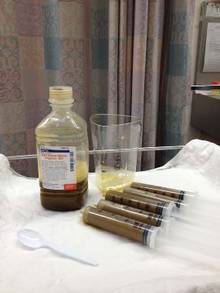
Well-designed and well-executed studies would not only address these immediate safety questions, but would also examine the more nebulous concerns about the long-term effects of tampering with an individual’s unique ecosystem of gut microbes. In recent years, research has begun to document how the balance and proportion of microbial species in the gut can either protect from – or predispose to – metabolic syndrome, obesity, diabetes, cardiovascular disease, arthritis, and even cognitive disorders.
An engrafting microbial transplant could predispose the recipient to develop illnesses that would otherwise never have been destined to occur, Dr. Slater said. "All of the evidence we have suggests that manipulating the gut microbiome is a powerful act that may have long-reaching and subtle effects."
The move toward a standardized FMT product and process is inevitable, Dr. Brandt said. "We’re not going to be doing fecal transplants much longer. This is a temporary situation. We’re already developing compounds that will do the same thing."
Researchers at the University of Guelph, Ontario, have developed a machine that distills and cultures microbes from human feces, producing a kind of super-probiotic that can be used in place of fresh stool.
Also, the Canadian biotech company Rebiotix is working on a similar product, which it intends to test in a phase II randomized controlled trial.
But until those machine-made products are available, physicians and patients with have to stay with the man-made version. "We have access to a substance that is free and has a virtually unlimited supply," Dr. Kelly said. "We cannot deny this effective therapy to these patients who’ve failed all other available treatments."
None of the sources quoted in this article had any financial declarations.
BETHESDA, MD. – The Food and Drug Administration has designated stool for transplant as a biologic drug, necessitating that any gastroenterologist performing fecal microbiota transplants obtain an Investigational New Drug permit.
By designating stool for transplant as a biologic drug, anyone who performs fecal microbiota transplants (FMTs) – whether they perform a single transplant for one patient or recruit dozens for a study – needs to have an IND permit.
The announcement was made at a 2-day public workshop convened by the National Institutes of Health and the Food and Drug Administration to sift through some of the evidence surrounding FMTs.
The intent of the IND requirement, said Dr. Jay Slater, director of FDA’s Division of Bacterial, Parasitic, and Allergenic Products, is not to stamp out the care that patients can now receive, but to make sure it’s safe, effective, and data driven.
"This is a low-tech procedure that already has a CPT code. A ‘how-to’ guide recently appeared online, and it walks you right through how to do the procedure. There are a very large number of people doing this off the grid. What we need are long-term controlled trials on this that will enhance our understanding of its safety and efficacy."
Although neither the biologic designation nor the IND requirement is brand new, they have not been well publicized, according to one of the innovators of FMT, Dr. Lawrence Brandt, of Albert Einstein Medical Center in New York.
"I am struck by the fact that FDA wants these INDs and yet FDA has never publicly set forth any message on it. So from this moment on, all of us who continue to do this without an IND are violating FDA’s policy," he said at the meeting.
One of the primary concerns now, according to Dr. Brandt, is a burgeoning public interest in FMTs, even to the point of do-it-yourself treatments. With online instructions for self-treatment, and a procedure that is rife with variations, regulators can no longer tacitly ignore the issue.
There’s no standardization of how the stool is prepared and filtered – it could be blended in a kitchen blender or by hand with a tongue depressor, or strained through gauze or a coffee filter, he said. Dosing is all over the place, listed as spoonfuls, grams, and milliliters. Different institutions screen donors in different ways. Some patients get a bowel prep, which can be mild or aggressive, and some don’t get one at all. Should the stool be fresh, and if so, how fresh? Is frozen okay? These are all issues that need to be examined from a safety and efficacy perspective and standardized.
Even the method of delivery varies. The transplant can be administered via nasogastric tube or colonoscope, or by enema. There’s no standardization of data collection either. Some large institutions keep records of everything from the first workup to the last visit. Doctors who perform transplants occasionally may not be as conscientious. And no one knows anything about the do-it-yourselfers, at home with a family member’s donation and a squeeze enema.
Dr. Brandt – and a number of clinicians at the meeting – agreed that answers must be found for all these questions. And they agree that well-conducted clinical trials are the best way to go forward.
Dr. Colleen Kelly, one of the gastroenterologists launching one of 22 currently recruiting studies on FMT, started doing the procedure 5 years ago. Her first patient was a 26-year-old medical student with recurrent bouts of a Clostridium difficile infection.
"I’d heard of [FMT], but never, ever thought of doing one," said Dr. Kelly, of Brown University Medical Center, Providence, R.I. "I thought it was something at the far fringes of medicine."
At her patient’s insistence, Dr. Kelly contacted Dr. Brandt and learned about his process for performing FMT. The following year, she performed 10 FMTs. She is now undertaking a study with Dr. Brandt to recruit about 48 patients with relapsing C. difficile to be randomized to FMT or to a sham treatment with their own stool. Patients who have clinical failure in the sham group will be offered FMT; patients in the active arm who fail on initial FMT will get another FMT from a different donor.
While most trials are examining the utility of FMT in patients with recurrent C. difficile, a few are investigating FMT for use in patients with Crohn’s disease and inflammatory bowel disease. Some clinicians and individual patients are now using FMTs for this condition outside a clinical trial, with no understanding of how a compromised intestinal mucosa might react to transplanted stool.
It will take years to accumulate the data necessary to fully understand FMT and all its implications, Dr. Kelly said. In the interim, the IND requirement will likely shrink the already-small pool of gastroenterologists performing FMTs. "Some will be motivated to get an IND, but the average person in practice won’t," she predicted. "You need to put hours and hours of work into it, and then you’re still under FDA’s oversight because this is not an approved therapy. So that means you have to submit adverse events reports, keep records, and report annually on your program. And at any time, without any warning, [the FDA] can come and inspect your facility."
Dr. Brandt agreed. "It’s a huge amount of paperwork documentation, record-keeping, and follow-up that the average practitioner is simply not going to do." The requirement for an IND means there are simply going to be fewer and fewer physicians who do them, he said.
Admittedly, though, the risks of no regulation can endanger patients, Dr. Kelly said. "If things go on completely unregulated, stupid things will happen," including the spread of infectious diseases like hepatitis C and parasitic infestations.
Indeed, Dr. Alexander Khoruts of the University of Minnesota, Minneapolis, who spoke at the workshop, described the case of an FMT "do-it-yourselfer" who called for some advice on improving her outcomes. Specifically, she had mixed stool from a neighbor and her son’s mother-in-law and administered it to herself without results. "She wanted to know if maybe the chlorine in the water killed off everything. ... Six months later she called me back and said her C. diff was gone, but now she had parasites."
"There are already predatory practices out there [performing FMTs]," Dr. Khoruts said. "I got an e-mail from someone who couldn’t make the drive up to see me, but she found someone near her who would do it for $10,000."
Well-designed and well-executed studies would not only address these immediate safety questions, but would also examine the more nebulous concerns about the long-term effects of tampering with an individual’s unique ecosystem of gut microbes. In recent years, research has begun to document how the balance and proportion of microbial species in the gut can either protect from – or predispose to – metabolic syndrome, obesity, diabetes, cardiovascular disease, arthritis, and even cognitive disorders.
An engrafting microbial transplant could predispose the recipient to develop illnesses that would otherwise never have been destined to occur, Dr. Slater said. "All of the evidence we have suggests that manipulating the gut microbiome is a powerful act that may have long-reaching and subtle effects."
The move toward a standardized FMT product and process is inevitable, Dr. Brandt said. "We’re not going to be doing fecal transplants much longer. This is a temporary situation. We’re already developing compounds that will do the same thing."
Researchers at the University of Guelph, Ontario, have developed a machine that distills and cultures microbes from human feces, producing a kind of super-probiotic that can be used in place of fresh stool.
Also, the Canadian biotech company Rebiotix is working on a similar product, which it intends to test in a phase II randomized controlled trial.
But until those machine-made products are available, physicians and patients with have to stay with the man-made version. "We have access to a substance that is free and has a virtually unlimited supply," Dr. Kelly said. "We cannot deny this effective therapy to these patients who’ve failed all other available treatments."
None of the sources quoted in this article had any financial declarations.
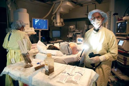
Golimumab indication expanded to include ulcerative colitis
The approval of the biologic drug golimumab has been expanded to include the treatment of adults with moderate to severe ulcerative colitis that is refractory to prior treatment or requires continuous steroid therapy, the Food and Drug Administration announced on May 15.
Golimumab (Simponi), a tumor necrosis factor–blocker, was approved in 2009 for treatment of moderate to severe active rheumatoid arthritis, psoriatic arthritis, and ankylosing spondylitis, all in adults. Simponi is marketed by Janssen Ortho Biotech.
The approval for ulcerative colitis was based on two studies of patients with moderate to severe ulcerative colitis, according to the FDA statement announcing the approval. In one study, which enrolled 513 patients who could not tolerate or had not responded to other treatments, "a greater proportion of Simponi-treated patients achieved clinical response, clinical remission and, as seen during endoscopy, had improved appearance of the colon after 6 weeks," compared with those on placebo, the statement said.
In a study of 310 patients who had responded to golimumab and were then randomized to continue treatment with golimumab or were switched to placebo, "a greater proportion of Simponi-treated patients maintained clinical response through week 54, had clinical remission at both weeks 30 and 54 and, as seen during endoscopy, had improved appearance of the colon at both weeks 30 and 54 compared with the placebo group," the FDA said. The most common adverse effects associated with golimumab are upper respiratory infection and redness at the injection site.
The risks of serious infections, invasive fungal infections, reactivation of hepatitis B infection, lymphoma, heart failure, nervous system disorders, and allergic reactions are increased with treatment.
Serious adverse events associated with golimumab should be reported to the FDA at 800-332-1088 or www.fda.gov/medwatch/.
Early surgery for adhesive bowel obstruction can save lives
INDIANAPOLIS – Patients requiring surgery for adhesive small bowel obstruction have markedly lower major morbidity and mortality rates if they’re operated on within 24 hours of hospital admission, according to an analysis of a large national database.
This finding is at odds with the conventional wisdom.
Both the World Society of Emergency Surgery and the Eastern Association for the Surgery of Trauma recommend in published guidelines an initial 3-5 days of nonoperative management to give the obstruction a chance to resolve on its own, Dr. Pedro G. Teixeira noted in presenting the study findings at the annual meeting of the American Surgical Association.
He and his coinvestigators identified 4,163 patients in the American College of Surgeons National Surgical Quality Improvement Program (NSQIP) database for 2005-2010 who underwent emergency laparotomy for adhesive bowel obstruction. Thirty-day mortality was 3% in those operated upon within 24 hours of hospital admission. It rose in stepwise fashion thereafter: 4% mortality with surgery at 24-48 hours, 7% with surgery at 48-72 hours, and 9% a threefold increase – when surgery was delayed beyond 72 hours, according to Dr. Teixeira of the University of Southern California, Los Angeles.
Similarly, the incidence of systemic infectious complications, including pneumonia, urinary tract infections, and sepsis, climbed from 12% with early operation to 17% when surgery occurred at 24-48 hours, 21% at 48-72 hours, and 24% thereafter.
In a multivariate analysis adjusted for baseline comorbidities and other potential confounding variables, surgery delayed for 24 hours or more after admission was associated with a highly significant 58% increased risk of mortality, a 33% increase in surgical site infections, a 36% greater risk of pneumonia, and a 47% increased risk of septic shock, he continued.
Discussant Gregory J. Jurkovich commented that this study challenges current dogma and harkens back to a century-old adage that has since been cast aside, namely, "Never let the sun set on a bowel obstruction."
The trouble is, however, that having a low threshold for surgery within 24 hours would subject a massive number of patients to an unnecessary operation.
An analysis of Nationwide Inpatient Sample data for 2009 by other investigators concluded that bowel obstruction resolved on its own within 3 days in 60% of patients and within 5 days in 80%. Fewer than 20% of the patients who presented with adhesive small bowel obstruction without evidence of ischemia underwent surgery, noted Dr. Jurkovich, director of surgery at Denver Health Medical Center and professor of trauma surgery and vice chairman of the department of surgery at the University of Colorado at Denver.
Dr. Teixeira concurred that bowel obstruction will resolve on its own in most patients. The challenge for surgeons in light of his study findings, he stressed, is to expedite the identification of those patients who will fail the period of nonoperative management. The best tool for that, in his view, is a CT scan of the abdomen and pelvis with water-soluble contrast.
At the University of Southern California, he explained, a patient who presents with adhesive bowel obstruction without evidence of ischemia undergoes the CT scan and is admitted to the surgical observation unit for close monitoring.
"At our institution, failure to demonstrate contrast progression through the colon within 24 hours would be a very strong indication for surgical exploration," according to Dr. Teixeira.
He reported having no financial conflicts.
The study by Dr. Teixeira is intriguing in
that it suggests a return to practice patterns from a prior era.

|
| Dr. Chad Whelan |
The study does report increased risk in
complications including mortality with delays in surgery for small bowel
obstructions, even with risk adjustment. However, this is not a controlled
trial which limits our ability to reach definitive conclusions from it. Still,
hospitalists often are the primary physicians for patients admitted for small
bowel obstructions and should be aware of these findings so that they can
ensure that they have early surgical involvement.
Chad Whelan, M.D., is associate chief medical officer for
performance improvement and innovation and an associate professor of medicine
at the University
of Chicago Medical Center.
The study by Dr. Teixeira is intriguing in
that it suggests a return to practice patterns from a prior era.
|
|
| Dr. Chad Whelan |
The study does report increased risk in
complications including mortality with delays in surgery for small bowel
obstructions, even with risk adjustment. However, this is not a controlled
trial which limits our ability to reach definitive conclusions from it. Still,
hospitalists often are the primary physicians for patients admitted for small
bowel obstructions and should be aware of these findings so that they can
ensure that they have early surgical involvement.
Chad Whelan, M.D., is associate chief medical officer for
performance improvement and innovation and an associate professor of medicine
at the University
of Chicago Medical Center.
INDIANAPOLIS – Patients requiring surgery for adhesive small bowel obstruction have markedly lower major morbidity and mortality rates if they’re operated on within 24 hours of hospital admission, according to an analysis of a large national database.
This finding is at odds with the conventional wisdom.
Both the World Society of Emergency Surgery and the Eastern Association for the Surgery of Trauma recommend in published guidelines an initial 3-5 days of nonoperative management to give the obstruction a chance to resolve on its own, Dr. Pedro G. Teixeira noted in presenting the study findings at the annual meeting of the American Surgical Association.
He and his coinvestigators identified 4,163 patients in the American College of Surgeons National Surgical Quality Improvement Program (NSQIP) database for 2005-2010 who underwent emergency laparotomy for adhesive bowel obstruction. Thirty-day mortality was 3% in those operated upon within 24 hours of hospital admission. It rose in stepwise fashion thereafter: 4% mortality with surgery at 24-48 hours, 7% with surgery at 48-72 hours, and 9% a threefold increase – when surgery was delayed beyond 72 hours, according to Dr. Teixeira of the University of Southern California, Los Angeles.
Similarly, the incidence of systemic infectious complications, including pneumonia, urinary tract infections, and sepsis, climbed from 12% with early operation to 17% when surgery occurred at 24-48 hours, 21% at 48-72 hours, and 24% thereafter.
In a multivariate analysis adjusted for baseline comorbidities and other potential confounding variables, surgery delayed for 24 hours or more after admission was associated with a highly significant 58% increased risk of mortality, a 33% increase in surgical site infections, a 36% greater risk of pneumonia, and a 47% increased risk of septic shock, he continued.
Discussant Gregory J. Jurkovich commented that this study challenges current dogma and harkens back to a century-old adage that has since been cast aside, namely, "Never let the sun set on a bowel obstruction."
The trouble is, however, that having a low threshold for surgery within 24 hours would subject a massive number of patients to an unnecessary operation.
An analysis of Nationwide Inpatient Sample data for 2009 by other investigators concluded that bowel obstruction resolved on its own within 3 days in 60% of patients and within 5 days in 80%. Fewer than 20% of the patients who presented with adhesive small bowel obstruction without evidence of ischemia underwent surgery, noted Dr. Jurkovich, director of surgery at Denver Health Medical Center and professor of trauma surgery and vice chairman of the department of surgery at the University of Colorado at Denver.
Dr. Teixeira concurred that bowel obstruction will resolve on its own in most patients. The challenge for surgeons in light of his study findings, he stressed, is to expedite the identification of those patients who will fail the period of nonoperative management. The best tool for that, in his view, is a CT scan of the abdomen and pelvis with water-soluble contrast.
At the University of Southern California, he explained, a patient who presents with adhesive bowel obstruction without evidence of ischemia undergoes the CT scan and is admitted to the surgical observation unit for close monitoring.
"At our institution, failure to demonstrate contrast progression through the colon within 24 hours would be a very strong indication for surgical exploration," according to Dr. Teixeira.
He reported having no financial conflicts.
AT THE ASA ANNUAL MEETING
Major Finding: Surgery for adhesive small bowel obstruction had a 30-day mortality rate of 3% if performed within 24 hours of hospital admission, rising stepwise to 9% when the operation was delayed beyond 72 hours.
Data Source: This was a retrospective analysis of 4,163 patients in the American College of Surgeons National Quality Improvement Program database for 2005-2010 who underwent emergency laparotomy for adhesive bowel obstruction.
Disclosures: The presenter reported having no conflicts of interest.
Children with ulcerative colitis benefited from fecal transplants
Seven out of nine children with active ulcerative colitis experienced at least a temporary clinical response within 1 week of a series of fecal transplants – with four of the patients staying in complete remission 1 month later.
The procedures represent the first time fecal transplantation has been used to treat ulcerative colitis, Dr. Sachin Kunde and his colleagues reported online in the Journal of Pediatric Gastroenterology and Nutrition (2013 March 29 [doi: 10.1097/MPG.0b013e318292fa0d]).
"Utilization of fecal material transplantation in ulcerative colitis may not be as simple as its use in recurrent C. difficile infection," wrote Dr. Kunde of the Helen DeVos Children’s Hospital, Grand Rapids, Mich., and his coauthors. Still, they said, this early success in a devastating, hard-to-manage disease should be the launching point for larger trials.

The study group comprised 10 patients who ranged in age from 7 to 20 years. All had mild to moderate, active ulcerative colitis. Disease duration ranged from 1 to 8 years. Participants had stable disease and received medical treatment for at least 2 months before the procedure. Only one patient had used anti–tumor necrosis factor (anti-TNF)-alpha medications.
The intervention consisted of a 5-day series of daily enemas containing fresh stool from a donor. Each of the patients chose an adult donor; most were first-degree relatives. One patient chose a close family friend. The donors took daily over-the-counter stool softeners to produce the required amount – a mean of 90 g/day. This was blended with 250 mL of sterile normal saline, strained, and divided into four 60-mL portions. In each treatment, all four of the portions were infused, each over a 15-minute period.
The patients did not receive any bowel preparation before the procedures. They received the enemas while lying on the left side, and then rotating to the right side and back again to allow the solution to travel into the colon.
Ten patients entered treatment. However, one could not retain the enemas and so was not included in the final analysis. The remaining patients were able to tolerate an enema volume of 75-240 mL (average retention, 165 mL). Retention was not directly related to age, the investigators noted, since the subject who could not retain it was the oldest. Retention times ranged from 3 to 24 hours.
There were no serious adverse events during the study. Patients did report the expected discomfort of left-sided abdominal fullness. One experienced a moderate fever and chills 3 hours after the first two transplants, which spontaneously resolved over 6 hours. For the final three procedures, that patient took prophylactic acetaminophen and diphenhydramine.
Another patient had a low-grade fever after one transplant, which resolved without intervention. There were no cases of sepsis.
One patient experienced a disabling hematochezia 3 weeks after the transplant series. This was judged to be a flare unrelated to the transplant.
In the first week after the transplant series, seven of the nine patients in the analysis (78%) had a clinical response, defined as a decrease of more than 15 points in the Pediatric Ulcerative Colitis Activity Index. Six of the nine (67%) maintained that improvement at 1 month.
Three experienced clinical remission at 1 week, which lasted for 1 month. Two patients had a PUCAI score of 0 from week 3 until the end of the 7-week follow-up period.
The pilot study shows the feasibility of fecal transplants for ulcerative colitis, Dr. Kunde said. But while the technique is similar to that employed in C. difficile treatment, a more prolonged treatment seems necessary to elicit a response in ulcerative colitis.
"In order to better understand how fecal transplants can be used to treat ulcerative colitis, many unanswered questions need to be addressed. We must further investigate standardization of ... preparation, ideal donor selection, ideal route of administration, and optimal duration of scheduling [the transplants] to induce and maintain a clinical response. Most importantly, the effects of fecal material transplant on the colonic microbiome and mucosal inflammation in ulcerative colitis need to be explored."
The Helen Devos Children’s Hospital supported the research. The authors did not disclose any financial relationships.
There has been much excitement about the use
of fecal material to treat recurrent Clostridium difficile infection.
Along the theory that the gut microbiome may be the “final frontier” of many
human diseases, so-called fecal microbiota transplantation (FMT) is of great
interest for other GI disorders.
Given the observation that patients with inflammatory
bowel disease (IBD) have an altered gut microbiome, clinicians and scientists
have wondered whether modifying the gut flora via FMT would provide symptom
improvement or disease control. It is much more complicated than treatment for C.
difficile, since IBD is a much more complex disorder, and as a single
organism or infectious etiology is not known. Clearly, our current
understanding is far from satisfying any of Koch’s postulates
of infectious disease. The observed dysbiosis may instead be a result of the underlying
inflammatory disorder or even a result of some of our treatments for IBD.
Nonetheless, providing FMT to patients with IBD is an interesting concept.
Dr. Kunde and his colleagues had promising results,
but they were careful in their selection of patients, the severity of disease,
and concomitant therapies. Safety appears acceptable, but there were some
short-term adverse events. We should await additional studies with mechanistic
and translational components and, importantly, safety follow-up to guide us
further.
Dr. David T. Rubin is a professor of medicine, the codirector
of the Inflammatory Bowel Disease Center, and the associate section chief for educational
programs at the University of Chicago. He had no relevant disclosures.
Seven out of nine children with active ulcerative colitis experienced at least a temporary clinical response within 1 week of a series of fecal transplants – with four of the patients staying in complete remission 1 month later.
The procedures represent the first time fecal transplantation has been used to treat ulcerative colitis, Dr. Sachin Kunde and his colleagues reported online in the Journal of Pediatric Gastroenterology and Nutrition (2013 March 29 [doi: 10.1097/MPG.0b013e318292fa0d]).
"Utilization of fecal material transplantation in ulcerative colitis may not be as simple as its use in recurrent C. difficile infection," wrote Dr. Kunde of the Helen DeVos Children’s Hospital, Grand Rapids, Mich., and his coauthors. Still, they said, this early success in a devastating, hard-to-manage disease should be the launching point for larger trials.
The study group comprised 10 patients who ranged in age from 7 to 20 years. All had mild to moderate, active ulcerative colitis. Disease duration ranged from 1 to 8 years. Participants had stable disease and received medical treatment for at least 2 months before the procedure. Only one patient had used anti–tumor necrosis factor (anti-TNF)-alpha medications.
The intervention consisted of a 5-day series of daily enemas containing fresh stool from a donor. Each of the patients chose an adult donor; most were first-degree relatives. One patient chose a close family friend. The donors took daily over-the-counter stool softeners to produce the required amount – a mean of 90 g/day. This was blended with 250 mL of sterile normal saline, strained, and divided into four 60-mL portions. In each treatment, all four of the portions were infused, each over a 15-minute period.
The patients did not receive any bowel preparation before the procedures. They received the enemas while lying on the left side, and then rotating to the right side and back again to allow the solution to travel into the colon.
Ten patients entered treatment. However, one could not retain the enemas and so was not included in the final analysis. The remaining patients were able to tolerate an enema volume of 75-240 mL (average retention, 165 mL). Retention was not directly related to age, the investigators noted, since the subject who could not retain it was the oldest. Retention times ranged from 3 to 24 hours.
There were no serious adverse events during the study. Patients did report the expected discomfort of left-sided abdominal fullness. One experienced a moderate fever and chills 3 hours after the first two transplants, which spontaneously resolved over 6 hours. For the final three procedures, that patient took prophylactic acetaminophen and diphenhydramine.
Another patient had a low-grade fever after one transplant, which resolved without intervention. There were no cases of sepsis.
One patient experienced a disabling hematochezia 3 weeks after the transplant series. This was judged to be a flare unrelated to the transplant.
In the first week after the transplant series, seven of the nine patients in the analysis (78%) had a clinical response, defined as a decrease of more than 15 points in the Pediatric Ulcerative Colitis Activity Index. Six of the nine (67%) maintained that improvement at 1 month.
Three experienced clinical remission at 1 week, which lasted for 1 month. Two patients had a PUCAI score of 0 from week 3 until the end of the 7-week follow-up period.
The pilot study shows the feasibility of fecal transplants for ulcerative colitis, Dr. Kunde said. But while the technique is similar to that employed in C. difficile treatment, a more prolonged treatment seems necessary to elicit a response in ulcerative colitis.
"In order to better understand how fecal transplants can be used to treat ulcerative colitis, many unanswered questions need to be addressed. We must further investigate standardization of ... preparation, ideal donor selection, ideal route of administration, and optimal duration of scheduling [the transplants] to induce and maintain a clinical response. Most importantly, the effects of fecal material transplant on the colonic microbiome and mucosal inflammation in ulcerative colitis need to be explored."
The Helen Devos Children’s Hospital supported the research. The authors did not disclose any financial relationships.
FROM PEDIATRIC GASTROENTEROLOGY AND NUTRITION
Major finding: Fecal transplants effected clinical improvement in 78% of children with ulcerative colitis.
Data source: Nine children were enrolled in a small prospective study.
Disclosures: The Helen Devos Children’s Hospital supported the research. The authors did not disclose any financial relationships.

Gluten-free diet may hold benefit in IBS
A gluten-free diet reduced stool frequency as well as small bowel permeability in irritable bowel syndrome patients without celiac disease.
The findings, published in the May issue of Gastroenterology, "support the need for further clinical intervention studies to evaluate the clinical effects of gluten withdrawal in patients with diarrhea-predominant IBS," reported Dr. Maria I. Vazquez-Roque and her colleagues.
Dr. Vasquez-Roque, of the Mayo Clinic’s Clinical Enteric Neuroscience Translational and Epidemiological Research Program, in Rochester, Minn., and her colleagues recruited 45 subjects (43 women) from a database of more than 800 patients with irritable bowel syndrome who had been evaluated at the Mayo Clinic.
All patients had diarrhea-predominant IBS (IBS-D), were not on a gluten-free diet prior to the study, and did not have celiac disease.
They were randomized to either a gluten-free diet or a gluten-containing diet for 28 days. Patients’ meals and snacks were ingested or prepared in the Mayo Clinical Research Unit, and study participants were asked to eat only the foods provided by the study dietitians during the entire study period, the authors wrote. Dietitians assessed diet compliance using direct questioning of participants.
After 28 days of the study, the investigators looked at several clinical and histologic markers.
First, they tallied stool frequency, and found that patients on the gluten-containing diet had more stools per day than gluten-free patients did (P = .04), with a 95% confidence interval for the absolute difference between number of stools per day at –0.652 to –0.015 (Gastroenterology 2013 Jan. 28 [doi: 10.1053/j.gastro.2013.01.049]).
"While the absolute difference in stool frequency is small, it is important to appreciate that this increased frequency is on a background of the typical increase in stool frequency (average 2.6 bowel movements/day at baseline) and consistency in patients with IBS-D," the authors wrote.
This effect was more pronounced in subjects who were HLA-DQ2 or 8 positive (95% CI, –1.005 to –0.092; P = .019), they added.
Next, the investigators looked at small bowel permeability.
They found increased small bowel permeability with gluten-containing diet patients relative to their gluten-free counterparts, based on both cumulative mannitol excretion levels and lactulose:mannitol ratio. Similarly, this effect was more pronounced in HLA-DQ2 or 8 positive patients.
"While the clinical significance of these changes in permeability is not demonstrated in the current study, the abundant experimental evidence from the published literature is that increased mucosal permeability enhances inflammation and leads to increased sensitivity," they wrote.
Finally, the authors looked at tight-junction mRNA expression.
"Alterations in intestinal permeability and jejunal mucosal tight junction (TJ) signaling have been described in IBS-D, including postinfectious IBS-D," they wrote.
They found that expressions of ZO-1, occludin, and claudin-1 mRNA in colonic mucosa were significantly lower with the gluten-containing diet, compared with the gluten-free patients, particularly in subjects with HLA-DQ2 or 8 positive status.
There were no significant variations in tight junction signaling in the small bowel mucosa.
The authors conceded several limitations to their study. For one, it "did not evaluate effects of gluten on the microbiome, afferent functions, or cytokine expression in the mucosal biopsies from patients before and after the interventions. These would be interesting parameters to include in future studies," they wrote.
Additionally, "our study does not specifically address the effects of gluten protein per se, and it is possible that other proteins in wheat flour may be responsible for the changes observed."
Nevertheless, "our data provide mechanistic explanations for the observation that gluten withdrawal may improve patient symptoms in IBS," they concluded.
The researchers disclosed receiving funding for this study from the National Institutes of Health. One investigator also disclosed having received grants from Alba Therapeutics, maker of the drug larazotide, used in celiac disease.
Recently, we have seen the emergence of
nonceliac gluten sensitivity as a distinct clinical entity. It is as if the medical
community has caught up to the food industry and the patients because there has
been an explosion in the availability of gluten-free foods and some patients,
without celiac disease, have been telling us they feel better when gluten has
been withdrawn from their diet.
A recent study from Australia demonstrated in a double-blind,
randomized study of a gluten-containing diet versus a gluten-free diet
that individuals with irritable bowel syndrome (IBS) who had self-selected as
being gluten sensitive experienced more gastrointestinal symptoms and fatigue
when exposed to the gluten-containing diet.
The investigators from the Mayo Clinic in Rochester,
Minn. extended these studies to patients with diarrhea-predominant
IBS who had not self-selected as being gluten sensitive. They demonstrated that
the gluten-free diet reduced stool frequency and small bowel permeability and
that the effect was most prominent in those who possessed the celiac disease at
risk genes, HLA DQ2 and DQ8.
Gluten is not fully digested by our digestive system,
unlike meat protein. The large amino acid molecules that remain after digestion
appear responsible for the development of celiac disease in some individuals
who are HLA DQ2 or -8 positive. It now appears that gluten (or other
proteins found in wheat) may induce changes that result in symptoms in patients
labeled as having IBS.
So those that have already self-diagnosed as being
gluten sensitive may in fact just be the tip of the iceberg of gluten
sensitivity.
Peter H.R. Green, M.D., is the Director of
the Celiac Disease Center, Columbia University, New York, and professor of
clinical medicine at the College of Physicians and Surgeons. Dr. Green is on
the scientific advisory boards for Alvine Pharmaceuticals and ImmusanT.
A gluten-free diet reduced stool frequency as well as small bowel permeability in irritable bowel syndrome patients without celiac disease.
The findings, published in the May issue of Gastroenterology, "support the need for further clinical intervention studies to evaluate the clinical effects of gluten withdrawal in patients with diarrhea-predominant IBS," reported Dr. Maria I. Vazquez-Roque and her colleagues.
Dr. Vasquez-Roque, of the Mayo Clinic’s Clinical Enteric Neuroscience Translational and Epidemiological Research Program, in Rochester, Minn., and her colleagues recruited 45 subjects (43 women) from a database of more than 800 patients with irritable bowel syndrome who had been evaluated at the Mayo Clinic.
All patients had diarrhea-predominant IBS (IBS-D), were not on a gluten-free diet prior to the study, and did not have celiac disease.
They were randomized to either a gluten-free diet or a gluten-containing diet for 28 days. Patients’ meals and snacks were ingested or prepared in the Mayo Clinical Research Unit, and study participants were asked to eat only the foods provided by the study dietitians during the entire study period, the authors wrote. Dietitians assessed diet compliance using direct questioning of participants.
After 28 days of the study, the investigators looked at several clinical and histologic markers.
First, they tallied stool frequency, and found that patients on the gluten-containing diet had more stools per day than gluten-free patients did (P = .04), with a 95% confidence interval for the absolute difference between number of stools per day at –0.652 to –0.015 (Gastroenterology 2013 Jan. 28 [doi: 10.1053/j.gastro.2013.01.049]).
"While the absolute difference in stool frequency is small, it is important to appreciate that this increased frequency is on a background of the typical increase in stool frequency (average 2.6 bowel movements/day at baseline) and consistency in patients with IBS-D," the authors wrote.
This effect was more pronounced in subjects who were HLA-DQ2 or 8 positive (95% CI, –1.005 to –0.092; P = .019), they added.
Next, the investigators looked at small bowel permeability.
They found increased small bowel permeability with gluten-containing diet patients relative to their gluten-free counterparts, based on both cumulative mannitol excretion levels and lactulose:mannitol ratio. Similarly, this effect was more pronounced in HLA-DQ2 or 8 positive patients.
"While the clinical significance of these changes in permeability is not demonstrated in the current study, the abundant experimental evidence from the published literature is that increased mucosal permeability enhances inflammation and leads to increased sensitivity," they wrote.
Finally, the authors looked at tight-junction mRNA expression.
"Alterations in intestinal permeability and jejunal mucosal tight junction (TJ) signaling have been described in IBS-D, including postinfectious IBS-D," they wrote.
They found that expressions of ZO-1, occludin, and claudin-1 mRNA in colonic mucosa were significantly lower with the gluten-containing diet, compared with the gluten-free patients, particularly in subjects with HLA-DQ2 or 8 positive status.
There were no significant variations in tight junction signaling in the small bowel mucosa.
The authors conceded several limitations to their study. For one, it "did not evaluate effects of gluten on the microbiome, afferent functions, or cytokine expression in the mucosal biopsies from patients before and after the interventions. These would be interesting parameters to include in future studies," they wrote.
Additionally, "our study does not specifically address the effects of gluten protein per se, and it is possible that other proteins in wheat flour may be responsible for the changes observed."
Nevertheless, "our data provide mechanistic explanations for the observation that gluten withdrawal may improve patient symptoms in IBS," they concluded.
The researchers disclosed receiving funding for this study from the National Institutes of Health. One investigator also disclosed having received grants from Alba Therapeutics, maker of the drug larazotide, used in celiac disease.
FROM GASTROENTEROLOGY
Major finding: Irritable bowel syndrome patients on a gluten-free diet had fewer bowel movements per day than did gluten-consuming counterparts (P = .04).
Data source: A 4-week trial of 45 patients with diarrhea-predominant irritable bowel syndrome, randomized to a gluten-free or gluten-containing diet.
Disclosures: The researchers disclosed receiving funding for this study from the National Institutes of Health. One investigator also disclosed having received grants from Alba Therapeutics, maker of the drug larazotide, used in celiac disease.