User login
FDA Panel Endorses Ultrasound System for Screening Dense Breasts
An automated breast ultrasound system is on track to become the first such device with an indication for use as an adjunct to mammography when screening asymptomatic women with dense breasts for breast cancer.
The Food and Drug Administration’s Radiological Devices Panel voted 13-0 that the benefits of U-Systems Inc.’s somo-v automated breast ultrasound system outweigh the risks for breast cancer screening.
During their meeting, the advisory group also voted unanimously that the noninvasive device is safe and effective, despite FDA concerns over whether the firm’s pivotal study data could be generalized to a broader population.

Approval is not guaranteed, but the agency usually follows the advice of its advisory panels.
U-Systems is seeking premarket approval to increase breast cancer detection in asymptomatic women with dense breasts who have not had a previous clinical breast intervention following a negative mammogram screening. Somo-v was 510(k) cleared in 2005 for diagnostic use as an adjunct to x-ray mammography.
A key question the FDA asked the panel was whether the proposed expanded indication – and the way it would be applied in practice – was appropriately reflected in the firm’s pivotal study design. The panel agreed that it was.
The proposed indication for use "is fine the way it’s written," said panel member Dr. Carl D’Orsi, professor of radiology and hematology/oncology at Emory University in Atlanta. "It should not be changed," Dr. D’Orsi said, reflecting the panel’s consensus.
Premarket approval would make somo-v the first ultrasound device available in the United States with a breast cancer screening indication, according to the company. The system is already the only ultrasound device approved for breast cancer screening in Canada and Europe.
Supporting the premarket approval application is a retrospective, multireader study that enrolled 200 subjects at 13 U.S. sites – culled from the firm’s larger SOMO-INSIGHT clinical trial – to evaluate whether digital mammography in combination with the somo-v automated breast ultrasound system (ABUS) is more sensitive in detecting breast cancer in women with dense breast tissue than x-ray mammography screening alone.
The primary end point of the study was to determine radiologist readers’ performance with and without ABUS using a statistical methodology called "receiver operator characteristic" curve analysis. The mean area under the curve (AUC) values were 0.604 for x-ray mammography alone and 0.747 for x-ray mammography plus ABUS.
"Results from our pivotal clinical retrospective reader study demonstrate that use of screening mammography with ABUS provides a substantial, statistically significant clinical improvement in a reader’s ability to detect mammographically negative breast cancers," said CEO Ronald Ho during the meeting.
"Our primary end point was met," added principal investigator Maryellen Giger, Ph.D., professor of radiology at the University of Chicago.

The difference of 0.143 in the AUC values "was statistically significant, given that the lower bound of an estimated 95% confidence interval for the difference in AUC is 0.074," the FDA agreed.
According to the company, mammography’s sensitivity for the detection of breast cancer is 85% in women overall, but the rate is reduced to 65% in women with dense breast tissue. "Dense breast tissue has been proven to conceal malignant lesions, limiting the effectiveness of mammography," Mr. Ho said. Dense breast tissue also increases a woman’s risk of breast cancer by up to four to six times, according to U-Systems.
"What differentiates our device is that we can detect lesions that are missed by mammography ... and we do it in a very fast work flow that’s reproducible and operator independent," the firm stated. The technology is designed to follow the same work flow as a traditional mammography procedure and provide an exam in 15 minutes.
Real-World Management Questioned
The FDA questioned whether the pivotal study’s results could be extrapolated to a real-world population of patients. One hundred sixty-four subjects were used in the study’s primary statistical analysis (133 normal cases chosen at random and 31 cancer cases), with 12 noncancer cases being evaluated by mammography alone.
Patients with prior clinical breast interventions, such as those who had a breast augmentation or breast cancer treatments including biopsy, radiation, lumpectomy, or mastectomy, were excluded from the study. U-Systems said it excluded these patients to eliminate potential bias and/or confounding effects.
But while the firm is seeking approval of somo-v as a screening tool for women with dense breasts "who have not had a previous clinical breast intervention," the FDA argued that in the real world, "patient management that includes an ABUS scan may be considered for patients who have had prior clinical breast intervention."
The panel discussed whether the exclusion of patients with a prior clinical breast intervention should be specified in the indication, and agreed that it should, given the pivotal study design. Additional studies would be needed to remove the restriction, the panel said.
Some panelists expressed concern over whether somo-v use on biopsy patients, for instance, might increase the device’s false-positive rate, given surgical scars’ similarity to malignancies on ultrasound.
Nevertheless, a majority did not seem overly concerned about potential off-label use on patients with prior interventions. "It seems to me that that’s not a big problem," said panel member Dr. Daniel Kopans, professor of radiology at Harvard Medical School, Boston. "If FDA approves the technology even with these limitations, those of us in breast imaging will use it as we see fit. ... I don’t think that [the indication] is a huge limitation."
Operator Training Concerns
The panel also discussed the sponsor’s proposed physician training program, concluding that it is adequate. However, after the panel voted on safety and effectiveness, some members debated whether potential use of the system by nonimaging specialists could adversely impact outcomes and whether controls were needed to restrict use to accredited radiologists.
According to the company, somo-v’s automation is intended to "reduce operator dependence," and the firm claims that "any medical staff member can be trained to operate the ABUS device consistently and reproducibly." But some panel members cautioned that the simplicity could lead to harm in clinical practice.
"Potentially, you could be in a situation where the mammogram is done by breast-imaging specialists, but the ultrasound is done by nonimaging specialists, which could significantly affect results," said panelist Dr. Robert Faulk, a diagnostic radiologist in private practice in Omaha, Neb.
Dr. Kopans agreed, citing the Mammography Quality Standards Act’s radiologist accreditation training and urging that "similar constraints" are needed for somo-v.
But in response, Janine Morris, the FDA’s acting radiologic devices division director, reminded the panelists that the FDA "doesn’t regulate [the] practice of medicine."
Somo-v relies on proprietary hardware and software, and incorporates a scan head that automatically acquires wide field-of-view breast images while the patient lies supine on a standard exam table.
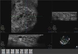
The images are transferred to the system’s somo-VIEWer 3D workstation, which displays the images in a 3D reconstructed coronal view, as well as standard ultrasound formats, including transverse, radial, multislice, sagittal and antiradial views.
Elsevier Global Medical News and "The Gray Sheet" are owned by Elsevier.
An automated breast ultrasound system is on track to become the first such device with an indication for use as an adjunct to mammography when screening asymptomatic women with dense breasts for breast cancer.
The Food and Drug Administration’s Radiological Devices Panel voted 13-0 that the benefits of U-Systems Inc.’s somo-v automated breast ultrasound system outweigh the risks for breast cancer screening.
During their meeting, the advisory group also voted unanimously that the noninvasive device is safe and effective, despite FDA concerns over whether the firm’s pivotal study data could be generalized to a broader population.
Approval is not guaranteed, but the agency usually follows the advice of its advisory panels.
U-Systems is seeking premarket approval to increase breast cancer detection in asymptomatic women with dense breasts who have not had a previous clinical breast intervention following a negative mammogram screening. Somo-v was 510(k) cleared in 2005 for diagnostic use as an adjunct to x-ray mammography.
A key question the FDA asked the panel was whether the proposed expanded indication – and the way it would be applied in practice – was appropriately reflected in the firm’s pivotal study design. The panel agreed that it was.
The proposed indication for use "is fine the way it’s written," said panel member Dr. Carl D’Orsi, professor of radiology and hematology/oncology at Emory University in Atlanta. "It should not be changed," Dr. D’Orsi said, reflecting the panel’s consensus.
Premarket approval would make somo-v the first ultrasound device available in the United States with a breast cancer screening indication, according to the company. The system is already the only ultrasound device approved for breast cancer screening in Canada and Europe.
Supporting the premarket approval application is a retrospective, multireader study that enrolled 200 subjects at 13 U.S. sites – culled from the firm’s larger SOMO-INSIGHT clinical trial – to evaluate whether digital mammography in combination with the somo-v automated breast ultrasound system (ABUS) is more sensitive in detecting breast cancer in women with dense breast tissue than x-ray mammography screening alone.
The primary end point of the study was to determine radiologist readers’ performance with and without ABUS using a statistical methodology called "receiver operator characteristic" curve analysis. The mean area under the curve (AUC) values were 0.604 for x-ray mammography alone and 0.747 for x-ray mammography plus ABUS.
"Results from our pivotal clinical retrospective reader study demonstrate that use of screening mammography with ABUS provides a substantial, statistically significant clinical improvement in a reader’s ability to detect mammographically negative breast cancers," said CEO Ronald Ho during the meeting.
"Our primary end point was met," added principal investigator Maryellen Giger, Ph.D., professor of radiology at the University of Chicago.
The difference of 0.143 in the AUC values "was statistically significant, given that the lower bound of an estimated 95% confidence interval for the difference in AUC is 0.074," the FDA agreed.
According to the company, mammography’s sensitivity for the detection of breast cancer is 85% in women overall, but the rate is reduced to 65% in women with dense breast tissue. "Dense breast tissue has been proven to conceal malignant lesions, limiting the effectiveness of mammography," Mr. Ho said. Dense breast tissue also increases a woman’s risk of breast cancer by up to four to six times, according to U-Systems.
"What differentiates our device is that we can detect lesions that are missed by mammography ... and we do it in a very fast work flow that’s reproducible and operator independent," the firm stated. The technology is designed to follow the same work flow as a traditional mammography procedure and provide an exam in 15 minutes.
Real-World Management Questioned
The FDA questioned whether the pivotal study’s results could be extrapolated to a real-world population of patients. One hundred sixty-four subjects were used in the study’s primary statistical analysis (133 normal cases chosen at random and 31 cancer cases), with 12 noncancer cases being evaluated by mammography alone.
Patients with prior clinical breast interventions, such as those who had a breast augmentation or breast cancer treatments including biopsy, radiation, lumpectomy, or mastectomy, were excluded from the study. U-Systems said it excluded these patients to eliminate potential bias and/or confounding effects.
But while the firm is seeking approval of somo-v as a screening tool for women with dense breasts "who have not had a previous clinical breast intervention," the FDA argued that in the real world, "patient management that includes an ABUS scan may be considered for patients who have had prior clinical breast intervention."
The panel discussed whether the exclusion of patients with a prior clinical breast intervention should be specified in the indication, and agreed that it should, given the pivotal study design. Additional studies would be needed to remove the restriction, the panel said.
Some panelists expressed concern over whether somo-v use on biopsy patients, for instance, might increase the device’s false-positive rate, given surgical scars’ similarity to malignancies on ultrasound.
Nevertheless, a majority did not seem overly concerned about potential off-label use on patients with prior interventions. "It seems to me that that’s not a big problem," said panel member Dr. Daniel Kopans, professor of radiology at Harvard Medical School, Boston. "If FDA approves the technology even with these limitations, those of us in breast imaging will use it as we see fit. ... I don’t think that [the indication] is a huge limitation."
Operator Training Concerns
The panel also discussed the sponsor’s proposed physician training program, concluding that it is adequate. However, after the panel voted on safety and effectiveness, some members debated whether potential use of the system by nonimaging specialists could adversely impact outcomes and whether controls were needed to restrict use to accredited radiologists.
According to the company, somo-v’s automation is intended to "reduce operator dependence," and the firm claims that "any medical staff member can be trained to operate the ABUS device consistently and reproducibly." But some panel members cautioned that the simplicity could lead to harm in clinical practice.
"Potentially, you could be in a situation where the mammogram is done by breast-imaging specialists, but the ultrasound is done by nonimaging specialists, which could significantly affect results," said panelist Dr. Robert Faulk, a diagnostic radiologist in private practice in Omaha, Neb.
Dr. Kopans agreed, citing the Mammography Quality Standards Act’s radiologist accreditation training and urging that "similar constraints" are needed for somo-v.
But in response, Janine Morris, the FDA’s acting radiologic devices division director, reminded the panelists that the FDA "doesn’t regulate [the] practice of medicine."
Somo-v relies on proprietary hardware and software, and incorporates a scan head that automatically acquires wide field-of-view breast images while the patient lies supine on a standard exam table.
The images are transferred to the system’s somo-VIEWer 3D workstation, which displays the images in a 3D reconstructed coronal view, as well as standard ultrasound formats, including transverse, radial, multislice, sagittal and antiradial views.
Elsevier Global Medical News and "The Gray Sheet" are owned by Elsevier.
An automated breast ultrasound system is on track to become the first such device with an indication for use as an adjunct to mammography when screening asymptomatic women with dense breasts for breast cancer.
The Food and Drug Administration’s Radiological Devices Panel voted 13-0 that the benefits of U-Systems Inc.’s somo-v automated breast ultrasound system outweigh the risks for breast cancer screening.
During their meeting, the advisory group also voted unanimously that the noninvasive device is safe and effective, despite FDA concerns over whether the firm’s pivotal study data could be generalized to a broader population.
Approval is not guaranteed, but the agency usually follows the advice of its advisory panels.
U-Systems is seeking premarket approval to increase breast cancer detection in asymptomatic women with dense breasts who have not had a previous clinical breast intervention following a negative mammogram screening. Somo-v was 510(k) cleared in 2005 for diagnostic use as an adjunct to x-ray mammography.
A key question the FDA asked the panel was whether the proposed expanded indication – and the way it would be applied in practice – was appropriately reflected in the firm’s pivotal study design. The panel agreed that it was.
The proposed indication for use "is fine the way it’s written," said panel member Dr. Carl D’Orsi, professor of radiology and hematology/oncology at Emory University in Atlanta. "It should not be changed," Dr. D’Orsi said, reflecting the panel’s consensus.
Premarket approval would make somo-v the first ultrasound device available in the United States with a breast cancer screening indication, according to the company. The system is already the only ultrasound device approved for breast cancer screening in Canada and Europe.
Supporting the premarket approval application is a retrospective, multireader study that enrolled 200 subjects at 13 U.S. sites – culled from the firm’s larger SOMO-INSIGHT clinical trial – to evaluate whether digital mammography in combination with the somo-v automated breast ultrasound system (ABUS) is more sensitive in detecting breast cancer in women with dense breast tissue than x-ray mammography screening alone.
The primary end point of the study was to determine radiologist readers’ performance with and without ABUS using a statistical methodology called "receiver operator characteristic" curve analysis. The mean area under the curve (AUC) values were 0.604 for x-ray mammography alone and 0.747 for x-ray mammography plus ABUS.
"Results from our pivotal clinical retrospective reader study demonstrate that use of screening mammography with ABUS provides a substantial, statistically significant clinical improvement in a reader’s ability to detect mammographically negative breast cancers," said CEO Ronald Ho during the meeting.
"Our primary end point was met," added principal investigator Maryellen Giger, Ph.D., professor of radiology at the University of Chicago.
The difference of 0.143 in the AUC values "was statistically significant, given that the lower bound of an estimated 95% confidence interval for the difference in AUC is 0.074," the FDA agreed.
According to the company, mammography’s sensitivity for the detection of breast cancer is 85% in women overall, but the rate is reduced to 65% in women with dense breast tissue. "Dense breast tissue has been proven to conceal malignant lesions, limiting the effectiveness of mammography," Mr. Ho said. Dense breast tissue also increases a woman’s risk of breast cancer by up to four to six times, according to U-Systems.
"What differentiates our device is that we can detect lesions that are missed by mammography ... and we do it in a very fast work flow that’s reproducible and operator independent," the firm stated. The technology is designed to follow the same work flow as a traditional mammography procedure and provide an exam in 15 minutes.
Real-World Management Questioned
The FDA questioned whether the pivotal study’s results could be extrapolated to a real-world population of patients. One hundred sixty-four subjects were used in the study’s primary statistical analysis (133 normal cases chosen at random and 31 cancer cases), with 12 noncancer cases being evaluated by mammography alone.
Patients with prior clinical breast interventions, such as those who had a breast augmentation or breast cancer treatments including biopsy, radiation, lumpectomy, or mastectomy, were excluded from the study. U-Systems said it excluded these patients to eliminate potential bias and/or confounding effects.
But while the firm is seeking approval of somo-v as a screening tool for women with dense breasts "who have not had a previous clinical breast intervention," the FDA argued that in the real world, "patient management that includes an ABUS scan may be considered for patients who have had prior clinical breast intervention."
The panel discussed whether the exclusion of patients with a prior clinical breast intervention should be specified in the indication, and agreed that it should, given the pivotal study design. Additional studies would be needed to remove the restriction, the panel said.
Some panelists expressed concern over whether somo-v use on biopsy patients, for instance, might increase the device’s false-positive rate, given surgical scars’ similarity to malignancies on ultrasound.
Nevertheless, a majority did not seem overly concerned about potential off-label use on patients with prior interventions. "It seems to me that that’s not a big problem," said panel member Dr. Daniel Kopans, professor of radiology at Harvard Medical School, Boston. "If FDA approves the technology even with these limitations, those of us in breast imaging will use it as we see fit. ... I don’t think that [the indication] is a huge limitation."
Operator Training Concerns
The panel also discussed the sponsor’s proposed physician training program, concluding that it is adequate. However, after the panel voted on safety and effectiveness, some members debated whether potential use of the system by nonimaging specialists could adversely impact outcomes and whether controls were needed to restrict use to accredited radiologists.
According to the company, somo-v’s automation is intended to "reduce operator dependence," and the firm claims that "any medical staff member can be trained to operate the ABUS device consistently and reproducibly." But some panel members cautioned that the simplicity could lead to harm in clinical practice.
"Potentially, you could be in a situation where the mammogram is done by breast-imaging specialists, but the ultrasound is done by nonimaging specialists, which could significantly affect results," said panelist Dr. Robert Faulk, a diagnostic radiologist in private practice in Omaha, Neb.
Dr. Kopans agreed, citing the Mammography Quality Standards Act’s radiologist accreditation training and urging that "similar constraints" are needed for somo-v.
But in response, Janine Morris, the FDA’s acting radiologic devices division director, reminded the panelists that the FDA "doesn’t regulate [the] practice of medicine."
Somo-v relies on proprietary hardware and software, and incorporates a scan head that automatically acquires wide field-of-view breast images while the patient lies supine on a standard exam table.
The images are transferred to the system’s somo-VIEWer 3D workstation, which displays the images in a 3D reconstructed coronal view, as well as standard ultrasound formats, including transverse, radial, multislice, sagittal and antiradial views.
Elsevier Global Medical News and "The Gray Sheet" are owned by Elsevier.
Looks Aren’t Everything in Breast Reconstruction
Before-and-after photographs are the stock in trade of house painters, auto repair shops, and, yes, plastic and reconstructive surgeons. But a new study may make the last group pause, since it hints that far more is at play in breast cancer patients’ definition of “successful’’ breast reconstruction surgery than how their breasts appear.
The study from Liverpool, England (J Plast Reconstr Aesthet Surg (2012): doi:10.1016/j.bjps.2012.03.005) sidestepped traditional measures used to evaluate outcomes of aesthetic breast surgery and instead asked open-ended questions of survivors who had undergone reconstruction 1-8 years previously.
What the researchers discovered, not surprisingly, is that reconstruction patients are quite unlike cosmetic surgery patients in fundamental and important ways.

Of particular interest in the initial study cohort of 95 patients were 38 whose subjective evaluations of their surgical results completely contradicted objective ratings of cosmesis, the final appearance of the reconstructed breast(s) by surgeons and surgical nurses.
Incredibly, the association between women’s assessments and objective cosmesis ratings failed even to reach statistical significance.
In a structured data analysis of themes that arose in open-ended interviews with 27 of the survivors, the strongest link to women’s satisfaction with the procedure was the surgeon-patient relationship.
Next came the significance of reconstruction in what patients saw as the “completion of the cancer journey,” the authors wrote. “Patients who focused on this were positive about reconstruction that practitioners had rated negatively.”
A previous study asked patients about scarring, finding a correlation between scarring and dissatisfaction with reconstruction. But scarring wasn’t even a blip on the radar when, quoting from the Liverpool study, “we allowed patients to tell us what mattered to them rather than imposing our preconceptions.”
“It seems that surgeons and patients normally ‘talk different languages’; one technical and the other drawing more from relationships and patients’ sense of how normal they feel and appear and from their sense that reconstruction completes their cancer journey,” the investigators concluded. “In preoperative consultations, surgeons concentrate almost exclusively on the technical and cosmetic aspects of reconstruction: what can be achieved and what complications can occur.”
Of course, women who struggled with complications tended to factor that in to their assessments of their results, even if their final cosmetic outcome was considered by surgeons to be excellent.
Others were disappointed despite what seemed to surgeons to be excellent cosmetic results because, as one said, “I was expecting to feel feminine again, but I don’t, I don’t at all.”
What is perhaps even more interesting is to eavesdrop on the comments of women whose surgeons judged their cosmetic result to be poor.
Said one, “I had a really good relationship with (the surgeon) and I just found it so reassuring to see her. That was part of the whole thing really. She was just so positive, and so, well, just understanding I think … I was really glad that I had chosen that form of reconstruction because I had this regular contact with her.”
Said another, who felt “normal” despite what her surgeon considered to be a poor result: “If I didn’t have it done, I wouldn’t have felt normal at all. It would always remind me of what had happened.”
A highly complex patient-surgeon dance occurs when breast surgery is performed for more than cosmetic reasons, the study found.
One woman, disappointed with the way her reconstructed breast fit in a bra, could not bring herself to voice her concern with the surgeon she credited with saving her life.
“It’s very difficult to come face to face with somebody who says, ‘You’ve had cancer but we can get rid of it,’ and does their best… without seeming ungrateful,” she said, tearfully.
The study concludes with a fascinating discussion about the potential clinical implications of the findings.
Considering the profound influence of the patient-surgeon relationship on these particular patients, the investigators offer a cautionary suggestion to avoid being overly effusive about the cosmetic result they may see. Patients, they explain, may not necessarily share their enthusiasm, if they continue to struggle with the sense that cancer has marred their bodies, their sense of self, or their security in relationships.
“Both patient and surgeon have invested physically and emotionally in the procedure and it is difficult for either to admit to the other that it was “not worth it,” they note.
Women, on the other hand, who appear to be disproportionately pleased with the result of surgery that objectively achieved a poor result may simply be expressing relief and gratitude. “Their apparent satisfaction,” they wrote, “should not excuse poor surgical practice.” Rather, routine assessments of reconstructive practice should be made by objective sources, not simply patient report.
On the other hand, in individual patients, the objective in reconstructive surgery is patient satisfaction, they suggest. An unhappy patient might spur a conscientious surgeon to keep trying, perhaps through repeated procedures, to achieve a better result – a strategy that could be inappropriate and pointless considering that in some cases, “the reason for disappointment with reconstruction include many that the surgeon cannot influence surgically.”
It all suggests that communication between surgeons and reconstruction candidates and patients needs to be deep and candidly honest, informed by the emotional, sexual, and existential meaning the surgery holds.
In a word, it’s complicated.
Betsy Bates Freed, Psych.D., is a clinical psychologist in Santa Barbara, Calif., and a medical journalist.
Before-and-after photographs are the stock in trade of house painters, auto repair shops, and, yes, plastic and reconstructive surgeons. But a new study may make the last group pause, since it hints that far more is at play in breast cancer patients’ definition of “successful’’ breast reconstruction surgery than how their breasts appear.
The study from Liverpool, England (J Plast Reconstr Aesthet Surg (2012): doi:10.1016/j.bjps.2012.03.005) sidestepped traditional measures used to evaluate outcomes of aesthetic breast surgery and instead asked open-ended questions of survivors who had undergone reconstruction 1-8 years previously.
What the researchers discovered, not surprisingly, is that reconstruction patients are quite unlike cosmetic surgery patients in fundamental and important ways.
Of particular interest in the initial study cohort of 95 patients were 38 whose subjective evaluations of their surgical results completely contradicted objective ratings of cosmesis, the final appearance of the reconstructed breast(s) by surgeons and surgical nurses.
Incredibly, the association between women’s assessments and objective cosmesis ratings failed even to reach statistical significance.
In a structured data analysis of themes that arose in open-ended interviews with 27 of the survivors, the strongest link to women’s satisfaction with the procedure was the surgeon-patient relationship.
Next came the significance of reconstruction in what patients saw as the “completion of the cancer journey,” the authors wrote. “Patients who focused on this were positive about reconstruction that practitioners had rated negatively.”
A previous study asked patients about scarring, finding a correlation between scarring and dissatisfaction with reconstruction. But scarring wasn’t even a blip on the radar when, quoting from the Liverpool study, “we allowed patients to tell us what mattered to them rather than imposing our preconceptions.”
“It seems that surgeons and patients normally ‘talk different languages’; one technical and the other drawing more from relationships and patients’ sense of how normal they feel and appear and from their sense that reconstruction completes their cancer journey,” the investigators concluded. “In preoperative consultations, surgeons concentrate almost exclusively on the technical and cosmetic aspects of reconstruction: what can be achieved and what complications can occur.”
Of course, women who struggled with complications tended to factor that in to their assessments of their results, even if their final cosmetic outcome was considered by surgeons to be excellent.
Others were disappointed despite what seemed to surgeons to be excellent cosmetic results because, as one said, “I was expecting to feel feminine again, but I don’t, I don’t at all.”
What is perhaps even more interesting is to eavesdrop on the comments of women whose surgeons judged their cosmetic result to be poor.
Said one, “I had a really good relationship with (the surgeon) and I just found it so reassuring to see her. That was part of the whole thing really. She was just so positive, and so, well, just understanding I think … I was really glad that I had chosen that form of reconstruction because I had this regular contact with her.”
Said another, who felt “normal” despite what her surgeon considered to be a poor result: “If I didn’t have it done, I wouldn’t have felt normal at all. It would always remind me of what had happened.”
A highly complex patient-surgeon dance occurs when breast surgery is performed for more than cosmetic reasons, the study found.
One woman, disappointed with the way her reconstructed breast fit in a bra, could not bring herself to voice her concern with the surgeon she credited with saving her life.
“It’s very difficult to come face to face with somebody who says, ‘You’ve had cancer but we can get rid of it,’ and does their best… without seeming ungrateful,” she said, tearfully.
The study concludes with a fascinating discussion about the potential clinical implications of the findings.
Considering the profound influence of the patient-surgeon relationship on these particular patients, the investigators offer a cautionary suggestion to avoid being overly effusive about the cosmetic result they may see. Patients, they explain, may not necessarily share their enthusiasm, if they continue to struggle with the sense that cancer has marred their bodies, their sense of self, or their security in relationships.
“Both patient and surgeon have invested physically and emotionally in the procedure and it is difficult for either to admit to the other that it was “not worth it,” they note.
Women, on the other hand, who appear to be disproportionately pleased with the result of surgery that objectively achieved a poor result may simply be expressing relief and gratitude. “Their apparent satisfaction,” they wrote, “should not excuse poor surgical practice.” Rather, routine assessments of reconstructive practice should be made by objective sources, not simply patient report.
On the other hand, in individual patients, the objective in reconstructive surgery is patient satisfaction, they suggest. An unhappy patient might spur a conscientious surgeon to keep trying, perhaps through repeated procedures, to achieve a better result – a strategy that could be inappropriate and pointless considering that in some cases, “the reason for disappointment with reconstruction include many that the surgeon cannot influence surgically.”
It all suggests that communication between surgeons and reconstruction candidates and patients needs to be deep and candidly honest, informed by the emotional, sexual, and existential meaning the surgery holds.
In a word, it’s complicated.
Betsy Bates Freed, Psych.D., is a clinical psychologist in Santa Barbara, Calif., and a medical journalist.
Before-and-after photographs are the stock in trade of house painters, auto repair shops, and, yes, plastic and reconstructive surgeons. But a new study may make the last group pause, since it hints that far more is at play in breast cancer patients’ definition of “successful’’ breast reconstruction surgery than how their breasts appear.
The study from Liverpool, England (J Plast Reconstr Aesthet Surg (2012): doi:10.1016/j.bjps.2012.03.005) sidestepped traditional measures used to evaluate outcomes of aesthetic breast surgery and instead asked open-ended questions of survivors who had undergone reconstruction 1-8 years previously.
What the researchers discovered, not surprisingly, is that reconstruction patients are quite unlike cosmetic surgery patients in fundamental and important ways.
Of particular interest in the initial study cohort of 95 patients were 38 whose subjective evaluations of their surgical results completely contradicted objective ratings of cosmesis, the final appearance of the reconstructed breast(s) by surgeons and surgical nurses.
Incredibly, the association between women’s assessments and objective cosmesis ratings failed even to reach statistical significance.
In a structured data analysis of themes that arose in open-ended interviews with 27 of the survivors, the strongest link to women’s satisfaction with the procedure was the surgeon-patient relationship.
Next came the significance of reconstruction in what patients saw as the “completion of the cancer journey,” the authors wrote. “Patients who focused on this were positive about reconstruction that practitioners had rated negatively.”
A previous study asked patients about scarring, finding a correlation between scarring and dissatisfaction with reconstruction. But scarring wasn’t even a blip on the radar when, quoting from the Liverpool study, “we allowed patients to tell us what mattered to them rather than imposing our preconceptions.”
“It seems that surgeons and patients normally ‘talk different languages’; one technical and the other drawing more from relationships and patients’ sense of how normal they feel and appear and from their sense that reconstruction completes their cancer journey,” the investigators concluded. “In preoperative consultations, surgeons concentrate almost exclusively on the technical and cosmetic aspects of reconstruction: what can be achieved and what complications can occur.”
Of course, women who struggled with complications tended to factor that in to their assessments of their results, even if their final cosmetic outcome was considered by surgeons to be excellent.
Others were disappointed despite what seemed to surgeons to be excellent cosmetic results because, as one said, “I was expecting to feel feminine again, but I don’t, I don’t at all.”
What is perhaps even more interesting is to eavesdrop on the comments of women whose surgeons judged their cosmetic result to be poor.
Said one, “I had a really good relationship with (the surgeon) and I just found it so reassuring to see her. That was part of the whole thing really. She was just so positive, and so, well, just understanding I think … I was really glad that I had chosen that form of reconstruction because I had this regular contact with her.”
Said another, who felt “normal” despite what her surgeon considered to be a poor result: “If I didn’t have it done, I wouldn’t have felt normal at all. It would always remind me of what had happened.”
A highly complex patient-surgeon dance occurs when breast surgery is performed for more than cosmetic reasons, the study found.
One woman, disappointed with the way her reconstructed breast fit in a bra, could not bring herself to voice her concern with the surgeon she credited with saving her life.
“It’s very difficult to come face to face with somebody who says, ‘You’ve had cancer but we can get rid of it,’ and does their best… without seeming ungrateful,” she said, tearfully.
The study concludes with a fascinating discussion about the potential clinical implications of the findings.
Considering the profound influence of the patient-surgeon relationship on these particular patients, the investigators offer a cautionary suggestion to avoid being overly effusive about the cosmetic result they may see. Patients, they explain, may not necessarily share their enthusiasm, if they continue to struggle with the sense that cancer has marred their bodies, their sense of self, or their security in relationships.
“Both patient and surgeon have invested physically and emotionally in the procedure and it is difficult for either to admit to the other that it was “not worth it,” they note.
Women, on the other hand, who appear to be disproportionately pleased with the result of surgery that objectively achieved a poor result may simply be expressing relief and gratitude. “Their apparent satisfaction,” they wrote, “should not excuse poor surgical practice.” Rather, routine assessments of reconstructive practice should be made by objective sources, not simply patient report.
On the other hand, in individual patients, the objective in reconstructive surgery is patient satisfaction, they suggest. An unhappy patient might spur a conscientious surgeon to keep trying, perhaps through repeated procedures, to achieve a better result – a strategy that could be inappropriate and pointless considering that in some cases, “the reason for disappointment with reconstruction include many that the surgeon cannot influence surgically.”
It all suggests that communication between surgeons and reconstruction candidates and patients needs to be deep and candidly honest, informed by the emotional, sexual, and existential meaning the surgery holds.
In a word, it’s complicated.
Betsy Bates Freed, Psych.D., is a clinical psychologist in Santa Barbara, Calif., and a medical journalist.
Oncologists Favor Psychosocial Care, But Give It Short Shrift
MIAMI – Oncologists endorse the idea of connecting cancer patients to psychosocial care at the conclusion of active treatment. But practice doesn’t align with beliefs, perhaps because they are unfamiliar with where to refer their patients for care.
Among 57 oncologists who responded to a survey in the southeastern United States, 35, or 61%, considered psychosocial care to be beneficial. A majority thought it was "important" following cancer treatment, reported Laurie Freeman-Gibb at the annual conference of the American Psychosocial Oncology Society (APOS).
But the oncologists said they spent just 4.2 minutes, on average, discussing psychosocial care during consultations, according to Ms. Freeman-Gibb, a lecturer in the department of nursing at the University of Windsor in Ontario, and her colleague Dr. Andrew Hatchett, Ph.D., of the University of Louisiana at Lafayette’s department of kinesiology.
And since only about 1 in 6 oncologists responded to the survey – it was sent to 350 practitioners – the findings may present an overly optimistic picture of what happens in real-life practice when a patient leaves active treatment and returns to the community for care.
"I think it’s sometimes a time constraint," said Ms. Freeman-Gibb. "If you only have 20 minutes to see this person and you open the floodgates to what’s really going on, you might never get out the door ... especially if you don’t know whom to tell the patient to call."
Dr. Hatchett said the impetus for the study was a series of conversations he had with survivors, in which they seemed to indicate a "disconnect" in support after their active treatment ended. "It seemed as though after treatment the survivor was left to their own devices to acquire any additional help," he said.
Many oncologists told the researchers that they would like to refer survivors for follow-up psychosocial care, but they don’t know what’s available, the investigators said.
No comprehensive registry exists that would outline the locations and qualifications of therapists, exercise and rehabilitation specialists, and support agencies that specialize in the psychosocial needs of cancer survivors. In Ireland, a national registry does just that, detailing not only the services available but also their cost, said Ms. Freeman-Gibb.
The organization that sponsored the meeting, APOS, offers a free helpline intended to connect cancer patients and survivors with community counseling services and other sources of support. However, the oncologists in the survey were unaware of that resource, the investigators noted.
Development of a "network of resources" remains a goal of the researchers, who plan to conduct an expanded online survey of a larger pool of oncologists to build on the findings of their pilot questionnaire.
Having a better sense of available resources might make oncologists more comfortable bringing up survivors’ psychosocial adjustment, added Ms. Freeman-Gibb: "Their attitude is great. They say they would love to refer patients. But they don’t."
No outside funding was used to conduct the study.
MIAMI – Oncologists endorse the idea of connecting cancer patients to psychosocial care at the conclusion of active treatment. But practice doesn’t align with beliefs, perhaps because they are unfamiliar with where to refer their patients for care.
Among 57 oncologists who responded to a survey in the southeastern United States, 35, or 61%, considered psychosocial care to be beneficial. A majority thought it was "important" following cancer treatment, reported Laurie Freeman-Gibb at the annual conference of the American Psychosocial Oncology Society (APOS).
But the oncologists said they spent just 4.2 minutes, on average, discussing psychosocial care during consultations, according to Ms. Freeman-Gibb, a lecturer in the department of nursing at the University of Windsor in Ontario, and her colleague Dr. Andrew Hatchett, Ph.D., of the University of Louisiana at Lafayette’s department of kinesiology.
And since only about 1 in 6 oncologists responded to the survey – it was sent to 350 practitioners – the findings may present an overly optimistic picture of what happens in real-life practice when a patient leaves active treatment and returns to the community for care.
"I think it’s sometimes a time constraint," said Ms. Freeman-Gibb. "If you only have 20 minutes to see this person and you open the floodgates to what’s really going on, you might never get out the door ... especially if you don’t know whom to tell the patient to call."
Dr. Hatchett said the impetus for the study was a series of conversations he had with survivors, in which they seemed to indicate a "disconnect" in support after their active treatment ended. "It seemed as though after treatment the survivor was left to their own devices to acquire any additional help," he said.
Many oncologists told the researchers that they would like to refer survivors for follow-up psychosocial care, but they don’t know what’s available, the investigators said.
No comprehensive registry exists that would outline the locations and qualifications of therapists, exercise and rehabilitation specialists, and support agencies that specialize in the psychosocial needs of cancer survivors. In Ireland, a national registry does just that, detailing not only the services available but also their cost, said Ms. Freeman-Gibb.
The organization that sponsored the meeting, APOS, offers a free helpline intended to connect cancer patients and survivors with community counseling services and other sources of support. However, the oncologists in the survey were unaware of that resource, the investigators noted.
Development of a "network of resources" remains a goal of the researchers, who plan to conduct an expanded online survey of a larger pool of oncologists to build on the findings of their pilot questionnaire.
Having a better sense of available resources might make oncologists more comfortable bringing up survivors’ psychosocial adjustment, added Ms. Freeman-Gibb: "Their attitude is great. They say they would love to refer patients. But they don’t."
No outside funding was used to conduct the study.
MIAMI – Oncologists endorse the idea of connecting cancer patients to psychosocial care at the conclusion of active treatment. But practice doesn’t align with beliefs, perhaps because they are unfamiliar with where to refer their patients for care.
Among 57 oncologists who responded to a survey in the southeastern United States, 35, or 61%, considered psychosocial care to be beneficial. A majority thought it was "important" following cancer treatment, reported Laurie Freeman-Gibb at the annual conference of the American Psychosocial Oncology Society (APOS).
But the oncologists said they spent just 4.2 minutes, on average, discussing psychosocial care during consultations, according to Ms. Freeman-Gibb, a lecturer in the department of nursing at the University of Windsor in Ontario, and her colleague Dr. Andrew Hatchett, Ph.D., of the University of Louisiana at Lafayette’s department of kinesiology.
And since only about 1 in 6 oncologists responded to the survey – it was sent to 350 practitioners – the findings may present an overly optimistic picture of what happens in real-life practice when a patient leaves active treatment and returns to the community for care.
"I think it’s sometimes a time constraint," said Ms. Freeman-Gibb. "If you only have 20 minutes to see this person and you open the floodgates to what’s really going on, you might never get out the door ... especially if you don’t know whom to tell the patient to call."
Dr. Hatchett said the impetus for the study was a series of conversations he had with survivors, in which they seemed to indicate a "disconnect" in support after their active treatment ended. "It seemed as though after treatment the survivor was left to their own devices to acquire any additional help," he said.
Many oncologists told the researchers that they would like to refer survivors for follow-up psychosocial care, but they don’t know what’s available, the investigators said.
No comprehensive registry exists that would outline the locations and qualifications of therapists, exercise and rehabilitation specialists, and support agencies that specialize in the psychosocial needs of cancer survivors. In Ireland, a national registry does just that, detailing not only the services available but also their cost, said Ms. Freeman-Gibb.
The organization that sponsored the meeting, APOS, offers a free helpline intended to connect cancer patients and survivors with community counseling services and other sources of support. However, the oncologists in the survey were unaware of that resource, the investigators noted.
Development of a "network of resources" remains a goal of the researchers, who plan to conduct an expanded online survey of a larger pool of oncologists to build on the findings of their pilot questionnaire.
Having a better sense of available resources might make oncologists more comfortable bringing up survivors’ psychosocial adjustment, added Ms. Freeman-Gibb: "Their attitude is great. They say they would love to refer patients. But they don’t."
No outside funding was used to conduct the study.
FROM THE ANNUAL CONFERENCE OF THE AMERICAN PSYCHOSOCIAL ONCOLOGY SOCIETY
Maintenance Immunotherapy Extends Survival in Multiple Advanced Cancers
CHICAGO – Maintenance immunotherapy with low-dose interleukin-2 and 13-cis-retinoic acid demonstrated an "unexpected" survival benefit in a phase II trial among 500 patients who had derived clinical benefit from chemotherapy for a variety of stage IV cancers, investigators from Italy reported.
The 15-year disease-free survival and overall survival rates reached 32.6% and 36.8%, respectively, in the open, nonrandomized trial, and a breakdown by tumor type showed better outcomes than seen in historic controls, Dr. Francesco Recchia reported at the annual meeting of the American Association for Cancer Research.

Comparing their data with outcomes for the most commonly treated metastatic cancers in the Surveillance, Epidemiology, and End Results (SEER) database in the United States, the investigators found their 5-year overall survival rates were 42.7% vs. 23.3% for breast cancer, 26.4% vs. 3.6% for lung cancer, 43.6% vs. 11.7% for colorectal cancer, and 23% vs. 11% for renal cancer.
"The good and unexpected results of this immunotherapy regimen were seen in all types of cancer: ovarian cancer and recurrent ovarian cancer, non–small cell lung cancer, cardiac metastases of sarcoma, colorectal cancer, gastric cancer, renal cell carcinoma, melanoma, head and neck cancer, breast cancer, and pancreatic cancer," said Dr. Recchia, director of oncology at the Civilian Hospital in Avezzano (Italy).
"The best merit of our research is the low cost of this therapy," he added.
The impetus for the phase II trial was an observation, in 1995, that a patient, who could not tolerate standard high-dose (18 x 106 IU/m2) IL-2 therapy for metastatic melanoma, experienced a long-lasting response to low-dose subcutaneous IL-2. Dr. Recchia and his colleagues conducted a series of phase I and II randomized studies of low-dose IL-2 with and without retinoic acid, ultimately determining that the combination of the two agents was most effective in increasing natural killer (NK) cell populations and decreasing vascular endothelial growth factor (VEGF) expression.
In the current study, after a median follow-up of 60 months, Dr. Recchia and his colleagues observed a statistically significant increase in the number of NK cells – mediators of lytic activity against cancer cell lines – from a baseline mean of 309/mm3 to a mean of 579/mm3. They also found a statistically significant decrease in VEGF expression, from a baseline mean of 520 pg/mm3 to a mean of 150 pg/mm3 in the study participants.
"Both of these mechanisms are associated with slowing cancer growth," he said in an interview.
All of the patients enrolled in the study were in chemotherapy-induced remission, with good performance status, from stage IV cancer, and all (median age 61 years) were treated for 1 year with self-administered subcutaneous IL-2 (1.8 x 10⁶ IU) and oral 13-cis-retinoic acid (0.5 mg/kg) 5 days per week for two consecutive 3-week cycles, with a 1-week rest. This was followed by continued intermittently scheduled therapy for 5 years or until disease progression, with assessment of NK cells, serum VEGF, tumor response, and toxicity every 4 months, Dr. Recchia said.
The study’s primary end point was NK cell and VEGF response, he added, noting that disease-free survival, overall survival, and toxicity in various tumor types were secondary end points.
The treatment was not associated with any World Health Organization grade 3 or 4 toxicities, Dr. Recchia reported. Grade 2 cutaneous toxicity (20%), fever (13%), mild hypothyroidism (5%), and triglyceride elevation (15%) were observed, he said; one patient discontinued treatment because of grade 2 urticaria.
Prior to using the maintenance regimen in clinical practice, the findings of the current study have to be validated in a blinded, controlled, randomized trial, said Dr. Recchia, noting that a phase III study is underway in patients with advanced breast cancer.
Dr. Recchia reported having no relevant financial conflicts of interest.
CHICAGO – Maintenance immunotherapy with low-dose interleukin-2 and 13-cis-retinoic acid demonstrated an "unexpected" survival benefit in a phase II trial among 500 patients who had derived clinical benefit from chemotherapy for a variety of stage IV cancers, investigators from Italy reported.
The 15-year disease-free survival and overall survival rates reached 32.6% and 36.8%, respectively, in the open, nonrandomized trial, and a breakdown by tumor type showed better outcomes than seen in historic controls, Dr. Francesco Recchia reported at the annual meeting of the American Association for Cancer Research.
Comparing their data with outcomes for the most commonly treated metastatic cancers in the Surveillance, Epidemiology, and End Results (SEER) database in the United States, the investigators found their 5-year overall survival rates were 42.7% vs. 23.3% for breast cancer, 26.4% vs. 3.6% for lung cancer, 43.6% vs. 11.7% for colorectal cancer, and 23% vs. 11% for renal cancer.
"The good and unexpected results of this immunotherapy regimen were seen in all types of cancer: ovarian cancer and recurrent ovarian cancer, non–small cell lung cancer, cardiac metastases of sarcoma, colorectal cancer, gastric cancer, renal cell carcinoma, melanoma, head and neck cancer, breast cancer, and pancreatic cancer," said Dr. Recchia, director of oncology at the Civilian Hospital in Avezzano (Italy).
"The best merit of our research is the low cost of this therapy," he added.
The impetus for the phase II trial was an observation, in 1995, that a patient, who could not tolerate standard high-dose (18 x 106 IU/m2) IL-2 therapy for metastatic melanoma, experienced a long-lasting response to low-dose subcutaneous IL-2. Dr. Recchia and his colleagues conducted a series of phase I and II randomized studies of low-dose IL-2 with and without retinoic acid, ultimately determining that the combination of the two agents was most effective in increasing natural killer (NK) cell populations and decreasing vascular endothelial growth factor (VEGF) expression.
In the current study, after a median follow-up of 60 months, Dr. Recchia and his colleagues observed a statistically significant increase in the number of NK cells – mediators of lytic activity against cancer cell lines – from a baseline mean of 309/mm3 to a mean of 579/mm3. They also found a statistically significant decrease in VEGF expression, from a baseline mean of 520 pg/mm3 to a mean of 150 pg/mm3 in the study participants.
"Both of these mechanisms are associated with slowing cancer growth," he said in an interview.
All of the patients enrolled in the study were in chemotherapy-induced remission, with good performance status, from stage IV cancer, and all (median age 61 years) were treated for 1 year with self-administered subcutaneous IL-2 (1.8 x 10⁶ IU) and oral 13-cis-retinoic acid (0.5 mg/kg) 5 days per week for two consecutive 3-week cycles, with a 1-week rest. This was followed by continued intermittently scheduled therapy for 5 years or until disease progression, with assessment of NK cells, serum VEGF, tumor response, and toxicity every 4 months, Dr. Recchia said.
The study’s primary end point was NK cell and VEGF response, he added, noting that disease-free survival, overall survival, and toxicity in various tumor types were secondary end points.
The treatment was not associated with any World Health Organization grade 3 or 4 toxicities, Dr. Recchia reported. Grade 2 cutaneous toxicity (20%), fever (13%), mild hypothyroidism (5%), and triglyceride elevation (15%) were observed, he said; one patient discontinued treatment because of grade 2 urticaria.
Prior to using the maintenance regimen in clinical practice, the findings of the current study have to be validated in a blinded, controlled, randomized trial, said Dr. Recchia, noting that a phase III study is underway in patients with advanced breast cancer.
Dr. Recchia reported having no relevant financial conflicts of interest.
CHICAGO – Maintenance immunotherapy with low-dose interleukin-2 and 13-cis-retinoic acid demonstrated an "unexpected" survival benefit in a phase II trial among 500 patients who had derived clinical benefit from chemotherapy for a variety of stage IV cancers, investigators from Italy reported.
The 15-year disease-free survival and overall survival rates reached 32.6% and 36.8%, respectively, in the open, nonrandomized trial, and a breakdown by tumor type showed better outcomes than seen in historic controls, Dr. Francesco Recchia reported at the annual meeting of the American Association for Cancer Research.
Comparing their data with outcomes for the most commonly treated metastatic cancers in the Surveillance, Epidemiology, and End Results (SEER) database in the United States, the investigators found their 5-year overall survival rates were 42.7% vs. 23.3% for breast cancer, 26.4% vs. 3.6% for lung cancer, 43.6% vs. 11.7% for colorectal cancer, and 23% vs. 11% for renal cancer.
"The good and unexpected results of this immunotherapy regimen were seen in all types of cancer: ovarian cancer and recurrent ovarian cancer, non–small cell lung cancer, cardiac metastases of sarcoma, colorectal cancer, gastric cancer, renal cell carcinoma, melanoma, head and neck cancer, breast cancer, and pancreatic cancer," said Dr. Recchia, director of oncology at the Civilian Hospital in Avezzano (Italy).
"The best merit of our research is the low cost of this therapy," he added.
The impetus for the phase II trial was an observation, in 1995, that a patient, who could not tolerate standard high-dose (18 x 106 IU/m2) IL-2 therapy for metastatic melanoma, experienced a long-lasting response to low-dose subcutaneous IL-2. Dr. Recchia and his colleagues conducted a series of phase I and II randomized studies of low-dose IL-2 with and without retinoic acid, ultimately determining that the combination of the two agents was most effective in increasing natural killer (NK) cell populations and decreasing vascular endothelial growth factor (VEGF) expression.
In the current study, after a median follow-up of 60 months, Dr. Recchia and his colleagues observed a statistically significant increase in the number of NK cells – mediators of lytic activity against cancer cell lines – from a baseline mean of 309/mm3 to a mean of 579/mm3. They also found a statistically significant decrease in VEGF expression, from a baseline mean of 520 pg/mm3 to a mean of 150 pg/mm3 in the study participants.
"Both of these mechanisms are associated with slowing cancer growth," he said in an interview.
All of the patients enrolled in the study were in chemotherapy-induced remission, with good performance status, from stage IV cancer, and all (median age 61 years) were treated for 1 year with self-administered subcutaneous IL-2 (1.8 x 10⁶ IU) and oral 13-cis-retinoic acid (0.5 mg/kg) 5 days per week for two consecutive 3-week cycles, with a 1-week rest. This was followed by continued intermittently scheduled therapy for 5 years or until disease progression, with assessment of NK cells, serum VEGF, tumor response, and toxicity every 4 months, Dr. Recchia said.
The study’s primary end point was NK cell and VEGF response, he added, noting that disease-free survival, overall survival, and toxicity in various tumor types were secondary end points.
The treatment was not associated with any World Health Organization grade 3 or 4 toxicities, Dr. Recchia reported. Grade 2 cutaneous toxicity (20%), fever (13%), mild hypothyroidism (5%), and triglyceride elevation (15%) were observed, he said; one patient discontinued treatment because of grade 2 urticaria.
Prior to using the maintenance regimen in clinical practice, the findings of the current study have to be validated in a blinded, controlled, randomized trial, said Dr. Recchia, noting that a phase III study is underway in patients with advanced breast cancer.
Dr. Recchia reported having no relevant financial conflicts of interest.
FROM THE ANNUAL MEETING OF THE AMERICAN ASSOCIATION FOR CANCER RESEARCH
NICE Rejects Bevacizumab/Capecitabine for Breast Cancer
The cost-effectiveness agency for England and Wales says that it will not recommend bevacizumab in combination with capecitabine to treat advanced metastatic breast cancer.
In new draft guidance issued April 18, the National Institute for Health and Clinical Excellence (NICE) cited insufficient evidence in support of the drug combination, one of two bevacizumab regimens licensed in the European Union for the first-line treatment of advanced metastatic breast cancer.
Roche’s bevacizumab (Avastin) is a humanized monoclonal antibody that blocks vascular endothelial growth factor, reducing blood flow to tumors. The U.S. Food and Drug Administration revoked bevacizumab’s breast cancer indications altogether in November 2011, citing an unfavorable risk-benefit ratio.
The European Medicines Agency did not follow suit, however. Bevacizumab is licensed in Europe in combination with paclitaxel and also with capecitabine, which is used when paclitaxel is judged inappropriate, for advanced metastatic breast cancer.
In February 2011, NICE rejected bevacizumab and paclitaxel for the same indication.
In announcing its decision on bevacizumab and capecitabine, NICE cited uncertainties about overall survival benefit in results from RIBBON-1, a randomized controlled trial of bevacizumab in 1,237 women with advanced breast cancer. In the capecitabine arm of the trial, 615 patients were randomized to capecitabine plus bevacizumab or capecitabine plus placebo.
Although the results showed that capecitabine and bevacizumab prolonged progression-free survival by 2.9 months compared with capecitabine monotherapy, it remained unclear whether that benefit translated into an improvement in overall survival, NICE said, because so many patients on monotherapy crossed over to bevacizumab in the trial’s open-label postprogression phase.
NICE cited in its negative guidance the fact that no quality of life data had been collected in the trial, and that serious adverse reactions were higher with bevacizumab plus capecitabine (36.6%) than with capecitabine plus placebo (22.9%). "In addition, the number of patients with hypertension, proteinuria, sensory neuropathy, and venous thromboembolic events was higher with bevacizumab plus capecitabine compared with capecitabine plus placebo," NICE said in an April 18 press statement about its decision. "The Committee concluded that bevacizumab plus capecitabine had a less favorable toxicity profile than capecitabine plus placebo."
Finally, as in all NICE decisions, there was the question of cost. The manufacturer’s best-case incremental cost-effectiveness ratio (ICER) for bevacizumab plus capecitabine was £82,000 (U.S.$131,400) per quality-adjusted life year gained, and this only for a subgroup of patients. The ICER placed bevacizumab firmly out of NICE’s cost-effectiveness range.
Bevacizumab, which is administered by intravenous infusion, is dosed at 15 mg/kg every 3 weeks, amounting, NICE said, to an average monthly cost of around £3,689 (U.S.$5,913) per patient.
The cost-effectiveness agency for England and Wales says that it will not recommend bevacizumab in combination with capecitabine to treat advanced metastatic breast cancer.
In new draft guidance issued April 18, the National Institute for Health and Clinical Excellence (NICE) cited insufficient evidence in support of the drug combination, one of two bevacizumab regimens licensed in the European Union for the first-line treatment of advanced metastatic breast cancer.
Roche’s bevacizumab (Avastin) is a humanized monoclonal antibody that blocks vascular endothelial growth factor, reducing blood flow to tumors. The U.S. Food and Drug Administration revoked bevacizumab’s breast cancer indications altogether in November 2011, citing an unfavorable risk-benefit ratio.
The European Medicines Agency did not follow suit, however. Bevacizumab is licensed in Europe in combination with paclitaxel and also with capecitabine, which is used when paclitaxel is judged inappropriate, for advanced metastatic breast cancer.
In February 2011, NICE rejected bevacizumab and paclitaxel for the same indication.
In announcing its decision on bevacizumab and capecitabine, NICE cited uncertainties about overall survival benefit in results from RIBBON-1, a randomized controlled trial of bevacizumab in 1,237 women with advanced breast cancer. In the capecitabine arm of the trial, 615 patients were randomized to capecitabine plus bevacizumab or capecitabine plus placebo.
Although the results showed that capecitabine and bevacizumab prolonged progression-free survival by 2.9 months compared with capecitabine monotherapy, it remained unclear whether that benefit translated into an improvement in overall survival, NICE said, because so many patients on monotherapy crossed over to bevacizumab in the trial’s open-label postprogression phase.
NICE cited in its negative guidance the fact that no quality of life data had been collected in the trial, and that serious adverse reactions were higher with bevacizumab plus capecitabine (36.6%) than with capecitabine plus placebo (22.9%). "In addition, the number of patients with hypertension, proteinuria, sensory neuropathy, and venous thromboembolic events was higher with bevacizumab plus capecitabine compared with capecitabine plus placebo," NICE said in an April 18 press statement about its decision. "The Committee concluded that bevacizumab plus capecitabine had a less favorable toxicity profile than capecitabine plus placebo."
Finally, as in all NICE decisions, there was the question of cost. The manufacturer’s best-case incremental cost-effectiveness ratio (ICER) for bevacizumab plus capecitabine was £82,000 (U.S.$131,400) per quality-adjusted life year gained, and this only for a subgroup of patients. The ICER placed bevacizumab firmly out of NICE’s cost-effectiveness range.
Bevacizumab, which is administered by intravenous infusion, is dosed at 15 mg/kg every 3 weeks, amounting, NICE said, to an average monthly cost of around £3,689 (U.S.$5,913) per patient.
The cost-effectiveness agency for England and Wales says that it will not recommend bevacizumab in combination with capecitabine to treat advanced metastatic breast cancer.
In new draft guidance issued April 18, the National Institute for Health and Clinical Excellence (NICE) cited insufficient evidence in support of the drug combination, one of two bevacizumab regimens licensed in the European Union for the first-line treatment of advanced metastatic breast cancer.
Roche’s bevacizumab (Avastin) is a humanized monoclonal antibody that blocks vascular endothelial growth factor, reducing blood flow to tumors. The U.S. Food and Drug Administration revoked bevacizumab’s breast cancer indications altogether in November 2011, citing an unfavorable risk-benefit ratio.
The European Medicines Agency did not follow suit, however. Bevacizumab is licensed in Europe in combination with paclitaxel and also with capecitabine, which is used when paclitaxel is judged inappropriate, for advanced metastatic breast cancer.
In February 2011, NICE rejected bevacizumab and paclitaxel for the same indication.
In announcing its decision on bevacizumab and capecitabine, NICE cited uncertainties about overall survival benefit in results from RIBBON-1, a randomized controlled trial of bevacizumab in 1,237 women with advanced breast cancer. In the capecitabine arm of the trial, 615 patients were randomized to capecitabine plus bevacizumab or capecitabine plus placebo.
Although the results showed that capecitabine and bevacizumab prolonged progression-free survival by 2.9 months compared with capecitabine monotherapy, it remained unclear whether that benefit translated into an improvement in overall survival, NICE said, because so many patients on monotherapy crossed over to bevacizumab in the trial’s open-label postprogression phase.
NICE cited in its negative guidance the fact that no quality of life data had been collected in the trial, and that serious adverse reactions were higher with bevacizumab plus capecitabine (36.6%) than with capecitabine plus placebo (22.9%). "In addition, the number of patients with hypertension, proteinuria, sensory neuropathy, and venous thromboembolic events was higher with bevacizumab plus capecitabine compared with capecitabine plus placebo," NICE said in an April 18 press statement about its decision. "The Committee concluded that bevacizumab plus capecitabine had a less favorable toxicity profile than capecitabine plus placebo."
Finally, as in all NICE decisions, there was the question of cost. The manufacturer’s best-case incremental cost-effectiveness ratio (ICER) for bevacizumab plus capecitabine was £82,000 (U.S.$131,400) per quality-adjusted life year gained, and this only for a subgroup of patients. The ICER placed bevacizumab firmly out of NICE’s cost-effectiveness range.
Bevacizumab, which is administered by intravenous infusion, is dosed at 15 mg/kg every 3 weeks, amounting, NICE said, to an average monthly cost of around £3,689 (U.S.$5,913) per patient.
Trastuzumab Raises Cardiotoxicity Fivefold in Breast Cancer Patients
The risk of cardiotoxicity is five times higher in breast cancer patients given trastuzumab than in those receiving a standard chemotherapy regimen alone, according to a new systematic review from the Cochrane Collaboration.
The review found that regimens containing trastuzumab (Herceptin) significantly increased congestive heart failure and left ventricular ejection fraction (LVEF) decline, with relative risks of 5.11 and 1.83, respectively, in women with HER2-positive early and locally advanced breast cancer (Cochrane Database Syst. Rev. 2012;4 [doi: 10.1002/14651858.CD006243.pub2]).
However, trastuzumab regimens also significantly increased overall and disease-free survival, with hazard ratios of 0.66 and 0.60, respectively, noted Dr. Lorenzo Moja of the University of Milan and his coauthors. All these results were highly significant, with P values ranging from less than .0008 for risk of LVEF decline to less than .00001 for the others.
In a plain-language summary comparing trastuzumab-containing regimens with standard therapy alone in 1,000 women, the investigators wrote that 33 more women would have their lives prolonged with trastuzumab (933 women vs. 900 women with standard therapy alone). However, about 26 in 1,000 women taking trastuzumab would have serious heart toxicity, which is 21 more than the group treated with standard therapy alone.
Trastuzumab’s cardiotoxic effects have been well known, but the magnitude of the effect reported in the systematic review may be larger than what people have thought, commented Dr. Daniel J. Lenihan, director of clinical research in the cardiovascular medicine division at Vanderbilt University, Nashville, Tenn., in an interview.
And that risk may be even greater in the world outside of clinical studies, said Dr. Melinda Telli of Stanford (Calif.) University. The patients in the studies included in the systematic review were younger, and none had baseline cardiac disease, observed Dr. Telli, also in an interview.
Another issue: In practice, oncologists are offering trastuzumab to women at lower risk for cancer recurrence than those in the trials. Thus, she said, "it’s more likely the risks are underestimated in this Cochrane review."
Although the data in the systematic review were previously published, having them encapsulated – along with a number of scenarios outlining potential risks and benefits in women with different cancer recurrence and cardiac risk factors – is a significant addition to the literature, said Dr. Telli.
Cochrane reviews are known for being thorough and balanced. This review began by looking at about 3,900 studies; after applying exclusion criteria, the list was winnowed down to 35 publications that covered 8 randomized controlled clinical trials enrolling 11,991 women. A little more than 7,000 women were assigned to a trastuzumab-containing arm, and 4,971 women to a regimen without trastuzumab. The median age in the trials was 49 years. Pre- and postmenopausal women were included, but those with metastatic disease or preexisting heart conditions were excluded.
The review concluded that high-risk women with few cardiac risk factors would benefit from trastuzumab, while those at lower risk "must be carefully evaluated," adding, "The oncologist should share the decision with the patient concerning whether and how to start the treatment."
Dr. Lenihan said he was concerned that the potential cardiotoxicity might cause oncologists to steer away from trastuzumab. He is a proponent of a multidisciplinary team that involves a cardiologist at the outset of therapy.
If cardiac effects develop, "the key is not to ignore it, but to pay attention," said Dr. Lenihan, who is also president of the International CardiOncology Society USA/Canada.
Early identification enables rapid treatment, which can stabilize or correct the heart issues, he said. That allows patients to return to their cancer therapy.
Dr. Lenihan and his colleagues at Vanderbilt University are currently conducting a study testing various cardiac biomarkers to detect toxicity during chemotherapy.
It is still unknown, however, whether the cardiotoxicity that develops during therapy is ultimately reversible, or becomes a lifelong issue. While the ejection fraction may recover after withdrawal of trastuzumab, at least one study – the Herceptin Adjuvant (HERA) trial – has shown that some women had long-term loss of heart muscle cells, said Dr. Telli.
"So we know that the heart is taking a hit," she said, adding that the trastuzumab damage is not "some sort of reversible thing."
The key, she said, is for oncologists to weigh the risks and benefits individually in each patient.
A targeted therapy, trastuzumab is approved for treatment of HER2-positive breast cancer and of metastatic HER2-positive adenocarcinoma of the stomach or gastroesophageal junction. About 20% of tumors in women with early breast cancer are HER2-positive.
Dr. Telli reported no conflicts of interest. Dr. Lenihan reported receiving consulting fees from Roche and AstraZeneca and research support from Acorda.
The risk of cardiotoxicity is five times higher in breast cancer patients given trastuzumab than in those receiving a standard chemotherapy regimen alone, according to a new systematic review from the Cochrane Collaboration.
The review found that regimens containing trastuzumab (Herceptin) significantly increased congestive heart failure and left ventricular ejection fraction (LVEF) decline, with relative risks of 5.11 and 1.83, respectively, in women with HER2-positive early and locally advanced breast cancer (Cochrane Database Syst. Rev. 2012;4 [doi: 10.1002/14651858.CD006243.pub2]).
However, trastuzumab regimens also significantly increased overall and disease-free survival, with hazard ratios of 0.66 and 0.60, respectively, noted Dr. Lorenzo Moja of the University of Milan and his coauthors. All these results were highly significant, with P values ranging from less than .0008 for risk of LVEF decline to less than .00001 for the others.
In a plain-language summary comparing trastuzumab-containing regimens with standard therapy alone in 1,000 women, the investigators wrote that 33 more women would have their lives prolonged with trastuzumab (933 women vs. 900 women with standard therapy alone). However, about 26 in 1,000 women taking trastuzumab would have serious heart toxicity, which is 21 more than the group treated with standard therapy alone.
Trastuzumab’s cardiotoxic effects have been well known, but the magnitude of the effect reported in the systematic review may be larger than what people have thought, commented Dr. Daniel J. Lenihan, director of clinical research in the cardiovascular medicine division at Vanderbilt University, Nashville, Tenn., in an interview.
And that risk may be even greater in the world outside of clinical studies, said Dr. Melinda Telli of Stanford (Calif.) University. The patients in the studies included in the systematic review were younger, and none had baseline cardiac disease, observed Dr. Telli, also in an interview.
Another issue: In practice, oncologists are offering trastuzumab to women at lower risk for cancer recurrence than those in the trials. Thus, she said, "it’s more likely the risks are underestimated in this Cochrane review."
Although the data in the systematic review were previously published, having them encapsulated – along with a number of scenarios outlining potential risks and benefits in women with different cancer recurrence and cardiac risk factors – is a significant addition to the literature, said Dr. Telli.
Cochrane reviews are known for being thorough and balanced. This review began by looking at about 3,900 studies; after applying exclusion criteria, the list was winnowed down to 35 publications that covered 8 randomized controlled clinical trials enrolling 11,991 women. A little more than 7,000 women were assigned to a trastuzumab-containing arm, and 4,971 women to a regimen without trastuzumab. The median age in the trials was 49 years. Pre- and postmenopausal women were included, but those with metastatic disease or preexisting heart conditions were excluded.
The review concluded that high-risk women with few cardiac risk factors would benefit from trastuzumab, while those at lower risk "must be carefully evaluated," adding, "The oncologist should share the decision with the patient concerning whether and how to start the treatment."
Dr. Lenihan said he was concerned that the potential cardiotoxicity might cause oncologists to steer away from trastuzumab. He is a proponent of a multidisciplinary team that involves a cardiologist at the outset of therapy.
If cardiac effects develop, "the key is not to ignore it, but to pay attention," said Dr. Lenihan, who is also president of the International CardiOncology Society USA/Canada.
Early identification enables rapid treatment, which can stabilize or correct the heart issues, he said. That allows patients to return to their cancer therapy.
Dr. Lenihan and his colleagues at Vanderbilt University are currently conducting a study testing various cardiac biomarkers to detect toxicity during chemotherapy.
It is still unknown, however, whether the cardiotoxicity that develops during therapy is ultimately reversible, or becomes a lifelong issue. While the ejection fraction may recover after withdrawal of trastuzumab, at least one study – the Herceptin Adjuvant (HERA) trial – has shown that some women had long-term loss of heart muscle cells, said Dr. Telli.
"So we know that the heart is taking a hit," she said, adding that the trastuzumab damage is not "some sort of reversible thing."
The key, she said, is for oncologists to weigh the risks and benefits individually in each patient.
A targeted therapy, trastuzumab is approved for treatment of HER2-positive breast cancer and of metastatic HER2-positive adenocarcinoma of the stomach or gastroesophageal junction. About 20% of tumors in women with early breast cancer are HER2-positive.
Dr. Telli reported no conflicts of interest. Dr. Lenihan reported receiving consulting fees from Roche and AstraZeneca and research support from Acorda.
The risk of cardiotoxicity is five times higher in breast cancer patients given trastuzumab than in those receiving a standard chemotherapy regimen alone, according to a new systematic review from the Cochrane Collaboration.
The review found that regimens containing trastuzumab (Herceptin) significantly increased congestive heart failure and left ventricular ejection fraction (LVEF) decline, with relative risks of 5.11 and 1.83, respectively, in women with HER2-positive early and locally advanced breast cancer (Cochrane Database Syst. Rev. 2012;4 [doi: 10.1002/14651858.CD006243.pub2]).
However, trastuzumab regimens also significantly increased overall and disease-free survival, with hazard ratios of 0.66 and 0.60, respectively, noted Dr. Lorenzo Moja of the University of Milan and his coauthors. All these results were highly significant, with P values ranging from less than .0008 for risk of LVEF decline to less than .00001 for the others.
In a plain-language summary comparing trastuzumab-containing regimens with standard therapy alone in 1,000 women, the investigators wrote that 33 more women would have their lives prolonged with trastuzumab (933 women vs. 900 women with standard therapy alone). However, about 26 in 1,000 women taking trastuzumab would have serious heart toxicity, which is 21 more than the group treated with standard therapy alone.
Trastuzumab’s cardiotoxic effects have been well known, but the magnitude of the effect reported in the systematic review may be larger than what people have thought, commented Dr. Daniel J. Lenihan, director of clinical research in the cardiovascular medicine division at Vanderbilt University, Nashville, Tenn., in an interview.
And that risk may be even greater in the world outside of clinical studies, said Dr. Melinda Telli of Stanford (Calif.) University. The patients in the studies included in the systematic review were younger, and none had baseline cardiac disease, observed Dr. Telli, also in an interview.
Another issue: In practice, oncologists are offering trastuzumab to women at lower risk for cancer recurrence than those in the trials. Thus, she said, "it’s more likely the risks are underestimated in this Cochrane review."
Although the data in the systematic review were previously published, having them encapsulated – along with a number of scenarios outlining potential risks and benefits in women with different cancer recurrence and cardiac risk factors – is a significant addition to the literature, said Dr. Telli.
Cochrane reviews are known for being thorough and balanced. This review began by looking at about 3,900 studies; after applying exclusion criteria, the list was winnowed down to 35 publications that covered 8 randomized controlled clinical trials enrolling 11,991 women. A little more than 7,000 women were assigned to a trastuzumab-containing arm, and 4,971 women to a regimen without trastuzumab. The median age in the trials was 49 years. Pre- and postmenopausal women were included, but those with metastatic disease or preexisting heart conditions were excluded.
The review concluded that high-risk women with few cardiac risk factors would benefit from trastuzumab, while those at lower risk "must be carefully evaluated," adding, "The oncologist should share the decision with the patient concerning whether and how to start the treatment."
Dr. Lenihan said he was concerned that the potential cardiotoxicity might cause oncologists to steer away from trastuzumab. He is a proponent of a multidisciplinary team that involves a cardiologist at the outset of therapy.
If cardiac effects develop, "the key is not to ignore it, but to pay attention," said Dr. Lenihan, who is also president of the International CardiOncology Society USA/Canada.
Early identification enables rapid treatment, which can stabilize or correct the heart issues, he said. That allows patients to return to their cancer therapy.
Dr. Lenihan and his colleagues at Vanderbilt University are currently conducting a study testing various cardiac biomarkers to detect toxicity during chemotherapy.
It is still unknown, however, whether the cardiotoxicity that develops during therapy is ultimately reversible, or becomes a lifelong issue. While the ejection fraction may recover after withdrawal of trastuzumab, at least one study – the Herceptin Adjuvant (HERA) trial – has shown that some women had long-term loss of heart muscle cells, said Dr. Telli.
"So we know that the heart is taking a hit," she said, adding that the trastuzumab damage is not "some sort of reversible thing."
The key, she said, is for oncologists to weigh the risks and benefits individually in each patient.
A targeted therapy, trastuzumab is approved for treatment of HER2-positive breast cancer and of metastatic HER2-positive adenocarcinoma of the stomach or gastroesophageal junction. About 20% of tumors in women with early breast cancer are HER2-positive.
Dr. Telli reported no conflicts of interest. Dr. Lenihan reported receiving consulting fees from Roche and AstraZeneca and research support from Acorda.
FROM THE COCHRANE DATABASE OF SYSTEMATIC REVIEWS
Use of Ultrasound Expands Across Surgical Specialties
Ultrasound, a technology that was once mainly in the purview of radiologists, is becoming an integral part of the surgeon’s toolbox.
Across specialties, an increasing number of surgeons are incorporating ultrasound into their practices, whether in the office or in the operating room, especially as procedures become less invasive.
The once-cumbersome machines have gotten smaller over the past three decades, the technology has improved dramatically, and – compared with some other types of imaging – ultrasound is more cost-effective and affordable, experts say.

"It’s a technology that I think is going to expand in use," said Dr. Jay K. Harness, an early adopter of ultrasound in breast surgery and coauthor of the textbook "Ultrasound in Surgical Practice." "I started using it in the 1990s, and it’s like we’ve gone from analog TV to digital."
Dr. Heidi Frankel, chair of American College of Surgeons’ (ACS) National Ultrasound Faculty, compared the developments in use of ultrasound in surgery to laparoscopy.
"Ultimately, the patients and the market pushed the need for it," said Dr. Frankel, professor of surgery at University of Maryland Shock Trauma Center, Baltimore. "Surgeons who didn’t do it had to learn to do it, and it became part of surgical training. I suspect the same thing is going to happen with ultrasound."
And while training and certification requirements vary widely, the surgeons currently using ultrasound predict that guidelines may ultimately become somewhat standardized within and across specialties.
Breast Surgery
Dr. Harness, who is also a member of the ACS National Ultrasound Faculty and the past-president of the American Society of Breast Surgeons (ASBS), was among the first to incorporate ultrasound into his practice. Today, he said, "for the contemporary breast surgeon, [ultrasound] is a fundamental tool ... Ultrasound is our stethoscope."
In breast surgery, ultrasound is an adjunctive tool for the physical exam, said Dr. Harness. It is used for diagnostic biopsy, and it can speed up the diagnostic process. Ultrasound also is used in the operating room, notably for procedures such as lumpectomies. And finally, ultrasound can help guide the placement of partial breast brachyradiation therapy devices.
Breast surgeons can obtain ultrasound certification through ASBS, although becoming certified is not a requirement. "A major goal of the Society’s breast ultrasound certification program is to improve the quality of care for patients with breast disease by encouraging education and training to advance expertise and clinical competency for surgeons who use ultrasound and ultrasound-guided procedures in their practices," according to the ASBS website.
Dr. Harness said that ultrasound combined with other imaging techniques such as MRI provides the most complete imaging possible, given that there’s no single technology that captures everything.
Abdominal Surgery
In the 1980s, not many surgeons used ultrasound in their day-to-day practice and there were few publications on the topic, said Dr. Junji Machi. At that time, x-rays were commonly used to check areas such as the bile duct for stones. "But, it was cumbersome and took 15-30 minutes. So we used ultrasound instead. It was quicker, and the results were accurate," said Dr. Machi, professor of surgery at University of Hawaii, and director of the Abdominal Ultrasound Module at the ACS.
Dr. Machi has published a number of studies on cases and findings, and he uses ultrasound, especially during liver, biliary, and pancreatic surgery.
"For liver surgery, intraoperative ultrasound is a must," he said. "Without it, we consider the procedure suboptimal, because we may miss lesions and cannot perform the best operations."
Among the indications for ultrasound use in abdominal surgery were laparoscopic procedures, which were introduced in the 1990s, he said. "You can see, but not touch, the tissue," said Dr. Machi. "So we needed something to evaluate under the surface. ... With ultrasound, we can see without much dissection."
Surgeons could be discouraged from using ultrasound because the learning curve is steep (usually several months’ experience is needed) and because the equipment is relatively expensive. Yet, "once you learn it, it’s much more cost effective," he said, adding that this imaging technique does not expose the patient and operative team to radiation.
There’s no specialty limitation in the use of ultrasound, and abdominal surgeons can perform it once they master ultrasound. The ACS ultrasound course is an excellent way to learn, said Dr. Machi. Surgeons receive a certificate upon completion of a course.
Indeed, "the training issues are really paramount," said Dr. Ellen Hagopian, who did her liver training in France, and studied with radiologists and surgeons. Ultrasound is used much more frequently in Europe than in the United States, she said. "I sit on the education and training committee of Americas Hepato-Pancreato-Biliary Association [AHPBA], and I think ultrasound training throughout the fellowship programs needs to be more consistent."
Dr. Hagopian, who sits on ACS’s National Ultrasound Faculty board, coordinated and taught the first advanced ultrasound course in hepato-pancreato-biliary surgery at the AHPBA Annual Meeting in March. The course will be given again this year at the ACS Clinical Congress in San Francisco. Surgeons receive a certification of completion.
"Feedback and interest at AHPBA was excellent," said Dr. Hagopian. "There were many more surgeons who wanted to take the course than we could accommodate." She anticipates that similar interest will be shown during the ACS Clinical Congress.
Head and Neck Surgery
"Basically, the best way to evaluate the thyroid gland is ultrasound," said Dr. Robert Sofferman, an otolaryngologist who does thyroid and parathyroid surgery as part of his practice. "It’s cheaper and incredibly accurate."
Dr. Sofferman, professor emeritus of surgery at the University of Vermont in Burlington, said that some 15 years ago, ultrasound machines were too big and didn’t have good resolution. But over time, the equipment has become smaller, better, portable, and less expensive, he said. He predicts that the machines will eventually shrink to the size of an iPad.
"We operate in the neck on a daily basis," said Dr. Sofferman. "Ultrasound is very helpful for us. I couldn’t do my work without it."
Having the imaging available in the office is also convenient for the patients. "We arrange everything before the patient comes in. We do the physical exam. We do the ultrasound exam. We do ultrasound-guided biopsy, so the patient is done in one visit."
He said ultrasound can be used to evaluate the size and characteristics of a tumor before deciding whether to operate or to monitor the tumor’s response to treatment. "It’s a technology that has some advantages. It’s an extension of our physical examination."
It also comes with a few drawbacks. Aside from the price, which can be upwards of $30,000, ultrasound can slow down the day, and it does have a moderate learning curve, according to Dr. Sofferman.
Neither the ACS nor American Association of Otolaryngology-Head and Neck Surgery (AAO-HNS) require certification for the use of ultrasound. However, the ACS offers training in thyroid/parathyroid ultrasound to all surgeons who do neck surgery, either during the annual Clinical Congress or in other courses. Participants who complete training receive certification from the ACS.
Vascular Surgery
Ultrasound is a major component of vascular surgery training and practice, and many vascular surgeons have obtained the Registered Vascular Technologist (RVT) credential from the American Registry for Diagnostic Medical Sonography (ARDMS.)
While the RVT credential has been available since the 1980s, the ARDMS introduced the Registered Physician in Vascular Interpretation (RPVI) credential in 2006 to provide a certification process that focuses more on interpretation of vascular ultrasound tests than on performing the examinations. The RPVI credential is open to all qualified physicians and is not restricted to vascular surgeons.
"The advantage of the RPVI is that it is a national, standardized credential that is available to all physicians with an interest in vascular testing," said Dr. Gene Zierler, a member of the National Ultrasound Faculty and professor of surgery at the University of Washington, Seattle. "So that means it can document expertise in vascular ultrasound across multiple specialties and training programs."
Starting in 2014, the RPVI will be a requirement for vascular surgery board certification, a decision supported by the Society for Vascular Surgery.
Trauma Surgery
Ultrasound is the quickest and most reliable diagnostic tool under emergency conditions, according to Dr. Frankel.
"We work with very time-sensitive injuries. Ultrasound in my specialty is absolutely necessary. We image trauma patients this way. We can’t wait for a radiologist to come in," she said.
Dr. Frankel, who began using ultrasound in 1994, echoed her colleagues who said that technology has improved over time. Her hospital has begun using ultrasound in the intensive care unit, and "our residents are starting to use it early on. And medical students are picking it up," she said.
Whether surgeons are permitted to use ultrasound in the hospital varies by system or institution. Hospitals may require board-certified surgeons to perform a certain number of ultrasound procedures before granting them certification or to show certain certification or credentialing in ultrasound, experts said.
"There’s no science [to show] how many of these procedures I should have done to get credentialed by the hospital to use ultrasound," Dr. Frankel said.
She and other surgeons predicted that the individual surgical societies will ultimately specify the training, certification, or accreditation requirements for performing ultrasound. In the meantime, ultrasound seems to be finding its place in surgery, and surgeons who have been using it say they wouldn’t practice without it.
"It adds to the joy of practicing medicine," said Dr. Sofferman. "It makes it enjoyable for us to be able to see everything. That’s a definite advantage. I couldn’t be as efficient and as accurate without it."
None of the surgeons reported any relevant disclosures.
Ultrasound, a technology that was once mainly in the purview of radiologists, is becoming an integral part of the surgeon’s toolbox.
Across specialties, an increasing number of surgeons are incorporating ultrasound into their practices, whether in the office or in the operating room, especially as procedures become less invasive.
The once-cumbersome machines have gotten smaller over the past three decades, the technology has improved dramatically, and – compared with some other types of imaging – ultrasound is more cost-effective and affordable, experts say.
"It’s a technology that I think is going to expand in use," said Dr. Jay K. Harness, an early adopter of ultrasound in breast surgery and coauthor of the textbook "Ultrasound in Surgical Practice." "I started using it in the 1990s, and it’s like we’ve gone from analog TV to digital."
Dr. Heidi Frankel, chair of American College of Surgeons’ (ACS) National Ultrasound Faculty, compared the developments in use of ultrasound in surgery to laparoscopy.
"Ultimately, the patients and the market pushed the need for it," said Dr. Frankel, professor of surgery at University of Maryland Shock Trauma Center, Baltimore. "Surgeons who didn’t do it had to learn to do it, and it became part of surgical training. I suspect the same thing is going to happen with ultrasound."
And while training and certification requirements vary widely, the surgeons currently using ultrasound predict that guidelines may ultimately become somewhat standardized within and across specialties.
Breast Surgery
Dr. Harness, who is also a member of the ACS National Ultrasound Faculty and the past-president of the American Society of Breast Surgeons (ASBS), was among the first to incorporate ultrasound into his practice. Today, he said, "for the contemporary breast surgeon, [ultrasound] is a fundamental tool ... Ultrasound is our stethoscope."
In breast surgery, ultrasound is an adjunctive tool for the physical exam, said Dr. Harness. It is used for diagnostic biopsy, and it can speed up the diagnostic process. Ultrasound also is used in the operating room, notably for procedures such as lumpectomies. And finally, ultrasound can help guide the placement of partial breast brachyradiation therapy devices.
Breast surgeons can obtain ultrasound certification through ASBS, although becoming certified is not a requirement. "A major goal of the Society’s breast ultrasound certification program is to improve the quality of care for patients with breast disease by encouraging education and training to advance expertise and clinical competency for surgeons who use ultrasound and ultrasound-guided procedures in their practices," according to the ASBS website.
Dr. Harness said that ultrasound combined with other imaging techniques such as MRI provides the most complete imaging possible, given that there’s no single technology that captures everything.
Abdominal Surgery
In the 1980s, not many surgeons used ultrasound in their day-to-day practice and there were few publications on the topic, said Dr. Junji Machi. At that time, x-rays were commonly used to check areas such as the bile duct for stones. "But, it was cumbersome and took 15-30 minutes. So we used ultrasound instead. It was quicker, and the results were accurate," said Dr. Machi, professor of surgery at University of Hawaii, and director of the Abdominal Ultrasound Module at the ACS.
Dr. Machi has published a number of studies on cases and findings, and he uses ultrasound, especially during liver, biliary, and pancreatic surgery.
"For liver surgery, intraoperative ultrasound is a must," he said. "Without it, we consider the procedure suboptimal, because we may miss lesions and cannot perform the best operations."
Among the indications for ultrasound use in abdominal surgery were laparoscopic procedures, which were introduced in the 1990s, he said. "You can see, but not touch, the tissue," said Dr. Machi. "So we needed something to evaluate under the surface. ... With ultrasound, we can see without much dissection."
Surgeons could be discouraged from using ultrasound because the learning curve is steep (usually several months’ experience is needed) and because the equipment is relatively expensive. Yet, "once you learn it, it’s much more cost effective," he said, adding that this imaging technique does not expose the patient and operative team to radiation.
There’s no specialty limitation in the use of ultrasound, and abdominal surgeons can perform it once they master ultrasound. The ACS ultrasound course is an excellent way to learn, said Dr. Machi. Surgeons receive a certificate upon completion of a course.
Indeed, "the training issues are really paramount," said Dr. Ellen Hagopian, who did her liver training in France, and studied with radiologists and surgeons. Ultrasound is used much more frequently in Europe than in the United States, she said. "I sit on the education and training committee of Americas Hepato-Pancreato-Biliary Association [AHPBA], and I think ultrasound training throughout the fellowship programs needs to be more consistent."
Dr. Hagopian, who sits on ACS’s National Ultrasound Faculty board, coordinated and taught the first advanced ultrasound course in hepato-pancreato-biliary surgery at the AHPBA Annual Meeting in March. The course will be given again this year at the ACS Clinical Congress in San Francisco. Surgeons receive a certification of completion.
"Feedback and interest at AHPBA was excellent," said Dr. Hagopian. "There were many more surgeons who wanted to take the course than we could accommodate." She anticipates that similar interest will be shown during the ACS Clinical Congress.
Head and Neck Surgery
"Basically, the best way to evaluate the thyroid gland is ultrasound," said Dr. Robert Sofferman, an otolaryngologist who does thyroid and parathyroid surgery as part of his practice. "It’s cheaper and incredibly accurate."
Dr. Sofferman, professor emeritus of surgery at the University of Vermont in Burlington, said that some 15 years ago, ultrasound machines were too big and didn’t have good resolution. But over time, the equipment has become smaller, better, portable, and less expensive, he said. He predicts that the machines will eventually shrink to the size of an iPad.
"We operate in the neck on a daily basis," said Dr. Sofferman. "Ultrasound is very helpful for us. I couldn’t do my work without it."
Having the imaging available in the office is also convenient for the patients. "We arrange everything before the patient comes in. We do the physical exam. We do the ultrasound exam. We do ultrasound-guided biopsy, so the patient is done in one visit."
He said ultrasound can be used to evaluate the size and characteristics of a tumor before deciding whether to operate or to monitor the tumor’s response to treatment. "It’s a technology that has some advantages. It’s an extension of our physical examination."
It also comes with a few drawbacks. Aside from the price, which can be upwards of $30,000, ultrasound can slow down the day, and it does have a moderate learning curve, according to Dr. Sofferman.
Neither the ACS nor American Association of Otolaryngology-Head and Neck Surgery (AAO-HNS) require certification for the use of ultrasound. However, the ACS offers training in thyroid/parathyroid ultrasound to all surgeons who do neck surgery, either during the annual Clinical Congress or in other courses. Participants who complete training receive certification from the ACS.
Vascular Surgery
Ultrasound is a major component of vascular surgery training and practice, and many vascular surgeons have obtained the Registered Vascular Technologist (RVT) credential from the American Registry for Diagnostic Medical Sonography (ARDMS.)
While the RVT credential has been available since the 1980s, the ARDMS introduced the Registered Physician in Vascular Interpretation (RPVI) credential in 2006 to provide a certification process that focuses more on interpretation of vascular ultrasound tests than on performing the examinations. The RPVI credential is open to all qualified physicians and is not restricted to vascular surgeons.
"The advantage of the RPVI is that it is a national, standardized credential that is available to all physicians with an interest in vascular testing," said Dr. Gene Zierler, a member of the National Ultrasound Faculty and professor of surgery at the University of Washington, Seattle. "So that means it can document expertise in vascular ultrasound across multiple specialties and training programs."
Starting in 2014, the RPVI will be a requirement for vascular surgery board certification, a decision supported by the Society for Vascular Surgery.
Trauma Surgery
Ultrasound is the quickest and most reliable diagnostic tool under emergency conditions, according to Dr. Frankel.
"We work with very time-sensitive injuries. Ultrasound in my specialty is absolutely necessary. We image trauma patients this way. We can’t wait for a radiologist to come in," she said.
Dr. Frankel, who began using ultrasound in 1994, echoed her colleagues who said that technology has improved over time. Her hospital has begun using ultrasound in the intensive care unit, and "our residents are starting to use it early on. And medical students are picking it up," she said.
Whether surgeons are permitted to use ultrasound in the hospital varies by system or institution. Hospitals may require board-certified surgeons to perform a certain number of ultrasound procedures before granting them certification or to show certain certification or credentialing in ultrasound, experts said.
"There’s no science [to show] how many of these procedures I should have done to get credentialed by the hospital to use ultrasound," Dr. Frankel said.
She and other surgeons predicted that the individual surgical societies will ultimately specify the training, certification, or accreditation requirements for performing ultrasound. In the meantime, ultrasound seems to be finding its place in surgery, and surgeons who have been using it say they wouldn’t practice without it.
"It adds to the joy of practicing medicine," said Dr. Sofferman. "It makes it enjoyable for us to be able to see everything. That’s a definite advantage. I couldn’t be as efficient and as accurate without it."
None of the surgeons reported any relevant disclosures.
Ultrasound, a technology that was once mainly in the purview of radiologists, is becoming an integral part of the surgeon’s toolbox.
Across specialties, an increasing number of surgeons are incorporating ultrasound into their practices, whether in the office or in the operating room, especially as procedures become less invasive.
The once-cumbersome machines have gotten smaller over the past three decades, the technology has improved dramatically, and – compared with some other types of imaging – ultrasound is more cost-effective and affordable, experts say.
"It’s a technology that I think is going to expand in use," said Dr. Jay K. Harness, an early adopter of ultrasound in breast surgery and coauthor of the textbook "Ultrasound in Surgical Practice." "I started using it in the 1990s, and it’s like we’ve gone from analog TV to digital."
Dr. Heidi Frankel, chair of American College of Surgeons’ (ACS) National Ultrasound Faculty, compared the developments in use of ultrasound in surgery to laparoscopy.
"Ultimately, the patients and the market pushed the need for it," said Dr. Frankel, professor of surgery at University of Maryland Shock Trauma Center, Baltimore. "Surgeons who didn’t do it had to learn to do it, and it became part of surgical training. I suspect the same thing is going to happen with ultrasound."
And while training and certification requirements vary widely, the surgeons currently using ultrasound predict that guidelines may ultimately become somewhat standardized within and across specialties.
Breast Surgery
Dr. Harness, who is also a member of the ACS National Ultrasound Faculty and the past-president of the American Society of Breast Surgeons (ASBS), was among the first to incorporate ultrasound into his practice. Today, he said, "for the contemporary breast surgeon, [ultrasound] is a fundamental tool ... Ultrasound is our stethoscope."
In breast surgery, ultrasound is an adjunctive tool for the physical exam, said Dr. Harness. It is used for diagnostic biopsy, and it can speed up the diagnostic process. Ultrasound also is used in the operating room, notably for procedures such as lumpectomies. And finally, ultrasound can help guide the placement of partial breast brachyradiation therapy devices.
Breast surgeons can obtain ultrasound certification through ASBS, although becoming certified is not a requirement. "A major goal of the Society’s breast ultrasound certification program is to improve the quality of care for patients with breast disease by encouraging education and training to advance expertise and clinical competency for surgeons who use ultrasound and ultrasound-guided procedures in their practices," according to the ASBS website.
Dr. Harness said that ultrasound combined with other imaging techniques such as MRI provides the most complete imaging possible, given that there’s no single technology that captures everything.
Abdominal Surgery
In the 1980s, not many surgeons used ultrasound in their day-to-day practice and there were few publications on the topic, said Dr. Junji Machi. At that time, x-rays were commonly used to check areas such as the bile duct for stones. "But, it was cumbersome and took 15-30 minutes. So we used ultrasound instead. It was quicker, and the results were accurate," said Dr. Machi, professor of surgery at University of Hawaii, and director of the Abdominal Ultrasound Module at the ACS.
Dr. Machi has published a number of studies on cases and findings, and he uses ultrasound, especially during liver, biliary, and pancreatic surgery.
"For liver surgery, intraoperative ultrasound is a must," he said. "Without it, we consider the procedure suboptimal, because we may miss lesions and cannot perform the best operations."
Among the indications for ultrasound use in abdominal surgery were laparoscopic procedures, which were introduced in the 1990s, he said. "You can see, but not touch, the tissue," said Dr. Machi. "So we needed something to evaluate under the surface. ... With ultrasound, we can see without much dissection."
Surgeons could be discouraged from using ultrasound because the learning curve is steep (usually several months’ experience is needed) and because the equipment is relatively expensive. Yet, "once you learn it, it’s much more cost effective," he said, adding that this imaging technique does not expose the patient and operative team to radiation.
There’s no specialty limitation in the use of ultrasound, and abdominal surgeons can perform it once they master ultrasound. The ACS ultrasound course is an excellent way to learn, said Dr. Machi. Surgeons receive a certificate upon completion of a course.
Indeed, "the training issues are really paramount," said Dr. Ellen Hagopian, who did her liver training in France, and studied with radiologists and surgeons. Ultrasound is used much more frequently in Europe than in the United States, she said. "I sit on the education and training committee of Americas Hepato-Pancreato-Biliary Association [AHPBA], and I think ultrasound training throughout the fellowship programs needs to be more consistent."
Dr. Hagopian, who sits on ACS’s National Ultrasound Faculty board, coordinated and taught the first advanced ultrasound course in hepato-pancreato-biliary surgery at the AHPBA Annual Meeting in March. The course will be given again this year at the ACS Clinical Congress in San Francisco. Surgeons receive a certification of completion.
"Feedback and interest at AHPBA was excellent," said Dr. Hagopian. "There were many more surgeons who wanted to take the course than we could accommodate." She anticipates that similar interest will be shown during the ACS Clinical Congress.
Head and Neck Surgery
"Basically, the best way to evaluate the thyroid gland is ultrasound," said Dr. Robert Sofferman, an otolaryngologist who does thyroid and parathyroid surgery as part of his practice. "It’s cheaper and incredibly accurate."
Dr. Sofferman, professor emeritus of surgery at the University of Vermont in Burlington, said that some 15 years ago, ultrasound machines were too big and didn’t have good resolution. But over time, the equipment has become smaller, better, portable, and less expensive, he said. He predicts that the machines will eventually shrink to the size of an iPad.
"We operate in the neck on a daily basis," said Dr. Sofferman. "Ultrasound is very helpful for us. I couldn’t do my work without it."
Having the imaging available in the office is also convenient for the patients. "We arrange everything before the patient comes in. We do the physical exam. We do the ultrasound exam. We do ultrasound-guided biopsy, so the patient is done in one visit."
He said ultrasound can be used to evaluate the size and characteristics of a tumor before deciding whether to operate or to monitor the tumor’s response to treatment. "It’s a technology that has some advantages. It’s an extension of our physical examination."
It also comes with a few drawbacks. Aside from the price, which can be upwards of $30,000, ultrasound can slow down the day, and it does have a moderate learning curve, according to Dr. Sofferman.
Neither the ACS nor American Association of Otolaryngology-Head and Neck Surgery (AAO-HNS) require certification for the use of ultrasound. However, the ACS offers training in thyroid/parathyroid ultrasound to all surgeons who do neck surgery, either during the annual Clinical Congress or in other courses. Participants who complete training receive certification from the ACS.
Vascular Surgery
Ultrasound is a major component of vascular surgery training and practice, and many vascular surgeons have obtained the Registered Vascular Technologist (RVT) credential from the American Registry for Diagnostic Medical Sonography (ARDMS.)
While the RVT credential has been available since the 1980s, the ARDMS introduced the Registered Physician in Vascular Interpretation (RPVI) credential in 2006 to provide a certification process that focuses more on interpretation of vascular ultrasound tests than on performing the examinations. The RPVI credential is open to all qualified physicians and is not restricted to vascular surgeons.
"The advantage of the RPVI is that it is a national, standardized credential that is available to all physicians with an interest in vascular testing," said Dr. Gene Zierler, a member of the National Ultrasound Faculty and professor of surgery at the University of Washington, Seattle. "So that means it can document expertise in vascular ultrasound across multiple specialties and training programs."
Starting in 2014, the RPVI will be a requirement for vascular surgery board certification, a decision supported by the Society for Vascular Surgery.
Trauma Surgery
Ultrasound is the quickest and most reliable diagnostic tool under emergency conditions, according to Dr. Frankel.
"We work with very time-sensitive injuries. Ultrasound in my specialty is absolutely necessary. We image trauma patients this way. We can’t wait for a radiologist to come in," she said.
Dr. Frankel, who began using ultrasound in 1994, echoed her colleagues who said that technology has improved over time. Her hospital has begun using ultrasound in the intensive care unit, and "our residents are starting to use it early on. And medical students are picking it up," she said.
Whether surgeons are permitted to use ultrasound in the hospital varies by system or institution. Hospitals may require board-certified surgeons to perform a certain number of ultrasound procedures before granting them certification or to show certain certification or credentialing in ultrasound, experts said.
"There’s no science [to show] how many of these procedures I should have done to get credentialed by the hospital to use ultrasound," Dr. Frankel said.
She and other surgeons predicted that the individual surgical societies will ultimately specify the training, certification, or accreditation requirements for performing ultrasound. In the meantime, ultrasound seems to be finding its place in surgery, and surgeons who have been using it say they wouldn’t practice without it.
"It adds to the joy of practicing medicine," said Dr. Sofferman. "It makes it enjoyable for us to be able to see everything. That’s a definite advantage. I couldn’t be as efficient and as accurate without it."
None of the surgeons reported any relevant disclosures.
NCCN: Skip ALND in Some Early Breast Cancer
Axillary lymph node dissection can be skipped in some early-stage breast cancer patients with minimal sentinel node involvement, as well as in those with negative findings on sentinel node biopsy, according to revised clinical practice guidelines issued by the National Comprehensive Cancer Network.
The recommendation that no further axial surgery be considered for certain patients with only one or two involved sentinel lymph nodes reflects an upgrade of the National Comprehensive Cancer Network (NCCN) guideline on sentinel node biopsy to a category 1 recommendation.
The change is based on the findings of recent prospective studies. In particular, the American College of Surgeons Oncology Group (ACOSOG) Z0011 trial, found no survival difference between early-stage breast cancer patients who underwent axillary lymph node dissection [ALND] and those who did not after one or two sentinel nodes were determined to be positive on biopsy (JAMA 2011;305:569-75).
The revised recommendation extends only to those patients with early-stage disease (tumor grades 1 or 2) who undergo breast-conserving surgery and whole-breast radiation and who have not received neoadjuvant chemotherapy, according to the guidelines. A primary objective is to reduce the risk of lymphedema associated with axillary lymph node dissection.
"In the absence of definitive data demonstrating superior survival from the performance of axillary lymph node dissection, patients who have particularly favorable tumors, patients for whom the selection of adjuvant systemic therapy is unlikely to be affected, for the elderly or those with serious comorbid conditions, the performance of axillary lymph node dissection may be considered optional," the guidelines advise.
The update was presented at the NCCN annual meeting in Hollywood, Fla. The Breast Cancer Panel lists potential conflict of interest disclosures on the NCCN website.
Axillary lymph node dissection can be skipped in some early-stage breast cancer patients with minimal sentinel node involvement, as well as in those with negative findings on sentinel node biopsy, according to revised clinical practice guidelines issued by the National Comprehensive Cancer Network.
The recommendation that no further axial surgery be considered for certain patients with only one or two involved sentinel lymph nodes reflects an upgrade of the National Comprehensive Cancer Network (NCCN) guideline on sentinel node biopsy to a category 1 recommendation.
The change is based on the findings of recent prospective studies. In particular, the American College of Surgeons Oncology Group (ACOSOG) Z0011 trial, found no survival difference between early-stage breast cancer patients who underwent axillary lymph node dissection [ALND] and those who did not after one or two sentinel nodes were determined to be positive on biopsy (JAMA 2011;305:569-75).
The revised recommendation extends only to those patients with early-stage disease (tumor grades 1 or 2) who undergo breast-conserving surgery and whole-breast radiation and who have not received neoadjuvant chemotherapy, according to the guidelines. A primary objective is to reduce the risk of lymphedema associated with axillary lymph node dissection.
"In the absence of definitive data demonstrating superior survival from the performance of axillary lymph node dissection, patients who have particularly favorable tumors, patients for whom the selection of adjuvant systemic therapy is unlikely to be affected, for the elderly or those with serious comorbid conditions, the performance of axillary lymph node dissection may be considered optional," the guidelines advise.
The update was presented at the NCCN annual meeting in Hollywood, Fla. The Breast Cancer Panel lists potential conflict of interest disclosures on the NCCN website.
Axillary lymph node dissection can be skipped in some early-stage breast cancer patients with minimal sentinel node involvement, as well as in those with negative findings on sentinel node biopsy, according to revised clinical practice guidelines issued by the National Comprehensive Cancer Network.
The recommendation that no further axial surgery be considered for certain patients with only one or two involved sentinel lymph nodes reflects an upgrade of the National Comprehensive Cancer Network (NCCN) guideline on sentinel node biopsy to a category 1 recommendation.
The change is based on the findings of recent prospective studies. In particular, the American College of Surgeons Oncology Group (ACOSOG) Z0011 trial, found no survival difference between early-stage breast cancer patients who underwent axillary lymph node dissection [ALND] and those who did not after one or two sentinel nodes were determined to be positive on biopsy (JAMA 2011;305:569-75).
The revised recommendation extends only to those patients with early-stage disease (tumor grades 1 or 2) who undergo breast-conserving surgery and whole-breast radiation and who have not received neoadjuvant chemotherapy, according to the guidelines. A primary objective is to reduce the risk of lymphedema associated with axillary lymph node dissection.
"In the absence of definitive data demonstrating superior survival from the performance of axillary lymph node dissection, patients who have particularly favorable tumors, patients for whom the selection of adjuvant systemic therapy is unlikely to be affected, for the elderly or those with serious comorbid conditions, the performance of axillary lymph node dissection may be considered optional," the guidelines advise.
The update was presented at the NCCN annual meeting in Hollywood, Fla. The Breast Cancer Panel lists potential conflict of interest disclosures on the NCCN website.
FROM THE NATIONAL COMPREHENSIVE CANCER NETWORK
Evidence Suggests Pregnancies Can Survive Maternal Cancer Treatment
Emerging data on pregnancy and cancer can now help women and their doctors chart a safer course between effective treatment and protecting the developing fetus.
Two registries – one in the United States and another in Europe – agree: It’s not only possible to save a pregnancy in many situations, but children born to these women appear to be largely unaffected by in utero chemotherapy exposure. The combined studies followed more than 200 exposed children for up to 18 years; neither one found any elevated risk of congenital anomaly or any kind of cognitive or developmental delay.

"This is practice-changing information," Dr. Frédéric Amant, primary author on the European paper, said in an interview. "Until now, physicians were reluctant to administer chemotherapy and usually opted for termination, or at least for a delay in treatment or premature delivery in order to get treatment going," he said.
Dr. Amant’s study leads off a special section on cancer in pregnancy, published in the March issue of Lancet Oncology. The paper examined evidence that both supports and questions the clinical wisdom of treating a disease that threatens two very different patients – an adult woman and the fetus she carries.
Every case is different, depending on the type of cancer, its grade and potential aggressiveness, the stage of pregnancy, and the woman’s own desires, said Dr. Elyce Cardonick, an ob.gyn. at Cooper University Hospital in Camden, N.J., and the lead investigator of the Pregnancy and Cancer Registry.
"With solid tumors you usually have time to do the surgery, get the pathologic diagnosis, and let the patient recover. But if there is leukemia, for instance, you have to move faster. The first question should be ‘Could we delay treatment for this patient if she was not pregnant?’ I don’t want to limit their treatment, but at the same time, it’s true that they might not be getting the most up-to-date treatment – the newest agent on the block – because you would want to go with something there is at least some experience with. But this doesn’t necessarily mean they are going to do worse."
Cancer occurs in about 1 in 1,000 pregnancies, said Dr. Sarah Temkin, a gynecologic oncologist at the University of Maryland, Baltimore. Many of her patients are referred after a routine screening during early pregnancy finds something abnormal, or when a woman with an existing cancer is incidentally found to be pregnant. But signs that might raise a red flag in other situations don’t necessarily alert physicians to danger in pregnant women, she said in an interview. Pregnancy could obfuscate some symptoms, which might be further downplayed in light of a mother’s relatively young age. Breast cancer is a prime example.
"There are two problems, especially for breast cancers, which are the most common ones we see in pregnancy. First, a woman’s breasts are changing anyway during that time. Breast cancer is so rare in women of earlier childbearing age that both the patient and the doctor tend to disregard any new lumps and bumps."
But despite its rarity, cancer in all forms appears to be increasing among pregnant women, she said. This is probably a direct relation to age. "Cancer rates increase with increasing age, and women are becoming mothers at older and older ages."
When cancer coincides with pregnancy, Dr. Temkin views the mother’s health as paramount. "The mother is the person with cancer, and she deserves whatever the standard of care is for that particular cancer – the best care that would be offered to her if she was not pregnant."
Chemotherapy and Fetal Outcomes
To examine long-term neurodevelopmental risks associated with maternal cancer treatment, Dr. Amant, a gynecologic oncologist at the Leuven (Belgium) Cancer Institute, and his colleagues, are following 70 children from the age of 18 months until 18 years. In the newly published interim analysis, the mean follow-up time is 22 months. All the children had intrauterine exposure to chemotherapy, radiation, oncologic surgery, or combinations of these.
The analysis, which also appeared in Lancet Oncology’s special issue, includes data on all the children, including 18 who are now older than 9 years (Lancet Oncol. 2012;13:256-64).
The children – 68 singletons and one set of twins – were born from pregnancies exposed to a total of 236 chemotherapy cycles. Exposure varied by the mother’s cancer type and its stage at diagnosis. In all, 34 mothers were treated with only chemotherapy, 27 had chemotherapy and surgery, 1 received chemotherapy and radiation, and 6 women were treated with all three modalities. Most of the children also had in utero exposure to multiple imaging studies, including MRI, ultrasound, echocardiography, CT, and mammography. Chemotherapy regimens included doxorubicin, epirubicin, idarubicin and daunorubicin.

Fetuses also were exposed to a variety of other drugs, including antibiotics, antiemetics, pain medications, colony stimulating factors, and anxiolytics.
Breast cancer was the most common disease type (35). There were 18 cases of hematologic cancers, 6 ovarian cancers, 4 cervical cancers, and 1 each of basal cell carcinoma, brain tumor, Ewing’s sarcoma, colorectal cancer, and nasopharyngeal cancer.
The mean gestational age at cancer diagnosis was 18 weeks, although fetuses ranged in age from 2 to 33 weeks when their mothers were diagnosed. About a third of the babies (23) were born at term.
Seven were born at 28-32 weeks, nine at 32-34 weeks, and 31 at 34-37 weeks. Weight for gestational age was below the 10th percentile in 14 children (21%).
Seven congenital anomalies were found in the group of 70 children (10%) – a rate not significantly different from that in the background population. There were only two major malformations, for a 2.9% rate. Malformations included the following:
• Hip subluxation, pectus excavatum and hemangioma, associated with chemotherapy only.
• Bilateral partial syndactyly, associated with chemotherapy plus radiotherapy.
• Bilateral small protuberance on one finger, and rectal atresia, associated with chemotherapy plus surgery.
• Bilateral double cartilage ring in a child exposed to chemotherapy, surgery and radiotherapy.
None of the children showed any congenital cardiac issues.
All of the children showed neurocognitive development that was within normal range, except for the set of twins, who were delivered by cesarean section at 32.5 weeks after a preterm premature rupture of membranes. These children were so delayed that they were not able to complete cognitive testing. Their mother developed an acute myeloid leukemia – one of the true "emergency" cancers diagnosed in pregnancy, Dr. Amant said. The babies had been exposed to idarubicin and cytosine arabinoside at 15.5, 21.5, 26.5, and 31.5 weeks’ gestation.
The boy, who weighed 1,640 g at birth, had a normal karyotype but, at 3 years, brain imaging showed a unilateral polymicrogyria in the left perisylvian area. He showed an early developmental delay; at age 9 years, he had the developmental capacity of a 12-month old.
The girl, who weighed 1,390 g at birth, also had an early developmental delay, but at age 9 years she attended school with support. Her parents refused brain imaging.
"The other 68 children did well," Dr. Amant said. "This doesn’t mean they were all normal in every way, but in any population you will see learning and developmental delay issues. We think the problem for the delayed children was not related to chemotherapy exposure, but more likely to their prematurity."
Dr. Amant saw a direct correlation between gestational age and intelligence quota. "When we controlled for age, gender, and country of birth, we found that the IQ score increased by almost 12 points for each additional month of gestation."
The U.S. Experience

Dr. Cardonick has found similar results. Her registry now contains information on 280 women who were enrolled over a 13-year period. The children born from these pregnancies have been assessed annually since birth. It also includes 70 controls – children whose mothers had cancer but who were not exposed to chemotherapy.
She published interim results of the registry in 2010 (Cancer J. 2010;16:76-82). At that point, it contained information on 231 women and 157 children. The most common malignancy was breast cancer (128); the mean gestational age at diagnosis was 13 weeks. About a third of the women (54) were advised to terminate their pregnancy; 12 did so.
Among those who continued both pregnancy and treatment, neonatal outcomes were generally good. There were nine premature deliveries related to preterm labor or premature rupture of membranes. The congenital anomaly rate born was 4%, which was in line with the normal background rate and slightly lower than that seen in Dr. Amant’s cohort.
The infants’ mean birth weight was 2,647 g, which was significantly less than the mean 2,873 g in the control group, but probably clinically irrelevant, Dr. Cardonick wrote in the paper.
She continues to follow these children annually. At this point, the children are a mean 5 years old; her oldest subject is 14 years. So far, the rate of neurocognitive issues in the group is no different than would be observed in any other group, and none of the children has developed any health problem that could be conclusively tied to intrauterine chemotherapy exposure.
Her experience stresses several key factors that must be considered in this situation. Her patients received chemotherapy at a mean of 20 weeks’ gestation – safely outside the critical early period. Only two women received chemotherapy before 10 weeks; both were treated before they knew they were pregnant. Both children born of these pregnancies were considered well. One with intrauterine exposure to cytarabine was developmentally normal at age 7 years. The other, who was exposed to oxaliplatin and capecitabine, was normal at age 2 years.
Treatment also was stopped a mean of 40 days before delivery, to allow the mother’s bone marrow to fully recover before giving birth.
A second U.S. study was reported at the 2011 meeting of the American Society of Clinical Oncology. A poster by Dr. Jennifer Litton and her colleagues examined physiological outcomes in 81 children exposed to chemotherapy for maternal breast cancer. The mothers had taken a standardized chemotherapy regimen of 5-fluorouracil, doxorubicin and cyclophosphamide (FAC) given during the second and third trimesters (J. Clin. Oncol. 2011;29[May 20 suppl.]:abstract 1099).
One child was born with Down syndrome, one with a club foot, and one with ureteral reflux. Three parents reported language delay in later follow-up surveys. Other reported health issues included 15 children with allergies and/or eczema, 2 with asthma, and 1 with absence seizures.
Dr. Litton, a breast oncologist at the University of Texas M.D. Anderson Cancer Center in Houston, also cowrote a 2010 review of breast cancer treatment in pregnancy, in which she discusses maternal and fetal outcomes from several cohorts, and the possible impact of intrauterine exposure to a variety of chemotherapy agents (Oncologist 2010;15:1238-47).
Risks Vary With Cancer Type
Breast cancer during pregnancy may be the simplest to treat. If the cancer is caught very early, it may be reasonable to delay treatment until the fetus has passed the critical first trimester, waiting until organs are formed and the risk of chemically induced damage is reduced, Dr. Temkin said. "It’s safe to do breast surgery during pregnancy and it’s safe to give chemotherapy after the first trimester."
But physicians can miss a new breast tumor during a prenatal exam, so some present at a more advanced stage, according to Dr. Amant, who is also the lead author of the Lancet’s breast cancer report (Lancet 2012;379:570-9).
Infiltrating ductal adenocarcinomas account for more than 70% of the breast cancers diagnosed during pregnancy. These can be aggressive, said Dr. Amant. Estrogen receptor status is probably no different in pregnant and nonpregnant women.
If the tumor is discovered early and is pathologically favorable, chemotherapy probably can be delayed until 14 weeks’ gestation, allowing nearly complete fetal organogenesis without worsening the mother’s outcome. Women also may elect an early termination if the pathology is unfavorable, or for other personal reasons, Dr. Temkin said. "I think a lot of it depends on when the cancer is diagnosed. Patients of mine who already have a diagnosis and then become pregnant almost always elect to terminate. But if the cancer is discovered when the pregnancy is farther along, most will continue, especially if the woman is highly emotionally invested," she noted.
Tougher Cancers, Tougher Choices
Treating gynecologic cancers during pregnancy often comes down to a choice between the mother’s health and maintenance of the pregnancy, Dr. Temkin said. "The standard of care for ovarian cancers is surgery or radiation to the pelvis, where the fetus is. Cervical cancer is treated with a hysterectomy or radiation, and neither treatment is compatible with keeping a pregnancy. Neoadjuvant therapy is not considered standard of care for these tumors. These are complex decisions for the patient: ‘Do I accept a different treatment [that might not be as effective] or maintain the pregnancy?’ "
In early cervical cancers without nodal spread, the most common tactic is close observation with periodic imaging to rule out spread; therapy is given after delivery, Dr. Phillippe Morice wrote in the Lancet section’s review on gynecologic malignancies (Lancet 2012;379:558-69).
"Delayed treatment until fetal maturation for patients with stage IA disease has an excellent prognosis and is now the standard of care," wrote Dr. Morice of the Institut de Cancérologie Gustave-Roussy in Villejuif, France.
Locally advanced disease is often not compatible with pregnancy. "The main treatment choice is either neoadjuvant chemotherapy or chemotherapy and radiotherapy. In pregnant patients, this approach means that the pregnancy must be ended before the initiation of therapy, but in exceptional cases in which surgery to end the pregnancy is not technically feasible ([that is], a bulky cervical tumor), radiation therapy can be delivered with the fetus in utero, resulting in a spontaneous abortion in about 3 weeks," he wrote.
Ovarian tumors can be surgically staged and – if it is of low malignant potential – can be laparoscopically removed, usually without endangering the pregnancy. Large tumors or those with aggressive pathology, like epithelial tumors, are much more difficult. Advanced or large tumors often have uterine and pelvic involvement, and treatment usually means a hysterectomy.
The literature contains reports of a very few women who have undergone chemotherapy to control peritoneal spread while keeping a pregnancy. However, despite giving birth to normally developed children, a number of these women died from recurrent disease, Dr. Morice noted.
Hematologic Cancers: True Emergency
Cancers of the blood are rare in pregnancy, occurring in only 1 of every 6,000. But when they do occur, they can be devastating, Dr. Benjamin Brenner wrote in the special series (Lancet 2012;379:580-7).
Pregnant patients with Hodgkin’s lymphoma generally do as well as their nonpregnant counterparts and can receive the same chemotherapy regimens, observing the first-trimester delay to favor the fetus.
Those who present with non-Hodgkin’s lymphoma are likely to have a very poor outlook. This disease is very rare in pregnant women, and symptoms can overlap with Hodgkin’s. Those factors, combined with a desire to avoid imaging, can delay diagnosis until the cancer is more advanced, said Dr. Brenner of the Rambam Health Care Campus, Haifa, Israel.
Acute leukemia is also rare, but demands urgent attention regardless of gestational stage, Dr. Brenner warned. "Patients diagnosed with acute leukemia during the first trimester are recommended to terminate the pregnancy, in view of the high risk of toxic effects on the fetus and mother, along with the expected need for further intensive treatment including stem-cell transplantation, which is absolutely contraindicated during gestation."
Talking It Out
Despite the emerging positive evidence, treating cancer during pregnancy can be a tough sell, Dr. Amant said. "Women have been told over and over to avoid taking so much as an aspirin. It’s very difficult to convince them that a fetus can not only survive a mother’s cancer treatment, but have a good chance of developing normally."
The stress of a cancer diagnosis during a desired pregnancy is very hard on patients, Dr. Temkin added. "Pregnancy is a time when many women come to grips with their own mortality as well as that of giving new life. Adding a diagnosis of cancer of top of that – especially in the face of a much-desired pregnancy – can be devastating."
These women are faced with two options: terminate the pregnancy and concentrate on their own treatment, or continue the pregnancy knowing that their unborn child will be exposed to the possible risks of radiation, chemotherapy, and surgery. Either option can "inflict terrible guilt on a pregnant woman. We can try to minimize that to some degree, but it’s important to know from the outset that what is the right solution for one patient is not right for the next."
Connecting with other women who have experienced the same situation can be of immense value, Dr. Cardonick said. She participates in an online support group called "Hope for Two."
The organization’s main goal is to link new patients with survivors who can help educate them as well as lend emotional support. Patients call in or fill out a secure online request for a personal match-up with a survivor, who is often a woman who has had the same type of cancer.
The website also contains links to news and medical articles, books, and financial assistance sources, and allows new patients to securely contact Dr. Cardonick’s pregnancy registry. "We keep in touch with the baby’s pediatrician and the mom every year, to see how things are going and [to] collect information," she said. "The best way to treat these women in the future depends on the information we continue to gather in the present."
None of the researchers interviewed for this article had any relevant financial disclosures. Dr. Litton noted that she had no financial disclosures for her 2011 ASCO poster.
Emerging data on pregnancy and cancer can now help women and their doctors chart a safer course between effective treatment and protecting the developing fetus.
Two registries – one in the United States and another in Europe – agree: It’s not only possible to save a pregnancy in many situations, but children born to these women appear to be largely unaffected by in utero chemotherapy exposure. The combined studies followed more than 200 exposed children for up to 18 years; neither one found any elevated risk of congenital anomaly or any kind of cognitive or developmental delay.
"This is practice-changing information," Dr. Frédéric Amant, primary author on the European paper, said in an interview. "Until now, physicians were reluctant to administer chemotherapy and usually opted for termination, or at least for a delay in treatment or premature delivery in order to get treatment going," he said.
Dr. Amant’s study leads off a special section on cancer in pregnancy, published in the March issue of Lancet Oncology. The paper examined evidence that both supports and questions the clinical wisdom of treating a disease that threatens two very different patients – an adult woman and the fetus she carries.
Every case is different, depending on the type of cancer, its grade and potential aggressiveness, the stage of pregnancy, and the woman’s own desires, said Dr. Elyce Cardonick, an ob.gyn. at Cooper University Hospital in Camden, N.J., and the lead investigator of the Pregnancy and Cancer Registry.
"With solid tumors you usually have time to do the surgery, get the pathologic diagnosis, and let the patient recover. But if there is leukemia, for instance, you have to move faster. The first question should be ‘Could we delay treatment for this patient if she was not pregnant?’ I don’t want to limit their treatment, but at the same time, it’s true that they might not be getting the most up-to-date treatment – the newest agent on the block – because you would want to go with something there is at least some experience with. But this doesn’t necessarily mean they are going to do worse."
Cancer occurs in about 1 in 1,000 pregnancies, said Dr. Sarah Temkin, a gynecologic oncologist at the University of Maryland, Baltimore. Many of her patients are referred after a routine screening during early pregnancy finds something abnormal, or when a woman with an existing cancer is incidentally found to be pregnant. But signs that might raise a red flag in other situations don’t necessarily alert physicians to danger in pregnant women, she said in an interview. Pregnancy could obfuscate some symptoms, which might be further downplayed in light of a mother’s relatively young age. Breast cancer is a prime example.
"There are two problems, especially for breast cancers, which are the most common ones we see in pregnancy. First, a woman’s breasts are changing anyway during that time. Breast cancer is so rare in women of earlier childbearing age that both the patient and the doctor tend to disregard any new lumps and bumps."
But despite its rarity, cancer in all forms appears to be increasing among pregnant women, she said. This is probably a direct relation to age. "Cancer rates increase with increasing age, and women are becoming mothers at older and older ages."
When cancer coincides with pregnancy, Dr. Temkin views the mother’s health as paramount. "The mother is the person with cancer, and she deserves whatever the standard of care is for that particular cancer – the best care that would be offered to her if she was not pregnant."
Chemotherapy and Fetal Outcomes
To examine long-term neurodevelopmental risks associated with maternal cancer treatment, Dr. Amant, a gynecologic oncologist at the Leuven (Belgium) Cancer Institute, and his colleagues, are following 70 children from the age of 18 months until 18 years. In the newly published interim analysis, the mean follow-up time is 22 months. All the children had intrauterine exposure to chemotherapy, radiation, oncologic surgery, or combinations of these.
The analysis, which also appeared in Lancet Oncology’s special issue, includes data on all the children, including 18 who are now older than 9 years (Lancet Oncol. 2012;13:256-64).
The children – 68 singletons and one set of twins – were born from pregnancies exposed to a total of 236 chemotherapy cycles. Exposure varied by the mother’s cancer type and its stage at diagnosis. In all, 34 mothers were treated with only chemotherapy, 27 had chemotherapy and surgery, 1 received chemotherapy and radiation, and 6 women were treated with all three modalities. Most of the children also had in utero exposure to multiple imaging studies, including MRI, ultrasound, echocardiography, CT, and mammography. Chemotherapy regimens included doxorubicin, epirubicin, idarubicin and daunorubicin.
Fetuses also were exposed to a variety of other drugs, including antibiotics, antiemetics, pain medications, colony stimulating factors, and anxiolytics.
Breast cancer was the most common disease type (35). There were 18 cases of hematologic cancers, 6 ovarian cancers, 4 cervical cancers, and 1 each of basal cell carcinoma, brain tumor, Ewing’s sarcoma, colorectal cancer, and nasopharyngeal cancer.
The mean gestational age at cancer diagnosis was 18 weeks, although fetuses ranged in age from 2 to 33 weeks when their mothers were diagnosed. About a third of the babies (23) were born at term.
Seven were born at 28-32 weeks, nine at 32-34 weeks, and 31 at 34-37 weeks. Weight for gestational age was below the 10th percentile in 14 children (21%).
Seven congenital anomalies were found in the group of 70 children (10%) – a rate not significantly different from that in the background population. There were only two major malformations, for a 2.9% rate. Malformations included the following:
• Hip subluxation, pectus excavatum and hemangioma, associated with chemotherapy only.
• Bilateral partial syndactyly, associated with chemotherapy plus radiotherapy.
• Bilateral small protuberance on one finger, and rectal atresia, associated with chemotherapy plus surgery.
• Bilateral double cartilage ring in a child exposed to chemotherapy, surgery and radiotherapy.
None of the children showed any congenital cardiac issues.
All of the children showed neurocognitive development that was within normal range, except for the set of twins, who were delivered by cesarean section at 32.5 weeks after a preterm premature rupture of membranes. These children were so delayed that they were not able to complete cognitive testing. Their mother developed an acute myeloid leukemia – one of the true "emergency" cancers diagnosed in pregnancy, Dr. Amant said. The babies had been exposed to idarubicin and cytosine arabinoside at 15.5, 21.5, 26.5, and 31.5 weeks’ gestation.
The boy, who weighed 1,640 g at birth, had a normal karyotype but, at 3 years, brain imaging showed a unilateral polymicrogyria in the left perisylvian area. He showed an early developmental delay; at age 9 years, he had the developmental capacity of a 12-month old.
The girl, who weighed 1,390 g at birth, also had an early developmental delay, but at age 9 years she attended school with support. Her parents refused brain imaging.
"The other 68 children did well," Dr. Amant said. "This doesn’t mean they were all normal in every way, but in any population you will see learning and developmental delay issues. We think the problem for the delayed children was not related to chemotherapy exposure, but more likely to their prematurity."
Dr. Amant saw a direct correlation between gestational age and intelligence quota. "When we controlled for age, gender, and country of birth, we found that the IQ score increased by almost 12 points for each additional month of gestation."
The U.S. Experience
Dr. Cardonick has found similar results. Her registry now contains information on 280 women who were enrolled over a 13-year period. The children born from these pregnancies have been assessed annually since birth. It also includes 70 controls – children whose mothers had cancer but who were not exposed to chemotherapy.
She published interim results of the registry in 2010 (Cancer J. 2010;16:76-82). At that point, it contained information on 231 women and 157 children. The most common malignancy was breast cancer (128); the mean gestational age at diagnosis was 13 weeks. About a third of the women (54) were advised to terminate their pregnancy; 12 did so.
Among those who continued both pregnancy and treatment, neonatal outcomes were generally good. There were nine premature deliveries related to preterm labor or premature rupture of membranes. The congenital anomaly rate born was 4%, which was in line with the normal background rate and slightly lower than that seen in Dr. Amant’s cohort.
The infants’ mean birth weight was 2,647 g, which was significantly less than the mean 2,873 g in the control group, but probably clinically irrelevant, Dr. Cardonick wrote in the paper.
She continues to follow these children annually. At this point, the children are a mean 5 years old; her oldest subject is 14 years. So far, the rate of neurocognitive issues in the group is no different than would be observed in any other group, and none of the children has developed any health problem that could be conclusively tied to intrauterine chemotherapy exposure.
Her experience stresses several key factors that must be considered in this situation. Her patients received chemotherapy at a mean of 20 weeks’ gestation – safely outside the critical early period. Only two women received chemotherapy before 10 weeks; both were treated before they knew they were pregnant. Both children born of these pregnancies were considered well. One with intrauterine exposure to cytarabine was developmentally normal at age 7 years. The other, who was exposed to oxaliplatin and capecitabine, was normal at age 2 years.
Treatment also was stopped a mean of 40 days before delivery, to allow the mother’s bone marrow to fully recover before giving birth.
A second U.S. study was reported at the 2011 meeting of the American Society of Clinical Oncology. A poster by Dr. Jennifer Litton and her colleagues examined physiological outcomes in 81 children exposed to chemotherapy for maternal breast cancer. The mothers had taken a standardized chemotherapy regimen of 5-fluorouracil, doxorubicin and cyclophosphamide (FAC) given during the second and third trimesters (J. Clin. Oncol. 2011;29[May 20 suppl.]:abstract 1099).
One child was born with Down syndrome, one with a club foot, and one with ureteral reflux. Three parents reported language delay in later follow-up surveys. Other reported health issues included 15 children with allergies and/or eczema, 2 with asthma, and 1 with absence seizures.
Dr. Litton, a breast oncologist at the University of Texas M.D. Anderson Cancer Center in Houston, also cowrote a 2010 review of breast cancer treatment in pregnancy, in which she discusses maternal and fetal outcomes from several cohorts, and the possible impact of intrauterine exposure to a variety of chemotherapy agents (Oncologist 2010;15:1238-47).
Risks Vary With Cancer Type
Breast cancer during pregnancy may be the simplest to treat. If the cancer is caught very early, it may be reasonable to delay treatment until the fetus has passed the critical first trimester, waiting until organs are formed and the risk of chemically induced damage is reduced, Dr. Temkin said. "It’s safe to do breast surgery during pregnancy and it’s safe to give chemotherapy after the first trimester."
But physicians can miss a new breast tumor during a prenatal exam, so some present at a more advanced stage, according to Dr. Amant, who is also the lead author of the Lancet’s breast cancer report (Lancet 2012;379:570-9).
Infiltrating ductal adenocarcinomas account for more than 70% of the breast cancers diagnosed during pregnancy. These can be aggressive, said Dr. Amant. Estrogen receptor status is probably no different in pregnant and nonpregnant women.
If the tumor is discovered early and is pathologically favorable, chemotherapy probably can be delayed until 14 weeks’ gestation, allowing nearly complete fetal organogenesis without worsening the mother’s outcome. Women also may elect an early termination if the pathology is unfavorable, or for other personal reasons, Dr. Temkin said. "I think a lot of it depends on when the cancer is diagnosed. Patients of mine who already have a diagnosis and then become pregnant almost always elect to terminate. But if the cancer is discovered when the pregnancy is farther along, most will continue, especially if the woman is highly emotionally invested," she noted.
Tougher Cancers, Tougher Choices
Treating gynecologic cancers during pregnancy often comes down to a choice between the mother’s health and maintenance of the pregnancy, Dr. Temkin said. "The standard of care for ovarian cancers is surgery or radiation to the pelvis, where the fetus is. Cervical cancer is treated with a hysterectomy or radiation, and neither treatment is compatible with keeping a pregnancy. Neoadjuvant therapy is not considered standard of care for these tumors. These are complex decisions for the patient: ‘Do I accept a different treatment [that might not be as effective] or maintain the pregnancy?’ "
In early cervical cancers without nodal spread, the most common tactic is close observation with periodic imaging to rule out spread; therapy is given after delivery, Dr. Phillippe Morice wrote in the Lancet section’s review on gynecologic malignancies (Lancet 2012;379:558-69).
"Delayed treatment until fetal maturation for patients with stage IA disease has an excellent prognosis and is now the standard of care," wrote Dr. Morice of the Institut de Cancérologie Gustave-Roussy in Villejuif, France.
Locally advanced disease is often not compatible with pregnancy. "The main treatment choice is either neoadjuvant chemotherapy or chemotherapy and radiotherapy. In pregnant patients, this approach means that the pregnancy must be ended before the initiation of therapy, but in exceptional cases in which surgery to end the pregnancy is not technically feasible ([that is], a bulky cervical tumor), radiation therapy can be delivered with the fetus in utero, resulting in a spontaneous abortion in about 3 weeks," he wrote.
Ovarian tumors can be surgically staged and – if it is of low malignant potential – can be laparoscopically removed, usually without endangering the pregnancy. Large tumors or those with aggressive pathology, like epithelial tumors, are much more difficult. Advanced or large tumors often have uterine and pelvic involvement, and treatment usually means a hysterectomy.
The literature contains reports of a very few women who have undergone chemotherapy to control peritoneal spread while keeping a pregnancy. However, despite giving birth to normally developed children, a number of these women died from recurrent disease, Dr. Morice noted.
Hematologic Cancers: True Emergency
Cancers of the blood are rare in pregnancy, occurring in only 1 of every 6,000. But when they do occur, they can be devastating, Dr. Benjamin Brenner wrote in the special series (Lancet 2012;379:580-7).
Pregnant patients with Hodgkin’s lymphoma generally do as well as their nonpregnant counterparts and can receive the same chemotherapy regimens, observing the first-trimester delay to favor the fetus.
Those who present with non-Hodgkin’s lymphoma are likely to have a very poor outlook. This disease is very rare in pregnant women, and symptoms can overlap with Hodgkin’s. Those factors, combined with a desire to avoid imaging, can delay diagnosis until the cancer is more advanced, said Dr. Brenner of the Rambam Health Care Campus, Haifa, Israel.
Acute leukemia is also rare, but demands urgent attention regardless of gestational stage, Dr. Brenner warned. "Patients diagnosed with acute leukemia during the first trimester are recommended to terminate the pregnancy, in view of the high risk of toxic effects on the fetus and mother, along with the expected need for further intensive treatment including stem-cell transplantation, which is absolutely contraindicated during gestation."
Talking It Out
Despite the emerging positive evidence, treating cancer during pregnancy can be a tough sell, Dr. Amant said. "Women have been told over and over to avoid taking so much as an aspirin. It’s very difficult to convince them that a fetus can not only survive a mother’s cancer treatment, but have a good chance of developing normally."
The stress of a cancer diagnosis during a desired pregnancy is very hard on patients, Dr. Temkin added. "Pregnancy is a time when many women come to grips with their own mortality as well as that of giving new life. Adding a diagnosis of cancer of top of that – especially in the face of a much-desired pregnancy – can be devastating."
These women are faced with two options: terminate the pregnancy and concentrate on their own treatment, or continue the pregnancy knowing that their unborn child will be exposed to the possible risks of radiation, chemotherapy, and surgery. Either option can "inflict terrible guilt on a pregnant woman. We can try to minimize that to some degree, but it’s important to know from the outset that what is the right solution for one patient is not right for the next."
Connecting with other women who have experienced the same situation can be of immense value, Dr. Cardonick said. She participates in an online support group called "Hope for Two."
The organization’s main goal is to link new patients with survivors who can help educate them as well as lend emotional support. Patients call in or fill out a secure online request for a personal match-up with a survivor, who is often a woman who has had the same type of cancer.
The website also contains links to news and medical articles, books, and financial assistance sources, and allows new patients to securely contact Dr. Cardonick’s pregnancy registry. "We keep in touch with the baby’s pediatrician and the mom every year, to see how things are going and [to] collect information," she said. "The best way to treat these women in the future depends on the information we continue to gather in the present."
None of the researchers interviewed for this article had any relevant financial disclosures. Dr. Litton noted that she had no financial disclosures for her 2011 ASCO poster.
Emerging data on pregnancy and cancer can now help women and their doctors chart a safer course between effective treatment and protecting the developing fetus.
Two registries – one in the United States and another in Europe – agree: It’s not only possible to save a pregnancy in many situations, but children born to these women appear to be largely unaffected by in utero chemotherapy exposure. The combined studies followed more than 200 exposed children for up to 18 years; neither one found any elevated risk of congenital anomaly or any kind of cognitive or developmental delay.
"This is practice-changing information," Dr. Frédéric Amant, primary author on the European paper, said in an interview. "Until now, physicians were reluctant to administer chemotherapy and usually opted for termination, or at least for a delay in treatment or premature delivery in order to get treatment going," he said.
Dr. Amant’s study leads off a special section on cancer in pregnancy, published in the March issue of Lancet Oncology. The paper examined evidence that both supports and questions the clinical wisdom of treating a disease that threatens two very different patients – an adult woman and the fetus she carries.
Every case is different, depending on the type of cancer, its grade and potential aggressiveness, the stage of pregnancy, and the woman’s own desires, said Dr. Elyce Cardonick, an ob.gyn. at Cooper University Hospital in Camden, N.J., and the lead investigator of the Pregnancy and Cancer Registry.
"With solid tumors you usually have time to do the surgery, get the pathologic diagnosis, and let the patient recover. But if there is leukemia, for instance, you have to move faster. The first question should be ‘Could we delay treatment for this patient if she was not pregnant?’ I don’t want to limit their treatment, but at the same time, it’s true that they might not be getting the most up-to-date treatment – the newest agent on the block – because you would want to go with something there is at least some experience with. But this doesn’t necessarily mean they are going to do worse."
Cancer occurs in about 1 in 1,000 pregnancies, said Dr. Sarah Temkin, a gynecologic oncologist at the University of Maryland, Baltimore. Many of her patients are referred after a routine screening during early pregnancy finds something abnormal, or when a woman with an existing cancer is incidentally found to be pregnant. But signs that might raise a red flag in other situations don’t necessarily alert physicians to danger in pregnant women, she said in an interview. Pregnancy could obfuscate some symptoms, which might be further downplayed in light of a mother’s relatively young age. Breast cancer is a prime example.
"There are two problems, especially for breast cancers, which are the most common ones we see in pregnancy. First, a woman’s breasts are changing anyway during that time. Breast cancer is so rare in women of earlier childbearing age that both the patient and the doctor tend to disregard any new lumps and bumps."
But despite its rarity, cancer in all forms appears to be increasing among pregnant women, she said. This is probably a direct relation to age. "Cancer rates increase with increasing age, and women are becoming mothers at older and older ages."
When cancer coincides with pregnancy, Dr. Temkin views the mother’s health as paramount. "The mother is the person with cancer, and she deserves whatever the standard of care is for that particular cancer – the best care that would be offered to her if she was not pregnant."
Chemotherapy and Fetal Outcomes
To examine long-term neurodevelopmental risks associated with maternal cancer treatment, Dr. Amant, a gynecologic oncologist at the Leuven (Belgium) Cancer Institute, and his colleagues, are following 70 children from the age of 18 months until 18 years. In the newly published interim analysis, the mean follow-up time is 22 months. All the children had intrauterine exposure to chemotherapy, radiation, oncologic surgery, or combinations of these.
The analysis, which also appeared in Lancet Oncology’s special issue, includes data on all the children, including 18 who are now older than 9 years (Lancet Oncol. 2012;13:256-64).
The children – 68 singletons and one set of twins – were born from pregnancies exposed to a total of 236 chemotherapy cycles. Exposure varied by the mother’s cancer type and its stage at diagnosis. In all, 34 mothers were treated with only chemotherapy, 27 had chemotherapy and surgery, 1 received chemotherapy and radiation, and 6 women were treated with all three modalities. Most of the children also had in utero exposure to multiple imaging studies, including MRI, ultrasound, echocardiography, CT, and mammography. Chemotherapy regimens included doxorubicin, epirubicin, idarubicin and daunorubicin.
Fetuses also were exposed to a variety of other drugs, including antibiotics, antiemetics, pain medications, colony stimulating factors, and anxiolytics.
Breast cancer was the most common disease type (35). There were 18 cases of hematologic cancers, 6 ovarian cancers, 4 cervical cancers, and 1 each of basal cell carcinoma, brain tumor, Ewing’s sarcoma, colorectal cancer, and nasopharyngeal cancer.
The mean gestational age at cancer diagnosis was 18 weeks, although fetuses ranged in age from 2 to 33 weeks when their mothers were diagnosed. About a third of the babies (23) were born at term.
Seven were born at 28-32 weeks, nine at 32-34 weeks, and 31 at 34-37 weeks. Weight for gestational age was below the 10th percentile in 14 children (21%).
Seven congenital anomalies were found in the group of 70 children (10%) – a rate not significantly different from that in the background population. There were only two major malformations, for a 2.9% rate. Malformations included the following:
• Hip subluxation, pectus excavatum and hemangioma, associated with chemotherapy only.
• Bilateral partial syndactyly, associated with chemotherapy plus radiotherapy.
• Bilateral small protuberance on one finger, and rectal atresia, associated with chemotherapy plus surgery.
• Bilateral double cartilage ring in a child exposed to chemotherapy, surgery and radiotherapy.
None of the children showed any congenital cardiac issues.
All of the children showed neurocognitive development that was within normal range, except for the set of twins, who were delivered by cesarean section at 32.5 weeks after a preterm premature rupture of membranes. These children were so delayed that they were not able to complete cognitive testing. Their mother developed an acute myeloid leukemia – one of the true "emergency" cancers diagnosed in pregnancy, Dr. Amant said. The babies had been exposed to idarubicin and cytosine arabinoside at 15.5, 21.5, 26.5, and 31.5 weeks’ gestation.
The boy, who weighed 1,640 g at birth, had a normal karyotype but, at 3 years, brain imaging showed a unilateral polymicrogyria in the left perisylvian area. He showed an early developmental delay; at age 9 years, he had the developmental capacity of a 12-month old.
The girl, who weighed 1,390 g at birth, also had an early developmental delay, but at age 9 years she attended school with support. Her parents refused brain imaging.
"The other 68 children did well," Dr. Amant said. "This doesn’t mean they were all normal in every way, but in any population you will see learning and developmental delay issues. We think the problem for the delayed children was not related to chemotherapy exposure, but more likely to their prematurity."
Dr. Amant saw a direct correlation between gestational age and intelligence quota. "When we controlled for age, gender, and country of birth, we found that the IQ score increased by almost 12 points for each additional month of gestation."
The U.S. Experience
Dr. Cardonick has found similar results. Her registry now contains information on 280 women who were enrolled over a 13-year period. The children born from these pregnancies have been assessed annually since birth. It also includes 70 controls – children whose mothers had cancer but who were not exposed to chemotherapy.
She published interim results of the registry in 2010 (Cancer J. 2010;16:76-82). At that point, it contained information on 231 women and 157 children. The most common malignancy was breast cancer (128); the mean gestational age at diagnosis was 13 weeks. About a third of the women (54) were advised to terminate their pregnancy; 12 did so.
Among those who continued both pregnancy and treatment, neonatal outcomes were generally good. There were nine premature deliveries related to preterm labor or premature rupture of membranes. The congenital anomaly rate born was 4%, which was in line with the normal background rate and slightly lower than that seen in Dr. Amant’s cohort.
The infants’ mean birth weight was 2,647 g, which was significantly less than the mean 2,873 g in the control group, but probably clinically irrelevant, Dr. Cardonick wrote in the paper.
She continues to follow these children annually. At this point, the children are a mean 5 years old; her oldest subject is 14 years. So far, the rate of neurocognitive issues in the group is no different than would be observed in any other group, and none of the children has developed any health problem that could be conclusively tied to intrauterine chemotherapy exposure.
Her experience stresses several key factors that must be considered in this situation. Her patients received chemotherapy at a mean of 20 weeks’ gestation – safely outside the critical early period. Only two women received chemotherapy before 10 weeks; both were treated before they knew they were pregnant. Both children born of these pregnancies were considered well. One with intrauterine exposure to cytarabine was developmentally normal at age 7 years. The other, who was exposed to oxaliplatin and capecitabine, was normal at age 2 years.
Treatment also was stopped a mean of 40 days before delivery, to allow the mother’s bone marrow to fully recover before giving birth.
A second U.S. study was reported at the 2011 meeting of the American Society of Clinical Oncology. A poster by Dr. Jennifer Litton and her colleagues examined physiological outcomes in 81 children exposed to chemotherapy for maternal breast cancer. The mothers had taken a standardized chemotherapy regimen of 5-fluorouracil, doxorubicin and cyclophosphamide (FAC) given during the second and third trimesters (J. Clin. Oncol. 2011;29[May 20 suppl.]:abstract 1099).
One child was born with Down syndrome, one with a club foot, and one with ureteral reflux. Three parents reported language delay in later follow-up surveys. Other reported health issues included 15 children with allergies and/or eczema, 2 with asthma, and 1 with absence seizures.
Dr. Litton, a breast oncologist at the University of Texas M.D. Anderson Cancer Center in Houston, also cowrote a 2010 review of breast cancer treatment in pregnancy, in which she discusses maternal and fetal outcomes from several cohorts, and the possible impact of intrauterine exposure to a variety of chemotherapy agents (Oncologist 2010;15:1238-47).
Risks Vary With Cancer Type
Breast cancer during pregnancy may be the simplest to treat. If the cancer is caught very early, it may be reasonable to delay treatment until the fetus has passed the critical first trimester, waiting until organs are formed and the risk of chemically induced damage is reduced, Dr. Temkin said. "It’s safe to do breast surgery during pregnancy and it’s safe to give chemotherapy after the first trimester."
But physicians can miss a new breast tumor during a prenatal exam, so some present at a more advanced stage, according to Dr. Amant, who is also the lead author of the Lancet’s breast cancer report (Lancet 2012;379:570-9).
Infiltrating ductal adenocarcinomas account for more than 70% of the breast cancers diagnosed during pregnancy. These can be aggressive, said Dr. Amant. Estrogen receptor status is probably no different in pregnant and nonpregnant women.
If the tumor is discovered early and is pathologically favorable, chemotherapy probably can be delayed until 14 weeks’ gestation, allowing nearly complete fetal organogenesis without worsening the mother’s outcome. Women also may elect an early termination if the pathology is unfavorable, or for other personal reasons, Dr. Temkin said. "I think a lot of it depends on when the cancer is diagnosed. Patients of mine who already have a diagnosis and then become pregnant almost always elect to terminate. But if the cancer is discovered when the pregnancy is farther along, most will continue, especially if the woman is highly emotionally invested," she noted.
Tougher Cancers, Tougher Choices
Treating gynecologic cancers during pregnancy often comes down to a choice between the mother’s health and maintenance of the pregnancy, Dr. Temkin said. "The standard of care for ovarian cancers is surgery or radiation to the pelvis, where the fetus is. Cervical cancer is treated with a hysterectomy or radiation, and neither treatment is compatible with keeping a pregnancy. Neoadjuvant therapy is not considered standard of care for these tumors. These are complex decisions for the patient: ‘Do I accept a different treatment [that might not be as effective] or maintain the pregnancy?’ "
In early cervical cancers without nodal spread, the most common tactic is close observation with periodic imaging to rule out spread; therapy is given after delivery, Dr. Phillippe Morice wrote in the Lancet section’s review on gynecologic malignancies (Lancet 2012;379:558-69).
"Delayed treatment until fetal maturation for patients with stage IA disease has an excellent prognosis and is now the standard of care," wrote Dr. Morice of the Institut de Cancérologie Gustave-Roussy in Villejuif, France.
Locally advanced disease is often not compatible with pregnancy. "The main treatment choice is either neoadjuvant chemotherapy or chemotherapy and radiotherapy. In pregnant patients, this approach means that the pregnancy must be ended before the initiation of therapy, but in exceptional cases in which surgery to end the pregnancy is not technically feasible ([that is], a bulky cervical tumor), radiation therapy can be delivered with the fetus in utero, resulting in a spontaneous abortion in about 3 weeks," he wrote.
Ovarian tumors can be surgically staged and – if it is of low malignant potential – can be laparoscopically removed, usually without endangering the pregnancy. Large tumors or those with aggressive pathology, like epithelial tumors, are much more difficult. Advanced or large tumors often have uterine and pelvic involvement, and treatment usually means a hysterectomy.
The literature contains reports of a very few women who have undergone chemotherapy to control peritoneal spread while keeping a pregnancy. However, despite giving birth to normally developed children, a number of these women died from recurrent disease, Dr. Morice noted.
Hematologic Cancers: True Emergency
Cancers of the blood are rare in pregnancy, occurring in only 1 of every 6,000. But when they do occur, they can be devastating, Dr. Benjamin Brenner wrote in the special series (Lancet 2012;379:580-7).
Pregnant patients with Hodgkin’s lymphoma generally do as well as their nonpregnant counterparts and can receive the same chemotherapy regimens, observing the first-trimester delay to favor the fetus.
Those who present with non-Hodgkin’s lymphoma are likely to have a very poor outlook. This disease is very rare in pregnant women, and symptoms can overlap with Hodgkin’s. Those factors, combined with a desire to avoid imaging, can delay diagnosis until the cancer is more advanced, said Dr. Brenner of the Rambam Health Care Campus, Haifa, Israel.
Acute leukemia is also rare, but demands urgent attention regardless of gestational stage, Dr. Brenner warned. "Patients diagnosed with acute leukemia during the first trimester are recommended to terminate the pregnancy, in view of the high risk of toxic effects on the fetus and mother, along with the expected need for further intensive treatment including stem-cell transplantation, which is absolutely contraindicated during gestation."
Talking It Out
Despite the emerging positive evidence, treating cancer during pregnancy can be a tough sell, Dr. Amant said. "Women have been told over and over to avoid taking so much as an aspirin. It’s very difficult to convince them that a fetus can not only survive a mother’s cancer treatment, but have a good chance of developing normally."
The stress of a cancer diagnosis during a desired pregnancy is very hard on patients, Dr. Temkin added. "Pregnancy is a time when many women come to grips with their own mortality as well as that of giving new life. Adding a diagnosis of cancer of top of that – especially in the face of a much-desired pregnancy – can be devastating."
These women are faced with two options: terminate the pregnancy and concentrate on their own treatment, or continue the pregnancy knowing that their unborn child will be exposed to the possible risks of radiation, chemotherapy, and surgery. Either option can "inflict terrible guilt on a pregnant woman. We can try to minimize that to some degree, but it’s important to know from the outset that what is the right solution for one patient is not right for the next."
Connecting with other women who have experienced the same situation can be of immense value, Dr. Cardonick said. She participates in an online support group called "Hope for Two."
The organization’s main goal is to link new patients with survivors who can help educate them as well as lend emotional support. Patients call in or fill out a secure online request for a personal match-up with a survivor, who is often a woman who has had the same type of cancer.
The website also contains links to news and medical articles, books, and financial assistance sources, and allows new patients to securely contact Dr. Cardonick’s pregnancy registry. "We keep in touch with the baby’s pediatrician and the mom every year, to see how things are going and [to] collect information," she said. "The best way to treat these women in the future depends on the information we continue to gather in the present."
None of the researchers interviewed for this article had any relevant financial disclosures. Dr. Litton noted that she had no financial disclosures for her 2011 ASCO poster.
Older Women May Not Need Radiation for Luminal A Breast Tumors
CHICAGO – Older breast cancer patients with the luminal A tumor subtype may be able to forgo whole breast radiation therapy if they are taking tamoxifen or other breast cancer hormone therapy.
Preliminary results from a randomized clinical trial of tamoxifen with and without radiation therapy showed that radiation therapy had minimal impact on the risk of developing ipsilateral breast tumor recurrence in patients aged 50 years and older with T1 and T2 node-negative breast cancer identified with luminal A disease based on 6-marker immunohistochemistry testing.

In contrast, radiation therapy appeared to decrease the risk of breast cancer relapse in patients with luminal B, human epidermal growth factor receptor 2 (HER2)–enriched, and basal tumor subtypes, according to radiation oncologist Dr. Fei-Fei Liu of Princess Margaret Hospital, the Ontario Cancer Institute, and the University of Toronto, all in Toronto. She discussed the findings in a press conference April 1 at the annual meeting of the American Association for Cancer Research.
Luminal A tumors – estrogen receptor (ER) or progesterone receptor (PR) positive, HER2-negative, and low Ki-67 (a nuclear marker for cell proliferation) – are associated with a relatively good prognosis compared with the other subtypes, she noted.
To determine the predictive value of tumor subtype for ipsilateral recurrence in this population, Dr. Liu and her colleagues performed molecular subtyping for ER, PR, Ki-67, HER2, EGFR, and cytokeratine 5/6 on 304 available tumor specimens from 769 women in a clinical trial. All had been randomized to treatment with tamoxifen and whole breast radiation or tamoxifen alone between December 1992 and June 2000.
Based on the subtyping results, the patients were classified as luminal A, luminal B, luminal HER2, HER2-enriched, basal-like, or triple-negative phenotype–non-basal and followed for a median of 10 years, she said.
In the overall group of 769 women, the recurrence rate at a median of 10 years was 13.8% in the tamoxifen-only group and 5.0% in the combination group, Dr. Liu reported, noting that, in a multivariate analysis, "patient age, tumor size, estrogen-receptor positive status, and combination tamoxifen/whole breast radiation therapy were all significant factors."
Of the 304 tissue microarrays, 145 came from patients treated with tamoxifen only and 159 from patients treated with tamoxifen plus radiotherapy. In this population, the best outcomes were observed in 133 patients with luminal A tumors; their rate of ipsilateral recurrence was statistically similar at 8% among those in the tamoxifen-only group and 4.6% in the tamoxifen plus radiation group, Dr. Liu reported.
The 103 women older than 60 years with luminal A tumors fared particularly well, with rates of 4.3% with tamoxifen alone and 6.0% with tamoxifen and radiotherapy. The respective rates for the 114 women with grade I-II luminal A tumors also were similar at 4.9% and 5.5%, she said.
In contrast, the ipsilateral recurrence rates for the tamoxifen only and tamoxifen plus radiation groups were, respectively, 16.1% and 3.9% in 82 patients with luminal B tumors (differentiated from luminal A tumors by Ki-67 expression greater than 14%), said Dr. Liu.
Further, although the number of HER2 (11), HER2-enriched (11), and basal-like tumors (16) were small, "we saw higher relapse rates in patients who did not undergo radiation therapy," she said.
Luminal A lymph-node negative breast cancer represents approximately 25% of all cases of newly diagnosed breast cancer in North America, according to Dr. Liu. In addition to sparing a large number of women unnecessary radiation exposures, unwanted side effects, and inconvenience, bypassing radiation therapy in early cases would save millions of health care dollars each year.
"We estimated a savings of $20 million annually in Ontario if radiation therapy were avoided in early luminal A [breast cancer]," she said. "Extrapolating the figures to the [United States] resulted in a savings of about $400 million per year."
Dr. Liu stressed that the data presented at the meeting are preliminary but noted that, if the findings are validated through the analysis of a larger number of tumor samples, "we would recommend adding Ki-67 to the current standard immunohistochemistry panel, and discussing the possibility of avoiding radiation with lymph node–negative patients in whom the luminal A subtype is identified, if they are taking tamoxifen or an equivalent medication, especially patients who are 60 years old or older."
Dr. Liu disclosed no conflicts of interest.
CHICAGO – Older breast cancer patients with the luminal A tumor subtype may be able to forgo whole breast radiation therapy if they are taking tamoxifen or other breast cancer hormone therapy.
Preliminary results from a randomized clinical trial of tamoxifen with and without radiation therapy showed that radiation therapy had minimal impact on the risk of developing ipsilateral breast tumor recurrence in patients aged 50 years and older with T1 and T2 node-negative breast cancer identified with luminal A disease based on 6-marker immunohistochemistry testing.
In contrast, radiation therapy appeared to decrease the risk of breast cancer relapse in patients with luminal B, human epidermal growth factor receptor 2 (HER2)–enriched, and basal tumor subtypes, according to radiation oncologist Dr. Fei-Fei Liu of Princess Margaret Hospital, the Ontario Cancer Institute, and the University of Toronto, all in Toronto. She discussed the findings in a press conference April 1 at the annual meeting of the American Association for Cancer Research.
Luminal A tumors – estrogen receptor (ER) or progesterone receptor (PR) positive, HER2-negative, and low Ki-67 (a nuclear marker for cell proliferation) – are associated with a relatively good prognosis compared with the other subtypes, she noted.
To determine the predictive value of tumor subtype for ipsilateral recurrence in this population, Dr. Liu and her colleagues performed molecular subtyping for ER, PR, Ki-67, HER2, EGFR, and cytokeratine 5/6 on 304 available tumor specimens from 769 women in a clinical trial. All had been randomized to treatment with tamoxifen and whole breast radiation or tamoxifen alone between December 1992 and June 2000.
Based on the subtyping results, the patients were classified as luminal A, luminal B, luminal HER2, HER2-enriched, basal-like, or triple-negative phenotype–non-basal and followed for a median of 10 years, she said.
In the overall group of 769 women, the recurrence rate at a median of 10 years was 13.8% in the tamoxifen-only group and 5.0% in the combination group, Dr. Liu reported, noting that, in a multivariate analysis, "patient age, tumor size, estrogen-receptor positive status, and combination tamoxifen/whole breast radiation therapy were all significant factors."
Of the 304 tissue microarrays, 145 came from patients treated with tamoxifen only and 159 from patients treated with tamoxifen plus radiotherapy. In this population, the best outcomes were observed in 133 patients with luminal A tumors; their rate of ipsilateral recurrence was statistically similar at 8% among those in the tamoxifen-only group and 4.6% in the tamoxifen plus radiation group, Dr. Liu reported.
The 103 women older than 60 years with luminal A tumors fared particularly well, with rates of 4.3% with tamoxifen alone and 6.0% with tamoxifen and radiotherapy. The respective rates for the 114 women with grade I-II luminal A tumors also were similar at 4.9% and 5.5%, she said.
In contrast, the ipsilateral recurrence rates for the tamoxifen only and tamoxifen plus radiation groups were, respectively, 16.1% and 3.9% in 82 patients with luminal B tumors (differentiated from luminal A tumors by Ki-67 expression greater than 14%), said Dr. Liu.
Further, although the number of HER2 (11), HER2-enriched (11), and basal-like tumors (16) were small, "we saw higher relapse rates in patients who did not undergo radiation therapy," she said.
Luminal A lymph-node negative breast cancer represents approximately 25% of all cases of newly diagnosed breast cancer in North America, according to Dr. Liu. In addition to sparing a large number of women unnecessary radiation exposures, unwanted side effects, and inconvenience, bypassing radiation therapy in early cases would save millions of health care dollars each year.
"We estimated a savings of $20 million annually in Ontario if radiation therapy were avoided in early luminal A [breast cancer]," she said. "Extrapolating the figures to the [United States] resulted in a savings of about $400 million per year."
Dr. Liu stressed that the data presented at the meeting are preliminary but noted that, if the findings are validated through the analysis of a larger number of tumor samples, "we would recommend adding Ki-67 to the current standard immunohistochemistry panel, and discussing the possibility of avoiding radiation with lymph node–negative patients in whom the luminal A subtype is identified, if they are taking tamoxifen or an equivalent medication, especially patients who are 60 years old or older."
Dr. Liu disclosed no conflicts of interest.
CHICAGO – Older breast cancer patients with the luminal A tumor subtype may be able to forgo whole breast radiation therapy if they are taking tamoxifen or other breast cancer hormone therapy.
Preliminary results from a randomized clinical trial of tamoxifen with and without radiation therapy showed that radiation therapy had minimal impact on the risk of developing ipsilateral breast tumor recurrence in patients aged 50 years and older with T1 and T2 node-negative breast cancer identified with luminal A disease based on 6-marker immunohistochemistry testing.
In contrast, radiation therapy appeared to decrease the risk of breast cancer relapse in patients with luminal B, human epidermal growth factor receptor 2 (HER2)–enriched, and basal tumor subtypes, according to radiation oncologist Dr. Fei-Fei Liu of Princess Margaret Hospital, the Ontario Cancer Institute, and the University of Toronto, all in Toronto. She discussed the findings in a press conference April 1 at the annual meeting of the American Association for Cancer Research.
Luminal A tumors – estrogen receptor (ER) or progesterone receptor (PR) positive, HER2-negative, and low Ki-67 (a nuclear marker for cell proliferation) – are associated with a relatively good prognosis compared with the other subtypes, she noted.
To determine the predictive value of tumor subtype for ipsilateral recurrence in this population, Dr. Liu and her colleagues performed molecular subtyping for ER, PR, Ki-67, HER2, EGFR, and cytokeratine 5/6 on 304 available tumor specimens from 769 women in a clinical trial. All had been randomized to treatment with tamoxifen and whole breast radiation or tamoxifen alone between December 1992 and June 2000.
Based on the subtyping results, the patients were classified as luminal A, luminal B, luminal HER2, HER2-enriched, basal-like, or triple-negative phenotype–non-basal and followed for a median of 10 years, she said.
In the overall group of 769 women, the recurrence rate at a median of 10 years was 13.8% in the tamoxifen-only group and 5.0% in the combination group, Dr. Liu reported, noting that, in a multivariate analysis, "patient age, tumor size, estrogen-receptor positive status, and combination tamoxifen/whole breast radiation therapy were all significant factors."
Of the 304 tissue microarrays, 145 came from patients treated with tamoxifen only and 159 from patients treated with tamoxifen plus radiotherapy. In this population, the best outcomes were observed in 133 patients with luminal A tumors; their rate of ipsilateral recurrence was statistically similar at 8% among those in the tamoxifen-only group and 4.6% in the tamoxifen plus radiation group, Dr. Liu reported.
The 103 women older than 60 years with luminal A tumors fared particularly well, with rates of 4.3% with tamoxifen alone and 6.0% with tamoxifen and radiotherapy. The respective rates for the 114 women with grade I-II luminal A tumors also were similar at 4.9% and 5.5%, she said.
In contrast, the ipsilateral recurrence rates for the tamoxifen only and tamoxifen plus radiation groups were, respectively, 16.1% and 3.9% in 82 patients with luminal B tumors (differentiated from luminal A tumors by Ki-67 expression greater than 14%), said Dr. Liu.
Further, although the number of HER2 (11), HER2-enriched (11), and basal-like tumors (16) were small, "we saw higher relapse rates in patients who did not undergo radiation therapy," she said.
Luminal A lymph-node negative breast cancer represents approximately 25% of all cases of newly diagnosed breast cancer in North America, according to Dr. Liu. In addition to sparing a large number of women unnecessary radiation exposures, unwanted side effects, and inconvenience, bypassing radiation therapy in early cases would save millions of health care dollars each year.
"We estimated a savings of $20 million annually in Ontario if radiation therapy were avoided in early luminal A [breast cancer]," she said. "Extrapolating the figures to the [United States] resulted in a savings of about $400 million per year."
Dr. Liu stressed that the data presented at the meeting are preliminary but noted that, if the findings are validated through the analysis of a larger number of tumor samples, "we would recommend adding Ki-67 to the current standard immunohistochemistry panel, and discussing the possibility of avoiding radiation with lymph node–negative patients in whom the luminal A subtype is identified, if they are taking tamoxifen or an equivalent medication, especially patients who are 60 years old or older."
Dr. Liu disclosed no conflicts of interest.
FROM THE ANNUAL MEETING OF THE AMERICAN ASSOCIATION FOR CANCER RESEARCH
Major Finding: The rate of ipsilateral breast tumor recurrence in women older than 60 years with luminal A tumor subtypes was 4.3% among those taking tamoxifen only and 6.0% among those undergoing whole breast radiation therapy in addition to tamoxifen.
Data Source: Investigators presented preliminary results from microarray analyses of 304 patients in a randomized clinical trial of tamoxifen with and without radiation therapy.
Disclosures: Dr. Liu disclosed no conflicts of interest.