User login
Atrial fibrillation boosts VTE risk
BARCELONA – Atrial fibrillation is at least as strong a risk factor for venous thromboembolism as for ischemic stroke, Bjorn Hornestam, MD, asserted at the annual congress of the European Society of Cardiology.
This novel finding from a Swedish national registry study suggests it’s time for thoughtful consideration of a revision of risk scores in patients with atrial fibrillation (AF), according to Dr. Hornestam, director of cardiology at Sahlgrenska University Hospital in Gothenburg, Sweden.
“VTE risk is not included as an outcome in the CHA2DS2-VASc score, so we underestimate the total thromboembolic risk in AF patients,” he said.
Dr. Hornestam presented a Swedish registry study of 1.36 million patients, including 470,738 patients with new-onset AF and no previous diagnosis of VTE or ischemic stroke and twice as many controls without AF who were matched to the AF patients by age, gender, and county.

The VTE risk was highest during the first 30 days after diagnosis of AF. Women with new-onset AF had an 8.3-fold increased risk of VTE compared with controls during this early period, by a margin of 55.8 versus 6.4 cases per 1,000 person-years. Men with newly diagnosed AF had a 7.2-fold increased risk of VTE in the first 30 days, reflecting a rate of 40.1 per 1,000 person-years compared to 5.6 per 1,000 in controls.
The VTE risk dropped off precipitously in men after the first month. The rate was cut in half by 2 months after AF diagnosis and was no different from that of controls by 9 months.
In women, too, the early elevated VTE risk was halved by 2 months out, but thereafter the rate of decline in VTE risk slowed. Even 10 years after AF diagnosis, women had a 21% greater VTE risk than did matched controls.
Of note, the risk of VTE during the first 12 months after diagnosis of AF was nearly twice as great in both men and women under age 65 than in those older than 75.
These data raise the question of whether standard therapy in AF patients needs to be modified, especially during what now appears to be the critical time frame of the first 3-6 months after diagnosis of the arrhythmia, Dr. Hornestam said.
He reported having no financial conflicts of interest regarding this study.
BARCELONA – Atrial fibrillation is at least as strong a risk factor for venous thromboembolism as for ischemic stroke, Bjorn Hornestam, MD, asserted at the annual congress of the European Society of Cardiology.
This novel finding from a Swedish national registry study suggests it’s time for thoughtful consideration of a revision of risk scores in patients with atrial fibrillation (AF), according to Dr. Hornestam, director of cardiology at Sahlgrenska University Hospital in Gothenburg, Sweden.
“VTE risk is not included as an outcome in the CHA2DS2-VASc score, so we underestimate the total thromboembolic risk in AF patients,” he said.
Dr. Hornestam presented a Swedish registry study of 1.36 million patients, including 470,738 patients with new-onset AF and no previous diagnosis of VTE or ischemic stroke and twice as many controls without AF who were matched to the AF patients by age, gender, and county.
The VTE risk was highest during the first 30 days after diagnosis of AF. Women with new-onset AF had an 8.3-fold increased risk of VTE compared with controls during this early period, by a margin of 55.8 versus 6.4 cases per 1,000 person-years. Men with newly diagnosed AF had a 7.2-fold increased risk of VTE in the first 30 days, reflecting a rate of 40.1 per 1,000 person-years compared to 5.6 per 1,000 in controls.
The VTE risk dropped off precipitously in men after the first month. The rate was cut in half by 2 months after AF diagnosis and was no different from that of controls by 9 months.
In women, too, the early elevated VTE risk was halved by 2 months out, but thereafter the rate of decline in VTE risk slowed. Even 10 years after AF diagnosis, women had a 21% greater VTE risk than did matched controls.
Of note, the risk of VTE during the first 12 months after diagnosis of AF was nearly twice as great in both men and women under age 65 than in those older than 75.
These data raise the question of whether standard therapy in AF patients needs to be modified, especially during what now appears to be the critical time frame of the first 3-6 months after diagnosis of the arrhythmia, Dr. Hornestam said.
He reported having no financial conflicts of interest regarding this study.
AT THE ESC CONGRESS 2017
Key clinical point:
Major finding: The risk of a first venous thromboembolism is increased 7.2- to 8.3-fold during the first 30 days following diagnosis of AF and remains moderately elevated in women even 10 years later.
Data source: An observational Swedish national registry study of more than 1.3 million patients, including 470,738 with newly diagnosed atrial fibrillation and their matched controls.
Disclosures: The presenter reported having no financial conflicts of interest regarding this study, which was conducted free of commercial support.
Addressing sexual health with adolescents must be a priority
You are an important source of sexual and reproductive information for adolescents, and can be instrumental in preventing unintended pregnancies and sexually transmitted infections as well as promoting healthy relationships. A new clinical report from the American Academy of Pediatrics’ Committee on Adolescence provides a variety of tools and techniques to help you with these discussions with your patients.
Confidentiality and consent are two of the most important factors to consider when discussing sexual health with adolescents, according to Arik V. Marcell, MD, and Gale R. Burstein, MD, coauthors of the report (Pediatrics. 2017. doi: 10.1542/peds.2017-2858). As confidentiality is promoted over the course of time, adolescents are more likely to return for care. Promoting a confidential environment also allows adolescent patients to feel free to communicate about sensitive topics such as sexual behaviors, partners, or gender. This is especially true for lesbian, gay, bisexual, transgender, and questioning (LGBTQ) youth, who may need more social indications that you and your office are sympathetic to sensitive issues. One way to do this is to post an office policy that details the confidential services you provide and ensure that the post is visible to all parents and adolescents. Explain this information at the beginning of the visit, starting at age 11-14 years. State laws regarding consent and confidentiality vary from state to state. The Guttmacher Institute and the Center for Adolescent Health and the Law provide resources summarizing laws for each state.
Clear and effective communication is another important feature in providing sexual health care to adolescents. Adolescence is a time of great change, and many adolescents may not feel comfortable asking questions, even though they want them answered. The AAP report recommends utilizing several interview techniques to get solid answers from adolescents.
Asking direct but open-ended questions is a great way to get useful answers from adolescents and it avoids yes/no answers. After listening to a patient’s response, use a reflection response, or one that mirrors the feeling of the patient. This allows the patient to feel that he or she is being heard. Restating and summarizing the interview also is an effective tool and allows the patient to understand what has been discussed. Asking questions that provide insight into the patient allows you to better understand the patient as a person. The use of reassuring and supportive statements is important to support patients and allow them to feel more comfortable, according to the report.
Apart from communication strategies and creating a safe and welcoming environment where confidentiality is promoted, there are several topics from the report to focus on.
• Reproductive life plans. It is important to broach this topic with adolescents. Many adolescents don’t have a pregnancy plan or understand how this would affect their lives. Asking questions concerning the desire to become pregnant, how many children the patient would like to have and when, and past and present pregnancy status will help adolescents plan and understand the issues surrounding pregnancy.
• Sexual assault and sexual abuse. These topics are particularly relevant to adolescents. Young people aged 12-34 years experience some of the highest rates of rape and sexual assault. When questioning a young person about sexual assault, questions should include whether she has been touched by anyone in an uncomfortable way, forced into unwanted sexual contact, or whether she has been “date raped” (Pediatrics. 2008. doi: 10.1542/peds.2008-1581). When dealing with sexual assault, you always should comply with state guidelines regarding abuse, rape, and incest.
• Physical exams. These exams, including breast and female and male genital exams, should be approached with care. When conducting a physical exam, you always should have a chaperone present and allow the option of letting a parent or guardian be in the room. You always should describe what areas will be examined and inform the patient that if he or she feels uncomfortable at any point to tell you to stop. Some adolescents may not feel comfortable with their bodies and with disrobing; in this event, offering a gown may help make the patient more comfortable. It is important these exams are done to identify any pathologies and indications of sexual maturity.

In addition, counseling adolescents concerning sexual health should include contraception information (Pediatrics. 2014, Oct 1. doi: 10.1542/peds.2014-2300). This should include information about condoms as well as long-acting reversible contraceptive methods, said Dr. Breuner, chairperson of the Committee on Adolescence.
None of the contributors to the report had relevant financial disclosures.
Sexual development – including puberty, dating and coitarche – is a hallmark of adolescence. By late adolescence, the majority of teens are sexually active. A recent Centers for Disease Control and Prevention report revealed that sexually transmitted infections are at an all-time high in the United States, with the majority of infections occurring in adolescents. Conversely teen birth rates are at historic lows, although it remains unclear if this trend will continue as federal funding to teen pregnancy prevention programs has been cut. Given this changing environment, pediatricians must advocate and provide education for teens and their families on sexual health and development.
National organizations have authored practice guidelines to assist clinicians in providing care for teens. Despite these efforts, recent research has demonstrated that we are frequently falling short of providing optimal care. The question remains: How can pediatricians in a busy practice integrate these guidelines?
Clinicians can learn how to create a welcoming environment for teens, including LGBTQ youth; explain confidentiality to patients and their families; obtain sensitive histories; and effectively counsel adolescents on healthy relationships, safe sex, and pregnancy prevention. This report also outlines sexually transmitted infection screening and treatment guidelines. Implementing these changes – such as asking family to step out during the confidential history – can help identify and treat underlying risk behaviors in adolescents and support the development of the teen “patient-in-training.”
Kelly Curran , MD, is assistant professor in the section of adolescent medicine in the department of pediatrics at the University of Oklahoma, Oklahoma City. She also is a member of the Pediatric News editorial advisory board. Dr. Curran was asked to comment on the AAP clinical report. Email her at pdnews@frontlinemedcom.com .
You are an important source of sexual and reproductive information for adolescents, and can be instrumental in preventing unintended pregnancies and sexually transmitted infections as well as promoting healthy relationships. A new clinical report from the American Academy of Pediatrics’ Committee on Adolescence provides a variety of tools and techniques to help you with these discussions with your patients.
Confidentiality and consent are two of the most important factors to consider when discussing sexual health with adolescents, according to Arik V. Marcell, MD, and Gale R. Burstein, MD, coauthors of the report (Pediatrics. 2017. doi: 10.1542/peds.2017-2858). As confidentiality is promoted over the course of time, adolescents are more likely to return for care. Promoting a confidential environment also allows adolescent patients to feel free to communicate about sensitive topics such as sexual behaviors, partners, or gender. This is especially true for lesbian, gay, bisexual, transgender, and questioning (LGBTQ) youth, who may need more social indications that you and your office are sympathetic to sensitive issues. One way to do this is to post an office policy that details the confidential services you provide and ensure that the post is visible to all parents and adolescents. Explain this information at the beginning of the visit, starting at age 11-14 years. State laws regarding consent and confidentiality vary from state to state. The Guttmacher Institute and the Center for Adolescent Health and the Law provide resources summarizing laws for each state.
Clear and effective communication is another important feature in providing sexual health care to adolescents. Adolescence is a time of great change, and many adolescents may not feel comfortable asking questions, even though they want them answered. The AAP report recommends utilizing several interview techniques to get solid answers from adolescents.
Asking direct but open-ended questions is a great way to get useful answers from adolescents and it avoids yes/no answers. After listening to a patient’s response, use a reflection response, or one that mirrors the feeling of the patient. This allows the patient to feel that he or she is being heard. Restating and summarizing the interview also is an effective tool and allows the patient to understand what has been discussed. Asking questions that provide insight into the patient allows you to better understand the patient as a person. The use of reassuring and supportive statements is important to support patients and allow them to feel more comfortable, according to the report.
Apart from communication strategies and creating a safe and welcoming environment where confidentiality is promoted, there are several topics from the report to focus on.
• Reproductive life plans. It is important to broach this topic with adolescents. Many adolescents don’t have a pregnancy plan or understand how this would affect their lives. Asking questions concerning the desire to become pregnant, how many children the patient would like to have and when, and past and present pregnancy status will help adolescents plan and understand the issues surrounding pregnancy.
• Sexual assault and sexual abuse. These topics are particularly relevant to adolescents. Young people aged 12-34 years experience some of the highest rates of rape and sexual assault. When questioning a young person about sexual assault, questions should include whether she has been touched by anyone in an uncomfortable way, forced into unwanted sexual contact, or whether she has been “date raped” (Pediatrics. 2008. doi: 10.1542/peds.2008-1581). When dealing with sexual assault, you always should comply with state guidelines regarding abuse, rape, and incest.
• Physical exams. These exams, including breast and female and male genital exams, should be approached with care. When conducting a physical exam, you always should have a chaperone present and allow the option of letting a parent or guardian be in the room. You always should describe what areas will be examined and inform the patient that if he or she feels uncomfortable at any point to tell you to stop. Some adolescents may not feel comfortable with their bodies and with disrobing; in this event, offering a gown may help make the patient more comfortable. It is important these exams are done to identify any pathologies and indications of sexual maturity.
In addition, counseling adolescents concerning sexual health should include contraception information (Pediatrics. 2014, Oct 1. doi: 10.1542/peds.2014-2300). This should include information about condoms as well as long-acting reversible contraceptive methods, said Dr. Breuner, chairperson of the Committee on Adolescence.
None of the contributors to the report had relevant financial disclosures.
FROM PEDIATRICS
Nail biopsies made simple
CHICAGO – Maral Skelsey, MD, doesn’t get flowers from her patients very often. But, she said, a big bouquet recently landed on her desk after she had performed a nail biopsy on a patient. The note from the patient read, “That wasn’t as bad as I thought it would be!”
The patient’s relief after the procedure highlights the apprehension that both patients and dermatologists can feel when a nail biopsy becomes necessary, said Dr. Skelsey, director of dermatologic surgery at Georgetown University, Washington, D.C.
Speaking at the summer meeting of the American Academy of Dermatology, Dr. Skelsey said that the most important advice she can give about the nail biopsy is, “Do it early and often.”
Dr. Skelsey reminded the audience that the musician Bob Marley died of malignant melanoma; the first sign of his cancer was a longitudinal melanonychia that went unbiopsied. “The biggest mistake we make is not doing it,” she said.
In performing a nail biopsy, said Dr. Skelsey, the goals are, first and foremost, to optimize the pathologic diagnosis. Correct technique can help avoid complications such as bleeding, infection, and nail dystrophy; the right approach can minimize pain and anxiety, she added.
In preparing for a biopsy for melanonychia, “dermoscopy can be very helpful” in assessing the location of the pigment and fine-tuning planning for the biopsy, said Dr. Skelsey. Also, if the streak of melanonychia has reached the distal nail, sending the clipping for pathology can be useful as well.
For dorsal pigmentation, the proximal nail matrix should be biopsied.
“Do not use a punch biopsy on the nail fold to diagnose melanoma – you will get a false negative,” Dr. Skelsey said. It’s not possible to get an accurate diagnosis going through the nail plate to the nail bed, she said.
The preoperative assessment is usually straightforward. Pertinent items in the patient’s history include any medication allergies, current anticoagulation, and any history of prior trauma to the digit to be biopsied. Occasionally, imaging may be helpful, and patients should always be assessed for vascular insufficiency, she noted.
Preoperatively, she asks her patients to remove nail polish and pretreat the area with povidone iodine for 2 days prior to the procedure. Patients need to have a ride home after the procedure, and should be prepared to elevate the affected extremity for 48 hours post procedure. If a toenail is biopsied, they’re advised to come with a postop shoe.
Her patients receive a 5-minute isopropyl alcohol wash of the area to be biopsied just before the procedure, followed by air drying and a 5-minute scrub with 7.5% povidone iodine, which then is wiped off preprocedure.
For hemostasis, a tourniquet can be improvised with a sterile glove finger and a hemostat; there are also dedicated finger cots available that work well for this purpose, she said. In addition to nail nippers and a nail elevator, an English nail splitter can be helpful, said Dr. Skelsey.
For anesthesia, she said she ordinarily uses a 30 gauge needle with buffered lidocaine and epinephrine at room temperature to deliver a wing block. Beginning about 1 cm proximal and lateral to the junction of the proximal and lateral nail fold, the dermatologist can slowly inject about 1.5 cc per side. As the block takes effect, the lateral nail fold will blanch distally in a wing-shaped pattern. This technique, she said, also has the benefit of acting as a volumetric tourniquet.
“To avulse or not to avulse?” asked Dr. Skelsey. “I used to avulse almost everything,” she said, but noted that a complete avulsion is a “pretty traumatic” procedure. Now, unless a full avulsion is required for complete and accurate pathology, she will usually perform a partial nail plate avulsion.
A partial avulsion can reduce pain and morbidity, and can be done by two different methods: the partial proximal avulsion, and the “trap door” avulsion. In a trap door avulsion, she said, the distal matrix is primarily visualized, so this may be a good option for a longitudinal melanonychia arising from the distal matrix. A Freer elevator is used to detach the nail plate from the bed and the matrix, after which the nail plate can be lifted with a hemostat.
In a partial proximal avulsion, the proximal nail fold is reflected, so it’s a better option when the proximal nail matrix needs evaluation, she said.
After the avulsion has been done, “the matrix has been exposed. Now what? Punch or shave?” asked Dr. Skelsey. She noted that she used to perform punch biopsies on “everything,” and that it’s a good option if the pigmented area spans 3 mm or less. One issue, though, is that the specimen can get stuck in the puncher, and extraction can make it difficult to deliver an intact specimen.
Shave biopsies, Dr. Skelsey said, are effective in dealing with nail matrix lesions. They can yield an accurate pathologic diagnosis, and the biopsied digits healed without nail dystrophy in about three quarters of the cases in one study, she said. Potential recurrence of pigmentation is one drawback of the shave technique, she said.
With a shave biopsy, she performs tangential incisions of the proximal and lateral nail folds, and scores and reflects the nail. Then, the band of pigment is shaved tangentially. She cauterizes the area, and sometimes will use a bit of an absorbable gelatin sponge (Gelfoam) as well. Then the proximal nail fold and nail plate are sutured.
Replacing the nail plate results in better cosmesis and is much more comfortable for the patient, she said. An 18-gauge needle can be used to bore a hole through the avulsed nail plate, which may be held in an antiseptic solution soak during the biopsy. The sutures should then be placed from skin to nail plate, so nail fragments aren’t driven into the skin during the suturing process. Finally, specimen margins should be inked, and separate labeled formalin jars are needed for the nail plate, nail bed, and the matrix.
Dr. Skelsey reported that she had no conflicts of interest.
koakes@frontlinemedcom.com
On Twitter @karioakes
AT THE 2017 AAD SUMMER MEETING
Lung recovery high after ECMO in near-fatal pediatric asthma
TORONTO – Extracorporeal membrane oxygenation (ECMO) is associated with lung recovery rates as high as 90% in pediatric patients with near-fatal asthma, but the risk of complications was also high and the cannulation technique employed made a significant difference to outcomes, according to a study presented at the CHEST annual meeting.
“ECMO for near-fatal asthma is a potentially life-saving intervention, however, clinicians should be aware of the potentially severe complications, particularly with veno-arterial cannulation in this population,” said Rebecca Kohlberg-Davis, MD, a pediatric resident at Connecticut Children’s Medical Center, Hartford.
ECMO is being used increasingly in the setting of near-fatal pediatric asthma but there are limited data on outcomes in this population. Dr. Kohlberg-Davis and her colleagues conducted a retrospective analysis of all children with asthma who were treated with ECMO using the Extracorporeal Life Support Organization (ELSO) registry.
Between 1988 and 2016, 371 children with status asthmaticus underwent ECMO cannulation using one of two methods. Sixty-five percent were treated with ECMO using veno-venous (VV) cannulation and 33% were treated using veno-arterial (VA) cannulation. Both VV and VA require insertion of a cannula to take deoxygenated blood from a central vein or the right atrium. VA ECMO returns the oxygenated blood, under pressure, to the arterial side of the circulation (typically to the aorta), supporting cardiac output, while VV ECMO returns oxygenated blood back to a large vein and does not support circulation.
The median age of the study participants was 7.5 years and 56% were male. The median ECMO run duration was 123 hours.
Overall, lung recovery was seen in 83% of patients, and 77% were discharged from the hospital. Of the children who received VV cannulation, 90% experienced lung recovery, while VA cannulation was associated with only a 69% rate of lung recovery and significantly more complications. Among those who experienced lung recovery, those who received VV cannulation had a 3.6-fold higher likelihood of survival (P = .006), Dr. Kohlberg-Davis reported.
At presentation, 88% of patients had hypercarbic respiratory failure, 34% had hypoxemic respiratory failure, and 27% had mixed respiratory failure. Children with hypercarbic respiratory failure were more likely to receive VV cannulation (P = .003), while children with hypoxemic or combined respiratory failure were more likely to receive VA cannulation. Those with hypoxemic respiratory failure had a significantly lower likelihood of lung recovery (odds ratio, 4.9; P less than .0001), she said.
Eighty percent of runs were associated with one or more complications and 20% had three or more complications. Of that 80%, most involved cardiovascular complications (53%), while 36% were hemorrhagic and 35% were mechanical. The most common cardiovascular complications included the need for inotropic support (in 39% of patients) and hypertension requiring vasodilators (in 18% of patients). The most common hemorrhagic complications were cannula-site bleeding (23%) and surgical-site bleeding (8%), while mechanical complications were mostly clots (19%) and cannulation problems (12%).
Children who received VA cannulation had a significantly higher rate of neurologic complications, compared with those who received VV cannulation (22% vs. 5%), and these included cerebral hemorrhage or infarct in 6% and clinical brain death in 5%.
“If early cannulation with VV ECMO could prevent the need for VA ECMO, this might lead to lower neurological complication and increased survival,” said Dr. Kohlberg-Davis. Current guidelines recommend considering cannulation at an oxygenation index – used to measure the fraction of inspired oxygen and its usage within the body – between 40 and 60. This study suggests that initiating cannulation at a lower OI is associated with better outcomes and fewer complications, she said.
The authors reported having nothing to disclose.
This is a large study looking at the use of extracorporeal membrane oxygenation (ECMO) patients dying of status asthmaticus. It is interesting that the pCO2 seemed to predict the type of ECMO used and outcomes. Of course, an ounce of prevention (i.e., appropriate asthma management) is the most important thing to say about any pediatric intensive care unit asthma study! Having said all of this, we have known that venovenous ECMO is preferred for a long time.
This is a large study looking at the use of extracorporeal membrane oxygenation (ECMO) patients dying of status asthmaticus. It is interesting that the pCO2 seemed to predict the type of ECMO used and outcomes. Of course, an ounce of prevention (i.e., appropriate asthma management) is the most important thing to say about any pediatric intensive care unit asthma study! Having said all of this, we have known that venovenous ECMO is preferred for a long time.
This is a large study looking at the use of extracorporeal membrane oxygenation (ECMO) patients dying of status asthmaticus. It is interesting that the pCO2 seemed to predict the type of ECMO used and outcomes. Of course, an ounce of prevention (i.e., appropriate asthma management) is the most important thing to say about any pediatric intensive care unit asthma study! Having said all of this, we have known that venovenous ECMO is preferred for a long time.
TORONTO – Extracorporeal membrane oxygenation (ECMO) is associated with lung recovery rates as high as 90% in pediatric patients with near-fatal asthma, but the risk of complications was also high and the cannulation technique employed made a significant difference to outcomes, according to a study presented at the CHEST annual meeting.
“ECMO for near-fatal asthma is a potentially life-saving intervention, however, clinicians should be aware of the potentially severe complications, particularly with veno-arterial cannulation in this population,” said Rebecca Kohlberg-Davis, MD, a pediatric resident at Connecticut Children’s Medical Center, Hartford.
ECMO is being used increasingly in the setting of near-fatal pediatric asthma but there are limited data on outcomes in this population. Dr. Kohlberg-Davis and her colleagues conducted a retrospective analysis of all children with asthma who were treated with ECMO using the Extracorporeal Life Support Organization (ELSO) registry.
Between 1988 and 2016, 371 children with status asthmaticus underwent ECMO cannulation using one of two methods. Sixty-five percent were treated with ECMO using veno-venous (VV) cannulation and 33% were treated using veno-arterial (VA) cannulation. Both VV and VA require insertion of a cannula to take deoxygenated blood from a central vein or the right atrium. VA ECMO returns the oxygenated blood, under pressure, to the arterial side of the circulation (typically to the aorta), supporting cardiac output, while VV ECMO returns oxygenated blood back to a large vein and does not support circulation.
The median age of the study participants was 7.5 years and 56% were male. The median ECMO run duration was 123 hours.
Overall, lung recovery was seen in 83% of patients, and 77% were discharged from the hospital. Of the children who received VV cannulation, 90% experienced lung recovery, while VA cannulation was associated with only a 69% rate of lung recovery and significantly more complications. Among those who experienced lung recovery, those who received VV cannulation had a 3.6-fold higher likelihood of survival (P = .006), Dr. Kohlberg-Davis reported.
At presentation, 88% of patients had hypercarbic respiratory failure, 34% had hypoxemic respiratory failure, and 27% had mixed respiratory failure. Children with hypercarbic respiratory failure were more likely to receive VV cannulation (P = .003), while children with hypoxemic or combined respiratory failure were more likely to receive VA cannulation. Those with hypoxemic respiratory failure had a significantly lower likelihood of lung recovery (odds ratio, 4.9; P less than .0001), she said.
Eighty percent of runs were associated with one or more complications and 20% had three or more complications. Of that 80%, most involved cardiovascular complications (53%), while 36% were hemorrhagic and 35% were mechanical. The most common cardiovascular complications included the need for inotropic support (in 39% of patients) and hypertension requiring vasodilators (in 18% of patients). The most common hemorrhagic complications were cannula-site bleeding (23%) and surgical-site bleeding (8%), while mechanical complications were mostly clots (19%) and cannulation problems (12%).
Children who received VA cannulation had a significantly higher rate of neurologic complications, compared with those who received VV cannulation (22% vs. 5%), and these included cerebral hemorrhage or infarct in 6% and clinical brain death in 5%.
“If early cannulation with VV ECMO could prevent the need for VA ECMO, this might lead to lower neurological complication and increased survival,” said Dr. Kohlberg-Davis. Current guidelines recommend considering cannulation at an oxygenation index – used to measure the fraction of inspired oxygen and its usage within the body – between 40 and 60. This study suggests that initiating cannulation at a lower OI is associated with better outcomes and fewer complications, she said.
The authors reported having nothing to disclose.
AT CHEST 2017
Key clinical point: ECMO is a life-saving option in children with asthma, but it is associated with significant complications.
Major finding: The use of ECMO resulted in lung recovery in 83% of pediatric patients with near-fatal asthma; 77% were discharged from the hospital.
Data source: Retrospective analysis of children with asthma treated with ECMO in the Extracorporeal Life Support Organization (ELSO) registry (n = 371).
Disclosures: The authors reported having nothing to disclose.
VIDEO: Rapid influenza test obviates empiric antivirals
TORONTO – A test that only requires a maximum 2-hour wait for results was highly accurate at detecting influenza and respiratory syncytial virus infection in lung transplant patients, according to research presented at the CHEST annual meeting on Oct. 30.
This rapid and highly accurate test for detecting three common respiratory viruses has dramatically cut the need for empiric treatments and the risk for causing nosocomial infections in lung transplant patients who develop severe upper respiratory infections, Macé M. Schuurmans, MD, noted during the presentation.
This study involved 100 consecutive lung transplant patients who presented at Zurich University Hospital with signs of severe upper respiratory infection. The researchers ran the rapid and standard diagnostic tests for each patient and found that, relative to the standard test, the rapid test had positive and negative predictive values of 95%.
The number of empiric treatments with oseltamivir (Tamiflu) and ribavirin to treat a suspected influenza or respiratory syncytial virus infection (RSV) has “strongly diminished” by about two-thirds, noted Dr. Schuurmans, who is a pulmonologist at the hospital.
Until the rapid test became available, Dr. Shuurmans and his associates used a standard polymerase chain reaction test that takes 36-48 hours to yield a result. Using this test made treating patients empirically with oseltamivir and oral antibiotics for a couple of days a necessity, he said in a video interview. The older test also required isolating patients to avoid the potential spread of influenza or RSV in the hospital.
The rapid test, which became available for U.S. use in early 2017, covers influenza A and B and RSV in a single test with a single mouth-swab specimen.
“We now routinely use the rapid test and don’t prescribe empiric antivirals or antibiotics as often,” Dr. Schuurmans said. “There is much less drug cost and fewer potential adverse effects from empiric treatment.” Specimens still also undergo conventional testing, however, because that can identify eight additional viruses that the rapid test doesn’t cover.
Dr. Schuurmans acknowledged that further study needs to assess the cost-benefit of the rapid test to confirm that its added expense is offset by reduced expenses for empiric treatment and hospital isolation.
He had no disclosures. The study received no commercial support.
The video associated with this article is no longer available on this site. Please view all of our videos on the MDedge YouTube channel
mzoler@frontlinemedcom.com
On Twitter @mitchelzoler
AT CHEST 2017
Key clinical point:
Major finding: The rapid test had positive and negative predictive values of 95%.
Data source: A single-center observational study of 100 consecutive lung transplant recipients who presented with severe, acute respiratory infection.
Disclosures: Dr. Schuurmans had no disclosures. The study received no commercial support.
Middle-aged hepatocellular carcinoma patients increasingly ineligible for transplant
WASHINGTON – Fewer than half of studied hepatocellular carcinoma patients born between 1945 and 1965 were eligible for transplant, despite a 58% increase in HCC rate during the past decade, according to a study presented at the annual meeting of the American Association for the Study of Liver Diseases 2017.
This disparity is a cause for concern given that this cohort constitutes nearly 75% of hepatitis C virus (HCV) infections in the United States.
“Understanding hepatocellular carcinoma trends among the 1945-1965 birth cohort is particularly important given the increasing number of chronic liver diseases in that group,” said presenter Ann Robinson, MD, of Highland Hospital, Oakland, Calif.
In a retrospective study, researchers evaluated 38,045 patients born between 1945 and 1965 and who were on the Surveillance, Epidemiology, and End Results (SEER) registry and diagnosed with HCC between 2004 and 2014.
Patients were predominantly male (81.6%), white (50%), insured by Medicare or private insurance (66.2%), and diagnosed with localized tumors (52%).
White and Hispanic patients displayed the largest increase in HCC diagnoses during the study period, growing by 67.6% and 66.1%, respectively, followed by Native American and African American patients, whose HCC diagnoses increased by 61% and 57.2%, respectively.
Overall, 57.2% of patients studied did not meet the Milan criteria, according to Dr. Robinson.
Disparities in patients’ meeting the Milan criteria were apparent once researchers adjusted for patients’ sex, race, insurance status, or cancer subtype.
The largest disparity was seen among patients who were uninsured or on Medicaid, who were half as likely to meet Milan criteria at time of diagnosis, compared with insured patients (odds ratio, less than 0.5; P less than .001).
African Americans also saw lower odds of eligibility for transplantation (OR, less than 0.75; P less than .001), compared with white patients.
While the difference between men and women was statistically significant (OR, 0.875; P = .022), the difference in odds was not as prominent as that of uninsured patients or African American patients was.
These disparities may have to do with a lack of patient knowledge or less frequent screening among these patients, as well as an overall rise in nonalcoholic fatty liver disease, according to Dr. Robinson and her fellow investigators.
“It’s been well documented in prior studies that there is an underutilization of screenings both for one-time hepatitis and baby boomer population, despite recommendations by the CDC [Centers for Disease Control and Prevention]” said Dr. Robinson. Other factors may include whether patients know they should be receive these screenings, whether providers have educated their patients about this, and how much the provider knows about the screening guidelines.
The number of patients who meet the Milan criteria are growing, however, according to investigators. In 2013-2014, 46.3% of baby boomers met the Milan criteria, compared with 36.4% in 2004-2006.
Identifying vulnerabilities within these cohorts and increasing education for both providers and patients will help narrow the gap even further, explained Dr. Robinson.
“Looking at etiology-specific differences to know which populations are not receiving screening, [focusing on] things that can help us communicate this with patients, as well as distribute this information among care providers, and breaking down barriers to treatment,” are all important factors, according to Dr. Robinson.
Investigators were limited by SEER’s exclusion of etiology of HCC and comorbidities. Additionally, the researchers were unaware whether patients were receiving surveillance that was within practice guidelines.
Presenters reported no relevant financial disclosures.
ezimmerman@frontlinemedcom.com
On Twitter @eaztweets
AT THE LIVER MEETING 2017
Key clinical point:
Major finding: Of HCC patients born between 1945 and 1965, 57.2% did not meet the Milan criteria.
Data source: Retrospective study of 38,045 patients born between 1945 and 1965 who were diagnosed with HCC during 2004-2014 and who were added to the SEER registry.
Disclosures: Presenters reported no relevant financial disclosures.
Introducing the VA Boston Medical Forum
The case history has been the cornerstone of clinical learning since the first record of medical encounters in ancient Egypt.1 The methodical process of taking a patient history by Hippocratic physicians enabled an empirical approach to medicine centuries before the scientific revolution. From Freud in psychiatry to Giovanni Morgagni in pathology—case reports have been the time-honored and time-tested vehicle for teaching medicine.2
Most American physicians grew up reading the most famous modern series of histories, the “Case Records of the Massachusetts General Hospital,” published in that pinnacle of medical scholarship, The New England Journal of Medicine. Now, also from Boston, I’m proud to announce that Federal Practitioner has its own case series, The VA Boston Medical Forum (HIV-Positive Veteran With Progressive Visual Changes, page 18).
The VA Boston Medical Forum is a printed (and electronic, these days) version of the case conferences held at the flagship VA Boston Healthcare System (VABHS), which has academic affiliations with the Boston Medical Center, Beth Israel Deaconess Medical Center, and Brigham and Women’s Hospital. Brian Hoffman, professor emeritus at Harvard Medical School, who previously served as the chief of internal medicine at the VABHS, founded the series, which has continued for more than 10 years.
The didactic driving force of this medical forum are the VABHS chief medical residents and their director of residency education. It is—as you will see in this issue—a case report taken from a weekly multidisciplinary conference. We feel the authors have captured much of the interactive ambience of those case conferences, including laboratory values, medical images, extensive references, and takeaway points, as though you were there at morning rounds.
Each case involves a VA patient and presents in traditional case history format a discussion of the diagnosis and treatment of a challenging patient. Just as they do at the actual case conferences, the chief medical residents moderate these discussions, which also feature expert opinions from nationally recognized leaders in their respective medical specialties.
From the many cases they present, the chief medical residents and their director of residency education will select cases that focus on clinical problems relevant to those caring for veterans, such as homelessness, comorbid substance use disorders, along with thought provoking and complex medical presentations that will test the clinical reasoning of the most experienced diagnostician.
Over many years as a medical educator, I have come to believe that whether it is ethics or surgery, we all learn best from an interesting case history and a good medical mystery. We hope to provide both in this conversational, question-and-answer format. Think back to your days on the wards: You can have all that intellectual stimulation without the night call and “pimping.” So from the comfort of your favorite reading spot, we invite you to sit back and enjoy. This is continuing medical education at its best, and I am proud to welcome our readers to the inaugural case of what we at Federal Practitioner hope will be an enduring feature. We thank the authors of the Boston Medical Forum for their dedication to enhancing VA academic medicine and, most important, helping us all to be smarter caregivers for our veterans.
1. Nissen T, Wynn R. The history of the case report: a selective review. JRSM Open. 2014;5(4): 2054270414523410.
2. Nuland SB. Doctors: The Biography of Medicine. New York: Alfred Knopf, 1988.
The case history has been the cornerstone of clinical learning since the first record of medical encounters in ancient Egypt.1 The methodical process of taking a patient history by Hippocratic physicians enabled an empirical approach to medicine centuries before the scientific revolution. From Freud in psychiatry to Giovanni Morgagni in pathology—case reports have been the time-honored and time-tested vehicle for teaching medicine.2
Most American physicians grew up reading the most famous modern series of histories, the “Case Records of the Massachusetts General Hospital,” published in that pinnacle of medical scholarship, The New England Journal of Medicine. Now, also from Boston, I’m proud to announce that Federal Practitioner has its own case series, The VA Boston Medical Forum (HIV-Positive Veteran With Progressive Visual Changes, page 18).
The VA Boston Medical Forum is a printed (and electronic, these days) version of the case conferences held at the flagship VA Boston Healthcare System (VABHS), which has academic affiliations with the Boston Medical Center, Beth Israel Deaconess Medical Center, and Brigham and Women’s Hospital. Brian Hoffman, professor emeritus at Harvard Medical School, who previously served as the chief of internal medicine at the VABHS, founded the series, which has continued for more than 10 years.
The didactic driving force of this medical forum are the VABHS chief medical residents and their director of residency education. It is—as you will see in this issue—a case report taken from a weekly multidisciplinary conference. We feel the authors have captured much of the interactive ambience of those case conferences, including laboratory values, medical images, extensive references, and takeaway points, as though you were there at morning rounds.
Each case involves a VA patient and presents in traditional case history format a discussion of the diagnosis and treatment of a challenging patient. Just as they do at the actual case conferences, the chief medical residents moderate these discussions, which also feature expert opinions from nationally recognized leaders in their respective medical specialties.
From the many cases they present, the chief medical residents and their director of residency education will select cases that focus on clinical problems relevant to those caring for veterans, such as homelessness, comorbid substance use disorders, along with thought provoking and complex medical presentations that will test the clinical reasoning of the most experienced diagnostician.
Over many years as a medical educator, I have come to believe that whether it is ethics or surgery, we all learn best from an interesting case history and a good medical mystery. We hope to provide both in this conversational, question-and-answer format. Think back to your days on the wards: You can have all that intellectual stimulation without the night call and “pimping.” So from the comfort of your favorite reading spot, we invite you to sit back and enjoy. This is continuing medical education at its best, and I am proud to welcome our readers to the inaugural case of what we at Federal Practitioner hope will be an enduring feature. We thank the authors of the Boston Medical Forum for their dedication to enhancing VA academic medicine and, most important, helping us all to be smarter caregivers for our veterans.
1. Nissen T, Wynn R. The history of the case report: a selective review. JRSM Open. 2014;5(4): 2054270414523410.
2. Nuland SB. Doctors: The Biography of Medicine. New York: Alfred Knopf, 1988.
1. Nissen T, Wynn R. The history of the case report: a selective review. JRSM Open. 2014;5(4): 2054270414523410.
2. Nuland SB. Doctors: The Biography of Medicine. New York: Alfred Knopf, 1988.
Getting Creative About Reducing Kidney Stones
A “smart” water bottle—or money—or a coach? What’s the best way to encourage people at risk for kidney stones to drink more water? The prevalence of urinary stones has nearly doubled in the past 15 years, affecting 1 in 11 people, according to the National Institute of Health (NIH). The NIH says little high-quality research exists related to how to prevent stones, and most therapies treat people with the condition only after they are in excruciating pain.
To test new solutions, researchers from the Urinary Stone Disease Research Network and Duke Clinical Research are recruiting 1,642 participants for Prevention of Urinary Stones with Hydration (PUSH), a 2-year multisite clinical trial funded by the National Institute of Diabetes and Digestive and Kidney Diseases (NIDDK).
People with kidney stones, when counseled to drink more water, usually only increase intake by small amounts. So participants in the intervention group will receive water bottles (Hidrate Spark) that connect to an app and monitor how much they drink, with a goal of 2.5 liters of water per day. They will also receive financial incentives if they achieve their fluid targets, and meet with a health coach who will help them identify barriers to drinking more liquids and help devise solutions.
“Urinary stones are painful and debilitating, and their treatment is expensive,” said Ziya Kirkali, MD, program director of urology clinical research and epidemiology in NIDDK’s Division of Kidney, Urologic, and Hematologic Diseases. “If successful, the study could change management of kidney stones.”
X-GEM finds drug to have economic value in MM
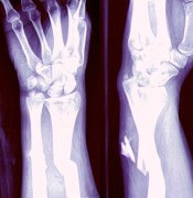
NEW YORK, NY—Investigators have developed a model that suggests the clinical benefits of denosumab translate into economic value.
The investigators designed their model, X-GEM (Exgeva-Global Economic Model), using results from a large multiple myeloma (MM) trial that showed denosumab to be non-inferior to zoledronic acid (ZA) for skeletal-related events (SRE).
Noopur S. Raje, MD, of Massachusetts General Hospital Cancer Center in Boston, discussed this work at Lymphoma & Myeloma 2017.
The abstract was selected as the best clinical myeloma abstract of the meeting.
“I do think this is a very timely topic because people are not just concerned about the clinical outcomes in patients,” Dr Raje said. “[T]here’s a lot of buzz around the economic evaluation of what we do.”
Trial results
The phase 3 study (NCT01345019) on which X-GEM is based enrolled 1718 MM patients and randomized them to either denosumab or ZA.
The results, reported this year at ASCO, showed denosumab to be non-inferior to ZA in time to first on-study SRE, the primary endpoint.
Denosumab was also found to be non-inferior to ZA for the secondary endpoint of overall survival.
For the exploratory endpoint of progression-free survival (PFS), denosumab-treated patients experienced a significant benefit in terms of PFS. The median PFS was 46.09 months with denosumab and 35.38 months with ZA (difference, 10.71 months).
“Now, we’ve never really seen a survival difference or progression-free survival difference in patients, even in treatment studies, amounting to about 10.7 months,” Dr Raje said. “So this, we found, was quite remarkable in the study.”
Dr Raje also highlighted some safety features from the study. There was significantly less renal toxicity with denosumab than with ZA.
“And in patients who had a creatinine clearance of less than 60 [mL/min], there was almost a doubling of renal toxicity in patients getting zoledronic acid,” she said.
“[D]enosumab may, in fact, be the safer alternative, specifically, in our patients with multiple myeloma who we all know have this problem of renal toxicity throughout the course of their lifetime with myeloma.”
X-GEM
Investigators based X-GEM on the original model published by Stopeck et al. in 2012, which evaluated the cost-effectiveness of denosumab to prevent SREs compared with ZA in patients with solid tumors—prostate, breast, and lung cancer.
“[Stopeck’s] data did show that denosumab was, in fact, cost-effective when compared to zoledronic acid in respect to SREs,” Dr Raje noted.
The timeline for the economic analysis in MM patients spanned the time from diagnosis to death, and patients were evaluated for SREs every 4 weeks on study.
Investigators used 2 cost scenarios. The first was based on an average sales price for 28 days, which was $1928 for denosumab and $45 for ZA. The second was based on the wholesale acquisition cost, which was $2155 for denosumab and $922 for ZA.
“No surprise to anybody,” Dr Raje noted, “denosumab is a lot more expensive. But obviously, this does not tell the whole story. Built into the X-GEM model are a whole host of other factors.”
These include the costs of administration, adverse events, the number of SREs, treatment of an MM patient, and the quality-adjusted life year (QALY) gain with either denosumab or zoledronic acid.
“When you count up all these costs and calculate them based on the data set from the 1800 patients, we found that there was really a difference of zoledronic acid costing a little bit more than denosumab,” Dr Raje said.
From the payer perspective, when clinical outcomes were monetized, the net monetary benefit of denosumab compared with ZA was $5959.
And from a societal perspective, the model calculated the net monetary benefit for denosumab to be $10,259.
The societal perspective included SRE direct costs (hospital, outpatient, and emergency department visits, long-term care, hospice, physical therapy and devices, skilled nursing facility, and strong opioids), direct non-medical costs (driving for treatment, parking, and caregiver costs), and indirect costs (short-term disability and productivity loss).
The investigators concluded that denosumab is cost-effective below a willingness-to-pay threshold of $150,000/QALY regardless of ZA price, whether wholesale or average sales price.
“The bone-specific benefits and observed prolongation of progression-free survival in combination with the economic analysis provides denosumab as a valuable option for patients with multiple myeloma,” Dr Raje said. “In total, we still think it’s more cost-effective to use denosumab when compared to zoledronic acid in this patient population.”
At present, treatment options to prevent bone complications in MM patients are limited to bisphosphonates, including ZA. Denosumab is under review by the US Food and Drug Administration for an expanded indication to include MM.
Both the phase 3 and cost-effectiveness studies were funded by Amgen, Inc. Dr Raje and co-investigators of the study have either consulted for or are employed by Amgen, Inc. ![]()
Ibrutinib approved to treat cGVHD in Canada

Health Canada has approved the BTK inhibitor ibrutinib (IMBRUVICA®) for the treatment of patients with steroid-dependent or -refractory chronic graft-versus-host disease (cGVHD).
This is the sixth approval for ibrutinib in Canada.
The drug is approved as monotherapy for patients with previously untreated chronic lymphocytic leukemia (CLL), CLL patients who have received at least 1 prior therapy, patients with Waldenström’s macroglobulinemia, and patients with relapsed or refractory mantle cell lymphoma.
Ibrutinib is also approved for use in combination with bendamustine and rituximab for the treatment of CLL patients who have received at least 1 prior therapy.
Ibrutinib is co-developed by Cilag GmbH International (a member of the Janssen Pharmaceutical Companies) and Pharmacyclics LLC, an AbbVie company. Janssen Inc. markets ibrutinib in Canada.
The approval of ibrutinib to treat cGVHD is based on results of a phase 2 trial, which were presented at the 2016 ASH Annual Meeting.
The trial included 42 patients with a median age of 56 (range, 19 to 74). The most common underlying malignancies that led to patients’ transplants were acute lymphocytic leukemia, acute myeloid leukemia, and CLL.
At baseline, the patients had persistent cGVHD symptoms despite receiving standard treatment with corticosteroids. Most patients’ symptoms included mouth ulcers and skin rashes, and more than 50% had 2 or more organs affected by cGVHD.
The median time since cGVHD diagnosis was 14 months, the median number of prior cGVHD treatments was 2 (range, 1 to 3), and 60% of patients had a Karnofsky performance score of ≤ 80.
Fifty-two percent of patients were receiving ongoing immunosuppressants and systemic corticosteroids at baseline.
Sixty-seven percent of patients responded to treatment with ibrutinib, and 21% had a complete response. In 48% of patients, responses lasted for 5 months or longer. Responses were seen across all organs affected by cGVHD (ie, skin, mouth, gastrointestinal tract, and liver).
The patients’ median steroid dose was reduced over time, from 0.31 mg/kg/day at baseline to 0.14 mg/kg/day at week 48. Five patients were able to completely discontinue corticosteroids while in response.
The most common (≥20%) adverse events (AEs) of all grades were fatigue (57%), bruising (40%), diarrhea (36%), stomatitis (29%), muscle spasms (29%), nausea (26%), hemorrhage (26%), and pneumonia (21%).
Atrial fibrillation (grade 3) occurred in 1 patient (2%). Serious AEs occurred in 52% of patients. The most common serious AEs (2 or more patients) were pneumonia, sepsis (septic shock), cellulitis, headache, and pyrexia.
There were 2 fatal events, a case of pneumonia and a case of pulmonary aspergillosis.
Twenty-four percent of patients discontinued ibrutinib due to AEs. The most common AEs leading to discontinuation were fatigue and pneumonia. AEs leading to dose reductions occurred in 26% of patients.
The recommended dose of ibrutinib for cGVHD is 420 mg (three 140 mg capsules) once daily. ![]()