User login
Recognize gender expression in youth
It has been known for decades that sex and gender cannot be determined solely by birth anatomy and chromosomes.1 Over the past decade, the medical community has been able to better understand the biologic underpinnings of gender identity, and we are gaining a better appreciation for the diversity of gender identities and gender expressions that exist.

Gender expression can be defined as the manner in which an individual chooses to present their gender to others through physical appearance and behaviors, such as style of hair or dress, voice or movement.2 Gender nonconformity (GNC) is when an individual’s gender expression does not fully conform with societal expectations often based on an individual’s sex assigned at birth. It is important to note that gender expression is independent of gender identity and may or may not align with gender identity. For example, a person whose sex assigned at birth is female may adopt hairstyles and clothing that are considered more masculine and enjoy activities that are typically associated with masculinity (for example, sports) yet identify as female. The majority of research to date focuses most on transgender individuals, broadly defined as those whose gender identity does not fully align with the sex assigned at birth.3,4 As our understanding of gender expression and GNC expands, more research is emerging on the prevalence of gender nonconformity in youth and potential associations with various health outcomes.
Stigma, discrimination, and harassment are known to have documented effects on health. GNC youth have been shown to experience discrimination and harassment at rates higher than their gender conforming peers.5,6 A recent study by Lowry et al. sought to examine the association between GNC and indicators of mental distress and substance use in adolescents.7 The authors analyzed a subset of cross-sectional data from more than 6,000 youth who had participated in the Youth Risk Behavior Surveillance–United States, 2015 (YRBS) in three large urban school districts (two in California and one in Florida). In addition to the standard YRBS questions, students at these three school districts were asked about their gender expression using the following question: “A person’s appearance style, dress, or the way they walk or talk may affect how people describe them. How do you think people at your school would describe you?” Based on responses, youth were categorized on a 7-point GNC scale with 1 being most gender conforming (a very feminine female student or very masculine male student) to 7 being most GNC (a very masculine female student or a very feminine male student). The study sample was ethnically diverse with 16% of students identifying as white non-Hispanic, 19% identifying as black non-Hispanic, and 55% identifying as Hispanic of any race.
In the study population, approximately one in five students reported either moderate (students who described themselves as equally feminine and masculine) or high (female students who described themselves as very/mostly/somewhat masculine or male students who described themselves as very/mostly/somewhat feminine) levels of GNC. Among female students, moderate GNC was significantly associated with feeling sad and hopeless, seriously considering attempting suicide, and making a suicide plan. However, in female students substance use was not associated with GNC. Among male students, suicidal thoughts, plans, and attempts all demonstrated a linear increase with GNC, with the greatest prevalence occurring in male students expressing high levels of GNC. Prevalence of substance use, specifically nonmedical use of prescription drugs, cocaine use, methamphetamine use, heroin use, and intravenous drug also was associated with high GNC in male students. Study authors hypothesize that these differences occur because GNC male youth experience more overt harassment, compared with GNC female youth, but further study is needed.
Our understanding of the diversity of gender expressions present in youth populations continues to evolve. Findings from this study add to a growing body of evidence demonstrating a relatively high prevalence of GNC in youth populations, and potential health disparities these youth may face. This study underscores the need for continued study in this area. Family support and acceptance have been demonstrated to be strong protective factors for transgender-, lesbian-, and gay-identified youth. Studies identifying protective factors for GNC youth are needed.4
As health care providers, we need to continue to ask patients and families about gender identity and be aware of gender expression. When youth present as GNC, we should recognize that they may be at increased risk and, in addition to assessing overall mental health and risk for substance use, also assess for degree of social/familial support and potential stressors.4 We also should continue to advocate for support systems within schools sensitive to the needs of GNC students, as these may be a potential avenue to improve overall mental health for students. It is important to continue to expand our understanding of the diverse gender identities and expressions of the youth we serve. This hopefully will allow us to identify not only potential risk factors and health disparities, but also protective factors that can help better inform the development of effective interventions so all youth can reach their full potential.
Dr. Chelvakumar is an attending physician in the division of adolescent medicine at Nationwide Children’s Hospital and an assistant professor of clinical pediatrics at the Ohio State University, both in Columbus. Email her at pdnews@mdedge.com.
References
1. “WPATH (World Professional Association of Transgender Health) Board Responds to Federal Effort to Redefine Gender,” press release, Oct. 23, 2018.
2. “LGBTQ+ Definitions” at Trans Student Educational Resources.3. J Sex Res. 2013;50(3-4):299-317.
4. JAMA Pediatr. 2018 Nov 1;172(11):1010-1.
5. Psychol Sex Orientat Gend Divers. 2016 Dec;3(4):489-98.
6. J Adolesc Health. 2016; 58(2)(supple):S1-2.
7. JAMA Pediatr. 2018 Nov;172(11):1020-8.
It has been known for decades that sex and gender cannot be determined solely by birth anatomy and chromosomes.1 Over the past decade, the medical community has been able to better understand the biologic underpinnings of gender identity, and we are gaining a better appreciation for the diversity of gender identities and gender expressions that exist.
Gender expression can be defined as the manner in which an individual chooses to present their gender to others through physical appearance and behaviors, such as style of hair or dress, voice or movement.2 Gender nonconformity (GNC) is when an individual’s gender expression does not fully conform with societal expectations often based on an individual’s sex assigned at birth. It is important to note that gender expression is independent of gender identity and may or may not align with gender identity. For example, a person whose sex assigned at birth is female may adopt hairstyles and clothing that are considered more masculine and enjoy activities that are typically associated with masculinity (for example, sports) yet identify as female. The majority of research to date focuses most on transgender individuals, broadly defined as those whose gender identity does not fully align with the sex assigned at birth.3,4 As our understanding of gender expression and GNC expands, more research is emerging on the prevalence of gender nonconformity in youth and potential associations with various health outcomes.
Stigma, discrimination, and harassment are known to have documented effects on health. GNC youth have been shown to experience discrimination and harassment at rates higher than their gender conforming peers.5,6 A recent study by Lowry et al. sought to examine the association between GNC and indicators of mental distress and substance use in adolescents.7 The authors analyzed a subset of cross-sectional data from more than 6,000 youth who had participated in the Youth Risk Behavior Surveillance–United States, 2015 (YRBS) in three large urban school districts (two in California and one in Florida). In addition to the standard YRBS questions, students at these three school districts were asked about their gender expression using the following question: “A person’s appearance style, dress, or the way they walk or talk may affect how people describe them. How do you think people at your school would describe you?” Based on responses, youth were categorized on a 7-point GNC scale with 1 being most gender conforming (a very feminine female student or very masculine male student) to 7 being most GNC (a very masculine female student or a very feminine male student). The study sample was ethnically diverse with 16% of students identifying as white non-Hispanic, 19% identifying as black non-Hispanic, and 55% identifying as Hispanic of any race.
In the study population, approximately one in five students reported either moderate (students who described themselves as equally feminine and masculine) or high (female students who described themselves as very/mostly/somewhat masculine or male students who described themselves as very/mostly/somewhat feminine) levels of GNC. Among female students, moderate GNC was significantly associated with feeling sad and hopeless, seriously considering attempting suicide, and making a suicide plan. However, in female students substance use was not associated with GNC. Among male students, suicidal thoughts, plans, and attempts all demonstrated a linear increase with GNC, with the greatest prevalence occurring in male students expressing high levels of GNC. Prevalence of substance use, specifically nonmedical use of prescription drugs, cocaine use, methamphetamine use, heroin use, and intravenous drug also was associated with high GNC in male students. Study authors hypothesize that these differences occur because GNC male youth experience more overt harassment, compared with GNC female youth, but further study is needed.
Our understanding of the diversity of gender expressions present in youth populations continues to evolve. Findings from this study add to a growing body of evidence demonstrating a relatively high prevalence of GNC in youth populations, and potential health disparities these youth may face. This study underscores the need for continued study in this area. Family support and acceptance have been demonstrated to be strong protective factors for transgender-, lesbian-, and gay-identified youth. Studies identifying protective factors for GNC youth are needed.4
As health care providers, we need to continue to ask patients and families about gender identity and be aware of gender expression. When youth present as GNC, we should recognize that they may be at increased risk and, in addition to assessing overall mental health and risk for substance use, also assess for degree of social/familial support and potential stressors.4 We also should continue to advocate for support systems within schools sensitive to the needs of GNC students, as these may be a potential avenue to improve overall mental health for students. It is important to continue to expand our understanding of the diverse gender identities and expressions of the youth we serve. This hopefully will allow us to identify not only potential risk factors and health disparities, but also protective factors that can help better inform the development of effective interventions so all youth can reach their full potential.
Dr. Chelvakumar is an attending physician in the division of adolescent medicine at Nationwide Children’s Hospital and an assistant professor of clinical pediatrics at the Ohio State University, both in Columbus. Email her at pdnews@mdedge.com.
References
1. “WPATH (World Professional Association of Transgender Health) Board Responds to Federal Effort to Redefine Gender,” press release, Oct. 23, 2018.
2. “LGBTQ+ Definitions” at Trans Student Educational Resources.3. J Sex Res. 2013;50(3-4):299-317.
4. JAMA Pediatr. 2018 Nov 1;172(11):1010-1.
5. Psychol Sex Orientat Gend Divers. 2016 Dec;3(4):489-98.
6. J Adolesc Health. 2016; 58(2)(supple):S1-2.
7. JAMA Pediatr. 2018 Nov;172(11):1020-8.
It has been known for decades that sex and gender cannot be determined solely by birth anatomy and chromosomes.1 Over the past decade, the medical community has been able to better understand the biologic underpinnings of gender identity, and we are gaining a better appreciation for the diversity of gender identities and gender expressions that exist.
Gender expression can be defined as the manner in which an individual chooses to present their gender to others through physical appearance and behaviors, such as style of hair or dress, voice or movement.2 Gender nonconformity (GNC) is when an individual’s gender expression does not fully conform with societal expectations often based on an individual’s sex assigned at birth. It is important to note that gender expression is independent of gender identity and may or may not align with gender identity. For example, a person whose sex assigned at birth is female may adopt hairstyles and clothing that are considered more masculine and enjoy activities that are typically associated with masculinity (for example, sports) yet identify as female. The majority of research to date focuses most on transgender individuals, broadly defined as those whose gender identity does not fully align with the sex assigned at birth.3,4 As our understanding of gender expression and GNC expands, more research is emerging on the prevalence of gender nonconformity in youth and potential associations with various health outcomes.
Stigma, discrimination, and harassment are known to have documented effects on health. GNC youth have been shown to experience discrimination and harassment at rates higher than their gender conforming peers.5,6 A recent study by Lowry et al. sought to examine the association between GNC and indicators of mental distress and substance use in adolescents.7 The authors analyzed a subset of cross-sectional data from more than 6,000 youth who had participated in the Youth Risk Behavior Surveillance–United States, 2015 (YRBS) in three large urban school districts (two in California and one in Florida). In addition to the standard YRBS questions, students at these three school districts were asked about their gender expression using the following question: “A person’s appearance style, dress, or the way they walk or talk may affect how people describe them. How do you think people at your school would describe you?” Based on responses, youth were categorized on a 7-point GNC scale with 1 being most gender conforming (a very feminine female student or very masculine male student) to 7 being most GNC (a very masculine female student or a very feminine male student). The study sample was ethnically diverse with 16% of students identifying as white non-Hispanic, 19% identifying as black non-Hispanic, and 55% identifying as Hispanic of any race.
In the study population, approximately one in five students reported either moderate (students who described themselves as equally feminine and masculine) or high (female students who described themselves as very/mostly/somewhat masculine or male students who described themselves as very/mostly/somewhat feminine) levels of GNC. Among female students, moderate GNC was significantly associated with feeling sad and hopeless, seriously considering attempting suicide, and making a suicide plan. However, in female students substance use was not associated with GNC. Among male students, suicidal thoughts, plans, and attempts all demonstrated a linear increase with GNC, with the greatest prevalence occurring in male students expressing high levels of GNC. Prevalence of substance use, specifically nonmedical use of prescription drugs, cocaine use, methamphetamine use, heroin use, and intravenous drug also was associated with high GNC in male students. Study authors hypothesize that these differences occur because GNC male youth experience more overt harassment, compared with GNC female youth, but further study is needed.
Our understanding of the diversity of gender expressions present in youth populations continues to evolve. Findings from this study add to a growing body of evidence demonstrating a relatively high prevalence of GNC in youth populations, and potential health disparities these youth may face. This study underscores the need for continued study in this area. Family support and acceptance have been demonstrated to be strong protective factors for transgender-, lesbian-, and gay-identified youth. Studies identifying protective factors for GNC youth are needed.4
As health care providers, we need to continue to ask patients and families about gender identity and be aware of gender expression. When youth present as GNC, we should recognize that they may be at increased risk and, in addition to assessing overall mental health and risk for substance use, also assess for degree of social/familial support and potential stressors.4 We also should continue to advocate for support systems within schools sensitive to the needs of GNC students, as these may be a potential avenue to improve overall mental health for students. It is important to continue to expand our understanding of the diverse gender identities and expressions of the youth we serve. This hopefully will allow us to identify not only potential risk factors and health disparities, but also protective factors that can help better inform the development of effective interventions so all youth can reach their full potential.
Dr. Chelvakumar is an attending physician in the division of adolescent medicine at Nationwide Children’s Hospital and an assistant professor of clinical pediatrics at the Ohio State University, both in Columbus. Email her at pdnews@mdedge.com.
References
1. “WPATH (World Professional Association of Transgender Health) Board Responds to Federal Effort to Redefine Gender,” press release, Oct. 23, 2018.
2. “LGBTQ+ Definitions” at Trans Student Educational Resources.3. J Sex Res. 2013;50(3-4):299-317.
4. JAMA Pediatr. 2018 Nov 1;172(11):1010-1.
5. Psychol Sex Orientat Gend Divers. 2016 Dec;3(4):489-98.
6. J Adolesc Health. 2016; 58(2)(supple):S1-2.
7. JAMA Pediatr. 2018 Nov;172(11):1020-8.

Fatigue in MS: Common, often profound, tough to treat
BERLIN – In addition to the pain, motor and sensory impairments, and cognitive dysfunction that can stalk multiple sclerosis (MS) patients, for many, there’s also the challenge of an invisible, tough-to-quantify entity: fatigue.

“Approximately 80% of patients suffer from fatigue, so it’s an immense problem in MS. There’s no real clear relationship with disease severity,” Vincent de Groot, MD, said at the annual congress of the European Committee for Treatment and Research in Multiple Sclerosis. “Despite what a lot of people think, there’s no clear or strong relationship between fatigue and the amount of physical activity people undertake daily,” he noted.
“Patients all know what we are talking about when we ask about fatigue,” but there are a variety of definitions of fatigue used in research, a fact that has limited progress in the field, said Dr. de Groot.
Primary fatigue is related to the pathophysiology of MS itself, while secondary fatigue can result from MS symptoms, such as poor sleep from spasms. Secondary fatigue can also be a side effect of MS medications; baclofen, used for spasticity, is a good example, said Dr. de Groot. “We must not underestimate how many problems these drugs can give people.”
What’s the mechanism by which MS causes primary fatigue? “The simple answer is that we do not know,” said Dr. de Groot, a physiatrist and researcher at Vrije University, Amsterdam.
Though immune-mediated fatigue had been proposed as a factor for patients with MS, Dr. de Groot said that his own lab’s work has not found any connection between fatigue levels and any immune-related biomarkers. “So I don’t think the immune hypothesis has a lot of evidence.”
Similarly, though there might be mechanistic reasons to suspect the hypothalamic-pituitary-adrenal (HPA) axis as a culprit for MS fatigue, no consistent association has been found between any markers for HPA axis disruption and fatigue ratings, Dr. de Groot said.
Newer theories center on MS-related disruptions in brain connectivity, with imaging studies now able to detect some of these disruptions in functional connectivity that correlate with fatigue. “Right now, I think this is the hypothesis to bet money on,” Dr. de Groot said.
Many factors come into play, including environmental and psychological factors, he said.
“What can we do to treat MS-related fatigue?” Though several drugs have been used, “if you carefully look at the systematic reviews, the evidence is very, very disappointing,” Dr. de Groot said. For both amantadine and modafinil, “there is no evidence that these drugs are effective,” he said, citing a systematic review and meta-analysis that found a pooled effect size of 0.07 (95% confidence interval, –0.22 to 0.37) for medications (Mult Scler Int. 2014 May; doi: 10.1155/2014/798285).
Only two trials have looked at multidisciplinary rehabilitation for MS-related fatigue, Dr. de Groot said. Two studies that looked at multidisciplinary strategies that pulled in a variety of disciplines to help develop tailored fatigue management strategies saw no between-group effect when the multidisciplinary intervention was compared with nurse-provided information or with non–fatigue-related rehabilitation.
In an effort to determine whether MS-related fatigue is truly refractory to treatment, Dr. de Groot said that he and his colleagues decided to take “three steps back” to look at the individual interventions that make up a multidisciplinary approach to tackling fatigue. “So, we looked at exercise therapy, energy conservation management, and cognitive-behavioral therapy,” beginning with a literature review, he said.
Members of his research group found that effect sizes ranged from small to moderate for the three approaches, but there were methodologic problems with many of the studies; in the case of cognitive-behavioral therapy (CBT), the effect size waned over time, Dr. de Groot said. A newer randomized, controlled trial showed a relatively robust effect size of 0.52 for Internet-delivered CBT, which may provide a promising and practical approach (J Neurol Neurosurg Psychiatry. 2018 Sep;89[9]:970-6. doi: 10.1136/jnnp-2017-317463).
Looking at fatigue and societal participation, Dr. de Groot and his colleagues examined what effect aerobic training, energy conservation management, and CBT had on the two outcome measures. The three interventions were studied in three stand-alone trials, he said.
Patients were assessed at baseline, and at 8, 16, 26, and 52 weeks. The assessments were performed by a blinded researcher and were the same across trials: For fatigue, researchers used the Checklist Individual Strength–fatigue (CIS20R-fatigue), and for societal participation, they administered the Impact on Participation and Autonomy (IPA).
Each trial included 90 patients, randomized 1:1 to receive high- or low-intensity treatment. Patients had to have MS with no exacerbations within the prior 6 months and an Expanded Disability Status Scale score of 6 or less. However, the included patients had severe fatigue, with a CIS20R-fatigue subscore of 35 or higher, and the fatigue could not be attributable to such secondary causes as infection, depression, or thyroid or sleep problems. Finally, patients could not have been treated for fatigue in the 3 months prior to enrollment.
Those in the high-intensity treatment group received 12 sessions focused on the particular intervention over 4 months, provided by an expert therapist. Each type of intervention had a treatment protocol that was followed over the 4 months. Patients receiving low-intensity treatment saw an MS-specialized nurse three times over 4 months.
The aerobic training intervention had patients performing high-intensity exercise on a cycle ergometer for 30 minutes, three times weekly for 16 weeks. In addition to the 12 supervised sessions, patients also completed 36 home-based sessions. The level of intensity for each patient was personalized based on their baseline cardiopulmonary exercise test, Dr. de Groot said.
At the end of 1 year, patients in the high-intensity group and those in the low-intensity group reported virtually the same fatigue scores. Though there was an initial drop in fatigue for those in the high-intensity group, compared with baseline and with the low-intensity participants, values on the CIS20R never dropped below 35, the “severe fatigue” cutoff.
And, Dr. de Groot said, there was no effect on societal participation or in other fatigue scores. In sum, the effect size was barely significant at –0.54 (95% CI, –1.00 to –0.06), with a number needed to treat of 9.
Adherence to attempting the workouts was fairly good for participants in the high-intensity group; 74% completed the sessions, with 71% doing so at the prescribed workload. The median rate of perceived exertion on a 1-20 scale was 14.
However, the thrice-weekly exercise bouts didn’t improve aerobic fitness parameters: Neither V02peak, V02peak adjusted for body mass, nor anaerobic threshold changed for those in the high-intensity group. Peak power did increase by 11.7 watts (P = .048).
Energy conservation education, whether delivered in high- or low-intensity format, had almost no effect on fatigue scores, with a number needed to treat of 158 – a figure that is “neither significant nor clinically meaningful,” Dr. de Groot said. Other fatigue scores and societal participation levels also went unchanged.
However, CBT delivered in a series of 10 modules to address various beliefs and coping mechanisms about MS, fatigue, pain, and activity regulation did have a positive effect on fatigue. Here, the effect size was –0.79 (95% CI, –1.26 to –0.32). The number needed to treat was 3, and CIS20R values did dip below the “severe fatigue” threshold during treatment. A similar effect, Dr. de Groot said, was seen for other fatigue and quality of life measures, though societal participation scores didn’t change. No significant improvement was seen for the low-intensity CBT group.
“Severe MS-related fatigue can be reduced effectively with CBT in the short term. More research is needed on how to maintain this effect in the long term,” Dr. de Groot said. Still, “it’s currently the best treatment option,” he said.
The fact that patients reverted to their preintervention fatigue levels regardless of the intervention shows that effective treatment for MS-related fatigue should probably be ongoing, viewed more as a process than an occurrence, Dr. de Groot said.
To that end, Dr. de Groot and his colleagues are conducting a randomized, controlled trial that includes 166 MS patients with fatigue. The study has two arms: The first is a noninferiority trial comparing face-to-face CBT with e-learning delivery of the content, and the second looks at the efficacy of ongoing booster sessions after initial CBT.
An online database of randomized, controlled trials of rehabilitation for MS patients can be found at www.appeco.net.
The study was funded by Fonds NutsOhra, a private Dutch foundation. Dr. de Groot reported no relevant conflicts of interest.
SOURCE: de Groot V. Mult Scler. 2018;24(S2):83, Abstract 225.
BERLIN – In addition to the pain, motor and sensory impairments, and cognitive dysfunction that can stalk multiple sclerosis (MS) patients, for many, there’s also the challenge of an invisible, tough-to-quantify entity: fatigue.
“Approximately 80% of patients suffer from fatigue, so it’s an immense problem in MS. There’s no real clear relationship with disease severity,” Vincent de Groot, MD, said at the annual congress of the European Committee for Treatment and Research in Multiple Sclerosis. “Despite what a lot of people think, there’s no clear or strong relationship between fatigue and the amount of physical activity people undertake daily,” he noted.
“Patients all know what we are talking about when we ask about fatigue,” but there are a variety of definitions of fatigue used in research, a fact that has limited progress in the field, said Dr. de Groot.
Primary fatigue is related to the pathophysiology of MS itself, while secondary fatigue can result from MS symptoms, such as poor sleep from spasms. Secondary fatigue can also be a side effect of MS medications; baclofen, used for spasticity, is a good example, said Dr. de Groot. “We must not underestimate how many problems these drugs can give people.”
What’s the mechanism by which MS causes primary fatigue? “The simple answer is that we do not know,” said Dr. de Groot, a physiatrist and researcher at Vrije University, Amsterdam.
Though immune-mediated fatigue had been proposed as a factor for patients with MS, Dr. de Groot said that his own lab’s work has not found any connection between fatigue levels and any immune-related biomarkers. “So I don’t think the immune hypothesis has a lot of evidence.”
Similarly, though there might be mechanistic reasons to suspect the hypothalamic-pituitary-adrenal (HPA) axis as a culprit for MS fatigue, no consistent association has been found between any markers for HPA axis disruption and fatigue ratings, Dr. de Groot said.
Newer theories center on MS-related disruptions in brain connectivity, with imaging studies now able to detect some of these disruptions in functional connectivity that correlate with fatigue. “Right now, I think this is the hypothesis to bet money on,” Dr. de Groot said.
Many factors come into play, including environmental and psychological factors, he said.
“What can we do to treat MS-related fatigue?” Though several drugs have been used, “if you carefully look at the systematic reviews, the evidence is very, very disappointing,” Dr. de Groot said. For both amantadine and modafinil, “there is no evidence that these drugs are effective,” he said, citing a systematic review and meta-analysis that found a pooled effect size of 0.07 (95% confidence interval, –0.22 to 0.37) for medications (Mult Scler Int. 2014 May; doi: 10.1155/2014/798285).
Only two trials have looked at multidisciplinary rehabilitation for MS-related fatigue, Dr. de Groot said. Two studies that looked at multidisciplinary strategies that pulled in a variety of disciplines to help develop tailored fatigue management strategies saw no between-group effect when the multidisciplinary intervention was compared with nurse-provided information or with non–fatigue-related rehabilitation.
In an effort to determine whether MS-related fatigue is truly refractory to treatment, Dr. de Groot said that he and his colleagues decided to take “three steps back” to look at the individual interventions that make up a multidisciplinary approach to tackling fatigue. “So, we looked at exercise therapy, energy conservation management, and cognitive-behavioral therapy,” beginning with a literature review, he said.
Members of his research group found that effect sizes ranged from small to moderate for the three approaches, but there were methodologic problems with many of the studies; in the case of cognitive-behavioral therapy (CBT), the effect size waned over time, Dr. de Groot said. A newer randomized, controlled trial showed a relatively robust effect size of 0.52 for Internet-delivered CBT, which may provide a promising and practical approach (J Neurol Neurosurg Psychiatry. 2018 Sep;89[9]:970-6. doi: 10.1136/jnnp-2017-317463).
Looking at fatigue and societal participation, Dr. de Groot and his colleagues examined what effect aerobic training, energy conservation management, and CBT had on the two outcome measures. The three interventions were studied in three stand-alone trials, he said.
Patients were assessed at baseline, and at 8, 16, 26, and 52 weeks. The assessments were performed by a blinded researcher and were the same across trials: For fatigue, researchers used the Checklist Individual Strength–fatigue (CIS20R-fatigue), and for societal participation, they administered the Impact on Participation and Autonomy (IPA).
Each trial included 90 patients, randomized 1:1 to receive high- or low-intensity treatment. Patients had to have MS with no exacerbations within the prior 6 months and an Expanded Disability Status Scale score of 6 or less. However, the included patients had severe fatigue, with a CIS20R-fatigue subscore of 35 or higher, and the fatigue could not be attributable to such secondary causes as infection, depression, or thyroid or sleep problems. Finally, patients could not have been treated for fatigue in the 3 months prior to enrollment.
Those in the high-intensity treatment group received 12 sessions focused on the particular intervention over 4 months, provided by an expert therapist. Each type of intervention had a treatment protocol that was followed over the 4 months. Patients receiving low-intensity treatment saw an MS-specialized nurse three times over 4 months.
The aerobic training intervention had patients performing high-intensity exercise on a cycle ergometer for 30 minutes, three times weekly for 16 weeks. In addition to the 12 supervised sessions, patients also completed 36 home-based sessions. The level of intensity for each patient was personalized based on their baseline cardiopulmonary exercise test, Dr. de Groot said.
At the end of 1 year, patients in the high-intensity group and those in the low-intensity group reported virtually the same fatigue scores. Though there was an initial drop in fatigue for those in the high-intensity group, compared with baseline and with the low-intensity participants, values on the CIS20R never dropped below 35, the “severe fatigue” cutoff.
And, Dr. de Groot said, there was no effect on societal participation or in other fatigue scores. In sum, the effect size was barely significant at –0.54 (95% CI, –1.00 to –0.06), with a number needed to treat of 9.
Adherence to attempting the workouts was fairly good for participants in the high-intensity group; 74% completed the sessions, with 71% doing so at the prescribed workload. The median rate of perceived exertion on a 1-20 scale was 14.
However, the thrice-weekly exercise bouts didn’t improve aerobic fitness parameters: Neither V02peak, V02peak adjusted for body mass, nor anaerobic threshold changed for those in the high-intensity group. Peak power did increase by 11.7 watts (P = .048).
Energy conservation education, whether delivered in high- or low-intensity format, had almost no effect on fatigue scores, with a number needed to treat of 158 – a figure that is “neither significant nor clinically meaningful,” Dr. de Groot said. Other fatigue scores and societal participation levels also went unchanged.
However, CBT delivered in a series of 10 modules to address various beliefs and coping mechanisms about MS, fatigue, pain, and activity regulation did have a positive effect on fatigue. Here, the effect size was –0.79 (95% CI, –1.26 to –0.32). The number needed to treat was 3, and CIS20R values did dip below the “severe fatigue” threshold during treatment. A similar effect, Dr. de Groot said, was seen for other fatigue and quality of life measures, though societal participation scores didn’t change. No significant improvement was seen for the low-intensity CBT group.
“Severe MS-related fatigue can be reduced effectively with CBT in the short term. More research is needed on how to maintain this effect in the long term,” Dr. de Groot said. Still, “it’s currently the best treatment option,” he said.
The fact that patients reverted to their preintervention fatigue levels regardless of the intervention shows that effective treatment for MS-related fatigue should probably be ongoing, viewed more as a process than an occurrence, Dr. de Groot said.
To that end, Dr. de Groot and his colleagues are conducting a randomized, controlled trial that includes 166 MS patients with fatigue. The study has two arms: The first is a noninferiority trial comparing face-to-face CBT with e-learning delivery of the content, and the second looks at the efficacy of ongoing booster sessions after initial CBT.
An online database of randomized, controlled trials of rehabilitation for MS patients can be found at www.appeco.net.
The study was funded by Fonds NutsOhra, a private Dutch foundation. Dr. de Groot reported no relevant conflicts of interest.
SOURCE: de Groot V. Mult Scler. 2018;24(S2):83, Abstract 225.
BERLIN – In addition to the pain, motor and sensory impairments, and cognitive dysfunction that can stalk multiple sclerosis (MS) patients, for many, there’s also the challenge of an invisible, tough-to-quantify entity: fatigue.
“Approximately 80% of patients suffer from fatigue, so it’s an immense problem in MS. There’s no real clear relationship with disease severity,” Vincent de Groot, MD, said at the annual congress of the European Committee for Treatment and Research in Multiple Sclerosis. “Despite what a lot of people think, there’s no clear or strong relationship between fatigue and the amount of physical activity people undertake daily,” he noted.
“Patients all know what we are talking about when we ask about fatigue,” but there are a variety of definitions of fatigue used in research, a fact that has limited progress in the field, said Dr. de Groot.
Primary fatigue is related to the pathophysiology of MS itself, while secondary fatigue can result from MS symptoms, such as poor sleep from spasms. Secondary fatigue can also be a side effect of MS medications; baclofen, used for spasticity, is a good example, said Dr. de Groot. “We must not underestimate how many problems these drugs can give people.”
What’s the mechanism by which MS causes primary fatigue? “The simple answer is that we do not know,” said Dr. de Groot, a physiatrist and researcher at Vrije University, Amsterdam.
Though immune-mediated fatigue had been proposed as a factor for patients with MS, Dr. de Groot said that his own lab’s work has not found any connection between fatigue levels and any immune-related biomarkers. “So I don’t think the immune hypothesis has a lot of evidence.”
Similarly, though there might be mechanistic reasons to suspect the hypothalamic-pituitary-adrenal (HPA) axis as a culprit for MS fatigue, no consistent association has been found between any markers for HPA axis disruption and fatigue ratings, Dr. de Groot said.
Newer theories center on MS-related disruptions in brain connectivity, with imaging studies now able to detect some of these disruptions in functional connectivity that correlate with fatigue. “Right now, I think this is the hypothesis to bet money on,” Dr. de Groot said.
Many factors come into play, including environmental and psychological factors, he said.
“What can we do to treat MS-related fatigue?” Though several drugs have been used, “if you carefully look at the systematic reviews, the evidence is very, very disappointing,” Dr. de Groot said. For both amantadine and modafinil, “there is no evidence that these drugs are effective,” he said, citing a systematic review and meta-analysis that found a pooled effect size of 0.07 (95% confidence interval, –0.22 to 0.37) for medications (Mult Scler Int. 2014 May; doi: 10.1155/2014/798285).
Only two trials have looked at multidisciplinary rehabilitation for MS-related fatigue, Dr. de Groot said. Two studies that looked at multidisciplinary strategies that pulled in a variety of disciplines to help develop tailored fatigue management strategies saw no between-group effect when the multidisciplinary intervention was compared with nurse-provided information or with non–fatigue-related rehabilitation.
In an effort to determine whether MS-related fatigue is truly refractory to treatment, Dr. de Groot said that he and his colleagues decided to take “three steps back” to look at the individual interventions that make up a multidisciplinary approach to tackling fatigue. “So, we looked at exercise therapy, energy conservation management, and cognitive-behavioral therapy,” beginning with a literature review, he said.
Members of his research group found that effect sizes ranged from small to moderate for the three approaches, but there were methodologic problems with many of the studies; in the case of cognitive-behavioral therapy (CBT), the effect size waned over time, Dr. de Groot said. A newer randomized, controlled trial showed a relatively robust effect size of 0.52 for Internet-delivered CBT, which may provide a promising and practical approach (J Neurol Neurosurg Psychiatry. 2018 Sep;89[9]:970-6. doi: 10.1136/jnnp-2017-317463).
Looking at fatigue and societal participation, Dr. de Groot and his colleagues examined what effect aerobic training, energy conservation management, and CBT had on the two outcome measures. The three interventions were studied in three stand-alone trials, he said.
Patients were assessed at baseline, and at 8, 16, 26, and 52 weeks. The assessments were performed by a blinded researcher and were the same across trials: For fatigue, researchers used the Checklist Individual Strength–fatigue (CIS20R-fatigue), and for societal participation, they administered the Impact on Participation and Autonomy (IPA).
Each trial included 90 patients, randomized 1:1 to receive high- or low-intensity treatment. Patients had to have MS with no exacerbations within the prior 6 months and an Expanded Disability Status Scale score of 6 or less. However, the included patients had severe fatigue, with a CIS20R-fatigue subscore of 35 or higher, and the fatigue could not be attributable to such secondary causes as infection, depression, or thyroid or sleep problems. Finally, patients could not have been treated for fatigue in the 3 months prior to enrollment.
Those in the high-intensity treatment group received 12 sessions focused on the particular intervention over 4 months, provided by an expert therapist. Each type of intervention had a treatment protocol that was followed over the 4 months. Patients receiving low-intensity treatment saw an MS-specialized nurse three times over 4 months.
The aerobic training intervention had patients performing high-intensity exercise on a cycle ergometer for 30 minutes, three times weekly for 16 weeks. In addition to the 12 supervised sessions, patients also completed 36 home-based sessions. The level of intensity for each patient was personalized based on their baseline cardiopulmonary exercise test, Dr. de Groot said.
At the end of 1 year, patients in the high-intensity group and those in the low-intensity group reported virtually the same fatigue scores. Though there was an initial drop in fatigue for those in the high-intensity group, compared with baseline and with the low-intensity participants, values on the CIS20R never dropped below 35, the “severe fatigue” cutoff.
And, Dr. de Groot said, there was no effect on societal participation or in other fatigue scores. In sum, the effect size was barely significant at –0.54 (95% CI, –1.00 to –0.06), with a number needed to treat of 9.
Adherence to attempting the workouts was fairly good for participants in the high-intensity group; 74% completed the sessions, with 71% doing so at the prescribed workload. The median rate of perceived exertion on a 1-20 scale was 14.
However, the thrice-weekly exercise bouts didn’t improve aerobic fitness parameters: Neither V02peak, V02peak adjusted for body mass, nor anaerobic threshold changed for those in the high-intensity group. Peak power did increase by 11.7 watts (P = .048).
Energy conservation education, whether delivered in high- or low-intensity format, had almost no effect on fatigue scores, with a number needed to treat of 158 – a figure that is “neither significant nor clinically meaningful,” Dr. de Groot said. Other fatigue scores and societal participation levels also went unchanged.
However, CBT delivered in a series of 10 modules to address various beliefs and coping mechanisms about MS, fatigue, pain, and activity regulation did have a positive effect on fatigue. Here, the effect size was –0.79 (95% CI, –1.26 to –0.32). The number needed to treat was 3, and CIS20R values did dip below the “severe fatigue” threshold during treatment. A similar effect, Dr. de Groot said, was seen for other fatigue and quality of life measures, though societal participation scores didn’t change. No significant improvement was seen for the low-intensity CBT group.
“Severe MS-related fatigue can be reduced effectively with CBT in the short term. More research is needed on how to maintain this effect in the long term,” Dr. de Groot said. Still, “it’s currently the best treatment option,” he said.
The fact that patients reverted to their preintervention fatigue levels regardless of the intervention shows that effective treatment for MS-related fatigue should probably be ongoing, viewed more as a process than an occurrence, Dr. de Groot said.
To that end, Dr. de Groot and his colleagues are conducting a randomized, controlled trial that includes 166 MS patients with fatigue. The study has two arms: The first is a noninferiority trial comparing face-to-face CBT with e-learning delivery of the content, and the second looks at the efficacy of ongoing booster sessions after initial CBT.
An online database of randomized, controlled trials of rehabilitation for MS patients can be found at www.appeco.net.
The study was funded by Fonds NutsOhra, a private Dutch foundation. Dr. de Groot reported no relevant conflicts of interest.
SOURCE: de Groot V. Mult Scler. 2018;24(S2):83, Abstract 225.
EXPERT ANALYSIS FROM ECTRIMS 2018

Particular lesions early after CIS predict long-term MS disability
BERLIN – The presence of infratentorial and deep white matter lesions early in the course of relapse-onset multiple sclerosis was associated with high levels of disability 30 years later in a study looking at MRI predictors.
Univariate predictors of an Expanded Disability Status Scale (EDSS) score of more than 3.5, which is indicative of impaired mobility after 30 years, were the presence of an IT lesion at baseline, with an odds ratio (OR) of 12.4 (95% confidence interval [CI], 3.35-3.46; P less than .001), the presence of a deep white matter (DWM) lesion 1 year after presenting with clinically-isolated syndrome (CIS; OR = 10.65; 95% CI, 2.84-38.84; P less than .001), and the presence of an infratentorial (IT) lesion 1 year post CIS presentation (OR = 11.1; 95% CI, 3.31-37.22; P less than .001). At 5 years after a CIS presentation, the EDSS score, EDSS score change, and a DWM lesion score of more than 5 were indicative of worse disability after 30 years.
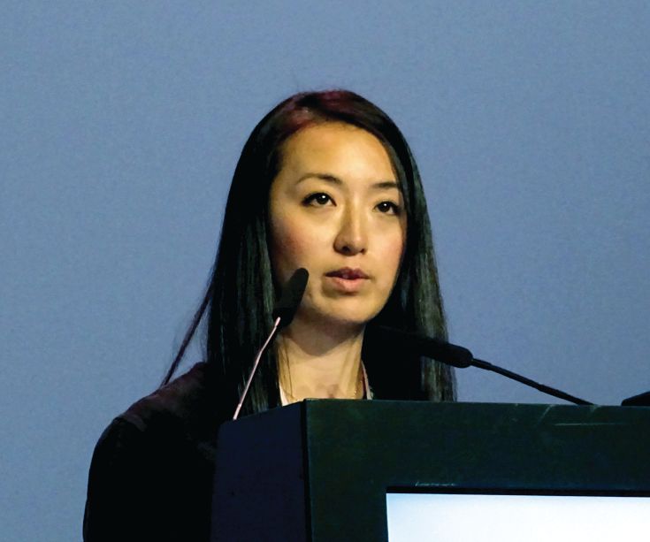
There was no significant association with age of onset, gender, CIS type, baseline EDSS, or disease duration, study investigator Karen Chung, MBBS, reported at the annual congress of the European Committee for Treatment and Research in Multiple Sclerosis.
“As we know, approximately 85% of people with MS initially present with a clinically isolated syndrome,” said Dr. Chung, a clinical research associate at the Queen Square Multiple Sclerosis Centre in the UCL Institute of Neurology in London. She added that the long-term prognosis after CIS is very variable, with some patients developing little detectable disability over time, while others may experience considerable decline.
There have been few studies examining whether there are any MRI parameters that might help predict patients’ long-term outcomes, so the aim of the study Dr. Chung presented was to see if there were any MRI parameters that might be predictive of clinical outcome 30 years after the onset of CIS.
Dr. Chung and her coauthors examined data on 120 of 132 individuals from the First London CIS Cohort who were prospectively recruited between 1984 and 1987 and had known outcomes. They looked at prospectively gathered MRI data and EDSS data at baseline, 5, 10, 14, 20, and 30 years. MRI data were obtained for 1 year after the CIS event, and data on the lowest EDSS score after the CIS event were retrospectively determined from patient notes or recall. The cohort was predominantly female (61%), with a mean of 31.5 years at CIS onset. Around half (52%) presented with optic neuritis, 27% with a spinal cord syndrome, and 20% with a brainstem syndrome. The high percentage of patients presenting with optic neuritis may be due to the fact that one of the recruiting centers was a specialist eye hospital, Dr. Chung noted later during discussion.
The 2010 McDonald Criteria were used to determine whether patients had CIS, relapsing-remitting MS (RRMS), secondary progressive MS (SPMS), or death related to MS.
“We looked at all the MRIs available to us and quantified the T2 lesion count for whole brain as well as by location,” Dr. Chung explained. The locations considered were juxtacortical, periventricular, infratentorial, and deep white matter.
“I think it is important to remind ourselves that we have come a long way with MRI technology in the 30 years timespan,” she added, noting that there was “clearly a difference in the quality.”
Clinical outcomes at 30 years were as follows: 80 patients (67%) developed MS, of whom 35 (44%) had RRMS, 26 (33%) had SPMS, 15 (20%) had died as a result of MS, and 3 (4%) had died for unknown or unrelated causes. Of the 40 patients (33%) who remained with CIS, 10 (25%) died without developing MS.
“This is a largely untreated cohort where, within the 80 people with MS, 11 (14%) were treated with a DMT [disease-modifying treatment] at some point,” Dr. Chung reported. All DMTs used were first-line injectable agents, she observed.
EDSS scores could be obtained for 107 patients. At 30 years, people with low EDSS scores were those who remained with CIS or RRMS, and as EDSS scores increased, the severity of MS increased.
“Overall, T2 lesions were better predictors of 30-year outcome than EDSS,” Dr. Chung said. For combinations of predictors, patients who had at least one IT and one DWM lesion within 1 year of a CIS had a higher probability (94%) of having an EDSS score of more than 3.5 at 30 years than when compared with those with neither lesion (13%) and those with one DWM but no IT lesions (49%).
Looking at the best independent predictors up to 5 years, the predicted probability of an EDSS score of more than 3.5 if there were no IT lesions and fewer than five DWM lesions was 18%. But if there were no IT but more than 5 DWM lesions, the probability of disability at 30 years rose to 52%. The probabilities rose even higher to 63% if there was one or more IT and five or less DWM lesions and 90% if there was one or more IT and more than five DWM lesions.
“In this cohort, the presence of early infratentorial and deep white matter lesions following a CIS are associated with higher level of disability 30 years later,” Dr. Chung concluded. “Early predictive models can add information to risk-benefit stratification.”
During discussion, one delegate expressed concerns that these data were “not generalizable to the current situation.” This was a cohort of patients that largely wasn’t treated or if they were, treatment was delayed by more than 10 years. These data were interesting “from a historical perspective,” he argued, “but I don’t understand, how in the absence of contemporary therapies this is applicable in a way that will allow us to use this information to make prognoses for the future.”
Dr. Chung agreed, noting that this was more of a natural history study. “However, I think it is applicable in clinical practice in my opinion.
“When you have a patient presenting with a CIS, at the time of diagnosis, especially now when we can diagnose patients earlier with the new 2017 criteria, it will be helpful for the patient and ourselves to apply some of the information I presented to help perhaps in aiding decisions regarding treatment.”
The study was funded by a grant from the MS Society of Great Britain. Dr. Chung disclosed receiving honoraria from Teva, Biogen, and Roche.
SOURCE: Chung K et al. Mult Scler. 2018;24(S2):58-59, Abstract 157.
BERLIN – The presence of infratentorial and deep white matter lesions early in the course of relapse-onset multiple sclerosis was associated with high levels of disability 30 years later in a study looking at MRI predictors.
Univariate predictors of an Expanded Disability Status Scale (EDSS) score of more than 3.5, which is indicative of impaired mobility after 30 years, were the presence of an IT lesion at baseline, with an odds ratio (OR) of 12.4 (95% confidence interval [CI], 3.35-3.46; P less than .001), the presence of a deep white matter (DWM) lesion 1 year after presenting with clinically-isolated syndrome (CIS; OR = 10.65; 95% CI, 2.84-38.84; P less than .001), and the presence of an infratentorial (IT) lesion 1 year post CIS presentation (OR = 11.1; 95% CI, 3.31-37.22; P less than .001). At 5 years after a CIS presentation, the EDSS score, EDSS score change, and a DWM lesion score of more than 5 were indicative of worse disability after 30 years.
There was no significant association with age of onset, gender, CIS type, baseline EDSS, or disease duration, study investigator Karen Chung, MBBS, reported at the annual congress of the European Committee for Treatment and Research in Multiple Sclerosis.
“As we know, approximately 85% of people with MS initially present with a clinically isolated syndrome,” said Dr. Chung, a clinical research associate at the Queen Square Multiple Sclerosis Centre in the UCL Institute of Neurology in London. She added that the long-term prognosis after CIS is very variable, with some patients developing little detectable disability over time, while others may experience considerable decline.
There have been few studies examining whether there are any MRI parameters that might help predict patients’ long-term outcomes, so the aim of the study Dr. Chung presented was to see if there were any MRI parameters that might be predictive of clinical outcome 30 years after the onset of CIS.
Dr. Chung and her coauthors examined data on 120 of 132 individuals from the First London CIS Cohort who were prospectively recruited between 1984 and 1987 and had known outcomes. They looked at prospectively gathered MRI data and EDSS data at baseline, 5, 10, 14, 20, and 30 years. MRI data were obtained for 1 year after the CIS event, and data on the lowest EDSS score after the CIS event were retrospectively determined from patient notes or recall. The cohort was predominantly female (61%), with a mean of 31.5 years at CIS onset. Around half (52%) presented with optic neuritis, 27% with a spinal cord syndrome, and 20% with a brainstem syndrome. The high percentage of patients presenting with optic neuritis may be due to the fact that one of the recruiting centers was a specialist eye hospital, Dr. Chung noted later during discussion.
The 2010 McDonald Criteria were used to determine whether patients had CIS, relapsing-remitting MS (RRMS), secondary progressive MS (SPMS), or death related to MS.
“We looked at all the MRIs available to us and quantified the T2 lesion count for whole brain as well as by location,” Dr. Chung explained. The locations considered were juxtacortical, periventricular, infratentorial, and deep white matter.
“I think it is important to remind ourselves that we have come a long way with MRI technology in the 30 years timespan,” she added, noting that there was “clearly a difference in the quality.”
Clinical outcomes at 30 years were as follows: 80 patients (67%) developed MS, of whom 35 (44%) had RRMS, 26 (33%) had SPMS, 15 (20%) had died as a result of MS, and 3 (4%) had died for unknown or unrelated causes. Of the 40 patients (33%) who remained with CIS, 10 (25%) died without developing MS.
“This is a largely untreated cohort where, within the 80 people with MS, 11 (14%) were treated with a DMT [disease-modifying treatment] at some point,” Dr. Chung reported. All DMTs used were first-line injectable agents, she observed.
EDSS scores could be obtained for 107 patients. At 30 years, people with low EDSS scores were those who remained with CIS or RRMS, and as EDSS scores increased, the severity of MS increased.
“Overall, T2 lesions were better predictors of 30-year outcome than EDSS,” Dr. Chung said. For combinations of predictors, patients who had at least one IT and one DWM lesion within 1 year of a CIS had a higher probability (94%) of having an EDSS score of more than 3.5 at 30 years than when compared with those with neither lesion (13%) and those with one DWM but no IT lesions (49%).
Looking at the best independent predictors up to 5 years, the predicted probability of an EDSS score of more than 3.5 if there were no IT lesions and fewer than five DWM lesions was 18%. But if there were no IT but more than 5 DWM lesions, the probability of disability at 30 years rose to 52%. The probabilities rose even higher to 63% if there was one or more IT and five or less DWM lesions and 90% if there was one or more IT and more than five DWM lesions.
“In this cohort, the presence of early infratentorial and deep white matter lesions following a CIS are associated with higher level of disability 30 years later,” Dr. Chung concluded. “Early predictive models can add information to risk-benefit stratification.”
During discussion, one delegate expressed concerns that these data were “not generalizable to the current situation.” This was a cohort of patients that largely wasn’t treated or if they were, treatment was delayed by more than 10 years. These data were interesting “from a historical perspective,” he argued, “but I don’t understand, how in the absence of contemporary therapies this is applicable in a way that will allow us to use this information to make prognoses for the future.”
Dr. Chung agreed, noting that this was more of a natural history study. “However, I think it is applicable in clinical practice in my opinion.
“When you have a patient presenting with a CIS, at the time of diagnosis, especially now when we can diagnose patients earlier with the new 2017 criteria, it will be helpful for the patient and ourselves to apply some of the information I presented to help perhaps in aiding decisions regarding treatment.”
The study was funded by a grant from the MS Society of Great Britain. Dr. Chung disclosed receiving honoraria from Teva, Biogen, and Roche.
SOURCE: Chung K et al. Mult Scler. 2018;24(S2):58-59, Abstract 157.
BERLIN – The presence of infratentorial and deep white matter lesions early in the course of relapse-onset multiple sclerosis was associated with high levels of disability 30 years later in a study looking at MRI predictors.
Univariate predictors of an Expanded Disability Status Scale (EDSS) score of more than 3.5, which is indicative of impaired mobility after 30 years, were the presence of an IT lesion at baseline, with an odds ratio (OR) of 12.4 (95% confidence interval [CI], 3.35-3.46; P less than .001), the presence of a deep white matter (DWM) lesion 1 year after presenting with clinically-isolated syndrome (CIS; OR = 10.65; 95% CI, 2.84-38.84; P less than .001), and the presence of an infratentorial (IT) lesion 1 year post CIS presentation (OR = 11.1; 95% CI, 3.31-37.22; P less than .001). At 5 years after a CIS presentation, the EDSS score, EDSS score change, and a DWM lesion score of more than 5 were indicative of worse disability after 30 years.
There was no significant association with age of onset, gender, CIS type, baseline EDSS, or disease duration, study investigator Karen Chung, MBBS, reported at the annual congress of the European Committee for Treatment and Research in Multiple Sclerosis.
“As we know, approximately 85% of people with MS initially present with a clinically isolated syndrome,” said Dr. Chung, a clinical research associate at the Queen Square Multiple Sclerosis Centre in the UCL Institute of Neurology in London. She added that the long-term prognosis after CIS is very variable, with some patients developing little detectable disability over time, while others may experience considerable decline.
There have been few studies examining whether there are any MRI parameters that might help predict patients’ long-term outcomes, so the aim of the study Dr. Chung presented was to see if there were any MRI parameters that might be predictive of clinical outcome 30 years after the onset of CIS.
Dr. Chung and her coauthors examined data on 120 of 132 individuals from the First London CIS Cohort who were prospectively recruited between 1984 and 1987 and had known outcomes. They looked at prospectively gathered MRI data and EDSS data at baseline, 5, 10, 14, 20, and 30 years. MRI data were obtained for 1 year after the CIS event, and data on the lowest EDSS score after the CIS event were retrospectively determined from patient notes or recall. The cohort was predominantly female (61%), with a mean of 31.5 years at CIS onset. Around half (52%) presented with optic neuritis, 27% with a spinal cord syndrome, and 20% with a brainstem syndrome. The high percentage of patients presenting with optic neuritis may be due to the fact that one of the recruiting centers was a specialist eye hospital, Dr. Chung noted later during discussion.
The 2010 McDonald Criteria were used to determine whether patients had CIS, relapsing-remitting MS (RRMS), secondary progressive MS (SPMS), or death related to MS.
“We looked at all the MRIs available to us and quantified the T2 lesion count for whole brain as well as by location,” Dr. Chung explained. The locations considered were juxtacortical, periventricular, infratentorial, and deep white matter.
“I think it is important to remind ourselves that we have come a long way with MRI technology in the 30 years timespan,” she added, noting that there was “clearly a difference in the quality.”
Clinical outcomes at 30 years were as follows: 80 patients (67%) developed MS, of whom 35 (44%) had RRMS, 26 (33%) had SPMS, 15 (20%) had died as a result of MS, and 3 (4%) had died for unknown or unrelated causes. Of the 40 patients (33%) who remained with CIS, 10 (25%) died without developing MS.
“This is a largely untreated cohort where, within the 80 people with MS, 11 (14%) were treated with a DMT [disease-modifying treatment] at some point,” Dr. Chung reported. All DMTs used were first-line injectable agents, she observed.
EDSS scores could be obtained for 107 patients. At 30 years, people with low EDSS scores were those who remained with CIS or RRMS, and as EDSS scores increased, the severity of MS increased.
“Overall, T2 lesions were better predictors of 30-year outcome than EDSS,” Dr. Chung said. For combinations of predictors, patients who had at least one IT and one DWM lesion within 1 year of a CIS had a higher probability (94%) of having an EDSS score of more than 3.5 at 30 years than when compared with those with neither lesion (13%) and those with one DWM but no IT lesions (49%).
Looking at the best independent predictors up to 5 years, the predicted probability of an EDSS score of more than 3.5 if there were no IT lesions and fewer than five DWM lesions was 18%. But if there were no IT but more than 5 DWM lesions, the probability of disability at 30 years rose to 52%. The probabilities rose even higher to 63% if there was one or more IT and five or less DWM lesions and 90% if there was one or more IT and more than five DWM lesions.
“In this cohort, the presence of early infratentorial and deep white matter lesions following a CIS are associated with higher level of disability 30 years later,” Dr. Chung concluded. “Early predictive models can add information to risk-benefit stratification.”
During discussion, one delegate expressed concerns that these data were “not generalizable to the current situation.” This was a cohort of patients that largely wasn’t treated or if they were, treatment was delayed by more than 10 years. These data were interesting “from a historical perspective,” he argued, “but I don’t understand, how in the absence of contemporary therapies this is applicable in a way that will allow us to use this information to make prognoses for the future.”
Dr. Chung agreed, noting that this was more of a natural history study. “However, I think it is applicable in clinical practice in my opinion.
“When you have a patient presenting with a CIS, at the time of diagnosis, especially now when we can diagnose patients earlier with the new 2017 criteria, it will be helpful for the patient and ourselves to apply some of the information I presented to help perhaps in aiding decisions regarding treatment.”
The study was funded by a grant from the MS Society of Great Britain. Dr. Chung disclosed receiving honoraria from Teva, Biogen, and Roche.
SOURCE: Chung K et al. Mult Scler. 2018;24(S2):58-59, Abstract 157.
REPORTING FROM ECTRIMS 2018
Key clinical point: Early magnetic resonance parameters can provide information that can help risk-stratify patients.
Major finding: Infratentorial and deep white matter lesions early in the course of relapse-onset multiple sclerosis were associated with high levels of disability 30 years later.
Study details: Data on 120 patients with clinically isolated syndrome recruited as part of the First London CIS Cohort between 1984 and 1987.
Disclosures: The MS Society of Great Britain funded the study. Dr. Chung disclosed receiving honoraria from Teva, Biogen, and Roche.
Source: Chung K et al. Mult Scler. 2018;24(S2):58-9, Abstract 157.
FDA urged to bring rheumatologists into gadolinium safety discussion
CHICAGO – The Food and Drug Administration’s updated safety warning about gadolinium-based contrast agents was dismissed as too little, too late during a postsession discussion at the annual meeting of the American College of Rheumatology.
The class warning that “gadolinium is retained for months or years in brain, bone, and other organs,” a directive that a new patient medication guide be given to everyone before receiving a gadolinium-based contrast agent, and a request that manufacturers conduct human and animal studies to further assess the safety of these products, was highlighted at the FDA update session on safety issues in the treatment of rheumatic disease.
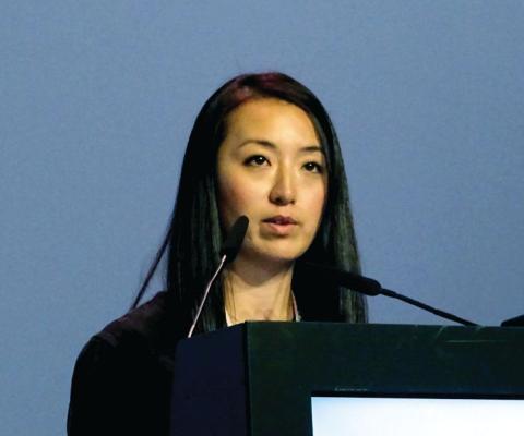
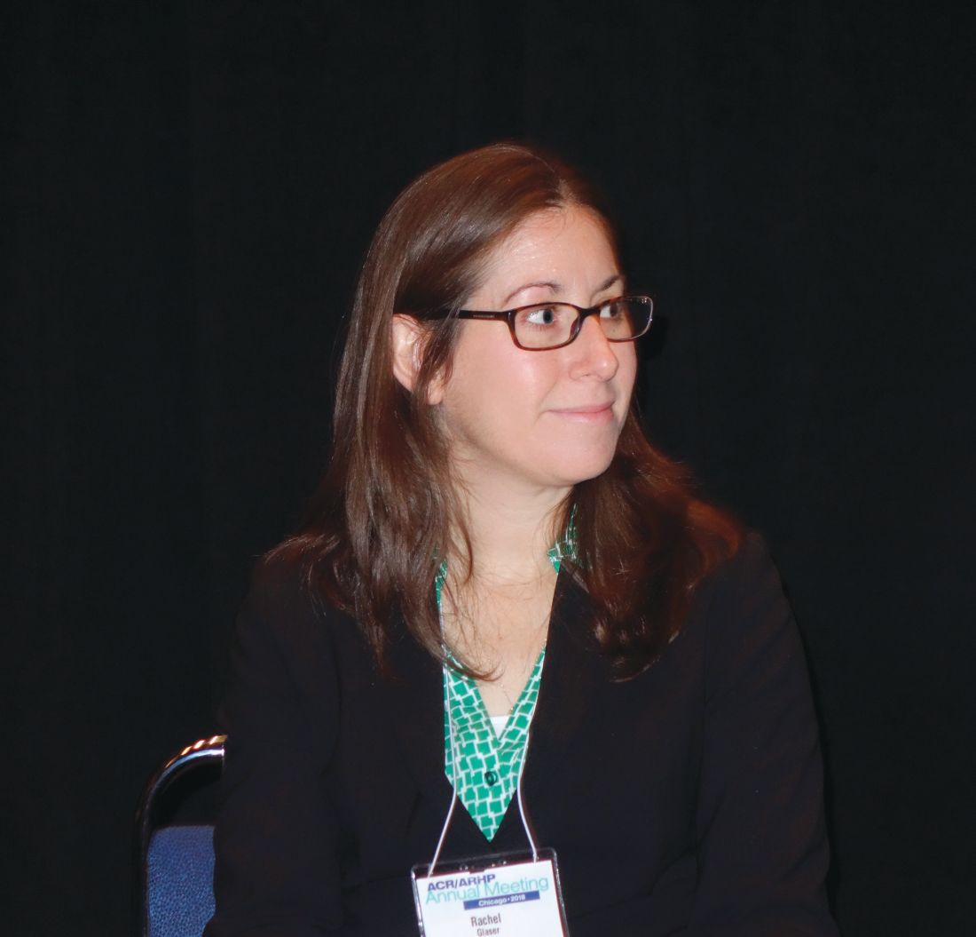
The request for further studies grew out of the FDA Advisory Committee on Medical Imaging’s Fall 2017 meeting, which concluded that, “based on available data, there is insufficient evidence of a causal relationship between adverse events and gadolinium retention and recommended the need for additional studies to inform regulatory action,” explained Rachel L. Glaser, MD, a medical officer in the agency’s division of pulmonary, allergy, and rheumatology products and a practicing rheumatologist.
Jonathan Kay, MD, rose from the audience to take the FDA to task for delegating matters related to gadolinium-based contrast agent safety to the radiologists comprising the Advisory Committee on Medical Imaging. “I think the FDA ought to bring this matter to a group of physicians who actually care for patients to get our input on this. Maybe the Arthritis Advisory Committee,” he said.
“It’s been known for more than 15 years that gadolinium deposits in the bone, and for more than 5 years that gadolinium deposits in the brain of patients who’ve received multiple magnetic resonance studies. I’m concerned that the FDA has this issue in the purview of the radiologists because radiologists administer these contrast agents and then don’t see the patients back in follow-up, whereas rheumatologists do,” noted Dr. Kay, professor of medicine and director of clinical research in rheumatology at the University of Massachusetts, Worcester.
He blasted the FDA’s call for further human and animal studies, noting “nephrogenic systemic fibrosis was the human experiment that was done with gadolinium contrast, showing that these agents, when they deposit in tissue, are extremely toxic. I can’t see other human studies being done to determine the potential consequences of these agents when they deposit long term in brain, bone, and other tissues. And the animal studies have already been done demonstrating the danger of these compounds. Might you take this out of the realm of radiologists and involve physicians who take care of patients chronically and observe the long-term consequences of these toxic effects?”
Dr. Glaser responded that the deliberations of the agency’s Advisory Committee on Medical Imaging are reviewed under the purview of the FDA’s Center for Drug Evaluation and Research. “That does include radiologists, but also other clinical reviewers of various backgrounds, including pharmacologists and toxicologists.”
Dr. Kay did not relent, further commenting that he has personally seen “over 150 patients with the devastating consequences of nephrogenic systemic fibrosis.” He added that a prominent radiologist has stated in a public forum that the risk of getting injured from crossing the street is greater than that from getting an MRI with gadolinium. “That’s completely wrong,” he declared.
Dr. Glaser and Dr. Kay reported having no financial conflicts.
CHICAGO – The Food and Drug Administration’s updated safety warning about gadolinium-based contrast agents was dismissed as too little, too late during a postsession discussion at the annual meeting of the American College of Rheumatology.
The class warning that “gadolinium is retained for months or years in brain, bone, and other organs,” a directive that a new patient medication guide be given to everyone before receiving a gadolinium-based contrast agent, and a request that manufacturers conduct human and animal studies to further assess the safety of these products, was highlighted at the FDA update session on safety issues in the treatment of rheumatic disease.
The request for further studies grew out of the FDA Advisory Committee on Medical Imaging’s Fall 2017 meeting, which concluded that, “based on available data, there is insufficient evidence of a causal relationship between adverse events and gadolinium retention and recommended the need for additional studies to inform regulatory action,” explained Rachel L. Glaser, MD, a medical officer in the agency’s division of pulmonary, allergy, and rheumatology products and a practicing rheumatologist.
Jonathan Kay, MD, rose from the audience to take the FDA to task for delegating matters related to gadolinium-based contrast agent safety to the radiologists comprising the Advisory Committee on Medical Imaging. “I think the FDA ought to bring this matter to a group of physicians who actually care for patients to get our input on this. Maybe the Arthritis Advisory Committee,” he said.
“It’s been known for more than 15 years that gadolinium deposits in the bone, and for more than 5 years that gadolinium deposits in the brain of patients who’ve received multiple magnetic resonance studies. I’m concerned that the FDA has this issue in the purview of the radiologists because radiologists administer these contrast agents and then don’t see the patients back in follow-up, whereas rheumatologists do,” noted Dr. Kay, professor of medicine and director of clinical research in rheumatology at the University of Massachusetts, Worcester.
He blasted the FDA’s call for further human and animal studies, noting “nephrogenic systemic fibrosis was the human experiment that was done with gadolinium contrast, showing that these agents, when they deposit in tissue, are extremely toxic. I can’t see other human studies being done to determine the potential consequences of these agents when they deposit long term in brain, bone, and other tissues. And the animal studies have already been done demonstrating the danger of these compounds. Might you take this out of the realm of radiologists and involve physicians who take care of patients chronically and observe the long-term consequences of these toxic effects?”
Dr. Glaser responded that the deliberations of the agency’s Advisory Committee on Medical Imaging are reviewed under the purview of the FDA’s Center for Drug Evaluation and Research. “That does include radiologists, but also other clinical reviewers of various backgrounds, including pharmacologists and toxicologists.”
Dr. Kay did not relent, further commenting that he has personally seen “over 150 patients with the devastating consequences of nephrogenic systemic fibrosis.” He added that a prominent radiologist has stated in a public forum that the risk of getting injured from crossing the street is greater than that from getting an MRI with gadolinium. “That’s completely wrong,” he declared.
Dr. Glaser and Dr. Kay reported having no financial conflicts.
CHICAGO – The Food and Drug Administration’s updated safety warning about gadolinium-based contrast agents was dismissed as too little, too late during a postsession discussion at the annual meeting of the American College of Rheumatology.
The class warning that “gadolinium is retained for months or years in brain, bone, and other organs,” a directive that a new patient medication guide be given to everyone before receiving a gadolinium-based contrast agent, and a request that manufacturers conduct human and animal studies to further assess the safety of these products, was highlighted at the FDA update session on safety issues in the treatment of rheumatic disease.
The request for further studies grew out of the FDA Advisory Committee on Medical Imaging’s Fall 2017 meeting, which concluded that, “based on available data, there is insufficient evidence of a causal relationship between adverse events and gadolinium retention and recommended the need for additional studies to inform regulatory action,” explained Rachel L. Glaser, MD, a medical officer in the agency’s division of pulmonary, allergy, and rheumatology products and a practicing rheumatologist.
Jonathan Kay, MD, rose from the audience to take the FDA to task for delegating matters related to gadolinium-based contrast agent safety to the radiologists comprising the Advisory Committee on Medical Imaging. “I think the FDA ought to bring this matter to a group of physicians who actually care for patients to get our input on this. Maybe the Arthritis Advisory Committee,” he said.
“It’s been known for more than 15 years that gadolinium deposits in the bone, and for more than 5 years that gadolinium deposits in the brain of patients who’ve received multiple magnetic resonance studies. I’m concerned that the FDA has this issue in the purview of the radiologists because radiologists administer these contrast agents and then don’t see the patients back in follow-up, whereas rheumatologists do,” noted Dr. Kay, professor of medicine and director of clinical research in rheumatology at the University of Massachusetts, Worcester.
He blasted the FDA’s call for further human and animal studies, noting “nephrogenic systemic fibrosis was the human experiment that was done with gadolinium contrast, showing that these agents, when they deposit in tissue, are extremely toxic. I can’t see other human studies being done to determine the potential consequences of these agents when they deposit long term in brain, bone, and other tissues. And the animal studies have already been done demonstrating the danger of these compounds. Might you take this out of the realm of radiologists and involve physicians who take care of patients chronically and observe the long-term consequences of these toxic effects?”
Dr. Glaser responded that the deliberations of the agency’s Advisory Committee on Medical Imaging are reviewed under the purview of the FDA’s Center for Drug Evaluation and Research. “That does include radiologists, but also other clinical reviewers of various backgrounds, including pharmacologists and toxicologists.”
Dr. Kay did not relent, further commenting that he has personally seen “over 150 patients with the devastating consequences of nephrogenic systemic fibrosis.” He added that a prominent radiologist has stated in a public forum that the risk of getting injured from crossing the street is greater than that from getting an MRI with gadolinium. “That’s completely wrong,” he declared.
Dr. Glaser and Dr. Kay reported having no financial conflicts.
EXPERT ANALYSIS FROM THE ACR ANNUAL MEETING
Rates, costs, mortality of RA-related interstitial lung disease analyzed in new study
Interstitial lung disease (ILD) is becoming more prevalent in patients with RA while shortening survival and leading to substantial health care costs, according to a retrospective study of RA-ILD prevalence, incidence, costs, and mortality.
“To our knowledge, this is the first study to describe the incidence and prevalence of RA-ILD among the general population and to estimate costs among U.S. patients with RA-ILD,” wrote lead author Karina Raimundo, principal health economist at Genentech, and her coauthors in the Journal of Rheumatology.
The study reviewed data from the Truven Health MarketScan Commercial and Medicare Supplemental health insurance databases, along with linking a subset of patients to the Social Security Administration Death Index to determine mortality. From 2004 to 2013, with the number of patients ranging from 892 to 3,232 per year, yearly prevalence estimates ranged from 3.2 (95% confidence interval, 3.0-3.4) to 6.0 (95% CI, 5.7-6.2) RA-ILD cases per 100,000 people. Yearly incidence ranged from 2.7 (95% CI, 2.5-2.9) to 3.8 (95% CI, 3.5-4.0) cases per 100,000 people.
While incidence was relatively stable, prevalence increased over the 10-year period. The authors noted that increased prevalence suggests improved survival of RA-ILD patients but were unable to definitively state why, with explanations ranging from more effective therapies to earlier diagnosis of the disease. “Our data do not allow more in-depth evaluation of this issue, and it merits further analysis.”
In addition, they found that average yearly costs across all study years ranged from $40,941 (standard deviation, $55,682) to $51,849 (SD, $77,125), with the main cost drivers being inpatient admissions, outpatient services, and outpatient pharmacy. By the 5-year mark of first diagnosis, 35.9% of RA-ILD patients who could be linked to the SSDI had died; those patients – with a mean age of 65 years – also had a median survival of 7.8 years (95% CI, 7.1-8.3). Generally, a 65-year-old person in the United States would be expected to live for 19 more years.
The authors acknowledged the study’s limitations, including reliance on administrative claims data, subsequent misclassification of RA-ILD status, a lack of information on cause of death, and an underestimation of mortality caused by the inability to link all patients to the Social Security Administration Death Index.
The study was funded by Genentech and F. Hoffmann–La Roche. No other conflicts of interest were reported.
SOURCE: Raimundo K et al. J Rheumatol. 2018 Nov 15. doi: 10.3899/jrheum.171315.
Interstitial lung disease (ILD) is becoming more prevalent in patients with RA while shortening survival and leading to substantial health care costs, according to a retrospective study of RA-ILD prevalence, incidence, costs, and mortality.
“To our knowledge, this is the first study to describe the incidence and prevalence of RA-ILD among the general population and to estimate costs among U.S. patients with RA-ILD,” wrote lead author Karina Raimundo, principal health economist at Genentech, and her coauthors in the Journal of Rheumatology.
The study reviewed data from the Truven Health MarketScan Commercial and Medicare Supplemental health insurance databases, along with linking a subset of patients to the Social Security Administration Death Index to determine mortality. From 2004 to 2013, with the number of patients ranging from 892 to 3,232 per year, yearly prevalence estimates ranged from 3.2 (95% confidence interval, 3.0-3.4) to 6.0 (95% CI, 5.7-6.2) RA-ILD cases per 100,000 people. Yearly incidence ranged from 2.7 (95% CI, 2.5-2.9) to 3.8 (95% CI, 3.5-4.0) cases per 100,000 people.
While incidence was relatively stable, prevalence increased over the 10-year period. The authors noted that increased prevalence suggests improved survival of RA-ILD patients but were unable to definitively state why, with explanations ranging from more effective therapies to earlier diagnosis of the disease. “Our data do not allow more in-depth evaluation of this issue, and it merits further analysis.”
In addition, they found that average yearly costs across all study years ranged from $40,941 (standard deviation, $55,682) to $51,849 (SD, $77,125), with the main cost drivers being inpatient admissions, outpatient services, and outpatient pharmacy. By the 5-year mark of first diagnosis, 35.9% of RA-ILD patients who could be linked to the SSDI had died; those patients – with a mean age of 65 years – also had a median survival of 7.8 years (95% CI, 7.1-8.3). Generally, a 65-year-old person in the United States would be expected to live for 19 more years.
The authors acknowledged the study’s limitations, including reliance on administrative claims data, subsequent misclassification of RA-ILD status, a lack of information on cause of death, and an underestimation of mortality caused by the inability to link all patients to the Social Security Administration Death Index.
The study was funded by Genentech and F. Hoffmann–La Roche. No other conflicts of interest were reported.
SOURCE: Raimundo K et al. J Rheumatol. 2018 Nov 15. doi: 10.3899/jrheum.171315.
Interstitial lung disease (ILD) is becoming more prevalent in patients with RA while shortening survival and leading to substantial health care costs, according to a retrospective study of RA-ILD prevalence, incidence, costs, and mortality.
“To our knowledge, this is the first study to describe the incidence and prevalence of RA-ILD among the general population and to estimate costs among U.S. patients with RA-ILD,” wrote lead author Karina Raimundo, principal health economist at Genentech, and her coauthors in the Journal of Rheumatology.
The study reviewed data from the Truven Health MarketScan Commercial and Medicare Supplemental health insurance databases, along with linking a subset of patients to the Social Security Administration Death Index to determine mortality. From 2004 to 2013, with the number of patients ranging from 892 to 3,232 per year, yearly prevalence estimates ranged from 3.2 (95% confidence interval, 3.0-3.4) to 6.0 (95% CI, 5.7-6.2) RA-ILD cases per 100,000 people. Yearly incidence ranged from 2.7 (95% CI, 2.5-2.9) to 3.8 (95% CI, 3.5-4.0) cases per 100,000 people.
While incidence was relatively stable, prevalence increased over the 10-year period. The authors noted that increased prevalence suggests improved survival of RA-ILD patients but were unable to definitively state why, with explanations ranging from more effective therapies to earlier diagnosis of the disease. “Our data do not allow more in-depth evaluation of this issue, and it merits further analysis.”
In addition, they found that average yearly costs across all study years ranged from $40,941 (standard deviation, $55,682) to $51,849 (SD, $77,125), with the main cost drivers being inpatient admissions, outpatient services, and outpatient pharmacy. By the 5-year mark of first diagnosis, 35.9% of RA-ILD patients who could be linked to the SSDI had died; those patients – with a mean age of 65 years – also had a median survival of 7.8 years (95% CI, 7.1-8.3). Generally, a 65-year-old person in the United States would be expected to live for 19 more years.
The authors acknowledged the study’s limitations, including reliance on administrative claims data, subsequent misclassification of RA-ILD status, a lack of information on cause of death, and an underestimation of mortality caused by the inability to link all patients to the Social Security Administration Death Index.
The study was funded by Genentech and F. Hoffmann–La Roche. No other conflicts of interest were reported.
SOURCE: Raimundo K et al. J Rheumatol. 2018 Nov 15. doi: 10.3899/jrheum.171315.
FROM THE JOURNAL OF RHEUMATOLOGY
Key clinical point:
Major finding: Prevalence of RA-related interstitial lung disease ranged from 3.2 to 6.0 cases per 100,000 people, while incidence ranged from 2.7 to 3.8 per 100,000 people.
Study details: A retrospective cohort analysis of RA-related interstitial lung disease cases from 2004 to 2013 gathered via health insurance databases and Social Security records.
Disclosures: The study was funded by Genentech and F. Hoffmann–La Roche. No other conflicts of interest were reported.
Source: Raimundo K et al. J Rheumatol. 2018 Nov 15. doi: 10.3899/jrheum.171315.
Supporting Health Literacy for IHS Patients
Health care is more effective when patients understand what they can do to be healthier. But many patients cannot read routine preventive information. To help busy clinicians learn more about what they can do to support patient wellness, the Indian Health Service (IHs) Health Literacy workgroup has developed a 20-minute health literacy and plain-language training module.
The IHS also is:
- Pretesting printed educational materials through focus groups and in-depth interviews to ensure messages are clear;
- Revising the IHS website;
- Hosting webinars on health literacy; and
- Planning to refresh and promote “As Me 3” campaign, which encourages patients to ask 3 specific questions of their providers to better understand their conditions.
In addition to taking the training module, available at the Learning Management System, the IHS suggests clinicians participate in ongoing training in health literacy, plain language and culturally and linguistically appropriate services; use the “health factor” tab in the electronic health record to assess and document the patient’s level of understanding; and learn about the teach-back technique to enhance patients’ understanding.
Health care is more effective when patients understand what they can do to be healthier. But many patients cannot read routine preventive information. To help busy clinicians learn more about what they can do to support patient wellness, the Indian Health Service (IHs) Health Literacy workgroup has developed a 20-minute health literacy and plain-language training module.
The IHS also is:
- Pretesting printed educational materials through focus groups and in-depth interviews to ensure messages are clear;
- Revising the IHS website;
- Hosting webinars on health literacy; and
- Planning to refresh and promote “As Me 3” campaign, which encourages patients to ask 3 specific questions of their providers to better understand their conditions.
In addition to taking the training module, available at the Learning Management System, the IHS suggests clinicians participate in ongoing training in health literacy, plain language and culturally and linguistically appropriate services; use the “health factor” tab in the electronic health record to assess and document the patient’s level of understanding; and learn about the teach-back technique to enhance patients’ understanding.
Health care is more effective when patients understand what they can do to be healthier. But many patients cannot read routine preventive information. To help busy clinicians learn more about what they can do to support patient wellness, the Indian Health Service (IHs) Health Literacy workgroup has developed a 20-minute health literacy and plain-language training module.
The IHS also is:
- Pretesting printed educational materials through focus groups and in-depth interviews to ensure messages are clear;
- Revising the IHS website;
- Hosting webinars on health literacy; and
- Planning to refresh and promote “As Me 3” campaign, which encourages patients to ask 3 specific questions of their providers to better understand their conditions.
In addition to taking the training module, available at the Learning Management System, the IHS suggests clinicians participate in ongoing training in health literacy, plain language and culturally and linguistically appropriate services; use the “health factor” tab in the electronic health record to assess and document the patient’s level of understanding; and learn about the teach-back technique to enhance patients’ understanding.
Smoke-free policy and blood pressure
Also today, bariatric surgery increases the risk for suicide and self-harm, a novel topical JAK inhibitor shows promise for atopic dermatitis, and livers infected with hepatitis C are safe for transplant.
Amazon Alexa
Apple Podcasts
Google Podcasts
Spotify
Also today, bariatric surgery increases the risk for suicide and self-harm, a novel topical JAK inhibitor shows promise for atopic dermatitis, and livers infected with hepatitis C are safe for transplant.
Amazon Alexa
Apple Podcasts
Google Podcasts
Spotify
Also today, bariatric surgery increases the risk for suicide and self-harm, a novel topical JAK inhibitor shows promise for atopic dermatitis, and livers infected with hepatitis C are safe for transplant.
Amazon Alexa
Apple Podcasts
Google Podcasts
Spotify

Older people with CHIP are safe donor source for HSCT

New research suggests older individuals with clonal hematopoiesis of indeterminate potential (CHIP) are a safe donor source for allogeneic hematopoietic stem cell transplant (HSCT).
Researchers found that transplants from older donors with CHIP resulted in similar survival as transplants from older donors without CHIP.
HSCT recipients with CHIP donors had a lower risk of relapse or progression but a higher risk of chronic graft-versus-host disease (GHVD), and they were more likely to develop donor-cell malignancies.
Frederik Damm, MD, of Charité – University Medical Center Berlin in Germany, and his colleagues reported these findings in the Journal of Clinical Oncology.
Little is known about the influence of donor CHIP in allogeneic HSCT, according to the researchers.
However, an increasing number of older patients are receiving donations from older, mostly sibling, donors who may have CHIP. CHIP increases with age and is associated with a greater risk of hematologic cancers, cardiovascular disease, and death from coronary heart disease.
With this in mind, Dr. Damm and his colleagues set out to determine how donor CHIP affects the outcome of HSCT.
Between 1993 and 2017, the researchers collected blood samples from 500 healthy, related HSCT donors, age 55 or older, at 10 transplant centers in Germany and France. The researchers sequenced the samples with a 66-gene panel.
The team also assessed overall survival (OS), non-relapse mortality (NRM), cumulative incidence of relapse/progression (CIR/P), cytomegalovirus (CMV) reactivation, and acute and chronic GVHD in the HSCT recipients.
Mutation analysis
The researchers identified 92 clonal mutations in 80 (16.0%) of the 500 donors. The variant allele frequency was 5.9% (range, 2% to 43%). In 20 patients, 25 variant alleles were present at a frequency of 10% or greater.
The researchers detected a single gene mutation in 70 donors, nine donors had two mutations, and one donor had four mutations. The most frequently mutated genes were DNMT3A (40/500, 8%), TET2 (11/500, 2.2%), and ASXL1 (7/500, 1.4%).
Baseline characteristics such as age, sex, or stem cells harvested per kilogram of body weight were similar between donors with and without CHIP.
Recipient characteristics were also evenly distributed regarding donor CHIP status and baseline characteristics.
However, the researchers noted that donor CHIP was present significantly more often in donors for recipients with myeloid compared to lymphoid neoplasms—19.2% and 6.3%, respectively (P≤0.001).
The prevalence of donor CHIP increased with age, from 10.3% (ages 60 to 64) to 20.3% (ages 65 to 69), 22.5% (ages 70 to 74), and 28.6% (ages 75 to 79).
The researchers observed no statistically significant association between CHIP and age, most likely, they wrote, “because 82% of all donors in our cohort were age 60 to 69 years.”
Transplant outcome
Donor CHIP led to slightly faster leukocyte engraftment, although it did not impact thrombocyte engraftment time.
The cumulative incidence of leukocyte engraftment after 15 days was 64.1% for HSCT recipients with CHIP donors and 51.4% for those with non-CHIP donors (P=0.023).
Chronic GVHD after HSCT was more likely for recipients with CHIP donors, with a 5-year cumulative incidence of 52.9%, compared to 35.7% for patients with non-CHIP donors (P=0.008).
Multivariate analysis showed donor CHIP to be an independent risk factor for chronic GVHD when adjusted for antithymocyte globulin application and donor age.
The researchers identified donor DNMT3A mutation as the predominant CHIP factor for developing chronic GVHD. The incidence of chronic GVHD was 58.5% in patients with DNMT3A-mutated donors and 36.6% in patients with wild-type donors (P=0.006).
The researchers noted that no other CHIP mutations or characteristics affected the development of chronic GVHD.
Donor CHIP status did not impact the incidence of acute GVHD or CMV reactivation.
Transplant recipients with CHIP donors had a lower CIR/P (P=0.027) than recipients with non-CHIP donors. Patients whose donors had CHIP with a DNMT3A mutation also had a lower CIR/P (P=0.029).
Donor CHIP had no effect on NRM.
Neither donor CHIP nor donor DNMT3A mutational status impacted CIR/P or NRM in patients who underwent HSCT while they were in complete response (CR).
However, patients not in CR at the time of HSCT had a lower CIR/P when the donor was CHIP-positive (hazard ratio [HR]=0.49; 95% confidence interval [CI], 0.27 to 0.88; P=0.019) or had a DNMT3A mutation (HR=0.34; 95% CI, 0.15 to 0.81; P=0.015).
Two of 82 patients with CHIP donors developed donor-cell malignancies, but there were no donor-cell malignancies in recipients with non-CHIP donors (P=0.026).
Survival
“To our knowledge, we showed, for the first time, that donor CHIP did not affect the survival of 500 HSCT recipients,” the researchers wrote.
At a median follow-up of 3.3 years, the median OS was 2 years, and the 5-year OS was 37.6%.
The researchers observed no survival differences in patients with CHIP donors or non-CHIP donors (HR=0.88; 95% CI, 0.65 to 1.321; P=0.434). And the same was true for DNMT3A mutation (HR=0.89; 95% CI, 0.58 to 1.35; P=0.573).
The researchers pointed out that patients with myelodysplastic syndromes or acute myeloid leukemia who were not in CR when they underwent HSCT had a survival benefit with donor CHIP (HR=0.49; 95% CI, 0.28 to 0.86; P=0.011).
“This finding could implicate an important clinical potential in this difficult-to-treat subgroup,” the researchers noted.
However, this was not the case for patients with myeloproliferative neoplasia, for whom donor CHIP tended to reduce survival (P=0.077).
The research team recommended future studies be conducted in younger and unrelated donors to confirm the results of this study.
The research was funded by numerous grants and fellowships, and the researchers reported no relevant conflicts of interest.
New research suggests older individuals with clonal hematopoiesis of indeterminate potential (CHIP) are a safe donor source for allogeneic hematopoietic stem cell transplant (HSCT).
Researchers found that transplants from older donors with CHIP resulted in similar survival as transplants from older donors without CHIP.
HSCT recipients with CHIP donors had a lower risk of relapse or progression but a higher risk of chronic graft-versus-host disease (GHVD), and they were more likely to develop donor-cell malignancies.
Frederik Damm, MD, of Charité – University Medical Center Berlin in Germany, and his colleagues reported these findings in the Journal of Clinical Oncology.
Little is known about the influence of donor CHIP in allogeneic HSCT, according to the researchers.
However, an increasing number of older patients are receiving donations from older, mostly sibling, donors who may have CHIP. CHIP increases with age and is associated with a greater risk of hematologic cancers, cardiovascular disease, and death from coronary heart disease.
With this in mind, Dr. Damm and his colleagues set out to determine how donor CHIP affects the outcome of HSCT.
Between 1993 and 2017, the researchers collected blood samples from 500 healthy, related HSCT donors, age 55 or older, at 10 transplant centers in Germany and France. The researchers sequenced the samples with a 66-gene panel.
The team also assessed overall survival (OS), non-relapse mortality (NRM), cumulative incidence of relapse/progression (CIR/P), cytomegalovirus (CMV) reactivation, and acute and chronic GVHD in the HSCT recipients.
Mutation analysis
The researchers identified 92 clonal mutations in 80 (16.0%) of the 500 donors. The variant allele frequency was 5.9% (range, 2% to 43%). In 20 patients, 25 variant alleles were present at a frequency of 10% or greater.
The researchers detected a single gene mutation in 70 donors, nine donors had two mutations, and one donor had four mutations. The most frequently mutated genes were DNMT3A (40/500, 8%), TET2 (11/500, 2.2%), and ASXL1 (7/500, 1.4%).
Baseline characteristics such as age, sex, or stem cells harvested per kilogram of body weight were similar between donors with and without CHIP.
Recipient characteristics were also evenly distributed regarding donor CHIP status and baseline characteristics.
However, the researchers noted that donor CHIP was present significantly more often in donors for recipients with myeloid compared to lymphoid neoplasms—19.2% and 6.3%, respectively (P≤0.001).
The prevalence of donor CHIP increased with age, from 10.3% (ages 60 to 64) to 20.3% (ages 65 to 69), 22.5% (ages 70 to 74), and 28.6% (ages 75 to 79).
The researchers observed no statistically significant association between CHIP and age, most likely, they wrote, “because 82% of all donors in our cohort were age 60 to 69 years.”
Transplant outcome
Donor CHIP led to slightly faster leukocyte engraftment, although it did not impact thrombocyte engraftment time.
The cumulative incidence of leukocyte engraftment after 15 days was 64.1% for HSCT recipients with CHIP donors and 51.4% for those with non-CHIP donors (P=0.023).
Chronic GVHD after HSCT was more likely for recipients with CHIP donors, with a 5-year cumulative incidence of 52.9%, compared to 35.7% for patients with non-CHIP donors (P=0.008).
Multivariate analysis showed donor CHIP to be an independent risk factor for chronic GVHD when adjusted for antithymocyte globulin application and donor age.
The researchers identified donor DNMT3A mutation as the predominant CHIP factor for developing chronic GVHD. The incidence of chronic GVHD was 58.5% in patients with DNMT3A-mutated donors and 36.6% in patients with wild-type donors (P=0.006).
The researchers noted that no other CHIP mutations or characteristics affected the development of chronic GVHD.
Donor CHIP status did not impact the incidence of acute GVHD or CMV reactivation.
Transplant recipients with CHIP donors had a lower CIR/P (P=0.027) than recipients with non-CHIP donors. Patients whose donors had CHIP with a DNMT3A mutation also had a lower CIR/P (P=0.029).
Donor CHIP had no effect on NRM.
Neither donor CHIP nor donor DNMT3A mutational status impacted CIR/P or NRM in patients who underwent HSCT while they were in complete response (CR).
However, patients not in CR at the time of HSCT had a lower CIR/P when the donor was CHIP-positive (hazard ratio [HR]=0.49; 95% confidence interval [CI], 0.27 to 0.88; P=0.019) or had a DNMT3A mutation (HR=0.34; 95% CI, 0.15 to 0.81; P=0.015).
Two of 82 patients with CHIP donors developed donor-cell malignancies, but there were no donor-cell malignancies in recipients with non-CHIP donors (P=0.026).
Survival
“To our knowledge, we showed, for the first time, that donor CHIP did not affect the survival of 500 HSCT recipients,” the researchers wrote.
At a median follow-up of 3.3 years, the median OS was 2 years, and the 5-year OS was 37.6%.
The researchers observed no survival differences in patients with CHIP donors or non-CHIP donors (HR=0.88; 95% CI, 0.65 to 1.321; P=0.434). And the same was true for DNMT3A mutation (HR=0.89; 95% CI, 0.58 to 1.35; P=0.573).
The researchers pointed out that patients with myelodysplastic syndromes or acute myeloid leukemia who were not in CR when they underwent HSCT had a survival benefit with donor CHIP (HR=0.49; 95% CI, 0.28 to 0.86; P=0.011).
“This finding could implicate an important clinical potential in this difficult-to-treat subgroup,” the researchers noted.
However, this was not the case for patients with myeloproliferative neoplasia, for whom donor CHIP tended to reduce survival (P=0.077).
The research team recommended future studies be conducted in younger and unrelated donors to confirm the results of this study.
The research was funded by numerous grants and fellowships, and the researchers reported no relevant conflicts of interest.
New research suggests older individuals with clonal hematopoiesis of indeterminate potential (CHIP) are a safe donor source for allogeneic hematopoietic stem cell transplant (HSCT).
Researchers found that transplants from older donors with CHIP resulted in similar survival as transplants from older donors without CHIP.
HSCT recipients with CHIP donors had a lower risk of relapse or progression but a higher risk of chronic graft-versus-host disease (GHVD), and they were more likely to develop donor-cell malignancies.
Frederik Damm, MD, of Charité – University Medical Center Berlin in Germany, and his colleagues reported these findings in the Journal of Clinical Oncology.
Little is known about the influence of donor CHIP in allogeneic HSCT, according to the researchers.
However, an increasing number of older patients are receiving donations from older, mostly sibling, donors who may have CHIP. CHIP increases with age and is associated with a greater risk of hematologic cancers, cardiovascular disease, and death from coronary heart disease.
With this in mind, Dr. Damm and his colleagues set out to determine how donor CHIP affects the outcome of HSCT.
Between 1993 and 2017, the researchers collected blood samples from 500 healthy, related HSCT donors, age 55 or older, at 10 transplant centers in Germany and France. The researchers sequenced the samples with a 66-gene panel.
The team also assessed overall survival (OS), non-relapse mortality (NRM), cumulative incidence of relapse/progression (CIR/P), cytomegalovirus (CMV) reactivation, and acute and chronic GVHD in the HSCT recipients.
Mutation analysis
The researchers identified 92 clonal mutations in 80 (16.0%) of the 500 donors. The variant allele frequency was 5.9% (range, 2% to 43%). In 20 patients, 25 variant alleles were present at a frequency of 10% or greater.
The researchers detected a single gene mutation in 70 donors, nine donors had two mutations, and one donor had four mutations. The most frequently mutated genes were DNMT3A (40/500, 8%), TET2 (11/500, 2.2%), and ASXL1 (7/500, 1.4%).
Baseline characteristics such as age, sex, or stem cells harvested per kilogram of body weight were similar between donors with and without CHIP.
Recipient characteristics were also evenly distributed regarding donor CHIP status and baseline characteristics.
However, the researchers noted that donor CHIP was present significantly more often in donors for recipients with myeloid compared to lymphoid neoplasms—19.2% and 6.3%, respectively (P≤0.001).
The prevalence of donor CHIP increased with age, from 10.3% (ages 60 to 64) to 20.3% (ages 65 to 69), 22.5% (ages 70 to 74), and 28.6% (ages 75 to 79).
The researchers observed no statistically significant association between CHIP and age, most likely, they wrote, “because 82% of all donors in our cohort were age 60 to 69 years.”
Transplant outcome
Donor CHIP led to slightly faster leukocyte engraftment, although it did not impact thrombocyte engraftment time.
The cumulative incidence of leukocyte engraftment after 15 days was 64.1% for HSCT recipients with CHIP donors and 51.4% for those with non-CHIP donors (P=0.023).
Chronic GVHD after HSCT was more likely for recipients with CHIP donors, with a 5-year cumulative incidence of 52.9%, compared to 35.7% for patients with non-CHIP donors (P=0.008).
Multivariate analysis showed donor CHIP to be an independent risk factor for chronic GVHD when adjusted for antithymocyte globulin application and donor age.
The researchers identified donor DNMT3A mutation as the predominant CHIP factor for developing chronic GVHD. The incidence of chronic GVHD was 58.5% in patients with DNMT3A-mutated donors and 36.6% in patients with wild-type donors (P=0.006).
The researchers noted that no other CHIP mutations or characteristics affected the development of chronic GVHD.
Donor CHIP status did not impact the incidence of acute GVHD or CMV reactivation.
Transplant recipients with CHIP donors had a lower CIR/P (P=0.027) than recipients with non-CHIP donors. Patients whose donors had CHIP with a DNMT3A mutation also had a lower CIR/P (P=0.029).
Donor CHIP had no effect on NRM.
Neither donor CHIP nor donor DNMT3A mutational status impacted CIR/P or NRM in patients who underwent HSCT while they were in complete response (CR).
However, patients not in CR at the time of HSCT had a lower CIR/P when the donor was CHIP-positive (hazard ratio [HR]=0.49; 95% confidence interval [CI], 0.27 to 0.88; P=0.019) or had a DNMT3A mutation (HR=0.34; 95% CI, 0.15 to 0.81; P=0.015).
Two of 82 patients with CHIP donors developed donor-cell malignancies, but there were no donor-cell malignancies in recipients with non-CHIP donors (P=0.026).
Survival
“To our knowledge, we showed, for the first time, that donor CHIP did not affect the survival of 500 HSCT recipients,” the researchers wrote.
At a median follow-up of 3.3 years, the median OS was 2 years, and the 5-year OS was 37.6%.
The researchers observed no survival differences in patients with CHIP donors or non-CHIP donors (HR=0.88; 95% CI, 0.65 to 1.321; P=0.434). And the same was true for DNMT3A mutation (HR=0.89; 95% CI, 0.58 to 1.35; P=0.573).
The researchers pointed out that patients with myelodysplastic syndromes or acute myeloid leukemia who were not in CR when they underwent HSCT had a survival benefit with donor CHIP (HR=0.49; 95% CI, 0.28 to 0.86; P=0.011).
“This finding could implicate an important clinical potential in this difficult-to-treat subgroup,” the researchers noted.
However, this was not the case for patients with myeloproliferative neoplasia, for whom donor CHIP tended to reduce survival (P=0.077).
The research team recommended future studies be conducted in younger and unrelated donors to confirm the results of this study.
The research was funded by numerous grants and fellowships, and the researchers reported no relevant conflicts of interest.

EC approves pegfilgrastim biosimilar

The European Commission (EC) has approved Mundipharma’s pegfilgrastim product Pelmeg, a biosimilar of Amgen’s Neulasta.
Pelmeg is approved for use in reducing the duration of neutropenia and the incidence of febrile neutropenia in adults who receive cytotoxic chemotherapy for malignancies, with the exceptions of chronic myeloid leukemia and myelodysplastic syndromes.
The approval is valid in all countries of the European Union as well as Norway, Iceland, and Liechtenstein.
The EC’s approval of Pelmeg was supported by research showing pharmacokinetic comparability between Pelmeg and Neulasta at a dose of 6 mg, pharmacodynamic comparability at doses of 6 mg and 3 mg, and no clinically meaningful differences in the safety and immunogenicity profiles of Pelmeg and Neulasta.1,2,3
1. Roth K. et al. Demonstration of pharmacokinetic and pharmacodynamic comparability in healthy volunteers for B12019, a proposed pegfilgrastim biosimilar. ECCO 2017, abstract 241.
2. Roth K. et al. Comparability of pharmacodynamics and immunogenicity of B12019, a proposed pegfilgrastim biosimilar to Neulasta®. ASH 2017, abstract 1002.
3. Roth K. et al. Pharmacokinetic and pharmacodynamic comparability of B12019, a proposed pegfilgrastim biosimilar. ESMO 2017, poster 1573.
The European Commission (EC) has approved Mundipharma’s pegfilgrastim product Pelmeg, a biosimilar of Amgen’s Neulasta.
Pelmeg is approved for use in reducing the duration of neutropenia and the incidence of febrile neutropenia in adults who receive cytotoxic chemotherapy for malignancies, with the exceptions of chronic myeloid leukemia and myelodysplastic syndromes.
The approval is valid in all countries of the European Union as well as Norway, Iceland, and Liechtenstein.
The EC’s approval of Pelmeg was supported by research showing pharmacokinetic comparability between Pelmeg and Neulasta at a dose of 6 mg, pharmacodynamic comparability at doses of 6 mg and 3 mg, and no clinically meaningful differences in the safety and immunogenicity profiles of Pelmeg and Neulasta.1,2,3
1. Roth K. et al. Demonstration of pharmacokinetic and pharmacodynamic comparability in healthy volunteers for B12019, a proposed pegfilgrastim biosimilar. ECCO 2017, abstract 241.
2. Roth K. et al. Comparability of pharmacodynamics and immunogenicity of B12019, a proposed pegfilgrastim biosimilar to Neulasta®. ASH 2017, abstract 1002.
3. Roth K. et al. Pharmacokinetic and pharmacodynamic comparability of B12019, a proposed pegfilgrastim biosimilar. ESMO 2017, poster 1573.
The European Commission (EC) has approved Mundipharma’s pegfilgrastim product Pelmeg, a biosimilar of Amgen’s Neulasta.
Pelmeg is approved for use in reducing the duration of neutropenia and the incidence of febrile neutropenia in adults who receive cytotoxic chemotherapy for malignancies, with the exceptions of chronic myeloid leukemia and myelodysplastic syndromes.
The approval is valid in all countries of the European Union as well as Norway, Iceland, and Liechtenstein.
The EC’s approval of Pelmeg was supported by research showing pharmacokinetic comparability between Pelmeg and Neulasta at a dose of 6 mg, pharmacodynamic comparability at doses of 6 mg and 3 mg, and no clinically meaningful differences in the safety and immunogenicity profiles of Pelmeg and Neulasta.1,2,3
1. Roth K. et al. Demonstration of pharmacokinetic and pharmacodynamic comparability in healthy volunteers for B12019, a proposed pegfilgrastim biosimilar. ECCO 2017, abstract 241.
2. Roth K. et al. Comparability of pharmacodynamics and immunogenicity of B12019, a proposed pegfilgrastim biosimilar to Neulasta®. ASH 2017, abstract 1002.
3. Roth K. et al. Pharmacokinetic and pharmacodynamic comparability of B12019, a proposed pegfilgrastim biosimilar. ESMO 2017, poster 1573.

FDA grants priority review to quizartinib
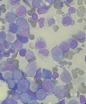
The U.S. Food and Drug Administration (FDA) has accepted for priority review a new drug application (NDA) for the FLT3 inhibitor quizartinib.
With this NDA, Daiichi Sankyo is seeking approval for quizartinib to treat adults with relapsed/refractory FLT3-ITD acute myeloid leukemia (AML).
The FDA grants priority review to applications for products that are expected to provide significant improvements in the treatment, diagnosis, or prevention of serious conditions.
The FDA aims to take action on a priority review application within 6 months rather than the standard 10 months.
The FDA is expected to make a decision on the quizartinib NDA by May 25, 2019.
In addition to priority review, quizartinib has breakthrough therapy designation and fast track designation from the FDA.
Trial results
The NDA for quizartinib is supported by results from the phase 3 QuANTUM-R study. Topline results from this study were presented at the 23rd Congress of the European Hematology Association in June, and new analyses are set to be presented at the 2018 ASH Annual Meeting in December (abstract 563).
QuANTUM-R enrolled adults with FLT3-ITD AML (at least 3% FLT3-ITD allelic ratio) who had refractory disease or had relapsed within 6 months of their first complete response (CR).
Patients were randomized to receive once-daily treatment with quizartinib (n=245) or a salvage chemotherapy regimen (n=122)—low-dose cytarabine (LoDAC, n=29); combination mitoxantrone, etoposide, and cytarabine (MEC, n=40); or combination fludarabine, cytarabine, and idarubicin (FLAG-IDA, n=53).
Patients who responded to treatment could proceed to hematopoietic stem cell transplant (HSCT), and those in the quizartinib arm could resume quizartinib after HSCT.
In all, 241 patients received quizartinib, and 94 received salvage chemotherapy—LoDAC (n=22), MEC (n=25), and FLAG-IDA (n=47). Of the 28 patients in the chemotherapy group who were not treated, most withdrew consent.
Thirty-two percent of quizartinib-treated patients and 12% of the chemotherapy group went on to HSCT.
Efficacy
The median follow-up was 23.5 months. The efficacy results include all randomized patients.
The overall response rate was 69% in the quizartinib arm and 30% in the chemotherapy arm. The composite CR rate was 48% in the quizartinib arm and 27% in the chemotherapy arm. This includes:
- The CR rate (4% and 1%, respectively)
- The rate of CR with incomplete platelet recovery (4% and 0%, respectively)
- The rate of CR with incomplete hematologic recovery (40% and 26%, respectively).
The median event-free survival was 6.0 weeks in the quizartinib arm and 3.7 weeks in the chemotherapy arm (hazard ratio=0.90, P=0.1071).
The median overall survival was 6.2 months in the quizartinib arm and 4.7 months in the chemotherapy arm (hazard ratio=0.76, P=0.0177). The 1-year overall survival rate was 27% and 20%, respectively.
Safety
The safety results include only patients who received their assigned treatment.
Grade 3 or higher hematologic treatment-emergent adverse events occurring in at least 5% of patients (in the quizartinib and chemotherapy groups, respectively) included:
- Thrombocytopenia (35% and 34%)
- Anemia (30% and 29%)
- Neutropenia (32% and 25%)
- Febrile neutropenia (31% and 21%)
- Leukopenia (17% and 16%).
Grade 3 or higher non-hematologic treatment-emergent adverse events occurring in at least 5% of patients (in the quizartinib and chemotherapy groups, respectively) included:
- Sepsis/septic shock (16% and 18%)
- Hypokalemia (12% and 9%)
- Pneumonia (12% and 9%)
- Fatigue (8% and 1%)
- Dyspnea (5% for both)
- Hypophosphatemia (5% for both).
The U.S. Food and Drug Administration (FDA) has accepted for priority review a new drug application (NDA) for the FLT3 inhibitor quizartinib.
With this NDA, Daiichi Sankyo is seeking approval for quizartinib to treat adults with relapsed/refractory FLT3-ITD acute myeloid leukemia (AML).
The FDA grants priority review to applications for products that are expected to provide significant improvements in the treatment, diagnosis, or prevention of serious conditions.
The FDA aims to take action on a priority review application within 6 months rather than the standard 10 months.
The FDA is expected to make a decision on the quizartinib NDA by May 25, 2019.
In addition to priority review, quizartinib has breakthrough therapy designation and fast track designation from the FDA.
Trial results
The NDA for quizartinib is supported by results from the phase 3 QuANTUM-R study. Topline results from this study were presented at the 23rd Congress of the European Hematology Association in June, and new analyses are set to be presented at the 2018 ASH Annual Meeting in December (abstract 563).
QuANTUM-R enrolled adults with FLT3-ITD AML (at least 3% FLT3-ITD allelic ratio) who had refractory disease or had relapsed within 6 months of their first complete response (CR).
Patients were randomized to receive once-daily treatment with quizartinib (n=245) or a salvage chemotherapy regimen (n=122)—low-dose cytarabine (LoDAC, n=29); combination mitoxantrone, etoposide, and cytarabine (MEC, n=40); or combination fludarabine, cytarabine, and idarubicin (FLAG-IDA, n=53).
Patients who responded to treatment could proceed to hematopoietic stem cell transplant (HSCT), and those in the quizartinib arm could resume quizartinib after HSCT.
In all, 241 patients received quizartinib, and 94 received salvage chemotherapy—LoDAC (n=22), MEC (n=25), and FLAG-IDA (n=47). Of the 28 patients in the chemotherapy group who were not treated, most withdrew consent.
Thirty-two percent of quizartinib-treated patients and 12% of the chemotherapy group went on to HSCT.
Efficacy
The median follow-up was 23.5 months. The efficacy results include all randomized patients.
The overall response rate was 69% in the quizartinib arm and 30% in the chemotherapy arm. The composite CR rate was 48% in the quizartinib arm and 27% in the chemotherapy arm. This includes:
- The CR rate (4% and 1%, respectively)
- The rate of CR with incomplete platelet recovery (4% and 0%, respectively)
- The rate of CR with incomplete hematologic recovery (40% and 26%, respectively).
The median event-free survival was 6.0 weeks in the quizartinib arm and 3.7 weeks in the chemotherapy arm (hazard ratio=0.90, P=0.1071).
The median overall survival was 6.2 months in the quizartinib arm and 4.7 months in the chemotherapy arm (hazard ratio=0.76, P=0.0177). The 1-year overall survival rate was 27% and 20%, respectively.
Safety
The safety results include only patients who received their assigned treatment.
Grade 3 or higher hematologic treatment-emergent adverse events occurring in at least 5% of patients (in the quizartinib and chemotherapy groups, respectively) included:
- Thrombocytopenia (35% and 34%)
- Anemia (30% and 29%)
- Neutropenia (32% and 25%)
- Febrile neutropenia (31% and 21%)
- Leukopenia (17% and 16%).
Grade 3 or higher non-hematologic treatment-emergent adverse events occurring in at least 5% of patients (in the quizartinib and chemotherapy groups, respectively) included:
- Sepsis/septic shock (16% and 18%)
- Hypokalemia (12% and 9%)
- Pneumonia (12% and 9%)
- Fatigue (8% and 1%)
- Dyspnea (5% for both)
- Hypophosphatemia (5% for both).
The U.S. Food and Drug Administration (FDA) has accepted for priority review a new drug application (NDA) for the FLT3 inhibitor quizartinib.
With this NDA, Daiichi Sankyo is seeking approval for quizartinib to treat adults with relapsed/refractory FLT3-ITD acute myeloid leukemia (AML).
The FDA grants priority review to applications for products that are expected to provide significant improvements in the treatment, diagnosis, or prevention of serious conditions.
The FDA aims to take action on a priority review application within 6 months rather than the standard 10 months.
The FDA is expected to make a decision on the quizartinib NDA by May 25, 2019.
In addition to priority review, quizartinib has breakthrough therapy designation and fast track designation from the FDA.
Trial results
The NDA for quizartinib is supported by results from the phase 3 QuANTUM-R study. Topline results from this study were presented at the 23rd Congress of the European Hematology Association in June, and new analyses are set to be presented at the 2018 ASH Annual Meeting in December (abstract 563).
QuANTUM-R enrolled adults with FLT3-ITD AML (at least 3% FLT3-ITD allelic ratio) who had refractory disease or had relapsed within 6 months of their first complete response (CR).
Patients were randomized to receive once-daily treatment with quizartinib (n=245) or a salvage chemotherapy regimen (n=122)—low-dose cytarabine (LoDAC, n=29); combination mitoxantrone, etoposide, and cytarabine (MEC, n=40); or combination fludarabine, cytarabine, and idarubicin (FLAG-IDA, n=53).
Patients who responded to treatment could proceed to hematopoietic stem cell transplant (HSCT), and those in the quizartinib arm could resume quizartinib after HSCT.
In all, 241 patients received quizartinib, and 94 received salvage chemotherapy—LoDAC (n=22), MEC (n=25), and FLAG-IDA (n=47). Of the 28 patients in the chemotherapy group who were not treated, most withdrew consent.
Thirty-two percent of quizartinib-treated patients and 12% of the chemotherapy group went on to HSCT.
Efficacy
The median follow-up was 23.5 months. The efficacy results include all randomized patients.
The overall response rate was 69% in the quizartinib arm and 30% in the chemotherapy arm. The composite CR rate was 48% in the quizartinib arm and 27% in the chemotherapy arm. This includes:
- The CR rate (4% and 1%, respectively)
- The rate of CR with incomplete platelet recovery (4% and 0%, respectively)
- The rate of CR with incomplete hematologic recovery (40% and 26%, respectively).
The median event-free survival was 6.0 weeks in the quizartinib arm and 3.7 weeks in the chemotherapy arm (hazard ratio=0.90, P=0.1071).
The median overall survival was 6.2 months in the quizartinib arm and 4.7 months in the chemotherapy arm (hazard ratio=0.76, P=0.0177). The 1-year overall survival rate was 27% and 20%, respectively.
Safety
The safety results include only patients who received their assigned treatment.
Grade 3 or higher hematologic treatment-emergent adverse events occurring in at least 5% of patients (in the quizartinib and chemotherapy groups, respectively) included:
- Thrombocytopenia (35% and 34%)
- Anemia (30% and 29%)
- Neutropenia (32% and 25%)
- Febrile neutropenia (31% and 21%)
- Leukopenia (17% and 16%).
Grade 3 or higher non-hematologic treatment-emergent adverse events occurring in at least 5% of patients (in the quizartinib and chemotherapy groups, respectively) included:
- Sepsis/septic shock (16% and 18%)
- Hypokalemia (12% and 9%)
- Pneumonia (12% and 9%)
- Fatigue (8% and 1%)
- Dyspnea (5% for both)
- Hypophosphatemia (5% for both).