User login
Toolkit for providing mental health care to Muslim patients launched
SAN FRANCISCO – A toolkit that seeks to help clinicians provide culturally and religiously informed mental health care for Muslim patients was officially launched at the annual meeting of the American Psychiatric Association.
Rania Awaad, MD, and Belinda S. Bandstra, MD, sat down at the annual meeting of the American Psychiatric Association to discuss how to use the toolkit and why it – and other resources on providing nuanced mental health care – are needed.

In this video, Dr. Awaad explores some of the origins of Islamophobia in the United States and how she came to do this work while in medical school. The travel ban affecting mostly Muslim countries has had a ripple effect on community members, she said. “The feeling is ‘My country isn’t named in the travel ban, but will I be next?’ ”
In addition to the fear and distrust fostered by the political climate are the challenges of abiding by the Islamic faith’s precepts.
“Patients will just do things on their own – and not consult their clinician,” Dr. Awaad said, referring to those might change the times in which they take medication during the sacred month of Ramadan because of fasting that is expected of observant Muslims. “It’s important for the patients to know that anyone acutely ill is exempt from fasting.” Medical- and faith-based consultation are important for these patients, Dr. Awaad said, pointing to a recent article that outlines best practices for treating patients with psychiatric disorders during Ramadan (Lancet Psychiatry. 2019 May 2. doi: 10.1016/S2215-0366[19]30161-0).
She also discussed “Islamophobia and Psychiatry” (Springer, 2019), a book she coedited that she said provides evidence of the detrimental effect that Islamophobia has on the mental health of Muslims.
Dr. Awaad is director of the Muslim Mental Health Lab and Wellness Program and codirector of the Diversity Clinic at Stanford (Calif.) University. Dr. Bandstra is assistant director of residency training in Stanford’s department of psychiatry and behavioral sciences. Dr. Awaad and Dr. Bandstra had no relevant disclosures.
SAN FRANCISCO – A toolkit that seeks to help clinicians provide culturally and religiously informed mental health care for Muslim patients was officially launched at the annual meeting of the American Psychiatric Association.
Rania Awaad, MD, and Belinda S. Bandstra, MD, sat down at the annual meeting of the American Psychiatric Association to discuss how to use the toolkit and why it – and other resources on providing nuanced mental health care – are needed.
In this video, Dr. Awaad explores some of the origins of Islamophobia in the United States and how she came to do this work while in medical school. The travel ban affecting mostly Muslim countries has had a ripple effect on community members, she said. “The feeling is ‘My country isn’t named in the travel ban, but will I be next?’ ”
In addition to the fear and distrust fostered by the political climate are the challenges of abiding by the Islamic faith’s precepts.
“Patients will just do things on their own – and not consult their clinician,” Dr. Awaad said, referring to those might change the times in which they take medication during the sacred month of Ramadan because of fasting that is expected of observant Muslims. “It’s important for the patients to know that anyone acutely ill is exempt from fasting.” Medical- and faith-based consultation are important for these patients, Dr. Awaad said, pointing to a recent article that outlines best practices for treating patients with psychiatric disorders during Ramadan (Lancet Psychiatry. 2019 May 2. doi: 10.1016/S2215-0366[19]30161-0).
She also discussed “Islamophobia and Psychiatry” (Springer, 2019), a book she coedited that she said provides evidence of the detrimental effect that Islamophobia has on the mental health of Muslims.
Dr. Awaad is director of the Muslim Mental Health Lab and Wellness Program and codirector of the Diversity Clinic at Stanford (Calif.) University. Dr. Bandstra is assistant director of residency training in Stanford’s department of psychiatry and behavioral sciences. Dr. Awaad and Dr. Bandstra had no relevant disclosures.
SAN FRANCISCO – A toolkit that seeks to help clinicians provide culturally and religiously informed mental health care for Muslim patients was officially launched at the annual meeting of the American Psychiatric Association.
Rania Awaad, MD, and Belinda S. Bandstra, MD, sat down at the annual meeting of the American Psychiatric Association to discuss how to use the toolkit and why it – and other resources on providing nuanced mental health care – are needed.
In this video, Dr. Awaad explores some of the origins of Islamophobia in the United States and how she came to do this work while in medical school. The travel ban affecting mostly Muslim countries has had a ripple effect on community members, she said. “The feeling is ‘My country isn’t named in the travel ban, but will I be next?’ ”
In addition to the fear and distrust fostered by the political climate are the challenges of abiding by the Islamic faith’s precepts.
“Patients will just do things on their own – and not consult their clinician,” Dr. Awaad said, referring to those might change the times in which they take medication during the sacred month of Ramadan because of fasting that is expected of observant Muslims. “It’s important for the patients to know that anyone acutely ill is exempt from fasting.” Medical- and faith-based consultation are important for these patients, Dr. Awaad said, pointing to a recent article that outlines best practices for treating patients with psychiatric disorders during Ramadan (Lancet Psychiatry. 2019 May 2. doi: 10.1016/S2215-0366[19]30161-0).
She also discussed “Islamophobia and Psychiatry” (Springer, 2019), a book she coedited that she said provides evidence of the detrimental effect that Islamophobia has on the mental health of Muslims.
Dr. Awaad is director of the Muslim Mental Health Lab and Wellness Program and codirector of the Diversity Clinic at Stanford (Calif.) University. Dr. Bandstra is assistant director of residency training in Stanford’s department of psychiatry and behavioral sciences. Dr. Awaad and Dr. Bandstra had no relevant disclosures.
REPORTING FROM APA 2019

More empathy for women
At the risk of too much personal self-disclosure, I feel the need to write about my having developed more empathy for women. Having been described as a “manly man,” by a woman who feels she knows me, it has always been difficult for me to understand women. Fortunately, an experience I’ve had has given me more insight into women – shallow though it may still be.

About a year ago, I had learned I had prostate carcinoma, which is now in remission – thanks to a proctectomy, radiation, and hormone therapy. The antitestosterone hormones I need to take for 2 years are turning me into an old woman, thus my newfound empathy.
After the surgery, I found myself leaking – something that I probably only experienced as a child and of which I have little memory. I now have some more empathy for the problems women have with leaking each month or in general – it is a constant preoccupation. The leuprolide shots I am taking are giving me hot flashes, causing me to be more emotional about things I really don’t understand, and apparently I am at risk for getting osteoporosis – all things that happen to women that have been mildly on my radar for years but for which I lacked direct and personal experience.
Since having my testosterone turned off by the leuprolide, my joints are more prone to aches and pains from various injuries over the years. Because I understand that “motion is lotion,” I have some control of this problem. However, the hormone therapy has greatly reduced my endurance, so my exercise tolerance is far more limited – I understand fatigue now. When I was telling another woman who feels she knows me about my experience, she told me it was hormones that made it more difficult to lose weight. And, I am gaining weight.
All in all, I believe my experience has given me more empathy for women, but I realize I still have a very long way to go. Nonetheless, I will continue in my quest to understand the opposite sex, as I am told “women hold up half the sky,” and I have always believed that to be true.
Fortunately, women are ascending in psychiatry and, with some serious dedication, the dearth of scientific understanding of women’s issues will be a thing of the past. and fill that void of knowledge that we men psychiatrists have in our testosterone-bathed brains.
Dr. Bell is a staff psychiatrist at Jackson Park Hospital’s Medical/Surgical-Psychiatry Inpatient Unit in Chicago, clinical psychiatrist emeritus in the department of psychiatry at the University of Illinois at Chicago, former president/CEO of Community Mental Health Council, and former director of the Institute for Juvenile Research (birthplace of child psychiatry), also in Chicago.
At the risk of too much personal self-disclosure, I feel the need to write about my having developed more empathy for women. Having been described as a “manly man,” by a woman who feels she knows me, it has always been difficult for me to understand women. Fortunately, an experience I’ve had has given me more insight into women – shallow though it may still be.
About a year ago, I had learned I had prostate carcinoma, which is now in remission – thanks to a proctectomy, radiation, and hormone therapy. The antitestosterone hormones I need to take for 2 years are turning me into an old woman, thus my newfound empathy.
After the surgery, I found myself leaking – something that I probably only experienced as a child and of which I have little memory. I now have some more empathy for the problems women have with leaking each month or in general – it is a constant preoccupation. The leuprolide shots I am taking are giving me hot flashes, causing me to be more emotional about things I really don’t understand, and apparently I am at risk for getting osteoporosis – all things that happen to women that have been mildly on my radar for years but for which I lacked direct and personal experience.
Since having my testosterone turned off by the leuprolide, my joints are more prone to aches and pains from various injuries over the years. Because I understand that “motion is lotion,” I have some control of this problem. However, the hormone therapy has greatly reduced my endurance, so my exercise tolerance is far more limited – I understand fatigue now. When I was telling another woman who feels she knows me about my experience, she told me it was hormones that made it more difficult to lose weight. And, I am gaining weight.
All in all, I believe my experience has given me more empathy for women, but I realize I still have a very long way to go. Nonetheless, I will continue in my quest to understand the opposite sex, as I am told “women hold up half the sky,” and I have always believed that to be true.
Fortunately, women are ascending in psychiatry and, with some serious dedication, the dearth of scientific understanding of women’s issues will be a thing of the past. and fill that void of knowledge that we men psychiatrists have in our testosterone-bathed brains.
Dr. Bell is a staff psychiatrist at Jackson Park Hospital’s Medical/Surgical-Psychiatry Inpatient Unit in Chicago, clinical psychiatrist emeritus in the department of psychiatry at the University of Illinois at Chicago, former president/CEO of Community Mental Health Council, and former director of the Institute for Juvenile Research (birthplace of child psychiatry), also in Chicago.
At the risk of too much personal self-disclosure, I feel the need to write about my having developed more empathy for women. Having been described as a “manly man,” by a woman who feels she knows me, it has always been difficult for me to understand women. Fortunately, an experience I’ve had has given me more insight into women – shallow though it may still be.
About a year ago, I had learned I had prostate carcinoma, which is now in remission – thanks to a proctectomy, radiation, and hormone therapy. The antitestosterone hormones I need to take for 2 years are turning me into an old woman, thus my newfound empathy.
After the surgery, I found myself leaking – something that I probably only experienced as a child and of which I have little memory. I now have some more empathy for the problems women have with leaking each month or in general – it is a constant preoccupation. The leuprolide shots I am taking are giving me hot flashes, causing me to be more emotional about things I really don’t understand, and apparently I am at risk for getting osteoporosis – all things that happen to women that have been mildly on my radar for years but for which I lacked direct and personal experience.
Since having my testosterone turned off by the leuprolide, my joints are more prone to aches and pains from various injuries over the years. Because I understand that “motion is lotion,” I have some control of this problem. However, the hormone therapy has greatly reduced my endurance, so my exercise tolerance is far more limited – I understand fatigue now. When I was telling another woman who feels she knows me about my experience, she told me it was hormones that made it more difficult to lose weight. And, I am gaining weight.
All in all, I believe my experience has given me more empathy for women, but I realize I still have a very long way to go. Nonetheless, I will continue in my quest to understand the opposite sex, as I am told “women hold up half the sky,” and I have always believed that to be true.
Fortunately, women are ascending in psychiatry and, with some serious dedication, the dearth of scientific understanding of women’s issues will be a thing of the past. and fill that void of knowledge that we men psychiatrists have in our testosterone-bathed brains.
Dr. Bell is a staff psychiatrist at Jackson Park Hospital’s Medical/Surgical-Psychiatry Inpatient Unit in Chicago, clinical psychiatrist emeritus in the department of psychiatry at the University of Illinois at Chicago, former president/CEO of Community Mental Health Council, and former director of the Institute for Juvenile Research (birthplace of child psychiatry), also in Chicago.

Report on newly recognized cause of dementia should be read widely
Alzheimer’s disease is recognized as the most common cause of dementia, and many in the laity use the two terms almost interchangeably. However, there is increasing recognition that dementia in old age is a complex disorder, with mixed neuropathologies being the norm rather than the exception (Ann Neurol. 2018 Jan;83[1]:74-83).

Alzheimer’s disease (AD) and cerebrovascular pathologies are the most common, but another pathology is receiving increasing attention in relation to cognitive disorders in very old individuals – that related to the transactive response DNA binding protein of 43 kDa (TDP-43). This protein is expressed in most human tissues, including the brain, is localized mostly in nuclei, and binds to RNA and DNA as well as numerous proteins, with the role of regulating gene expression.
It has been known for nearly 2 decades that TDP-43 can become abnormally phosphorylated and translocated to the cytoplasm to produce a proteinopathy that forms the basis of a significant proportion of frontotemporal dementia (FTD) and the majority of amyotrophic lateral sclerosis. More recently, it has also been reported to be common in the brains of older people (over age 80 years) and associated with a cognitive disorder characterized by an amnestic picture that mimics AD. Since the protein deposition is predominantly in the limbic regions (amygdala, hippocampus, insula), it has been termed “‘limbic-predominant, age-related TDP-43 encephalopathy”, or LATE.
A recently convened international working group has published consensus criteria for LATE and provided guidelines for its staging. Community-based autopsy studies suggest that 20%-50% of people aged over 80 years have the neuropathologic change associated with LATE. The clinical presentation resembles amnestic dementia syndrome, much like AD. Both LATE and AD pathologies often occur in the same individual, but the relative predominance of one or the other varies greatly between individuals. The genetic risks of LATE overlap with those for FTD and AD, and other risk factors may also be shared with AD, which remains an area for further investigation. There are at present no specific biomarkers of LATE. It is associated with hippocampal sclerosis in some cases, which may be visible on MRI, but hippocampal sclerosis itself is not specific to TDP-43 pathology.
of LATE and calls for systematic study of the causes of dementia – which may be nearly as common as AD in the very old. The report should be read widely and should remind us of the diverse pathologies that contribute to cognitive disorders, alone and in combination with one another.
Dr. Sachdev is Scientia Professor of Neuropsychiatry and codirector of the Center for Healthy Brain Aging at the University of New South Wales, Sydney; and clinical director of the Neuropsychiatric Institute at the Prince of Wales Hospital, also in Sydney. His major areas of research are drug-induced movement disorders, brain imaging, cognitive aging and dementia. Dr. Sachdev also served on the Neurocognitive Disorders Work Group of the DSM-5.
Alzheimer’s disease is recognized as the most common cause of dementia, and many in the laity use the two terms almost interchangeably. However, there is increasing recognition that dementia in old age is a complex disorder, with mixed neuropathologies being the norm rather than the exception (Ann Neurol. 2018 Jan;83[1]:74-83).
Alzheimer’s disease (AD) and cerebrovascular pathologies are the most common, but another pathology is receiving increasing attention in relation to cognitive disorders in very old individuals – that related to the transactive response DNA binding protein of 43 kDa (TDP-43). This protein is expressed in most human tissues, including the brain, is localized mostly in nuclei, and binds to RNA and DNA as well as numerous proteins, with the role of regulating gene expression.
It has been known for nearly 2 decades that TDP-43 can become abnormally phosphorylated and translocated to the cytoplasm to produce a proteinopathy that forms the basis of a significant proportion of frontotemporal dementia (FTD) and the majority of amyotrophic lateral sclerosis. More recently, it has also been reported to be common in the brains of older people (over age 80 years) and associated with a cognitive disorder characterized by an amnestic picture that mimics AD. Since the protein deposition is predominantly in the limbic regions (amygdala, hippocampus, insula), it has been termed “‘limbic-predominant, age-related TDP-43 encephalopathy”, or LATE.
A recently convened international working group has published consensus criteria for LATE and provided guidelines for its staging. Community-based autopsy studies suggest that 20%-50% of people aged over 80 years have the neuropathologic change associated with LATE. The clinical presentation resembles amnestic dementia syndrome, much like AD. Both LATE and AD pathologies often occur in the same individual, but the relative predominance of one or the other varies greatly between individuals. The genetic risks of LATE overlap with those for FTD and AD, and other risk factors may also be shared with AD, which remains an area for further investigation. There are at present no specific biomarkers of LATE. It is associated with hippocampal sclerosis in some cases, which may be visible on MRI, but hippocampal sclerosis itself is not specific to TDP-43 pathology.
of LATE and calls for systematic study of the causes of dementia – which may be nearly as common as AD in the very old. The report should be read widely and should remind us of the diverse pathologies that contribute to cognitive disorders, alone and in combination with one another.
Dr. Sachdev is Scientia Professor of Neuropsychiatry and codirector of the Center for Healthy Brain Aging at the University of New South Wales, Sydney; and clinical director of the Neuropsychiatric Institute at the Prince of Wales Hospital, also in Sydney. His major areas of research are drug-induced movement disorders, brain imaging, cognitive aging and dementia. Dr. Sachdev also served on the Neurocognitive Disorders Work Group of the DSM-5.
Alzheimer’s disease is recognized as the most common cause of dementia, and many in the laity use the two terms almost interchangeably. However, there is increasing recognition that dementia in old age is a complex disorder, with mixed neuropathologies being the norm rather than the exception (Ann Neurol. 2018 Jan;83[1]:74-83).
Alzheimer’s disease (AD) and cerebrovascular pathologies are the most common, but another pathology is receiving increasing attention in relation to cognitive disorders in very old individuals – that related to the transactive response DNA binding protein of 43 kDa (TDP-43). This protein is expressed in most human tissues, including the brain, is localized mostly in nuclei, and binds to RNA and DNA as well as numerous proteins, with the role of regulating gene expression.
It has been known for nearly 2 decades that TDP-43 can become abnormally phosphorylated and translocated to the cytoplasm to produce a proteinopathy that forms the basis of a significant proportion of frontotemporal dementia (FTD) and the majority of amyotrophic lateral sclerosis. More recently, it has also been reported to be common in the brains of older people (over age 80 years) and associated with a cognitive disorder characterized by an amnestic picture that mimics AD. Since the protein deposition is predominantly in the limbic regions (amygdala, hippocampus, insula), it has been termed “‘limbic-predominant, age-related TDP-43 encephalopathy”, or LATE.
A recently convened international working group has published consensus criteria for LATE and provided guidelines for its staging. Community-based autopsy studies suggest that 20%-50% of people aged over 80 years have the neuropathologic change associated with LATE. The clinical presentation resembles amnestic dementia syndrome, much like AD. Both LATE and AD pathologies often occur in the same individual, but the relative predominance of one or the other varies greatly between individuals. The genetic risks of LATE overlap with those for FTD and AD, and other risk factors may also be shared with AD, which remains an area for further investigation. There are at present no specific biomarkers of LATE. It is associated with hippocampal sclerosis in some cases, which may be visible on MRI, but hippocampal sclerosis itself is not specific to TDP-43 pathology.
of LATE and calls for systematic study of the causes of dementia – which may be nearly as common as AD in the very old. The report should be read widely and should remind us of the diverse pathologies that contribute to cognitive disorders, alone and in combination with one another.
Dr. Sachdev is Scientia Professor of Neuropsychiatry and codirector of the Center for Healthy Brain Aging at the University of New South Wales, Sydney; and clinical director of the Neuropsychiatric Institute at the Prince of Wales Hospital, also in Sydney. His major areas of research are drug-induced movement disorders, brain imaging, cognitive aging and dementia. Dr. Sachdev also served on the Neurocognitive Disorders Work Group of the DSM-5.

Hip-hop offers lens into psyche of black boys, men
SAN FRANCISCO – The lyrics found in hip-hop can help mental health professionals understand the triumphs and trauma experienced by African American boys and men, Sarah Y. Vinson, MD, said at the annual meeting of the American Psychiatric Association. This understanding can enable clinicians to recognize hopelessness and pain in those patients that they otherwise might have missed.
In this video, Dr. Vinson said her session at the APA meeting looked at the history of hip-hop and focused on the perspectives embedded in the work of several artists/groups, including N.W.A, Tupac Shakur, Childish Gambino (aka Donald Glover), J. Cole, and Kendrick Lamar.
One of the take-home points for clinicians, Dr. Vinson said, is that hip-hop, an art form that has spread across the world, came out of resilience. Another is that suicidality in black men might not look the same as it does in other patients. “It doesn’t necessarily look like cutting your own wrists or having thoughts of killing yourself – it may look like reckless behaviors that put you at risk of being killed by somebody else.”
Dr. Vinson, who is triple boarded in child and adolescent, adult, and forensic psychiatry, is in private practice in Atlanta. She had no financial disclosures.
SAN FRANCISCO – The lyrics found in hip-hop can help mental health professionals understand the triumphs and trauma experienced by African American boys and men, Sarah Y. Vinson, MD, said at the annual meeting of the American Psychiatric Association. This understanding can enable clinicians to recognize hopelessness and pain in those patients that they otherwise might have missed.
In this video, Dr. Vinson said her session at the APA meeting looked at the history of hip-hop and focused on the perspectives embedded in the work of several artists/groups, including N.W.A, Tupac Shakur, Childish Gambino (aka Donald Glover), J. Cole, and Kendrick Lamar.
One of the take-home points for clinicians, Dr. Vinson said, is that hip-hop, an art form that has spread across the world, came out of resilience. Another is that suicidality in black men might not look the same as it does in other patients. “It doesn’t necessarily look like cutting your own wrists or having thoughts of killing yourself – it may look like reckless behaviors that put you at risk of being killed by somebody else.”
Dr. Vinson, who is triple boarded in child and adolescent, adult, and forensic psychiatry, is in private practice in Atlanta. She had no financial disclosures.
SAN FRANCISCO – The lyrics found in hip-hop can help mental health professionals understand the triumphs and trauma experienced by African American boys and men, Sarah Y. Vinson, MD, said at the annual meeting of the American Psychiatric Association. This understanding can enable clinicians to recognize hopelessness and pain in those patients that they otherwise might have missed.
In this video, Dr. Vinson said her session at the APA meeting looked at the history of hip-hop and focused on the perspectives embedded in the work of several artists/groups, including N.W.A, Tupac Shakur, Childish Gambino (aka Donald Glover), J. Cole, and Kendrick Lamar.
One of the take-home points for clinicians, Dr. Vinson said, is that hip-hop, an art form that has spread across the world, came out of resilience. Another is that suicidality in black men might not look the same as it does in other patients. “It doesn’t necessarily look like cutting your own wrists or having thoughts of killing yourself – it may look like reckless behaviors that put you at risk of being killed by somebody else.”
Dr. Vinson, who is triple boarded in child and adolescent, adult, and forensic psychiatry, is in private practice in Atlanta. She had no financial disclosures.
REPORTING FROM APA 2019

Opioid use associated with common bile duct dilation
SAN DIEGO – Biliary duct dilation in the setting of an intact gallbladder and normal bilirubin levels was more common among those who used opioids, based on the results of a large, retrospective, single-center cohort study.
Patients were included in the study if they had a documented measurement for the diameter of the common bile duct, with no evidence of an obstructive lesion and a normal bilirubin level. The mean common bile duct diameter was significantly higher at 8.67 mm for 867 patients who used opioids, compared with 7.24 mm for 818 similar patients who did not use opioids (P less than .001). The association was strongest among opioid users with an intact gallbladder.
“Opiate use is associated with biliary dilation in the setting of normal bilirubin,” Monique Barakat, MD, a gastroenterologist at Stanford (Calif.) University, reported at the annual Digestive Disease Week. “Known opiate users with normal LFTs [liver function tests] may not require expensive and potentially risky endoscopic evaluation for biliary dilation.”
Dr. Barakat and senior author Subhas Banerjee, MD, professor of gastroenterology and hepatology at Stanford, decided to examine a possible association between biliary duct dilation and opioid use based on previous small clinical studies that found a possible association. Along with opioid status, Dr. Barakat and her coauthor also looked at patient age, cholecystectomy status, ethnicity, weight, and height for possible associations with bile duct diameter.
The researchers took a random 20% sample of adults seen for all causes in the ED at Stanford over a 5-year period. Using a health informatic platform based on the electronic medical record, they identified all patients who had received an abdominal CT or MRI. Patients were included in the study if they had a documented measurement for the diameter of the common bile duct, with no evidence of obstructive lesion and a normal bilirubin level.
Compared with 818 patients who did not use opioids, the 867 patients who used opioids had a significantly larger common bile duct diameter. Using 7 mm as the threshold for biliary duct enlargement, 84% of patients who used opioids had an enlarged common bile duct, compared with 27% of nonopioid users (P less than .001), said Dr. Barakat, recipient of an early investigator award for the study.
“We frequently get referrals for bile duct dilation with concern for more sinister causes of biliary duct dilation – stones, strictures, and malignancy,” said Dr. Barakat. Because of the increase in cross-sectional imaging via CT or MRI, bile duct dilation is being detected at increasingly higher rates.
Dr. Barakat said that about one-third of referrals to the therapeutic endoscopy clinic at Stanford are now for patients with biliary dilation and normal liver function tests. And similar increases are being “seen across all settings – so office, primary care clinic, inpatient, and most markedly, the emergency department. Coupled with this, the population is aging, and patients who present to each of these settings are more likely, if they are older, to undergo cross-sectional imaging.”
Other contributors to higher rates of bile duct dilation include increased rates of obesity and increased prevalence of nonalcoholic steatohepatitis (NASH). About 20% of individuals with NASH will also have abnormal LFTs, she said, and NASH can be the trigger for cross-sectional imaging.
For most of these patients with biliary duct dilation and normal LFTs, no obstructive process was found on endoscopic evaluation.
Although gastroenterology textbooks may say that bile duct diameter increases with age, Dr. Barakat and colleagues didn’t find this to be the case. Among nonopioid users in the study cohort, age did not predict of common bile duct diameter. Among the entire cohort, “Advancing age weakly predicts increased common bile duct diameter,” she said, suggesting that factors other than age along may drive increased bile duct diameter.
Limitations included the retrospective nature of the study, as well as the limitations of information from the electronic medical record. Also, interobserver variability may have come into play, as bile duct diameter measurements were made by multiple radiologists in the course of clinical care.
The study was funded by the National Institutes of Health. Dr. Barakat reported no relevant financial disclosures.
SAN DIEGO – Biliary duct dilation in the setting of an intact gallbladder and normal bilirubin levels was more common among those who used opioids, based on the results of a large, retrospective, single-center cohort study.
Patients were included in the study if they had a documented measurement for the diameter of the common bile duct, with no evidence of an obstructive lesion and a normal bilirubin level. The mean common bile duct diameter was significantly higher at 8.67 mm for 867 patients who used opioids, compared with 7.24 mm for 818 similar patients who did not use opioids (P less than .001). The association was strongest among opioid users with an intact gallbladder.
“Opiate use is associated with biliary dilation in the setting of normal bilirubin,” Monique Barakat, MD, a gastroenterologist at Stanford (Calif.) University, reported at the annual Digestive Disease Week. “Known opiate users with normal LFTs [liver function tests] may not require expensive and potentially risky endoscopic evaluation for biliary dilation.”
Dr. Barakat and senior author Subhas Banerjee, MD, professor of gastroenterology and hepatology at Stanford, decided to examine a possible association between biliary duct dilation and opioid use based on previous small clinical studies that found a possible association. Along with opioid status, Dr. Barakat and her coauthor also looked at patient age, cholecystectomy status, ethnicity, weight, and height for possible associations with bile duct diameter.
The researchers took a random 20% sample of adults seen for all causes in the ED at Stanford over a 5-year period. Using a health informatic platform based on the electronic medical record, they identified all patients who had received an abdominal CT or MRI. Patients were included in the study if they had a documented measurement for the diameter of the common bile duct, with no evidence of obstructive lesion and a normal bilirubin level.
Compared with 818 patients who did not use opioids, the 867 patients who used opioids had a significantly larger common bile duct diameter. Using 7 mm as the threshold for biliary duct enlargement, 84% of patients who used opioids had an enlarged common bile duct, compared with 27% of nonopioid users (P less than .001), said Dr. Barakat, recipient of an early investigator award for the study.
“We frequently get referrals for bile duct dilation with concern for more sinister causes of biliary duct dilation – stones, strictures, and malignancy,” said Dr. Barakat. Because of the increase in cross-sectional imaging via CT or MRI, bile duct dilation is being detected at increasingly higher rates.
Dr. Barakat said that about one-third of referrals to the therapeutic endoscopy clinic at Stanford are now for patients with biliary dilation and normal liver function tests. And similar increases are being “seen across all settings – so office, primary care clinic, inpatient, and most markedly, the emergency department. Coupled with this, the population is aging, and patients who present to each of these settings are more likely, if they are older, to undergo cross-sectional imaging.”
Other contributors to higher rates of bile duct dilation include increased rates of obesity and increased prevalence of nonalcoholic steatohepatitis (NASH). About 20% of individuals with NASH will also have abnormal LFTs, she said, and NASH can be the trigger for cross-sectional imaging.
For most of these patients with biliary duct dilation and normal LFTs, no obstructive process was found on endoscopic evaluation.
Although gastroenterology textbooks may say that bile duct diameter increases with age, Dr. Barakat and colleagues didn’t find this to be the case. Among nonopioid users in the study cohort, age did not predict of common bile duct diameter. Among the entire cohort, “Advancing age weakly predicts increased common bile duct diameter,” she said, suggesting that factors other than age along may drive increased bile duct diameter.
Limitations included the retrospective nature of the study, as well as the limitations of information from the electronic medical record. Also, interobserver variability may have come into play, as bile duct diameter measurements were made by multiple radiologists in the course of clinical care.
The study was funded by the National Institutes of Health. Dr. Barakat reported no relevant financial disclosures.
SAN DIEGO – Biliary duct dilation in the setting of an intact gallbladder and normal bilirubin levels was more common among those who used opioids, based on the results of a large, retrospective, single-center cohort study.
Patients were included in the study if they had a documented measurement for the diameter of the common bile duct, with no evidence of an obstructive lesion and a normal bilirubin level. The mean common bile duct diameter was significantly higher at 8.67 mm for 867 patients who used opioids, compared with 7.24 mm for 818 similar patients who did not use opioids (P less than .001). The association was strongest among opioid users with an intact gallbladder.
“Opiate use is associated with biliary dilation in the setting of normal bilirubin,” Monique Barakat, MD, a gastroenterologist at Stanford (Calif.) University, reported at the annual Digestive Disease Week. “Known opiate users with normal LFTs [liver function tests] may not require expensive and potentially risky endoscopic evaluation for biliary dilation.”
Dr. Barakat and senior author Subhas Banerjee, MD, professor of gastroenterology and hepatology at Stanford, decided to examine a possible association between biliary duct dilation and opioid use based on previous small clinical studies that found a possible association. Along with opioid status, Dr. Barakat and her coauthor also looked at patient age, cholecystectomy status, ethnicity, weight, and height for possible associations with bile duct diameter.
The researchers took a random 20% sample of adults seen for all causes in the ED at Stanford over a 5-year period. Using a health informatic platform based on the electronic medical record, they identified all patients who had received an abdominal CT or MRI. Patients were included in the study if they had a documented measurement for the diameter of the common bile duct, with no evidence of obstructive lesion and a normal bilirubin level.
Compared with 818 patients who did not use opioids, the 867 patients who used opioids had a significantly larger common bile duct diameter. Using 7 mm as the threshold for biliary duct enlargement, 84% of patients who used opioids had an enlarged common bile duct, compared with 27% of nonopioid users (P less than .001), said Dr. Barakat, recipient of an early investigator award for the study.
“We frequently get referrals for bile duct dilation with concern for more sinister causes of biliary duct dilation – stones, strictures, and malignancy,” said Dr. Barakat. Because of the increase in cross-sectional imaging via CT or MRI, bile duct dilation is being detected at increasingly higher rates.
Dr. Barakat said that about one-third of referrals to the therapeutic endoscopy clinic at Stanford are now for patients with biliary dilation and normal liver function tests. And similar increases are being “seen across all settings – so office, primary care clinic, inpatient, and most markedly, the emergency department. Coupled with this, the population is aging, and patients who present to each of these settings are more likely, if they are older, to undergo cross-sectional imaging.”
Other contributors to higher rates of bile duct dilation include increased rates of obesity and increased prevalence of nonalcoholic steatohepatitis (NASH). About 20% of individuals with NASH will also have abnormal LFTs, she said, and NASH can be the trigger for cross-sectional imaging.
For most of these patients with biliary duct dilation and normal LFTs, no obstructive process was found on endoscopic evaluation.
Although gastroenterology textbooks may say that bile duct diameter increases with age, Dr. Barakat and colleagues didn’t find this to be the case. Among nonopioid users in the study cohort, age did not predict of common bile duct diameter. Among the entire cohort, “Advancing age weakly predicts increased common bile duct diameter,” she said, suggesting that factors other than age along may drive increased bile duct diameter.
Limitations included the retrospective nature of the study, as well as the limitations of information from the electronic medical record. Also, interobserver variability may have come into play, as bile duct diameter measurements were made by multiple radiologists in the course of clinical care.
The study was funded by the National Institutes of Health. Dr. Barakat reported no relevant financial disclosures.
REPORTING FROM DDW 2019
PPI metabolism may be altered in about one-third of bariatric surgery candidates
SAN DIEGO – Rapid proton pump inhibitor (PPI) metabolism was present in nearly one-third of patients who underwent bariatric surgery, results from a small, single-center study showed. Patients who were fast metabolizers also exhibited a higher, although not significant, incidence of early marginal ulceration following Roux-en-Y gastric bypass.

“Roux-en-Y gastric bypass [RYGB] is one of the most effective surgical approaches to mitigating obesity and its attendant comorbidities including diabetes, hypertension, hyperlipidemia, reflux, and sleep apnea,” lead study author Sabrena F. Noria, MD, PhD, said in an interview at the annual Digestive Disease Week. “However, as with all surgeries, there are associated risks, the more common of which is marginal ulceration, or ulcer formation at the gastrojejunostomy, which occurs at a rate of 1%-16%.”
Dr. Noria, surgical research director of the comprehensive weight management, metabolic/bariatric surgery program at the Ohio State University’s Wexner Medical Center, noted that marginal ulcers (MUs) are divided into early (within 90 days) and late (more than 90 days), based on their time of onset after surgery, and are usually diagnosed during upper endoscopy on postoperative patients who complain of epigastric pain, dysphagia, nausea/vomiting, and/or dehydration.
“Given that MUs are associated with multiple hospital readmissions for pain and dehydration, multiple diagnostic and therapeutic endoscopic procedures, and escalation in both antiulcer and analgesic medication, their clinical impact cannot be overstated, especially since RYGB is the second most commonly performed bariatric procedure in the U.S.,” she said. “Given that the majority of marginal ulcers occur early after surgery, bariatric surgery programs have adopted the prophylactic use of proton pump inhibitors for up to 90 days postoperatively. While studies have demonstrated up to a two-fold decrease in ulcer formation, sample heterogeneity, in terms of combining both early and late ulcers, make it difficult to determine the effect on early ulcer formation.”
In an effort to compare preoperative endoscopic findings and MU formation in patients with and without altered PPI metabolism, the researchers prospectively enrolled 94 bariatric patients to undergo genetic testing pertinent to drug metabolism for a comprehensive panel of medications using a commercially available pharmacogenetic testing kit for the activity of cytochrome P450 in drug metabolism. They grouped patients by whether they were fast or normal metabolizers, and compared preoperative endoscopic findings for patients on PPIs at baseline and rates of early (within 90 days) and late ulceration (between 90 and 180 days).
Dr. Noria reported that 28 patients (30%) in the entire cohort met criteria for being fast metabolizers. The researchers observed no differences in baseline body mass index, age, gender, or former smoking status between both groups. Among those treated with a PPI at baseline, fast metabolizers demonstrated a trend toward a higher incidence of gastritis on preoperative endoscopy, compared with controls (89% vs. 65%, respectively; P = .12), while detection of Helicobacter pylori and Barrett’s esophagus were nonsignificant between groups. Eight patients (17%) who underwent RYGB developed marginal ulcers within 6 months of the index operation, of which four (50%) were diagnosed within 90 days and categorized as early ulcers. Development of early ulceration was higher among fast metabolizers, compared with controls (13% vs. 6%), but this did not reach statistical significance (P = .60). All late ulcerations occurred within the control group.
“While none of our findings are statistically significant given the small sample size, there were two findings I found clinically compelling,” Dr. Noria said. “First, in the group of patients who were on PPIs preoperatively, we found a 24% increase in the presence of pathologically diagnosed gastritis in the rapid-metabolizer group, during screening endoscopy. This suggests that either these patients were undertreated or were not treated with the appropriate medication. The second interesting finding was an over doubling of early ulcer formation in the RYGB group who were rapid metabolizers. However, again I would caution against drawing any real conclusions as our sample size was not powered to detect any difference.”
She also acknowledged that the study was limited by the inability to determine the effect of confounders such as surgical approach and the lack of randomization.
Anahita D. Jalilvand, MD, a general surgery resident, postdoctoral research fellow, and PhD candidate, was instrumental to this study, Dr. Noria said.
The trial was sponsored by Pathnostics, a pharmacogenetic testing company, who covered the cost of the tests. The researchers reported having no financial disclosures.
SAN DIEGO – Rapid proton pump inhibitor (PPI) metabolism was present in nearly one-third of patients who underwent bariatric surgery, results from a small, single-center study showed. Patients who were fast metabolizers also exhibited a higher, although not significant, incidence of early marginal ulceration following Roux-en-Y gastric bypass.
“Roux-en-Y gastric bypass [RYGB] is one of the most effective surgical approaches to mitigating obesity and its attendant comorbidities including diabetes, hypertension, hyperlipidemia, reflux, and sleep apnea,” lead study author Sabrena F. Noria, MD, PhD, said in an interview at the annual Digestive Disease Week. “However, as with all surgeries, there are associated risks, the more common of which is marginal ulceration, or ulcer formation at the gastrojejunostomy, which occurs at a rate of 1%-16%.”
Dr. Noria, surgical research director of the comprehensive weight management, metabolic/bariatric surgery program at the Ohio State University’s Wexner Medical Center, noted that marginal ulcers (MUs) are divided into early (within 90 days) and late (more than 90 days), based on their time of onset after surgery, and are usually diagnosed during upper endoscopy on postoperative patients who complain of epigastric pain, dysphagia, nausea/vomiting, and/or dehydration.
“Given that MUs are associated with multiple hospital readmissions for pain and dehydration, multiple diagnostic and therapeutic endoscopic procedures, and escalation in both antiulcer and analgesic medication, their clinical impact cannot be overstated, especially since RYGB is the second most commonly performed bariatric procedure in the U.S.,” she said. “Given that the majority of marginal ulcers occur early after surgery, bariatric surgery programs have adopted the prophylactic use of proton pump inhibitors for up to 90 days postoperatively. While studies have demonstrated up to a two-fold decrease in ulcer formation, sample heterogeneity, in terms of combining both early and late ulcers, make it difficult to determine the effect on early ulcer formation.”
In an effort to compare preoperative endoscopic findings and MU formation in patients with and without altered PPI metabolism, the researchers prospectively enrolled 94 bariatric patients to undergo genetic testing pertinent to drug metabolism for a comprehensive panel of medications using a commercially available pharmacogenetic testing kit for the activity of cytochrome P450 in drug metabolism. They grouped patients by whether they were fast or normal metabolizers, and compared preoperative endoscopic findings for patients on PPIs at baseline and rates of early (within 90 days) and late ulceration (between 90 and 180 days).
Dr. Noria reported that 28 patients (30%) in the entire cohort met criteria for being fast metabolizers. The researchers observed no differences in baseline body mass index, age, gender, or former smoking status between both groups. Among those treated with a PPI at baseline, fast metabolizers demonstrated a trend toward a higher incidence of gastritis on preoperative endoscopy, compared with controls (89% vs. 65%, respectively; P = .12), while detection of Helicobacter pylori and Barrett’s esophagus were nonsignificant between groups. Eight patients (17%) who underwent RYGB developed marginal ulcers within 6 months of the index operation, of which four (50%) were diagnosed within 90 days and categorized as early ulcers. Development of early ulceration was higher among fast metabolizers, compared with controls (13% vs. 6%), but this did not reach statistical significance (P = .60). All late ulcerations occurred within the control group.
“While none of our findings are statistically significant given the small sample size, there were two findings I found clinically compelling,” Dr. Noria said. “First, in the group of patients who were on PPIs preoperatively, we found a 24% increase in the presence of pathologically diagnosed gastritis in the rapid-metabolizer group, during screening endoscopy. This suggests that either these patients were undertreated or were not treated with the appropriate medication. The second interesting finding was an over doubling of early ulcer formation in the RYGB group who were rapid metabolizers. However, again I would caution against drawing any real conclusions as our sample size was not powered to detect any difference.”
She also acknowledged that the study was limited by the inability to determine the effect of confounders such as surgical approach and the lack of randomization.
Anahita D. Jalilvand, MD, a general surgery resident, postdoctoral research fellow, and PhD candidate, was instrumental to this study, Dr. Noria said.
The trial was sponsored by Pathnostics, a pharmacogenetic testing company, who covered the cost of the tests. The researchers reported having no financial disclosures.
SAN DIEGO – Rapid proton pump inhibitor (PPI) metabolism was present in nearly one-third of patients who underwent bariatric surgery, results from a small, single-center study showed. Patients who were fast metabolizers also exhibited a higher, although not significant, incidence of early marginal ulceration following Roux-en-Y gastric bypass.
“Roux-en-Y gastric bypass [RYGB] is one of the most effective surgical approaches to mitigating obesity and its attendant comorbidities including diabetes, hypertension, hyperlipidemia, reflux, and sleep apnea,” lead study author Sabrena F. Noria, MD, PhD, said in an interview at the annual Digestive Disease Week. “However, as with all surgeries, there are associated risks, the more common of which is marginal ulceration, or ulcer formation at the gastrojejunostomy, which occurs at a rate of 1%-16%.”
Dr. Noria, surgical research director of the comprehensive weight management, metabolic/bariatric surgery program at the Ohio State University’s Wexner Medical Center, noted that marginal ulcers (MUs) are divided into early (within 90 days) and late (more than 90 days), based on their time of onset after surgery, and are usually diagnosed during upper endoscopy on postoperative patients who complain of epigastric pain, dysphagia, nausea/vomiting, and/or dehydration.
“Given that MUs are associated with multiple hospital readmissions for pain and dehydration, multiple diagnostic and therapeutic endoscopic procedures, and escalation in both antiulcer and analgesic medication, their clinical impact cannot be overstated, especially since RYGB is the second most commonly performed bariatric procedure in the U.S.,” she said. “Given that the majority of marginal ulcers occur early after surgery, bariatric surgery programs have adopted the prophylactic use of proton pump inhibitors for up to 90 days postoperatively. While studies have demonstrated up to a two-fold decrease in ulcer formation, sample heterogeneity, in terms of combining both early and late ulcers, make it difficult to determine the effect on early ulcer formation.”
In an effort to compare preoperative endoscopic findings and MU formation in patients with and without altered PPI metabolism, the researchers prospectively enrolled 94 bariatric patients to undergo genetic testing pertinent to drug metabolism for a comprehensive panel of medications using a commercially available pharmacogenetic testing kit for the activity of cytochrome P450 in drug metabolism. They grouped patients by whether they were fast or normal metabolizers, and compared preoperative endoscopic findings for patients on PPIs at baseline and rates of early (within 90 days) and late ulceration (between 90 and 180 days).
Dr. Noria reported that 28 patients (30%) in the entire cohort met criteria for being fast metabolizers. The researchers observed no differences in baseline body mass index, age, gender, or former smoking status between both groups. Among those treated with a PPI at baseline, fast metabolizers demonstrated a trend toward a higher incidence of gastritis on preoperative endoscopy, compared with controls (89% vs. 65%, respectively; P = .12), while detection of Helicobacter pylori and Barrett’s esophagus were nonsignificant between groups. Eight patients (17%) who underwent RYGB developed marginal ulcers within 6 months of the index operation, of which four (50%) were diagnosed within 90 days and categorized as early ulcers. Development of early ulceration was higher among fast metabolizers, compared with controls (13% vs. 6%), but this did not reach statistical significance (P = .60). All late ulcerations occurred within the control group.
“While none of our findings are statistically significant given the small sample size, there were two findings I found clinically compelling,” Dr. Noria said. “First, in the group of patients who were on PPIs preoperatively, we found a 24% increase in the presence of pathologically diagnosed gastritis in the rapid-metabolizer group, during screening endoscopy. This suggests that either these patients were undertreated or were not treated with the appropriate medication. The second interesting finding was an over doubling of early ulcer formation in the RYGB group who were rapid metabolizers. However, again I would caution against drawing any real conclusions as our sample size was not powered to detect any difference.”
She also acknowledged that the study was limited by the inability to determine the effect of confounders such as surgical approach and the lack of randomization.
Anahita D. Jalilvand, MD, a general surgery resident, postdoctoral research fellow, and PhD candidate, was instrumental to this study, Dr. Noria said.
The trial was sponsored by Pathnostics, a pharmacogenetic testing company, who covered the cost of the tests. The researchers reported having no financial disclosures.
REPORTING FROM DDW 2019

About one-third of anxiety patients relapse after stopping antidepressants
SAN FRANCISCO – Relapse is more likely in the absence of medication and, if they resume their antidepressant after relapse, some patients experience adverse events or drug resistance.
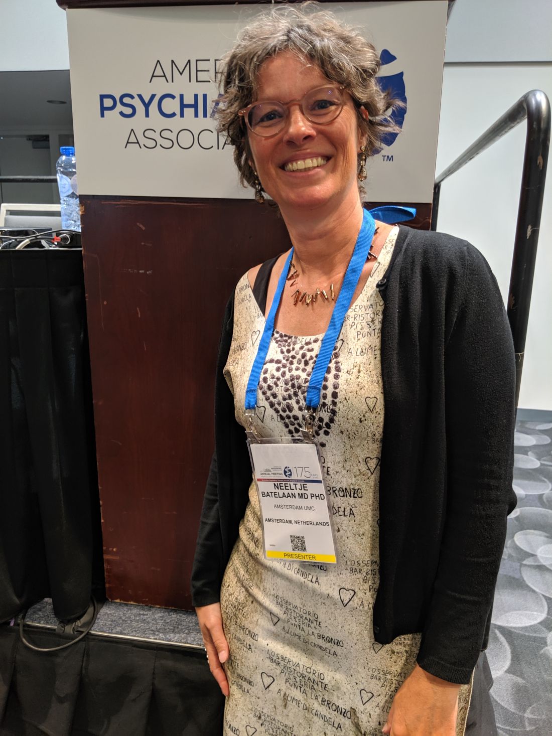
“It’s important that we realize that anxiety disorders can be treated effectively in the short term, but it’s very difficult to treat them for the long term. We know that within a year, it’s better to continue the medication. There’s a lack of data to give evidence-based advice after 1 year,” Neeltje Batelaan, MD, PhD, a psychiatrist and senior researcher at VU University Medical Center, Amsterdam, said in an interview.
Dr. Batelaan moderated a session at the annual meeting of the American Psychiatric Association on discontinuation of antidepressant medications in these patients.
Anxiety disorders can often be successfully treated with antidepressants, but their adverse effects can become less tolerable over time, especially after patients go into remission. When patients begin treatment, they are willing to endure side effects in the service of resolving their symptoms. But when they go into remission, “they want to get on with their lives, so their sexual side effects or their weight gain rates a lot worse,” Dr. Batelaan said.
A meta-analysis of 28 studies, with follow-up periods ranging from 8 to 52 weeks, found that the anxiety relapse risk after discontinuation of antidepressants was 36.4%, compared with 16.4% in those who stayed on medication. Even continuing antidepressants isn’t completely protective, she noted. The study found a number needed to treat of five to prevent one relapse.
Researchers at the VU University Medical Center developed a cognitive-behavioral therapy (CBT) regimen aimed at reducing anxiety relapses. In their study, 87 patients with a remitted anxiety disorder who wanted to stop their antidepressants were randomized to do so either with or without CBT intervention.
Unfortunately, the study had to be stopped when an interim analysis showed a lack of efficacy. In fact, patients who received CBT actually had higher relapse rates. Surprisingly, just 37% of patients succeeded in completely discontinuing medication, which hints at the inherent challenges of the transition.
“Unfortunately, building a CBT relapse prevention did not come true, but we learned some valuable lessons that will guide further studies,” Willemijn Scholten, PhD, a postdoc researcher at VU University Medical Center, said during one of the presentations.
In his presentation, Anton (Ton) Van Balkom, MD, PhD, professor of psychiatry at VU University Medical Center, recounted the case of a woman who had been functioning well with an antidepressant but grew tired of the sexual side effects. She carefully discontinued her medication under his guidance, but in 2 months she experienced her first panic attack in 30 years. Reintroducing the medication failed to resolve the issue, and it took years of effort before cognitive behavioral therapy resulted in a remission.
Further, a meta-analysis of nine studies showed that 17% of patients with remitted anxiety who went off and then restarted their antidepressants experienced tachycardia.
To help reduce tachycardia, Dr. Van Balkom suggested alternative options to antidepressants in less-complicated patients with anxiety, and to anticipate long-term use of antidepressants once those medications are employed.
Dr. Batelaan agreed with that assessment, drawing an analogy with type 2 diabetes. “You first start with a diet, and advise the patient to lose weight, and then if that’s not successful you go to [medication] and you realize it’s lifelong. Maybe we have to differentiate antidepressant therapies and [not start them until necessary]. But if you have to start them, realize that there’s a difficult decision waiting ahead.”
SAN FRANCISCO – Relapse is more likely in the absence of medication and, if they resume their antidepressant after relapse, some patients experience adverse events or drug resistance.
“It’s important that we realize that anxiety disorders can be treated effectively in the short term, but it’s very difficult to treat them for the long term. We know that within a year, it’s better to continue the medication. There’s a lack of data to give evidence-based advice after 1 year,” Neeltje Batelaan, MD, PhD, a psychiatrist and senior researcher at VU University Medical Center, Amsterdam, said in an interview.
Dr. Batelaan moderated a session at the annual meeting of the American Psychiatric Association on discontinuation of antidepressant medications in these patients.
Anxiety disorders can often be successfully treated with antidepressants, but their adverse effects can become less tolerable over time, especially after patients go into remission. When patients begin treatment, they are willing to endure side effects in the service of resolving their symptoms. But when they go into remission, “they want to get on with their lives, so their sexual side effects or their weight gain rates a lot worse,” Dr. Batelaan said.
A meta-analysis of 28 studies, with follow-up periods ranging from 8 to 52 weeks, found that the anxiety relapse risk after discontinuation of antidepressants was 36.4%, compared with 16.4% in those who stayed on medication. Even continuing antidepressants isn’t completely protective, she noted. The study found a number needed to treat of five to prevent one relapse.
Researchers at the VU University Medical Center developed a cognitive-behavioral therapy (CBT) regimen aimed at reducing anxiety relapses. In their study, 87 patients with a remitted anxiety disorder who wanted to stop their antidepressants were randomized to do so either with or without CBT intervention.
Unfortunately, the study had to be stopped when an interim analysis showed a lack of efficacy. In fact, patients who received CBT actually had higher relapse rates. Surprisingly, just 37% of patients succeeded in completely discontinuing medication, which hints at the inherent challenges of the transition.
“Unfortunately, building a CBT relapse prevention did not come true, but we learned some valuable lessons that will guide further studies,” Willemijn Scholten, PhD, a postdoc researcher at VU University Medical Center, said during one of the presentations.
In his presentation, Anton (Ton) Van Balkom, MD, PhD, professor of psychiatry at VU University Medical Center, recounted the case of a woman who had been functioning well with an antidepressant but grew tired of the sexual side effects. She carefully discontinued her medication under his guidance, but in 2 months she experienced her first panic attack in 30 years. Reintroducing the medication failed to resolve the issue, and it took years of effort before cognitive behavioral therapy resulted in a remission.
Further, a meta-analysis of nine studies showed that 17% of patients with remitted anxiety who went off and then restarted their antidepressants experienced tachycardia.
To help reduce tachycardia, Dr. Van Balkom suggested alternative options to antidepressants in less-complicated patients with anxiety, and to anticipate long-term use of antidepressants once those medications are employed.
Dr. Batelaan agreed with that assessment, drawing an analogy with type 2 diabetes. “You first start with a diet, and advise the patient to lose weight, and then if that’s not successful you go to [medication] and you realize it’s lifelong. Maybe we have to differentiate antidepressant therapies and [not start them until necessary]. But if you have to start them, realize that there’s a difficult decision waiting ahead.”
SAN FRANCISCO – Relapse is more likely in the absence of medication and, if they resume their antidepressant after relapse, some patients experience adverse events or drug resistance.
“It’s important that we realize that anxiety disorders can be treated effectively in the short term, but it’s very difficult to treat them for the long term. We know that within a year, it’s better to continue the medication. There’s a lack of data to give evidence-based advice after 1 year,” Neeltje Batelaan, MD, PhD, a psychiatrist and senior researcher at VU University Medical Center, Amsterdam, said in an interview.
Dr. Batelaan moderated a session at the annual meeting of the American Psychiatric Association on discontinuation of antidepressant medications in these patients.
Anxiety disorders can often be successfully treated with antidepressants, but their adverse effects can become less tolerable over time, especially after patients go into remission. When patients begin treatment, they are willing to endure side effects in the service of resolving their symptoms. But when they go into remission, “they want to get on with their lives, so their sexual side effects or their weight gain rates a lot worse,” Dr. Batelaan said.
A meta-analysis of 28 studies, with follow-up periods ranging from 8 to 52 weeks, found that the anxiety relapse risk after discontinuation of antidepressants was 36.4%, compared with 16.4% in those who stayed on medication. Even continuing antidepressants isn’t completely protective, she noted. The study found a number needed to treat of five to prevent one relapse.
Researchers at the VU University Medical Center developed a cognitive-behavioral therapy (CBT) regimen aimed at reducing anxiety relapses. In their study, 87 patients with a remitted anxiety disorder who wanted to stop their antidepressants were randomized to do so either with or without CBT intervention.
Unfortunately, the study had to be stopped when an interim analysis showed a lack of efficacy. In fact, patients who received CBT actually had higher relapse rates. Surprisingly, just 37% of patients succeeded in completely discontinuing medication, which hints at the inherent challenges of the transition.
“Unfortunately, building a CBT relapse prevention did not come true, but we learned some valuable lessons that will guide further studies,” Willemijn Scholten, PhD, a postdoc researcher at VU University Medical Center, said during one of the presentations.
In his presentation, Anton (Ton) Van Balkom, MD, PhD, professor of psychiatry at VU University Medical Center, recounted the case of a woman who had been functioning well with an antidepressant but grew tired of the sexual side effects. She carefully discontinued her medication under his guidance, but in 2 months she experienced her first panic attack in 30 years. Reintroducing the medication failed to resolve the issue, and it took years of effort before cognitive behavioral therapy resulted in a remission.
Further, a meta-analysis of nine studies showed that 17% of patients with remitted anxiety who went off and then restarted their antidepressants experienced tachycardia.
To help reduce tachycardia, Dr. Van Balkom suggested alternative options to antidepressants in less-complicated patients with anxiety, and to anticipate long-term use of antidepressants once those medications are employed.
Dr. Batelaan agreed with that assessment, drawing an analogy with type 2 diabetes. “You first start with a diet, and advise the patient to lose weight, and then if that’s not successful you go to [medication] and you realize it’s lifelong. Maybe we have to differentiate antidepressant therapies and [not start them until necessary]. But if you have to start them, realize that there’s a difficult decision waiting ahead.”
REPORTING FROM APA 2019
Rheumatoid arthritis treatment less aggressive, not less favorable in older adults
BIRMINGHAM, ENGLAND – Being diagnosed with rheumatoid arthritis at age 75 years or older made it less likely that patients would receive intensive therapy than their younger counterparts, but that did not mean that they were treated any less favorably overall, according to findings derived from the Early RA Network cohort.
“The claim that the elderly are treated less aggressively isn’t completely true throughout the whole treat-to-target strategy,” said Simone Howard of King’s College London at the British Society for Rheumatology annual conference. While older patients were less likely to receive intensive treatment up to 2 years after their diagnosis, there was a shorter delay between the onset of symptoms and the first outpatient visit to a rheumatology clinic.
When compared against patients who were younger than 65 years, those aged 65-74 years and 75 years and older were 11% (P = .02) and 15% (P = .02) more likely to have their first outpatient visit within 10 months.
Furthermore, no significant differences were seen between any age groups in the time to first initiation of a conventional synthetic disease-modifying antirheumatic drug (csDMARD), which averaged nearly 3 months after symptoms appeared.
Ms. Howard, who has previously worked at the European Medicines Agency, noted that, during her time at the EMA, “there was a real push to incorporate the elderly into the regulatory framework more. In parallel, there were also reports of the elderly being treated less aggressively. So the question was, where was that coming from?”
Similar therapeutic approaches are advocated for older and younger RA patients, and to look for any disparities, Ms. Howard and associates turned to the Early RA Network (ERAN) to “investigate potential treatment bias against the elderly.”
ERAN is a hospital-based inception cohort of 1,236 patients with early RA who were recruited across 23 centers in the United Kingdom and Ireland between 2002 and 2014.
Of 1,131 patients used in the analyses, 9.7% (n = 110) were 75 years or older, 21.5% (n = 243) were aged 65-74 years, and 68.8% (n = 778) were 65 years or younger. The majority (67.7%) of patients were female.
Patients aged 75 years and older were more likely to present with comorbidities than the youngest group, and they had higher health assessment questionnaire scores at baseline. However, they were no more likely to have high disease activity at the first visit, which was defined as a disease activity score in 28 joints of more than 5, and the older patients were 27% less likely to be seropositive (P = .004).
“It’s when we come to pharmacological aspects of care that we are seeing treatment biases,” Ms. Howard noted. Patients over 75 years were significantly more likely than the youngest age group to be treated with glucocorticoids or csDMARD monotherapy at 1 year, and 23% more likely to be on less aggressive therapy (P equal to or less than .0001). Aggressive therapy was defined as the use of a combination of csDMARDs or the use of biologic drugs.
At 2 years, the oldest patients were 46% more likely than those under 65 years to be on less-intensive therapies (P equal to or less than .0001), with those aged 65-74 years 19% more likely to be on glucocorticoid or csDMARD therapy (P = .005).
Factors such as patient choice and tolerance were not considered in the analyses and could be important, Ms. Howard conceded in response to a question after her presentation.
Another point raised was that perhaps the prescribing of aggressive therapy would rationally be different in someone diagnosed with RA at age 85 versus 65 because the duration of time that would be likely to be lived with accumulating joint damage would be shorter at the older age and that would be balanced against the other effects of the therapy. So, there may be important reasons in shared decision making that influenced the treatment choices other than the age of patients.
Ms. Howard agreed, noting that this demonstrated the need to be careful around the language used for defining what constituted aggressive or intensive therapy.
She and her coauthors reported no conflicts of interest.
SOURCE: Howard S et al. Rheumatology. 2019;58(suppl 3), Abstract 011.
BIRMINGHAM, ENGLAND – Being diagnosed with rheumatoid arthritis at age 75 years or older made it less likely that patients would receive intensive therapy than their younger counterparts, but that did not mean that they were treated any less favorably overall, according to findings derived from the Early RA Network cohort.
“The claim that the elderly are treated less aggressively isn’t completely true throughout the whole treat-to-target strategy,” said Simone Howard of King’s College London at the British Society for Rheumatology annual conference. While older patients were less likely to receive intensive treatment up to 2 years after their diagnosis, there was a shorter delay between the onset of symptoms and the first outpatient visit to a rheumatology clinic.
When compared against patients who were younger than 65 years, those aged 65-74 years and 75 years and older were 11% (P = .02) and 15% (P = .02) more likely to have their first outpatient visit within 10 months.
Furthermore, no significant differences were seen between any age groups in the time to first initiation of a conventional synthetic disease-modifying antirheumatic drug (csDMARD), which averaged nearly 3 months after symptoms appeared.
Ms. Howard, who has previously worked at the European Medicines Agency, noted that, during her time at the EMA, “there was a real push to incorporate the elderly into the regulatory framework more. In parallel, there were also reports of the elderly being treated less aggressively. So the question was, where was that coming from?”
Similar therapeutic approaches are advocated for older and younger RA patients, and to look for any disparities, Ms. Howard and associates turned to the Early RA Network (ERAN) to “investigate potential treatment bias against the elderly.”
ERAN is a hospital-based inception cohort of 1,236 patients with early RA who were recruited across 23 centers in the United Kingdom and Ireland between 2002 and 2014.
Of 1,131 patients used in the analyses, 9.7% (n = 110) were 75 years or older, 21.5% (n = 243) were aged 65-74 years, and 68.8% (n = 778) were 65 years or younger. The majority (67.7%) of patients were female.
Patients aged 75 years and older were more likely to present with comorbidities than the youngest group, and they had higher health assessment questionnaire scores at baseline. However, they were no more likely to have high disease activity at the first visit, which was defined as a disease activity score in 28 joints of more than 5, and the older patients were 27% less likely to be seropositive (P = .004).
“It’s when we come to pharmacological aspects of care that we are seeing treatment biases,” Ms. Howard noted. Patients over 75 years were significantly more likely than the youngest age group to be treated with glucocorticoids or csDMARD monotherapy at 1 year, and 23% more likely to be on less aggressive therapy (P equal to or less than .0001). Aggressive therapy was defined as the use of a combination of csDMARDs or the use of biologic drugs.
At 2 years, the oldest patients were 46% more likely than those under 65 years to be on less-intensive therapies (P equal to or less than .0001), with those aged 65-74 years 19% more likely to be on glucocorticoid or csDMARD therapy (P = .005).
Factors such as patient choice and tolerance were not considered in the analyses and could be important, Ms. Howard conceded in response to a question after her presentation.
Another point raised was that perhaps the prescribing of aggressive therapy would rationally be different in someone diagnosed with RA at age 85 versus 65 because the duration of time that would be likely to be lived with accumulating joint damage would be shorter at the older age and that would be balanced against the other effects of the therapy. So, there may be important reasons in shared decision making that influenced the treatment choices other than the age of patients.
Ms. Howard agreed, noting that this demonstrated the need to be careful around the language used for defining what constituted aggressive or intensive therapy.
She and her coauthors reported no conflicts of interest.
SOURCE: Howard S et al. Rheumatology. 2019;58(suppl 3), Abstract 011.
BIRMINGHAM, ENGLAND – Being diagnosed with rheumatoid arthritis at age 75 years or older made it less likely that patients would receive intensive therapy than their younger counterparts, but that did not mean that they were treated any less favorably overall, according to findings derived from the Early RA Network cohort.
“The claim that the elderly are treated less aggressively isn’t completely true throughout the whole treat-to-target strategy,” said Simone Howard of King’s College London at the British Society for Rheumatology annual conference. While older patients were less likely to receive intensive treatment up to 2 years after their diagnosis, there was a shorter delay between the onset of symptoms and the first outpatient visit to a rheumatology clinic.
When compared against patients who were younger than 65 years, those aged 65-74 years and 75 years and older were 11% (P = .02) and 15% (P = .02) more likely to have their first outpatient visit within 10 months.
Furthermore, no significant differences were seen between any age groups in the time to first initiation of a conventional synthetic disease-modifying antirheumatic drug (csDMARD), which averaged nearly 3 months after symptoms appeared.
Ms. Howard, who has previously worked at the European Medicines Agency, noted that, during her time at the EMA, “there was a real push to incorporate the elderly into the regulatory framework more. In parallel, there were also reports of the elderly being treated less aggressively. So the question was, where was that coming from?”
Similar therapeutic approaches are advocated for older and younger RA patients, and to look for any disparities, Ms. Howard and associates turned to the Early RA Network (ERAN) to “investigate potential treatment bias against the elderly.”
ERAN is a hospital-based inception cohort of 1,236 patients with early RA who were recruited across 23 centers in the United Kingdom and Ireland between 2002 and 2014.
Of 1,131 patients used in the analyses, 9.7% (n = 110) were 75 years or older, 21.5% (n = 243) were aged 65-74 years, and 68.8% (n = 778) were 65 years or younger. The majority (67.7%) of patients were female.
Patients aged 75 years and older were more likely to present with comorbidities than the youngest group, and they had higher health assessment questionnaire scores at baseline. However, they were no more likely to have high disease activity at the first visit, which was defined as a disease activity score in 28 joints of more than 5, and the older patients were 27% less likely to be seropositive (P = .004).
“It’s when we come to pharmacological aspects of care that we are seeing treatment biases,” Ms. Howard noted. Patients over 75 years were significantly more likely than the youngest age group to be treated with glucocorticoids or csDMARD monotherapy at 1 year, and 23% more likely to be on less aggressive therapy (P equal to or less than .0001). Aggressive therapy was defined as the use of a combination of csDMARDs or the use of biologic drugs.
At 2 years, the oldest patients were 46% more likely than those under 65 years to be on less-intensive therapies (P equal to or less than .0001), with those aged 65-74 years 19% more likely to be on glucocorticoid or csDMARD therapy (P = .005).
Factors such as patient choice and tolerance were not considered in the analyses and could be important, Ms. Howard conceded in response to a question after her presentation.
Another point raised was that perhaps the prescribing of aggressive therapy would rationally be different in someone diagnosed with RA at age 85 versus 65 because the duration of time that would be likely to be lived with accumulating joint damage would be shorter at the older age and that would be balanced against the other effects of the therapy. So, there may be important reasons in shared decision making that influenced the treatment choices other than the age of patients.
Ms. Howard agreed, noting that this demonstrated the need to be careful around the language used for defining what constituted aggressive or intensive therapy.
She and her coauthors reported no conflicts of interest.
SOURCE: Howard S et al. Rheumatology. 2019;58(suppl 3), Abstract 011.
REPORTING FROM BSR 2019
Eosinophil levels affect glucocorticoid response in mild, persistent asthma
according to investigators.

Two recent studies presented at the American Thoracic Society’s international conference demonstrated the benefits of glucocorticoid therapy among patients with mild persistent or intermittent asthma while highlighting differential responses to steroids among patients with high versus low levels of eosinophils in sputum. Both studies were simultaneously published in the New England Journal of Medicine.
The first study, SIENA, led by Stephen C. Lazarus, MD of the University of California, San Francisco, and colleagues, involved 295 patients with mild, persistent asthma. Patients were classified as having either a high or low level of eosinophils in sputum, with a low level defined by two sputum samples consisting of less than 2% eosinophils. After a single-blind placebo run-in period of 6 weeks, patients were randomized to receive either mometasone (an inhaled glucocorticoid), tiotropium (a long-acting muscarinic antagonist [LAMA]), or placebo for 12 weeks each, with subsequent crossover through the two remaining treatments. The primary outcome was the response to each active agent, compared with placebo among low-eosinophil patients who had a differential response to a trial agent.
Out of 295 patients, 221 (75%) had low eosinophils and 74 (25%) had high eosinophils. In the low-eosinophil subgroup, 59% of patients had a differential response to a trial agent; among these, 57% responded better to mometasone, compared with 43% who responded better to placebo, and 60% responded better to tiotropium, compared with 40% who responded better to placebo.
Turning to secondary analyses, among patients with high eosinophil levels who had a differential response, 74% responded better to mometasone, compared with 26% who responded better to placebo, and 57% responded better to tiotropium, compared with 43% who responded better to placebo.
In an additional exploratory analysis, adults with low eosinophil levels had better responses to tiotropium than placebo (62% vs 38%).
The researchers stated that a key finding of the study is that three-quarters of the mild, persistent asthma population had low eosinophil levels, far fewer than expected and that the difference in their response to mometasone compared to tiotropium was not significant.
“Our results raise the question of whether treatment guidelines should be reevaluated for patients with mild, persistent asthma for whom evidence of type 2 inflammation is lacking,” the investigators wrote. “The need for a change in treatment strategy is further highlighted by a growing body of literature suggesting that mild, persistent asthma can be managed safely without the daily use of inhaled glucocorticoids and by data showing that patients with a low eosinophil level may not have a favorable response to inhaled glucocorticoids” (New Engl J Med. 2019 May 19. doi: 10.1056/NEJMoa1814917).
The second study, Novel START, conducted by lead author Richard Beasley, DSc, of the Medical Research Institute of New Zealand, Wellington, and colleagues, compared the efficacy of two inhaled glucocorticoid regimens and albuterol alone for patients with mild persistent or intermittent asthma, measured by annualized exacerbation rate.
Initial randomization involved 675 patients, of whom 668 were included in the final analysis. Patients were randomized into three groups: albuterol as needed (100 mcg, two inhalations as needed for asthma symptoms), budesonide maintenance (200 mcg, one inhalation twice daily with as-needed albuterol), or budesonide/formoterol (budesonide 200 mcg and formoterol 6 mcg, one inhalation as needed). Along with annualized exacerbation rate, several secondary outcomes assessed symptoms, respiratory function, and number of severe exacerbations.
Data analysis showed that patients in the budesonide groups had similar rates of annualized exacerbation, both of which were significantly better than the exacerbation rate in the albuterol-only group; the absolute rate of exacerbations per patient per year was 0.175, 0.195, and 0.400 for budesonide maintenance, budesonide/formoterol, and albuterol only, respectively. Similarly, the median fraction of exhaled nitric oxide (FENO) was lower in the budesonide groups than in the albuterol-only group. Patients in the budesonide/formoterol group had a 56% lower relative risk of severe pulmonary exacerbation than patients in the budesonide maintenance group and a 60% lower relative risk than the albuterol group. However, maintenance budesonide provided better symptom relief than budesonide/formoterol, “which suggests that for the patient for whom asthma symptoms rather than exacerbations are the most bothersome, maintenance treatment has value,” the investigators wrote (New Engl J Med. 2019 May 19. doi: 10.1056/NEJMoa1901963).
“The findings of our trial are consistent with evidence regarding the treatment of moderate and severe asthma – that maintenance and reliever therapy” with inhaled glucocorticoid/formoterol “results in a lower risk of severe exacerbations than maintenance therapy with an inhaled glucocorticoid–[long-acting beta agonist] and as-needed SABA,” the investigators concluded.
SIENA was funded by National Heart, Lung, and Blood Institute, with medications provided by Boehringer Ingelheim, Merck, and Teva; the investigators reported relationships with Sanofi, Vectura, Circassia, DBV Technologies, and others. Novel START was funded by AstraZeneca and the Health Research Council of New Zealand; the investigators reported relationships with GlaxoSmithKline, Genentech, Theravance Biopharma, and others.
SOURCES: Beasley et al. New Engl J Med. 2019 May 19. doi: 10.1056/NEJMoa1901963; Lazarus et al. New Engl J Med. 2019 May 19. doi: 10.1056/NEJMoa1814917.
Gary W. K. Wong, MD, commented in an editorial accompanying the two studies, “Approximately 1 year ago, the SYGMA 1 and SYGMA 2 trials, involving patients with mild persistent asthma, suggested that as-needed use of a budesonide–formoterol combination was as effective as budesonide maintenance therapy in the prevention of exacerbations, with the added benefit of decreased overall glucocorticoid exposure; however, it remained unknown if this strategy was appropriate for patients with mild intermittent asthma and among patients lacking eosinophilic inflammation. The two reported studies attempt to address these knowledge gaps.”
He noted that both trials showed that “patients with mild asthma whose only asthma treatment was a SABA [short-acting beta2-agonists] as needed for relief of asthma symptoms were at considerable risk for exacerbations. Replacement of as-needed SABA treatment with as-needed budesonide/formoterol or inhaled glucocorticoid maintenance therapy could reduce such risk by approximately 50%. When considering maintenance therapy for persistent asthma, one must be aware that not all types of airway inflammation respond equally well to inhaled glucocorticoid therapy.”
Gary W.K. Wong, MD, is a professor in the department of pediatrics at Prince of Wales Hospital, Chinese University of Hong Kong. He made his remarks in an editorial in the New England Journal of Medicine (2019 May 19. doi: 10.1056/NEJMe1905354). Dr. Wong disclosed that he has no relevant financial conflicts of interest.
Gary W. K. Wong, MD, commented in an editorial accompanying the two studies, “Approximately 1 year ago, the SYGMA 1 and SYGMA 2 trials, involving patients with mild persistent asthma, suggested that as-needed use of a budesonide–formoterol combination was as effective as budesonide maintenance therapy in the prevention of exacerbations, with the added benefit of decreased overall glucocorticoid exposure; however, it remained unknown if this strategy was appropriate for patients with mild intermittent asthma and among patients lacking eosinophilic inflammation. The two reported studies attempt to address these knowledge gaps.”
He noted that both trials showed that “patients with mild asthma whose only asthma treatment was a SABA [short-acting beta2-agonists] as needed for relief of asthma symptoms were at considerable risk for exacerbations. Replacement of as-needed SABA treatment with as-needed budesonide/formoterol or inhaled glucocorticoid maintenance therapy could reduce such risk by approximately 50%. When considering maintenance therapy for persistent asthma, one must be aware that not all types of airway inflammation respond equally well to inhaled glucocorticoid therapy.”
Gary W.K. Wong, MD, is a professor in the department of pediatrics at Prince of Wales Hospital, Chinese University of Hong Kong. He made his remarks in an editorial in the New England Journal of Medicine (2019 May 19. doi: 10.1056/NEJMe1905354). Dr. Wong disclosed that he has no relevant financial conflicts of interest.
Gary W. K. Wong, MD, commented in an editorial accompanying the two studies, “Approximately 1 year ago, the SYGMA 1 and SYGMA 2 trials, involving patients with mild persistent asthma, suggested that as-needed use of a budesonide–formoterol combination was as effective as budesonide maintenance therapy in the prevention of exacerbations, with the added benefit of decreased overall glucocorticoid exposure; however, it remained unknown if this strategy was appropriate for patients with mild intermittent asthma and among patients lacking eosinophilic inflammation. The two reported studies attempt to address these knowledge gaps.”
He noted that both trials showed that “patients with mild asthma whose only asthma treatment was a SABA [short-acting beta2-agonists] as needed for relief of asthma symptoms were at considerable risk for exacerbations. Replacement of as-needed SABA treatment with as-needed budesonide/formoterol or inhaled glucocorticoid maintenance therapy could reduce such risk by approximately 50%. When considering maintenance therapy for persistent asthma, one must be aware that not all types of airway inflammation respond equally well to inhaled glucocorticoid therapy.”
Gary W.K. Wong, MD, is a professor in the department of pediatrics at Prince of Wales Hospital, Chinese University of Hong Kong. He made his remarks in an editorial in the New England Journal of Medicine (2019 May 19. doi: 10.1056/NEJMe1905354). Dr. Wong disclosed that he has no relevant financial conflicts of interest.
according to investigators.
Two recent studies presented at the American Thoracic Society’s international conference demonstrated the benefits of glucocorticoid therapy among patients with mild persistent or intermittent asthma while highlighting differential responses to steroids among patients with high versus low levels of eosinophils in sputum. Both studies were simultaneously published in the New England Journal of Medicine.
The first study, SIENA, led by Stephen C. Lazarus, MD of the University of California, San Francisco, and colleagues, involved 295 patients with mild, persistent asthma. Patients were classified as having either a high or low level of eosinophils in sputum, with a low level defined by two sputum samples consisting of less than 2% eosinophils. After a single-blind placebo run-in period of 6 weeks, patients were randomized to receive either mometasone (an inhaled glucocorticoid), tiotropium (a long-acting muscarinic antagonist [LAMA]), or placebo for 12 weeks each, with subsequent crossover through the two remaining treatments. The primary outcome was the response to each active agent, compared with placebo among low-eosinophil patients who had a differential response to a trial agent.
Out of 295 patients, 221 (75%) had low eosinophils and 74 (25%) had high eosinophils. In the low-eosinophil subgroup, 59% of patients had a differential response to a trial agent; among these, 57% responded better to mometasone, compared with 43% who responded better to placebo, and 60% responded better to tiotropium, compared with 40% who responded better to placebo.
Turning to secondary analyses, among patients with high eosinophil levels who had a differential response, 74% responded better to mometasone, compared with 26% who responded better to placebo, and 57% responded better to tiotropium, compared with 43% who responded better to placebo.
In an additional exploratory analysis, adults with low eosinophil levels had better responses to tiotropium than placebo (62% vs 38%).
The researchers stated that a key finding of the study is that three-quarters of the mild, persistent asthma population had low eosinophil levels, far fewer than expected and that the difference in their response to mometasone compared to tiotropium was not significant.
“Our results raise the question of whether treatment guidelines should be reevaluated for patients with mild, persistent asthma for whom evidence of type 2 inflammation is lacking,” the investigators wrote. “The need for a change in treatment strategy is further highlighted by a growing body of literature suggesting that mild, persistent asthma can be managed safely without the daily use of inhaled glucocorticoids and by data showing that patients with a low eosinophil level may not have a favorable response to inhaled glucocorticoids” (New Engl J Med. 2019 May 19. doi: 10.1056/NEJMoa1814917).
The second study, Novel START, conducted by lead author Richard Beasley, DSc, of the Medical Research Institute of New Zealand, Wellington, and colleagues, compared the efficacy of two inhaled glucocorticoid regimens and albuterol alone for patients with mild persistent or intermittent asthma, measured by annualized exacerbation rate.
Initial randomization involved 675 patients, of whom 668 were included in the final analysis. Patients were randomized into three groups: albuterol as needed (100 mcg, two inhalations as needed for asthma symptoms), budesonide maintenance (200 mcg, one inhalation twice daily with as-needed albuterol), or budesonide/formoterol (budesonide 200 mcg and formoterol 6 mcg, one inhalation as needed). Along with annualized exacerbation rate, several secondary outcomes assessed symptoms, respiratory function, and number of severe exacerbations.
Data analysis showed that patients in the budesonide groups had similar rates of annualized exacerbation, both of which were significantly better than the exacerbation rate in the albuterol-only group; the absolute rate of exacerbations per patient per year was 0.175, 0.195, and 0.400 for budesonide maintenance, budesonide/formoterol, and albuterol only, respectively. Similarly, the median fraction of exhaled nitric oxide (FENO) was lower in the budesonide groups than in the albuterol-only group. Patients in the budesonide/formoterol group had a 56% lower relative risk of severe pulmonary exacerbation than patients in the budesonide maintenance group and a 60% lower relative risk than the albuterol group. However, maintenance budesonide provided better symptom relief than budesonide/formoterol, “which suggests that for the patient for whom asthma symptoms rather than exacerbations are the most bothersome, maintenance treatment has value,” the investigators wrote (New Engl J Med. 2019 May 19. doi: 10.1056/NEJMoa1901963).
“The findings of our trial are consistent with evidence regarding the treatment of moderate and severe asthma – that maintenance and reliever therapy” with inhaled glucocorticoid/formoterol “results in a lower risk of severe exacerbations than maintenance therapy with an inhaled glucocorticoid–[long-acting beta agonist] and as-needed SABA,” the investigators concluded.
SIENA was funded by National Heart, Lung, and Blood Institute, with medications provided by Boehringer Ingelheim, Merck, and Teva; the investigators reported relationships with Sanofi, Vectura, Circassia, DBV Technologies, and others. Novel START was funded by AstraZeneca and the Health Research Council of New Zealand; the investigators reported relationships with GlaxoSmithKline, Genentech, Theravance Biopharma, and others.
SOURCES: Beasley et al. New Engl J Med. 2019 May 19. doi: 10.1056/NEJMoa1901963; Lazarus et al. New Engl J Med. 2019 May 19. doi: 10.1056/NEJMoa1814917.
according to investigators.
Two recent studies presented at the American Thoracic Society’s international conference demonstrated the benefits of glucocorticoid therapy among patients with mild persistent or intermittent asthma while highlighting differential responses to steroids among patients with high versus low levels of eosinophils in sputum. Both studies were simultaneously published in the New England Journal of Medicine.
The first study, SIENA, led by Stephen C. Lazarus, MD of the University of California, San Francisco, and colleagues, involved 295 patients with mild, persistent asthma. Patients were classified as having either a high or low level of eosinophils in sputum, with a low level defined by two sputum samples consisting of less than 2% eosinophils. After a single-blind placebo run-in period of 6 weeks, patients were randomized to receive either mometasone (an inhaled glucocorticoid), tiotropium (a long-acting muscarinic antagonist [LAMA]), or placebo for 12 weeks each, with subsequent crossover through the two remaining treatments. The primary outcome was the response to each active agent, compared with placebo among low-eosinophil patients who had a differential response to a trial agent.
Out of 295 patients, 221 (75%) had low eosinophils and 74 (25%) had high eosinophils. In the low-eosinophil subgroup, 59% of patients had a differential response to a trial agent; among these, 57% responded better to mometasone, compared with 43% who responded better to placebo, and 60% responded better to tiotropium, compared with 40% who responded better to placebo.
Turning to secondary analyses, among patients with high eosinophil levels who had a differential response, 74% responded better to mometasone, compared with 26% who responded better to placebo, and 57% responded better to tiotropium, compared with 43% who responded better to placebo.
In an additional exploratory analysis, adults with low eosinophil levels had better responses to tiotropium than placebo (62% vs 38%).
The researchers stated that a key finding of the study is that three-quarters of the mild, persistent asthma population had low eosinophil levels, far fewer than expected and that the difference in their response to mometasone compared to tiotropium was not significant.
“Our results raise the question of whether treatment guidelines should be reevaluated for patients with mild, persistent asthma for whom evidence of type 2 inflammation is lacking,” the investigators wrote. “The need for a change in treatment strategy is further highlighted by a growing body of literature suggesting that mild, persistent asthma can be managed safely without the daily use of inhaled glucocorticoids and by data showing that patients with a low eosinophil level may not have a favorable response to inhaled glucocorticoids” (New Engl J Med. 2019 May 19. doi: 10.1056/NEJMoa1814917).
The second study, Novel START, conducted by lead author Richard Beasley, DSc, of the Medical Research Institute of New Zealand, Wellington, and colleagues, compared the efficacy of two inhaled glucocorticoid regimens and albuterol alone for patients with mild persistent or intermittent asthma, measured by annualized exacerbation rate.
Initial randomization involved 675 patients, of whom 668 were included in the final analysis. Patients were randomized into three groups: albuterol as needed (100 mcg, two inhalations as needed for asthma symptoms), budesonide maintenance (200 mcg, one inhalation twice daily with as-needed albuterol), or budesonide/formoterol (budesonide 200 mcg and formoterol 6 mcg, one inhalation as needed). Along with annualized exacerbation rate, several secondary outcomes assessed symptoms, respiratory function, and number of severe exacerbations.
Data analysis showed that patients in the budesonide groups had similar rates of annualized exacerbation, both of which were significantly better than the exacerbation rate in the albuterol-only group; the absolute rate of exacerbations per patient per year was 0.175, 0.195, and 0.400 for budesonide maintenance, budesonide/formoterol, and albuterol only, respectively. Similarly, the median fraction of exhaled nitric oxide (FENO) was lower in the budesonide groups than in the albuterol-only group. Patients in the budesonide/formoterol group had a 56% lower relative risk of severe pulmonary exacerbation than patients in the budesonide maintenance group and a 60% lower relative risk than the albuterol group. However, maintenance budesonide provided better symptom relief than budesonide/formoterol, “which suggests that for the patient for whom asthma symptoms rather than exacerbations are the most bothersome, maintenance treatment has value,” the investigators wrote (New Engl J Med. 2019 May 19. doi: 10.1056/NEJMoa1901963).
“The findings of our trial are consistent with evidence regarding the treatment of moderate and severe asthma – that maintenance and reliever therapy” with inhaled glucocorticoid/formoterol “results in a lower risk of severe exacerbations than maintenance therapy with an inhaled glucocorticoid–[long-acting beta agonist] and as-needed SABA,” the investigators concluded.
SIENA was funded by National Heart, Lung, and Blood Institute, with medications provided by Boehringer Ingelheim, Merck, and Teva; the investigators reported relationships with Sanofi, Vectura, Circassia, DBV Technologies, and others. Novel START was funded by AstraZeneca and the Health Research Council of New Zealand; the investigators reported relationships with GlaxoSmithKline, Genentech, Theravance Biopharma, and others.
SOURCES: Beasley et al. New Engl J Med. 2019 May 19. doi: 10.1056/NEJMoa1901963; Lazarus et al. New Engl J Med. 2019 May 19. doi: 10.1056/NEJMoa1814917.
FROM ATS 2019
Key clinical point: Inhaled glucocorticoid/formoterol therapy for mild persistent or intermittent asthma is effective, but responses to steroids differ among patients with high versus low levels of eosinophils in sputum.
Major finding: Among patients with low eosinophils levels of, 57% responded better to mometasone versus 43% who responded better to placebo. Among those with high eosinophil levels, 74% responded better to mometasone versus 26% who responded better to placebo.
Study details: The SIENA study included 295 patients with mild, persistent asthma and eosinophils measured in sputum samples, and the Novel START study included 688 patients with the mild persistent or intermittent asthma, measured by annualized exacerbation rate.
Disclosures: SIENA was funded by National Heart, Lung, and Blood Institute, with medications provided by Boehringer Ingelheim, Merck, and Teva; the investigators reported relationships with Sanofi, Vectura, Circassia, DBV Technologies, and others. Novel START was funded by AstraZeneca and the Health Research Council of New Zealand; the investigators reported relationships with GlaxoSmithKline, Genentech, Theravance Biopharma, and others.
Sources: Beasley et al. New Engl J Med. 2019 May 19. doi: 10.1056/NEJMoa1901963; Lazarus et al. New Engl J Med. 2019 May 19. doi: 10.1056/NEJMoa1814917.

Use of Oncotype DX to tailor breast cancer treatment likely to reduce costs
Routine use of the Oncotype DX recurrence score to personalize treatment of early breast cancer could reduce the first-year costs of care, according to results of a population-based cohort study.
“Practice changes based on evidence from the TAILORx trial on using tumor genomic profiles to personalize care could result in small decreases in U.S. national cancer care costs in the initial 12 months post breast cancer diagnosis,” Angela Mariotto, PhD, and colleagues concluded in a study published in JNCI: Journal of the National Cancer Institute. “Longer-term studies will be needed to evaluate the true long-term economic impact and nonmonetary benefits of personalized breast cancer care.”
Findings of the landmark TAILORx trial showed that, with use of the 21-gene score, the majority of women who have node-negative, hormone receptor–positive, HER2-negative breast cancers could safely skip adjuvant chemotherapy (N Engl J Med. 2018;379:111-21). But cost impact of its uptake into routine practice is unclear.
Dr. Mariotto, of the National Cancer Institute and her colleagues used data from the Surveillance, Epidemiology and End Results (SEER), SEER-Medicare, and SEER–Genomic Health datasets to assess how expected changes in practice after the trial might affect costs. They estimated Oncotype DX testing and chemotherapy rates and mean initial costs in 2018 dollars in the pre-TAILORx period (2010-2015) and post-TAILORx period (2018), assuming all women in the latter period received the test and score-suggested therapy.
Going from the pretrial period to the posttrial period, Oncotype DX testing costs were projected to increase from $115 million to $231 million, but chemotherapy use was projected to decrease from 25% to 17%. Mean total initial costs of care fell from $2.816 billion in the pretrial period to $2.766 billion in the posttrial period, for a net savings of $49 million (a 1.8% decrease).
Findings were similar in a variety of sensitivity scenarios entailing alternative compliance with testing, score-suggested treatment, and estimation methods. The only exception was the scenario in which all women aged 50 years or younger having a recurrence score of 16-25 opted to receive chemotherapy, wherein initial care costs could increase by $105 million (a 4% increase).
The investigators reported that they had no relevant conflicts of interest. The study was supported by the National Cancer Institute and its Coordinating Center For Clinical Trials; a Lombardi Comprehensive Cancer Center American Cancer Society Young Investigator Award; and the Cancer Prevention Research Fellowship, sponsored by the American Society of Preventive Oncology and Breast Cancer Research Foundation.
SOURCE: Mariotto A et al. J Natl Cancer Inst. 2019 Apr 24. doi: 10.1093/jnci/djz068.
Routine use of the Oncotype DX recurrence score to personalize treatment of early breast cancer could reduce the first-year costs of care, according to results of a population-based cohort study.
“Practice changes based on evidence from the TAILORx trial on using tumor genomic profiles to personalize care could result in small decreases in U.S. national cancer care costs in the initial 12 months post breast cancer diagnosis,” Angela Mariotto, PhD, and colleagues concluded in a study published in JNCI: Journal of the National Cancer Institute. “Longer-term studies will be needed to evaluate the true long-term economic impact and nonmonetary benefits of personalized breast cancer care.”
Findings of the landmark TAILORx trial showed that, with use of the 21-gene score, the majority of women who have node-negative, hormone receptor–positive, HER2-negative breast cancers could safely skip adjuvant chemotherapy (N Engl J Med. 2018;379:111-21). But cost impact of its uptake into routine practice is unclear.
Dr. Mariotto, of the National Cancer Institute and her colleagues used data from the Surveillance, Epidemiology and End Results (SEER), SEER-Medicare, and SEER–Genomic Health datasets to assess how expected changes in practice after the trial might affect costs. They estimated Oncotype DX testing and chemotherapy rates and mean initial costs in 2018 dollars in the pre-TAILORx period (2010-2015) and post-TAILORx period (2018), assuming all women in the latter period received the test and score-suggested therapy.
Going from the pretrial period to the posttrial period, Oncotype DX testing costs were projected to increase from $115 million to $231 million, but chemotherapy use was projected to decrease from 25% to 17%. Mean total initial costs of care fell from $2.816 billion in the pretrial period to $2.766 billion in the posttrial period, for a net savings of $49 million (a 1.8% decrease).
Findings were similar in a variety of sensitivity scenarios entailing alternative compliance with testing, score-suggested treatment, and estimation methods. The only exception was the scenario in which all women aged 50 years or younger having a recurrence score of 16-25 opted to receive chemotherapy, wherein initial care costs could increase by $105 million (a 4% increase).
The investigators reported that they had no relevant conflicts of interest. The study was supported by the National Cancer Institute and its Coordinating Center For Clinical Trials; a Lombardi Comprehensive Cancer Center American Cancer Society Young Investigator Award; and the Cancer Prevention Research Fellowship, sponsored by the American Society of Preventive Oncology and Breast Cancer Research Foundation.
SOURCE: Mariotto A et al. J Natl Cancer Inst. 2019 Apr 24. doi: 10.1093/jnci/djz068.
Routine use of the Oncotype DX recurrence score to personalize treatment of early breast cancer could reduce the first-year costs of care, according to results of a population-based cohort study.
“Practice changes based on evidence from the TAILORx trial on using tumor genomic profiles to personalize care could result in small decreases in U.S. national cancer care costs in the initial 12 months post breast cancer diagnosis,” Angela Mariotto, PhD, and colleagues concluded in a study published in JNCI: Journal of the National Cancer Institute. “Longer-term studies will be needed to evaluate the true long-term economic impact and nonmonetary benefits of personalized breast cancer care.”
Findings of the landmark TAILORx trial showed that, with use of the 21-gene score, the majority of women who have node-negative, hormone receptor–positive, HER2-negative breast cancers could safely skip adjuvant chemotherapy (N Engl J Med. 2018;379:111-21). But cost impact of its uptake into routine practice is unclear.
Dr. Mariotto, of the National Cancer Institute and her colleagues used data from the Surveillance, Epidemiology and End Results (SEER), SEER-Medicare, and SEER–Genomic Health datasets to assess how expected changes in practice after the trial might affect costs. They estimated Oncotype DX testing and chemotherapy rates and mean initial costs in 2018 dollars in the pre-TAILORx period (2010-2015) and post-TAILORx period (2018), assuming all women in the latter period received the test and score-suggested therapy.
Going from the pretrial period to the posttrial period, Oncotype DX testing costs were projected to increase from $115 million to $231 million, but chemotherapy use was projected to decrease from 25% to 17%. Mean total initial costs of care fell from $2.816 billion in the pretrial period to $2.766 billion in the posttrial period, for a net savings of $49 million (a 1.8% decrease).
Findings were similar in a variety of sensitivity scenarios entailing alternative compliance with testing, score-suggested treatment, and estimation methods. The only exception was the scenario in which all women aged 50 years or younger having a recurrence score of 16-25 opted to receive chemotherapy, wherein initial care costs could increase by $105 million (a 4% increase).
The investigators reported that they had no relevant conflicts of interest. The study was supported by the National Cancer Institute and its Coordinating Center For Clinical Trials; a Lombardi Comprehensive Cancer Center American Cancer Society Young Investigator Award; and the Cancer Prevention Research Fellowship, sponsored by the American Society of Preventive Oncology and Breast Cancer Research Foundation.
SOURCE: Mariotto A et al. J Natl Cancer Inst. 2019 Apr 24. doi: 10.1093/jnci/djz068.
FROM JNCI: JOURNAL OF THE NATIONAL CANCER INSTITUTE