User login
Dealing with staffing shortfalls
Five options for covering unfilled positions
Being in stressful situations is part of being a hospitalist. During a hospitalist’s work shift, one of the key determinants of stress is adequate staffing. With use of survey data from 569 hospital medicine groups (HMGs) across the nation, one of the topics examined in the 2018 State of Hospital Medicine Report is how HMGs cope with unfilled hospitalist physician positions.

The survey presented five options for covering unfilled hospitalist physician positions: use of locum tenens, use of moonlighters, use of voluntary extra shifts by the HMG’s existing hospitalists, use of required extra shifts, and leaving some shifts uncovered. Recipients were instructed to select all options that applied, so totals exceeded 100%. The data is organized according to HMGs that serve adults only, children only, and both adults and children.
For all three types of HMGs, the most common tactic to fill coverage gaps is through voluntary extra shifts by existing clinicians, reportedly used by 70.3% of HMGs that cover adults only, 66.7% by those that cover children only, and 76.9% by those that cover both adults and children. Data for adults-only HMGs was further broken down by geographic region, academic status, teaching status, group size, and employment model. Among adults-only HMGs, there is a direct correlation between group size and having members voluntarily work extra shifts, with 91.1% of groups with 30 or more full-time equivalent positions employing this tactic.
For HMGs that cover adults only and those that cover children only, the second most common tactic is to use moonlighters (57.4% and 53.3% respectively), while use of moonlighters is the third most commonly employed surveyed tactic for HMGs that cover both adults and children (53.8%).
HMGs that serve both adults and children were much more likely to utilize locum tenens to cover unfilled positions (69.2%) than were groups that serve adults only (44.0%) or children only (26.7%). The variability in the use of locum tenens is likely because of the willingness and/or ability of the respective groups to afford this option because it is generally the most expensive option of those surveyed.
Requiring that members of the group work extra shifts is the least popular staffing method among adults-only HMGs (10.0%) and HMGs serving both children and adults (7.7%). This strategy is unpopular, especially when there is little advance warning. Surprisingly, 40.0% of HMGs that see children only require members to work extra shifts to cover unfilled slots. This could be because pediatric HMGs are often smaller, and it would be more difficult to absorb the work if the shift is left uncovered. In fact, many pediatric HMGs staff with only one clinician at a time, so there may be no option besides requiring someone else in the group to come in and work.Of the options surveyed, perhaps the most uncomfortable for those hospitalist physicians on duty is to leave some shifts uncovered. The rapid growth and development of the specialty of hospital medicine has made it difficult for HMGs to continuously hire qualified hospitalists fast enough to meet demand. The survey found 46.2% of HMGs that serve both adults and children and 31.4% of groups that serve adults only have employed the staffing model of going short-staffed for at least some shifts. HMGs serving children-only are much less likely to go short-staffed (20.0%).
I work with a large HMG that has more than 70 members, and when it has been short-staffed, it tries to ensure a full complement of evening and night staff as the top priority because these shifts are typically more stressful. Since we have more hospitalist capacity during the day to absorb the loss of a physician, we pull staff from their daytime rounding schedules to execute this strategy. While going short-staffed is not ideal, this option has worked for many groups out of sheer necessity.
Dr. Stephan is a hospitalist at Allina Health’s Abbott Northwestern Hospital in Minneapolis and is a member of the SHM Practice Analysis Committee.
Five options for covering unfilled positions
Five options for covering unfilled positions
Being in stressful situations is part of being a hospitalist. During a hospitalist’s work shift, one of the key determinants of stress is adequate staffing. With use of survey data from 569 hospital medicine groups (HMGs) across the nation, one of the topics examined in the 2018 State of Hospital Medicine Report is how HMGs cope with unfilled hospitalist physician positions.
The survey presented five options for covering unfilled hospitalist physician positions: use of locum tenens, use of moonlighters, use of voluntary extra shifts by the HMG’s existing hospitalists, use of required extra shifts, and leaving some shifts uncovered. Recipients were instructed to select all options that applied, so totals exceeded 100%. The data is organized according to HMGs that serve adults only, children only, and both adults and children.
For all three types of HMGs, the most common tactic to fill coverage gaps is through voluntary extra shifts by existing clinicians, reportedly used by 70.3% of HMGs that cover adults only, 66.7% by those that cover children only, and 76.9% by those that cover both adults and children. Data for adults-only HMGs was further broken down by geographic region, academic status, teaching status, group size, and employment model. Among adults-only HMGs, there is a direct correlation between group size and having members voluntarily work extra shifts, with 91.1% of groups with 30 or more full-time equivalent positions employing this tactic.
For HMGs that cover adults only and those that cover children only, the second most common tactic is to use moonlighters (57.4% and 53.3% respectively), while use of moonlighters is the third most commonly employed surveyed tactic for HMGs that cover both adults and children (53.8%).
HMGs that serve both adults and children were much more likely to utilize locum tenens to cover unfilled positions (69.2%) than were groups that serve adults only (44.0%) or children only (26.7%). The variability in the use of locum tenens is likely because of the willingness and/or ability of the respective groups to afford this option because it is generally the most expensive option of those surveyed.
Requiring that members of the group work extra shifts is the least popular staffing method among adults-only HMGs (10.0%) and HMGs serving both children and adults (7.7%). This strategy is unpopular, especially when there is little advance warning. Surprisingly, 40.0% of HMGs that see children only require members to work extra shifts to cover unfilled slots. This could be because pediatric HMGs are often smaller, and it would be more difficult to absorb the work if the shift is left uncovered. In fact, many pediatric HMGs staff with only one clinician at a time, so there may be no option besides requiring someone else in the group to come in and work.Of the options surveyed, perhaps the most uncomfortable for those hospitalist physicians on duty is to leave some shifts uncovered. The rapid growth and development of the specialty of hospital medicine has made it difficult for HMGs to continuously hire qualified hospitalists fast enough to meet demand. The survey found 46.2% of HMGs that serve both adults and children and 31.4% of groups that serve adults only have employed the staffing model of going short-staffed for at least some shifts. HMGs serving children-only are much less likely to go short-staffed (20.0%).
I work with a large HMG that has more than 70 members, and when it has been short-staffed, it tries to ensure a full complement of evening and night staff as the top priority because these shifts are typically more stressful. Since we have more hospitalist capacity during the day to absorb the loss of a physician, we pull staff from their daytime rounding schedules to execute this strategy. While going short-staffed is not ideal, this option has worked for many groups out of sheer necessity.
Dr. Stephan is a hospitalist at Allina Health’s Abbott Northwestern Hospital in Minneapolis and is a member of the SHM Practice Analysis Committee.
Being in stressful situations is part of being a hospitalist. During a hospitalist’s work shift, one of the key determinants of stress is adequate staffing. With use of survey data from 569 hospital medicine groups (HMGs) across the nation, one of the topics examined in the 2018 State of Hospital Medicine Report is how HMGs cope with unfilled hospitalist physician positions.
The survey presented five options for covering unfilled hospitalist physician positions: use of locum tenens, use of moonlighters, use of voluntary extra shifts by the HMG’s existing hospitalists, use of required extra shifts, and leaving some shifts uncovered. Recipients were instructed to select all options that applied, so totals exceeded 100%. The data is organized according to HMGs that serve adults only, children only, and both adults and children.
For all three types of HMGs, the most common tactic to fill coverage gaps is through voluntary extra shifts by existing clinicians, reportedly used by 70.3% of HMGs that cover adults only, 66.7% by those that cover children only, and 76.9% by those that cover both adults and children. Data for adults-only HMGs was further broken down by geographic region, academic status, teaching status, group size, and employment model. Among adults-only HMGs, there is a direct correlation between group size and having members voluntarily work extra shifts, with 91.1% of groups with 30 or more full-time equivalent positions employing this tactic.
For HMGs that cover adults only and those that cover children only, the second most common tactic is to use moonlighters (57.4% and 53.3% respectively), while use of moonlighters is the third most commonly employed surveyed tactic for HMGs that cover both adults and children (53.8%).
HMGs that serve both adults and children were much more likely to utilize locum tenens to cover unfilled positions (69.2%) than were groups that serve adults only (44.0%) or children only (26.7%). The variability in the use of locum tenens is likely because of the willingness and/or ability of the respective groups to afford this option because it is generally the most expensive option of those surveyed.
Requiring that members of the group work extra shifts is the least popular staffing method among adults-only HMGs (10.0%) and HMGs serving both children and adults (7.7%). This strategy is unpopular, especially when there is little advance warning. Surprisingly, 40.0% of HMGs that see children only require members to work extra shifts to cover unfilled slots. This could be because pediatric HMGs are often smaller, and it would be more difficult to absorb the work if the shift is left uncovered. In fact, many pediatric HMGs staff with only one clinician at a time, so there may be no option besides requiring someone else in the group to come in and work.Of the options surveyed, perhaps the most uncomfortable for those hospitalist physicians on duty is to leave some shifts uncovered. The rapid growth and development of the specialty of hospital medicine has made it difficult for HMGs to continuously hire qualified hospitalists fast enough to meet demand. The survey found 46.2% of HMGs that serve both adults and children and 31.4% of groups that serve adults only have employed the staffing model of going short-staffed for at least some shifts. HMGs serving children-only are much less likely to go short-staffed (20.0%).
I work with a large HMG that has more than 70 members, and when it has been short-staffed, it tries to ensure a full complement of evening and night staff as the top priority because these shifts are typically more stressful. Since we have more hospitalist capacity during the day to absorb the loss of a physician, we pull staff from their daytime rounding schedules to execute this strategy. While going short-staffed is not ideal, this option has worked for many groups out of sheer necessity.
Dr. Stephan is a hospitalist at Allina Health’s Abbott Northwestern Hospital in Minneapolis and is a member of the SHM Practice Analysis Committee.

CBT intervention tied to Internet addiction remission in men
A manualized cognitive-behavioral therapy (CBT) program that combines group and individual interventions might effectively treat Internet and computer game addiction in men, results of a multicenter randomized trial suggest.
In the study, Klaus Wölfling, PhD, and colleagues randomized 143 men (mean age, 26.2 years) with Internet addiction (IA), which was defined as a score greater than 13 on the Assessment of Internet and Computer Game Addiction Self-Report (AICA-S). The men were recruited at four outpatient clinics in Germany and Austria from Jan. 24, 2012, to June 14, 2017. The primary endpoint of remission, defined as a score of less than 7 on the AICA-S, was achieved by 50 patients (69.4%) in the treatment arm, compared with 17 patients (23.9%) of the wait-list control arm. The greatest declines in AICA-S scores were seen by midtreatment, but mean scores continued at similar levels through follow-up. The study was published in JAMA Psychiatry (2019 Jul 10. doi: 10.1001/jamapsychiatry.2019.1676).
Dr. Wölfling and colleagues chose to limit the scope of the study to male participants because they represent most of those affected by IA; however, the authors admitted this limits the results’ generalizability. They also noted that many of the patients were ambivalent toward treatment, which is a core characteristic of patients affected by IA. Recruitment was slow, so the investigators had to evaluate a smaller number of participants than planned, although they felt their analyses were still statistically powerful enough to detect difference in endpoints. The authors also noted that, although they tried to control for comorbidities, which are often associated with IA, they had to define exclusion criteria for certain conditions such as major depression and some personality disorders.
IA was included in the DSM-5 in 2013 as a condition warranting further research, the authors of this study noted. More recently, it was “introduced as a new diagnosis in the International Classification of Diseases, 11th Revision, in the section Disorders Due to Substance Use or Addictive Behaviors.”
A manualized cognitive-behavioral therapy (CBT) program that combines group and individual interventions might effectively treat Internet and computer game addiction in men, results of a multicenter randomized trial suggest.
In the study, Klaus Wölfling, PhD, and colleagues randomized 143 men (mean age, 26.2 years) with Internet addiction (IA), which was defined as a score greater than 13 on the Assessment of Internet and Computer Game Addiction Self-Report (AICA-S). The men were recruited at four outpatient clinics in Germany and Austria from Jan. 24, 2012, to June 14, 2017. The primary endpoint of remission, defined as a score of less than 7 on the AICA-S, was achieved by 50 patients (69.4%) in the treatment arm, compared with 17 patients (23.9%) of the wait-list control arm. The greatest declines in AICA-S scores were seen by midtreatment, but mean scores continued at similar levels through follow-up. The study was published in JAMA Psychiatry (2019 Jul 10. doi: 10.1001/jamapsychiatry.2019.1676).
Dr. Wölfling and colleagues chose to limit the scope of the study to male participants because they represent most of those affected by IA; however, the authors admitted this limits the results’ generalizability. They also noted that many of the patients were ambivalent toward treatment, which is a core characteristic of patients affected by IA. Recruitment was slow, so the investigators had to evaluate a smaller number of participants than planned, although they felt their analyses were still statistically powerful enough to detect difference in endpoints. The authors also noted that, although they tried to control for comorbidities, which are often associated with IA, they had to define exclusion criteria for certain conditions such as major depression and some personality disorders.
IA was included in the DSM-5 in 2013 as a condition warranting further research, the authors of this study noted. More recently, it was “introduced as a new diagnosis in the International Classification of Diseases, 11th Revision, in the section Disorders Due to Substance Use or Addictive Behaviors.”
A manualized cognitive-behavioral therapy (CBT) program that combines group and individual interventions might effectively treat Internet and computer game addiction in men, results of a multicenter randomized trial suggest.
In the study, Klaus Wölfling, PhD, and colleagues randomized 143 men (mean age, 26.2 years) with Internet addiction (IA), which was defined as a score greater than 13 on the Assessment of Internet and Computer Game Addiction Self-Report (AICA-S). The men were recruited at four outpatient clinics in Germany and Austria from Jan. 24, 2012, to June 14, 2017. The primary endpoint of remission, defined as a score of less than 7 on the AICA-S, was achieved by 50 patients (69.4%) in the treatment arm, compared with 17 patients (23.9%) of the wait-list control arm. The greatest declines in AICA-S scores were seen by midtreatment, but mean scores continued at similar levels through follow-up. The study was published in JAMA Psychiatry (2019 Jul 10. doi: 10.1001/jamapsychiatry.2019.1676).
Dr. Wölfling and colleagues chose to limit the scope of the study to male participants because they represent most of those affected by IA; however, the authors admitted this limits the results’ generalizability. They also noted that many of the patients were ambivalent toward treatment, which is a core characteristic of patients affected by IA. Recruitment was slow, so the investigators had to evaluate a smaller number of participants than planned, although they felt their analyses were still statistically powerful enough to detect difference in endpoints. The authors also noted that, although they tried to control for comorbidities, which are often associated with IA, they had to define exclusion criteria for certain conditions such as major depression and some personality disorders.
IA was included in the DSM-5 in 2013 as a condition warranting further research, the authors of this study noted. More recently, it was “introduced as a new diagnosis in the International Classification of Diseases, 11th Revision, in the section Disorders Due to Substance Use or Addictive Behaviors.”
FROM JAMA PSYCHIATRY
Novel topical psoriasis treatment targets nerve pathways
MILAN – A novel topical nonsteroidal treatment for psoriasis showed sufficient efficacy in phase 2b clinical trials to proceed to phase 3 studies, with improvements in severity, pain, and burning in adults with mild to moderate psoriasis.
At the end of 12 weeks of treatment, 29% of patients receiving the medication – which targets nerve pathways – experienced a decrease of at least 2 grades on the 5-point Investigator’s Global Assessment (IGA) scale, compared with 13% of those receiving the topical vehicle only (P = .036). A similar proportion of patients achieved 75% improvement on the Psoriasis Area and Severity Index (PASI-75)compared with those on vehicle alone (27% versus 13%; P = .045).
, said Paul F. Lizzul, MD, PhD, presenting the findings during a late-breaking abstract session at the World Congress of Dermatology.
Pruritus severity also dropped by about 60%, but the decrease did not differ significantly from the change seen with vehicle alone, said Dr. Lizzul, chief medical officer for Sienna Biopharmaceuticals, Westlake Village, Calif., which funded the study. He and his coinvestigators found this “interesting, surprising, and different from what we had seen previously,” he said. “We think a few things happened here,” including intensive querying on itch by means of daily diaries, a different approach than had been taken in the investigator’s earlier SNA-120 trials. “We think in this way we probably biased patients’ expectations, altering reporting on this subjective measure,” he added.
“There’s been really a lack of innovation in the topical world in developing nonsteroidal therapies for the majority of patients who are treated with topicals, said Dr. Lizzul. Keratinocytes within psoriatic plaques are known to have elevated levels of nerve growth factor (NGF), he explained. Together with tropomyosin receptor kinase A (TrkA), NGF is implicated in the pathogenesis of psoriasis; it stimulates keratinocyte hyperproliferation, is a factor in neurogenic inflammation, and contributes to pruritus. Upregulation of TrkA expression is seen in nerve fibers within pruritic psoriasis plaques as well, said Dr. Lizzul, senior author of the study. (The first author was Kristina Callis Duffin, MD, cochair of the dermatology department at the University of Utah, Salt Lake City.)
In fact, the pruritus that plagues many psoriasis patients, said Dr. Lizzul, may “serve as a clinical biomarker for elevated NGF/TrkA expression.” And certain clinical phenomena observed in psoriasis, such as the Koebner phenomenon and plaque resolution along the path of damaged nerves, provide other clues. “Clearly, astute clinicians going back many, many years have recognized the very important role that nerves and neuropeptides play in psoriasis,” he added.
SNA-120 targets NGF TrKA activity, and “achieves high local drug concentration in the skin, with low systemic availability,” he said.
The randomized, double-blind, vehicle-controlled study enrolled 208 adults with mild to moderate psoriasis (scores of 2 or 3 on the IGA), with pruritus of at least moderate intensity (5 or higher on a 10-point itch numeric rating scale, or I-NRS). The mean age of the patients was 50 years, and about half were male. Most (84%-90% across study arms) were white. At baseline, the mean I-NRS was 7.3-7.4, and the mean PASI score at baseline ranged from 5.9 to 6.5.
Patients were randomized to receive SNA-120 twice daily at either 0.05% (70 patients) or 0.5% (69 patients) in an ointment formulation, or vehicle alone twice daily (69 patients). Efficacy was tracked by measuring decrease in IGA by one or two grades, the number of patients achieving PASI-50 and PASI-75, reduction in itch, and a composite of a decrease of at least 2 grades on the IGA and having clear or almost clear skin.
The investigators also tracked reduction in burning and pain as measured on a 10-point numeric rating scale. Though itch scores didn’t differ significantly from reductions seen with the topical vehicle alone, pain and burning were both reduced significantly compared with vehicle by week 12 of the study (P = .033 for pain; P = .043 for burning).
All improvements were seen only with the lower dose, not the 0.5% dose of SNA-120, noted Dr. Lizzul, adding: “This is not necessarily surprising in the world of kinase inhibitors, where you can see these J-shaped or inverse dose-response curves.”
In addition to recording adverse events, the researchers assessed safety by obtaining laboratory values and electrocardiograms. Plasma SNA-120 levels at study weeks 2, 4, and 8 were obtained for pharmacokinetic analysis. Systemic uptake was virtually nil, and the safety profile overall was good, said Dr. Lizzul.
Next steps are phase 3 clinical trials that will evaluate global improvement as well as pain, burning, and itch in psoriasis, he noted.
Dr. Lizzul is an employee of Sienna Biopharmaceuticals, which is developing SNA-120.
MILAN – A novel topical nonsteroidal treatment for psoriasis showed sufficient efficacy in phase 2b clinical trials to proceed to phase 3 studies, with improvements in severity, pain, and burning in adults with mild to moderate psoriasis.
At the end of 12 weeks of treatment, 29% of patients receiving the medication – which targets nerve pathways – experienced a decrease of at least 2 grades on the 5-point Investigator’s Global Assessment (IGA) scale, compared with 13% of those receiving the topical vehicle only (P = .036). A similar proportion of patients achieved 75% improvement on the Psoriasis Area and Severity Index (PASI-75)compared with those on vehicle alone (27% versus 13%; P = .045).
, said Paul F. Lizzul, MD, PhD, presenting the findings during a late-breaking abstract session at the World Congress of Dermatology.
Pruritus severity also dropped by about 60%, but the decrease did not differ significantly from the change seen with vehicle alone, said Dr. Lizzul, chief medical officer for Sienna Biopharmaceuticals, Westlake Village, Calif., which funded the study. He and his coinvestigators found this “interesting, surprising, and different from what we had seen previously,” he said. “We think a few things happened here,” including intensive querying on itch by means of daily diaries, a different approach than had been taken in the investigator’s earlier SNA-120 trials. “We think in this way we probably biased patients’ expectations, altering reporting on this subjective measure,” he added.
“There’s been really a lack of innovation in the topical world in developing nonsteroidal therapies for the majority of patients who are treated with topicals, said Dr. Lizzul. Keratinocytes within psoriatic plaques are known to have elevated levels of nerve growth factor (NGF), he explained. Together with tropomyosin receptor kinase A (TrkA), NGF is implicated in the pathogenesis of psoriasis; it stimulates keratinocyte hyperproliferation, is a factor in neurogenic inflammation, and contributes to pruritus. Upregulation of TrkA expression is seen in nerve fibers within pruritic psoriasis plaques as well, said Dr. Lizzul, senior author of the study. (The first author was Kristina Callis Duffin, MD, cochair of the dermatology department at the University of Utah, Salt Lake City.)
In fact, the pruritus that plagues many psoriasis patients, said Dr. Lizzul, may “serve as a clinical biomarker for elevated NGF/TrkA expression.” And certain clinical phenomena observed in psoriasis, such as the Koebner phenomenon and plaque resolution along the path of damaged nerves, provide other clues. “Clearly, astute clinicians going back many, many years have recognized the very important role that nerves and neuropeptides play in psoriasis,” he added.
SNA-120 targets NGF TrKA activity, and “achieves high local drug concentration in the skin, with low systemic availability,” he said.
The randomized, double-blind, vehicle-controlled study enrolled 208 adults with mild to moderate psoriasis (scores of 2 or 3 on the IGA), with pruritus of at least moderate intensity (5 or higher on a 10-point itch numeric rating scale, or I-NRS). The mean age of the patients was 50 years, and about half were male. Most (84%-90% across study arms) were white. At baseline, the mean I-NRS was 7.3-7.4, and the mean PASI score at baseline ranged from 5.9 to 6.5.
Patients were randomized to receive SNA-120 twice daily at either 0.05% (70 patients) or 0.5% (69 patients) in an ointment formulation, or vehicle alone twice daily (69 patients). Efficacy was tracked by measuring decrease in IGA by one or two grades, the number of patients achieving PASI-50 and PASI-75, reduction in itch, and a composite of a decrease of at least 2 grades on the IGA and having clear or almost clear skin.
The investigators also tracked reduction in burning and pain as measured on a 10-point numeric rating scale. Though itch scores didn’t differ significantly from reductions seen with the topical vehicle alone, pain and burning were both reduced significantly compared with vehicle by week 12 of the study (P = .033 for pain; P = .043 for burning).
All improvements were seen only with the lower dose, not the 0.5% dose of SNA-120, noted Dr. Lizzul, adding: “This is not necessarily surprising in the world of kinase inhibitors, where you can see these J-shaped or inverse dose-response curves.”
In addition to recording adverse events, the researchers assessed safety by obtaining laboratory values and electrocardiograms. Plasma SNA-120 levels at study weeks 2, 4, and 8 were obtained for pharmacokinetic analysis. Systemic uptake was virtually nil, and the safety profile overall was good, said Dr. Lizzul.
Next steps are phase 3 clinical trials that will evaluate global improvement as well as pain, burning, and itch in psoriasis, he noted.
Dr. Lizzul is an employee of Sienna Biopharmaceuticals, which is developing SNA-120.
MILAN – A novel topical nonsteroidal treatment for psoriasis showed sufficient efficacy in phase 2b clinical trials to proceed to phase 3 studies, with improvements in severity, pain, and burning in adults with mild to moderate psoriasis.
At the end of 12 weeks of treatment, 29% of patients receiving the medication – which targets nerve pathways – experienced a decrease of at least 2 grades on the 5-point Investigator’s Global Assessment (IGA) scale, compared with 13% of those receiving the topical vehicle only (P = .036). A similar proportion of patients achieved 75% improvement on the Psoriasis Area and Severity Index (PASI-75)compared with those on vehicle alone (27% versus 13%; P = .045).
, said Paul F. Lizzul, MD, PhD, presenting the findings during a late-breaking abstract session at the World Congress of Dermatology.
Pruritus severity also dropped by about 60%, but the decrease did not differ significantly from the change seen with vehicle alone, said Dr. Lizzul, chief medical officer for Sienna Biopharmaceuticals, Westlake Village, Calif., which funded the study. He and his coinvestigators found this “interesting, surprising, and different from what we had seen previously,” he said. “We think a few things happened here,” including intensive querying on itch by means of daily diaries, a different approach than had been taken in the investigator’s earlier SNA-120 trials. “We think in this way we probably biased patients’ expectations, altering reporting on this subjective measure,” he added.
“There’s been really a lack of innovation in the topical world in developing nonsteroidal therapies for the majority of patients who are treated with topicals, said Dr. Lizzul. Keratinocytes within psoriatic plaques are known to have elevated levels of nerve growth factor (NGF), he explained. Together with tropomyosin receptor kinase A (TrkA), NGF is implicated in the pathogenesis of psoriasis; it stimulates keratinocyte hyperproliferation, is a factor in neurogenic inflammation, and contributes to pruritus. Upregulation of TrkA expression is seen in nerve fibers within pruritic psoriasis plaques as well, said Dr. Lizzul, senior author of the study. (The first author was Kristina Callis Duffin, MD, cochair of the dermatology department at the University of Utah, Salt Lake City.)
In fact, the pruritus that plagues many psoriasis patients, said Dr. Lizzul, may “serve as a clinical biomarker for elevated NGF/TrkA expression.” And certain clinical phenomena observed in psoriasis, such as the Koebner phenomenon and plaque resolution along the path of damaged nerves, provide other clues. “Clearly, astute clinicians going back many, many years have recognized the very important role that nerves and neuropeptides play in psoriasis,” he added.
SNA-120 targets NGF TrKA activity, and “achieves high local drug concentration in the skin, with low systemic availability,” he said.
The randomized, double-blind, vehicle-controlled study enrolled 208 adults with mild to moderate psoriasis (scores of 2 or 3 on the IGA), with pruritus of at least moderate intensity (5 or higher on a 10-point itch numeric rating scale, or I-NRS). The mean age of the patients was 50 years, and about half were male. Most (84%-90% across study arms) were white. At baseline, the mean I-NRS was 7.3-7.4, and the mean PASI score at baseline ranged from 5.9 to 6.5.
Patients were randomized to receive SNA-120 twice daily at either 0.05% (70 patients) or 0.5% (69 patients) in an ointment formulation, or vehicle alone twice daily (69 patients). Efficacy was tracked by measuring decrease in IGA by one or two grades, the number of patients achieving PASI-50 and PASI-75, reduction in itch, and a composite of a decrease of at least 2 grades on the IGA and having clear or almost clear skin.
The investigators also tracked reduction in burning and pain as measured on a 10-point numeric rating scale. Though itch scores didn’t differ significantly from reductions seen with the topical vehicle alone, pain and burning were both reduced significantly compared with vehicle by week 12 of the study (P = .033 for pain; P = .043 for burning).
All improvements were seen only with the lower dose, not the 0.5% dose of SNA-120, noted Dr. Lizzul, adding: “This is not necessarily surprising in the world of kinase inhibitors, where you can see these J-shaped or inverse dose-response curves.”
In addition to recording adverse events, the researchers assessed safety by obtaining laboratory values and electrocardiograms. Plasma SNA-120 levels at study weeks 2, 4, and 8 were obtained for pharmacokinetic analysis. Systemic uptake was virtually nil, and the safety profile overall was good, said Dr. Lizzul.
Next steps are phase 3 clinical trials that will evaluate global improvement as well as pain, burning, and itch in psoriasis, he noted.
Dr. Lizzul is an employee of Sienna Biopharmaceuticals, which is developing SNA-120.
REPORTING FROM WCD2019
SGLT2 inhibitors, GLP-1 RAs are effective, but costly in T2D
Polyester. Plywood. Pizza. Skin allergens lurk in unusual places
NEWPORT BEACH, CALIF. – , according to dermatologist Jennifer H. Perryman, MD.
Here’s a closer look at the allergens highlighted by Dr. Perryman in a presentation at Skin Disease Education Foundation’s Women’s & Pediatric Dermatology Seminar:
Formaldehyde: It’s everywhere
“In general, formaldehyde is found on everyone in this room in two different places: preservatives in skin care products and in a lot of our clothing,” said Dr. Perryman, who practices in Greeley and Fort Collins, Colo.
The preservative is used in an even wider variety of products, including fluids used in industry (such as metalworking) and topical medications. But people are especially likely to encounter it in clothing – via formaldehyde textile resins – as well as in cosmetics, soaps, and lotions.
On the clothing front, Dr. Perryman said, formaldehyde textile resins have been used since the 1930s. They’re used to treat blends of synthetic and cotton fibers and bed sheets. Beware of “wrinkle resistant” and “permanent press” clothing (although not all have been treated with this resin). “Newer formaldehyde textile resins have less formaldehyde release, but they may be more expensive, and some industries may not use them,” she said.
Avoiding formaldehyde textile resins isn’t a simple matter.” You have to go out of your way to stay away from a polyester-cotton blend,” she said. “And don’t forget bedsheets,” she added, noting that the packaging on some sheets include information about cotton count, “but when you flip over the label it says it’s ‘50% cotton and 50% polyester or other.’ ”
Some patients will bring their own bedsheets to hotels so they don’t experience flares from hotel bedsheets, she added.
Other products can trigger this skin allergy. Beware, Dr. Perryman said, of formaldehyde exposure from paper, cardboard, cigarette smoke, processed wood products like plywood, foam housing and industrial insulation, embalming fluid and tissue fixatives, and some paints and adhesives.
What are the signs that someone may have a case of formaldehyde allergy? It may cause patchy generalized dermatitis, erythroderma, and nummular dermatitis. It may spare the hands, feet, and face because those parts of the body have less exposure to clothing, and it’s likely to especially affect body areas where clothing is tight. And for unknown reasons, this allergy is more common in the elderly, Dr. Perryman said.
Textile dye: Beware polyester
This allergy is mainly triggered by synthetic fabrics like polyester, rayon, and acetate, she noted. Darker colors are more allergenic. Clothes made of natural fibers such as cotton, silk, linen, and wool are alternatives. These are not dyed with these dyes, she said, adding that a reaction to wool will be from irritation, not from the dye.
Paraphenylenediamine: Keep an eye out for this dye ingredient
Paraphenylenediamine, which can trigger allergic reactions, is found in leather dye, fur dye, and some (but not all) hair dyes. Be aware that it can cross-react with other allergens like sulfonamide medications.
If a patch test turns up a reaction to “Black-Rubber Mix,” which includes paraphenylenediamine, consider whether the patient has exposure to the rubber in tires. Car mechanics may be affected by this allergy, Dr. Perryman said.
Neomycin: A drop of trouble
Allergy to the antibiotic neomycin can be triggered by exposure to gentamicin and tobramycin eye drops. Patients may believe they have an infection, Dr. Perryman said, so consider getting a culture. In some cases, an allergic reaction to neomycin may be incorrectly diagnosed as cellulitis.
Nickel: Not just a jewelry hazard
Jewelry and coins can trigger nickel allergies, but be aware that systemic nickel allergy can also trigger skin problems from a patient’s diet. It may be necessary to put patients on a low-nickel diet that avoids foods such as healthy grains, greens (especially spinach), nuts, legumes, and chocolate. “I always feel bad” putting patients on a restrictive diet, Dr. Perryman said, but it can be helpful to take 500 mg of vitamin C three times a day since it binds to nickel.
Cobalt: Watch the chocolate and coffee
Jewelry with cobalt can cause an allergic reaction. Dr. Perryman tells patients to buy an inexpensive “spot test” product online that detects whether jewelry has nickel or cobalt. Cobalt allergy can also trigger symptoms in patients exposed to “hard metal” industrial tools, cement, and masonry. Workers in the plastics and dye industries may be exposed too.
Like nickel, Dr. Perryman said, systemic cobalt allergy related to diet is also possible. The list of foods that contain higher levels of cobalt is long, and includes apricots, beans, beer, chocolate, coffee, nuts, tea, and whole-grain flour.
Dr. Perryman also mentioned several other allergens to keep in mind:
- Chromate can trigger reactions in people who wear leather shoes (the metal can be used in tanning). It can also cause problems in workers exposed to it via cement, bricks, drywall, and metal plating.
- Chromium picolinate, an over-the-counter supplement, can cause systemic dermatitis.
- Gold in jewelry can trigger an allergic reaction. Talk to patients about replating their jewelry, Dr. Perryman said.
- Rubber can trigger reactions due to exposure to rubber bands, makeup sponges, and rubber gloves (even nitrile ones). Be aware that both rubber and latex allergies may coexist and consider a blood test for latex allergy.
- Systemic balsam allergy related to an individual’s diet is possible. Tomato is an especially big villain on this front, along with citrus fruits, spices, cola, chili, and chocolate.
Dr. Perryman disclosed consulting work for IntraDerm. SDEF and this news organization are owned by the same parent company.
NEWPORT BEACH, CALIF. – , according to dermatologist Jennifer H. Perryman, MD.
Here’s a closer look at the allergens highlighted by Dr. Perryman in a presentation at Skin Disease Education Foundation’s Women’s & Pediatric Dermatology Seminar:
Formaldehyde: It’s everywhere
“In general, formaldehyde is found on everyone in this room in two different places: preservatives in skin care products and in a lot of our clothing,” said Dr. Perryman, who practices in Greeley and Fort Collins, Colo.
The preservative is used in an even wider variety of products, including fluids used in industry (such as metalworking) and topical medications. But people are especially likely to encounter it in clothing – via formaldehyde textile resins – as well as in cosmetics, soaps, and lotions.
On the clothing front, Dr. Perryman said, formaldehyde textile resins have been used since the 1930s. They’re used to treat blends of synthetic and cotton fibers and bed sheets. Beware of “wrinkle resistant” and “permanent press” clothing (although not all have been treated with this resin). “Newer formaldehyde textile resins have less formaldehyde release, but they may be more expensive, and some industries may not use them,” she said.
Avoiding formaldehyde textile resins isn’t a simple matter.” You have to go out of your way to stay away from a polyester-cotton blend,” she said. “And don’t forget bedsheets,” she added, noting that the packaging on some sheets include information about cotton count, “but when you flip over the label it says it’s ‘50% cotton and 50% polyester or other.’ ”
Some patients will bring their own bedsheets to hotels so they don’t experience flares from hotel bedsheets, she added.
Other products can trigger this skin allergy. Beware, Dr. Perryman said, of formaldehyde exposure from paper, cardboard, cigarette smoke, processed wood products like plywood, foam housing and industrial insulation, embalming fluid and tissue fixatives, and some paints and adhesives.
What are the signs that someone may have a case of formaldehyde allergy? It may cause patchy generalized dermatitis, erythroderma, and nummular dermatitis. It may spare the hands, feet, and face because those parts of the body have less exposure to clothing, and it’s likely to especially affect body areas where clothing is tight. And for unknown reasons, this allergy is more common in the elderly, Dr. Perryman said.
Textile dye: Beware polyester
This allergy is mainly triggered by synthetic fabrics like polyester, rayon, and acetate, she noted. Darker colors are more allergenic. Clothes made of natural fibers such as cotton, silk, linen, and wool are alternatives. These are not dyed with these dyes, she said, adding that a reaction to wool will be from irritation, not from the dye.
Paraphenylenediamine: Keep an eye out for this dye ingredient
Paraphenylenediamine, which can trigger allergic reactions, is found in leather dye, fur dye, and some (but not all) hair dyes. Be aware that it can cross-react with other allergens like sulfonamide medications.
If a patch test turns up a reaction to “Black-Rubber Mix,” which includes paraphenylenediamine, consider whether the patient has exposure to the rubber in tires. Car mechanics may be affected by this allergy, Dr. Perryman said.
Neomycin: A drop of trouble
Allergy to the antibiotic neomycin can be triggered by exposure to gentamicin and tobramycin eye drops. Patients may believe they have an infection, Dr. Perryman said, so consider getting a culture. In some cases, an allergic reaction to neomycin may be incorrectly diagnosed as cellulitis.
Nickel: Not just a jewelry hazard
Jewelry and coins can trigger nickel allergies, but be aware that systemic nickel allergy can also trigger skin problems from a patient’s diet. It may be necessary to put patients on a low-nickel diet that avoids foods such as healthy grains, greens (especially spinach), nuts, legumes, and chocolate. “I always feel bad” putting patients on a restrictive diet, Dr. Perryman said, but it can be helpful to take 500 mg of vitamin C three times a day since it binds to nickel.
Cobalt: Watch the chocolate and coffee
Jewelry with cobalt can cause an allergic reaction. Dr. Perryman tells patients to buy an inexpensive “spot test” product online that detects whether jewelry has nickel or cobalt. Cobalt allergy can also trigger symptoms in patients exposed to “hard metal” industrial tools, cement, and masonry. Workers in the plastics and dye industries may be exposed too.
Like nickel, Dr. Perryman said, systemic cobalt allergy related to diet is also possible. The list of foods that contain higher levels of cobalt is long, and includes apricots, beans, beer, chocolate, coffee, nuts, tea, and whole-grain flour.
Dr. Perryman also mentioned several other allergens to keep in mind:
- Chromate can trigger reactions in people who wear leather shoes (the metal can be used in tanning). It can also cause problems in workers exposed to it via cement, bricks, drywall, and metal plating.
- Chromium picolinate, an over-the-counter supplement, can cause systemic dermatitis.
- Gold in jewelry can trigger an allergic reaction. Talk to patients about replating their jewelry, Dr. Perryman said.
- Rubber can trigger reactions due to exposure to rubber bands, makeup sponges, and rubber gloves (even nitrile ones). Be aware that both rubber and latex allergies may coexist and consider a blood test for latex allergy.
- Systemic balsam allergy related to an individual’s diet is possible. Tomato is an especially big villain on this front, along with citrus fruits, spices, cola, chili, and chocolate.
Dr. Perryman disclosed consulting work for IntraDerm. SDEF and this news organization are owned by the same parent company.
NEWPORT BEACH, CALIF. – , according to dermatologist Jennifer H. Perryman, MD.
Here’s a closer look at the allergens highlighted by Dr. Perryman in a presentation at Skin Disease Education Foundation’s Women’s & Pediatric Dermatology Seminar:
Formaldehyde: It’s everywhere
“In general, formaldehyde is found on everyone in this room in two different places: preservatives in skin care products and in a lot of our clothing,” said Dr. Perryman, who practices in Greeley and Fort Collins, Colo.
The preservative is used in an even wider variety of products, including fluids used in industry (such as metalworking) and topical medications. But people are especially likely to encounter it in clothing – via formaldehyde textile resins – as well as in cosmetics, soaps, and lotions.
On the clothing front, Dr. Perryman said, formaldehyde textile resins have been used since the 1930s. They’re used to treat blends of synthetic and cotton fibers and bed sheets. Beware of “wrinkle resistant” and “permanent press” clothing (although not all have been treated with this resin). “Newer formaldehyde textile resins have less formaldehyde release, but they may be more expensive, and some industries may not use them,” she said.
Avoiding formaldehyde textile resins isn’t a simple matter.” You have to go out of your way to stay away from a polyester-cotton blend,” she said. “And don’t forget bedsheets,” she added, noting that the packaging on some sheets include information about cotton count, “but when you flip over the label it says it’s ‘50% cotton and 50% polyester or other.’ ”
Some patients will bring their own bedsheets to hotels so they don’t experience flares from hotel bedsheets, she added.
Other products can trigger this skin allergy. Beware, Dr. Perryman said, of formaldehyde exposure from paper, cardboard, cigarette smoke, processed wood products like plywood, foam housing and industrial insulation, embalming fluid and tissue fixatives, and some paints and adhesives.
What are the signs that someone may have a case of formaldehyde allergy? It may cause patchy generalized dermatitis, erythroderma, and nummular dermatitis. It may spare the hands, feet, and face because those parts of the body have less exposure to clothing, and it’s likely to especially affect body areas where clothing is tight. And for unknown reasons, this allergy is more common in the elderly, Dr. Perryman said.
Textile dye: Beware polyester
This allergy is mainly triggered by synthetic fabrics like polyester, rayon, and acetate, she noted. Darker colors are more allergenic. Clothes made of natural fibers such as cotton, silk, linen, and wool are alternatives. These are not dyed with these dyes, she said, adding that a reaction to wool will be from irritation, not from the dye.
Paraphenylenediamine: Keep an eye out for this dye ingredient
Paraphenylenediamine, which can trigger allergic reactions, is found in leather dye, fur dye, and some (but not all) hair dyes. Be aware that it can cross-react with other allergens like sulfonamide medications.
If a patch test turns up a reaction to “Black-Rubber Mix,” which includes paraphenylenediamine, consider whether the patient has exposure to the rubber in tires. Car mechanics may be affected by this allergy, Dr. Perryman said.
Neomycin: A drop of trouble
Allergy to the antibiotic neomycin can be triggered by exposure to gentamicin and tobramycin eye drops. Patients may believe they have an infection, Dr. Perryman said, so consider getting a culture. In some cases, an allergic reaction to neomycin may be incorrectly diagnosed as cellulitis.
Nickel: Not just a jewelry hazard
Jewelry and coins can trigger nickel allergies, but be aware that systemic nickel allergy can also trigger skin problems from a patient’s diet. It may be necessary to put patients on a low-nickel diet that avoids foods such as healthy grains, greens (especially spinach), nuts, legumes, and chocolate. “I always feel bad” putting patients on a restrictive diet, Dr. Perryman said, but it can be helpful to take 500 mg of vitamin C three times a day since it binds to nickel.
Cobalt: Watch the chocolate and coffee
Jewelry with cobalt can cause an allergic reaction. Dr. Perryman tells patients to buy an inexpensive “spot test” product online that detects whether jewelry has nickel or cobalt. Cobalt allergy can also trigger symptoms in patients exposed to “hard metal” industrial tools, cement, and masonry. Workers in the plastics and dye industries may be exposed too.
Like nickel, Dr. Perryman said, systemic cobalt allergy related to diet is also possible. The list of foods that contain higher levels of cobalt is long, and includes apricots, beans, beer, chocolate, coffee, nuts, tea, and whole-grain flour.
Dr. Perryman also mentioned several other allergens to keep in mind:
- Chromate can trigger reactions in people who wear leather shoes (the metal can be used in tanning). It can also cause problems in workers exposed to it via cement, bricks, drywall, and metal plating.
- Chromium picolinate, an over-the-counter supplement, can cause systemic dermatitis.
- Gold in jewelry can trigger an allergic reaction. Talk to patients about replating their jewelry, Dr. Perryman said.
- Rubber can trigger reactions due to exposure to rubber bands, makeup sponges, and rubber gloves (even nitrile ones). Be aware that both rubber and latex allergies may coexist and consider a blood test for latex allergy.
- Systemic balsam allergy related to an individual’s diet is possible. Tomato is an especially big villain on this front, along with citrus fruits, spices, cola, chili, and chocolate.
Dr. Perryman disclosed consulting work for IntraDerm. SDEF and this news organization are owned by the same parent company.
REPORTING FROM SDEF WOMEN’S & PEDIATRIC DERMATOLOGY SEMINAR
Parent education improves pediatric influenza vaccination rates
according to a randomized clinical trial published in Pediatrics.

Vanessa P. Scott, MD, MS, of Columbia University, New York, and colleagues randomized 400 parent-child dyads into any of three arms: receiving a handout based on national data, receiving a handout based on local data, or receiving usual care. This convenience sample was drawn from two pediatric clinics in New York between August 2016 and March 2017.
After adjustment for parents’ education level, the trial found that parents who received either handout were significantly more likely than were those receiving usual care to vaccinate their children by the end of season (75% and 65%, respectively; adjusted odds ratio, 1.68; 95% confidence interval, 1.06-2.67), but the effects of any intervention versus those of usual care on vaccination on day of visit were not statistically significant (59% vs. 53%; aOR, 1.36; 95% CI, 0.89-2.09).The researchers had hoped that using a targeted approach based on local data would increase vaccine receipt, but that was not seen in the results.
They did find that, across all three arms in the trial, baseline parental intent to vaccinate (likely versus unlikely) was associated with vaccination rates: Both vaccination on clinic visit day (70% vs. 22%; aOR, 8.38; 95% CI, 4.85-14.34) and vaccination by end of season (87% vs. 29%; aOR, 18.26; 95% CI, 9.94-33.52) were affected.
Strengths of the study included the randomized, controlled design and assessment of baseline factors, such as intention to vaccinate, to reduce confounding effects. Limitations included use of a convenience sample, which could have introduced selection bias.
One author was an unremunerated coinvestigator of an unrelated trial that received an investigator-initiated grant from the Pfizer Medical Education Group. Two authors were funded by other grants, but no potential conflicts of interests to disclose were indicated by any of the authors in this study.
SOURCE: Scott VP et al. Pediatrics. 2019. doi: 10.1542/peds.2018-2580.
according to a randomized clinical trial published in Pediatrics.
Vanessa P. Scott, MD, MS, of Columbia University, New York, and colleagues randomized 400 parent-child dyads into any of three arms: receiving a handout based on national data, receiving a handout based on local data, or receiving usual care. This convenience sample was drawn from two pediatric clinics in New York between August 2016 and March 2017.
After adjustment for parents’ education level, the trial found that parents who received either handout were significantly more likely than were those receiving usual care to vaccinate their children by the end of season (75% and 65%, respectively; adjusted odds ratio, 1.68; 95% confidence interval, 1.06-2.67), but the effects of any intervention versus those of usual care on vaccination on day of visit were not statistically significant (59% vs. 53%; aOR, 1.36; 95% CI, 0.89-2.09).The researchers had hoped that using a targeted approach based on local data would increase vaccine receipt, but that was not seen in the results.
They did find that, across all three arms in the trial, baseline parental intent to vaccinate (likely versus unlikely) was associated with vaccination rates: Both vaccination on clinic visit day (70% vs. 22%; aOR, 8.38; 95% CI, 4.85-14.34) and vaccination by end of season (87% vs. 29%; aOR, 18.26; 95% CI, 9.94-33.52) were affected.
Strengths of the study included the randomized, controlled design and assessment of baseline factors, such as intention to vaccinate, to reduce confounding effects. Limitations included use of a convenience sample, which could have introduced selection bias.
One author was an unremunerated coinvestigator of an unrelated trial that received an investigator-initiated grant from the Pfizer Medical Education Group. Two authors were funded by other grants, but no potential conflicts of interests to disclose were indicated by any of the authors in this study.
SOURCE: Scott VP et al. Pediatrics. 2019. doi: 10.1542/peds.2018-2580.
according to a randomized clinical trial published in Pediatrics.
Vanessa P. Scott, MD, MS, of Columbia University, New York, and colleagues randomized 400 parent-child dyads into any of three arms: receiving a handout based on national data, receiving a handout based on local data, or receiving usual care. This convenience sample was drawn from two pediatric clinics in New York between August 2016 and March 2017.
After adjustment for parents’ education level, the trial found that parents who received either handout were significantly more likely than were those receiving usual care to vaccinate their children by the end of season (75% and 65%, respectively; adjusted odds ratio, 1.68; 95% confidence interval, 1.06-2.67), but the effects of any intervention versus those of usual care on vaccination on day of visit were not statistically significant (59% vs. 53%; aOR, 1.36; 95% CI, 0.89-2.09).The researchers had hoped that using a targeted approach based on local data would increase vaccine receipt, but that was not seen in the results.
They did find that, across all three arms in the trial, baseline parental intent to vaccinate (likely versus unlikely) was associated with vaccination rates: Both vaccination on clinic visit day (70% vs. 22%; aOR, 8.38; 95% CI, 4.85-14.34) and vaccination by end of season (87% vs. 29%; aOR, 18.26; 95% CI, 9.94-33.52) were affected.
Strengths of the study included the randomized, controlled design and assessment of baseline factors, such as intention to vaccinate, to reduce confounding effects. Limitations included use of a convenience sample, which could have introduced selection bias.
One author was an unremunerated coinvestigator of an unrelated trial that received an investigator-initiated grant from the Pfizer Medical Education Group. Two authors were funded by other grants, but no potential conflicts of interests to disclose were indicated by any of the authors in this study.
SOURCE: Scott VP et al. Pediatrics. 2019. doi: 10.1542/peds.2018-2580.
FROM PEDIATRICS
Hyponatremia is a potential and serious complication of prolonged labor

Sarah C. Lassey, MD, of the Brigham and Women’s Hospital, Boston, and colleagues noted that a rise in the number of women choosing a home labor means health care professionals may be more likely to encounter patients with hyponatremia secondary to prolonged labor and excessive hypotonic fluid consumption.
They presented the cases of two patients who were transferred to their labor and delivery unit with hyponatremia after a prolonged labor, published in Obstetrics & Gynecology. One woman presented with somnolence and confusion. The other woman was alert on arrival, but post partum had slurred speech and blurry vision.
“A likely mechanism for hyponatremia in these cases includes endogenous oxytocin and excessive free water consumption in the setting of hypovolemia,” they suggested.
One of the patients was managed with an intravenous infusion of normal saline and went on to make a full recovery after being monitored in the intensive care unit.
The other patient also was managed with normal saline, but her serum sodium level rose by 6 mmol/L within 3 hours of delivery, raising concern that the serum sodium correction was dangerously rapid. After consultation with a nephrologist, the patient’s saline IV was discontinued, and she was given desmopressin acetate to “reduce rapid diuresis of free water excretion,” they said.
“The goal of treatment should be to raise the serum sodium concentration by 1-2 mmol/L per hour, with a maximum increase ranging from 8 mmol/L in 24 hours to 25 mmol/L in 48 hours,” Dr. Lassey and colleagues noted. “The risk of raising the serum sodium concentration too quickly is osmotic demyelination syndrome, which has been reported with a serum sodium level increase of more than 12 mmol/L in 24 hours.”
Hyponatremia should be considered in women presenting as home birth transfers and in women undergoing prolonged labor at the hospital, they concluded. It also makes sense to perform electrolyte testing on admission for women with a long labor prior to coming to the hospital and for those whose labor becomes prolonged who drink a lot of fluids.
Considering the risks associated with hyponatremia, if suspicion is high, it may be prudent to start isotonic fluids before lab results are back, they said. Symptoms of mild hyponatremia include headache, lethargy, nausea, vomiting, and anorexia. Moderate hyponatremia can present as agitation, disorientation, and psychosis; if severe, hyponatremia may present as seizure, coma, or death.
Also alert your neonatal colleagues in cases of maternal intrapartum hyponatremia, because “neonatal serum sodium level will be reflective of the mother’s serum sodium level,” Dr. Lassey and associates added.
Dr. Daniela Carusi received payment from UptoDate. The other authors did not report any potential conflicts of interest.
SOURCE: Lassey SC et al. Obstet Gynecol. 2019. doi: 10.1097/AOG.0000000000003306.
Sarah C. Lassey, MD, of the Brigham and Women’s Hospital, Boston, and colleagues noted that a rise in the number of women choosing a home labor means health care professionals may be more likely to encounter patients with hyponatremia secondary to prolonged labor and excessive hypotonic fluid consumption.
They presented the cases of two patients who were transferred to their labor and delivery unit with hyponatremia after a prolonged labor, published in Obstetrics & Gynecology. One woman presented with somnolence and confusion. The other woman was alert on arrival, but post partum had slurred speech and blurry vision.
“A likely mechanism for hyponatremia in these cases includes endogenous oxytocin and excessive free water consumption in the setting of hypovolemia,” they suggested.
One of the patients was managed with an intravenous infusion of normal saline and went on to make a full recovery after being monitored in the intensive care unit.
The other patient also was managed with normal saline, but her serum sodium level rose by 6 mmol/L within 3 hours of delivery, raising concern that the serum sodium correction was dangerously rapid. After consultation with a nephrologist, the patient’s saline IV was discontinued, and she was given desmopressin acetate to “reduce rapid diuresis of free water excretion,” they said.
“The goal of treatment should be to raise the serum sodium concentration by 1-2 mmol/L per hour, with a maximum increase ranging from 8 mmol/L in 24 hours to 25 mmol/L in 48 hours,” Dr. Lassey and colleagues noted. “The risk of raising the serum sodium concentration too quickly is osmotic demyelination syndrome, which has been reported with a serum sodium level increase of more than 12 mmol/L in 24 hours.”
Hyponatremia should be considered in women presenting as home birth transfers and in women undergoing prolonged labor at the hospital, they concluded. It also makes sense to perform electrolyte testing on admission for women with a long labor prior to coming to the hospital and for those whose labor becomes prolonged who drink a lot of fluids.
Considering the risks associated with hyponatremia, if suspicion is high, it may be prudent to start isotonic fluids before lab results are back, they said. Symptoms of mild hyponatremia include headache, lethargy, nausea, vomiting, and anorexia. Moderate hyponatremia can present as agitation, disorientation, and psychosis; if severe, hyponatremia may present as seizure, coma, or death.
Also alert your neonatal colleagues in cases of maternal intrapartum hyponatremia, because “neonatal serum sodium level will be reflective of the mother’s serum sodium level,” Dr. Lassey and associates added.
Dr. Daniela Carusi received payment from UptoDate. The other authors did not report any potential conflicts of interest.
SOURCE: Lassey SC et al. Obstet Gynecol. 2019. doi: 10.1097/AOG.0000000000003306.
Sarah C. Lassey, MD, of the Brigham and Women’s Hospital, Boston, and colleagues noted that a rise in the number of women choosing a home labor means health care professionals may be more likely to encounter patients with hyponatremia secondary to prolonged labor and excessive hypotonic fluid consumption.
They presented the cases of two patients who were transferred to their labor and delivery unit with hyponatremia after a prolonged labor, published in Obstetrics & Gynecology. One woman presented with somnolence and confusion. The other woman was alert on arrival, but post partum had slurred speech and blurry vision.
“A likely mechanism for hyponatremia in these cases includes endogenous oxytocin and excessive free water consumption in the setting of hypovolemia,” they suggested.
One of the patients was managed with an intravenous infusion of normal saline and went on to make a full recovery after being monitored in the intensive care unit.
The other patient also was managed with normal saline, but her serum sodium level rose by 6 mmol/L within 3 hours of delivery, raising concern that the serum sodium correction was dangerously rapid. After consultation with a nephrologist, the patient’s saline IV was discontinued, and she was given desmopressin acetate to “reduce rapid diuresis of free water excretion,” they said.
“The goal of treatment should be to raise the serum sodium concentration by 1-2 mmol/L per hour, with a maximum increase ranging from 8 mmol/L in 24 hours to 25 mmol/L in 48 hours,” Dr. Lassey and colleagues noted. “The risk of raising the serum sodium concentration too quickly is osmotic demyelination syndrome, which has been reported with a serum sodium level increase of more than 12 mmol/L in 24 hours.”
Hyponatremia should be considered in women presenting as home birth transfers and in women undergoing prolonged labor at the hospital, they concluded. It also makes sense to perform electrolyte testing on admission for women with a long labor prior to coming to the hospital and for those whose labor becomes prolonged who drink a lot of fluids.
Considering the risks associated with hyponatremia, if suspicion is high, it may be prudent to start isotonic fluids before lab results are back, they said. Symptoms of mild hyponatremia include headache, lethargy, nausea, vomiting, and anorexia. Moderate hyponatremia can present as agitation, disorientation, and psychosis; if severe, hyponatremia may present as seizure, coma, or death.
Also alert your neonatal colleagues in cases of maternal intrapartum hyponatremia, because “neonatal serum sodium level will be reflective of the mother’s serum sodium level,” Dr. Lassey and associates added.
Dr. Daniela Carusi received payment from UptoDate. The other authors did not report any potential conflicts of interest.
SOURCE: Lassey SC et al. Obstet Gynecol. 2019. doi: 10.1097/AOG.0000000000003306.
FROM OBSTETRICS & GYNECOLOGY

Subcutaneous marstacimab appears safe in hemophilia A and B
MELBOURNE – Subcutaneous administration of the monoclonal antibody marstacimab (PF-06741086) provides significant reductions in bleeding rates for patients with hemophilia A and B, with reasonably safety and tolerability, according to research presented at the International Society on Thrombosis and Haemostasis congress.

Johnny Mahlangu, MBBCh, of the University of Witwatersrand in Johannesburg, South Africa, presented data from a multicenter, international phase 1B/2 open-label study involving 26 patients with severe hemophilia, who had experienced at least six acute bleeding episodes in the 6 months prior to enrollment. Twenty-three patients had hemophilia A, the remaining three patients had hemophilia B, and all were receiving on-demand treatment.
Patients were divided into one of four cohorts. Cohort 1 received a weekly 300-mg dose subcutaneously for 30 days, cohort 2 received a 300-mg loading dose followed by 150 mg weekly for 30 days, cohort 3 received 450 mg weekly for 30 days, and cohort 4 also received a 300-mg weekly dose for 30 days but consisted of patients with inhibitors.
With the primary outcome being safety, the researchers reported no treatment-related serious adverse events. There were four grade 3/4 adverse events, including two subjects who reported injection site reactions, and some generalized pruritus and erythematous rash.
Two patients discontinued treatment after reaching prespecified dose-limiting toxicity related to decreasing fibrinogen levels, compared with baseline. However, Dr. Mahlangu pointed out that, in one of these patients, the fibrinogen levels were still within normal levels but protocol required removing the patient from the study.
The study did see a significant 85%-97% reduction in annualized bleed rates across the four cohorts, including among patients with inhibitors.
“Most patients who were exposed to marstacimab actually did not bleed at all when they were receiving marstacimab, compared to when they weren’t receiving marstacimab,” Dr. Mahlangu said.
While three patients developed antidrug antibodies, this did not appear to impact the pharmacokinetics, pharmacodynamics, or safety, he said. No patients developed neutralizing antibodies, and the pharmacodynamics showed no difference between patients with hemophilia A and B.
“I would like to believe that the results of this study are fairly promising in terms of the safety, the efficacy, the pharmoacokinetics, and the pharmacodynamics,” Dr. Mahlangu said.
There is an unmet need for therapies that can be used in patients with either hemophilia A or B, and with or without inhibitors, Dr. Mahlangu said in an interview. Another unmet need that marstacimab could potentially address is for subcutaneous treatment options, he added.
“We are particularly pleased by the fact that injection-site reactions are very low, and they seem not to carry on every time the patients have injected,” Dr. Mahlangu said.
Commenting on the presentation, session cochair Julia Phillips, MD, from PathLab in Waikato, New Zealand, said subcutaneous treatments offered a huge advantage to patients with hemophilia and their families.
“Often more than one member of the family is affected. So [for] a family with children, if they have several sons with hemophilia, then doing IV injections before school on a regular basis can be quite a big burden on the family,” she said in an interview.
The study was sponsored by Pfizer. Dr. Mahlangu reported research support from and scientific advisory board and speakers bureau roles with several pharmaceutical companies, including Pfizer.
SOURCE: Mahlangu J et al. 2019. ISTH CONGRESS, Abstract OC 11.2.
MELBOURNE – Subcutaneous administration of the monoclonal antibody marstacimab (PF-06741086) provides significant reductions in bleeding rates for patients with hemophilia A and B, with reasonably safety and tolerability, according to research presented at the International Society on Thrombosis and Haemostasis congress.
Johnny Mahlangu, MBBCh, of the University of Witwatersrand in Johannesburg, South Africa, presented data from a multicenter, international phase 1B/2 open-label study involving 26 patients with severe hemophilia, who had experienced at least six acute bleeding episodes in the 6 months prior to enrollment. Twenty-three patients had hemophilia A, the remaining three patients had hemophilia B, and all were receiving on-demand treatment.
Patients were divided into one of four cohorts. Cohort 1 received a weekly 300-mg dose subcutaneously for 30 days, cohort 2 received a 300-mg loading dose followed by 150 mg weekly for 30 days, cohort 3 received 450 mg weekly for 30 days, and cohort 4 also received a 300-mg weekly dose for 30 days but consisted of patients with inhibitors.
With the primary outcome being safety, the researchers reported no treatment-related serious adverse events. There were four grade 3/4 adverse events, including two subjects who reported injection site reactions, and some generalized pruritus and erythematous rash.
Two patients discontinued treatment after reaching prespecified dose-limiting toxicity related to decreasing fibrinogen levels, compared with baseline. However, Dr. Mahlangu pointed out that, in one of these patients, the fibrinogen levels were still within normal levels but protocol required removing the patient from the study.
The study did see a significant 85%-97% reduction in annualized bleed rates across the four cohorts, including among patients with inhibitors.
“Most patients who were exposed to marstacimab actually did not bleed at all when they were receiving marstacimab, compared to when they weren’t receiving marstacimab,” Dr. Mahlangu said.
While three patients developed antidrug antibodies, this did not appear to impact the pharmacokinetics, pharmacodynamics, or safety, he said. No patients developed neutralizing antibodies, and the pharmacodynamics showed no difference between patients with hemophilia A and B.
“I would like to believe that the results of this study are fairly promising in terms of the safety, the efficacy, the pharmoacokinetics, and the pharmacodynamics,” Dr. Mahlangu said.
There is an unmet need for therapies that can be used in patients with either hemophilia A or B, and with or without inhibitors, Dr. Mahlangu said in an interview. Another unmet need that marstacimab could potentially address is for subcutaneous treatment options, he added.
“We are particularly pleased by the fact that injection-site reactions are very low, and they seem not to carry on every time the patients have injected,” Dr. Mahlangu said.
Commenting on the presentation, session cochair Julia Phillips, MD, from PathLab in Waikato, New Zealand, said subcutaneous treatments offered a huge advantage to patients with hemophilia and their families.
“Often more than one member of the family is affected. So [for] a family with children, if they have several sons with hemophilia, then doing IV injections before school on a regular basis can be quite a big burden on the family,” she said in an interview.
The study was sponsored by Pfizer. Dr. Mahlangu reported research support from and scientific advisory board and speakers bureau roles with several pharmaceutical companies, including Pfizer.
SOURCE: Mahlangu J et al. 2019. ISTH CONGRESS, Abstract OC 11.2.
MELBOURNE – Subcutaneous administration of the monoclonal antibody marstacimab (PF-06741086) provides significant reductions in bleeding rates for patients with hemophilia A and B, with reasonably safety and tolerability, according to research presented at the International Society on Thrombosis and Haemostasis congress.
Johnny Mahlangu, MBBCh, of the University of Witwatersrand in Johannesburg, South Africa, presented data from a multicenter, international phase 1B/2 open-label study involving 26 patients with severe hemophilia, who had experienced at least six acute bleeding episodes in the 6 months prior to enrollment. Twenty-three patients had hemophilia A, the remaining three patients had hemophilia B, and all were receiving on-demand treatment.
Patients were divided into one of four cohorts. Cohort 1 received a weekly 300-mg dose subcutaneously for 30 days, cohort 2 received a 300-mg loading dose followed by 150 mg weekly for 30 days, cohort 3 received 450 mg weekly for 30 days, and cohort 4 also received a 300-mg weekly dose for 30 days but consisted of patients with inhibitors.
With the primary outcome being safety, the researchers reported no treatment-related serious adverse events. There were four grade 3/4 adverse events, including two subjects who reported injection site reactions, and some generalized pruritus and erythematous rash.
Two patients discontinued treatment after reaching prespecified dose-limiting toxicity related to decreasing fibrinogen levels, compared with baseline. However, Dr. Mahlangu pointed out that, in one of these patients, the fibrinogen levels were still within normal levels but protocol required removing the patient from the study.
The study did see a significant 85%-97% reduction in annualized bleed rates across the four cohorts, including among patients with inhibitors.
“Most patients who were exposed to marstacimab actually did not bleed at all when they were receiving marstacimab, compared to when they weren’t receiving marstacimab,” Dr. Mahlangu said.
While three patients developed antidrug antibodies, this did not appear to impact the pharmacokinetics, pharmacodynamics, or safety, he said. No patients developed neutralizing antibodies, and the pharmacodynamics showed no difference between patients with hemophilia A and B.
“I would like to believe that the results of this study are fairly promising in terms of the safety, the efficacy, the pharmoacokinetics, and the pharmacodynamics,” Dr. Mahlangu said.
There is an unmet need for therapies that can be used in patients with either hemophilia A or B, and with or without inhibitors, Dr. Mahlangu said in an interview. Another unmet need that marstacimab could potentially address is for subcutaneous treatment options, he added.
“We are particularly pleased by the fact that injection-site reactions are very low, and they seem not to carry on every time the patients have injected,” Dr. Mahlangu said.
Commenting on the presentation, session cochair Julia Phillips, MD, from PathLab in Waikato, New Zealand, said subcutaneous treatments offered a huge advantage to patients with hemophilia and their families.
“Often more than one member of the family is affected. So [for] a family with children, if they have several sons with hemophilia, then doing IV injections before school on a regular basis can be quite a big burden on the family,” she said in an interview.
The study was sponsored by Pfizer. Dr. Mahlangu reported research support from and scientific advisory board and speakers bureau roles with several pharmaceutical companies, including Pfizer.
SOURCE: Mahlangu J et al. 2019. ISTH CONGRESS, Abstract OC 11.2.
REPORTING FROM 2019 ISTH CONGRESS

Bilateral salpingectomy gains favor for sterilization
NASHVILLE, TENN. –

“[It is] probably the newest thing on the block ... this is becoming super widespread,” Eve Espey, MD, said of the procedure during a contraceptive update at the annual clinical and scientific meeting of the American College of Obstetricians and Gynecologists.
Although evidence directly supporting bilateral salpingectomy for sterilization is lacking, there are good reasons to consider it, she said.
For example, the procedure is likely more effective than tubal ligation with no increased risk for complications, and is probably more likely to cut ovarian cancer risk than is tubal ligation, explained Dr. Espey, professor and chair of the department of obstetrics and gynecology and director of the family planning fellowship at the University of New Mexico, Albuquerque.
“So we don’t actually have good [randomized controlled trials] on effectiveness for [bilateral] salpingectomy, but it is most like a partial salpingectomy, which is highly effective, so there is reason to believe that it might be more effective,” she added. The downsides are that the procedure may take longer, it may impair ovarian blood supply, and long-term population-level data on outcomes are lacking.
ACOG said in a 2015 committee opinion that when counseling women, bilateral salpingectomy can be discussed and considered “a method that provides effective contraception,” but also stressed the need for randomized controlled trials to support any related reduction in ovarian cancer risk. That opinion (#620) was replaced in April 2019 by Committee Opinion #774, which addresses opportunistic salpingectomy for epithelial ovarian cancer prevention, and which states that “the risks and benefits of salpingectomy should be discussed with patients who desire permanent sterilization.”
“[The Society of Gynecologic Oncology] is much, much more emphatic,” Dr. Espey said, citing a 2013 Clinical Practice Statement calling for discussion and consideration of risk-reducing salpingectomy in lieu of tubal ligation for women at average risk of ovarian cancer (after childbearing).
Dr. Espey also noted that during a recent grand rounds on sterilization, about 90% of participants said they were doing bilateral salpingectomy in the setting of vaginal delivery. “So I think we’re going to see this coming not just with C-section, but also with vaginal delivery.”
Dr. Espey reported having no relevant financial disclosures.
NASHVILLE, TENN. –
“[It is] probably the newest thing on the block ... this is becoming super widespread,” Eve Espey, MD, said of the procedure during a contraceptive update at the annual clinical and scientific meeting of the American College of Obstetricians and Gynecologists.
Although evidence directly supporting bilateral salpingectomy for sterilization is lacking, there are good reasons to consider it, she said.
For example, the procedure is likely more effective than tubal ligation with no increased risk for complications, and is probably more likely to cut ovarian cancer risk than is tubal ligation, explained Dr. Espey, professor and chair of the department of obstetrics and gynecology and director of the family planning fellowship at the University of New Mexico, Albuquerque.
“So we don’t actually have good [randomized controlled trials] on effectiveness for [bilateral] salpingectomy, but it is most like a partial salpingectomy, which is highly effective, so there is reason to believe that it might be more effective,” she added. The downsides are that the procedure may take longer, it may impair ovarian blood supply, and long-term population-level data on outcomes are lacking.
ACOG said in a 2015 committee opinion that when counseling women, bilateral salpingectomy can be discussed and considered “a method that provides effective contraception,” but also stressed the need for randomized controlled trials to support any related reduction in ovarian cancer risk. That opinion (#620) was replaced in April 2019 by Committee Opinion #774, which addresses opportunistic salpingectomy for epithelial ovarian cancer prevention, and which states that “the risks and benefits of salpingectomy should be discussed with patients who desire permanent sterilization.”
“[The Society of Gynecologic Oncology] is much, much more emphatic,” Dr. Espey said, citing a 2013 Clinical Practice Statement calling for discussion and consideration of risk-reducing salpingectomy in lieu of tubal ligation for women at average risk of ovarian cancer (after childbearing).
Dr. Espey also noted that during a recent grand rounds on sterilization, about 90% of participants said they were doing bilateral salpingectomy in the setting of vaginal delivery. “So I think we’re going to see this coming not just with C-section, but also with vaginal delivery.”
Dr. Espey reported having no relevant financial disclosures.
NASHVILLE, TENN. –
“[It is] probably the newest thing on the block ... this is becoming super widespread,” Eve Espey, MD, said of the procedure during a contraceptive update at the annual clinical and scientific meeting of the American College of Obstetricians and Gynecologists.
Although evidence directly supporting bilateral salpingectomy for sterilization is lacking, there are good reasons to consider it, she said.
For example, the procedure is likely more effective than tubal ligation with no increased risk for complications, and is probably more likely to cut ovarian cancer risk than is tubal ligation, explained Dr. Espey, professor and chair of the department of obstetrics and gynecology and director of the family planning fellowship at the University of New Mexico, Albuquerque.
“So we don’t actually have good [randomized controlled trials] on effectiveness for [bilateral] salpingectomy, but it is most like a partial salpingectomy, which is highly effective, so there is reason to believe that it might be more effective,” she added. The downsides are that the procedure may take longer, it may impair ovarian blood supply, and long-term population-level data on outcomes are lacking.
ACOG said in a 2015 committee opinion that when counseling women, bilateral salpingectomy can be discussed and considered “a method that provides effective contraception,” but also stressed the need for randomized controlled trials to support any related reduction in ovarian cancer risk. That opinion (#620) was replaced in April 2019 by Committee Opinion #774, which addresses opportunistic salpingectomy for epithelial ovarian cancer prevention, and which states that “the risks and benefits of salpingectomy should be discussed with patients who desire permanent sterilization.”
“[The Society of Gynecologic Oncology] is much, much more emphatic,” Dr. Espey said, citing a 2013 Clinical Practice Statement calling for discussion and consideration of risk-reducing salpingectomy in lieu of tubal ligation for women at average risk of ovarian cancer (after childbearing).
Dr. Espey also noted that during a recent grand rounds on sterilization, about 90% of participants said they were doing bilateral salpingectomy in the setting of vaginal delivery. “So I think we’re going to see this coming not just with C-section, but also with vaginal delivery.”
Dr. Espey reported having no relevant financial disclosures.
EXPERT COMMENTARY FROM ACOG 2019

Update on Diet and Acne
Acne is a common condition that most often affects adolescents but is not uncommon in adults. It can result in considerable anxiety, depression, and medical and pharmaceutical costs. Additionally, oral antibiotics, the standard treatment for acne, are increasingly under suspicion for causing bacterial resistance as well as disruption of the cutaneous and gut microbiomes.1,2 These factors are among those that often drive patients and physicians to search for alternative and complementary treatments, including dietary modification.
Over the last few decades, the interaction between diet and acne has been one of the most fluid areas of research in dermatology. The role of diet in acne incidence and presentation has evolved from the general view in the 1970s that there was no connection to today’s more data-driven understanding that the acne disease course likely is modified by specific dietary components. Better designed and more rigorous studies have supported a link between acne severity and glycemic index (GI)/glycemic load (GL) and possibly dairy consumption. The ability to use data-driven evidence to counsel patients regarding dietary treatment of acne is increasingly important to counteract the pseudoadvice that patients can easily find on the Internet.
This article summarizes the history of beliefs about diet and acne, reviews more recent published data regarding dietary components that can modify acne severity, and outlines the current American Academy of Dermatology (AAD) guidelines and recommendations for diet and acne.
History of Diet and Acne
In most of the current literature, acne frequently is referred to as a disease of modern civilization or a consequence of the typical Western diet.3 For clarity, the Western diet is most commonly described as “a dietary regimen characterized by high amounts of sugary desserts, refined grains, high protein, high-fat dairy products, and high-sugar drinks.”4 The role of dairy in the etiology of acne typically is discussed separately from the Western diet. It has been reported that acne is not found in nonwesternized populations where a Paleolithic diet, which does not include consumption of high-GI carbohydrates, milk, or other dairy products, is common.5
Extending this line of argument, acne vulgaris has been called a metabolic syndrome of the sebaceous follicle and one of the mammalian target of rapamycin complex 1–driven diseases of civilization, along with cancer, obesity, and diabetes mellitus.3 This view seems somewhat extreme and discounts other drivers of acne incidence and severity. Twin studies have shown that acne is highly heritable, with 81% of the population variance attributed to genetic factors.6 Similar incidence numbers for acne vulgaris have been reported worldwide, and global incidence in late adolescence is rising; however, it is unknown whether this increase is a result of the adoption of the Western diet, which is thought to encourage early onset of puberty; genetic drift; changes in regional and cultural understanding and reporting of acne; or a byproduct of unknown environmental factors.4 More nuanced views acknowledge that acne is a multifactorial disease,7 and therefore genetic and possibly epigenetic factors as well as the cutaneous and gut microbiomes also must be taken into account. An interesting historical perspective on acne by Mahmood and Shipman8 outlined acne descriptions, diagnoses, topical treatments, and dietary advice going back to ancient Greek and Egyptian civilizations. They also cited recommendations from the 1930s that suggested avoiding “starchy foods, bread rolls, noodles, spaghetti, potatoes, oily nuts, chop suey, chow mein, and waffles” and listed the following foods as suitable to cure acne: “cooked and raw fruit, farina, rice, wheat, oatmeal, green vegetables, boiled or broiled meat and poultry, clear soup, vegetable soup, and an abundance of water.”8
More Recent Evidence of Dietary Influence on Acne
Importantly, the available research does not demonstrate that diet causes acne but rather that it may influence or aggravate existing acne. Data collection for acne studies also can be confounded by the interplay of many factors, such as increased access to health care, socioeconomic status, and shifting cultural perceptions of skin care and beauty.4 An important facet of any therapeutic recommendation is that it should be supported by confirmable mechanistic pathways.
GI and GL
Over the last few decades, a number of observational and intervention studies have focused on the possible influence of the GI/GL of foods on acne incidence and/or severity. A high GI diet is characterized by a relatively high intake of carbohydrate-containing foods that are quickly digested and absorbed, increasing blood glucose and insulin concentrations. Glycemic load takes the portion size of dietary carbohydrates into consideration and therefore is a measure of both the quality and quantity of carbohydrate-containing foods.9 TheGI/GL values of more than 2480 food items are available in the literature.10
Evidence from several studies supports the role of high GI/GL diets in exacerbating acne and suggests that transitioning to low GI/GL diets may lead to decreased lesion counts after 12 weeks.11-13 In one randomized controlled trial, male participants aged 15 to 25 years with mild to moderate facial acne were instructed either to eat a high protein/low GI diet or a conventional high GL control diet.13 After 12 weeks, total lesion counts had decreased more in the low GI diet group than the control. As partial confirmation of a mechanistic pathway for a high GI diet and acne, the low GI group demonstrated lower free androgen index and insulin levels than the control group.13 In a Korean study, a 10-week low GL regimen led to a reduction in acne lesion count, a decrease in sebaceous gland size, decreased inflammation, and reduced expression of sterol regulatory element-binding protein 1 and IL-8.14
More recent studies have further solidified the role of high GI/GL diets in acne severity.9,15,16 High GI/GL diets are believed to stimulate acne pathways by stimulating insulinlike growth factor 1 (IGF-1), which induces proliferation of both keratinocytes and sebocytes and simulates androgen production.17 An excellent diagram showing the connection between high GI diets (and dairy) and IGF-1, insulin and its receptors, androgen and its receptors, mammalian target of rapamycin, and the pilosebaceous unit was published in the literature in 2016.4 Interestingly, metformin has been shown to be an effective adjunctive therapy in the treatment of moderate to severe acne vulgaris.18,19
Milk and Dairy Consumption
Milk consumption also has been examined for its potential role in the pathogenesis of acne, including its ability to increase insulin and IGF-1 levels and bind to the human IGF-1 receptor as well as the fact that it contains bovine IGF-1 and dihydrotestosterone precursors.20 Although not studied quite as extensively or rigorously as GI/GL, consumption of milk and dairy products does appear to have the potential to exacerbate acne lesions. Beginning with a series of retrospective and prospective epidemiologic studies published from 2005 to 2008,21-23 a link between clinical acne and milk or dairy consumption in adolescent subjects was reported. A recent meta-analysis found a positive relationship between dairy, total milk, whole milk, low-fat milk, and skim milk consumption and acne occurrence but no significant association between yogurt/cheese consumption and acne development.24
AAD Guidelines
In their public forum, the AAD has advised that a low-glycemic diet may reduce the number of lesions in acne patients and highlighted data from around the world that support the concept that a high-glycemic diet and dairy are correlated with acne severity. They stated that consumption of milk—whole, low fat, and skim—may be linked to an increase in acne breakouts but that no studies have found that products made from milk, such as yogurt or cheese, lead to more breakouts.25
Other Considerations
Acne can be a serious quality-of-life issue with considerable psychological distress, physical morbidity, and social prejudice.9 Consequently, acne patients may be more willing to accept nonprofessional treatment advice, and there is no shortage of non–health care “experts” willing to provide an array of unfounded and fantastical advice. Dietary recommendations found online range from specific “miracle” foods to the more data-driven suggestions to “avoid dairy” or “eat low GI foods.” An important study recently published in Cutis concluded that most of the information found online regarding diet and acne is unfounded and/or misleading.26
Two additional reasons for recommending that acne patients consider dietary modification are not directly related to the disease: (1) the general health benefits of a lower GI/GL diet, and (2) the potential for decreasing the use of antibiotics. Antibiotic resistance is a growing problem across medicine, and dermatologists prescribe more antibiotics per provider than any other specialty.17 Dietary modification, where appropriate, could provide an approach to limiting the use of antibiotics in acne.
Final Thoughts
When advising acne patients, dermatologists can refer to the Table for general guidelines that incorporate the most current data-driven information on the relationship between diet and acne. Dietary modification, of course, will not work for all but can be safely recommended in cases of mild to moderate acne.
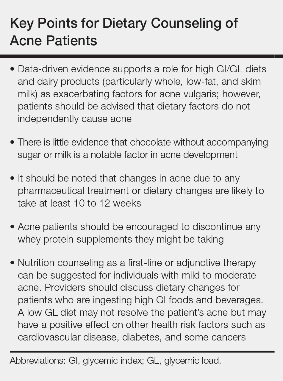
- Barbieri JS, Bhate K, Hartnett KP, et al. Trends in oral antibiotic prescription in dermatology, 2008 to 2016 [published online January 16, 2019]. JAMA Dermatol. doi:10.1001/jamadermatol.2018.4944.
- Barbieri JS, Spaccarelli N, Margolis DJ, et al. Approaches to limit systemic antibiotic use in acne: systemic alternatives, emerging topical therapies, dietary modification, and laser and light-based treatments. J Am Acad Dermatol. 2019;80:538-549.
- Melnik BC. Acne vulgaris: the metabolic syndrome of the pilosebaceous follicle [published online September 8, 2017]. Clin Dermatol. 2018;36:29-40.
- Lynn DD, Umari T, Dunnick CA, et al. The epidemiology of acne vulgaris in late adolescence. Adolesc Health Med Ther. 2016;7:13-25.
- Cordain L, Lindeberg S, Hurtado M, et al. Acne vulgaris: a disease of Western civilization. Arch Dermatol. 2002;138:1584-1590.
- Zaenglein AL, Pathy AL, Schlosser BJ, et al. Guidelines of care for the management of acne vulgaris [published online February 17, 2016]. J Am Acad Dermatol. 2016;74:945.e33-973.e33.
- Rezakovic´ S, Bukvic´ Mokos Z, Basta-Juzbašic´ A. Acne and diet: facts and controversies. Acta Dermatovenerol Croat. 2012;20:170-174.
- Mahmood NF, Shipman AR. The age-old problem of acne. Int J Womens Dermatol. 2017;3:71-76.
- Burris J, Shikany JM, Rietkerk W, et al. A low glycemic index and glycemic load diet decreases insulin-like growth factor-1 among adults with moderate and severe acne: a short-duration, 2-week randomized controlled trial. J Acad Nutr Diet. 2018;118:1874-1885.
- Atkinson FS, Foster-Powell K, Brand-Miller JC. International tables of glycemic index and glycemic load values: 2008 [published online October 3, 2008]. Diabetes Care. 2008;31:2281-2283.
- Smith RN, Braue A, Varigos GA, et al. The effect of a low glycemic load diet on acne vulgaris and the fatty acid composition of skin surface triglycerides. J Dermatol Sci. 2008;50:41-52
- Smith RN, Braue A, Varigos GA, et al. A low-glycemic-load diet improves symptoms in acne vulgaris patients: a randomized controlled trial. Am J Clin Nutr. 2007;86:107-115.
- Smith RN, Mann NJ, Braue A, et al. The effect of a high-protein, low glycemic-load diet versus a conventional, high glycemic-load diet on biochemical parameters associated with acne vulgaris: a randomized, investigator-masked, controlled trial. J Am Acad Dermatol. 2007;57:247-256.
- Kwon HH, Yoon JY, Hong JS, et al. Clinical and histological effect of a low glycaemic load diet in treatment of acne vulgaris in Korean patients: a randomized, controlled trial. Acta Derm Venereol. 2012;92:241-246.
- Burris J, Rietkerk W, Woolf K. Differences in dietary glycemic load and hormones in New York City adults with no and moderate/severe acne. J Acad Nutr Diet. 2017;117:1375-1383.
- Burris J, Rietkerk W, Woolf K. Relationships of self-reported dietary factors and perceived acne severity in a cohort of New York young adults [published online January 9, 2014]. J Acad Nutr Diet. 2014;114:384-392.
- Barbieri JS, Bhate K, Hartnett KP, et al. Trends in oral antibiotic prescription in dermatology, 2008 to 2016 [published online January 16, 2019]. JAMA Dermatol. 2019. doi:10.1001/jamadermatol.2018.4944.
- Lee JK, Smith AD. Metformin as an adjunct therapy for the treatment of moderate to severe acne vulgaris [published online November 15, 2017]. Dermatol Online J. 2017;23. pii:13030/qt53m2q13s.
- Robinson S, Kwan Z, Tang MM. Metformin as an adjunct therapy for the treatment of moderate to severe acne vulgaris: a randomized open-labeled study [published online May 1, 2019]. Dermatol Ther. 2019. doi:10.1111/dth.12953.
- Barbieri JS, Spaccarelli N, Margolis DJ, et al. Approaches to limitsystemic antibiotic use in acne: systemic alternatives, emerging topical therapies, dietary modification, and laser and light-based treatments [published online October 5, 2018]. J Am Acad Dermatol. 2019;80:538-549.
- Adebamowo CA, Spiegelman D, Berkey CS, et al. Milk consumption and acne in adolescent girls. Dermatol Online J. 2006;12:1.
- Adebamowo CA, Spiegelman D, Berkey CS, et al. Milk consumption and acne in teenaged boys. J Am Acad Dermatol. 2008;58:787-793.
- Adebamowo CA, Spiegelman D, Danby FW, et al. High school dietary dairy intake and teenage acne. J Am Acad Dermatol. 2005;52:207-214.
- Aghasi M, Golzarand M, Shab-Bidar S, et al. Dairy intake and acne development: a meta-analysis of observational studies. Clin Nutr. 2019;38:1067-1075.
- Can the right diet get rid of acne? American Academy of Dermatology website. https://www.aad.org/public/diseases/acne-and-rosacea/can-the-right-diet-get-rid-of-acne. Accessed June 13, 2019.
- Khanna R, Shifrin N, Nektalova T, et al. Diet and dermatology: Google search results for acne, psoriasis, and eczema. Cutis. 2018;102:44-46, 48.
Acne is a common condition that most often affects adolescents but is not uncommon in adults. It can result in considerable anxiety, depression, and medical and pharmaceutical costs. Additionally, oral antibiotics, the standard treatment for acne, are increasingly under suspicion for causing bacterial resistance as well as disruption of the cutaneous and gut microbiomes.1,2 These factors are among those that often drive patients and physicians to search for alternative and complementary treatments, including dietary modification.
Over the last few decades, the interaction between diet and acne has been one of the most fluid areas of research in dermatology. The role of diet in acne incidence and presentation has evolved from the general view in the 1970s that there was no connection to today’s more data-driven understanding that the acne disease course likely is modified by specific dietary components. Better designed and more rigorous studies have supported a link between acne severity and glycemic index (GI)/glycemic load (GL) and possibly dairy consumption. The ability to use data-driven evidence to counsel patients regarding dietary treatment of acne is increasingly important to counteract the pseudoadvice that patients can easily find on the Internet.
This article summarizes the history of beliefs about diet and acne, reviews more recent published data regarding dietary components that can modify acne severity, and outlines the current American Academy of Dermatology (AAD) guidelines and recommendations for diet and acne.
History of Diet and Acne
In most of the current literature, acne frequently is referred to as a disease of modern civilization or a consequence of the typical Western diet.3 For clarity, the Western diet is most commonly described as “a dietary regimen characterized by high amounts of sugary desserts, refined grains, high protein, high-fat dairy products, and high-sugar drinks.”4 The role of dairy in the etiology of acne typically is discussed separately from the Western diet. It has been reported that acne is not found in nonwesternized populations where a Paleolithic diet, which does not include consumption of high-GI carbohydrates, milk, or other dairy products, is common.5
Extending this line of argument, acne vulgaris has been called a metabolic syndrome of the sebaceous follicle and one of the mammalian target of rapamycin complex 1–driven diseases of civilization, along with cancer, obesity, and diabetes mellitus.3 This view seems somewhat extreme and discounts other drivers of acne incidence and severity. Twin studies have shown that acne is highly heritable, with 81% of the population variance attributed to genetic factors.6 Similar incidence numbers for acne vulgaris have been reported worldwide, and global incidence in late adolescence is rising; however, it is unknown whether this increase is a result of the adoption of the Western diet, which is thought to encourage early onset of puberty; genetic drift; changes in regional and cultural understanding and reporting of acne; or a byproduct of unknown environmental factors.4 More nuanced views acknowledge that acne is a multifactorial disease,7 and therefore genetic and possibly epigenetic factors as well as the cutaneous and gut microbiomes also must be taken into account. An interesting historical perspective on acne by Mahmood and Shipman8 outlined acne descriptions, diagnoses, topical treatments, and dietary advice going back to ancient Greek and Egyptian civilizations. They also cited recommendations from the 1930s that suggested avoiding “starchy foods, bread rolls, noodles, spaghetti, potatoes, oily nuts, chop suey, chow mein, and waffles” and listed the following foods as suitable to cure acne: “cooked and raw fruit, farina, rice, wheat, oatmeal, green vegetables, boiled or broiled meat and poultry, clear soup, vegetable soup, and an abundance of water.”8
More Recent Evidence of Dietary Influence on Acne
Importantly, the available research does not demonstrate that diet causes acne but rather that it may influence or aggravate existing acne. Data collection for acne studies also can be confounded by the interplay of many factors, such as increased access to health care, socioeconomic status, and shifting cultural perceptions of skin care and beauty.4 An important facet of any therapeutic recommendation is that it should be supported by confirmable mechanistic pathways.
GI and GL
Over the last few decades, a number of observational and intervention studies have focused on the possible influence of the GI/GL of foods on acne incidence and/or severity. A high GI diet is characterized by a relatively high intake of carbohydrate-containing foods that are quickly digested and absorbed, increasing blood glucose and insulin concentrations. Glycemic load takes the portion size of dietary carbohydrates into consideration and therefore is a measure of both the quality and quantity of carbohydrate-containing foods.9 TheGI/GL values of more than 2480 food items are available in the literature.10
Evidence from several studies supports the role of high GI/GL diets in exacerbating acne and suggests that transitioning to low GI/GL diets may lead to decreased lesion counts after 12 weeks.11-13 In one randomized controlled trial, male participants aged 15 to 25 years with mild to moderate facial acne were instructed either to eat a high protein/low GI diet or a conventional high GL control diet.13 After 12 weeks, total lesion counts had decreased more in the low GI diet group than the control. As partial confirmation of a mechanistic pathway for a high GI diet and acne, the low GI group demonstrated lower free androgen index and insulin levels than the control group.13 In a Korean study, a 10-week low GL regimen led to a reduction in acne lesion count, a decrease in sebaceous gland size, decreased inflammation, and reduced expression of sterol regulatory element-binding protein 1 and IL-8.14
More recent studies have further solidified the role of high GI/GL diets in acne severity.9,15,16 High GI/GL diets are believed to stimulate acne pathways by stimulating insulinlike growth factor 1 (IGF-1), which induces proliferation of both keratinocytes and sebocytes and simulates androgen production.17 An excellent diagram showing the connection between high GI diets (and dairy) and IGF-1, insulin and its receptors, androgen and its receptors, mammalian target of rapamycin, and the pilosebaceous unit was published in the literature in 2016.4 Interestingly, metformin has been shown to be an effective adjunctive therapy in the treatment of moderate to severe acne vulgaris.18,19
Milk and Dairy Consumption
Milk consumption also has been examined for its potential role in the pathogenesis of acne, including its ability to increase insulin and IGF-1 levels and bind to the human IGF-1 receptor as well as the fact that it contains bovine IGF-1 and dihydrotestosterone precursors.20 Although not studied quite as extensively or rigorously as GI/GL, consumption of milk and dairy products does appear to have the potential to exacerbate acne lesions. Beginning with a series of retrospective and prospective epidemiologic studies published from 2005 to 2008,21-23 a link between clinical acne and milk or dairy consumption in adolescent subjects was reported. A recent meta-analysis found a positive relationship between dairy, total milk, whole milk, low-fat milk, and skim milk consumption and acne occurrence but no significant association between yogurt/cheese consumption and acne development.24
AAD Guidelines
In their public forum, the AAD has advised that a low-glycemic diet may reduce the number of lesions in acne patients and highlighted data from around the world that support the concept that a high-glycemic diet and dairy are correlated with acne severity. They stated that consumption of milk—whole, low fat, and skim—may be linked to an increase in acne breakouts but that no studies have found that products made from milk, such as yogurt or cheese, lead to more breakouts.25
Other Considerations
Acne can be a serious quality-of-life issue with considerable psychological distress, physical morbidity, and social prejudice.9 Consequently, acne patients may be more willing to accept nonprofessional treatment advice, and there is no shortage of non–health care “experts” willing to provide an array of unfounded and fantastical advice. Dietary recommendations found online range from specific “miracle” foods to the more data-driven suggestions to “avoid dairy” or “eat low GI foods.” An important study recently published in Cutis concluded that most of the information found online regarding diet and acne is unfounded and/or misleading.26
Two additional reasons for recommending that acne patients consider dietary modification are not directly related to the disease: (1) the general health benefits of a lower GI/GL diet, and (2) the potential for decreasing the use of antibiotics. Antibiotic resistance is a growing problem across medicine, and dermatologists prescribe more antibiotics per provider than any other specialty.17 Dietary modification, where appropriate, could provide an approach to limiting the use of antibiotics in acne.
Final Thoughts
When advising acne patients, dermatologists can refer to the Table for general guidelines that incorporate the most current data-driven information on the relationship between diet and acne. Dietary modification, of course, will not work for all but can be safely recommended in cases of mild to moderate acne.
Acne is a common condition that most often affects adolescents but is not uncommon in adults. It can result in considerable anxiety, depression, and medical and pharmaceutical costs. Additionally, oral antibiotics, the standard treatment for acne, are increasingly under suspicion for causing bacterial resistance as well as disruption of the cutaneous and gut microbiomes.1,2 These factors are among those that often drive patients and physicians to search for alternative and complementary treatments, including dietary modification.
Over the last few decades, the interaction between diet and acne has been one of the most fluid areas of research in dermatology. The role of diet in acne incidence and presentation has evolved from the general view in the 1970s that there was no connection to today’s more data-driven understanding that the acne disease course likely is modified by specific dietary components. Better designed and more rigorous studies have supported a link between acne severity and glycemic index (GI)/glycemic load (GL) and possibly dairy consumption. The ability to use data-driven evidence to counsel patients regarding dietary treatment of acne is increasingly important to counteract the pseudoadvice that patients can easily find on the Internet.
This article summarizes the history of beliefs about diet and acne, reviews more recent published data regarding dietary components that can modify acne severity, and outlines the current American Academy of Dermatology (AAD) guidelines and recommendations for diet and acne.
History of Diet and Acne
In most of the current literature, acne frequently is referred to as a disease of modern civilization or a consequence of the typical Western diet.3 For clarity, the Western diet is most commonly described as “a dietary regimen characterized by high amounts of sugary desserts, refined grains, high protein, high-fat dairy products, and high-sugar drinks.”4 The role of dairy in the etiology of acne typically is discussed separately from the Western diet. It has been reported that acne is not found in nonwesternized populations where a Paleolithic diet, which does not include consumption of high-GI carbohydrates, milk, or other dairy products, is common.5
Extending this line of argument, acne vulgaris has been called a metabolic syndrome of the sebaceous follicle and one of the mammalian target of rapamycin complex 1–driven diseases of civilization, along with cancer, obesity, and diabetes mellitus.3 This view seems somewhat extreme and discounts other drivers of acne incidence and severity. Twin studies have shown that acne is highly heritable, with 81% of the population variance attributed to genetic factors.6 Similar incidence numbers for acne vulgaris have been reported worldwide, and global incidence in late adolescence is rising; however, it is unknown whether this increase is a result of the adoption of the Western diet, which is thought to encourage early onset of puberty; genetic drift; changes in regional and cultural understanding and reporting of acne; or a byproduct of unknown environmental factors.4 More nuanced views acknowledge that acne is a multifactorial disease,7 and therefore genetic and possibly epigenetic factors as well as the cutaneous and gut microbiomes also must be taken into account. An interesting historical perspective on acne by Mahmood and Shipman8 outlined acne descriptions, diagnoses, topical treatments, and dietary advice going back to ancient Greek and Egyptian civilizations. They also cited recommendations from the 1930s that suggested avoiding “starchy foods, bread rolls, noodles, spaghetti, potatoes, oily nuts, chop suey, chow mein, and waffles” and listed the following foods as suitable to cure acne: “cooked and raw fruit, farina, rice, wheat, oatmeal, green vegetables, boiled or broiled meat and poultry, clear soup, vegetable soup, and an abundance of water.”8
More Recent Evidence of Dietary Influence on Acne
Importantly, the available research does not demonstrate that diet causes acne but rather that it may influence or aggravate existing acne. Data collection for acne studies also can be confounded by the interplay of many factors, such as increased access to health care, socioeconomic status, and shifting cultural perceptions of skin care and beauty.4 An important facet of any therapeutic recommendation is that it should be supported by confirmable mechanistic pathways.
GI and GL
Over the last few decades, a number of observational and intervention studies have focused on the possible influence of the GI/GL of foods on acne incidence and/or severity. A high GI diet is characterized by a relatively high intake of carbohydrate-containing foods that are quickly digested and absorbed, increasing blood glucose and insulin concentrations. Glycemic load takes the portion size of dietary carbohydrates into consideration and therefore is a measure of both the quality and quantity of carbohydrate-containing foods.9 TheGI/GL values of more than 2480 food items are available in the literature.10
Evidence from several studies supports the role of high GI/GL diets in exacerbating acne and suggests that transitioning to low GI/GL diets may lead to decreased lesion counts after 12 weeks.11-13 In one randomized controlled trial, male participants aged 15 to 25 years with mild to moderate facial acne were instructed either to eat a high protein/low GI diet or a conventional high GL control diet.13 After 12 weeks, total lesion counts had decreased more in the low GI diet group than the control. As partial confirmation of a mechanistic pathway for a high GI diet and acne, the low GI group demonstrated lower free androgen index and insulin levels than the control group.13 In a Korean study, a 10-week low GL regimen led to a reduction in acne lesion count, a decrease in sebaceous gland size, decreased inflammation, and reduced expression of sterol regulatory element-binding protein 1 and IL-8.14
More recent studies have further solidified the role of high GI/GL diets in acne severity.9,15,16 High GI/GL diets are believed to stimulate acne pathways by stimulating insulinlike growth factor 1 (IGF-1), which induces proliferation of both keratinocytes and sebocytes and simulates androgen production.17 An excellent diagram showing the connection between high GI diets (and dairy) and IGF-1, insulin and its receptors, androgen and its receptors, mammalian target of rapamycin, and the pilosebaceous unit was published in the literature in 2016.4 Interestingly, metformin has been shown to be an effective adjunctive therapy in the treatment of moderate to severe acne vulgaris.18,19
Milk and Dairy Consumption
Milk consumption also has been examined for its potential role in the pathogenesis of acne, including its ability to increase insulin and IGF-1 levels and bind to the human IGF-1 receptor as well as the fact that it contains bovine IGF-1 and dihydrotestosterone precursors.20 Although not studied quite as extensively or rigorously as GI/GL, consumption of milk and dairy products does appear to have the potential to exacerbate acne lesions. Beginning with a series of retrospective and prospective epidemiologic studies published from 2005 to 2008,21-23 a link between clinical acne and milk or dairy consumption in adolescent subjects was reported. A recent meta-analysis found a positive relationship between dairy, total milk, whole milk, low-fat milk, and skim milk consumption and acne occurrence but no significant association between yogurt/cheese consumption and acne development.24
AAD Guidelines
In their public forum, the AAD has advised that a low-glycemic diet may reduce the number of lesions in acne patients and highlighted data from around the world that support the concept that a high-glycemic diet and dairy are correlated with acne severity. They stated that consumption of milk—whole, low fat, and skim—may be linked to an increase in acne breakouts but that no studies have found that products made from milk, such as yogurt or cheese, lead to more breakouts.25
Other Considerations
Acne can be a serious quality-of-life issue with considerable psychological distress, physical morbidity, and social prejudice.9 Consequently, acne patients may be more willing to accept nonprofessional treatment advice, and there is no shortage of non–health care “experts” willing to provide an array of unfounded and fantastical advice. Dietary recommendations found online range from specific “miracle” foods to the more data-driven suggestions to “avoid dairy” or “eat low GI foods.” An important study recently published in Cutis concluded that most of the information found online regarding diet and acne is unfounded and/or misleading.26
Two additional reasons for recommending that acne patients consider dietary modification are not directly related to the disease: (1) the general health benefits of a lower GI/GL diet, and (2) the potential for decreasing the use of antibiotics. Antibiotic resistance is a growing problem across medicine, and dermatologists prescribe more antibiotics per provider than any other specialty.17 Dietary modification, where appropriate, could provide an approach to limiting the use of antibiotics in acne.
Final Thoughts
When advising acne patients, dermatologists can refer to the Table for general guidelines that incorporate the most current data-driven information on the relationship between diet and acne. Dietary modification, of course, will not work for all but can be safely recommended in cases of mild to moderate acne.
- Barbieri JS, Bhate K, Hartnett KP, et al. Trends in oral antibiotic prescription in dermatology, 2008 to 2016 [published online January 16, 2019]. JAMA Dermatol. doi:10.1001/jamadermatol.2018.4944.
- Barbieri JS, Spaccarelli N, Margolis DJ, et al. Approaches to limit systemic antibiotic use in acne: systemic alternatives, emerging topical therapies, dietary modification, and laser and light-based treatments. J Am Acad Dermatol. 2019;80:538-549.
- Melnik BC. Acne vulgaris: the metabolic syndrome of the pilosebaceous follicle [published online September 8, 2017]. Clin Dermatol. 2018;36:29-40.
- Lynn DD, Umari T, Dunnick CA, et al. The epidemiology of acne vulgaris in late adolescence. Adolesc Health Med Ther. 2016;7:13-25.
- Cordain L, Lindeberg S, Hurtado M, et al. Acne vulgaris: a disease of Western civilization. Arch Dermatol. 2002;138:1584-1590.
- Zaenglein AL, Pathy AL, Schlosser BJ, et al. Guidelines of care for the management of acne vulgaris [published online February 17, 2016]. J Am Acad Dermatol. 2016;74:945.e33-973.e33.
- Rezakovic´ S, Bukvic´ Mokos Z, Basta-Juzbašic´ A. Acne and diet: facts and controversies. Acta Dermatovenerol Croat. 2012;20:170-174.
- Mahmood NF, Shipman AR. The age-old problem of acne. Int J Womens Dermatol. 2017;3:71-76.
- Burris J, Shikany JM, Rietkerk W, et al. A low glycemic index and glycemic load diet decreases insulin-like growth factor-1 among adults with moderate and severe acne: a short-duration, 2-week randomized controlled trial. J Acad Nutr Diet. 2018;118:1874-1885.
- Atkinson FS, Foster-Powell K, Brand-Miller JC. International tables of glycemic index and glycemic load values: 2008 [published online October 3, 2008]. Diabetes Care. 2008;31:2281-2283.
- Smith RN, Braue A, Varigos GA, et al. The effect of a low glycemic load diet on acne vulgaris and the fatty acid composition of skin surface triglycerides. J Dermatol Sci. 2008;50:41-52
- Smith RN, Braue A, Varigos GA, et al. A low-glycemic-load diet improves symptoms in acne vulgaris patients: a randomized controlled trial. Am J Clin Nutr. 2007;86:107-115.
- Smith RN, Mann NJ, Braue A, et al. The effect of a high-protein, low glycemic-load diet versus a conventional, high glycemic-load diet on biochemical parameters associated with acne vulgaris: a randomized, investigator-masked, controlled trial. J Am Acad Dermatol. 2007;57:247-256.
- Kwon HH, Yoon JY, Hong JS, et al. Clinical and histological effect of a low glycaemic load diet in treatment of acne vulgaris in Korean patients: a randomized, controlled trial. Acta Derm Venereol. 2012;92:241-246.
- Burris J, Rietkerk W, Woolf K. Differences in dietary glycemic load and hormones in New York City adults with no and moderate/severe acne. J Acad Nutr Diet. 2017;117:1375-1383.
- Burris J, Rietkerk W, Woolf K. Relationships of self-reported dietary factors and perceived acne severity in a cohort of New York young adults [published online January 9, 2014]. J Acad Nutr Diet. 2014;114:384-392.
- Barbieri JS, Bhate K, Hartnett KP, et al. Trends in oral antibiotic prescription in dermatology, 2008 to 2016 [published online January 16, 2019]. JAMA Dermatol. 2019. doi:10.1001/jamadermatol.2018.4944.
- Lee JK, Smith AD. Metformin as an adjunct therapy for the treatment of moderate to severe acne vulgaris [published online November 15, 2017]. Dermatol Online J. 2017;23. pii:13030/qt53m2q13s.
- Robinson S, Kwan Z, Tang MM. Metformin as an adjunct therapy for the treatment of moderate to severe acne vulgaris: a randomized open-labeled study [published online May 1, 2019]. Dermatol Ther. 2019. doi:10.1111/dth.12953.
- Barbieri JS, Spaccarelli N, Margolis DJ, et al. Approaches to limitsystemic antibiotic use in acne: systemic alternatives, emerging topical therapies, dietary modification, and laser and light-based treatments [published online October 5, 2018]. J Am Acad Dermatol. 2019;80:538-549.
- Adebamowo CA, Spiegelman D, Berkey CS, et al. Milk consumption and acne in adolescent girls. Dermatol Online J. 2006;12:1.
- Adebamowo CA, Spiegelman D, Berkey CS, et al. Milk consumption and acne in teenaged boys. J Am Acad Dermatol. 2008;58:787-793.
- Adebamowo CA, Spiegelman D, Danby FW, et al. High school dietary dairy intake and teenage acne. J Am Acad Dermatol. 2005;52:207-214.
- Aghasi M, Golzarand M, Shab-Bidar S, et al. Dairy intake and acne development: a meta-analysis of observational studies. Clin Nutr. 2019;38:1067-1075.
- Can the right diet get rid of acne? American Academy of Dermatology website. https://www.aad.org/public/diseases/acne-and-rosacea/can-the-right-diet-get-rid-of-acne. Accessed June 13, 2019.
- Khanna R, Shifrin N, Nektalova T, et al. Diet and dermatology: Google search results for acne, psoriasis, and eczema. Cutis. 2018;102:44-46, 48.
- Barbieri JS, Bhate K, Hartnett KP, et al. Trends in oral antibiotic prescription in dermatology, 2008 to 2016 [published online January 16, 2019]. JAMA Dermatol. doi:10.1001/jamadermatol.2018.4944.
- Barbieri JS, Spaccarelli N, Margolis DJ, et al. Approaches to limit systemic antibiotic use in acne: systemic alternatives, emerging topical therapies, dietary modification, and laser and light-based treatments. J Am Acad Dermatol. 2019;80:538-549.
- Melnik BC. Acne vulgaris: the metabolic syndrome of the pilosebaceous follicle [published online September 8, 2017]. Clin Dermatol. 2018;36:29-40.
- Lynn DD, Umari T, Dunnick CA, et al. The epidemiology of acne vulgaris in late adolescence. Adolesc Health Med Ther. 2016;7:13-25.
- Cordain L, Lindeberg S, Hurtado M, et al. Acne vulgaris: a disease of Western civilization. Arch Dermatol. 2002;138:1584-1590.
- Zaenglein AL, Pathy AL, Schlosser BJ, et al. Guidelines of care for the management of acne vulgaris [published online February 17, 2016]. J Am Acad Dermatol. 2016;74:945.e33-973.e33.
- Rezakovic´ S, Bukvic´ Mokos Z, Basta-Juzbašic´ A. Acne and diet: facts and controversies. Acta Dermatovenerol Croat. 2012;20:170-174.
- Mahmood NF, Shipman AR. The age-old problem of acne. Int J Womens Dermatol. 2017;3:71-76.
- Burris J, Shikany JM, Rietkerk W, et al. A low glycemic index and glycemic load diet decreases insulin-like growth factor-1 among adults with moderate and severe acne: a short-duration, 2-week randomized controlled trial. J Acad Nutr Diet. 2018;118:1874-1885.
- Atkinson FS, Foster-Powell K, Brand-Miller JC. International tables of glycemic index and glycemic load values: 2008 [published online October 3, 2008]. Diabetes Care. 2008;31:2281-2283.
- Smith RN, Braue A, Varigos GA, et al. The effect of a low glycemic load diet on acne vulgaris and the fatty acid composition of skin surface triglycerides. J Dermatol Sci. 2008;50:41-52
- Smith RN, Braue A, Varigos GA, et al. A low-glycemic-load diet improves symptoms in acne vulgaris patients: a randomized controlled trial. Am J Clin Nutr. 2007;86:107-115.
- Smith RN, Mann NJ, Braue A, et al. The effect of a high-protein, low glycemic-load diet versus a conventional, high glycemic-load diet on biochemical parameters associated with acne vulgaris: a randomized, investigator-masked, controlled trial. J Am Acad Dermatol. 2007;57:247-256.
- Kwon HH, Yoon JY, Hong JS, et al. Clinical and histological effect of a low glycaemic load diet in treatment of acne vulgaris in Korean patients: a randomized, controlled trial. Acta Derm Venereol. 2012;92:241-246.
- Burris J, Rietkerk W, Woolf K. Differences in dietary glycemic load and hormones in New York City adults with no and moderate/severe acne. J Acad Nutr Diet. 2017;117:1375-1383.
- Burris J, Rietkerk W, Woolf K. Relationships of self-reported dietary factors and perceived acne severity in a cohort of New York young adults [published online January 9, 2014]. J Acad Nutr Diet. 2014;114:384-392.
- Barbieri JS, Bhate K, Hartnett KP, et al. Trends in oral antibiotic prescription in dermatology, 2008 to 2016 [published online January 16, 2019]. JAMA Dermatol. 2019. doi:10.1001/jamadermatol.2018.4944.
- Lee JK, Smith AD. Metformin as an adjunct therapy for the treatment of moderate to severe acne vulgaris [published online November 15, 2017]. Dermatol Online J. 2017;23. pii:13030/qt53m2q13s.
- Robinson S, Kwan Z, Tang MM. Metformin as an adjunct therapy for the treatment of moderate to severe acne vulgaris: a randomized open-labeled study [published online May 1, 2019]. Dermatol Ther. 2019. doi:10.1111/dth.12953.
- Barbieri JS, Spaccarelli N, Margolis DJ, et al. Approaches to limitsystemic antibiotic use in acne: systemic alternatives, emerging topical therapies, dietary modification, and laser and light-based treatments [published online October 5, 2018]. J Am Acad Dermatol. 2019;80:538-549.
- Adebamowo CA, Spiegelman D, Berkey CS, et al. Milk consumption and acne in adolescent girls. Dermatol Online J. 2006;12:1.
- Adebamowo CA, Spiegelman D, Berkey CS, et al. Milk consumption and acne in teenaged boys. J Am Acad Dermatol. 2008;58:787-793.
- Adebamowo CA, Spiegelman D, Danby FW, et al. High school dietary dairy intake and teenage acne. J Am Acad Dermatol. 2005;52:207-214.
- Aghasi M, Golzarand M, Shab-Bidar S, et al. Dairy intake and acne development: a meta-analysis of observational studies. Clin Nutr. 2019;38:1067-1075.
- Can the right diet get rid of acne? American Academy of Dermatology website. https://www.aad.org/public/diseases/acne-and-rosacea/can-the-right-diet-get-rid-of-acne. Accessed June 13, 2019.
- Khanna R, Shifrin N, Nektalova T, et al. Diet and dermatology: Google search results for acne, psoriasis, and eczema. Cutis. 2018;102:44-46, 48.