User login
Mortality higher in older adults hospitalized for IBD
Adults older than 65 years with inflammatory bowel diseases (IBD) had significantly higher rates of inpatient mortality, compared with those younger than 65 years, independent of factors including disease severity, based on data from more than 200,000 hospital admissions.
Older adults use a disproportionate share of health care resources, but data on outcomes among hospitalized older adults with gastrointestinal illness are limited, Jeffrey Schwartz, MD, of Beth Israel Deaconess Medical Center and Harvard Medical School, both in Boston, and colleagues wrote in the Journal of Clinical Gastroenterology.
“In particular, there remains a significant concern that elderly patients are more susceptible to the development of opportunistic infections and malignancy in the setting of biological therapy, which has evolved into the standard of care for IBD over the past 10 years,” they wrote.
In their study, the researchers identified 162,800 hospital admissions for Crohn’s disease and 96,450 admissions for ulcerative colitis. Of these, 20% and 30%, respectively, were older than 65 years, which the researchers designated as the geriatric group.
In a multivariate analysis, age older than 65 years was significantly associated with increased mortality in both Crohn’s disease (odds ratio, 3.47; 95% confidence interval, 2.72-4.44; P < .001) and ulcerative colitis (OR, 2.75; 95% CI, 2.16-3.49; P < .001). The association was independent of factors included comorbidities, admission type, hospital type, inpatient surgery, and IBD subtype.
The most frequent cause of death in both groups across all ages and disease subtypes was infections (approximately 80% for all groups). The total hospital length of stay was significantly longer for geriatric patients, compared with younger patients with Crohn’s disease, in multivariate analysis (average increase, 0.19 days; P = .009). The total charges also were significantly higher among geriatric Crohn’s disease patients, compared with younger patients (average increase, $2,467; P = .012). No significant differences in hospital stay or total charges appeared between geriatric and younger patients with ulcerative colitis.
The study findings were limited by several factors such as the inclusion of older patients with IBD who were hospitalized for other reasons and by the potential for increased mortality because of comorbidities among elderly patients, the researchers noted. However, the findings support the limited data from similar previous studies and showed greater inpatient mortality for older adults with IBD, compared with hospital inpatients overall.
“Given the high prevalence of IBD patients that require inpatient admission, as well as the rapidly aging nature of the U.S. population, further studies are needed targeting geriatric patients with UC [ulcerative colitis] and CD [Crohn’s disease] to improve their overall management and quality of care to determine if this mortality risk can be reduced,” they concluded.
Tune in to risks in older adults
The study is important because the percentage of the population older than 65 years has been increasing; “at the same time, we are seeing more elderly patients being newly diagnosed with Crohn’s disease and ulcerative colitis,” said Russell D. Cohen, MD, of the University of Chicago, in an interview. “These patients are more vulnerable to complications of the diseases, such as infections, as well as complications from the medications used to treat these diseases.” However, older adults are often excluded from clinical trials and even from many observational studies in IBD, he noted.
“We have known from past studies that infections such as sepsis are a leading cause of death in our IBD patients,” said Dr. Cohen. “It is also understandable that those patients who have had complicated courses and those with other comorbidities have a higher mortality rate. However, what was surprising in the current study is that, even when the authors controlled for these factors, the geriatric patients still had two and three-quarters to three and a half times the mortality than those who were younger.”
The take-home message for clinicians is that “the geriatric patient with IBD is at a much higher rate for inpatient mortality, most commonly from infectious complications, than younger patients,” Dr. Cohen emphasized. “Quicker attention to what may seem minor but could become a potentially life-threatening infection is imperative. Caution with the use of multiple immune suppressing medications in older patients is paramount, as is timely surgical intervention in IBD patients in whom medications simply are not working.”
Focus research on infection prevention, cost burden
“More research should be directed at finding out whether these deadly infections could be prevented, perhaps by preventative ‘prophylactic’ antibiotics in the elderly patients, especially those on multiple immunosuppressive agents,” said Dr. Cohen. “In addition, research into the undue cost burden that these patients place on our health care system and counter that with better access to the newer, safer biological therapies [most of which Medicare does not cover] rather than corticosteroids.”
The study received no outside funding. The researchers had no financial conflicts to disclose. Dr. Cohen disclosed relationships with multiple companies including AbbVie, Bristol-Myers Squibb/Celgene, Eli Lilly, Gilead Sciences, Janssen, Pfizer, Takeda, and UCB Pharma.
SOURCE: Schwartz J et al. J Clin Gastroenterol. 2020 Nov 23. doi: 10.1097/MCG.0000000000001458.
Adults older than 65 years with inflammatory bowel diseases (IBD) had significantly higher rates of inpatient mortality, compared with those younger than 65 years, independent of factors including disease severity, based on data from more than 200,000 hospital admissions.
Older adults use a disproportionate share of health care resources, but data on outcomes among hospitalized older adults with gastrointestinal illness are limited, Jeffrey Schwartz, MD, of Beth Israel Deaconess Medical Center and Harvard Medical School, both in Boston, and colleagues wrote in the Journal of Clinical Gastroenterology.
“In particular, there remains a significant concern that elderly patients are more susceptible to the development of opportunistic infections and malignancy in the setting of biological therapy, which has evolved into the standard of care for IBD over the past 10 years,” they wrote.
In their study, the researchers identified 162,800 hospital admissions for Crohn’s disease and 96,450 admissions for ulcerative colitis. Of these, 20% and 30%, respectively, were older than 65 years, which the researchers designated as the geriatric group.
In a multivariate analysis, age older than 65 years was significantly associated with increased mortality in both Crohn’s disease (odds ratio, 3.47; 95% confidence interval, 2.72-4.44; P < .001) and ulcerative colitis (OR, 2.75; 95% CI, 2.16-3.49; P < .001). The association was independent of factors included comorbidities, admission type, hospital type, inpatient surgery, and IBD subtype.
The most frequent cause of death in both groups across all ages and disease subtypes was infections (approximately 80% for all groups). The total hospital length of stay was significantly longer for geriatric patients, compared with younger patients with Crohn’s disease, in multivariate analysis (average increase, 0.19 days; P = .009). The total charges also were significantly higher among geriatric Crohn’s disease patients, compared with younger patients (average increase, $2,467; P = .012). No significant differences in hospital stay or total charges appeared between geriatric and younger patients with ulcerative colitis.
The study findings were limited by several factors such as the inclusion of older patients with IBD who were hospitalized for other reasons and by the potential for increased mortality because of comorbidities among elderly patients, the researchers noted. However, the findings support the limited data from similar previous studies and showed greater inpatient mortality for older adults with IBD, compared with hospital inpatients overall.
“Given the high prevalence of IBD patients that require inpatient admission, as well as the rapidly aging nature of the U.S. population, further studies are needed targeting geriatric patients with UC [ulcerative colitis] and CD [Crohn’s disease] to improve their overall management and quality of care to determine if this mortality risk can be reduced,” they concluded.
Tune in to risks in older adults
The study is important because the percentage of the population older than 65 years has been increasing; “at the same time, we are seeing more elderly patients being newly diagnosed with Crohn’s disease and ulcerative colitis,” said Russell D. Cohen, MD, of the University of Chicago, in an interview. “These patients are more vulnerable to complications of the diseases, such as infections, as well as complications from the medications used to treat these diseases.” However, older adults are often excluded from clinical trials and even from many observational studies in IBD, he noted.
“We have known from past studies that infections such as sepsis are a leading cause of death in our IBD patients,” said Dr. Cohen. “It is also understandable that those patients who have had complicated courses and those with other comorbidities have a higher mortality rate. However, what was surprising in the current study is that, even when the authors controlled for these factors, the geriatric patients still had two and three-quarters to three and a half times the mortality than those who were younger.”
The take-home message for clinicians is that “the geriatric patient with IBD is at a much higher rate for inpatient mortality, most commonly from infectious complications, than younger patients,” Dr. Cohen emphasized. “Quicker attention to what may seem minor but could become a potentially life-threatening infection is imperative. Caution with the use of multiple immune suppressing medications in older patients is paramount, as is timely surgical intervention in IBD patients in whom medications simply are not working.”
Focus research on infection prevention, cost burden
“More research should be directed at finding out whether these deadly infections could be prevented, perhaps by preventative ‘prophylactic’ antibiotics in the elderly patients, especially those on multiple immunosuppressive agents,” said Dr. Cohen. “In addition, research into the undue cost burden that these patients place on our health care system and counter that with better access to the newer, safer biological therapies [most of which Medicare does not cover] rather than corticosteroids.”
The study received no outside funding. The researchers had no financial conflicts to disclose. Dr. Cohen disclosed relationships with multiple companies including AbbVie, Bristol-Myers Squibb/Celgene, Eli Lilly, Gilead Sciences, Janssen, Pfizer, Takeda, and UCB Pharma.
SOURCE: Schwartz J et al. J Clin Gastroenterol. 2020 Nov 23. doi: 10.1097/MCG.0000000000001458.
Adults older than 65 years with inflammatory bowel diseases (IBD) had significantly higher rates of inpatient mortality, compared with those younger than 65 years, independent of factors including disease severity, based on data from more than 200,000 hospital admissions.
Older adults use a disproportionate share of health care resources, but data on outcomes among hospitalized older adults with gastrointestinal illness are limited, Jeffrey Schwartz, MD, of Beth Israel Deaconess Medical Center and Harvard Medical School, both in Boston, and colleagues wrote in the Journal of Clinical Gastroenterology.
“In particular, there remains a significant concern that elderly patients are more susceptible to the development of opportunistic infections and malignancy in the setting of biological therapy, which has evolved into the standard of care for IBD over the past 10 years,” they wrote.
In their study, the researchers identified 162,800 hospital admissions for Crohn’s disease and 96,450 admissions for ulcerative colitis. Of these, 20% and 30%, respectively, were older than 65 years, which the researchers designated as the geriatric group.
In a multivariate analysis, age older than 65 years was significantly associated with increased mortality in both Crohn’s disease (odds ratio, 3.47; 95% confidence interval, 2.72-4.44; P < .001) and ulcerative colitis (OR, 2.75; 95% CI, 2.16-3.49; P < .001). The association was independent of factors included comorbidities, admission type, hospital type, inpatient surgery, and IBD subtype.
The most frequent cause of death in both groups across all ages and disease subtypes was infections (approximately 80% for all groups). The total hospital length of stay was significantly longer for geriatric patients, compared with younger patients with Crohn’s disease, in multivariate analysis (average increase, 0.19 days; P = .009). The total charges also were significantly higher among geriatric Crohn’s disease patients, compared with younger patients (average increase, $2,467; P = .012). No significant differences in hospital stay or total charges appeared between geriatric and younger patients with ulcerative colitis.
The study findings were limited by several factors such as the inclusion of older patients with IBD who were hospitalized for other reasons and by the potential for increased mortality because of comorbidities among elderly patients, the researchers noted. However, the findings support the limited data from similar previous studies and showed greater inpatient mortality for older adults with IBD, compared with hospital inpatients overall.
“Given the high prevalence of IBD patients that require inpatient admission, as well as the rapidly aging nature of the U.S. population, further studies are needed targeting geriatric patients with UC [ulcerative colitis] and CD [Crohn’s disease] to improve their overall management and quality of care to determine if this mortality risk can be reduced,” they concluded.
Tune in to risks in older adults
The study is important because the percentage of the population older than 65 years has been increasing; “at the same time, we are seeing more elderly patients being newly diagnosed with Crohn’s disease and ulcerative colitis,” said Russell D. Cohen, MD, of the University of Chicago, in an interview. “These patients are more vulnerable to complications of the diseases, such as infections, as well as complications from the medications used to treat these diseases.” However, older adults are often excluded from clinical trials and even from many observational studies in IBD, he noted.
“We have known from past studies that infections such as sepsis are a leading cause of death in our IBD patients,” said Dr. Cohen. “It is also understandable that those patients who have had complicated courses and those with other comorbidities have a higher mortality rate. However, what was surprising in the current study is that, even when the authors controlled for these factors, the geriatric patients still had two and three-quarters to three and a half times the mortality than those who were younger.”
The take-home message for clinicians is that “the geriatric patient with IBD is at a much higher rate for inpatient mortality, most commonly from infectious complications, than younger patients,” Dr. Cohen emphasized. “Quicker attention to what may seem minor but could become a potentially life-threatening infection is imperative. Caution with the use of multiple immune suppressing medications in older patients is paramount, as is timely surgical intervention in IBD patients in whom medications simply are not working.”
Focus research on infection prevention, cost burden
“More research should be directed at finding out whether these deadly infections could be prevented, perhaps by preventative ‘prophylactic’ antibiotics in the elderly patients, especially those on multiple immunosuppressive agents,” said Dr. Cohen. “In addition, research into the undue cost burden that these patients place on our health care system and counter that with better access to the newer, safer biological therapies [most of which Medicare does not cover] rather than corticosteroids.”
The study received no outside funding. The researchers had no financial conflicts to disclose. Dr. Cohen disclosed relationships with multiple companies including AbbVie, Bristol-Myers Squibb/Celgene, Eli Lilly, Gilead Sciences, Janssen, Pfizer, Takeda, and UCB Pharma.
SOURCE: Schwartz J et al. J Clin Gastroenterol. 2020 Nov 23. doi: 10.1097/MCG.0000000000001458.
FROM THE JOURNAL OF CLINICAL GASTROENTEROLOGY
Obesity, hypoxia predict severity in children with COVID-19
based on data from 281 patients at 8 locations.
Manifestations of COVID-19 in children include respiratory disease similar to that seen in adults, but the full spectrum of disease in children has been studied mainly in single settings or with a focus on one clinical manifestation, wrote Danielle M. Fernandes, MD, of Albert Einstein College of Medicine, New York, and colleagues.
In a study published in the Journal of Pediatrics, the researchers identified 281 children hospitalized with COVID-19 and/or multisystem inflammatory syndrome in children (MIS-C) at 8 sites in Connecticut, New Jersey, and New York. A total of 143 (51%) had respiratory disease, 69 (25%) had MIS-C, and 69 (25%) had other manifestations of illness including 32 patients with gastrointestinal problems, 21 infants with fever, 6 cases of neurologic disease, 6 cases of diabetic ketoacidosis, and 4 patients with other indications. The median age of the patients was 10 years, 60% were male, 51% were Hispanic, and 23% were non-Hispanic Black. The most common comorbidities were obesity (34%) and asthma (14%).
Independent predictors of disease severity in children found
After controlling for multiple variables, obesity and hypoxia at hospital admission were significant independent predictors of severe respiratory disease, with odds ratios of 3.39 and 4.01, respectively. In addition, lower absolute lymphocyte count (OR, 8.33 per unit decrease in 109 cells/L) and higher C-reactive protein (OR, 1.06 per unit increase in mg/dL) were significantly predictive of severe MIS-C (P = .001 and P = .017, respectively).
“The association between weight and severe respiratory COVID-19 is consistent with the adult literature; however, the mechanisms of this association require further study,” Dr. Fernandes and associates noted.
Overall, children with MIS-C were significantly more likely to be non-Hispanic Black, compared with children with respiratory disease, an 18% difference. However, neither race/ethnicity nor socioeconomic status were significant predictors of disease severity, the researchers wrote.
During the study period, 7 patients (2%) died and 114 (41%) were admitted to the ICU.
“We found a wide array of clinical manifestations in children and youth hospitalized with SARS-CoV-2,” Dr. Fernandes and associates wrote. Notably, gastrointestinal symptoms, ocular symptoms, and dermatologic symptoms have rarely been noted in adults with COVID-19, but occurred in more than 30% of the pediatric patients.
“We also found that SARS-CoV-2 can be an incidental finding in a substantial number of hospitalized pediatric patients,” the researchers said.
The findings were limited by several factors including a population of patients only from Connecticut, New Jersey, and New York, and the possibility that decisions on hospital and ICU admission may have varied by location, the researchers said. In addition, approaches may have varied in the absence of data on the optimal treatment of MIS-C.
“This study builds on the growing body of evidence showing that mortality in hospitalized pediatric patients is low, compared with adults,” Dr. Fernandes and associates said. “However, it highlights that the young population is not universally spared from morbidity, and that even previously healthy children and youth can develop severe disease requiring supportive therapy.”
Findings confirm other clinical experience
The study was important to show that, “although most children are spared severe illness from COVID-19, some children are hospitalized both with acute COVID-19 respiratory disease, with MIS-C and with a range of other complications,” Adrienne Randolph, MD, of Boston Children’s Hospital and Harvard Medical School, Boston, said in an interview.
Dr. Randolph said she was not surprised by the study findings, “as we are also seeing these types of complications at Boston Children’s Hospital where I work.”
Additional research is needed on the outcomes of these patients, “especially the longer-term sequelae of having COVID-19 or MIS-C early in life,” she emphasized.
The take-home message to clinicians from the findings at this time is to be aware that children and adolescents can become severely ill from COVID-19–related complications, said Dr. Randolph. “Some of the laboratory values on presentation appear to be associated with disease severity.”
The study received no outside funding. The researchers had no financial conflicts to disclose. Dr. Randolph disclosed funding from the Centers for Disease Control and Prevention to lead the Overcoming COVID-19 Study in U.S. Children and Adults.
SOURCE: Fernandes DM et al. J Pediatr. 2020 Nov 13. doi: 10.1016/j.jpeds.2020.11.016.
based on data from 281 patients at 8 locations.
Manifestations of COVID-19 in children include respiratory disease similar to that seen in adults, but the full spectrum of disease in children has been studied mainly in single settings or with a focus on one clinical manifestation, wrote Danielle M. Fernandes, MD, of Albert Einstein College of Medicine, New York, and colleagues.
In a study published in the Journal of Pediatrics, the researchers identified 281 children hospitalized with COVID-19 and/or multisystem inflammatory syndrome in children (MIS-C) at 8 sites in Connecticut, New Jersey, and New York. A total of 143 (51%) had respiratory disease, 69 (25%) had MIS-C, and 69 (25%) had other manifestations of illness including 32 patients with gastrointestinal problems, 21 infants with fever, 6 cases of neurologic disease, 6 cases of diabetic ketoacidosis, and 4 patients with other indications. The median age of the patients was 10 years, 60% were male, 51% were Hispanic, and 23% were non-Hispanic Black. The most common comorbidities were obesity (34%) and asthma (14%).
Independent predictors of disease severity in children found
After controlling for multiple variables, obesity and hypoxia at hospital admission were significant independent predictors of severe respiratory disease, with odds ratios of 3.39 and 4.01, respectively. In addition, lower absolute lymphocyte count (OR, 8.33 per unit decrease in 109 cells/L) and higher C-reactive protein (OR, 1.06 per unit increase in mg/dL) were significantly predictive of severe MIS-C (P = .001 and P = .017, respectively).
“The association between weight and severe respiratory COVID-19 is consistent with the adult literature; however, the mechanisms of this association require further study,” Dr. Fernandes and associates noted.
Overall, children with MIS-C were significantly more likely to be non-Hispanic Black, compared with children with respiratory disease, an 18% difference. However, neither race/ethnicity nor socioeconomic status were significant predictors of disease severity, the researchers wrote.
During the study period, 7 patients (2%) died and 114 (41%) were admitted to the ICU.
“We found a wide array of clinical manifestations in children and youth hospitalized with SARS-CoV-2,” Dr. Fernandes and associates wrote. Notably, gastrointestinal symptoms, ocular symptoms, and dermatologic symptoms have rarely been noted in adults with COVID-19, but occurred in more than 30% of the pediatric patients.
“We also found that SARS-CoV-2 can be an incidental finding in a substantial number of hospitalized pediatric patients,” the researchers said.
The findings were limited by several factors including a population of patients only from Connecticut, New Jersey, and New York, and the possibility that decisions on hospital and ICU admission may have varied by location, the researchers said. In addition, approaches may have varied in the absence of data on the optimal treatment of MIS-C.
“This study builds on the growing body of evidence showing that mortality in hospitalized pediatric patients is low, compared with adults,” Dr. Fernandes and associates said. “However, it highlights that the young population is not universally spared from morbidity, and that even previously healthy children and youth can develop severe disease requiring supportive therapy.”
Findings confirm other clinical experience
The study was important to show that, “although most children are spared severe illness from COVID-19, some children are hospitalized both with acute COVID-19 respiratory disease, with MIS-C and with a range of other complications,” Adrienne Randolph, MD, of Boston Children’s Hospital and Harvard Medical School, Boston, said in an interview.
Dr. Randolph said she was not surprised by the study findings, “as we are also seeing these types of complications at Boston Children’s Hospital where I work.”
Additional research is needed on the outcomes of these patients, “especially the longer-term sequelae of having COVID-19 or MIS-C early in life,” she emphasized.
The take-home message to clinicians from the findings at this time is to be aware that children and adolescents can become severely ill from COVID-19–related complications, said Dr. Randolph. “Some of the laboratory values on presentation appear to be associated with disease severity.”
The study received no outside funding. The researchers had no financial conflicts to disclose. Dr. Randolph disclosed funding from the Centers for Disease Control and Prevention to lead the Overcoming COVID-19 Study in U.S. Children and Adults.
SOURCE: Fernandes DM et al. J Pediatr. 2020 Nov 13. doi: 10.1016/j.jpeds.2020.11.016.
based on data from 281 patients at 8 locations.
Manifestations of COVID-19 in children include respiratory disease similar to that seen in adults, but the full spectrum of disease in children has been studied mainly in single settings or with a focus on one clinical manifestation, wrote Danielle M. Fernandes, MD, of Albert Einstein College of Medicine, New York, and colleagues.
In a study published in the Journal of Pediatrics, the researchers identified 281 children hospitalized with COVID-19 and/or multisystem inflammatory syndrome in children (MIS-C) at 8 sites in Connecticut, New Jersey, and New York. A total of 143 (51%) had respiratory disease, 69 (25%) had MIS-C, and 69 (25%) had other manifestations of illness including 32 patients with gastrointestinal problems, 21 infants with fever, 6 cases of neurologic disease, 6 cases of diabetic ketoacidosis, and 4 patients with other indications. The median age of the patients was 10 years, 60% were male, 51% were Hispanic, and 23% were non-Hispanic Black. The most common comorbidities were obesity (34%) and asthma (14%).
Independent predictors of disease severity in children found
After controlling for multiple variables, obesity and hypoxia at hospital admission were significant independent predictors of severe respiratory disease, with odds ratios of 3.39 and 4.01, respectively. In addition, lower absolute lymphocyte count (OR, 8.33 per unit decrease in 109 cells/L) and higher C-reactive protein (OR, 1.06 per unit increase in mg/dL) were significantly predictive of severe MIS-C (P = .001 and P = .017, respectively).
“The association between weight and severe respiratory COVID-19 is consistent with the adult literature; however, the mechanisms of this association require further study,” Dr. Fernandes and associates noted.
Overall, children with MIS-C were significantly more likely to be non-Hispanic Black, compared with children with respiratory disease, an 18% difference. However, neither race/ethnicity nor socioeconomic status were significant predictors of disease severity, the researchers wrote.
During the study period, 7 patients (2%) died and 114 (41%) were admitted to the ICU.
“We found a wide array of clinical manifestations in children and youth hospitalized with SARS-CoV-2,” Dr. Fernandes and associates wrote. Notably, gastrointestinal symptoms, ocular symptoms, and dermatologic symptoms have rarely been noted in adults with COVID-19, but occurred in more than 30% of the pediatric patients.
“We also found that SARS-CoV-2 can be an incidental finding in a substantial number of hospitalized pediatric patients,” the researchers said.
The findings were limited by several factors including a population of patients only from Connecticut, New Jersey, and New York, and the possibility that decisions on hospital and ICU admission may have varied by location, the researchers said. In addition, approaches may have varied in the absence of data on the optimal treatment of MIS-C.
“This study builds on the growing body of evidence showing that mortality in hospitalized pediatric patients is low, compared with adults,” Dr. Fernandes and associates said. “However, it highlights that the young population is not universally spared from morbidity, and that even previously healthy children and youth can develop severe disease requiring supportive therapy.”
Findings confirm other clinical experience
The study was important to show that, “although most children are spared severe illness from COVID-19, some children are hospitalized both with acute COVID-19 respiratory disease, with MIS-C and with a range of other complications,” Adrienne Randolph, MD, of Boston Children’s Hospital and Harvard Medical School, Boston, said in an interview.
Dr. Randolph said she was not surprised by the study findings, “as we are also seeing these types of complications at Boston Children’s Hospital where I work.”
Additional research is needed on the outcomes of these patients, “especially the longer-term sequelae of having COVID-19 or MIS-C early in life,” she emphasized.
The take-home message to clinicians from the findings at this time is to be aware that children and adolescents can become severely ill from COVID-19–related complications, said Dr. Randolph. “Some of the laboratory values on presentation appear to be associated with disease severity.”
The study received no outside funding. The researchers had no financial conflicts to disclose. Dr. Randolph disclosed funding from the Centers for Disease Control and Prevention to lead the Overcoming COVID-19 Study in U.S. Children and Adults.
SOURCE: Fernandes DM et al. J Pediatr. 2020 Nov 13. doi: 10.1016/j.jpeds.2020.11.016.
FROM THE JOURNAL OF PEDIATRICS
Risk associated with perioperative atrial fibrillation
Background: New-onset POAF occurs with 10% of noncardiac surgery and 15%-42% of cardiac surgery. POAF is believed to be self-limiting and most patients revert to sinus rhythm before hospital discharge. Previous studies on this topic are both limited and conflicting, but several suggest there is an association of stroke and mortality with POAF.
Study design: Systematic review and meta-analysis. Odds ratios with 95% confidence intervals were used for early outcomes and hazard ratios were used for long-term outcomes.
Setting: Prospective and retrospective cohort studies.
Synopsis: A total of 35 carefully selected studies were analyzed for a total of 2,458,010 patients. Outcomes of interest were early stroke or mortality within 30 days of surgery and long-term stroke or mortality after 30 days. The reference group was patients without POAF at baseline. Subgroup analysis included separating patients into cardiac surgery and noncardiac surgery.
New-onset POAF was associated with increased risk of early stroke (OR, 1.62; 95% CI, 1.47-1.80) and early mortality (OR, 1.44; 95% CI, 1.11-1.88). POAF also was associated with risk for long-term stroke (hazard ratio, 1.37; 95% CI, 1.07-1.77) and long-term mortality (HR, 1.37; 95% CI, 1.27-1.49). The risk of long-term stroke from new-onset POAF was highest among patients who received noncardiac surgery.
Despite identifying high-quality studies with thoughtful analysis, some data had the potential for publication bias. The representative sample did not report paroxysmal vs. persistent atrial fibrillation separately. Furthermore, the study had the potential to be confounded by detection bias of preexisting atrial fibrillation.
Bottom line: New-onset POAF is associated with early and long-term risk of stroke and mortality. Subsequent strategies to reduce this risk have yet to be determined.
Citation: Lin MH et al. Perioperative/postoperative atrial fibrillation and risk of subsequent stroke and/or mortality. Stroke. 2019 May;50:1364-71.
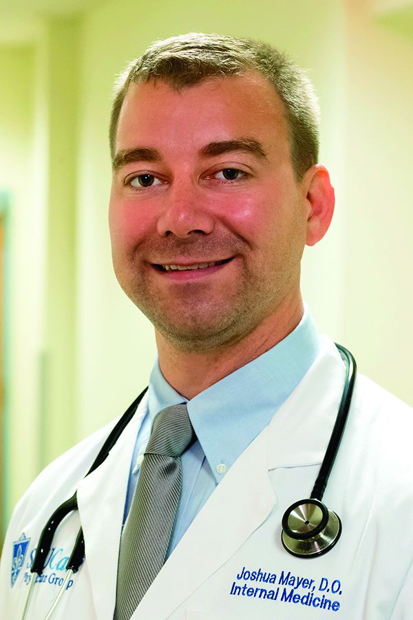
Dr. Mayer is a hospitalist and assistant professor of medicine at St. Louis University School of Medicine.
Background: New-onset POAF occurs with 10% of noncardiac surgery and 15%-42% of cardiac surgery. POAF is believed to be self-limiting and most patients revert to sinus rhythm before hospital discharge. Previous studies on this topic are both limited and conflicting, but several suggest there is an association of stroke and mortality with POAF.
Study design: Systematic review and meta-analysis. Odds ratios with 95% confidence intervals were used for early outcomes and hazard ratios were used for long-term outcomes.
Setting: Prospective and retrospective cohort studies.
Synopsis: A total of 35 carefully selected studies were analyzed for a total of 2,458,010 patients. Outcomes of interest were early stroke or mortality within 30 days of surgery and long-term stroke or mortality after 30 days. The reference group was patients without POAF at baseline. Subgroup analysis included separating patients into cardiac surgery and noncardiac surgery.
New-onset POAF was associated with increased risk of early stroke (OR, 1.62; 95% CI, 1.47-1.80) and early mortality (OR, 1.44; 95% CI, 1.11-1.88). POAF also was associated with risk for long-term stroke (hazard ratio, 1.37; 95% CI, 1.07-1.77) and long-term mortality (HR, 1.37; 95% CI, 1.27-1.49). The risk of long-term stroke from new-onset POAF was highest among patients who received noncardiac surgery.
Despite identifying high-quality studies with thoughtful analysis, some data had the potential for publication bias. The representative sample did not report paroxysmal vs. persistent atrial fibrillation separately. Furthermore, the study had the potential to be confounded by detection bias of preexisting atrial fibrillation.
Bottom line: New-onset POAF is associated with early and long-term risk of stroke and mortality. Subsequent strategies to reduce this risk have yet to be determined.
Citation: Lin MH et al. Perioperative/postoperative atrial fibrillation and risk of subsequent stroke and/or mortality. Stroke. 2019 May;50:1364-71.
Dr. Mayer is a hospitalist and assistant professor of medicine at St. Louis University School of Medicine.
Background: New-onset POAF occurs with 10% of noncardiac surgery and 15%-42% of cardiac surgery. POAF is believed to be self-limiting and most patients revert to sinus rhythm before hospital discharge. Previous studies on this topic are both limited and conflicting, but several suggest there is an association of stroke and mortality with POAF.
Study design: Systematic review and meta-analysis. Odds ratios with 95% confidence intervals were used for early outcomes and hazard ratios were used for long-term outcomes.
Setting: Prospective and retrospective cohort studies.
Synopsis: A total of 35 carefully selected studies were analyzed for a total of 2,458,010 patients. Outcomes of interest were early stroke or mortality within 30 days of surgery and long-term stroke or mortality after 30 days. The reference group was patients without POAF at baseline. Subgroup analysis included separating patients into cardiac surgery and noncardiac surgery.
New-onset POAF was associated with increased risk of early stroke (OR, 1.62; 95% CI, 1.47-1.80) and early mortality (OR, 1.44; 95% CI, 1.11-1.88). POAF also was associated with risk for long-term stroke (hazard ratio, 1.37; 95% CI, 1.07-1.77) and long-term mortality (HR, 1.37; 95% CI, 1.27-1.49). The risk of long-term stroke from new-onset POAF was highest among patients who received noncardiac surgery.
Despite identifying high-quality studies with thoughtful analysis, some data had the potential for publication bias. The representative sample did not report paroxysmal vs. persistent atrial fibrillation separately. Furthermore, the study had the potential to be confounded by detection bias of preexisting atrial fibrillation.
Bottom line: New-onset POAF is associated with early and long-term risk of stroke and mortality. Subsequent strategies to reduce this risk have yet to be determined.
Citation: Lin MH et al. Perioperative/postoperative atrial fibrillation and risk of subsequent stroke and/or mortality. Stroke. 2019 May;50:1364-71.
Dr. Mayer is a hospitalist and assistant professor of medicine at St. Louis University School of Medicine.
Age no barrier to weight loss in those with morbid obesity
Older adults should be recommended for hospital-based lifestyle interventions to reduce weight, say U.K. investigators after finding there was no difference in weight loss between older and younger individuals in their program for those with morbid obesity.
Thomas M. Barber, PhD, and colleagues looked back at nearly 250 randomly selected adults who attended their obesity service over an 11-year period.
Older individuals, defined as aged 60 years and over, had higher rates of type 2 diabetes but experienced a similar percentage weight loss and reduction in body mass index (BMI) as younger patients over the course of around 40 months.
“Age should be no barrier to lifestyle management of obesity,” said Dr. Barber, of University Hospitals Coventry (England) and Warwickshire, in a news release from his institution. “Rather than putting up barriers to older people accessing weight-loss programs, we should be proactively facilitating that process. To do otherwise would risk further and unnecessary neglect of older people through societal ageist misconceptions.”
He urged service providers and policy makers to “appreciate the importance of weight loss in older people with obesity for the maintenance of health and well-being and the facilitation of healthy aging. Furthermore, age per se should not contribute toward clinical decisions regarding the implementation of lifestyle management of older people.”
The research was published online Nov. 22 in Clinical Endocrinology.
Real-world data will inform clinical practice
Jason Halford, PhD, a professor of biological psychology and health behavior, said in an interview: “The fear is that older patients are perceived not to respond” to lifestyle interventions to control obesity, “and that’s clearly a fallacy, according to this study.”
The findings are strengthened by the fact that these are real-world data, “and so it will inform clinical practice,” he added.
And one of the “more interesting” findings was that [type 2] diabetes was “more prevalent” in the older group “but they’re still losing weight,” he noted.
“Traditionally it’s been thought that people with type 2 diabetes find it more difficult to lose weight because you’re trying to manage two conditions,” said Dr. Halford, of the University of Leeds (England), who is also president-elect of the European Association for the Study of Obesity.
Don’t discount older patients
The researchers note that many of the comorbidities associated with obesity “develop over time” and that “no one is immune to obesity,” regardless of their age, sex, ethnicity, and socioeconomic status.
Barber said there are “a number of reasons” why health care professionals “may discount weight loss in older people,” including “an ‘ageist’ perspective that weight-loss is not relevant to older people and misconceptions of reduced ability of older people to lose weight through dietary modification and increased exercise.”
And “older people may feel that hospital-based obesity services are not for them,” he noted.
To determine the effect of age on the ability to lose weight through lifestyle interventions, Dr. Barber and colleagues randomly selected 242 patients with morbid obesity who attended their hospital-based service between 2005 and 2016.
Of these, 167 were aged 18-60 years and 75 were aged 60 years and older. Most participants were women (75.4% of the younger patients and 60.0% of the older patients).
The proportion of patients with confirmed diabetes was markedly higher in the older group, compared with the younger group, at 62.7% versus 35.3%, although older patients had a significantly lower baseline BMI, at 46.9 versus 49.7 kg/m2 (P < .05).
The average duration of the lifestyle intervention was over 3 years (41.5 months) in the younger patients and 33.6 months in the older patients.
There was no significant difference in percentage weight loss between younger and older patients, at 6.9% and 7.3%, respectively, and no difference in percentage reduction in BMI, at 8.1% versus 7.8%.
Further analysis demonstrated that there was no significant correlation between age at referral to the hospital-based service and percentage weight loss (correlation coefficient, –0.13).
Dr. Halford said it would have been “useful” to know the proportion of patients achieving 5% and 10% weight loss because, if a third of patients lost more than 10% of their weight, “even in an elderly population, that would suggest there’d be real benefits in terms of things like type 2 diabetes,” he noted.
And he would like to have seen more data around how long participants had been struggling with obesity, as it’s “just an assumption that the second group is further down the path because they’re older, but we can’t be 100% sure.”
The team noted the study is limited by being retrospective and including a random selection of patients attending the service rather than the entire cohort.
Dr. Halford agreed but said the analysis is a “starting point” and could be used as a platform to conduct “much more systematic research on this area.”
No funding or relevant financial relationships were declared.
A version of this article originally appeared on Medscape.com.
Older adults should be recommended for hospital-based lifestyle interventions to reduce weight, say U.K. investigators after finding there was no difference in weight loss between older and younger individuals in their program for those with morbid obesity.
Thomas M. Barber, PhD, and colleagues looked back at nearly 250 randomly selected adults who attended their obesity service over an 11-year period.
Older individuals, defined as aged 60 years and over, had higher rates of type 2 diabetes but experienced a similar percentage weight loss and reduction in body mass index (BMI) as younger patients over the course of around 40 months.
“Age should be no barrier to lifestyle management of obesity,” said Dr. Barber, of University Hospitals Coventry (England) and Warwickshire, in a news release from his institution. “Rather than putting up barriers to older people accessing weight-loss programs, we should be proactively facilitating that process. To do otherwise would risk further and unnecessary neglect of older people through societal ageist misconceptions.”
He urged service providers and policy makers to “appreciate the importance of weight loss in older people with obesity for the maintenance of health and well-being and the facilitation of healthy aging. Furthermore, age per se should not contribute toward clinical decisions regarding the implementation of lifestyle management of older people.”
The research was published online Nov. 22 in Clinical Endocrinology.
Real-world data will inform clinical practice
Jason Halford, PhD, a professor of biological psychology and health behavior, said in an interview: “The fear is that older patients are perceived not to respond” to lifestyle interventions to control obesity, “and that’s clearly a fallacy, according to this study.”
The findings are strengthened by the fact that these are real-world data, “and so it will inform clinical practice,” he added.
And one of the “more interesting” findings was that [type 2] diabetes was “more prevalent” in the older group “but they’re still losing weight,” he noted.
“Traditionally it’s been thought that people with type 2 diabetes find it more difficult to lose weight because you’re trying to manage two conditions,” said Dr. Halford, of the University of Leeds (England), who is also president-elect of the European Association for the Study of Obesity.
Don’t discount older patients
The researchers note that many of the comorbidities associated with obesity “develop over time” and that “no one is immune to obesity,” regardless of their age, sex, ethnicity, and socioeconomic status.
Barber said there are “a number of reasons” why health care professionals “may discount weight loss in older people,” including “an ‘ageist’ perspective that weight-loss is not relevant to older people and misconceptions of reduced ability of older people to lose weight through dietary modification and increased exercise.”
And “older people may feel that hospital-based obesity services are not for them,” he noted.
To determine the effect of age on the ability to lose weight through lifestyle interventions, Dr. Barber and colleagues randomly selected 242 patients with morbid obesity who attended their hospital-based service between 2005 and 2016.
Of these, 167 were aged 18-60 years and 75 were aged 60 years and older. Most participants were women (75.4% of the younger patients and 60.0% of the older patients).
The proportion of patients with confirmed diabetes was markedly higher in the older group, compared with the younger group, at 62.7% versus 35.3%, although older patients had a significantly lower baseline BMI, at 46.9 versus 49.7 kg/m2 (P < .05).
The average duration of the lifestyle intervention was over 3 years (41.5 months) in the younger patients and 33.6 months in the older patients.
There was no significant difference in percentage weight loss between younger and older patients, at 6.9% and 7.3%, respectively, and no difference in percentage reduction in BMI, at 8.1% versus 7.8%.
Further analysis demonstrated that there was no significant correlation between age at referral to the hospital-based service and percentage weight loss (correlation coefficient, –0.13).
Dr. Halford said it would have been “useful” to know the proportion of patients achieving 5% and 10% weight loss because, if a third of patients lost more than 10% of their weight, “even in an elderly population, that would suggest there’d be real benefits in terms of things like type 2 diabetes,” he noted.
And he would like to have seen more data around how long participants had been struggling with obesity, as it’s “just an assumption that the second group is further down the path because they’re older, but we can’t be 100% sure.”
The team noted the study is limited by being retrospective and including a random selection of patients attending the service rather than the entire cohort.
Dr. Halford agreed but said the analysis is a “starting point” and could be used as a platform to conduct “much more systematic research on this area.”
No funding or relevant financial relationships were declared.
A version of this article originally appeared on Medscape.com.
Older adults should be recommended for hospital-based lifestyle interventions to reduce weight, say U.K. investigators after finding there was no difference in weight loss between older and younger individuals in their program for those with morbid obesity.
Thomas M. Barber, PhD, and colleagues looked back at nearly 250 randomly selected adults who attended their obesity service over an 11-year period.
Older individuals, defined as aged 60 years and over, had higher rates of type 2 diabetes but experienced a similar percentage weight loss and reduction in body mass index (BMI) as younger patients over the course of around 40 months.
“Age should be no barrier to lifestyle management of obesity,” said Dr. Barber, of University Hospitals Coventry (England) and Warwickshire, in a news release from his institution. “Rather than putting up barriers to older people accessing weight-loss programs, we should be proactively facilitating that process. To do otherwise would risk further and unnecessary neglect of older people through societal ageist misconceptions.”
He urged service providers and policy makers to “appreciate the importance of weight loss in older people with obesity for the maintenance of health and well-being and the facilitation of healthy aging. Furthermore, age per se should not contribute toward clinical decisions regarding the implementation of lifestyle management of older people.”
The research was published online Nov. 22 in Clinical Endocrinology.
Real-world data will inform clinical practice
Jason Halford, PhD, a professor of biological psychology and health behavior, said in an interview: “The fear is that older patients are perceived not to respond” to lifestyle interventions to control obesity, “and that’s clearly a fallacy, according to this study.”
The findings are strengthened by the fact that these are real-world data, “and so it will inform clinical practice,” he added.
And one of the “more interesting” findings was that [type 2] diabetes was “more prevalent” in the older group “but they’re still losing weight,” he noted.
“Traditionally it’s been thought that people with type 2 diabetes find it more difficult to lose weight because you’re trying to manage two conditions,” said Dr. Halford, of the University of Leeds (England), who is also president-elect of the European Association for the Study of Obesity.
Don’t discount older patients
The researchers note that many of the comorbidities associated with obesity “develop over time” and that “no one is immune to obesity,” regardless of their age, sex, ethnicity, and socioeconomic status.
Barber said there are “a number of reasons” why health care professionals “may discount weight loss in older people,” including “an ‘ageist’ perspective that weight-loss is not relevant to older people and misconceptions of reduced ability of older people to lose weight through dietary modification and increased exercise.”
And “older people may feel that hospital-based obesity services are not for them,” he noted.
To determine the effect of age on the ability to lose weight through lifestyle interventions, Dr. Barber and colleagues randomly selected 242 patients with morbid obesity who attended their hospital-based service between 2005 and 2016.
Of these, 167 were aged 18-60 years and 75 were aged 60 years and older. Most participants were women (75.4% of the younger patients and 60.0% of the older patients).
The proportion of patients with confirmed diabetes was markedly higher in the older group, compared with the younger group, at 62.7% versus 35.3%, although older patients had a significantly lower baseline BMI, at 46.9 versus 49.7 kg/m2 (P < .05).
The average duration of the lifestyle intervention was over 3 years (41.5 months) in the younger patients and 33.6 months in the older patients.
There was no significant difference in percentage weight loss between younger and older patients, at 6.9% and 7.3%, respectively, and no difference in percentage reduction in BMI, at 8.1% versus 7.8%.
Further analysis demonstrated that there was no significant correlation between age at referral to the hospital-based service and percentage weight loss (correlation coefficient, –0.13).
Dr. Halford said it would have been “useful” to know the proportion of patients achieving 5% and 10% weight loss because, if a third of patients lost more than 10% of their weight, “even in an elderly population, that would suggest there’d be real benefits in terms of things like type 2 diabetes,” he noted.
And he would like to have seen more data around how long participants had been struggling with obesity, as it’s “just an assumption that the second group is further down the path because they’re older, but we can’t be 100% sure.”
The team noted the study is limited by being retrospective and including a random selection of patients attending the service rather than the entire cohort.
Dr. Halford agreed but said the analysis is a “starting point” and could be used as a platform to conduct “much more systematic research on this area.”
No funding or relevant financial relationships were declared.
A version of this article originally appeared on Medscape.com.
Obesity phenotyping matches patients with more effective interventions
A phenotype-guided strategy for systematically matching weight-loss patients to their potentially ideal weight-loss drug roughly doubled treatment efficacy, compared with usual practice, in a single-center, randomized study with 268 patients.
In contrast, in 200 patients who received weight loss–drug therapy selected by routine means, 35% achieved greater than 10% loss compared with their starting weight, Andres J. Acosta, MD, said at the virtual ObesityWeek® Interactive 2020 meeting.
The phenotype-guided strategy also led to an average 16% weight loss from baseline after 12 months, compared with a 9% average loss among the usual-care controls, reported Dr. Acosta, a gastroenterologist at the Mayo Clinic in Rochester, Minn.
A “one-size-fits-all approach to weight loss treatment is not working,” he declared. “Our long-term goal is to develop a personalized approach to obesity management.”
Personalized weight loss treatment isn’t new
“The better we can match treatment to a patient’s needs, the more likely it will succeed. That’s not a brand new idea. They are trying to standardize the way that we classify the disorders that play a role in why a person gains weight or has trouble losing weight,” commented John D. Clark III, MD, an internal medicine physician and weight-management specialist at UT Southwestern Medical Center in Dallas.
The increased weight loss levels that Dr. Acosta reported in patients who underwent the study’s phenotyping protocol and received tailored treatment “are similar to the numbers we see when a patient’s treatment is the right fit for them. You see weight loss in these ranges,” Dr. Clark said in an interview.
The study run by Dr. Acosta and his associates consisted of two phases. First, they established normal and abnormal ranges for four different obesity phenotypes by studying 100 patients with obesity. The patients underwent an extensive and uniform workup designed to classify their obesity phenotype.
Four obesity phenotypes
The researchers categorized patients into one of four types:
- Disordered initial eating satiation, called ‘hungry brain,” and assessed by measuring food intake at a buffet, ad libidum meal.
- Disordered maintenance of satiety, called “hungry gut,” assessed by both a gastric-emptying study as well as patient self-assessment for postprandial fullness.
- “Emotional hunger,” assessed with two questionnaires.
- Disordered energy expenditure, called “slow burn,” assessed by measuring basal metabolic rate, and self-reports of both exercise and nonexercise activity.
Dr. Acosta estimated that the complete workup to assess all four potential phenotypes costs about $1,200.
The researchers then applied the 75th percentile value from each of these assessments to 450 patients with obesity in their clinic to see the prevalence of the four phenotypes. They identified a single phenotype in 58% of these patients, including 18% with hungry gut, 16% with hungry brain, 12% with emotional hunger, and 12% with slow burn. An additional 27% of the patients were positive for two or more phenotypes (including 9% who were positive for all four phenotypes), and 15% did not test positive for any of the four phenotypes.
Phenotype-guided treatments
They then applied their findings in a prospective randomized study that matched a drug intervention to each of the four phenotypes during a year-long, comprehensive weight-loss program at the Mayo Clinic’s Weight Management Clinic. The study randomized 100 patients to the phenotype-driven arm, with 68 of these patients receiving their assigned drug, and 200 patients served as controls. Patients averaged about 47 years old, and their average body mass index was about 41 kg/m2.
The investigational arm included 30 patients classified as having a hungry brain, with 20 of these patients treated with phentermine plus topiramate and 10 treated with lorcaserin (before it was withdrawn by the Food and Drug Administration); 12 with hungry gut and treated with liraglutide (Saxenda); 19 with emotional hunger who received naltrexone SR/bupropion SR (Contrave); and seven with slow burn who received phentermine.
The control arm included 200 patients seeking weight loss treatment at Mayo who did not undergo phenotyping and received their drug treatment based on their personal preference in consultation with their Mayo physician. In this group, drug treatment broke down as 106 patients (53%) on phentermine plus topiramate, 41 (21%) on liraglutide, 34 (17%) on phentermine alone, 14 (7%) on naltrexone SR/bupropion SR, and 5 patients (3%) on locaserin (percentages total 101% because of rounding).
Overall, phenotyping led to more patients treated with naltrexone SR/buproprion SR and lorcaserin and fewer treated with phentermine or phentermine and topiramate ER. All patients were eligible to also receive behavioral interventions as needed.
“We do a lot of testing to identify the phenotype,” in addition to gathering additional clues from a detailed history, said Dr. Acosta. Patients identified with more than one phenotype in routine practice at Mayo are often begun on more than one drug. When phenotyping fails to classify a patient, Dr. Acosta puts the patient on a low-calorie diet and then does a follow-up assessment “to see if the phenotype pops up as a metabolic adaptation.”
“This is something we’re all working toward” in the obesity management field. “How can we better identify the underlying causes in a way that can fit into the work flow. How can we move from research to things we can use daily in the clinic,” observed Dr. Clark. “We need a lot more investigation to determine how well this works in the real world. Are there other tools we can use that are not as expensive” as what Dr. Acosta used for this study?
“For this proof of concept study, it made sense to be very rigorous, but that probably is not realistic for every patient. What are other ways to get this information, or perhaps only use an extensive workup when initial weight loss attempts are unsuccessful,” Dr. Clark suggested.
A phenotype-guided strategy for systematically matching weight-loss patients to their potentially ideal weight-loss drug roughly doubled treatment efficacy, compared with usual practice, in a single-center, randomized study with 268 patients.
In contrast, in 200 patients who received weight loss–drug therapy selected by routine means, 35% achieved greater than 10% loss compared with their starting weight, Andres J. Acosta, MD, said at the virtual ObesityWeek® Interactive 2020 meeting.
The phenotype-guided strategy also led to an average 16% weight loss from baseline after 12 months, compared with a 9% average loss among the usual-care controls, reported Dr. Acosta, a gastroenterologist at the Mayo Clinic in Rochester, Minn.
A “one-size-fits-all approach to weight loss treatment is not working,” he declared. “Our long-term goal is to develop a personalized approach to obesity management.”
Personalized weight loss treatment isn’t new
“The better we can match treatment to a patient’s needs, the more likely it will succeed. That’s not a brand new idea. They are trying to standardize the way that we classify the disorders that play a role in why a person gains weight or has trouble losing weight,” commented John D. Clark III, MD, an internal medicine physician and weight-management specialist at UT Southwestern Medical Center in Dallas.
The increased weight loss levels that Dr. Acosta reported in patients who underwent the study’s phenotyping protocol and received tailored treatment “are similar to the numbers we see when a patient’s treatment is the right fit for them. You see weight loss in these ranges,” Dr. Clark said in an interview.
The study run by Dr. Acosta and his associates consisted of two phases. First, they established normal and abnormal ranges for four different obesity phenotypes by studying 100 patients with obesity. The patients underwent an extensive and uniform workup designed to classify their obesity phenotype.
Four obesity phenotypes
The researchers categorized patients into one of four types:
- Disordered initial eating satiation, called ‘hungry brain,” and assessed by measuring food intake at a buffet, ad libidum meal.
- Disordered maintenance of satiety, called “hungry gut,” assessed by both a gastric-emptying study as well as patient self-assessment for postprandial fullness.
- “Emotional hunger,” assessed with two questionnaires.
- Disordered energy expenditure, called “slow burn,” assessed by measuring basal metabolic rate, and self-reports of both exercise and nonexercise activity.
Dr. Acosta estimated that the complete workup to assess all four potential phenotypes costs about $1,200.
The researchers then applied the 75th percentile value from each of these assessments to 450 patients with obesity in their clinic to see the prevalence of the four phenotypes. They identified a single phenotype in 58% of these patients, including 18% with hungry gut, 16% with hungry brain, 12% with emotional hunger, and 12% with slow burn. An additional 27% of the patients were positive for two or more phenotypes (including 9% who were positive for all four phenotypes), and 15% did not test positive for any of the four phenotypes.
Phenotype-guided treatments
They then applied their findings in a prospective randomized study that matched a drug intervention to each of the four phenotypes during a year-long, comprehensive weight-loss program at the Mayo Clinic’s Weight Management Clinic. The study randomized 100 patients to the phenotype-driven arm, with 68 of these patients receiving their assigned drug, and 200 patients served as controls. Patients averaged about 47 years old, and their average body mass index was about 41 kg/m2.
The investigational arm included 30 patients classified as having a hungry brain, with 20 of these patients treated with phentermine plus topiramate and 10 treated with lorcaserin (before it was withdrawn by the Food and Drug Administration); 12 with hungry gut and treated with liraglutide (Saxenda); 19 with emotional hunger who received naltrexone SR/bupropion SR (Contrave); and seven with slow burn who received phentermine.
The control arm included 200 patients seeking weight loss treatment at Mayo who did not undergo phenotyping and received their drug treatment based on their personal preference in consultation with their Mayo physician. In this group, drug treatment broke down as 106 patients (53%) on phentermine plus topiramate, 41 (21%) on liraglutide, 34 (17%) on phentermine alone, 14 (7%) on naltrexone SR/bupropion SR, and 5 patients (3%) on locaserin (percentages total 101% because of rounding).
Overall, phenotyping led to more patients treated with naltrexone SR/buproprion SR and lorcaserin and fewer treated with phentermine or phentermine and topiramate ER. All patients were eligible to also receive behavioral interventions as needed.
“We do a lot of testing to identify the phenotype,” in addition to gathering additional clues from a detailed history, said Dr. Acosta. Patients identified with more than one phenotype in routine practice at Mayo are often begun on more than one drug. When phenotyping fails to classify a patient, Dr. Acosta puts the patient on a low-calorie diet and then does a follow-up assessment “to see if the phenotype pops up as a metabolic adaptation.”
“This is something we’re all working toward” in the obesity management field. “How can we better identify the underlying causes in a way that can fit into the work flow. How can we move from research to things we can use daily in the clinic,” observed Dr. Clark. “We need a lot more investigation to determine how well this works in the real world. Are there other tools we can use that are not as expensive” as what Dr. Acosta used for this study?
“For this proof of concept study, it made sense to be very rigorous, but that probably is not realistic for every patient. What are other ways to get this information, or perhaps only use an extensive workup when initial weight loss attempts are unsuccessful,” Dr. Clark suggested.
A phenotype-guided strategy for systematically matching weight-loss patients to their potentially ideal weight-loss drug roughly doubled treatment efficacy, compared with usual practice, in a single-center, randomized study with 268 patients.
In contrast, in 200 patients who received weight loss–drug therapy selected by routine means, 35% achieved greater than 10% loss compared with their starting weight, Andres J. Acosta, MD, said at the virtual ObesityWeek® Interactive 2020 meeting.
The phenotype-guided strategy also led to an average 16% weight loss from baseline after 12 months, compared with a 9% average loss among the usual-care controls, reported Dr. Acosta, a gastroenterologist at the Mayo Clinic in Rochester, Minn.
A “one-size-fits-all approach to weight loss treatment is not working,” he declared. “Our long-term goal is to develop a personalized approach to obesity management.”
Personalized weight loss treatment isn’t new
“The better we can match treatment to a patient’s needs, the more likely it will succeed. That’s not a brand new idea. They are trying to standardize the way that we classify the disorders that play a role in why a person gains weight or has trouble losing weight,” commented John D. Clark III, MD, an internal medicine physician and weight-management specialist at UT Southwestern Medical Center in Dallas.
The increased weight loss levels that Dr. Acosta reported in patients who underwent the study’s phenotyping protocol and received tailored treatment “are similar to the numbers we see when a patient’s treatment is the right fit for them. You see weight loss in these ranges,” Dr. Clark said in an interview.
The study run by Dr. Acosta and his associates consisted of two phases. First, they established normal and abnormal ranges for four different obesity phenotypes by studying 100 patients with obesity. The patients underwent an extensive and uniform workup designed to classify their obesity phenotype.
Four obesity phenotypes
The researchers categorized patients into one of four types:
- Disordered initial eating satiation, called ‘hungry brain,” and assessed by measuring food intake at a buffet, ad libidum meal.
- Disordered maintenance of satiety, called “hungry gut,” assessed by both a gastric-emptying study as well as patient self-assessment for postprandial fullness.
- “Emotional hunger,” assessed with two questionnaires.
- Disordered energy expenditure, called “slow burn,” assessed by measuring basal metabolic rate, and self-reports of both exercise and nonexercise activity.
Dr. Acosta estimated that the complete workup to assess all four potential phenotypes costs about $1,200.
The researchers then applied the 75th percentile value from each of these assessments to 450 patients with obesity in their clinic to see the prevalence of the four phenotypes. They identified a single phenotype in 58% of these patients, including 18% with hungry gut, 16% with hungry brain, 12% with emotional hunger, and 12% with slow burn. An additional 27% of the patients were positive for two or more phenotypes (including 9% who were positive for all four phenotypes), and 15% did not test positive for any of the four phenotypes.
Phenotype-guided treatments
They then applied their findings in a prospective randomized study that matched a drug intervention to each of the four phenotypes during a year-long, comprehensive weight-loss program at the Mayo Clinic’s Weight Management Clinic. The study randomized 100 patients to the phenotype-driven arm, with 68 of these patients receiving their assigned drug, and 200 patients served as controls. Patients averaged about 47 years old, and their average body mass index was about 41 kg/m2.
The investigational arm included 30 patients classified as having a hungry brain, with 20 of these patients treated with phentermine plus topiramate and 10 treated with lorcaserin (before it was withdrawn by the Food and Drug Administration); 12 with hungry gut and treated with liraglutide (Saxenda); 19 with emotional hunger who received naltrexone SR/bupropion SR (Contrave); and seven with slow burn who received phentermine.
The control arm included 200 patients seeking weight loss treatment at Mayo who did not undergo phenotyping and received their drug treatment based on their personal preference in consultation with their Mayo physician. In this group, drug treatment broke down as 106 patients (53%) on phentermine plus topiramate, 41 (21%) on liraglutide, 34 (17%) on phentermine alone, 14 (7%) on naltrexone SR/bupropion SR, and 5 patients (3%) on locaserin (percentages total 101% because of rounding).
Overall, phenotyping led to more patients treated with naltrexone SR/buproprion SR and lorcaserin and fewer treated with phentermine or phentermine and topiramate ER. All patients were eligible to also receive behavioral interventions as needed.
“We do a lot of testing to identify the phenotype,” in addition to gathering additional clues from a detailed history, said Dr. Acosta. Patients identified with more than one phenotype in routine practice at Mayo are often begun on more than one drug. When phenotyping fails to classify a patient, Dr. Acosta puts the patient on a low-calorie diet and then does a follow-up assessment “to see if the phenotype pops up as a metabolic adaptation.”
“This is something we’re all working toward” in the obesity management field. “How can we better identify the underlying causes in a way that can fit into the work flow. How can we move from research to things we can use daily in the clinic,” observed Dr. Clark. “We need a lot more investigation to determine how well this works in the real world. Are there other tools we can use that are not as expensive” as what Dr. Acosta used for this study?
“For this proof of concept study, it made sense to be very rigorous, but that probably is not realistic for every patient. What are other ways to get this information, or perhaps only use an extensive workup when initial weight loss attempts are unsuccessful,” Dr. Clark suggested.
FROM OBESITY WEEK 2020
SHM urges Congress to reverse changes in reimbursement rates under 2021 Medicare Physician Fee Schedule
Approximately 8% reduction in reimbursement for hospitalists
On Dec. 1, the Centers for Medicare & Medicaid Services (CMS) released the 2021 Medicare Physician Fee Schedule, which finalized proposed changes to Medicare reimbursement rates, including a significant negative budget neutrality adjustment. For hospitalists, the Society of Hospital Medicine estimates that the adjustment will amount to an estimated 8% reduction in Medicare reimbursement rates, which will go into effect on Jan. 1, 2021.

“These cuts are coming at the exact wrong time. During the chaos of 2020, when hospitalists have been essential to responding to the COVID-19 pandemic, they should not be met with a significant pay reduction in 2021,” said Eric E. Howell, MD, MHM, chief executive officer of the Society of Hospital Medicine. “While we at SHM support increasing pay for outpatient primary care, which is driving these cuts, we do not believe now is the right time to make significant adjustments to the Medicare Physician Fee Schedule. We now call on Congress to do the right thing for hospitalists and other frontline providers who have otherwise been lauded as heroes.”
SHM will continue to fight for hospitalists and to advocate to reverse these cuts. To send a message of support to your representatives, visit SHM’s Legislative Action Center and click on “Support the Holding Providers Harmless from Medicare Cuts During COVID-19 Act of 2020.” To learn more about and become involved with SHM’s advocacy efforts, visit hospitalmedicine.org/advocacy.
Approximately 8% reduction in reimbursement for hospitalists
Approximately 8% reduction in reimbursement for hospitalists
On Dec. 1, the Centers for Medicare & Medicaid Services (CMS) released the 2021 Medicare Physician Fee Schedule, which finalized proposed changes to Medicare reimbursement rates, including a significant negative budget neutrality adjustment. For hospitalists, the Society of Hospital Medicine estimates that the adjustment will amount to an estimated 8% reduction in Medicare reimbursement rates, which will go into effect on Jan. 1, 2021.
“These cuts are coming at the exact wrong time. During the chaos of 2020, when hospitalists have been essential to responding to the COVID-19 pandemic, they should not be met with a significant pay reduction in 2021,” said Eric E. Howell, MD, MHM, chief executive officer of the Society of Hospital Medicine. “While we at SHM support increasing pay for outpatient primary care, which is driving these cuts, we do not believe now is the right time to make significant adjustments to the Medicare Physician Fee Schedule. We now call on Congress to do the right thing for hospitalists and other frontline providers who have otherwise been lauded as heroes.”
SHM will continue to fight for hospitalists and to advocate to reverse these cuts. To send a message of support to your representatives, visit SHM’s Legislative Action Center and click on “Support the Holding Providers Harmless from Medicare Cuts During COVID-19 Act of 2020.” To learn more about and become involved with SHM’s advocacy efforts, visit hospitalmedicine.org/advocacy.
On Dec. 1, the Centers for Medicare & Medicaid Services (CMS) released the 2021 Medicare Physician Fee Schedule, which finalized proposed changes to Medicare reimbursement rates, including a significant negative budget neutrality adjustment. For hospitalists, the Society of Hospital Medicine estimates that the adjustment will amount to an estimated 8% reduction in Medicare reimbursement rates, which will go into effect on Jan. 1, 2021.
“These cuts are coming at the exact wrong time. During the chaos of 2020, when hospitalists have been essential to responding to the COVID-19 pandemic, they should not be met with a significant pay reduction in 2021,” said Eric E. Howell, MD, MHM, chief executive officer of the Society of Hospital Medicine. “While we at SHM support increasing pay for outpatient primary care, which is driving these cuts, we do not believe now is the right time to make significant adjustments to the Medicare Physician Fee Schedule. We now call on Congress to do the right thing for hospitalists and other frontline providers who have otherwise been lauded as heroes.”
SHM will continue to fight for hospitalists and to advocate to reverse these cuts. To send a message of support to your representatives, visit SHM’s Legislative Action Center and click on “Support the Holding Providers Harmless from Medicare Cuts During COVID-19 Act of 2020.” To learn more about and become involved with SHM’s advocacy efforts, visit hospitalmedicine.org/advocacy.

Leading hospitalists during a pandemic
As I write this, we are entering the third surge of the COVID-19 pandemic, with new cases, hospitalizations, and deaths from COVID-19 skyrocketing around the country. Worst of all, this surge has been most severely affecting areas of the nation least prepared to handle it (rural) and populations already marginalized by the health care system (Latinx and Black). Despite the onslaught of COVID-19, “pandemic fatigue” has begun to set in amongst colleagues, friends, and family, leading to challenges in adhering to social distancing and other infection-control measures, both at work and home.

In the face of the pandemic’s onslaught, hospitalists – who have faced the brunt of caring for patients with COVID-19, despite the absence of reporting about the subspecialty’s role – are faced with mustering the grit to respond with resolve, coordinated action, and empathy. Luckily, hospitalists are equipped with the very characteristics needed to lead teams, groups, and hospitals through the crisis of this pandemic. Ask yourself, why did you become a hospitalist? If you wanted steady predictability and control, there were many office-based specialties you could have chosen. You chose to become a hospitalist because you seek the challenges of clinical variety, problem-solving, systems improvement, and you are a natural team leader, whether you have been designated as such or not. In the words of John Quincy Adams, “if your actions inspire others to dream more, learn more, do more, and become more, you are a leader.”
As a leader, how can you lead your team through the series of trials and tribulations that this year has thrown at you? From COVID-19 to racism directed against Black and Latinx people to the behavioral health crisis, 2020 has likely made you feel as if you’re stuck in a ghoulish carnival fun house without an exit.
Yet this is where some leaders hit their stride, in what Bennis and Thomas describe as the “crucible of leadership.”1 There are many types of “crucibles of leadership,” according to Bennis and Thomas, and this year has thrown most of these at us: prejudice/bias, physical fatigue and illness, sudden elevation of responsibility to lead new processes, not to mention family stressors. Leaders who succeed in guiding their colleagues through these challenges have manifested critical skills: engaging others in shared meaning, having a distinctive and compelling voice, displaying integrity, and having adaptive capacity.
What exactly is adaptive capacity, the most important of these, in my opinion? Adaptive capacity requires understanding the new context of a crisis and how it has shifted team members’ needs and perceptions. It also requires what Bennis and Thomas call hardiness and what I call grit – the ability to face adversity, get knocked down, get up, and do it again.
There is probably no better example of a crisis leader with extraordinary adaptive capacity than Anglo-Irish explorer Sir Ernest Shackleton. Bitten by the bug of exploration, Shackleton failed at reaching the South Pole (1908-1909) but subsequently attempted to cross the Antarctic, departing South Georgia Island on Dec. 5, 1914. Depressingly for Shackleton, his ship, the Endurance, became stuck in sea ice on Jan. 19, 1915 before even reaching the continent. Drifting with the ice floe, his crew had set up a winter station hoping to be released from the ice later, but the Endurance was crushed by the pressure of sea ice and sank on Nov. 21, 1915. From there, Shackleton hoped to drift north to Paulet Island, 250 miles away, but eventually was forced to take his crew on lifeboats to the nearest land, Elephant Island, 346 miles from where the Endurance sank. He then took five of his men on an open boat, 828-mile journey to South Georgia Island. Encountering hurricane-force winds, the team landed on South Georgia Island 15 days later, only to face a climb of 32 miles over mountainous terrain to reach a whaling station. Shackleton eventually organized his men’s rescue on Elephant Island, reaching them on Aug. 30, 1916, 4½ months after he had set out for South Georgia Island. His entire crew survived, only to have two of them killed later in World War I.
You might consider Shackleton a failure for not even coming close to his original goal, but his success in saving his crew is regarded as the epitome of crisis leadership. As Harvard Business School professor Nancy F. Koehn, PhD, whose case study of Shackleton is one of the most popular at HBS, stated, “He thought he was going to be an entrepreneur of exploration, but he became an entrepreneur of survival.”2 Upon realizing the futility of his original mission, he pivoted immediately to the survival of his crew. “A man must shape himself to a new mark directly the old one goes to ground,” wrote Shackleton in his diary.3
Realizing that preserving his crew’s morale was critical, he maintained the crew’s everyday activities, despite the prospect of dying on the ice. He realized that he needed to keep up his own courage and confidence as well as that of his crew. Despite his ability to share the strategic focus of getting to safety with his men, he didn’t lose sight of day-to-day needs, such as keeping the crew entertained. When he encountered crew members who seemed problematic to his mission goals, he assigned them to his own tent.
Despite the extreme cold, his decision-making did not freeze – he acted decisively. He took risks when he thought appropriate, twice needing to abandon his efforts to drag a lifeboat full of supplies with his men toward the sea. “You can’t be afraid to make smart mistakes,” says Dr. Koehn. “That’s something we have no training in.”4 Most importantly, Shackleton took ultimate responsibility for his men’s survival, never resting until they had all been rescued. And he modeled a culture of shared responsibility for one another5 – he had once offered his only biscuit of the day on a prior expedition to his fellow explorer Frank Wild.
As winter arrives in 2020 and deepens into 2021, we will all be faced with leading our teams across the ice and to the safety of spring, and hopefully a vaccine. Whether we can get there with our entire crew depends on effective crisis leadership. But we can draw on the lessons provided by Shackleton and other crisis leaders in the past to guide us in the present.
Author disclosure: I studied the HBS case study “Leadership in Crisis: Ernest Shackleton and the Epic Voyage of the Endurance” as part of a 12-month certificate course in Safety, Quality, Informatics, and Leadership (SQIL) offered by Harvard Medical School.
Dr. Chang is chief of pediatric hospital medicine at Baystate Children’s Hospital in Springfield, Mass., and associate professor of pediatrics at the University of Massachusetts, also in Springfield.
References
1. HBR’s 10 must reads on leadership. Boston: Harvard Business Review Press, 2011.
2. Lagace M. Shackleton: An entrepreneur of survival. Harvard Business School. Working Knowledge website. Published 2003. Accessed 2020 Nov 19.
3. Koehn N. Leadership lessons from the Shackleton Expedition. The New York Times. 2011 Dec 25.
4. Potier B. Shackleton in business school. Harvard Public Affairs and Communications. The Harvard Gazette website. Published 2004. Accessed 2020 Nov 19.
5. Perkins D. 4 Lessons in crisis leadership from Shackleton’s expedition. In Leadership Essentials by HarpersCollins Leadership. Vol 2020. New York: HarpersCollins, 2020.
As I write this, we are entering the third surge of the COVID-19 pandemic, with new cases, hospitalizations, and deaths from COVID-19 skyrocketing around the country. Worst of all, this surge has been most severely affecting areas of the nation least prepared to handle it (rural) and populations already marginalized by the health care system (Latinx and Black). Despite the onslaught of COVID-19, “pandemic fatigue” has begun to set in amongst colleagues, friends, and family, leading to challenges in adhering to social distancing and other infection-control measures, both at work and home.
In the face of the pandemic’s onslaught, hospitalists – who have faced the brunt of caring for patients with COVID-19, despite the absence of reporting about the subspecialty’s role – are faced with mustering the grit to respond with resolve, coordinated action, and empathy. Luckily, hospitalists are equipped with the very characteristics needed to lead teams, groups, and hospitals through the crisis of this pandemic. Ask yourself, why did you become a hospitalist? If you wanted steady predictability and control, there were many office-based specialties you could have chosen. You chose to become a hospitalist because you seek the challenges of clinical variety, problem-solving, systems improvement, and you are a natural team leader, whether you have been designated as such or not. In the words of John Quincy Adams, “if your actions inspire others to dream more, learn more, do more, and become more, you are a leader.”
As a leader, how can you lead your team through the series of trials and tribulations that this year has thrown at you? From COVID-19 to racism directed against Black and Latinx people to the behavioral health crisis, 2020 has likely made you feel as if you’re stuck in a ghoulish carnival fun house without an exit.
Yet this is where some leaders hit their stride, in what Bennis and Thomas describe as the “crucible of leadership.”1 There are many types of “crucibles of leadership,” according to Bennis and Thomas, and this year has thrown most of these at us: prejudice/bias, physical fatigue and illness, sudden elevation of responsibility to lead new processes, not to mention family stressors. Leaders who succeed in guiding their colleagues through these challenges have manifested critical skills: engaging others in shared meaning, having a distinctive and compelling voice, displaying integrity, and having adaptive capacity.
What exactly is adaptive capacity, the most important of these, in my opinion? Adaptive capacity requires understanding the new context of a crisis and how it has shifted team members’ needs and perceptions. It also requires what Bennis and Thomas call hardiness and what I call grit – the ability to face adversity, get knocked down, get up, and do it again.
There is probably no better example of a crisis leader with extraordinary adaptive capacity than Anglo-Irish explorer Sir Ernest Shackleton. Bitten by the bug of exploration, Shackleton failed at reaching the South Pole (1908-1909) but subsequently attempted to cross the Antarctic, departing South Georgia Island on Dec. 5, 1914. Depressingly for Shackleton, his ship, the Endurance, became stuck in sea ice on Jan. 19, 1915 before even reaching the continent. Drifting with the ice floe, his crew had set up a winter station hoping to be released from the ice later, but the Endurance was crushed by the pressure of sea ice and sank on Nov. 21, 1915. From there, Shackleton hoped to drift north to Paulet Island, 250 miles away, but eventually was forced to take his crew on lifeboats to the nearest land, Elephant Island, 346 miles from where the Endurance sank. He then took five of his men on an open boat, 828-mile journey to South Georgia Island. Encountering hurricane-force winds, the team landed on South Georgia Island 15 days later, only to face a climb of 32 miles over mountainous terrain to reach a whaling station. Shackleton eventually organized his men’s rescue on Elephant Island, reaching them on Aug. 30, 1916, 4½ months after he had set out for South Georgia Island. His entire crew survived, only to have two of them killed later in World War I.
You might consider Shackleton a failure for not even coming close to his original goal, but his success in saving his crew is regarded as the epitome of crisis leadership. As Harvard Business School professor Nancy F. Koehn, PhD, whose case study of Shackleton is one of the most popular at HBS, stated, “He thought he was going to be an entrepreneur of exploration, but he became an entrepreneur of survival.”2 Upon realizing the futility of his original mission, he pivoted immediately to the survival of his crew. “A man must shape himself to a new mark directly the old one goes to ground,” wrote Shackleton in his diary.3
Realizing that preserving his crew’s morale was critical, he maintained the crew’s everyday activities, despite the prospect of dying on the ice. He realized that he needed to keep up his own courage and confidence as well as that of his crew. Despite his ability to share the strategic focus of getting to safety with his men, he didn’t lose sight of day-to-day needs, such as keeping the crew entertained. When he encountered crew members who seemed problematic to his mission goals, he assigned them to his own tent.
Despite the extreme cold, his decision-making did not freeze – he acted decisively. He took risks when he thought appropriate, twice needing to abandon his efforts to drag a lifeboat full of supplies with his men toward the sea. “You can’t be afraid to make smart mistakes,” says Dr. Koehn. “That’s something we have no training in.”4 Most importantly, Shackleton took ultimate responsibility for his men’s survival, never resting until they had all been rescued. And he modeled a culture of shared responsibility for one another5 – he had once offered his only biscuit of the day on a prior expedition to his fellow explorer Frank Wild.
As winter arrives in 2020 and deepens into 2021, we will all be faced with leading our teams across the ice and to the safety of spring, and hopefully a vaccine. Whether we can get there with our entire crew depends on effective crisis leadership. But we can draw on the lessons provided by Shackleton and other crisis leaders in the past to guide us in the present.
Author disclosure: I studied the HBS case study “Leadership in Crisis: Ernest Shackleton and the Epic Voyage of the Endurance” as part of a 12-month certificate course in Safety, Quality, Informatics, and Leadership (SQIL) offered by Harvard Medical School.
Dr. Chang is chief of pediatric hospital medicine at Baystate Children’s Hospital in Springfield, Mass., and associate professor of pediatrics at the University of Massachusetts, also in Springfield.
References
1. HBR’s 10 must reads on leadership. Boston: Harvard Business Review Press, 2011.
2. Lagace M. Shackleton: An entrepreneur of survival. Harvard Business School. Working Knowledge website. Published 2003. Accessed 2020 Nov 19.
3. Koehn N. Leadership lessons from the Shackleton Expedition. The New York Times. 2011 Dec 25.
4. Potier B. Shackleton in business school. Harvard Public Affairs and Communications. The Harvard Gazette website. Published 2004. Accessed 2020 Nov 19.
5. Perkins D. 4 Lessons in crisis leadership from Shackleton’s expedition. In Leadership Essentials by HarpersCollins Leadership. Vol 2020. New York: HarpersCollins, 2020.
As I write this, we are entering the third surge of the COVID-19 pandemic, with new cases, hospitalizations, and deaths from COVID-19 skyrocketing around the country. Worst of all, this surge has been most severely affecting areas of the nation least prepared to handle it (rural) and populations already marginalized by the health care system (Latinx and Black). Despite the onslaught of COVID-19, “pandemic fatigue” has begun to set in amongst colleagues, friends, and family, leading to challenges in adhering to social distancing and other infection-control measures, both at work and home.
In the face of the pandemic’s onslaught, hospitalists – who have faced the brunt of caring for patients with COVID-19, despite the absence of reporting about the subspecialty’s role – are faced with mustering the grit to respond with resolve, coordinated action, and empathy. Luckily, hospitalists are equipped with the very characteristics needed to lead teams, groups, and hospitals through the crisis of this pandemic. Ask yourself, why did you become a hospitalist? If you wanted steady predictability and control, there were many office-based specialties you could have chosen. You chose to become a hospitalist because you seek the challenges of clinical variety, problem-solving, systems improvement, and you are a natural team leader, whether you have been designated as such or not. In the words of John Quincy Adams, “if your actions inspire others to dream more, learn more, do more, and become more, you are a leader.”
As a leader, how can you lead your team through the series of trials and tribulations that this year has thrown at you? From COVID-19 to racism directed against Black and Latinx people to the behavioral health crisis, 2020 has likely made you feel as if you’re stuck in a ghoulish carnival fun house without an exit.
Yet this is where some leaders hit their stride, in what Bennis and Thomas describe as the “crucible of leadership.”1 There are many types of “crucibles of leadership,” according to Bennis and Thomas, and this year has thrown most of these at us: prejudice/bias, physical fatigue and illness, sudden elevation of responsibility to lead new processes, not to mention family stressors. Leaders who succeed in guiding their colleagues through these challenges have manifested critical skills: engaging others in shared meaning, having a distinctive and compelling voice, displaying integrity, and having adaptive capacity.
What exactly is adaptive capacity, the most important of these, in my opinion? Adaptive capacity requires understanding the new context of a crisis and how it has shifted team members’ needs and perceptions. It also requires what Bennis and Thomas call hardiness and what I call grit – the ability to face adversity, get knocked down, get up, and do it again.
There is probably no better example of a crisis leader with extraordinary adaptive capacity than Anglo-Irish explorer Sir Ernest Shackleton. Bitten by the bug of exploration, Shackleton failed at reaching the South Pole (1908-1909) but subsequently attempted to cross the Antarctic, departing South Georgia Island on Dec. 5, 1914. Depressingly for Shackleton, his ship, the Endurance, became stuck in sea ice on Jan. 19, 1915 before even reaching the continent. Drifting with the ice floe, his crew had set up a winter station hoping to be released from the ice later, but the Endurance was crushed by the pressure of sea ice and sank on Nov. 21, 1915. From there, Shackleton hoped to drift north to Paulet Island, 250 miles away, but eventually was forced to take his crew on lifeboats to the nearest land, Elephant Island, 346 miles from where the Endurance sank. He then took five of his men on an open boat, 828-mile journey to South Georgia Island. Encountering hurricane-force winds, the team landed on South Georgia Island 15 days later, only to face a climb of 32 miles over mountainous terrain to reach a whaling station. Shackleton eventually organized his men’s rescue on Elephant Island, reaching them on Aug. 30, 1916, 4½ months after he had set out for South Georgia Island. His entire crew survived, only to have two of them killed later in World War I.
You might consider Shackleton a failure for not even coming close to his original goal, but his success in saving his crew is regarded as the epitome of crisis leadership. As Harvard Business School professor Nancy F. Koehn, PhD, whose case study of Shackleton is one of the most popular at HBS, stated, “He thought he was going to be an entrepreneur of exploration, but he became an entrepreneur of survival.”2 Upon realizing the futility of his original mission, he pivoted immediately to the survival of his crew. “A man must shape himself to a new mark directly the old one goes to ground,” wrote Shackleton in his diary.3
Realizing that preserving his crew’s morale was critical, he maintained the crew’s everyday activities, despite the prospect of dying on the ice. He realized that he needed to keep up his own courage and confidence as well as that of his crew. Despite his ability to share the strategic focus of getting to safety with his men, he didn’t lose sight of day-to-day needs, such as keeping the crew entertained. When he encountered crew members who seemed problematic to his mission goals, he assigned them to his own tent.
Despite the extreme cold, his decision-making did not freeze – he acted decisively. He took risks when he thought appropriate, twice needing to abandon his efforts to drag a lifeboat full of supplies with his men toward the sea. “You can’t be afraid to make smart mistakes,” says Dr. Koehn. “That’s something we have no training in.”4 Most importantly, Shackleton took ultimate responsibility for his men’s survival, never resting until they had all been rescued. And he modeled a culture of shared responsibility for one another5 – he had once offered his only biscuit of the day on a prior expedition to his fellow explorer Frank Wild.
As winter arrives in 2020 and deepens into 2021, we will all be faced with leading our teams across the ice and to the safety of spring, and hopefully a vaccine. Whether we can get there with our entire crew depends on effective crisis leadership. But we can draw on the lessons provided by Shackleton and other crisis leaders in the past to guide us in the present.
Author disclosure: I studied the HBS case study “Leadership in Crisis: Ernest Shackleton and the Epic Voyage of the Endurance” as part of a 12-month certificate course in Safety, Quality, Informatics, and Leadership (SQIL) offered by Harvard Medical School.
Dr. Chang is chief of pediatric hospital medicine at Baystate Children’s Hospital in Springfield, Mass., and associate professor of pediatrics at the University of Massachusetts, also in Springfield.
References
1. HBR’s 10 must reads on leadership. Boston: Harvard Business Review Press, 2011.
2. Lagace M. Shackleton: An entrepreneur of survival. Harvard Business School. Working Knowledge website. Published 2003. Accessed 2020 Nov 19.
3. Koehn N. Leadership lessons from the Shackleton Expedition. The New York Times. 2011 Dec 25.
4. Potier B. Shackleton in business school. Harvard Public Affairs and Communications. The Harvard Gazette website. Published 2004. Accessed 2020 Nov 19.
5. Perkins D. 4 Lessons in crisis leadership from Shackleton’s expedition. In Leadership Essentials by HarpersCollins Leadership. Vol 2020. New York: HarpersCollins, 2020.
Are we shortchanging patients with obesity?
Every Wednesday evening after supper, I record in a marble notebook some anthropomorphic measurements: my weight taken first thing Monday morning and my waist circumference. I also add how I did with exercise since the previous week’s entry and some comments about sleep, energy, and nutrition.
My personal log now comprises dozens of pages. To my surprise, the first entry was 5 years ago to the month. The earlier entries were far from weekly and contained a lot of narrative on how my food-restriction scheme that month was being violated.
Looking just at the numbers, I did about as well as a control group participant in any medical study of diet modification. Until just a few months ago, there was no trend in either weight or waist circumference over those 5 years, including 2 years of retirement. But it wasn’t for lack of trying. Keeping the journal for as long as I have – and recently, as consistently as I have – suggests serious intent but inadequate execution of the same principles I offered patients, who rarely did much better. But recent studies suggest that perhaps quite a few could.
Are we underestimating our patients’ potential?
A recent abstract from the European and International Congress on Obesity suggests that the impressions clinicians get from our office encounters may leave us underestimating the potential for our patients to lose enough weight to move them from one level of risk to another.
Using a national database of primary care visits, the investigators isolated about 550,000 records. Of these, about 60,000 (11%) had records showing weight reductions of 10%-25% (mean, 13%) over at least 4 years. Weight loss was by intent rather than from illness. The remaining individuals maintained their weight within 5% of the first measurement for the duration of the study.
Participants with stable body weight were compared with the successful weight reducers. This analysis showed that the risk for type 2 diabetes, osteoarthritis, sleep apnea, hypertension, and dyslipidemia all measurably declined in weight reducers. This held true whether the patient’s baseline body mass index (BMI) showed modest or severe obesity. Patients with the highest BMI at enrollment actually reduced their risks for hypertension and dyslipidemia below population norms.
This study raises tantalizing, as yet unanswered questions: How did the successful 11% achieve their weight loss goals? Was it via a weight loss program, bariatric surgery, dietitian consult, or with no external assistance?
And of great significance to clinicians: What happened to the people who achieved 5%-10% weight reduction, as that is a more typical outcome of diabetes prevention trials or studies of weight-loss medications? Were they excluded from the study because they did not lose enough weight to achieve the unequivocal health benefit?
Because the data came from an enormous database, the weight management strategies leading to success or failure – what we really need to know to nudge our own patients into the favorable categories – remain hidden.
The Advantage of Intensive Interventions
Some answers emerged from a recently reported study in the New England Journal of Medicine comparing supervised diet and lifestyle adjustments (treatment group) with the less intense oversight typically offered by primary care clinicians (usual-care group).
The treatment group not only received the intensive lifestyle intervention, which focused on reduced caloric intake and increased physical activity, but also participated in mandated training sessions on how to best use the resources provided by the study. Much of the care was delegated by physicians to “coaches” who focused on nutrition, exercise, and behavioral health, including supermarket strategy.
Nearly a quarter of the participants in the intensive intervention group achieved the 10% weight reduction needed to change health risk in a meaningful way. A similar proportion lost less than 10% of their body weight, and about half did not have a notable weight change. Peak weight loss at 6 months averaged 17 lb, and 9.6 lb at 2 years. While this may not seem very impressive considering the extensive resources utilized, there were those who experienced an extraordinary health upgrade not otherwise available, short of bariatric surgery.
What does this mean for us?
Both studies indicate that, even under the best-controlled, resource-replete circumstances, the rate of failure to achieve desired progress is very high. But there is a success rate.
The likelihood of success is difficult to interpret from the European data, as it compared only those with major weight loss and those with weight stability, excluding patients with less robust loss or weight gain. The controlled study, however, holds forth an alluring opportunity benefiting a quarter of the targeted participants and even about 5% of the controls who realized that they were being observed.
We also learn that supervision requires a lot more than having a well-meaning but not very well-trained physician ask a patient to log measurements and food intake. Health coaches seem to make the impact.
Failure rates of 50% have a way of dampening enthusiasm, but it may be best to approach the scourge of obesity by offering treatment to everyone with the expectation that not all will experience greatly enhanced quality of life and longevity. Not everyone will benefit, but these two studies confirm that we do have an underutilized capacity to help more people benefit than we currently do.
Richard M. Plotzker, MD, is a retired endocrinologist with 40 years of experience treating patients in both the private practice and hospital settings. He has been a Medscape contributor since 2012.
A version of this article originally appeared on Medscape.com.
Every Wednesday evening after supper, I record in a marble notebook some anthropomorphic measurements: my weight taken first thing Monday morning and my waist circumference. I also add how I did with exercise since the previous week’s entry and some comments about sleep, energy, and nutrition.
My personal log now comprises dozens of pages. To my surprise, the first entry was 5 years ago to the month. The earlier entries were far from weekly and contained a lot of narrative on how my food-restriction scheme that month was being violated.
Looking just at the numbers, I did about as well as a control group participant in any medical study of diet modification. Until just a few months ago, there was no trend in either weight or waist circumference over those 5 years, including 2 years of retirement. But it wasn’t for lack of trying. Keeping the journal for as long as I have – and recently, as consistently as I have – suggests serious intent but inadequate execution of the same principles I offered patients, who rarely did much better. But recent studies suggest that perhaps quite a few could.
Are we underestimating our patients’ potential?
A recent abstract from the European and International Congress on Obesity suggests that the impressions clinicians get from our office encounters may leave us underestimating the potential for our patients to lose enough weight to move them from one level of risk to another.
Using a national database of primary care visits, the investigators isolated about 550,000 records. Of these, about 60,000 (11%) had records showing weight reductions of 10%-25% (mean, 13%) over at least 4 years. Weight loss was by intent rather than from illness. The remaining individuals maintained their weight within 5% of the first measurement for the duration of the study.
Participants with stable body weight were compared with the successful weight reducers. This analysis showed that the risk for type 2 diabetes, osteoarthritis, sleep apnea, hypertension, and dyslipidemia all measurably declined in weight reducers. This held true whether the patient’s baseline body mass index (BMI) showed modest or severe obesity. Patients with the highest BMI at enrollment actually reduced their risks for hypertension and dyslipidemia below population norms.
This study raises tantalizing, as yet unanswered questions: How did the successful 11% achieve their weight loss goals? Was it via a weight loss program, bariatric surgery, dietitian consult, or with no external assistance?
And of great significance to clinicians: What happened to the people who achieved 5%-10% weight reduction, as that is a more typical outcome of diabetes prevention trials or studies of weight-loss medications? Were they excluded from the study because they did not lose enough weight to achieve the unequivocal health benefit?
Because the data came from an enormous database, the weight management strategies leading to success or failure – what we really need to know to nudge our own patients into the favorable categories – remain hidden.
The Advantage of Intensive Interventions
Some answers emerged from a recently reported study in the New England Journal of Medicine comparing supervised diet and lifestyle adjustments (treatment group) with the less intense oversight typically offered by primary care clinicians (usual-care group).
The treatment group not only received the intensive lifestyle intervention, which focused on reduced caloric intake and increased physical activity, but also participated in mandated training sessions on how to best use the resources provided by the study. Much of the care was delegated by physicians to “coaches” who focused on nutrition, exercise, and behavioral health, including supermarket strategy.
Nearly a quarter of the participants in the intensive intervention group achieved the 10% weight reduction needed to change health risk in a meaningful way. A similar proportion lost less than 10% of their body weight, and about half did not have a notable weight change. Peak weight loss at 6 months averaged 17 lb, and 9.6 lb at 2 years. While this may not seem very impressive considering the extensive resources utilized, there were those who experienced an extraordinary health upgrade not otherwise available, short of bariatric surgery.
What does this mean for us?
Both studies indicate that, even under the best-controlled, resource-replete circumstances, the rate of failure to achieve desired progress is very high. But there is a success rate.
The likelihood of success is difficult to interpret from the European data, as it compared only those with major weight loss and those with weight stability, excluding patients with less robust loss or weight gain. The controlled study, however, holds forth an alluring opportunity benefiting a quarter of the targeted participants and even about 5% of the controls who realized that they were being observed.
We also learn that supervision requires a lot more than having a well-meaning but not very well-trained physician ask a patient to log measurements and food intake. Health coaches seem to make the impact.
Failure rates of 50% have a way of dampening enthusiasm, but it may be best to approach the scourge of obesity by offering treatment to everyone with the expectation that not all will experience greatly enhanced quality of life and longevity. Not everyone will benefit, but these two studies confirm that we do have an underutilized capacity to help more people benefit than we currently do.
Richard M. Plotzker, MD, is a retired endocrinologist with 40 years of experience treating patients in both the private practice and hospital settings. He has been a Medscape contributor since 2012.
A version of this article originally appeared on Medscape.com.
Every Wednesday evening after supper, I record in a marble notebook some anthropomorphic measurements: my weight taken first thing Monday morning and my waist circumference. I also add how I did with exercise since the previous week’s entry and some comments about sleep, energy, and nutrition.
My personal log now comprises dozens of pages. To my surprise, the first entry was 5 years ago to the month. The earlier entries were far from weekly and contained a lot of narrative on how my food-restriction scheme that month was being violated.
Looking just at the numbers, I did about as well as a control group participant in any medical study of diet modification. Until just a few months ago, there was no trend in either weight or waist circumference over those 5 years, including 2 years of retirement. But it wasn’t for lack of trying. Keeping the journal for as long as I have – and recently, as consistently as I have – suggests serious intent but inadequate execution of the same principles I offered patients, who rarely did much better. But recent studies suggest that perhaps quite a few could.
Are we underestimating our patients’ potential?
A recent abstract from the European and International Congress on Obesity suggests that the impressions clinicians get from our office encounters may leave us underestimating the potential for our patients to lose enough weight to move them from one level of risk to another.
Using a national database of primary care visits, the investigators isolated about 550,000 records. Of these, about 60,000 (11%) had records showing weight reductions of 10%-25% (mean, 13%) over at least 4 years. Weight loss was by intent rather than from illness. The remaining individuals maintained their weight within 5% of the first measurement for the duration of the study.
Participants with stable body weight were compared with the successful weight reducers. This analysis showed that the risk for type 2 diabetes, osteoarthritis, sleep apnea, hypertension, and dyslipidemia all measurably declined in weight reducers. This held true whether the patient’s baseline body mass index (BMI) showed modest or severe obesity. Patients with the highest BMI at enrollment actually reduced their risks for hypertension and dyslipidemia below population norms.
This study raises tantalizing, as yet unanswered questions: How did the successful 11% achieve their weight loss goals? Was it via a weight loss program, bariatric surgery, dietitian consult, or with no external assistance?
And of great significance to clinicians: What happened to the people who achieved 5%-10% weight reduction, as that is a more typical outcome of diabetes prevention trials or studies of weight-loss medications? Were they excluded from the study because they did not lose enough weight to achieve the unequivocal health benefit?
Because the data came from an enormous database, the weight management strategies leading to success or failure – what we really need to know to nudge our own patients into the favorable categories – remain hidden.
The Advantage of Intensive Interventions
Some answers emerged from a recently reported study in the New England Journal of Medicine comparing supervised diet and lifestyle adjustments (treatment group) with the less intense oversight typically offered by primary care clinicians (usual-care group).
The treatment group not only received the intensive lifestyle intervention, which focused on reduced caloric intake and increased physical activity, but also participated in mandated training sessions on how to best use the resources provided by the study. Much of the care was delegated by physicians to “coaches” who focused on nutrition, exercise, and behavioral health, including supermarket strategy.
Nearly a quarter of the participants in the intensive intervention group achieved the 10% weight reduction needed to change health risk in a meaningful way. A similar proportion lost less than 10% of their body weight, and about half did not have a notable weight change. Peak weight loss at 6 months averaged 17 lb, and 9.6 lb at 2 years. While this may not seem very impressive considering the extensive resources utilized, there were those who experienced an extraordinary health upgrade not otherwise available, short of bariatric surgery.
What does this mean for us?
Both studies indicate that, even under the best-controlled, resource-replete circumstances, the rate of failure to achieve desired progress is very high. But there is a success rate.
The likelihood of success is difficult to interpret from the European data, as it compared only those with major weight loss and those with weight stability, excluding patients with less robust loss or weight gain. The controlled study, however, holds forth an alluring opportunity benefiting a quarter of the targeted participants and even about 5% of the controls who realized that they were being observed.
We also learn that supervision requires a lot more than having a well-meaning but not very well-trained physician ask a patient to log measurements and food intake. Health coaches seem to make the impact.
Failure rates of 50% have a way of dampening enthusiasm, but it may be best to approach the scourge of obesity by offering treatment to everyone with the expectation that not all will experience greatly enhanced quality of life and longevity. Not everyone will benefit, but these two studies confirm that we do have an underutilized capacity to help more people benefit than we currently do.
Richard M. Plotzker, MD, is a retired endocrinologist with 40 years of experience treating patients in both the private practice and hospital settings. He has been a Medscape contributor since 2012.
A version of this article originally appeared on Medscape.com.
AGA white paper highlights interventional endoscopic ultrasound
Despite the surgery-sparing potential demonstrated by interventional endoscopic ultrasound (I-EUS), widespread clinical adoption will require more prospective trials, formalized training programs for endoscopists, and greater support from key stakeholders, according to an AGA white paper.
The publication, which was conceived during a session at the 2019 AGA Tech Summit, addresses the current status and future directions of I-EUS, included EUS-guided access, EUS-guided tumor ablation, and endohepatology.
“We hope this white paper guides those interested in adoption of these technologies into clinical practice and serves as a foundation for future research and innovation in the field,” the investigators wrote in Clinical Gastroenterology and Hepatology.
According to senior author senior author Joo Ha Hwang, MD, PhD, of Stanford (Calif.) University, and colleagues, some of the described techniques are not new, but they have yet to be fully realized.
“Some of these techniques initially were reported more than a decade ago,” the investigators wrote, “however, with further device development and refinement in technique there is potential for expanding the application of these techniques and new technologies to a broader group of interventional gastroenterologists.”
For each I-EUS modality, Dr. Hwang and colleagues reviewed available evidence, and if group consensus was reached, offered practical recommendations.
EUS-guided access
“There has been exponential growth in EUS-guided biliary (including gallbladder) access and drainage procedures, as well as entero-enteric anastomotic procedures in recent years,” the investigators wrote. “This change can be attributed to the availability of lumen-apposing metal stents (LAMS).”
Previous studies have reported promising success rates with LAMS across a variety of EUS-guided procedures, including biliary drainage (equal to or greater than 85%), gallbladder drainage (90%-98%), and gastrojejunostomy (greater than 90%).
Success with other techniques, however, has been mixed.
While LAMS “have gained popularity in the management of pseudocysts and walled-off necrotic collections,” data regarding superiority over plastic stents have been conflicting, and LAMS may increase risk of bleeding in necrotic cavities, wrote Dr. Hwang and colleagues.
“Placement of coaxial plastic stents through the lumen of LAMS has been advocated to try to minimize the risk of complications related to LAMS,” they added.
According to the white paper, EUS-guided pancreatic interventions remain most challenging; both pancreaticogastrostomy and EUS-guided pancreatic rendezvous are associated with technical failure rates up to 40%, and adverse event rates may be as high as 35%.
“Unlike other EUS-guided drainage and access procedures, there has been limited improvement in technology to make EUS-guided pancreatic access easier or safer,” the investigators noted.
Dr. Hwang and colleagues concluded this discussion of LAMS by calling for randomized prospective trials. They also noted the expense of LAMS, which may cost $4,000-$6,000.
EUS-guided tumor ablation
“Because of the close proximity of the gastrointestinal tract to organs such as the esophagus, liver, and pancreas, EUS would appear to be an ideal tool to provide imaging and potentially ablation of benign and malignant lesions in these locations,” wrote Dr. Hwang and colleagues.
But several challenges may stand in the way, they noted, including insufficient endoscope length and working channel caliber, “the tortuosity of the gastrointestinal lumen” and its location relative to some parts of the liver and pancreas, prohibitive tumor characteristics, and cost. In addition, concerns remain for collateral damage to surrounding organs.
“Further studies evaluating the safety and treatment response to ablation of solid neoplasms is required,” the investigators wrote, noting that this may require further development of noninvasive methods to monitor treated lesions.
EUS-guided liver applications
According to Dr. Hwang and colleagues, a growing body of evidence supports EUS-guided liver biopsy, including a high rate of histologic diagnoses (93.9%), Doppler-based detection of blood flow within the needle track prior to needle removal, ability to perform several needle actuations through a single puncture in the liver capsule, rapid patient recovery, ability to sample both liver lobes, potential for simultaneous endoscopy, and lower overall cost (accounting for complications, recovery time, and nondiagnostic yield).
And biopsies may be the first of many EUS-guided liver procedures to come, the investigators suggested.
“[EUS-guided liver biopsy] likely will be followed by EUS-guided portal pressure gradient measurement and EUS-guided shear wave elastography,” the investigators wrote. “There now is potential for a one-stop-shop diagnosis and staging of liver disease.”
Still, work is needed to facilitate greater clinical adoption of interventional EUS.
“[W]idespread implementation of interventional EUS is likely to require support from gastrointestinal societies and buy-in from other key stakeholders including payors,” wrote Dr. Hwang and colleagues. “Continued work by the gastrointestinal societies and manufacturers in providing training programs, and creating instruments, environments, and policies that motivate endoscopists to adopt new practices, is essential for growing the field of interventional EUS.”
The white paper was resulted from a session focused on interventional EUS a the 2019 ASGA Tech Summit, organized by the AGA Center for GI Innovation and Technology. The investigators disclosed additional relationships with Boston Scientific Corporation, Vyaire Medical, Cook Medical, and others.
SOURCE: DeWitt JM et al. Clin Gastroenterol Hepatol. 2020 Sep 17. doi: 10.1016/j.cgh.2020.09.029.
This story was updated on 12/4/2020.
Despite the surgery-sparing potential demonstrated by interventional endoscopic ultrasound (I-EUS), widespread clinical adoption will require more prospective trials, formalized training programs for endoscopists, and greater support from key stakeholders, according to an AGA white paper.
The publication, which was conceived during a session at the 2019 AGA Tech Summit, addresses the current status and future directions of I-EUS, included EUS-guided access, EUS-guided tumor ablation, and endohepatology.
“We hope this white paper guides those interested in adoption of these technologies into clinical practice and serves as a foundation for future research and innovation in the field,” the investigators wrote in Clinical Gastroenterology and Hepatology.
According to senior author senior author Joo Ha Hwang, MD, PhD, of Stanford (Calif.) University, and colleagues, some of the described techniques are not new, but they have yet to be fully realized.
“Some of these techniques initially were reported more than a decade ago,” the investigators wrote, “however, with further device development and refinement in technique there is potential for expanding the application of these techniques and new technologies to a broader group of interventional gastroenterologists.”
For each I-EUS modality, Dr. Hwang and colleagues reviewed available evidence, and if group consensus was reached, offered practical recommendations.
EUS-guided access
“There has been exponential growth in EUS-guided biliary (including gallbladder) access and drainage procedures, as well as entero-enteric anastomotic procedures in recent years,” the investigators wrote. “This change can be attributed to the availability of lumen-apposing metal stents (LAMS).”
Previous studies have reported promising success rates with LAMS across a variety of EUS-guided procedures, including biliary drainage (equal to or greater than 85%), gallbladder drainage (90%-98%), and gastrojejunostomy (greater than 90%).
Success with other techniques, however, has been mixed.
While LAMS “have gained popularity in the management of pseudocysts and walled-off necrotic collections,” data regarding superiority over plastic stents have been conflicting, and LAMS may increase risk of bleeding in necrotic cavities, wrote Dr. Hwang and colleagues.
“Placement of coaxial plastic stents through the lumen of LAMS has been advocated to try to minimize the risk of complications related to LAMS,” they added.
According to the white paper, EUS-guided pancreatic interventions remain most challenging; both pancreaticogastrostomy and EUS-guided pancreatic rendezvous are associated with technical failure rates up to 40%, and adverse event rates may be as high as 35%.
“Unlike other EUS-guided drainage and access procedures, there has been limited improvement in technology to make EUS-guided pancreatic access easier or safer,” the investigators noted.
Dr. Hwang and colleagues concluded this discussion of LAMS by calling for randomized prospective trials. They also noted the expense of LAMS, which may cost $4,000-$6,000.
EUS-guided tumor ablation
“Because of the close proximity of the gastrointestinal tract to organs such as the esophagus, liver, and pancreas, EUS would appear to be an ideal tool to provide imaging and potentially ablation of benign and malignant lesions in these locations,” wrote Dr. Hwang and colleagues.
But several challenges may stand in the way, they noted, including insufficient endoscope length and working channel caliber, “the tortuosity of the gastrointestinal lumen” and its location relative to some parts of the liver and pancreas, prohibitive tumor characteristics, and cost. In addition, concerns remain for collateral damage to surrounding organs.
“Further studies evaluating the safety and treatment response to ablation of solid neoplasms is required,” the investigators wrote, noting that this may require further development of noninvasive methods to monitor treated lesions.
EUS-guided liver applications
According to Dr. Hwang and colleagues, a growing body of evidence supports EUS-guided liver biopsy, including a high rate of histologic diagnoses (93.9%), Doppler-based detection of blood flow within the needle track prior to needle removal, ability to perform several needle actuations through a single puncture in the liver capsule, rapid patient recovery, ability to sample both liver lobes, potential for simultaneous endoscopy, and lower overall cost (accounting for complications, recovery time, and nondiagnostic yield).
And biopsies may be the first of many EUS-guided liver procedures to come, the investigators suggested.
“[EUS-guided liver biopsy] likely will be followed by EUS-guided portal pressure gradient measurement and EUS-guided shear wave elastography,” the investigators wrote. “There now is potential for a one-stop-shop diagnosis and staging of liver disease.”
Still, work is needed to facilitate greater clinical adoption of interventional EUS.
“[W]idespread implementation of interventional EUS is likely to require support from gastrointestinal societies and buy-in from other key stakeholders including payors,” wrote Dr. Hwang and colleagues. “Continued work by the gastrointestinal societies and manufacturers in providing training programs, and creating instruments, environments, and policies that motivate endoscopists to adopt new practices, is essential for growing the field of interventional EUS.”
The white paper was resulted from a session focused on interventional EUS a the 2019 ASGA Tech Summit, organized by the AGA Center for GI Innovation and Technology. The investigators disclosed additional relationships with Boston Scientific Corporation, Vyaire Medical, Cook Medical, and others.
SOURCE: DeWitt JM et al. Clin Gastroenterol Hepatol. 2020 Sep 17. doi: 10.1016/j.cgh.2020.09.029.
This story was updated on 12/4/2020.
Despite the surgery-sparing potential demonstrated by interventional endoscopic ultrasound (I-EUS), widespread clinical adoption will require more prospective trials, formalized training programs for endoscopists, and greater support from key stakeholders, according to an AGA white paper.
The publication, which was conceived during a session at the 2019 AGA Tech Summit, addresses the current status and future directions of I-EUS, included EUS-guided access, EUS-guided tumor ablation, and endohepatology.
“We hope this white paper guides those interested in adoption of these technologies into clinical practice and serves as a foundation for future research and innovation in the field,” the investigators wrote in Clinical Gastroenterology and Hepatology.
According to senior author senior author Joo Ha Hwang, MD, PhD, of Stanford (Calif.) University, and colleagues, some of the described techniques are not new, but they have yet to be fully realized.
“Some of these techniques initially were reported more than a decade ago,” the investigators wrote, “however, with further device development and refinement in technique there is potential for expanding the application of these techniques and new technologies to a broader group of interventional gastroenterologists.”
For each I-EUS modality, Dr. Hwang and colleagues reviewed available evidence, and if group consensus was reached, offered practical recommendations.
EUS-guided access
“There has been exponential growth in EUS-guided biliary (including gallbladder) access and drainage procedures, as well as entero-enteric anastomotic procedures in recent years,” the investigators wrote. “This change can be attributed to the availability of lumen-apposing metal stents (LAMS).”
Previous studies have reported promising success rates with LAMS across a variety of EUS-guided procedures, including biliary drainage (equal to or greater than 85%), gallbladder drainage (90%-98%), and gastrojejunostomy (greater than 90%).
Success with other techniques, however, has been mixed.
While LAMS “have gained popularity in the management of pseudocysts and walled-off necrotic collections,” data regarding superiority over plastic stents have been conflicting, and LAMS may increase risk of bleeding in necrotic cavities, wrote Dr. Hwang and colleagues.
“Placement of coaxial plastic stents through the lumen of LAMS has been advocated to try to minimize the risk of complications related to LAMS,” they added.
According to the white paper, EUS-guided pancreatic interventions remain most challenging; both pancreaticogastrostomy and EUS-guided pancreatic rendezvous are associated with technical failure rates up to 40%, and adverse event rates may be as high as 35%.
“Unlike other EUS-guided drainage and access procedures, there has been limited improvement in technology to make EUS-guided pancreatic access easier or safer,” the investigators noted.
Dr. Hwang and colleagues concluded this discussion of LAMS by calling for randomized prospective trials. They also noted the expense of LAMS, which may cost $4,000-$6,000.
EUS-guided tumor ablation
“Because of the close proximity of the gastrointestinal tract to organs such as the esophagus, liver, and pancreas, EUS would appear to be an ideal tool to provide imaging and potentially ablation of benign and malignant lesions in these locations,” wrote Dr. Hwang and colleagues.
But several challenges may stand in the way, they noted, including insufficient endoscope length and working channel caliber, “the tortuosity of the gastrointestinal lumen” and its location relative to some parts of the liver and pancreas, prohibitive tumor characteristics, and cost. In addition, concerns remain for collateral damage to surrounding organs.
“Further studies evaluating the safety and treatment response to ablation of solid neoplasms is required,” the investigators wrote, noting that this may require further development of noninvasive methods to monitor treated lesions.
EUS-guided liver applications
According to Dr. Hwang and colleagues, a growing body of evidence supports EUS-guided liver biopsy, including a high rate of histologic diagnoses (93.9%), Doppler-based detection of blood flow within the needle track prior to needle removal, ability to perform several needle actuations through a single puncture in the liver capsule, rapid patient recovery, ability to sample both liver lobes, potential for simultaneous endoscopy, and lower overall cost (accounting for complications, recovery time, and nondiagnostic yield).
And biopsies may be the first of many EUS-guided liver procedures to come, the investigators suggested.
“[EUS-guided liver biopsy] likely will be followed by EUS-guided portal pressure gradient measurement and EUS-guided shear wave elastography,” the investigators wrote. “There now is potential for a one-stop-shop diagnosis and staging of liver disease.”
Still, work is needed to facilitate greater clinical adoption of interventional EUS.
“[W]idespread implementation of interventional EUS is likely to require support from gastrointestinal societies and buy-in from other key stakeholders including payors,” wrote Dr. Hwang and colleagues. “Continued work by the gastrointestinal societies and manufacturers in providing training programs, and creating instruments, environments, and policies that motivate endoscopists to adopt new practices, is essential for growing the field of interventional EUS.”
The white paper was resulted from a session focused on interventional EUS a the 2019 ASGA Tech Summit, organized by the AGA Center for GI Innovation and Technology. The investigators disclosed additional relationships with Boston Scientific Corporation, Vyaire Medical, Cook Medical, and others.
SOURCE: DeWitt JM et al. Clin Gastroenterol Hepatol. 2020 Sep 17. doi: 10.1016/j.cgh.2020.09.029.
This story was updated on 12/4/2020.
FROM CLINICAL GASTROENTEROLOGY AND HEPATOLOGY
AGA publishes recommendations for managing IBD in elderly patients
The American Gastroenterological Association has published a Clinical Practice Update for management of inflammatory bowel disease (IBD) in elderly patients, including 15 best practice advice statements.
According to lead author Ashwin N. Ananthakrishnan, MD, MPH, of Massachusetts General Hospital and Harvard Medical School, both in Boston, and colleagues, this topic is becoming increasingly relevant, as the population is aging, and prevalence of IBD among elderly is rising approximately 5% per year.
“Up to 15% of IBD in North America and Asia is diagnosed after the age of 60 years,” the investigators wrote in Gastroenterology.
Dr. Ananthakrishnan and colleagues noted that “care of elderly IBD patients poses unique challenges with respect to diagnosis and therapeutic decision-making.”
Challenges include greater frequency of comorbidities, increased risk of infection with anti–tumor necrosis factor therapy, increased risk of lymphoma with thiopurine therapy, greater likelihood of surgical complications, and, for Crohn’s disease, an elevated mortality rate, according to the update.
Another challenge is a lack of data.
“It should be noted that most clinical data to inform these practices are based on observational data or indirect evidence as elderly IBD patients comprise a very small proportion of subjects enrolled in IBD clinical trials or long-term pharmacovigilance initiatives,” the investigators wrote.
With this in mind, the update offers guidance for diagnosis, treatment, and ongoing health maintenance.
Diagnosis
Dr. Ananthakrishnan and colleagues first suggested that clinicians remain vigilant for IBD in elderly people, in consideration of the 15% prevalence rate in this subpopulation.
For elderly individuals with a low probability of IBD, the investigators recommended fecal calprotectin or lactoferrin to determine if endoscopy is needed. For elderly patients with chronic diarrhea or hematochezia, plus moderate to high suspicion of IBD, colorectal neoplasia, or microscopic colitis, they recommended colonoscopy.
Lastly, the expert panel suggested that elderly patients presenting with segmental left-sided colitis and diverticulosis may also have Crohn’s disease or IBD unclassified.
Treatment
The clinical practice update offers 10 best practice statements for treating elderly patients with IBD. There is a recurring emphasis on treatment personalization, which should be informed by patient goals and priorities, risk/presence of severe disease, chronological age, functional status, independence, comorbidities, frailty, and several other age-associated risk factors (e.g., venous thromboembolism).
Concerning specific therapies, the investigators cautioned against systemic corticosteroids for maintenance therapy; instead, nonsystemic corticosteroids (e.g., budesonide) are favored, or possibly early biological therapy if budesonide is not indicated. When selecting a biologic, Dr. Ananthakrishnan and colleagues recommended those associated with a lower risk of malignancy and infection (e.g., ustekinumab or vedolizumab).
The advantages of thiopurine monotherapy being oral, relatively inexpensive compared to biologicals and having a long track record of success in maintenance of remission must be balanced against the need for ongoing serological monitoring, and increased risk of some malignancies.
Finally, the expert panel recommended that all elderly patients receive multidisciplinary care, which may include primary care providers, mental health professionals, nutritionists, and other specialists. It may also be productive to consult with family and caregivers during treatment planning.
Health maintenance
The last two best practice advice statements concern health maintenance.
First, the investigators recommended that elderly patients with IBD adhere to vaccination schedules, including herpes zoster, pneumococcus, and influenza vaccines, ideally, before starting immunosuppression.
Second, Dr. Ananthakrishnan and colleagues advised that cessation of colorectal cancer surveillance may be considered in elderly patients with IBD; however, this decision should take into account a variety of factors, including comorbidities, age, life expectancy, likelihood of endoscopic resection, and surgical candidacy.
The review was commissioned and approved by the AGA Institute Clinical Practice Updates Committee and the AGA Governing Board. The investigators disclosed relationships with Gilead, Sun Pharma, Kyn Therapeutics, and others.
SOURCE: Ananthakrishnan AN et al. Gastroenterology. 2020 Sep 30. doi: 10.1053/j.gastro.2020.08.060.
This story was updated on 12/4/2020.
The American Gastroenterological Association has published a Clinical Practice Update for management of inflammatory bowel disease (IBD) in elderly patients, including 15 best practice advice statements.
According to lead author Ashwin N. Ananthakrishnan, MD, MPH, of Massachusetts General Hospital and Harvard Medical School, both in Boston, and colleagues, this topic is becoming increasingly relevant, as the population is aging, and prevalence of IBD among elderly is rising approximately 5% per year.
“Up to 15% of IBD in North America and Asia is diagnosed after the age of 60 years,” the investigators wrote in Gastroenterology.
Dr. Ananthakrishnan and colleagues noted that “care of elderly IBD patients poses unique challenges with respect to diagnosis and therapeutic decision-making.”
Challenges include greater frequency of comorbidities, increased risk of infection with anti–tumor necrosis factor therapy, increased risk of lymphoma with thiopurine therapy, greater likelihood of surgical complications, and, for Crohn’s disease, an elevated mortality rate, according to the update.
Another challenge is a lack of data.
“It should be noted that most clinical data to inform these practices are based on observational data or indirect evidence as elderly IBD patients comprise a very small proportion of subjects enrolled in IBD clinical trials or long-term pharmacovigilance initiatives,” the investigators wrote.
With this in mind, the update offers guidance for diagnosis, treatment, and ongoing health maintenance.
Diagnosis
Dr. Ananthakrishnan and colleagues first suggested that clinicians remain vigilant for IBD in elderly people, in consideration of the 15% prevalence rate in this subpopulation.
For elderly individuals with a low probability of IBD, the investigators recommended fecal calprotectin or lactoferrin to determine if endoscopy is needed. For elderly patients with chronic diarrhea or hematochezia, plus moderate to high suspicion of IBD, colorectal neoplasia, or microscopic colitis, they recommended colonoscopy.
Lastly, the expert panel suggested that elderly patients presenting with segmental left-sided colitis and diverticulosis may also have Crohn’s disease or IBD unclassified.
Treatment
The clinical practice update offers 10 best practice statements for treating elderly patients with IBD. There is a recurring emphasis on treatment personalization, which should be informed by patient goals and priorities, risk/presence of severe disease, chronological age, functional status, independence, comorbidities, frailty, and several other age-associated risk factors (e.g., venous thromboembolism).
Concerning specific therapies, the investigators cautioned against systemic corticosteroids for maintenance therapy; instead, nonsystemic corticosteroids (e.g., budesonide) are favored, or possibly early biological therapy if budesonide is not indicated. When selecting a biologic, Dr. Ananthakrishnan and colleagues recommended those associated with a lower risk of malignancy and infection (e.g., ustekinumab or vedolizumab).
The advantages of thiopurine monotherapy being oral, relatively inexpensive compared to biologicals and having a long track record of success in maintenance of remission must be balanced against the need for ongoing serological monitoring, and increased risk of some malignancies.
Finally, the expert panel recommended that all elderly patients receive multidisciplinary care, which may include primary care providers, mental health professionals, nutritionists, and other specialists. It may also be productive to consult with family and caregivers during treatment planning.
Health maintenance
The last two best practice advice statements concern health maintenance.
First, the investigators recommended that elderly patients with IBD adhere to vaccination schedules, including herpes zoster, pneumococcus, and influenza vaccines, ideally, before starting immunosuppression.
Second, Dr. Ananthakrishnan and colleagues advised that cessation of colorectal cancer surveillance may be considered in elderly patients with IBD; however, this decision should take into account a variety of factors, including comorbidities, age, life expectancy, likelihood of endoscopic resection, and surgical candidacy.
The review was commissioned and approved by the AGA Institute Clinical Practice Updates Committee and the AGA Governing Board. The investigators disclosed relationships with Gilead, Sun Pharma, Kyn Therapeutics, and others.
SOURCE: Ananthakrishnan AN et al. Gastroenterology. 2020 Sep 30. doi: 10.1053/j.gastro.2020.08.060.
This story was updated on 12/4/2020.
The American Gastroenterological Association has published a Clinical Practice Update for management of inflammatory bowel disease (IBD) in elderly patients, including 15 best practice advice statements.
According to lead author Ashwin N. Ananthakrishnan, MD, MPH, of Massachusetts General Hospital and Harvard Medical School, both in Boston, and colleagues, this topic is becoming increasingly relevant, as the population is aging, and prevalence of IBD among elderly is rising approximately 5% per year.
“Up to 15% of IBD in North America and Asia is diagnosed after the age of 60 years,” the investigators wrote in Gastroenterology.
Dr. Ananthakrishnan and colleagues noted that “care of elderly IBD patients poses unique challenges with respect to diagnosis and therapeutic decision-making.”
Challenges include greater frequency of comorbidities, increased risk of infection with anti–tumor necrosis factor therapy, increased risk of lymphoma with thiopurine therapy, greater likelihood of surgical complications, and, for Crohn’s disease, an elevated mortality rate, according to the update.
Another challenge is a lack of data.
“It should be noted that most clinical data to inform these practices are based on observational data or indirect evidence as elderly IBD patients comprise a very small proportion of subjects enrolled in IBD clinical trials or long-term pharmacovigilance initiatives,” the investigators wrote.
With this in mind, the update offers guidance for diagnosis, treatment, and ongoing health maintenance.
Diagnosis
Dr. Ananthakrishnan and colleagues first suggested that clinicians remain vigilant for IBD in elderly people, in consideration of the 15% prevalence rate in this subpopulation.
For elderly individuals with a low probability of IBD, the investigators recommended fecal calprotectin or lactoferrin to determine if endoscopy is needed. For elderly patients with chronic diarrhea or hematochezia, plus moderate to high suspicion of IBD, colorectal neoplasia, or microscopic colitis, they recommended colonoscopy.
Lastly, the expert panel suggested that elderly patients presenting with segmental left-sided colitis and diverticulosis may also have Crohn’s disease or IBD unclassified.
Treatment
The clinical practice update offers 10 best practice statements for treating elderly patients with IBD. There is a recurring emphasis on treatment personalization, which should be informed by patient goals and priorities, risk/presence of severe disease, chronological age, functional status, independence, comorbidities, frailty, and several other age-associated risk factors (e.g., venous thromboembolism).
Concerning specific therapies, the investigators cautioned against systemic corticosteroids for maintenance therapy; instead, nonsystemic corticosteroids (e.g., budesonide) are favored, or possibly early biological therapy if budesonide is not indicated. When selecting a biologic, Dr. Ananthakrishnan and colleagues recommended those associated with a lower risk of malignancy and infection (e.g., ustekinumab or vedolizumab).
The advantages of thiopurine monotherapy being oral, relatively inexpensive compared to biologicals and having a long track record of success in maintenance of remission must be balanced against the need for ongoing serological monitoring, and increased risk of some malignancies.
Finally, the expert panel recommended that all elderly patients receive multidisciplinary care, which may include primary care providers, mental health professionals, nutritionists, and other specialists. It may also be productive to consult with family and caregivers during treatment planning.
Health maintenance
The last two best practice advice statements concern health maintenance.
First, the investigators recommended that elderly patients with IBD adhere to vaccination schedules, including herpes zoster, pneumococcus, and influenza vaccines, ideally, before starting immunosuppression.
Second, Dr. Ananthakrishnan and colleagues advised that cessation of colorectal cancer surveillance may be considered in elderly patients with IBD; however, this decision should take into account a variety of factors, including comorbidities, age, life expectancy, likelihood of endoscopic resection, and surgical candidacy.
The review was commissioned and approved by the AGA Institute Clinical Practice Updates Committee and the AGA Governing Board. The investigators disclosed relationships with Gilead, Sun Pharma, Kyn Therapeutics, and others.
SOURCE: Ananthakrishnan AN et al. Gastroenterology. 2020 Sep 30. doi: 10.1053/j.gastro.2020.08.060.
This story was updated on 12/4/2020.
FROM GASTROENTEROLOGY