User login
The Official Newspaper of the American Association for Thoracic Surgery
Endocarditis in dental patients rises after guidelines discourage prophylaxis
The number of prescriptions for antibiotic prophylaxis before invasive dental procedures has dropped sharply in England since 2008, while the incidence of infective endocarditis has risen significantly in the same time period, researchers found.
A study led by Dr. Martin Thornhill of the University of Sheffield (England) School of Clinical Dentistry, and published online Nov. 18 in the Lancet (doi: 10.1016/S0140-6736(14)62007-9) showed that after the National Institute for Health and Care Excellence issued guidelines against antibiotic prophylaxis, even for patients at high risk of endocarditis, prescriptions fell precipitously from a mean 10,900 per month in 2004-2008 in England to a mean 2,236 a month between April 2008 and April 2013, with only 1,235 issued in the last month of the study period. The NICE guidance, which went further than other published recommendations that have aimed to limit, but not eliminate, the use of antibiotic prophylaxis as a form of endocarditis prevention, cited the absence of a robust evidence base supporting its effectiveness, and also the risk of adverse drug reactions.
Dr. Thornhill and his colleagues reviewed both national prescription records and hospital discharge records for patients with a primary diagnosis of infective endocarditis. Prescriptions of antibiotic prophylaxis for the prevention of infective endocarditis fell significantly after introduction of the NICE guidance.
The incidence of infective endocarditis, in contrast, rose by 0.11 cases per 10 million people per month following the 2008 guidance (95% confidence interval, 0.05-0.16; P < .0001). By March 2013, the researchers found that there were 34.9 more cases per month than would have been expected had the previous trend continued (95% CI, 7.9-61.9). Moreover, the increase was significant for patients determined to be at low or moderate risk as well as for those deemed high risk. The researchers did not find a statistically significant increase in endocarditis-related mortality corresponding to the drop in prescriptions.
Dr. Thornhill and his colleagues cautioned that their results did not establish a causal association between the drop in prescriptions and the rise in cases, and that further investigations were now warranted.
The study was funded by Heart Research UK, Simplyhealth, and the U.S. National Institutes of Health. Two of its authors were involved in guidelines on infective endocarditis issued by the American Heart Association in 2007. One author helped produce European Society of Cardiology endocarditis guidelines in 2009, and also acted as a consultant to NICE during the drafting of the 2008 guidelines on antibiotic prophylaxis in endocarditis.
FROM THE LANCET
Key clinical point: Antibiotic prophylaxis before invasive dental procedures dropped sharply while the incidence of infective endocarditis rose significantly.
Major finding: The incidence of infective endocarditis rose by 0.11 cases per 10 million people per month following the 2008 guidance.
Data source: Researchers reviewed both national prescription records and hospital discharge records for patients with a primary diagnosis of infective endocarditis in the United Kingdom.
Disclosures: The study was funded by Heart Research UK, Simplyhealth, and the U.S. National Institutes of Health. Two of its authors were involved in guidelines on infective endocarditis issued by the American Heart Association in 2007. One author helped produce European Society of Cardiology endocarditis guidelines in 2009, and also acted as a consultant to NICE during the drafting of the 2008 guidelines on antibiotic prophylaxis in endocarditis.
Aspirin fails to protect elderly at-risk patients from cardiac events
CHICAGO– Daily low-dose aspirin did not prevent atherosclerotic events in high-risk, elderly Japanese patients in the Japanese Primary Prevention Project.
After a median follow-up of 5 years, the composite primary outcome of cardiovascular death, nonfatal stroke, and nonfatal myocardial infarction occurred in 2.77% of patients with hypertension, dyslipidemia, or diabetes on aspirin and 2.96% of those not on aspirin, a nonsignificant difference.
Subgroup analyses did not identify significant differences between study groups, Dr. Kazuyuki Shimada reported at the American Heart Association scientific sessions.The study was stopped prematurely because the number of primary events was insufficient for the study to reach statistical power.
Daily low-dose aspirin, compared with no aspirin, however, significantly reduced the rate of nonfatal myocardial infarction (0.30% vs. 0.58%; HR, 0.53; P = .02) and transient ischemic attack (0.26% vs. 0.49%; HR, 0.57; P = .04).
However, these benefits must be weighed against an 85% increased risk of serious extracranial hemorrhage in patients on daily aspirin (0.86% vs. 0.51%; HR, 1.85, P = .004), Dr. Shimada of Shin-Oyama City Hospital, Tochigi, Japan, said.
Prespecified gastrointestinal adverse events, including stomach/abdominal pain, gastroduodenal ulcer, reflux esophagitis, and gastrointestinal hemorrhage, were also increased in patients on aspirin, according to results of the late-breaking study, simultaneously published online in JAMA (doi:10.1001/jama.2014.15690).

Invited discussant Dr. Dorairaj Prabhakaran of the Public Health Foundation of India in Delhi said the negative results do not spell the “end of the road” for aspirin in primary prevention, but emphasize the need to use an individualized, stepwise risk-benefit approach to aspirin therapy.
“The benefit is very unlikely in very low-risk populations such as those with less than 1% [cardiovascular] events per year,” he said. “There would be a role in special groups such as younger populations, lower-income countries, but these are not evaluated well.”
During the discussion following the study presentation, panelists raised concerns about the development of polypills, most of which are for secondary prevention but typically contain aspirin. Other panelists said the study provides a sobering reminder of the risks faced by patients who put themselves on a daily aspirin regimen without consulting a physician.
The Japanese Primary Prevention Project (JPPP) evenly randomized 14,658 patients, aged at least 60 years, with hypertension, dyslipidemia, or diabetes to enteric aspirin 100 mg or no aspirin. A total of 194 patients were excluded because of protocol violations, study withdrawal, or failing to meet inclusion criteria, leaving 7,220 patients in the aspirin group and 7,244 in the no-aspirin group for the modified intention-to-treat population.
In addition to the lower-than-expected total event rate in both the aspirin and no-aspirin groups (193 vs. 207), the use of statins in both arms could have contributed to the negative results, Dr. Shimada reported. Aspirin adherence also fell from 89% in year 1 to only 76% in year 5, while aspirin use in the no-aspirin group increased from 1.5% to 9.8%.
Further analyses are planned to determine whether aspirin is beneficial in select subgroups or in the prevention of cancer.
Dr. J. Michael Gaziano of Brigham and Women’s Hospital in Boston commented in an accompanying JAMA editorial (doi:10.1001/jama.2014.16047) that information from three ongoing primary prevention aspirin trials in patients at higher-than-average risk “will prove helpful for clinical decision making involving the role of aspirin for primary prevention.”
Those trials include the ASCEND study of aspirin 100 mg with or without omega-3 fatty acids in patients at least 40 years old with diabetes, the ARRIVE trial in middle-aged and older patients at moderate risk of cardiovascular disease, and the ASPREE study in the elderly older than 65 years.
JPPP was sponsored by the Japanese Ministry of Health, Labor, and Welfare, and the Waksman Foundation of Japan. Bayer Yakuhin provided the aspirin. Dr. Shimada reported honorarium from MSD, Shionogi, Takeda, Daiichi-Sankyo, and Dainihon-Sumitomo, and serving as a consultant/advisory board member for Omron. Dr. Prabhakaran reported no relevant financial disclosures. Dr. Gaziano reported serving on the executive committee of the ARRIVE trial and as a consultant for and receiving speaking honoraria from Bayer.
CHICAGO– Daily low-dose aspirin did not prevent atherosclerotic events in high-risk, elderly Japanese patients in the Japanese Primary Prevention Project.
After a median follow-up of 5 years, the composite primary outcome of cardiovascular death, nonfatal stroke, and nonfatal myocardial infarction occurred in 2.77% of patients with hypertension, dyslipidemia, or diabetes on aspirin and 2.96% of those not on aspirin, a nonsignificant difference.
Subgroup analyses did not identify significant differences between study groups, Dr. Kazuyuki Shimada reported at the American Heart Association scientific sessions.The study was stopped prematurely because the number of primary events was insufficient for the study to reach statistical power.
Daily low-dose aspirin, compared with no aspirin, however, significantly reduced the rate of nonfatal myocardial infarction (0.30% vs. 0.58%; HR, 0.53; P = .02) and transient ischemic attack (0.26% vs. 0.49%; HR, 0.57; P = .04).
However, these benefits must be weighed against an 85% increased risk of serious extracranial hemorrhage in patients on daily aspirin (0.86% vs. 0.51%; HR, 1.85, P = .004), Dr. Shimada of Shin-Oyama City Hospital, Tochigi, Japan, said.
Prespecified gastrointestinal adverse events, including stomach/abdominal pain, gastroduodenal ulcer, reflux esophagitis, and gastrointestinal hemorrhage, were also increased in patients on aspirin, according to results of the late-breaking study, simultaneously published online in JAMA (doi:10.1001/jama.2014.15690).
Invited discussant Dr. Dorairaj Prabhakaran of the Public Health Foundation of India in Delhi said the negative results do not spell the “end of the road” for aspirin in primary prevention, but emphasize the need to use an individualized, stepwise risk-benefit approach to aspirin therapy.
“The benefit is very unlikely in very low-risk populations such as those with less than 1% [cardiovascular] events per year,” he said. “There would be a role in special groups such as younger populations, lower-income countries, but these are not evaluated well.”
During the discussion following the study presentation, panelists raised concerns about the development of polypills, most of which are for secondary prevention but typically contain aspirin. Other panelists said the study provides a sobering reminder of the risks faced by patients who put themselves on a daily aspirin regimen without consulting a physician.
The Japanese Primary Prevention Project (JPPP) evenly randomized 14,658 patients, aged at least 60 years, with hypertension, dyslipidemia, or diabetes to enteric aspirin 100 mg or no aspirin. A total of 194 patients were excluded because of protocol violations, study withdrawal, or failing to meet inclusion criteria, leaving 7,220 patients in the aspirin group and 7,244 in the no-aspirin group for the modified intention-to-treat population.
In addition to the lower-than-expected total event rate in both the aspirin and no-aspirin groups (193 vs. 207), the use of statins in both arms could have contributed to the negative results, Dr. Shimada reported. Aspirin adherence also fell from 89% in year 1 to only 76% in year 5, while aspirin use in the no-aspirin group increased from 1.5% to 9.8%.
Further analyses are planned to determine whether aspirin is beneficial in select subgroups or in the prevention of cancer.
Dr. J. Michael Gaziano of Brigham and Women’s Hospital in Boston commented in an accompanying JAMA editorial (doi:10.1001/jama.2014.16047) that information from three ongoing primary prevention aspirin trials in patients at higher-than-average risk “will prove helpful for clinical decision making involving the role of aspirin for primary prevention.”
Those trials include the ASCEND study of aspirin 100 mg with or without omega-3 fatty acids in patients at least 40 years old with diabetes, the ARRIVE trial in middle-aged and older patients at moderate risk of cardiovascular disease, and the ASPREE study in the elderly older than 65 years.
JPPP was sponsored by the Japanese Ministry of Health, Labor, and Welfare, and the Waksman Foundation of Japan. Bayer Yakuhin provided the aspirin. Dr. Shimada reported honorarium from MSD, Shionogi, Takeda, Daiichi-Sankyo, and Dainihon-Sumitomo, and serving as a consultant/advisory board member for Omron. Dr. Prabhakaran reported no relevant financial disclosures. Dr. Gaziano reported serving on the executive committee of the ARRIVE trial and as a consultant for and receiving speaking honoraria from Bayer.
AT THE AHA SCIENTIFIC SESSIONS
Key clinical point: Low-dose aspirin may not prevent adverse cardiovascular outcomes in patients with atherosclerotic risk factors.
Major finding: The cumulative rate of the combined outcome of cardiovascular death, nonfatal stroke, and nonfatal myocardial infarction was 2.77% with aspirin and 2.96% with no aspirin, a nonsignificant difference.
Data source: JPPP, a randomized, open-label parallel-group trial in 14,658 elderly Japanese patients with atherosclerotic risk factors.
Disclosures: JPPP was sponsored by the Japanese Ministry of Health, Labor, and Welfare, and the Waksman Foundation of Japan. Bayer Yakuhin provided the aspirin. Dr. Shimada reported honorarium from MSD, Shionogi, Takeda, Daiichi-Sankyo, and Dainihon-Sumitomo, and serving as a consultant/advisory board member for Omron.
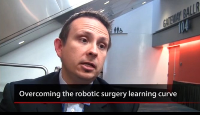
VIDEO: How to negotiate the robotic surgery learning curve
SAN FRANCISCO– Before attempting robotic surgery, it’s important to be comfortable with both the open and laparoscopic versions of the procedure, according to Dr. Kenneth Meredith, director of robotic surgery at the University of Wisconsin, Madison.
In experienced hands, robotic results can be good. In a case series of 138 robotic-assisted Ivor Lewis esophagectomies at the university for esophageal cancer, the median intensive care unit stay was 2 days and median hospital stay 9 days, Dr. Meredith. Complications occurred in about a quarter of patients.
In a video interview at the American College of Surgeons Clinical Congress, Dr. Meredith explained the significance of the findings and gave tips on how to negotiate the robotic surgery learning curve.
The video associated with this article is no longer available on this site. Please view all of our videos on the MDedge YouTube channel
AT THE AMERICAN COLLEGE OF SURGEONS CLINICAL CONGRESS
VIDEO: Nonclinical factors affect lung resection survival
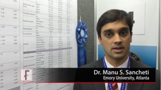
SAN FRANCISCO– A study of data from more than 200,000 patients identified several nonclinical factors ssociated with 30-day survival after lung resection for non–small cell lung cancer, Dr. Manu S. Sancheti reported at the annual clinical congress of the American College of Surgeons.
The analysis by Dr. Sancheti and his associates won the top prize for poster presentations at the congress.
In an interview at the award presentation, Dr. Sancheti of Emory University, Atlanta, described his results and ideas about how physicians and health care systems might use this information to improve care.
He reported having no financial disclosures.
The video associated with this article is no longer available on this site. Please view all of our videos on the MDedge YouTube channel
On Twitter @sherryboschert
AT THE ACS CLINICAL CONGRESS
Extracorporeal membrane oxygenation doubled survival rate of conventional CPR
AUSTIN, TEX. – Extracorporeal membrane oxygenation delivered during cardiopulmonary resuscitation allowed nearly twice as many patients to survive after discharge when compared against typical CPR-only procedures in a small, retrospective study.
“It’s no secret that conventional CPR is not terrifically successful,” Graham Peigh, a second-year medical student at Jefferson Medical College in Philadelphia, said during the Hot Topics in Pulmonary and Critical Care session at the annual meeting of the American College of Chest Physicians. “Extracorporeal membrane oxygenation [ECMO] gives patients a second chance at life.”

Mr. Peigh and his colleagues retrospectively analyzed 100 ECMO procedures performed on adults at a single teaching hospital during 2010-2013 and found that when ECMO was added to CPR, the survival rate to discharge went from 15% as calculated in a previously reported meta-analysis (J. Gen. Intern. Med. 1998;13:805-16) to 29% (P = .04).
When an arrested patient does not respond to CPR, cannulation through the femoral artery and vein can be combined with compressions to improve chances of survival.
In their analysis of ECMO delivered in an academic hospital setting, Mr. Peigh and his colleagues found that in the 24 cases in which ECMO was added to conventional CPR after the patients failed to respond to CPR alone, the survival rate with full neurologic recovery was 29%.
ECMO support was delivered in a number of scenarios, ranging from acute myocardial infarction to malignant arrhythmia to at least one case each of drug overdose induced cardiac arrest, septic shock, postcardiotomy failure, and acute rejection.
The ECMO support was provided for a mean of 5 days. The mean age for all patients studied was 47 years, and 15 were male. All cases followed a 24-hour hypothermia protocol.
Six of the ECMO-CPR patients died post ECMO of anoxic brain injury, stroke, or sepsis while still in the hospital, but the remaining seven patients (54%) survived after discharge and made full neurologic recoveries. The other 11 died during ECMO-CPR. During ECMO-CPR, 11 patients died of anoxic brain injury, stroke, metabolic acidosis, bowel necrosis, and family withdrawal of life support. Predictors of ECMO death were a pre-ECMO creatinine level of 1.7 mg/dL (P = .02) and the presence of acidosis (P = .04).
The ECMO survivor cohort also had what Mr. Peigh said were “encouraging” organ function results, with kidney and liver function remaining essentially unchanged after discharge.“Two of the patients who died of anoxic brain injuries were able to donate multiple organs for transplant,” Mr. Peigh said.
Previously reported ECMO data have shown there is at least a 20% increase in survival without notable neurologic effect, compared with conventional CPR (Lancet 2008;372:554-61; Crit. Care Med. 2011;39:1-7).
However, since these data were derived from centers where code teams were available at all times to treat a high volume of cardiac arrest patients, Mr. Peigh said the results – although indicative of the procedure’s value – “were not generalizable” to all institutions. But he noted that his and his colleagues’ study showed that even in institutions without a dedicated ECMO-CPR code team, ECMO-CPR resulted in demonstrably better outcomes for patients unresponsive to conventional CPR.
On Twitter @whitneymcknight
AUSTIN, TEX. – Extracorporeal membrane oxygenation delivered during cardiopulmonary resuscitation allowed nearly twice as many patients to survive after discharge when compared against typical CPR-only procedures in a small, retrospective study.
“It’s no secret that conventional CPR is not terrifically successful,” Graham Peigh, a second-year medical student at Jefferson Medical College in Philadelphia, said during the Hot Topics in Pulmonary and Critical Care session at the annual meeting of the American College of Chest Physicians. “Extracorporeal membrane oxygenation [ECMO] gives patients a second chance at life.”
Mr. Peigh and his colleagues retrospectively analyzed 100 ECMO procedures performed on adults at a single teaching hospital during 2010-2013 and found that when ECMO was added to CPR, the survival rate to discharge went from 15% as calculated in a previously reported meta-analysis (J. Gen. Intern. Med. 1998;13:805-16) to 29% (P = .04).
When an arrested patient does not respond to CPR, cannulation through the femoral artery and vein can be combined with compressions to improve chances of survival.
In their analysis of ECMO delivered in an academic hospital setting, Mr. Peigh and his colleagues found that in the 24 cases in which ECMO was added to conventional CPR after the patients failed to respond to CPR alone, the survival rate with full neurologic recovery was 29%.
ECMO support was delivered in a number of scenarios, ranging from acute myocardial infarction to malignant arrhythmia to at least one case each of drug overdose induced cardiac arrest, septic shock, postcardiotomy failure, and acute rejection.
The ECMO support was provided for a mean of 5 days. The mean age for all patients studied was 47 years, and 15 were male. All cases followed a 24-hour hypothermia protocol.
Six of the ECMO-CPR patients died post ECMO of anoxic brain injury, stroke, or sepsis while still in the hospital, but the remaining seven patients (54%) survived after discharge and made full neurologic recoveries. The other 11 died during ECMO-CPR. During ECMO-CPR, 11 patients died of anoxic brain injury, stroke, metabolic acidosis, bowel necrosis, and family withdrawal of life support. Predictors of ECMO death were a pre-ECMO creatinine level of 1.7 mg/dL (P = .02) and the presence of acidosis (P = .04).
The ECMO survivor cohort also had what Mr. Peigh said were “encouraging” organ function results, with kidney and liver function remaining essentially unchanged after discharge.“Two of the patients who died of anoxic brain injuries were able to donate multiple organs for transplant,” Mr. Peigh said.
Previously reported ECMO data have shown there is at least a 20% increase in survival without notable neurologic effect, compared with conventional CPR (Lancet 2008;372:554-61; Crit. Care Med. 2011;39:1-7).
However, since these data were derived from centers where code teams were available at all times to treat a high volume of cardiac arrest patients, Mr. Peigh said the results – although indicative of the procedure’s value – “were not generalizable” to all institutions. But he noted that his and his colleagues’ study showed that even in institutions without a dedicated ECMO-CPR code team, ECMO-CPR resulted in demonstrably better outcomes for patients unresponsive to conventional CPR.
On Twitter @whitneymcknight
AT CHEST 2014
Key clinical point: ECMO during CPR can improve clinical outcomes for patients in cardiac arrest.
Major finding: The discharge to survival rate for patients given ECMO was 54% with full neurologic recovery.
Data source: A retrospective analysis of 24 ECMO-CPR procedures performed at a single site during 2010-2013.
Disclosures: Mr. Peigh said he had no relevant disclosures.

Confronting aspirin unresponsiveness in congenital heart surgery
Thrombosis occurs in up to 15% of pediatric patients following cardiac surgery, and is associated with increased mortality. Although aspirin is commonly administered to pediatric patients after high-risk congenital cardiac surgery to reduce thrombosis risk, aspirin responsiveness is rarely assessed, according to Dr. Sirisha Emani and colleagues .
“In our observational study, aspirin unresponsiveness occurred in approximately 11% of patients undergoing specific high-risk cardiac procedures, and postoperative thrombosis was associated with aspirin unresponsiveness in this patient population,” said Dr. Emani.
In order to determine whether inadequate response to aspirin was associated with increased risk of thrombosis following high-risk procedures, the researchers performed a prospective analysis of 62 patients undergoing congenital cardiac surgical procedures involving placement of prosthetic material into the circulation or coronary artery manipulation who received aspirin.
Response to aspirin was determined using the Verify Now system at least 48 hours following administration. Patients were prospectively monitored for development of thrombosis events by imaging (echocardiogram, cardiac catheterization, MRI) and review of clinical events (shunt thrombosis, stroke, or limb ischemia) until the time of hospital discharge.
Aspirin responsiveness was tested a median of 2 days after initiation of therapy. The rate of aspirin unresponsiveness (Aspirin Responsive Unit, ARU greater than 550) was 7/62 (11.3%) in all patients and was highest in patients less than 5 kg who received 20.25 mg aspirin. Thrombosis events were demonstrated in 7 patients (11.3%). Thrombosis was observed in 6 (86%) of 7 patients who were unresponsive to aspirin as opposed to 1 (2%) of 54 patients who were responsive to aspirin, a significant difference. In two neonates who were unresponsive at 20.25 and 40.5 mg of aspirin, increase in dosage to 40.5 and 81 mg, respectively, resulted in an aspirin response, suggesting insufficiency rather than true unresponsiveness.
“Monitoring of aspirin therapy and consideration of dose adjustment or alternative agents for unresponsive patients may be justified and warrants further investigation in a prospective trial,” concluded Dr. Emani.

|
Dr. Robert Jaquiss |
Aspirin is sometimes used in pediatric cardiac surgical patients with either therapeutic or prophylactic intent, and the anticipated anti-platelet activity is simply assumed to follow. This study demonstrates that in children, this assumption may be flawed in as many as 11% of patients. Furthermore, in those patients in whom the assumption of efficacy was wrong, thrombosis was alarmingly common. This information should be of concern to physicians and surgeons who prescribe aspirin for children with cardiovascular abnormalities, and certainly merits further study.
Dr. Robert Jaquiss is chief of pediatric heart surgery, Duke University Medical Center, Durham, N.C.
|
|
Dr. Robert Jaquiss |
Aspirin is sometimes used in pediatric cardiac surgical patients with either therapeutic or prophylactic intent, and the anticipated anti-platelet activity is simply assumed to follow. This study demonstrates that in children, this assumption may be flawed in as many as 11% of patients. Furthermore, in those patients in whom the assumption of efficacy was wrong, thrombosis was alarmingly common. This information should be of concern to physicians and surgeons who prescribe aspirin for children with cardiovascular abnormalities, and certainly merits further study.
Dr. Robert Jaquiss is chief of pediatric heart surgery, Duke University Medical Center, Durham, N.C.
Thrombosis occurs in up to 15% of pediatric patients following cardiac surgery, and is associated with increased mortality. Although aspirin is commonly administered to pediatric patients after high-risk congenital cardiac surgery to reduce thrombosis risk, aspirin responsiveness is rarely assessed, according to Dr. Sirisha Emani and colleagues .
“In our observational study, aspirin unresponsiveness occurred in approximately 11% of patients undergoing specific high-risk cardiac procedures, and postoperative thrombosis was associated with aspirin unresponsiveness in this patient population,” said Dr. Emani.
In order to determine whether inadequate response to aspirin was associated with increased risk of thrombosis following high-risk procedures, the researchers performed a prospective analysis of 62 patients undergoing congenital cardiac surgical procedures involving placement of prosthetic material into the circulation or coronary artery manipulation who received aspirin.
Response to aspirin was determined using the Verify Now system at least 48 hours following administration. Patients were prospectively monitored for development of thrombosis events by imaging (echocardiogram, cardiac catheterization, MRI) and review of clinical events (shunt thrombosis, stroke, or limb ischemia) until the time of hospital discharge.
Aspirin responsiveness was tested a median of 2 days after initiation of therapy. The rate of aspirin unresponsiveness (Aspirin Responsive Unit, ARU greater than 550) was 7/62 (11.3%) in all patients and was highest in patients less than 5 kg who received 20.25 mg aspirin. Thrombosis events were demonstrated in 7 patients (11.3%). Thrombosis was observed in 6 (86%) of 7 patients who were unresponsive to aspirin as opposed to 1 (2%) of 54 patients who were responsive to aspirin, a significant difference. In two neonates who were unresponsive at 20.25 and 40.5 mg of aspirin, increase in dosage to 40.5 and 81 mg, respectively, resulted in an aspirin response, suggesting insufficiency rather than true unresponsiveness.
“Monitoring of aspirin therapy and consideration of dose adjustment or alternative agents for unresponsive patients may be justified and warrants further investigation in a prospective trial,” concluded Dr. Emani.

Featured Articles in the October 2014 Issue of The Journal of Thoracic and Cardiovascular Surgery
One of the primary features of the October issue of JTCVS is the topic of multiple arterial revascularization. This issue begins the study with an editorial and follows up with several articles on revascularization accompanied by commentaries. The next issue of Seminars in Thoracic and Cardiovascular Surgery will continue to highlight this topic through commentaries written by the authors of the revascularization papers listed below. Each commentary will discuss the content, results, and implications of the other papers. Multiple arterial grafting may be the next frontier for cardiac surgery.
Editorial
Coronary bypass: Is it time to take the next step—the routine use of the second arterial graft?
Michael E. Halkos and Robert A. Guyton
In this issue, 3 studies from highly experienced coronary centers lend further support for a multiarterial grafting strategy. Graft patency and even long-term survival may be improved by this strategy. The evidence, although not level A, is persuasive; a second arterial graft should become routine in most cases.
Acquired Cardiovascular Disease
Total arterial revascularization with internal thoracic and radial artery grafts in triple-vessel coronary artery disease is associated with improved survival
Brian F. Buxton, William Y. Shi, James Tatoulis, John A. Fuller, Alexander Rosalion, and Philip A. Hayward
Total arterial revascularization using internal thoracic and radial artery conduits was associated with improved late survival in patients with 3-vessel coronary artery disease compared with conventional single internal thoracic and saphenous vein grafts. This benefit may result in superior graft patency and protection of the native circulation.
▶ Buxton and colleagues from Australia reviewed over 6,000 isolated CABG patients with 3 vessel CAD over a 15 year period (1995-2010) and propensity matched 384 pairs of patients with either all arterial grafting vs. the use of one IMA and SVG. They found a highly significant survival advantage at 15 years in the total arterial revascularization group vs. the one IMA with SVG group (Kaplan Meier, following multivariable Cox regression, and after propensity matching). The authors concluded that, “total arterial revascularization should be encouraged in patients with a reasonable life expectancy”. Interestingly, only 36% of patients had use of bilateral IMA grafts, 97% of patients had at least one radial artery graft (52% had single radial grafts and 48% had bilateral grafts), and only 51% of RIMA grafts were used in-situ. The authors highlighted the potential benefit derived from use of the radial artery graft. This article is of particular interest because it involved a large number of patients (propensity matched) over a 15 year period, and it nicely demonstrated a highly significant benefit to long term survival when arterial grafting was utilized. [Summary and Comment by Dr. Jennifer Lawton, associate medical editor, Thoracic Surgery News].
Editorial Commentary in JTCVS: Total arterial revascularization: When will its time come?
Todd K. Rosengart
The long-term impact of diabetes on graft patency after coronary artery bypass grafting surgery: A substudy of the multicenter Radial Artery Patency Study
Saswata Deb, Steve K. Singh, Fuad Moussa, Hideki Tsubota, Dai Une, Alex Kiss, George Tomlinson, Mehdi Afshar, Ryan Sless, Eric A. Cohen, Sam Radhakrishnan, James Dubbin, Leonard Schwartz, and Stephen E. Fremes on behalf of the Radial Artery Patency Study Investigators
Radial artery grafts compared with saphenous vein grafts were associated with a lower rate of late graft occlusion in diabetic patients after coronary artery bypass surgery. Predictors against late graft occlusion included the use of radial arteries and high-grade target vessel stenosis. The type of conduit and late occlusion were influenced by diabetic status.
▶ Deb and colleagues from Toronto, Ottawa, and Texas Heart Institute reviewed patency rates in 269 low risk (<80 yrs, elective, EF>35%) isolated CABG patients (with 3 vessel CAD) from the Radial Artery Patency Study (each patient underwent randomization dictating use of radial artery or vein graft to separate territories). Patients underwent diagnostic angiography (N=234) or CT angio (N=35) at least 5 years postoperatively. New data presented are long term (>5 yr) patency in diabetics. The authors noted that the proportion of complete graft occlusion was significantly lower in radial artery grafts vs. saphenous grafts in diabetics; however, it was similar in non-diabetics. Interestingly, there was no difference in the occlusion rate of the IMA graft in diabetics compared to non-diabetics. Multivariate modeling demonstrated female sex, smoking history, and elevated creatinine as increased risk factors for late graft occlusion; whereas use of the radial artery and high grade target stenosis (>90% vs. 70-89% stenosis) were protective. The authors concluded that the study supports the use of the radial artery as a second conduit is appropriate in diabetic patients. Using blinded angiographic follow-up, this study importantly supports the use of arterial grafting, and specifically the use of the radial artery, in diabetic patients. [Summary and Comment by Dr. Jennifer Lawton, associate medical editor, Thoracic Surgery News].
Editorial Commentary in JTCVS: Arterial grafting and the challenge of the patient with diabetes
Paul A. Kurlansky
Surgical revascularization techniques that minimize surgical risk and maximize late survival after coronary artery bypass grafting in patients with diabetes mellitus
Sajjad Raza, Joseph F. Sabik III, Khalil Masabni, Ponnuthurai Ainkaran, Bruce W. Lytle, and Eugene H. Blackstone
BITA grafting with complete revascularization maximized long-term survival and is recommended for patients with diabetes undergoing surgical revascularization. It should be used in all patients with diabetes whose risk of DSWI is low and might be best avoided in obese diabetic women with diffuse atherosclerotic burden.
▶ Raza and colleagues from the Cleveland Clinic reviewed over 11,922 diabetic isolated CABG patients over a 39 year period (1972-2011) and attempted to identify patients that would derive the greatest survival benefit from an optimal surgical technique by evaluating 12 possible surgical combinations (no use of IMA, use of one IMA, use of 2 IMA grafts, incomplete revascularization, complete revascularization, off-pump, and on- pump CABG). After adjusting for patient characteristics, use of 2 IMA grafts was better than one IMA graft (21% lower late mortality), two IMA grafts was associated with more deep sternal wound infections (yet this had a small effect on survival), and complete revascularization was associated with lower late mortality (10%) compared to incomplete revascularization. Interestingly, additional risks for deep sternal wound infection included: female sex, medically treated diabetes mellitus, peripheral artery disease, prior myocardial infarction, and higher BMI; and HgA1C was not. The authors concluded that the strategy with the best predicted survival was one including use of 2 IMA grafts, complete revascularization, and off-pump techniques. The worst strategy was one including no IMA grafts, incomplete revascularization, and on-pump techniques. They noted that the survival benefit associated with the best combination was largely due to the benefit of the use of 2 IMA grafts. This article importantly reinforces the known benefit to long term survival with the use of 2 IMA grafts over one in a very large cohort of diabetic patients. [Summary and Comment by Dr. Jennifer Lawton, associate medical editor, Thoracic Surgery News].
Editorial Commentary in JTCVS: Two internal thoracic arteries really are better
Andrea Carpenter
Readers who found these articles interesting may also like to read the following papers in recent and future issues of the JTCVS sister publications, Seminars in Thoracic and Cardiovascular Surgery and Operative Techniques in Thoracic and Cardiovascular Surgery.
Seminars
▶ Discussion in Cardiothoracic Treatment and Care: Coronary Artery Bypass Grafting. John Puskas, Harold Lazar, Michael Mack, Joseph Sabik, David Taggart. Semin Thorac Cardiovasc Surg 2014 Spring; 26(1):75-94.
▶ News and Views: Editorials on Multiple Arterial Grafting. Brian Buxton and Stephen Fremes (expected publication December 2014).
▶ News and Views: Role of PCI in the Treatment of Left Main Coronary Disease. A. P. Kappetein. (expected publication December 2014).
▶ State of the Art: Post-CABG antiplatelet therapy. Victor Ferraris (expected publication December 2014).
Operative Techniques
▶ Repair of Postinfarct Ventricular Septal Defect: Anterior Apical Ventricular Septal Defect. John Conte. Oper Tech Thorac Cardiovasc Surg. 2014 Spring;19(1):96-114.
▶ Repair of Postinfarction Ventricular Septal Defect: Posterior Inferior Ventricular Septal Defect.Thomas Gleason. Oper Tech Thorac Cardiovasc Surg. 2014 Spring; 19(1):115-126
Upcoming Issues of JTCVS
The November issue of JTCVS will feature editorials, articles, and commentaries on graft patency and long-term survival with off-pump CABG, cerebral protection during congenital heart surgery, and the current status of surgery for non-small cell lung cancer. December will include editorials, articles, and commentaries on cerebral protection during aortic surgery and CABG for poor LVF.
Articles in Press
Don’t forget to visit the Journal’s Articles in Press section at http://jtcvs.com/inpress. Articles appear online shortly after acceptance in their submitted format, which is replaced with the final formatted version once the authors have approved their proofs. Once an article goes online in the Articles in Press section it is indexed in Medline, fully searchable and citable before ever appearing in print. You can also sign up for the Articles in Press email alerts or RSS feed, much as you would sign up for an electronic table of contents alert for the print issue. Go to http://jtcvs.com/user/alerts/saveaipalert.
The October issue of Thoracic Surgery News is now available online
Be sure to visit our interactive digital or PDF version of the October issue of Thoracic Surgery News, now available online.
This month we are featuring topics ranging from two stories with videos on local anesthesia and the new developments in TAVR, and a newly FDA-approved device for expanding the number of lungs available for transplant.
Also, in our News from the AATS section there is a call for abstracts and videos for AATS Week (comprising the 95th AATS Annual Meeting and the AATS Mitral Conclave), as well as information about signing up for the AATS Leadership Academy and for students and residents to get financial assistance to attend the 95th AATS Annual Meeting.
Click to view our October PDF here or the interactive digital edition.
Medical Students, Surgery and I-6 Cardiology Residents: Apply to Be an AATS Member for a Day
The AATS Member for a Day Program, sponsored by the AATS Graham Foundation, provides an opportunity for medical students, general surgery residents, and I-6 cardiothoracic residents to accompany an AATS Member Mentor during portions of the 2015 AATS Annual Meeting in Seattle, WA from April 25th – 29th.
Program Goals:
Offer insight into specialty of cardiothoracic surgery.
Provide an opportunity to network and build relationships within the cardiothoracic surgical community.
Up to 30 North American medical students, general surgery residents, and I-6 cardiothoracic residents will be selected to participate in this program.
Successful applicants will receive:
Complimentary hotel accommodations for a minimum of three and a maximum of four nights in an AATS Annual Meeting occupied hotel.
A $500 stipend to help offset travel costs.
An additional $250 stipend to offset the cost of meals.
Deadline: Friday, January 2, 2015
For more information and application, visit http://aats.org/Association/Member-for-a-day.cgi.
Point/Counterpoint: Is TEVAR required for all Type B aortic dissections?
Yes, TEVAR is clearly indicated.
Aortic dissection is a devastating condition afflicting an estimated two to eight per 100,000 people annually and comprises a large portion of the clinical entity known as the acute aortic syndromes. Patients presenting with an uncomplicated type B acute aortic dissection (TBAD) generally have low in-hospital mortality rates (2.4%-9%) when managed appropriately with anti-impulse therapy. However, survival continues to decrease with follow-up, with survival ranging between 80% and more than 95% at 1 year, progressing to approximately 75% at 3-4 years, and 48%-65% at 10 years. In late follow-up, the development of a new dissection with complications is estimated to occur in 20%-50% of patients. Complicated aortic dissections affect between 22% and 47%, and when present, mortality reaches more than 50% within the first week. TEVAR in these patients has been shown to be clearly indicated in a variety of studies with marked improvements in early mortality and late survival. Thus, one can see that aortic dissection is a disease that needs to be managed lifelong, and is associated with a high risk of mortality for the next 10 years after the initial presentation.1,2,3
The long-term effects of a patent false lumen have been well documented. Several studies following patients with chronic TBAD have documented progressive enlargement in aortic diameter with a patent false lumen. The mean increase in maximum aortic diameter ranges from 3.8 to 7.1 mm annually with any flow in the false lumen (FL) versus 1-2 mm per year with a thrombosed FL. Patients with a patent FL had 7.5 times increased risk of a dissection-related death or need for surgery as compared to patients with thrombosis of the FL. Dissection-related death or need for surgery occurred at a significantly earlier follow-up period in the patients with a patent FL.1,2,3
The aortic diameter may also influence the patency of the FL at presentation. In a review of 110 patients presenting with acute uncomplicated TBAD, 44% were identified to have a patent FL on initial imaging. Thirty-one percent of these patients had a maximum aortic diameter of 45 mm or more versus 14% of patients with a thrombosed FL (P = .053). Incidentally, patients with FL patency were on average 4 years younger than their thrombosed counterparts (62 vs. 66 years, P = .009).
Moreover, it appears that the long-term risks associated with a patent FL are further augmented by aortic dilatation at presentation. When combining both risk factors (FL patency and aortic diameter of 40 mm or more), only 22% of patients are dissection-related event–free at 5-year follow-up.Onitsuka et al.4 substantiated this finding on multivariate analysis. Interestingly, 10 of the 76 patients included in that study met both conditions, and seven of those patients (70%) experienced a dissection-related death or surgical conversion. Certainly patients meeting both criteria merit close follow-up for the development of aortic enlargement or symptoms of impending rupture.
The natural history of TBAD lends itself to at least some thrombus formation within the FL and is a common finding as the dissection becomes chronic. But in fact, partial thrombosis of the FL is associated with higher mortality in patients discharged from the hospital with stable TBAD at 1- and 3-year follow-up (15.4% and 31.6%, respectively). Matched patients with a patent FL had a 5.4% and 13.7% rate of mortality at 1 and 3 years, and patients with complete FL thrombosis were found to have mortality rates of 0% and 22.6% at the same follow-up.
Aortic remodeling after TEVAR
Placement of a thoracic endograft under these acute circumstances can often significantly alter the preoperative morphology of the true and false lumen. Schoder and colleagues5 followed changes in the TL and FL diameter in 20 patients after TEVAR for acute complicated dissection. Ninety percent of patients were found to have complete FL thrombosis of the thoracic aorta at 1 year, with a mean decrease in FL diameter of 11.6 mm. Two patients with a patent FL showed a mean increase in the maximal aortic diameter of 4.5 mm. In a similar study, Conrad et al.6 documented aortic remodeling of 21 patients in the year following TEVAR, 88% of whom had thrombosis of the FL. Most often the mobile septum is easily displaced by the radial force of the stent graft, with minimal limitation of expansion to the design diameter. Thus, endograft selection should be directed by the diameter of the normal unaffected aorta with minimal oversizing commonly limited to 5%-10%. Balloon profiling is not typically necessary.

The INSTEAD trial7 evaluated the management of uncomplicated type B aortic dissection and compared optimum medical therapy (OMT) to OMT with TEVAR. A total of 140 subjects were enrolled at seven European sites with 68 patients enrolled in OMT and 72 in OMT with TEVAR. In patients treated with TEVAR there was 90.6% complete FL thrombosis with a maximum true lumen diameter of 32.6 mm as compared to 22% and 18.7 mm in those treated with medical therapy alone. Furthermore, there was a 12.4% absolute risk reduction in aortic specific mortality and a 19.1% absolute risk reduction in disease progression in patients treated with TEVAR.
It is clear that patients that present with complicated type B aortic dissections mandate intervention with TEVAR and potentially other interventions to alleviate the complications at presentation. INSTEAD demonstrates that elective TEVAR results in favorable aortic remodeling and long-term survival, reinterventions were low, and it prevents late expansion and malperfusion. TEVAR was also associated with improved 5-year aortic-specific survival. TEVAR appears to be beneficial in those patients who present initially with a false lumen diameter of greater than 22 mm and an aortic diameter of greater than 40 mm with a patent false lumen.
References
1. Circ. Cardiovasc. Interv. 2013;4:407-16.
2. J. Vasc. Surg. 2012;55:641-51.
3. J. Vasc. Surg. 2011;54:985-92
4. Ann. Thorac. Surg. 2004;78:1268-73.
5. Ann. Thorac. Surg. 2007;83:1059-66.
6. J. Vasc. Surg. 2009;50:510-17.
7. Circulation 2009;120:2519-28.
Dr. Arko is with the Aortic Institute, Sanger Heart & Vascular Institute, Charlotte, N.C. He reported no relevant conflicts.
No, evidence supports careful choice of patients.
While the role of TEVAR has been proven to treat complications of acute type B dissections,1 its value as a prophylactic treatment in uncomplicated cases remains controversial. Optimal medical treatment (OMT) with strict blood pressure (SBP less than 120 mm Hg) and heart rate control is associated with a low morbidity and mortality, despite the risk of progressive aortic dilation. On the other hand TEVAR can result in early death and significant neurologic complications; other devastating complications of TEVAR include retrograde aortic dissection and access vessel rupture with a high associated mortality.
A meta-analysis of the published literature reported a high technical success of TEVAR for uncomplicated type B dissection and a relatively high conversion rate (20%) for patient treated with OMT, however the results did not identify an advantage for TEVAR with respect to 30-day and 2-year mortality.2

An expert panel review of the world literature also did not find significant data to support use of TEVAR for uncomplicated type B dissection.3 In the only randomized prospective trial to examine the role of TEVAR for uncomplicated type B dissection, the INSTEAD trial randomized 140 patients to OMT vs. OMT and TEVAR.4 The study results also did not support the use of TEVAR for the treatment of uncomplicated type B dissection, there was no survival advantage at 2 years, while TEVAR was associated with a 11.1% overall mortality and 4.3% neurologic complication rate, compared with 4.4% and 1.4% in the OMT group. The initial study did however report improved aortic remodeling at 2 years with TEVAR. The results of INSTEAD have been challenged because critical analysis of the INSTEAD trial has determined that the results were underpowered and that there was a 21% crossover in the OMT group and four patients received TEVAR that should have been excluded.5
Subsequent long-term analysis of the INSTEAD XL data do demonstrate a significant survival benefit and freedom from aortic adverse events in the TEVAR group after the initial 2-year analysis.6 At the 5-year follow up only 27 patients remained without a TEVAR. Fortunately there were no adverse events in the patients that crossed over to TEVAR from the OMT group demonstrating the safety of delayed TEVAR in this group. The high rate of aortic associated adverse events may favor early TEVAR. The INSTEAD XL study did identify a large primary tear (more than 10 mm) and an initial aortic diameter of 40 mm as risk factors to crossover suggesting a more aggressive approach in this subset of patients.
So while the INSTEAD XL trial now supports the use of TEVAR for uncomplicated type B dissections this was a relatively small trial that was underpowered in its initial analysis. Expert review of the world literature still supports medical management in the initial phase of treatment. Obviously in cases of failure of medical management TEVAR provides an effective treatment to restore the true lumen and visceral perfusion with possible sustained remodeling of the false lumen.
Given the not insignificant morbidity associated with TEVAR placement, routine treatment of all acute, uncomplicated type B dissections cannot be supported with the current evidence. However, a strategy of selective treatment based on size of the entry tear, extent of dissection, false lumen diameter and extent of thrombosis, effectiveness of antihypertension medications, ability to comply with medical therapy, and surveillance may be implemented. Furthermore treatment at centers of excellence with extensive TEVAR experience based on established protocols favor improved patient outcomes.
References
1. N. Engl. J. Med. 199;340:1546-52
2. Vasc. Endovascular. Surg. 2013 Oct 12;47(7):497-501. Epub 2013 Jul 12.
3. J. Am. Coll. Cardiol. 2013;61(16):1661-78.
4. Circulation 2009;120:2519-28.
5. Circulation 2009;120:2513-14.
6. Circ. Cardiovasc. Interv. 2013;6:407-16.
Dr. Shames is professor of surgery and radiology and program director of vascular surgery at the University of South Florida, Tampa. He reported no relevant conflicts.
Yes, TEVAR is clearly indicated.
Aortic dissection is a devastating condition afflicting an estimated two to eight per 100,000 people annually and comprises a large portion of the clinical entity known as the acute aortic syndromes. Patients presenting with an uncomplicated type B acute aortic dissection (TBAD) generally have low in-hospital mortality rates (2.4%-9%) when managed appropriately with anti-impulse therapy. However, survival continues to decrease with follow-up, with survival ranging between 80% and more than 95% at 1 year, progressing to approximately 75% at 3-4 years, and 48%-65% at 10 years. In late follow-up, the development of a new dissection with complications is estimated to occur in 20%-50% of patients. Complicated aortic dissections affect between 22% and 47%, and when present, mortality reaches more than 50% within the first week. TEVAR in these patients has been shown to be clearly indicated in a variety of studies with marked improvements in early mortality and late survival. Thus, one can see that aortic dissection is a disease that needs to be managed lifelong, and is associated with a high risk of mortality for the next 10 years after the initial presentation.1,2,3
The long-term effects of a patent false lumen have been well documented. Several studies following patients with chronic TBAD have documented progressive enlargement in aortic diameter with a patent false lumen. The mean increase in maximum aortic diameter ranges from 3.8 to 7.1 mm annually with any flow in the false lumen (FL) versus 1-2 mm per year with a thrombosed FL. Patients with a patent FL had 7.5 times increased risk of a dissection-related death or need for surgery as compared to patients with thrombosis of the FL. Dissection-related death or need for surgery occurred at a significantly earlier follow-up period in the patients with a patent FL.1,2,3
The aortic diameter may also influence the patency of the FL at presentation. In a review of 110 patients presenting with acute uncomplicated TBAD, 44% were identified to have a patent FL on initial imaging. Thirty-one percent of these patients had a maximum aortic diameter of 45 mm or more versus 14% of patients with a thrombosed FL (P = .053). Incidentally, patients with FL patency were on average 4 years younger than their thrombosed counterparts (62 vs. 66 years, P = .009).
Moreover, it appears that the long-term risks associated with a patent FL are further augmented by aortic dilatation at presentation. When combining both risk factors (FL patency and aortic diameter of 40 mm or more), only 22% of patients are dissection-related event–free at 5-year follow-up.Onitsuka et al.4 substantiated this finding on multivariate analysis. Interestingly, 10 of the 76 patients included in that study met both conditions, and seven of those patients (70%) experienced a dissection-related death or surgical conversion. Certainly patients meeting both criteria merit close follow-up for the development of aortic enlargement or symptoms of impending rupture.
The natural history of TBAD lends itself to at least some thrombus formation within the FL and is a common finding as the dissection becomes chronic. But in fact, partial thrombosis of the FL is associated with higher mortality in patients discharged from the hospital with stable TBAD at 1- and 3-year follow-up (15.4% and 31.6%, respectively). Matched patients with a patent FL had a 5.4% and 13.7% rate of mortality at 1 and 3 years, and patients with complete FL thrombosis were found to have mortality rates of 0% and 22.6% at the same follow-up.
Aortic remodeling after TEVAR
Placement of a thoracic endograft under these acute circumstances can often significantly alter the preoperative morphology of the true and false lumen. Schoder and colleagues5 followed changes in the TL and FL diameter in 20 patients after TEVAR for acute complicated dissection. Ninety percent of patients were found to have complete FL thrombosis of the thoracic aorta at 1 year, with a mean decrease in FL diameter of 11.6 mm. Two patients with a patent FL showed a mean increase in the maximal aortic diameter of 4.5 mm. In a similar study, Conrad et al.6 documented aortic remodeling of 21 patients in the year following TEVAR, 88% of whom had thrombosis of the FL. Most often the mobile septum is easily displaced by the radial force of the stent graft, with minimal limitation of expansion to the design diameter. Thus, endograft selection should be directed by the diameter of the normal unaffected aorta with minimal oversizing commonly limited to 5%-10%. Balloon profiling is not typically necessary.
The INSTEAD trial7 evaluated the management of uncomplicated type B aortic dissection and compared optimum medical therapy (OMT) to OMT with TEVAR. A total of 140 subjects were enrolled at seven European sites with 68 patients enrolled in OMT and 72 in OMT with TEVAR. In patients treated with TEVAR there was 90.6% complete FL thrombosis with a maximum true lumen diameter of 32.6 mm as compared to 22% and 18.7 mm in those treated with medical therapy alone. Furthermore, there was a 12.4% absolute risk reduction in aortic specific mortality and a 19.1% absolute risk reduction in disease progression in patients treated with TEVAR.
It is clear that patients that present with complicated type B aortic dissections mandate intervention with TEVAR and potentially other interventions to alleviate the complications at presentation. INSTEAD demonstrates that elective TEVAR results in favorable aortic remodeling and long-term survival, reinterventions were low, and it prevents late expansion and malperfusion. TEVAR was also associated with improved 5-year aortic-specific survival. TEVAR appears to be beneficial in those patients who present initially with a false lumen diameter of greater than 22 mm and an aortic diameter of greater than 40 mm with a patent false lumen.
References
1. Circ. Cardiovasc. Interv. 2013;4:407-16.
2. J. Vasc. Surg. 2012;55:641-51.
3. J. Vasc. Surg. 2011;54:985-92
4. Ann. Thorac. Surg. 2004;78:1268-73.
5. Ann. Thorac. Surg. 2007;83:1059-66.
6. J. Vasc. Surg. 2009;50:510-17.
7. Circulation 2009;120:2519-28.
Dr. Arko is with the Aortic Institute, Sanger Heart & Vascular Institute, Charlotte, N.C. He reported no relevant conflicts.
No, evidence supports careful choice of patients.
While the role of TEVAR has been proven to treat complications of acute type B dissections,1 its value as a prophylactic treatment in uncomplicated cases remains controversial. Optimal medical treatment (OMT) with strict blood pressure (SBP less than 120 mm Hg) and heart rate control is associated with a low morbidity and mortality, despite the risk of progressive aortic dilation. On the other hand TEVAR can result in early death and significant neurologic complications; other devastating complications of TEVAR include retrograde aortic dissection and access vessel rupture with a high associated mortality.
A meta-analysis of the published literature reported a high technical success of TEVAR for uncomplicated type B dissection and a relatively high conversion rate (20%) for patient treated with OMT, however the results did not identify an advantage for TEVAR with respect to 30-day and 2-year mortality.2
An expert panel review of the world literature also did not find significant data to support use of TEVAR for uncomplicated type B dissection.3 In the only randomized prospective trial to examine the role of TEVAR for uncomplicated type B dissection, the INSTEAD trial randomized 140 patients to OMT vs. OMT and TEVAR.4 The study results also did not support the use of TEVAR for the treatment of uncomplicated type B dissection, there was no survival advantage at 2 years, while TEVAR was associated with a 11.1% overall mortality and 4.3% neurologic complication rate, compared with 4.4% and 1.4% in the OMT group. The initial study did however report improved aortic remodeling at 2 years with TEVAR. The results of INSTEAD have been challenged because critical analysis of the INSTEAD trial has determined that the results were underpowered and that there was a 21% crossover in the OMT group and four patients received TEVAR that should have been excluded.5
Subsequent long-term analysis of the INSTEAD XL data do demonstrate a significant survival benefit and freedom from aortic adverse events in the TEVAR group after the initial 2-year analysis.6 At the 5-year follow up only 27 patients remained without a TEVAR. Fortunately there were no adverse events in the patients that crossed over to TEVAR from the OMT group demonstrating the safety of delayed TEVAR in this group. The high rate of aortic associated adverse events may favor early TEVAR. The INSTEAD XL study did identify a large primary tear (more than 10 mm) and an initial aortic diameter of 40 mm as risk factors to crossover suggesting a more aggressive approach in this subset of patients.
So while the INSTEAD XL trial now supports the use of TEVAR for uncomplicated type B dissections this was a relatively small trial that was underpowered in its initial analysis. Expert review of the world literature still supports medical management in the initial phase of treatment. Obviously in cases of failure of medical management TEVAR provides an effective treatment to restore the true lumen and visceral perfusion with possible sustained remodeling of the false lumen.
Given the not insignificant morbidity associated with TEVAR placement, routine treatment of all acute, uncomplicated type B dissections cannot be supported with the current evidence. However, a strategy of selective treatment based on size of the entry tear, extent of dissection, false lumen diameter and extent of thrombosis, effectiveness of antihypertension medications, ability to comply with medical therapy, and surveillance may be implemented. Furthermore treatment at centers of excellence with extensive TEVAR experience based on established protocols favor improved patient outcomes.
References
1. N. Engl. J. Med. 199;340:1546-52
2. Vasc. Endovascular. Surg. 2013 Oct 12;47(7):497-501. Epub 2013 Jul 12.
3. J. Am. Coll. Cardiol. 2013;61(16):1661-78.
4. Circulation 2009;120:2519-28.
5. Circulation 2009;120:2513-14.
6. Circ. Cardiovasc. Interv. 2013;6:407-16.
Dr. Shames is professor of surgery and radiology and program director of vascular surgery at the University of South Florida, Tampa. He reported no relevant conflicts.
