User login
Sharon Worcester is an award-winning medical journalist for MDedge News. She has been with the company since 1996, first as the Southeast Bureau Chief (1996-2009) when the company was known as International Medical News Group, then as a freelance writer (2010-2015) before returning as a reporter in 2015. She previously worked as a daily newspaper reporter covering health and local government. Sharon currently reports primarily on oncology and hematology. She has a BA from Eckerd College and an MA in Mass Communication/Print Journalism from the University of Florida. Connect with her via LinkedIn and follow her on twitter @SW_MedReporter.
Photodamage Effectively Treated With Quasi-Ablative Approach
KISSIMMEE, FLA. – The nonablative 1540-nm fractional laser can be safely converted to a quasi-ablative device for treating facial actinic keratoses and photodamage, according to Dr. Moshe Lapidoth.
By slightly modifying technique, a 50%-75% improvement in actinic keratoses and photodamage was noted in 17 patients who underwent two to three treatments at 4-week intervals, Dr. Lapidoth reported at the annual meeting of the American Society for Laser Medicine and Surgery. The laser was used in noncontact mode hovering 5-10 mm above the skin rather than in contact mode.
The patients had actinic keratoses and photodamage requiring ablative treatment of the epidermis. Treatment was applied using a fluence of 75 J/cm2, a 15-ms pulse duration, and a 10-mm spot size. Two blinded assessors and the participants evaluated clinical improvement at 3 months after the final treatment using a quartile grading scale (Lasers Med. Sci. 2012 April 27 [doi: 10.1007/s10103-012-1103-6])
A score of 0 was associated with no improvement; a score of 1 with 1%-25% improvement, a score of 2 with 26%-50% improvement, a score of 3 with 51%-75% improvement, and a score of 4 with 76%-100% improvement. The mean score for actinic keratoses was 3.4, and for skin appearance was 3.2, said Dr. Lapidoth, head of the laser department at Rabin Medical Center in Israel.
Side effects after each treatment included erythema, mild edema, erosion in two patients, and mild desquamation, but no scarring or postinflammatory pigmentary changes occurred, he said.
Although more current fractional laser devices are designed to be either ablative or nonablative, the 1540-nm fractional laser was designed to be nonablative.
"The question is, ‘Can you take a 1540-nm laser, which is totally nonablative, and turn it to be ablative or quasi-ablative?’ and the answer is, ‘yes,’ Dr. Lapidoth said.
This treatment approach can be used in conjunction with contact mode, he added, explaining that the nonablative contact mode can be used first to target the dermis for treatment of fine wrinkles and scars, followed by the ablative noncontact mode to treat actinic keratoses and resistant lentigines and other lesions.
Dr. Lapidoth reported having no disclosures.
FROM THE ANNUAL MEETING OF THE AMERICAN SOCIETY FOR LASER MEDICINE AND SURGERY
Major Finding: A quasi-ablative technique using a nonablative 1540-nm fractional laser resulted in a 50%-75% improvement in actinic keratoses and skin appearance in 17 patients.
Data Source: This was a prospective study of 17 patients treated.
Disclosures: Dr. Lapidoth reported having no disclosures.
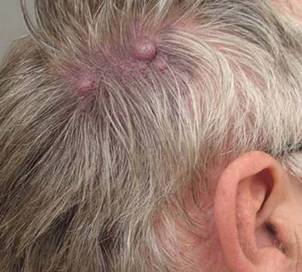
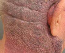
Port Wine Stains: Hypertrophy Risk Rises With Age
KISSIMMEE, FLA. – Hypertrophy of port wine stains is more common than not in the majority of patients older than age 50 with the vascular anomaly, and is most often associated with red and purple lesions, according to findings from a retrospective patient-questionnaire study.
"By the age of 50, over 70% had hypertrophy of their port wine stain," said Dr. Anne Margreet van Drooge of the Netherlands Institute of Pigment Disorders at the University of Amsterdam.
Hypertrophy is known to be fairly common in port wine stains, but data are lacking on its pathogenesis and development. In this study, the incidence of hypertrophy in port wine stains was about 20% in all patients, and both color and hypertrophy were frequently reported as reasons for seeking treatment.
Only 4% of all hypertrophy cases were associated with pink lesions, Dr. van Drooge said at the annual meeting of the American Society for Laser Medicine and Surgery.
Of the 335 patients included in the review, 68 had hypertrophy, classified as thickening, nodular, or both. Cases were divided almost equally into age groups of under 18 years, 18-30 years, and greater than 30 years. The median age of those with hypertrophic port wine stains was 50 years, and the median age at the time of hypertrophic development was 31 years, although the ages of onset of nodular hypertrophy and diffuse thickening were 39 years and 12 years, respectively. Nodular hypertrophy was rarely seen in those younger than 18 years.
In all study patients, most of the port wine stains were on the face. Half were red in color, and 44% were purple. No differences were seen between those with and without hypertrophy with respect to gender, lesion size, or lesion location, she noted.
Patients included in the study had been referred to a single outpatient clinic between 2005 and 2009. Medical records and photographs were examined to identify and characterize hypertrophic port wine stains.
The findings of different ages at onset for different types of hypertrophy suggest that unique pathomechanics may exist for each type, Dr. van Drooge said. "There is definitely a correlation between the color of the port wine stain and the risk for the development of hypertrophy. In a multivariate analysis of all patient characteristics, we found that both darker color and older age were significantly associated with hypertrophy, independent from each other."
Improved understanding of hypertrophy in port wine stains is important given the adverse psychosocial effects these lesions can have on patients; in this study, 5% of patients said they avoided social interaction because of their port wine stain, she noted, adding that the findings have implications for treatment of port wine stains.
Although this study provided no direct support for the notion that treatment may prevent hypertrophy, some studies have suggested that it may, adding that treatment guidelines should be adapted to include more emphasis on hypertrophy and its prevention, keeping in mind the findings of this study with respect to age and lesion color.
"However, I don’t think it is possible to treat only darker port wine stains in the effort to prevent hypertrophy, since pink port wine stains – particularly in younger patients – eventually can darken, and with that, the risk for hypertrophy increases," she noted.
Dr. van Drooge had no disclosures to report.
FROM THE ANNUAL MEETING OF THE AMERICAN SOCIETY FOR LASER MEDICINE AND SURGERY
Optimized Pulsed Light 'Impressively' Clears Port Wine Stains
KISSIMMEE, FLA. – Optimized intense pulsed light was found to safely enable effective clearance of port wine stains and capillary malformations in 16 patients.
The outcomes were so impressive in two patients, who achieved 80-100% improvement after only one treatment with this "new class of filtered IPL," that additional planned treatments were canceled, Dr. Maurice Adatto reported at the annual meeting of the American Society for Laser Medicine and Surgery.
The 16 study patients from two centers had Fitzpatrick skin types II-IV and a mean age of 40 years (range, 11-71 years). All patients experienced improvement with one to four treatments, with 10 of the 16 patients achieving greater than 50% improvement, said Dr. Adatto, a dermatologist in private practice in Geneva, Switzerland.
The 10 females and 6 males had port wine stains of the face, neck, trunk, or extremities, with the exception of two patients who had capillary malformations.
Treatments were applied using Palomar’s MaxG IPL system with a spot size of 10 x 15 mm. Patients were treated using either one pass at a fluence of 50 J/cm2 and a 10-ms pulse duration, or two passes, with the first pass at 34-36 J/cm2 and a 10-ms pulse duration and the second at 20-28 J/cm2 and a 5-ms pulse duration.
Outcomes were assessed on the basis of clinical images generated using Miravex’s Antera 3D system to evaluate hemoglobin clearance. Photos were taken between 48 and 96 hours after each treatment and also at 2 months after each treatment, Dr. Adatto said.
The only reported side effects were limited or slight transitory purpura for 3-5 days, and mild to moderate edema for 1-3 days.
Lasers are more often used to treat port wine stains, but the higher peak power and the short pulse widths available with the new optimized pulsed light technology appear to allow for safe and effective clearance, Dr. Adatto said. Further optimization of the parameters and safety profile of the optimized pulsed light device is possible, and another study of its use in pediatric patients is currently underway, he noted.
Dr. Adatto disclosed that the equipment from Palomar used in the study was purchased at a discount. He had no other disclosures to report.
FROM THE ANNUAL MEETING OF THE AMERICAN SOCIETY FOR LASER MEDICINE AND SURGERY
Photoepilation System Painlessly Delivers Results
KISSIMMEE, FLA. – A combined 800-nm diode laser and vacuum system for photoepilation resulted in high patient satisfaction and was safe and virtually painless for more than 2,000 treated patients.
Photoepilation is among the most popular aesthetic treatments worldwide, and is the second most popular aesthetic treatment in Latin America, where most patients have darker skin types that can be prone to greater risk of adverse outcomes, Dr. Rafael Nunes said at the annual meeting of the American Society for Laser Medicine and Surgery. On the basis of this study, however, the LightSheer Duet laser system appears to be a safe method for successful photoepilation even in darker-skinned patients.
Of the 2,158 study patients, 1,845 were women and 313 were men; they had a mean age of 36 years (range, 18-63 years) and skin types I-IV. Photoepilation was performed on various body and facial areas at one of four participating laser centers in Brazil over an 18-month period, with a mean of six treatments every 28-45 days. The investigators treated patients with a Lumenis 800-nm LightSheer Duet diode laser and vacuum-assist technology at a fluence of 6-12 J/cm2, a spot size of 22 x 35 mm, a pulse width of 30-100 ms, and a single pass, said Dr. Nunes, a plastic surgeon in private practice in Rio de Janeiro.
At the 3- to 6-month follow-up, after completion of 4-10 treatment sessions, 92% of patients rated their outcomes as "good" or "great."
The system requires no pretreatment ice packs, creams, or anesthesia, and the vacuum system should be used only on the low setting to prevent injury – particularly in areas where there is skin laxity.
No permanent or severe adverse effects occurred in any of the patients. The only adverse effect was transient erythema in six patients (0.3%). Treatment tolerance was also impressive, with patients experiencing only a small sensation of heat at the treatment site, and "no discomfort whatsoever" following treatment, Dr. Nunes said.
Dr. Nunes is a speaker or consultant for Allergan, Alma Lasers, Q-MED (Galderma), Lumenis, MedixSysteme, Palomar, and Solta Medical.
FROM THE ANNUAL MEETING OF THE AMERICAN SOCIETY FOR LASER MEDICINE AND SURGERY
Microsecond Nd:YAG Safe, Effective for Facial Telangiectasia
KISSIMMEE, FLA. – A microsecond 1,064-nm Nd:YAG laser was found to be effective for treating facial telangiectasias without the scarring and discomfort that can occur with millisecond pulse systems.
A prospective single-center study enrolled 20 patients aged 35-70 years with Fitzpatrick skin types I-III. After undergoing one treatment per month for 2 months with the 650-microsecond LightPod Neo 1,064-nm Nd:YAG laser (manufactured by Aerolase), the patients experienced significant, and even "dramatic," improvements in the appearance of facial telangiectasias on the nose and cheek, Dr. David J. Goldberg reported at the annual meeting of the American Society for Laser Medicine and Surgery.

"We saw significant improvements in the appearance of telangiectasia with minimal discomfort, presumably because of the microsecond pulse duration," said Dr. Goldberg, a dermatologist at Mount Sinai School of Medicine, New York.
The treatments were applied at a fluence of 199 J/cm2, a pulse duration of 650 microseconds, and a 2-mm spot size. The multisetting device was set at treatment level 6. Evaluation of results was based on clinical assessment of digital photographs and both patient and investigator assessment of the reduction in the size and appearance of telangiectasias based on a 5-point scale.
"Most people know that facial telangiectasias can be treated with a variety of wavelengths, and the 532-nm laser has traditionally been effective for small vessels," Dr. Goldberg said.
However, longer wavelength lasers, such as the 1,064-nm Nd:YAG with a millisecond pulse duration (3-30 milliseconds), which have also been used to treat larger vessels, are associated with "well-documented scarring and greater discomfort," he noted.
These adverse effects were not seen with the use of the microsecond system, he said, adding: "I think this is really the most important part of the study because of the previous documentation of problems with Nd:YAG laser systems for the treatment of patients with telangiectasias."
The study was supported by a research grant from Aerolase. Dr. Goldberg had no other disclosures to report.
KISSIMMEE, FLA. – A microsecond 1,064-nm Nd:YAG laser was found to be effective for treating facial telangiectasias without the scarring and discomfort that can occur with millisecond pulse systems.
A prospective single-center study enrolled 20 patients aged 35-70 years with Fitzpatrick skin types I-III. After undergoing one treatment per month for 2 months with the 650-microsecond LightPod Neo 1,064-nm Nd:YAG laser (manufactured by Aerolase), the patients experienced significant, and even "dramatic," improvements in the appearance of facial telangiectasias on the nose and cheek, Dr. David J. Goldberg reported at the annual meeting of the American Society for Laser Medicine and Surgery.
"We saw significant improvements in the appearance of telangiectasia with minimal discomfort, presumably because of the microsecond pulse duration," said Dr. Goldberg, a dermatologist at Mount Sinai School of Medicine, New York.
The treatments were applied at a fluence of 199 J/cm2, a pulse duration of 650 microseconds, and a 2-mm spot size. The multisetting device was set at treatment level 6. Evaluation of results was based on clinical assessment of digital photographs and both patient and investigator assessment of the reduction in the size and appearance of telangiectasias based on a 5-point scale.
"Most people know that facial telangiectasias can be treated with a variety of wavelengths, and the 532-nm laser has traditionally been effective for small vessels," Dr. Goldberg said.
However, longer wavelength lasers, such as the 1,064-nm Nd:YAG with a millisecond pulse duration (3-30 milliseconds), which have also been used to treat larger vessels, are associated with "well-documented scarring and greater discomfort," he noted.
These adverse effects were not seen with the use of the microsecond system, he said, adding: "I think this is really the most important part of the study because of the previous documentation of problems with Nd:YAG laser systems for the treatment of patients with telangiectasias."
The study was supported by a research grant from Aerolase. Dr. Goldberg had no other disclosures to report.
FROM THE ANNUAL MEETING OF THE AMERICAN SOCIETY FOR LASER MEDICINE AND SURGERY
Major Finding: Twenty patients experienced significant improvements at 6 months after undergoing one treatment per month for 2 months.
Data Source: This was a prospective single-center study of patients with facial telangiectasias, aged 35-70 years with Fitzpatrick skin types I-III, who underwent treatment with the 650-microsecond LightPod Neo 1,064-nm Nd:YAG laser.
Disclosures: The study was sponsored by Aerolase. Dr. Goldberg had no other disclosures to report.
Avoidable Mistakes Noted in Several Laser Surgery Lawsuits
KISSIMMEE, FLA. – Many of the leading causes of litigation associated with cutaneous laser surgery are preventable, findings from a comprehensive legal database search suggest.
For example, 17 cases of litigation identified by the search were associated with a failure to conduct a test spot on a patient undergoing laser hair removal, and this deficiency went "almost hand in hand" with failure to evaluate skin type, Dr. H. Ray Jalian, a clinical fellow at Massachusetts General Hospital, Boston, said at the annual meeting of the American Society for Laser Medicine and Surgery.
Failure to obtain informed consent was also a common – and preventable – cause of litigation among the 182 cases that Dr. Jalian and his colleagues identified during their online search of more than 40,000 documents. All of the cases involved injury associated with cutaneous laser therapy and occurred between 1989 and 2012.
The search, conducted in "an effort to close practice gaps and minimize the risk of future lawsuits for laser surgeons," was performed using keywords such as laser, laser and skin, laser and malpractice, laser and hair, laser and tattoo, and intense pulsed light accident.
Burns were the most common type of injury associated with litigation, followed by scars, pigmentation, and disfigurement. Some cases also listed emotional distress and physical suffering. There were four cases involving eye injury, and two cases of death – both associated with anesthesia complications. One of these cases involved a patient who underwent general anesthesia for classic CO2 resurfacing, and the other involved an anaphylactic reaction to topical lidocaine, Dr. Jalian said.
The most common type of procedure associated with litigation was hair removal, followed by various types of rejuvenation, treatment of leg veins, tattoo removal, and treatment of vascular lesions, neoplastic scars, and pigmentary disorders. The most common cause of action was negligence, followed by treatment below the standard of care, lack of informed consent, loss of consortium, and assault and battery. Poorly trained – or untrained – staff, and improper use of appropriate lasers and/or laser settings were also among the complaints.
Of the cases for which a summary judgment, verdict, or settlement was made public, about half were in favor of the defendant and half were in favor of the plaintiff. Monetary awards ranged from $5,000 to over $2,000,000 for a case involving burns and hyperpigmentation secondary to a classic C02 resurfacing case, Dr. Jalian noted.
Although the findings are limited by the use of a single database, they do highlight the fact that there are several preventable causes of litigation – an important consideration for all laser surgeons, given that 75% of all physicians – and close to 100% of those in high risk specialties – will face a medical malpractice lawsuit during their careers, he said.
Dr. Jalian had no disclosures to report.
FROM THE ANNUAL MEETING OF THE AMERICAN SOCIETY FOR LASER MEDICINE AND SURGERY
Major Finding: Of 182 lawsuits related to cutaneous laser surgery, 17 cases were associated with failure to conduct a test spot.
Data Source: A legal database search of more than 40,000 documents from 1989-2012 yielded 182 cases related to cutaneous laser surgery.
Disclosures: Dr Jalian had no disclosures to report.
Broadband Light Halted Signs of Aging in Small Study
KISSIMMEE, FLA. – Broadband light therapy virtually halted the signs of aging in women treated at least annually for 8 years, according to Dr. Patrick Bitter Jr.
"This really is the closest thing we have to a fountain of youth for delaying skin aging," Dr. Bitter said at the annual meeting of the American Society for Laser Medicine and Surgery.
Eleven women underwent treatment of the entire face with the Sciton BroadBand Light system at least once a year over an 8-year period. Blinded evaluators (490), including 51 dermatologists, indicated that the women aged no more than about 6 months during that time, said Dr. Bitter, a dermatologist in private practice in Los Gatos, Calif.
The mean age of the women at the start of treatment was 45 years, and the dermatologists who participated in the evaluations of "before" and "after" photographs rated their age as a mean of 45 years based on the "before" images. At an average of 8 years follow-up, the evaluators rated the age of the participants at a mean of 45 to 45.5 years, which was a mean of 9 years younger than the participants’ actual ages, he said.
Study participants included women in Dr. Bitter’s practice who received at least one treatment annually with good "before" photos who had not received any laser treatments or cosmetic surgery. Skin care regimens varied among the women.
The evaluators – only the dermatologists’ responses were included in the data Dr. Bitter presented – were blinded to any treatments the women underwent, and photos were taken in a way that ensured the evaluation was based entirely on skin appearance and not on other signs of aging (such as graying hair), he noted.
Broadband light technologies are known to be effective for reversing the signs of aging, such as wrinkles, redness, and brown spots, and data have shown that broadband light can improve the skin histologically. However, this is the first blinded evaluation of long-term results among those receiving regular treatments, Dr. Bitter said, noting that this study was prompted in part by a Swiss study published several years ago that speculated that such treatments might actually accelerate aging of the skin when used over time. His experience in 14 years of using broadband light treatments suggested otherwise, and these findings provide further evidence that long-term use reverses and prevents the signs of aging, he said.
New data from a corollary study to be presented at an upcoming meeting of the Society for Investigative Dermatology will provide additional evidence that light "does more than just make reds and browns go away," he said. "What is seems to do is make skin cells behave like younger skin cells."
Dr. Bitter noted that the difference between a patient’s really liking results and not seeing a difference is technique. "This is a great technology, buy you need to know how to use it," he said, explaining that it is important to know the optimal parameters based on skin type, to perform enough passes at each session, and to conduct enough sessions.

In an interview, Dr. Robert A. Weiss of the department of dermatology at Johns Hopkins University, Baltimore, agreed that the technique – and a thorough knowledge of the parameters, limitations, and application of the intense pulsed light that is used – is critical to success. "Continued sun protection is critical, as well," he said.
Like Dr. Bitter, Dr. Weiss has seen effective long-term results with IPL technology. In a study published in 2002, he and his colleagues reported on 80 randomly selected patients with skin types I-IV who were treated with filtered flashlamp IPL between 1996 and 1997. At 4-year follow-up after the first of a median of three treatments, 83% of patients had improved skin texture, 82% had improved telangiectasias, and 79% had improved pigmentation (Dermatol. Surg. 2002;28:1115-9). "The end result is clearer, brighter, younger, healthier-looking skin," he said.
Dr. Bitter owns the trademark for PhotoFacial intense pulsed light treatments. Dr. Weiss reported at the time his study was published that he was a consultant and preceptor for Lumenis, and that the devices used in the study were purchased at a discount.
KISSIMMEE, FLA. – Broadband light therapy virtually halted the signs of aging in women treated at least annually for 8 years, according to Dr. Patrick Bitter Jr.
"This really is the closest thing we have to a fountain of youth for delaying skin aging," Dr. Bitter said at the annual meeting of the American Society for Laser Medicine and Surgery.
Eleven women underwent treatment of the entire face with the Sciton BroadBand Light system at least once a year over an 8-year period. Blinded evaluators (490), including 51 dermatologists, indicated that the women aged no more than about 6 months during that time, said Dr. Bitter, a dermatologist in private practice in Los Gatos, Calif.
The mean age of the women at the start of treatment was 45 years, and the dermatologists who participated in the evaluations of "before" and "after" photographs rated their age as a mean of 45 years based on the "before" images. At an average of 8 years follow-up, the evaluators rated the age of the participants at a mean of 45 to 45.5 years, which was a mean of 9 years younger than the participants’ actual ages, he said.
Study participants included women in Dr. Bitter’s practice who received at least one treatment annually with good "before" photos who had not received any laser treatments or cosmetic surgery. Skin care regimens varied among the women.
The evaluators – only the dermatologists’ responses were included in the data Dr. Bitter presented – were blinded to any treatments the women underwent, and photos were taken in a way that ensured the evaluation was based entirely on skin appearance and not on other signs of aging (such as graying hair), he noted.
Broadband light technologies are known to be effective for reversing the signs of aging, such as wrinkles, redness, and brown spots, and data have shown that broadband light can improve the skin histologically. However, this is the first blinded evaluation of long-term results among those receiving regular treatments, Dr. Bitter said, noting that this study was prompted in part by a Swiss study published several years ago that speculated that such treatments might actually accelerate aging of the skin when used over time. His experience in 14 years of using broadband light treatments suggested otherwise, and these findings provide further evidence that long-term use reverses and prevents the signs of aging, he said.
New data from a corollary study to be presented at an upcoming meeting of the Society for Investigative Dermatology will provide additional evidence that light "does more than just make reds and browns go away," he said. "What is seems to do is make skin cells behave like younger skin cells."
Dr. Bitter noted that the difference between a patient’s really liking results and not seeing a difference is technique. "This is a great technology, buy you need to know how to use it," he said, explaining that it is important to know the optimal parameters based on skin type, to perform enough passes at each session, and to conduct enough sessions.
In an interview, Dr. Robert A. Weiss of the department of dermatology at Johns Hopkins University, Baltimore, agreed that the technique – and a thorough knowledge of the parameters, limitations, and application of the intense pulsed light that is used – is critical to success. "Continued sun protection is critical, as well," he said.
Like Dr. Bitter, Dr. Weiss has seen effective long-term results with IPL technology. In a study published in 2002, he and his colleagues reported on 80 randomly selected patients with skin types I-IV who were treated with filtered flashlamp IPL between 1996 and 1997. At 4-year follow-up after the first of a median of three treatments, 83% of patients had improved skin texture, 82% had improved telangiectasias, and 79% had improved pigmentation (Dermatol. Surg. 2002;28:1115-9). "The end result is clearer, brighter, younger, healthier-looking skin," he said.
Dr. Bitter owns the trademark for PhotoFacial intense pulsed light treatments. Dr. Weiss reported at the time his study was published that he was a consultant and preceptor for Lumenis, and that the devices used in the study were purchased at a discount.
FROM THE ANNUAL MEETING OF THE AMERICAN SOCIETY FOR LASER MEDICINE AND SURGERY
Major Finding: After 8 years of at least once-a-year treatment, the women in the study were rated as appearing a mean 9 years younger than their actual age.
Data Source: This was a blinded evaluation of long-term outcomes with regular broadband light therapy in 11 women.
Disclosures: Dr. Bitter owns the trademark for FotoFacial intense pulsed light treatments. Dr. Weiss reported at the time his study was published he was a consultant and preceptor for Lumenis, and that the devices used in the study were purchased at a discount.
Laser, Light Combo Improves Pigmentation in Single Session
KISSIMMEE, FLA. – The combined use of a nonablative, fractional 1,540-nm laser and optimized pulsed light provided significantly greater improvement in dyspigmentation after a single treatment session, compared with the 1,540-nm laser alone, based on findings from a study comparing the two treatments in 36 patients.
The observation of improved pigment after a single treatment in the combination group represents a reduced time course for achieving outcomes; typically, several treatments are required to produce results, Dr. Chung-Yin Stanley Chan said at the annual meeting of the American Society for Laser Medicine and Surgery.
The novel combined approach to facial rejuvenation also modestly improved wrinkles, although this outcome did not differ significantly between the two groups, Dr. Chan and his colleagues noted.
The mean pigment improvement score in the 10 subjects assigned to the combination treatment group was 2.4 points on a 1- to 4-point scale, indicating at least 50% improvement, with 96% of patients in that group experiencing improvement. The mean pigment improvement score in the 26 subjects assigned to the laser-only treatment group was 1.2 points.
Fitzgerald wrinkle scores improved from 6.4 to 6.0 (on a scale of 1-9) in the combination group, and from 5.3 to 4.9 in the laser-only treatment group, said Dr. Chan, a dermatologist in private practice in Chestnut Hill, Mass., where the study was conducted.
Both groups received treatment with an Er:Glass laser (at 1,540 nm, with 50 mJ/microbeam and a 15-millisecond pulse duration), and the combination group also received treatment with optimized pulsed light (operated at 500-670 nm and 870-1,200 nm, with a fluence of 32 J/cm2, and with a 20-millisecond pulse duration). The order of treatment in the combination group was randomized, and the order had no effect on the outcome. However, treatment was better tolerated when optimized pulsed light was applied first, followed by the laser treatment, Dr. Chan noted.
The side effects were similar in the two groups, and included only effects "that would be expected with nonablative fractional laser treatment," he said, explaining that erythema and edema occurred in all patients, flaking and xerosis occurred in some patients, and postinflammatory hyperpigmentation was extremely rare. "In fact, all side effects resolved within 1 month, and most resolved within 1 week," he said, adding that no difference was seen in the incidence of adverse effects between the combination and laser-only treatment groups.
All patients reported being satisfied with the outcome, and most reported being "very satisfied," he said.
Outcomes were assessed by three blinded dermatologists trained in the evaluation of wrinkles and pigmentation, one of whom was Dr. Chan. The dermatologists compared clinical photographs taken at baseline and at 1 month following treatment.
A single treatment with this combined approach to facial rejuvenation can lead to modest improvement in wrinkles and significant improvement in pigmentation, he concluded, noting that multiple treatments using this combined approach could hypothetically lead to further improvements.
The study was sponsored by Palomar. Dr. Chan said he had no other relevant financial disclosures.
FROM THE ANNUAL MEETING OF THE AMERICAN SOCIETY FOR LASER MEDICINE AND SURGERY
Major Finding: The mean pigment improvement score in the 10 patients assigned to the combination treatment group was 2.4 points on a 1- to 4-point scale, compared with 1.2 points in the 26 patients assigned to the laser-only treatment group.
Data Source: A study comparing combined 1,540-nm laser treatment and optimized pulsed light with 1,540-nm laser treatment alone for facial rejuvenation was conducted.
Disclosures: This study was sponsored by Palomar. Dr. Chan said he had no other relevant financial disclosures.
Pandemic Flu Highlights Racial/Ethnic Disparities Worldwide
ATLANTA – Surveillance data from Minnesota and Wellington, New Zealand, underscore the extent of the racial and ethnic disparities in the rates of influenza and hospitalization for influenza that occurred during the 2009 H1N1 influenza pandemic, and some of those data demonstrate that these disparities persisted at similar levels in 2010-2011.
The findings underscore the need for improved public health strategies that alleviate the socioeconomic factors that contribute to the disparities, the investigators agreed.

The combined incidence of hospitalization for influenza per 100,000 population in Minnesota during the pandemic influenza season in 2009 and the subsequent influenza season was 60.8 for blacks, 42.8 for Native Americans, 35.3 for Hispanics, 23.7 for Asians, and 17.8 for whites. Nonwhites accounted for 31% of cases during the pandemic period, and 23% during the following season, Craig Morin of the Minnesota Department of Health reported in a poster at the International Conference on Emerging Infectious Diseases.
The median age of hospitalized patients during the 2009 pandemic was 28.8 years; the median age in the 2010-2011 season was 52.6 years.
"After adjustment for age, the relative rates associated with nonwhite vs. white non-Hispanic race was 2.46 during the 2009 pandemic period, and 2.52 during the 2010-2011 season," Mr. Morin wrote. The differences were statistically significant.
Cases included in this study were laboratory-confirmed, hospitalized influenza cases. During the pandemic season, cases included Minnesota residents hospitalized between April 2009 and April 2010 for confirmed A (H1N1) pmd09 influenza. During the subsequent season, cases included Minnesota residents hospitalized between October 2010 and April 2011 for laboratory-confirmed A H3 or A (H1N1) pmd09 influenza.
Ethnicity information was available for 1,703 cases during the pandemic period and for 467 cases during the subsequent season.
The findings indicate that racial and ethnic disparities occurred to a similar degree during both seasons, and suggest that more study is needed to further delineate the relationship between race/ethnicity and socioeconomic factors, vaccine coverage, prevalence of chronic illnesses, and access to health care. A better understanding of these relationships would be helpful for designing public health interventions most likely to alleviate the disparities, Mr. Morin concluded.
Another study presented at the conference also demonstrated disparities in influenza rates and outcomes based on socioeconomic status. These findings among residents of New Zealand during the pandemic season also highlighted a need for public health interventions to reduce the factors contributing to disparities.
During the pandemic season, rates of infection were highest among the most socioeconomically deprived groups. For example, the rates were significantly higher among Pacific peoples than among European/other individuals (adjusted risk ratio, 1.49). Also, after the researchers adjusted for infection rates, they found that hospitalization rates were higher for Maori (adjusted risk ratio, 2.44), Pacific peoples (adjusted risk ratio, 4.16), and the most deprived socioeconomic status quintile of the population (adjusted risk ratio, 1.37). Mortality risk was also significantly higher for Pacific peoples (adjusted risk ratio, 3.28), Dr. Michael G. Baker of the University of Otaga, Wellington, reported at the conference.
Furthermore, general practitioner consultation rates were inversely associated with disease risk, and the rates of consultation were significantly lower for Maori and Pacific peoples and for those in more deprived quintiles after adjustment for infection rates, Dr. Baker said.
The findings reinforce the importance of identifying factors that contribute to elevated risk, reducing socioeconomic inequality, and improving access to primary care services for disadvantaged populations, he concluded.
Mr. Morin’s study was supported by the Centers for Disease Control and Prevention Emerging Infections Program. Neither Mr. Morin nor Dr. Baker had disclosures to report.
ATLANTA – Surveillance data from Minnesota and Wellington, New Zealand, underscore the extent of the racial and ethnic disparities in the rates of influenza and hospitalization for influenza that occurred during the 2009 H1N1 influenza pandemic, and some of those data demonstrate that these disparities persisted at similar levels in 2010-2011.
The findings underscore the need for improved public health strategies that alleviate the socioeconomic factors that contribute to the disparities, the investigators agreed.
The combined incidence of hospitalization for influenza per 100,000 population in Minnesota during the pandemic influenza season in 2009 and the subsequent influenza season was 60.8 for blacks, 42.8 for Native Americans, 35.3 for Hispanics, 23.7 for Asians, and 17.8 for whites. Nonwhites accounted for 31% of cases during the pandemic period, and 23% during the following season, Craig Morin of the Minnesota Department of Health reported in a poster at the International Conference on Emerging Infectious Diseases.
The median age of hospitalized patients during the 2009 pandemic was 28.8 years; the median age in the 2010-2011 season was 52.6 years.
"After adjustment for age, the relative rates associated with nonwhite vs. white non-Hispanic race was 2.46 during the 2009 pandemic period, and 2.52 during the 2010-2011 season," Mr. Morin wrote. The differences were statistically significant.
Cases included in this study were laboratory-confirmed, hospitalized influenza cases. During the pandemic season, cases included Minnesota residents hospitalized between April 2009 and April 2010 for confirmed A (H1N1) pmd09 influenza. During the subsequent season, cases included Minnesota residents hospitalized between October 2010 and April 2011 for laboratory-confirmed A H3 or A (H1N1) pmd09 influenza.
Ethnicity information was available for 1,703 cases during the pandemic period and for 467 cases during the subsequent season.
The findings indicate that racial and ethnic disparities occurred to a similar degree during both seasons, and suggest that more study is needed to further delineate the relationship between race/ethnicity and socioeconomic factors, vaccine coverage, prevalence of chronic illnesses, and access to health care. A better understanding of these relationships would be helpful for designing public health interventions most likely to alleviate the disparities, Mr. Morin concluded.
Another study presented at the conference also demonstrated disparities in influenza rates and outcomes based on socioeconomic status. These findings among residents of New Zealand during the pandemic season also highlighted a need for public health interventions to reduce the factors contributing to disparities.
During the pandemic season, rates of infection were highest among the most socioeconomically deprived groups. For example, the rates were significantly higher among Pacific peoples than among European/other individuals (adjusted risk ratio, 1.49). Also, after the researchers adjusted for infection rates, they found that hospitalization rates were higher for Maori (adjusted risk ratio, 2.44), Pacific peoples (adjusted risk ratio, 4.16), and the most deprived socioeconomic status quintile of the population (adjusted risk ratio, 1.37). Mortality risk was also significantly higher for Pacific peoples (adjusted risk ratio, 3.28), Dr. Michael G. Baker of the University of Otaga, Wellington, reported at the conference.
Furthermore, general practitioner consultation rates were inversely associated with disease risk, and the rates of consultation were significantly lower for Maori and Pacific peoples and for those in more deprived quintiles after adjustment for infection rates, Dr. Baker said.
The findings reinforce the importance of identifying factors that contribute to elevated risk, reducing socioeconomic inequality, and improving access to primary care services for disadvantaged populations, he concluded.
Mr. Morin’s study was supported by the Centers for Disease Control and Prevention Emerging Infections Program. Neither Mr. Morin nor Dr. Baker had disclosures to report.
FROM THE INTERNATIONAL CONFERENCE ON EMERGING INFECTIOUS DISEASES
Build Trust to Overcome Vaccine Hesitancy
ATLANTA – Childhood immunization programs in the United States have been "a remarkable success" when it comes to vaccine coverage and disease prevention, but an increasing threat to this success is "vaccine hesitancy" – the term applied to concerns about safety and efficacy of vaccines that lead some parents to resist vaccination of their children as recommended, according to Dr. Arthur Reingold.
Although the vast majority of unimmunized children are unimmunized not as a result of vaccine hesitancy, but as a result of a failure of immunization programs to reach them (which itself is often a factor of socioeconomic status), there does exist a genuine anti-vaccine movement involving people who "see the world differently in terms of whether vaccines are a good thing or a bad thing," Dr. Reingold, professor of epidemiology and associate dean for research at the University of California, Berkeley, said at the International Conference on Emerging Infectious Diseases.

The problem of unreached children is something that increased resources and attention can certainly do a great deal to eliminate; the increasing problem of vaccine hesitancy requires something more – the building of trust, he said.
Some parents may be accepting of vaccines, but may have questions or concerns about safety. Others may reject vaccination due to religious or other personal beliefs, or they may accept some, but not all vaccinations because they believe the current schedule is "somehow an affront to the immune system," Dr. Reingold said.
"Increasingly, many parents are anxious and have questions, so we can’t assume that they will accept vaccination," he said, noting that addressing these concerns is time-consuming and challenging for the pediatrician or family doctor, but taking the time is an important aspect of building trust.
Trust-building is one of the best approaches to overcoming vaccine hesitancy, he said.
Lack of trust, along with misinformation, lack of information, and fear resulting from the belief that a child has died or been injured as a result of vaccination are the major contributors to vaccine hesitancy, he said.
Those most likely to refuse vaccination for their children are those with a higher income and mothers who are married and who have a college degree – and who have doubts about the effectiveness of vaccines, Dr. Reingold noted, adding that these are parents who "generally don’t necessarily trust the government or their physician to provide them with accurate information."
This is a very different group from those whose children are unimmunized due to lack of access, he said.
Adding to the concerns about vaccine hesitancy is the fact that several states already allow for exemptions based on religious or personal convictions, and a recent report said that seven states are weighing making it much easier to opt out.
Data show that rates of vaccine-preventable disease are higher in states that make it easy to opt out. Pertussis and measles are two examples of diseases that increase as vaccine rates decrease.
"Making it easier will certainly have effects," Dr. Reingold said.
Again, the key is trust-building, and there are approaches – as demonstrated world-wide through successful vaccination programs among initially resistant populations – that can be quite successful, he said.
He cited the polio vaccination program in India, where the disease has been eliminated.
Quite a bit of resistance was initially encountered among Muslim populations in North India, but a plan that involved community-level action and inclusion of local residents on leadership teams and as community organizers proved successful in winning support for the program.
The results in India show that compulsory vaccination – an approach that has been attempted periodically throughout the history of immunization programs – is not necessary for success.
"It is possible to build trust and to get people to accept vaccination, but it is hard work, and it takes resources," he said.
To build trust, a program requires transparency, accurate information, avoidance of financial conflicts of interest on the part of those promoting the program or a specific vaccine, a good system of monitoring and investigating adverse events, and a reasonable compensation program for those who are injured as a result of vaccination, he said.
The adverse event monitoring and investigation aspect is of particular importance given the fears that parents increasingly have regarding vaccines, and that means making the most of existing systems, including passive surveillance and the Vaccine Adverse Event Reporting System, he said.
"We know that when we give large numbers of vaccines to large numbers of children, sometimes bad things will happen shortly after they are vaccinated. ... It is very difficult to convince them, even with good epidemiologic data, that this is coincidence rather than a cause and effect relationship," Dr. Reingold said, adding, "So we do need very good programs in place to monitor adverse events, and we need to be in a position to study those events and determine whether or not there is a cause and effect relationship."
In the past, many such concerns about cause and effect have been debunked by solid, careful epidemiological study, he noted.
"But nevertheless, we need to be in a position to study these allegations and refute them with good data. ... Building and retaining trust in vaccines and their safety is critical for ensuring continued public support and acceptance of immunizations," he said.
Dr. Reingold said he had no disclosures to report.
ATLANTA – Childhood immunization programs in the United States have been "a remarkable success" when it comes to vaccine coverage and disease prevention, but an increasing threat to this success is "vaccine hesitancy" – the term applied to concerns about safety and efficacy of vaccines that lead some parents to resist vaccination of their children as recommended, according to Dr. Arthur Reingold.
Although the vast majority of unimmunized children are unimmunized not as a result of vaccine hesitancy, but as a result of a failure of immunization programs to reach them (which itself is often a factor of socioeconomic status), there does exist a genuine anti-vaccine movement involving people who "see the world differently in terms of whether vaccines are a good thing or a bad thing," Dr. Reingold, professor of epidemiology and associate dean for research at the University of California, Berkeley, said at the International Conference on Emerging Infectious Diseases.
The problem of unreached children is something that increased resources and attention can certainly do a great deal to eliminate; the increasing problem of vaccine hesitancy requires something more – the building of trust, he said.
Some parents may be accepting of vaccines, but may have questions or concerns about safety. Others may reject vaccination due to religious or other personal beliefs, or they may accept some, but not all vaccinations because they believe the current schedule is "somehow an affront to the immune system," Dr. Reingold said.
"Increasingly, many parents are anxious and have questions, so we can’t assume that they will accept vaccination," he said, noting that addressing these concerns is time-consuming and challenging for the pediatrician or family doctor, but taking the time is an important aspect of building trust.
Trust-building is one of the best approaches to overcoming vaccine hesitancy, he said.
Lack of trust, along with misinformation, lack of information, and fear resulting from the belief that a child has died or been injured as a result of vaccination are the major contributors to vaccine hesitancy, he said.
Those most likely to refuse vaccination for their children are those with a higher income and mothers who are married and who have a college degree – and who have doubts about the effectiveness of vaccines, Dr. Reingold noted, adding that these are parents who "generally don’t necessarily trust the government or their physician to provide them with accurate information."
This is a very different group from those whose children are unimmunized due to lack of access, he said.
Adding to the concerns about vaccine hesitancy is the fact that several states already allow for exemptions based on religious or personal convictions, and a recent report said that seven states are weighing making it much easier to opt out.
Data show that rates of vaccine-preventable disease are higher in states that make it easy to opt out. Pertussis and measles are two examples of diseases that increase as vaccine rates decrease.
"Making it easier will certainly have effects," Dr. Reingold said.
Again, the key is trust-building, and there are approaches – as demonstrated world-wide through successful vaccination programs among initially resistant populations – that can be quite successful, he said.
He cited the polio vaccination program in India, where the disease has been eliminated.
Quite a bit of resistance was initially encountered among Muslim populations in North India, but a plan that involved community-level action and inclusion of local residents on leadership teams and as community organizers proved successful in winning support for the program.
The results in India show that compulsory vaccination – an approach that has been attempted periodically throughout the history of immunization programs – is not necessary for success.
"It is possible to build trust and to get people to accept vaccination, but it is hard work, and it takes resources," he said.
To build trust, a program requires transparency, accurate information, avoidance of financial conflicts of interest on the part of those promoting the program or a specific vaccine, a good system of monitoring and investigating adverse events, and a reasonable compensation program for those who are injured as a result of vaccination, he said.
The adverse event monitoring and investigation aspect is of particular importance given the fears that parents increasingly have regarding vaccines, and that means making the most of existing systems, including passive surveillance and the Vaccine Adverse Event Reporting System, he said.
"We know that when we give large numbers of vaccines to large numbers of children, sometimes bad things will happen shortly after they are vaccinated. ... It is very difficult to convince them, even with good epidemiologic data, that this is coincidence rather than a cause and effect relationship," Dr. Reingold said, adding, "So we do need very good programs in place to monitor adverse events, and we need to be in a position to study those events and determine whether or not there is a cause and effect relationship."
In the past, many such concerns about cause and effect have been debunked by solid, careful epidemiological study, he noted.
"But nevertheless, we need to be in a position to study these allegations and refute them with good data. ... Building and retaining trust in vaccines and their safety is critical for ensuring continued public support and acceptance of immunizations," he said.
Dr. Reingold said he had no disclosures to report.
EXPERT ANALYSIS FROM THE INTERNATIONAL CONFERENCE ON EMERGING INFECTIOUS DISEASES