User login
Junior surgical trainees hew closer to surgery risk calculators than do faculty members
SAN DIEGO – Researchers say that they’ve developed an easy and inexpensive way to instantly track divergences in thinking by faculty and students as they ponder cases presented in Mortality and Morbidity (M&M) conferences. They’ve already produced an intriguing early finding: Interns and junior residents hew more closely than do their elders to estimates provided by a surgical risk calculator.
The research has the potential to shed light on problems in the much-maligned M&M, says study leader Ira Leeds, MD, of Johns Hopkins University, Baltimore. He presented the study findings at the annual Clinical Congress of the American College of Surgeons.
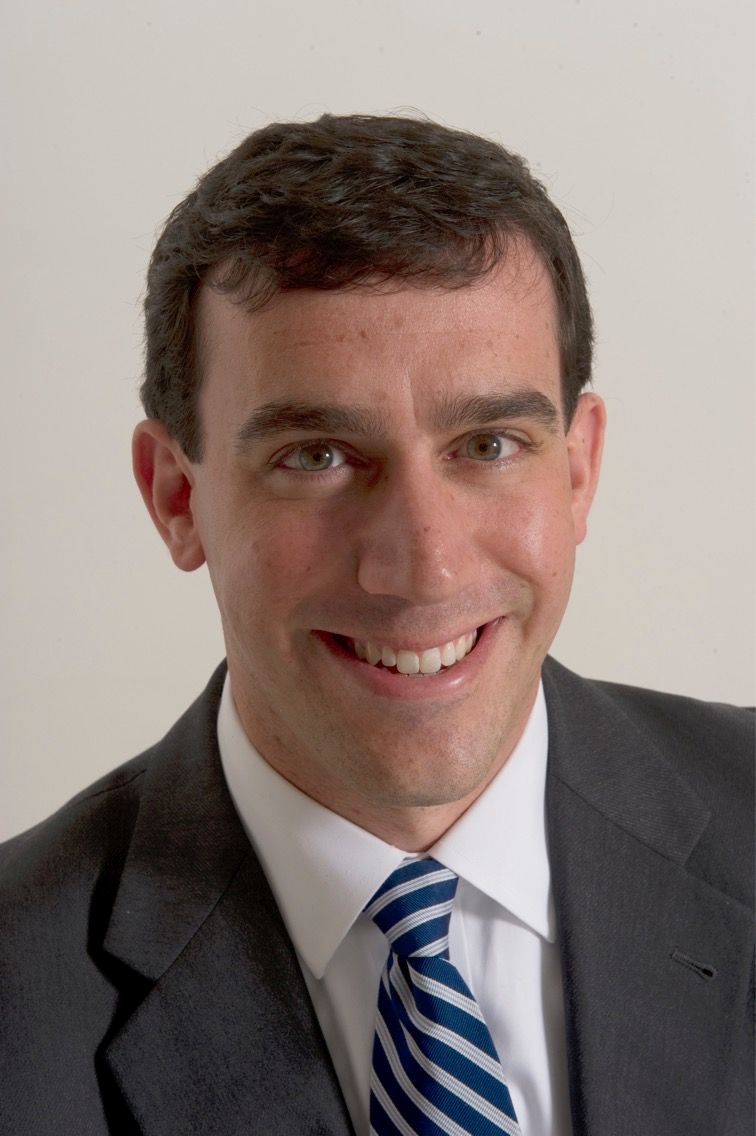
“This project demonstrates that educational technologies can reveal important gaps in surgical education,” said Dr. Leeds, who made comments during his presentation and in an interview.
At issue: The M&M conference, a mainstay of medical education. “This has been defined as the ‘golden hour’ of surgical education,” Dr. Leeds said. “By discussing someone else’s complications, you can learn how to handle your own in the future.”
However, he added, “there’s very little evidence that we’re currently learning from M&M.”
Dr. Leeds and his colleagues are studying the M&M’s role in medical education to see if it can be improved. The new study, a prospective time-series analysis of weekly M&M conferences, aims to understand the potential value of a real-time feedback system. The idea is to develop a way to alert participants to discrepancies in their perceptions about cases.
The researchers turned to a company called Poll Everywhere, whose technology allowed them to collect instant opinions about M&M cases from those in attendance. During 2016-2017, 110 faculty, residents, and interns used Poll Everywhere’s smartphone app to do two things – make guesses about the root causes of adverse events and estimate the risk of complications from surgical procedures over the next 30 days.
“We can see all the results streaming in real time,” said Dr. Leeds, noting that the service cost $600 per year.
The participants, about two-thirds of whom were male, included faculty (35%), fellows and senior residents (28%), and interns and junior residents (37%). They’d been trained an average of 9 years.
The 34 M&M cases represented a mixture of surgical specialties, including oncology, trauma, transplant, and others.
In terms of the root cause analysis, the technology allowed researchers to instantly detect if the guesses of faculty and students were far apart.
The researchers also compared the risk estimates from the participants to those provided by the NSQIP Risk Calculator. They found that the participants tended to boost their estimate of risk, compared with the calculator, by an absolute mean difference of 7.7 percentage points.
“They were overestimating risk by nearly 8 percentage points,” Dr. Leeds said. This isn’t surprising, since other research has revealed a trend toward overestimation of risk by physicians, compared with calculators, he added.
There wasn’t a major difference between the general level of higher estimation of risk among faculty and senior residents (mean of 8.6 and 7.2 percentage points higher than the calculator, respectively). But interns and junior residents estimated risk higher than the calculator by a mean of 4.9 percentage points.
What’s going on? Are the less experienced staff members outperforming their teachers? Another possibility, Dr. Leeds said, is that “the senior surgeons are better picking up on nuances that aren’t being captured by predictive models or the underdeveloped intuition of a junior trainee.”
Rachel Dawn Aufforth, MD, of Johns Hopkins Medicine, who served as discussant for the presentation by Dr. Leeds, said she looks forward to seeing if this technology can improve resident education. She also wondered why estimates via the risk calculator were chosen as a baseline, especially considering that surgeons tend to estimate higher levels of risk.
“One of the things we’ve been trying to do is look at time-series differences,” Dr. Leeds said. “Are they getting better over an academic year? And does that vary by faculty, especially for interns? The calculator isn’t changing or learning on its own.”
In the big picture, the study shows that “collecting real-time risk estimates and root cause assignment is feasible and can be performed as part of routine M&M conferences,” he said.
The study was funded in part by Johns Hopkins University School of Medicine Institute for Excellence in Education. Dr. Leeds reports no relevant disclosures.
SAN DIEGO – Researchers say that they’ve developed an easy and inexpensive way to instantly track divergences in thinking by faculty and students as they ponder cases presented in Mortality and Morbidity (M&M) conferences. They’ve already produced an intriguing early finding: Interns and junior residents hew more closely than do their elders to estimates provided by a surgical risk calculator.
The research has the potential to shed light on problems in the much-maligned M&M, says study leader Ira Leeds, MD, of Johns Hopkins University, Baltimore. He presented the study findings at the annual Clinical Congress of the American College of Surgeons.
“This project demonstrates that educational technologies can reveal important gaps in surgical education,” said Dr. Leeds, who made comments during his presentation and in an interview.
At issue: The M&M conference, a mainstay of medical education. “This has been defined as the ‘golden hour’ of surgical education,” Dr. Leeds said. “By discussing someone else’s complications, you can learn how to handle your own in the future.”
However, he added, “there’s very little evidence that we’re currently learning from M&M.”
Dr. Leeds and his colleagues are studying the M&M’s role in medical education to see if it can be improved. The new study, a prospective time-series analysis of weekly M&M conferences, aims to understand the potential value of a real-time feedback system. The idea is to develop a way to alert participants to discrepancies in their perceptions about cases.
The researchers turned to a company called Poll Everywhere, whose technology allowed them to collect instant opinions about M&M cases from those in attendance. During 2016-2017, 110 faculty, residents, and interns used Poll Everywhere’s smartphone app to do two things – make guesses about the root causes of adverse events and estimate the risk of complications from surgical procedures over the next 30 days.
“We can see all the results streaming in real time,” said Dr. Leeds, noting that the service cost $600 per year.
The participants, about two-thirds of whom were male, included faculty (35%), fellows and senior residents (28%), and interns and junior residents (37%). They’d been trained an average of 9 years.
The 34 M&M cases represented a mixture of surgical specialties, including oncology, trauma, transplant, and others.
In terms of the root cause analysis, the technology allowed researchers to instantly detect if the guesses of faculty and students were far apart.
The researchers also compared the risk estimates from the participants to those provided by the NSQIP Risk Calculator. They found that the participants tended to boost their estimate of risk, compared with the calculator, by an absolute mean difference of 7.7 percentage points.
“They were overestimating risk by nearly 8 percentage points,” Dr. Leeds said. This isn’t surprising, since other research has revealed a trend toward overestimation of risk by physicians, compared with calculators, he added.
There wasn’t a major difference between the general level of higher estimation of risk among faculty and senior residents (mean of 8.6 and 7.2 percentage points higher than the calculator, respectively). But interns and junior residents estimated risk higher than the calculator by a mean of 4.9 percentage points.
What’s going on? Are the less experienced staff members outperforming their teachers? Another possibility, Dr. Leeds said, is that “the senior surgeons are better picking up on nuances that aren’t being captured by predictive models or the underdeveloped intuition of a junior trainee.”
Rachel Dawn Aufforth, MD, of Johns Hopkins Medicine, who served as discussant for the presentation by Dr. Leeds, said she looks forward to seeing if this technology can improve resident education. She also wondered why estimates via the risk calculator were chosen as a baseline, especially considering that surgeons tend to estimate higher levels of risk.
“One of the things we’ve been trying to do is look at time-series differences,” Dr. Leeds said. “Are they getting better over an academic year? And does that vary by faculty, especially for interns? The calculator isn’t changing or learning on its own.”
In the big picture, the study shows that “collecting real-time risk estimates and root cause assignment is feasible and can be performed as part of routine M&M conferences,” he said.
The study was funded in part by Johns Hopkins University School of Medicine Institute for Excellence in Education. Dr. Leeds reports no relevant disclosures.
SAN DIEGO – Researchers say that they’ve developed an easy and inexpensive way to instantly track divergences in thinking by faculty and students as they ponder cases presented in Mortality and Morbidity (M&M) conferences. They’ve already produced an intriguing early finding: Interns and junior residents hew more closely than do their elders to estimates provided by a surgical risk calculator.
The research has the potential to shed light on problems in the much-maligned M&M, says study leader Ira Leeds, MD, of Johns Hopkins University, Baltimore. He presented the study findings at the annual Clinical Congress of the American College of Surgeons.
“This project demonstrates that educational technologies can reveal important gaps in surgical education,” said Dr. Leeds, who made comments during his presentation and in an interview.
At issue: The M&M conference, a mainstay of medical education. “This has been defined as the ‘golden hour’ of surgical education,” Dr. Leeds said. “By discussing someone else’s complications, you can learn how to handle your own in the future.”
However, he added, “there’s very little evidence that we’re currently learning from M&M.”
Dr. Leeds and his colleagues are studying the M&M’s role in medical education to see if it can be improved. The new study, a prospective time-series analysis of weekly M&M conferences, aims to understand the potential value of a real-time feedback system. The idea is to develop a way to alert participants to discrepancies in their perceptions about cases.
The researchers turned to a company called Poll Everywhere, whose technology allowed them to collect instant opinions about M&M cases from those in attendance. During 2016-2017, 110 faculty, residents, and interns used Poll Everywhere’s smartphone app to do two things – make guesses about the root causes of adverse events and estimate the risk of complications from surgical procedures over the next 30 days.
“We can see all the results streaming in real time,” said Dr. Leeds, noting that the service cost $600 per year.
The participants, about two-thirds of whom were male, included faculty (35%), fellows and senior residents (28%), and interns and junior residents (37%). They’d been trained an average of 9 years.
The 34 M&M cases represented a mixture of surgical specialties, including oncology, trauma, transplant, and others.
In terms of the root cause analysis, the technology allowed researchers to instantly detect if the guesses of faculty and students were far apart.
The researchers also compared the risk estimates from the participants to those provided by the NSQIP Risk Calculator. They found that the participants tended to boost their estimate of risk, compared with the calculator, by an absolute mean difference of 7.7 percentage points.
“They were overestimating risk by nearly 8 percentage points,” Dr. Leeds said. This isn’t surprising, since other research has revealed a trend toward overestimation of risk by physicians, compared with calculators, he added.
There wasn’t a major difference between the general level of higher estimation of risk among faculty and senior residents (mean of 8.6 and 7.2 percentage points higher than the calculator, respectively). But interns and junior residents estimated risk higher than the calculator by a mean of 4.9 percentage points.
What’s going on? Are the less experienced staff members outperforming their teachers? Another possibility, Dr. Leeds said, is that “the senior surgeons are better picking up on nuances that aren’t being captured by predictive models or the underdeveloped intuition of a junior trainee.”
Rachel Dawn Aufforth, MD, of Johns Hopkins Medicine, who served as discussant for the presentation by Dr. Leeds, said she looks forward to seeing if this technology can improve resident education. She also wondered why estimates via the risk calculator were chosen as a baseline, especially considering that surgeons tend to estimate higher levels of risk.
“One of the things we’ve been trying to do is look at time-series differences,” Dr. Leeds said. “Are they getting better over an academic year? And does that vary by faculty, especially for interns? The calculator isn’t changing or learning on its own.”
In the big picture, the study shows that “collecting real-time risk estimates and root cause assignment is feasible and can be performed as part of routine M&M conferences,” he said.
The study was funded in part by Johns Hopkins University School of Medicine Institute for Excellence in Education. Dr. Leeds reports no relevant disclosures.
AT THE ACS CLINICAL CONGRESS
Surgeons paid a price for presidential procedures
SAN DIEGO – A surgical team was forced to perform a delicate oral procedure on a rocking yacht while making sure to preserve presidential whiskers. A domineering doctor ignored fellow physicians while a president spent months dying in agony. And, after helping to save the leader of the free world, the leader of the American College of Surgeons found himself viciously attacked by his own colleagues.
When a quartet of ill U.S. presidents developed major medical problems, an audience at the annual clinical congress of the American College of Surgeons learned, their treating physicians ended up with major headaches of their own.
President Grover Cleveland, for example, required his surgical team to remove an oral tumor in total secrecy in 1893, robbing him of a big chunk of his upper palate. “The president had a mustache, and the mustache had to be left alone, and there could be no scars,” said the Hospital for Special Surgery’s J. Patrick O’Leary, MD, FACS, who spoke in a session focused on the history of presidential medicine.
The only light came from a single incandescent bulb, and the procedure was performed at sea, on a yacht anchored off Long Island, N.Y.
“If you were presented with these parameters as a surgeon today, my guess is that you would have demurred on taking on this project,” Dr. O’Leary said. “It was a prescription for a disaster.”
President Cleveland survived for another 15 years. James Garfield, a fellow Civil War veteran, wasn’t so fortunate. In 1881, he was astonishingly unlucky, the unwitting victim of a fumbling physician who dominated his care after an assassin shot him in the chest.
That physician, Willard Bliss, MD, dismissed other doctors who knew the president well and isolated this gregarious man from friends and family. He also ignored emerging knowledge about germ control. And he fed Garfield a heavy diet that the digestively sensitive president probably couldn’t have tolerated in the best of times. The result: endless vomiting, the loss of almost 80 pounds, and an unsuccessful rectal feeding regimen.
Toward the end of the president’s gruesome summer-long decline, Dr. Bliss told all but two doctors to stay away, John B. Hanks, MD, of the University of Virginia, Charlottesville, said in his presentation. Then the president died of a wound that Dr. Hanks said would have been survivable with proper care even in the 1880s.
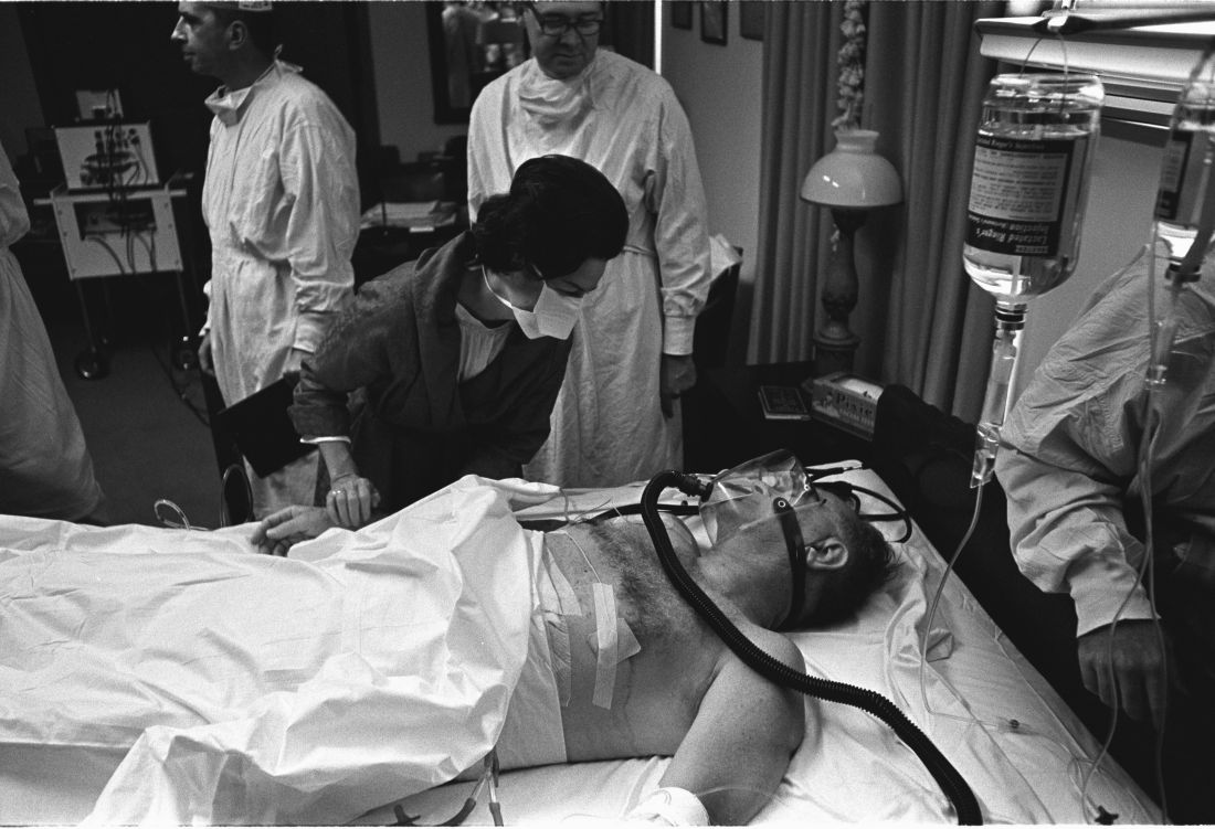
History has been unkind to Dr. Bliss, in part because his patient died. But another presidential physician faced bizarre post surgery scorn from his ACS colleagues, even though his patient lived, according to Justin Barr, MD, PhD, of Duke University, Durham, N.C.
In 1956, surgeon Isidor Ravdin, MD, of the University of Pennsylvania, Philadelphia, was called in when President Dwight D. Eisenhower needed surgery to eliminate a bowel obstruction.
A team of physicians agreed that the president needed surgery. “They felt they were dealing with an elderly, sick patient who’d been in shock during his illness and had recently suffered a myocardial infarction,” Dr. Barr said. “They unanimously decided to proceed with a bypass over resection.”
It’s clear today that the physicians made the correct choice, Dr. Barr said. But his colleagues attacked Dr. Ravdin, who later complained that criticisms multiplied in direct ratio to distance from the operating room.
At the time, Dr. Ravdin was chair of the ACS Board of Regents. The entire board accused him of violating college policies regarding “ghost surgery” (performing procedures without the patient’s knowledge) and “itinerant surgery” (traveling to perform a procedure and then leaving).
Dr. Ravdin acknowledged that he had performed itinerant surgery to some extent, but he denied the ghost surgery charge. In fact, he and the president became friends.
His colleagues also attacked him over his decision to not perform a resection procedure. “They were accusing him of not only being an unethical surgeon, but also an incompetent one,” said Dr. Barr, who calls the letters about the allegations “truly bewildering.”
Also bewildering: Lyndon B. Johnson’s choice to display his gallbladder surgery scar to the press in 1965, spawning one of the most infamous photos of his presidency.
Few surgeons see their handiwork so prominently displayed. Fortunately for them, the operating theater was in a naval hospital, not on a boat. And, as far as we know, no one fretted over the fate of a single facial hair.
SAN DIEGO – A surgical team was forced to perform a delicate oral procedure on a rocking yacht while making sure to preserve presidential whiskers. A domineering doctor ignored fellow physicians while a president spent months dying in agony. And, after helping to save the leader of the free world, the leader of the American College of Surgeons found himself viciously attacked by his own colleagues.
When a quartet of ill U.S. presidents developed major medical problems, an audience at the annual clinical congress of the American College of Surgeons learned, their treating physicians ended up with major headaches of their own.
President Grover Cleveland, for example, required his surgical team to remove an oral tumor in total secrecy in 1893, robbing him of a big chunk of his upper palate. “The president had a mustache, and the mustache had to be left alone, and there could be no scars,” said the Hospital for Special Surgery’s J. Patrick O’Leary, MD, FACS, who spoke in a session focused on the history of presidential medicine.
The only light came from a single incandescent bulb, and the procedure was performed at sea, on a yacht anchored off Long Island, N.Y.
“If you were presented with these parameters as a surgeon today, my guess is that you would have demurred on taking on this project,” Dr. O’Leary said. “It was a prescription for a disaster.”
President Cleveland survived for another 15 years. James Garfield, a fellow Civil War veteran, wasn’t so fortunate. In 1881, he was astonishingly unlucky, the unwitting victim of a fumbling physician who dominated his care after an assassin shot him in the chest.
That physician, Willard Bliss, MD, dismissed other doctors who knew the president well and isolated this gregarious man from friends and family. He also ignored emerging knowledge about germ control. And he fed Garfield a heavy diet that the digestively sensitive president probably couldn’t have tolerated in the best of times. The result: endless vomiting, the loss of almost 80 pounds, and an unsuccessful rectal feeding regimen.
Toward the end of the president’s gruesome summer-long decline, Dr. Bliss told all but two doctors to stay away, John B. Hanks, MD, of the University of Virginia, Charlottesville, said in his presentation. Then the president died of a wound that Dr. Hanks said would have been survivable with proper care even in the 1880s.
History has been unkind to Dr. Bliss, in part because his patient died. But another presidential physician faced bizarre post surgery scorn from his ACS colleagues, even though his patient lived, according to Justin Barr, MD, PhD, of Duke University, Durham, N.C.
In 1956, surgeon Isidor Ravdin, MD, of the University of Pennsylvania, Philadelphia, was called in when President Dwight D. Eisenhower needed surgery to eliminate a bowel obstruction.
A team of physicians agreed that the president needed surgery. “They felt they were dealing with an elderly, sick patient who’d been in shock during his illness and had recently suffered a myocardial infarction,” Dr. Barr said. “They unanimously decided to proceed with a bypass over resection.”
It’s clear today that the physicians made the correct choice, Dr. Barr said. But his colleagues attacked Dr. Ravdin, who later complained that criticisms multiplied in direct ratio to distance from the operating room.
At the time, Dr. Ravdin was chair of the ACS Board of Regents. The entire board accused him of violating college policies regarding “ghost surgery” (performing procedures without the patient’s knowledge) and “itinerant surgery” (traveling to perform a procedure and then leaving).
Dr. Ravdin acknowledged that he had performed itinerant surgery to some extent, but he denied the ghost surgery charge. In fact, he and the president became friends.
His colleagues also attacked him over his decision to not perform a resection procedure. “They were accusing him of not only being an unethical surgeon, but also an incompetent one,” said Dr. Barr, who calls the letters about the allegations “truly bewildering.”
Also bewildering: Lyndon B. Johnson’s choice to display his gallbladder surgery scar to the press in 1965, spawning one of the most infamous photos of his presidency.
Few surgeons see their handiwork so prominently displayed. Fortunately for them, the operating theater was in a naval hospital, not on a boat. And, as far as we know, no one fretted over the fate of a single facial hair.
SAN DIEGO – A surgical team was forced to perform a delicate oral procedure on a rocking yacht while making sure to preserve presidential whiskers. A domineering doctor ignored fellow physicians while a president spent months dying in agony. And, after helping to save the leader of the free world, the leader of the American College of Surgeons found himself viciously attacked by his own colleagues.
When a quartet of ill U.S. presidents developed major medical problems, an audience at the annual clinical congress of the American College of Surgeons learned, their treating physicians ended up with major headaches of their own.
President Grover Cleveland, for example, required his surgical team to remove an oral tumor in total secrecy in 1893, robbing him of a big chunk of his upper palate. “The president had a mustache, and the mustache had to be left alone, and there could be no scars,” said the Hospital for Special Surgery’s J. Patrick O’Leary, MD, FACS, who spoke in a session focused on the history of presidential medicine.
The only light came from a single incandescent bulb, and the procedure was performed at sea, on a yacht anchored off Long Island, N.Y.
“If you were presented with these parameters as a surgeon today, my guess is that you would have demurred on taking on this project,” Dr. O’Leary said. “It was a prescription for a disaster.”
President Cleveland survived for another 15 years. James Garfield, a fellow Civil War veteran, wasn’t so fortunate. In 1881, he was astonishingly unlucky, the unwitting victim of a fumbling physician who dominated his care after an assassin shot him in the chest.
That physician, Willard Bliss, MD, dismissed other doctors who knew the president well and isolated this gregarious man from friends and family. He also ignored emerging knowledge about germ control. And he fed Garfield a heavy diet that the digestively sensitive president probably couldn’t have tolerated in the best of times. The result: endless vomiting, the loss of almost 80 pounds, and an unsuccessful rectal feeding regimen.
Toward the end of the president’s gruesome summer-long decline, Dr. Bliss told all but two doctors to stay away, John B. Hanks, MD, of the University of Virginia, Charlottesville, said in his presentation. Then the president died of a wound that Dr. Hanks said would have been survivable with proper care even in the 1880s.
History has been unkind to Dr. Bliss, in part because his patient died. But another presidential physician faced bizarre post surgery scorn from his ACS colleagues, even though his patient lived, according to Justin Barr, MD, PhD, of Duke University, Durham, N.C.
In 1956, surgeon Isidor Ravdin, MD, of the University of Pennsylvania, Philadelphia, was called in when President Dwight D. Eisenhower needed surgery to eliminate a bowel obstruction.
A team of physicians agreed that the president needed surgery. “They felt they were dealing with an elderly, sick patient who’d been in shock during his illness and had recently suffered a myocardial infarction,” Dr. Barr said. “They unanimously decided to proceed with a bypass over resection.”
It’s clear today that the physicians made the correct choice, Dr. Barr said. But his colleagues attacked Dr. Ravdin, who later complained that criticisms multiplied in direct ratio to distance from the operating room.
At the time, Dr. Ravdin was chair of the ACS Board of Regents. The entire board accused him of violating college policies regarding “ghost surgery” (performing procedures without the patient’s knowledge) and “itinerant surgery” (traveling to perform a procedure and then leaving).
Dr. Ravdin acknowledged that he had performed itinerant surgery to some extent, but he denied the ghost surgery charge. In fact, he and the president became friends.
His colleagues also attacked him over his decision to not perform a resection procedure. “They were accusing him of not only being an unethical surgeon, but also an incompetent one,” said Dr. Barr, who calls the letters about the allegations “truly bewildering.”
Also bewildering: Lyndon B. Johnson’s choice to display his gallbladder surgery scar to the press in 1965, spawning one of the most infamous photos of his presidency.
Few surgeons see their handiwork so prominently displayed. Fortunately for them, the operating theater was in a naval hospital, not on a boat. And, as far as we know, no one fretted over the fate of a single facial hair.
AT THE ACS CLINICAL CONGRESS
Firearms’ injury toll of $3 billion just ‘a drop in the bucket’
SAN DIEGO – The true impact of firearms injuries may be greatly underestimated, according to a study presented at the American College of Surgeons Clinical Congress.
An analysis released earlier this month estimated that firearms injuries cost nearly $3 billion a year in emergency department and inpatient treatment costs. The real cost is likely to be 10-20 times higher, said the lead author of the study, Faiz Gani, MD, a research fellow with the Johns Hopkins Surgery Center for Outcomes Research, Baltimore.
“This is just a drop in the bucket,” Dr. Gani said in an interview at the annual clinical congress of the American College of Surgeons.

Dr. Gani and his colleagues launched their study (Health Affairs 2017;36[10]:1729-38) to better understand the cost of firearms injuries, including nonfatal and accidental injuries.
Most estimates of the cost of firearm injuries are outdated or focused on states or single trauma centers, he said. “Contemporary [costs] for emergency rooms are unknown,” he said. “Also, the numbers come down and shoot up. It’s important to continually study this.”
The statistics are especially important to surgeons, who handle these injuries. “A lot of times the surgeon is the primary health care provider if the patient is injured severely. It’s important that we as surgeons know what’s going on.”
The researchers retrospectively analyzed data from the Nationwide Emergency Department Sample of the Healthcare Cost and Utilization Project for the years 2006-2014. They identified 150,930 patients who appeared alive in emergency departments over that period with firearms injuries, and they estimated the total weighted number at 704,916.
They found that the incidence of firearms injury admissions actually fell during 2006-2013 (from 27.9 visits per 100,000 people to 21.5, P < .001) but bumped up by 23.7% to 26.6 during 2013-2014 (P < .001).
Not surprisingly, more men were injured than women: 45.8 firearms-injured men per 100,000 patients presenting at emergency departments, compared with 5.5 firearms-injured women. Assaults (49.5%) and accidents (35.3%) accounted for most cases, followed by attempted suicides (5.3%) and legal intervention (2.4%).
Those who were assaulted had a higher likelihood of being poor, while those who tried to kill themselves were more likely to have the highest incomes among firearms-injured patients.
The average costs of emergency and inpatient care for patients injured by firearms were $5,254 and $95,887, respectively, collectively amounting to about $2.8 billion each year.
Dr. Gani mentioned that the estimation of the cost and impact of firearms injuries don’t account for people who died of firearms injuries before reaching the emergency department, he says, including patients who committed suicide and died at home.
The cost estimates also don’t take follow-up care, rehabilitation, and lifelong disability into account. The surgical portion of the cost is likely to be much higher because the study doesn’t take future surgical procedures into account, he said.
Based on estimates by the Centers for Disease Control and Prevention of the impact of the injuries, Dr. Gani argued that the true annual cost could be 10 or 20 times the nearly $3 billion estimated by the study.
Discussant Elliott R. Haut, MD, FACS, a trauma surgeon at Johns Hopkins Medicine in Baltimore, agreed that the study estimates of cost and impact estimated in the study represent a small part of a larger toll. Some families and individuals can pay those costs more than once. He recalls hearing from family members of firearm victims who recognize him because they’ve been at the hospital for other shooting incidents. “We’ve all heard someone say, ‘You were here the last time when my brother/cousin/uncle was shot,’ ” he said.
Future research should focus on better understanding the long-term cost of firearm injuries and the influence of socioeconomics and demographics, Dr. Gani said.
Dr. Gani and Dr. Haut reported no relevant disclosures.
SAN DIEGO – The true impact of firearms injuries may be greatly underestimated, according to a study presented at the American College of Surgeons Clinical Congress.
An analysis released earlier this month estimated that firearms injuries cost nearly $3 billion a year in emergency department and inpatient treatment costs. The real cost is likely to be 10-20 times higher, said the lead author of the study, Faiz Gani, MD, a research fellow with the Johns Hopkins Surgery Center for Outcomes Research, Baltimore.
“This is just a drop in the bucket,” Dr. Gani said in an interview at the annual clinical congress of the American College of Surgeons.
Dr. Gani and his colleagues launched their study (Health Affairs 2017;36[10]:1729-38) to better understand the cost of firearms injuries, including nonfatal and accidental injuries.
Most estimates of the cost of firearm injuries are outdated or focused on states or single trauma centers, he said. “Contemporary [costs] for emergency rooms are unknown,” he said. “Also, the numbers come down and shoot up. It’s important to continually study this.”
The statistics are especially important to surgeons, who handle these injuries. “A lot of times the surgeon is the primary health care provider if the patient is injured severely. It’s important that we as surgeons know what’s going on.”
The researchers retrospectively analyzed data from the Nationwide Emergency Department Sample of the Healthcare Cost and Utilization Project for the years 2006-2014. They identified 150,930 patients who appeared alive in emergency departments over that period with firearms injuries, and they estimated the total weighted number at 704,916.
They found that the incidence of firearms injury admissions actually fell during 2006-2013 (from 27.9 visits per 100,000 people to 21.5, P < .001) but bumped up by 23.7% to 26.6 during 2013-2014 (P < .001).
Not surprisingly, more men were injured than women: 45.8 firearms-injured men per 100,000 patients presenting at emergency departments, compared with 5.5 firearms-injured women. Assaults (49.5%) and accidents (35.3%) accounted for most cases, followed by attempted suicides (5.3%) and legal intervention (2.4%).
Those who were assaulted had a higher likelihood of being poor, while those who tried to kill themselves were more likely to have the highest incomes among firearms-injured patients.
The average costs of emergency and inpatient care for patients injured by firearms were $5,254 and $95,887, respectively, collectively amounting to about $2.8 billion each year.
Dr. Gani mentioned that the estimation of the cost and impact of firearms injuries don’t account for people who died of firearms injuries before reaching the emergency department, he says, including patients who committed suicide and died at home.
The cost estimates also don’t take follow-up care, rehabilitation, and lifelong disability into account. The surgical portion of the cost is likely to be much higher because the study doesn’t take future surgical procedures into account, he said.
Based on estimates by the Centers for Disease Control and Prevention of the impact of the injuries, Dr. Gani argued that the true annual cost could be 10 or 20 times the nearly $3 billion estimated by the study.
Discussant Elliott R. Haut, MD, FACS, a trauma surgeon at Johns Hopkins Medicine in Baltimore, agreed that the study estimates of cost and impact estimated in the study represent a small part of a larger toll. Some families and individuals can pay those costs more than once. He recalls hearing from family members of firearm victims who recognize him because they’ve been at the hospital for other shooting incidents. “We’ve all heard someone say, ‘You were here the last time when my brother/cousin/uncle was shot,’ ” he said.
Future research should focus on better understanding the long-term cost of firearm injuries and the influence of socioeconomics and demographics, Dr. Gani said.
Dr. Gani and Dr. Haut reported no relevant disclosures.
SAN DIEGO – The true impact of firearms injuries may be greatly underestimated, according to a study presented at the American College of Surgeons Clinical Congress.
An analysis released earlier this month estimated that firearms injuries cost nearly $3 billion a year in emergency department and inpatient treatment costs. The real cost is likely to be 10-20 times higher, said the lead author of the study, Faiz Gani, MD, a research fellow with the Johns Hopkins Surgery Center for Outcomes Research, Baltimore.
“This is just a drop in the bucket,” Dr. Gani said in an interview at the annual clinical congress of the American College of Surgeons.
Dr. Gani and his colleagues launched their study (Health Affairs 2017;36[10]:1729-38) to better understand the cost of firearms injuries, including nonfatal and accidental injuries.
Most estimates of the cost of firearm injuries are outdated or focused on states or single trauma centers, he said. “Contemporary [costs] for emergency rooms are unknown,” he said. “Also, the numbers come down and shoot up. It’s important to continually study this.”
The statistics are especially important to surgeons, who handle these injuries. “A lot of times the surgeon is the primary health care provider if the patient is injured severely. It’s important that we as surgeons know what’s going on.”
The researchers retrospectively analyzed data from the Nationwide Emergency Department Sample of the Healthcare Cost and Utilization Project for the years 2006-2014. They identified 150,930 patients who appeared alive in emergency departments over that period with firearms injuries, and they estimated the total weighted number at 704,916.
They found that the incidence of firearms injury admissions actually fell during 2006-2013 (from 27.9 visits per 100,000 people to 21.5, P < .001) but bumped up by 23.7% to 26.6 during 2013-2014 (P < .001).
Not surprisingly, more men were injured than women: 45.8 firearms-injured men per 100,000 patients presenting at emergency departments, compared with 5.5 firearms-injured women. Assaults (49.5%) and accidents (35.3%) accounted for most cases, followed by attempted suicides (5.3%) and legal intervention (2.4%).
Those who were assaulted had a higher likelihood of being poor, while those who tried to kill themselves were more likely to have the highest incomes among firearms-injured patients.
The average costs of emergency and inpatient care for patients injured by firearms were $5,254 and $95,887, respectively, collectively amounting to about $2.8 billion each year.
Dr. Gani mentioned that the estimation of the cost and impact of firearms injuries don’t account for people who died of firearms injuries before reaching the emergency department, he says, including patients who committed suicide and died at home.
The cost estimates also don’t take follow-up care, rehabilitation, and lifelong disability into account. The surgical portion of the cost is likely to be much higher because the study doesn’t take future surgical procedures into account, he said.
Based on estimates by the Centers for Disease Control and Prevention of the impact of the injuries, Dr. Gani argued that the true annual cost could be 10 or 20 times the nearly $3 billion estimated by the study.
Discussant Elliott R. Haut, MD, FACS, a trauma surgeon at Johns Hopkins Medicine in Baltimore, agreed that the study estimates of cost and impact estimated in the study represent a small part of a larger toll. Some families and individuals can pay those costs more than once. He recalls hearing from family members of firearm victims who recognize him because they’ve been at the hospital for other shooting incidents. “We’ve all heard someone say, ‘You were here the last time when my brother/cousin/uncle was shot,’ ” he said.
Future research should focus on better understanding the long-term cost of firearm injuries and the influence of socioeconomics and demographics, Dr. Gani said.
Dr. Gani and Dr. Haut reported no relevant disclosures.
AT THE ACS CLINICAL CONGRESS
Many women have unprotected sex in year after bariatric surgery
More than 40% of reproductive-age women reported having unprotected sex in the year after undergoing bariatric surgery, despite recommendations to avoid pregnancy for at least a year, a new study finds. Another 4% of women reported trying to conceive in the 12 months after surgery.
“We were surprised to find such a large percentage of women were not using contraception, and we were also surprised to find so many were actively trying to conceive,” the study’s lead author Marie N. Menke, MD, MPH, of the University of Pittsburgh, said in an interview. “Reproductive-age women who are considering bariatric surgery should be counseled that they should plan to use contraceptives after surgery for about 12-18 months.”
Pregnancy isn’t recommended over that period mainly because of the risks to the fetus, Dr. Menke said. “These risks are different from obesity in general. We don’t know exactly why surgery causes these risks, but ideally, patients would be weight-stable prior to conception.”
In 2013, the American Association of Clinical Endocrinologists, the Obesity Society, and the American Society for Metabolic & Bariatric Surgery provided a Grade D recommendation for a 12-18 month delay in conception (Obesity [Silver Spring] 2013;21[suppl 1]:S1-27). A recent study provided more insight into the potential risks of pregnancy soon after weight-loss procedures. It reported that, compared with those who gave birth more than 4 years later, women who gave birth within 2 years of bariatric surgery had higher risks of premature birth, admission to neonatal intensive care units, and small-for-gestational-age infants (JAMA Surg. 2017 Feb 1;152[2]:1-8).
The current prospective cohort study included women who underwent bariatric surgery for the first time during 2005-2009 at 10 U.S. hospitals. The women, all aged 18-44 years, answered questions for as long as 7 years, until January 2015 (Obstet Gynecol. 2017 Oct 6. doi: 10.1097/AOG.0000000000002323).
The analysis included 710 women who provided conception data. The median body mass index of the women was 46.3 kg/m2. Most patients underwent Roux-en-Y gastric bypass (73%), followed by laparoscopic adjustable gastric banding (23%).
Researchers found that 4.3% of the women tried to conceive in the first year after surgery (95% confidence interval, 2.4-6.3), and 13.1% did so in the second year (95% CI, 9.3-17.0; P less than .001).
During the first year after surgery, 12.7% of women had no intercourse (95% CI, 9.4-16.0), 40.5% had only protected intercourse (95% CI, 35.6-45.4), and 41.5% (95% CI, 36.4-46.6) had unprotected intercourse while not trying to conceive.
Why are the unprotected sex numbers so high? “We wonder if some women simply feel that they cannot get pregnant,” Dr. Menke said. “Our group has previously reported that 42% of women who had attempted to conceive prior to bariatric surgery had a history of infertility. Some of these women went on to deliver a live birth, but many did not.”
The study reports that 183 of the 710 women did ultimately conceive and that the number may be as high as 237 because data were missing for 54 women who may have not wanted to report a nonlive birth. Of the women who reported conceiving, 68.9% had live births, 1.1% had stillbirths, 1.1% had ectopic pregnancies, 21.9% miscarried and 7.1% had abortions.
“We’d really like this research to be a reason to consider presurgical contraceptive counseling as one of the steps prior to bariatric surgery,” Dr. Menke said. “This may include a referral by the bariatric surgeon to a physician would who provide counseling, prescribe, or both.”
The study was funded by the National Institute of Diabetes and Digestive and Kidney Diseases. Dr. Menke reported having no relevant disclosures. Her coauthors reported financial relationships with pharmaceutical and medical device companies with products to treat metabolic diseases.
More than 40% of reproductive-age women reported having unprotected sex in the year after undergoing bariatric surgery, despite recommendations to avoid pregnancy for at least a year, a new study finds. Another 4% of women reported trying to conceive in the 12 months after surgery.
“We were surprised to find such a large percentage of women were not using contraception, and we were also surprised to find so many were actively trying to conceive,” the study’s lead author Marie N. Menke, MD, MPH, of the University of Pittsburgh, said in an interview. “Reproductive-age women who are considering bariatric surgery should be counseled that they should plan to use contraceptives after surgery for about 12-18 months.”
Pregnancy isn’t recommended over that period mainly because of the risks to the fetus, Dr. Menke said. “These risks are different from obesity in general. We don’t know exactly why surgery causes these risks, but ideally, patients would be weight-stable prior to conception.”
In 2013, the American Association of Clinical Endocrinologists, the Obesity Society, and the American Society for Metabolic & Bariatric Surgery provided a Grade D recommendation for a 12-18 month delay in conception (Obesity [Silver Spring] 2013;21[suppl 1]:S1-27). A recent study provided more insight into the potential risks of pregnancy soon after weight-loss procedures. It reported that, compared with those who gave birth more than 4 years later, women who gave birth within 2 years of bariatric surgery had higher risks of premature birth, admission to neonatal intensive care units, and small-for-gestational-age infants (JAMA Surg. 2017 Feb 1;152[2]:1-8).
The current prospective cohort study included women who underwent bariatric surgery for the first time during 2005-2009 at 10 U.S. hospitals. The women, all aged 18-44 years, answered questions for as long as 7 years, until January 2015 (Obstet Gynecol. 2017 Oct 6. doi: 10.1097/AOG.0000000000002323).
The analysis included 710 women who provided conception data. The median body mass index of the women was 46.3 kg/m2. Most patients underwent Roux-en-Y gastric bypass (73%), followed by laparoscopic adjustable gastric banding (23%).
Researchers found that 4.3% of the women tried to conceive in the first year after surgery (95% confidence interval, 2.4-6.3), and 13.1% did so in the second year (95% CI, 9.3-17.0; P less than .001).
During the first year after surgery, 12.7% of women had no intercourse (95% CI, 9.4-16.0), 40.5% had only protected intercourse (95% CI, 35.6-45.4), and 41.5% (95% CI, 36.4-46.6) had unprotected intercourse while not trying to conceive.
Why are the unprotected sex numbers so high? “We wonder if some women simply feel that they cannot get pregnant,” Dr. Menke said. “Our group has previously reported that 42% of women who had attempted to conceive prior to bariatric surgery had a history of infertility. Some of these women went on to deliver a live birth, but many did not.”
The study reports that 183 of the 710 women did ultimately conceive and that the number may be as high as 237 because data were missing for 54 women who may have not wanted to report a nonlive birth. Of the women who reported conceiving, 68.9% had live births, 1.1% had stillbirths, 1.1% had ectopic pregnancies, 21.9% miscarried and 7.1% had abortions.
“We’d really like this research to be a reason to consider presurgical contraceptive counseling as one of the steps prior to bariatric surgery,” Dr. Menke said. “This may include a referral by the bariatric surgeon to a physician would who provide counseling, prescribe, or both.”
The study was funded by the National Institute of Diabetes and Digestive and Kidney Diseases. Dr. Menke reported having no relevant disclosures. Her coauthors reported financial relationships with pharmaceutical and medical device companies with products to treat metabolic diseases.
More than 40% of reproductive-age women reported having unprotected sex in the year after undergoing bariatric surgery, despite recommendations to avoid pregnancy for at least a year, a new study finds. Another 4% of women reported trying to conceive in the 12 months after surgery.
“We were surprised to find such a large percentage of women were not using contraception, and we were also surprised to find so many were actively trying to conceive,” the study’s lead author Marie N. Menke, MD, MPH, of the University of Pittsburgh, said in an interview. “Reproductive-age women who are considering bariatric surgery should be counseled that they should plan to use contraceptives after surgery for about 12-18 months.”
Pregnancy isn’t recommended over that period mainly because of the risks to the fetus, Dr. Menke said. “These risks are different from obesity in general. We don’t know exactly why surgery causes these risks, but ideally, patients would be weight-stable prior to conception.”
In 2013, the American Association of Clinical Endocrinologists, the Obesity Society, and the American Society for Metabolic & Bariatric Surgery provided a Grade D recommendation for a 12-18 month delay in conception (Obesity [Silver Spring] 2013;21[suppl 1]:S1-27). A recent study provided more insight into the potential risks of pregnancy soon after weight-loss procedures. It reported that, compared with those who gave birth more than 4 years later, women who gave birth within 2 years of bariatric surgery had higher risks of premature birth, admission to neonatal intensive care units, and small-for-gestational-age infants (JAMA Surg. 2017 Feb 1;152[2]:1-8).
The current prospective cohort study included women who underwent bariatric surgery for the first time during 2005-2009 at 10 U.S. hospitals. The women, all aged 18-44 years, answered questions for as long as 7 years, until January 2015 (Obstet Gynecol. 2017 Oct 6. doi: 10.1097/AOG.0000000000002323).
The analysis included 710 women who provided conception data. The median body mass index of the women was 46.3 kg/m2. Most patients underwent Roux-en-Y gastric bypass (73%), followed by laparoscopic adjustable gastric banding (23%).
Researchers found that 4.3% of the women tried to conceive in the first year after surgery (95% confidence interval, 2.4-6.3), and 13.1% did so in the second year (95% CI, 9.3-17.0; P less than .001).
During the first year after surgery, 12.7% of women had no intercourse (95% CI, 9.4-16.0), 40.5% had only protected intercourse (95% CI, 35.6-45.4), and 41.5% (95% CI, 36.4-46.6) had unprotected intercourse while not trying to conceive.
Why are the unprotected sex numbers so high? “We wonder if some women simply feel that they cannot get pregnant,” Dr. Menke said. “Our group has previously reported that 42% of women who had attempted to conceive prior to bariatric surgery had a history of infertility. Some of these women went on to deliver a live birth, but many did not.”
The study reports that 183 of the 710 women did ultimately conceive and that the number may be as high as 237 because data were missing for 54 women who may have not wanted to report a nonlive birth. Of the women who reported conceiving, 68.9% had live births, 1.1% had stillbirths, 1.1% had ectopic pregnancies, 21.9% miscarried and 7.1% had abortions.
“We’d really like this research to be a reason to consider presurgical contraceptive counseling as one of the steps prior to bariatric surgery,” Dr. Menke said. “This may include a referral by the bariatric surgeon to a physician would who provide counseling, prescribe, or both.”
The study was funded by the National Institute of Diabetes and Digestive and Kidney Diseases. Dr. Menke reported having no relevant disclosures. Her coauthors reported financial relationships with pharmaceutical and medical device companies with products to treat metabolic diseases.
FROM OBSTETRICS & GYNECOLOGY
Key clinical point:
Major finding: In the first year after surgery, 4.3% of women surveyed tried to conceive (95% CI, 2.4-6.3), and another 41.5% had unprotected intercourse (95% CI, 36.4-46.6).
Data source: Prospective cohort study of 710 women, aged 18-44 years, who underwent bariatric surgery for the first time during 2005-2009 at 10 U.S. hospitals.
Disclosures: The study was funded by the National Institute of Diabetes and Digestive and Kidney Diseases. Dr. Menke reported having no relevant disclosures. Her coauthors reported financial relationships with pharmaceutical and medical device companies with products to treat metabolic diseases.
Chronic passive exposure to cannabis smoke linked to dependence
Secondhand exposure to cannabis smoke appears linked to signs of cannabis dependence, a researcher said at a Drug Enforcement Agency (DEA) Museum forum on marijuana.
The researcher, Adriaan W. Bruijnzeel, PhD, said at the forum that his team’s work with rats raises the prospect of a link between chronic passive exposure and addiction.

“Immediately after the smoke exposure, you can detect high levels of THC [tetrahydrocannabinol], and then there’s a very quick drop,” Dr. Bruijnzeel, of the department of psychiatry at the University of Florida, Gainesville, said at the Oct. 5 forum at the DEA Museum in Arlington, Va. “Negative mood state associated with the cessation of drug intake helps to maintain the drug addiction.”
In an interview, Mark S. Gold, MD, an expert in addiction who serves as the chair of RiverMend Health’s scientific advisory boards, said Dr. Bruijnzeel’s findings offer lessons for clinicians, particularly for those who treat children.
“Like tobacco smoke, marijuana smoke is an environmental toxin that causes brain changes and addiction,” said Dr. Gold, the 17th Distinguished Alumni Professor at the University of Florida who is also with Washington University in St. Louis. “Second and thirdhand effects of cannabis are important new risks to consider when evaluating the children of marijuana smokers.”
Meanwhile, presenters at the forum, which was webcast, also described marijuana misuse as a threat to general mental health and the adolescent brain, as well as a danger to drivers.
Can preventing cannabis use reduce mental illness? “I think the answer is yes, but the pathway is likely to be fairly complicated and not as straightforward as causation,” said Arpana Agrawal, PhD, a professor of psychiatry at Washington University in St. Louis. “Without a doubt, reduction of cannabis, particularly heavy and persistent use, will likely assist in recovery from psychiatric illness.”
In regard to marijuana’s link to psychosis, in particular, the picture is complex, she said. “There does not appear to be much evidence for a straightforward causal model,” she said. “There’s some evidence of risk in genetically vulnerable individuals, there’s overwhelming support for shared biology; factors other than shared biology are likely to be important. And there’s also some support for increased correlation in the context of high potency use.”
Also, evidence suggests that cannabis is a “reverse gateway” drug among U.S. youth, as well as young people in Australia. “The idea is that youth actually initiate their substance use trajectories with marijuana and then work their way back to nicotine,” Dr. Agrawal said.
A stark warning about marijuana use came from Bertha K. Madras, PhD, professor of psychobiology in the department of psychiatry at Harvard Medical School, Boston; a former deputy director of demand reduction at the Office of National Drug Control Policy; and a member of the President’s Commission on Combating Drug Addiction and the Opioid Crisis, chaired by Gov. Chris Christie (R-N.J.). “We’re not waging a war on drugs,” she said. “We are, in fact, defending our brain, which is the repository of our humanity. And supply reduction, which is what the DEA focuses on to some extent, is a form of prevention. ”
She encouraged adopting a prevention message to discourage all drug use by youth.
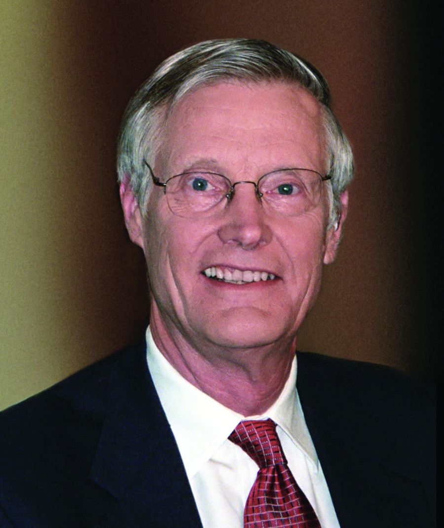
Robert L. DuPont, MD, who moderated the forum, said heavy, chronic cannabis users show impairment of psychomotor skills linked to driving for as long as 3 weeks after last use. He also pointed to 2010-2014 data from Washington state that showed increases in the percentages of drivers involved in fatal crashes who tested positive for THC, mainly in addition to alcohol and/or other drugs.
There’s hope that a test will be developed to determine the cannabis tissue level that causes the equivalent impairment as the 0.08 g/dL blood alcohol content standard used to define drunken driving because there is no consistent relationship between THC levels and impairment, said Dr. DuPont, who is the first director of the National Institute on Drug Abuse, the second drug czar, and the president of the Institute for Behavior and Health in Rockville, Md.
Still, several states make driving illegal for drivers with any level of THC and/or THC metabolites, according to the Governors Highway Safety Association. THC can stay in the body for days after marijuana use, while metabolites can remain for weeks, according to the Marijuana Policy Project. An additional 16 states outlaw driving with specific THC levels.
For his part, Dr. DuPont said that, although addressing the opioid epidemic is a top national priority, the legalization of marijuana may be the more enduring threat to the nation’s public health; legalization would make it the third legal drug, joining alcohol and tobacco, which are the two leading causes of illness and death in the country.
“This is the third time in the last 45 years that drugs are front and center” in the United States, Dr. DuPont said. “The first one was heroin addiction related to crime in the early 1970s; the second was the crack epidemic in the late 1980s. In many ways, I think [marijuana is] the more important issue for us.”
Dr. Brijnzeel had no disclosures. Dr. Gold serves as chairman of the scientific advisory boards for RiverMend Health. Dr. Agrawal disclosed NIDA grants. Dr. Madras reported serving on the RiverMend advisory board and working with several organizations, such as the U.S. Department of Justice and the American Bar Association. Dr. DuPont also serves on the RiverMend advisory board.
Secondhand exposure to cannabis smoke appears linked to signs of cannabis dependence, a researcher said at a Drug Enforcement Agency (DEA) Museum forum on marijuana.
The researcher, Adriaan W. Bruijnzeel, PhD, said at the forum that his team’s work with rats raises the prospect of a link between chronic passive exposure and addiction.
“Immediately after the smoke exposure, you can detect high levels of THC [tetrahydrocannabinol], and then there’s a very quick drop,” Dr. Bruijnzeel, of the department of psychiatry at the University of Florida, Gainesville, said at the Oct. 5 forum at the DEA Museum in Arlington, Va. “Negative mood state associated with the cessation of drug intake helps to maintain the drug addiction.”
In an interview, Mark S. Gold, MD, an expert in addiction who serves as the chair of RiverMend Health’s scientific advisory boards, said Dr. Bruijnzeel’s findings offer lessons for clinicians, particularly for those who treat children.
“Like tobacco smoke, marijuana smoke is an environmental toxin that causes brain changes and addiction,” said Dr. Gold, the 17th Distinguished Alumni Professor at the University of Florida who is also with Washington University in St. Louis. “Second and thirdhand effects of cannabis are important new risks to consider when evaluating the children of marijuana smokers.”
Meanwhile, presenters at the forum, which was webcast, also described marijuana misuse as a threat to general mental health and the adolescent brain, as well as a danger to drivers.
Can preventing cannabis use reduce mental illness? “I think the answer is yes, but the pathway is likely to be fairly complicated and not as straightforward as causation,” said Arpana Agrawal, PhD, a professor of psychiatry at Washington University in St. Louis. “Without a doubt, reduction of cannabis, particularly heavy and persistent use, will likely assist in recovery from psychiatric illness.”
In regard to marijuana’s link to psychosis, in particular, the picture is complex, she said. “There does not appear to be much evidence for a straightforward causal model,” she said. “There’s some evidence of risk in genetically vulnerable individuals, there’s overwhelming support for shared biology; factors other than shared biology are likely to be important. And there’s also some support for increased correlation in the context of high potency use.”
Also, evidence suggests that cannabis is a “reverse gateway” drug among U.S. youth, as well as young people in Australia. “The idea is that youth actually initiate their substance use trajectories with marijuana and then work their way back to nicotine,” Dr. Agrawal said.
A stark warning about marijuana use came from Bertha K. Madras, PhD, professor of psychobiology in the department of psychiatry at Harvard Medical School, Boston; a former deputy director of demand reduction at the Office of National Drug Control Policy; and a member of the President’s Commission on Combating Drug Addiction and the Opioid Crisis, chaired by Gov. Chris Christie (R-N.J.). “We’re not waging a war on drugs,” she said. “We are, in fact, defending our brain, which is the repository of our humanity. And supply reduction, which is what the DEA focuses on to some extent, is a form of prevention. ”
She encouraged adopting a prevention message to discourage all drug use by youth.
Robert L. DuPont, MD, who moderated the forum, said heavy, chronic cannabis users show impairment of psychomotor skills linked to driving for as long as 3 weeks after last use. He also pointed to 2010-2014 data from Washington state that showed increases in the percentages of drivers involved in fatal crashes who tested positive for THC, mainly in addition to alcohol and/or other drugs.
There’s hope that a test will be developed to determine the cannabis tissue level that causes the equivalent impairment as the 0.08 g/dL blood alcohol content standard used to define drunken driving because there is no consistent relationship between THC levels and impairment, said Dr. DuPont, who is the first director of the National Institute on Drug Abuse, the second drug czar, and the president of the Institute for Behavior and Health in Rockville, Md.
Still, several states make driving illegal for drivers with any level of THC and/or THC metabolites, according to the Governors Highway Safety Association. THC can stay in the body for days after marijuana use, while metabolites can remain for weeks, according to the Marijuana Policy Project. An additional 16 states outlaw driving with specific THC levels.
For his part, Dr. DuPont said that, although addressing the opioid epidemic is a top national priority, the legalization of marijuana may be the more enduring threat to the nation’s public health; legalization would make it the third legal drug, joining alcohol and tobacco, which are the two leading causes of illness and death in the country.
“This is the third time in the last 45 years that drugs are front and center” in the United States, Dr. DuPont said. “The first one was heroin addiction related to crime in the early 1970s; the second was the crack epidemic in the late 1980s. In many ways, I think [marijuana is] the more important issue for us.”
Dr. Brijnzeel had no disclosures. Dr. Gold serves as chairman of the scientific advisory boards for RiverMend Health. Dr. Agrawal disclosed NIDA grants. Dr. Madras reported serving on the RiverMend advisory board and working with several organizations, such as the U.S. Department of Justice and the American Bar Association. Dr. DuPont also serves on the RiverMend advisory board.
Secondhand exposure to cannabis smoke appears linked to signs of cannabis dependence, a researcher said at a Drug Enforcement Agency (DEA) Museum forum on marijuana.
The researcher, Adriaan W. Bruijnzeel, PhD, said at the forum that his team’s work with rats raises the prospect of a link between chronic passive exposure and addiction.
“Immediately after the smoke exposure, you can detect high levels of THC [tetrahydrocannabinol], and then there’s a very quick drop,” Dr. Bruijnzeel, of the department of psychiatry at the University of Florida, Gainesville, said at the Oct. 5 forum at the DEA Museum in Arlington, Va. “Negative mood state associated with the cessation of drug intake helps to maintain the drug addiction.”
In an interview, Mark S. Gold, MD, an expert in addiction who serves as the chair of RiverMend Health’s scientific advisory boards, said Dr. Bruijnzeel’s findings offer lessons for clinicians, particularly for those who treat children.
“Like tobacco smoke, marijuana smoke is an environmental toxin that causes brain changes and addiction,” said Dr. Gold, the 17th Distinguished Alumni Professor at the University of Florida who is also with Washington University in St. Louis. “Second and thirdhand effects of cannabis are important new risks to consider when evaluating the children of marijuana smokers.”
Meanwhile, presenters at the forum, which was webcast, also described marijuana misuse as a threat to general mental health and the adolescent brain, as well as a danger to drivers.
Can preventing cannabis use reduce mental illness? “I think the answer is yes, but the pathway is likely to be fairly complicated and not as straightforward as causation,” said Arpana Agrawal, PhD, a professor of psychiatry at Washington University in St. Louis. “Without a doubt, reduction of cannabis, particularly heavy and persistent use, will likely assist in recovery from psychiatric illness.”
In regard to marijuana’s link to psychosis, in particular, the picture is complex, she said. “There does not appear to be much evidence for a straightforward causal model,” she said. “There’s some evidence of risk in genetically vulnerable individuals, there’s overwhelming support for shared biology; factors other than shared biology are likely to be important. And there’s also some support for increased correlation in the context of high potency use.”
Also, evidence suggests that cannabis is a “reverse gateway” drug among U.S. youth, as well as young people in Australia. “The idea is that youth actually initiate their substance use trajectories with marijuana and then work their way back to nicotine,” Dr. Agrawal said.
A stark warning about marijuana use came from Bertha K. Madras, PhD, professor of psychobiology in the department of psychiatry at Harvard Medical School, Boston; a former deputy director of demand reduction at the Office of National Drug Control Policy; and a member of the President’s Commission on Combating Drug Addiction and the Opioid Crisis, chaired by Gov. Chris Christie (R-N.J.). “We’re not waging a war on drugs,” she said. “We are, in fact, defending our brain, which is the repository of our humanity. And supply reduction, which is what the DEA focuses on to some extent, is a form of prevention. ”
She encouraged adopting a prevention message to discourage all drug use by youth.
Robert L. DuPont, MD, who moderated the forum, said heavy, chronic cannabis users show impairment of psychomotor skills linked to driving for as long as 3 weeks after last use. He also pointed to 2010-2014 data from Washington state that showed increases in the percentages of drivers involved in fatal crashes who tested positive for THC, mainly in addition to alcohol and/or other drugs.
There’s hope that a test will be developed to determine the cannabis tissue level that causes the equivalent impairment as the 0.08 g/dL blood alcohol content standard used to define drunken driving because there is no consistent relationship between THC levels and impairment, said Dr. DuPont, who is the first director of the National Institute on Drug Abuse, the second drug czar, and the president of the Institute for Behavior and Health in Rockville, Md.
Still, several states make driving illegal for drivers with any level of THC and/or THC metabolites, according to the Governors Highway Safety Association. THC can stay in the body for days after marijuana use, while metabolites can remain for weeks, according to the Marijuana Policy Project. An additional 16 states outlaw driving with specific THC levels.
For his part, Dr. DuPont said that, although addressing the opioid epidemic is a top national priority, the legalization of marijuana may be the more enduring threat to the nation’s public health; legalization would make it the third legal drug, joining alcohol and tobacco, which are the two leading causes of illness and death in the country.
“This is the third time in the last 45 years that drugs are front and center” in the United States, Dr. DuPont said. “The first one was heroin addiction related to crime in the early 1970s; the second was the crack epidemic in the late 1980s. In many ways, I think [marijuana is] the more important issue for us.”
Dr. Brijnzeel had no disclosures. Dr. Gold serves as chairman of the scientific advisory boards for RiverMend Health. Dr. Agrawal disclosed NIDA grants. Dr. Madras reported serving on the RiverMend advisory board and working with several organizations, such as the U.S. Department of Justice and the American Bar Association. Dr. DuPont also serves on the RiverMend advisory board.
Wound expert: Consider hyperbaric oxygen therapy for diabetic foot ulcers
SAN DIEGO – Hyperbaric oxygen therapy, a mainstay of wound care, has a long and controversial history as a treatment for diabetic foot ulcers. Conflicting studies have spawned plenty of debate, and the most recent Cochrane Library review of existing research didn’t shed much light on the value of the treatment because the evidence was weak (Cochrane Database Syst Rev. 2015 Jun 24;[6]:CD004123).
But William H. Tettelbach, MD, a wound care specialist, told an audience at the annual scientific sessions of the American Diabetic Association that hyperbaric treatments are worth a try in certain cases. And he brought evidence to prove it – a 2015 report he coauthored that reviewed studies and offered clinical practice guidelines for hyperbaric oxygen therapy for the treatment of diabetic foot ulcers (DFUs) (Undersea Hyperb Med. 2015 May-Jun;42[3]:205-47).
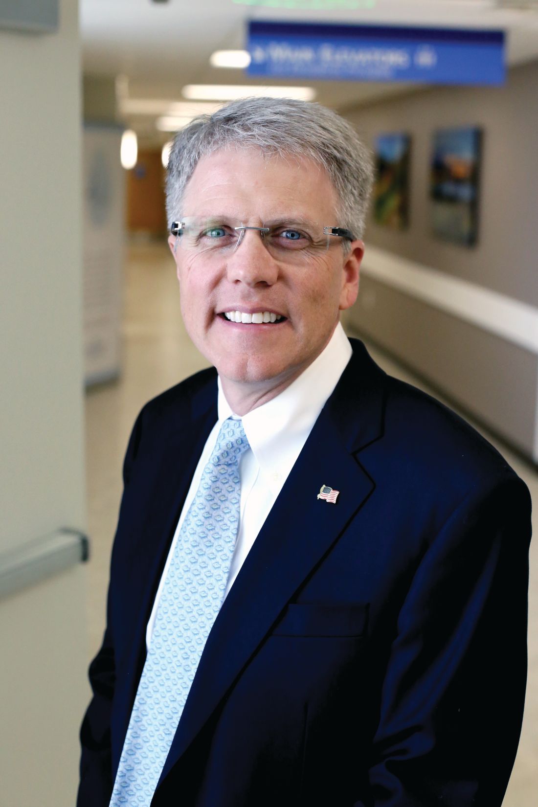
In an interview, Dr. Tettelbach discussed ideal candidates for the treatment and offered clinical advice to endocrinologists.
Question: What did your review of research tell you about the value of hyperbaric oxygen treatment for DFUs?
Answer: We came to the same conclusion that most of the papers have indicated over the years: Hyperbaric oxygen is effective and attains goals such as reducing rates of amputation in a select population of diabetic ulcer patients.
Patients who have Wagner grade 3 or greater ulcers or admitted for surgery due to a septic diabetic foot benefit from an evaluation by a hyperbaric medicine–trained physician and treatment when indicated. There is evidence and years of clinical experience indicating that these patients benefit and have improved outcomes when evaluated and treated appropriately with hyperbaric oxygen therapy.
In the United States, hyperbaric oxygen therapy is not indicated in Wagner grade 2, 1 or 0 diabetic foot ulcers, the ulcers that involve soft tissue but not deep structure like bone.
Q: Why has there been so much controversy over the value of this treatment?
A: In the past, there have been problems with commercial outpatient wound centers that are heavily driven by profits. Financial margins in wound care clinics can be tight, and the need to remain profitable has at times resulted in patients being treated inappropriately with hyperbaric oxygen therapy (Adv Skin Wound Care. 2017 Apr;30[4]:181-90).
Q: Why does hyperbaric oxygen treatment work in some cases?
A: When you place a patient in a hyperbaric chamber where they breathe 100% oxygen under pressure, you increase the percentage of oxygen in the blood. At such a high percentage, oxygen saturates the plasma versus just being carried by red blood cells, thereby allowing the oxygen to penetrate farther into hypoxic tissues. By increasing the oxygen, you have the ability to make the environment unfavorable for rapid proliferation of anaerobic or microaerophilic bacteria that do not survive a highly oxygen-rich environment. Increasing tissue oxygen tension to 30 mm Hg or greater increases the macrophages’ ability to have an oxidative burst needed to kill bacteria. Furthermore, there are antibiotics that require certain levels of oxygen for transport across the bacterial cell wall.
Q: What should physicians understand about hyperbaric oxygen therapy for DFUs?
A: Overall, hyperbaric practitioners need to be more selective in identifying and treating patients according to what the evidence supports. Poorly designed trials with misleading results should not drive medical decisions. We should revisit diabetic foot ulcers through well-thought-out studies that target those who would benefit as suggested by current evidence. Prior trials have been heavily weighted with Wagner grade 1 and 2 candidates or ischemic diabetic ulcers that are not revascularized. These are biased toward poor outcomes since the current evidence does not strongly support treating these types of individuals with adjunctive hyperbaric oxygen therapy (Ont Health Technol Assess Ser. 2017 May 12;17[5]:1-142. eCollection 2017).
Q: What conditions should trigger endocrinologists to think about hyperbaric oxygen therapy for their DFU patients?
A: Candidates for the therapy include diabetic ulcers that have persisted for longer than 30 days, since these ulcers are at a significantly higher risk of a complicating infection, along with those that have failed treatment or are becoming more symptomatic over time (Undersea Hyperb Med. 2017 Mar-Apr;44[2]:157-60).
At that point, it might make sense to refer those patients to a wound and hyperbaric specialist for further evaluation and management, especially to a wound center that offers hyperbaric oxygen therapy.
These wound centers can be found in smaller towns. But some folks will have to travel, perhaps to a wound center at a hospital that has room and board like they do for cancer patients.
Q: What about treatment after surgery?
A: Using hyperbariatric oxygen therapy to treat inpatients with septic diabetic foot ulcers – Wagner grade 3 or higher – immediately after surgery may reduce length of stay as well as lower the risk of requiring multiple surgical debridements.
Q: What are the best-case scenarios for treatment?
A: A significant portion of what we do is limb preservation. Hyperbaric oxygen therapy often can help save a digit, forefoot, or even an extremity.
But it’s not something that just happens overnight. It’s a long-term process. Underlying complicating osteomyelitis may require up to 40-60 adjunctive hyperbaric oxygen treatments, 5 days a week with weekends off, along with concurrent antibiotics, wound care, and vascular interventions when indicated.
Q: Is insurance ever an issue for this treatment?
A: Typically, not if one follows the indications set by the Centers for Medicare & Medicaid Services and the Undersea and Hyperbaric Medical Society.
Medicare lists 15 medical indications that it will cover, and a majority of commercial insurers will cover the same 15 indications and possibly more. But commercial insurers may require prior authorization of medical necessity before preceding with hyperbaric oxygen therapy (Diving Hyperb Med. 2016 Sep;46[3]:133-4).
SAN DIEGO – Hyperbaric oxygen therapy, a mainstay of wound care, has a long and controversial history as a treatment for diabetic foot ulcers. Conflicting studies have spawned plenty of debate, and the most recent Cochrane Library review of existing research didn’t shed much light on the value of the treatment because the evidence was weak (Cochrane Database Syst Rev. 2015 Jun 24;[6]:CD004123).
But William H. Tettelbach, MD, a wound care specialist, told an audience at the annual scientific sessions of the American Diabetic Association that hyperbaric treatments are worth a try in certain cases. And he brought evidence to prove it – a 2015 report he coauthored that reviewed studies and offered clinical practice guidelines for hyperbaric oxygen therapy for the treatment of diabetic foot ulcers (DFUs) (Undersea Hyperb Med. 2015 May-Jun;42[3]:205-47).
In an interview, Dr. Tettelbach discussed ideal candidates for the treatment and offered clinical advice to endocrinologists.
Question: What did your review of research tell you about the value of hyperbaric oxygen treatment for DFUs?
Answer: We came to the same conclusion that most of the papers have indicated over the years: Hyperbaric oxygen is effective and attains goals such as reducing rates of amputation in a select population of diabetic ulcer patients.
Patients who have Wagner grade 3 or greater ulcers or admitted for surgery due to a septic diabetic foot benefit from an evaluation by a hyperbaric medicine–trained physician and treatment when indicated. There is evidence and years of clinical experience indicating that these patients benefit and have improved outcomes when evaluated and treated appropriately with hyperbaric oxygen therapy.
In the United States, hyperbaric oxygen therapy is not indicated in Wagner grade 2, 1 or 0 diabetic foot ulcers, the ulcers that involve soft tissue but not deep structure like bone.
Q: Why has there been so much controversy over the value of this treatment?
A: In the past, there have been problems with commercial outpatient wound centers that are heavily driven by profits. Financial margins in wound care clinics can be tight, and the need to remain profitable has at times resulted in patients being treated inappropriately with hyperbaric oxygen therapy (Adv Skin Wound Care. 2017 Apr;30[4]:181-90).
Q: Why does hyperbaric oxygen treatment work in some cases?
A: When you place a patient in a hyperbaric chamber where they breathe 100% oxygen under pressure, you increase the percentage of oxygen in the blood. At such a high percentage, oxygen saturates the plasma versus just being carried by red blood cells, thereby allowing the oxygen to penetrate farther into hypoxic tissues. By increasing the oxygen, you have the ability to make the environment unfavorable for rapid proliferation of anaerobic or microaerophilic bacteria that do not survive a highly oxygen-rich environment. Increasing tissue oxygen tension to 30 mm Hg or greater increases the macrophages’ ability to have an oxidative burst needed to kill bacteria. Furthermore, there are antibiotics that require certain levels of oxygen for transport across the bacterial cell wall.
Q: What should physicians understand about hyperbaric oxygen therapy for DFUs?
A: Overall, hyperbaric practitioners need to be more selective in identifying and treating patients according to what the evidence supports. Poorly designed trials with misleading results should not drive medical decisions. We should revisit diabetic foot ulcers through well-thought-out studies that target those who would benefit as suggested by current evidence. Prior trials have been heavily weighted with Wagner grade 1 and 2 candidates or ischemic diabetic ulcers that are not revascularized. These are biased toward poor outcomes since the current evidence does not strongly support treating these types of individuals with adjunctive hyperbaric oxygen therapy (Ont Health Technol Assess Ser. 2017 May 12;17[5]:1-142. eCollection 2017).
Q: What conditions should trigger endocrinologists to think about hyperbaric oxygen therapy for their DFU patients?
A: Candidates for the therapy include diabetic ulcers that have persisted for longer than 30 days, since these ulcers are at a significantly higher risk of a complicating infection, along with those that have failed treatment or are becoming more symptomatic over time (Undersea Hyperb Med. 2017 Mar-Apr;44[2]:157-60).
At that point, it might make sense to refer those patients to a wound and hyperbaric specialist for further evaluation and management, especially to a wound center that offers hyperbaric oxygen therapy.
These wound centers can be found in smaller towns. But some folks will have to travel, perhaps to a wound center at a hospital that has room and board like they do for cancer patients.
Q: What about treatment after surgery?
A: Using hyperbariatric oxygen therapy to treat inpatients with septic diabetic foot ulcers – Wagner grade 3 or higher – immediately after surgery may reduce length of stay as well as lower the risk of requiring multiple surgical debridements.
Q: What are the best-case scenarios for treatment?
A: A significant portion of what we do is limb preservation. Hyperbaric oxygen therapy often can help save a digit, forefoot, or even an extremity.
But it’s not something that just happens overnight. It’s a long-term process. Underlying complicating osteomyelitis may require up to 40-60 adjunctive hyperbaric oxygen treatments, 5 days a week with weekends off, along with concurrent antibiotics, wound care, and vascular interventions when indicated.
Q: Is insurance ever an issue for this treatment?
A: Typically, not if one follows the indications set by the Centers for Medicare & Medicaid Services and the Undersea and Hyperbaric Medical Society.
Medicare lists 15 medical indications that it will cover, and a majority of commercial insurers will cover the same 15 indications and possibly more. But commercial insurers may require prior authorization of medical necessity before preceding with hyperbaric oxygen therapy (Diving Hyperb Med. 2016 Sep;46[3]:133-4).
SAN DIEGO – Hyperbaric oxygen therapy, a mainstay of wound care, has a long and controversial history as a treatment for diabetic foot ulcers. Conflicting studies have spawned plenty of debate, and the most recent Cochrane Library review of existing research didn’t shed much light on the value of the treatment because the evidence was weak (Cochrane Database Syst Rev. 2015 Jun 24;[6]:CD004123).
But William H. Tettelbach, MD, a wound care specialist, told an audience at the annual scientific sessions of the American Diabetic Association that hyperbaric treatments are worth a try in certain cases. And he brought evidence to prove it – a 2015 report he coauthored that reviewed studies and offered clinical practice guidelines for hyperbaric oxygen therapy for the treatment of diabetic foot ulcers (DFUs) (Undersea Hyperb Med. 2015 May-Jun;42[3]:205-47).
In an interview, Dr. Tettelbach discussed ideal candidates for the treatment and offered clinical advice to endocrinologists.
Question: What did your review of research tell you about the value of hyperbaric oxygen treatment for DFUs?
Answer: We came to the same conclusion that most of the papers have indicated over the years: Hyperbaric oxygen is effective and attains goals such as reducing rates of amputation in a select population of diabetic ulcer patients.
Patients who have Wagner grade 3 or greater ulcers or admitted for surgery due to a septic diabetic foot benefit from an evaluation by a hyperbaric medicine–trained physician and treatment when indicated. There is evidence and years of clinical experience indicating that these patients benefit and have improved outcomes when evaluated and treated appropriately with hyperbaric oxygen therapy.
In the United States, hyperbaric oxygen therapy is not indicated in Wagner grade 2, 1 or 0 diabetic foot ulcers, the ulcers that involve soft tissue but not deep structure like bone.
Q: Why has there been so much controversy over the value of this treatment?
A: In the past, there have been problems with commercial outpatient wound centers that are heavily driven by profits. Financial margins in wound care clinics can be tight, and the need to remain profitable has at times resulted in patients being treated inappropriately with hyperbaric oxygen therapy (Adv Skin Wound Care. 2017 Apr;30[4]:181-90).
Q: Why does hyperbaric oxygen treatment work in some cases?
A: When you place a patient in a hyperbaric chamber where they breathe 100% oxygen under pressure, you increase the percentage of oxygen in the blood. At such a high percentage, oxygen saturates the plasma versus just being carried by red blood cells, thereby allowing the oxygen to penetrate farther into hypoxic tissues. By increasing the oxygen, you have the ability to make the environment unfavorable for rapid proliferation of anaerobic or microaerophilic bacteria that do not survive a highly oxygen-rich environment. Increasing tissue oxygen tension to 30 mm Hg or greater increases the macrophages’ ability to have an oxidative burst needed to kill bacteria. Furthermore, there are antibiotics that require certain levels of oxygen for transport across the bacterial cell wall.
Q: What should physicians understand about hyperbaric oxygen therapy for DFUs?
A: Overall, hyperbaric practitioners need to be more selective in identifying and treating patients according to what the evidence supports. Poorly designed trials with misleading results should not drive medical decisions. We should revisit diabetic foot ulcers through well-thought-out studies that target those who would benefit as suggested by current evidence. Prior trials have been heavily weighted with Wagner grade 1 and 2 candidates or ischemic diabetic ulcers that are not revascularized. These are biased toward poor outcomes since the current evidence does not strongly support treating these types of individuals with adjunctive hyperbaric oxygen therapy (Ont Health Technol Assess Ser. 2017 May 12;17[5]:1-142. eCollection 2017).
Q: What conditions should trigger endocrinologists to think about hyperbaric oxygen therapy for their DFU patients?
A: Candidates for the therapy include diabetic ulcers that have persisted for longer than 30 days, since these ulcers are at a significantly higher risk of a complicating infection, along with those that have failed treatment or are becoming more symptomatic over time (Undersea Hyperb Med. 2017 Mar-Apr;44[2]:157-60).
At that point, it might make sense to refer those patients to a wound and hyperbaric specialist for further evaluation and management, especially to a wound center that offers hyperbaric oxygen therapy.
These wound centers can be found in smaller towns. But some folks will have to travel, perhaps to a wound center at a hospital that has room and board like they do for cancer patients.
Q: What about treatment after surgery?
A: Using hyperbariatric oxygen therapy to treat inpatients with septic diabetic foot ulcers – Wagner grade 3 or higher – immediately after surgery may reduce length of stay as well as lower the risk of requiring multiple surgical debridements.
Q: What are the best-case scenarios for treatment?
A: A significant portion of what we do is limb preservation. Hyperbaric oxygen therapy often can help save a digit, forefoot, or even an extremity.
But it’s not something that just happens overnight. It’s a long-term process. Underlying complicating osteomyelitis may require up to 40-60 adjunctive hyperbaric oxygen treatments, 5 days a week with weekends off, along with concurrent antibiotics, wound care, and vascular interventions when indicated.
Q: Is insurance ever an issue for this treatment?
A: Typically, not if one follows the indications set by the Centers for Medicare & Medicaid Services and the Undersea and Hyperbaric Medical Society.
Medicare lists 15 medical indications that it will cover, and a majority of commercial insurers will cover the same 15 indications and possibly more. But commercial insurers may require prior authorization of medical necessity before preceding with hyperbaric oxygen therapy (Diving Hyperb Med. 2016 Sep;46[3]:133-4).
EXPERT ANALYSIS AT THE ADA ANNUAL SCIENTIFIC SESSIONS
VHA warns California clinicians on assisted suicide
SAN DIEGO – The Veterans Health Administration has issued strict rules forbidding clinicians from doing anything to help patients kill themselves via physician-assisted suicide, which is now legal in certain cases in California, according to a psychiatrist who updated colleagues at the annual meeting of the American Psychiatric Association.
The VHA says clinicians may not make referrals to physicians who help patients commit suicide, said Kristin Beizai, MD, who is affiliated with the VA San Diego Healthcare System. Nor can they provide information about patient diagnoses outside of medical records or complete forms to support physician aid-in-dying (PAD), she said.
The VA San Diego system received queries from patients about assisted suicide even before the law in mid-2016 allowed physicians to provide deadly medications to some patients, said Dr. Beizai, who’s also with the University of California, San Diego.
“There was a lot of attention coming before the law was active,” she said, and more questions from patients arose after that time.
In addition to California, four other states (Colorado, Oregon, Vermont, and Washington) – and the District of Columbia – allow terminally ill people who meet certain conditions to commit suicide with the assistance of physicians. In addition, a court ruling in Montana appears to allow PAD there.
California’s law allowing PAD took effect after Gov. Jerry Brown in 2015 signed a bill passed by the legislature.
According to a recent report from the state of California, 111 people killed themselves during June-December 2016 by taking deadly medication prescribed by physicians. The patients must self-administer the medications. Of those 111 people, 90% were white, and most had college degrees. The majority had cancer.
The report also said 173 physicians had written 191 total lethal prescriptions, suggesting some patients – perhaps dozens – had not taken the drugs by the end of 2016.
In response to a query from Dr. Beizai about policies regarding PAD, VHA ethics officials sent word that clinicians must butt out when it comes to assisted suicide: “No practitioner functioning within his or her scope of duty may participate in fulfilling requests for euthanasia or PAD,” they said, regardless of what state law allows.
According to the VHA:
- Clinicians may not support PAD through any means, including referrals and evaluations, and federal funds may not be used for PAD.
- Clinicians cannot fill out forms supporting PAD. However, they must not hinder the release of medical records when requested by an outside provider.
- Patients can seek PAD outside of the VHA system if they wish. Patients who ask about PAD must be told that the VHA doesn’t offer the service. “We need to inform them very directly that we do not provide it and are not allowed to participate,” Dr. Beizai said.
- If a patient makes a request regarding PAD, the VHA clinician “must explore the source of the patient’s request and respond with the best possible, medically appropriate care that is consistent with legal standards and is legally permissible.”
Clinicians are encouraged to explore issues like pain, depression, fears, and anxiety, Dr. Beizai said. The VHA has set up a six-step protocol on this front.
But the VHA is making it clear that patients who seek PAD elsewhere must not be abandoned. “Don’t disconnect from the patients if they determine they want to go that route,” said Dr. Beizai, who gave that message to VA San Diego staffers during an educational outreach program regarding PAD.
Dr. Beizai reported no relevant disclosures.
SAN DIEGO – The Veterans Health Administration has issued strict rules forbidding clinicians from doing anything to help patients kill themselves via physician-assisted suicide, which is now legal in certain cases in California, according to a psychiatrist who updated colleagues at the annual meeting of the American Psychiatric Association.
The VHA says clinicians may not make referrals to physicians who help patients commit suicide, said Kristin Beizai, MD, who is affiliated with the VA San Diego Healthcare System. Nor can they provide information about patient diagnoses outside of medical records or complete forms to support physician aid-in-dying (PAD), she said.
The VA San Diego system received queries from patients about assisted suicide even before the law in mid-2016 allowed physicians to provide deadly medications to some patients, said Dr. Beizai, who’s also with the University of California, San Diego.
“There was a lot of attention coming before the law was active,” she said, and more questions from patients arose after that time.
In addition to California, four other states (Colorado, Oregon, Vermont, and Washington) – and the District of Columbia – allow terminally ill people who meet certain conditions to commit suicide with the assistance of physicians. In addition, a court ruling in Montana appears to allow PAD there.
California’s law allowing PAD took effect after Gov. Jerry Brown in 2015 signed a bill passed by the legislature.
According to a recent report from the state of California, 111 people killed themselves during June-December 2016 by taking deadly medication prescribed by physicians. The patients must self-administer the medications. Of those 111 people, 90% were white, and most had college degrees. The majority had cancer.
The report also said 173 physicians had written 191 total lethal prescriptions, suggesting some patients – perhaps dozens – had not taken the drugs by the end of 2016.
In response to a query from Dr. Beizai about policies regarding PAD, VHA ethics officials sent word that clinicians must butt out when it comes to assisted suicide: “No practitioner functioning within his or her scope of duty may participate in fulfilling requests for euthanasia or PAD,” they said, regardless of what state law allows.
According to the VHA:
- Clinicians may not support PAD through any means, including referrals and evaluations, and federal funds may not be used for PAD.
- Clinicians cannot fill out forms supporting PAD. However, they must not hinder the release of medical records when requested by an outside provider.
- Patients can seek PAD outside of the VHA system if they wish. Patients who ask about PAD must be told that the VHA doesn’t offer the service. “We need to inform them very directly that we do not provide it and are not allowed to participate,” Dr. Beizai said.
- If a patient makes a request regarding PAD, the VHA clinician “must explore the source of the patient’s request and respond with the best possible, medically appropriate care that is consistent with legal standards and is legally permissible.”
Clinicians are encouraged to explore issues like pain, depression, fears, and anxiety, Dr. Beizai said. The VHA has set up a six-step protocol on this front.
But the VHA is making it clear that patients who seek PAD elsewhere must not be abandoned. “Don’t disconnect from the patients if they determine they want to go that route,” said Dr. Beizai, who gave that message to VA San Diego staffers during an educational outreach program regarding PAD.
Dr. Beizai reported no relevant disclosures.
SAN DIEGO – The Veterans Health Administration has issued strict rules forbidding clinicians from doing anything to help patients kill themselves via physician-assisted suicide, which is now legal in certain cases in California, according to a psychiatrist who updated colleagues at the annual meeting of the American Psychiatric Association.
The VHA says clinicians may not make referrals to physicians who help patients commit suicide, said Kristin Beizai, MD, who is affiliated with the VA San Diego Healthcare System. Nor can they provide information about patient diagnoses outside of medical records or complete forms to support physician aid-in-dying (PAD), she said.
The VA San Diego system received queries from patients about assisted suicide even before the law in mid-2016 allowed physicians to provide deadly medications to some patients, said Dr. Beizai, who’s also with the University of California, San Diego.
“There was a lot of attention coming before the law was active,” she said, and more questions from patients arose after that time.
In addition to California, four other states (Colorado, Oregon, Vermont, and Washington) – and the District of Columbia – allow terminally ill people who meet certain conditions to commit suicide with the assistance of physicians. In addition, a court ruling in Montana appears to allow PAD there.
California’s law allowing PAD took effect after Gov. Jerry Brown in 2015 signed a bill passed by the legislature.
According to a recent report from the state of California, 111 people killed themselves during June-December 2016 by taking deadly medication prescribed by physicians. The patients must self-administer the medications. Of those 111 people, 90% were white, and most had college degrees. The majority had cancer.
The report also said 173 physicians had written 191 total lethal prescriptions, suggesting some patients – perhaps dozens – had not taken the drugs by the end of 2016.
In response to a query from Dr. Beizai about policies regarding PAD, VHA ethics officials sent word that clinicians must butt out when it comes to assisted suicide: “No practitioner functioning within his or her scope of duty may participate in fulfilling requests for euthanasia or PAD,” they said, regardless of what state law allows.
According to the VHA:
- Clinicians may not support PAD through any means, including referrals and evaluations, and federal funds may not be used for PAD.
- Clinicians cannot fill out forms supporting PAD. However, they must not hinder the release of medical records when requested by an outside provider.
- Patients can seek PAD outside of the VHA system if they wish. Patients who ask about PAD must be told that the VHA doesn’t offer the service. “We need to inform them very directly that we do not provide it and are not allowed to participate,” Dr. Beizai said.
- If a patient makes a request regarding PAD, the VHA clinician “must explore the source of the patient’s request and respond with the best possible, medically appropriate care that is consistent with legal standards and is legally permissible.”
Clinicians are encouraged to explore issues like pain, depression, fears, and anxiety, Dr. Beizai said. The VHA has set up a six-step protocol on this front.
But the VHA is making it clear that patients who seek PAD elsewhere must not be abandoned. “Don’t disconnect from the patients if they determine they want to go that route,” said Dr. Beizai, who gave that message to VA San Diego staffers during an educational outreach program regarding PAD.
Dr. Beizai reported no relevant disclosures.
AT APA
Neurologist to endocrinologists: Listen to your patients’ feet
SAN DIEGO – Listen to the feet of your patients, and don’t just focus on glucose control as a way to combat diabetic neuropathy, according to Eva L. Feldman, MD, PhD.
Regular foot exams are crucial for patients with diabetes because of the high risk of diabetic neuropathy (DN), Dr. Feldman, professor of neurology and director of the Program for Neurology Research and Discovery at University of Michigan, Ann Arbor, said in a presentation at the annual scientific sessions of the American Diabetes Association. 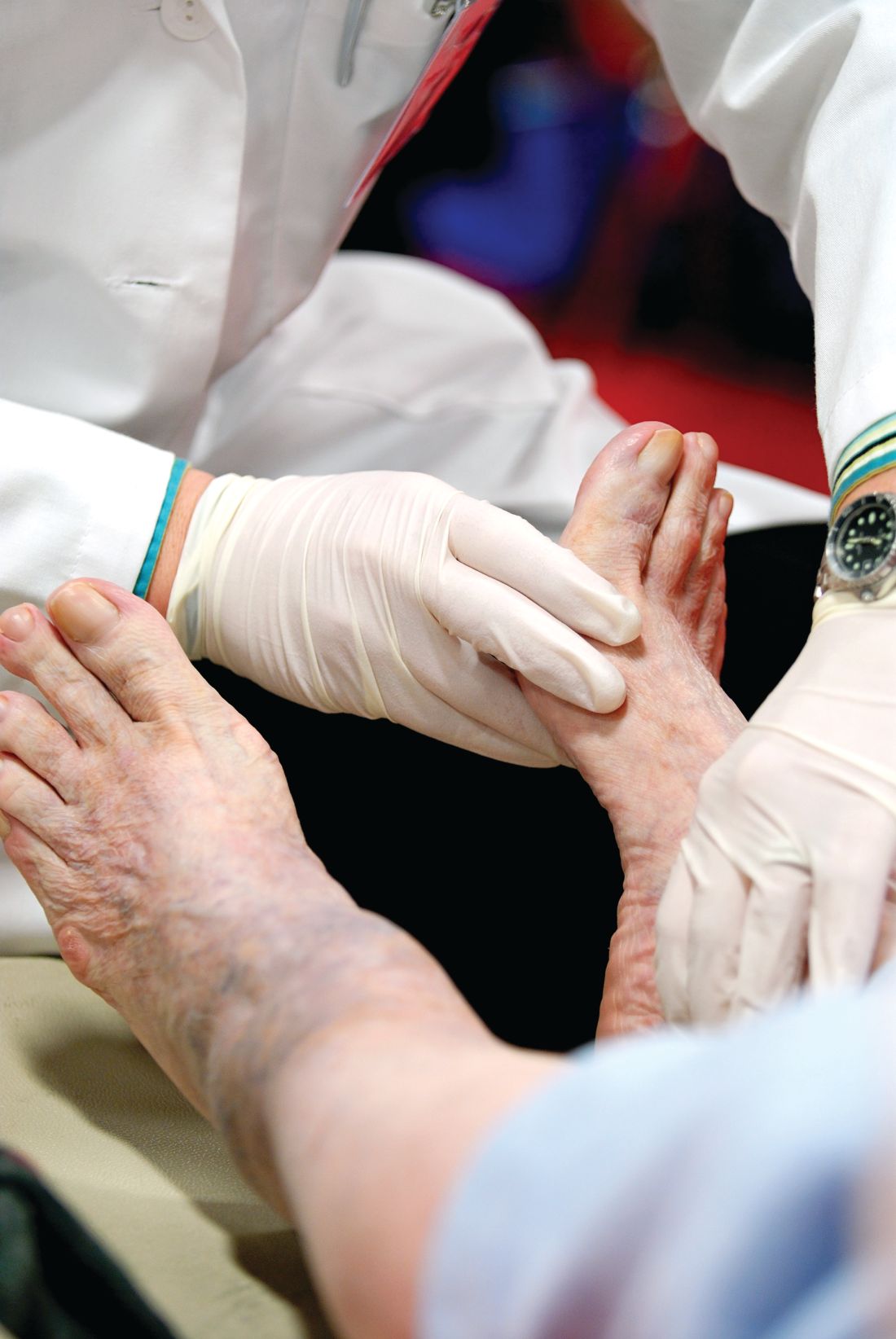
“Diabetic neuropathy has some important clinical consequences in a patient’s life. There’s clearly impaired function, a lower quality of life, and increased mortality. There’s also a high association between DN and cardiovascular disease and a high risk of amputation,” said Dr. Feldman, coauthor of the ADA’s new clinical guidelines for diabetic neuropathy (Diabetes Care. 2017;40[1]:136-54).
And in patients with type 2 diabetes, “diabetic neuropathy is not just hyperglycemia, which is what we’ve focused all our efforts on up until the past 5 years. There is also a role for dyslipidemia and other metabolic impairments,” she said.
In a follow-up interview, Dr. Feldman elaborated on a common misconception about DN, simple tools for foot exams by endocrinologists, and how to know when it’s time for a referral to a neurologist or podiatrist.
Q: What do you think physicians/endocrinologists misunderstand about diabetic neuropathy?
A: Commonly, physicians think if patients with diabetes do not complain of pain or numbness, they do not have DN. This simply isn’t correct. Over 80% of patients with DN have insensate feet – they simply do not have feeling in their feet. Physicians must examine a patient’s foot at least once a year to ensure the patient has not developed DN.
Q: What should endocrinologists understand about how diabetic neuropathy develops?
A: We know that excellent glucose control has a significant impact on DN in patients with type 1 diabetes. In patients with type 2 diabetes, we know that excellent glucose control plays a much less significant role. While it’s important, it must be coupled with control of other components of metabolic syndrome – elevated blood lipids, obesity, and hypertension.

A: Take a 126 Hz tuning fork and determine if the patient can feel vibration on the joint of the great toe for at least 10 seconds. Then take a 10-gram filament and a pin and determine if the patient can feel both of these instruments when they are applied to the joint of the great toe. Some physicians also take a 10-gram filament and apply it to the sole of the foot. I would not suggest using a pin on the sole of the foot.
Q: What else should they look for when they inspect feet? And how often would you recommend that endocrinologists do this per patient?
A: Inspection for callous formation, fissure formation, and fungal infections is important, and a foot exam should be done once yearly.
Q: What about testing whether patents can feel temperature?
A: The pin tests the same class of nerve fibers so this is routinely not done in an endocrinologist’s office.
Q: When should endocrinologists refer out for neuropathy?
A: Endocrinologists can treat DN by treating the diabetic condition and, in type 2 diabetes, the metabolic syndrome. Referral to a neurologist is indicated if there are atypical symptoms or signs, such as motor impairment that’s greater than sensory impairment, a significant asymmetry, or a very rapid onset. All patients with severe DN should be under the care of a podiatrist to prevent the development of nonhealing wounds and ulcers.
Q: What have you learned about how diabetic neuropathy affects the lives of patients?
A: DN definitely affects the quality of a patient’s life, not only in terms of work productivity and ability to perform activities of daily living. Quality of life and general enjoyment of life can frequently be adversely affected.
Q: How successful is treatment for diabetic neuropathy?
A: Treatment for pain can be very successful, and we have outlined a protocol in our recent ADA guidelines. For patients with uncontrollable pain, frequently a referral to a pain clinic is in order.
Dr. Feldman reports no relevant disclosures.
SAN DIEGO – Listen to the feet of your patients, and don’t just focus on glucose control as a way to combat diabetic neuropathy, according to Eva L. Feldman, MD, PhD.
Regular foot exams are crucial for patients with diabetes because of the high risk of diabetic neuropathy (DN), Dr. Feldman, professor of neurology and director of the Program for Neurology Research and Discovery at University of Michigan, Ann Arbor, said in a presentation at the annual scientific sessions of the American Diabetes Association.
“Diabetic neuropathy has some important clinical consequences in a patient’s life. There’s clearly impaired function, a lower quality of life, and increased mortality. There’s also a high association between DN and cardiovascular disease and a high risk of amputation,” said Dr. Feldman, coauthor of the ADA’s new clinical guidelines for diabetic neuropathy (Diabetes Care. 2017;40[1]:136-54).
And in patients with type 2 diabetes, “diabetic neuropathy is not just hyperglycemia, which is what we’ve focused all our efforts on up until the past 5 years. There is also a role for dyslipidemia and other metabolic impairments,” she said.
In a follow-up interview, Dr. Feldman elaborated on a common misconception about DN, simple tools for foot exams by endocrinologists, and how to know when it’s time for a referral to a neurologist or podiatrist.
Q: What do you think physicians/endocrinologists misunderstand about diabetic neuropathy?
A: Commonly, physicians think if patients with diabetes do not complain of pain or numbness, they do not have DN. This simply isn’t correct. Over 80% of patients with DN have insensate feet – they simply do not have feeling in their feet. Physicians must examine a patient’s foot at least once a year to ensure the patient has not developed DN.
Q: What should endocrinologists understand about how diabetic neuropathy develops?
A: We know that excellent glucose control has a significant impact on DN in patients with type 1 diabetes. In patients with type 2 diabetes, we know that excellent glucose control plays a much less significant role. While it’s important, it must be coupled with control of other components of metabolic syndrome – elevated blood lipids, obesity, and hypertension.
A: Take a 126 Hz tuning fork and determine if the patient can feel vibration on the joint of the great toe for at least 10 seconds. Then take a 10-gram filament and a pin and determine if the patient can feel both of these instruments when they are applied to the joint of the great toe. Some physicians also take a 10-gram filament and apply it to the sole of the foot. I would not suggest using a pin on the sole of the foot.
Q: What else should they look for when they inspect feet? And how often would you recommend that endocrinologists do this per patient?
A: Inspection for callous formation, fissure formation, and fungal infections is important, and a foot exam should be done once yearly.
Q: What about testing whether patents can feel temperature?
A: The pin tests the same class of nerve fibers so this is routinely not done in an endocrinologist’s office.
Q: When should endocrinologists refer out for neuropathy?
A: Endocrinologists can treat DN by treating the diabetic condition and, in type 2 diabetes, the metabolic syndrome. Referral to a neurologist is indicated if there are atypical symptoms or signs, such as motor impairment that’s greater than sensory impairment, a significant asymmetry, or a very rapid onset. All patients with severe DN should be under the care of a podiatrist to prevent the development of nonhealing wounds and ulcers.
Q: What have you learned about how diabetic neuropathy affects the lives of patients?
A: DN definitely affects the quality of a patient’s life, not only in terms of work productivity and ability to perform activities of daily living. Quality of life and general enjoyment of life can frequently be adversely affected.
Q: How successful is treatment for diabetic neuropathy?
A: Treatment for pain can be very successful, and we have outlined a protocol in our recent ADA guidelines. For patients with uncontrollable pain, frequently a referral to a pain clinic is in order.
Dr. Feldman reports no relevant disclosures.
SAN DIEGO – Listen to the feet of your patients, and don’t just focus on glucose control as a way to combat diabetic neuropathy, according to Eva L. Feldman, MD, PhD.
Regular foot exams are crucial for patients with diabetes because of the high risk of diabetic neuropathy (DN), Dr. Feldman, professor of neurology and director of the Program for Neurology Research and Discovery at University of Michigan, Ann Arbor, said in a presentation at the annual scientific sessions of the American Diabetes Association.
“Diabetic neuropathy has some important clinical consequences in a patient’s life. There’s clearly impaired function, a lower quality of life, and increased mortality. There’s also a high association between DN and cardiovascular disease and a high risk of amputation,” said Dr. Feldman, coauthor of the ADA’s new clinical guidelines for diabetic neuropathy (Diabetes Care. 2017;40[1]:136-54).
And in patients with type 2 diabetes, “diabetic neuropathy is not just hyperglycemia, which is what we’ve focused all our efforts on up until the past 5 years. There is also a role for dyslipidemia and other metabolic impairments,” she said.
In a follow-up interview, Dr. Feldman elaborated on a common misconception about DN, simple tools for foot exams by endocrinologists, and how to know when it’s time for a referral to a neurologist or podiatrist.
Q: What do you think physicians/endocrinologists misunderstand about diabetic neuropathy?
A: Commonly, physicians think if patients with diabetes do not complain of pain or numbness, they do not have DN. This simply isn’t correct. Over 80% of patients with DN have insensate feet – they simply do not have feeling in their feet. Physicians must examine a patient’s foot at least once a year to ensure the patient has not developed DN.
Q: What should endocrinologists understand about how diabetic neuropathy develops?
A: We know that excellent glucose control has a significant impact on DN in patients with type 1 diabetes. In patients with type 2 diabetes, we know that excellent glucose control plays a much less significant role. While it’s important, it must be coupled with control of other components of metabolic syndrome – elevated blood lipids, obesity, and hypertension.
A: Take a 126 Hz tuning fork and determine if the patient can feel vibration on the joint of the great toe for at least 10 seconds. Then take a 10-gram filament and a pin and determine if the patient can feel both of these instruments when they are applied to the joint of the great toe. Some physicians also take a 10-gram filament and apply it to the sole of the foot. I would not suggest using a pin on the sole of the foot.
Q: What else should they look for when they inspect feet? And how often would you recommend that endocrinologists do this per patient?
A: Inspection for callous formation, fissure formation, and fungal infections is important, and a foot exam should be done once yearly.
Q: What about testing whether patents can feel temperature?
A: The pin tests the same class of nerve fibers so this is routinely not done in an endocrinologist’s office.
Q: When should endocrinologists refer out for neuropathy?
A: Endocrinologists can treat DN by treating the diabetic condition and, in type 2 diabetes, the metabolic syndrome. Referral to a neurologist is indicated if there are atypical symptoms or signs, such as motor impairment that’s greater than sensory impairment, a significant asymmetry, or a very rapid onset. All patients with severe DN should be under the care of a podiatrist to prevent the development of nonhealing wounds and ulcers.
Q: What have you learned about how diabetic neuropathy affects the lives of patients?
A: DN definitely affects the quality of a patient’s life, not only in terms of work productivity and ability to perform activities of daily living. Quality of life and general enjoyment of life can frequently be adversely affected.
Q: How successful is treatment for diabetic neuropathy?
A: Treatment for pain can be very successful, and we have outlined a protocol in our recent ADA guidelines. For patients with uncontrollable pain, frequently a referral to a pain clinic is in order.
Dr. Feldman reports no relevant disclosures.
AT THE ADA ANNUAL SCIENTIFIC SESSIONS
Forced commitments still low under California’s AOT law
Critics have feared that California’s “Laura’s Law,” designed to prevent violence by making it easier for officials to force outpatient care upon people with mental illness who have resisted treatment, would violate the civil rights of Golden State residents.
But early statistics gathered by a Northern California psychiatrist suggest that few people have been forced involuntarily into outpatient care in large counties that have adopted the tougher approach in recent years. Instead, Californians targeted by the law appear to be choosing – under pressure – the alternative of accepting outpatient treatment.
California enacted Laura’s Law in 2003 after the death of a 19-year-old woman named Laura Wilcox at a mental health clinic in Northern California. Ms. Wilcox, who worked at the clinic, and two others were shot to death by a patient whose family had failed to persuade officials to force him to get treatment.
As California’s version of assisted outpatient treatment, or AOT, Laura’s Law, allows courts to order people into outpatient treatment in cases that meet certain criteria. Among other things, the person must have a mental illness, be considered “unlikely to survive safely in the community without supervision,” and have a “history of lack of compliance with treatment.” The law “also obligates any county that implements and accepts the law to provide excellent and comprehensive community-based services, not only for court-ordered patients but anybody who wants care,” Dr. Nelson said in an interview after his presentation.
As a state law, the measure must be adopted on a county-by-county basis. Tiny and remote Nevada County, where the shootings occurred, adopted the policy first when it was forced to do so by a lawsuit, Dr. Nelson said. But other counties were slow to sign on.
“Unlike Kendra’s Law, which passed earlier in New York, and both mandated and funded assisted outpatient treatment for all counties, Laura’s Law was completely unfunded. It was left to the discretion of each county to decide for itself whether or not to implement,” said Dr. Nelson, of Marin County, just north of San Francisco, who has served on the councils of the Northern California Psychiatric Society and California Psychiatric Association.
But things changed in 2013, when the state allowed money from a special tax on wealthy residents to be used for AOT programs, Dr. Nelson said. Counties began implementing Laura’s Law in 2014, and the number has now reached 17, representing about two-thirds of the state’s population, he said.
Dr. Nelson gathered statistics from several of California’s 58 counties. He found that over a period of 5 months of implementation, 59 people were referred to the AOT program, and 12 voluntarily accepted outpatient therapy in San Francisco County, which encompasses the city of San Francisco only and has 864,000 residents.
Over a period of 5 months in San Diego County, which has 3.3 million residents, there were 376 referrals, including those to a program that uses home visits to urge people to accept outpatient care. Twenty people accepted voluntary outpatient treatment. No court-ordered treatment was required for anyone in either San Diego or San Francisco counties.
Over a period of 12 months in Orange County, with 3.2 million residents, there were 389 referrals, and 126 people voluntarily accepted AOT. Only three were ordered into treatment by courts. Over 6 months in Placer County, with just 375,000 residents, eight patients voluntarily accepted AOT, and one was ordered into treatment by a court but refused it.
(The Orange County Register reports that refusal to abide by court-ordered outpatient treatment results in no civil or criminal penalties.)
Los Angeles County, by far the most populous in the state with 10.1 million residents, reported 805 AOT referrals, 30 involuntary commitments to outpatient care, and 239 voluntary agreements to outpatient care. The numbers are from 2016 to 2017, but Dr. Nelson did not have details about the exact period covered.
Dr. Nelson also looked at 7 years of statistics from Nevada County, which was forced into adopting an AOT program by a lawsuit. The county, which has fewer than 100,000 residents, reported 67 referrals to the AOT program and 30 cases of court-ordered treatment.
A report estimates that over the first 30 months of the program, Nevada County saved $1.80 for every $1 spent on its AOT program by preventing acute psychiatric hospitalizations and imprisonment.
The low numbers of forced commitments to outpatient care across the state make sense, Dr. Nelson said, since the law wasn’t intended to “force a bunch of folks with typically high recidivism with mental illness into involuntary treatment.”
Instead, he said, the law has promoted a so-called black robe effect – essentially, a form of intimidation by judge.
Dr. Nelson said he has tracked the debate over forced outpatient care for years and has mixed feelings about the law.
There’s value to coaxing people with mental illness into care when they are unable to recognize their illness, he said. “But there are way too many gaps and holes for people to fall into,” he said. “They have the potential to be mistreated, poorly treated, poorly diagnosed.”
While Laura’s Law requires counties to offer comprehensive outpatient mental health care, “it’s really difficult to know whether the programs are meeting all of the conditions of the law,” Dr. Nelson said.
Still, he said, Laura’s Law is forcing counties to discuss how to care for people with mental illness who are targeted by the legislation, he said. “They are beginning to have a lively conversation about how we are going to reach out to these people and help them, even to just avoid the implementation of Laura’s Law.”
Dr. Nelson has no relevant disclosures.
Critics have feared that California’s “Laura’s Law,” designed to prevent violence by making it easier for officials to force outpatient care upon people with mental illness who have resisted treatment, would violate the civil rights of Golden State residents.
But early statistics gathered by a Northern California psychiatrist suggest that few people have been forced involuntarily into outpatient care in large counties that have adopted the tougher approach in recent years. Instead, Californians targeted by the law appear to be choosing – under pressure – the alternative of accepting outpatient treatment.
California enacted Laura’s Law in 2003 after the death of a 19-year-old woman named Laura Wilcox at a mental health clinic in Northern California. Ms. Wilcox, who worked at the clinic, and two others were shot to death by a patient whose family had failed to persuade officials to force him to get treatment.
As California’s version of assisted outpatient treatment, or AOT, Laura’s Law, allows courts to order people into outpatient treatment in cases that meet certain criteria. Among other things, the person must have a mental illness, be considered “unlikely to survive safely in the community without supervision,” and have a “history of lack of compliance with treatment.” The law “also obligates any county that implements and accepts the law to provide excellent and comprehensive community-based services, not only for court-ordered patients but anybody who wants care,” Dr. Nelson said in an interview after his presentation.
As a state law, the measure must be adopted on a county-by-county basis. Tiny and remote Nevada County, where the shootings occurred, adopted the policy first when it was forced to do so by a lawsuit, Dr. Nelson said. But other counties were slow to sign on.
“Unlike Kendra’s Law, which passed earlier in New York, and both mandated and funded assisted outpatient treatment for all counties, Laura’s Law was completely unfunded. It was left to the discretion of each county to decide for itself whether or not to implement,” said Dr. Nelson, of Marin County, just north of San Francisco, who has served on the councils of the Northern California Psychiatric Society and California Psychiatric Association.
But things changed in 2013, when the state allowed money from a special tax on wealthy residents to be used for AOT programs, Dr. Nelson said. Counties began implementing Laura’s Law in 2014, and the number has now reached 17, representing about two-thirds of the state’s population, he said.
Dr. Nelson gathered statistics from several of California’s 58 counties. He found that over a period of 5 months of implementation, 59 people were referred to the AOT program, and 12 voluntarily accepted outpatient therapy in San Francisco County, which encompasses the city of San Francisco only and has 864,000 residents.
Over a period of 5 months in San Diego County, which has 3.3 million residents, there were 376 referrals, including those to a program that uses home visits to urge people to accept outpatient care. Twenty people accepted voluntary outpatient treatment. No court-ordered treatment was required for anyone in either San Diego or San Francisco counties.
Over a period of 12 months in Orange County, with 3.2 million residents, there were 389 referrals, and 126 people voluntarily accepted AOT. Only three were ordered into treatment by courts. Over 6 months in Placer County, with just 375,000 residents, eight patients voluntarily accepted AOT, and one was ordered into treatment by a court but refused it.
(The Orange County Register reports that refusal to abide by court-ordered outpatient treatment results in no civil or criminal penalties.)
Los Angeles County, by far the most populous in the state with 10.1 million residents, reported 805 AOT referrals, 30 involuntary commitments to outpatient care, and 239 voluntary agreements to outpatient care. The numbers are from 2016 to 2017, but Dr. Nelson did not have details about the exact period covered.
Dr. Nelson also looked at 7 years of statistics from Nevada County, which was forced into adopting an AOT program by a lawsuit. The county, which has fewer than 100,000 residents, reported 67 referrals to the AOT program and 30 cases of court-ordered treatment.
A report estimates that over the first 30 months of the program, Nevada County saved $1.80 for every $1 spent on its AOT program by preventing acute psychiatric hospitalizations and imprisonment.
The low numbers of forced commitments to outpatient care across the state make sense, Dr. Nelson said, since the law wasn’t intended to “force a bunch of folks with typically high recidivism with mental illness into involuntary treatment.”
Instead, he said, the law has promoted a so-called black robe effect – essentially, a form of intimidation by judge.
Dr. Nelson said he has tracked the debate over forced outpatient care for years and has mixed feelings about the law.
There’s value to coaxing people with mental illness into care when they are unable to recognize their illness, he said. “But there are way too many gaps and holes for people to fall into,” he said. “They have the potential to be mistreated, poorly treated, poorly diagnosed.”
While Laura’s Law requires counties to offer comprehensive outpatient mental health care, “it’s really difficult to know whether the programs are meeting all of the conditions of the law,” Dr. Nelson said.
Still, he said, Laura’s Law is forcing counties to discuss how to care for people with mental illness who are targeted by the legislation, he said. “They are beginning to have a lively conversation about how we are going to reach out to these people and help them, even to just avoid the implementation of Laura’s Law.”
Dr. Nelson has no relevant disclosures.
Critics have feared that California’s “Laura’s Law,” designed to prevent violence by making it easier for officials to force outpatient care upon people with mental illness who have resisted treatment, would violate the civil rights of Golden State residents.
But early statistics gathered by a Northern California psychiatrist suggest that few people have been forced involuntarily into outpatient care in large counties that have adopted the tougher approach in recent years. Instead, Californians targeted by the law appear to be choosing – under pressure – the alternative of accepting outpatient treatment.
California enacted Laura’s Law in 2003 after the death of a 19-year-old woman named Laura Wilcox at a mental health clinic in Northern California. Ms. Wilcox, who worked at the clinic, and two others were shot to death by a patient whose family had failed to persuade officials to force him to get treatment.
As California’s version of assisted outpatient treatment, or AOT, Laura’s Law, allows courts to order people into outpatient treatment in cases that meet certain criteria. Among other things, the person must have a mental illness, be considered “unlikely to survive safely in the community without supervision,” and have a “history of lack of compliance with treatment.” The law “also obligates any county that implements and accepts the law to provide excellent and comprehensive community-based services, not only for court-ordered patients but anybody who wants care,” Dr. Nelson said in an interview after his presentation.
As a state law, the measure must be adopted on a county-by-county basis. Tiny and remote Nevada County, where the shootings occurred, adopted the policy first when it was forced to do so by a lawsuit, Dr. Nelson said. But other counties were slow to sign on.
“Unlike Kendra’s Law, which passed earlier in New York, and both mandated and funded assisted outpatient treatment for all counties, Laura’s Law was completely unfunded. It was left to the discretion of each county to decide for itself whether or not to implement,” said Dr. Nelson, of Marin County, just north of San Francisco, who has served on the councils of the Northern California Psychiatric Society and California Psychiatric Association.
But things changed in 2013, when the state allowed money from a special tax on wealthy residents to be used for AOT programs, Dr. Nelson said. Counties began implementing Laura’s Law in 2014, and the number has now reached 17, representing about two-thirds of the state’s population, he said.
Dr. Nelson gathered statistics from several of California’s 58 counties. He found that over a period of 5 months of implementation, 59 people were referred to the AOT program, and 12 voluntarily accepted outpatient therapy in San Francisco County, which encompasses the city of San Francisco only and has 864,000 residents.
Over a period of 5 months in San Diego County, which has 3.3 million residents, there were 376 referrals, including those to a program that uses home visits to urge people to accept outpatient care. Twenty people accepted voluntary outpatient treatment. No court-ordered treatment was required for anyone in either San Diego or San Francisco counties.
Over a period of 12 months in Orange County, with 3.2 million residents, there were 389 referrals, and 126 people voluntarily accepted AOT. Only three were ordered into treatment by courts. Over 6 months in Placer County, with just 375,000 residents, eight patients voluntarily accepted AOT, and one was ordered into treatment by a court but refused it.
(The Orange County Register reports that refusal to abide by court-ordered outpatient treatment results in no civil or criminal penalties.)
Los Angeles County, by far the most populous in the state with 10.1 million residents, reported 805 AOT referrals, 30 involuntary commitments to outpatient care, and 239 voluntary agreements to outpatient care. The numbers are from 2016 to 2017, but Dr. Nelson did not have details about the exact period covered.
Dr. Nelson also looked at 7 years of statistics from Nevada County, which was forced into adopting an AOT program by a lawsuit. The county, which has fewer than 100,000 residents, reported 67 referrals to the AOT program and 30 cases of court-ordered treatment.
A report estimates that over the first 30 months of the program, Nevada County saved $1.80 for every $1 spent on its AOT program by preventing acute psychiatric hospitalizations and imprisonment.
The low numbers of forced commitments to outpatient care across the state make sense, Dr. Nelson said, since the law wasn’t intended to “force a bunch of folks with typically high recidivism with mental illness into involuntary treatment.”
Instead, he said, the law has promoted a so-called black robe effect – essentially, a form of intimidation by judge.
Dr. Nelson said he has tracked the debate over forced outpatient care for years and has mixed feelings about the law.
There’s value to coaxing people with mental illness into care when they are unable to recognize their illness, he said. “But there are way too many gaps and holes for people to fall into,” he said. “They have the potential to be mistreated, poorly treated, poorly diagnosed.”
While Laura’s Law requires counties to offer comprehensive outpatient mental health care, “it’s really difficult to know whether the programs are meeting all of the conditions of the law,” Dr. Nelson said.
Still, he said, Laura’s Law is forcing counties to discuss how to care for people with mental illness who are targeted by the legislation, he said. “They are beginning to have a lively conversation about how we are going to reach out to these people and help them, even to just avoid the implementation of Laura’s Law.”
Dr. Nelson has no relevant disclosures.
EXPERT ANALYSIS FROM APA
Risk of sexual dysfunction in diabetes is high, but treatments can help
SAN DIEGO –
This isn’t normal for men of that age, according to Hunter B. Wessells, MD.“It’s not just that they’re aging. It’s a 20-year acceleration of the aging process,” he said at the annual scientific sessions of the American Diabetes Association.
That’s not all. In some cases, men with diabetes may experience decreased libido that’s potentially caused by low testosterone, said Dr. Wessells, professor and Wilma Wise Nelson, Ole A. Nelson, and Mabel Wise Nelson Endowed Chair in Urology at the University of Washington, Seattle
Still, research findings offer useful insights into the frequency of sexual dysfunction in people with diabetes and the potential – and limitations – of available treatments, said Dr. Wessells.
In patients with well-controlled diabetes, “these conditions impact quality of life to a greater degree than complications like nephropathy, neuropathy, and retinopathy,” he said in an interview. “Thus, treatment of urological symptoms can be a high-yield endeavor.”
In both sexes, Dr. Wessells said, diabetes can disrupt the mechanism of desire, arousal, and orgasm by affecting a long list of bodily functions such as central nervous system stimulation, hormone activity, autonomic and somatic nerve activity, and processing of calcium ions and nitric acid.
In men, diabetes boosts the risk of erectile dysfunction to a larger extent than do related conditions such as obesity, heart disease, and depression. “But they are interrelated,” he said. “The primary mechanisms include the metabolic effects of high glucose, autonomic nerve damage, and microvascular disease.”
Low testosterone levels also can cause problems in patients with diabetes, he said. “Type 2 diabetes has greater effects on testosterone than type 1. It is most closely linked to weight in the type 1 population and affects only a small percentage.”
A 2017 systematic review and meta-analysis of 145 studies with more than 88,000 subjects (average age 55.8 ± 7.9 years) suggests that ED was more common in type 2 diabetes (66.3%) than type 1 diabetes (37.5%) after statistical adjustment to account for publication bias (Diabet Med. 2017 Jul 18. doi: 10.1111/dme.13403).
A smaller analysis found that men with diabetes had almost four times the odds (odd ratio = 3.62) of ED compared with healthy controls (Diabet Med. 2017 Jul 18. doi: 10.1111/dme.13403). Phosphodiesterase-5 inhibitors – such as sildenafil, vardenafil, and tadalafil – are one option for men with diabetes and ED, Dr. Wessells said. “They work pretty well, but men with diabetes tend to have more severe ED. They’re going to get better, but will they get better enough to be normal? That’s the question.”
A 2007 Cochrane Library analysis found that men with diabetes and ED gained from PDE5 inhibitors overall (Cochrane Database Syst Rev. 2007 Jan 24[1]:CD002187. doi: 10.1002/14651858.CD002187.pub3).
“They’re not going to do as well as the general population,” Dr. Wessells said, “but we should try these as first-line agents in absence of things like severe unstable cardiovascular disease and other risk factors.”
Second-line therapies, typically offered by urologists, include penile prostheses and injection therapy, he said. A 2014 analysis of previous research found that men with diabetes were “more than 50% more likely to be prescribed secondary ED treatments over the 2-year observation period, and more than twice as likely to undergo penile prosthesis surgery” (Int J Impot Res. 2014 May-Jun;26[3]:112-5).
As for women, a 2009 study found that of 424 sexually active women with type 1 diabetes (97% of whom were white), 35% showed signs of female sexual dysfunction (FSD). Of those with FSD, problems included loss of libido (57%); problems with orgasm (51%), lubrication (47%), and/or arousal (38%); and pain (21%) (Diabetes Care. 2009 May;32[5]:780-5).
Only one drug, flibanserin (Addyi), is approved for FSD in the United States. Its impact on patients with diabetes is unknown, Dr. Wessells said, and the drug has the potential for significant adverse events.
The good news: Research is providing insight into which men and women are more likely to develop sexual dysfunction, Dr. Wessells said.
Age is important in both genders. For women, depression and being married appear to be risk factors, he said. “This needs more exploration to help us understand how to intervene.”
And in men, he said, ED is linked to jumps in hemoglobin A1c, while men on intensive glycemic therapy have a lower risk.
“Maybe we can find out who needs to be targeted for earlier intervention,” he said. This is especially important for men because ED becomes more likely to be irreversible after just a few years, he said.
Dr. Wessells reports no relevant disclosures.
SAN DIEGO –
This isn’t normal for men of that age, according to Hunter B. Wessells, MD.“It’s not just that they’re aging. It’s a 20-year acceleration of the aging process,” he said at the annual scientific sessions of the American Diabetes Association.
That’s not all. In some cases, men with diabetes may experience decreased libido that’s potentially caused by low testosterone, said Dr. Wessells, professor and Wilma Wise Nelson, Ole A. Nelson, and Mabel Wise Nelson Endowed Chair in Urology at the University of Washington, Seattle
Still, research findings offer useful insights into the frequency of sexual dysfunction in people with diabetes and the potential – and limitations – of available treatments, said Dr. Wessells.
In patients with well-controlled diabetes, “these conditions impact quality of life to a greater degree than complications like nephropathy, neuropathy, and retinopathy,” he said in an interview. “Thus, treatment of urological symptoms can be a high-yield endeavor.”
In both sexes, Dr. Wessells said, diabetes can disrupt the mechanism of desire, arousal, and orgasm by affecting a long list of bodily functions such as central nervous system stimulation, hormone activity, autonomic and somatic nerve activity, and processing of calcium ions and nitric acid.
In men, diabetes boosts the risk of erectile dysfunction to a larger extent than do related conditions such as obesity, heart disease, and depression. “But they are interrelated,” he said. “The primary mechanisms include the metabolic effects of high glucose, autonomic nerve damage, and microvascular disease.”
Low testosterone levels also can cause problems in patients with diabetes, he said. “Type 2 diabetes has greater effects on testosterone than type 1. It is most closely linked to weight in the type 1 population and affects only a small percentage.”
A 2017 systematic review and meta-analysis of 145 studies with more than 88,000 subjects (average age 55.8 ± 7.9 years) suggests that ED was more common in type 2 diabetes (66.3%) than type 1 diabetes (37.5%) after statistical adjustment to account for publication bias (Diabet Med. 2017 Jul 18. doi: 10.1111/dme.13403).
A smaller analysis found that men with diabetes had almost four times the odds (odd ratio = 3.62) of ED compared with healthy controls (Diabet Med. 2017 Jul 18. doi: 10.1111/dme.13403). Phosphodiesterase-5 inhibitors – such as sildenafil, vardenafil, and tadalafil – are one option for men with diabetes and ED, Dr. Wessells said. “They work pretty well, but men with diabetes tend to have more severe ED. They’re going to get better, but will they get better enough to be normal? That’s the question.”
A 2007 Cochrane Library analysis found that men with diabetes and ED gained from PDE5 inhibitors overall (Cochrane Database Syst Rev. 2007 Jan 24[1]:CD002187. doi: 10.1002/14651858.CD002187.pub3).
“They’re not going to do as well as the general population,” Dr. Wessells said, “but we should try these as first-line agents in absence of things like severe unstable cardiovascular disease and other risk factors.”
Second-line therapies, typically offered by urologists, include penile prostheses and injection therapy, he said. A 2014 analysis of previous research found that men with diabetes were “more than 50% more likely to be prescribed secondary ED treatments over the 2-year observation period, and more than twice as likely to undergo penile prosthesis surgery” (Int J Impot Res. 2014 May-Jun;26[3]:112-5).
As for women, a 2009 study found that of 424 sexually active women with type 1 diabetes (97% of whom were white), 35% showed signs of female sexual dysfunction (FSD). Of those with FSD, problems included loss of libido (57%); problems with orgasm (51%), lubrication (47%), and/or arousal (38%); and pain (21%) (Diabetes Care. 2009 May;32[5]:780-5).
Only one drug, flibanserin (Addyi), is approved for FSD in the United States. Its impact on patients with diabetes is unknown, Dr. Wessells said, and the drug has the potential for significant adverse events.
The good news: Research is providing insight into which men and women are more likely to develop sexual dysfunction, Dr. Wessells said.
Age is important in both genders. For women, depression and being married appear to be risk factors, he said. “This needs more exploration to help us understand how to intervene.”
And in men, he said, ED is linked to jumps in hemoglobin A1c, while men on intensive glycemic therapy have a lower risk.
“Maybe we can find out who needs to be targeted for earlier intervention,” he said. This is especially important for men because ED becomes more likely to be irreversible after just a few years, he said.
Dr. Wessells reports no relevant disclosures.
SAN DIEGO –
This isn’t normal for men of that age, according to Hunter B. Wessells, MD.“It’s not just that they’re aging. It’s a 20-year acceleration of the aging process,” he said at the annual scientific sessions of the American Diabetes Association.
That’s not all. In some cases, men with diabetes may experience decreased libido that’s potentially caused by low testosterone, said Dr. Wessells, professor and Wilma Wise Nelson, Ole A. Nelson, and Mabel Wise Nelson Endowed Chair in Urology at the University of Washington, Seattle
Still, research findings offer useful insights into the frequency of sexual dysfunction in people with diabetes and the potential – and limitations – of available treatments, said Dr. Wessells.
In patients with well-controlled diabetes, “these conditions impact quality of life to a greater degree than complications like nephropathy, neuropathy, and retinopathy,” he said in an interview. “Thus, treatment of urological symptoms can be a high-yield endeavor.”
In both sexes, Dr. Wessells said, diabetes can disrupt the mechanism of desire, arousal, and orgasm by affecting a long list of bodily functions such as central nervous system stimulation, hormone activity, autonomic and somatic nerve activity, and processing of calcium ions and nitric acid.
In men, diabetes boosts the risk of erectile dysfunction to a larger extent than do related conditions such as obesity, heart disease, and depression. “But they are interrelated,” he said. “The primary mechanisms include the metabolic effects of high glucose, autonomic nerve damage, and microvascular disease.”
Low testosterone levels also can cause problems in patients with diabetes, he said. “Type 2 diabetes has greater effects on testosterone than type 1. It is most closely linked to weight in the type 1 population and affects only a small percentage.”
A 2017 systematic review and meta-analysis of 145 studies with more than 88,000 subjects (average age 55.8 ± 7.9 years) suggests that ED was more common in type 2 diabetes (66.3%) than type 1 diabetes (37.5%) after statistical adjustment to account for publication bias (Diabet Med. 2017 Jul 18. doi: 10.1111/dme.13403).
A smaller analysis found that men with diabetes had almost four times the odds (odd ratio = 3.62) of ED compared with healthy controls (Diabet Med. 2017 Jul 18. doi: 10.1111/dme.13403). Phosphodiesterase-5 inhibitors – such as sildenafil, vardenafil, and tadalafil – are one option for men with diabetes and ED, Dr. Wessells said. “They work pretty well, but men with diabetes tend to have more severe ED. They’re going to get better, but will they get better enough to be normal? That’s the question.”
A 2007 Cochrane Library analysis found that men with diabetes and ED gained from PDE5 inhibitors overall (Cochrane Database Syst Rev. 2007 Jan 24[1]:CD002187. doi: 10.1002/14651858.CD002187.pub3).
“They’re not going to do as well as the general population,” Dr. Wessells said, “but we should try these as first-line agents in absence of things like severe unstable cardiovascular disease and other risk factors.”
Second-line therapies, typically offered by urologists, include penile prostheses and injection therapy, he said. A 2014 analysis of previous research found that men with diabetes were “more than 50% more likely to be prescribed secondary ED treatments over the 2-year observation period, and more than twice as likely to undergo penile prosthesis surgery” (Int J Impot Res. 2014 May-Jun;26[3]:112-5).
As for women, a 2009 study found that of 424 sexually active women with type 1 diabetes (97% of whom were white), 35% showed signs of female sexual dysfunction (FSD). Of those with FSD, problems included loss of libido (57%); problems with orgasm (51%), lubrication (47%), and/or arousal (38%); and pain (21%) (Diabetes Care. 2009 May;32[5]:780-5).
Only one drug, flibanserin (Addyi), is approved for FSD in the United States. Its impact on patients with diabetes is unknown, Dr. Wessells said, and the drug has the potential for significant adverse events.
The good news: Research is providing insight into which men and women are more likely to develop sexual dysfunction, Dr. Wessells said.
Age is important in both genders. For women, depression and being married appear to be risk factors, he said. “This needs more exploration to help us understand how to intervene.”
And in men, he said, ED is linked to jumps in hemoglobin A1c, while men on intensive glycemic therapy have a lower risk.
“Maybe we can find out who needs to be targeted for earlier intervention,” he said. This is especially important for men because ED becomes more likely to be irreversible after just a few years, he said.
Dr. Wessells reports no relevant disclosures.
EXPERT ANALYSIS AT THE ADA ANNUAL SCIENTIFIC SESSIONS