User login
Interdisciplinary care reduces deep dyspareunia in endometriosis
VANCOUVER – Interdisciplinary care for women with deep dyspareunia led to a reduction in severe cases, but had no effect on superficial dyspareunia, a study showed.
The findings were measured after 1 year in a treatment program that included laparoscopic surgery, hormonal suppression, biofeedback, pelvic floor physiotherapy, cognitive-behavioral therapy, and mindfulness-based therapy.
About half of women with endometriosis experience deep dyspareunia, which can have a strong, negative impact on quality of life and relationships, Paul Yong, MD, PhD, reported at the World Congress on Endometriosis. For women who do not respond to surgical or medical interventions, interdisciplinary approaches that address the biopsychosocial aspect of pain in endometriosis could be of benefit, he said.

He said the researchers didn’t expect the superficial dyspareunia to respond to the treatments, since it is likely tied to nerve issues. But as a secondary measure, superficial dyspareunia acted as a sort of built-in control to show that the interventions were not having a broad placebo or nonspecific effect on pain.
The study included 296 women who came to the BC Women’s Hospital Centre for Pelvic Pain and Endometriosis with complaints of deep dyspareunia between December 2013 and December 2014. In total, 58% had a confirmed diagnosis of endometriosis, 24% had suspected endometriosis, and 18% had no endometriosis. More than half of the women had irritable bowel syndrome, 42% had painful bladder syndrome, and 30% had pelvic floor dysfunction.
About 55% of patients underwent surgery, while 13% were on hormonal treatment at both baseline and 1 year, 11% were on a pain adjuvant, 30% were on opioids, and 17% were enrolled in a pain program.
At baseline, half of the women reported severe symptoms of deep dyspareunia, but that percentage dropped to 30.4% at 1 year. Moderate cases increased from 17.7% at baseline to 25.0% at 1 year, and mild cases increased from 27.3% to 44.6% (P less than .0001).
A secondary analysis of patients with superficial dyspareunia showed no statistically significant changes in category frequencies. Severe cases represented 22.4% of the population at baseline and 20.2% at 1 year, and similarities were also seen in proportions of moderate (20.2% vs. 19.8%) and mild cases (57.4% vs. 60.1%, P = .65).
“This really suggests that the treatment program has some specificity for deep dyspareunia,” Dr. Yong said.
Depressive symptoms at baseline, as assessed by the Patient Health Questionnaire–9 scale, predicted deep dyspareunia at 1 year (odds ratio, 1.07; 95% confidence interval, 1.03-1.11).
The efficacy against deep dyspareunia is encouraging, but superficial dyspareunia is also a key concern.
“The study suggests that the typical treatments for endometriosis may not impact at all this really prevalent issue of superficial pain, so there’s more for us to do there because that can be a cause of long-term suffering and decreased quality of life,” said Lori Brotto, PhD, director of the sexual health laboratory at the University of British Columbia, who moderated the session during which the research was presented.
Dr. Yong called for more research, specifically a trial targeting treatment of depressive symptoms to determine any effect on endometriosis-related sexual pain or function.
“I think there needs to be more work in the role of depression – not only screening for depression in women with endometriosis, but trying to understand what role it plays in endometriosis symptoms,” Dr. Yong said.
The study was funded by the Canadian Institutes of Health Research. Dr. Yong and Dr. Brotto reported having no relevant financial disclosures.
VANCOUVER – Interdisciplinary care for women with deep dyspareunia led to a reduction in severe cases, but had no effect on superficial dyspareunia, a study showed.
The findings were measured after 1 year in a treatment program that included laparoscopic surgery, hormonal suppression, biofeedback, pelvic floor physiotherapy, cognitive-behavioral therapy, and mindfulness-based therapy.
About half of women with endometriosis experience deep dyspareunia, which can have a strong, negative impact on quality of life and relationships, Paul Yong, MD, PhD, reported at the World Congress on Endometriosis. For women who do not respond to surgical or medical interventions, interdisciplinary approaches that address the biopsychosocial aspect of pain in endometriosis could be of benefit, he said.
He said the researchers didn’t expect the superficial dyspareunia to respond to the treatments, since it is likely tied to nerve issues. But as a secondary measure, superficial dyspareunia acted as a sort of built-in control to show that the interventions were not having a broad placebo or nonspecific effect on pain.
The study included 296 women who came to the BC Women’s Hospital Centre for Pelvic Pain and Endometriosis with complaints of deep dyspareunia between December 2013 and December 2014. In total, 58% had a confirmed diagnosis of endometriosis, 24% had suspected endometriosis, and 18% had no endometriosis. More than half of the women had irritable bowel syndrome, 42% had painful bladder syndrome, and 30% had pelvic floor dysfunction.
About 55% of patients underwent surgery, while 13% were on hormonal treatment at both baseline and 1 year, 11% were on a pain adjuvant, 30% were on opioids, and 17% were enrolled in a pain program.
At baseline, half of the women reported severe symptoms of deep dyspareunia, but that percentage dropped to 30.4% at 1 year. Moderate cases increased from 17.7% at baseline to 25.0% at 1 year, and mild cases increased from 27.3% to 44.6% (P less than .0001).
A secondary analysis of patients with superficial dyspareunia showed no statistically significant changes in category frequencies. Severe cases represented 22.4% of the population at baseline and 20.2% at 1 year, and similarities were also seen in proportions of moderate (20.2% vs. 19.8%) and mild cases (57.4% vs. 60.1%, P = .65).
“This really suggests that the treatment program has some specificity for deep dyspareunia,” Dr. Yong said.
Depressive symptoms at baseline, as assessed by the Patient Health Questionnaire–9 scale, predicted deep dyspareunia at 1 year (odds ratio, 1.07; 95% confidence interval, 1.03-1.11).
The efficacy against deep dyspareunia is encouraging, but superficial dyspareunia is also a key concern.
“The study suggests that the typical treatments for endometriosis may not impact at all this really prevalent issue of superficial pain, so there’s more for us to do there because that can be a cause of long-term suffering and decreased quality of life,” said Lori Brotto, PhD, director of the sexual health laboratory at the University of British Columbia, who moderated the session during which the research was presented.
Dr. Yong called for more research, specifically a trial targeting treatment of depressive symptoms to determine any effect on endometriosis-related sexual pain or function.
“I think there needs to be more work in the role of depression – not only screening for depression in women with endometriosis, but trying to understand what role it plays in endometriosis symptoms,” Dr. Yong said.
The study was funded by the Canadian Institutes of Health Research. Dr. Yong and Dr. Brotto reported having no relevant financial disclosures.
AT WCE 2017
Key clinical point:
Major finding: Severe cases of deep dyspareunia dropped from 50.0% to 30.4% at 1 year.
Data source: A retrospective analysis of 296 patients at a single center.
Disclosures: The study was funded by the Canadian Institutes of Health Research. Dr. Yong and Dr. Brotto reported having no relevant financial disclosures.
PREMM5: Updated Lynch syndrome test available
An updated Lynch syndrome (LS) prediction model improves accuracy and may be used to assess individuals who are currently unaffected by cancer. The model predicts an individual’s risk of carrying one of five known gene mutations associated with LS. It could be applied to individuals with a suspicious family history of cancer, as well as to colon cancer patients who may not have tumor immunohistochemical and microsatellite instability testing results that can spot potential LS patients based on specific tumor characteristics.
LS is linked to a 40%-80% lifetime risk of colorectal cancer and heightened risk of gynecologic cancer in women, as well as gastrointestinal, genitourinary, and additional cancers.
Addition of the PMS2 gene is important because there is some evidence that it is the most prevalent gene in LS, although it has a weaker phenotype, with cancer diagnoses at older ages and less striking family histories. Still, its higher frequency makes it an important player. “It’s a big deal. We originally thought the majority of LS was caused by MLH1 and MSH2 gene alterations, but that is no longer the case,” said lead author Fay Kastrinos, MD, MPH, director of the hereditary GI cancer risk and prevention program at New York–Presbyterian Hospital/Columbia University Medical Center.
The researchers recommend that anyone with a probability over 2.5% should be referred to evaluation for genetic testing, which could include germline genetic testing, microsatellite instability, or immunohistochemistry testing of a colorectal cancer tumor.
The new model is a successor to PRMM1,2,6, which evaluated a patient’s risk of a mutation in MLH1, MLH2, or MLH6. That model was developed from a population predominantly composed of colon cancer patients.
The researchers expanded the population for the PREMM5 model to include a total of 18,734 subjects who were evaluated for a wide range of clinical characteristics, as well as personal and family cancer history, and were tested for mutations in all five genes.
Of that population, 5% had mutations in one of the LS genes, and the model distinguished mutation carriers from noncarriers with an area under the curve of 0.81 (95% confidence interval, 0.79-0.82).
When the team applied the model to a validation cohort of 1,058 patients with colorectal cancer, it achieved a similar level of accuracy (AUC, 0.83; 95% CI, 0.75-0.92). When looking at the prediction of each specific gene, the model fared worse in predicting PMS2 mutations (AUC, 0.64; 95% CI, 0.60-0.68) than for other genes.
When applied to the PREMM5 development cohort, PREMM1,2,6 over-predicted mutation-positive status. (For MLH1, MSH2, and MSH6, it predicted a prevalence of 8.0%, compared with an observed frequency of 4.5%.)
Dr. Kastrinos stressed the potential use of the model among individuals unaffected by cancer. She noted that over 46% of the derivation cohort had no personal cancer history, only family histories of LS-associated cancers. “So, there is the potential for this model to be used in preventive health settings to assess familial cancer risk in someone who doesn’t have cancer but may be discovered to be at increased risk during a routine medical evaluation,” she said.
Gastroenterologists could also employ it with patients presenting for screening colonoscopy to assess for personal or family cancer history suggestive of LS. Patients ultimately found to have LS can be followed more closely, and close relatives can also be considered for testing. “If we can promote the identification of those with LS who are unaffected by cancer, we can make a tremendous impact in the prevention of malignancies associated with Lynch syndrome,” said Dr. Kastrinos.
Dr. Kastrinos reported having no financial disclosures.
FROM THE JOURNAL OF CLINICAL ONCOLOGY
Key clinical point: The researchers recommend testing for patients who score a probability greater than or equal to 2.5%.
Major finding: The model distinguished carriers from noncarriers with an AUC of 0.81.
Data source: Development cohort of 18,734 and validation cohort of 1,058.
Disclosures: No source of funding was disclosed. Dr. Kastrinos reported having no financial disclosures.

Intraoperative radiation looks good for DCIS
LAS VEGAS – Intraoperative radiation therapy (IORT) is effective in patients with ductal carcinoma in situ (DCIS), and bilateral digital mammography and breast MRI effectively predicted which patients would be most suited for the procedure, according to a nonrandomized study.
The TARGIT-A trial showed that IORT was noninferior to external beam radiation therapy in women with early-stage breast cancer (Lancet. 2014 Feb 15;383:603-13), but the technique hasn’t been tested in DCIS patients.
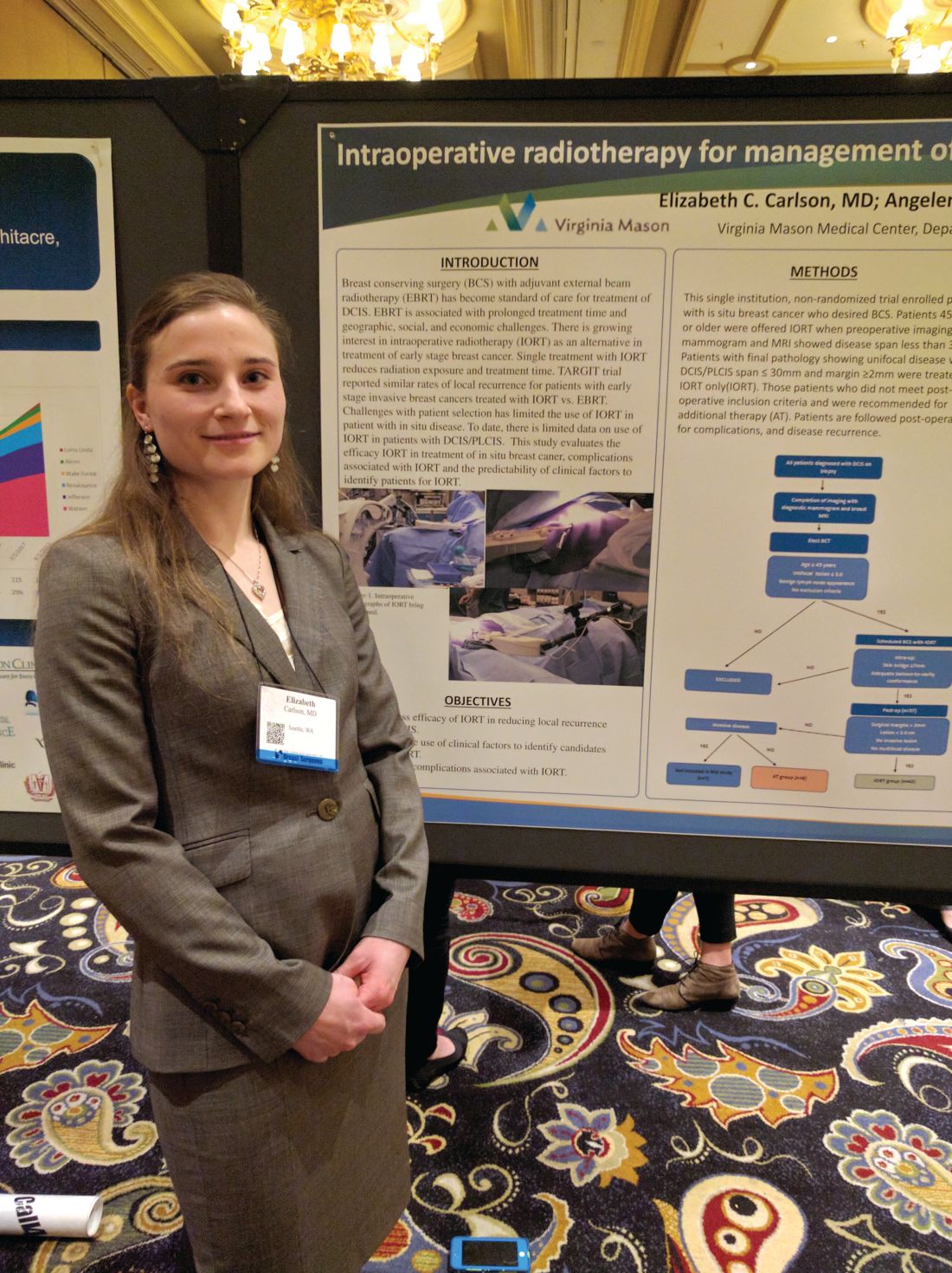
The researchers conducted a prospective, nonrandomized study of DCIS in women who underwent core biopsy between February 2012 and July 2016 at Virginia Mason Medical Center. Those who elected breast conserving therapy were assessed for IORT using bilateral digital mammography and contrast enhanced MRI.
For criteria, the researchers selected patients over age 44 years with unifocal DCIS and a lesion size of up to 3.0 cm. Physicians recommended additional therapy when the pathology report revealed DCIS larger than 3 cm and/or margins of up to 2 mm.
Of the 57 patients initially enrolled, 7 were found to have invasive disease and were excluded. Another eight patients were excluded because of margins, tumor size, or multifocal disease, and were recommended for additional treatment.
The remaining 42 patients with DCIS were treated with IORT. At a mean follow-up time of 32 months, the researchers observed no local recurrences, and no Radiation Therapy Oncology Group grade 3 or 4 complications were reported.
Hyperpigmentation occurred more often in the IORT group, at 40% (17 of 42 patients), than in the additional treatment group, at 13% (1 of 8), a nonsignificant difference. The hyperpigmentation tended to be minimal and focused on the surgical scar. There were no differences between the groups with respect to superficial wound separation, infection, seroma, fat necrosis, transient radiation dermatitis, or rib fracture.
“Our conclusions are that are selection criteria with mammography and MRI are effective in identifying patients who are appropriate for IORT, and importantly, at 32 months follow-up, no patients had recurrences, so we’re not negatively impacting oncological outcome,” said Dr. Carlson.
The fact that eight patients were recommended for additional treatment based on margins, a disease span greater than 3 cm, or multifocal disease “is actually quite heartening because it implies that the mammogram and the MRI are quite effective at identifying patients that met our criteria,” said Dr. Carlson.
The study is small and had no control group, but Dr. Carlson is confident of the results. “We didn’t do a randomized trial because we believed strongly that IORT will be effective in these patients with good cosmesis. When you compare it to local recurrence rates in the TARGIT-A trial, we’re still doing quite well, so to some extent that can be used as a baseline benchmark,” she said.
LAS VEGAS – Intraoperative radiation therapy (IORT) is effective in patients with ductal carcinoma in situ (DCIS), and bilateral digital mammography and breast MRI effectively predicted which patients would be most suited for the procedure, according to a nonrandomized study.
The TARGIT-A trial showed that IORT was noninferior to external beam radiation therapy in women with early-stage breast cancer (Lancet. 2014 Feb 15;383:603-13), but the technique hasn’t been tested in DCIS patients.
The researchers conducted a prospective, nonrandomized study of DCIS in women who underwent core biopsy between February 2012 and July 2016 at Virginia Mason Medical Center. Those who elected breast conserving therapy were assessed for IORT using bilateral digital mammography and contrast enhanced MRI.
For criteria, the researchers selected patients over age 44 years with unifocal DCIS and a lesion size of up to 3.0 cm. Physicians recommended additional therapy when the pathology report revealed DCIS larger than 3 cm and/or margins of up to 2 mm.
Of the 57 patients initially enrolled, 7 were found to have invasive disease and were excluded. Another eight patients were excluded because of margins, tumor size, or multifocal disease, and were recommended for additional treatment.
The remaining 42 patients with DCIS were treated with IORT. At a mean follow-up time of 32 months, the researchers observed no local recurrences, and no Radiation Therapy Oncology Group grade 3 or 4 complications were reported.
Hyperpigmentation occurred more often in the IORT group, at 40% (17 of 42 patients), than in the additional treatment group, at 13% (1 of 8), a nonsignificant difference. The hyperpigmentation tended to be minimal and focused on the surgical scar. There were no differences between the groups with respect to superficial wound separation, infection, seroma, fat necrosis, transient radiation dermatitis, or rib fracture.
“Our conclusions are that are selection criteria with mammography and MRI are effective in identifying patients who are appropriate for IORT, and importantly, at 32 months follow-up, no patients had recurrences, so we’re not negatively impacting oncological outcome,” said Dr. Carlson.
The fact that eight patients were recommended for additional treatment based on margins, a disease span greater than 3 cm, or multifocal disease “is actually quite heartening because it implies that the mammogram and the MRI are quite effective at identifying patients that met our criteria,” said Dr. Carlson.
The study is small and had no control group, but Dr. Carlson is confident of the results. “We didn’t do a randomized trial because we believed strongly that IORT will be effective in these patients with good cosmesis. When you compare it to local recurrence rates in the TARGIT-A trial, we’re still doing quite well, so to some extent that can be used as a baseline benchmark,” she said.
AT ASBS 2017
Key clinical point: Intraoperative radiation appears safe and effective, and is more convenient than postoperative radiation.
Major finding: There were no ductal carcinoma in situ recurrences at a median 32 months’ follow-up.
Data source: Uncontrolled, prospective study of 42 patients.
Disclosures: The study was funded by the National Institutes of Health. Dr. Carlson reported having no financial disclosures.
Sexual dysfunction, depression common in endometriosis patients
VANCOUVER – Endometriosis is associated with an increased risk of a range of sexual dysfunctions, and depression and anxiety are also common, according to a study conducted in Brazilian women.
Depression and anxiety were so common that the study itself had to be altered. Almost a third of participants were excluded from the study due to depressive symptoms. So many, in fact, that the researchers decided to include patients with mild to moderate depression and anxiety, and ultimately exclude only those with severe symptoms.

The final sample included 254 women with endometriosis and 329 control subjects between 18 and 45 years old. They all had an active sex life in the previous 6 months, and were heterosexual.
Forty-three percent of women in the endometriosis group had sexual dysfunction, compared with 18% in the control group (P less than .001), as measured by the Female Sexual Quotient. They also had higher rates of individual types of sexual dysfunction, including sexual desire dysfunction (25% vs. 16%; P = .007); sexual arousal dysfunction (16% vs. 4%; P less than .001); pain during intercourse/sex dysfunction (46% vs. 16%; P less than .001); and orgasm and/or sexual satisfaction dysfunction (30% vs. 13%; P less than .001).
The researchers also examined the relationship between sexual dysfunction and psychological factors, and found statistically significant positive associations with anxiety and depression.
The underlying psychological issues are of particular concern to Dr. Fairbanks. “It’s the main step to begin anything with them. We have to know if they have anxiety and depression or not,” she said.
Her team now asks patients with endometriosis about their sex life in order to establish a baseline that can be referred to. “After 1 year, 2 years, 5 years, we reapply the questionnaire to show them that their sexual life was not so good in the beginning.”
The study was eye-opening to Lori Brotto, PhD, professor of gynecology at the University of British Columbia, Vancouver, who was a discussant at the session. “The call to action is for doctors to really pay attention to sexual dysfunction, even though that might not be the primary issue that brings the patient to them,” Dr. Brotto said.
The high rates of depression and anxiety also suggest a need for a more rounded approach to endometriosis treatment. “A physician may think, ‘we’ll treat the endometriosis, and all other parts of her should improve as well.’ This study suggests that primarily physical interventions may not be sufficient for addressing something like sexual health, which is really mind and body together,” said Dr. Brotto.
The study was not funded. Dr. Fairbanks and Dr. Brotto reported having no relevant financial disclosures.
VANCOUVER – Endometriosis is associated with an increased risk of a range of sexual dysfunctions, and depression and anxiety are also common, according to a study conducted in Brazilian women.
Depression and anxiety were so common that the study itself had to be altered. Almost a third of participants were excluded from the study due to depressive symptoms. So many, in fact, that the researchers decided to include patients with mild to moderate depression and anxiety, and ultimately exclude only those with severe symptoms.
The final sample included 254 women with endometriosis and 329 control subjects between 18 and 45 years old. They all had an active sex life in the previous 6 months, and were heterosexual.
Forty-three percent of women in the endometriosis group had sexual dysfunction, compared with 18% in the control group (P less than .001), as measured by the Female Sexual Quotient. They also had higher rates of individual types of sexual dysfunction, including sexual desire dysfunction (25% vs. 16%; P = .007); sexual arousal dysfunction (16% vs. 4%; P less than .001); pain during intercourse/sex dysfunction (46% vs. 16%; P less than .001); and orgasm and/or sexual satisfaction dysfunction (30% vs. 13%; P less than .001).
The researchers also examined the relationship between sexual dysfunction and psychological factors, and found statistically significant positive associations with anxiety and depression.
The underlying psychological issues are of particular concern to Dr. Fairbanks. “It’s the main step to begin anything with them. We have to know if they have anxiety and depression or not,” she said.
Her team now asks patients with endometriosis about their sex life in order to establish a baseline that can be referred to. “After 1 year, 2 years, 5 years, we reapply the questionnaire to show them that their sexual life was not so good in the beginning.”
The study was eye-opening to Lori Brotto, PhD, professor of gynecology at the University of British Columbia, Vancouver, who was a discussant at the session. “The call to action is for doctors to really pay attention to sexual dysfunction, even though that might not be the primary issue that brings the patient to them,” Dr. Brotto said.
The high rates of depression and anxiety also suggest a need for a more rounded approach to endometriosis treatment. “A physician may think, ‘we’ll treat the endometriosis, and all other parts of her should improve as well.’ This study suggests that primarily physical interventions may not be sufficient for addressing something like sexual health, which is really mind and body together,” said Dr. Brotto.
The study was not funded. Dr. Fairbanks and Dr. Brotto reported having no relevant financial disclosures.
AT WCE 2017
Key clinical point: Sexual dysfunction is common but not always addressed by clinicians.
Major finding: Women with endometriosis had more than doubled risk of sexual dysfunction.
Data source: Forty-three percent of the endometriosis group had dysfunction, compared with 17.6% of controls.
Disclosures: The study was not funded. Dr. Fairbanks and Dr. Brotto reported having no relevant financial disclosures.
Genes may play role in breast cancer racial disparities
A new analysis of data from The Cancer Genome Atlas revealed 142 genes differentially expressed between white and African Americans that may influence breast cancer survival.
Researchers sifted through gene expression, protein expression, somatic mutations, somatic DNA copy number alteration, and DNA methylation patterns between 154 black patients of African ancestry (mean age at diagnosis, 55.66 years); and 776 white patients (mean age, 59.51) of European ancestry. They also looked at germline genetic variants for breast cancer subtypes and estimated subtype heritability.
African American patients were more likely than were whites to have basal-like breast cancer (odds ratio [OR], 3.80; 95% confidence interval [CI], 2.46-5.87; P less than .001) and HER2-enriched breast cancer (OR, 2.22; 95% CI, 1.10-4.47; P =.027).
After adjustment for subtype, 142 genes were differentially expressed between the two populations, a finding that held true after correction for testing of multiple comparisons (Bonferroni-adjusted P less than .05), reported Dezheng Huo, MD, PhD, of the department of public health sciences, University of Chicago, and his coauthors (JAMA Oncol. 2017 May 4. doi: 10.1001/jamaoncol.2017.0595).
During a median follow-up of 29 months, African Americans were more likely to experience recurrences (hazard ratio [HR], HR = 1.67, 95 CI, 1.02-2.74; P =.043). They had higher odds of basal-like breast cancer (OR, 3.80; 95% CI, 2.46-5.87; P less than .001) and human epidermal growth factor receptor 2 ERBB2 (HER2)-enriched breast cancer (OR, 2.22; 95% CI, 1.10-4.47; P =.027).
After adjusting for intrinsic subtypes, the researchers found differential expression of 16 DNA methylation probes, 4 DNA copy number segments, and 1 protein,

“If you have African ancestry, you’re more likely to have tumors that grow really fast. If we have drugs that can treat that kind of tumor, we need to find you and get you into treatment as quickly as possible,” she said.
A new analysis of data from The Cancer Genome Atlas revealed 142 genes differentially expressed between white and African Americans that may influence breast cancer survival.
Researchers sifted through gene expression, protein expression, somatic mutations, somatic DNA copy number alteration, and DNA methylation patterns between 154 black patients of African ancestry (mean age at diagnosis, 55.66 years); and 776 white patients (mean age, 59.51) of European ancestry. They also looked at germline genetic variants for breast cancer subtypes and estimated subtype heritability.
African American patients were more likely than were whites to have basal-like breast cancer (odds ratio [OR], 3.80; 95% confidence interval [CI], 2.46-5.87; P less than .001) and HER2-enriched breast cancer (OR, 2.22; 95% CI, 1.10-4.47; P =.027).
After adjustment for subtype, 142 genes were differentially expressed between the two populations, a finding that held true after correction for testing of multiple comparisons (Bonferroni-adjusted P less than .05), reported Dezheng Huo, MD, PhD, of the department of public health sciences, University of Chicago, and his coauthors (JAMA Oncol. 2017 May 4. doi: 10.1001/jamaoncol.2017.0595).
During a median follow-up of 29 months, African Americans were more likely to experience recurrences (hazard ratio [HR], HR = 1.67, 95 CI, 1.02-2.74; P =.043). They had higher odds of basal-like breast cancer (OR, 3.80; 95% CI, 2.46-5.87; P less than .001) and human epidermal growth factor receptor 2 ERBB2 (HER2)-enriched breast cancer (OR, 2.22; 95% CI, 1.10-4.47; P =.027).
After adjusting for intrinsic subtypes, the researchers found differential expression of 16 DNA methylation probes, 4 DNA copy number segments, and 1 protein,
“If you have African ancestry, you’re more likely to have tumors that grow really fast. If we have drugs that can treat that kind of tumor, we need to find you and get you into treatment as quickly as possible,” she said.
FROM JAMA ONCOLOGY
Key clinical point: Genetic differences may play role in racial disparities in breast cancer outcomes.
Major finding: The Cancer Genome Atlas data show that 142 genes were differentially expressed between whites and African Americans.
Data source: Analysis of genetic and outcome data from 930 patients.
Disclosures: The study was funded by the National Cancer Institute, the Breast Cancer Research Foundation, the National Human Genome Research Institute, Susan G. Komen, and the American Cancer Society. Dr. Olopade is a cofounder of CancerIQ and Prosigna.
Decision to remove breast cancer metastases depends on location of lesions
LAS VEGAS – Determination of whether excision of persistent breast cancer metastases can benefit the patient and even prolong survival depends on the location of the metastatic lesions, an investigator said at the annual meeting of the American Society of Breast Surgeons.
Before making the decision, it’s important to restage the patient’s disease and to recheck the receptor status if a biopsy is accessible, said Roshni Rao, MD, of the University of Texas Southwestern Medical Center.
Brain

Survival is worse when there are concurrent extracranial metastases or when brain metastases are greater than 5 cm, in patients with triple negative tumors, and in patients with a Karnofsky score of 70 or less, Dr. Rao said.
Surgery has the greatest benefit in patients with a single metastasis, with no extracranial disease, and who are able to undergo adjuvant whole brain radiation. In these cases, surgery improves survival, lowers recurrence rates, and reduces the risk of death neurological causes, she said.
Long-term survival is most common with continuous adjuvant therapy, either trastuzumab or hormonal, and in patients with a longer time interval to development of metastases. One series showed a 20-month survival increase.
Liver
Approximately 15% of patients with synchronous metastases will have liver metastases, and about half of stage IV patients will experience liver metastases at some point during treatment.
There is evidence from colorectal cancer that removing liver metastases is beneficial, and that has prompted interest in a similar approach in breast cancer. Liver resection has also become safer with new advances.
Dr. Rao discussed a single-institution study which took an aggressive approach to liver resection in 85 patients (Ann Surg. 2006;244:897-907). The researchers found that increased survival was associated with a good response to adjuvant chemotherapy, an r0 or r1 resection, and in patients who had a previous liver resection and were healthy enough to undergo another resection.
Overall, existing studies support liver resection if there are one to three lesions, if negative margins can be achieved, if the tumors are hormone receptor positive, and if the cancer is hormone positive and has good response to chemotherapy.
Dr. Rao emphasized that liver resections should be performed with a multidisciplinary team and should only be attempted at centers with low morbidity and where the doctors are experienced with liver resection.
When it’s possible, liver resection is beneficial. “There have been multiple reports of long-term survivors with no evidence of disease. There is likely a survival benefit with careful selection of these patients,” Dr. Rao said.
Lung
Lung surgeries are becoming safer, especially with the availability of video-assisted techniques, and pulmonary metastases are increasingly being spotted using more sensitive techniques such as higher resolution computed tomography.
A lung metastasis registry analysis showed three factors improved survival: prolonged disease-free survival, especially longer than 36 months; a complete resection; and a small number of metastases and success in resecting them all (Eur J Cardiothorac Surg. 2002;22:335-44). A more recent meta-analysis showed the same results (J Thorac Dis. 2015;7:1441-51).
Bone
Bone metastases remain rare choices for surgical treatment. Most of the time, morbidity will be too high, and there are good options for systemic treatment. That leaves surgery reserved mostly for stabilization or the treatment of fractures.
However, there are a couple of exceptions, according to Dr. Rao. One multi-institutional randomized trial looked at metastatic epidural spinal cord compression. Subjects either underwent decompressive surgery with stabilization and radiation or radiation alone. Patients in the surgical group had a longer ambulatory period and had a lower usage rate of steroids and opioids. Morbidity outcomes were similar in both groups. Patients whose primary tumor was in the breast seemed to benefit the most with respect to ambulatory time (Lancet. 2005;9486:643-8; J Neurosurg Spine. 2008;8:271-8).
Sternal metastases represent another special case. 70% of the time, patients with sternal metastases have it as their only metastatic site. A French series of 33 patients who underwent aggressive chest wall and rib resection reported a 36% complication rate, while another study of 28 patients showed a 21% complication rate. Those complications are a problem, “but if you’re able to perform this in a resected tumor, there are long term survivors. As usual, triple negative breast cancers predicted a worse prognosis,” said Dr. Rao.
Dr. Rao concluded that resection of metastatic sites has a role. “I think it’s our responsibility as breast surgeons who are many times continuously following these patients to consider appropriate operations,” said Dr. Rao.
However, she did sound one note of caution. Surgery can interrupt therapy that is helping a patient. “Let’s say you have someone get a big liver resection, and then they have a tough time with recovery. There could be a long period of time they can’t get the therapy that was keeping them alive. That’s the real concern,” she said.
LAS VEGAS – Determination of whether excision of persistent breast cancer metastases can benefit the patient and even prolong survival depends on the location of the metastatic lesions, an investigator said at the annual meeting of the American Society of Breast Surgeons.
Before making the decision, it’s important to restage the patient’s disease and to recheck the receptor status if a biopsy is accessible, said Roshni Rao, MD, of the University of Texas Southwestern Medical Center.
Brain
Survival is worse when there are concurrent extracranial metastases or when brain metastases are greater than 5 cm, in patients with triple negative tumors, and in patients with a Karnofsky score of 70 or less, Dr. Rao said.
Surgery has the greatest benefit in patients with a single metastasis, with no extracranial disease, and who are able to undergo adjuvant whole brain radiation. In these cases, surgery improves survival, lowers recurrence rates, and reduces the risk of death neurological causes, she said.
Long-term survival is most common with continuous adjuvant therapy, either trastuzumab or hormonal, and in patients with a longer time interval to development of metastases. One series showed a 20-month survival increase.
Liver
Approximately 15% of patients with synchronous metastases will have liver metastases, and about half of stage IV patients will experience liver metastases at some point during treatment.
There is evidence from colorectal cancer that removing liver metastases is beneficial, and that has prompted interest in a similar approach in breast cancer. Liver resection has also become safer with new advances.
Dr. Rao discussed a single-institution study which took an aggressive approach to liver resection in 85 patients (Ann Surg. 2006;244:897-907). The researchers found that increased survival was associated with a good response to adjuvant chemotherapy, an r0 or r1 resection, and in patients who had a previous liver resection and were healthy enough to undergo another resection.
Overall, existing studies support liver resection if there are one to three lesions, if negative margins can be achieved, if the tumors are hormone receptor positive, and if the cancer is hormone positive and has good response to chemotherapy.
Dr. Rao emphasized that liver resections should be performed with a multidisciplinary team and should only be attempted at centers with low morbidity and where the doctors are experienced with liver resection.
When it’s possible, liver resection is beneficial. “There have been multiple reports of long-term survivors with no evidence of disease. There is likely a survival benefit with careful selection of these patients,” Dr. Rao said.
Lung
Lung surgeries are becoming safer, especially with the availability of video-assisted techniques, and pulmonary metastases are increasingly being spotted using more sensitive techniques such as higher resolution computed tomography.
A lung metastasis registry analysis showed three factors improved survival: prolonged disease-free survival, especially longer than 36 months; a complete resection; and a small number of metastases and success in resecting them all (Eur J Cardiothorac Surg. 2002;22:335-44). A more recent meta-analysis showed the same results (J Thorac Dis. 2015;7:1441-51).
Bone
Bone metastases remain rare choices for surgical treatment. Most of the time, morbidity will be too high, and there are good options for systemic treatment. That leaves surgery reserved mostly for stabilization or the treatment of fractures.
However, there are a couple of exceptions, according to Dr. Rao. One multi-institutional randomized trial looked at metastatic epidural spinal cord compression. Subjects either underwent decompressive surgery with stabilization and radiation or radiation alone. Patients in the surgical group had a longer ambulatory period and had a lower usage rate of steroids and opioids. Morbidity outcomes were similar in both groups. Patients whose primary tumor was in the breast seemed to benefit the most with respect to ambulatory time (Lancet. 2005;9486:643-8; J Neurosurg Spine. 2008;8:271-8).
Sternal metastases represent another special case. 70% of the time, patients with sternal metastases have it as their only metastatic site. A French series of 33 patients who underwent aggressive chest wall and rib resection reported a 36% complication rate, while another study of 28 patients showed a 21% complication rate. Those complications are a problem, “but if you’re able to perform this in a resected tumor, there are long term survivors. As usual, triple negative breast cancers predicted a worse prognosis,” said Dr. Rao.
Dr. Rao concluded that resection of metastatic sites has a role. “I think it’s our responsibility as breast surgeons who are many times continuously following these patients to consider appropriate operations,” said Dr. Rao.
However, she did sound one note of caution. Surgery can interrupt therapy that is helping a patient. “Let’s say you have someone get a big liver resection, and then they have a tough time with recovery. There could be a long period of time they can’t get the therapy that was keeping them alive. That’s the real concern,” she said.
EXPERT ANALYSIS FROM ASBS 2017
Breast density is no reason to perform MRI
LAS VEGAS – In women with higher density (HD) breasts, preoperative MRI revealed more abnormalities than were seen in women with lower density (LD) breasts, but there was no difference in the number of secondary cancers detected or long-term recurrence rates.
Breast density is often cited by radiologists as a reason to conduct a preoperative MRI, but the study suggests that it should not be a driving factor. “It’s a real challenge when our radiologists provide us reports that say, ‘Due to increased density, we recommend MRI,’ because it’s really hard to then disregard that. I think this is very important data,” said Judy Boughey, MD, professor of surgery at the Mayo Clinic, Rochester, MN, who moderated the session at the annual meeting of the American Society of Breast Surgeons where the research was presented.
“MRI is a valuable tool, and we’re still trying to figure out who it should be performed in,” said lead author Sarah McLaughlin, MD, of the Mayo Clinic, Jacksonville, FL, in an interview.
The researchers retrospectively analyzed data from 683 women at their institution who underwent preoperative MRI between 2007 and 2011. They grouped them by mammography results into LD (33%; Breast Imaging–Reporting and Data System density, 1 and 2) and HD (67%; BI-RADS density, 3 and 4).
Patients in the HD group more often had ipsilateral MRI findings (42% vs. 31%; P = .005), but ,of those with MRI findings, a similar number of patients in each group needed a second site biopsy (HD 65% vs. LD 67%; P = .78).
In all patients who had an additional MRI finding, the odds of detecting an additional ipsilateral cancer were not statistically significant between HD (32%) and LD (23%; P = .15) patients.
HD patients were also more likely to have abnormalities in the contralateral breast (25% vs. 14%; P = .009), but there were no statistically significant differences in rates of second-site biopsy recommendations or in the percentages of abnormalities that turned out to be cancerous (HD 6% vs. LD 3%; P = 1.0).
Following MRI, 70% of LD patients expressed a preference for breast-conserving surgery, compared with 53% of HD patients (P = .0001).
Over a median 7 years of follow-up, there was no difference in freedom from recurrence rates between the two groups (91% in LD vs. 90% in HD; P = .57).
“To me, it says that you don’t have to order an MRI just because they have cancer in a high density breast. You can feel reassured by your surgical plan and treatment recommendations based on conventional imaging,” said Dr. McLaughlin.
The researchers can’t determine if having an MRI done increased patient worry and potentially led to the higher rate of mastectomies chosen by women in the HD group. “Is that a result of the MRI? I don’t think we can say that, but there’s this whole other discussion piece that goes into it. You definitely see patients who say, ‘But it found these other things, and I’m going to have a mastectomy.’ So, there’s that patient preference and worry piece,” said Dr. McLaughlin.
The study results should offer some reassurance to patients. “There were no differences in local recurrence rates according to density. Maybe the next angle is allaying some of that fear, because the outcomes were the same. It’s really driven more by tumor biology and multimodality therapy,” said Dr. McLaughlin.
The study doesn’t provide the final word on breast density and MRI, according to Dr. Boughey. “I think this is an area that needs to be studied more with a clinical trial. There are several going on in different countries, and this is an area where we need level 1 data. This study does fit with what many other studies have shown, which is that MRI probably doesn’t have as much benefit as patients believe it does, so our role really is to try to help educate patients.”
AT ASBS 2017
Key clinical point: High breast density is probably not cause enough to order preoperative MRI.
Major finding: Freedom from recurrence rates were 90% in high density and 91% in low.
Data source: Retrospective analysis of 683 women at a single institution.
Disclosures: The study was funded internally. Dr. McLaughlin and Dr. Boughey reported having no relevant financial relationships.
Sutureless, guided lumpectomy produced clean margins in majority
LAS VEGAS – An automated, minimally invasive, stereotactic-guided lumpectomy performed well in an outpatient setting, with no sutures required, potentially decreasing patient morbidity, according to Pat Whitworth, MD.
Pain scores were low, and all high-risk lesions were successfully removed.
The new technology is part of the natural progression of cancer resection, said Dr. Whitworth, director of the Nashville (Tenn.) Breast Center, at the annual meeting of the American Society of Breast Surgeons.
“We went from radical mastectomy, to modified radical mastectomy, to lumpectomy. This is just part of that progression where we match what we do to what the patient really needs,” he said.
Dr. Whitworth said he believes that radiologists will soon be using such technology to treat breast cancer, which puts the onus on breast cancer surgeons to adopt it themselves. “I think it’s important for breast surgeons to acquire the necessary skill and techniques to use the same tools and work collaboratively with radiologists, because this is coming,” he said.
To see if it were possible to achieve results similar to those with lumpectomy, Dr. Whitworth analyzed data from 279 women who had a small ductal carcinoma in situ (DCIS), invasive carcinoma, or high-risk lesion removed using a 15- or 20-mm radiofrequency basket capture with imaging guidance (lumpectomy). Patients who received a cancer diagnosis underwent a second, 20-mm basket capture to obtain shaved margins.
The procedure was conducted under local anesthesia and sedation, and the incisions were closed using Steri-Strip skin closures. The average pain score was 1.55 out of 10 (range, 0-7).
Of 125 patients found to have DCIS (n = 52) and invasive lesions (n = 73), the first capture achieved clear margins in 69 cases, and the shaved margin capture achieved clean margins in another 33 cases.
The remaining 23 patients (18%) had a positive margin by histologic standards following lumpectomy and shaved margin. Of the 22 with reported results, 17 (77%) had no residual lesion following open surgery.
The results convinced Dr. Wentworth of the technique’s utility, particularly in patients who may have a heightened surgical risk, he said. “We think this can replace open lumpectomy in selected patients, with favorable margin clearance.”
The approach is fairly simple, but Dr. Wentworth recommended beginning with stereotactic-guided technology before attempting the ultrasound-guided version, which requires a little more skill. “The biggest challenge is learning that you have to use a lot more local anesthetic. When this technology first came out, people tried to use the same amount of local anesthetic that they used for standard vacuum-assisted core biopsy, and it’s very painful unless you put 30 or 40 cc of dilute anesthetic in there,” said Dr. Wentworth.
The study was funded by Medtronic, which markets the technology. Dr. Whitworth is a principal at Targeted Medical Education, which receives funding from Medtronic.
AT ASBS 2017
Key clinical point: The technique could replace standard lumpectomy in patients at high surgical risk.
Major finding: Clean margins were obtained in 102 of 125 women with diagnosed cancer.
Data source: A retrospective analysis of 279 patients.
Disclosures: The study was funded by Medtronic, which markets the technology. Dr. Whitworth is a principal at Targeted Medical Education, which receives funding from Medtronic.
DCIS tool IDs axillary node biopsy candidates
LAS VEGAS – A new tool identifies patients with ductal carcinoma in situ (DCIS) who are at high risk for being upstaged as a result of the pathology report. The screen could encourage patients to undergo axillary nodal staging during the core needle biopsy (CNB), thus avoiding a second procedure.
“The risk factors have been well described, but they haven’t helped give individual risk or individual percentages. Our goal was to try to individualize that risk so that we could counsel patients,” said lead researcher James Jakub, MD, chair of the division of breast endocrine and metabolic surgery at Mayo Clinic Rochester, at the annual meeting of the American Society of Breast Surgeons.

The researchers found that grade on CNB, mass lesion on imaging, multifocal/centric disease, and linear dimension combined to predict the likelihood of being upstaged to invasive disease (C statistic, 0.71; 95% confidence interval, 0.66-0.77).
They then combined those characteristics to create a nomogram and tested it against the validation set. In that group, 11% of patients were upstaged to invasive disease. The nomogram performed almost identically in the external validation set (C statistic, 0.71; 95% CI, 0.63-0.79). The model predicted 56 upstages, and 46 occurred.
This wasn’t the first attempt to create a nomogram to predict upstaging in DCIS patients, but others were not tested against external datasets. “That’s a weakness with other studies. Unless it’s validated externally, you don’t really know if you can apply it to your population. That was a very large plus: having colleagues who were willing to collaborate to validate it,” said Dr. Jakub.
The team is working on posting the nomogram online for widespread use.
It remains to be seen whether the availability of the nomogram will, in fact, change patients’ decision-making and result in more axillary nodal biopsies during CNB procedures in high risk patients.
“We haven’t looked into that. It will be interesting to see how it influences [sentinel lymph node] biopsy rate at our institution, and we’d love to hear from institutions who apply it. My sense is that patients who are at high risk are going to be interested in doing that. I think it will definitely change their management,” said Dr. Jakub.
LAS VEGAS – A new tool identifies patients with ductal carcinoma in situ (DCIS) who are at high risk for being upstaged as a result of the pathology report. The screen could encourage patients to undergo axillary nodal staging during the core needle biopsy (CNB), thus avoiding a second procedure.
“The risk factors have been well described, but they haven’t helped give individual risk or individual percentages. Our goal was to try to individualize that risk so that we could counsel patients,” said lead researcher James Jakub, MD, chair of the division of breast endocrine and metabolic surgery at Mayo Clinic Rochester, at the annual meeting of the American Society of Breast Surgeons.
The researchers found that grade on CNB, mass lesion on imaging, multifocal/centric disease, and linear dimension combined to predict the likelihood of being upstaged to invasive disease (C statistic, 0.71; 95% confidence interval, 0.66-0.77).
They then combined those characteristics to create a nomogram and tested it against the validation set. In that group, 11% of patients were upstaged to invasive disease. The nomogram performed almost identically in the external validation set (C statistic, 0.71; 95% CI, 0.63-0.79). The model predicted 56 upstages, and 46 occurred.
This wasn’t the first attempt to create a nomogram to predict upstaging in DCIS patients, but others were not tested against external datasets. “That’s a weakness with other studies. Unless it’s validated externally, you don’t really know if you can apply it to your population. That was a very large plus: having colleagues who were willing to collaborate to validate it,” said Dr. Jakub.
The team is working on posting the nomogram online for widespread use.
It remains to be seen whether the availability of the nomogram will, in fact, change patients’ decision-making and result in more axillary nodal biopsies during CNB procedures in high risk patients.
“We haven’t looked into that. It will be interesting to see how it influences [sentinel lymph node] biopsy rate at our institution, and we’d love to hear from institutions who apply it. My sense is that patients who are at high risk are going to be interested in doing that. I think it will definitely change their management,” said Dr. Jakub.
AT ASBS 2017
Key clinical point: High risk patients who choose axillary biopsy could avoid a second procedure.
Major finding: The 4-item nomogram predicted upstaging with a C statistic of 0.71.
Data source: A retrospective sample (n = 827) and validation study (n = 579).
Disclosures: The source of funding was not disclosed. Dr. Jakub reported having no financial disclosures.
Resection reduces inflammatory breast cancer lesion recurrence
LAS VEGAS – In patients with nonmetastatic inflammatory breast cancer (IBC), aggressive surgical resection can reduce local/regional recurrence rates to levels comparable to those seen in patients with noninflammatory breast cancer.
The key to the improved outcome is the presence of a plastic surgeon who can help close a tricky incision. “We have a multidisciplinary team that allows us to call upon the plastic surgeons if we can’t control the incision, and they help us with a flap closure,” said Kelly Rosso, MD, a breast surgical oncology fellow at the MD Anderson Cancer Center, Houston, who presented the study results at the annual meeting of the American Society of Breast Surgeons.

To see if their surgical interventions were producing better outcomes, the team analyzed data from 114 patients with nonmetastatic inflammatory breast cancer who had received trimodality therapy (neoadjuvant chemotherapy, surgery, and radiation therapy) from 2007 to 2015.
The median patient age was 52 years, and the median follow-up period was 3.6 years. In total, 55% of patients had N2 IBC, and 45% had N3 disease.
Nearly all (113 of 114) patients had negative surgical margins. A total of 29 patients died during the follow-up period, and the 5-year survival rate was 69%. The odds of a local/regional recurrence over 2 years was 3% (95% confidence interval, 1%-10%). The 2-year odds of recurrence or distant metastasis was 23% (95% CI, 16%-32%).
Dr. Lucci said that he suspects that many surgeons avoid resection of inflammatory breast cancer lesions under the assumption that the disease is metastatic in nature and fear that an attempt at local control would put the patient through unnecessary surgery with little benefit.
The new study challenges that belief. “I think this is a changing paradigm. We have to consider that those patients need to be thought of as still resectable and think about getting rid of that local disease, at the very least to improve their quality of life and hopefully to improve their overall outcome,” Dr. Lucci said.
The results should be readily achievable in other centers, he added. “If they have a plastic surgeon who’s willing to help them, they can resect to negative margins just as well [as in noninflammatory patients]. And, if they have a radiation therapist who’s dedicated to doing a more aggressive course, they can achieve the same results.”
Dr. Rosso and Dr. Lucci reported having no financial disclosures.
LAS VEGAS – In patients with nonmetastatic inflammatory breast cancer (IBC), aggressive surgical resection can reduce local/regional recurrence rates to levels comparable to those seen in patients with noninflammatory breast cancer.
The key to the improved outcome is the presence of a plastic surgeon who can help close a tricky incision. “We have a multidisciplinary team that allows us to call upon the plastic surgeons if we can’t control the incision, and they help us with a flap closure,” said Kelly Rosso, MD, a breast surgical oncology fellow at the MD Anderson Cancer Center, Houston, who presented the study results at the annual meeting of the American Society of Breast Surgeons.
To see if their surgical interventions were producing better outcomes, the team analyzed data from 114 patients with nonmetastatic inflammatory breast cancer who had received trimodality therapy (neoadjuvant chemotherapy, surgery, and radiation therapy) from 2007 to 2015.
The median patient age was 52 years, and the median follow-up period was 3.6 years. In total, 55% of patients had N2 IBC, and 45% had N3 disease.
Nearly all (113 of 114) patients had negative surgical margins. A total of 29 patients died during the follow-up period, and the 5-year survival rate was 69%. The odds of a local/regional recurrence over 2 years was 3% (95% confidence interval, 1%-10%). The 2-year odds of recurrence or distant metastasis was 23% (95% CI, 16%-32%).
Dr. Lucci said that he suspects that many surgeons avoid resection of inflammatory breast cancer lesions under the assumption that the disease is metastatic in nature and fear that an attempt at local control would put the patient through unnecessary surgery with little benefit.
The new study challenges that belief. “I think this is a changing paradigm. We have to consider that those patients need to be thought of as still resectable and think about getting rid of that local disease, at the very least to improve their quality of life and hopefully to improve their overall outcome,” Dr. Lucci said.
The results should be readily achievable in other centers, he added. “If they have a plastic surgeon who’s willing to help them, they can resect to negative margins just as well [as in noninflammatory patients]. And, if they have a radiation therapist who’s dedicated to doing a more aggressive course, they can achieve the same results.”
Dr. Rosso and Dr. Lucci reported having no financial disclosures.
At ASBS 2017
Key clinical point:
Major finding: The odds of a local/regional recurrence over 2 years were 3%.
Data source: A retrospective analysis of 114 patients.
Disclosures: Dr. Rosso and Dr. Lucci reported having no financial disclosures.