User login
Teledermatology improves diagnoses in the ED
Teledermatologic consults via mobile phones invalidated, clarified, or enlarged an emergency physician’s diagnosis and management in 68% of 110 cases seen in the emergency department, based on data from an observational study published online May 7 in JAMA Dermatology.
"Easy access to a dermatologist helped ED physicians manage their skin problems," wrote Dr. Tu Anh Duong, who conducted the study while at the Hôpital Henri Mondor, Créteil, France, and her associates.
Management concordance between emergency physicians and dermatologists was high for diagnostic and specialist consultations within 24 hours, moderate for immediate hospitalization, and poor for patient discharge, the researchers noted (JAMA Dermatol. 2014 May 7 [doi:10.1001/jamadermatol.2013.7792]).
The study included 110 adults aged 18 years and older seen consecutively for dermatologic conditions at four French hospitals. The majority of the conditions were skin infections (28%) or eczema/urticaria (24%). In each case, an ED physician sent digital images and possible diagnoses to a dermatologist, and both physicians recorded their diagnoses and management choices. For 83 cases, the physicians participated in a videoconference, which improved diagnostic performance in 57 cases (69%) and invalidated photographic diagnoses in 8 cases (10%).
The findings confirm the convenience and effectiveness of using mobile devices for teledermatology services in the ED and suggest that that such devices be used for real-time communication between physicians after images are sent, the researchers said. However, "the use of mobile devices requires a functional high-speed telecommunication network, a secure protocol for image transfer, a reliable storage system, and some minimal organization," they added.
The study was funded by a research grant from the Société Française de Dermatologie. Mobile phones and communication costs were provided by the Société Française du Radiotéléphonie. Dr. Duong disclosed receiving a grant from Basilea Pharmaceutica for the first year of her PhD program.
On Twitter @hsplete
FROM JAMA DERMATOLOGY
Key clinical point: Mobile phones are feasible for use in teledermatology, which can improve the quality of care and cost of managing dermatologic conditions.
Major finding: Teledermatology improved the diagnosis in 68% of 110 cases seen in the ED.
Data source: An observational study including 110 consecutive patients seen for dermatologic conditions in the EDs of four hospitals.
Disclosures: The study was funded by a research grant from the Société Française de Dermatologie. Dr. Duong disclosed receiving a grant from Basilea Pharmaceutica for the first year of her PhD program.
Cutting-edge cancer research recognized at AAD meeting in Denver
A trio of up-and-coming dermatologists received the La Roche-Posay North American Foundation’s 2014 Research Awards, presented at the annual meeting of the American Academy of Dermatology in Denver.
The awards were given to candidates with promising projects in clinical, biological, and pharmacological research linked to dermatology.
Dr. Thomas Strub of the Icahn School of Medicine at Mount Sinai, New York, received the first-place grant of $10,000 for his study "Epigenetic Mechanisms Underlying Drug Resistance of Malignant Melanoma."
"It is a privilege for me to be chosen by the La Roche-Posay North American Foundation for supporting my research goals," Dr. Strub said in a statement. "Melanoma therapies remain limited, and drug resistance invariably occurs, with patients ultimately relapsing. I hope my research will shed light onto the epigenome of resistant melanoma tumors and insight on the elements involved in melanoma, which will ultimately improve patient outcomes."
Dr. Shadmehr Demehri of Washington University, St. Louis, received the second prize of $5,000 for his study "The Role of Calcipotriol in Treatment of Pre-cancerous Skin Lesions."
"It’s a great honor to be a recipient of the La Roche-Posay North American Foundation’s research award," Dr. Demehri said in a statement. "My research focuses on determining the role of immune activation in cancer therapy. Specifically, I aim to understand the effects of epithelial cytokine expression on induction of antitumor immunity. Considering the rising prevalence of skin cancer in our population, the outcomes of our investigation can have significant impact on care for patients with skin and other types of cancers."
Dr. Emily Newsom of Wayne State University, Detroit, received the third prize award of $5,000 for her study "Mapping Cytogenetic Abnormalities in Cutaneous T-cell Lymphoma using FISH."
"I appreciate the La Roche-Posay North American Foundation’s commitment to furthering research in dermatology. This award will allow me to advance my career and research in cutaneous lymphoma," Dr. Newsom said in a statement.
The winners were selected by the La Roche-Posay North American Scientific Committee.
For additional information about La Roche-Posay, visit www.laroche-posay.us and "like" the La Roche-Posay Facebook page at www.facebook.com/LaRochePosayusa.
Show off your social media skills in AAD’s selfie photo contest
Social media take center stage at this year’s American Academy of Dermatology annual meeting, as the AAD offers its first Twitter "selfie" photo contest. The winner will receive a $50 Starbucks gift card; how’s that for an incentive?
To enter, you (or a friend) need to have a Twitter account. Send your best selfie (it can be one person, two people, or a group shot) of you and your friends enjoying the meeting. Note: Photos may be taken in or around the convention center, or out and about in Denver, but no photos may be taken from inside the meeting session rooms.
Once you’re on Twitter, tweet your best selfie to @AADMtgs, and be sure to use the hashtag #aad14.
The contest kicks off on Friday, March 21, at noon (Mountain Standard Time), and ends on Tuesday, March 25, at noon (MST). A panel will review the photos, and the winner will be notified by noon on Friday, March 28 (Central Standard Time). Gift cards will be mailed or e-mailed. More information is available at the AAD’s website, aad.org.
While you’re tweeting your selfies, keep up with the latest news and development from the meeting by following @sknews, as well as our intrepid reporter, Naseem Miller (@naseemmiller).
On Twitter @hsplete
AAD 2014 sessions offer something for everyone
The American Academy’s 2014 annual meeting in Denver will feature new CME sessions and updates on the latest dermatology research.
This year’s program features expert commentary on key issues in medical dermatology, including "Melanoma Multidisciplinary Care 2014: What You Need to Know" on Sunday, March 23, from 1 p.m. to 3 p.m. in Room 705/707 and "Dermatologic Manifestations of New Oncology Drugs," also on Sunday, March 23, from 1 p.m. to 3 p.m. in the Mile High Ballroom 3B. Looking for the latest in aesthetic dermatology? Check out the "Advanced Botulinum Toxin" live demonstration session on Saturday, March 22, from 2 p.m. to 5 p.m. in the Bellco Theater.
There will be expert sessions on pregnancy dermatoses, cutaneous T-cell lymphoma, pediatric dermatology, skin of color, and the latest on treatments for hair and nail conditions. The full scientific session list is available online.
A series of practice management lectures includes topics such as "How to Have an Unforgettably Positive Office Visit" on Saturday, March 22, from 10:00 a.m. to 12:00 p.m. in Room 709/7111 and "Hot Buttons: Recognizing What Sets You Off and Managing Your Triggers" on Sunday, March 23, from 1:00 p.m. to 3:00 p.m. in Room 702.
There is also a mobile device app that meeting attendees can download that contains session schedules, exhibitor and attendee lists, and more.
Can’t attend the meeting? Visit www.eDermatologyNews.com for live conference coverage.
On Twitter @Sknews
How to identify inducible urticaria
CHAMPIONSGATE, FLA. – Physical urticarias, also known as inducible urticarias, comprise 20%-30% of all chronic urticaria cases, and these patients are rarely responsive to corticosteroids, according to Dr. Adam Friedman.
Physical urticarias are often episodic, and spontaneous resolution is less likely in this subset of patients, compared with chronic urticaria patients overall (33% vs. 20%), said Dr. Friedman, director of dermatologic research at Albert Einstein College of Medicine in New York.
Identifying the unique source of a patient’s inducible urticarias is important because it paves the way for more successful treatment, he said at the Orlando Dermatology Aesthetic and Clinical Conference.
Causes of physical urticaria include dermatographism, cholinergic factors, heat, cold, water, sun, and vibration.
To diagnose physical urticaria, ask patients about family history and whether they have pictures documenting the urticaria episodes, Dr. Friedman said. The reaction in patients with physical urticaria is reproducible in response to environmental factors, he noted.

Dr. Friedman described several provocative tests for the most common physical urticarias.
If cold-induced urticaria is suspected, place an ice cube on the forearm to see whether hives occur after it is removed. If patients report hives after sweating or exposure to heat, the heat may be the cause; test by using localized heat (a test tube of water at 44 degrees Celsius).
Less obvious, but genuine, causes of physical urticaria include delayed pressure-induced urticaria and angioedema, which may be the culprit in patients who report swelling in the shoulder within 4-6 hours after carrying a heavy purse or other shoulder bag. To test for this trigger in the office, use a 15-pound sandbag on the target area. In cases of aquagenic urticaria, patients develop hives with exposure to water, which can be reproduced with a wet compress. And, believe it or not, individuals can develop vibratory-induced urticaria on the hands after mowing the lawn (this one is harder to reproduce in the office).
For physical urticaria, as with any type, "Make sure you have the right diagnosis," Dr. Friedman said. The patient may come in without anything on the skin, complaining of episodes of hives. The urticarial differential diagnosis is quite extensive, so a good history is important to clinching the diagnosis.
Biopsies and lab tests aren’t usually helpful for physical urticarias, Dr. Friedman said, "but if something suggests a workup, go after it."
The treatment of physical urticaria can be complicated, and includes both avoiding triggers (if possible) and managing the inflammatory response. The first-line preference is high-dose, second-generation H1 antihistamines. "You can go as high as fourfold the recommended daily dose," Dr. Friedman said.
"Don’t be afraid to try some off-label uses for recalcitrant physical urticarias," advised Dr. Friedman. Options for cold urticarias, for example, include zafirlukast, montelukast, and omalizumab. Although omalizumab is currently used off label for these patients, its approval by the FDA is expected later this year, Dr. Friedman said. "This is a very exciting time, with this new therapy at our fingertips," he added.
For patients with aquagenic urticaria, stanozolol has shown some efficacy, as well as narrow-band UVB or PUVA photochemotherapy. Potential treatments for patients with delayed-pressure urticaria include NSAIDs, dapsone, sulfasalazine, antimalarials, and even intravenous immunoglobulin. Systemic steroids should be avoided as a go-to solution, but can help bridge a patient onto a nonsteroidal immunosuppressant that has a slow onset of action.
In managing physical urticarias, as with urticarias in general, "climb the therapeutic ladder, and remember that combination therapy is king," Dr. Friedman said.
He disclosed ties with Onset, Valeant, and other companies.
CHAMPIONSGATE, FLA. – Physical urticarias, also known as inducible urticarias, comprise 20%-30% of all chronic urticaria cases, and these patients are rarely responsive to corticosteroids, according to Dr. Adam Friedman.
Physical urticarias are often episodic, and spontaneous resolution is less likely in this subset of patients, compared with chronic urticaria patients overall (33% vs. 20%), said Dr. Friedman, director of dermatologic research at Albert Einstein College of Medicine in New York.
Identifying the unique source of a patient’s inducible urticarias is important because it paves the way for more successful treatment, he said at the Orlando Dermatology Aesthetic and Clinical Conference.
Causes of physical urticaria include dermatographism, cholinergic factors, heat, cold, water, sun, and vibration.
To diagnose physical urticaria, ask patients about family history and whether they have pictures documenting the urticaria episodes, Dr. Friedman said. The reaction in patients with physical urticaria is reproducible in response to environmental factors, he noted.
Dr. Friedman described several provocative tests for the most common physical urticarias.
If cold-induced urticaria is suspected, place an ice cube on the forearm to see whether hives occur after it is removed. If patients report hives after sweating or exposure to heat, the heat may be the cause; test by using localized heat (a test tube of water at 44 degrees Celsius).
Less obvious, but genuine, causes of physical urticaria include delayed pressure-induced urticaria and angioedema, which may be the culprit in patients who report swelling in the shoulder within 4-6 hours after carrying a heavy purse or other shoulder bag. To test for this trigger in the office, use a 15-pound sandbag on the target area. In cases of aquagenic urticaria, patients develop hives with exposure to water, which can be reproduced with a wet compress. And, believe it or not, individuals can develop vibratory-induced urticaria on the hands after mowing the lawn (this one is harder to reproduce in the office).
For physical urticaria, as with any type, "Make sure you have the right diagnosis," Dr. Friedman said. The patient may come in without anything on the skin, complaining of episodes of hives. The urticarial differential diagnosis is quite extensive, so a good history is important to clinching the diagnosis.
Biopsies and lab tests aren’t usually helpful for physical urticarias, Dr. Friedman said, "but if something suggests a workup, go after it."
The treatment of physical urticaria can be complicated, and includes both avoiding triggers (if possible) and managing the inflammatory response. The first-line preference is high-dose, second-generation H1 antihistamines. "You can go as high as fourfold the recommended daily dose," Dr. Friedman said.
"Don’t be afraid to try some off-label uses for recalcitrant physical urticarias," advised Dr. Friedman. Options for cold urticarias, for example, include zafirlukast, montelukast, and omalizumab. Although omalizumab is currently used off label for these patients, its approval by the FDA is expected later this year, Dr. Friedman said. "This is a very exciting time, with this new therapy at our fingertips," he added.
For patients with aquagenic urticaria, stanozolol has shown some efficacy, as well as narrow-band UVB or PUVA photochemotherapy. Potential treatments for patients with delayed-pressure urticaria include NSAIDs, dapsone, sulfasalazine, antimalarials, and even intravenous immunoglobulin. Systemic steroids should be avoided as a go-to solution, but can help bridge a patient onto a nonsteroidal immunosuppressant that has a slow onset of action.
In managing physical urticarias, as with urticarias in general, "climb the therapeutic ladder, and remember that combination therapy is king," Dr. Friedman said.
He disclosed ties with Onset, Valeant, and other companies.
AT THE ODAC CONFERENCE

Survey: Anti-MRSA drugs routinely prescribed for simple abscesses
Nearly 90% of more than 500 dermatologists surveyed said they would initially prescribe an antibiotic to cover possible methicillin-resistant Staphylococcus aureus when incising and draining an uncomplicated cutaneous abscess. Further, 24% reported prescribing antibiotics that are not active against the pathogen, and 82% said they cultured simple abscesses in 50% of cases.
The survey findings, while limited by their self-reporting nature, point to the need for clearer guidelines on the best approaches to simple abscesses.
"A comprehensive clinical guideline based on local antimicrobial rates, and increased knowledge of local resistance patterns and microbiologic data could not only improve abscess management and antibacterial stewardship, but could also combat the rising health care costs associated with SSTIs [skin and soft tissue infections] and their complications," wrote Dr. Adam Friedman and his colleagues in the February issue of the Journal of Drugs in Dermatology.
Simple excision and drainage (I&D) is the standard treatment for an uncomplicated skin abscess. With the increase in community-acquired methicillin-resistant Staphylococcus aureus (CA-MRSA) as the culprit behind SSTIs in the United States, the Centers for Disease Control and Prevention recommends that health professionals consider CA-MRSA in all cases of skin abscesses, perform I&D, and use culture results to guide treatment, the researchers noted.
Guidelines from the Infectious Diseases Society of America recommend antibiotics for abscesses after I&D in certain circumstances, and coverage in the outpatient setting if needed. IDSA and other organizations currently recommend empiric coverage of CA-MRSA for acute bacterial skin and skin structure infections if and when antibiotics are incorporated into the therapeutic regimen (Clin. Infect. Dis. 2011;52: e18-e55).
The IDSA guidelines do not recommend culture for simple abscesses; however, 82% of the survey respondents said they cultured simple abscesses more than 50% of the time.
Overall, 459 respondents said that, for an uncomplicated abscess, they would initially prescribe an antibiotic that would cover methicillin-resistant Staphylococcus aureus (MRSA). Tetracycline class antibiotics were the top choice (35%), while 27% of respondents said they would use trimethoprim sulfamethoxazole. Interestingly, 24% said they would use a beta-lactam or cephalosporin, which do not cover MRSA, noted Dr. Friedman, of Montefiore–Albert Einstein College of Medicine, New York, and his colleagues.

To assess dermatologists’ perceptions about the treatment of cutaneous abscesses, the researchers used data from 510 board-certified dermatologists who responded to a 28-question survey via e-mail during May-June 2012. Approximately half (49%) of the respondents practiced primarily in urban settings, 42% in suburban settings, and 9% in rural settings; 53% were in private practice, and 32% worked in an academic setting. Surveys from physicians in training and incomplete surveys were excluded (J. Drugs Dermatol. 2014;13:611-16).
The survey involved a clinical scenario of a 2-cm nondraining abscess in a previously healthy patient, and respondents were asked to consider management strategies depending on the location of the abscess (face, trunk, and fronts and backs of hands and feet) and the age of the patient (6 months, 3 years, 15 years, 50 years, and 82 years).
"As the age of the patient in the clinical scenario rose, the respondents reported an increased likelihood of incorporating I&D into treatment," the researchers wrote.
"Respondents were significantly less likely to do I&D on a 6-month-old than a 3-year-old, a 3-year-old than a 15-year-old, a 15-year-old than a 50-year-old, and a 50-year-old than an 82-year-old," they noted. These differences were statistically significant between each jump in age.
The likelihood of incorporating I&D into treatment for truncal and distal lower extremities increased between each age group, with a trend toward significance, but the differences were not significant.
Of note, data from a recent study by Kemper et al. (Clin. Pediatr. [Phila.] 2011;50:525-8) showed that dermatologists were more likely than pediatricians to perform I&D on children, the researchers said.
Dr. Friedman disclosed relationships with Onset, Valeant, and other companies.
On Twitter @hsplete
Nearly 90% of more than 500 dermatologists surveyed said they would initially prescribe an antibiotic to cover possible methicillin-resistant Staphylococcus aureus when incising and draining an uncomplicated cutaneous abscess. Further, 24% reported prescribing antibiotics that are not active against the pathogen, and 82% said they cultured simple abscesses in 50% of cases.
The survey findings, while limited by their self-reporting nature, point to the need for clearer guidelines on the best approaches to simple abscesses.
"A comprehensive clinical guideline based on local antimicrobial rates, and increased knowledge of local resistance patterns and microbiologic data could not only improve abscess management and antibacterial stewardship, but could also combat the rising health care costs associated with SSTIs [skin and soft tissue infections] and their complications," wrote Dr. Adam Friedman and his colleagues in the February issue of the Journal of Drugs in Dermatology.
Simple excision and drainage (I&D) is the standard treatment for an uncomplicated skin abscess. With the increase in community-acquired methicillin-resistant Staphylococcus aureus (CA-MRSA) as the culprit behind SSTIs in the United States, the Centers for Disease Control and Prevention recommends that health professionals consider CA-MRSA in all cases of skin abscesses, perform I&D, and use culture results to guide treatment, the researchers noted.
Guidelines from the Infectious Diseases Society of America recommend antibiotics for abscesses after I&D in certain circumstances, and coverage in the outpatient setting if needed. IDSA and other organizations currently recommend empiric coverage of CA-MRSA for acute bacterial skin and skin structure infections if and when antibiotics are incorporated into the therapeutic regimen (Clin. Infect. Dis. 2011;52: e18-e55).
The IDSA guidelines do not recommend culture for simple abscesses; however, 82% of the survey respondents said they cultured simple abscesses more than 50% of the time.
Overall, 459 respondents said that, for an uncomplicated abscess, they would initially prescribe an antibiotic that would cover methicillin-resistant Staphylococcus aureus (MRSA). Tetracycline class antibiotics were the top choice (35%), while 27% of respondents said they would use trimethoprim sulfamethoxazole. Interestingly, 24% said they would use a beta-lactam or cephalosporin, which do not cover MRSA, noted Dr. Friedman, of Montefiore–Albert Einstein College of Medicine, New York, and his colleagues.
To assess dermatologists’ perceptions about the treatment of cutaneous abscesses, the researchers used data from 510 board-certified dermatologists who responded to a 28-question survey via e-mail during May-June 2012. Approximately half (49%) of the respondents practiced primarily in urban settings, 42% in suburban settings, and 9% in rural settings; 53% were in private practice, and 32% worked in an academic setting. Surveys from physicians in training and incomplete surveys were excluded (J. Drugs Dermatol. 2014;13:611-16).
The survey involved a clinical scenario of a 2-cm nondraining abscess in a previously healthy patient, and respondents were asked to consider management strategies depending on the location of the abscess (face, trunk, and fronts and backs of hands and feet) and the age of the patient (6 months, 3 years, 15 years, 50 years, and 82 years).
"As the age of the patient in the clinical scenario rose, the respondents reported an increased likelihood of incorporating I&D into treatment," the researchers wrote.
"Respondents were significantly less likely to do I&D on a 6-month-old than a 3-year-old, a 3-year-old than a 15-year-old, a 15-year-old than a 50-year-old, and a 50-year-old than an 82-year-old," they noted. These differences were statistically significant between each jump in age.
The likelihood of incorporating I&D into treatment for truncal and distal lower extremities increased between each age group, with a trend toward significance, but the differences were not significant.
Of note, data from a recent study by Kemper et al. (Clin. Pediatr. [Phila.] 2011;50:525-8) showed that dermatologists were more likely than pediatricians to perform I&D on children, the researchers said.
Dr. Friedman disclosed relationships with Onset, Valeant, and other companies.
On Twitter @hsplete
FROM THE JOURNAL OF DRUGS IN DERMATOLOGY
Major finding: Ninety percent of dermatologists surveyed said they would prescribe an antibiotic that would cover MRSA when faced with an uncomplicated cutaneous abscess.
Data source: E-mail survey responses from 510 board-certified dermatologists
Disclosures: Dr. Friedman disclosed relationships with Onset, Valeant, and other companies.

Botulinum toxin tantalizes as a rosacea tamer
CHAMPIONSGATE, FLA. – Injections of botulinum neurotoxin type A on the nose, cheeks, and chin can significantly improve the appearance of some rosacea patients, in part by reducing overactivity of the sebaceous gland, according to Dr. Erin Gilbert of SUNY Downstate Medical Center, New York.
"I have had remarkably consistent results" using neuromodulators to treat patients with papulopustular and erythematotelangiectatic rosacea, Dr. Gilbert said in a presentation at the Orlando Dermatology Aesthetic and Clinical Conference.
Current therapies for rosacea include topical antibiotics, azelaic acid, metronidazole, sodium sulfacetamide, and the recently approved brimonidine, Dr. Gilbert said. Subantimicrobially dosed doxycycline remains the first and only oral therapy currently approved by the Food and Drug Administration, she noted.

Botulinum toxin represents a cutting-edge treatment option for rosacea that capitalizes on the skin’s biochemistry: Specific neuropeptide genes are up- or downregulated in rosacea patients, explained Dr. Gilbert, who also holds a Ph.D. in neural and behavioral sciences.
In addition, the expression of non-neuronal transient receptor potential (TRPV2, 3, and 4) ion channels is differentially upregulated in phymatous, erythematotelangiectatic, and papulopustular rosacea subtypes, she said.
When botulinum toxin type A is injected in the nose, cheeks, and chin of rosacea patients, the sebaceous gland activity and vasodilatory responses decrease. This translates to clinical findings, including reduced flushing and oil production, decreased inflammatory lesion counts, and reduced pore size, said Dr. Gilbert.
"The question is, what’s the mechanism?" she said. The answer: "Rosacea is likely improving when nerves stop talking to blood vessels and to the immune system."
For what it is worth, histology on patients with papulopustular and erythematotelangiectatic rosacea shows significant fibrosis, she noted.
Additional research is needed, but Dr. Steven H. Dayan of the University of Illinois, Chicago, and his colleagues published data on a short series of 13 patients in the Journal of Drugs in Dermatology. Their data showed substantial reduction of flushing, redness, and inflammation within a week of treatment, with effects lasting up to 3 months. No adverse events were reported (J. Drugs Dermatol. 2012;11:e76-e79).
To treat rosacea patients with botulinum toxin type A, "you have to map out the treatment area," Dr. Gilbert said. She uses 0.5-2 units in intradermal blebs spaced 1 cm apart.
She has observed improvements at 7-14 days after a single treatment, with a maximum effect evident in 2-8 weeks, but with effects persisting for an average of 4-6 months and sometimes as long as 7 months.
Her additional treatment pearls include reconstituting each of the three FDA-approved neurotoxins with 1 cc of saline, and using small syringes. She generally injects 7-10 units per cheek. "Don’t forget to treat the nose," she said.
Botulinum toxin type A (onabotulinumtoxinA) is not approved by the FDA to treat rosacea, but a randomized, double-blind, placebo-controlled pilot study comparing incobotulinumtoxinA to placebo for the treatment of rosacea is underway, conducted by Dr. Dayan and sponsored by Merz Pharmaceuticals.
Dr. Gilbert has served as a consultant for Merz Aesthetics, Allergan, and Medicis Aesthetics, and as a consultant and speaker for Johnson & Johnston and Glytone.
On Twitter @hsplete
CHAMPIONSGATE, FLA. – Injections of botulinum neurotoxin type A on the nose, cheeks, and chin can significantly improve the appearance of some rosacea patients, in part by reducing overactivity of the sebaceous gland, according to Dr. Erin Gilbert of SUNY Downstate Medical Center, New York.
"I have had remarkably consistent results" using neuromodulators to treat patients with papulopustular and erythematotelangiectatic rosacea, Dr. Gilbert said in a presentation at the Orlando Dermatology Aesthetic and Clinical Conference.
Current therapies for rosacea include topical antibiotics, azelaic acid, metronidazole, sodium sulfacetamide, and the recently approved brimonidine, Dr. Gilbert said. Subantimicrobially dosed doxycycline remains the first and only oral therapy currently approved by the Food and Drug Administration, she noted.
Botulinum toxin represents a cutting-edge treatment option for rosacea that capitalizes on the skin’s biochemistry: Specific neuropeptide genes are up- or downregulated in rosacea patients, explained Dr. Gilbert, who also holds a Ph.D. in neural and behavioral sciences.
In addition, the expression of non-neuronal transient receptor potential (TRPV2, 3, and 4) ion channels is differentially upregulated in phymatous, erythematotelangiectatic, and papulopustular rosacea subtypes, she said.
When botulinum toxin type A is injected in the nose, cheeks, and chin of rosacea patients, the sebaceous gland activity and vasodilatory responses decrease. This translates to clinical findings, including reduced flushing and oil production, decreased inflammatory lesion counts, and reduced pore size, said Dr. Gilbert.
"The question is, what’s the mechanism?" she said. The answer: "Rosacea is likely improving when nerves stop talking to blood vessels and to the immune system."
For what it is worth, histology on patients with papulopustular and erythematotelangiectatic rosacea shows significant fibrosis, she noted.
Additional research is needed, but Dr. Steven H. Dayan of the University of Illinois, Chicago, and his colleagues published data on a short series of 13 patients in the Journal of Drugs in Dermatology. Their data showed substantial reduction of flushing, redness, and inflammation within a week of treatment, with effects lasting up to 3 months. No adverse events were reported (J. Drugs Dermatol. 2012;11:e76-e79).
To treat rosacea patients with botulinum toxin type A, "you have to map out the treatment area," Dr. Gilbert said. She uses 0.5-2 units in intradermal blebs spaced 1 cm apart.
She has observed improvements at 7-14 days after a single treatment, with a maximum effect evident in 2-8 weeks, but with effects persisting for an average of 4-6 months and sometimes as long as 7 months.
Her additional treatment pearls include reconstituting each of the three FDA-approved neurotoxins with 1 cc of saline, and using small syringes. She generally injects 7-10 units per cheek. "Don’t forget to treat the nose," she said.
Botulinum toxin type A (onabotulinumtoxinA) is not approved by the FDA to treat rosacea, but a randomized, double-blind, placebo-controlled pilot study comparing incobotulinumtoxinA to placebo for the treatment of rosacea is underway, conducted by Dr. Dayan and sponsored by Merz Pharmaceuticals.
Dr. Gilbert has served as a consultant for Merz Aesthetics, Allergan, and Medicis Aesthetics, and as a consultant and speaker for Johnson & Johnston and Glytone.
On Twitter @hsplete
EXPERT ANALYSIS FROM THE ODAC CONFERENCE

Desoximetasone spray succeeds in psoriasis studies
Significantly more psoriasis patients randomized to a 0.25% desoximetasone spray showed clinical success and treatment success, compared with those who used a placebo vehicle, based on data from a pair of phase III studies. The findings were published in the December issue of Journal of Drugs in Dermatology by Dr. Leon Kircik of Indiana University School of Medicine, Indianapolis, and his colleagues.
The study population included adults with moderate to severe plaque psoriasis. The patients used the spray twice daily for 28 days, and they were assessed at baseline and during the study using the Physician Global Assessment score and the Total Lesion Severity Score. No significant differences in adverse events were reported between the treatment and placebo groups, and no patients reported stinging or burning from the spray formulation (J. Drugs Dermatol. 2013;12:1404-10).
FROM JOURNAL OF DRUGS IN DERMATOLOGY
FDA updates clobazam label to reflect risk of severe skin reactions
The antiseizure medication clobazam can cause potentially severe skin reactions that may lead to injury and death, and the drug label has been changed to reflect this information, according to a statement issued by the Food and Drug Administration on Dec. 3.
The FDA has approved changes to the Warnings and Precautions section of the medication label and the patient Medication Guide to describe the possible reactions: Stevens-Johnson syndrome and toxic epidermal necrolysis. These reactions can occur at any time while a patient is taking clobazam (Onfi), but the risk is greatest during the first 8 weeks of treatment or when the patient has discontinued and then resumed use of the drug, the FDA said.
The label change is based in part on the results of an FDA case series of 20 patients (6 U.S. patients, including 5 children, and 14 foreign patients), in which all cases of Stevens-Johnson syndrome and toxic epidermal necrolysis associated with clobazam resulted in hospitalization; in addition, one case resulted in blindness, and another resulted in death.
"Patients should not stop taking Onfi without first talking to their health care professionals," according to the agency’s statement. "Stopping Onfi suddenly can cause serious withdrawal problems, such as seizures that will not stop, hallucinations (hearing or seeing things that are not real), shaking, nervousness, and stomach or muscle cramps."
The FDA advised health professionals to consider clobazam as a cause in patients with possible drug-induced skin reactions, and to discontinue the drug at the first sign of rash, "unless it is clearly not drug related."
Health professionals should inform patients taking clobazam, a benzodiazepine used to manage seizures in children with severe epilepsy (Lennox-Gastaut syndrome) in combination with other antiepileptic medications, to seek medical attention immediately if they develop a rash, skin blistering or peeling, hives, or sores in the mouth.
"Serious skin reactions have not generally been associated with other benzodiazepines," the FDA noted.
Since clobazam was approved in October 2011 and through September 2013, the FDA says that approximately 31,000 patients received a dispensed prescription for the drug from U.S. outpatient retail pharmacies, which account for the majority of all clobazam bottle sales. The agency also noted that clobazam has been marketed outside the United States for about 40 years under various brand names for the treatment of anxiety and seizures.
To view the complete safety announcement online, visit the FDA's website.
Impact of psoriasis on sexual activity
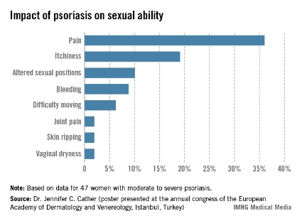
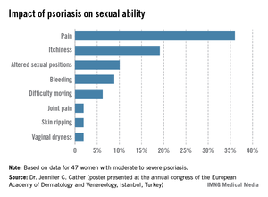
One third of a group of women with psoriasis reported that the pain associated with their condition interfered with their sexual activity, according to findings from a survey presented by Dr. Jennifer C. Cather.
Based on responses from a survey of 60 women with moderate to severe psoriasis, the specific complaints that were the most common ways in which psoriasis interfered with sexual activity were itchiness (19%), the need to adjust sexual position (10%), and bleeding (9%), Dr. Cather reported at the Skin Disease Education Foundation’s annual Las Vegas dermatology seminar. The survey was part of an effort to determine the impact of psoriasis on women’s sexual activity, desires, and relationships.
The data were previously presented in a poster at the annual congress of the European Academy of Dermatology and Venereology (Istanbul.
SDEF and this news organization are owned by Frontline Medical Communications. Dr. Cather disclosed that she is a consultant, speaker, or researcher for AbbVie, Novartis, Leo, Janssen, Amgen, Celgene, Merck, and Pfizer.
EXPERT ANALYSIS FROM SDEF LAS VEGAS DERMATOLOGY SEMINAR