User login
After-hours texting and professional boundaries
Recently, I was out on a Friday night with a friend who is a resident in another program. I hadn’t seen her in a very long time because of our hectic schedules. Around 10 p.m., she received a text from her attending asking her if she had left the scripts ready for the patient who was leaving on Monday.
Much has been written about professional boundaries and bosses texting their employees. For most jobs, a boss texting after hours over nonurgent matters is completely out of line. But in the medical field, there are no limits. People say, “Oh well, it’s the physician life.” Well maybe if we had more professional boundaries, our quality of life would be better. Maybe there wouldn’t be such a huge rate of burnout.
I encourage physicians to remember to contact your resident and coworkers during business hours. If the matter is not placing patients in danger, it can wait till the next morning. Nobody wants to pick up his phone in the middle of dinner to deal with patient care–related expectations that can be addressed the next business day.
Receiving a text brings all the stress of work back in the middle of our time off in which we are trying to take care of ourselves and the rest of our lives. It adds unnecessary stress to the overall high stress level and undermines our attempt to have a social life and meet a friend. A quick Internet search shows many blogs, journals, and different websites discussing this issue, but the voices of doctors and other health care providers are strangely silent on this topic.
I consider emails a more professional way of communicating than a text. I check my email often during a 24-hour period, and when I do, I’m ready for any potential information I might receive. I do not get notifications on my phone from my work email. But like my friend, I can’t avoid texts. We should have the opportunity to use our right to disconnect.
Some may argue, “Put your phone on silent if you don’t want to deal with it.” But not only do I use my phone for my life outside of work (as a resident, I make an effort to have one), but I want to be available for my peers and juniors when they are in the hospital. I want to be a resident my coworkers can text when they have a question and appreciate my advice. That is a decision I have made about the type of resident I want to be, and I am comfortable with it. Now if they text me asking a question that can wait till business hours the following day, they are crossing boundaries.
It might seem like a gray line. Somebody – maybe residency programs or our professional organizations – should address this so we have clear guidelines to protect our off-work time. Doesn’t our culture need to change the “physician life” so that we don’t bring our work responsibilities out for dinner on a Friday night? If the issue doesn’t need to be resolved quickly, it should be a given that texting is inappropriate.
Dr. Serrano is a PGY3 psychiatry resident at the Einstein Medical Center in Philadelphia.

Family, culture, and cultural identity key to improving assessment and treatment
Editor’s Note: This is the third installment of Curbside Consult, written by two Group for the Advancement of Psychiatry (GAP) committees – the Committee on Family Psychiatry and the Committee on Cultural Psychiatry.
Erica is a 19-year-old woman from an ethnically mixed background. Her father is an African American retired U.S. military officer who met her mother while he was stationed in Japan, where her mother was born and raised. Erica lived in Japan until she was 8, when the family moved to Seattle, and her father began a new career in a large company.
At age 10, she began to have episodes during which she felt her heart racing, was short of breath, and was diaphoretic. These episodes often took place when the family went out to large public spaces, like a shopping mall, and she would be separated from her parents. They increased in frequency until she was taken to a pediatrician and saw a counselor. Her panic symptoms gradually disappeared during high school, but since starting college and moving away from home, she has felt lost in her environment, and feels as though no one understands her background and upbringing. For example, she abruptly left the first college party she attended, feeling that “everybody had met everybody already and nobody noticed me; maybe they don’t like the way I look.” She has had a return of panic episodes that are now more frequent, coming on without warning, and they have begun to wake her from sleep.

Discussion
Incorporating a family and culture lens to the case can improve assessment and treatment planning. Given the brevity of the case description, many of our recommendations focus on obtaining additional information. It is also important to rule out any medical conditions that could be causing her symptoms. Finally, since great heterogeneity exists within every culture, the clinician should investigate the meanings associated with specific cultural backgrounds for each person to avoid stereotyping that would interfere with an accurate assessment and treatment plan.
Possible cultural conflicts
Although the identified patient in this case is the young woman, the number of culture- and family-related stresses experienced by each member of her family cannot be overstated. The family is truly multicultural, in every sense of the word. Many Americans associate the word “culture” with the influence of race, ethnicity, and country of origin, but the term encompasses many other aspects of the family’s background as well, including the father’s immersion in military culture as an officer of color, the culture of American ex-pats in Japan and of Japanese immigrants to the West Coast of the United States, multiracial children’s adaptations to Japanese and state schools, and U.S. corporate culture. Every member of the family has therefore had to adapt to many cultural transitions. All of these moves require new relationships with the dominant culture and immediate community, which increases stresses within the family as well as between the family and the broader society in which they live. Conflict between the parents increases vigilance and anxiety in children, and since Erica does not have siblings, the likelihood of being caught in a conflict between both parent would be high.
One adaptation that the family has undergone involves negotiating variations in “individualism” – a cultural ideology that prizes the role and desires of the individual over that of the group – and “collectivism” – in which the values and expectations of the social group are prioritized. Each society (for example, urban Japan) and social group (for example, the U.S. military) is characterized by a combination of individualistic and collectivistic traits, though a certain cultural flavor tends to predominate.
If Erica’s mother’s family of origin in Japan was very traditionally collectivistic, she may have experienced a difficult cultural shift when she moved to the United States, which tends to value more individualistic self-presentation. This conflict may have affected Erica’s development, particularly if it were an area of difficulty during her mother’s adaptation to her move or a potential sore spot in the father’s relationship with his wife. It would be good to explore to what extent the mother embraced her move to her new home in Seattle, and whether she also tried to keep her culture of origin alive in the family, while she negotiated her acculturation to U.S. society. In a cosmopolitan city like Seattle with a growing Asian population, this may be easier than in past years, but still might require a great deal of effort. The mother’s adaptation seems an important topic for further assessment, including discovering whether she found work in the United States, if she created new social networks, or whether she remained isolated with her husband and her daughter.
As to the father, if he grew up in a U.S. community that relied on a direct communication style that valued individual assertion as a component of identity and self-esteem, he may have seen his wife’s approach to communication as too reserved, private, or other-directed, especially if her English proficiency or his Japanese fluency was limited. These conflicts may have affected Erica’s sense of self but must be evaluated directly rather than assumed; each parent’s fluency and communication style are fruitful questions to explore. It is possible that conflicts in this area may have interfered with Erica’s incorporation of either parent as a role model for her as a young woman in contemporary U.S. society, threatening her sense of self and leading to anxiety symptoms.

Another area of potential stress for the family involves the experience of discrimination tied to racism or other forms of prejudice. Each family member is vulnerable in this area in his/her own right, given the potential mismatches between their racial/ethnic background and the sometimes intolerant views of dominant social groupings in their societies of origin or their societies of migration. Erica may be most vulnerable in this regard, in light of her dual minority background. The clinician should assess to what extent either or both families of origin may be unhappy about the marriage and biracial child and the possible resulting impact on Erica’s sense of herself. Does she feel more attracted to one aspect of her background? In terms of Erica’s developing identity, it is important to understand whether the family maintains one culture as the family of heritage or works toward developing a multicultural identity. If neither parent can completely identify with Erica, they must work with her to find a way to mesh both cultures. If they do not, she may feel that she has to choose her presentation to the world as African American, Japanese, multiracial, or “post racial.” This may complicate her sense of connection to one or both parents. To some extent this may be affected by which parent she most takes after physically or to which side of the extended family she feels closer. The clinician should consider all these issues in conducting a culturally competent assessment and family-based intervention.
Development context of Erica’s symptom course
Since Erica was asymptomatic during the first 2 years after the move to the United States, it may be possible to assume that early adaptation went reasonably well. The onset of symptoms at age 10 may stem from numerous causes. Biologically, she may have been in prepuberty, which can increase emotional reactivity. Psychologically, this is typically the point at which children become more self-conscious and peer pressure ratchets up, so her possible lack of instinctive understanding of U.S. cultural norms, or her biracial makeup, may have become more salient, either in the form of an intrapsychic racial identity conflict or as an object of interpersonal bullying. It would be helpful to understand these details so as to attend to them in psychotherapy.
If she had begun middle school, academic pressure may have increased. However, her clinicians also should consider the possibility that one or both of her parents may have become stressed by the multiple cultural adaptations required by the move, or that the marriage had become strained, and that she responded to parental stress with increased anxiety. These are possibilities that need to be explored during the assessment.
Her ability to deal with her difficulties and finish high school speaks to her and her family’s resilience. To plan future treatment, it would be useful to explore what aspect of her treatment at age 10 was most helpful (medication, therapy, or both). Ideally, it would be helpful to understand the cultural elements of the relationship between the patient and the pediatrician and counselor. However, given her history, it is no surprise that when she is asked once more to navigate a new culture of college, this time without parental support, that her symptoms reoccur. If she is far from home, in a college where racial tensions are high or where there is not a large multiracial population, or if her parents are having trouble with empty nesting, this would make things more difficult. Her own cultural identity may have been challenged by an environment where she felt “no one understands her background and upbringing,” where “nobody noticed me,” and where “maybe they don’t like the way I look.”
How should clinicians explore these issues?
Erica’s clinicians should seek to understand how she herself defines her background, identity, and upbringing to help her examine possible conflictual issues that are causing distress. The DSM-5 Cultural Formulation Interview is a useful tool for achieving this goal, including its supplementary modules such as the one on Cultural Identity. As part of this assessment, her clinician could ask questions like: How does she see herself and how do other people see her in terms of her identity? How does she present herself? Is she being harassed on campus? Are any of these issues causes of her anxiety? Direct assessment of these topics is necessary to avoid initial impressions that might be affected by the clinician’s own identity, values, and biases. Her clinician should also be conversant in the stresses of the college environment, both at the intrapsychic and interpersonal levels.
While the usual treatments for anxiety, including cognitive-behavioral therapy and medications if necessary, may well be part of her clinical care, helping her understand her own personal cultural identity, how to negotiate the stresses of living on her college campus, and increasing both family and community supports are critical to her well-being and mental health.
While it is true that few people could exactly share Erica’s life experience, there are many pan-nationals and expats who would very much relate to her feelings. Erica faces many of the challenges of those in a group called “third culture kids,” a term coined in the 1950s by social scientists to describe the experience of children raised by Americans working in other countries. The expatriate lifestyle they described as an “interstitial culture” – different from but including elements of both the home culture and the host culture – often is specific to the work group (for example, military, business) that the adults were engaged in. For these children, the question of cultural identity, and “where is home,” is a complex one. For them, unlike their parents, the United States is not home but a foreign land. But their host country is not exactly home, either. Patients like Erica may benefit from reading “Third Culture Kids: The Experience of Growing Up Among Worlds,” by David C. Pollock and Ruth E. Van Reken, and clinicians would likely benefit as well.
Finally, Erica’s therapist could encourage her to find connections in the international student community; there are usually groups on campus for them, and they would understand a multicultural experience. Her therapist also should meet with, or speak with, her parents to see whether there are stresses at home, and could encourage them to support her by frequent visits or calls. When Erica finds a place where she feels at home, we believe her anxiety will decrease.
Key take-home points
1. Ask about, do not assume, the person’s own understanding of his/her background and identity to obtain more specific and precise information so as to guard against stereotyping that could lead to erroneous assessments.
2. Understand the heterogeneity of culture and the complexity of cultural identity.
3. Ask about the family. Who is in the family, both nuclear and extended? Draw a simple genogram. Envision and implement assessment beyond just the individual patient.
4. Assess the impact of culture change on cultural identity and family dynamics.
5. Use the DSM-5 Cultural Formulation Interview to help guide the cultural assessment.
Contributors
Ellen M. Berman, MD – University of Pennsylvania, Perelman School of Medicine, Philadelphia
Roberto Lewis-Fernández, MD – Columbia University and New York State Psychiatric Institute
Francis G. Lu, MD – University of California, Davis
The contributors have revised selected patient details to shield the identities of the patients/cases and to comply with HIPAA requirements. This column is meant to be educational and does not constitute medical advice. The opinions expressed are those of the contributors and do not represent those of the organizations they are employed by or those affiliated with GAP.
Resources
Diagnostic and Statistical Manual of Mental Disorders, 5th edition (DSM-5). (Arlington, Va.: American Psychiatric Association Publishing, 2013).
Lewis-Fernández, R., et al. (eds.) DSM-5 Handbook on the Cultural Formulation Interview (Arlington, Va.: American Psychiatric Association Publishing, 2016).
Lim, R. (ed.). Clinical Manual of Cultural Psychiatry, 2nd edition (Arlington, Va.: American Psychiatric Association Publishing, 2015).
Pollock, D. and Van Reken, R. Third Culture Kids: The Experience of Growing Up Among Worlds (London: Nicholas Breasley Publishing, 2009).
Curbside Consult is inspired by the DSM-5’s emphasis on developing a cultural formulation of patients’ illnesses, and addressing family dynamics and resilience in promoting care that fosters prevention and recovery. We request that you submit cases to cpnews@frontlinemedcom.com in which your understanding and treatment are affected by challenging cultural and family issues. We will then write back with our best answers about how one might proceed in such a case. Your case and our response will be published in Clinical Psychiatry News. Please limit your case description to 250 words and include the following details:
1. Patient’s presenting problem or reason for the visit.
2. Patient’s age and gender.
3. Indicators of the patient’s identity – self-identified race/ethnicity, culture, religion/spirituality, socioeconomic status, education, among other variables.
4. Patient’s living situation, family composition, and genogram information (if available).
5. Patient’s geographic location (rural, suburban, urban) and occupation.
6. Patient’s and family’s degree of participation in their identified culture.
7. Questions of the individual submitting the case, including concerns about the role of the family and culture in the case, diagnosis, and treatment planning.
8. Please follow local ethical requirements, disguise the case to protect confidentiality and attend to HIPAA requirements, so that patients or family members reading the article would not recognize themselves.
Additional information might be requested, and editing of the case, questions, and commentary might be needed prior to final publication.
Editor’s Note: This is the third installment of Curbside Consult, written by two Group for the Advancement of Psychiatry (GAP) committees – the Committee on Family Psychiatry and the Committee on Cultural Psychiatry.
Erica is a 19-year-old woman from an ethnically mixed background. Her father is an African American retired U.S. military officer who met her mother while he was stationed in Japan, where her mother was born and raised. Erica lived in Japan until she was 8, when the family moved to Seattle, and her father began a new career in a large company.
At age 10, she began to have episodes during which she felt her heart racing, was short of breath, and was diaphoretic. These episodes often took place when the family went out to large public spaces, like a shopping mall, and she would be separated from her parents. They increased in frequency until she was taken to a pediatrician and saw a counselor. Her panic symptoms gradually disappeared during high school, but since starting college and moving away from home, she has felt lost in her environment, and feels as though no one understands her background and upbringing. For example, she abruptly left the first college party she attended, feeling that “everybody had met everybody already and nobody noticed me; maybe they don’t like the way I look.” She has had a return of panic episodes that are now more frequent, coming on without warning, and they have begun to wake her from sleep.
Discussion
Incorporating a family and culture lens to the case can improve assessment and treatment planning. Given the brevity of the case description, many of our recommendations focus on obtaining additional information. It is also important to rule out any medical conditions that could be causing her symptoms. Finally, since great heterogeneity exists within every culture, the clinician should investigate the meanings associated with specific cultural backgrounds for each person to avoid stereotyping that would interfere with an accurate assessment and treatment plan.
Possible cultural conflicts
Although the identified patient in this case is the young woman, the number of culture- and family-related stresses experienced by each member of her family cannot be overstated. The family is truly multicultural, in every sense of the word. Many Americans associate the word “culture” with the influence of race, ethnicity, and country of origin, but the term encompasses many other aspects of the family’s background as well, including the father’s immersion in military culture as an officer of color, the culture of American ex-pats in Japan and of Japanese immigrants to the West Coast of the United States, multiracial children’s adaptations to Japanese and state schools, and U.S. corporate culture. Every member of the family has therefore had to adapt to many cultural transitions. All of these moves require new relationships with the dominant culture and immediate community, which increases stresses within the family as well as between the family and the broader society in which they live. Conflict between the parents increases vigilance and anxiety in children, and since Erica does not have siblings, the likelihood of being caught in a conflict between both parent would be high.
One adaptation that the family has undergone involves negotiating variations in “individualism” – a cultural ideology that prizes the role and desires of the individual over that of the group – and “collectivism” – in which the values and expectations of the social group are prioritized. Each society (for example, urban Japan) and social group (for example, the U.S. military) is characterized by a combination of individualistic and collectivistic traits, though a certain cultural flavor tends to predominate.
If Erica’s mother’s family of origin in Japan was very traditionally collectivistic, she may have experienced a difficult cultural shift when she moved to the United States, which tends to value more individualistic self-presentation. This conflict may have affected Erica’s development, particularly if it were an area of difficulty during her mother’s adaptation to her move or a potential sore spot in the father’s relationship with his wife. It would be good to explore to what extent the mother embraced her move to her new home in Seattle, and whether she also tried to keep her culture of origin alive in the family, while she negotiated her acculturation to U.S. society. In a cosmopolitan city like Seattle with a growing Asian population, this may be easier than in past years, but still might require a great deal of effort. The mother’s adaptation seems an important topic for further assessment, including discovering whether she found work in the United States, if she created new social networks, or whether she remained isolated with her husband and her daughter.
As to the father, if he grew up in a U.S. community that relied on a direct communication style that valued individual assertion as a component of identity and self-esteem, he may have seen his wife’s approach to communication as too reserved, private, or other-directed, especially if her English proficiency or his Japanese fluency was limited. These conflicts may have affected Erica’s sense of self but must be evaluated directly rather than assumed; each parent’s fluency and communication style are fruitful questions to explore. It is possible that conflicts in this area may have interfered with Erica’s incorporation of either parent as a role model for her as a young woman in contemporary U.S. society, threatening her sense of self and leading to anxiety symptoms.
Another area of potential stress for the family involves the experience of discrimination tied to racism or other forms of prejudice. Each family member is vulnerable in this area in his/her own right, given the potential mismatches between their racial/ethnic background and the sometimes intolerant views of dominant social groupings in their societies of origin or their societies of migration. Erica may be most vulnerable in this regard, in light of her dual minority background. The clinician should assess to what extent either or both families of origin may be unhappy about the marriage and biracial child and the possible resulting impact on Erica’s sense of herself. Does she feel more attracted to one aspect of her background? In terms of Erica’s developing identity, it is important to understand whether the family maintains one culture as the family of heritage or works toward developing a multicultural identity. If neither parent can completely identify with Erica, they must work with her to find a way to mesh both cultures. If they do not, she may feel that she has to choose her presentation to the world as African American, Japanese, multiracial, or “post racial.” This may complicate her sense of connection to one or both parents. To some extent this may be affected by which parent she most takes after physically or to which side of the extended family she feels closer. The clinician should consider all these issues in conducting a culturally competent assessment and family-based intervention.
Development context of Erica’s symptom course
Since Erica was asymptomatic during the first 2 years after the move to the United States, it may be possible to assume that early adaptation went reasonably well. The onset of symptoms at age 10 may stem from numerous causes. Biologically, she may have been in prepuberty, which can increase emotional reactivity. Psychologically, this is typically the point at which children become more self-conscious and peer pressure ratchets up, so her possible lack of instinctive understanding of U.S. cultural norms, or her biracial makeup, may have become more salient, either in the form of an intrapsychic racial identity conflict or as an object of interpersonal bullying. It would be helpful to understand these details so as to attend to them in psychotherapy.
If she had begun middle school, academic pressure may have increased. However, her clinicians also should consider the possibility that one or both of her parents may have become stressed by the multiple cultural adaptations required by the move, or that the marriage had become strained, and that she responded to parental stress with increased anxiety. These are possibilities that need to be explored during the assessment.
Her ability to deal with her difficulties and finish high school speaks to her and her family’s resilience. To plan future treatment, it would be useful to explore what aspect of her treatment at age 10 was most helpful (medication, therapy, or both). Ideally, it would be helpful to understand the cultural elements of the relationship between the patient and the pediatrician and counselor. However, given her history, it is no surprise that when she is asked once more to navigate a new culture of college, this time without parental support, that her symptoms reoccur. If she is far from home, in a college where racial tensions are high or where there is not a large multiracial population, or if her parents are having trouble with empty nesting, this would make things more difficult. Her own cultural identity may have been challenged by an environment where she felt “no one understands her background and upbringing,” where “nobody noticed me,” and where “maybe they don’t like the way I look.”
How should clinicians explore these issues?
Erica’s clinicians should seek to understand how she herself defines her background, identity, and upbringing to help her examine possible conflictual issues that are causing distress. The DSM-5 Cultural Formulation Interview is a useful tool for achieving this goal, including its supplementary modules such as the one on Cultural Identity. As part of this assessment, her clinician could ask questions like: How does she see herself and how do other people see her in terms of her identity? How does she present herself? Is she being harassed on campus? Are any of these issues causes of her anxiety? Direct assessment of these topics is necessary to avoid initial impressions that might be affected by the clinician’s own identity, values, and biases. Her clinician should also be conversant in the stresses of the college environment, both at the intrapsychic and interpersonal levels.
While the usual treatments for anxiety, including cognitive-behavioral therapy and medications if necessary, may well be part of her clinical care, helping her understand her own personal cultural identity, how to negotiate the stresses of living on her college campus, and increasing both family and community supports are critical to her well-being and mental health.
While it is true that few people could exactly share Erica’s life experience, there are many pan-nationals and expats who would very much relate to her feelings. Erica faces many of the challenges of those in a group called “third culture kids,” a term coined in the 1950s by social scientists to describe the experience of children raised by Americans working in other countries. The expatriate lifestyle they described as an “interstitial culture” – different from but including elements of both the home culture and the host culture – often is specific to the work group (for example, military, business) that the adults were engaged in. For these children, the question of cultural identity, and “where is home,” is a complex one. For them, unlike their parents, the United States is not home but a foreign land. But their host country is not exactly home, either. Patients like Erica may benefit from reading “Third Culture Kids: The Experience of Growing Up Among Worlds,” by David C. Pollock and Ruth E. Van Reken, and clinicians would likely benefit as well.
Finally, Erica’s therapist could encourage her to find connections in the international student community; there are usually groups on campus for them, and they would understand a multicultural experience. Her therapist also should meet with, or speak with, her parents to see whether there are stresses at home, and could encourage them to support her by frequent visits or calls. When Erica finds a place where she feels at home, we believe her anxiety will decrease.
Key take-home points
1. Ask about, do not assume, the person’s own understanding of his/her background and identity to obtain more specific and precise information so as to guard against stereotyping that could lead to erroneous assessments.
2. Understand the heterogeneity of culture and the complexity of cultural identity.
3. Ask about the family. Who is in the family, both nuclear and extended? Draw a simple genogram. Envision and implement assessment beyond just the individual patient.
4. Assess the impact of culture change on cultural identity and family dynamics.
5. Use the DSM-5 Cultural Formulation Interview to help guide the cultural assessment.
Contributors
Ellen M. Berman, MD – University of Pennsylvania, Perelman School of Medicine, Philadelphia
Roberto Lewis-Fernández, MD – Columbia University and New York State Psychiatric Institute
Francis G. Lu, MD – University of California, Davis
The contributors have revised selected patient details to shield the identities of the patients/cases and to comply with HIPAA requirements. This column is meant to be educational and does not constitute medical advice. The opinions expressed are those of the contributors and do not represent those of the organizations they are employed by or those affiliated with GAP.
Resources
Diagnostic and Statistical Manual of Mental Disorders, 5th edition (DSM-5). (Arlington, Va.: American Psychiatric Association Publishing, 2013).
Lewis-Fernández, R., et al. (eds.) DSM-5 Handbook on the Cultural Formulation Interview (Arlington, Va.: American Psychiatric Association Publishing, 2016).
Lim, R. (ed.). Clinical Manual of Cultural Psychiatry, 2nd edition (Arlington, Va.: American Psychiatric Association Publishing, 2015).
Pollock, D. and Van Reken, R. Third Culture Kids: The Experience of Growing Up Among Worlds (London: Nicholas Breasley Publishing, 2009).
Curbside Consult is inspired by the DSM-5’s emphasis on developing a cultural formulation of patients’ illnesses, and addressing family dynamics and resilience in promoting care that fosters prevention and recovery. We request that you submit cases to cpnews@frontlinemedcom.com in which your understanding and treatment are affected by challenging cultural and family issues. We will then write back with our best answers about how one might proceed in such a case. Your case and our response will be published in Clinical Psychiatry News. Please limit your case description to 250 words and include the following details:
1. Patient’s presenting problem or reason for the visit.
2. Patient’s age and gender.
3. Indicators of the patient’s identity – self-identified race/ethnicity, culture, religion/spirituality, socioeconomic status, education, among other variables.
4. Patient’s living situation, family composition, and genogram information (if available).
5. Patient’s geographic location (rural, suburban, urban) and occupation.
6. Patient’s and family’s degree of participation in their identified culture.
7. Questions of the individual submitting the case, including concerns about the role of the family and culture in the case, diagnosis, and treatment planning.
8. Please follow local ethical requirements, disguise the case to protect confidentiality and attend to HIPAA requirements, so that patients or family members reading the article would not recognize themselves.
Additional information might be requested, and editing of the case, questions, and commentary might be needed prior to final publication.

Simple Strategy for Addressing Problematic Patient Behavior
Linden Spital, NP, a psychiatric mental-health nurse practitioner, staffs the Psychiatric Consultation Liaison Service at the University of Michigan in Ann Arbor. Nearly every hospital larger than about 200 beds, she says, could benefit from a similar service, and hospitalists could play an important role in creating it.
I wrote about the idea for a generally similar service in my April 2015 column, but at the time, I didn’t know of an institution that had something like this in place.
Along with her hospitalist colleagues, Anupama (Anu) Goyal, MBChB, and Rob Chang, MD, Linden has launched a service to provide assistance to bedside caregivers dealing with very difficult patients (eg, those who are verbally or physically threatening to staff, unreasonably demanding and angry, have bizarre behavior, etc.).
Sample Cases

Two recent cases illustrate the role of the service. A female patient in her 60s had several admissions characterized by what many caregivers agreed were unreasonably precise demands regarding how her care should be delivered. She was verbally abusive of caregivers, especially those who were young or of a different race, and her family member tended to reinforce these maladaptive behaviors. Staff found it very stressful to care for her and had concerns that her care suffered as a result.
Linden served as a resource and support for staff, plus worked with providers to set limits on the patient and family behavior and to separate patient behaviors that were and weren’t modifiable. Linden’s efforts helped clarify the goals for the patient’s care and reduced staff distress. Even though the patient’s behavior didn’t change significantly, staff anecdotally reported less distress and concern that the patient’s care suffered as a result.

Another case involved a man in his 50s who had a progressive neurodegenerative disease and was admitted because of increasingly aggressive behavior in his skilled-nursing facility (SNF). Providers at the SNF attributed the poor behavior to changes in medications. His behavior was very difficult to manage, and staff asked for Linden’s help. She worked with the patient and realized much of his difficult behavior stemmed from his frustration with communicating verbally because of his neurologic disease. Rather than pursue increasing psychotropics, Linden promoted efforts to develop a system of hand signals the patient could use to communicate needs. His behavior improved, presumably by reducing his own frustration and improving his autonomy.

Atypical Consults
This psychiatric consultation liaison service has some overlap with traditional inpatient psychiatry services, but it is configured so that the caregiver is essentially embedded on the medical units of the hospital and assists in the care of patients who wouldn’t typically be appropriate for a psychiatry consult. For example, patients and/or families who act out because of anger over being on observation status are appropriate for this service but would usually not be appropriate for a psychiatry consult. The two examples above aren’t ideal cases for a standard psychiatry consult; however, the attending hospitalist needed help nonetheless.
Operational Details
The liaison service started with a successful trial on two hospital units in 2013. Linden began serving as the sole clinician on the service in January 2015. She is available during the daytime on weekdays, and any staff can request her participation in the care of a patient. Her visits are billed when appropriate, but many aren’t billed (for example, if her primary work was to conference with staff regarding management of a patient).
Consults can be requested by anyone (nurses, etc., as well as physicians, though only the latter would be billable) via an electronic health record entry that helps ensure whether the request is for this service versus the inpatient psychiatry service. The order includes a standard list of potential reasons for consult that can be selected and amplified with free text comments. She also receives verbal consult requests as she moves through the hospital.
Linden’s position is budgeted through the psychiatry department and funded by the hospital with only modest professional fee collections.
An Idea That Is Catching On?
Anu Goyal made me aware of a study from 2004 that summarized findings from experience with a similar service at Washington University in St. Louis, but the service was cancelled after a short time due to its cost.1 She also found a few studies from the 1990s and a 2001 study from Australia that report on a similar service.
But maybe the idea is catching on again, at least a little.
On April 25, The Wall Street Journal published an article titled “Hospitals Test Putting Psychiatrists on Medical Wards.”2 It described programs at Brigham and Women’s Hospital in Boston, Johns Hopkins Hospital in Baltimore, and NewYork-Presbyterian/Columbia University Medical Center in New York City. They share some similarities with the service at the University of Michigan. However, according to the article, the three big-city programs tilt more toward a traditional consultation model than what Linden does.
I think every hospital should be thinking about a service other than traditional consult psychiatry that could help with challenging patient behavior. The University of Michigan model or similar ones seem like a good place to start. TH
Reference
- Yakimo R, Kurlowicz L, Murray R. Evaluation of outcomes in psychiatric consultation-liaison nursing practice. Arch Psychiatr Nurs. 2004;18(6):215-227.
2. Ladnado L. Hospitals test putting psychiatrists on medical wards. The Wall Street Journal website. Accessed July 3, 2016.

Linden Spital, NP, a psychiatric mental-health nurse practitioner, staffs the Psychiatric Consultation Liaison Service at the University of Michigan in Ann Arbor. Nearly every hospital larger than about 200 beds, she says, could benefit from a similar service, and hospitalists could play an important role in creating it.
I wrote about the idea for a generally similar service in my April 2015 column, but at the time, I didn’t know of an institution that had something like this in place.
Along with her hospitalist colleagues, Anupama (Anu) Goyal, MBChB, and Rob Chang, MD, Linden has launched a service to provide assistance to bedside caregivers dealing with very difficult patients (eg, those who are verbally or physically threatening to staff, unreasonably demanding and angry, have bizarre behavior, etc.).
Sample Cases
Two recent cases illustrate the role of the service. A female patient in her 60s had several admissions characterized by what many caregivers agreed were unreasonably precise demands regarding how her care should be delivered. She was verbally abusive of caregivers, especially those who were young or of a different race, and her family member tended to reinforce these maladaptive behaviors. Staff found it very stressful to care for her and had concerns that her care suffered as a result.
Linden served as a resource and support for staff, plus worked with providers to set limits on the patient and family behavior and to separate patient behaviors that were and weren’t modifiable. Linden’s efforts helped clarify the goals for the patient’s care and reduced staff distress. Even though the patient’s behavior didn’t change significantly, staff anecdotally reported less distress and concern that the patient’s care suffered as a result.
Another case involved a man in his 50s who had a progressive neurodegenerative disease and was admitted because of increasingly aggressive behavior in his skilled-nursing facility (SNF). Providers at the SNF attributed the poor behavior to changes in medications. His behavior was very difficult to manage, and staff asked for Linden’s help. She worked with the patient and realized much of his difficult behavior stemmed from his frustration with communicating verbally because of his neurologic disease. Rather than pursue increasing psychotropics, Linden promoted efforts to develop a system of hand signals the patient could use to communicate needs. His behavior improved, presumably by reducing his own frustration and improving his autonomy.
Atypical Consults
This psychiatric consultation liaison service has some overlap with traditional inpatient psychiatry services, but it is configured so that the caregiver is essentially embedded on the medical units of the hospital and assists in the care of patients who wouldn’t typically be appropriate for a psychiatry consult. For example, patients and/or families who act out because of anger over being on observation status are appropriate for this service but would usually not be appropriate for a psychiatry consult. The two examples above aren’t ideal cases for a standard psychiatry consult; however, the attending hospitalist needed help nonetheless.
Operational Details
The liaison service started with a successful trial on two hospital units in 2013. Linden began serving as the sole clinician on the service in January 2015. She is available during the daytime on weekdays, and any staff can request her participation in the care of a patient. Her visits are billed when appropriate, but many aren’t billed (for example, if her primary work was to conference with staff regarding management of a patient).
Consults can be requested by anyone (nurses, etc., as well as physicians, though only the latter would be billable) via an electronic health record entry that helps ensure whether the request is for this service versus the inpatient psychiatry service. The order includes a standard list of potential reasons for consult that can be selected and amplified with free text comments. She also receives verbal consult requests as she moves through the hospital.
Linden’s position is budgeted through the psychiatry department and funded by the hospital with only modest professional fee collections.
An Idea That Is Catching On?
Anu Goyal made me aware of a study from 2004 that summarized findings from experience with a similar service at Washington University in St. Louis, but the service was cancelled after a short time due to its cost.1 She also found a few studies from the 1990s and a 2001 study from Australia that report on a similar service.
But maybe the idea is catching on again, at least a little.
On April 25, The Wall Street Journal published an article titled “Hospitals Test Putting Psychiatrists on Medical Wards.”2 It described programs at Brigham and Women’s Hospital in Boston, Johns Hopkins Hospital in Baltimore, and NewYork-Presbyterian/Columbia University Medical Center in New York City. They share some similarities with the service at the University of Michigan. However, according to the article, the three big-city programs tilt more toward a traditional consultation model than what Linden does.
I think every hospital should be thinking about a service other than traditional consult psychiatry that could help with challenging patient behavior. The University of Michigan model or similar ones seem like a good place to start. TH
Reference
- Yakimo R, Kurlowicz L, Murray R. Evaluation of outcomes in psychiatric consultation-liaison nursing practice. Arch Psychiatr Nurs. 2004;18(6):215-227.
2. Ladnado L. Hospitals test putting psychiatrists on medical wards. The Wall Street Journal website. Accessed July 3, 2016.
Operating with pain: Reader response
The feature article “Operating with pain: Surgeon workplace injury underrecognized" touched a nerve with our readers! The following comments appeared in the ACS Communities on the topic of pain and occupational injury among surgeons:
I recently was forced to undergo an anterior cervical decompression and fusion (ACDF) involving C4,5,6 and 7 due to worsening radicular pain and weakness due to severe cervical spinal stenosis. This problem was likely initiated by multiple injuries that I sustained as a wrestler in my younger days, but was no doubt exacerbated by 23 years bent over the OR table in extended periods of flexion, ignoring pain, and working every day no matter how I felt with no time for nuisances like physical therapy. Such is the mentality of the surgeon. In any case, my experience demonstrates that surgery, unlike many other medical specialties, takes a physical toll on its practitioners and also requires a certain level of fitness for surgeons to practice well.

The concept of wellness among surgeons is relatively new, in my opinion. Historically, surgical training was notorious for long hours, extended periods of sleep deprivation, irregular eating habits, strained interpersonal relationships, and frankly, sometimes an emotionally abusive environment. Many changes have been made to adapt training to be more livable, but these changes have been predominantly in the areas of work hours, sleep, and time off. Little has been done to teach adaptive strategies for the physical demands of performing surgery day after day.
Do we need a formal “plan” to educate surgeons how to save their backs and necks? Perhaps not, but surgeons do need to be aware that the cumulative “wear and tear” on our bodies can definitely affect how well we do our jobs, the number of years we are able to do our jobs, and the enjoyment with which we do our jobs. So tell the resident to stand up straight, teach them to operate with the table at the correct height, to hold the instruments in an ergonomic fashion, etc. Let’s begin to make proper ergonomics a part of our surgical culture so we may serve our patients for many years to come.
Bryan K. Richmond, MD, MBA, FACS
Charleston, W.Va.
I remember attending the ACS meeting as a chief resident. There was a laparoscopic instrument rep taking a survey about ergonomics, especially arm, shoulder, neck, and back pain during surgery. I just remember laughing and saying, “Heck no, no issues for me.” Well, now as a 50-year-old, I’ve got issues! I find that I have to be very aware of my posture during procedures and not spend too much time in one position. Hand cramps are not an infrequent problem during longer surgeries as well. Getting used to wearing slightly looser gloves has helped some.
Peter Krone, MD, FACS
Granbury, Tex.
As much as we all love surgery, it seems operating is definitely taking its toll on most every surgeon I know. Short of changing how we operate (i.e., lap vs. robotic, etc.), it seems there is little we can do to protect ourselves. I learned of gel mats a few years ago. They are awesome for longer cases. For me personally, it has been bilateral carpal tunnel releases and a C5,6/6,7 ACDF for degenerative changes causing radiculopathy. Fortunately, both operations were 100% successful.
I applaud you for looking at the (virtual lack of) ergonomics in surgery.
Brent C. Jackson, MD, FACS
Sacramento, Calif.
I finished my vascular surgical fellowship in 1991. Being old school, I continue to do some general surgery along with my comprehensive vascular surgery. In 2008, I had an urgent ACDF. Shortly thereafter, I attended the Southern Association of Vascular Surgery meeting and took an informal survey. I found that at least 60% of vascular surgeons in practice for 10 years had required an ACDF obviously secondary to loupes. Now in the endovascular cases wearing lead, lumbar back issues are also becoming very common in our field. This is a hugely important topic, and ergonomic study and training should become an integral part of training and retraining.
Thomas Appleby, MD, FACS
Charleston, S.C.
I am naturally right handed and have had the opposite experience of having left-handed surgeons teach me how to operate with my left hand throughout training. It is something I continue to do today. It comes in quite handy when helping one of my colleagues since they feel I am standing on the ‘wrong’ side of the table for most open procedures – but that was best for the left-handed approaches I learned.
Laparoscopic instruments are not friendly to anyone’s hands. Also, how fun is it to stand on your left foot for an entire case while operating a right foot control for your instruments?
Colette Whitby, MD, FACS
Southbridge, Mass.
Ergonomics in the OR is one of the subjects being considered at the Governor’s Competency and Wellness work group. More information is available, but certainly more has to be done.
David Welsh, MD, FACS
Batesville, IN
The feature article “Operating with pain: Surgeon workplace injury underrecognized" touched a nerve with our readers! The following comments appeared in the ACS Communities on the topic of pain and occupational injury among surgeons:
I recently was forced to undergo an anterior cervical decompression and fusion (ACDF) involving C4,5,6 and 7 due to worsening radicular pain and weakness due to severe cervical spinal stenosis. This problem was likely initiated by multiple injuries that I sustained as a wrestler in my younger days, but was no doubt exacerbated by 23 years bent over the OR table in extended periods of flexion, ignoring pain, and working every day no matter how I felt with no time for nuisances like physical therapy. Such is the mentality of the surgeon. In any case, my experience demonstrates that surgery, unlike many other medical specialties, takes a physical toll on its practitioners and also requires a certain level of fitness for surgeons to practice well.
The concept of wellness among surgeons is relatively new, in my opinion. Historically, surgical training was notorious for long hours, extended periods of sleep deprivation, irregular eating habits, strained interpersonal relationships, and frankly, sometimes an emotionally abusive environment. Many changes have been made to adapt training to be more livable, but these changes have been predominantly in the areas of work hours, sleep, and time off. Little has been done to teach adaptive strategies for the physical demands of performing surgery day after day.
Do we need a formal “plan” to educate surgeons how to save their backs and necks? Perhaps not, but surgeons do need to be aware that the cumulative “wear and tear” on our bodies can definitely affect how well we do our jobs, the number of years we are able to do our jobs, and the enjoyment with which we do our jobs. So tell the resident to stand up straight, teach them to operate with the table at the correct height, to hold the instruments in an ergonomic fashion, etc. Let’s begin to make proper ergonomics a part of our surgical culture so we may serve our patients for many years to come.
Bryan K. Richmond, MD, MBA, FACS
Charleston, W.Va.
I remember attending the ACS meeting as a chief resident. There was a laparoscopic instrument rep taking a survey about ergonomics, especially arm, shoulder, neck, and back pain during surgery. I just remember laughing and saying, “Heck no, no issues for me.” Well, now as a 50-year-old, I’ve got issues! I find that I have to be very aware of my posture during procedures and not spend too much time in one position. Hand cramps are not an infrequent problem during longer surgeries as well. Getting used to wearing slightly looser gloves has helped some.
Peter Krone, MD, FACS
Granbury, Tex.
As much as we all love surgery, it seems operating is definitely taking its toll on most every surgeon I know. Short of changing how we operate (i.e., lap vs. robotic, etc.), it seems there is little we can do to protect ourselves. I learned of gel mats a few years ago. They are awesome for longer cases. For me personally, it has been bilateral carpal tunnel releases and a C5,6/6,7 ACDF for degenerative changes causing radiculopathy. Fortunately, both operations were 100% successful.
I applaud you for looking at the (virtual lack of) ergonomics in surgery.
Brent C. Jackson, MD, FACS
Sacramento, Calif.
I finished my vascular surgical fellowship in 1991. Being old school, I continue to do some general surgery along with my comprehensive vascular surgery. In 2008, I had an urgent ACDF. Shortly thereafter, I attended the Southern Association of Vascular Surgery meeting and took an informal survey. I found that at least 60% of vascular surgeons in practice for 10 years had required an ACDF obviously secondary to loupes. Now in the endovascular cases wearing lead, lumbar back issues are also becoming very common in our field. This is a hugely important topic, and ergonomic study and training should become an integral part of training and retraining.
Thomas Appleby, MD, FACS
Charleston, S.C.
I am naturally right handed and have had the opposite experience of having left-handed surgeons teach me how to operate with my left hand throughout training. It is something I continue to do today. It comes in quite handy when helping one of my colleagues since they feel I am standing on the ‘wrong’ side of the table for most open procedures – but that was best for the left-handed approaches I learned.
Laparoscopic instruments are not friendly to anyone’s hands. Also, how fun is it to stand on your left foot for an entire case while operating a right foot control for your instruments?
Colette Whitby, MD, FACS
Southbridge, Mass.
Ergonomics in the OR is one of the subjects being considered at the Governor’s Competency and Wellness work group. More information is available, but certainly more has to be done.
David Welsh, MD, FACS
Batesville, IN
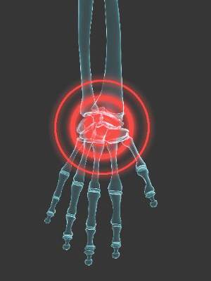
Prescription opioid analgesics: Is there a risk for birth defects?
Concerns have been raised about the widespread use of opioid analgesics in the general population, and specifically among pregnant women.
In women of reproductive age, U.S. claims data for the years 2008-2012 indicate that 39.4% of Medicaid-enrolled women and 27.7% of privately insured women had at least one opioid analgesic prescription claim.1 Among pregnant women, approximately 4.4% reported use of opioid analgesics any time in the month before and throughout gestation in a sample of 11,724 control mothers who participated in the National Birth Defects Prevention Study (NBDPS) 1998-2011.2

Previous studies of first-trimester exposure to opioid analgesics have shown inconsistent associations with selected major congenital anomalies. A 2011 analysis of 1,693 first-trimester codeine-exposed pregnancies enrolled in the Norwegian Mother and Child Cohort Study found no evidence of an increased risk of major birth defects overall (odds ratio, 0.8; 95% confidence interval, 0.5-1.1), compared with 65,316 unexposed pregnancies. However, this study had insufficient power to examine modest associations with most specific major birth defects.3
In contrast, two recent case-control studies have demonstrated significantly increased risks for early pregnancy opioid analgesic use and specific defects.
Broussard et al (2011) used data from the NBDPS 1997-2005 to test hypotheses related to specific defect associations previously reported in the literature, as well as to explore associations with additional defects. Therapeutic opioid use in early pregnancy was reported by 2.6% of 17,449 case mothers and 2.0% of 6,701 control mothers. The authors confirmed associations with some specific heart defects, (for example, conoventricular septal defects, atrioventricular septal defects, and hypoplastic left heart syndrome), and spina bifida, with ORs ranging from 2.0 to 2.7. In exploratory analysis, gastroschisis was also significantly associated with opioid analgesic exposure in early pregnancy (OR, 1.8; 95% CI, 1.1-2.9). Although additional analyses were performed stratified by specific opioid product (codeine, hydrocodone, oxycodone, meperidine), as might be expected, the most commonly used products (codeine and hydrocodone) were linked to a greater number of elevated ORs for specific defects.4
In the second case-control study, Yazdy et al (2013) used data collected in four cities in the United States and Canada and from two birth defect registries between 1998 and 2010 to examine periconceptional opioid use and risk for neural tube defects. The adjusted OR for spina bifida, comparing mothers of malformed infants with mothers of non-malformed infants, was 2.5 (95% CI, 1.3-5.0). To help rule out the possible contribution of recall bias, a sensitivity analysis was also conducted comparing mothers of malformed infants with mothers of infants with other malformations not implicated in the Broussard NBDPS study. The adjusted OR in this secondary analysis was somewhat attenuated but remained significantly elevated (adjusted OR, 2.2; 95% CI, 1.1-4.1).5
The usual limitations apply to these large case-control studies, including the potential for mothers to inaccurately recall the use of, and specific gestational timing of, exposure to these medications. However, the sensitivity analysis conducted in the Yazdy et al study suggests that this bias did not account for the association between opioid analgesics and spina bifida. Although a wide range of important potential confounders were considered in both studies, unmeasured confounding could be a contributing factor. For example, it would be useful to have a comparison group of pregnant women with similar indications but no treatment with opioid analgesics during embryogenesis.
From a clinical perspective, given the proportion of unplanned pregnancies and the high prevalence of opioid analgesic dispensing in the United States, the potential for pregnancy should be considered when these medications are prescribed for women in their reproductive years. In a recognized pregnancy, to date, the risks reported for early pregnancy exposure are generally modest for specific defects. The rarity of even the most common of these specific defects means that these elevated ORs translate to very low absolute risks for any individual pregnancy.
As with other medications with a similar profile, need for the medication should be considered at the time of prescribing, and if an opioid analgesic is needed, consider the lowest effective dose of the most effective medication given for the shortest period of time necessary.
References
1. MMWR Morb Mortal Wkly Rep. 2015; 64:37-41.
2. Birth Defects Res A Clin Mol Teratol. 2015 Aug;103(8):703-12.
3. Eur J Clin Pharmacol. 2011 Dec;67(12):1253-61.
4. Am J Obstet Gynecol. 2011 Apr;204(4):314.e1-11.
5. Obstet Gynecol. 2013 Oct;122(4):838-44.
Dr. Chambers is professor of pediatrics and director of clinical research at Rady Children’s Hospital, and associate director of the Clinical and Translational Research Institute at the University of California, San Diego. She is director of MotherToBaby California, past president of the Organization of Teratology Information Specialists, and past president of the Teratology Society. She receives no industry funding for the study of opioid analgesic medications in pregnancy. She does receive research funding for studies of other medications and vaccines in pregnancy from Amgen, AbbVie, Bristol Myers Squibb, Celgene, GlaxoSmithKline, Janssen, Pfizer, Roche-Genentech, Sanofi-Genzyme, Seqirus, Takeda, Teva, UCB. Email her at obnews@frontlinemedcom.com.
Concerns have been raised about the widespread use of opioid analgesics in the general population, and specifically among pregnant women.
In women of reproductive age, U.S. claims data for the years 2008-2012 indicate that 39.4% of Medicaid-enrolled women and 27.7% of privately insured women had at least one opioid analgesic prescription claim.1 Among pregnant women, approximately 4.4% reported use of opioid analgesics any time in the month before and throughout gestation in a sample of 11,724 control mothers who participated in the National Birth Defects Prevention Study (NBDPS) 1998-2011.2
Previous studies of first-trimester exposure to opioid analgesics have shown inconsistent associations with selected major congenital anomalies. A 2011 analysis of 1,693 first-trimester codeine-exposed pregnancies enrolled in the Norwegian Mother and Child Cohort Study found no evidence of an increased risk of major birth defects overall (odds ratio, 0.8; 95% confidence interval, 0.5-1.1), compared with 65,316 unexposed pregnancies. However, this study had insufficient power to examine modest associations with most specific major birth defects.3
In contrast, two recent case-control studies have demonstrated significantly increased risks for early pregnancy opioid analgesic use and specific defects.
Broussard et al (2011) used data from the NBDPS 1997-2005 to test hypotheses related to specific defect associations previously reported in the literature, as well as to explore associations with additional defects. Therapeutic opioid use in early pregnancy was reported by 2.6% of 17,449 case mothers and 2.0% of 6,701 control mothers. The authors confirmed associations with some specific heart defects, (for example, conoventricular septal defects, atrioventricular septal defects, and hypoplastic left heart syndrome), and spina bifida, with ORs ranging from 2.0 to 2.7. In exploratory analysis, gastroschisis was also significantly associated with opioid analgesic exposure in early pregnancy (OR, 1.8; 95% CI, 1.1-2.9). Although additional analyses were performed stratified by specific opioid product (codeine, hydrocodone, oxycodone, meperidine), as might be expected, the most commonly used products (codeine and hydrocodone) were linked to a greater number of elevated ORs for specific defects.4
In the second case-control study, Yazdy et al (2013) used data collected in four cities in the United States and Canada and from two birth defect registries between 1998 and 2010 to examine periconceptional opioid use and risk for neural tube defects. The adjusted OR for spina bifida, comparing mothers of malformed infants with mothers of non-malformed infants, was 2.5 (95% CI, 1.3-5.0). To help rule out the possible contribution of recall bias, a sensitivity analysis was also conducted comparing mothers of malformed infants with mothers of infants with other malformations not implicated in the Broussard NBDPS study. The adjusted OR in this secondary analysis was somewhat attenuated but remained significantly elevated (adjusted OR, 2.2; 95% CI, 1.1-4.1).5
The usual limitations apply to these large case-control studies, including the potential for mothers to inaccurately recall the use of, and specific gestational timing of, exposure to these medications. However, the sensitivity analysis conducted in the Yazdy et al study suggests that this bias did not account for the association between opioid analgesics and spina bifida. Although a wide range of important potential confounders were considered in both studies, unmeasured confounding could be a contributing factor. For example, it would be useful to have a comparison group of pregnant women with similar indications but no treatment with opioid analgesics during embryogenesis.
From a clinical perspective, given the proportion of unplanned pregnancies and the high prevalence of opioid analgesic dispensing in the United States, the potential for pregnancy should be considered when these medications are prescribed for women in their reproductive years. In a recognized pregnancy, to date, the risks reported for early pregnancy exposure are generally modest for specific defects. The rarity of even the most common of these specific defects means that these elevated ORs translate to very low absolute risks for any individual pregnancy.
As with other medications with a similar profile, need for the medication should be considered at the time of prescribing, and if an opioid analgesic is needed, consider the lowest effective dose of the most effective medication given for the shortest period of time necessary.
References
1. MMWR Morb Mortal Wkly Rep. 2015; 64:37-41.
2. Birth Defects Res A Clin Mol Teratol. 2015 Aug;103(8):703-12.
3. Eur J Clin Pharmacol. 2011 Dec;67(12):1253-61.
4. Am J Obstet Gynecol. 2011 Apr;204(4):314.e1-11.
5. Obstet Gynecol. 2013 Oct;122(4):838-44.
Dr. Chambers is professor of pediatrics and director of clinical research at Rady Children’s Hospital, and associate director of the Clinical and Translational Research Institute at the University of California, San Diego. She is director of MotherToBaby California, past president of the Organization of Teratology Information Specialists, and past president of the Teratology Society. She receives no industry funding for the study of opioid analgesic medications in pregnancy. She does receive research funding for studies of other medications and vaccines in pregnancy from Amgen, AbbVie, Bristol Myers Squibb, Celgene, GlaxoSmithKline, Janssen, Pfizer, Roche-Genentech, Sanofi-Genzyme, Seqirus, Takeda, Teva, UCB. Email her at obnews@frontlinemedcom.com.

Practice Management Toolbox: Adapting the patient-centered specialty practice model for populations with cirrhosis
United States health care is moving rapidly from volume- to value-based reimbursement. An essential part of this movement will be the development of alternative payment models where a specific bundle of care (colonoscopy), episode of care (a year of care for attributed Crohn’s patients), or ongoing care of a specialty-centric patient population (patients with cirrhosis) are covered within a contract that links health outcomes, quality of care, and payment together. Gastroenterologists are slowly becoming aware of these concepts. Primary care has its patient-centered medical home and now specialists have a patient-centric specialty practice where patient populations are cared for principally by a specialty practice within a well-defined care delivery structure. Previous columns have illustrated these concepts, and this month, Meier and colleagues provide an excellent definition and example of how practices can participate in this new world order.
John I Allen, M.D., MBA, AGAF, Special Section Editor
The Patient Protection and Affordable Care Act secures access to health insurance coverage for many previously uninsured individuals, while transforming the structure of health care delivery to promote high value care that is coordinated and patient-centered.1 The development of innovative care delivery models is a key feature of this transformation. In turn, patient-centered medical homes (PCMHs) have proliferated in number, with PCMH-related pilot and demonstration projects spanning numerous federal agencies.2
Defining the patient-centered medical home
The concept of the PCMH predates the Patient Protection and Affordable Care Act, with its origins in the American Academy of Pediatrics’ 1967 discussion of the medical home. In 2007, a joint release by the American Academy of Family Physicians, the American Academy of Pediatrics, the American College of Physicians, and the American Osteopathic Association defined the PCMH as “an approach to providing comprehensive primary care for children, youth and adults.” Furthermore, the PCMH would serve as “a health care setting that facilitates partnerships between individual patients, and their personal physicians, and when appropriate, the patient’s family.” Several key principles underscore the PCMH model; the patient has a personal physician, and care is physician-directed at the level of the practice, oriented around the whole person, and coordinated to ensure the highest quality and safety of medical care. The PCMH facilitates enhanced access to care, and the payment structure incentivizes and compensates high-quality care delivery.3
Advent of the patient-centered specialty practice
In 2013 the National Committee for Quality Assurance (NCQA), which operates the Patient Centered Medical Home Recognition program, released a set of standards governing the recognition of the patient-centered specialty practice (PCSP).4 Similar to the PCMH model, the PCSP model places the patient at the center of care delivery. Key functions of the PCSP include placing the patient at the center of care, sharing of information, and coordinating across all practices (specialty and primary) for patients.5 The NCQA outlines six standards for PCSP recognition; these standards focus on planning, management, tracking and coordination of care, performance measurement, performance improvement, referrals, communication to patients, care access, and coordination and management for populations.5
Centering patient care in the specialty practice
With the release of the NCQA PCSP standards, provider groups and regulators should work to identify circumstances under which the PCSP might best operate as an effective model for the populations they serve. For example, in the case of mental illness that is both severe and persistent, the creation of a PCMH may disrupt existing patient-provider relationships, increase fragmentation of medical care, and position poorly equipped providers at the center of a care process that requires greater specialization.6
Drawing from this example, we suggest that select patient populations with advanced chronic liver disease may benefit from the development of patient-centered care models that operate such as a PCSP. This may include patients with compensated cirrhosis and other comorbid illnesses such as diabetes mellitus or depression, populations with active complications of portal hypertension, or patients who await or have received liver transplantation.7,8 We suggest examining the potential value of this model in circumstances where the complexity of patient needs or the sensitivity and vulnerability of the patient circumstance may best be managed by an established, trusted, and specialized provider. In the case of patients with cirrhosis and substance abuse from alcohol, a hybrid specialty model that incorporates addiction medicine, social work, and psychiatry providers in addition to hepatologists and allied health providers may be warranted.7
Designing the patient-centered specialty practice for populations with cirrhosis and liver transplant recipients
Practices and providers should carefully assess the context for PCSP adoption, identifying whether the specialty care model will promote greater efficiency and quality than that realized in the absence of a PCSP model. The PCSP model should align with patient needs, considering factors such as cause of disease, disease stage, comorbidities and complications, and socioeconomic factors. Although the NCQA PCSP standards should guide model development, there are specific needs and complexities of cirrhosis and liver transplant populations that may prove highly relevant in identifying one or more ideal model designs. This may entail development of additional PCSP standards beyond what is recognized by NCQA.
Table 1 outlines key questions providers might address when considering the development of the PCSP in this patient population. We divide these questions into three main categories: 1) understanding the context for PCSP model adoption, 2) identifying opportunities to align the PCSP model with the specific needs of the patient population, and 3) selecting a model design. Incorporating careful consideration of the questions highlighted within each of these categories can help inform practitioners on the merits of various PCSP models.

Understanding the context for patient-centered specialty practice adoption
Drawing from Alakeson et al.,9 we suggest that providers embarking on PCSP model adoption first consider how the quality of care and strength of patient-provider relationships for the target population will improve. The selection of an appropriate patient population is a key determinant in the answer to this question. Focusing specifically on the cirrhosis population, the PCSP may need to be directed toward a disease stage (i.e., decompensated cirrhosis) where the specialist is the most frequent and continuous point of system access. Similarly, the PCSP might yield the greatest gains in quality when access is a function of requiring specialized knowledge in the day-to-day management of care delivery (i.e., compensated liver disease or long-term post liver transplant recipients).
The case for the PCSP may be particularly strong in instances where the primary care provider lacks sufficient knowledge to appropriately manage patient care. For example, treatment of mental and behavioral health conditions that are comorbid with cirrhosis may best be suited to a specialized and established care team that has secured patient trust. Many transplant centers in the United States have explicitly created teams in this regard in the context of regulatory requirements for being a transplant program.10
Identifying opportunities to align the patient-centered specialty practice model with the specific needs of the patient population
Liver transplant and cirrhosis patients exhibit variability in the cause of disease, with genetic, social-behavioral, and other causal mechanisms operating as factors in the expression of disease. Developing a model focused on reduction in the risk of need for transplant might differ from these former two examples in target population and specialist team. A relevant example is with hospital readmissions because multiple studies to date have documented at least a 20% frequency of re-hospitalization within 30 days of index readmission.11,12 Although disease severity indicated by Model for End Stage Liver Disease score explains a significant amount of the variability in risk for readmission, there are other factors including frailty13 and complications from index hospitalization14 that also contribute to 30-day readmission. The use of case management and remote monitoring strategies for patients at risk for hospital readmission is likely to be included in a PCSP focused on reducing inpatient utilization.
Variability in the social and economic context surrounding a patient’s daily life should also factor into model design. In the case of Medicaid coverage, a well-designed model might address discontinuities in specific provider and service access arising from churning in Medicaid eligibility and coverage.
Selecting a model design
Three examples of specialty care medical home designs have been described in the literature including the integrated model concept and two variants of the partnership model design. The integrated model concept provides specialty and primary care in one location, whereas the partnership models include an on-site liaison at the specialty practice, either a nurse practitioner who provides some degree of care and is able to draw from the services from an off-site primary care physician or otherwise an on-site nurse care manager who serves as an information source and advocate.9
We suggest that selection of model design should consider the number of specialty and primary care providers required to construct a comprehensive care team and whether there is reasonable capacity for patients to access comprehensive care in multiple settings. Providing a spectrum of services through separately located but coordinated PCSP and PCMH care models may be practical for some target populations. In other instances, multisite care programs may place an undesirable and impractical burden on patients with complex needs or low health system literacy.
As the field of PCSP model development moves forward, we suggest that providers learn from shared discussions of experience. If appropriate, innovation and shared learning should inform the development of additional standards to ensure that PCSP development for cirrhosis and transplant patients adheres to meaningful quality standards. As is clear from discussion, cirrhosis and liver transplant patients are a diverse group with a range of needs that fall across a spectrum of complexity. The development of well-structured PCSP models may require a high degree of specialization, where model adaptation acknowledges how specific disease-based needs, clinical comorbidities, and external support networks vary across groups.
One suggestion for moving forward is to focus early efforts narrowly on small and highly complex patient groups where the expected value of PCSP is large. This may entail beginning with patient groups whose clinical complexity may otherwise disqualify them from participation in traditional patient-centered care model demonstrations and evaluations. An ideal target population would be patients with decompensated cirrhosis who are ineligible for liver transplantation on the basis of multimorbidity. In addition, specialty providers might consider partnering with state agencies or patient groups in the development, testing, and funding of such programs. These partnerships may help to identify target patient populations with potential to benefit from participation in demonstration projects that innovate through the use of new PCSP model designs.
References
1. Sheen, E., Dorn, S.D., Brill, J.V., et al. Health care reform and the road ahead for gastroenterology. Clin Gastroenterol Hepatol. 2012;10:1062-5.
2. Agency for Healthcare Research and Quality. PCMH activities across federal agencies (Table 1). Available at: http://www.pcmh.ahrq.gov/sites/default/files/attachments/federal-pcmh-activities-table-1.pdf. Accessed May 13, 2015.
3. AAP, AAFP, ACP, and AOA. Joint principles of the patient-centered medical home, March 2007. Available at: http://www.acponline.org/running_practice/delivery_and_payment_models/pcmh/demonstrations/jointprinc_05_17.pdf. Accessed November 24, 2015.
4. Huang, X. Rosenthal, M.B. Transforming specialty practice–the patient-centered medical neighborhood. N Engl J Med. 2014;370:1376-9.
5. National Committee of Quality Assurance. Patient-centered specialty practice recognition: white paper. 2013. Available at: http://www.ncqa.org/Portals/0/Newsroom/2013/PCSP%20Launch/PCSPR%202013%20White%20Paper%203.26.13%20formatted.pdf. Accessed November 24, 2015.
6. National Committee of Quality Assurance. Patient-centered specialty practice frequently asked questions. Available at: http://www.ncqa.org/Programs/Recognition/Practices/PatientCenteredSpecialtyPracticePCSP/PatientCenteredSpecialtyPracticeFAQs.aspx Accessed June 4, 2015.
6. Kanwal, F. Coordinating care in patients with cirrhosis. Clin Gastroenterol Hepatol. 2013;11:859-61.
7. Fortune, B.E., Golus, A., Barsky, C.L., et al. Linking a hepatology clinical service line to quality improvement. Clin Gastroenterol Hepatol. 2015;13:1391-5.
8. Alakeson, V., Frank, R.G., Katz, R.E. Specialty care medical homes for people with severe, persistent mental disorders. Health Affairs. 2010;29:867-73.
9. Talwalkar, J.A. Potential impacts of the Affordable Care Act on the clinical practice of hepatology. Hepatology. 2014;59:1681-7.
9. Volk, M.L., Tocco, R.S., Bazick, J., et al. Hospital readmissions among patients with decompensated cirrhosis. Am J Gastroenterol. 2012;107:247-52.
10. Berman, K., Tandra, S., Forssell, K., et al. Incidence and predictors of 30-day readmission among patients hospitalized for advanced liver disease. Clin Gastroenterol Hepatol. 2011;9:254-9.
11. Tapper, E.B., Finkelstein, D., Mittleman, M.A., et al. Standard assessments of frailty are validated predictors of mortality in hospitalized patients with cirrhosis. Hepatology. 2015;62:584-90.
12. Eappen, S., Lane, B.H., Rosenberg, B., et al. Relationship between occurrence of surgical complications and hospital finances. JAMA. 2013;309:1599-606.
Dr. Meier, Dr. Shah, and Dr. Talwalkar are in the department of health care policy and research, department of health sciences research; Dr. Talwalkar is also in the division of gastroenterology and hepatology and the William von Liebig Center for Transplantation and Regenerative Medicine; Mayo Clinic, Rochester, Minn.
United States health care is moving rapidly from volume- to value-based reimbursement. An essential part of this movement will be the development of alternative payment models where a specific bundle of care (colonoscopy), episode of care (a year of care for attributed Crohn’s patients), or ongoing care of a specialty-centric patient population (patients with cirrhosis) are covered within a contract that links health outcomes, quality of care, and payment together. Gastroenterologists are slowly becoming aware of these concepts. Primary care has its patient-centered medical home and now specialists have a patient-centric specialty practice where patient populations are cared for principally by a specialty practice within a well-defined care delivery structure. Previous columns have illustrated these concepts, and this month, Meier and colleagues provide an excellent definition and example of how practices can participate in this new world order.
John I Allen, M.D., MBA, AGAF, Special Section Editor
The Patient Protection and Affordable Care Act secures access to health insurance coverage for many previously uninsured individuals, while transforming the structure of health care delivery to promote high value care that is coordinated and patient-centered.1 The development of innovative care delivery models is a key feature of this transformation. In turn, patient-centered medical homes (PCMHs) have proliferated in number, with PCMH-related pilot and demonstration projects spanning numerous federal agencies.2
Defining the patient-centered medical home
The concept of the PCMH predates the Patient Protection and Affordable Care Act, with its origins in the American Academy of Pediatrics’ 1967 discussion of the medical home. In 2007, a joint release by the American Academy of Family Physicians, the American Academy of Pediatrics, the American College of Physicians, and the American Osteopathic Association defined the PCMH as “an approach to providing comprehensive primary care for children, youth and adults.” Furthermore, the PCMH would serve as “a health care setting that facilitates partnerships between individual patients, and their personal physicians, and when appropriate, the patient’s family.” Several key principles underscore the PCMH model; the patient has a personal physician, and care is physician-directed at the level of the practice, oriented around the whole person, and coordinated to ensure the highest quality and safety of medical care. The PCMH facilitates enhanced access to care, and the payment structure incentivizes and compensates high-quality care delivery.3
Advent of the patient-centered specialty practice
In 2013 the National Committee for Quality Assurance (NCQA), which operates the Patient Centered Medical Home Recognition program, released a set of standards governing the recognition of the patient-centered specialty practice (PCSP).4 Similar to the PCMH model, the PCSP model places the patient at the center of care delivery. Key functions of the PCSP include placing the patient at the center of care, sharing of information, and coordinating across all practices (specialty and primary) for patients.5 The NCQA outlines six standards for PCSP recognition; these standards focus on planning, management, tracking and coordination of care, performance measurement, performance improvement, referrals, communication to patients, care access, and coordination and management for populations.5
Centering patient care in the specialty practice
With the release of the NCQA PCSP standards, provider groups and regulators should work to identify circumstances under which the PCSP might best operate as an effective model for the populations they serve. For example, in the case of mental illness that is both severe and persistent, the creation of a PCMH may disrupt existing patient-provider relationships, increase fragmentation of medical care, and position poorly equipped providers at the center of a care process that requires greater specialization.6
Drawing from this example, we suggest that select patient populations with advanced chronic liver disease may benefit from the development of patient-centered care models that operate such as a PCSP. This may include patients with compensated cirrhosis and other comorbid illnesses such as diabetes mellitus or depression, populations with active complications of portal hypertension, or patients who await or have received liver transplantation.7,8 We suggest examining the potential value of this model in circumstances where the complexity of patient needs or the sensitivity and vulnerability of the patient circumstance may best be managed by an established, trusted, and specialized provider. In the case of patients with cirrhosis and substance abuse from alcohol, a hybrid specialty model that incorporates addiction medicine, social work, and psychiatry providers in addition to hepatologists and allied health providers may be warranted.7
Designing the patient-centered specialty practice for populations with cirrhosis and liver transplant recipients
Practices and providers should carefully assess the context for PCSP adoption, identifying whether the specialty care model will promote greater efficiency and quality than that realized in the absence of a PCSP model. The PCSP model should align with patient needs, considering factors such as cause of disease, disease stage, comorbidities and complications, and socioeconomic factors. Although the NCQA PCSP standards should guide model development, there are specific needs and complexities of cirrhosis and liver transplant populations that may prove highly relevant in identifying one or more ideal model designs. This may entail development of additional PCSP standards beyond what is recognized by NCQA.
Table 1 outlines key questions providers might address when considering the development of the PCSP in this patient population. We divide these questions into three main categories: 1) understanding the context for PCSP model adoption, 2) identifying opportunities to align the PCSP model with the specific needs of the patient population, and 3) selecting a model design. Incorporating careful consideration of the questions highlighted within each of these categories can help inform practitioners on the merits of various PCSP models.
Understanding the context for patient-centered specialty practice adoption
Drawing from Alakeson et al.,9 we suggest that providers embarking on PCSP model adoption first consider how the quality of care and strength of patient-provider relationships for the target population will improve. The selection of an appropriate patient population is a key determinant in the answer to this question. Focusing specifically on the cirrhosis population, the PCSP may need to be directed toward a disease stage (i.e., decompensated cirrhosis) where the specialist is the most frequent and continuous point of system access. Similarly, the PCSP might yield the greatest gains in quality when access is a function of requiring specialized knowledge in the day-to-day management of care delivery (i.e., compensated liver disease or long-term post liver transplant recipients).
The case for the PCSP may be particularly strong in instances where the primary care provider lacks sufficient knowledge to appropriately manage patient care. For example, treatment of mental and behavioral health conditions that are comorbid with cirrhosis may best be suited to a specialized and established care team that has secured patient trust. Many transplant centers in the United States have explicitly created teams in this regard in the context of regulatory requirements for being a transplant program.10
Identifying opportunities to align the patient-centered specialty practice model with the specific needs of the patient population
Liver transplant and cirrhosis patients exhibit variability in the cause of disease, with genetic, social-behavioral, and other causal mechanisms operating as factors in the expression of disease. Developing a model focused on reduction in the risk of need for transplant might differ from these former two examples in target population and specialist team. A relevant example is with hospital readmissions because multiple studies to date have documented at least a 20% frequency of re-hospitalization within 30 days of index readmission.11,12 Although disease severity indicated by Model for End Stage Liver Disease score explains a significant amount of the variability in risk for readmission, there are other factors including frailty13 and complications from index hospitalization14 that also contribute to 30-day readmission. The use of case management and remote monitoring strategies for patients at risk for hospital readmission is likely to be included in a PCSP focused on reducing inpatient utilization.
Variability in the social and economic context surrounding a patient’s daily life should also factor into model design. In the case of Medicaid coverage, a well-designed model might address discontinuities in specific provider and service access arising from churning in Medicaid eligibility and coverage.
Selecting a model design
Three examples of specialty care medical home designs have been described in the literature including the integrated model concept and two variants of the partnership model design. The integrated model concept provides specialty and primary care in one location, whereas the partnership models include an on-site liaison at the specialty practice, either a nurse practitioner who provides some degree of care and is able to draw from the services from an off-site primary care physician or otherwise an on-site nurse care manager who serves as an information source and advocate.9
We suggest that selection of model design should consider the number of specialty and primary care providers required to construct a comprehensive care team and whether there is reasonable capacity for patients to access comprehensive care in multiple settings. Providing a spectrum of services through separately located but coordinated PCSP and PCMH care models may be practical for some target populations. In other instances, multisite care programs may place an undesirable and impractical burden on patients with complex needs or low health system literacy.
As the field of PCSP model development moves forward, we suggest that providers learn from shared discussions of experience. If appropriate, innovation and shared learning should inform the development of additional standards to ensure that PCSP development for cirrhosis and transplant patients adheres to meaningful quality standards. As is clear from discussion, cirrhosis and liver transplant patients are a diverse group with a range of needs that fall across a spectrum of complexity. The development of well-structured PCSP models may require a high degree of specialization, where model adaptation acknowledges how specific disease-based needs, clinical comorbidities, and external support networks vary across groups.
One suggestion for moving forward is to focus early efforts narrowly on small and highly complex patient groups where the expected value of PCSP is large. This may entail beginning with patient groups whose clinical complexity may otherwise disqualify them from participation in traditional patient-centered care model demonstrations and evaluations. An ideal target population would be patients with decompensated cirrhosis who are ineligible for liver transplantation on the basis of multimorbidity. In addition, specialty providers might consider partnering with state agencies or patient groups in the development, testing, and funding of such programs. These partnerships may help to identify target patient populations with potential to benefit from participation in demonstration projects that innovate through the use of new PCSP model designs.
References
1. Sheen, E., Dorn, S.D., Brill, J.V., et al. Health care reform and the road ahead for gastroenterology. Clin Gastroenterol Hepatol. 2012;10:1062-5.
2. Agency for Healthcare Research and Quality. PCMH activities across federal agencies (Table 1). Available at: http://www.pcmh.ahrq.gov/sites/default/files/attachments/federal-pcmh-activities-table-1.pdf. Accessed May 13, 2015.
3. AAP, AAFP, ACP, and AOA. Joint principles of the patient-centered medical home, March 2007. Available at: http://www.acponline.org/running_practice/delivery_and_payment_models/pcmh/demonstrations/jointprinc_05_17.pdf. Accessed November 24, 2015.
4. Huang, X. Rosenthal, M.B. Transforming specialty practice–the patient-centered medical neighborhood. N Engl J Med. 2014;370:1376-9.
5. National Committee of Quality Assurance. Patient-centered specialty practice recognition: white paper. 2013. Available at: http://www.ncqa.org/Portals/0/Newsroom/2013/PCSP%20Launch/PCSPR%202013%20White%20Paper%203.26.13%20formatted.pdf. Accessed November 24, 2015.
6. National Committee of Quality Assurance. Patient-centered specialty practice frequently asked questions. Available at: http://www.ncqa.org/Programs/Recognition/Practices/PatientCenteredSpecialtyPracticePCSP/PatientCenteredSpecialtyPracticeFAQs.aspx Accessed June 4, 2015.
6. Kanwal, F. Coordinating care in patients with cirrhosis. Clin Gastroenterol Hepatol. 2013;11:859-61.
7. Fortune, B.E., Golus, A., Barsky, C.L., et al. Linking a hepatology clinical service line to quality improvement. Clin Gastroenterol Hepatol. 2015;13:1391-5.
8. Alakeson, V., Frank, R.G., Katz, R.E. Specialty care medical homes for people with severe, persistent mental disorders. Health Affairs. 2010;29:867-73.
9. Talwalkar, J.A. Potential impacts of the Affordable Care Act on the clinical practice of hepatology. Hepatology. 2014;59:1681-7.
9. Volk, M.L., Tocco, R.S., Bazick, J., et al. Hospital readmissions among patients with decompensated cirrhosis. Am J Gastroenterol. 2012;107:247-52.
10. Berman, K., Tandra, S., Forssell, K., et al. Incidence and predictors of 30-day readmission among patients hospitalized for advanced liver disease. Clin Gastroenterol Hepatol. 2011;9:254-9.
11. Tapper, E.B., Finkelstein, D., Mittleman, M.A., et al. Standard assessments of frailty are validated predictors of mortality in hospitalized patients with cirrhosis. Hepatology. 2015;62:584-90.
12. Eappen, S., Lane, B.H., Rosenberg, B., et al. Relationship between occurrence of surgical complications and hospital finances. JAMA. 2013;309:1599-606.
Dr. Meier, Dr. Shah, and Dr. Talwalkar are in the department of health care policy and research, department of health sciences research; Dr. Talwalkar is also in the division of gastroenterology and hepatology and the William von Liebig Center for Transplantation and Regenerative Medicine; Mayo Clinic, Rochester, Minn.

Lesson in Improper Allocations, Unaccounted for NP/PA Contributions
I visited during a hot Florida summer in the mid 1990s and could readily see that the practice was great in most respects. The large multispecialty group had recruited talented hospitalists and had put in place effective operational practices. All seemed to be going well, but inappropriate overhead allocation was undermining the success of their efforts.

The multispecialty group employing the hospitalists used the same formula to allocate overhead to the hospitalists that was in place for other specialties. And compensation was essentially each doctor’s collections minus overhead, leaving the hospitalists with annual compensation much lower than they could reasonably expect. With the group deducting from hospitalist collections the same overhead expenses charged to other specialties, including a share of outpatient buildings, staff, and supplies, the hospitalists were paying a lot for services they weren’t using. This group corrected the errors but not until some talented doctors had resigned because of the compensation formula.
This was a common mistake made by multispecialty groups that employed hospitalists years ago. Today, nearly all such groups assess an appropriately smaller portion of overhead to hospitalists than office-based doctors.
Typical Hospitalist Overhead
It is still tricky to correctly assess and allocate hospitalist overhead. This meaningfully influences the apparent total cost of the program and hence the amount of support paid by the hospital or other entity. (This support is often referred to as a “subsidy,” though I don’t care for that term because of its negative connotation.)
For example, costs for billing and collections services, malpractice insurance, temporary staffing (locums), and an overhead allocation that pays for things like the salaries of medical group administrators and clerical staff may or may not be attributed to the hospitalist budget or “cost center.” This is one of several factors that make it awfully tricky to compare the total costs and/or hospital financial support between different hospitalist groups.
SHM’s State of Hospital Medicine report includes detailed instructions regarding which expenses the survey respondents should include as overhead costs, but I think it’s safe to assume that not all responses are fully compliant. I’m confident there is a meaningful amount of “noise” in these figures. Numbers like the median financial support per FTE hospitalist per year ($156,063 in the 2014 report) should only be used as a guideline and not a precise number that might apply in your setting. My reasoning is that the collections rate and compensation amount can vary tremendously from one practice to another and will typically have a far larger influence on the amount of financial support provided by the hospital than which expenses are or aren’t included as overhead. But I am confining this discussion to the latter.
APC Costs: One Factor Driving Increased Support
SHM has been surveying the financial support per physician FTE for about 15 years, and it has shown a steady increase. It was about $60,000 per FTE annually when first surveyed in the late 1990s; it has gone up every survey since. The best explanation for this seems to an increase in hospitalist compensation while production and revenue have remained relatively flat.
There likely are many other factors in play. One important one is physician assistant and nurse practitioner costs. The survey divides the total annual support provided to the whole hospitalist practice by the total number of physician FTEs. NPs and PAs are becoming more common in hospitalist groups; 65% of groups included them in 2014, up from 54% in 2012. Yet the cost of employing them, primarily salary and benefits, appears in the numerator but not the denominator of the support per physician FTE figure.
This means a group that adds NP/PA staffing, which typically requires an accompanying increase in hospital financial support, while maintaining the same number of physician FTEs will show an increase in hospital support per physician FTE. But this fails to capture that the practice’s work product (i.e., patients seen) has increased as a result of increasing its clinical staff.
This is a tricky issue to fix. SHM’s Practice Analysis Committee, which manages the survey, is aware of the issue and may make future adjustments to account for it. The best method might be to convert total staffing by physicians and NP/PAs into physician-equivalent FTEs (I described one method for doing this in my August 2009 column titled “Volume Variables”) or some other method that clearly accounts for both physician and NP/PA staffing levels. Other alternatives would be to divide the annual support by the number of billed encounters or some other measure of “work output” or to report percent of the total practice revenue that comes from hospital support versus professional fee collections and other sources.
Why Allocation of NP/PA Costs and FTEs Matter
Another way to think of this issue is that including NP/PA costs but not their work (FTEs) in the financial support per FTE figure overlooks the important work they can do for a hospitalist practice. And it can lead one to conclude hospitals’ costs per clinician FTE are rising faster than is actually the case.
This is only one of the tricky issues in accurately understanding hospitalist overhead and costs to the hospital they serve. TH
I visited during a hot Florida summer in the mid 1990s and could readily see that the practice was great in most respects. The large multispecialty group had recruited talented hospitalists and had put in place effective operational practices. All seemed to be going well, but inappropriate overhead allocation was undermining the success of their efforts.
The multispecialty group employing the hospitalists used the same formula to allocate overhead to the hospitalists that was in place for other specialties. And compensation was essentially each doctor’s collections minus overhead, leaving the hospitalists with annual compensation much lower than they could reasonably expect. With the group deducting from hospitalist collections the same overhead expenses charged to other specialties, including a share of outpatient buildings, staff, and supplies, the hospitalists were paying a lot for services they weren’t using. This group corrected the errors but not until some talented doctors had resigned because of the compensation formula.
This was a common mistake made by multispecialty groups that employed hospitalists years ago. Today, nearly all such groups assess an appropriately smaller portion of overhead to hospitalists than office-based doctors.
Typical Hospitalist Overhead
It is still tricky to correctly assess and allocate hospitalist overhead. This meaningfully influences the apparent total cost of the program and hence the amount of support paid by the hospital or other entity. (This support is often referred to as a “subsidy,” though I don’t care for that term because of its negative connotation.)
For example, costs for billing and collections services, malpractice insurance, temporary staffing (locums), and an overhead allocation that pays for things like the salaries of medical group administrators and clerical staff may or may not be attributed to the hospitalist budget or “cost center.” This is one of several factors that make it awfully tricky to compare the total costs and/or hospital financial support between different hospitalist groups.
SHM’s State of Hospital Medicine report includes detailed instructions regarding which expenses the survey respondents should include as overhead costs, but I think it’s safe to assume that not all responses are fully compliant. I’m confident there is a meaningful amount of “noise” in these figures. Numbers like the median financial support per FTE hospitalist per year ($156,063 in the 2014 report) should only be used as a guideline and not a precise number that might apply in your setting. My reasoning is that the collections rate and compensation amount can vary tremendously from one practice to another and will typically have a far larger influence on the amount of financial support provided by the hospital than which expenses are or aren’t included as overhead. But I am confining this discussion to the latter.
APC Costs: One Factor Driving Increased Support
SHM has been surveying the financial support per physician FTE for about 15 years, and it has shown a steady increase. It was about $60,000 per FTE annually when first surveyed in the late 1990s; it has gone up every survey since. The best explanation for this seems to an increase in hospitalist compensation while production and revenue have remained relatively flat.
There likely are many other factors in play. One important one is physician assistant and nurse practitioner costs. The survey divides the total annual support provided to the whole hospitalist practice by the total number of physician FTEs. NPs and PAs are becoming more common in hospitalist groups; 65% of groups included them in 2014, up from 54% in 2012. Yet the cost of employing them, primarily salary and benefits, appears in the numerator but not the denominator of the support per physician FTE figure.
This means a group that adds NP/PA staffing, which typically requires an accompanying increase in hospital financial support, while maintaining the same number of physician FTEs will show an increase in hospital support per physician FTE. But this fails to capture that the practice’s work product (i.e., patients seen) has increased as a result of increasing its clinical staff.
This is a tricky issue to fix. SHM’s Practice Analysis Committee, which manages the survey, is aware of the issue and may make future adjustments to account for it. The best method might be to convert total staffing by physicians and NP/PAs into physician-equivalent FTEs (I described one method for doing this in my August 2009 column titled “Volume Variables”) or some other method that clearly accounts for both physician and NP/PA staffing levels. Other alternatives would be to divide the annual support by the number of billed encounters or some other measure of “work output” or to report percent of the total practice revenue that comes from hospital support versus professional fee collections and other sources.
Why Allocation of NP/PA Costs and FTEs Matter
Another way to think of this issue is that including NP/PA costs but not their work (FTEs) in the financial support per FTE figure overlooks the important work they can do for a hospitalist practice. And it can lead one to conclude hospitals’ costs per clinician FTE are rising faster than is actually the case.
This is only one of the tricky issues in accurately understanding hospitalist overhead and costs to the hospital they serve. TH
Zika virus: The path to fetal infection
The question of how viruses can enter the intrauterine compartment and infect the fetus has long been a focus of research. It is of particular urgency today as the Zika virus spreads and causes perinatal infection that threatens the developing fetus with serious adverse outcomes such microcephaly and other brain anomalies, placental insufficiency, and fetal growth restriction.
We know that viruses can take a variety of routes to the fetal compartment, but we have also learned that the placenta has a robust level of inherent resistance to viruses. This resistance likely explains why we don’t see more viral infections in pregnancy.

Recent studies performed at our institution suggest that placental trophoblasts – the placenta’s primary line of defense – have inherent resistance to viruses such as Zika. It appears, therefore, that the Zika virus invades the intrauterine cavity by crossing the trophoblasts, perhaps earlier in pregnancy and prior to the development of full trophoblast resistance, by entering through breaks in this outer layer, or by utilizing alternative pathways to access the fetal compartment.
Further study of the placenta and its various cell types and mechanisms of viral defense will be critical for designing therapeutic strategies for preventing perinatal infections.
Various routes and affinities
Viruses have long been known to affect mothers and their unborn children. The rubella virus, for instance, posed a significant threat to the fetus until a vaccine program was introduced almost 50 years ago. Cytomegalovirus (CMV), on the other hand, continues be passed from mothers to their unborn children. While not as threatening as rubella once was, it can in some cases cause severe defects.
One might expect viruses to infect the placenta and then secondarily infect the fetus. While this may indeed occur, direct placental infection is not the only route by which viruses may enter the intrauterine compartment. Some viruses may be carried by macrophages or other immune cells through the placenta and into the fetal compartment, while others colonize the uterine cavity prior to conception, ready to proliferate during pregnancy.

In still other cases, viruses may be inadvertently introduced during medical procedures such as amniocentesis or transmitted through transvaginal ascending infection, most likely after rupture of the membranes. Viruses may also be transported through infected sperm (this appears to be one of the Zika virus’s modes of transportation), and as is the case with HIV and herpes simplex viruses, transmission sometimes occurs during delivery.
When we investigate whether or not the fetus is protected against particular viruses, we must therefore think about the multifaceted mechanisms by which viruses may be transmitted. With respect to the placenta specifically, we seek to understand how viruses enter the placenta, and how the placenta resists the propagation of some viruses while allowing other viruses to gain entry to the intrauterine compartment.
An additional consideration – one that is of utmost importance in the case of Zika – is whether viruses have any special affinity for particular fetal tissues. Some viruses, like CMV, infect multiple types of fetal tissue. The Zika virus, on the other hand, appears to target neuronal tissue in the fetus. In May, investigators of two studies reported that a strain of the Zika virus efficiently infected human cortical neural progenitor cells (Cell Stem Cell. 2016 May 5;18[5]:587-90), and that Zika infection of mice early in pregnancy resulted in infection of the placenta and of the fetal brain (Cell. 2016 May 19;165[5]:1081-91).
Interestingly, other flaviviruses such as the dengue and chikungunya viruses have not been associated with microcephaly or other congenital disorders. This suggests that the Zika virus employs unique mechanisms to infect or bypass the placental barrier and, in turn, to cause neuronal-focused damage.
Placental passage
The villous trophoblasts, cells that are bathed in maternal blood, form the placenta’s first line of defense. Viruses, including the Zika virus, must cross or somehow bypass this initial barrier before crossing the placental basement membrane and endothelial cells, if they are to potentially invade the intrauterine cavity and infect the fetal brain and other tissues.
Research has demonstrated that cells of various types of tissue may express certain proteins, such as AXL, MER and TYRO3. While not yet proven, these proteins may mediate the entry of viruses such as Zika, enabling them to cross the placental trophoblast layer. These proteins are indeed expressed in trophoblasts, especially in early pregnancy, but we do not yet know if the proteins actually aid Zika’s passage through the placenta.
Another mechanism that has been postulated in the case of Zika infection is antibody-dependent enhancement, a process by which a current infection is enhanced by prior infection with another virus from the same family. Some experts believe that pre-existing immunity to the dengue virus – another member of the flavivirus family that has been endemic in Brazil – may be enhancing the spread of Zika infection as antibodies against dengue cross-react with the Zika virus.
While antibody-dependent enhancement has been shown to occur and to advance infection in various body systems, it has not been proven to affect the placenta. Until we learn more, we must simply appreciate that the presence of antibodies from another member of a family of viruses does not necessarily confer resistance. Instead, it may enable new infections to advance.
One might view pregnancy as a time of immune compromise, but we have shown in our laboratories that trophoblasts in fact have inherent resistance to a number of viruses. In a recent study, we found that trophoblasts are refractory to direct infection with the Zika virus. We isolated trophoblast cells from healthy full-term human placentas, cultured these cells for several days, and infected them with the Zika virus. We then measured viral replication and compared the infectivity of these cells with the infectivity of human brain microvascular endothelial cells – nontrophoblast cells that served as a control.
Our findings were extremely interesting to us: The trophoblast cells appeared to be significantly more resistant to the Zika virus than the nontrophoblast cells.
We learned, moreover, that this resistance was mediated by a particular interferon released by the trophoblast cells – type III interferon IFN1 – and that this type III interferon appeared to protect not only the trophoblasts but the nontrophoblast cells as well. It acted in both an autocrine and a paracrine manner to protect cells from the Zika virus. When we blocked the antiviral signaling of this interferon, resistance to the virus was attenuated.
These findings suggest that while Zika appears able to cross through the placenta and infect the fetus, the mechanism does not involve direct infection of the trophoblasts, at least in the later stages of pregnancy. The virus must either evade the type III interferon antiviral signals generated by the trophoblasts or somehow bypass these cells to cross the placenta (Cell Host Microbe. 2016 May 11;19[5]:705-12).
Interestingly, the Cell study mentioned above, in which Zika infection of mice early in pregnancy infected placental cells and the brain, also showed reduced Zika presence in the mouse mononuclear trophoblasts and syncytiotrophoblasts, in areas of the placenta analogous to the human villi.
Some experts have suggested, based the study of other viruses, that the Zika virus is better able to infect the placenta when the infection occurs early in the first trimester or the second trimester. It is indeed possible – and makes intuitive sense – that first-trimester trophoblasts confer less resistance and a lower level of protection than the mature trophoblasts we studied. At this point, however, we cannot say with certainty whether or not the placenta is more or less permissive to Zika infection at different points in pregnancy.
Interestingly, investigators who prospectively followed a small cohort of pregnant women in Brazil with suspected Zika infection identified abnormalities in fetuses of women who were infected at various points of their pregnancies, even in the third trimester. Fetuses infected in the first trimester had findings suggestive of pathologic change during embryogenesis, but central nervous system abnormalities were seen in fetuses infected as late as 27 weeks of gestation, the investigators said (N Engl J Med. 2016 Mar 4. doi: 10.1056/NEJMoa1602412).
The interferon-conferred resistance demonstrated in our recent study is one of two mechanisms we’ve identified by which placental trophoblasts orchestrate resistance to viral infection. In earlier research, we found that resistance can be conferred to nontrophoblast cells by the delivery of micro-RNAs. These micro-RNAs (C19MC miRNAs) are uniquely expressed in the placenta and packaged within trophoblast-derived nanovesicles called exosomes. The nanovesicles can latch onto other cells in the vicinity of the trophoblasts, attenuating viral replication in these recipient cells.
This earlier in-vitro study involved a panel of diverse and unrelated viruses, including coxsackievirus B3, poliovirus, vesicular stomatitis virus, and human cytomegalovirus (Proc Natl Acad Sci U S A. 2013 Jul 16;110[29]:12048-53). It did not include the Zika virus, but our ongoing preliminary research suggests that the same mechanisms might be active against Zika.
Furthering research
Research at our institution and in other laboratories has shed light on various ways in which the fetus is protected from viruses, but we must learn more in order to understand how particular viruses, such as Zika, are able to reach the fetal compartment and cause particular birth defects.
We must further investigate the role and importance of antibody-dependent enhancement, and we must continue to study the placenta and its various cell types. Continuing efforts to better elucidate the placenta’s defense mechanisms and to identify cell types that are more or less resistant to the Zika virus – and understand their differences – may lead us to potential therapeutic strategies.
Dr. Sadovsky is scientific director of the Magee-Womens Research Institute, Elsie Hilliard Hillman Chair of Women’s Health Research, and professor of ob.gyn., reproductive sciences, microbiology, and molecular genetics at the University of Pittsburgh. Dr. Coyne is associate professor of microbiology and molecular genetics, and ob.gyn. and reproductive sciences, at the University of Pittsburgh.* Their research addressed in this Master Class was supported by grants from the National Institutes of Health, State of Pennsylvania Formula Research Funds, and Burroughs Wellcome Fund.
*Correction, 7/05/2016: An earlier version of this article misstated Dr. Coyne's academic title.
The question of how viruses can enter the intrauterine compartment and infect the fetus has long been a focus of research. It is of particular urgency today as the Zika virus spreads and causes perinatal infection that threatens the developing fetus with serious adverse outcomes such microcephaly and other brain anomalies, placental insufficiency, and fetal growth restriction.
We know that viruses can take a variety of routes to the fetal compartment, but we have also learned that the placenta has a robust level of inherent resistance to viruses. This resistance likely explains why we don’t see more viral infections in pregnancy.
Recent studies performed at our institution suggest that placental trophoblasts – the placenta’s primary line of defense – have inherent resistance to viruses such as Zika. It appears, therefore, that the Zika virus invades the intrauterine cavity by crossing the trophoblasts, perhaps earlier in pregnancy and prior to the development of full trophoblast resistance, by entering through breaks in this outer layer, or by utilizing alternative pathways to access the fetal compartment.
Further study of the placenta and its various cell types and mechanisms of viral defense will be critical for designing therapeutic strategies for preventing perinatal infections.
Various routes and affinities
Viruses have long been known to affect mothers and their unborn children. The rubella virus, for instance, posed a significant threat to the fetus until a vaccine program was introduced almost 50 years ago. Cytomegalovirus (CMV), on the other hand, continues be passed from mothers to their unborn children. While not as threatening as rubella once was, it can in some cases cause severe defects.
One might expect viruses to infect the placenta and then secondarily infect the fetus. While this may indeed occur, direct placental infection is not the only route by which viruses may enter the intrauterine compartment. Some viruses may be carried by macrophages or other immune cells through the placenta and into the fetal compartment, while others colonize the uterine cavity prior to conception, ready to proliferate during pregnancy.
In still other cases, viruses may be inadvertently introduced during medical procedures such as amniocentesis or transmitted through transvaginal ascending infection, most likely after rupture of the membranes. Viruses may also be transported through infected sperm (this appears to be one of the Zika virus’s modes of transportation), and as is the case with HIV and herpes simplex viruses, transmission sometimes occurs during delivery.
When we investigate whether or not the fetus is protected against particular viruses, we must therefore think about the multifaceted mechanisms by which viruses may be transmitted. With respect to the placenta specifically, we seek to understand how viruses enter the placenta, and how the placenta resists the propagation of some viruses while allowing other viruses to gain entry to the intrauterine compartment.
An additional consideration – one that is of utmost importance in the case of Zika – is whether viruses have any special affinity for particular fetal tissues. Some viruses, like CMV, infect multiple types of fetal tissue. The Zika virus, on the other hand, appears to target neuronal tissue in the fetus. In May, investigators of two studies reported that a strain of the Zika virus efficiently infected human cortical neural progenitor cells (Cell Stem Cell. 2016 May 5;18[5]:587-90), and that Zika infection of mice early in pregnancy resulted in infection of the placenta and of the fetal brain (Cell. 2016 May 19;165[5]:1081-91).
Interestingly, other flaviviruses such as the dengue and chikungunya viruses have not been associated with microcephaly or other congenital disorders. This suggests that the Zika virus employs unique mechanisms to infect or bypass the placental barrier and, in turn, to cause neuronal-focused damage.
Placental passage
The villous trophoblasts, cells that are bathed in maternal blood, form the placenta’s first line of defense. Viruses, including the Zika virus, must cross or somehow bypass this initial barrier before crossing the placental basement membrane and endothelial cells, if they are to potentially invade the intrauterine cavity and infect the fetal brain and other tissues.
Research has demonstrated that cells of various types of tissue may express certain proteins, such as AXL, MER and TYRO3. While not yet proven, these proteins may mediate the entry of viruses such as Zika, enabling them to cross the placental trophoblast layer. These proteins are indeed expressed in trophoblasts, especially in early pregnancy, but we do not yet know if the proteins actually aid Zika’s passage through the placenta.
Another mechanism that has been postulated in the case of Zika infection is antibody-dependent enhancement, a process by which a current infection is enhanced by prior infection with another virus from the same family. Some experts believe that pre-existing immunity to the dengue virus – another member of the flavivirus family that has been endemic in Brazil – may be enhancing the spread of Zika infection as antibodies against dengue cross-react with the Zika virus.
While antibody-dependent enhancement has been shown to occur and to advance infection in various body systems, it has not been proven to affect the placenta. Until we learn more, we must simply appreciate that the presence of antibodies from another member of a family of viruses does not necessarily confer resistance. Instead, it may enable new infections to advance.
One might view pregnancy as a time of immune compromise, but we have shown in our laboratories that trophoblasts in fact have inherent resistance to a number of viruses. In a recent study, we found that trophoblasts are refractory to direct infection with the Zika virus. We isolated trophoblast cells from healthy full-term human placentas, cultured these cells for several days, and infected them with the Zika virus. We then measured viral replication and compared the infectivity of these cells with the infectivity of human brain microvascular endothelial cells – nontrophoblast cells that served as a control.
Our findings were extremely interesting to us: The trophoblast cells appeared to be significantly more resistant to the Zika virus than the nontrophoblast cells.
We learned, moreover, that this resistance was mediated by a particular interferon released by the trophoblast cells – type III interferon IFN1 – and that this type III interferon appeared to protect not only the trophoblasts but the nontrophoblast cells as well. It acted in both an autocrine and a paracrine manner to protect cells from the Zika virus. When we blocked the antiviral signaling of this interferon, resistance to the virus was attenuated.
These findings suggest that while Zika appears able to cross through the placenta and infect the fetus, the mechanism does not involve direct infection of the trophoblasts, at least in the later stages of pregnancy. The virus must either evade the type III interferon antiviral signals generated by the trophoblasts or somehow bypass these cells to cross the placenta (Cell Host Microbe. 2016 May 11;19[5]:705-12).
Interestingly, the Cell study mentioned above, in which Zika infection of mice early in pregnancy infected placental cells and the brain, also showed reduced Zika presence in the mouse mononuclear trophoblasts and syncytiotrophoblasts, in areas of the placenta analogous to the human villi.
Some experts have suggested, based the study of other viruses, that the Zika virus is better able to infect the placenta when the infection occurs early in the first trimester or the second trimester. It is indeed possible – and makes intuitive sense – that first-trimester trophoblasts confer less resistance and a lower level of protection than the mature trophoblasts we studied. At this point, however, we cannot say with certainty whether or not the placenta is more or less permissive to Zika infection at different points in pregnancy.
Interestingly, investigators who prospectively followed a small cohort of pregnant women in Brazil with suspected Zika infection identified abnormalities in fetuses of women who were infected at various points of their pregnancies, even in the third trimester. Fetuses infected in the first trimester had findings suggestive of pathologic change during embryogenesis, but central nervous system abnormalities were seen in fetuses infected as late as 27 weeks of gestation, the investigators said (N Engl J Med. 2016 Mar 4. doi: 10.1056/NEJMoa1602412).
The interferon-conferred resistance demonstrated in our recent study is one of two mechanisms we’ve identified by which placental trophoblasts orchestrate resistance to viral infection. In earlier research, we found that resistance can be conferred to nontrophoblast cells by the delivery of micro-RNAs. These micro-RNAs (C19MC miRNAs) are uniquely expressed in the placenta and packaged within trophoblast-derived nanovesicles called exosomes. The nanovesicles can latch onto other cells in the vicinity of the trophoblasts, attenuating viral replication in these recipient cells.
This earlier in-vitro study involved a panel of diverse and unrelated viruses, including coxsackievirus B3, poliovirus, vesicular stomatitis virus, and human cytomegalovirus (Proc Natl Acad Sci U S A. 2013 Jul 16;110[29]:12048-53). It did not include the Zika virus, but our ongoing preliminary research suggests that the same mechanisms might be active against Zika.
Furthering research
Research at our institution and in other laboratories has shed light on various ways in which the fetus is protected from viruses, but we must learn more in order to understand how particular viruses, such as Zika, are able to reach the fetal compartment and cause particular birth defects.
We must further investigate the role and importance of antibody-dependent enhancement, and we must continue to study the placenta and its various cell types. Continuing efforts to better elucidate the placenta’s defense mechanisms and to identify cell types that are more or less resistant to the Zika virus – and understand their differences – may lead us to potential therapeutic strategies.
Dr. Sadovsky is scientific director of the Magee-Womens Research Institute, Elsie Hilliard Hillman Chair of Women’s Health Research, and professor of ob.gyn., reproductive sciences, microbiology, and molecular genetics at the University of Pittsburgh. Dr. Coyne is associate professor of microbiology and molecular genetics, and ob.gyn. and reproductive sciences, at the University of Pittsburgh.* Their research addressed in this Master Class was supported by grants from the National Institutes of Health, State of Pennsylvania Formula Research Funds, and Burroughs Wellcome Fund.
*Correction, 7/05/2016: An earlier version of this article misstated Dr. Coyne's academic title.

Zika virus challenges ob.gyn. practice
Viral illnesses in pregnancy are not unheard of. When a patient presents with symptoms, we often think of an influenza type of infection that will be cleared within a short period of time and with few negative consequences for the developing fetus. Other infections that can occur include TORCH – Toxoplasmosis, Other (syphilis, varicella-zoster, parvovirus B19), Rubella, Cytomegalovirus (CMV), and Herpes – infections, but these are also relatively common.
Rarely do we in the United States consider a gravida’s vulnerability to tropical infectious diseases such as dengue, chikungunya, and now Zika virus. With the popularity and ease of international travel, and the potential for women’s exposure to more exotic diseases, the practice of ob.gyn. must undergo a significant transition in perspective. It is vital for us to understand these illnesses because of their potency and reported injury to both the mother and baby, for several reasons.

First, there is the public health concern. As of June 16, 2016, the Pan American Health Organization of the World Health Organization, reported 39 countries and territories in the Americas with confirmed cases of Zika virus, with 21 of those countries having confirmed cases in pregnant women.
As of June 9, 2016, the Centers for Disease Control and Prevention reported that 234 pregnant women in the United States have laboratory evidence of possible Zika infection, along with 189 pregnant women living in U.S. territories. Since the current outbreak, which began in July 2015 in Brazil, seven countries – accounting for more than 1,600 cases – have reported babies with congenital syndrome associated with Zika virus, the majority of which have been in Brazil. With the Summer Olympics in Rio starting in August 2016, the potential spread of Zika virus is dizzying.
Second, there is the counseling and management concern. Without a treatment or vaccine available, ob.gyns. must stay current on the latest research and findings to inform their patients of the risks associated with travel to an area with confirmed, or areas at risk for developing, Zika virus transmission.
Third, there is a diagnostic concern. Women who have visited areas with Zika virus, or who have had intimate contact with someone who has traveled to these areas, must be diagnosed and then counseled immediately.
We have devoted this Master Class to a discussion of Zika virus and the work being conducted in the United States to understand this disease. We have invited Dr. Yoel Sadovsky, an expert on placental development and trophoblast function, and his colleague, Carolyn Coyne, Ph.D., a leading researcher on host-virus interactions, to address this important topic.
Dr. Reece, who specializes in maternal-fetal medicine, is vice president for medical affairs at the University of Maryland, Baltimore, as well as the John Z. and Akiko K. Bowers Distinguished Professor and dean of the school of medicine. Dr. Reece said he had no relevant financial disclosures. He is the medical editor of this column. Contact him at obnews@frontlinemedcom.com.
Viral illnesses in pregnancy are not unheard of. When a patient presents with symptoms, we often think of an influenza type of infection that will be cleared within a short period of time and with few negative consequences for the developing fetus. Other infections that can occur include TORCH – Toxoplasmosis, Other (syphilis, varicella-zoster, parvovirus B19), Rubella, Cytomegalovirus (CMV), and Herpes – infections, but these are also relatively common.
Rarely do we in the United States consider a gravida’s vulnerability to tropical infectious diseases such as dengue, chikungunya, and now Zika virus. With the popularity and ease of international travel, and the potential for women’s exposure to more exotic diseases, the practice of ob.gyn. must undergo a significant transition in perspective. It is vital for us to understand these illnesses because of their potency and reported injury to both the mother and baby, for several reasons.
First, there is the public health concern. As of June 16, 2016, the Pan American Health Organization of the World Health Organization, reported 39 countries and territories in the Americas with confirmed cases of Zika virus, with 21 of those countries having confirmed cases in pregnant women.
As of June 9, 2016, the Centers for Disease Control and Prevention reported that 234 pregnant women in the United States have laboratory evidence of possible Zika infection, along with 189 pregnant women living in U.S. territories. Since the current outbreak, which began in July 2015 in Brazil, seven countries – accounting for more than 1,600 cases – have reported babies with congenital syndrome associated with Zika virus, the majority of which have been in Brazil. With the Summer Olympics in Rio starting in August 2016, the potential spread of Zika virus is dizzying.
Second, there is the counseling and management concern. Without a treatment or vaccine available, ob.gyns. must stay current on the latest research and findings to inform their patients of the risks associated with travel to an area with confirmed, or areas at risk for developing, Zika virus transmission.
Third, there is a diagnostic concern. Women who have visited areas with Zika virus, or who have had intimate contact with someone who has traveled to these areas, must be diagnosed and then counseled immediately.
We have devoted this Master Class to a discussion of Zika virus and the work being conducted in the United States to understand this disease. We have invited Dr. Yoel Sadovsky, an expert on placental development and trophoblast function, and his colleague, Carolyn Coyne, Ph.D., a leading researcher on host-virus interactions, to address this important topic.
Dr. Reece, who specializes in maternal-fetal medicine, is vice president for medical affairs at the University of Maryland, Baltimore, as well as the John Z. and Akiko K. Bowers Distinguished Professor and dean of the school of medicine. Dr. Reece said he had no relevant financial disclosures. He is the medical editor of this column. Contact him at obnews@frontlinemedcom.com.

Weighty issues: Exploring the connection between diabetes, depression
“You’re wearing an Unna boot – what happened?”
“Doc, my wife made too many tempting desserts while we were in Florida, and when we got back, I had an infected toe. My doctor gave me antibiotics, but the toe turned blue, so they had to amputate.”
I had been treating this man for depression for many years and did not know about his having diabetes, so I asked, “Are you diabetic?”
“No,” he answered. “My doctor says I’ve been prediabetic for 20 years, and he’s put me on a low dose of metformin. … My friends are on twice as much. I don’t even have to have one of those meters.

“I can just go to the senior center or stop by my doctor’s office once a week and have my sugar checked. My wife says she won’t tempt me with any more desserts; she’s buying fruit, which I’m not used to, and I’m not eating bread anymore.
“Hey, those pills you’re giving me for my depression are working great. I am eating and sleeping and doing things I love to do. The wife and I are getting over the kids’ divorces, and we are still able to help out with the grandchild, who has been so sick. When we got back home, I tried to get back with my poker buddies, but one of them is in cardiac rehab; he had a heart attack, and another one, his wife says he’s got Alzheimer’s – he did lose a lot last year and that wasn’t like him. … Yeah, I guess I’m OK. As soon as this toe heals, I’ll be 100%.”
This dialogue is a composite; the names have been changed to protect the innocent, but unfortunately, it is an oft-told tale. The relationship between diabetes and depression has been known for a long time.1Each one is a risk factor for the other, and together and separately, they are a risk factor for dementia.
For quite a while, it was thought that having a diabetes diagnosis and having to manage it in and of itself was depressing, and that therefore, people would become depressed. It was also thought that people who are depressed might try to soothe themselves with copious amounts of comfort food and alcohol, and would thereby develop diabetes. Certainly, many people’s routes to depression and diabetes are just that – psychological reactions to having the other disease. But research shows that there is a much deeper physiologic relationship between the two.
Since diabetes and depression or their sequelae are among the 14 leading causes of death in the United States, psychiatrists and other medical professionals need to collaborate in the treatment of these diseases in their patients. Medical homes are good, but most patients continue to receive treatment for all disorders separately and in isolation. If it were not for the medical professional asking directly, or discovering some medication after the patient has given permission for an electronic medication prescribing overview of all his medications, treatment for diabetes or depression might be unknown by the other medical professional. Our noncommunicative EHRs will not help here. The only thing that will help is open communication between the patient and all of his medical treaters.
Now that I am educated and alarmed about the diabetes-depression connection, I send a note to the primary care physician and follow up with a few articles from Science Daily such as “Depression, early death among seniors with diabetes: Strong link found by research,”2 or “Treating major depression in older adults with diabetes may lower risk of death”3, or the clincher, “Treatment for diabetes and depression improves both, researchers say.”4
For patients with type 2 diabetes, the form of the illness usually referred to in research on diabetes and depression, the body becomes insensitive to insulin, i.e., insulin resistance develops. We now know that insulin resistance occurs throughout the body, including the brain. Insulin receptors are present in all organs of the body, including the brain. We also know that the higher fasting glucose level seen in prediabetes is an indication of the development of insulin resistance. Insulin’s job is to get glucose into cells for ready availability of energy and into muscle for backup energy.
If glucose is too plentiful, as it is when sugary foods are overconsumed, insulin directs the rest of the glucose to be stored as fat in the liver, inside blood vessels, around organs, and subcutaneously. Ultimately, there is nowhere else to store the excess energy, and insulin resistance develops. The pancreas, which secretes insulin, keeps on pumping insulin and can poop out, requiring exogenous insulin to keep things moving.5Treatments can include insulin itself, medications that increase insulin sensitivity, diet, and exercise to deplete the energy stores, or bariatric surgery, which, by the way, is said to cure both diabetes and depression within 3 weeks after surgery (this effect is negated if patients regain their weight.)
What the research shows
Clinical research from the University of Pennsylvania6 and Massachusetts General Hospital7shows that having a third, nonphysician treater work with patients diagnosed with both disorders improves outcomes. Both of those protocols used cognitive-behavioral therapy (CBT) and motivational interviewing, group treatment, and telephone contact as modalities. One also used electronic monitoring of medication dosing and the record of the glucometer to follow patients’ progress.
In both studies, patients in the protocol groups did better than the treatment-as-usual groups in terms of relief of depression and control of diabetes. In the private primary care physician and psychiatrist office setting, a third party is not practical, but psychiatrists can add motivational interviewing and some aspects of CBT. Also, both psychiatrists and primary care physicians can use electronic medication monitoring and blood glucose monitoring. Recently, Apple released apps that the company said will make it easy for patients with those devices8, but the old glucometer and pharmacy follow-up for prescriptions also can be useful. Medication (bottle cap) monitors can be expensive and may not be practical for some patients.
A prospective study of 2,525 patients showed that those with depression and metabolic risk factors were more than six times more likely to develop diabetes than patients who had depression alone, metabolic risk factors alone, or neither. These results allow for gross sorting out of which people with depression are more likely to develop diabetes.9This can provide an opportunity to intervene before diabetes sets in – and would have saved the toe of the patient I described earlier.
At the cellular level, at least in mice, it appears that insulin resistance in the brain alters dopamine turnover and causes behavioral disorders that look like anxiety and depression.10Mice with a brain-specific knockout of the insulin receptor showed “mitochondrial dysfunction and oxidative distress in the dorsal striatum and the nucleus accumbens. Increased levels of MAO A and B leading to increased turnover of dopamine in the mesolimbic system were also observed.”
The depression in these mice was relieved with the use of imipramine and phenelzine, and the researchers also noted that previous research had shown a decrease in depressive-like behavior with the insulin sensitizer rosiglitazone, which reduces glucose in the brain when given to obese, diabetic mice. Certainly, further research is necessary, as is research in humans. But this demonstrates what might be happening to our patients who have metabolic syndrome or diabetes and depression, and may offer suggestions for appropriate treatments.
“If you see something, say something.”
In short, early effective intervention in the metabolic/prediabetes state is best. Taking weights and heights, calculating BMIs, and either measuring or observing waist circumference, can give us a hunch that metabolic syndrome exists. We do our patients a favor if we mention this – and enlist their curiosity and efforts in avoiding or mitigating the ravages of diabetes and worsening depression.
Dr. Harris, a diplomate of the American Board of Obesity Medicine, is in private practice and adult and geriatric psychiatry in Hartford, Conn. She also works as a psychiatric consultant to continuing care retirement organizations and professional groups. Dr. Harris, a former president of the Black Psychiatrists of America, is a Distinguished Fellow of the American Psychiatric Association. Besides psychotherapy, her major clinical interests include geriatrics, and the interface between general medicine and psychiatry.
References
1. U.S. Medicine, November 2009.
2. Science Daily, March 29, 2014.
3. Science Daily, Jan. 27, 2016.
4. Science Daily, Jan. 18, 2012.
5. “Diabetes Facts and Guidelines,” Yale Diabetes Center, 2011.
6. Ann Fam Med. 2012 Jan-Feb;10(1):15-22.
7. Diabetes Care. 2014;37(3):625-33.
8. Macworld, May 10, 2016.
9. Mol Psychiatry. 2016 Feb 23. doi: 10:1038/mp 2016.7.
10. Proc Natl Acad Sci USA. 2014 Mar 17;112(11):3463-8.
“You’re wearing an Unna boot – what happened?”
“Doc, my wife made too many tempting desserts while we were in Florida, and when we got back, I had an infected toe. My doctor gave me antibiotics, but the toe turned blue, so they had to amputate.”
I had been treating this man for depression for many years and did not know about his having diabetes, so I asked, “Are you diabetic?”
“No,” he answered. “My doctor says I’ve been prediabetic for 20 years, and he’s put me on a low dose of metformin. … My friends are on twice as much. I don’t even have to have one of those meters.
“I can just go to the senior center or stop by my doctor’s office once a week and have my sugar checked. My wife says she won’t tempt me with any more desserts; she’s buying fruit, which I’m not used to, and I’m not eating bread anymore.
“Hey, those pills you’re giving me for my depression are working great. I am eating and sleeping and doing things I love to do. The wife and I are getting over the kids’ divorces, and we are still able to help out with the grandchild, who has been so sick. When we got back home, I tried to get back with my poker buddies, but one of them is in cardiac rehab; he had a heart attack, and another one, his wife says he’s got Alzheimer’s – he did lose a lot last year and that wasn’t like him. … Yeah, I guess I’m OK. As soon as this toe heals, I’ll be 100%.”
This dialogue is a composite; the names have been changed to protect the innocent, but unfortunately, it is an oft-told tale. The relationship between diabetes and depression has been known for a long time.1Each one is a risk factor for the other, and together and separately, they are a risk factor for dementia.
For quite a while, it was thought that having a diabetes diagnosis and having to manage it in and of itself was depressing, and that therefore, people would become depressed. It was also thought that people who are depressed might try to soothe themselves with copious amounts of comfort food and alcohol, and would thereby develop diabetes. Certainly, many people’s routes to depression and diabetes are just that – psychological reactions to having the other disease. But research shows that there is a much deeper physiologic relationship between the two.
Since diabetes and depression or their sequelae are among the 14 leading causes of death in the United States, psychiatrists and other medical professionals need to collaborate in the treatment of these diseases in their patients. Medical homes are good, but most patients continue to receive treatment for all disorders separately and in isolation. If it were not for the medical professional asking directly, or discovering some medication after the patient has given permission for an electronic medication prescribing overview of all his medications, treatment for diabetes or depression might be unknown by the other medical professional. Our noncommunicative EHRs will not help here. The only thing that will help is open communication between the patient and all of his medical treaters.
Now that I am educated and alarmed about the diabetes-depression connection, I send a note to the primary care physician and follow up with a few articles from Science Daily such as “Depression, early death among seniors with diabetes: Strong link found by research,”2 or “Treating major depression in older adults with diabetes may lower risk of death”3, or the clincher, “Treatment for diabetes and depression improves both, researchers say.”4
For patients with type 2 diabetes, the form of the illness usually referred to in research on diabetes and depression, the body becomes insensitive to insulin, i.e., insulin resistance develops. We now know that insulin resistance occurs throughout the body, including the brain. Insulin receptors are present in all organs of the body, including the brain. We also know that the higher fasting glucose level seen in prediabetes is an indication of the development of insulin resistance. Insulin’s job is to get glucose into cells for ready availability of energy and into muscle for backup energy.
If glucose is too plentiful, as it is when sugary foods are overconsumed, insulin directs the rest of the glucose to be stored as fat in the liver, inside blood vessels, around organs, and subcutaneously. Ultimately, there is nowhere else to store the excess energy, and insulin resistance develops. The pancreas, which secretes insulin, keeps on pumping insulin and can poop out, requiring exogenous insulin to keep things moving.5Treatments can include insulin itself, medications that increase insulin sensitivity, diet, and exercise to deplete the energy stores, or bariatric surgery, which, by the way, is said to cure both diabetes and depression within 3 weeks after surgery (this effect is negated if patients regain their weight.)
What the research shows
Clinical research from the University of Pennsylvania6 and Massachusetts General Hospital7shows that having a third, nonphysician treater work with patients diagnosed with both disorders improves outcomes. Both of those protocols used cognitive-behavioral therapy (CBT) and motivational interviewing, group treatment, and telephone contact as modalities. One also used electronic monitoring of medication dosing and the record of the glucometer to follow patients’ progress.
In both studies, patients in the protocol groups did better than the treatment-as-usual groups in terms of relief of depression and control of diabetes. In the private primary care physician and psychiatrist office setting, a third party is not practical, but psychiatrists can add motivational interviewing and some aspects of CBT. Also, both psychiatrists and primary care physicians can use electronic medication monitoring and blood glucose monitoring. Recently, Apple released apps that the company said will make it easy for patients with those devices8, but the old glucometer and pharmacy follow-up for prescriptions also can be useful. Medication (bottle cap) monitors can be expensive and may not be practical for some patients.
A prospective study of 2,525 patients showed that those with depression and metabolic risk factors were more than six times more likely to develop diabetes than patients who had depression alone, metabolic risk factors alone, or neither. These results allow for gross sorting out of which people with depression are more likely to develop diabetes.9This can provide an opportunity to intervene before diabetes sets in – and would have saved the toe of the patient I described earlier.
At the cellular level, at least in mice, it appears that insulin resistance in the brain alters dopamine turnover and causes behavioral disorders that look like anxiety and depression.10Mice with a brain-specific knockout of the insulin receptor showed “mitochondrial dysfunction and oxidative distress in the dorsal striatum and the nucleus accumbens. Increased levels of MAO A and B leading to increased turnover of dopamine in the mesolimbic system were also observed.”
The depression in these mice was relieved with the use of imipramine and phenelzine, and the researchers also noted that previous research had shown a decrease in depressive-like behavior with the insulin sensitizer rosiglitazone, which reduces glucose in the brain when given to obese, diabetic mice. Certainly, further research is necessary, as is research in humans. But this demonstrates what might be happening to our patients who have metabolic syndrome or diabetes and depression, and may offer suggestions for appropriate treatments.
“If you see something, say something.”
In short, early effective intervention in the metabolic/prediabetes state is best. Taking weights and heights, calculating BMIs, and either measuring or observing waist circumference, can give us a hunch that metabolic syndrome exists. We do our patients a favor if we mention this – and enlist their curiosity and efforts in avoiding or mitigating the ravages of diabetes and worsening depression.
Dr. Harris, a diplomate of the American Board of Obesity Medicine, is in private practice and adult and geriatric psychiatry in Hartford, Conn. She also works as a psychiatric consultant to continuing care retirement organizations and professional groups. Dr. Harris, a former president of the Black Psychiatrists of America, is a Distinguished Fellow of the American Psychiatric Association. Besides psychotherapy, her major clinical interests include geriatrics, and the interface between general medicine and psychiatry.
References
1. U.S. Medicine, November 2009.
2. Science Daily, March 29, 2014.
3. Science Daily, Jan. 27, 2016.
4. Science Daily, Jan. 18, 2012.
5. “Diabetes Facts and Guidelines,” Yale Diabetes Center, 2011.
6. Ann Fam Med. 2012 Jan-Feb;10(1):15-22.
7. Diabetes Care. 2014;37(3):625-33.
8. Macworld, May 10, 2016.
9. Mol Psychiatry. 2016 Feb 23. doi: 10:1038/mp 2016.7.
10. Proc Natl Acad Sci USA. 2014 Mar 17;112(11):3463-8.
