User login
How many strikes?
(Larissa MacFarquar, Aug. 7 & 14, 2017). It is a very complex question, and one for which there has never been an easy answer, certainly not an answer that can be applied universally. However, my reflex response was “sooner rather than later!”
What prompted my hasty from-the-hip answer is 40-plus years of watching the legal system grind along at a pace that too often fails to take into account the emotional needs of a child’s developing personality. While lawyers file for extensions and wait for slots in dockets bloated with less time-sensitive cases, children float in limbo waiting to hear where their home will be and who will constitute their family.

Even if he is lucky enough to be housed with a single foster home, the odds are that his stays there will be punctuated with returns to his parent as the parent is given one more chance to beat back the demons that have stood in the way of at least an adequate, if not a model, parenthood. The New Yorker article chronicles one such odyssey that spans a mother’s four pregnancies with several fathers.
In the crudest terms, here is the question: “How many strikes does one get before one loses his or her parental rights?” It is a bit easier to make the call when there have been incidents in which a parent’s action or inaction has put the child’s physical health in jeopardy. However, the social workers, physicians, and law enforcement officials who must shoulder the burden of these decisions involving the abusive parent often find themselves in no-win situations. Giving the parent who is suspected of physical abuse having been “just a little heavy handed” one more chance could result in death or life-long impairment.

I suspect the rationale for giving the parent another chance is based on the belief that the biologic family should always be the preferred option; an assumption that can be called into question. While I don’t think these decisions should be made with the strict application of an algorithm, I believe there is more room for evidence-based decision-making. That evidence may not be currently available, but I think we should be asking questions to get that information. For example, for an individual with a specific substance addiction or mental illness with a certain diagnosis, what are the chances of a remedy that will allow that individual to become a functional parent? And how long will it take?
Information like this may be helpful for those folks with the difficult job of deciding when a parent should lose his parental rights in a time course that takes into account the emotional needs of his children.
Dr. Wilkoff practiced primary care pediatrics in Brunswick, Maine, for nearly 40 years. He has authored several books on behavioral pediatrics, including “How to Say No to Your Toddler.” Email him at pdnews@frontlinemedcom.com.
(Larissa MacFarquar, Aug. 7 & 14, 2017). It is a very complex question, and one for which there has never been an easy answer, certainly not an answer that can be applied universally. However, my reflex response was “sooner rather than later!”
What prompted my hasty from-the-hip answer is 40-plus years of watching the legal system grind along at a pace that too often fails to take into account the emotional needs of a child’s developing personality. While lawyers file for extensions and wait for slots in dockets bloated with less time-sensitive cases, children float in limbo waiting to hear where their home will be and who will constitute their family.
Even if he is lucky enough to be housed with a single foster home, the odds are that his stays there will be punctuated with returns to his parent as the parent is given one more chance to beat back the demons that have stood in the way of at least an adequate, if not a model, parenthood. The New Yorker article chronicles one such odyssey that spans a mother’s four pregnancies with several fathers.
In the crudest terms, here is the question: “How many strikes does one get before one loses his or her parental rights?” It is a bit easier to make the call when there have been incidents in which a parent’s action or inaction has put the child’s physical health in jeopardy. However, the social workers, physicians, and law enforcement officials who must shoulder the burden of these decisions involving the abusive parent often find themselves in no-win situations. Giving the parent who is suspected of physical abuse having been “just a little heavy handed” one more chance could result in death or life-long impairment.
I suspect the rationale for giving the parent another chance is based on the belief that the biologic family should always be the preferred option; an assumption that can be called into question. While I don’t think these decisions should be made with the strict application of an algorithm, I believe there is more room for evidence-based decision-making. That evidence may not be currently available, but I think we should be asking questions to get that information. For example, for an individual with a specific substance addiction or mental illness with a certain diagnosis, what are the chances of a remedy that will allow that individual to become a functional parent? And how long will it take?
Information like this may be helpful for those folks with the difficult job of deciding when a parent should lose his parental rights in a time course that takes into account the emotional needs of his children.
Dr. Wilkoff practiced primary care pediatrics in Brunswick, Maine, for nearly 40 years. He has authored several books on behavioral pediatrics, including “How to Say No to Your Toddler.” Email him at pdnews@frontlinemedcom.com.
(Larissa MacFarquar, Aug. 7 & 14, 2017). It is a very complex question, and one for which there has never been an easy answer, certainly not an answer that can be applied universally. However, my reflex response was “sooner rather than later!”
What prompted my hasty from-the-hip answer is 40-plus years of watching the legal system grind along at a pace that too often fails to take into account the emotional needs of a child’s developing personality. While lawyers file for extensions and wait for slots in dockets bloated with less time-sensitive cases, children float in limbo waiting to hear where their home will be and who will constitute their family.
Even if he is lucky enough to be housed with a single foster home, the odds are that his stays there will be punctuated with returns to his parent as the parent is given one more chance to beat back the demons that have stood in the way of at least an adequate, if not a model, parenthood. The New Yorker article chronicles one such odyssey that spans a mother’s four pregnancies with several fathers.
In the crudest terms, here is the question: “How many strikes does one get before one loses his or her parental rights?” It is a bit easier to make the call when there have been incidents in which a parent’s action or inaction has put the child’s physical health in jeopardy. However, the social workers, physicians, and law enforcement officials who must shoulder the burden of these decisions involving the abusive parent often find themselves in no-win situations. Giving the parent who is suspected of physical abuse having been “just a little heavy handed” one more chance could result in death or life-long impairment.
I suspect the rationale for giving the parent another chance is based on the belief that the biologic family should always be the preferred option; an assumption that can be called into question. While I don’t think these decisions should be made with the strict application of an algorithm, I believe there is more room for evidence-based decision-making. That evidence may not be currently available, but I think we should be asking questions to get that information. For example, for an individual with a specific substance addiction or mental illness with a certain diagnosis, what are the chances of a remedy that will allow that individual to become a functional parent? And how long will it take?
Information like this may be helpful for those folks with the difficult job of deciding when a parent should lose his parental rights in a time course that takes into account the emotional needs of his children.
Dr. Wilkoff practiced primary care pediatrics in Brunswick, Maine, for nearly 40 years. He has authored several books on behavioral pediatrics, including “How to Say No to Your Toddler.” Email him at pdnews@frontlinemedcom.com.
Inactivated quadrivalent influenza vaccine safe, effective in 6- to 35-month-olds
MADRID – An intramuscular inactivated quadrivalent influenza vaccine reduced the risk of laboratory-confirmed influenza by up to 69% in previously unvaccinated children aged 6-35 months in a large randomized trial, Stephanie Pepin, MD, reported at the annual meeting of the European Society for Paediatric Infectious Diseases.

The quadrivalent influenza vaccine (QIV) is licensed by the Food and Drug Administration as Fluzone Quadrivalent for use in patients as young as 6 months of age. It contains two A- and two B-lineage influenza strains in order to address the common problem of mismatches between circulating influenza B and the single B-lineage strain included in trivalent vaccines.
The incidence of any laboratory confirmed strain of influenza illness during the period from 14 days post-vaccination to the end of flu season was 4.72% in the QIV group, compared with 9.84% in controls who received placebo, which translated to 52% efficacy. The incidence of influenza from vaccine-similar strains as determined by the Sanger sequencing method was 1.01% in children randomized to QIV, compared with 3.28% with placebo, for a 69% efficacy rate.
The QIV had a safety profile in this young population that was similar to the older licensed trivalent vaccine. The most frequently reported adverse reactions to the QIV were injection site pain, irritability, loss of appetite, abnormal crying, and malaise, each reported in 19%-25% of children after the first injection and in 14%-18% after the second. These were typically mild grade 1 or 2 reactions, which arose in the first 3 days after vaccination and resolved spontaneously 1-3 days later.
The trial was sponsored by Sanofi Pasteur and presented by a company employee.
MADRID – An intramuscular inactivated quadrivalent influenza vaccine reduced the risk of laboratory-confirmed influenza by up to 69% in previously unvaccinated children aged 6-35 months in a large randomized trial, Stephanie Pepin, MD, reported at the annual meeting of the European Society for Paediatric Infectious Diseases.
The quadrivalent influenza vaccine (QIV) is licensed by the Food and Drug Administration as Fluzone Quadrivalent for use in patients as young as 6 months of age. It contains two A- and two B-lineage influenza strains in order to address the common problem of mismatches between circulating influenza B and the single B-lineage strain included in trivalent vaccines.
The incidence of any laboratory confirmed strain of influenza illness during the period from 14 days post-vaccination to the end of flu season was 4.72% in the QIV group, compared with 9.84% in controls who received placebo, which translated to 52% efficacy. The incidence of influenza from vaccine-similar strains as determined by the Sanger sequencing method was 1.01% in children randomized to QIV, compared with 3.28% with placebo, for a 69% efficacy rate.
The QIV had a safety profile in this young population that was similar to the older licensed trivalent vaccine. The most frequently reported adverse reactions to the QIV were injection site pain, irritability, loss of appetite, abnormal crying, and malaise, each reported in 19%-25% of children after the first injection and in 14%-18% after the second. These were typically mild grade 1 or 2 reactions, which arose in the first 3 days after vaccination and resolved spontaneously 1-3 days later.
The trial was sponsored by Sanofi Pasteur and presented by a company employee.
MADRID – An intramuscular inactivated quadrivalent influenza vaccine reduced the risk of laboratory-confirmed influenza by up to 69% in previously unvaccinated children aged 6-35 months in a large randomized trial, Stephanie Pepin, MD, reported at the annual meeting of the European Society for Paediatric Infectious Diseases.
The quadrivalent influenza vaccine (QIV) is licensed by the Food and Drug Administration as Fluzone Quadrivalent for use in patients as young as 6 months of age. It contains two A- and two B-lineage influenza strains in order to address the common problem of mismatches between circulating influenza B and the single B-lineage strain included in trivalent vaccines.
The incidence of any laboratory confirmed strain of influenza illness during the period from 14 days post-vaccination to the end of flu season was 4.72% in the QIV group, compared with 9.84% in controls who received placebo, which translated to 52% efficacy. The incidence of influenza from vaccine-similar strains as determined by the Sanger sequencing method was 1.01% in children randomized to QIV, compared with 3.28% with placebo, for a 69% efficacy rate.
The QIV had a safety profile in this young population that was similar to the older licensed trivalent vaccine. The most frequently reported adverse reactions to the QIV were injection site pain, irritability, loss of appetite, abnormal crying, and malaise, each reported in 19%-25% of children after the first injection and in 14%-18% after the second. These were typically mild grade 1 or 2 reactions, which arose in the first 3 days after vaccination and resolved spontaneously 1-3 days later.
The trial was sponsored by Sanofi Pasteur and presented by a company employee.
AT ESPID 2017
Key clinical point:
Major finding: The intramuscular inactivated influenza vaccine, Fluzone Quadrivalent, reduced the risk of laboratory-confirmed influenza by up to 69% in 6- to 35-month-olds.
Data source: This randomized, multinational, placebo-controlled trial included 5,806 healthy 6- to 35-month-old children.
Disclosures: The study was sponsored by Sanofi Pasteur and presented by a company employee.
Beating your wandering attention
Like many adults, I suspect that I may have been living under the cloud of an undiagnosed case of attention-deficit/hyperactivity disorder (ADHD). What else could explain why my mind wanders during the second hole of my wife’s narrative of her morning golf outing with her friends? Why have I never been in a class or lecture in which after 20 minutes I began wishing I were somewhere else? In my student days, I felt compelled to leave my studies and go to the refrigerator every 15 minutes – even though I wasn’t hungry. Sounds like ADHD to me.
But I know what you are thinking. This guy graduated from medical school, and has been married to the same woman for nearly 50 years. He has no criminal record and has held the same job for more than 40 years. I will admit that my life trajectory is atypical for someone even with a mild case of adult ADHD.
Actually, I don’t really believe that I have an undiagnosed case of ADHD. But I do feel that my attention span is at the short end of the normal spectrum. And I think that by good fortune I stumbled on several strategies that helped me thrive in an academic environment despite my relative attention deficit.
Most noteworthy among those strategies was my habit of listening to heavy metal music with a throbbing beat while I was studying. At my recent college reunion, former classmates whom I hadn’t seen in 50 years reminded me of how often I drove them to quieter study oases with the driving rhythms of the Rolling Stones’ misogynistic anthem “Under My Thumb.”My wife still recalls her amazement the first (and last) time she offered to keep me company while I studied for a pathophysiology exam. She found me hunched over my notes spread out on a coffee table, my knees bouncing to the beat of Joe Cocker crowing the Beatles’ classic “She Came in Through the Bathroom Window” (still one of my all-time favorites). Earbuds hadn’t been invented, and I considered earphones the size of chili bowls too dorky.
I always have assumed that my study habits were just a little weird. But recently I discovered an article describing the work of Alexander Pantelyat, MD, assistant professor of neurology and cofounder of the Johns Hopkins Center for Music and Medicine (“Does Listening to Music Improve Your Focus?” by Heidi Mitchell, Wall Street Journal, July 26, 2017). Dr. Pantelyat notes that the early enthusiasm for playing Mozart to newborns has faded with the understanding that any improvement in learning skills was short-lived. However, he sees some evidence that hearing music of a genre you enjoy may help you focus better than listening to music that you don’t like. He says, “If you enjoy heavy metal, you might be more focused when you listen to it.”
As Dr. Pantelyat cautions, the response to music is highly individual. I generally have not recommended my peculiar study habits to my patients. However, my experience has left me more open-minded when trying to help young people struggling to find a study strategy that works. You may not share my affinity for the Rolling Stones and Joe Cocker, but you have to admit you would rather have your patients listen to their music than take drugs they may not need.

Like many adults, I suspect that I may have been living under the cloud of an undiagnosed case of attention-deficit/hyperactivity disorder (ADHD). What else could explain why my mind wanders during the second hole of my wife’s narrative of her morning golf outing with her friends? Why have I never been in a class or lecture in which after 20 minutes I began wishing I were somewhere else? In my student days, I felt compelled to leave my studies and go to the refrigerator every 15 minutes – even though I wasn’t hungry. Sounds like ADHD to me.
But I know what you are thinking. This guy graduated from medical school, and has been married to the same woman for nearly 50 years. He has no criminal record and has held the same job for more than 40 years. I will admit that my life trajectory is atypical for someone even with a mild case of adult ADHD.
Actually, I don’t really believe that I have an undiagnosed case of ADHD. But I do feel that my attention span is at the short end of the normal spectrum. And I think that by good fortune I stumbled on several strategies that helped me thrive in an academic environment despite my relative attention deficit.
Most noteworthy among those strategies was my habit of listening to heavy metal music with a throbbing beat while I was studying. At my recent college reunion, former classmates whom I hadn’t seen in 50 years reminded me of how often I drove them to quieter study oases with the driving rhythms of the Rolling Stones’ misogynistic anthem “Under My Thumb.”My wife still recalls her amazement the first (and last) time she offered to keep me company while I studied for a pathophysiology exam. She found me hunched over my notes spread out on a coffee table, my knees bouncing to the beat of Joe Cocker crowing the Beatles’ classic “She Came in Through the Bathroom Window” (still one of my all-time favorites). Earbuds hadn’t been invented, and I considered earphones the size of chili bowls too dorky.
I always have assumed that my study habits were just a little weird. But recently I discovered an article describing the work of Alexander Pantelyat, MD, assistant professor of neurology and cofounder of the Johns Hopkins Center for Music and Medicine (“Does Listening to Music Improve Your Focus?” by Heidi Mitchell, Wall Street Journal, July 26, 2017). Dr. Pantelyat notes that the early enthusiasm for playing Mozart to newborns has faded with the understanding that any improvement in learning skills was short-lived. However, he sees some evidence that hearing music of a genre you enjoy may help you focus better than listening to music that you don’t like. He says, “If you enjoy heavy metal, you might be more focused when you listen to it.”
As Dr. Pantelyat cautions, the response to music is highly individual. I generally have not recommended my peculiar study habits to my patients. However, my experience has left me more open-minded when trying to help young people struggling to find a study strategy that works. You may not share my affinity for the Rolling Stones and Joe Cocker, but you have to admit you would rather have your patients listen to their music than take drugs they may not need.
Like many adults, I suspect that I may have been living under the cloud of an undiagnosed case of attention-deficit/hyperactivity disorder (ADHD). What else could explain why my mind wanders during the second hole of my wife’s narrative of her morning golf outing with her friends? Why have I never been in a class or lecture in which after 20 minutes I began wishing I were somewhere else? In my student days, I felt compelled to leave my studies and go to the refrigerator every 15 minutes – even though I wasn’t hungry. Sounds like ADHD to me.
But I know what you are thinking. This guy graduated from medical school, and has been married to the same woman for nearly 50 years. He has no criminal record and has held the same job for more than 40 years. I will admit that my life trajectory is atypical for someone even with a mild case of adult ADHD.
Actually, I don’t really believe that I have an undiagnosed case of ADHD. But I do feel that my attention span is at the short end of the normal spectrum. And I think that by good fortune I stumbled on several strategies that helped me thrive in an academic environment despite my relative attention deficit.
Most noteworthy among those strategies was my habit of listening to heavy metal music with a throbbing beat while I was studying. At my recent college reunion, former classmates whom I hadn’t seen in 50 years reminded me of how often I drove them to quieter study oases with the driving rhythms of the Rolling Stones’ misogynistic anthem “Under My Thumb.”My wife still recalls her amazement the first (and last) time she offered to keep me company while I studied for a pathophysiology exam. She found me hunched over my notes spread out on a coffee table, my knees bouncing to the beat of Joe Cocker crowing the Beatles’ classic “She Came in Through the Bathroom Window” (still one of my all-time favorites). Earbuds hadn’t been invented, and I considered earphones the size of chili bowls too dorky.
I always have assumed that my study habits were just a little weird. But recently I discovered an article describing the work of Alexander Pantelyat, MD, assistant professor of neurology and cofounder of the Johns Hopkins Center for Music and Medicine (“Does Listening to Music Improve Your Focus?” by Heidi Mitchell, Wall Street Journal, July 26, 2017). Dr. Pantelyat notes that the early enthusiasm for playing Mozart to newborns has faded with the understanding that any improvement in learning skills was short-lived. However, he sees some evidence that hearing music of a genre you enjoy may help you focus better than listening to music that you don’t like. He says, “If you enjoy heavy metal, you might be more focused when you listen to it.”
As Dr. Pantelyat cautions, the response to music is highly individual. I generally have not recommended my peculiar study habits to my patients. However, my experience has left me more open-minded when trying to help young people struggling to find a study strategy that works. You may not share my affinity for the Rolling Stones and Joe Cocker, but you have to admit you would rather have your patients listen to their music than take drugs they may not need.

Crizotinib shows responses in pediatric ALCL and IMT
Treatment with the ALK-inhibitor crizotinib produced high response rates in pediatric patients with ALK-positive anaplastic large cell lymphoma (ALCL) and inflammatory myofibroblastic tumors (IMTs), according to phase 2 trial results.
“The robust and sustained clinical responses to crizotinib in patients with relapsed or refractory ALK-driven ALCL and IMT highlight the importance of this oncogene and the sensitivity to ALK inhibition in these diseases,” said Yaël P. Mossé, MD, of Children’s Hospital of Philadelphia, and her associates.
Twenty-six patients with recurrent ALCL and 14 with unresectable IMTs – not older than 22 years – were enrolled. Six of the ALCL patients were treated at 165 mg/m2 (ALCL165) and 20 at 280 mg/m2 (ALCL280), which was found to be the recommended phase 2 dose. Ten of the ALCL280 patients had been treated at an equivalent to this dose in phase 1 and they were included in the phase 2 analysis. Those in the ALCL280 group tended to be older, at a median age of 12.2 years, than the other patients in the study, the investigators reported (J Clin Oncol. 2017 Aug 8 doi: 10.1200/JCO.2017.73.4830).
Eight of the IMT patients were enrolled in a dose-escalation portion of the study, with one receiving a 100 mg/m2 dose, one receiving 165 mg/m2, and the other six receiving 280 mg/m2. The other six were treated at 280 mg/m2. All of the IMT patient results are presented as one pool because those at the lower doses had toxicity and responses similar to those in patients given the higher dose.
Sixteen of 20 patients in the ALCL280 group – 80% – had a complete response, two had a partial response, and two were found to have stable disease. The median duration of treatment in this group was 0.4 years, and the median time to the first partial or complete response was 27 days. In the ALCL165 group, five of six, or 83%, had a complete response, along with one found to have stable disease. This group had a median treatment duration of 2.79 years, and the median time to the first partial or complete response was 26.5 days.
In the IMT group, 5 of 14, or 36%, had a complete response; 7, or 50%, had a partial response, and 2 had stable disease. They received treatment for a median of 1.63 years, and the median time to the first partial or complete response was 28.5 days.
Investigators reported that at least one grade 3 or 4 adverse event occurred in 83% of patients in the ALCL165 group, in all of the patients in the ALCL280 group, and in 71% in the IMT group. Adverse events considered possibly, probably, or definitely related to the study treatment occurred in 33% of the ALCL165 group, in 85% of the ALCL280 group, and in 57% of the IMT group. The most common adverse event was a decreased neutrophil count.
“Notable objective and sustained responses were observed in patients with ALK fusion-positive ALCL and IMT,” the investigators wrote, “establishing a precedent in pediatric oncology for studying the early-phase activity of a targeted agent in a biomarker-selected and histology-independent cohort of patients.
“In the cohort of patients with ALK-positive unresectable IMTs,” they continued, “ALK inhibition was a highly effective therapy and supports consideration of frontline therapy with crizotinib, a strategy that could also be relevant to adults with this rare disease.”
Treatment with the ALK-inhibitor crizotinib produced high response rates in pediatric patients with ALK-positive anaplastic large cell lymphoma (ALCL) and inflammatory myofibroblastic tumors (IMTs), according to phase 2 trial results.
“The robust and sustained clinical responses to crizotinib in patients with relapsed or refractory ALK-driven ALCL and IMT highlight the importance of this oncogene and the sensitivity to ALK inhibition in these diseases,” said Yaël P. Mossé, MD, of Children’s Hospital of Philadelphia, and her associates.
Twenty-six patients with recurrent ALCL and 14 with unresectable IMTs – not older than 22 years – were enrolled. Six of the ALCL patients were treated at 165 mg/m2 (ALCL165) and 20 at 280 mg/m2 (ALCL280), which was found to be the recommended phase 2 dose. Ten of the ALCL280 patients had been treated at an equivalent to this dose in phase 1 and they were included in the phase 2 analysis. Those in the ALCL280 group tended to be older, at a median age of 12.2 years, than the other patients in the study, the investigators reported (J Clin Oncol. 2017 Aug 8 doi: 10.1200/JCO.2017.73.4830).
Eight of the IMT patients were enrolled in a dose-escalation portion of the study, with one receiving a 100 mg/m2 dose, one receiving 165 mg/m2, and the other six receiving 280 mg/m2. The other six were treated at 280 mg/m2. All of the IMT patient results are presented as one pool because those at the lower doses had toxicity and responses similar to those in patients given the higher dose.
Sixteen of 20 patients in the ALCL280 group – 80% – had a complete response, two had a partial response, and two were found to have stable disease. The median duration of treatment in this group was 0.4 years, and the median time to the first partial or complete response was 27 days. In the ALCL165 group, five of six, or 83%, had a complete response, along with one found to have stable disease. This group had a median treatment duration of 2.79 years, and the median time to the first partial or complete response was 26.5 days.
In the IMT group, 5 of 14, or 36%, had a complete response; 7, or 50%, had a partial response, and 2 had stable disease. They received treatment for a median of 1.63 years, and the median time to the first partial or complete response was 28.5 days.
Investigators reported that at least one grade 3 or 4 adverse event occurred in 83% of patients in the ALCL165 group, in all of the patients in the ALCL280 group, and in 71% in the IMT group. Adverse events considered possibly, probably, or definitely related to the study treatment occurred in 33% of the ALCL165 group, in 85% of the ALCL280 group, and in 57% of the IMT group. The most common adverse event was a decreased neutrophil count.
“Notable objective and sustained responses were observed in patients with ALK fusion-positive ALCL and IMT,” the investigators wrote, “establishing a precedent in pediatric oncology for studying the early-phase activity of a targeted agent in a biomarker-selected and histology-independent cohort of patients.
“In the cohort of patients with ALK-positive unresectable IMTs,” they continued, “ALK inhibition was a highly effective therapy and supports consideration of frontline therapy with crizotinib, a strategy that could also be relevant to adults with this rare disease.”
Treatment with the ALK-inhibitor crizotinib produced high response rates in pediatric patients with ALK-positive anaplastic large cell lymphoma (ALCL) and inflammatory myofibroblastic tumors (IMTs), according to phase 2 trial results.
“The robust and sustained clinical responses to crizotinib in patients with relapsed or refractory ALK-driven ALCL and IMT highlight the importance of this oncogene and the sensitivity to ALK inhibition in these diseases,” said Yaël P. Mossé, MD, of Children’s Hospital of Philadelphia, and her associates.
Twenty-six patients with recurrent ALCL and 14 with unresectable IMTs – not older than 22 years – were enrolled. Six of the ALCL patients were treated at 165 mg/m2 (ALCL165) and 20 at 280 mg/m2 (ALCL280), which was found to be the recommended phase 2 dose. Ten of the ALCL280 patients had been treated at an equivalent to this dose in phase 1 and they were included in the phase 2 analysis. Those in the ALCL280 group tended to be older, at a median age of 12.2 years, than the other patients in the study, the investigators reported (J Clin Oncol. 2017 Aug 8 doi: 10.1200/JCO.2017.73.4830).
Eight of the IMT patients were enrolled in a dose-escalation portion of the study, with one receiving a 100 mg/m2 dose, one receiving 165 mg/m2, and the other six receiving 280 mg/m2. The other six were treated at 280 mg/m2. All of the IMT patient results are presented as one pool because those at the lower doses had toxicity and responses similar to those in patients given the higher dose.
Sixteen of 20 patients in the ALCL280 group – 80% – had a complete response, two had a partial response, and two were found to have stable disease. The median duration of treatment in this group was 0.4 years, and the median time to the first partial or complete response was 27 days. In the ALCL165 group, five of six, or 83%, had a complete response, along with one found to have stable disease. This group had a median treatment duration of 2.79 years, and the median time to the first partial or complete response was 26.5 days.
In the IMT group, 5 of 14, or 36%, had a complete response; 7, or 50%, had a partial response, and 2 had stable disease. They received treatment for a median of 1.63 years, and the median time to the first partial or complete response was 28.5 days.
Investigators reported that at least one grade 3 or 4 adverse event occurred in 83% of patients in the ALCL165 group, in all of the patients in the ALCL280 group, and in 71% in the IMT group. Adverse events considered possibly, probably, or definitely related to the study treatment occurred in 33% of the ALCL165 group, in 85% of the ALCL280 group, and in 57% of the IMT group. The most common adverse event was a decreased neutrophil count.
“Notable objective and sustained responses were observed in patients with ALK fusion-positive ALCL and IMT,” the investigators wrote, “establishing a precedent in pediatric oncology for studying the early-phase activity of a targeted agent in a biomarker-selected and histology-independent cohort of patients.
“In the cohort of patients with ALK-positive unresectable IMTs,” they continued, “ALK inhibition was a highly effective therapy and supports consideration of frontline therapy with crizotinib, a strategy that could also be relevant to adults with this rare disease.”
FROM JOURNAL OF CLINICAL ONCOLOGY
Key clinical point: The ALK-inhibitor crizotinib produced good responses in pediatric, relapsed/refractory ALK-positive anaplastic large cell lymphoma and unresectable inflammatory myofibroblastic tumors.
Major finding: Eighty percent of ALCL patients in the high-dose group had a complete response, along with 83% given the lower dose. In the IMT group, 36%, had a complete response.
Data source: A 26-patient trial with a two-stage design, including phase 2 results as well as some data from phase 1, conducted across four U.S. centers.
Disclosures: Several study authors reported financial conflicts, including stock or other ownership, speaking fees, or institutional research funding from Pfizer, Novartis, Johnson & Johnson and other companies.
Pertussis resurgence is real, but possible solutions exist
MADRID – The explanation for the ongoing resurgence in pertussis in adolescents and adults in the United States and other developed countries lies largely in the waning effectiveness of current acellular pertussis vaccines as early as 2-3 years post boosters, according to Stanley A. Plotkin, MD, chair of the steering committee for the Global Pertussis Initiative.
“The problem seems to lie in the lack of persistence of immunity after vaccination using the acellular pertussis vaccines. To say that this is not controversial would clearly be wrong, but that is my view,” he declared at the annual meeting of the European Society for Paediatric Infectious Diseases.
It’s a view supported by persuasive evidence, added Dr. Plotkin, emeritus professor of pediatrics at the University of Pennsylvania, Philadelphia.
In the United States, investigators at Northern California Kaiser Permanente have shown that the effectiveness of acellular pertussis in the Tdap vaccine wanes rapidly in adolescents. Indeed, it plunged from 69% effectiveness in the first year after vaccination to less than 9% by year 4 (Pediatrics. 2016 Mar;137[3]:e20153326).
In contrast, whole-cell pertussis vaccines provide roughly 6-10 years of protection against infection, and native infection provides persistent protection against reinfection for 7-20 years, Dr. Plotkin noted.

He was senior coauthor of a recent study that addresses why acellular pertussis vaccine immunity wanes so quickly. He and his coinvestigators demonstrated that while whole-cell pertussis vaccines promote vigorous Th1 and Th17 responses, which discourage pharyngeal colonization, acellular pertussis vaccines orient the immune system toward a less salutary Th1/Th2 response (Cold Spring Harb Perspect Biol. 2017 Mar 13. doi: 10.1101/cshperspect.a029454).
In addition, other investigators have shown that repeated booster doses of acellular pertussis vaccine generate higher levels of antigen-specific IgG4, which doesn’t bind complement and results in impaired phagocytosis and a suboptimal inflammatory response. In contrast, priming of the immune system via administration of a whole-cell pertussis vaccine at birth followed by acellular pertussis boosters results in improved phagocytosis and complement-mediated microbial killing via preferential induction of IgG1(Cold Spring Harb Perspect Biol. 2017 Mar 13. doi: 10.1101/cshperspect.a029553).
Possible solutions to the pertussis problem
Infants don’t need a new vaccine; that’s not where the vaccine failures are occurring. “Again, I stress that the problem so far has not been in infants, it has been in adolescents and adults,” he said.
A new vaccine is a daunting prospect. Given the huge investment vaccine manufacturers made in the 1990s to bring the current acellular vaccines to the market, they are hardly eager to launch development programs for new pertussis vaccines. They have other vaccine development priorities.
Moreover, the regulatory challenges are huge unless the Food and Drug Administration and other licensing authorities are willing to forgo the large, long, and expensive clinical trials that have traditionally been required. In lieu of such efficacy studies, they would need to consider studies demonstrating better immunogenicity based upon antibody titers, or animal studies.
“The possibility of a human challenge study in adults is an idea I like; I’m not sure about the FDA,” the pediatrician said.
Until a new or improved vaccine becomes available, the most important strategy to control the resurgence of pertussis is acellular vaccination of pregnant women in their third trimester to provide passive protection to the newborn via transplacental antibody. That practice is already recommended in the United States and many other countries. And while it reduces the risk of pertussis in early infancy – the most serious form of the disease – that strategy won’t have any real impact on the adult burden of disease, which Dr. Plotkin estimated at more than 600,000 cases annually.
Cocooning – a strategy of vaccinating all of a newborn’s family contacts – has been promoted in guidelines but has proved difficult to implement. “I think cocooning strategies by and large have been a failure,” he declared.
More frequent boosters of current acellular pertussis vaccines would presumably increase effectiveness, but that would be costly and tough to put in place on a public health scale.
A return to using conventional whole-cell pertussis vaccines would be a tough sell to the public and is probably flat out unacceptable. Developing a less reactogenic whole-cell vaccine might be a work-around, but it hasn’t been done yet.
The easiest way to improve acellular pertussis vaccine for adolescents and adults is to improve the pertussis toxin antigen component. Increasing the dose of pertussis toxin could generate more and longer-lasting antibodies to it. An even more exciting possibility is based upon evidence more than a decade old that genetic inactivation of pertussis toxin results in antibody levels far higher and presumably more bactericidal than the formalin-inactivated pertussis toxin included in current vaccines, according to Dr. Plotkin.
Adding stronger adjuvants to a Tdap vaccine for adolescents is another appealing strategy. There are plenty to choose from, including some that would presumably have an easier pathway to regulatory approval because they are already contained in licensed vaccines. This beefed-up adjuvant strategy, like the notion of changing the antigens in acellular pertussis vaccines to those from currently circulating strains, is feasible albeit more difficult than simply improving the pertussis toxin component of existing vaccines, he said.
The Global Pertussis Initiative is sponsored by Sanofi Pasteur. Dr. Plotkin reported serving as a consultant to that vaccine manufacturer and numerous others but declared he had no financial conflicts regarding his presentation.
MADRID – The explanation for the ongoing resurgence in pertussis in adolescents and adults in the United States and other developed countries lies largely in the waning effectiveness of current acellular pertussis vaccines as early as 2-3 years post boosters, according to Stanley A. Plotkin, MD, chair of the steering committee for the Global Pertussis Initiative.
“The problem seems to lie in the lack of persistence of immunity after vaccination using the acellular pertussis vaccines. To say that this is not controversial would clearly be wrong, but that is my view,” he declared at the annual meeting of the European Society for Paediatric Infectious Diseases.
It’s a view supported by persuasive evidence, added Dr. Plotkin, emeritus professor of pediatrics at the University of Pennsylvania, Philadelphia.
In the United States, investigators at Northern California Kaiser Permanente have shown that the effectiveness of acellular pertussis in the Tdap vaccine wanes rapidly in adolescents. Indeed, it plunged from 69% effectiveness in the first year after vaccination to less than 9% by year 4 (Pediatrics. 2016 Mar;137[3]:e20153326).
In contrast, whole-cell pertussis vaccines provide roughly 6-10 years of protection against infection, and native infection provides persistent protection against reinfection for 7-20 years, Dr. Plotkin noted.
He was senior coauthor of a recent study that addresses why acellular pertussis vaccine immunity wanes so quickly. He and his coinvestigators demonstrated that while whole-cell pertussis vaccines promote vigorous Th1 and Th17 responses, which discourage pharyngeal colonization, acellular pertussis vaccines orient the immune system toward a less salutary Th1/Th2 response (Cold Spring Harb Perspect Biol. 2017 Mar 13. doi: 10.1101/cshperspect.a029454).
In addition, other investigators have shown that repeated booster doses of acellular pertussis vaccine generate higher levels of antigen-specific IgG4, which doesn’t bind complement and results in impaired phagocytosis and a suboptimal inflammatory response. In contrast, priming of the immune system via administration of a whole-cell pertussis vaccine at birth followed by acellular pertussis boosters results in improved phagocytosis and complement-mediated microbial killing via preferential induction of IgG1(Cold Spring Harb Perspect Biol. 2017 Mar 13. doi: 10.1101/cshperspect.a029553).
Possible solutions to the pertussis problem
Infants don’t need a new vaccine; that’s not where the vaccine failures are occurring. “Again, I stress that the problem so far has not been in infants, it has been in adolescents and adults,” he said.
A new vaccine is a daunting prospect. Given the huge investment vaccine manufacturers made in the 1990s to bring the current acellular vaccines to the market, they are hardly eager to launch development programs for new pertussis vaccines. They have other vaccine development priorities.
Moreover, the regulatory challenges are huge unless the Food and Drug Administration and other licensing authorities are willing to forgo the large, long, and expensive clinical trials that have traditionally been required. In lieu of such efficacy studies, they would need to consider studies demonstrating better immunogenicity based upon antibody titers, or animal studies.
“The possibility of a human challenge study in adults is an idea I like; I’m not sure about the FDA,” the pediatrician said.
Until a new or improved vaccine becomes available, the most important strategy to control the resurgence of pertussis is acellular vaccination of pregnant women in their third trimester to provide passive protection to the newborn via transplacental antibody. That practice is already recommended in the United States and many other countries. And while it reduces the risk of pertussis in early infancy – the most serious form of the disease – that strategy won’t have any real impact on the adult burden of disease, which Dr. Plotkin estimated at more than 600,000 cases annually.
Cocooning – a strategy of vaccinating all of a newborn’s family contacts – has been promoted in guidelines but has proved difficult to implement. “I think cocooning strategies by and large have been a failure,” he declared.
More frequent boosters of current acellular pertussis vaccines would presumably increase effectiveness, but that would be costly and tough to put in place on a public health scale.
A return to using conventional whole-cell pertussis vaccines would be a tough sell to the public and is probably flat out unacceptable. Developing a less reactogenic whole-cell vaccine might be a work-around, but it hasn’t been done yet.
The easiest way to improve acellular pertussis vaccine for adolescents and adults is to improve the pertussis toxin antigen component. Increasing the dose of pertussis toxin could generate more and longer-lasting antibodies to it. An even more exciting possibility is based upon evidence more than a decade old that genetic inactivation of pertussis toxin results in antibody levels far higher and presumably more bactericidal than the formalin-inactivated pertussis toxin included in current vaccines, according to Dr. Plotkin.
Adding stronger adjuvants to a Tdap vaccine for adolescents is another appealing strategy. There are plenty to choose from, including some that would presumably have an easier pathway to regulatory approval because they are already contained in licensed vaccines. This beefed-up adjuvant strategy, like the notion of changing the antigens in acellular pertussis vaccines to those from currently circulating strains, is feasible albeit more difficult than simply improving the pertussis toxin component of existing vaccines, he said.
The Global Pertussis Initiative is sponsored by Sanofi Pasteur. Dr. Plotkin reported serving as a consultant to that vaccine manufacturer and numerous others but declared he had no financial conflicts regarding his presentation.
MADRID – The explanation for the ongoing resurgence in pertussis in adolescents and adults in the United States and other developed countries lies largely in the waning effectiveness of current acellular pertussis vaccines as early as 2-3 years post boosters, according to Stanley A. Plotkin, MD, chair of the steering committee for the Global Pertussis Initiative.
“The problem seems to lie in the lack of persistence of immunity after vaccination using the acellular pertussis vaccines. To say that this is not controversial would clearly be wrong, but that is my view,” he declared at the annual meeting of the European Society for Paediatric Infectious Diseases.
It’s a view supported by persuasive evidence, added Dr. Plotkin, emeritus professor of pediatrics at the University of Pennsylvania, Philadelphia.
In the United States, investigators at Northern California Kaiser Permanente have shown that the effectiveness of acellular pertussis in the Tdap vaccine wanes rapidly in adolescents. Indeed, it plunged from 69% effectiveness in the first year after vaccination to less than 9% by year 4 (Pediatrics. 2016 Mar;137[3]:e20153326).
In contrast, whole-cell pertussis vaccines provide roughly 6-10 years of protection against infection, and native infection provides persistent protection against reinfection for 7-20 years, Dr. Plotkin noted.
He was senior coauthor of a recent study that addresses why acellular pertussis vaccine immunity wanes so quickly. He and his coinvestigators demonstrated that while whole-cell pertussis vaccines promote vigorous Th1 and Th17 responses, which discourage pharyngeal colonization, acellular pertussis vaccines orient the immune system toward a less salutary Th1/Th2 response (Cold Spring Harb Perspect Biol. 2017 Mar 13. doi: 10.1101/cshperspect.a029454).
In addition, other investigators have shown that repeated booster doses of acellular pertussis vaccine generate higher levels of antigen-specific IgG4, which doesn’t bind complement and results in impaired phagocytosis and a suboptimal inflammatory response. In contrast, priming of the immune system via administration of a whole-cell pertussis vaccine at birth followed by acellular pertussis boosters results in improved phagocytosis and complement-mediated microbial killing via preferential induction of IgG1(Cold Spring Harb Perspect Biol. 2017 Mar 13. doi: 10.1101/cshperspect.a029553).
Possible solutions to the pertussis problem
Infants don’t need a new vaccine; that’s not where the vaccine failures are occurring. “Again, I stress that the problem so far has not been in infants, it has been in adolescents and adults,” he said.
A new vaccine is a daunting prospect. Given the huge investment vaccine manufacturers made in the 1990s to bring the current acellular vaccines to the market, they are hardly eager to launch development programs for new pertussis vaccines. They have other vaccine development priorities.
Moreover, the regulatory challenges are huge unless the Food and Drug Administration and other licensing authorities are willing to forgo the large, long, and expensive clinical trials that have traditionally been required. In lieu of such efficacy studies, they would need to consider studies demonstrating better immunogenicity based upon antibody titers, or animal studies.
“The possibility of a human challenge study in adults is an idea I like; I’m not sure about the FDA,” the pediatrician said.
Until a new or improved vaccine becomes available, the most important strategy to control the resurgence of pertussis is acellular vaccination of pregnant women in their third trimester to provide passive protection to the newborn via transplacental antibody. That practice is already recommended in the United States and many other countries. And while it reduces the risk of pertussis in early infancy – the most serious form of the disease – that strategy won’t have any real impact on the adult burden of disease, which Dr. Plotkin estimated at more than 600,000 cases annually.
Cocooning – a strategy of vaccinating all of a newborn’s family contacts – has been promoted in guidelines but has proved difficult to implement. “I think cocooning strategies by and large have been a failure,” he declared.
More frequent boosters of current acellular pertussis vaccines would presumably increase effectiveness, but that would be costly and tough to put in place on a public health scale.
A return to using conventional whole-cell pertussis vaccines would be a tough sell to the public and is probably flat out unacceptable. Developing a less reactogenic whole-cell vaccine might be a work-around, but it hasn’t been done yet.
The easiest way to improve acellular pertussis vaccine for adolescents and adults is to improve the pertussis toxin antigen component. Increasing the dose of pertussis toxin could generate more and longer-lasting antibodies to it. An even more exciting possibility is based upon evidence more than a decade old that genetic inactivation of pertussis toxin results in antibody levels far higher and presumably more bactericidal than the formalin-inactivated pertussis toxin included in current vaccines, according to Dr. Plotkin.
Adding stronger adjuvants to a Tdap vaccine for adolescents is another appealing strategy. There are plenty to choose from, including some that would presumably have an easier pathway to regulatory approval because they are already contained in licensed vaccines. This beefed-up adjuvant strategy, like the notion of changing the antigens in acellular pertussis vaccines to those from currently circulating strains, is feasible albeit more difficult than simply improving the pertussis toxin component of existing vaccines, he said.
The Global Pertussis Initiative is sponsored by Sanofi Pasteur. Dr. Plotkin reported serving as a consultant to that vaccine manufacturer and numerous others but declared he had no financial conflicts regarding his presentation.
EXPERT ANALYSIS FROM ESPID 2017
Student Hospitalist Scholars: First experiences with clinical research
Editor’s Note: The Society of Hospital Medicine’s (SHM’s) Physician in Training Committee launched a scholarship program in 2015 for medical students to help transform health care and revolutionize patient care. The program has been expanded for the 2017-2018 year, offering two options for students to receive funding and engage in scholarly work during their first, second, and third years of medical school. As a part of the program, recipients are required to write about their experience on a biweekly basis.
The work on my summer project is moving along. Right now, I am collecting data from patients who had clinical deterioration events and unplanned transfers to the PICU in Cincinnati Children’s Hospital over the past year or so.

My mentor has been very helpful in this process by setting up regular meetings with me and keeping communications open. He has provided me with some data from Cincinnati Children’s Hospital that identifies emergency transfer cases, as well as clinical deterioration cases. This saves me a significant amount of time and decreases the potential for errors in the data, because I don’t have to go back and decide which cases were emergency transfers on my own. We are discussing some of the exclusion criteria for the study at this point as well.
I’m enjoying this project, as it is one of my first experiences with clinical research. In addition to the research experience, I am also learning a good amount of medicine as I learn about the care given to these complex patients.
Farah Hussain is a 2nd-year medical student at University of Cincinnati College of Medicine and student researcher at Cincinnati Children’s Hospital Medical Center. Her research interests involve bettering patient care to vulnerable populations.
Editor’s Note: The Society of Hospital Medicine’s (SHM’s) Physician in Training Committee launched a scholarship program in 2015 for medical students to help transform health care and revolutionize patient care. The program has been expanded for the 2017-2018 year, offering two options for students to receive funding and engage in scholarly work during their first, second, and third years of medical school. As a part of the program, recipients are required to write about their experience on a biweekly basis.
The work on my summer project is moving along. Right now, I am collecting data from patients who had clinical deterioration events and unplanned transfers to the PICU in Cincinnati Children’s Hospital over the past year or so.
My mentor has been very helpful in this process by setting up regular meetings with me and keeping communications open. He has provided me with some data from Cincinnati Children’s Hospital that identifies emergency transfer cases, as well as clinical deterioration cases. This saves me a significant amount of time and decreases the potential for errors in the data, because I don’t have to go back and decide which cases were emergency transfers on my own. We are discussing some of the exclusion criteria for the study at this point as well.
I’m enjoying this project, as it is one of my first experiences with clinical research. In addition to the research experience, I am also learning a good amount of medicine as I learn about the care given to these complex patients.
Farah Hussain is a 2nd-year medical student at University of Cincinnati College of Medicine and student researcher at Cincinnati Children’s Hospital Medical Center. Her research interests involve bettering patient care to vulnerable populations.
Editor’s Note: The Society of Hospital Medicine’s (SHM’s) Physician in Training Committee launched a scholarship program in 2015 for medical students to help transform health care and revolutionize patient care. The program has been expanded for the 2017-2018 year, offering two options for students to receive funding and engage in scholarly work during their first, second, and third years of medical school. As a part of the program, recipients are required to write about their experience on a biweekly basis.
The work on my summer project is moving along. Right now, I am collecting data from patients who had clinical deterioration events and unplanned transfers to the PICU in Cincinnati Children’s Hospital over the past year or so.
My mentor has been very helpful in this process by setting up regular meetings with me and keeping communications open. He has provided me with some data from Cincinnati Children’s Hospital that identifies emergency transfer cases, as well as clinical deterioration cases. This saves me a significant amount of time and decreases the potential for errors in the data, because I don’t have to go back and decide which cases were emergency transfers on my own. We are discussing some of the exclusion criteria for the study at this point as well.
I’m enjoying this project, as it is one of my first experiences with clinical research. In addition to the research experience, I am also learning a good amount of medicine as I learn about the care given to these complex patients.
Farah Hussain is a 2nd-year medical student at University of Cincinnati College of Medicine and student researcher at Cincinnati Children’s Hospital Medical Center. Her research interests involve bettering patient care to vulnerable populations.
Alternative birthing practices increase risk of infection
All three have been associated with sporadic, serious neonatal infections.
The U.S. prevalence of water births – delivering a baby underwater – is currently unknown, but in the United Kingdom the practice is common. According to a 2015 National Health Service maternity survey, approximately 9% of women who underwent vaginal delivery opted for water birth (Arch Dis Child Fetal Neonatal Ed. 2016 Jul;101[4]:F357-65). Both the Royal College of Obstetricians and Gynaecologists and the Royal College of Midwives endorse this practice for healthy women with uncomplicated term pregnancies. According to a 2009 Cochrane Review, immersion during the first phase of labor reduces the use of epidural/spinal analgesia (Cochrane Database Syst Rev. 2009. doi: 10.1002/14651858.CD000111.pub3). The maternal benefits of delivery under water, though, have not been clearly defined.
Legionella pneumophila is an uncommon pathogen in children, but cases of neonatal Legionnaires’ disease have been reported after water birth. Two affected babies born in Arizona in 2016 were successfully treated and survived (MMWR Morb Mortal Wkly Rep. 2017. doi: 10.15585/mmwr.mm6622a4). A baby born in Texas in 2014 died of sepsis and respiratory failure (Emerg Infect Dis. 2015. doi: 10.3201/eid2101.140846). Canadian investigators have reported fatal disseminated herpes simplex virus infection in an infant after water birth; the mother had herpetic whitlow and a recent blister concerning for HSV on her thigh (J Pediatric Infect Dis Soc. 2017 May 16. doi: 10.1093/jpids/pix035).
Admittedly, each of these cases might have been prevented by adherence to recommended infection control practices, and the absolute risk of infection after water birth is unknown and likely to be small. Still, neither the American Academy of Pediatrics nor the American College of Obstetricians and Gynecologists currently recommend the practice. ACOG suggests that “births occur on land, not in water” and has called for well-designed, prospective studies of the maternal and perinatal benefits and risks associated with immersion during labor and delivery (Obstet Gynecol. 2016;128:1198-9).
Placentophagia – consuming the placenta after birth – has been promoted by celebrity moms, including Katherine Heigl and Kourtney Kardashian. Placenta can be cooked, blended raw into a smoothie, or dehydrated and encapsulated.
Proponents of placentophagia claim health benefits of this practice, including improved mood and energy, and increased breast milk production. There are few published data to support these claims. A recent case report suggests the practice has the potential to harm the baby. In June 2017, Oregon public health authorities described a neonate with recurrent episodes of group B streptococcal (GBS) bacteremia. An identical strain of GBS was cultured from capsules containing the mother’s dehydrated placenta – she had consumed six of the capsules daily beginning a few days after the baby’s birth. According to the Morbidity and Mortality Weekly Report communication, “no standards exist for processing placenta for consumption” and the “placenta encapsulation process does not eradicate infectious pathogens per se. … Placenta capsule ingestion should be avoided”(MMWR Morb Mortal Wkly Rep. 2017;66:677-8. doi: 10.15585/mmwr.mm6625a4).
Finally, the ritual practice of umbilical cord nonseverance or lotus birth deserves a mention. In a lotus birth, the umbilical cord is left uncut, allowing the placenta to remain attached to the baby until the cord dries and naturally separates, generally 3-10 days after delivery. Describing a spiritual connection between the baby and the placenta, proponents claim lotus birth promotes bonding and allows for a gentler transition between intra- and extrauterine life.
A review of PubMed turned up no formal studies of this practice, but case reports describe complications such as neonatal idiopathic hepatitis and neonatal sepsis. The Royal College of Obstetricians and Gynaecologists has issued a warning about lotus births, advising that babies be monitored closely for infection. RCOG spokesperson Dr. Patrick O’Brien said in a 2008 statement, “If left for a period of time after the birth, there is a risk of infection in the placenta which can consequently spread to the baby. The placenta is particularly prone to infection as it contains blood. Within a short time after birth, once the umbilical cord has stopped pulsating, the placenta has no circulation and is essentially dead tissue.”
Interestingly, a quick scan of Etsy, the popular e-commerce website, turned up a number of lotus birth kits for sale. These generally contain a decorative cloth bag as well as an herb mix containing lavender and eucalyptus to promote drying and mask the smell of the decomposing placenta.

In contrast, many pediatricians, me included, are not well informed about these practices and don’t routinely ask expectant moms about their plans. I propose that we can advocate for our patients-to-be by learning about these practices so that we can engage in an honest, respectful discussion about potential risks and benefits. For me, for now, the risks outweigh the benefits.
Dr. Bryant is a pediatrician specializing in infectious diseases at the University of Louisville (Ky.) and Norton Children’s Hospital, also in Louisville. She said she had no relevant financial disclosures. Email her at pdnews@frontlinemedcom.com.
All three have been associated with sporadic, serious neonatal infections.
The U.S. prevalence of water births – delivering a baby underwater – is currently unknown, but in the United Kingdom the practice is common. According to a 2015 National Health Service maternity survey, approximately 9% of women who underwent vaginal delivery opted for water birth (Arch Dis Child Fetal Neonatal Ed. 2016 Jul;101[4]:F357-65). Both the Royal College of Obstetricians and Gynaecologists and the Royal College of Midwives endorse this practice for healthy women with uncomplicated term pregnancies. According to a 2009 Cochrane Review, immersion during the first phase of labor reduces the use of epidural/spinal analgesia (Cochrane Database Syst Rev. 2009. doi: 10.1002/14651858.CD000111.pub3). The maternal benefits of delivery under water, though, have not been clearly defined.
Legionella pneumophila is an uncommon pathogen in children, but cases of neonatal Legionnaires’ disease have been reported after water birth. Two affected babies born in Arizona in 2016 were successfully treated and survived (MMWR Morb Mortal Wkly Rep. 2017. doi: 10.15585/mmwr.mm6622a4). A baby born in Texas in 2014 died of sepsis and respiratory failure (Emerg Infect Dis. 2015. doi: 10.3201/eid2101.140846). Canadian investigators have reported fatal disseminated herpes simplex virus infection in an infant after water birth; the mother had herpetic whitlow and a recent blister concerning for HSV on her thigh (J Pediatric Infect Dis Soc. 2017 May 16. doi: 10.1093/jpids/pix035).
Admittedly, each of these cases might have been prevented by adherence to recommended infection control practices, and the absolute risk of infection after water birth is unknown and likely to be small. Still, neither the American Academy of Pediatrics nor the American College of Obstetricians and Gynecologists currently recommend the practice. ACOG suggests that “births occur on land, not in water” and has called for well-designed, prospective studies of the maternal and perinatal benefits and risks associated with immersion during labor and delivery (Obstet Gynecol. 2016;128:1198-9).
Placentophagia – consuming the placenta after birth – has been promoted by celebrity moms, including Katherine Heigl and Kourtney Kardashian. Placenta can be cooked, blended raw into a smoothie, or dehydrated and encapsulated.
Proponents of placentophagia claim health benefits of this practice, including improved mood and energy, and increased breast milk production. There are few published data to support these claims. A recent case report suggests the practice has the potential to harm the baby. In June 2017, Oregon public health authorities described a neonate with recurrent episodes of group B streptococcal (GBS) bacteremia. An identical strain of GBS was cultured from capsules containing the mother’s dehydrated placenta – she had consumed six of the capsules daily beginning a few days after the baby’s birth. According to the Morbidity and Mortality Weekly Report communication, “no standards exist for processing placenta for consumption” and the “placenta encapsulation process does not eradicate infectious pathogens per se. … Placenta capsule ingestion should be avoided”(MMWR Morb Mortal Wkly Rep. 2017;66:677-8. doi: 10.15585/mmwr.mm6625a4).
Finally, the ritual practice of umbilical cord nonseverance or lotus birth deserves a mention. In a lotus birth, the umbilical cord is left uncut, allowing the placenta to remain attached to the baby until the cord dries and naturally separates, generally 3-10 days after delivery. Describing a spiritual connection between the baby and the placenta, proponents claim lotus birth promotes bonding and allows for a gentler transition between intra- and extrauterine life.
A review of PubMed turned up no formal studies of this practice, but case reports describe complications such as neonatal idiopathic hepatitis and neonatal sepsis. The Royal College of Obstetricians and Gynaecologists has issued a warning about lotus births, advising that babies be monitored closely for infection. RCOG spokesperson Dr. Patrick O’Brien said in a 2008 statement, “If left for a period of time after the birth, there is a risk of infection in the placenta which can consequently spread to the baby. The placenta is particularly prone to infection as it contains blood. Within a short time after birth, once the umbilical cord has stopped pulsating, the placenta has no circulation and is essentially dead tissue.”
Interestingly, a quick scan of Etsy, the popular e-commerce website, turned up a number of lotus birth kits for sale. These generally contain a decorative cloth bag as well as an herb mix containing lavender and eucalyptus to promote drying and mask the smell of the decomposing placenta.
In contrast, many pediatricians, me included, are not well informed about these practices and don’t routinely ask expectant moms about their plans. I propose that we can advocate for our patients-to-be by learning about these practices so that we can engage in an honest, respectful discussion about potential risks and benefits. For me, for now, the risks outweigh the benefits.
Dr. Bryant is a pediatrician specializing in infectious diseases at the University of Louisville (Ky.) and Norton Children’s Hospital, also in Louisville. She said she had no relevant financial disclosures. Email her at pdnews@frontlinemedcom.com.
All three have been associated with sporadic, serious neonatal infections.
The U.S. prevalence of water births – delivering a baby underwater – is currently unknown, but in the United Kingdom the practice is common. According to a 2015 National Health Service maternity survey, approximately 9% of women who underwent vaginal delivery opted for water birth (Arch Dis Child Fetal Neonatal Ed. 2016 Jul;101[4]:F357-65). Both the Royal College of Obstetricians and Gynaecologists and the Royal College of Midwives endorse this practice for healthy women with uncomplicated term pregnancies. According to a 2009 Cochrane Review, immersion during the first phase of labor reduces the use of epidural/spinal analgesia (Cochrane Database Syst Rev. 2009. doi: 10.1002/14651858.CD000111.pub3). The maternal benefits of delivery under water, though, have not been clearly defined.
Legionella pneumophila is an uncommon pathogen in children, but cases of neonatal Legionnaires’ disease have been reported after water birth. Two affected babies born in Arizona in 2016 were successfully treated and survived (MMWR Morb Mortal Wkly Rep. 2017. doi: 10.15585/mmwr.mm6622a4). A baby born in Texas in 2014 died of sepsis and respiratory failure (Emerg Infect Dis. 2015. doi: 10.3201/eid2101.140846). Canadian investigators have reported fatal disseminated herpes simplex virus infection in an infant after water birth; the mother had herpetic whitlow and a recent blister concerning for HSV on her thigh (J Pediatric Infect Dis Soc. 2017 May 16. doi: 10.1093/jpids/pix035).
Admittedly, each of these cases might have been prevented by adherence to recommended infection control practices, and the absolute risk of infection after water birth is unknown and likely to be small. Still, neither the American Academy of Pediatrics nor the American College of Obstetricians and Gynecologists currently recommend the practice. ACOG suggests that “births occur on land, not in water” and has called for well-designed, prospective studies of the maternal and perinatal benefits and risks associated with immersion during labor and delivery (Obstet Gynecol. 2016;128:1198-9).
Placentophagia – consuming the placenta after birth – has been promoted by celebrity moms, including Katherine Heigl and Kourtney Kardashian. Placenta can be cooked, blended raw into a smoothie, or dehydrated and encapsulated.
Proponents of placentophagia claim health benefits of this practice, including improved mood and energy, and increased breast milk production. There are few published data to support these claims. A recent case report suggests the practice has the potential to harm the baby. In June 2017, Oregon public health authorities described a neonate with recurrent episodes of group B streptococcal (GBS) bacteremia. An identical strain of GBS was cultured from capsules containing the mother’s dehydrated placenta – she had consumed six of the capsules daily beginning a few days after the baby’s birth. According to the Morbidity and Mortality Weekly Report communication, “no standards exist for processing placenta for consumption” and the “placenta encapsulation process does not eradicate infectious pathogens per se. … Placenta capsule ingestion should be avoided”(MMWR Morb Mortal Wkly Rep. 2017;66:677-8. doi: 10.15585/mmwr.mm6625a4).
Finally, the ritual practice of umbilical cord nonseverance or lotus birth deserves a mention. In a lotus birth, the umbilical cord is left uncut, allowing the placenta to remain attached to the baby until the cord dries and naturally separates, generally 3-10 days after delivery. Describing a spiritual connection between the baby and the placenta, proponents claim lotus birth promotes bonding and allows for a gentler transition between intra- and extrauterine life.
A review of PubMed turned up no formal studies of this practice, but case reports describe complications such as neonatal idiopathic hepatitis and neonatal sepsis. The Royal College of Obstetricians and Gynaecologists has issued a warning about lotus births, advising that babies be monitored closely for infection. RCOG spokesperson Dr. Patrick O’Brien said in a 2008 statement, “If left for a period of time after the birth, there is a risk of infection in the placenta which can consequently spread to the baby. The placenta is particularly prone to infection as it contains blood. Within a short time after birth, once the umbilical cord has stopped pulsating, the placenta has no circulation and is essentially dead tissue.”
Interestingly, a quick scan of Etsy, the popular e-commerce website, turned up a number of lotus birth kits for sale. These generally contain a decorative cloth bag as well as an herb mix containing lavender and eucalyptus to promote drying and mask the smell of the decomposing placenta.
In contrast, many pediatricians, me included, are not well informed about these practices and don’t routinely ask expectant moms about their plans. I propose that we can advocate for our patients-to-be by learning about these practices so that we can engage in an honest, respectful discussion about potential risks and benefits. For me, for now, the risks outweigh the benefits.
Dr. Bryant is a pediatrician specializing in infectious diseases at the University of Louisville (Ky.) and Norton Children’s Hospital, also in Louisville. She said she had no relevant financial disclosures. Email her at pdnews@frontlinemedcom.com.

Multimodal approach is state of the art for ulcerated infantile hemangiomas
CHICAGO –
About 16% of infantile hemangiomas become ulcerated at some point during their proliferative phase, said Kate Puttgen, MD, during a talk at the World Congress of Pediatric Dermatology.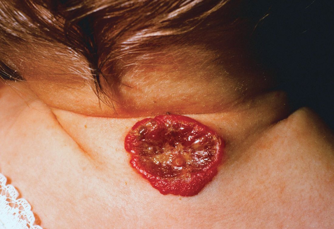
One clinical clue to picking up an infantile hemangioma (IH) that’s destined to ulcerate is an early grayish to white discoloration of the lesion, said Dr. Puttgen, chief of the division of pediatric dermatology at Johns Hopkins Medicine, Baltimore.
“Multimodal therapy is an absolute necessity” in treating an ulcerated IH, said Dr. Puttgen. Using an “all hands on deck” approach – a combination of topical and systemic modalities – can help bring the lesion under control.
Beta-blockers are first-line therapy to manage complicated IHs, with propranolol yielding a 98% response rate for all complicated IHs in the literature, said Dr. Puttgen.
Propranolol can decrease the volume and color of IHs and speed involution, in part by its ability to continue working after the proliferative growth phase of an IH. It’s also been shown to reduce the need for surgery in nasal IH, and it’s well tolerated, she added.
Evidence-based therapies for ulcerated hemangiomas include systemic propranolol at 1-3 mg/kg per day. That protocol will result in a healed ulcer within 2-6 weeks in most of the published case series, Dr. Puttgen noted.
Topical timolol also has evidence supporting its use for an ulcerated IH, and it has been found generally safe. In one study of 30 patients with IH, she said, three had mild adverse events consisting of sleep disturbance, diarrhea, and acrocyanosis. Another study reported success when brimonidine 0.2% and timolol 0.5% were used together. It’s possible, said Dr. Puttgen, that there’s a synergistic effect when combining the selective alpha-2 adrenergic agonist effect of brimonidine with timolol, which provides nonselective beta adrenergic blockade. However, she said, there has been an isolated report of brimonidine toxicity.
The ulcerated IHs need wound care, Dr. Puttgen added, with barrier creams and dressings. Pain management should be considered, because an ulcerated IH may have a large, friable, bleeding area. Pulsed-dye laser can also be a useful treatment modality for an ulcerating IH.
Going beyond the treatments for which the evidence is strongest and moving into more “state-of-the-art” treatments, “there may be a niche role for oral corticosteroids” as combination systemic therapy with propranolol, Dr. Puttgen said.
She shared images from a recently published report, in which she’s the senior author, showing the progression of an ulcerated IH. The hemangioma had received wound care and pulsed-dye laser treatment, and the infant was started on systemic propranolol. After 2 weeks, the IH had decreased significantly in volume, but the ulcerated area had actually increased. With the addition of oral corticosteroids, there was a reduction in ulceration after 2 weeks; and after 5 weeks of prednisolone, “the ulceration resolved without rebound,” said Dr. Puttgen. The corticosteroid was then tapered and propranolol was continued for an additional 2 months, then tapered. By 10 months, the IH had almost completely resolved (Br J Dermatol. 2017 Apr;176[4]:1064-7).
If a corticosteroid is added to propranolol, there may be benefit to a slower propranolol dose, Dr. Puttgen said. She suggests an altered dosing schedule, beginning with 1 mg/kg per day in two or three divided doses. Then, over a period of 2-7 days, the total daily dose can be increased to 1.5 mg/kg per day. Bumping the dose up to 2 mg/kg per day or higher should not happen until after 2 weeks at the reduced dosing schedule, she explained.
Dr. Puttgen disclosed that she is on the advisory board and has received honoraria from Pierre Fabre Dermatologie.
koakes@frontlinemedcom.com
On Twitter @karioakes
CHICAGO –
About 16% of infantile hemangiomas become ulcerated at some point during their proliferative phase, said Kate Puttgen, MD, during a talk at the World Congress of Pediatric Dermatology.
One clinical clue to picking up an infantile hemangioma (IH) that’s destined to ulcerate is an early grayish to white discoloration of the lesion, said Dr. Puttgen, chief of the division of pediatric dermatology at Johns Hopkins Medicine, Baltimore.
“Multimodal therapy is an absolute necessity” in treating an ulcerated IH, said Dr. Puttgen. Using an “all hands on deck” approach – a combination of topical and systemic modalities – can help bring the lesion under control.
Beta-blockers are first-line therapy to manage complicated IHs, with propranolol yielding a 98% response rate for all complicated IHs in the literature, said Dr. Puttgen.
Propranolol can decrease the volume and color of IHs and speed involution, in part by its ability to continue working after the proliferative growth phase of an IH. It’s also been shown to reduce the need for surgery in nasal IH, and it’s well tolerated, she added.
Evidence-based therapies for ulcerated hemangiomas include systemic propranolol at 1-3 mg/kg per day. That protocol will result in a healed ulcer within 2-6 weeks in most of the published case series, Dr. Puttgen noted.
Topical timolol also has evidence supporting its use for an ulcerated IH, and it has been found generally safe. In one study of 30 patients with IH, she said, three had mild adverse events consisting of sleep disturbance, diarrhea, and acrocyanosis. Another study reported success when brimonidine 0.2% and timolol 0.5% were used together. It’s possible, said Dr. Puttgen, that there’s a synergistic effect when combining the selective alpha-2 adrenergic agonist effect of brimonidine with timolol, which provides nonselective beta adrenergic blockade. However, she said, there has been an isolated report of brimonidine toxicity.
The ulcerated IHs need wound care, Dr. Puttgen added, with barrier creams and dressings. Pain management should be considered, because an ulcerated IH may have a large, friable, bleeding area. Pulsed-dye laser can also be a useful treatment modality for an ulcerating IH.
Going beyond the treatments for which the evidence is strongest and moving into more “state-of-the-art” treatments, “there may be a niche role for oral corticosteroids” as combination systemic therapy with propranolol, Dr. Puttgen said.
She shared images from a recently published report, in which she’s the senior author, showing the progression of an ulcerated IH. The hemangioma had received wound care and pulsed-dye laser treatment, and the infant was started on systemic propranolol. After 2 weeks, the IH had decreased significantly in volume, but the ulcerated area had actually increased. With the addition of oral corticosteroids, there was a reduction in ulceration after 2 weeks; and after 5 weeks of prednisolone, “the ulceration resolved without rebound,” said Dr. Puttgen. The corticosteroid was then tapered and propranolol was continued for an additional 2 months, then tapered. By 10 months, the IH had almost completely resolved (Br J Dermatol. 2017 Apr;176[4]:1064-7).
If a corticosteroid is added to propranolol, there may be benefit to a slower propranolol dose, Dr. Puttgen said. She suggests an altered dosing schedule, beginning with 1 mg/kg per day in two or three divided doses. Then, over a period of 2-7 days, the total daily dose can be increased to 1.5 mg/kg per day. Bumping the dose up to 2 mg/kg per day or higher should not happen until after 2 weeks at the reduced dosing schedule, she explained.
Dr. Puttgen disclosed that she is on the advisory board and has received honoraria from Pierre Fabre Dermatologie.
koakes@frontlinemedcom.com
On Twitter @karioakes
CHICAGO –
About 16% of infantile hemangiomas become ulcerated at some point during their proliferative phase, said Kate Puttgen, MD, during a talk at the World Congress of Pediatric Dermatology.
One clinical clue to picking up an infantile hemangioma (IH) that’s destined to ulcerate is an early grayish to white discoloration of the lesion, said Dr. Puttgen, chief of the division of pediatric dermatology at Johns Hopkins Medicine, Baltimore.
“Multimodal therapy is an absolute necessity” in treating an ulcerated IH, said Dr. Puttgen. Using an “all hands on deck” approach – a combination of topical and systemic modalities – can help bring the lesion under control.
Beta-blockers are first-line therapy to manage complicated IHs, with propranolol yielding a 98% response rate for all complicated IHs in the literature, said Dr. Puttgen.
Propranolol can decrease the volume and color of IHs and speed involution, in part by its ability to continue working after the proliferative growth phase of an IH. It’s also been shown to reduce the need for surgery in nasal IH, and it’s well tolerated, she added.
Evidence-based therapies for ulcerated hemangiomas include systemic propranolol at 1-3 mg/kg per day. That protocol will result in a healed ulcer within 2-6 weeks in most of the published case series, Dr. Puttgen noted.
Topical timolol also has evidence supporting its use for an ulcerated IH, and it has been found generally safe. In one study of 30 patients with IH, she said, three had mild adverse events consisting of sleep disturbance, diarrhea, and acrocyanosis. Another study reported success when brimonidine 0.2% and timolol 0.5% were used together. It’s possible, said Dr. Puttgen, that there’s a synergistic effect when combining the selective alpha-2 adrenergic agonist effect of brimonidine with timolol, which provides nonselective beta adrenergic blockade. However, she said, there has been an isolated report of brimonidine toxicity.
The ulcerated IHs need wound care, Dr. Puttgen added, with barrier creams and dressings. Pain management should be considered, because an ulcerated IH may have a large, friable, bleeding area. Pulsed-dye laser can also be a useful treatment modality for an ulcerating IH.
Going beyond the treatments for which the evidence is strongest and moving into more “state-of-the-art” treatments, “there may be a niche role for oral corticosteroids” as combination systemic therapy with propranolol, Dr. Puttgen said.
She shared images from a recently published report, in which she’s the senior author, showing the progression of an ulcerated IH. The hemangioma had received wound care and pulsed-dye laser treatment, and the infant was started on systemic propranolol. After 2 weeks, the IH had decreased significantly in volume, but the ulcerated area had actually increased. With the addition of oral corticosteroids, there was a reduction in ulceration after 2 weeks; and after 5 weeks of prednisolone, “the ulceration resolved without rebound,” said Dr. Puttgen. The corticosteroid was then tapered and propranolol was continued for an additional 2 months, then tapered. By 10 months, the IH had almost completely resolved (Br J Dermatol. 2017 Apr;176[4]:1064-7).
If a corticosteroid is added to propranolol, there may be benefit to a slower propranolol dose, Dr. Puttgen said. She suggests an altered dosing schedule, beginning with 1 mg/kg per day in two or three divided doses. Then, over a period of 2-7 days, the total daily dose can be increased to 1.5 mg/kg per day. Bumping the dose up to 2 mg/kg per day or higher should not happen until after 2 weeks at the reduced dosing schedule, she explained.
Dr. Puttgen disclosed that she is on the advisory board and has received honoraria from Pierre Fabre Dermatologie.
koakes@frontlinemedcom.com
On Twitter @karioakes
EXPERT ANALYSIS FROM WCPD 2017
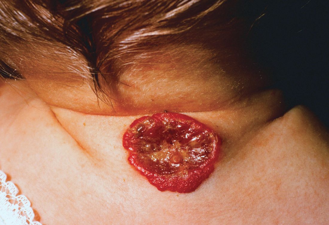
Young & Crusty
1. A 4-year-old boy presents with erythematous, oozing, excoriated plaques on the cheeks and chin (sparing the nose), trunk, and extensor surfaces of the arms and legs. His parents report that the “rash,” which flares on occasion, has been apparent since the boy was 6 months old, and he does anything he can to scratch the itch.
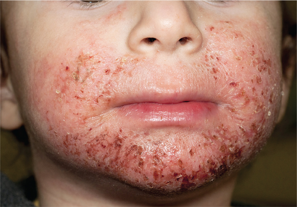
Diagnosis: Diagnosis of atopic dermatitis (AD) is made by age 5 in 85% to 90% of children who develop the disease, and by age 1 in 60% to 65%. AD persists into adulthood in up to one-third of patients. Although the cause is unknown, AD may be triggered by viral infections, food allergens, or weather, and in turn may trigger an inflammatory progression known as atopic march. Other factors affecting AD development include genetics and hygiene.
For more information:
“A Practical Overview of Pediatric Atopic Dermatitis, Part 2: Triggers and Grading.” Cutis. 2016;97(5):326-329.
“A Practical Overview of Pediatric Atopic Dermatitis, Part 3: Differential Diagnosis, Comorbidities, and Measurement of Disease Burden.” Cutis. 2016;97(6):408-412.
2. A child presents with a history of flaccid bullae and encased fluid progressing from clear yellow to turbid and darkish yellow. The pustules have ruptured, resulting in thin, light brown to golden yellow crusts and a collarette of scale at the periphery of the erosion. Itching is mild.
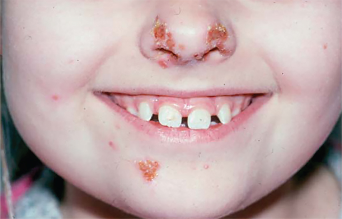
Diagnosis: Bullous impetigo most commonly affects neonates, hence the occasionally used (and inadvisably employed) name pemphigus neonatorum. Bullous impetigo appears to be less contagious than the nonbullous form and is usually sporadic in presentation. Typical areas of occurrence include the trunk and extremities, as well as intertriginous zones (eg, the diaper area, neck folds, and axillae).
For more information, see “Impetigo Update: New Challenges in the Era of Methicillin Resistance.” Cutis. 2010;85(2):65-70.
3. A 16-year-old Hispanic girl seeks consultation for a perioral rash that first appeared two weeks ago, shortly after she used an OTC depilatory agent. The rash has worsened despite application of topical neomycin ointment. The patient reports pruritus and occasional burning. Examination reveals erythema, hyperpigmentation, and excoriation of the skin around the corners of the mouth.
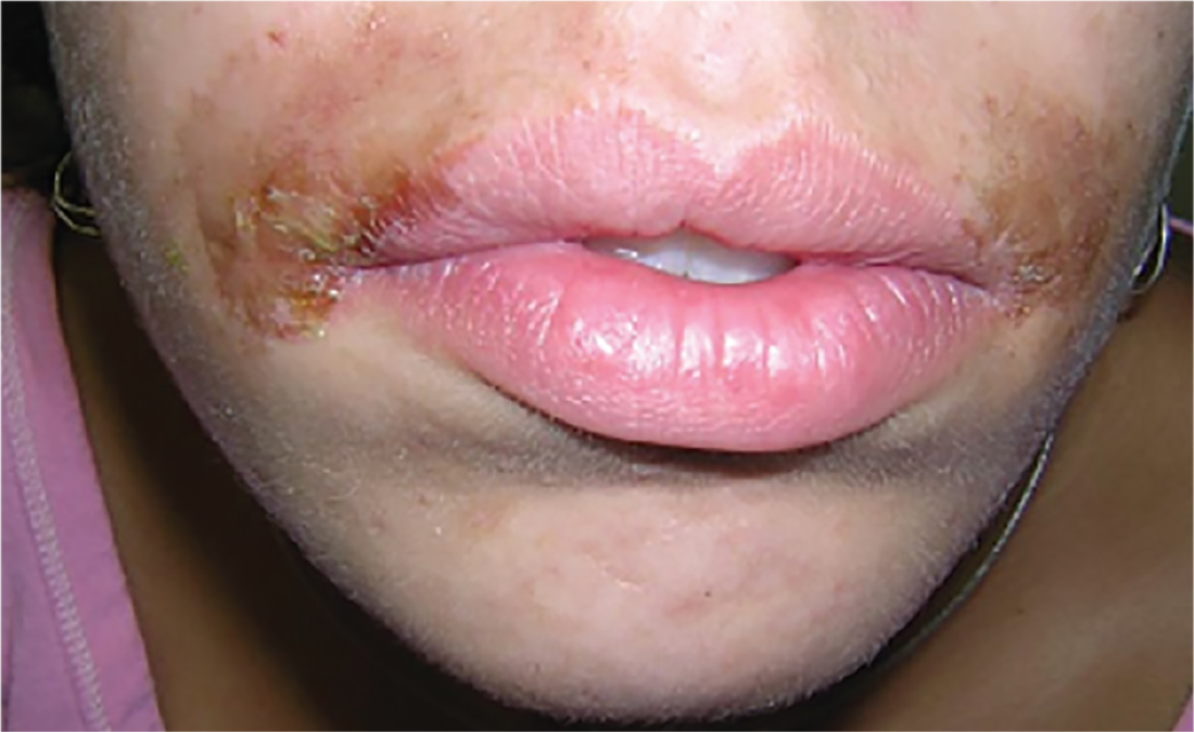
Diagnosis: An allergic contact dermatitis to both the depilatory agent and neomycin was suspected. The patient was advised to discontinue use of both products, and a medium-potency topical steroid was prescribed. She returned for follow-up in 10 days, at which time the condition had improved by about 75%. Therapy was changed to 1% hydrocortisone cream. The patient was instructed to return for patch testing if the dermatitis recurred.
Reprinted with permission from Emergency Medicine. 2010;42(8):19-20. http://www.mdedge.com/emed-journal/article/71585/dermatology/lesion-scrotum
4. The rash on this 5-month-old baby’s hands manifested several weeks ago. It spread to his arms and trunk and is now essentially everywhere except his face. Despite a number of treatment attempts, including oral antibiotics and OTC topical steroid creams, the problem persists.

Diagnosis: The diagnosis of scabies should be confirmed, whenever possible, with microscopic scabetic findings. In addition, treating the whole family and identifying the source of the infestation are crucial. Diagnosis of scabies is difficult in infants, as any part of an infant’s thin, soft, relatively hairless skin is fair game (whereas, in adults, scabies rarely affects skin above the neck). And although infants with scabies undoubtedly itch—probably just as much as adults—they are inept excoriators and even worse historians. In contrast, adults with scabies will scratch and complain continuously while in the exam room.
For more information, see “Baby Has Rash; Parents Feel Itchy.” Clinician Reviews. 2014;24(9):W3.
1. A 4-year-old boy presents with erythematous, oozing, excoriated plaques on the cheeks and chin (sparing the nose), trunk, and extensor surfaces of the arms and legs. His parents report that the “rash,” which flares on occasion, has been apparent since the boy was 6 months old, and he does anything he can to scratch the itch.
Diagnosis: Diagnosis of atopic dermatitis (AD) is made by age 5 in 85% to 90% of children who develop the disease, and by age 1 in 60% to 65%. AD persists into adulthood in up to one-third of patients. Although the cause is unknown, AD may be triggered by viral infections, food allergens, or weather, and in turn may trigger an inflammatory progression known as atopic march. Other factors affecting AD development include genetics and hygiene.
For more information:
“A Practical Overview of Pediatric Atopic Dermatitis, Part 2: Triggers and Grading.” Cutis. 2016;97(5):326-329.
“A Practical Overview of Pediatric Atopic Dermatitis, Part 3: Differential Diagnosis, Comorbidities, and Measurement of Disease Burden.” Cutis. 2016;97(6):408-412.
2. A child presents with a history of flaccid bullae and encased fluid progressing from clear yellow to turbid and darkish yellow. The pustules have ruptured, resulting in thin, light brown to golden yellow crusts and a collarette of scale at the periphery of the erosion. Itching is mild.
Diagnosis: Bullous impetigo most commonly affects neonates, hence the occasionally used (and inadvisably employed) name pemphigus neonatorum. Bullous impetigo appears to be less contagious than the nonbullous form and is usually sporadic in presentation. Typical areas of occurrence include the trunk and extremities, as well as intertriginous zones (eg, the diaper area, neck folds, and axillae).
For more information, see “Impetigo Update: New Challenges in the Era of Methicillin Resistance.” Cutis. 2010;85(2):65-70.
3. A 16-year-old Hispanic girl seeks consultation for a perioral rash that first appeared two weeks ago, shortly after she used an OTC depilatory agent. The rash has worsened despite application of topical neomycin ointment. The patient reports pruritus and occasional burning. Examination reveals erythema, hyperpigmentation, and excoriation of the skin around the corners of the mouth.
Diagnosis: An allergic contact dermatitis to both the depilatory agent and neomycin was suspected. The patient was advised to discontinue use of both products, and a medium-potency topical steroid was prescribed. She returned for follow-up in 10 days, at which time the condition had improved by about 75%. Therapy was changed to 1% hydrocortisone cream. The patient was instructed to return for patch testing if the dermatitis recurred.
Reprinted with permission from Emergency Medicine. 2010;42(8):19-20. http://www.mdedge.com/emed-journal/article/71585/dermatology/lesion-scrotum
4. The rash on this 5-month-old baby’s hands manifested several weeks ago. It spread to his arms and trunk and is now essentially everywhere except his face. Despite a number of treatment attempts, including oral antibiotics and OTC topical steroid creams, the problem persists.
Diagnosis: The diagnosis of scabies should be confirmed, whenever possible, with microscopic scabetic findings. In addition, treating the whole family and identifying the source of the infestation are crucial. Diagnosis of scabies is difficult in infants, as any part of an infant’s thin, soft, relatively hairless skin is fair game (whereas, in adults, scabies rarely affects skin above the neck). And although infants with scabies undoubtedly itch—probably just as much as adults—they are inept excoriators and even worse historians. In contrast, adults with scabies will scratch and complain continuously while in the exam room.
For more information, see “Baby Has Rash; Parents Feel Itchy.” Clinician Reviews. 2014;24(9):W3.
1. A 4-year-old boy presents with erythematous, oozing, excoriated plaques on the cheeks and chin (sparing the nose), trunk, and extensor surfaces of the arms and legs. His parents report that the “rash,” which flares on occasion, has been apparent since the boy was 6 months old, and he does anything he can to scratch the itch.
Diagnosis: Diagnosis of atopic dermatitis (AD) is made by age 5 in 85% to 90% of children who develop the disease, and by age 1 in 60% to 65%. AD persists into adulthood in up to one-third of patients. Although the cause is unknown, AD may be triggered by viral infections, food allergens, or weather, and in turn may trigger an inflammatory progression known as atopic march. Other factors affecting AD development include genetics and hygiene.
For more information:
“A Practical Overview of Pediatric Atopic Dermatitis, Part 2: Triggers and Grading.” Cutis. 2016;97(5):326-329.
“A Practical Overview of Pediatric Atopic Dermatitis, Part 3: Differential Diagnosis, Comorbidities, and Measurement of Disease Burden.” Cutis. 2016;97(6):408-412.
2. A child presents with a history of flaccid bullae and encased fluid progressing from clear yellow to turbid and darkish yellow. The pustules have ruptured, resulting in thin, light brown to golden yellow crusts and a collarette of scale at the periphery of the erosion. Itching is mild.
Diagnosis: Bullous impetigo most commonly affects neonates, hence the occasionally used (and inadvisably employed) name pemphigus neonatorum. Bullous impetigo appears to be less contagious than the nonbullous form and is usually sporadic in presentation. Typical areas of occurrence include the trunk and extremities, as well as intertriginous zones (eg, the diaper area, neck folds, and axillae).
For more information, see “Impetigo Update: New Challenges in the Era of Methicillin Resistance.” Cutis. 2010;85(2):65-70.
3. A 16-year-old Hispanic girl seeks consultation for a perioral rash that first appeared two weeks ago, shortly after she used an OTC depilatory agent. The rash has worsened despite application of topical neomycin ointment. The patient reports pruritus and occasional burning. Examination reveals erythema, hyperpigmentation, and excoriation of the skin around the corners of the mouth.
Diagnosis: An allergic contact dermatitis to both the depilatory agent and neomycin was suspected. The patient was advised to discontinue use of both products, and a medium-potency topical steroid was prescribed. She returned for follow-up in 10 days, at which time the condition had improved by about 75%. Therapy was changed to 1% hydrocortisone cream. The patient was instructed to return for patch testing if the dermatitis recurred.
Reprinted with permission from Emergency Medicine. 2010;42(8):19-20. http://www.mdedge.com/emed-journal/article/71585/dermatology/lesion-scrotum
4. The rash on this 5-month-old baby’s hands manifested several weeks ago. It spread to his arms and trunk and is now essentially everywhere except his face. Despite a number of treatment attempts, including oral antibiotics and OTC topical steroid creams, the problem persists.
Diagnosis: The diagnosis of scabies should be confirmed, whenever possible, with microscopic scabetic findings. In addition, treating the whole family and identifying the source of the infestation are crucial. Diagnosis of scabies is difficult in infants, as any part of an infant’s thin, soft, relatively hairless skin is fair game (whereas, in adults, scabies rarely affects skin above the neck). And although infants with scabies undoubtedly itch—probably just as much as adults—they are inept excoriators and even worse historians. In contrast, adults with scabies will scratch and complain continuously while in the exam room.
For more information, see “Baby Has Rash; Parents Feel Itchy.” Clinician Reviews. 2014;24(9):W3.
Why three strategies to correct vaccination hesitancy failed
, supporting findings in the literature that misinformation tends to linger in the memory and efforts to dislodge it may in fact reinforce it.
In a study of 120 students recruited from diverse departments of the University of Edinburgh, the Suor Orsola Benincasa University of Naples, and the Second University of Naples, 61% were women, and the mean age was 25 years. Most students had a bachelor’s (38%) or master’s degree (53%), while 11 were PhD students, reported Sara Pluviano, PhD, of the University of Edinburgh, and her associates.
The participants completed a baseline questionnaire assessing their beliefs and attitudes about immunization, and twice completed a questionnaire that had focused on general misperceptions about vaccines causing autism, vaccine side effects, and how likely they would be to give MMR vaccine to their children – immediately after an intervention and after a 7-day delay.
Students were exposed to one of four correction interventions.
In the first, participants were given a booklet confronting 10 “myths” with a number of “facts” about vaccines (myths vs. facts group). In the second, students were presented with a series of tables comparing the potential problems caused by measles, mumps, and rubella with the potential side effects caused by the MMR vaccine (graphics group). In the third intervention, participants were shown photos of unvaccinated children with measles, mumps, and rubella, in addition to the symptoms of each disease and warnings about vaccinating one’s own child (fear group). The fourth was the control condition, where participants were given two unrelated fact sheets with tips to help prevent medical errors and get safer health care (control group).
“The myths vs. facts format, at odds with its aims, induced stronger beliefs in the vaccine/autism link and in vaccines side effects over time, lending credit to the literature showing that countering false information in ways that repeat it may further contribute to its dissemination,” the researchers said. Also, “the exposure to fear appeals through images of sick children led to more increased misperceptions about vaccines causing autism. Moreover, this corrective strategy induced the strongest beliefs in vaccines side effects.
“The usage of fact/icon boxes resulted in less damage but did not bring any effective result,” they said.
“Our pattern of results thus confirms that there should be more testing of public health campaign messages,” Dr. Pluviano and her associates cautioned. “This is especially true because corrective strategies may appear effective immediately, yet backfire even after a short delay when the message they tried to convey gradually fades from memory, allowing common misconceptions to be more easily remembered and identified as true.”
Read more in PLOS One (2017 Jul 27. doi: 10.1371/journal.pone.0181640).
, supporting findings in the literature that misinformation tends to linger in the memory and efforts to dislodge it may in fact reinforce it.
In a study of 120 students recruited from diverse departments of the University of Edinburgh, the Suor Orsola Benincasa University of Naples, and the Second University of Naples, 61% were women, and the mean age was 25 years. Most students had a bachelor’s (38%) or master’s degree (53%), while 11 were PhD students, reported Sara Pluviano, PhD, of the University of Edinburgh, and her associates.
The participants completed a baseline questionnaire assessing their beliefs and attitudes about immunization, and twice completed a questionnaire that had focused on general misperceptions about vaccines causing autism, vaccine side effects, and how likely they would be to give MMR vaccine to their children – immediately after an intervention and after a 7-day delay.
Students were exposed to one of four correction interventions.
In the first, participants were given a booklet confronting 10 “myths” with a number of “facts” about vaccines (myths vs. facts group). In the second, students were presented with a series of tables comparing the potential problems caused by measles, mumps, and rubella with the potential side effects caused by the MMR vaccine (graphics group). In the third intervention, participants were shown photos of unvaccinated children with measles, mumps, and rubella, in addition to the symptoms of each disease and warnings about vaccinating one’s own child (fear group). The fourth was the control condition, where participants were given two unrelated fact sheets with tips to help prevent medical errors and get safer health care (control group).
“The myths vs. facts format, at odds with its aims, induced stronger beliefs in the vaccine/autism link and in vaccines side effects over time, lending credit to the literature showing that countering false information in ways that repeat it may further contribute to its dissemination,” the researchers said. Also, “the exposure to fear appeals through images of sick children led to more increased misperceptions about vaccines causing autism. Moreover, this corrective strategy induced the strongest beliefs in vaccines side effects.
“The usage of fact/icon boxes resulted in less damage but did not bring any effective result,” they said.
“Our pattern of results thus confirms that there should be more testing of public health campaign messages,” Dr. Pluviano and her associates cautioned. “This is especially true because corrective strategies may appear effective immediately, yet backfire even after a short delay when the message they tried to convey gradually fades from memory, allowing common misconceptions to be more easily remembered and identified as true.”
Read more in PLOS One (2017 Jul 27. doi: 10.1371/journal.pone.0181640).
, supporting findings in the literature that misinformation tends to linger in the memory and efforts to dislodge it may in fact reinforce it.
In a study of 120 students recruited from diverse departments of the University of Edinburgh, the Suor Orsola Benincasa University of Naples, and the Second University of Naples, 61% were women, and the mean age was 25 years. Most students had a bachelor’s (38%) or master’s degree (53%), while 11 were PhD students, reported Sara Pluviano, PhD, of the University of Edinburgh, and her associates.
The participants completed a baseline questionnaire assessing their beliefs and attitudes about immunization, and twice completed a questionnaire that had focused on general misperceptions about vaccines causing autism, vaccine side effects, and how likely they would be to give MMR vaccine to their children – immediately after an intervention and after a 7-day delay.
Students were exposed to one of four correction interventions.
In the first, participants were given a booklet confronting 10 “myths” with a number of “facts” about vaccines (myths vs. facts group). In the second, students were presented with a series of tables comparing the potential problems caused by measles, mumps, and rubella with the potential side effects caused by the MMR vaccine (graphics group). In the third intervention, participants were shown photos of unvaccinated children with measles, mumps, and rubella, in addition to the symptoms of each disease and warnings about vaccinating one’s own child (fear group). The fourth was the control condition, where participants were given two unrelated fact sheets with tips to help prevent medical errors and get safer health care (control group).
“The myths vs. facts format, at odds with its aims, induced stronger beliefs in the vaccine/autism link and in vaccines side effects over time, lending credit to the literature showing that countering false information in ways that repeat it may further contribute to its dissemination,” the researchers said. Also, “the exposure to fear appeals through images of sick children led to more increased misperceptions about vaccines causing autism. Moreover, this corrective strategy induced the strongest beliefs in vaccines side effects.
“The usage of fact/icon boxes resulted in less damage but did not bring any effective result,” they said.
“Our pattern of results thus confirms that there should be more testing of public health campaign messages,” Dr. Pluviano and her associates cautioned. “This is especially true because corrective strategies may appear effective immediately, yet backfire even after a short delay when the message they tried to convey gradually fades from memory, allowing common misconceptions to be more easily remembered and identified as true.”
Read more in PLOS One (2017 Jul 27. doi: 10.1371/journal.pone.0181640).
FROM PLOS ONE