User login
Budesonide fails to cut deaths in preemies
The administration of inhaled budesonide to extremely preterm infants did not increase the risk of neurodevelopmental disability, but did increase mortality, in a study by Dirk Bassler, MD, of the University of Zürich and his associates.
An older study led by Dr. Bassler and published in the New England Journal of Medicine showed that inhaled budesonide significantly reduced the incidence of bronchopulmonary dysplasia, which has been linked to higher mortality and chronic respiratory and cardiovascular impairment (N Engl J Med. 2015;373:1497-506).
Systemic glucocorticoids have been linked to greater risk of neurodevelopmental disability, but only a few studies have examined the effect of inhaled glucocorticoids, such as budesonide, in preterm infants. These studies, including the earlier one by Dr. Bassler and his colleagues, were either small, covered a short period of time or involved late administering of the drug.
In the two studies by Dr. Bassler and his colleagues, 863 preterm infants between 23 weeks’ and just under 28 weeks’ gestation who required any form of positive-pressure respiratory support were randomized to receive inhaled budesonide (two puffs, 200 mcg per puff) or placebo every 12 hours. They began within 24 hours of birth and continued for the first 14 days of life. Following that, patients received 1 puff every 12 hours until they no longer required supplemental oxygen and positive-pressure support, or reached a postmenstrual age of 32 weeks.
The treatment resulted in a significant reduction in bronchopulmonary dysplasia at a postmenstrual age of 36 weeks (28.2% in the budesonide group vs. 37.4%; P = .01), in the older study.
In the new study, which was also published in the New England Journal of Medicine, Dr. Bassler and his associates found higher mortality (19.9% vs. 14.5%; relative risk, 1.37; 95% confidence interval, 1.01-1.86; P = .04) in the group of patients who had received inhaled budesonide. Additionally, at a corrected age of 18-22 months, surviving infants who received inhaled budesonide had a similar risk of neurodevelopmental disability as those patients who took the placebo.
Broadly speaking, 48.1% of infants who received budesonide had a neurodevelopmental disability, compared with 51.4% of infants who received placebo (RR adjusted for gestational age, 0.93; 95% CI, 0.80-1.09; P = .40). The two groups also had no statistically significant differences in their frequencies of cerebral palsy, blindness, hearing loss, or cognitive delay.
“There was no significant difference between the groups in adverse long-term outcomes in our study. However, the fact that fewer infants died in the placebo group than in the budesonide group complicates the interpretation of the treatment of budesonide,” the researchers wrote.
Supported by a grant from the European Union and by Chiesi Farmaceutici. Disclosure forms provided by the authors are available with the full text of this article at NEJM.org.
SOURCE: N Engl J Med. 2018;378:148-57.
This is an important study regarding bronchopulmonary dysplasia prevention. The study suggests starting budesonide within 24 hours of life resulted in a lower rate of bronchopulmonary dysplasia than placebo but fewer infants died in the placebo group. A bigger question for me is “what is the evidence for starting inhaled steroids prior to neonatal intensive care unit discharge?” Pediatric pulmonologists would like to know if it decreases subsequent respiratory-related ER visits and readmissions.
This is an important study regarding bronchopulmonary dysplasia prevention. The study suggests starting budesonide within 24 hours of life resulted in a lower rate of bronchopulmonary dysplasia than placebo but fewer infants died in the placebo group. A bigger question for me is “what is the evidence for starting inhaled steroids prior to neonatal intensive care unit discharge?” Pediatric pulmonologists would like to know if it decreases subsequent respiratory-related ER visits and readmissions.
This is an important study regarding bronchopulmonary dysplasia prevention. The study suggests starting budesonide within 24 hours of life resulted in a lower rate of bronchopulmonary dysplasia than placebo but fewer infants died in the placebo group. A bigger question for me is “what is the evidence for starting inhaled steroids prior to neonatal intensive care unit discharge?” Pediatric pulmonologists would like to know if it decreases subsequent respiratory-related ER visits and readmissions.
The administration of inhaled budesonide to extremely preterm infants did not increase the risk of neurodevelopmental disability, but did increase mortality, in a study by Dirk Bassler, MD, of the University of Zürich and his associates.
An older study led by Dr. Bassler and published in the New England Journal of Medicine showed that inhaled budesonide significantly reduced the incidence of bronchopulmonary dysplasia, which has been linked to higher mortality and chronic respiratory and cardiovascular impairment (N Engl J Med. 2015;373:1497-506).
Systemic glucocorticoids have been linked to greater risk of neurodevelopmental disability, but only a few studies have examined the effect of inhaled glucocorticoids, such as budesonide, in preterm infants. These studies, including the earlier one by Dr. Bassler and his colleagues, were either small, covered a short period of time or involved late administering of the drug.
In the two studies by Dr. Bassler and his colleagues, 863 preterm infants between 23 weeks’ and just under 28 weeks’ gestation who required any form of positive-pressure respiratory support were randomized to receive inhaled budesonide (two puffs, 200 mcg per puff) or placebo every 12 hours. They began within 24 hours of birth and continued for the first 14 days of life. Following that, patients received 1 puff every 12 hours until they no longer required supplemental oxygen and positive-pressure support, or reached a postmenstrual age of 32 weeks.
The treatment resulted in a significant reduction in bronchopulmonary dysplasia at a postmenstrual age of 36 weeks (28.2% in the budesonide group vs. 37.4%; P = .01), in the older study.
In the new study, which was also published in the New England Journal of Medicine, Dr. Bassler and his associates found higher mortality (19.9% vs. 14.5%; relative risk, 1.37; 95% confidence interval, 1.01-1.86; P = .04) in the group of patients who had received inhaled budesonide. Additionally, at a corrected age of 18-22 months, surviving infants who received inhaled budesonide had a similar risk of neurodevelopmental disability as those patients who took the placebo.
Broadly speaking, 48.1% of infants who received budesonide had a neurodevelopmental disability, compared with 51.4% of infants who received placebo (RR adjusted for gestational age, 0.93; 95% CI, 0.80-1.09; P = .40). The two groups also had no statistically significant differences in their frequencies of cerebral palsy, blindness, hearing loss, or cognitive delay.
“There was no significant difference between the groups in adverse long-term outcomes in our study. However, the fact that fewer infants died in the placebo group than in the budesonide group complicates the interpretation of the treatment of budesonide,” the researchers wrote.
Supported by a grant from the European Union and by Chiesi Farmaceutici. Disclosure forms provided by the authors are available with the full text of this article at NEJM.org.
SOURCE: N Engl J Med. 2018;378:148-57.
The administration of inhaled budesonide to extremely preterm infants did not increase the risk of neurodevelopmental disability, but did increase mortality, in a study by Dirk Bassler, MD, of the University of Zürich and his associates.
An older study led by Dr. Bassler and published in the New England Journal of Medicine showed that inhaled budesonide significantly reduced the incidence of bronchopulmonary dysplasia, which has been linked to higher mortality and chronic respiratory and cardiovascular impairment (N Engl J Med. 2015;373:1497-506).
Systemic glucocorticoids have been linked to greater risk of neurodevelopmental disability, but only a few studies have examined the effect of inhaled glucocorticoids, such as budesonide, in preterm infants. These studies, including the earlier one by Dr. Bassler and his colleagues, were either small, covered a short period of time or involved late administering of the drug.
In the two studies by Dr. Bassler and his colleagues, 863 preterm infants between 23 weeks’ and just under 28 weeks’ gestation who required any form of positive-pressure respiratory support were randomized to receive inhaled budesonide (two puffs, 200 mcg per puff) or placebo every 12 hours. They began within 24 hours of birth and continued for the first 14 days of life. Following that, patients received 1 puff every 12 hours until they no longer required supplemental oxygen and positive-pressure support, or reached a postmenstrual age of 32 weeks.
The treatment resulted in a significant reduction in bronchopulmonary dysplasia at a postmenstrual age of 36 weeks (28.2% in the budesonide group vs. 37.4%; P = .01), in the older study.
In the new study, which was also published in the New England Journal of Medicine, Dr. Bassler and his associates found higher mortality (19.9% vs. 14.5%; relative risk, 1.37; 95% confidence interval, 1.01-1.86; P = .04) in the group of patients who had received inhaled budesonide. Additionally, at a corrected age of 18-22 months, surviving infants who received inhaled budesonide had a similar risk of neurodevelopmental disability as those patients who took the placebo.
Broadly speaking, 48.1% of infants who received budesonide had a neurodevelopmental disability, compared with 51.4% of infants who received placebo (RR adjusted for gestational age, 0.93; 95% CI, 0.80-1.09; P = .40). The two groups also had no statistically significant differences in their frequencies of cerebral palsy, blindness, hearing loss, or cognitive delay.
“There was no significant difference between the groups in adverse long-term outcomes in our study. However, the fact that fewer infants died in the placebo group than in the budesonide group complicates the interpretation of the treatment of budesonide,” the researchers wrote.
Supported by a grant from the European Union and by Chiesi Farmaceutici. Disclosure forms provided by the authors are available with the full text of this article at NEJM.org.
SOURCE: N Engl J Med. 2018;378:148-57.
FROM NEW ENGLAND JOURNAL OF MEDICINE
Key clinical point:
Major finding: Nearly 20% of infants in the budesonide group died, compared with 14.5% of the placebo group.
Data source: Randomized, controlled trial of 863 extremely preterm infants.
Disclosures: Supported by a grant from the European Union and by Chiesi Farmaceutici. Disclosure forms provided by the authors are available with the full text of this article at NEJM.org.
Source: N Engl J Med. 2018;378:148-57.
Disparities persist in infant safe sleep practices
Sleep-related deaths among infants in the United States decreased during the 1990s as a result of recommendations to place babies on their backs to sleep. However, the decline has leveled off in recent years, and health care providers should proactively counsel caregivers about safe sleep practices, wrote Jennifer M. Bombard, MSPH, of the Centers for Disease Control and Prevention and her colleagues in a study published online in the Morbidity and Mortality Weekly Report.

Overall, 22% of respondents from 32 states and New York City in 2015 reported placing babies in a position other than their backs to sleep. In addition, 61% of respondents from 14 states reported bed sharing, and 39% from 13 states and New York City reported using soft bedding, including bumper pads and thick blankets.
Unsafe sleep practices varied by maternal demographics; nonsupine sleep positioning was more likely among non-Hispanic blacks, individuals aged 25 years or younger, those with 12 years or less of education, and those participating in the Special Supplemental Nutrition Program for Women, Infants, and Children.
“These findings highlight the need to implement and evaluate interventions to continue improving safe sleep practices,” Ms. Bombard and her associates said.
They cited the Study of Attitudes and Factors Effecting Infant Care Practices, in which caregivers who received appropriate advice on safe sleep practices were significantly less likely to place infants in a nonsupine position to sleep. “Evidence-based approaches to increase use of safe sleep practices include developing health messages and educational tools for caregivers and educating health and child care professionals on safe sleep practices,” they noted.
The study was limited by several factors, including reliance on self reports and inclusion of only states with Pregnancy Risk Assessment Monitoring System records, the researchers said.
Ms. Bombard and her associates had no relevant financial disclosures.
pdnews@frontlinemedcom.com
SOURCE: Bombard J et al. MMWR. 2018 Jan 9. doi: 10.15585/mmwr.mm6701e1.
Sleep-related deaths among infants in the United States decreased during the 1990s as a result of recommendations to place babies on their backs to sleep. However, the decline has leveled off in recent years, and health care providers should proactively counsel caregivers about safe sleep practices, wrote Jennifer M. Bombard, MSPH, of the Centers for Disease Control and Prevention and her colleagues in a study published online in the Morbidity and Mortality Weekly Report.
Overall, 22% of respondents from 32 states and New York City in 2015 reported placing babies in a position other than their backs to sleep. In addition, 61% of respondents from 14 states reported bed sharing, and 39% from 13 states and New York City reported using soft bedding, including bumper pads and thick blankets.
Unsafe sleep practices varied by maternal demographics; nonsupine sleep positioning was more likely among non-Hispanic blacks, individuals aged 25 years or younger, those with 12 years or less of education, and those participating in the Special Supplemental Nutrition Program for Women, Infants, and Children.
“These findings highlight the need to implement and evaluate interventions to continue improving safe sleep practices,” Ms. Bombard and her associates said.
They cited the Study of Attitudes and Factors Effecting Infant Care Practices, in which caregivers who received appropriate advice on safe sleep practices were significantly less likely to place infants in a nonsupine position to sleep. “Evidence-based approaches to increase use of safe sleep practices include developing health messages and educational tools for caregivers and educating health and child care professionals on safe sleep practices,” they noted.
The study was limited by several factors, including reliance on self reports and inclusion of only states with Pregnancy Risk Assessment Monitoring System records, the researchers said.
Ms. Bombard and her associates had no relevant financial disclosures.
pdnews@frontlinemedcom.com
SOURCE: Bombard J et al. MMWR. 2018 Jan 9. doi: 10.15585/mmwr.mm6701e1.
Sleep-related deaths among infants in the United States decreased during the 1990s as a result of recommendations to place babies on their backs to sleep. However, the decline has leveled off in recent years, and health care providers should proactively counsel caregivers about safe sleep practices, wrote Jennifer M. Bombard, MSPH, of the Centers for Disease Control and Prevention and her colleagues in a study published online in the Morbidity and Mortality Weekly Report.
Overall, 22% of respondents from 32 states and New York City in 2015 reported placing babies in a position other than their backs to sleep. In addition, 61% of respondents from 14 states reported bed sharing, and 39% from 13 states and New York City reported using soft bedding, including bumper pads and thick blankets.
Unsafe sleep practices varied by maternal demographics; nonsupine sleep positioning was more likely among non-Hispanic blacks, individuals aged 25 years or younger, those with 12 years or less of education, and those participating in the Special Supplemental Nutrition Program for Women, Infants, and Children.
“These findings highlight the need to implement and evaluate interventions to continue improving safe sleep practices,” Ms. Bombard and her associates said.
They cited the Study of Attitudes and Factors Effecting Infant Care Practices, in which caregivers who received appropriate advice on safe sleep practices were significantly less likely to place infants in a nonsupine position to sleep. “Evidence-based approaches to increase use of safe sleep practices include developing health messages and educational tools for caregivers and educating health and child care professionals on safe sleep practices,” they noted.
The study was limited by several factors, including reliance on self reports and inclusion of only states with Pregnancy Risk Assessment Monitoring System records, the researchers said.
Ms. Bombard and her associates had no relevant financial disclosures.
pdnews@frontlinemedcom.com
SOURCE: Bombard J et al. MMWR. 2018 Jan 9. doi: 10.15585/mmwr.mm6701e1.
FROM MMWR
Key clinical point: Health care providers can improve safe sleep for babies by counseling caregivers.
Major finding: Of respondents from 32 states and New York City in 2015, 22% reported placing babies in a position other than their backs to sleep.
Study details: The data come from the 2009-2015 Pregnancy Risk Assessment Monitoring System.
Disclosures: The researchers had no relevant financial disclosures.
Source: Bombard J et al. MMWR 2018 Jan 9. doi: 10.15585/mmwr.mm6701e1.
Optimal rate of flow for high-flow nasal cannula in young children
Clinical question
Is there an optimal rate of flow for high-flow nasal cannula in respiratory distress?
Background
High-flow nasal cannula (HFNC) has been increasingly used to treat children with moderate to severe bronchiolitis, both in intensive care unit (ICU) settings and on inpatient wards. Studies have shown it may allow children with bronchiolitis to avoid ICU admission and intubation. In preterm infants it has been shown to decrease work of breathing. No prior studies, however, have examined optimizing the rate of flow for individual patients, and considerable heterogeneity exists in choosing initial HFNC flow rates.
Reliably measuring effort of breathing has proved challenging. Placing a manometer in the esophagus allows measurement of the pressure-rate product (PRP), a previously validated measure of effort of breathing computed by multiplying the difference between maximum and minimum esophageal pressures by the respiratory rate.1 An increasing PRP indicates increasing effort of breathing. The authors chose systems from Fisher & Paykel and Vapotherm for their testing.
Study design
Single-center prospective observational trial.
Setting
24-bed pediatric intensive care unit in a 347-bed urban free-standing children’s hospital.
Synopsis
A single center recruited patients aged 37 weeks corrected gestational age to 3 years who were admitted to the ICU with respiratory distress. Fifty-four patients met inclusion criteria and 21 were enrolled and completed the study. Prior data suggested a sample size of 20 would be sufficient to identify a clinically significant effect size. Median age was 6 months.
Thirteen patients had bronchiolitis, three had pneumonia, and five had other respiratory illnesses. Each patient received HFNC delivered by both systems in sequence with flow rates of 0.5, 1, 1.5, and 2 L/kg per minute to a maximum of 30 L/min. Following the trials, patients remained on HFNC as per usual care with twice-daily PRP measurements until weaned off HFNC.
A dose-dependent relationship existed between flow and change in PRP, with the greatest reduction in PRP at 2 L/kg per minute flow (P less than .001) and a slightly smaller but similar reduction in PRP at 1.5 L/kg per minute. When stratifying the subjects by weight, this effect was not statistically significant for patients heavier than 8 kg (P = .38), with all significant changes being in patients less than 8 kg (P less than .001) with a median drop in PRP of 25%. Further examining these younger and lighter patients, the greatest reduction in PRP was in the lightest patients (less than 5 kg).
Given the similarity in drop in PRP at 1.5 L/kg per minute and 2 L/kg per minute, the authors suggest this flow rate yields a plateau effect and minimal further improvement would be seen with increasing flow rates. A rate of 2 L/kg per minute was chosen as a maximum a priori as it was judged the highest level of HFNC patients could tolerate without worsening agitation or air leak. There was no difference seen between the two HFNC systems in the study. The authors did not report the fraction of inspired oxygen settings used, the size of HFNC cannulas, or how PRP changed over several days as HFNC was weaned.
Bottom line
The optimal HFNC rate to decrease effort of breathing for children less than 3 years old is between 1.5 and 2 L/kg/min with the greatest improvement expected in children under 5 kg.
Citation
Weiler T et al. The Relationship between High Flow Nasal Cannula Flow Rate and Effort of Breathing in Children. The Journal of Pediatrics. October 2017. doi: 10.1016/j.jpeds.2017.06.006.
Reference
1. Argent AC, Newth CJL, Klein M. The mechanics of breathing in children with acute severe croup. Intensive Care Med. 2008;34(2):324-32. doi: 10.1007/s00134-007-0910-x.
Dr. Stubblefield is a pediatric hospitalist at Nemours/Alfred I. duPont Hospital for Children in Wilmington, Del., and clinical assistant professor of pediatrics at Jefferson Medical College in Philadelphia.
Clinical question
Is there an optimal rate of flow for high-flow nasal cannula in respiratory distress?
Background
High-flow nasal cannula (HFNC) has been increasingly used to treat children with moderate to severe bronchiolitis, both in intensive care unit (ICU) settings and on inpatient wards. Studies have shown it may allow children with bronchiolitis to avoid ICU admission and intubation. In preterm infants it has been shown to decrease work of breathing. No prior studies, however, have examined optimizing the rate of flow for individual patients, and considerable heterogeneity exists in choosing initial HFNC flow rates.
Reliably measuring effort of breathing has proved challenging. Placing a manometer in the esophagus allows measurement of the pressure-rate product (PRP), a previously validated measure of effort of breathing computed by multiplying the difference between maximum and minimum esophageal pressures by the respiratory rate.1 An increasing PRP indicates increasing effort of breathing. The authors chose systems from Fisher & Paykel and Vapotherm for their testing.
Study design
Single-center prospective observational trial.
Setting
24-bed pediatric intensive care unit in a 347-bed urban free-standing children’s hospital.
Synopsis
A single center recruited patients aged 37 weeks corrected gestational age to 3 years who were admitted to the ICU with respiratory distress. Fifty-four patients met inclusion criteria and 21 were enrolled and completed the study. Prior data suggested a sample size of 20 would be sufficient to identify a clinically significant effect size. Median age was 6 months.
Thirteen patients had bronchiolitis, three had pneumonia, and five had other respiratory illnesses. Each patient received HFNC delivered by both systems in sequence with flow rates of 0.5, 1, 1.5, and 2 L/kg per minute to a maximum of 30 L/min. Following the trials, patients remained on HFNC as per usual care with twice-daily PRP measurements until weaned off HFNC.
A dose-dependent relationship existed between flow and change in PRP, with the greatest reduction in PRP at 2 L/kg per minute flow (P less than .001) and a slightly smaller but similar reduction in PRP at 1.5 L/kg per minute. When stratifying the subjects by weight, this effect was not statistically significant for patients heavier than 8 kg (P = .38), with all significant changes being in patients less than 8 kg (P less than .001) with a median drop in PRP of 25%. Further examining these younger and lighter patients, the greatest reduction in PRP was in the lightest patients (less than 5 kg).
Given the similarity in drop in PRP at 1.5 L/kg per minute and 2 L/kg per minute, the authors suggest this flow rate yields a plateau effect and minimal further improvement would be seen with increasing flow rates. A rate of 2 L/kg per minute was chosen as a maximum a priori as it was judged the highest level of HFNC patients could tolerate without worsening agitation or air leak. There was no difference seen between the two HFNC systems in the study. The authors did not report the fraction of inspired oxygen settings used, the size of HFNC cannulas, or how PRP changed over several days as HFNC was weaned.
Bottom line
The optimal HFNC rate to decrease effort of breathing for children less than 3 years old is between 1.5 and 2 L/kg/min with the greatest improvement expected in children under 5 kg.
Citation
Weiler T et al. The Relationship between High Flow Nasal Cannula Flow Rate and Effort of Breathing in Children. The Journal of Pediatrics. October 2017. doi: 10.1016/j.jpeds.2017.06.006.
Reference
1. Argent AC, Newth CJL, Klein M. The mechanics of breathing in children with acute severe croup. Intensive Care Med. 2008;34(2):324-32. doi: 10.1007/s00134-007-0910-x.
Dr. Stubblefield is a pediatric hospitalist at Nemours/Alfred I. duPont Hospital for Children in Wilmington, Del., and clinical assistant professor of pediatrics at Jefferson Medical College in Philadelphia.
Clinical question
Is there an optimal rate of flow for high-flow nasal cannula in respiratory distress?
Background
High-flow nasal cannula (HFNC) has been increasingly used to treat children with moderate to severe bronchiolitis, both in intensive care unit (ICU) settings and on inpatient wards. Studies have shown it may allow children with bronchiolitis to avoid ICU admission and intubation. In preterm infants it has been shown to decrease work of breathing. No prior studies, however, have examined optimizing the rate of flow for individual patients, and considerable heterogeneity exists in choosing initial HFNC flow rates.
Reliably measuring effort of breathing has proved challenging. Placing a manometer in the esophagus allows measurement of the pressure-rate product (PRP), a previously validated measure of effort of breathing computed by multiplying the difference between maximum and minimum esophageal pressures by the respiratory rate.1 An increasing PRP indicates increasing effort of breathing. The authors chose systems from Fisher & Paykel and Vapotherm for their testing.
Study design
Single-center prospective observational trial.
Setting
24-bed pediatric intensive care unit in a 347-bed urban free-standing children’s hospital.
Synopsis
A single center recruited patients aged 37 weeks corrected gestational age to 3 years who were admitted to the ICU with respiratory distress. Fifty-four patients met inclusion criteria and 21 were enrolled and completed the study. Prior data suggested a sample size of 20 would be sufficient to identify a clinically significant effect size. Median age was 6 months.
Thirteen patients had bronchiolitis, three had pneumonia, and five had other respiratory illnesses. Each patient received HFNC delivered by both systems in sequence with flow rates of 0.5, 1, 1.5, and 2 L/kg per minute to a maximum of 30 L/min. Following the trials, patients remained on HFNC as per usual care with twice-daily PRP measurements until weaned off HFNC.
A dose-dependent relationship existed between flow and change in PRP, with the greatest reduction in PRP at 2 L/kg per minute flow (P less than .001) and a slightly smaller but similar reduction in PRP at 1.5 L/kg per minute. When stratifying the subjects by weight, this effect was not statistically significant for patients heavier than 8 kg (P = .38), with all significant changes being in patients less than 8 kg (P less than .001) with a median drop in PRP of 25%. Further examining these younger and lighter patients, the greatest reduction in PRP was in the lightest patients (less than 5 kg).
Given the similarity in drop in PRP at 1.5 L/kg per minute and 2 L/kg per minute, the authors suggest this flow rate yields a plateau effect and minimal further improvement would be seen with increasing flow rates. A rate of 2 L/kg per minute was chosen as a maximum a priori as it was judged the highest level of HFNC patients could tolerate without worsening agitation or air leak. There was no difference seen between the two HFNC systems in the study. The authors did not report the fraction of inspired oxygen settings used, the size of HFNC cannulas, or how PRP changed over several days as HFNC was weaned.
Bottom line
The optimal HFNC rate to decrease effort of breathing for children less than 3 years old is between 1.5 and 2 L/kg/min with the greatest improvement expected in children under 5 kg.
Citation
Weiler T et al. The Relationship between High Flow Nasal Cannula Flow Rate and Effort of Breathing in Children. The Journal of Pediatrics. October 2017. doi: 10.1016/j.jpeds.2017.06.006.
Reference
1. Argent AC, Newth CJL, Klein M. The mechanics of breathing in children with acute severe croup. Intensive Care Med. 2008;34(2):324-32. doi: 10.1007/s00134-007-0910-x.
Dr. Stubblefield is a pediatric hospitalist at Nemours/Alfred I. duPont Hospital for Children in Wilmington, Del., and clinical assistant professor of pediatrics at Jefferson Medical College in Philadelphia.
Gene variants associated with high-risk pediatric ALL

New research has revealed germline variations associated with high-risk acute lymphoblastic leukemia (ALL) in children.
Researchers sequenced the TP53 tumor suppressor gene in nearly 4000 children with ALL and identified 22 pathogenic germline variants.
These variants were associated with inferior survival and an increased risk of developing second malignancies.
Jun J. Yang, PhD, of St. Jude Children’s Research Hospital in Memphis, Tennessee, and his colleagues reported these findings in the Journal of Clinical Oncology.
The researchers performed targeted sequencing of TP53 coding regions in 3801 children with ALL who were enrolled in 2 Children’s Oncology Group trials (AALL0232 and P9900).
The sequencing revealed 49 unique TP53 coding variants, which were found in 77 children. Twenty-two of the variants were pathogenic, and they were found in 26 children.
The researchers also analyzed data from 60,706 control subjects without ALL and found the 22 pathogenic variants were more likely to be found in the ALL patients than controls. The odds ratio was 5.2 (P<0.001).
Among the ALL patients, those who had the pathogenic variants were significantly older at diagnosis than those without the variants, with median ages of 15.5 years and 7.3 years, respectively (P<0.001).
The pathogenic variants were most common in patients with hypodiploid ALL. About 65% of patients who carried the pathogenic variants had hypodiploid ALL, as did 1.2% of children with wild-type genotype (P<0.001).
The pathogenic variants were associated with inferior event-free survival and overall survival as well. The hazard ratios were 4.2 (P<0.001) and 3.9 (P=0.001), respectively.
And the pathogenic variants were associated with a higher risk of second cancers. The 5-year cumulative incidence of second malignancies was 25.1% among patients with the pathogenic variants and 0.7% among patients without the variants (P<0.001).
“These germline variations are a double whammy for carriers,” Dr Yang said. “Not only is their risk of developing leukemia very high, they are also more likely to relapse or develop a second cancer.”
The association between the pathogenic variants and second cancers has prompted Dr Yang and his colleagues to explore ways to help patients manage their risk.
“Maybe these patients should avoid certain ALL therapies in order to reduce their risk of developing another cancer,” Dr Yang said. “I believe this finding may change treatment and follow-up for these high-risk patients.”
Dr Yang and his colleagues also noted that inherited variations in TP53 are a hallmark of Li-Fraumeni syndrome. And the syndrome might partially explain the high rate of second cancers in ALL patients with the pathogenic TP53 variants.
“The ALL treatment might have added to that risk,” Dr Yang said, “but we do not know for sure.” 
New research has revealed germline variations associated with high-risk acute lymphoblastic leukemia (ALL) in children.
Researchers sequenced the TP53 tumor suppressor gene in nearly 4000 children with ALL and identified 22 pathogenic germline variants.
These variants were associated with inferior survival and an increased risk of developing second malignancies.
Jun J. Yang, PhD, of St. Jude Children’s Research Hospital in Memphis, Tennessee, and his colleagues reported these findings in the Journal of Clinical Oncology.
The researchers performed targeted sequencing of TP53 coding regions in 3801 children with ALL who were enrolled in 2 Children’s Oncology Group trials (AALL0232 and P9900).
The sequencing revealed 49 unique TP53 coding variants, which were found in 77 children. Twenty-two of the variants were pathogenic, and they were found in 26 children.
The researchers also analyzed data from 60,706 control subjects without ALL and found the 22 pathogenic variants were more likely to be found in the ALL patients than controls. The odds ratio was 5.2 (P<0.001).
Among the ALL patients, those who had the pathogenic variants were significantly older at diagnosis than those without the variants, with median ages of 15.5 years and 7.3 years, respectively (P<0.001).
The pathogenic variants were most common in patients with hypodiploid ALL. About 65% of patients who carried the pathogenic variants had hypodiploid ALL, as did 1.2% of children with wild-type genotype (P<0.001).
The pathogenic variants were associated with inferior event-free survival and overall survival as well. The hazard ratios were 4.2 (P<0.001) and 3.9 (P=0.001), respectively.
And the pathogenic variants were associated with a higher risk of second cancers. The 5-year cumulative incidence of second malignancies was 25.1% among patients with the pathogenic variants and 0.7% among patients without the variants (P<0.001).
“These germline variations are a double whammy for carriers,” Dr Yang said. “Not only is their risk of developing leukemia very high, they are also more likely to relapse or develop a second cancer.”
The association between the pathogenic variants and second cancers has prompted Dr Yang and his colleagues to explore ways to help patients manage their risk.
“Maybe these patients should avoid certain ALL therapies in order to reduce their risk of developing another cancer,” Dr Yang said. “I believe this finding may change treatment and follow-up for these high-risk patients.”
Dr Yang and his colleagues also noted that inherited variations in TP53 are a hallmark of Li-Fraumeni syndrome. And the syndrome might partially explain the high rate of second cancers in ALL patients with the pathogenic TP53 variants.
“The ALL treatment might have added to that risk,” Dr Yang said, “but we do not know for sure.”
New research has revealed germline variations associated with high-risk acute lymphoblastic leukemia (ALL) in children.
Researchers sequenced the TP53 tumor suppressor gene in nearly 4000 children with ALL and identified 22 pathogenic germline variants.
These variants were associated with inferior survival and an increased risk of developing second malignancies.
Jun J. Yang, PhD, of St. Jude Children’s Research Hospital in Memphis, Tennessee, and his colleagues reported these findings in the Journal of Clinical Oncology.
The researchers performed targeted sequencing of TP53 coding regions in 3801 children with ALL who were enrolled in 2 Children’s Oncology Group trials (AALL0232 and P9900).
The sequencing revealed 49 unique TP53 coding variants, which were found in 77 children. Twenty-two of the variants were pathogenic, and they were found in 26 children.
The researchers also analyzed data from 60,706 control subjects without ALL and found the 22 pathogenic variants were more likely to be found in the ALL patients than controls. The odds ratio was 5.2 (P<0.001).
Among the ALL patients, those who had the pathogenic variants were significantly older at diagnosis than those without the variants, with median ages of 15.5 years and 7.3 years, respectively (P<0.001).
The pathogenic variants were most common in patients with hypodiploid ALL. About 65% of patients who carried the pathogenic variants had hypodiploid ALL, as did 1.2% of children with wild-type genotype (P<0.001).
The pathogenic variants were associated with inferior event-free survival and overall survival as well. The hazard ratios were 4.2 (P<0.001) and 3.9 (P=0.001), respectively.
And the pathogenic variants were associated with a higher risk of second cancers. The 5-year cumulative incidence of second malignancies was 25.1% among patients with the pathogenic variants and 0.7% among patients without the variants (P<0.001).
“These germline variations are a double whammy for carriers,” Dr Yang said. “Not only is their risk of developing leukemia very high, they are also more likely to relapse or develop a second cancer.”
The association between the pathogenic variants and second cancers has prompted Dr Yang and his colleagues to explore ways to help patients manage their risk.
“Maybe these patients should avoid certain ALL therapies in order to reduce their risk of developing another cancer,” Dr Yang said. “I believe this finding may change treatment and follow-up for these high-risk patients.”
Dr Yang and his colleagues also noted that inherited variations in TP53 are a hallmark of Li-Fraumeni syndrome. And the syndrome might partially explain the high rate of second cancers in ALL patients with the pathogenic TP53 variants.
“The ALL treatment might have added to that risk,” Dr Yang said, “but we do not know for sure.”
Phoenix Children’s Hospital integrates care from ground up
About 4 years ago, officials at Phoenix Children’s Hospital stopped and took a look around. The adult health care landscape was zooming toward value-based models and integrating care so that previously separate components were now working together. The health of whole populations mattered more than ever.
But their hospital, they found, accounted for just 9% of the “care touches” – interactions between a patient and a doctor – of their half-million pediatric population. They were not working closely with primary care doctors and independent specialists. Patients would come to the hospital, get treated, and then – poof, they were gone. The hospital came to a realization that there was a better way to provide care.

They began a process that led to what appears to be a “first-of-its-kind” model, an integrated care network created from the ground up by a hospital venturing out into the community and actively recruiting private primary care doctors and specialists. Now, more than 1,000 physicians from more than 100 practices in the Phoenix metro area are part of the network, joined with the hospital through contracts laden with incentives for meeting care and wellness goals. When good care is being given, the network gets paid, and everyone benefits financially.
“It’s amazing the difference we’re able to provide when we start linking together what used to be very disparate systems,” Mr. Johnson said.
Here are some features of the network:
Every group and practice, including the hospital, is now sharing their data. When a child shows up at the ER, the ER doctor can quickly see things like who the primary care doctor is, allergies, medications, and care history.
- Targets such as asthma control, providing basic wellness exams, and following patients appropriately, are tied to financial rewards.
- Children with complex or special health care needs, and patients who are high utilizers, have a care coordinator assigned to look more closely at their cases.
- A corporate entity created by the hospital and its independent community physician partners has a doctor-heavy board of directors composed of community primary care physicians, specialists, hospital-employed physicians, and hospital administrators.
Some of the care improvements have been dramatic, Mr. Johnson said. One teenager had made 55 ER visits and 21 inpatient visits over 9 months, but the pattern went unnoticed. With new tools – reports drawn from hospital records, insurance records, and records from other doctor visits – the problem became apparent. A care coordinator found that the mother didn’t quite understand how to administer the boy’s medication, prompting repeated medical crises and hundred of thousands of dollars in unnecessary costs. The teenager has since re-enrolled in school and has had no more hospital admissions, Johnson said.
He said that, at first, many community doctors had a “real skepticism” of being too closely tied to a hospital financially, but now doctors are getting in touch with the network about joining.
“There’s a leap of faith that has to happen in the initial stages,” he said. “When you get the insurance companies at the table to really work with you to build the right incentives around truly impactful and quality care, you can really start to move the needle. When you see – with data – that what you’re doing is having success, and they see the additional money coming from the incentives, that really helps.”
Amy Knight, MHA, chief operating officer of the Washington-based Children’s Hospital Association, said that, while other children’s hospitals have migrated toward more integrated care, they either haven’t needed to recruit community physicians as they have in Phoenix, because they already employed many primary care physicians, or market conditions have been such that they haven’t expanded as quickly.
“Phoenix saw a huge opportunity and was very smart about how they approached their own market,” she said. “They are definitely on the front end, the cutting edge of doing that.”
Since its network has expanded, Phoenix Children’s has hosted visitors who hope to draw lessons from their experience, she said.
“I think what most people go away with is: ‘Very interesting, very cool – not sure it would work in our market,’” she said. Still, lessons on thinking about risk and building a governance structure are widely applicable, she said.
“The house may look a little bit different, but some of the things you’re doing inside it may actually all look the same,” Ms. Knight said.
She expects a continued move toward more integrated care networks, despite the on-again, off-again political talk about repealing and replacing the Affordable Care Act.
“There’s probably some people stepping back in hesitancy, but I don’t think that the political discourse right now will necessarily change the trajectory that we’re on.”
About 4 years ago, officials at Phoenix Children’s Hospital stopped and took a look around. The adult health care landscape was zooming toward value-based models and integrating care so that previously separate components were now working together. The health of whole populations mattered more than ever.
But their hospital, they found, accounted for just 9% of the “care touches” – interactions between a patient and a doctor – of their half-million pediatric population. They were not working closely with primary care doctors and independent specialists. Patients would come to the hospital, get treated, and then – poof, they were gone. The hospital came to a realization that there was a better way to provide care.
They began a process that led to what appears to be a “first-of-its-kind” model, an integrated care network created from the ground up by a hospital venturing out into the community and actively recruiting private primary care doctors and specialists. Now, more than 1,000 physicians from more than 100 practices in the Phoenix metro area are part of the network, joined with the hospital through contracts laden with incentives for meeting care and wellness goals. When good care is being given, the network gets paid, and everyone benefits financially.
“It’s amazing the difference we’re able to provide when we start linking together what used to be very disparate systems,” Mr. Johnson said.
Here are some features of the network:
Every group and practice, including the hospital, is now sharing their data. When a child shows up at the ER, the ER doctor can quickly see things like who the primary care doctor is, allergies, medications, and care history.
- Targets such as asthma control, providing basic wellness exams, and following patients appropriately, are tied to financial rewards.
- Children with complex or special health care needs, and patients who are high utilizers, have a care coordinator assigned to look more closely at their cases.
- A corporate entity created by the hospital and its independent community physician partners has a doctor-heavy board of directors composed of community primary care physicians, specialists, hospital-employed physicians, and hospital administrators.
Some of the care improvements have been dramatic, Mr. Johnson said. One teenager had made 55 ER visits and 21 inpatient visits over 9 months, but the pattern went unnoticed. With new tools – reports drawn from hospital records, insurance records, and records from other doctor visits – the problem became apparent. A care coordinator found that the mother didn’t quite understand how to administer the boy’s medication, prompting repeated medical crises and hundred of thousands of dollars in unnecessary costs. The teenager has since re-enrolled in school and has had no more hospital admissions, Johnson said.
He said that, at first, many community doctors had a “real skepticism” of being too closely tied to a hospital financially, but now doctors are getting in touch with the network about joining.
“There’s a leap of faith that has to happen in the initial stages,” he said. “When you get the insurance companies at the table to really work with you to build the right incentives around truly impactful and quality care, you can really start to move the needle. When you see – with data – that what you’re doing is having success, and they see the additional money coming from the incentives, that really helps.”
Amy Knight, MHA, chief operating officer of the Washington-based Children’s Hospital Association, said that, while other children’s hospitals have migrated toward more integrated care, they either haven’t needed to recruit community physicians as they have in Phoenix, because they already employed many primary care physicians, or market conditions have been such that they haven’t expanded as quickly.
“Phoenix saw a huge opportunity and was very smart about how they approached their own market,” she said. “They are definitely on the front end, the cutting edge of doing that.”
Since its network has expanded, Phoenix Children’s has hosted visitors who hope to draw lessons from their experience, she said.
“I think what most people go away with is: ‘Very interesting, very cool – not sure it would work in our market,’” she said. Still, lessons on thinking about risk and building a governance structure are widely applicable, she said.
“The house may look a little bit different, but some of the things you’re doing inside it may actually all look the same,” Ms. Knight said.
She expects a continued move toward more integrated care networks, despite the on-again, off-again political talk about repealing and replacing the Affordable Care Act.
“There’s probably some people stepping back in hesitancy, but I don’t think that the political discourse right now will necessarily change the trajectory that we’re on.”
About 4 years ago, officials at Phoenix Children’s Hospital stopped and took a look around. The adult health care landscape was zooming toward value-based models and integrating care so that previously separate components were now working together. The health of whole populations mattered more than ever.
But their hospital, they found, accounted for just 9% of the “care touches” – interactions between a patient and a doctor – of their half-million pediatric population. They were not working closely with primary care doctors and independent specialists. Patients would come to the hospital, get treated, and then – poof, they were gone. The hospital came to a realization that there was a better way to provide care.
They began a process that led to what appears to be a “first-of-its-kind” model, an integrated care network created from the ground up by a hospital venturing out into the community and actively recruiting private primary care doctors and specialists. Now, more than 1,000 physicians from more than 100 practices in the Phoenix metro area are part of the network, joined with the hospital through contracts laden with incentives for meeting care and wellness goals. When good care is being given, the network gets paid, and everyone benefits financially.
“It’s amazing the difference we’re able to provide when we start linking together what used to be very disparate systems,” Mr. Johnson said.
Here are some features of the network:
Every group and practice, including the hospital, is now sharing their data. When a child shows up at the ER, the ER doctor can quickly see things like who the primary care doctor is, allergies, medications, and care history.
- Targets such as asthma control, providing basic wellness exams, and following patients appropriately, are tied to financial rewards.
- Children with complex or special health care needs, and patients who are high utilizers, have a care coordinator assigned to look more closely at their cases.
- A corporate entity created by the hospital and its independent community physician partners has a doctor-heavy board of directors composed of community primary care physicians, specialists, hospital-employed physicians, and hospital administrators.
Some of the care improvements have been dramatic, Mr. Johnson said. One teenager had made 55 ER visits and 21 inpatient visits over 9 months, but the pattern went unnoticed. With new tools – reports drawn from hospital records, insurance records, and records from other doctor visits – the problem became apparent. A care coordinator found that the mother didn’t quite understand how to administer the boy’s medication, prompting repeated medical crises and hundred of thousands of dollars in unnecessary costs. The teenager has since re-enrolled in school and has had no more hospital admissions, Johnson said.
He said that, at first, many community doctors had a “real skepticism” of being too closely tied to a hospital financially, but now doctors are getting in touch with the network about joining.
“There’s a leap of faith that has to happen in the initial stages,” he said. “When you get the insurance companies at the table to really work with you to build the right incentives around truly impactful and quality care, you can really start to move the needle. When you see – with data – that what you’re doing is having success, and they see the additional money coming from the incentives, that really helps.”
Amy Knight, MHA, chief operating officer of the Washington-based Children’s Hospital Association, said that, while other children’s hospitals have migrated toward more integrated care, they either haven’t needed to recruit community physicians as they have in Phoenix, because they already employed many primary care physicians, or market conditions have been such that they haven’t expanded as quickly.
“Phoenix saw a huge opportunity and was very smart about how they approached their own market,” she said. “They are definitely on the front end, the cutting edge of doing that.”
Since its network has expanded, Phoenix Children’s has hosted visitors who hope to draw lessons from their experience, she said.
“I think what most people go away with is: ‘Very interesting, very cool – not sure it would work in our market,’” she said. Still, lessons on thinking about risk and building a governance structure are widely applicable, she said.
“The house may look a little bit different, but some of the things you’re doing inside it may actually all look the same,” Ms. Knight said.
She expects a continued move toward more integrated care networks, despite the on-again, off-again political talk about repealing and replacing the Affordable Care Act.
“There’s probably some people stepping back in hesitancy, but I don’t think that the political discourse right now will necessarily change the trajectory that we’re on.”
Systemic corticosteroids not recommended for long-term treatment of eczema
Systemic corticosteroids (SCSs) should be limited to short courses as a transition to steroid-sparing therapies in patients with atopic dermatitis because of the potential for adverse effects, reported Sherry Yu, MD, of Massachusetts General Hospital, Boston, and her associates.

Another AE is adrenal insufficiency, defined as a cortisol level less than or equal to 500 nmol/L. In a meta-analysis of 74 articles with 3,753 participants, there was a significant increase in absolute risk of adrenal insufficiency with medium term use (1 month to less than 1 year) and long term use (more than 1 year) of SCSs, as well as medium and high doses of SCSs. Adrenal insufficiency can occur in as little as 4 weeks and may be subclinical. This can leave patients vulnerable to infection and stress from surgery. Adrenal insufficiency also can manifest itself as weakness, fatigue, and shock in more severe cases.
Dr. Yu and her colleagues noted that tapering may minimize the risk of adrenal insufficiency and that a single morning dose or alternate dosing strategy may minimize this risk.
Another finding from this study was the adverse effects the SCSs have on pediatric patients. One study showed that 7 of 10 (70%) of children taking maintenance doses of beclomethasone dipropionate had growth impairment after 6 months of therapy. Significant growth disruption also was observed in children who had taken beclomethasone dipropionate only 4 weeks.
Dr. Yu and her colleagues warned that SCS should not be used to treat pediatric AD “because of growth retardation and other AEs.”
In conclusion, Dr. Yu and her colleagues said, “the literature supports short-term use of short courses of oral corticosteroids (less than 3 weeks) to interrupt acute flares, but rebound flaring is commonly observed after discontinuation. If SCSs are used at all, their use should be short term when bridging to other systemic therapies or phototherapy.” Despite the support for short-term courses of SCSs, the “duration of an optimal short course is not well defined” in the literature.
Dr. Yu reported no relevant financial disclosures. The other three authors reported relationships with various companies in the industry.
SOURCE: Yu S et al. J Am Acad Dermatol. 2017 Oct 13. doi: 10.1016/j.jaad.2017.09.074.
Systemic corticosteroids (SCSs) should be limited to short courses as a transition to steroid-sparing therapies in patients with atopic dermatitis because of the potential for adverse effects, reported Sherry Yu, MD, of Massachusetts General Hospital, Boston, and her associates.
Another AE is adrenal insufficiency, defined as a cortisol level less than or equal to 500 nmol/L. In a meta-analysis of 74 articles with 3,753 participants, there was a significant increase in absolute risk of adrenal insufficiency with medium term use (1 month to less than 1 year) and long term use (more than 1 year) of SCSs, as well as medium and high doses of SCSs. Adrenal insufficiency can occur in as little as 4 weeks and may be subclinical. This can leave patients vulnerable to infection and stress from surgery. Adrenal insufficiency also can manifest itself as weakness, fatigue, and shock in more severe cases.
Dr. Yu and her colleagues noted that tapering may minimize the risk of adrenal insufficiency and that a single morning dose or alternate dosing strategy may minimize this risk.
Another finding from this study was the adverse effects the SCSs have on pediatric patients. One study showed that 7 of 10 (70%) of children taking maintenance doses of beclomethasone dipropionate had growth impairment after 6 months of therapy. Significant growth disruption also was observed in children who had taken beclomethasone dipropionate only 4 weeks.
Dr. Yu and her colleagues warned that SCS should not be used to treat pediatric AD “because of growth retardation and other AEs.”
In conclusion, Dr. Yu and her colleagues said, “the literature supports short-term use of short courses of oral corticosteroids (less than 3 weeks) to interrupt acute flares, but rebound flaring is commonly observed after discontinuation. If SCSs are used at all, their use should be short term when bridging to other systemic therapies or phototherapy.” Despite the support for short-term courses of SCSs, the “duration of an optimal short course is not well defined” in the literature.
Dr. Yu reported no relevant financial disclosures. The other three authors reported relationships with various companies in the industry.
SOURCE: Yu S et al. J Am Acad Dermatol. 2017 Oct 13. doi: 10.1016/j.jaad.2017.09.074.
Systemic corticosteroids (SCSs) should be limited to short courses as a transition to steroid-sparing therapies in patients with atopic dermatitis because of the potential for adverse effects, reported Sherry Yu, MD, of Massachusetts General Hospital, Boston, and her associates.
Another AE is adrenal insufficiency, defined as a cortisol level less than or equal to 500 nmol/L. In a meta-analysis of 74 articles with 3,753 participants, there was a significant increase in absolute risk of adrenal insufficiency with medium term use (1 month to less than 1 year) and long term use (more than 1 year) of SCSs, as well as medium and high doses of SCSs. Adrenal insufficiency can occur in as little as 4 weeks and may be subclinical. This can leave patients vulnerable to infection and stress from surgery. Adrenal insufficiency also can manifest itself as weakness, fatigue, and shock in more severe cases.
Dr. Yu and her colleagues noted that tapering may minimize the risk of adrenal insufficiency and that a single morning dose or alternate dosing strategy may minimize this risk.
Another finding from this study was the adverse effects the SCSs have on pediatric patients. One study showed that 7 of 10 (70%) of children taking maintenance doses of beclomethasone dipropionate had growth impairment after 6 months of therapy. Significant growth disruption also was observed in children who had taken beclomethasone dipropionate only 4 weeks.
Dr. Yu and her colleagues warned that SCS should not be used to treat pediatric AD “because of growth retardation and other AEs.”
In conclusion, Dr. Yu and her colleagues said, “the literature supports short-term use of short courses of oral corticosteroids (less than 3 weeks) to interrupt acute flares, but rebound flaring is commonly observed after discontinuation. If SCSs are used at all, their use should be short term when bridging to other systemic therapies or phototherapy.” Despite the support for short-term courses of SCSs, the “duration of an optimal short course is not well defined” in the literature.
Dr. Yu reported no relevant financial disclosures. The other three authors reported relationships with various companies in the industry.
SOURCE: Yu S et al. J Am Acad Dermatol. 2017 Oct 13. doi: 10.1016/j.jaad.2017.09.074.
FROM THE JOURNAL OF THE AMERICAN ACADEMY OF DERMATOLOGY
Key clinical point:
Major finding: Growth impairment was seen after 6 months in 7 out of 10 children with eczema taking maintenance doses of oral beclomethasone dipropionate.
Study details: A systematic review of 52 reviews and 12 studies concerning systemic corticosteroid use in atopic dermatitis patients from journal databases including PubMed and Medline
Disclosures: Dr. Yu reported no relevant financial disclosures. The other three authors reported relationships with various companies in the industry.
Source: Yu S et al. J Am Acad Dermatol. 2017 Oct 13. doi: 10.1016/j.jaad.2017.09.074.
USPSTF goes neutral on adolescent scoliosis screening
The U.S. Preventive Services Task Force neither recommended for nor recommended against routine screening for adolescent idiopathic scoliosis in new guidelines published Jan. 9 in JAMA.
The determination applies to asymptomatic adolescents 10-18 years old; it does not apply to children and adolescents who present with back pain, breathing difficulties, obvious spine deformities, or abnormal imaging.
Studies since then, however, have shifted the calculus a bit so that the group “no longer has moderate certainty that the harms of treatment outweigh the benefits ... As a result, the USPSTF has determined that the current evidence is insufficient to assess the balance of benefits and harms of screening for adolescent idiopathic scoliosis,” which led the group to issue an “I statement” for “insufficient evidence,” David C. Grossman, MD, MPH, of Kaiser Permanente
Washington Health Research Institute, Seattle, and the other members of the task force wrote.
An I statement means that “if the service is offered, patients should understand the uncertainty about the balance of benefits and harms ... The USPSTF recognizes that clinical decisions involve more considerations than evidence alone. Clinicians should understand the evidence but individualize decision making to the specific patient or situation.”
The task force did find that screening using the forward bend test, scoliometer, or both with radiologic confirmation does a good job at detecting scoliosis. It also found a growing body of evidence that bracing can interrupt or slow scoliosis progression; “however, evidence on whether reducing spinal curvature in adolescence has a long-term effect on health in adulthood is inadequate,” and “evidence on the effects of exercise and surgery on health or spinal curvature in childhood or adulthood is insufficient.” Also, the majority of individuals identified through screening will never require treatment, the task force said.
The guidance is based on a review of 448,276 subjects in 14 studies, more than half of which were published after the last guidance.
USPSTF noted that limited new evidence suggests curves “may respond similarly to physiotherapeutic, scoliosis-specific exercise treatment; if confirmed, this may represent a treatment option for mild curves before bracing is recommended.”
Meanwhile, “surgical treatment remains the standard of care for curves that progress to greater than 40-50 degrees; however, there are no controlled studies of surgical [versus] nonsurgical treatment in individuals with lower degrees of curvature,” the task force said in an evidence review that was also published Jan. 9 in JAMA and was led by pediatrician John Dunn, MD, of Kaiser Permanente Washington Health Research Institute, Seattle.
More than half of US states either mandate or recommend school-based screening for scoliosis. The American Academy of Orthopaedic Surgeons, the Scoliosis Research Society, the Pediatric Orthopaedic Society of North America, and the American Academy of Pediatrics advocate screening for scoliosis in girls at 10 and 12 years and in male adolescents at either 13 or 14 years as part of medical home preventive services. The United Kingdom National Screening Society does not recommend screening for scoliosis given the uncertainty surrounding the effectiveness of screening and treatment.
The work was funded by the Agency for Healthcare Research and Quality. The authors had no relevant disclosures.
SOURCE: US Preventive Services Task Force. JAMA. 2018 Jan 9;319(2):165-72; Dunn J et al. JAMA. 2018;319(2):173-87.
Twenty or more states, including highly populous states such as California, New York, Ohio, and Texas, mandate or strongly recommend school-based screening for scoliosis ... Given the new USPSTF recommendations and the I statement [suggesting insufficient evidence], it would be appropriate for states to advise students and parents of the insufficient data about benefits and harms of screening, while also sharing more recent evidence that bracing and exercise therapies may be helpful if scoliosis is clinically diagnosed in screen-positive youth.
The broad lack of evidence regarding the short-term effect of screening for adolescents and long-term health outcomes in later adolescence and into adulthood is a clear obstacle to moving adolescent idiopathic scoliosis recommendations beyond the I rating. Consequently, the gaps in current understanding serve to highlight immediate opportunities for clinical and health services research. For example, a multisite, multiyear observational study could provide evidence about the association between reduction in spinal curvature in adolescence and long-term health outcomes.
John Sarwark, MD , is head of orthopedic surgery at Ann & Robert H. Lurie Children’s Hospital and a professor of orthopedic surgery at Northwestern University, both in Chicago. Matthew Davis, MD , is head of academic general pediatrics and primary care at Lurie and a pediatrics professor at Northwestern. They made their comments in an editorial published Jan. 9 in JAMA and were not involved with the work ( 2018;319(2):127-129) .
Twenty or more states, including highly populous states such as California, New York, Ohio, and Texas, mandate or strongly recommend school-based screening for scoliosis ... Given the new USPSTF recommendations and the I statement [suggesting insufficient evidence], it would be appropriate for states to advise students and parents of the insufficient data about benefits and harms of screening, while also sharing more recent evidence that bracing and exercise therapies may be helpful if scoliosis is clinically diagnosed in screen-positive youth.
The broad lack of evidence regarding the short-term effect of screening for adolescents and long-term health outcomes in later adolescence and into adulthood is a clear obstacle to moving adolescent idiopathic scoliosis recommendations beyond the I rating. Consequently, the gaps in current understanding serve to highlight immediate opportunities for clinical and health services research. For example, a multisite, multiyear observational study could provide evidence about the association between reduction in spinal curvature in adolescence and long-term health outcomes.
John Sarwark, MD , is head of orthopedic surgery at Ann & Robert H. Lurie Children’s Hospital and a professor of orthopedic surgery at Northwestern University, both in Chicago. Matthew Davis, MD , is head of academic general pediatrics and primary care at Lurie and a pediatrics professor at Northwestern. They made their comments in an editorial published Jan. 9 in JAMA and were not involved with the work ( 2018;319(2):127-129) .
Twenty or more states, including highly populous states such as California, New York, Ohio, and Texas, mandate or strongly recommend school-based screening for scoliosis ... Given the new USPSTF recommendations and the I statement [suggesting insufficient evidence], it would be appropriate for states to advise students and parents of the insufficient data about benefits and harms of screening, while also sharing more recent evidence that bracing and exercise therapies may be helpful if scoliosis is clinically diagnosed in screen-positive youth.
The broad lack of evidence regarding the short-term effect of screening for adolescents and long-term health outcomes in later adolescence and into adulthood is a clear obstacle to moving adolescent idiopathic scoliosis recommendations beyond the I rating. Consequently, the gaps in current understanding serve to highlight immediate opportunities for clinical and health services research. For example, a multisite, multiyear observational study could provide evidence about the association between reduction in spinal curvature in adolescence and long-term health outcomes.
John Sarwark, MD , is head of orthopedic surgery at Ann & Robert H. Lurie Children’s Hospital and a professor of orthopedic surgery at Northwestern University, both in Chicago. Matthew Davis, MD , is head of academic general pediatrics and primary care at Lurie and a pediatrics professor at Northwestern. They made their comments in an editorial published Jan. 9 in JAMA and were not involved with the work ( 2018;319(2):127-129) .
The U.S. Preventive Services Task Force neither recommended for nor recommended against routine screening for adolescent idiopathic scoliosis in new guidelines published Jan. 9 in JAMA.
The determination applies to asymptomatic adolescents 10-18 years old; it does not apply to children and adolescents who present with back pain, breathing difficulties, obvious spine deformities, or abnormal imaging.
Studies since then, however, have shifted the calculus a bit so that the group “no longer has moderate certainty that the harms of treatment outweigh the benefits ... As a result, the USPSTF has determined that the current evidence is insufficient to assess the balance of benefits and harms of screening for adolescent idiopathic scoliosis,” which led the group to issue an “I statement” for “insufficient evidence,” David C. Grossman, MD, MPH, of Kaiser Permanente
Washington Health Research Institute, Seattle, and the other members of the task force wrote.
An I statement means that “if the service is offered, patients should understand the uncertainty about the balance of benefits and harms ... The USPSTF recognizes that clinical decisions involve more considerations than evidence alone. Clinicians should understand the evidence but individualize decision making to the specific patient or situation.”
The task force did find that screening using the forward bend test, scoliometer, or both with radiologic confirmation does a good job at detecting scoliosis. It also found a growing body of evidence that bracing can interrupt or slow scoliosis progression; “however, evidence on whether reducing spinal curvature in adolescence has a long-term effect on health in adulthood is inadequate,” and “evidence on the effects of exercise and surgery on health or spinal curvature in childhood or adulthood is insufficient.” Also, the majority of individuals identified through screening will never require treatment, the task force said.
The guidance is based on a review of 448,276 subjects in 14 studies, more than half of which were published after the last guidance.
USPSTF noted that limited new evidence suggests curves “may respond similarly to physiotherapeutic, scoliosis-specific exercise treatment; if confirmed, this may represent a treatment option for mild curves before bracing is recommended.”
Meanwhile, “surgical treatment remains the standard of care for curves that progress to greater than 40-50 degrees; however, there are no controlled studies of surgical [versus] nonsurgical treatment in individuals with lower degrees of curvature,” the task force said in an evidence review that was also published Jan. 9 in JAMA and was led by pediatrician John Dunn, MD, of Kaiser Permanente Washington Health Research Institute, Seattle.
More than half of US states either mandate or recommend school-based screening for scoliosis. The American Academy of Orthopaedic Surgeons, the Scoliosis Research Society, the Pediatric Orthopaedic Society of North America, and the American Academy of Pediatrics advocate screening for scoliosis in girls at 10 and 12 years and in male adolescents at either 13 or 14 years as part of medical home preventive services. The United Kingdom National Screening Society does not recommend screening for scoliosis given the uncertainty surrounding the effectiveness of screening and treatment.
The work was funded by the Agency for Healthcare Research and Quality. The authors had no relevant disclosures.
SOURCE: US Preventive Services Task Force. JAMA. 2018 Jan 9;319(2):165-72; Dunn J et al. JAMA. 2018;319(2):173-87.
The U.S. Preventive Services Task Force neither recommended for nor recommended against routine screening for adolescent idiopathic scoliosis in new guidelines published Jan. 9 in JAMA.
The determination applies to asymptomatic adolescents 10-18 years old; it does not apply to children and adolescents who present with back pain, breathing difficulties, obvious spine deformities, or abnormal imaging.
Studies since then, however, have shifted the calculus a bit so that the group “no longer has moderate certainty that the harms of treatment outweigh the benefits ... As a result, the USPSTF has determined that the current evidence is insufficient to assess the balance of benefits and harms of screening for adolescent idiopathic scoliosis,” which led the group to issue an “I statement” for “insufficient evidence,” David C. Grossman, MD, MPH, of Kaiser Permanente
Washington Health Research Institute, Seattle, and the other members of the task force wrote.
An I statement means that “if the service is offered, patients should understand the uncertainty about the balance of benefits and harms ... The USPSTF recognizes that clinical decisions involve more considerations than evidence alone. Clinicians should understand the evidence but individualize decision making to the specific patient or situation.”
The task force did find that screening using the forward bend test, scoliometer, or both with radiologic confirmation does a good job at detecting scoliosis. It also found a growing body of evidence that bracing can interrupt or slow scoliosis progression; “however, evidence on whether reducing spinal curvature in adolescence has a long-term effect on health in adulthood is inadequate,” and “evidence on the effects of exercise and surgery on health or spinal curvature in childhood or adulthood is insufficient.” Also, the majority of individuals identified through screening will never require treatment, the task force said.
The guidance is based on a review of 448,276 subjects in 14 studies, more than half of which were published after the last guidance.
USPSTF noted that limited new evidence suggests curves “may respond similarly to physiotherapeutic, scoliosis-specific exercise treatment; if confirmed, this may represent a treatment option for mild curves before bracing is recommended.”
Meanwhile, “surgical treatment remains the standard of care for curves that progress to greater than 40-50 degrees; however, there are no controlled studies of surgical [versus] nonsurgical treatment in individuals with lower degrees of curvature,” the task force said in an evidence review that was also published Jan. 9 in JAMA and was led by pediatrician John Dunn, MD, of Kaiser Permanente Washington Health Research Institute, Seattle.
More than half of US states either mandate or recommend school-based screening for scoliosis. The American Academy of Orthopaedic Surgeons, the Scoliosis Research Society, the Pediatric Orthopaedic Society of North America, and the American Academy of Pediatrics advocate screening for scoliosis in girls at 10 and 12 years and in male adolescents at either 13 or 14 years as part of medical home preventive services. The United Kingdom National Screening Society does not recommend screening for scoliosis given the uncertainty surrounding the effectiveness of screening and treatment.
The work was funded by the Agency for Healthcare Research and Quality. The authors had no relevant disclosures.
SOURCE: US Preventive Services Task Force. JAMA. 2018 Jan 9;319(2):165-72; Dunn J et al. JAMA. 2018;319(2):173-87.
FROM JAMA
Running on empty: CHIP funding could run out Jan. 19 for some states
Some states are facing a mid-January loss of funding for their Children’s Health Insurance Program (CHIP) despite spending approved by Congress in late December that was expected to keep the program running for 3 months, federal health officials said Jan. 5.
The $2.85 billion was supposed to fund states’ CHIP programs through March 31. But some states will start running out of money after Jan. 19, according to the Centers for Medicare & Medicaid Services. The CMS did not say which states are likely to be affected first.
The latest estimates for when federal funding runs out could cause states to soon freeze enrollment and alert parents that the program could soon shut down.
The CHIP program provides health coverage to 9 million children from lower-income households that make too much money to qualify for Medicaid. Its federal authorization ended Oct. 1, and states were then forced to use unspent funds to carry them over while the House and Senate try to agree on a way to continue funding.
Congress extended funding on Dec. 21 and touted that states would have money to last while Congress worked on a long-term funding solution. But the CMS said on Jan. 5 it could only guarantee that the appropriation will be enough to fund all states through Jan. 19.
The CMS said the agency is in discussions with states to help deal with the funding shortfall.
“The funding … should carry all the states through January 19th based upon best estimates of state expenditures to date,” said CMS spokesman Johnathan Monroe. “However, due to a number of variables relating to state expenditure rates and reporting, we are unable to say with certainty whether there is enough funding for every state to continue its CHIP program through March 31, 2018.”
“States need to know whether they will need to find additional funding for children covered under the Medicaid CHIP program at a much lower federal matching rate, send letters to families, and re-program their eligibility systems,” said Lisa Dubay, a senior fellow at the Urban Institute. “Of course, the implications for families with CHIP-eligible children cannot be understated: Parents are worried that their children will lose coverage. And they should be.”
Although the program enjoys bipartisan support on Capitol Hill, the Republican-controlled House and Senate have for months been unable to agree on how to continue funding CHIP, which began in 1997.
The House plan includes a controversial funding provision – opposed by Democrats -– that takes millions of dollars from the Affordable Care Act’s Prevention and Public Health Fund and increases Medicare premiums for some higher-earning beneficiaries.
The Senate Finance Committee reached an agreement to extend the program for 5 years but did not unite around a plan on funding.
Before the CHIP funding extension on Dec. 21, Alabama said it would freeze enrollment Jan. 1 and shut down the program Jan. 31. Colorado, Connecticut, and Virginia sent letters to CHIP families warning that the program could soon end.
After the funding extension, Alabama put a hold on shutting down CHIP.
“Some states will begin exhausting all available funding earlier than others,” a CMS official said Jan. 5. “But the exact timing of when states will exhaust their funding is a moving target.”
Bruce Lesley, president of First Focus, a child advocacy group, said Congress should have known its short-term funding plan was not enough.
“The math never worked on the patch, as it only bought a few weeks,” he said. “Congress must get this finalized before Jan. 19.”
Kaiser Health News is a nonprofit news service covering health issues. It is an editorially independent program of the Kaiser Family Foundation that is not affiliated with Kaiser Permanente. KHN’s coverage of children’s health care issues is supported in part by the Heising-Simons Foundation.
Some states are facing a mid-January loss of funding for their Children’s Health Insurance Program (CHIP) despite spending approved by Congress in late December that was expected to keep the program running for 3 months, federal health officials said Jan. 5.
The $2.85 billion was supposed to fund states’ CHIP programs through March 31. But some states will start running out of money after Jan. 19, according to the Centers for Medicare & Medicaid Services. The CMS did not say which states are likely to be affected first.
The latest estimates for when federal funding runs out could cause states to soon freeze enrollment and alert parents that the program could soon shut down.
The CHIP program provides health coverage to 9 million children from lower-income households that make too much money to qualify for Medicaid. Its federal authorization ended Oct. 1, and states were then forced to use unspent funds to carry them over while the House and Senate try to agree on a way to continue funding.
Congress extended funding on Dec. 21 and touted that states would have money to last while Congress worked on a long-term funding solution. But the CMS said on Jan. 5 it could only guarantee that the appropriation will be enough to fund all states through Jan. 19.
The CMS said the agency is in discussions with states to help deal with the funding shortfall.
“The funding … should carry all the states through January 19th based upon best estimates of state expenditures to date,” said CMS spokesman Johnathan Monroe. “However, due to a number of variables relating to state expenditure rates and reporting, we are unable to say with certainty whether there is enough funding for every state to continue its CHIP program through March 31, 2018.”
“States need to know whether they will need to find additional funding for children covered under the Medicaid CHIP program at a much lower federal matching rate, send letters to families, and re-program their eligibility systems,” said Lisa Dubay, a senior fellow at the Urban Institute. “Of course, the implications for families with CHIP-eligible children cannot be understated: Parents are worried that their children will lose coverage. And they should be.”
Although the program enjoys bipartisan support on Capitol Hill, the Republican-controlled House and Senate have for months been unable to agree on how to continue funding CHIP, which began in 1997.
The House plan includes a controversial funding provision – opposed by Democrats -– that takes millions of dollars from the Affordable Care Act’s Prevention and Public Health Fund and increases Medicare premiums for some higher-earning beneficiaries.
The Senate Finance Committee reached an agreement to extend the program for 5 years but did not unite around a plan on funding.
Before the CHIP funding extension on Dec. 21, Alabama said it would freeze enrollment Jan. 1 and shut down the program Jan. 31. Colorado, Connecticut, and Virginia sent letters to CHIP families warning that the program could soon end.
After the funding extension, Alabama put a hold on shutting down CHIP.
“Some states will begin exhausting all available funding earlier than others,” a CMS official said Jan. 5. “But the exact timing of when states will exhaust their funding is a moving target.”
Bruce Lesley, president of First Focus, a child advocacy group, said Congress should have known its short-term funding plan was not enough.
“The math never worked on the patch, as it only bought a few weeks,” he said. “Congress must get this finalized before Jan. 19.”
Kaiser Health News is a nonprofit news service covering health issues. It is an editorially independent program of the Kaiser Family Foundation that is not affiliated with Kaiser Permanente. KHN’s coverage of children’s health care issues is supported in part by the Heising-Simons Foundation.
Some states are facing a mid-January loss of funding for their Children’s Health Insurance Program (CHIP) despite spending approved by Congress in late December that was expected to keep the program running for 3 months, federal health officials said Jan. 5.
The $2.85 billion was supposed to fund states’ CHIP programs through March 31. But some states will start running out of money after Jan. 19, according to the Centers for Medicare & Medicaid Services. The CMS did not say which states are likely to be affected first.
The latest estimates for when federal funding runs out could cause states to soon freeze enrollment and alert parents that the program could soon shut down.
The CHIP program provides health coverage to 9 million children from lower-income households that make too much money to qualify for Medicaid. Its federal authorization ended Oct. 1, and states were then forced to use unspent funds to carry them over while the House and Senate try to agree on a way to continue funding.
Congress extended funding on Dec. 21 and touted that states would have money to last while Congress worked on a long-term funding solution. But the CMS said on Jan. 5 it could only guarantee that the appropriation will be enough to fund all states through Jan. 19.
The CMS said the agency is in discussions with states to help deal with the funding shortfall.
“The funding … should carry all the states through January 19th based upon best estimates of state expenditures to date,” said CMS spokesman Johnathan Monroe. “However, due to a number of variables relating to state expenditure rates and reporting, we are unable to say with certainty whether there is enough funding for every state to continue its CHIP program through March 31, 2018.”
“States need to know whether they will need to find additional funding for children covered under the Medicaid CHIP program at a much lower federal matching rate, send letters to families, and re-program their eligibility systems,” said Lisa Dubay, a senior fellow at the Urban Institute. “Of course, the implications for families with CHIP-eligible children cannot be understated: Parents are worried that their children will lose coverage. And they should be.”
Although the program enjoys bipartisan support on Capitol Hill, the Republican-controlled House and Senate have for months been unable to agree on how to continue funding CHIP, which began in 1997.
The House plan includes a controversial funding provision – opposed by Democrats -– that takes millions of dollars from the Affordable Care Act’s Prevention and Public Health Fund and increases Medicare premiums for some higher-earning beneficiaries.
The Senate Finance Committee reached an agreement to extend the program for 5 years but did not unite around a plan on funding.
Before the CHIP funding extension on Dec. 21, Alabama said it would freeze enrollment Jan. 1 and shut down the program Jan. 31. Colorado, Connecticut, and Virginia sent letters to CHIP families warning that the program could soon end.
After the funding extension, Alabama put a hold on shutting down CHIP.
“Some states will begin exhausting all available funding earlier than others,” a CMS official said Jan. 5. “But the exact timing of when states will exhaust their funding is a moving target.”
Bruce Lesley, president of First Focus, a child advocacy group, said Congress should have known its short-term funding plan was not enough.
“The math never worked on the patch, as it only bought a few weeks,” he said. “Congress must get this finalized before Jan. 19.”
Kaiser Health News is a nonprofit news service covering health issues. It is an editorially independent program of the Kaiser Family Foundation that is not affiliated with Kaiser Permanente. KHN’s coverage of children’s health care issues is supported in part by the Heising-Simons Foundation.
Metabolic and bariatric surgery reduces CVD risk in severely obese adolescents
Weight loss caused by metabolic and bariatric surgery (MBS) independently predicts the normalization of dyslipidemia, elevated blood pressure (EPB), hyperinsulinemia, diabetes, and elevated high-sensitivity C-reactive protein (hs-CRP) in severely obese adolescents, according to results of a longitudinal, multicenter prospective study.
In the study of 242 severely obese adolescents undergoing MBS between Feb. 28, 2007, and Dec. 30, 2011, Marc Michalsky, MD, of Nationwide Children’s Hospital, Columbus, Ohio, and his colleagues found that with every 10% increase in weight loss, patients were 24%, 11%, 14%, 13%, and 19% more likely to resolve dyslipidemia, EBP, hyperinsulinemia, diabetes, and elevated hs-CRP, respectively.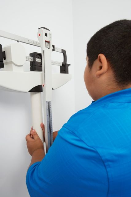
One of the most important facets of this study is the predictive nature of different patient risk factors on the future remission of cardiovascular disease symptoms.
For example, “the evidence suggests that better long-term outcomes may be anticipated among individuals undergoing MBS at lower BMI levels (i.e., less than 50),” they reported in the journal Pediatrics. “Increasing age at the time of MBS was associated with a reduced likelihood of dyslipidemia remission and normalization of hs-CRP,” which was true even in the narrow age range of this group of adolescents.
“The identification of specific predictors of CVD-RF [cardiovascular disease risk factors] normalization and/or remission on the basis of sex, race, preoperative BMI, and age at surgery may serve to improve future study design and insights regarding the optimization of treatment strategies,” wrote Dr. Michalsky and his colleagues. “Collectively, these data demonstrate a reduction in the risk for development of CVD in adulthood and offer additional, compelling support for MBS in adolescents.”
Dr. Inge has worked as a consultant for Standard Bariatrics, UpToDate, and Independent Medical Expert Consulting Services; all of these companies are unrelated to this research. John B. Dixon, PhD, has received support for his research through a National Health and Medical Research Council research fellowship. Anita Courcoulas, MD, has received grants from various health care groups and companies. All other authors had no relevant financial disclosures. The study was funded by a variety of institutional grants and the National Institutes of Health.
SOURCE: M Michalsky et al. Pediatrics. 2018 Jan 8. doi: 10.1542/peds.2017-2485.
Weight loss caused by metabolic and bariatric surgery (MBS) independently predicts the normalization of dyslipidemia, elevated blood pressure (EPB), hyperinsulinemia, diabetes, and elevated high-sensitivity C-reactive protein (hs-CRP) in severely obese adolescents, according to results of a longitudinal, multicenter prospective study.
In the study of 242 severely obese adolescents undergoing MBS between Feb. 28, 2007, and Dec. 30, 2011, Marc Michalsky, MD, of Nationwide Children’s Hospital, Columbus, Ohio, and his colleagues found that with every 10% increase in weight loss, patients were 24%, 11%, 14%, 13%, and 19% more likely to resolve dyslipidemia, EBP, hyperinsulinemia, diabetes, and elevated hs-CRP, respectively.
One of the most important facets of this study is the predictive nature of different patient risk factors on the future remission of cardiovascular disease symptoms.
For example, “the evidence suggests that better long-term outcomes may be anticipated among individuals undergoing MBS at lower BMI levels (i.e., less than 50),” they reported in the journal Pediatrics. “Increasing age at the time of MBS was associated with a reduced likelihood of dyslipidemia remission and normalization of hs-CRP,” which was true even in the narrow age range of this group of adolescents.
“The identification of specific predictors of CVD-RF [cardiovascular disease risk factors] normalization and/or remission on the basis of sex, race, preoperative BMI, and age at surgery may serve to improve future study design and insights regarding the optimization of treatment strategies,” wrote Dr. Michalsky and his colleagues. “Collectively, these data demonstrate a reduction in the risk for development of CVD in adulthood and offer additional, compelling support for MBS in adolescents.”
Dr. Inge has worked as a consultant for Standard Bariatrics, UpToDate, and Independent Medical Expert Consulting Services; all of these companies are unrelated to this research. John B. Dixon, PhD, has received support for his research through a National Health and Medical Research Council research fellowship. Anita Courcoulas, MD, has received grants from various health care groups and companies. All other authors had no relevant financial disclosures. The study was funded by a variety of institutional grants and the National Institutes of Health.
SOURCE: M Michalsky et al. Pediatrics. 2018 Jan 8. doi: 10.1542/peds.2017-2485.
Weight loss caused by metabolic and bariatric surgery (MBS) independently predicts the normalization of dyslipidemia, elevated blood pressure (EPB), hyperinsulinemia, diabetes, and elevated high-sensitivity C-reactive protein (hs-CRP) in severely obese adolescents, according to results of a longitudinal, multicenter prospective study.
In the study of 242 severely obese adolescents undergoing MBS between Feb. 28, 2007, and Dec. 30, 2011, Marc Michalsky, MD, of Nationwide Children’s Hospital, Columbus, Ohio, and his colleagues found that with every 10% increase in weight loss, patients were 24%, 11%, 14%, 13%, and 19% more likely to resolve dyslipidemia, EBP, hyperinsulinemia, diabetes, and elevated hs-CRP, respectively.
One of the most important facets of this study is the predictive nature of different patient risk factors on the future remission of cardiovascular disease symptoms.
For example, “the evidence suggests that better long-term outcomes may be anticipated among individuals undergoing MBS at lower BMI levels (i.e., less than 50),” they reported in the journal Pediatrics. “Increasing age at the time of MBS was associated with a reduced likelihood of dyslipidemia remission and normalization of hs-CRP,” which was true even in the narrow age range of this group of adolescents.
“The identification of specific predictors of CVD-RF [cardiovascular disease risk factors] normalization and/or remission on the basis of sex, race, preoperative BMI, and age at surgery may serve to improve future study design and insights regarding the optimization of treatment strategies,” wrote Dr. Michalsky and his colleagues. “Collectively, these data demonstrate a reduction in the risk for development of CVD in adulthood and offer additional, compelling support for MBS in adolescents.”
Dr. Inge has worked as a consultant for Standard Bariatrics, UpToDate, and Independent Medical Expert Consulting Services; all of these companies are unrelated to this research. John B. Dixon, PhD, has received support for his research through a National Health and Medical Research Council research fellowship. Anita Courcoulas, MD, has received grants from various health care groups and companies. All other authors had no relevant financial disclosures. The study was funded by a variety of institutional grants and the National Institutes of Health.
SOURCE: M Michalsky et al. Pediatrics. 2018 Jan 8. doi: 10.1542/peds.2017-2485.
FROM PEDIATRICS
Key clinical point:
Major finding: With every 10% increase in weight loss, patients were 24%, 11%, 14%, 13%, and 19% more likely to resolve dyslipidemia, elevated BP, hyperinsulinemia, diabetes and elevated high-sensitivity C-reactive protein, respectively.
Study details: This study was a longitudinal, multicenter prospective study of 242 severely obese adolescents undergoing metabolic and bariatric surgery between February 28, 2007 and December 30, 2011.
Disclosures: Dr. Inge has worked as a consultant for Standard Bariatrics, UpToDate, and Independent Medical Expert Consulting Services; all of these companies are unrelated to this research. John B. Dixon, PhD, has received support for his research through a National Health and Medical Research Council research fellowship. Anita Courcoulas, MD, has received grants from various healthcare groups and companies. All other authors had no relevant financial disclosures. The study was funded by a variety of institutional grants and the National Institutes of Health.
Source: M Michalsky et al. Pediatrics. 2018 Jan 8. doi: 10.1542/peds.2017-2485
Very preterm birth is linked to reduced risk of eczema
according to data from a meta-analysis of 18 studies.
Previous research suggests that low birth weight is protective against the development of atopic dermatitis, said Tingting Zhu, PhD, of West China Second University Hospital, Chengdu, and colleagues.
Preterm birth (before 37 completed weeks’ gestation) was divided into subgroups of extremely preterm (less than 28 weeks’ gestation), very preterm (28 weeks’ to less than 32 weeks’ gestation), and moderate/late preterm (32 weeks’ gestation to less than 37 weeks’ gestation).
In an analysis based on gestational age, children had a significantly reduced risk of eczema if they were very preterm (relative risk, 0.77; 95% confidence interval, 0.70-0.84, P less than .01; adjusted RR, 0.73; 95% CI, 0.64-0.82; P less than 0.01), compared with children born full term. The association between eczema and preterm birth was no longer significant among children born moderately preterm, Dr. Zhu and associates reported.
The reasons for the impact of very preterm birth on eczema are unclear, but maturation of the stratum corneum at 29-37 weeks’ gestational age could play a role, the researchers noted. Also, limited microflora in very preterm infants could affect acquiring immune tolerance and lead to reduced risk of eczema. The study was limited by several factors, including variations in gestational age and inconsistent assessments of eczema among the studies.
However, the large sample size lends strength to the results, and further studies are needed to explore how the environment, nutrition, immune system development, and skin barrier function impact the risk of eczema in very preterm infants, Dr. Zhu and associates said.
The researchers had no relevant financial disclosures. The researchers had no financial conflicts to disclose. The study was funded in part by the National Science Foundation of China, the Ministry of Health of China, and various other grants.
SOURCE: Zhu T et al. J Amer Dermatol. 2018. doi: 10.1016/j.jaad.2017.12.015.
according to data from a meta-analysis of 18 studies.
Previous research suggests that low birth weight is protective against the development of atopic dermatitis, said Tingting Zhu, PhD, of West China Second University Hospital, Chengdu, and colleagues.
Preterm birth (before 37 completed weeks’ gestation) was divided into subgroups of extremely preterm (less than 28 weeks’ gestation), very preterm (28 weeks’ to less than 32 weeks’ gestation), and moderate/late preterm (32 weeks’ gestation to less than 37 weeks’ gestation).
In an analysis based on gestational age, children had a significantly reduced risk of eczema if they were very preterm (relative risk, 0.77; 95% confidence interval, 0.70-0.84, P less than .01; adjusted RR, 0.73; 95% CI, 0.64-0.82; P less than 0.01), compared with children born full term. The association between eczema and preterm birth was no longer significant among children born moderately preterm, Dr. Zhu and associates reported.
The reasons for the impact of very preterm birth on eczema are unclear, but maturation of the stratum corneum at 29-37 weeks’ gestational age could play a role, the researchers noted. Also, limited microflora in very preterm infants could affect acquiring immune tolerance and lead to reduced risk of eczema. The study was limited by several factors, including variations in gestational age and inconsistent assessments of eczema among the studies.
However, the large sample size lends strength to the results, and further studies are needed to explore how the environment, nutrition, immune system development, and skin barrier function impact the risk of eczema in very preterm infants, Dr. Zhu and associates said.
The researchers had no relevant financial disclosures. The researchers had no financial conflicts to disclose. The study was funded in part by the National Science Foundation of China, the Ministry of Health of China, and various other grants.
SOURCE: Zhu T et al. J Amer Dermatol. 2018. doi: 10.1016/j.jaad.2017.12.015.
according to data from a meta-analysis of 18 studies.
Previous research suggests that low birth weight is protective against the development of atopic dermatitis, said Tingting Zhu, PhD, of West China Second University Hospital, Chengdu, and colleagues.
Preterm birth (before 37 completed weeks’ gestation) was divided into subgroups of extremely preterm (less than 28 weeks’ gestation), very preterm (28 weeks’ to less than 32 weeks’ gestation), and moderate/late preterm (32 weeks’ gestation to less than 37 weeks’ gestation).
In an analysis based on gestational age, children had a significantly reduced risk of eczema if they were very preterm (relative risk, 0.77; 95% confidence interval, 0.70-0.84, P less than .01; adjusted RR, 0.73; 95% CI, 0.64-0.82; P less than 0.01), compared with children born full term. The association between eczema and preterm birth was no longer significant among children born moderately preterm, Dr. Zhu and associates reported.
The reasons for the impact of very preterm birth on eczema are unclear, but maturation of the stratum corneum at 29-37 weeks’ gestational age could play a role, the researchers noted. Also, limited microflora in very preterm infants could affect acquiring immune tolerance and lead to reduced risk of eczema. The study was limited by several factors, including variations in gestational age and inconsistent assessments of eczema among the studies.
However, the large sample size lends strength to the results, and further studies are needed to explore how the environment, nutrition, immune system development, and skin barrier function impact the risk of eczema in very preterm infants, Dr. Zhu and associates said.
The researchers had no relevant financial disclosures. The researchers had no financial conflicts to disclose. The study was funded in part by the National Science Foundation of China, the Ministry of Health of China, and various other grants.
SOURCE: Zhu T et al. J Amer Dermatol. 2018. doi: 10.1016/j.jaad.2017.12.015.
FROM JOURNAL OF THE AMERICAN ACADEMY OF DERMATOLOGY
Key clinical point: Very preterm birth was associated with a significantly reduced risk of eczema, compared with full-term birth, but no difference in risk appeared between moderate preterm and full-term birth.
Major finding: Children had a significantly reduced risk of eczema if they were very preterm (RR, 0.77; 95% CI, 0.70-0.84; P less than .01; aRR 0.73, 95% CI, 0.64-0.82; P less than .01), compared with children born full term.
Data source: The data come from a meta-analysis of 18 studies.
Disclosures: The researchers had no financial conflicts to disclose. The study was funded in part by the National Science Foundation of China, the Ministry of Health of China, and various other grants.
Source: Zhu T et al. J Amer Dermatol. 2018. doi: 10.1016/j.jaad.2017.12.015.