User login
CRISPR gene editing takes next step in TTR amyloidosis
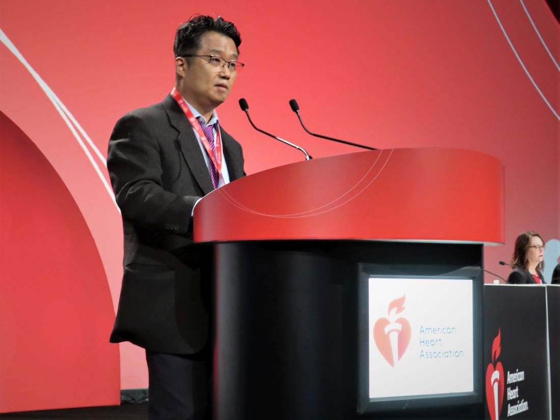
CHICAGO – Treatment with the investigational CRISPR-Cas9 gene-editing therapy, NTLA-2001, led to rapid responses in patients with transthyretin (TTR) amyloidosis with cardiomyopathy (ATTR-CM), interim phase 1 results show.
Serum levels of the disease-causing TTR protein were reduced by at least 90% at day 28 with a single infusion of NTLA-2001 at two different doses, with reductions sustained across 4-6 months’ follow-up.
NTLA-2001 was generally well-tolerated, and the results were similar in patients with New York Heart Association (NYHA) class I-III heart failure.
“These data further support and extend the early findings demonstrating the promise of CRISPR-based in vivo genome editing in humans,” said Julian Gillmore, MD, PhD, MBBS, who is leading the study at University College London.
“More specifically, the deep TTR reductions observed in patients with ATTR amyloidosis in this study provide a real possibility of genuine clinical improvement in a condition that has hitherto been ultimately progressive and invariably fatal,” he said.
The results were reported in a late-breaking session at the American Heart Association scientific sessions.
Mutations in the TTR gene and age-related changes in the stability of the TTR protein can cause misfolding of the TTR protein, resulting in amyloid deposits in skin and myocardial tissues.
An estimated 50,000 people worldwide are thought to have hereditary ATTR and up to 500,000 to have wild-type ATTR amyloidosis. Amyloid cardiomyopathy is underdiagnosed and fatal in 3-10 years without treatment. Current treatment options only slow progression and require lifelong administration, he said.
Results reported last year from the polyneuropathy arm of the study were hailed as a breakthrough and further proof-of-concept that CRISPR could be used to treat other diseases
CRISPR gene editing has shown success, for example, in beta-thalassemia and sickle cell disease but involved stem cells extracted from patients’ bone marrow, edited in the lab, and then replaced.
NTLA-2001 (Intellia Therapeutics/Regeneron) is an in vivo treatment that uses lipid nanoparticles containing messenger RNA for Cas9 and a single-guide RNA targeting TTR in the liver, where it’s almost exclusively produced.
The new analysis included 12 patients with heart failure: 3 in NYHA class I-II and 6 in NYHA class III who received a single dose of NTLA-2001 at 0.7 mg/kg, while the remaining 3 patients in NYHA class I-II received a single dose of 1.0 mg/kg.
During follow-up out to 6 months, TTR reductions averaged:
- 93% in the 0.7 mg/kg NYHA I-II group at 6 months.
- 94% in the 0.7 mg/kg NYHA III group at 4 months.
- 92% in the 1.0 mg/kg NYHA I-II group at 4 months.
Eight patients reported mild or moderate adverse events, and two patients experienced transient infusion reactions, including one grade 3 reaction in the 0.7 mg/kg NYHA class III group that resolved without clinical consequence. This group was expanded to six patients per study protocol. No additional treatment-related adverse events higher than grade 1 were reported, and no further dose escalation was undertaken, Dr. Gillmore reported.
There were no clinically relevant laboratory findings; one patient had a transient grade 1 liver enzyme elevation.
One disadvantage of CRISPR is the potential for off-target effects, but Dr. Gillmore said in an interview that the drug developers went through a “very rigorous process when selecting the guide RNA, which is what really targets the specificity of the TTR gene.”
“That’s a really, really important point,” he said. “When they did various studies using, for example, primary human hepatocytes, they found no evidence of off-target editing at concentrations of NTLA-2001 threefold greater than the EC90, the concentration at which one knocks down the protein by 90%. So, what we can say at the moment, is the specificity of NTLA-2001 for the TTR gene seems to be absolute.”
In terms of other challenges going forward, Dr. Gillmore added, “I think that it’s really to see whether the knockdown that is being achieved is going to translate into greater clinical benefit.”
Invited discussant Kevin M. Alexander, MD, of Stanford (Calif.) University, said therapies that stabilize or reduce TTR have recently emerged that have improved ATTR amyloidosis outcomes, including tafamidis and patisiran.
Nevertheless, there has been an unmet need to develop therapies that can halt or reverse disease, are effective in advanced ATTR, and have an improved route or frequency of administration, given that this is a chronic disease, he said.
Dr. Alexander noted that the reductions of greater than 90% were achieved with higher doses than used in the polyneuropathy arm reported last year but were well tolerated in patients that for the most part had wild-type ATTR (83%) and reflect the wild-type ATTR population in practice. “The data support consideration for subsequent efficacy trials for this compound.”
Unanswered questions in ongoing ATTR trials are whether TTR reductions translate into improved clinical outcomes, the long-term safety of TTR lowering, and the efficacy of NTLA-2001, particularly in higher-risk patients, such as those in NYHA class III and those with hereditary ATTR, Dr. Alexander said.
During a media briefing earlier in the day, invited discussant Kiran Musunuru, MD, University of Pennsylvania, Philadelphia, pointed out that, in the recent APOLLO-B trial of patisiran, patients with ATTR amyloidosis with cardiomyopathy had an average 87% TTR reduction but need intravenous infusions every 3 weeks for the rest of their lives.
“In contrast, gene editing is a one-and-done proposition,” he said. “You receive a single treatment that turns off the TTR gene permanently and the effects are durable and likely last a lifetime.”
Dr. Musunuru noted that patients who received patisiran also had significantly and substantially better functional capacity and quality of life, compared with those who received placebo. “Based on today’s results, we can expect future clinical trials for gene editing to have the same beneficial effects and possibly a mortality benefit as well.”
Today’s study is also important because it is part of the first wave of putting CRISPR into the body for an array of diseases, he commented.
“TTR gene editing stands out because it’s the very first CRISPR trial to show unequivocal success – you see that with a greater than 90% reduction in TTR,” Dr. Musunuru said. “So, in my view that makes it a milestone for modern medicine.”
Dosing at 55 mg, corresponding to a fixed 0.7 mg/kg dose, is ongoing in the dose-expansion portion of the trial, with enrollment across both arms expected to be completed by the end of 2022, Intellia Therapeutics reported.
The study was funded by Intellia Therapeutics and Regeneron Pharmaceuticals. Dr. Gillmore reports receiving consultancy fees from Alnylam, Ionis, AstraZeneca, Pfizer, Intellia, ATTRalus, and Novo Nordisk and has received grant support from Alnylam Pharmaceuticals. Dr. Alexander reports serving on advisory boards for Almylam and Arbor Biotechnologies; has consulted for Eidos, Ionis, Novo Nordisk, and Pfizer; and has received grants from AHA, Alnylam, Eidos, and the National Institutes of Health.
A version of this article first appeared on Medscape.com.
AT AHA 2022
No survival advantage for either torsemide or furosemide in HF: TRANSFORM-HF
CHICAGO – The choice of loop diuretic for decongestion in patients hospitalized with heart failure (HF) may make little difference to survival or readmission risk over the next year, at least when deciding between furosemide or torsemide, a randomized trial suggests.
Both drugs are old and widely used, but differences between the two loop diuretics in bioavailability, effects on potassium levels, and other features have led some clinicians to sometimes prefer torsemide. Until now, however, no randomized HF trials have compared the two drugs.
The new findings suggest clinicians can continue starting such patients with HF on either agent, at their discretion, without concern that the choice may compromise outcomes, say researchers from the TRANSFORM-HF trial, which compared furosemide-first and torsemide-first diuretic strategies in a diverse population of patients with HF.
Given that the two strategies were similarly effective for survival and rehospitalization, clinicians caring for patients with HF can focus more on “getting patients on the right dose for their loop diuretic, and prioritizing those therapies proven to improve clinical outcomes,” said Robert J. Mentz, MD, of Duke University Clinical Research Institute, Durham, N.C.
Dr. Mentz, a TRANSFORM-HF principal investigator, presented the primary results November 5 at the American Heart Association scientific sessions.
The trial had randomly assigned 2,859 patients hospitalized with HF and with a plan for oral loop diuretic therapy to initiate treatment with furosemide or torsemide. Clinicians were encouraged to maintain patients on the assigned diuretic, but crossovers to the other drug or other diuretic changes were allowed.
Rates of death from any cause, the primary endpoint, were about 26% in both groups over a median 17-month follow-up, regardless of ejection fraction (EF).
The composite rates of all-cause death or hospitalization at 12 months were also not significantly different, about 49% for those started on furosemide and about 47% for patients initially prescribed torsemide.
As a pragmatic comparative effectiveness trial, TRANSFORM-HF entered diverse patients with HF, broadly representative of actual clinical practice, who were managed according to routine practice and a streamlined study protocol at more than 60 U.S. centers, Dr. Mentz observed.
One of the pragmatic design’s advantages, he told this news organization, was “how efficient it was” as a randomized comparison of treatment strategies for clinical outcomes. It was “relatively low cost” and recruited patients quickly, compared with conventional randomized trials, “and we answered the question clearly.” The trial’s results, Dr. Mentz said, reflect “what happens in the real world.”
When might torsemide have the edge?
Although furosemide is the most commonly used loop diuretic in HF, and there are others besides it and torsemide, the latter has both known and theoretical advantages that set it apart. Torsemide is more than twice as potent as furosemide and more bioavailable, and its treatment effect lasts longer, the TRANSFORM-HF investigators have noted.
In addition, preclinical and small clinical studies suggest torsemide may have pleiotropic effects that might be theoretical advantages for patients with HF. For example, it appears to downregulate the renin-angiotensin-aldosterone system (RAAS) and reduce myocardial fibrosis and promote reverse ventricular remodeling, the group writes.
In practice, therefore, torsemide may be preferred in patients with furosemide resistance or “challenges with bioavailability, especially those with very advanced heart failure with congestion who may have gut edema, where oral furosemide and other loop diuretics are not effectively absorbed,” Biykem Bozkurt, MD, PhD, Baylor College of Medicine, Houston, told this news organization.
In such patients, she said, torsemide “is considered to be a better choice for individuals who have diuretic resistance with advanced congestion.”
The drug’s apparent pleiotropic effects, such as RAAS inhibition, may have less relevance to the TRANSFORM-HF primary endpoint of all-cause mortality than to clinical outcomes more likely associated with successful decongestion, such as HF hospitalization, Dr. Bozkurt proposed.
The trial’s pragmatic design, however, made it more feasible to focus on all-cause mortality and less practical to use measures of successful decongestion, such as volume loss or reduction in natriuretic peptide levels, she observed. Those are endpoints of special interest when diuretics are compared, “especially for the subgroup of patients who are diuretic resistant.”
Over the last 20 years or so, “we’ve learned that hospitalized heart failure is a very different disease process with a different natural history,” observed Clyde W. Yancy, MD, MSc, Northwestern University, Chicago, who was not part of the current study.
“So, the idea that something as nuanced as choice of one loop diuretic over the other, in that setting, would be sufficient to change the natural history, may be still a high bar for us,” he said in an interview.
“Based on these data, one would have to argue that whichever loop diuretic you select for the hospitalized patient – and a lot of that is driven by market exigencies right now – it turns out that the response is indistinguishable,” Dr. Yancy said. “That means if your hospital happens to have furosemide on the formulary, use it. If furosemide is not available but torsemide is available, use it.”
Dr. Yancy said he’d like to see a trial similar to TRANSFORM-HF but in outpatients receiving today’s guideline-directed medical therapy, which includes the sodium-glucose cotransporter 2 (SGLT2) inhibitors, drugs that increase the fractional excretion of sodium and have a “diureticlike” effect.
Such a trial, he said, would explore “the combination of not one, or two, but three agents with a diuretic effect – a loop diuretic, a mineralocorticoid antagonist, and an SGLT2 inhibitor – in ambulatory, optimized patients. It might make a difference.”
HF regardless of EF
The trial enrolled patients hospitalized with worsening or new-onset HF with a plan for long-term loop diuretic therapy who had either an EF of 40% or lower or, regardless of EF, elevated natriuretic peptide levels when hospitalized.
Of the 2,859 participants, whose mean age was about 65 years, about 36% were women and 34% African American. Overall, 1,428 were assigned to receive furosemide as their initial oral diuretic and 1,431 patients were assigned to the torsemide-first strategy.
The rate of death from any cause in both groups was 17 per 100 patient-years at a median of 17.4 months. The hazard ratio for torsemide vs. furosemide was 1.02 (95% confidence interval, 0.89-1.18; P = .77).
The corresponding HR at 12 months for all-cause death or hospitalization was 0.92 (95% CI, 0.83-1.02; P = .11). The relative risk for any hospitalization was 0.94 (95% CI, 0.84-1.07).
Pragmatic design: Other implications
Dosing was left to clinician discretion in the open-label study, as was whether patients maintained their assigned drug or switched over to the other agent. Indeed, 5.4% of patients crossed over to the other loop diuretic, and 2.8% went off loop diuretics entirely between in-hospital randomization and discharge, Dr. Mentz reported. By day 30, 6.7% had crossed over, and 7% had stopped taking loop diuretics.
The diuretic crossovers and discontinuations, Dr. Mentz said, likely biased the trial’s outcomes, such that the two strategies performed about equally well. Efforts were made, however, to at least partially overcome that limitation.
“We put measures in place to support adherence – sending letters to their primary doctors, giving them a wallet card so they would know which therapy they were on, having conversations about the importance of trying to stay on the randomized therapy,” Dr. Mentz said in an interview. Still, some clinicians saw differences between the two agents that prompted them, at some point, to switch patients from one loop diuretic to the other.
But interestingly, Dr. Mentz reported, the two strategies did not significantly differ in all-cause mortality or the composite of all-cause mortality or hospitalization in analysis by intention to treat.
Dr. Mentz discloses receiving honoraria from AstraZeneca, Bayer/Merck, Boehringer Ingelheim/Lilly, Cytokinetics, Pharmacosmos, Respicardia, Windtree Therapeutics, and Zoll; and research grants from American Regent and Novartis. Dr. Bozkurt discloses receiving honoraria from AstraZeneca, Baxter Health Care, and Sanofi Aventis and having other relationships with Renovacor, Respicardia, Abbott Vascular, Liva Nova, Vifor, and Cardurion. Dr. Yancy discloses a modest relationship with Abbott.
A version of this article first appeared on Medscape.com.
AT AHA 2022
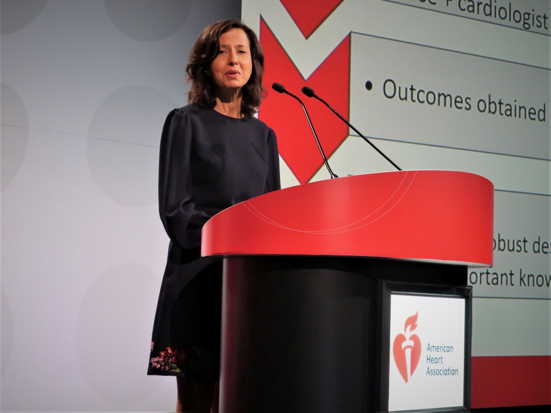
Acute heart failure risk assessment in ED improves outcomes: COACH
CHICAGO – Systematic mortality-risk assessment of patients who presented to hospital emergency departments for acute heart failure led to better patient outcomes in a controlled Canadian trial with more than 5,000 patients.
Thirty days after patients presented, the incidence of death from any cause or hospitalization for cardiovascular causes – one of two primary endpoints in the COACH study – was 12.1% among patients who underwent acute risk assessment and 14.5% in control patients who did not undergo this assessment, which translated into an adjusted, significant 12% relative risk reduction for the patients who underwent systematic assessment, Douglas S. Lee, MD, PhD, said at the American Heart Association scientific sessions.
The study’s second primary endpoint, the incidence of the same combined outcome 20 months after initial presentation, was 54.4% among the 2,480 patients assessed with the risk-assessment tool and 56.2% in the 2,972 controls, a significant, adjusted relative risk reduction of 5%.
This benefit was primarily driven by reductions in cardiovascular hospitalizations, which fell by an adjusted 16% in the intervention group compared with controls, and more specifically by hospitalizations for heart failure, which tallied a relative 20% less with the intervention. Both were significant between-group differences.
The other portion of the combined endpoint, all-cause mortality, was not significantly different between the patients who underwent the systematic emergency department assessment and the controls who were managed using usual emergency-department protocols.
Simultaneous with the report, the results also appeared online in the New England Journal of Medicine.
A pathway for early discharge and improved outcomes
“Implementation of this approach may lead to a pathway for early discharge from the hospital or emergency department, and improved patient outcomes,” said Dr. Lee, a professor at the University of Toronto, and a senior core scientist at the ICES Cardiovascular Research Program in Toronto.
“The treatment effect on the primary process outcome – patients admitted or discharged – will add useful insights into how intervention may improve,” commented Harriette Van Spall, MD, who was designated discussant for the report. The findings “fill an important knowledge gap,” added Dr. Van Spall, a cardiologist at McMaster University in Hamilton, Ont. The results “have important implications for health resource utilization,” she said.
The risk assessment tool used in the study is called the Emergency Heart failure Mortality Risk Grade for 30-day mortality (EHMRG30-ST), which was devised and validated by Dr. Lee and his associates. The assessment tool uses 11 clinical variables that include age, systolic blood pressure, heart rate, oxygen saturation, potassium and creatinine levels, and presence of ST depression on a 12-lead ECG.
The study design recommended that patients be discharged early and receive standardized transitional care as outpatients if they had a low risk of death within 7 days and within 30 days as estimated by the EHMRG30-ST. The protocol recommended that patients scored as high risk should be admitted to the hospital, and that clinicians use their clinical judgment for intermediate-risk patients but favor admission for intermediate to high risk and discharge for low to intermediate risk. The study ran at 10 hospitals in Ontario. Initially, all 10 hospitals assessed patients by usual care, and then, over time, each hospital began using the tool so that by the end of the study all 10 hospitals employed it. Among the 2,480 patients seen during the active phase, 2,442 actually underwent assessment, with 24% rated as low risk, 32% rated as intermediate risk, and 44% judged to have high risk.
The researchers also ran risk assessments retrospectively on the controls, who showed a roughly similar risk distribution, with 18% low risk, 28% intermediate risk, and 54% high risk.
The patients averaged 78 years of age, 55% were men, about 40% had diabetes, and about 64% had a prior heart failure diagnosis.
Heart failure admissions have become ‘a big deal’
Emergency department clinicians and heart failure cardiologists “have worked together for a long time” when making decisions about which patients with acute heart failure need hospital admission, commented Mary N. Walsh, MD, medical director of the heart failure and cardiac transplantation programs at Ascension St. Vincent Heart Center in Indianapolis. These decisions “became a big deal” a decade ago when the U.S. Centers for Medicare & Medicaid Services launched the Hospital Readmissions Reduction Program that began to penalize hospitals for high rates of hospital readmissions for several conditions including heart failure, she said in an interview.
“If a heart failure patient is not admitted, they can’t be readmitted,” Dr. Walsh noted.
“Many risk-assessment tools exist for patients once they are hospitalized, but these tools have not been used in emergency departments. The take-home message is that we need to start risk assessment sooner, in the emergency department,” she said.
But the specific approach tested in the COACH trial needs more study and may need further tweaking to work in the United States, where it is not clear who would pay for a program like the one tested in the trial. Canada’s unified health care payment system makes the COACH approach more financially feasible, Dr. Walsh commented.
COACH received no commercial funding. Dr. Lee, Dr. Van Spall, and Dr. Walsh had no disclosures.
AT AHA 2022
Puzzling, unique ECG from pig-to-human transplanted heart
In the first transplant of a genetically altered pig heart into a human in January, initial unexpected, prolonged ECG readings apparently did not affect the heart’s function, although the organ suddenly began to fail at day 50.
A study of these ECG changes, scheduled for presentation by Calvin Kagan, MD, and colleagues at the American Heart Association scientific sessions, offers insight into this novel operation.
As widely reported, the patient, 57-year-old David Bennett of Maryland, had end-stage heart disease and was a poor candidate for a ventricular assist device and was ineligible for a human heart, when he consented to be the first human to be transplanted with a pig heart that had a number of genes added or subtracted with the goal, in part, to prevent rejection.
The heart initially performed well after it was transplanted in an operation at the University of Maryland School of Medicine (UMSOM) in Baltimore on Jan. 7, but failed in the second month, and Mr. Bennett died on March 9.
The Food and Drug Administration had granted emergency authorization for the surgery through its expanded access (compassionate use) program, coauthor Muhammad Mohiuddin, MD, said in an interview.
“We have learned a lot and hope we can do more,” said Dr. Mohiuddin, scientific and program director of the cardiac xenotransplantation program at UMSOM.
“Suddenly on day 50, the heart started to get thicker and was not relaxing enough,” explained senior author Timm-Michael Dickfeld, MD, PhD, director of electrophysiology research at UMSOM. A biopsy revealed substantial buildup of interstitial fluid that restricted movement. The fluid was replaced by fibrous tissue, leading to irreversible damage.
Persistent, prolonged ECG parameters
In the heart from a genetically modified pig, three genes associated with antibody-mediated rejection and a gene associated with pig heart tissue growth had been inactivated and six human genes associated with immune acceptance had been added. The donor pig was supplied by Revivicor (Blacksburg, Va.).
The patient’s immunosuppressant therapy included an experimental antirejection medication (Kiniksa Pharmaceuticals; Lexington, Mass.).
The patient had daily 12-lead ECGs after the transplant.
In prior research using a pig heart transplanted into a pig body, ECG readings showed a short PR interval (50-120 ms), short QRS duration (70-90 ms) and short QT intervals (260-380 ms).
However, in the transplanted xenograft heart, the initial ECG readings showed a longer PR interval of 190 ms, QRS duration of 138 ms, and QT of 538 ms.
Prolonged intrinsic PR intervals remained stable during the postoperative course (210 ms, range 142-246 ms).
QRS duration also remained prolonged (145 ms, range 116-192 ms), but shortened during the postoperative course (days 21-40 vs. 41-60: 148 ms vs. 132 ms; P < .001).
Increased QT persisted (509 ms, range 384-650 ms) with dynamic fluctuations. The shortest QT duration was observed on day 14 (P < .001).
“In a human heart, when those parameters get longer, this can indicate signs of electrical or myocardial disease,” Dr. Dickfeld explained in a press release from the AHA.
“The QRS duration may prolong when, for example, the muscle and the electrical system itself is diseased, and that is why it takes a long time for electricity to travel from cell to cell and travel from one side of the heart to the other,” he said.
“In the human heart, the QT duration is correlated with an increased risk of abnormal heart rhythms,” he noted. “In our patient, it was concerning that the QT measure was prolonged. While we saw some fluctuations, the QT measure remained prolonged during the whole 61 days.”
‘Interesting study’
Two experts who were not involved with this research weighed in on the findings for this news organization.
“This very interesting study reinforces the difficulties in xenotransplantation, and the need for more research to be able to safely monitor recipients, as baseline values are unknown,” said Edward Vigmond, PhD.
Dr. Vigmond, from the Electrophysiology and Heart Modeling Institute at the University of Bordeaux in France, published a related study about a model of translation of pig to human electrophysiology.
The ECG is sensitive to the electrical activation pattern of the heart, along with the cellular and tissue electrical properties, he noted.
“Although pigs and humans may be similar in size, there are many differences between them,” Dr. Vigmond observed, including “the extent of the rapid conduction system of the heart, the number of nuclei in the muscle cells, the proteins in the cell membrane which control electrical activity, the orientation of the heart and thorax, and the handling of calcium inside the cell.”
“On top of this,” he continued, “donor hearts are denervated, so they no longer respond to nervous modulation, and circulating compounds in the blood which affect heart function vary between species.
“With all these differences, it is not surprising that the ECG of a pig heart transplanted into a human resembles neither that of a human nor that of a pig,” Dr. Vigmond said.
“It is interesting to note that the humanized-gene-edited porcine heart exhibited abnormal electrical conduction parameters from the outset,” said Mandeep R. Mehra, MD.
“Whether these changes were due to the gene modifications (i.e., already inherent in the pig ECG prior to transplant) or a result of the transplant operation challenges (such as the ischemia reperfusion injury and early immunological interactions) is uncertain and should be clarified,” said Dr. Mehra, of Harvard Medical School and Brigham and Women’s Medicine in Boston.
“Knowledge of these changes is important to determine whether a simple ECG parameter may be useful to identify changes that could indicate developing pathology,” Dr. Mehra added.
“In the older days of human transplantation, we often used ECG parameters such as a change in voltage amplitude to identify signals for rejection,” he continued. “Whether such changes occurred in this case could be another interesting aspect to explore as changes occurred in cardiac performance in response to the physiological and pathological challenges that were encountered in this sentinel case.”
The study authors reported having no outside sources of funding.
A version of this article first appeared on Medscape.com.
FROM AHA 2022
Diuretic agents equal to prevent CV events in hypertension: DCP
There was no difference in major cardiovascular outcomes with the use of two different diuretics – chlorthalidone or hydrochlorothiazide – in the treatment of hypertension in a new large randomized real-world study.
The Diuretic Comparison Project (DCP), which was conducted in more than 13,500 U.S. veterans age 65 years or over, showed almost identical rates of the primary composite endpoint, including myocardial infarction (MI), stroke, noncancer death, hospitalization for acute heart failure, or urgent revascularization, after a median of 2.4 years of follow-up.
There was no difference in any of the individual endpoints or other secondary cardiovascular outcomes.
However, in the subgroup of patients who had a history of MI or stroke (who made up about 10% of the study population), there was a significant reduction in the primary endpoint with chlorthalidone, whereas those without a history of MI or stroke appeared to have an increased risk for primary outcome events while receiving chlorthalidone compared with those receiving hydrochlorothiazide.
The DCP trial was presented at the American Heart Association scientific sessions by Areef Ishani, MD, director of the Minneapolis Primary Care and Specialty Care Integrated Care Community and director of the Veterans Affairs (VA) Midwest Health Care Network.
Asked how to interpret the result for clinical practice, Dr. Ishani said, “I think we can now say that either of these two drugs is appropriate to use for the treatment of hypertension.”
But he added that the decision on what to do with the subgroup of patients with previous MI or stroke was more “challenging.”
“We saw a highly significant benefit in this subgroup, but this was in the context of an overall negative trial,” he noted. “I think this is a discussion with the patients on how they want to hedge their bets. Because these two drugs are so similar, if they wanted to take one or the other because of this subgroup result I think that is a conversation to have, but I think we now need to conduct another trial specifically in this subgroup of patients to see if chlorthalidone really is of benefit in that group.”
Dr. Ishani explained that both chlorthalidone and hydrochlorothiazide have been around for more than 50 years and are considered first-line treatments for hypertension. Early studies suggested better cardiovascular outcomes and 24-hour blood pressure control with chlorthalidone, but recent observational studies have not shown more benefit with chlorthalidone. These studies have suggested that chlorthalidone may be associated with an increase in adverse events, such as hypokalemia, acute kidney injury, and chronic kidney disease.
Pragmatic study
The DCP trial was conducted to try to definitively answer this question of whether chlorthalidone is superior to hydrochlorothiazide. The pragmatic study had a “point-of-care” design that allowed participants and health care professionals to know which medication was being prescribed and to administer the medication in a real-world setting.
“Patients can continue with their normal care with their usual care team because we integrated this trial into primary care clinics,” Dr. Ishani said. “We followed participant results using their electronic health record. This study was nonintrusive, cost-effective, and inexpensive. Plus, we were able to recruit a large rural population, which is unusual for large, randomized trials, where we usually rely on big academic medical centers.”
Using VA electronic medical records, the investigators recruited primary care physicians who identified patients older than age 65 years who were receiving hydrochlorothiazide (25 mg or 50 mg) for hypertension. These patients (97% of whom were male) were then randomly assigned to continue receiving hydrochlorothiazide or to switch to an equivalent dose of chlorthalidone. Patients were followed through the electronic medical record as well as Medicare claims and the National Death Index.
Results after a median follow-up of 2.4 years showed no difference in blood pressure control between the two groups.
In terms of clinical events, the primary composite outcome of MI, stroke, noncancer death, hospitalization for acute heart failure, or urgent revascularization occurred in 10.4% of the chlorthalidone group and in 10.0% of the hydrochlorothiazide group (hazard ratio [HR], 1.04; 95% confidence interval [CI], 0.94-1.16; P = .4).
There was no difference in any individual components of the primary endpoint or the secondary outcomes of all-cause mortality, any revascularization, or erectile dysfunction.
In terms of adverse events, chlorthalidone was associated with an increase in hypokalemia (6% vs. 4.4%; HR, 1.38), but there was no difference in hospitalization for acute kidney injury.
Benefit in MI, stroke subgroup?
In the subgroup analysis, patients with a history of MI or stroke who were receiving chlorthalidone had a significant 27% reduction in the primary endpoint (HR, 0.73; 95% CI, 0.57-0.94). Conversely, patients without a history of MI or stroke appeared to do worse while taking chlorthalidone (HR, 1.12; 95% CI, 1.00-1.26).
“We were surprised by these results,” Dr. Ishani said. “We expected chlorthalidone to be more effective overall. However, learning about these differences in patients who have a history of cardiovascular disease may affect patient care. It’s best for people to talk with their health care clinicians about which of these medications is better for their individual needs.”
He added: “More research is needed to explore these results further because we don’t know how they may fit into treating the general population.”
Dr. Ishani noted that a limitations of this study was that most patients were receiving the low dose of chlorthalidone, and previous studies that suggested benefits with chlorthalidone used the higher dose.
“But the world has voted – we had 4,000 clinicians involved in this study, and the vast majority are using the low dose of hydrochlorothiazide. And this is a definitively negative study,” he said. “The world has also voted in that 10 times more patients were on hydrochlorothiazide than on chlorthalidone.”
Commenting on the study at an AHA press conference, Biykem Bozkurt, MD, PhD, Baylor College of Medicine, Houston, pointed out that in all of the landmark National Institutes of Health hypertension trials, there was a signal for benefit with chlorthalidone compared with other antihypertensives.
“We’ve always had this concept that chlorthalidone is better,” she said. “But this study shows no difference in major cardiovascular endpoints. There was more hypokalemia with chlorthalidone, but that’s recognizable as chlorthalidone is a more potent diuretic.”
Other limitations of the DCP trial are its open-label design, which could interject some bias; the enduring effects of hydrochlorothiazide – most of these patients were receiving this agent as background therapy; and inability to look at the effectiveness of decongestion of the agents in such a pragmatic study, Dr. Bozkurt noted.
She said she would like to see more analysis in the subgroup of patients with previous MI or stroke. “Does this result mean that chlorthalidone is better for sicker patients or is this result just due to chance?” she asked.
“While this study demonstrates equal effectiveness of these two diuretics in the targeted population, the question of subgroups of patients for which we use a more potent diuretic I think remains unanswered,” she concluded.
Designated discussant of the DCP trial at the late-breaking trial session, Daniel Levy, MD, director of the Framingham Heart Study at the National Heart, Lung, and Blood Institute, reminded attendees that chlorthalidone had shown impressive results in previous important hypertension studies including SHEP and ALLHAT.
He said the current DCP was a pragmatic study addressing a knowledge gap that “would never have been performed by industry.”
Dr. Levy concluded that the results showing no difference in outcomes between the two diuretics were “compelling,” although a few questions remain.
These include a possible bias toward hydrochlorothiazide – patients were selected who were already taking that drug and so would have already had a favorable response to it. In addition, because the trial was conducted in an older male population, he questioned whether the results could be generalized to women and younger patients.
The DCP study was funded by the VA Cooperative Studies Program. Dr. Ishani reported no disclosures.
A version of this article first appeared on Medscape.com.
FROM AHA 2022
Triglyceride-lowering fails to show CV benefit in large fibrate trial
Twenty-five percent reduction has no effect
CHICAGO – Despite a 25% reduction in triglycerides (TGs) along with similar reductions in very-low-density lipoprotein (VLDL), and remnant cholesterol, a novel agent failed to provide any protection in a multinational trial against a composite endpoint of major adverse cardiovascular events (MACE) in patients with type 2 diabetes.
“Our data further highlight the complexity of lipid mediators of residual risk among patients with insulin resistance who are receiving statin therapy,” reported Aruna Das Pradhan, MD, of Harvard Medical School, Boston, and Queen Mary University, London.

It is the most recent in a series of trials that have failed to associate a meaningful reduction in TGs with protection from a composite MACE endpoint. This is a pattern that dates back 20 years, even though earlier trials did suggest that hypertriglyceridemia was a targetable risk factor.
No benefit from fibrates seen in statin era
“We have not seen a significant cardiovascular event reduction with a fibrate in the statin era,” according to Karol Watson, MD, PhD, director of the UCLA Women’s Cardiovascular Health Center, Los Angeles.

In the statin era, which began soon after the Helsinki Heart Study was published in 1987, Dr. Watson counted at least five studies with fibrates that had a null result.
In the setting of good control of LDL cholesterol, “fibrates have not been shown to further lower CV risk,” said Dr. Watson, who was invited by the AHA to discuss the PROMINENT trial.
In PROMINENT, 10,497 patients with type 2 diabetes were randomized to pemafibrate, a peroxisome proliferator-activated receptor a (PPAR-a) agonist, or placebo. Pemafibrate is not currently available in North America or Europe, but it is licensed in Japan for the treatment of hypertriglyceridemia.
The primary efficacy endpoint of the double-blind trial was a composite endpoint of nonfatal myocardial infarction, ischemic stroke, coronary revascularization, or death.
The patients were eligible if they had TG levels from 200 to 400 mg/dL and HDL cholesterol levels of 40 mg/dL or below. Pemafibrate in a dose of 0.2 mg or placebo were taken twice daily. About two-thirds had a prior history of coronary heart disease. The goal was primary prevention in the remainder.
After a median follow-up of 3.4 years when the study was stopped for futility, the proportion of patients reaching a primary endpoint was slightly greater in the experimental arm (3.60 vs. 3.51 events per 100 patient-years). The hazard ratio, although not significant, was nominally in favor of placebo (hazard ratio, 1.03; P = .67).
When events within the composite endpoint were assessed individually, there was no signal of benefit for any outcome. The rates of death from any cause, although numerically higher in the pemafibrate group (2.44 vs. 2.34 per 100 patient years), were also comparable.
Lipid profile improved as predicted
Yet, in regard to an improvement in the lipid profile, pemafibrate performed as predicted. When compared to placebo 4 months into the trial, pemafibrate was associated with median reductions of 26.2% in TGs, 25.8% in VLDL, and 25.6% in remnant cholesterol, which is cholesterol transported in TG-rich lipoproteins after lipolysis and lipoprotein remodeling.
Furthermore, pemafibrate was associated with a median 27.6% reduction relative to placebo in apolipoprotein C-III and a median 4.8% reduction in apolipoprotein E, all of which would be expected to reduce CV risk.
The findings of PROMINENT were published online in the New England Journal of Medicine immediately after their presentation.
The findings of this study do not eliminate any hope for lowering residual CV risk with TG reductions, but they do suggest the relationship with other lipid subfractions is complex, according to Salim S. Virani, MD, PhD, a professor of cardiology at Baylor College of Medicine, Houston.
“I think that the lack of efficacy despite TG lowering may be largely due to a lack of an overall decrease in the apolipoprotein B level,” speculated Dr. Virani, who wrote an editorial that accompanied publication of the PROMINENT results.
He noted that pemafibrate is implicated in converting remnant cholesterol to LDL cholesterol, which might be one reason for a counterproductive effect on CV risk.
“In order for therapies that lower TG levels to be effective, they probably have to have mechanisms to increase clearance of TG-rich remnant lipoprotein cholesterol particles rather than just converting remnant lipoproteins to LDL,” Dr. Virani explained in an attempt to unravel the interplay of these variables.
Although this study enrolled patients “who would be predicted to have the most benefit from a TG-lowering strategy,” Dr. Watson agreed that these results do not necessarily extend to other means of lowering TG. However, it might draw into question the value of pemafibrate and perhaps other drugs in this class for treatment of hypertriglyceridemia. In addition to a lack of CV benefit, treatment was not without risks, including a higher rate of thromboembolism and adverse renal events.
Dr. Das Pradhan reported financial relationships with Denka, Medtelligence, Optum, Novo Nordisk, and Kowa, which provided funding for this trial. Dr. Watson reported financial relationships with Amarin, Amgen, Boehringer-Ingelheim, and Esperion.
Twenty-five percent reduction has no effect
Twenty-five percent reduction has no effect
CHICAGO – Despite a 25% reduction in triglycerides (TGs) along with similar reductions in very-low-density lipoprotein (VLDL), and remnant cholesterol, a novel agent failed to provide any protection in a multinational trial against a composite endpoint of major adverse cardiovascular events (MACE) in patients with type 2 diabetes.
“Our data further highlight the complexity of lipid mediators of residual risk among patients with insulin resistance who are receiving statin therapy,” reported Aruna Das Pradhan, MD, of Harvard Medical School, Boston, and Queen Mary University, London.
It is the most recent in a series of trials that have failed to associate a meaningful reduction in TGs with protection from a composite MACE endpoint. This is a pattern that dates back 20 years, even though earlier trials did suggest that hypertriglyceridemia was a targetable risk factor.
No benefit from fibrates seen in statin era
“We have not seen a significant cardiovascular event reduction with a fibrate in the statin era,” according to Karol Watson, MD, PhD, director of the UCLA Women’s Cardiovascular Health Center, Los Angeles.
In the statin era, which began soon after the Helsinki Heart Study was published in 1987, Dr. Watson counted at least five studies with fibrates that had a null result.
In the setting of good control of LDL cholesterol, “fibrates have not been shown to further lower CV risk,” said Dr. Watson, who was invited by the AHA to discuss the PROMINENT trial.
In PROMINENT, 10,497 patients with type 2 diabetes were randomized to pemafibrate, a peroxisome proliferator-activated receptor a (PPAR-a) agonist, or placebo. Pemafibrate is not currently available in North America or Europe, but it is licensed in Japan for the treatment of hypertriglyceridemia.
The primary efficacy endpoint of the double-blind trial was a composite endpoint of nonfatal myocardial infarction, ischemic stroke, coronary revascularization, or death.
The patients were eligible if they had TG levels from 200 to 400 mg/dL and HDL cholesterol levels of 40 mg/dL or below. Pemafibrate in a dose of 0.2 mg or placebo were taken twice daily. About two-thirds had a prior history of coronary heart disease. The goal was primary prevention in the remainder.
After a median follow-up of 3.4 years when the study was stopped for futility, the proportion of patients reaching a primary endpoint was slightly greater in the experimental arm (3.60 vs. 3.51 events per 100 patient-years). The hazard ratio, although not significant, was nominally in favor of placebo (hazard ratio, 1.03; P = .67).
When events within the composite endpoint were assessed individually, there was no signal of benefit for any outcome. The rates of death from any cause, although numerically higher in the pemafibrate group (2.44 vs. 2.34 per 100 patient years), were also comparable.
Lipid profile improved as predicted
Yet, in regard to an improvement in the lipid profile, pemafibrate performed as predicted. When compared to placebo 4 months into the trial, pemafibrate was associated with median reductions of 26.2% in TGs, 25.8% in VLDL, and 25.6% in remnant cholesterol, which is cholesterol transported in TG-rich lipoproteins after lipolysis and lipoprotein remodeling.
Furthermore, pemafibrate was associated with a median 27.6% reduction relative to placebo in apolipoprotein C-III and a median 4.8% reduction in apolipoprotein E, all of which would be expected to reduce CV risk.
The findings of PROMINENT were published online in the New England Journal of Medicine immediately after their presentation.
The findings of this study do not eliminate any hope for lowering residual CV risk with TG reductions, but they do suggest the relationship with other lipid subfractions is complex, according to Salim S. Virani, MD, PhD, a professor of cardiology at Baylor College of Medicine, Houston.
“I think that the lack of efficacy despite TG lowering may be largely due to a lack of an overall decrease in the apolipoprotein B level,” speculated Dr. Virani, who wrote an editorial that accompanied publication of the PROMINENT results.
He noted that pemafibrate is implicated in converting remnant cholesterol to LDL cholesterol, which might be one reason for a counterproductive effect on CV risk.
“In order for therapies that lower TG levels to be effective, they probably have to have mechanisms to increase clearance of TG-rich remnant lipoprotein cholesterol particles rather than just converting remnant lipoproteins to LDL,” Dr. Virani explained in an attempt to unravel the interplay of these variables.
Although this study enrolled patients “who would be predicted to have the most benefit from a TG-lowering strategy,” Dr. Watson agreed that these results do not necessarily extend to other means of lowering TG. However, it might draw into question the value of pemafibrate and perhaps other drugs in this class for treatment of hypertriglyceridemia. In addition to a lack of CV benefit, treatment was not without risks, including a higher rate of thromboembolism and adverse renal events.
Dr. Das Pradhan reported financial relationships with Denka, Medtelligence, Optum, Novo Nordisk, and Kowa, which provided funding for this trial. Dr. Watson reported financial relationships with Amarin, Amgen, Boehringer-Ingelheim, and Esperion.
AT AHA 2022
ACC/AHA issues updated guidance on aortic disease
focusing on surgical intervention considerations, consistent imaging practices, genetic and familial screenings, and the importance of multidisciplinary care.
“There has been a host of new evidence-based research available for clinicians in the past decade when it comes to aortic disease. It was time to reevaluate and update the previous, existing guidelines,” Eric M. Isselbacher, MD, MSc, chair of the writing committee, said in a statement.
“We hope this new guideline can inform clinical practices with up-to-date and synthesized recommendations, targeted toward a full multidisciplinary aortic team working to provide the best possible care for this vulnerable patient population,” added Dr. Isselbacher, codirector of the Thoracic Aortic Center at Massachusetts General Hospital, Boston.
The 2022 ACC/AHA Guideline for the Diagnosis and Management of Aortic Disease was simultaneously published online in the Journal of the American College of Cardiology and Circulation.
The new guideline replaces the 2010 ACCF/AHA Guidelines for the Diagnosis and Management of Patients With Thoracic Aortic Disease and the 2015 Surgery for Aortic Dilation in Patients With Bicuspid Aortic Valves: A Statement of Clarification From the ACC/AHA Task Force on Clinical Practice Guidelines.
The new guideline is intended to be used with the 2020 ACC/AHA Guideline for the Management of Patients With Valvular Heart Disease.
It brings together guidelines for both the thoracic and abdominal aorta and is targeted to cardiovascular clinicians involved in the care of people with aortic disease, including general cardiovascular care clinicians and emergency medicine clinicians, the writing group says.
Among the key recommendations in the new guideline are the following:
- Screen first-degree relatives of individuals diagnosed with aneurysms of the aortic root or ascending thoracic aorta, or those with aortic dissection to identify individuals most at risk for aortic disease. Screening would include genetic testing and imaging.
- Be consistent in the way CT or MRI are obtained and reported; in the measurement of aortic size and features; and in how often images are used for monitoring before and after repair surgery or other intervention. Ideally, all surveillance imaging for an individual should be done using the same modality and in the same lab, the guideline notes.
- For individuals who require aortic intervention, know that outcomes are optimized when surgery is performed by an experienced surgeon working in a multidisciplinary aortic team. The new guideline recommends “a specialized hospital team with expertise in the evaluation and management of aortic disease, in which care is delivered in a comprehensive, multidisciplinary manner.”
- At centers with multidisciplinary aortic teams and experienced surgeons, the threshold for surgical intervention for sporadic aortic root and ascending aortic aneurysms has been lowered from 5.5 cm to 5.0 cm in select individuals, and even lower in specific scenarios among patients with heritable thoracic aortic aneurysms.
- In patients who are significantly smaller or taller than average, surgical thresholds may incorporate indexing of the aortic root or ascending aortic diameter to either patient body surface area or height, or aortic cross-sectional area to patient height.
- Rapid aortic growth is a risk factor for rupture and the definition for rapid aneurysm growth rate has been updated. Surgery is now recommended for patients with aneurysms of aortic root and ascending thoracic aorta with a confirmed growth rate of ≥ 0.3 cm per year across 2 consecutive years or ≥ 0.5 cm in 1 year.
- In patients undergoing aortic root replacement surgery, valve-sparing aortic root replacement is reasonable if the valve is suitable for repair and when performed by experienced surgeons in a multidisciplinary aortic team.
- Patients with acute type A aortic dissection, if clinically stable, should be considered for transfer to a high-volume aortic center to improve survival. The operative repair of type A aortic dissection should entail at least an open distal anastomosis rather than just a simple supracoronary interposition graft.
- For management of uncomplicated type B aortic dissection, there is an increasing role for . Clinical trials of repair of thoracoabdominal aortic aneurysms with endografts are reporting results that suggest endovascular repair is an option for patients with suitable anatomy.
- Shared decision-making between the patient and multidisciplinary aortic team is highly encouraged, especially when the patient is on the borderline of thresholds for repair or eligible for different types of surgical repair.
- Shared decision-making should also be used with individuals who are pregnant or may become pregnant to consider the risks of pregnancy in individuals with aortic disease.
The guideline was developed in collaboration with and endorsed by the American Association for Thoracic Surgery, the American College of Radiology, the Society of Cardiovascular Anesthesiologists, the Society for Cardiovascular Angiography and Interventions, the Society of Thoracic Surgeons, and the Society for Vascular Medicine.
It has been endorsed by the Society of Interventional Radiology and the Society for Vascular Surgery.
A version of this article first appeared on Medscape.com.
FROM CIRCULATION
A 95-year-old White male with hypertension presented with itchy patches and bullae on the trunk and extremities
and is associated with various predisposing factors, including HLA genes, comorbidities, aging, and trigger factors such as drugs, trauma, radiation, chemotherapy, and infections. The autoimmune reaction is mediated by a dysregulation of T cells in which IgG and IgE autoantibodies form against hemidesmosomal proteins (BP180 and BP230). These autoantibodies induce neutrophil activation, recruitment, and degradation in the basement membrane of the skin.
Typically, patients present with intense pruritus followed by an urticarial or eczematous eruption. Tense blisters and bullae occur commonly on the trunk and extremities. Drug-associated bullous pemphigoid (DABP) is a common manifestation of the disease with histologic and immunologic features similar to those of the idiopathic version. Eruptions can be triggered by systemic or topical medications, and incidence of these reactions may be related to a genetic predisposition for the disease.
Some research suggests that drug-induced changes to the antigenic properties of the epidermal basement membrane result in an augmented immune response, while others point to structural modification in these zones that stimulate the immune system. Thiol- and phenol-based drugs have been largely implicated in the development of DABP because they are capable of structural modification and disruption of the dermo-epidermal junction in the basement membrane.
DABP often presents with patients taking multiple medications. Some of the most common medications are gliptins, PD-1 inhibitors, diuretics, antibiotics, anti-inflammatory drugs, and ACE-inhibitors, and other cardiovascular drugs. DABP may present with mucosal eruptions unlike its idiopathic counterpart that is mostly contained to the skin.

On this patient, two punch biopsies were taken. Histopathology revealed an eosinophil-rich subepidermal blister with a smooth epidermal undersurface consistent with bullous pemphigoid. Direct immunofluorescence was positive with a deposition of IgG and C3 at the epidermal side of salt split basement membrane zone.
Treatment for BP includes high potency topical and systemic steroids. Tetracyclines and niacinamide have been reported to improve the condition. Treatment is tailored to allow for cutaneous healing and control pruritus, but the physician must be mindful of the patient’s comorbidities and capacity for self-care. Prognosis is often better for DABP as withdrawal of the medication greatly accelerates clearance of the lesions. Worse prognosis is related to increased number of comorbidities and older age. Our patient’s BP is controlled currently with topical steroids and oral doxycycline.
This case and photo were submitted by Lucas Shapiro, BS, Nova Southeastern University College of Osteopathic Medicine, Tampa, and Dr. Bilu Martin.
Dr. Bilu Martin is a board-certified dermatologist in private practice at Premier Dermatology, MD, in Aventura, Fla. More diagnostic cases are available at mdedge.com/dermatology. To submit a case for possible publication, send an email to dermnews@mdedge.com.
References
1. Miyamoto D et al. An Bras Dermatol. 2019 Mar-Apr;94(2):133-46.
2. Moro et al. Biomolecules. 2020 Oct 10;10(10):1432.
3. Verheyden M et al. Acta Derm Venereol. 2020 Aug 17;100(15):adv00224.
and is associated with various predisposing factors, including HLA genes, comorbidities, aging, and trigger factors such as drugs, trauma, radiation, chemotherapy, and infections. The autoimmune reaction is mediated by a dysregulation of T cells in which IgG and IgE autoantibodies form against hemidesmosomal proteins (BP180 and BP230). These autoantibodies induce neutrophil activation, recruitment, and degradation in the basement membrane of the skin.
Typically, patients present with intense pruritus followed by an urticarial or eczematous eruption. Tense blisters and bullae occur commonly on the trunk and extremities. Drug-associated bullous pemphigoid (DABP) is a common manifestation of the disease with histologic and immunologic features similar to those of the idiopathic version. Eruptions can be triggered by systemic or topical medications, and incidence of these reactions may be related to a genetic predisposition for the disease.
Some research suggests that drug-induced changes to the antigenic properties of the epidermal basement membrane result in an augmented immune response, while others point to structural modification in these zones that stimulate the immune system. Thiol- and phenol-based drugs have been largely implicated in the development of DABP because they are capable of structural modification and disruption of the dermo-epidermal junction in the basement membrane.
DABP often presents with patients taking multiple medications. Some of the most common medications are gliptins, PD-1 inhibitors, diuretics, antibiotics, anti-inflammatory drugs, and ACE-inhibitors, and other cardiovascular drugs. DABP may present with mucosal eruptions unlike its idiopathic counterpart that is mostly contained to the skin.
On this patient, two punch biopsies were taken. Histopathology revealed an eosinophil-rich subepidermal blister with a smooth epidermal undersurface consistent with bullous pemphigoid. Direct immunofluorescence was positive with a deposition of IgG and C3 at the epidermal side of salt split basement membrane zone.
Treatment for BP includes high potency topical and systemic steroids. Tetracyclines and niacinamide have been reported to improve the condition. Treatment is tailored to allow for cutaneous healing and control pruritus, but the physician must be mindful of the patient’s comorbidities and capacity for self-care. Prognosis is often better for DABP as withdrawal of the medication greatly accelerates clearance of the lesions. Worse prognosis is related to increased number of comorbidities and older age. Our patient’s BP is controlled currently with topical steroids and oral doxycycline.
This case and photo were submitted by Lucas Shapiro, BS, Nova Southeastern University College of Osteopathic Medicine, Tampa, and Dr. Bilu Martin.
Dr. Bilu Martin is a board-certified dermatologist in private practice at Premier Dermatology, MD, in Aventura, Fla. More diagnostic cases are available at mdedge.com/dermatology. To submit a case for possible publication, send an email to dermnews@mdedge.com.
References
1. Miyamoto D et al. An Bras Dermatol. 2019 Mar-Apr;94(2):133-46.
2. Moro et al. Biomolecules. 2020 Oct 10;10(10):1432.
3. Verheyden M et al. Acta Derm Venereol. 2020 Aug 17;100(15):adv00224.
A 95-year-old White male with hypertension presented with a history of very itchy patches and bullae on the trunk and extremities.
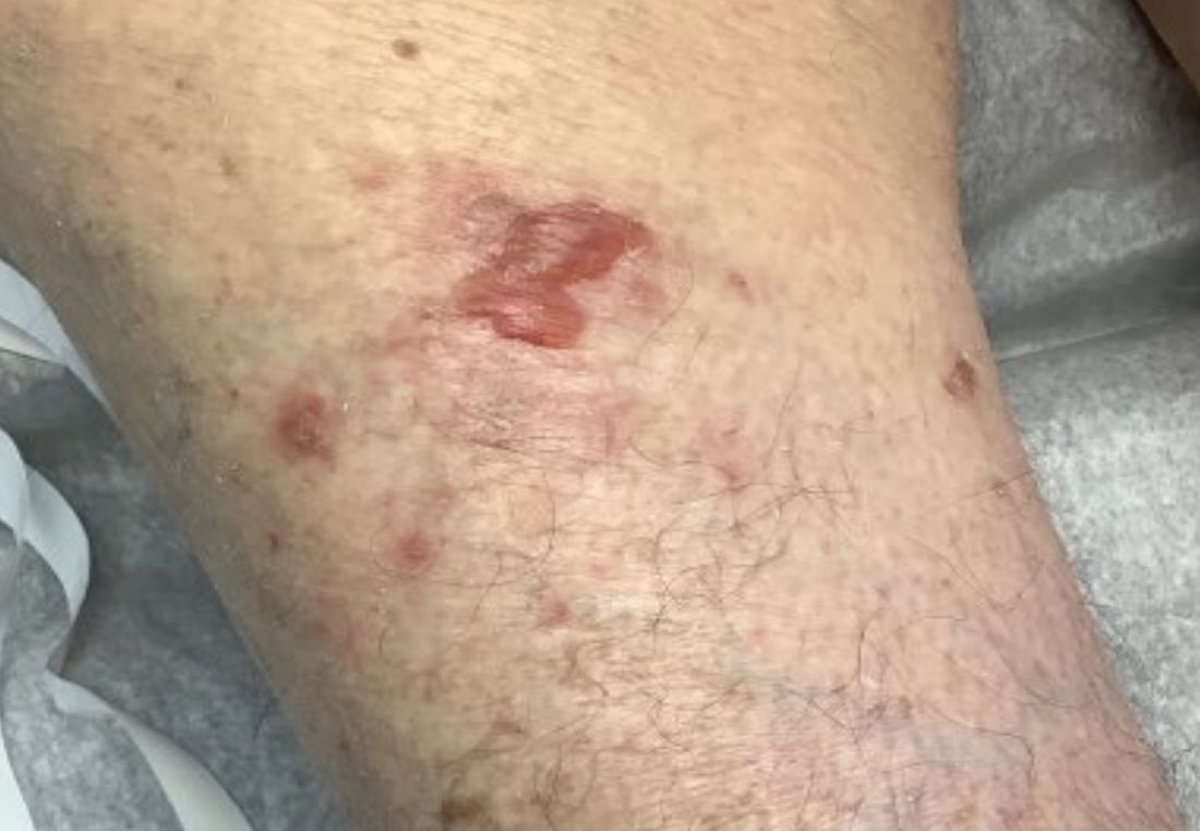
Combo thrombolytic approach fails to reduce ICH in stroke
A study evaluating a new approach using a combination of two thrombolytics designed to reduce bleeding risk in patients with acute ischemic stroke has not shown any benefit on the primary outcome of all intracranial hemorrhage (ICH).
However, there were some encouraging findings including a trend towards a reduction in symptomatic ICH, researchers report, and the combination approach did not show any depletion of fibrinogen levels, which suggests a potential lower bleeding risk.
“Although the main results of this study are neutral, we are encouraged that the combination approach with a low dose of alteplase followed by the new mutant pro-urokinase product looked as effective as full-dose alteplase alone, and there were some promising signs signaling a potential lower bleeding risk,” senior investigator, Diederik Dippel, MD, Erasmus University Medical Center, Rotterdam, the Netherlands, told this news organization.
The DUMAS study (Dual Thrombolytic Therapy With Mutant Pro-Urokinase and Low Dose Alteplase for Ischemic Stroke) was presented at the World Stroke Congress in Singapore by study coauthor Nadinda van der Ende, MD, also from Erasmus University Medical Center.
She pointed out that thrombolysis with intravenous alteplase increases the likelihood of a good outcome in acute ischemic stroke but can cause symptomatic intracranial hemorrhage, which can be associated with death and major disability.
Mutant pro-urokinase is a new thrombolytic agent, in development by Thrombolytic Science, Cambridge, Mass., formed by changing one amino acid in pro-urokinase to make it more stable. It is more fibrin specific than alteplase and therefore believed to have a lower risk of intracranial hemorrhage.
Fibrin is formed as the last step in the clotting process, and the precursor of fibrin in the blood is fibrinogen, Dr. van der Ende noted. Alteplase depletes fibrinogen, contributing to its increased bleeding risk, but mutant pro-urokinase is not believed to affect fibrinogen.
“Mutant pro-urokinase does not bind to intact fibrin. It only binds to fibrin that has already been primed by alteplase,” she explained.
The hypothesis behind the current study is that giving a small dose of alteplase will break down fibrin in the clot enough to expose the binding sites for mutant pro-urokinase, which can then be given to continue to lyse the clot.
As alteplase has a short half-life, it disappears quickly, and new fibrin is not affected. As mutant pro-urokinase can only lyse fibrin that is primed with alteplase, new hemostatic clots should stay intact. Animal studies have shown less bleeding from distant sites with this approach, Dr. van der Ende said.
The primary analysis of the phase 2 DUMAS study included 238 patients with mild ischemic stroke (median National Institutes of Health Stroke Scale [NIHSS] score 3) who met the standard criteria for IV alteplase.
They were randomized to alteplase alone at the regular dose of 0.9 mg/kg (max 90 mg) with a 10% bolus and the remaining given over 60 minutes; or to a combination of a 5-mg bolus of IV alteplase followed by mutant pro-urokinase at a dose of 40 mg given over 60 minutes.
The primary outcome was the rate of all intracranial hemorrhage (symptomatic and asymptomatic) detected by neuroimaging.
This occurred in 14% of patients in the full-dose alteplase group vs. 13% of patients in the combined alteplase/mutant pro-urokinase group, a nonsignificant difference: adjusted odds ratio, 0.99 (95% confidence interval, 0.46-2.14).
Secondary outcomes showed no significant differences in NIHSS scores at 24 hours or 5-7 days; functional outcome as measured by a shift analysis of the Modified Rankin Scale (mRS); final infarct volume; or perfusion deficit.
However, blood fibrinogen levels were not depleted and significantly higher in the alteplase/mutant pro-urokinase group than in the full-dose alteplase alone group.
In terms of safety, symptomatic ICH occurred in three patients in the alteplase group (3%) and in none (0%) in the combined alteplase/mutant pro-urokinase group; death occurred in 4% vs. 2% patients respectively; and major extracranial hemorrhage occurred in 1% in both groups.
Dr. Van der Ende concluded that the study showed an overall low rate of ICH; a combination of alteplase and mutant pro-urokinase was not superior to alteplase alone in reducing ICH rates in this population of patients with minor stroke; and mutant pro-urokinase appeared to be safe and, unlike alteplase, did not show any reduction in fibrinogen levels.
“We think the lack of an effect on fibrinogen with this new combination of a small alteplase bolus followed by mutant pro-urokinase infusion is promising,” Dr. Dippel commented. “The fact that there was no symptomatic ICH with the combination treatment is also encouraging. Although the primary endpoint of this trial was neutral, we still believe this is a very interesting approach, with the potential for reduced bleeding, compared with alteplase alone, but we need larger numbers to see an effect on outcomes.”
Dr. Dippel also pointed out that the study included only patients with minor stroke who were not eligible for endovascular therapy, and these patients have a low risk of a poor outcome and a low bleeding risk.
They are hoping to do another study in patients with more severe stroke, who have a higher bleeding risk and would have more to gain from this combination approach.
Because many patients with severe stroke now have immediate thrombectomy if they present to a comprehensive stroke center, a trial in severe stroke patients would have to be done in primary stroke centers, so if the patents are referred to thrombectomy, the thrombolytic would have a chance to work, Dr. Dippel added.
Commenting on the study for this news organization, Stefan Kiechl, MD, Medical University of Innsbruck (Austria), who is cochair of the World Stroke Congress scientific committee, said, “Alteplase is not fibrin specific, and also causes a degeneration of fibrinogen, which results in ‘fibrinogen depletion coagulopathy.’ It is assumed that 20%-40% of intracerebral bleeding after thrombolysis with alteplase is caused by this problem. DUMAS tests the combination of a substantially reduced alteplase [5 mg] dose plus mutant pro-urokinase to avoid this problem.”
The new thrombolysis protocol, however, did not result in a lower bleeding risk, compared to the comparator alteplase,” he added. “The main limitation of this study is that mainly patients with minor strokes were included. Patients with moderate and severe strokes, who have a substantial risk of bleeding, were not adequately addressed.”
The DUMAS trial was funded by an unrestricted grant from Thrombolytic Science, paid to the institution. Dr. Van der Ende and Dr. Dippel report no relevant disclosures.
A version of this article first appeared on Medscape.com.
FROM WSC 2022
Uptake of high-sensitivity troponin assays lags in U.S. hospitals
Most hospitals in the United States have yet to transition from conventional to high-sensitivity cardiac troponin (hs-cTn) assays, despite their greater sensitivity for myocardial injury, a new National Cardiovascular Data Registry (NCDR) registry study indicates.
hs-cTn assays have been used in routine clinical practice in Europe, Canada, and Australia since 2010, but the first such assay did not gain approval in the United States until 2017. Although single-center studies have examined their efficacy and potential downstream consequences, few data exist on hs-cTn implementation nationally, explained study author Cian McCarthy, MB, BCh, BAO, Massachusetts General Hospital, Boston.
The results were published online in the Journal of the American College of Cardiology and will be presented Nov. 5 at the American Heart Association scientific sessions.
For the study, Dr. McCarthy and colleagues examined 550 hospitals participating in the NCDR Chest Pain-MI registry from January 2019 through September 2021.
Of the 251,000 patients included in the analysis (mean age, 64 years; 41.5% female), 155,049 had a non–ST-segment myocardial infarction (NSTEMI), 15,989 had unstable angina, and 79,962 had low-risk chest pain.
The hs-cTn assays included Roche Diagnostic’s Elecsys Gen5 STAT troponin T assay (23%); Abbott’s ARCHITECT STAT (17%); Beckman Coulter’s ACCESS (21%); and Siemens’ Atellica IM (18%), Dimension VISTA (14%), Dimension EXL (4%), and ADVIA Centaur (2%) troponin I assays.
During the study period, 11.5% of patients were evaluated with hs-cTn assays and the remainder were evaluated with conventional troponin assays. These patients were slightly older (65.0 vs. 64.0 years), more commonly White (83.1% vs. 79.9%), less likely to be of Hispanic or Latino ethnicity (8.9% vs. 10.0%), and less likely to be uninsured (6.8% vs. 8.3%; P for all < .001).
A slightly higher proportion of patients evaluated with hs-cTn assays were diagnosed with unstable angina (7.1% vs. 6.3%), a lower proportion with NSTEMI (61.1% vs. 61.9%), and a similar proportion with low-risk chest pain (31.8% vs. 31.9%) compared with those evaluated by conventional troponin assays.
Implementation, defined as at least 25% of patients evaluated by hs-cTn in each quarter, increased from 3.3% in the first quarter of 2019 to 32.6% in the third quarter of 2021 (P trend < .001).
Using higher implementation thresholds of at least 50% and 75% of patients evaluated by hs-cTn, the prevalence in 2021 was 28.9% and 24.7%, respectively.
“So still, the majority of the hospitals by the end of the third quarter 2021 were not using these assays,” Dr. McCarthy said.
Potential explanations for the slow uptake are that prospective comparative effectiveness trials of These assays have predominantly been in international populations and real-world data on U.S. implementation have been limited to integrated health networks at academic institutions.
Approval of several assays was also delayed and the study data cut off just before the October publication of the 2021 AHA/ACC Chest Pain guideline. “So, whether the chest pain guideline with the new class 1 recommendation for hs-cTn will lead to further uptake is something that will need to be looked at in the future,” he said.
Downstream testing
In adjusted analyses, hs-cTn use was associated with more echocardiography among patients with non-ST elevation–acute coronary syndrome (NSTE-ACS) (82.4% vs. 75.0%; odds ratio [OR], 1.43; 95% confidence interval [CI], 1.19-1.73), but not among those with low-risk chest pain (19.7% vs. 19.4%; OR, 0.93; 95% CI, 0.71-1.22) compared with conventional cTn assays.
Importantly, hs-cTn was not associated with a difference in stress testing or CT coronary angiography utilization.
Use of hs-cTn was associated with lower use of invasive coronary angiography among patients with low-risk chest pain (3.7% vs. 4.5%; OR, 0.73, 95% CI, 0.58-0.92) but similar use for NSTE-ACS (96.3% vs. 95.8%; OR, 0.99, 95% CI, 0.82-1.19).
Among patients with NSTE-ACS, there also was no difference in revascularization with percutaneous coronary intervention (PCI) (52.7% vs. 52.3%; OR, 0.99; 95% CI, 0.94-1.04) or coronary bypass graft surgery (9.4% vs. 9.1%; OR, 1.06; 95% CI, 0.94-1.18).
PCI (0.1% vs. 0.2%; P = .05) and bypass graft surgery (both 0.1%) were uncommon among patients with low-risk chest pain.
In-hospital mortality was similar among patients with low-risk chest pain evaluated using hs-cTn assays vs. conventional troponin assays (0% vs. 0.02%; P = .16) and among patients with NSTE-ACS (2.8% vs. 3.2%; OR, 0.98, 95% CI, 0.87-1.11).
Length of stay was slightly shorter with hs-cTn use for patients with low-risk chest pain (median, 5.8 vs. 6.2 hours; P < .001) and patients with NSTE-ACS (66.9 vs. 67.8 hours; P = .01).
“There was always a concern that maybe high-sensitivity cardiac troponin would dramatically increase testing and could even increase length of stay, but I think these data are reassuring, in that this study suggests high-sensitivity cardiac troponin is associated with a small reduction in length of stay and possibly more appropriate use of testing with echocardiography in STEMI and a reduction in invasive angiography in low-risk patients,” Dr. McCarthy said. “But the majority of hospitals haven’t implemented the assay.”
The authors pointed out that because registry entry of patients with low-risk chest pain and unstable angina is optional for participating sites, the percentage of patients with NSTEMI is higher than in typical chest pain analyses. This higher pretest probability for MI may thus affect post-test accuracy for a true positive result. “That stated, this is the exact scenario where higher sensitivity might be associated with favorable impact on utilization.”
Among other limitations: There was the potential for unmeasured confounders, the accuracy of diagnoses could not be confirmed, patients with type 2 MI were excluded from the registry, and post-discharge safety was not assessed.
“These data indicate further opportunities to more widely and effectively implement hs-cTn in the U.S. hospitals persist that could optimize care for patients with possible or definitive ACS,” Dr. McCarthy and colleagues concluded.
The study was funded by the American College of Cardiology’s National Cardiovascular Data Registry. Dr. McCarthy is supported by the National Heart, Lung, and Blood Institute and has received consulting income from Abbott Laboratories.
A version of this article first appeared on Medscape.com.
FROM AHA 2022